Embed Size (px)
Citation preview

Send Orders for Reprints to [email protected]
Protein & Peptide Letters, 2015, 22, 557-568 557
Structural Modelling, Substrate Binding and Stability Studies of Endo-pectate Lyase (PL1B) of Family 1 Polysaccharide Lyase from Clostridium thermocellum
Soumyadeep Chakraborty1, Kedar Sharma1, Joyeeta Mukherjee2, Munishwar N. Gupta2 and Arun Goyal1*
1Department of Biotechnology, Indian Institute of Technology Guwahati, Guwahati-781039,
Assam, India; 2Department of Chemistry, Indian Institute of technology Delhi, New Delhi-110016,
Delhi, India
Abstract: An endo-pectate lyase (PL1B) of family 1 polysaccharide lyase from Clostridium thermo-
cellum was structurally characterized and its stability under chaotropic agent was determined. The pu-tative domain PL1B was identified from the protein sequence ABN53381.1 belonging to superfamily 3 of pectate lyase. Multiple sequence alignment of PL1B with other known pectate lyases revealed the conserved and semi-conserved residues. The secondary structure of PL1B predicted by PsiPred and confirmed by Circular Dichroism showed the presence of 2 �-helices (2.06%), 26 �-strands (40.54%) and 29 random coils (57.4%). The modelled protein represented right handed parallel �-helix structure, where three paral-lel �-sheets linked by loops coils around to form the �-helix core. Quality assessment of energy minimized structure by Ramachandran plot displayed 82.8% residues in favoured region. Superposition of PL1B structure with Bsp165-PelA from Bacillus sp. revealed the substrate binding cleft formed by the amino acid residues from the loops and �-sheet. Mo-lecular dynamic simulation of modelled PL1B structure inferred that it is quite stable and compact. Docking studies iden-tified Asp151, Arg209, Asn234, Arg236, Tyr271 and Ser272 as the key residues of PL1B involved during catalysis. Among them Arg209 is responsible for proton abstraction during �-elimination. Protein melting studies on PL1B showed that there was 12°C shift of peak from 74 to 86°C in presence of 0.6 mM Ca2+ ions, showing that they provide stability to the structure. The unfolding of PL1B by GuHCl or Urea by fluorescence study showed that the protein structure is stable and disintegrates at their higher concentrations.
Keywords: Clostridium thermocellum, parallel �-helix, pectate lyase, polysaccharide lyase, tri-galacturonic acid, unsaturated di-galacturonic acid.
INTRODUCTION
Pectate lyase is plant cell wall degrading enzyme that causes plant tissue maceration by depolymerizing the pectic polysaccharides present within middle lamella. Pectate lyases from diverse origins like bacterial, fungal and plants share sequence similarities [1]. These enzymes are com-monly found is plant pathogens, causing soft rot disease in plant [2,3]. Among them the most widely studied organism is Erwinia chrysanthemi, in which pectate lyase exist in five major and two minor isoforms having profound sequence similarities [3,4,5]. Pectate lyases cleave the non or less methyl esterified pectic polysaccharides by �-elimination, producing an 4,5-unsaturated galacturonate residue leaving a reducing end [6]. The crystal structure of pectate lyase C from Erwinia chrysanthemi, displayed a parallel �-helix fold [7]. The parallel �-helix folds into a stacked orientation for-ming a right handed cylinder. These networks of parallel �-
*Address correspondence to this author at the Department of Biotechnology, Indian Institute of Technology Guwahati, Guwahati-781039, Assam, India; Tel: +91 361 258 2208; Fax: +91 361 258 2249; E-mail: [email protected]
helices are stabilized by interconnecting hydrogen bonds and other interacting side chain residues of the �-sheet [8]. All the parallel �-helix found in pectate lyase C [7] and pectate lyase E [9] from Erwinia chrysanthemi, pectate lyase from Bacillus subtilis [10] and Bsp165PelA from Bacillus sp. [11] have a loop that protrudes out from one side of the structure. The loop sizes and their orientation vary in order to accom-modate the substrate and thus forming the catalytic centre of the enzyme [12]. Polysaccharide lyases are large group of carbohydrate active enzymes as listed at CAZy database (http://www.cazy.org/Polysaccharide-Lyases.html). Based on the sequence similarities of polysaccharide lyases they have been further sub-divided into 23 families, where pectate lyases are found in family 1, 2, 3, 9 and 10. Most of these enzymes are active under alkaline condition [13] and have an absolute requirement of Ca2+ ions for its activity [14]. It has been earlier shown that, there are two Ca2+ ions associated with pectate lyases. One Ca2+ ion remains associated with the enzyme irrespective of the presence of the substrate [15] and the second one facilitates the bridge formation between the substrate and enzyme [16].
����-����/15 $58.00+.00 © 2015 Bentham Science Publishers

558 Protein & Peptide Letters, 2015, Vol. 22, No. 6 Chakraborty et al.
Clostridium thermocellum an anaerobic, thermophillic and carbohydrate degrading bacterium has a unique organi-zation of multi-enzyme complexes known as cellulosome [17]. These cellulosomes together with the cohesin-dockerin interaction can host an array of enzymes that can facilitate plant cell wall degradation [18]. Often these enzymes are found to remain associated with carbohydrate binding mod-ules (CBMs) which help in degradation of crystalline sub-strates [19]. Till date cellulosomal pectate lyase A from Clostridium cellulovorans [20] and three pectate lyases PL1A, PL1B and PL9 from Clostridium thermocellum [21] have been characterized. The modular organization of the protein sequence ABN53381.1 from C. thermocellum con-taining the N-terminal catalytic module PL1B and other as-sociated modules has been reported earlier [21]. In the pre-sent study the three dimensional modelled structure of PL1B from C. thermocellum was determined by homology model-ling and docking of ligand was performed to ascertain the catalytic centre and residues involved. The validation of sec-ondary structure was done by CD analysis. The fluorescence study was performed to determine the structural integrity of the enzyme at different temperatures and in presence of cha-otropic agents.
MATERIALS AND METHODS
Sequence Alignment of PL1B
The protein sequence of family 1 polysaccharide lyase (PL1B) was retrieved from NCBI database, with a GeneBank accession number ABN53381.1 (nucleotide accession num-ber CP000568.1) and UniProt ID A3DHF2. Putative domain search in the query sequence was done by BLAST [22,23] analysis with known PDB structures from X-ray or NMR data. The query sequence PL1B was aligned with known sequences and the presence of conserved and semi conserved residues evaluated. Sequences were aligned by the ClustalW2 tool [24] available on EMBL-EBI services page (http://www.ebi.ac.uk/Tools/msa/clustalw2/). The alignment file was then viewed for conserved residues by ESPript 3.0 [25].
Secondary Structure Prediction of PL1B
The secondary structure of PL1B from its 353 aa se-quence was determined by two protein secondary structure prediction tools PsiPred (http://bioinf.cs.ucl.ac.uk/psipred/) and PDBSum (http://www.ebi.ac.uk/thornton-srv/databases/ pdbsum/Generate.html). Both of these tools are available on the ExPASy SIB bioinformatics portal (http://www.ex-pasy.org/proteomics).
Homology Modeling of PL1B
The three dimensional structure of PL1B was built by using Modeller 9v12 (http://salilab.org/modeller/). The com-parative protein structure modeling was done based on re-lated known templates satisfying the spatial restraints [26]. Multiple template approach was followed, where the query template of PL1B was modeled on the basis of other closely related pectate lyases. PDB templates and sequences chosen were, pectate lyase Bsp165PelA (3VMV_A) from Bacillus sp. N165 [11], pectate lyase (3ZSC_A) from Thermotoga
maritime [27], pectate lyase II (2QX3_A) from Xantho-monus campestris [28] and pectate lyase C (1PLU_A) from Erwinia chrysanthemi [14]. The 3D model of PL1B was generated only after 100 GA runs, further loop refinement of the existing coordinates was achieved by using the loop model class in Modeller. The best modeled structure was chosen by evaluating the least global DOPE (discrete opti-mized protein energy) assessment score [28] and highest GA341 assessment score [30,31] of the models.
Refinement and Quality Assessment of Modeled PL1B
Best model with least DOPE assessment score was en-ergy minimized on energy minimization server, YASARA (http://www.yasara.org/minimizationserver.htm). On this server an extensive statistical analysis of protein structure is done based on the common structural feature and 3D coordi-nates with thousands of known structures present in PDB. Hence the resulting energy function is known as knowledge based potential and has been incorporated into two new force fields [32]. The final energy minimized structure of PL1B was assessed by structural analysis and verification server (SAVES) on the NHI-MBI Laboratory servers (http://nihserver.mbi.ucla.edu/SAVES/) of UCLA, USA.
Molecular Dynamic Simulation of Modeled PL1B Struc-ture
The molecular dynamic (MD) simulation was performed by using widely accepted techniques with compatible force fields implemented in Gromacs 4.6.5 [33,34]. Protein forces were then calculated using GROMOS96 43a1 force field where PL1B was placed within single point charge (SPC) water model. After defining simulation box and solvent sys-tem (here water molecule) around the protein, charges on the protein molecule were neutralized by adding counter ions (Na+). This step replaced same number of water molecule which was at least 3.00 Å from the protein surface. An equilibration for 500 ps in NVT ensemble (constant number of particles, volume and temperature) was carried out re-straining the solute atoms. In the next step, the system was again equilibrated for 500 ps by NPT ensemble (constant number of particles, pressure and temperature) twice, once with restraints and then without restraints. Production run was performed for 5 ns with NPT ensemble adopting a 2 fs integration time. The linear constraint solver (LINCS) algo-rithm [35] was employed to constrain the bonds associated with hydrogen atoms. Throughout the production run the modeled PL1B structure was analyzed as a time dependent function to ascertain its stability in the solvent system.
Docking Analysis of Modeled PL1B
The catalytic centre and active site residues of PL1B were determined by docking the modeled structure with dif-ferent ligands. Docking simulations were performed in AutoDock 4.2.1 [36], together with the viewer MGLTools 1.5.6 (http://mgltools.scripps.edu/). The ligands initially se-lected for docking were downloaded from PubChem (http://www.ncbi.nlm.nih.gov) in 3D SDF format. Ligands like D-galacturonic acid (GAL) (CID 84740); unsaturated di-galacturonic acid (UnDiGAL) (CID 46173273), tri-galacturonic acid (TGA) (CID 3081397), de-esterified pectin

Structure of Endo-Pectate Lyase (PL1B) from Clostridium thermocellum Protein & Peptide Letters, 2015, Vol. 22, No. 6 559
(DEPEC) (CID 24892720), D-galacto methyl ester (GALME) and amino-galacturonic acid (Amino-GAL) (CID 193487) were downloaded and converted to PDB format by OpenBabel 2.3.2a [37]. The D-galacturonic acid is naturally occurring uronic acid which is a major component of pectin and is a monomeric unit of polygalacturonic acid. It contains an aldehyde group at C1 and a carboxyl group at C6, which is partially methyl esterified. These galacturonic acid moie-ties are linked by �-1,4 bonds [38]. Unsaturated di-galacturonic acid have two galacturonic acid residues linked by �-1,4 bond, among which one of the galacturonic acid moiety has an unsaturation between the C4 and C5 position at the non-reducing end of the molecule. Tri-galacturonic acid has three galacturonic acid moeities linked by �-1,4 bond. De-esterified pectin is made of repeated galacturonic acid moieties which are devoid of methyl esterification at the C5 position. Amino-galacturonic acid (2-amino-2-deoxy-D-galacturonic acid) is a deoxy D-galacturonic acid containing substitution of an amino group and it was isolated from dif-ferent bacterial sources [39,40]. Individual ligands were pre-pared for docking by merging non-polar hydrogen atoms and assigning partial charges or Gasteiger charges which are au-tomatically calculated by Autodock Tool 4.2.1. Finally the ligand was saved in PDBQT format after checking the cor-rect torsion parameters and then used for docking. Similarly, the macromolecule PL1B was prepared for docking by fol-lowing the same protocol used for the ligand and saved in PDBQT format. The grid box was then chosen in PL1B to correctly accommodate the ligand at the active site, such that it can rotate freely in the space provided. The X,Y and Z co-ordinates at the grid-centre were -10.005, 13.464 and 31.103, respectively, with 0.375Å grid point spacing. The different conformations of the ligand within the Grid box was sear-ched by Lamarckian Genetic Algorithm, with GA runs set at 30. All other parameters were set to default and the different conformations were generated. The best conformation was selected after evaluating the binding energies of each conformation. Ligand in a particular conformation having the least binding energy (�G) was chosen and the protein-ligand complex was generated [36]. The generated protein-ligand complexes were viewed in SPDB viewer [41] and PyMOL (http://www.PyMOL.org) to detect the hydrophobic interactions and polar contacts. The 2D schematic represen-tation of protein ligand interaction was generated in LIG-PLOT Ver 4.5.3 (http://www.ebi.ac.uk/thornton-srv/software/LIGPLOT/).
Determination of Secondary Structure of PL1B by Circu-lar Dichroism
PL1B was previously cloned, expressed and character-ized as reported earlier [21]. The secondary structure of PL1B was determined by Circular Dichroism (CD) analysis of the protein in 50 mM Tris-HCl buffer pH 8.6. The far-UV CD spectrum was recorded on spectropolarimeter (Jasco Corporation, JASCO J-815), equipped with a peltier system for temperature control at 25°C using a sample cell with a path length of 0.1 cm. The CD spectrum was collected at a scanning rate of 50 nm/min and 1 nm bandwidth in the far UV region 190 to 250 nm with an average of six scans. The molar residual ellipticity (mre) was calculated from the ellip-ticity (�) values at each wavelength [42]. The mre values of
190 to 240 nm were feed into the K2D3 server (http:// k2d3.ogic.ca/) to estimate the percentage of different secon-dary structures present in PL1B. CD data was compared with predicted secondary structure data to confirm the result.
Protein Melting Study of PL1B
The protein melting curve of PL1B was generated by subjecting recombinant protein to varying temperatures and measuring the change in the absorbance at 280 nm by a UV-Visible spectrophotometer (Varian, Carry 100-Bio) follow-ing the method mentioned earlier [43]. The purified PL1B was dialyzed against 50 mM Tris-HCl buffer, pH 8.6. 10 �l (1.4 mg/ml) of this dialyzed enzyme in 1 ml of same buffer was used. The absorbance at 280 nm (A280) was measured by increasing the temperature at 5°C per min from 25-95°C using a peltier temperature controller. Similar experiment was repeated in presence of 0.6 mM CaCl2 in 1 ml protein solution. A curve of relative absorbance versus temperature was plotted to display the melting profile of the recombinant protein PL1B.
Structural Stability of PL1B in Presence of Chaotropic Agents
The structural stability of PL1B was monitored in pres-ence of chaotropic agents like guanidine hydrochloride (GuHCl) or urea, under an isothermal condition. 100 �l reac-tion was setup using 10 �l (1.4 mg/ml) PL1B in 50 mM Tris-HCl buffer, pH 8.6 with varying concentrations of GuHCl or urea (0.2-8 M). The above solutions were incubated at room temperature for 4 h. The fluorescence spectrum of PL1B at each concentration of GuHCl or Urea was recorded on spec-trofluorometer (Fluoromax 4, Jobin) after 10 times dilution of the reaction mixture. The Tryptophan residues of the sam-ples were excited at 295 nm and the emission spectrum was recorded in the range, 310-400 nm. The loss in structural integrity of PL1B was monitored by the shifting of the emis-sion peak to a higher wavelength. A control sample (buffer and chaotropic agent) without PL1B was used for each con-centration of the chaotropic agent. The apparent Gibbs free energy for denaturation was calculated from the relationship between fraction of denatured protein and free energy chan-ge, as described by Ahmad et al. [44].
RESULTS AND DISCUSSION
Sequence Analysis of PL1B
BLAST analysis of amino acid sequence ABN53381.1 from Clostridium thermocellum ATCC 27405 revealed the presence of a putative catalytic domain (PL1B), which be-longs to superfamily 3 of pectate lyase. Sequence similarities of PL1B were observed with previously characterized pec-tate lyase sequences from different bacterial origin. Accord-ing to their sequence identities as mentioned in Table 1 these sequences were pectate lyase (Bsp165PelA) from Bacillus sp. N165 [11], pectate lyase from Thermotoga maritime [27], pectate lyase II from Xanthomonas campestris [28], pectate lyase C [14] and pectate lyase A [45] from Erwinia chrysan-themi, a thermostable pectate lyase from Bacillus sp. TS-47 [46] and pectate lyase (BsPel) from Bacillus subtilis [10]. Multiple sequence alignment of PL1B with the above men-

560 Protein & Peptide Letters, 2015, Vol. 22, No. 6 Chakraborty et al.
tioned pectate lyase sequences revealed conserved and semi-conserved regions (Fig. 1).
Secondary Structure of PL1B
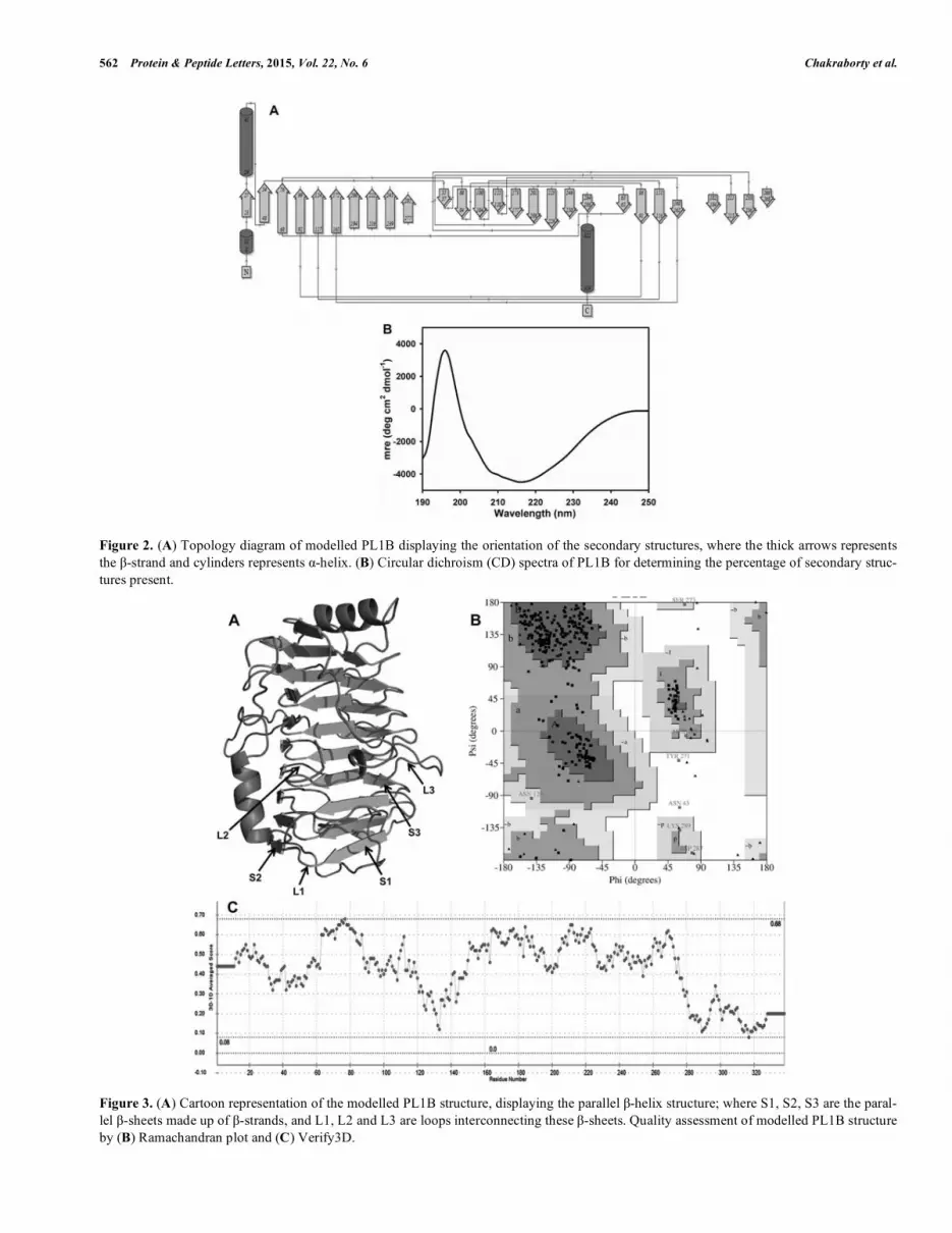
The predicted secondary structure of PL1B showed the presence of 2 �-helices (<5%) at the N- and C-terminals, 27 �-strands (>35%) and 29 random coils (>60%) (Fig. 2A) (Table. 2). The �-strands present throughout the structure forms the parallel �-sheets, which are quite common in the structures of pectate lyase and evident from the previous characterizations [7,9,10].
Secondary Structure Analysis of PL1B by Circular Di-chroism
The CD spectrum of PL1B was analyzed in the K2D3 server, where the dataset was compared with the available secondary structures of known proteins [47]. This revealed that PL1B contained only 2.06% �-helices (2 in number) 40.54% �-strands (27 in number), 57% random coils (29 in number) (Fig. 2B). This result is in accordance with the pre-dicted values of secondary structures determined earlier (Ta-ble. 2). We can also find that the modeled PL1B structure is rich in �-strands and random coils (Fig. 3A) as found in other pectate lyase C previously [48].
Modeled Structure and Quality Assessment of PL1B
The modeled structure of PL1B represented the conven-tional parallel �-helix structure (Fig. 3A). The basic struc-tural domains are parallel �-sheets, which fold into a right handed coiled structure. Three parallel �-sheets viz. S1, S2 and S3 are connected by random coils L1 (connecting S1 and S2), L2 (connecting S2 and S3) and L3 (connecting S3 and S1). This arrangement of S1, S2 and S3 �-sheets coils around the central core to form a final �-helix structure. The �-sheet S1 is composed of ten �-strands, while S2 and S3 are built from seven and nine �-strands, respectively. The L2 loop region contains an extended simple loop which is larger than that found in Bsp165PelA. The central core of the �-helix structure is made up of seven complete turns in the parallel �-sheets (Fig. 3A), as often found in other pectate lyase structures [9,14,28,49,50]. The quality of the modeled struc-
ture after energy minimization was assessed on the Saves server. Ramachandran plot developed by Procheck displayed that 82.8% residues were in the favoured region, 14.7% and 1.8% residues were in additional and generously allowed region, respectively, and only 0.7% residues were in the dis-allowed region (Fig. 3B). In verify 3D the 1D-3D plot showed that 80% of amino acid present in modeled PL1B structure has an overall score of � 0.2 (Fig. 3C). The cata-lytic core of PL1B was revealed after its superposition with Pectate lyase (Bsp165-PelA) from Bacillus sp. 165 [11] with a RMS deviation value of 0.492 Å (Fig. 4). The binding cleft of PL1B was located at the same position as previously iden-tified in Bsp165PelA. The binding cleft PL1B is formed by parts of L2, L3 loops and S3 �-sheet, which is enough deep to accommodate the ligand. The superposition of structures showed that the key residues Asp151, Arg209, Arg236 and Tyr271 of PL1B perfectly aligns with the key residues of Bsp165PelA (Fig. 4).
Molecular Dynamics Simulation of Modeled PL1B
Molecular dynamics simulation on modeled PL1B was performed for elucidating the overall structural stability and closeness of the secondary structures over a span of 5 ns. The results showed that significant changes in PL1B struc-ture in the initial 1.75 ns of simulation, whereas after 2 ns the fluctuations reduced and the overall deflection was less than 0.4 Å (Fig. 5). This suggested that the modeled PL1B struc-ture has a stable conformation. The difference in RMS devia-tion values between the energy minimized modeled structure of PL1B and the final structure developed after MD simula-tion was only 0.532 Å. Considering the results obtained after MD simulation it could be concluded that the modeled PL1B structure is stable and compact and was used for docking studies.
Docking Analysis and Ligand Binding Interaction
The docking analysis of PL1B with different ligands showed best binding affinity with tri-galacturonic acid (TGA) and the complex had the least binding energy of -6.99 Kcal/mol. Interaction of PL1B with all the docked ligands and their respective binding energies are listed in Table 3. In the PL1B-TGA complex structure, TGA was located within
Table. 1. BLAST analysis of PL1B.
Organism PDB ID Identity
(%)
Query coverage (%) E-value Total score
Bacillus sp. N165 3VMV_A 43 90 2e-63 206
Thermotoga maritima 3ZSC_A 33 92 1e-31 122
Xanthomonas campestris 2QX3_A 32 89 1e-31 121
Erwina chrysanthemi 1PLU_A 32 93 2e-28 113
Bacillus sp. TS-47 1VBL_A 35 44 3e-20 91.3
Bacillus subtilis 1GN8_A 33 44 1e-17 84.3
Erwina chrysanthemi 1JRG_A 30 62 4e-13 70.1

Structure of Endo-Pectate Lyase (PL1B) from Clostridium thermocellum Protein & Peptide Letters, 2015, Vol. 22, No. 6 561
Figure 1. Multiple sequence alignment of PL1B from Clostridium thermocellum with pectate lyases from Bacillus subtilis (Bs1GN8_A), Bacillus sp. TS-47 (Bs1VBL_A), Bacillus sp. 165 (Bs3VMV_A), Erwinia chrysanthemi (Ec1JRG_A and Ec1PLU_A), Thermotoga maritima (Tm3ZSC_A) and Xanthomonas campestris (Xc2QX3_A). The conserved residues are shown in black background and semi conserved resi-dues are shown in box. The secondary structure components (helices-�, strands-�, turns-T, and 310 helices-�) of PL1B are shown above the sequences. Conserved catalytic residues are marked as asterisks. The figure was developed by EsPript3.0 (http://espript.ibcp.fr/).
562 Protein & Peptide Letters, 2015, Vol. 22, No. 6 Chakraborty et al.
Figure 2. (A) Topology diagram of modelled PL1B displaying the orientation of the secondary structures, where the thick arrows represents the �-strand and cylinders represents �-helix. (B) Circular dichroism (CD) spectra of PL1B for determining the percentage of secondary struc-tures present.
Figure 3. (A) Cartoon representation of the modelled PL1B structure, displaying the parallel �-helix structure; where S1, S2, S3 are the paral-lel �-sheets made up of �-strands, and L1, L2 and L3 are loops interconnecting these �-sheets. Quality assessment of modelled PL1B structure by (B) Ramachandran plot and (C) Verify3D.

Structure of Endo-Pectate Lyase (PL1B) from Clostridium thermocellum Protein & Peptide Letters, 2015, Vol. 22, No. 6 563
Figure 4. Superposition of modelled PL1B (shown in black) structure with Bsp165PelA (shown in light-grey) from Bacillus sp., showing the binding cleft. Some residues from the binding cleft of PL1B are marked in black and those from Bsp165PelA are marked in light-grey.
Figure 5. Molecular dynamic (MD) simulation of modelled PL1B structure showed that the structure remained almost remains stable during the entire process of simulation.
Table. 2. Secondary structures present in PL1B structure.
Secondary structure element PsiPred (%) CD analysis (%)
�-helix <5 2.06
�-sheet >35 40.54
Random coil >60 57.4

564 Protein & Peptide Letters, 2015, Vol. 22, No. 6 Chakraborty et al.
Table. 3. Amino acid residues of PL1B showing polar interacting residues and residues around 4Å of the ligand at docking site, hy-drophobic residues are underlined.
Ligand Binding Energy (Kcal/mol) Polar Interactions Residues within 4Å
De-esterified pectin (DEPEC) -2.75
Arg209, Gly150, Asp151, Arg214,
Ser272, Asp257, Tyr271.
Asp257, Thr260, Arg236, Ser273, Ser186,
Gly150, Lys259, Ser258
D-Galacto methyl ester
(GALME) -3.42
Ser212, Arg236, Arg214, Arg209. Asn234, Ser186, Asp151, Leu183, Tyr271,
Tyr270, Ser212
Amino-galacturonic acid
(Amino-GAL) -4.74
Arg236, Arg214, Ser272, Ser212,
Arg209
Tyr271, Ser186, Asp151, Gly150, Leu183
D-Galacturonic acid (GAL) -4.59 Ser212, Arg209, Gly150 Leu183, Arg236, Tyr271, Asp151
Unsaturated Di-Galacturonic
acid (UnDiGAL) -5.34
Asn234, Ser212, Ser272, Arg214,
Arg209
Leu183, Tyr271, Tyr179, Asp151, Gly150,
Arg236
Tri-Galacturonic acid (TGA)
-6.99
Asp151, Arg236, Asn234, Tyr271,
Ser272, Arg209
Glu148, Gly149, Gly150, Tyr179, Leu183,
Arg214, Ser212, Ser272, Tyr270, Lys259,
Ser258, Asp257
the catalytic cleft formed by the extended loop of L2, parts of L3 loop and S3 �-sheet of the central core (Fig. 6A). Simi-lar binding groove was found in the structure of pectate lyase (Bsp165-PelA) from Bacillus sp. [11] having 43% sequence similarity with PL1B. The residues involved in polar interac-tion with TGA are Asp151, Arg209, Asn234, Arg236, Tyr271 and Ser272, showing interaction with the oxygen atoms in and around galacturonate moiety (Fig. 6B). In Bsp165-PelA from Bacillus sp. the interacting residues at the catalytic center were Thr121, Asp150, Lys177, Arg207 and Tyr269 [11]. Similarly, in PelC from Erwinia chrysanthemi the residues involved were Asp131, Asp170, Lys190, Arg218 and Tyr268 [15]. Comparing the residues of PL1B involved in the catalysis it was evident that Arg209 was in-volved in polar interactions with all the ligands, having the least distance as compared with other residues. Similarly, in PL1B-UnDiGAL complex the same residues of PL1B were found interacting with the oxygen atoms in and around the galacturonate moiety of UnDiGAL and Arg209 showed the nearest polar interaction (Fig. 6C). In all the other complexes almost similar residues of PL1B were found to interact with the ligand and Arg209 was found to be the nearest polar con-tact (Table 3). A close insight into the sequence alignment revealed that Arg209 in PL1B is conserved and it corre-sponds to Arg207 in Bsp165-PelA, Arg218 in PelC, Arg207 in pectate lyase II from Xanthomonas campestris [49] and Arg197 in pectate lyase from Thermotoga maritima [27]. Owing to the polar interaction and position of Arg209 in PL1B it is undoubtedly the residue which is involved in pro-ton abstraction during the catalysis by �-elimination mecha-nism. In the �-elimination mechanism of polysaccharide lyases the polygalacturonan chain is cleaved to produce a �4,5 unsaturated residues at the non-reducing end [6,51]. The important information revealed by the docking studies was the ligand specificity of PL1B. The binding energy of PL1B with GAL was -4.59 and with GALME it was -3.42, which indicated that PL1B binds more tightly to GAL as compared with GALME. GAL is the monomeric unit of po-lygalacturonic acid or pectic acid (non methyl-esterified), while GALME is the monomeric unit of pectin (methyl-
esterified) as also mentioned earlier in method section, there-fore, the docking analysis suggested that PL1B displays higher affinity against polygalacturonic acid or pectic acid as compared with methyl-esterified pectin. This binding pattern revealed that the modeled PL1B is a pectate lyase having higher affinity towards polygalacturonic acid.
Protein Melting Curve of PL1B
PL1B displayed a single melting peak starting at 60°C and ending at 74°C (Fig. 7). The presence of Ca2+ ions (0.6 mM) caused shifting of the peak (shown as dotted lines) to-wards higher temperature. The melting of PL1B in presence of Ca2+ ions started at 70°C and ended at 86°C (Fig. 7). This shifting of melting peak suggested that Ca2+ ions are impart-ing overall stability to the protein structure as also previously reported [43]. Hence, Ca2+ ions are important to PL1B, which not only enhances the activity [21], but also imparts stability to the protein structure.
Effect of Chaotropic Agents on the Structural Stability of PL1B
The structural stability of PL1B at pH 8.6 and 25°C was evaluated by increasing the concentration of chaotropic a-gents (GuHCl or Urea) from 0.2 to 8 M. It was observed that with the increase in the concentration of GuHCl or Urea the fluorescence peak intensity of PL1B decreased. Along with the lowering of the peak intensity, the shift in peak towards the larger wavelength was observed, which is commonly known as red shift. PL1B treated with 6M GuHCl displayed a red shift of the fluorescence peak maxima from 322 nm to 334 nm, resulting in a shift of 12 nm (Fig. 8A). PL1B on treatment with 4M urea displayed a red shift from 331 nm to 340 nm, resulting in a shift of 9 nm (Fig. 8B). The lowering of the fluorescence intensity and peak shift occurred as the protein loses it structural organization in the presence of chaotropic agent. The buried tryptophan residues in PL1B got exposed due to the loss of non-covalent interactions which stabilize the tertiary structure and thus result in the shift of the fluorescence peak. Similar transition curves were

Structure of Endo-Pectate Lyase (PL1B) from Clostridium thermocellum Protein & Peptide Letters, 2015, Vol. 22, No. 6 565
were reported for CtCBM35 of �-Mannanase from Clostrid-ium thermocellum [52]. The loss of structural integrity can be understood from the two-state transition curve of PL1B in presence of varying concentrations of GuHCl or Urea (Fig. 9A). Low unfolded fraction of PL1B was obtained until 1M and 0.75M concentration of GuHCl and urea respectively (Fig. 9A). The unfolded fractions of PL1B increased till 6M of GuHCl or urea, and then reached the saturation phase lea-ding to the final concentration of 8M. It was evident that PL1B was more stable in presence of GuHCl as compared to
Urea (Fig. 9A). The apparent free energy of PL1B varied maintaining a linear relationship with the concentrations of chaotropic agents. A two-step linearity was observed, one at a lower concentrations less than 1 M and another at higher concentrations from 2-6 M (Fig. 9B).
CONCLUSION
The three dimensional structure of endo-pectate lyase (PL1B) of family 1 polysaccharide lyase from Clostridium
Figure 6. (A) Surface view of the active site cleft of PL1B with TGA docked at the site (B) amino acid residues of PL1B interacting with TGA. The figure was generated in Pymol. 2D schematic representation of the ligand (C) TGA and (D) UnDiGAL interacting with the active side amino acid residues of PL1B. (- - -) represents the hydrogen bond with distances between them and the residues marked in spokes are within 4Å radius forming hydrophobic interaction with the ligand.

566 Protein & Peptide Letters, 2015, Vol. 22, No. 6 Chakraborty et al.
Figure 7. Protein-melting analysis displaying melting curve of PL1B ( ) and melting curve of PL1B in presence of 0.6 mM Ca2+ ions (- - -).
Figure 8. Tryptophan emission spectra of PL1B in presence of different concentration of chaotropic agents like (A) Guanidine hydrochloride (GuHCl) and (B) Urea.
Figure 9. (A) Fractions of PL1B unfolded as a function of different concentration of chaotropic agents. (B) Free energies (�Gapp) of different unfolded fractions of PL1B showing a linear relationship with the concentration of GuHCl and Urea. All the experiments were performed in triplicate and standard error was calculated from the mean of these three experiments

Structure of Endo-Pectate Lyase (PL1B) from Clostridium thermocellum Protein & Peptide Letters, 2015, Vol. 22, No. 6 567
thermocellum generated by comparative modeling was stable and compact. The secondary structure analyses of PL1B displayed presence of 3 �-helices (2.06%), 23 �-sheets (40.54%) and 26 random coils (57.4%). The 3-D modelled PL1B showed right handed parallel �-helix structure, where three parallel �-sheets were linked by coils. Key residues of PL1B involved during catalysis were Asp151, Arg209, Asn234, Arg236, Tyr271 and Ser272. Arg209 at the catalytic site was found to be responsible for proton abstraction during �-elimination. PL1B structure was stabilized in presence of Ca2+ ions, which was showed during the protein melting stu-dies. The effect of GuHCl or Urea on PL1B showed that the protein structure is stable and unfolds at their higher con-centrations. The most authentic validation of the predicted structure will be possible only after PL1B structure determination by X-ray crystallography. This work provided for the first time a closer insight into the structure of a cellulosomal pectinolytic enzyme from Clostridium sp.
CONFLICT OF INTEREST
The authors confirm that this article content has no conflicts of interest.
ACKNOWLEDGEMENTS
The authors thank Department of Biotechnology (DBT), Government of India for providing funds to develop Bioin-formatics infrastructure facility (BIF) at the Department of Biotechnology, IIT Guwahati. The authors acknowledge the help from Anil Kumar Verma during studies and analysis.
REFERENCES
[1] Henrissat, B.; Heffron, S.E.; Yoder, M.D.; Lietzke, S.E.; Jurnak, F. Functional implication of structure-based sequence alignment of proteins in the extracellular pectate lyase superfamily. Plant Physiol., 1996, 107, 963-976.
[2] Collmer, A.; and Keen, N.T. The role of pectic enzymes in plant pathogenesis. Annu. Rev. Phytopathol., 1986, 24, 383-409.
[3] Barras, F.; Van Gigsegem, F.; Chatterjee, A.K. Extracellular en-zymes and pathogenesis of soft-rot Erwinia. Annu. Rev. Phytopa-
thol., 1994, 32, 201-234. [4] Lojkowska, E.; Masclaux, C.; Boccara, M.; Robert-Baudouy, J.;
Hugouvieux-Cotte-Pattat, N. Characterization of the pelL gene en-coding a novel pectate lyase of Erwinia chrysanthemi 3937. Mol.
Microbiol., 1995, 16, 1183-1195. [5] Pissavin, C.; Robert-Baudouy, J.; Hugouvieux-Cotte-Pattat, N. J.
Regulation of pelZ, a gene of the pelB-pelC cluster encoding a new pectate lyase of Erwinia chrysanthemi. J. Bacteriol., 1996, 178(24), 7187-7196.
[6] Linhardt, R.J.; Galliher, P.M.; Cooney, C.L. Polysaccharide lyases. Appl. Biochem. Biotechnol., 1986, 12, 135-176.
[7] Yoder, M.D.; Keen, N.T.; Jurnak, F. New domain motif: structure of pectate lyase C, a secreted plant virulence factor. Science, 1993, 260, 1503-1507.
[8] Yoder, M.D.; Lietzke, S.E.; and Jurnak, F. Unusual structural fea-tures in the parallel �-helix in pectate lyases. Structure, 1993, 1, 241-251.
[9] Lietzke, S.E.; Keen, N.T.; Yoder, M.D.; Jurnak, F. The three-dimensional structure of pectate lyase E, a plant virulence factor from Erwinia citrysanthemi. Plant Physiol., 1994, 106, 849-862.
[10] Pickersgill R.; Jenkins J.; Harris G.; Nasser W.; Robert-Baudouy J. The structure of Bacillus subtilis pectate lyase in complex with cal-cium. Nat. Struct. Biol., 1994, 1, 717-723.
[11] Zheng, Y.; Huang, C.H.; Liu, W.; Ko, T.P.; Xue, Y.; Zhou, C.; Guo, R.T.; Ma. Y. Crystal structure and substrate-binding mode of a novel pectate lyase from alkaliphilic Bacillus sp. N16-5. Biochem.
Bioph. Res. Co., 2012, 420, 269-274.
[12] Kita, N.; Boyd, C.M.; Garrett, M.R.; Jurnak, F.; Keen, N.T. Differ-ential effect of sitedirected mutations in pelC on pectate lyase activ-ity, plant tissue maceration, and elicitor activity. J. Biol. Chem., 1996, 271, 26529-26535.
[13] Tardy, F.; Nasser, W.; Robert-Baudouy, J.; Hugouvieux-Cotte-Pattat, N. Comparative analysis of the five major Erwinia chrysan-themi pectate lyases: enzyme characteristics and potential inhibi-tors. J. Bacteriol., 1997, 179(8), 2503-2511.
[14] Yoder, M.D.; Jurnak, F. The refined three-dimensional structure of pectate lyase C from Erwinia chrysanthemi at 2.2 Angstrom resolu-tion (implications for an enzymatic mechanism). Plant Physiol., 1995, 107, 349-364.
[15] Herron, S.R.; Scavetta, R.D.; Garrett, M.; Legner, M.; Jurnak, F. Characterization and implications of Ca2+ binding to pectate lyase C. J. Biol. Chem., 2003, 278, 12271-12277.
[16] Scavetta, R.D.; Herron, S.R.; Hotchkiss, A.T.; Kita, N.; Keen, N.T.; Benen, J.A.; Kester, H.C.; Visser, J.; Jurnak, F. Structure of a plant cell wall fragment complexed to pectate lyase C. Plant Cell, 1999, 11, 1081-1092.
[17] Bayer, E.A.; Kening, R.; Lamed, R. Adherance of Clostridium thermocellum to cellulose. J. Bacteriol., 1983, 156(2), 818-827.
[18] Lamed, R.; Setter, E.; Bayer, E.A. Characterization of a cellulose-binding, cellulose-containing complex in Clostridium thermocel-
lum. J. Bacteriol., 1983, 156(2), 828-836. [19] Bayer, E.A.; Lamed, R.; White, B.A.; Flint, H.J. From cellulosomes
to cellulosomics. Chem. Record, 2008, 8, 364-377. [20] Tamaru, Y.; Doi, R.H. Pectate lyase A, an enzymatic subunit of the
Clostridium cellulovorans cellulosome. Proc. Natl. Acad. Sci. USA, 2001, 98, 4125-4129.
[21] Chakraborty, S.; Fernandes, V.O.; Dias, F.M.V.; Prates, J.A.M.; Ferreira, L.M.A.; Fontes, C.M.G.A.; Goyal, A.; Centeno, M.S.J. Role of pectinolytic enzymes identified in Clostridium thermocel-lum Cellulosome. Plos One, 2015, 10(2), e0116787.
[22] Altschul, S.F.; Gish, W.; Miller. W.; Myers, E.W.; Lipman, D.J. Basic local alignment search tool. J. Mol. Biol., 1990, 215(3), 403-410.
[23] Mount, D.W. Bioinformatics: Sequence and Genome Analysis, 2nd ed.; Cold Spring Harbor Press, 2004.
[24] Larkin, M.A.; Blackshields, G.; Brown, N.P.; Chenna, R.; McGetti-gan, P.A.; McWilliam, H.; Valentin, F.; Wallace, I.M.; Wilm. A.; Lopez, R.; Thompson, J.D.; Gibson, T.J.; Higgins, D.G. Clustal W and Clustal X version 2.0. Bioinformatics, 2007, 23, 2947-2948.
[25] Robert, X.; Gouet, P. Deciphering key features in protein structures with the new ENDscript server. Nucl. Acids Res., 2014, 42(W1), W320-W324.
[26] Eswar, N.; Marti-Renom, M.A.; Webb, B.; Madhusudhan, M.S.; Eramian, D.; Shen, M.; Pieper, U.; Sali. A. Comparative Protein Structure Modeling With MODELLER. Curr. Protoc. Bioinform., 2006, 15, 5.6.1-5.6.30.
[27] Yoder, M.D.; Jurnak, F. Protein motifs. 3. The parallel beta helix and other coiled folds. FASEB J., 1995, 9, 335-342.
[28] Kluskens, L.D.; Van Alebeek, G.W.M.; Voragen, A.G.J.; De Vos W.M.; Van Der Oost, J. Molecular and biochemical characteriza-tion of the thermoactive family 1 pectate lyase from the hyperther-mophilic bacterium Thermotoga maritime. Biochem. J., 2003, 370, 651-659.
[29] Shen, M.Y.; Sali, A. Statistical potential for assessment and predic-tion of protein structures. Protein Sci., 2006, 15, 2507-2524.
[30] John, B.; Sali, A. Comparative protein structure modeling by itera-tive alignment, model building and model assessment. Nucl. Acids Res., 2003, 31, 3982-3992.
[31] Melo, F.; Sanchez, R.; Sali, A. Statistical potentials for fold assess-ment. Protein Sci., 2002, 11, 430-448.
[32] Krieger, E.; Joo, K.; Lee, J.; Raman, S.; Thompson, J.; Tyka, M.; Baker, D.; and Karplus, K. Improving physical realism, stereo-chemistry, and side-chain accuracy in homology modeling: Four approaches that performed well in CASP8. Proteins, 2009, 77, 114-122.
[33] Berendsen, H.J.C.; van der Spoel, D.; van Drunen, R. GROMACS: A message-passing parallel molecular dynamics implementation. Comput. Phys. Commun., 1995, 91(1-3), 43-56.
[34] Pronk, S.; Pall, S.; Schulz, R.; Larsson, P.; Bjelkmar, R.; Apos-tolov, R.; Shirts, M.R.; Smith, J.C.; Kasson, P.M.; van der Spoel, D.; Hess, B.; Lindahl, E. GROMACS 4.5: a high-throughput and highly parallel open source molecular simulation toolkit. Bioinfor-
matics, 2013, 29(7), 845-854.

568 Protein & Peptide Letters, 2015, Vol. 22, No. 6 Chakraborty et al.
[35] Hess, B.; Bekker, H.; Berendsen, H.J.C.; Fraaije, J.G.M. LINCS: A linear constraint solver for molecular simulations. J. Comput. Chem., 1997, 18(12), 1463-1472.
[36] Morris, G.M.; Huey, R.; Lindstrom, W.; Sanner, M.F.; Belew, R.K.; Goodsell, D.S.; Olson, A.J. Autodock4 and AutoDockTools4: automated docking with selective receptor flexiblity. J. Comput. Chem., 2009, 16, 2785-91.
[37] O’Boyle, N.M.; Banck, M.; James, C.A.; Morley, C.; Vander-meersch, T.; Hutchison, G.R. Open Babel: An open chemical tool-box. J. Cheminformatics, 2011, 3, 33.
[38] O’Neill M.; Albersheim P.; Darvill A. The pectic polysaccharides of primary cell walls; In: Methods in Plant Biochemistry; Academic Press: London, 1990; Vol. 2, pp. 415-441.
[39] Moreau M.; Chaby R.; Szabo L. Isolation of a trisaccharide contain-ing 2-amino-2-deoxy-D-galacturonic acid from the Bordetella per-tussis endotoxin. J. Bacteriol., 1982, 150(1), 27-35.
[40] Yoshida K.; Ohtomo T.; Suganuma M. Isolation of a serologically different compact-colony-forming active substance from strains of Staphylococcus aureus. Microbiol. Immunol., 1990, 34(10), 801-8.
[41] Guex, N.; Peitsch, M.C. SWISS-MODEL and the Swiss-PdbViewer: An environment for comparative protein modeling. Electrophoresis, 1997, 18, 2714-2723.
[42] Kelly, S.M.; Jess, T.J.; Price N.C. How to study proteins by circular dichroism (Review). Biochim. Biophys. Acta, 2005, 1751, 119-139.
[43] Dvortsov, I.A.; Lunina, N.A.; Chekanovskaya, L.A.; Schwarz, W.H.; Zverlov, V.V.; Velikodvorskaya, G.A. Carbohydrate-binding properties of a separately folding protein module from beta-1,3-glucanase Lic16A of Clostridium thermocellum. Microbiology, 2009, 155, 2442-2449.
[44] Ahmad, F.; Yadav, S.; Taneja, S. Determining stability of protein from guanidinium chloride transition curve. Biochem. J., 1992, 287, 481-485.
[45] Thomas, L.M.; Doan, C.N.; Oliver, R.L.; Yoder, M.D. Structure of pectate lyase A: comparison to other isoforms. Acta Cryst., 2002, D58, 1008-1015.
[46] Takao, M.; Nakaniwa, T.; Yoshikawa, K.; Terashita, T.; Sakai. T. Molecular cloning, DNA sequence, and expression of the gene en-coding for thermostable pectate lyase of thermophilic Bacillus sp. TS 47. Biosci. Biotechnol. Biochem., 2001, 65, 322-329.
[47] Jeune, C.L.; Navarro, M.A.A.; Iratxeta, C.P. Prediction of protein secondary structure from circular dichroism using theoretically de-rived spectra. Proteins: Struct. Funct. Bioinf., 2012, 80, 374-381.
[48] Kamen, D.E.; Woody, R.W. A partially folded intermediate con-formation is induced in pectate lyase C by the addition of 8-anilino-1-naphthalenesulfonate (ANS). Protein Sc., 2001, 10, 2123-2130.
[49] Xiao, Z.; Bergeron, H.; Grosse, S.; Beauchemin, M.; Garron, M.L.; Shaya, D.; Sulea, T.; Cygler, M.; Lau, P.C. Improvement of the thermostability and activity of a pectate lyase by single amino acid substitutions, using a strategy based on melting-temperature-guided sequence alignment. Appl. Environ. Microbiol., 2008, 74, 1183-1189.
[50] Czerwinski, E.W.; Midoro-Horiuti, T.; White, M.A.; Brooks, E.G.; Goldblum, R.M. Crystal structure of Jun a 1, the major cedar pollen allergen from Juniperus ashei, reveals a parallel beta-helical core. J. Biol. Chem., 2005, 280, 3740-3746.
[51] Davis, G.; Henrissat, B. Structures and mechanisms of glycosyl hydrolases. Structure, 1995, 3, 853-859.
[52] Ghosh, A.; Verma, A.K.; Gautam, S.; Gupta, M.N.; Goyal, A. Structure and Functional Investigation of Ligand Binding by a Fam-ily 35 Carbohydrate Binding Module (CtCBM35) of �-Mannanase of family 26 glycoside hydrolase from Clostridium thermocellum. Biochem. (Moscow), 2014, 79(7), 672-86.
Received: January 14, 2015 Revised: May 3, 2015 Accepted: May 3, 2015


