Embed Size (px)
Citation preview

Inflammation, Vol. 6, No. l, 1982
S T U D I E S O N G L Y C O G E N - I N D U C E D
I N F L A M M A T I O N O F M I C E
D y n a m i c s o f I n f l a m m a t o r y Responses a n d In f luence
o f A n t i i n f l a m m a t o r y D r u g s and P r o t e a s e Inh ib i to r s
TATSUHISA YAMASHITA,' YOSHIO ISHIBASHI, 1 ISAO NAGAOKA, 1 KEIKO KASUYA, 1 KAORI MASUDA, 1
HARUAKI WARABI, ~ and YUICHI SHIOKAWA 2
Departments of ~ Physiological Chemistry and 2Internal Medicine School of Medicine, Juntendo University Hongo, Bunkyo-ku, Tokyo 113, Japan
Abstract--The intraperitoneal injection of glycogen in the mouse resulted, shortly thereafter, in the accumulation of 14-23 million neutrophils in the peri- toneal cavity and a four-fold increase in the numbers of circulating neutrophils. Preceding the influx of leukocytes, the exudation of plasma proteins and the chemotactic activity for mouse neutrophil in vitro increased in the peritoneal fluid. Among various protease inhibitors examined, chymostatin alone sup- pressed the plasma protein exudation. Indomethaein and dexamethasone re- duced the accumulation of white cells and protein exudation. These nonsteroidal and steroidal antiinflammatory drugs were equally effective whether given si- multaneously with or 60 rain before glycogen or whether administered intra- peritoneally or orally. Colchicine showed a suppressive effect on the leukocyte accumulation but enhanced the protein exudation.
INTRODUCTION
Various models have been used for analyzing mechanisms of inflammation because one model cannot express the whole inflammatory process of dis- eases. Glycogen has long been known to induce sterile inflammatory exu- dates when administered in the peritoneal cavities of a number of animal species (1, 2). Indeed, this technique has commonly been used to obtain polymorphonuclear neutrophils (PMNs). We also have been studying the in vitro function of PMNs using cells from glycogen-induced peritoneal exudates (3-7). However, there is little study on the mechanism of the in- flammatory process of glycogen peritonitis (8). Therefore, we took an in-
87
0360-3997/82/0300-0087503.00/0 �9 1982 Plenum Publishing Corporation

88 Yamashita et al.
terest in the analysis of the mechanism of the inflammatory process induced by glycogen.
The present work was undertaken to obtain information related to mobilization of neutrophils, in particular the dynamics of changes both in white cell populations in the circulation and peritoneal cavity and in the plasma exudation, and the actions of protease inhibitors and antiinflam- matory drugs on these inflammatory responses.
MATERIALS AND METHODS
Male ddy mice (about 6 weeks old), weighing approximately 25-30 g, were used. Glyco- gen (1.0 ml of a 15% solution in 0.9% NaC1) was injected directly into the peritoneal cavity through a No. 26-gauge �89 needle. At indicated times, mice were killed by rapid dislocation of the neck or with ether. After lifting the skin upwards to make a transverse incision, 5.0 ml of 0.4% sodium citrate in 0.85% NaCI solution was injected into the peritoneal cavity through a No. 23-gauge 1-inch needle and circulated by shaking. Then the peritoneal fluid was removed by inserting a needle into the flank and applying lateral traction. Aspirated fluid was trans- ferred to a graduated cylinder and its volume was determined. Peritoneal aspirates were centrifuged at 100g for 10 min at 4~ Sedimented cells were suspended in 1-5 ml of phos- phate-buffered saline containing 1 mM MgSO4 and 0.07 mM CaC12 (PBS) and counted in a hemocytometer chamber using white cell diluting fluid (Tiirk solution) and a white cell pipet at a dilution of 1 : 10. The white cell populationin peritoneal fluid was examined by light-micro- scopic scanning of Wright-Giemsa-stained smears of cell suspensions. Supernatant was used for measurement of contents of protein leaked into and glycogen remaining in the peritoneal cavity and also for the occurrence of chemotactic activity in the peritoneal fluids. On the other hand, blood (0.4 ml) was collected by cardiac puncture into syringes containing 0.04 ml of 1.5% EDTA in PBS. The number and population of white cells in blood were evaluated using a hemocytomcter chamber and smears stained with Wright-Giemsa, respectively, as in the peritoneal exudate.
Antiinflammatory drugs and protease inhibitors (1.0 ml each) were intraperitoneally ad- ministered following the intraperitoneal injection of glycogen. Indomethacin and dexamctha- sone were also administered oraUy 2-3 rain before the glycogen injection. Indomethacin and TPCK were dissolved first in dimethylsulfoxide and then diluted with phosphate-buffered saline without divalent cations [PBS(-)] to be 10% at a final concentration of DMSO. Dexametha- sone was dissolved first in Tween 80 and then diluted with PBS(-) to be 1% at a final concentra- tion of Tween 80. Other drugs were dissolved in PBS(-).
Protein concentrations were determined by a modification of the Biuret method of Weich- selbaum (9), using 0.3 ml supernatant of peritoneal exudates and bovine serum albumin as a standard.
Glycogen was measured by incubating 2.0 ml diluted sample with 4.0 ml of 0.2% anthrone in 95% H2SO4 at 100~ for 4' min and then immersing tubes in running water to stop the reac- tion as described previously (10), after 100- to 200-fold dilution (first 1-2 h) of supernatant with 0.4% citrate-0.85% NaC1.
The chemotactic activity in fluids was assayed by employing a modified Boyden chamber with Sartorius membrane filter (pore size 3 pro) containing 2 ml of supernatant in the lower compartment and 1 ml of mouse PMN suspension (5 • 104 cells/ml) in the upper compartment and by measuring the distance from the top of the triter to the farthest two cells at the same focal

Glycogen-Induced Inflammation 89
plane as described previously (3). Mouse PMNs were harvested 4 h after the intraperitoneal injection of 15% glycogen. After removal of contaminating red blood cells by a bypotonic treat- ment with 0.2% saline, cells were washed once with PBS(-) and suspended in PBS (or 0.4% citrate-0.85% NaCI).
Glycogen (type II, from oyster), indomethacin, dexamethasone, N-a-p-tosyl-L-lysine chloromethyl ketone (TLCK), L-l-tosyl-amido-2-phenylethyl-chloromethyl ketone (TPCK), pepstatin A, leupeptin (hemisulfate), ehymostatin, and soybean trypsin inhibitor were pur- chased from Sigma Chemical Company'(St. Louis, Missouri); aprotinin [trasylol, 50,000 KIU; kallikrein inhibitor unit)/5 ml ampule] from Farbenfabriken Bayer A.G. (Leverkusen, F.R.G.); diphenhydramine hydrochloride and iodoacetamide from Tokyo Kasei Kogyo Co. Ltd. (Japan); colchicine, cryst., from Wako Pure Chemical Industries Ltd. (Japan); dimethylsul- foxide (DMSO, analytical reagent grade) from Kokusan Chemical Works, Ltd. (Japan); Tween 80 from Iwai Kagaku Co., Ltd. (Japan); anthrone from Wako Pure Chemical Industries Ltd. (Japan); Sartorius membrane filter (type SM 11302, pore size 3.0 Urn)from Sartorius-Mem- branfilter GmbH (GiJttingen, F.R.G.)
R E S U L T S A N D D I S C U S S I O N
Effective Dose of Glycogen Producing Inflammatory Response and Time Course of Disappearance of Glycogen Administered. The concen- t r a t i on o f g lycogen sufficient to p r o d u c e the i n f l a m m a t o r y response was p re l imina r i ly inves t iga ted by examin ing the number and cell types o f l euko- cytes accumula t ed in the pe r i tonea l cavi ty 4 h af ter the admin i s t r a t i on o f va r ious concen t ra t ions o f g lycogen (Table 1). The in t r ape r i tonea l in ject ion o f 15% glycogen so lu t ion p r o d u c e d the mos t m a r k e d acute i n f l a m m a t o r y reac t ion which was charac te r i zed by an accumula t i on of neut rophi l s . Therefore , 15% glycogen so lu t ion was used as an i n f l a m m a t o r y s t imulan t in the fo l lowing exper iments . F igu re 1 shows the d i s appea rance o f glyco- gen in t r ape r i t onea l ly admin i s t e red f rom the pe r i tonea l cavity. As can be seen in the figure, g lycogen d i s appea red r ap id ly and was ha rd ly de tec ted in the pe r i t onea l f luid 6 h af ter admin i s t r a t ion .
Table 1. Effective Dose of Glycogen Producing Inflammatory ResponsC
Population (%) Glycogen Leukocytes
concentration (%) (• 10 6) Neutrophil Lymphocyte Macrophage
0.12 3.0 - 1.0 33 + 6 7 + 3 61 __+ 4 1 3.2 4- 1.0 57 4- 10 6 4- 2 38 __+ 9 3 4.7 + 1.8 64__+ 5 7 4- 2 31 4- 6 5 5.0 + 1.7 65 + 7 7 4- 3 29____. 6
10 7.0 + 1.3 76 + 7 3 + 1 23 _____ 6 15 16.4 4- 1.1 86 __+ 1 2 4- 1 14 + 3
~ represent the mean +SE of data from three mice.

90 Yamashita et al.
150
100
~, 50 (.9
\ 0 1 2 4 6 8
T ime
r"i .pj
16 42
( hrs )
Fig. 1. Disappearance of glycogen from the peritoneal cavity of mice after glycogen admin- istration. Values represent the mean + SE of data from six mice.
Kinetics o f Inflammatory Cell Accumulation in Mice in Response to Glycogen. At various times after the intraperitoneal injection of glycogen, the total number and types of cells which had accumulated in the individual peritoneal cavities were quantified (Figure 2). There was no significant in- crease, rather a slight decrease, in the total number of accumulated leuko- cytes at l h after injection of glycogen. Thereafter the total cell accumula- tion markedly increased, reached a maximum at 8 h, and then slowly began decreasing over the next 34 h. Neutrophils appeared in increasing num- bers without a decrease at 1 h after glycogen administration. The maximum neutrophil response occurred between 6 and 8 h. After this time, the num- ber of neutrophils declined gradually. After a marked decrease in numbers at 1 h after injection of glycogen into the peritoneal cavity, macrophages increased slowly, reached a maximum at 8 h, and remained at the same level for up to 42 h. On the other hand, there was no significant increase in the total number of lymphocytes recovered over the time period examined.
Leukocytes in blood increased from 4.4 _+ 0.6 (mean _+ SE) to 6.0 + 1.0 million/ml at 2 h after the intraperitoneal administration of glycogen, and the percentage of total leukocytes that were neutrophils increased from 13.3 _+ 2.4 to 40.2 _+ 5.1 (n = 6). This was equivalent to an increase from 0.6 million neutrophils per milliliter to 2.3 million per milliliter. The time course of these changes is illustrated in Figure 3, in which the number of neutro- phils reached a maximum at 2 h, then very slowly declined, and returned

Glycogen-Induced Inflammation 91
30
2o !
0 1 2 6 8 16 42
T ime ( hrs )
Fig. 2. Time course of accumulation of leukocytes after the intraperitoneal injection of gly- cogen in mice. Total numbers of leukocytes (D), neutrophils (e), monocytes (A), and lympho- cytes (<3) in the peritoneal cavity at the indicated time after glycogen injection are recorded. Values represent the mean + SE of data from six mice.
loo
o
o 50
7.0
6.0
5.0
4.0
3.0
Z
ft .
. ; r
0 1 2 4 6 8 16 42
T ime ( hrs )
Fig. 3. Changes in blood leukocyte populations after the intraperitoneal injection of glycogen in mice. Percentage of leukocytes as neutrophils (I), monocytes (A), lymphocytes (O), and the number of leukocytes per ml (El) in blood are recorded as mean + SE of data from six mice.

92 Yamashita et al.
to the normal value at 16 h. Other white cells (principally lymphocytes) showed a corresponding decline in percentage and then an increase as the number of neutrophils diminished. An increase in the number of blood neutrophils was evident at the time points before the obvious influx of neu- trophils into the peritoneal cavity.
The accumulation of neutrophils at the inflammatory site is supposed to occur based upon some chemotactic factors produced by inflammatory stimuli. Thus the fluids were tested in vitro for the presence of chemotactic activity for neutrophil. As seen in Figure 4, before the influx of neutrophils the chemotactic activity appeared in the peritoneal fluid and was specific for mouse but not guinea pig neutrophils. Namely the chemotactic activity reached a maximum within 1 h after glycogen injection and then decreased slowly.
Figure 5 shows the time course of the alteration of the protein content in the peritoneal fluid after glycogen administration. The protein content markedly increased between l and 2 h and then declined until 8 h. After 8 h, the decreasc was very slow, and the protein content reached the normal level at 42 h. The vascular permeability tests, measured by spectrophoto-
6.0
4.0
._u 3.0
1.0 . . . . . , // ,
0 1 2 4 6 8 16 42
Time ( hrs )
Fig. 4. Chemotactie activity for mouse PMNs in the peritoneal fluids of mice injected with glycogen. Mice were injected intraperitonea]ly with glycogen and the peritoneal fluids were removed at varying times thereafter and centrifuged. Chemotaxis and chemokincsis were as- saycd by incubating mouse PMNs at 37~ for 90 min in the presence of the supernatant of peritoneal fluids at lower compartment and at both upper and lower compartments of the chamber, respectively. Random movement was assayed in the presence of 0.4% citrateS.85% NaCI or PBS at the bottom of the chamber. Chemotactic or chernokinetic activity is expressed as /~m of chemotaxis or chemokincsis//~m of random movement. The chemotactic activity ((9) and chemokinetic activity (~) are recorded as mean -+ SE of data from six mice.

Glycogen-Induced Inflammation 93
20
, i r , 4 i 1 ~j
0 1 2 /* 6 8 16 L,2
Time ( hrs )
Fig. 5. Time course of the alteration of the protein content in the peritoneal fluid of mice injected with glycogen. Values represent the mean + SE of data from six mice.
metric estimation at 590 nm of plasma-bound dye (11) (Potamine sky blue; intravenous injection of 0.1 ml of a 4% solution in pBS 30 min before gly- cogen injection) which had leaked into the peritoneal cavity after glycogen administration, demonstrated a time-course pattern similar to that of the protein content. This suggests that an increase in protein content in the peritoneal fluids is the consequence of the leakage of plasma Components into the peritoneal cavity due to the increased vascular permeability. SDS- polyacrylamide gel electrophoreogram showed that peritoneal fluids had the same electrophoretic pattern as that of plasma with the same compo- nent ratio, which supports our data.
Influence of Protease Inhibitors and Antiinflammatory Drugs on In- flammatory Response Measured 6 h after Administration of Glycogen, It is reported that proteases participate in the process of inflammation (12- 14). Hence, various protease inhibitors were administered intraperito- neally with glycogen, and the accumulation of leukocytes and the vascu- lar permeability based on protein contents in the peritoneal fluid were examined after 6 h. The influence of alkylating agents such as TPCK, TLCK, and iodoacetamide on glycogen-induced inflammation was studied first (Figure 6). TPCK, a specific inhibitor for chymotrypsin, had no effect on accumulation of leukocytes or vascular permeability. TLCK, a specific in- hibitor for trypsin and iodoacetamide, an inhibitor for SH-protease, did not affect leukocyte mobilization but significantly accelerated the vas- cular permeability. Such animals were too weak to move and often died. Therefore, these reagents seem to cause widespread tissue damage. Natu- rally occurring macromolecular inhibitors (aprotinin and soybean trypsin

94
5O T
Permeability (mg protein) 40 30 20 10
.I
i
i i |
Vehicle (PBS) n= 8
Monoiodoaceta mide (lo pmoles/mouse ) n = 7
TLCK n=7 ( s ~Jmoleslmouse)
TPCK n=5 (50 u moles/mouse )
Yamashita et aL
Accumulation(x106 cells) 0 20 40 60
i i i
- - - 1
- - - 1 ,
** P<O.01
Fig. 6. Effect of alkylating agents on glycogen-induced inflammation in mice. Error bars indicate + SE. P values refer to significance of difference from control.
inhibitor) improved neither infiltration of lcukocytes nor leakage of plasma proteins (Figure 7). Of the low-molecular-weight protease inhibitors pro- duced by microorganisms, chymostatin, a chymotrypsin-like protease in- hibitor, showed a suppressive effect on increased vascular permeability, although leukocyte mobilization was not affected, suggesting that some protease participates in the process of the inflammation induced by glycogen.
P e r m e a b i l i t y (mg protein) 20 10 0
i I
H
H
H
H
Vehicle(PBS) n=2'7
P e p s t a t i n n = ? (100 pg Imou~)
Leupeptin n= 7 (tOO pg tmouse)
C h y m o s t a t i n n=7 ( i o o ~ l m o u s e )
T r a s y l o l n=6 ( 10~ KlUlmOuse)
5O,/ bean t r y p s i n i n h i b i t o r n=6 { 100 pglmOuse)
Accumulation (x 106celts) 0 20 40 60
! i |
I I
I i
I i
H
I I
*P< 0.05
Fig. 7. Effect of some protease inhibitors on glycogen-induced inflammation in mice. For other details, see the legend to Figure 6.

Glycogen-Induced Inflammation 95
Since the involvement of histamine and prostaglandins in the inflam- matory process is generally recognized, the suppressive effects of their in- hibitors on glycogen-induced inflammation were examined. Among the antiinflammatory agents examined, diphenhydramine, an antihistaminic agent, was not effective in the accumulation of leukocytes and increased vascular permeability (Figure 8). This suggests that histamine has a very limited role in our experimental situations, if it is involved at all. On the other hand, indomethacin and dexamethasone, prostaglandin-synthesis inhibitors, effectively suppressed not only plasma exudation but also cell infiltration, although their suppressive effect was more significant on plasma exudation. The simultaneous administration with glycogen seemed to be more effective than the administration of antiinflammatory drugs 1 h be- fore glycogen (data not shown). Oral administration of these agents showed the same suppressive effect as intraperitoneal administration (Figure 9). It is now recognized that arachidonic acid is released from cell membrane phospholipids by the activation of a membrane-bound enzyme, phospho- lipase A2, by a disturbance of the cell membrane and metabolized either by cyclooxygenase to produce prostaglandins and thromboxanes or by lipo- oxygenase to produce mono-, di-, and trihydroxyeicosatetraenoic acids (HETEs). Among these arachidonate metabolites, prostaglandins of the E series are said to potentiate the permeability-increasing effects of mediators such as bradykinin, histamine, and 5-hydroxytryptamine (15-19), whereas 5-HETE and 5, 12-di-HETE (leukotriene B4) have recently been reported to stimulate chemotaxis as well as chemokinesis of PMNs in vitro and also
Permeability (rag prot~irO 20 10 0 i !
..I-- I
Vehicle (PBS) n=27
Diphenhydramine n= 5 ( 500 p g l m o u s e )
Indomethacin n= 6 ( 500 pg/mouse)
Dexamethasone n=l~, ( 50 pg lmouse)
Accumulation (x106 cells) 0 20 40 60
I | I
] =
- - - ] j
H"
* P<0,05 ** P<0.01
Fig. 8. Effect of the intraperitoneal administration of antiinflammatory drugs on glycogen- induced inflammation in mice. For other details, see the legend to Figure 6.

96
Perrneability (rng protein) 20 10
! I
-C
Ve hicle(l%Tween 6O) n = 6
[ndomethacin n=6 (500 pgl mouse)
Dexamethasone n =5 ( 50 pg i rnouse)
Yamashita et al.
Accumulation (x106 ceils) 0 20 40 6O
i i i
I I*
*P<O.05 ..P<O.OI
Fig. 9. Effect of the oral administration of indomethacin and dexamethasone on glycogen- induced inflammation in mice. For other detai~, see the legend to Figure 6.
to elicit leukocyte migration in vivo (20-26). Nonsteroid antiinflamma- tory drugs, such as aspirin and indomethacin, prevent prostaglandin gen- eration by directly inhibiting the cyclooxygenase enzyme responsible for prostaglandin biosynthesis (27-35). Antiinflammatory steroids reduce prostaglandin synthesis by interfering indirectly with the phospholipase(s) which release the polyunsaturated fatty acid precursors for both cyclooxy- genase and lipooxygenase pathways (36-49). These observations suggest that the reduction by indomethacin and dexamethasone of the increased plasma exudation in our glycogen-induced inflammation would result from inhibition of the prostaglandin biosynthesis by interfering with cyclo- oxygenase and phospholipase A2, respectively. This decrease of the leakage of plasma would produce the decrease in the generation of chemotactie agents such as C5a and C3a at inflammatory areas, resulting in a reduction of the accumulation of leukocytes in the peritoneal cavity. Another possi- bility is the decreased formation by these drugs of chemotactic agents such as mono-HETEs and leukotriene B4 because especially leukotriene B4 proved to affect PMN migration with responses comparable with f-Met- Leu-Phe and the complement peptide C5a (23-25). The action of dexa- methasone, which inhibits phospholipase A2 activity, to reduce the release of the fatty acid substrates required for biosynthesis of lipooxygenase me- tabolites such as mono-HETEs and leukotrienes can be explained by this hypothesis, but the inhibitory action of indomethacin on leukocyte ac- cumulation cannot be explained. Because indomethacin has no antilipo- oxygenase activity, the generation of mono-HETEs and leukotrienes would hardly be inhibited. The third possible explanation of the suppression of leukocyte accumulation is a direct effect of these antiinflammatory agents
Glycogen-Induced Inflammation 97
on PMN migration. Borel found no inhibition by nontoxic levels of dexa- methasone and indomethacin of rabbit neutrophil chemotaxis (50). Indo- methacin was found to have no significant effect on in vitro rabbit and rat neutrophil chemotaxis and had a variable effect on human neutrophil chemotaxis (51). These findings suggest that the antiinflammatory activity of these agents may possibly not entail a direct effect on neutrophil migra- tion. Recently, however, Malmsten et al. have demonstrated the dose- dependent inhibition by indomethacin of f-Met-Leu-Phe stimulated migration at higher concentrations and a significant enhancement of mi- gration at a low concentration (22). Therefore, the migration of blood white cells from these antiinflammatory drug-treated mice remained dissolved.
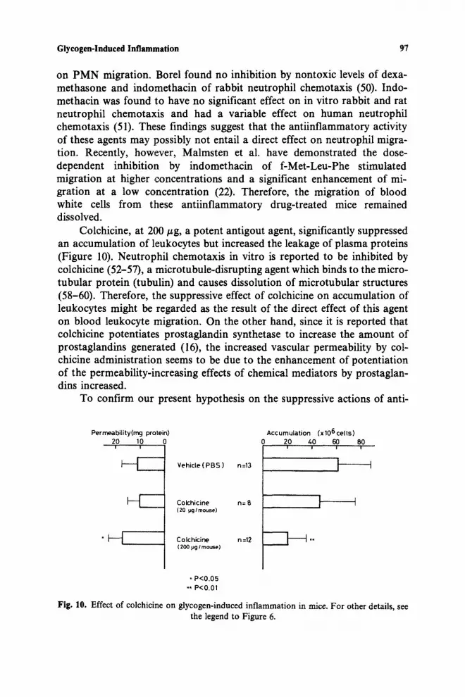
Colchicine, at 200 #g, a potent antigout agent, significantly suppressed an accumulation of leukocytes but increased the leakage of plasma proteins (Figure 10). Neutrophil chemotaxis in vitro is reported to he inhibited by colchicine (52-57), a microtubule-disrupting agent which binds to the micro- tubular protein (tubulin) and causes dissolution of microtubular structures (58-60). Therefore, the suppressive effect of colchicine on accumulation of leukocytes might be regarded as the result of the direct effect of this agent on blood leukocyte migration. On the other hand, since it is reported that colchicine potentiates prostaglandin synthetase to increase the amount of prostaglandins generated (16), the increased vascular permeability by col- chicine administration seems to be due to the enhancement of potentiation of the permeability-increasing effects of chemical mediators by prostaglan- dins increased.
To confirm our present hypothesis on the suppressive actions of anti-
Per rneabilit y (rag protein) 20 10 0 i i
I V - - Vehicle ( PBS ) n=13
Colchicine n= 8 (20 ~g / rnouse)
Colchic ine ( 200 pg / mouse)
n =12
Accumula t ion (x106 ce l l s ) 0 20 40 60 80
= i i !
1 i
I m
* P<O.05
** P<O.01
Fig. 1O. Effect of colchicine on glycogen-induced inflammation in mice. For other details, see the legend to Figure 6.

98 Yamashita et al.
inflammatory drugs on glycogen-induced inflammation, cellular and bio- chemical studies remain to be performed on leukocytes and peritoneal fluids from antiinflammatory-drug-treated mice.
Acknowledgments--This work was supported in part by grants for Project Research (No. 8102) from Juntendo University and for Scientific Research from Takeda Science Foundation, awarded to T. Yamashita.
REFERENCES
1. HIRSCH, J. G., and A. B. CHURCH. 1960. Studies of phagocytosis of group A streptococci by polymorphonuclear leukocytes in vitro. J. Exp. Med. 111:309-322.
2. HIRSCH, J .G. 1960. Further studies on preparation and properties of phagocytin. J. Exp. Med. 111:323:337.
3. YAMASHITA, T., N. IMAIZUMI, and S. YUASA. 1979. Effect of endocellular cryoprotectant upon polymorphonuclear neutrophil function during storage at low temperature. Cryo- biology 16:112-117.
4. YAMASHITA, T., K. TAKAMORI, and Y. TANAKA. 1979. A possible preferential inhibition of chemotaxis of polymorphonuclear neutrophils by a chemical modification. Experientia 35:1345-1347.
5. YAMASHITA, T., Y. TANAKA, and H. MATSUZAWA. 1980. Cytoplasmic and plasma mem- brane adenosine triphosphatase of polymorphonuclear neutrophils. Comparison of their enzymatic properties and attempt for a direct determination of myosin ATPase activity using polymorphonuclear neutrophil extract. Biochim. Biophys. Acta 599:246-253.
6. ISHIBASHI, Y., and T. YAMASHITA. 1981. Generation of a phagocytosis-stimulating factor by polymorphonuclear neutrophils during phagocytosis. Int. Arch. Allergy Appl. lm- munol. 64:181-189.
7. NAGAOKA, I., and T. YAMASHITA. 1981. Inactivation of phagocytosis-stimulating activity of tuftsin by polymorphonuelear neutrophils. A possible role of leueine aminopeptidase as an ecto-enzyme. Biochim. Biophys. Acta 675:85-=93.
8. SNYDERMAN, R., J .K. PHILLIPS, and S.E. MERGENHAGEN. 1971. Biological activity of complement in vivo. Role of C5 in the accumulation of polymorphonuclear leukocytes in inflammatory exudates. J. Exp. Med. 134:1131-1143.
9. WEICHSELBAUM, T. E. 1946. Accurate and rapid method for the determination of proteins in small amounts of blood serum and plasma. Am. d. Clin. PathoL, Tech. Sect. 10:40-49.
10. YAMASHITA, T. 1959. Studies on muscle glycogen (I). About the fractions of muscle glyco- gen of frog. Seikagaku 31:300-309 (in Japanese).
11. WHITTLE, B.A. 1964. The use of changes in capillary permeability in mice to distinguish between narcotic and nonnarcotic analgesics. Hr. J. PharmacoL 22:246-253.
12. HAYASHI, H. 1975. The intracellular neutral SH-dependent protease associated with in- flammatory reactions. Int. Hey. Cytol. 40:101-151.
13. MOVAT, H.Z. 1979. Kinins and kinin system as inflammatory mediators. In Chemical Messengers of the Inflammatory Process. J .C. Houck, editor. Elsevier/North-Holland Biomedical Press, Amsterdam. 47-112.
14. GLEISN~R, J .M. 1979. Lysosomal factors in inflammation. In Chemical Messengers of the Inflammatory Process. J .C. Houck, editor. Elsevier/North-Holland Biomedical Press, Amsterdam. 229-260.

Glycogen-lnduced Inflammation 99
15. MONCADA, S., S. H. FERREIRA, and J. R. VANE. 1973. Prostaglandins, aspirin-like drugs and the oedema of inflammation. Nature 246:217-219.
16. LEwxs, A. J., D. J. NELSON, and M. F. SUGRUE. 1975. On the ability of prostaglandin E1 and arachidonic acid to modulate experimentally induced oedema in the rat paw. Br. J. PharmacoL 55:51-56.
17. WILLIAMS, T. J., and J. MORLEY. 1973. Prostaglandins as potentiators of increased vas- cular permeability in inflammation. Nature 246:215-2t7.
18. THOMAS, G., and G. B. WEST. 1973. Prostaglandins as regulators of bradykinin responses. J. Pharm. Pharmacol. 25:747-748.
19. FERREIRA, S.H. 1979. Prostaglandins. In Chemical Messengers of the Inflammatory Process. J .C. Houck, editor. Elsevier/North-Holland Biomedical Press, Amsterdam. 113-151.
20. TURNER, S. R., J. A. TRAINER, and W.S. LYNN. 1975. Biogenesis of chemotactic mole- cules by the arachidonate lipoxygenase system of platelets. Nature 257:680-681.
21. GOETZL, E. J., J. M. WOODS, and R. R. GORMAN. 1977. Stimulation of human eosinophil and neutrophil polymorphonuclear leukocyte chemotaxis and random migration by 12-L- hydroxy-5,8,10,14-eicosatetraenoic acid. J. Clin. Invest. 59:179-183.
22. MALMSTEN, C. L., J. PALMBLAD, A.-M. UDEN, O. R~DMARK, L. ENGSTEDT, and B. SAM- UELSSON. 1980. Leukotriene B4: A highly potent and stereospecific factor stimulating mi- gration of polymorphonuclear leukocytes. Acta Physiol. Scand. 110:449-451.
23. GOETZL, E. J., and W. C. PICKETT. 1980. The human PMN leukocyte chemotactic activity of complex hydroxy-eicosatetraenoic acids (HETEs). J. Immunol. 125:1789-1791.
24. SMITH, M. J. H., A. W. FORD-HUTCHINSON, and M. A. BRAY. 1980. Leukotriene B: A po- tential mediator of inflammation. J. Pharm. Pharmacol. 32:517-518.
25. FORD-HuTCHINSON, A. W., M. A. BRAY, M. V. DOIG, M. E. SHIPLEY, and M. J. H. SMITH. 1980. Leukotriene B, a potent chemokinetic and aggregating substance released from poly- morphonuclear leukocytes. Nature 286:264-265.
26. GOETZL, E. J., A. R. BRASH, A. I. TAUBER, J. A. OATES, and W. C. HUBBARD. 1980. Modu- lation of human neutrophil function by monohydroxy-eicosatetraenoic acids. Immunol- ogy 39:491-501.
27. VANE, J. R. 1971. Inhibition of prostaglandin synthesis as a mechanism of action for as- pirin-like drugs. Nature (London) New Biol. 231:232-235.
28. SMITH, J. B., and A. L. WILLIS. 1971. Aspirin selectively inhibits prostaglandin produc- tion in human platelets. Nature (London), New Biol. 231:235-237.
29. FERREIRA, S. H., S. MONCADA, and J. R. VANE. 1971. Indomethacin and aspirin abolish prostaglandin release from the spleen. Nature (London), New Biol. 231:237-239.
30. FERREIRA, S. H., and J. R. VANE. 1974. New aspects of the mode of action of nonsteroid antiinflammatory drugs. Annu. Rev. Pharmacol. 14:57-73.
31. FLOWER, R.J . 1974. Drugs which inhibit prostaglandin biosynthesis. Pharmacol. Rev. 26:33-67.
32. HAMBERG, M., J. SVENSSON, and B. SAMUELSSON. 1974. Prostaglandin endoperoxides. A new concept concerning the mode of action and release of prostaglandins. Proc. Natl. Acad. Sci. U.S.A. 71:3824-3828.
33. HAMBERG, M., and B. SAMUELSSON. 1974. Prostaglandin endoperoxide. Novel transfor- mations of arachidonic acid in human platelets. Proc. Natl. Acad. Sci. U.S.A. 71:3400-3404.
34. ROTn, G. J., N. STANFORD, and P. W. MAJERUS. 1975. Acetylation of prostaglandm syn- thetase by aspirin. Proc. Natl. Acad. Sci. U.S.A. 72:3073-3076.
35. ROME, L. H., W. E. LANDS, G. J. ROTH, and P. W. MAJERUS. 1976. Aspirin as a quantita- tive acetylating reagent for the fatty acid oxygenase that forms prostaglandins. Prostaglan- dins 11:23-30.

100 Yamashita et al.
36. GRYGLEWSKI, R. J., B. PANCZENKO, R. KORBUT, L. GRODZINSKA, and A. OCETKIEWICZ. 1975. Corticostcroids inhibit prostaglandin release from perfused mesenteric blood ves- sels of rabbit and from perfused lungs of sensitized guinea pig. Prostaglandins 10:343-355.
37. LEWIS, G. P., and P. J. PIPER. 1975. Inhibition of release of prostaglandins as an explana- tion of some of the actions of antiinflammatory corticosteroids. Nature 2.54:308-311.
38. KANTROWlTZ, F., D.R. ROBINSON, and M.B. McGuIRE. 1975. Corticosteroids inhibit prostaglandin production by rheumatoid synovia. Nature 258:737-739.
39. TASHIIAN, A. H., JR., E. F. VOELKEL, J. McDoNOUGH, and L. LEVlNE. 1975. Hydrocorti- sone inhibits prostaglandin production by mouse fibrosarcoma cells. Nature 258:739-741.
40. FLOMAN, Y., and U. ZOR. 1976. Mechanism of steroid action in inflammation: Inhibition of prostaglandin synthesis and release. Prostaglandins 12:403-413.
41. NIJKAMP, F. P., R. J. FLOWER, S. MONCADA, and J. R. VANE. 1976. Partial purification of rabbit aorta contracting substance-releasing factor and inhibition of its activity by anti- inflammatory steroids. Nature 263:479-482.
42. HONG, S.-C. L., and L. LEVlNE. 1976. Inhibition of arachidonic acid release from cells as the biochemical action of antiinflammatory corticosteroids. Proc. Natl. Acad. Sci. U.S.A. 73:1730-1734.
43. BLACKWELL, G. J., R. J. FLOWER, F. P. NIJKAMP, and J. R. VANE. 1978. Phospholipase A2 activity of guinea-pig isolated perfused lungs: stimulation and inhibition by antiinflamma- tory steroids. Br. s Pharmacol. 62:79-89.
44. TAM, S., S.-C. L. HONG, and L. LEVlNE. 1977. Relationships, among the steroids, of anti- inflammatory properties and inhibition of prostaglandin production and arachidonic acid release by transformed mouse fibroblasts. s Pharmac. Exp. Ther. 203:162-168.
45. DANON,.A., and G. ASSOULINE. !978. Inhibition of prostaglandin biosynthesis by cortico- steroids requires RNA and protein synthesis. Nature 273:552-554.
46. FLOWER, R.J. , and G.J. BLACKWELL. 1979. Antiinflammatory steroids induce biosyn- thesis of a phospholipase A2 inhibitor which prevents prostaglandin generation. Nature 278:456-459.
47. DIRosA, M., and P. PERSIr 1979. Mechanism of inhibition of prostaglandin biosynthe- sis by hydrocortisone in rat leukocytes. Br. J. Pharmacol. 66:161-163.
48. CARNUCCIO, R., M. DIROSA, and P. PERSICO. 1980. Hydrocortisone-induced inhibitor of prostaglandin biosynthesis in rat leukocytes. Br. J. Pharmacol. 68:14-16.
49. TSURUFUJI, S., g. SUGIO, and F. TAKEMASA. 1979. The role of glucocorticoid receptor and gene expression in the antiinflammatory action of dexamethasone. Nature 280:408-410.
50. BOREL, J. F. 1973. Effect of some drugs on the chemotaxis of rabbit neutrophiis in vitro. Experientia 29:676-678.
51. RIVKIN, I., G. V. FOSCHI, and C. H. ROSEN. 1976. Inhibition of in vitro neutrophil chemo- taxis and spontaneous motility by antiinflammatory agents. Proc. Soc. Exp. Biol. Med. 153:236-240.
52. CANER, J. E. Z. 1965. Colcbicine inhibition of chemotaxis. Arthritis Rheum. 8:757-760. 53. WARD, P. A. 1971. Leukotactic factors in health and disease. Am. J. Pathol. 64:521-530. 54. BANDMANN, U., L. RYDGREN, and B. NORBERG. 1974. The difference between random
movement and chemotaxis. Effects of ant!tubulins on neutrophil granulocyte locomotion. Exp. Cell Res. 88:63-73.
55 ! RAMSEY, W. S., and A. HARRIS. 1973. Leucocyte locomotion and its inhibition by anti- mitotic drugs. Exp. Cell Res. 82:262-270.
56. BECKER, E. L., and H. J. SHOWELL. 1974. The ability of chemotactic factors to induce lyso- somal enzyme release. II. The mechanism of release. J. Immunol. i12:2055-2062.
57. DINARELLO, C.A., M.J . CHUSID, A.S. FAUCI, J .I . GALLIN, D.C. DALE, and S.M. WOLFF. 1976. Effect of prophylactic colchicine therapy 'on leukocyte function in patients with familial Mediterranean fever. Arthritis Rheum. 19:618-622.

Glycogen-Induced Inflammation 101
58. BORISY, G. G., and E. W. TAYLOR. 1967. The mechanism of action of colchicine. Binding of colchicine-3H to cellular protein. s Cell Biol. 34:525-533.
59. BORISY, G. G., and E. W. TAYLOR. 1967. The mechanism of action of colchicine. Colchi- cine binding to sea urchin eggs and the mitotic apparatus. s Cell Biol. 34:535-548.
60. MALAWISTA, S.E., and K.G. BENSCH. 1967. Human polymorphonuclear leukocytes: Demonstration of microtubules and effect of colchicine. Science 156:521-522.
61. ROBINSON, D. R., and L. LEVINE. 1974. Prostaglandin concentrations in synovial fluid in rheumatic diseases: Action of indomethacin and aspirin. In Prostaglandin Synthetase In- hibitors. H. J. Robinson and J. R. Vane, editors. Raven Press, New York. 223-228.


