Embed Size (px)
Citation preview
Thorax (1949), 4, 1.
,Z
SUBPHRENIC ABSCESS*BY
H. R. S. HARLEYFrom the Brompton Hospital, London
INTRODUCTIONSubphrenic infection occurring in the hidden
borderland between thorax and abdomen is diffi-cult to diagnose and to treat. Mistakes in manage-ment may be calamitous for the patient. Theproblems are enhanced because the clinical pictureis often obscured by the causal condition and byprevious operative intervention. The condition,moreover, is one with which few surgeons havehad extensive experience.
This paper is based upon a study of the recordsof 182 cases of subphrenic abscess and on a perusalof the literature. The subject is so extensive thatit would be impossible to discuss all aspects in thespace allotted, and no attempt will be made to doso. I propose to confine my remarks to certair.features of interest or importance.
ANATOMICAL CONSIDERATIONSFor the purposes of this discussion the sub-
phrenic region will be taken to include the areafrom the diaphragm above to the transverse colonand mesozolon below. This region is divided intosuprahepatic and infrahepatic compartments by theliver. The suprahepatic compartment is dividedintc right and left portions by the falciform liga-ment, and the infrahepatic compartment is similar-ly divided by the ligamentum teres and ligamentumvenosum.Our knowledge of the arrangement of the liga-
ments attached to the liver requires clarifying. itis a common misconception that the liver is sus-pended from the dome of the diaphragm by thecoronary, trianeular, and fa'ciform ligaments, butin fact the coronary and triangular ligaments arefixed to the posterior aspect of the liver, which,like most other abdominal viscera, is attached byits ligaments to the posterior abdominal wall,formed at this level by the diaphragm.The liver is normally kept in contact with the
dome of the diaphragm not by ligamentous attach-*Btswd on a paeXr ea1 at the M eting of the As-'cia ion of
Th,racic cu-XPons of Great Brita.n and Ireland in London onNov. 19, !9 8.
A
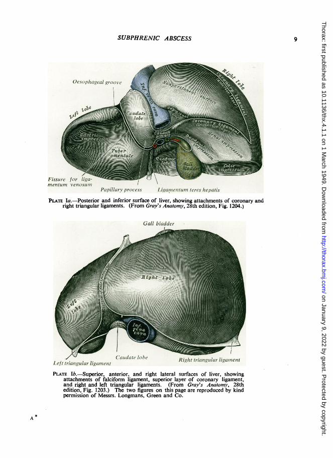
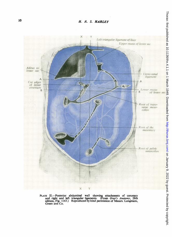
ments but by the mutual attraction of two closelyapplied serous surfaces separated only by a capil-lary layer of-fluid. If these surfaces are separatedby air, the li%er falls away from the diaphragmas though it were hinged posteriorly about its barearea, the hinge being formed by the coronary andtriangular ligaments. These points are illustratedin Plates I to III.The confusion in the topography and classifica-
tion of the subphrenic spaces is largely due to thesemisconceptions in respect of the attachments ofthe ligaments of the liver. In my opinion thereare five intraperitoneal and two extraperitonealspaces. The latter will not be considered. Of thefive intraperitoneal spaces, two are situated on theright side and three on the left.The two spaces on the right side are, I think,
best called suprahepatic and infrahepatic, and theycorrespond with those described by Barnard (1908)as the right anterior and right posterior intra-peritoneal spaces. Many surgeons, however, believethat there are two spaces above the right lobe ofthe liver, anterior and posterior, separated by theright triangular ligament. This view is held byOchsner and Graves (1933), Ochsner and De Bakey(1938), Delario (1934), Faxon (1940), Clagett andTinney (1944), Thorek (1947), and many otherauthorities. Lehman and Archer- (1937) andOverholt and Donchess (1935) recognize twaspaces above the liver on the right side, but believethat their separation is not of much practicalimportance. On the other hand Fifield and Love(1926) dispute, and Mitchell (1940) denies, theexistence of the right posterior superior space.A study of the anatomy of the parts clearly
shows that there is only one suprahepatic spaceon the right side. This space is bounded posteriorlyby the superior layers of the coronary and righttriangular ligaments, and these layers separate thesuperior from the posterior surface of the liver.The inferior layers of the coronary and right tri-angular ligaments skirt the renal and stiprarenalimpressions on the right lobe of the liver, and soseparate the posterior from the inferior surface of
on January 9, 2022 by guest. Protected by copyright.
http://thorax.bmj.com
/T
horax: first published as 10.1136/thx.4.1.1 on 1 March 1949. D
ownloaded from
2 H. A. S. HARLEYR i:rL*R% ig n T. a retrohepatic recess of the right subhepatic space'.
suprahepatic space This portion of the right infrahepatic space is thatwhich, in my opinion, has been described incor-
Liver rectly by many workers as the right posteriorsuperior intraperitoneal space. These features are
--supRieht illustrated in Figs. 1 and 2.//-suprarenal On the left side a large potential space separates
PosSthepratihc the diaphragm from the left lobe of the liver, theowl / recess of right fundus of the stomach, and the spleen. Thoughinfrahepatic
space- Diverticulum from left supra-Right kidney hepatic space (ant. left infrahepaticDuodenum space) Left
PlBRi&ht suprahepatic space
infrahepaticspace-- _Trransve rse L Liver[ / ~~~~~~~~colon11
Lesser omentum
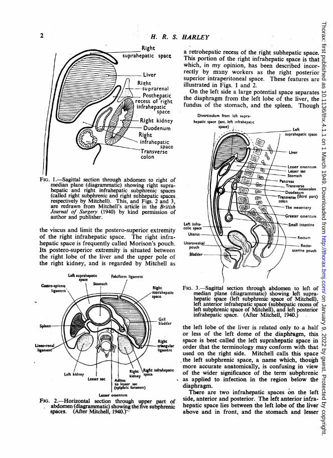
l _ - ~~~~~~~~LessersacFIG. 1.-Sagittal section through abdomen to right of Pancreas
median plane (diagrammatic) showing right supra- Transversehepatic and right infrahepatic subphrenic spaces Duodenum(called right subphrenic and right subhepatic spaces Transverse (third part)respectively by Mitchell). This, and Figs. 2 and 3, colonare redrawn from Mitchell's article in the British The mesenteryJournal of Surgery (1940) by kind permission ofauthor and publisher. Greater omentum
Left. Infra- e e > w -$Small Intestinethe-viscus and limit the postero-superior extremity clic spaceof the right infrahepatic space. The right infra- Rectumhepatic space is frequently called Morison's pouch. UterovesicalIts postero-superior extremity is situated between uterine pouchthe right lobe of the liver and the upper pole of .Bladderthe right kidney, and is regarded by Mitchell as
Left suprahepatic Faiciform ligamentspace J
Gasero-spienic St ach / Right FIG. 3.-Sagittal section through abdomen to left ofligament \ =</suprahepatic median plane (diagrammatic) showing left supra-
space hepatic spa,ce (left subphrenic space of Mitchell),left anterior infrahepatic space (subhepatic recess ofleft subphrenic space of Mitchell), and left posteriorinfrahepatic space. (After Mitchell, 1940.)
bladderSpleen . _ B 9L 1 s ]5t l l the left lobe of the liver is related only to a halfor less of the left dome of the diaphiagm, this
*Right space is best called the left suprahepatic space inJneo-renal \\J W ltriangular order that the terminology may conform with that
used on the right side. Mitchell calls this spacethe left subphrenic space, a name which, thoughmore accurate anatomically, is confusing in view
Left kidney kyRight sRigh nfrahepatc of the wider significance of the term' subphrenicLcsse r sac ^! litus t - as applied to infection in the region below the
(epiploic foramen) diaphragm.Lesser omentum There are two infrahepatic spaces on the left
FIG. 2.-Horizontal section through upper part of side, anterior and posterior. The leftanterior infra-abdomen (diagrammatic) showingthefive subphrenic hepatic space lies between the left lobe of the liverspices. (After Mitchell, 1940y-. above and in front, and the stomach and lesser
on January 9, 2022 by guest. Protected by copyright.
http://thorax.bmj.com
/T
horax: first published as 10.1136/thx.4.1.1 on 1 March 1949. D
ownloaded from
SUBPHRENIC ABSCESS
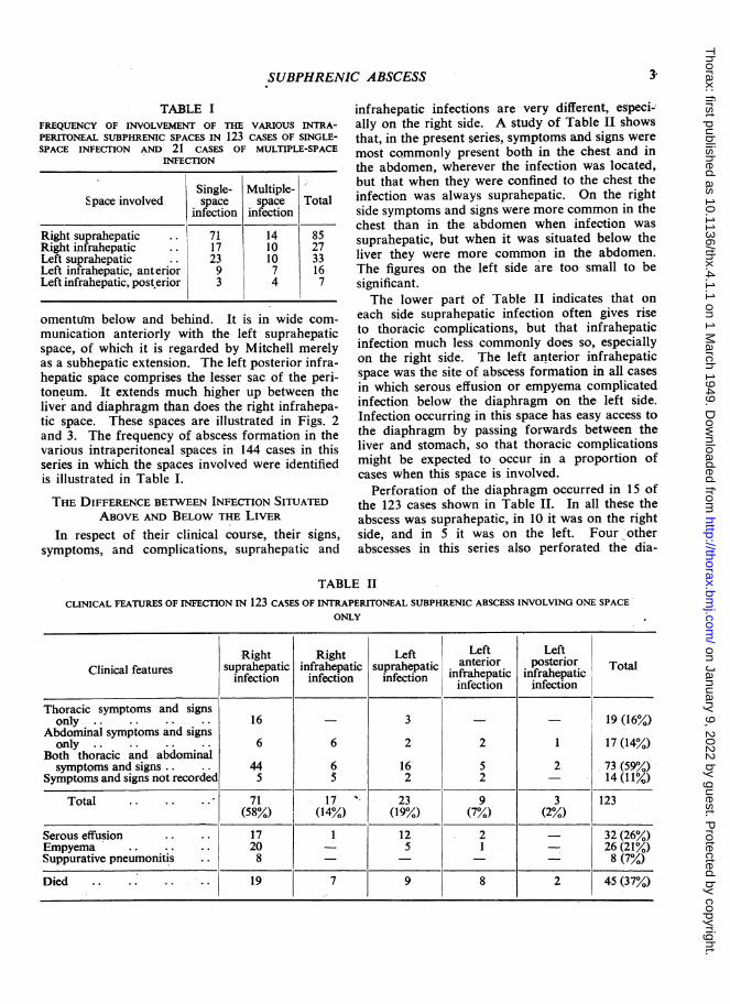
TABLE IFREQUENCY OF INVOLVEMENT OF THE VARIOUS INTRA-PERITONEAL SUBPHRENIC SPACES IN 123 CASES OF SINGLE-SPACE INFECnON AND 21 CASES OF MULTIPLE-SPACE
INFECTION
Single- Multiple-Space involved space space Total
infection infection
Right suprahepatic .. 71 14 85Right infrahepatic .. 17 10 27Left suprahepatic .. 23 10 33Left infrahepatic, anterior 9 7 16Left infrahepatic, post.erior 3 4 7
omentuYn below and behind. It is in wide com-munication anteriorly with the left suprahepaticspace, of which it is regarded by Mitchell merelyas a subhepatic extension. The left posterior infra-hepatic space comprises the lesser sac of the peri-toneum. It extends much higher up between theliver and diaphragm than does the right infrahepa-tic space. These spaces are illustrated in Figs. 2and 3. The frequency of abscess formation in thevarious intraperitoneal spaces in 144 cases in thisseries in which the spaces involved were identifiedis illustrated in Table I.
THE DIFFERENCE BETWEEN INFECTION SITUATEDABOVE AND BELOW THE LIVER
In respect of their clinical course, their signs,symptoms, and complications, suprahepatic and
infrahepatic infections are very different, especi-ally on the right side. A study of Table II showsthat, in the present series, symptoms and signs were
most commonly present both in the chest and inthe abdomen, wherever the infection was located,but that when they were confined to the chest theinfection was always suprahepatic. On the rightside symptoms and signs were more common in thechest than in the abdomen when infection was
suprahepatic, but when it was situated below theliver they were more common in the abdomen.The figures on the left side are too small to besignificant.The lower part of Table II indicates that on
each side suprahepatic infection often gives riseto thoracic complications, but that infrahepaticinfection much less commonly does so, especiallyon the right side. The left anterior infrahepaticspace was the site of abscess formation in all cases
in which serous effusion or empyema complicatedinfection below -the diaphragm on the left side.Infection occurring in this space has easy access tothe diaphragm by passing forwards between theliver and stomach, so that thoracic complicationsmight be expected to occur in a proportion ofcases when this space is involved.
Perforation of the diaphragm occurred in 15 ofthe 123 cases shown in Table II. In all these theabscess was suprahepatic, in 10 it was on the rightside, and in 5 it was on the left. Four -otherabscesses in this series also perforated the dia-
ILE II
CLINICAL FEATURES OF INFECTION IN 123 CASES OF INTRAPERITONEAL SUBPHRENIC ABSCESS INVOLVING ONE SPACEONLY
Right Right Left Left LeftClinical features suprahepatic infrahepatic suprahepatic anterior posterior Total
infection infection infection infrahepatic infrahepaticinfection infection
Thoracic symptoms and signsonly . . . . 16 -3 --19 (16%)
Abdominal symptoms and signsonly .. .. .. 6 6 2 2 1 17 (14%)
Both thoracic and abdominalsymptoms and signs.. .. 44 6 16 5 2 73 (59%)
Symptoms and signs not recorded 5 52 2 -14 (11%)
Total .. .. .. 71 17 23 9 3 123(58%) (14%) (19%) (7%) (2%)
Serous effusion .. .. 17 1 12 2 32 (26%)Empyema .. .. .. 20 5 1 26 (21%)Suppurative pneumonitis .. 8 8 (7%)
Died .. .. .. 19 7 9 8 2 45 (37%)
I
on January 9, 2022 by guest. Protected by copyright.
http://thorax.bmj.com
/T
horax: first published as 10.1136/thx.4.1.1 on 1 March 1949. D
ownloaded from
H. R. S. HARLEY
phragm, and in all these infection was presentabove the liver.The high mortality of infrahepatic infection is
worthy of note. This is due to its association withserious abdominal disease or complications.
SUBPHRENIC ABSCESS ON THE LEFr SIDE
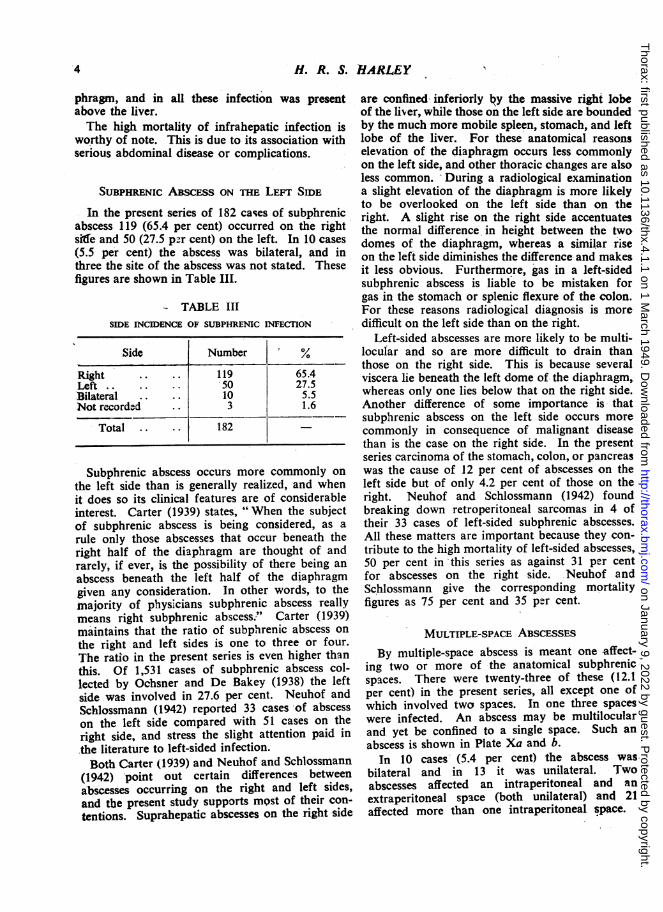
In the present series of 182 cases of subphrenicabscess 119 (65.4 per cent) occurred on the rightside and 50 (27.5 p-r cent) on the left. In 10 cases(5.5 per cent) the abscess was bilateral, and inthree the site of the abscess was not stated. Thesefigures are shown in Table III.
- TABLE IIISIDE INCIDENCE OF SUBPHRENIC INFECTION
Side Number %
Right .. .. 119 65.4Left .. .. .. 50 27.5Bilateral .. 10 5.5Not recorded .. 3 1.6
Total .. 182
Subphrenic abscess occurs more commonly on
the left side than is generally realized, and whenit does so its clinical features are of considerableinterest. Carter (1939) states, "When the subjectof subphrenic abscess is being considered, as a
rule only those abscesses that occur beneath theright half of the diaphragm are thought of andrarely, if ever, is the possibility of there being an
abscess beneath the left half of the diaphragmgiven any consideration. In other words, to themajority of physicians subphrenic abscess reallymeans right subphrenic abscess." Carter (1939)maintains that the ratio of subphrenic abscess on
the right and left sides is one to three or four.The ratio in the present series is even higher than
this. Of 1,531 cases of subphrenic abscess col-lected by Ochsner and De Bakey (1938) the leftside was involved in 27.6 per cent. Neuhof andSchlossmann (1942) reported 33 cases -of abscess
on the left side compared with 51 cases on theright side, and stress the slight attention paid in
the literature to left-sided infection.
Both Carter (1939) and Neuhof and Schlossmann(1942) point out certain differences betweenabscesses occurring on the right and left sides,and the present study supports most of their con-
tentions. Suprahepatic abscesses on the right side
are confined inferiorly lQy the massive right lobeof the liver, while those on the left side are boundedby the much more mobile spleen, stomach, and leftlobe of the liver. For these anatomical reasonselevation of the diaphragm occurs less commonlyon the left side, and other thoracic changes are alsoless common. During a radiological examinationa slight elevation of the diaphragm is more likelyto be overlooked on the left side than on theright. A slight rise on the right side accentuatesthe normal difference in height between the twodomes of the diaphragm, whereas a similar riseon the left side diminishes the difference- and makesit less obvious. Furthermore, gas in a left-sidedsubphrenic abscess is liable to be mistaken forgas in the stomach or splenic flexure of the colon.For these reasons radiological diagnosis is moredifficult on the left side than on the right.
Left-sided abscesses are more likely to be multi-locular and so are more difficult to drain thanthose on the right side. This is because severalviscera lie beneath the left dome of the diaphragm,whereas only one lies below that on the right side.Another difference of some importance is- thatsubphrenic abscess on the left side occurs morecommonly in consequence of malignant diseasethan is the case on the right side. In the presentseries carcinoma of the stomach, colon, or pancreaswas the cause of 12 per cent of abscesses on theleft side but of only 4.2 per cent of those on theright. Neuhof and Schlossmann (1942) foundbreaking down retroperitoneal sarcomas in 4 oftheir 33 cases of left-sided subphrenic abscesses.All these matters are important because they con-tribute to the high mortality of left-sided abscesses,50 per cent in this series as against 31 per centfor abscesses on the right side. Neuhof andSchlossmann give the corresponding mortalityfigures as 75 per cent and 35 per cent.
MULTIPLE-SPACE ABSCESSES
By multiple-space abscess is meant one affect-ting two or more of the anatomical subphrenicspaces. There were twenty-three of these (12.1per cent) in the present series, all except one ofwhich involved two spaces. In one three spaceswere infected. An abscess may be multilocularand yet be confined to a single space. Such an
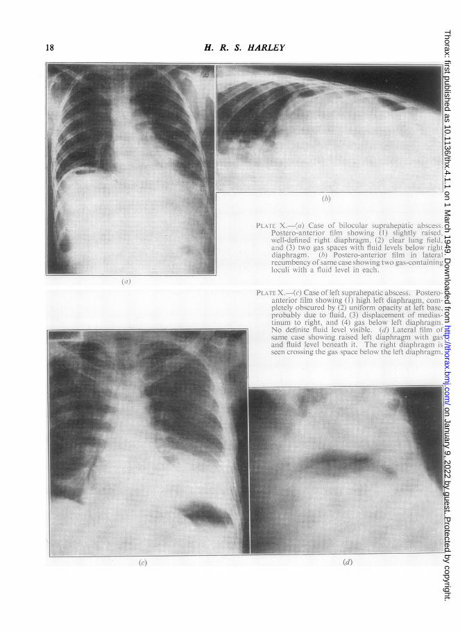
abscess is shown in Plate Xa and b.In 10 cases (5.4 per cent) the abscess was
bilateral and in 13 it was unilateral. Twoabscesses affected an intraperitoneal and an
extraperitoneal space (both unilateral) and 21affected more than one intraperitoneal space.
4
on January 9, 2022 by guest. Protected by copyright.
http://thorax.bmj.com
/T
horax: first published as 10.1136/thx.4.1.1 on 1 March 1949. D
ownloaded from
SUBPHRENIC ABSCESS'
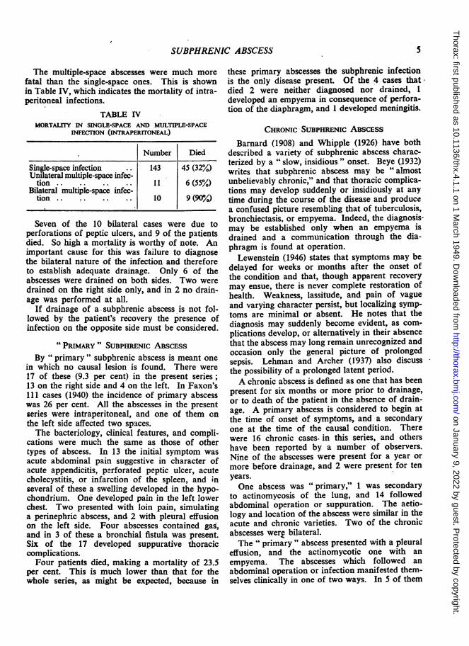
The multiple-space abscesses were much morefatal than the single-space ones. This is shownin Table IV, which indicates the mortality of intra-peritoneal infections.
TABLE IVMORTALiY IN SINGLE-SPACE AND MULTIPLE-SPACE
INFECTION (INTRAPERITONEAL)
Number Died
Single-space infection .. 143 45 (32%)Unilateral multiple-space infec-
tion .. .. .. 11 6 (55%)Bilateral multiple-space infec-
tion .. .. .. 10 9 (9C/o)
Seven of the 10 bilateral cases were due toperforations of peptic ulcers, and 9 of the patientsdied. So high a mortality is worthy of note. Animportant cause for this was failure to diagnosethe bilateral nature of the infection and thereforeto establish adequate drainage. Only 6 of theabscesses were drained on both sides. Two weredrained on the right side only, and in 2 no drain-age was performed at all.
If drainage of a-subphrenic abscess is not fol-lowed by the patient's recovery the presence ofinfection on the opposite side must be considered.
"PRIMARY" SUBPHRENIC ABSCESSBy "6 primary " subphrenic abscess is meant one
in which no causal lesion is found. There were17 of these (9.3 per cent) in the present series;13 on the right side and 4 on the left. In Faxon's111 cases (1940) the incidence of primary abscesswas 26 per cent. All the abscesses in the presentseries were intraperitoneal, and one of them onthe left side affected two spaces.The bacteriology, clinical features, and compli-
cations were much the same as those of othertypes of abscess. In 13 the initial symptom wasacute abdominal pain suggestive in character ofacute appendicitis, perforated peptic ulcer, acutecholecystitis, or infarction of the spleen, and inseveral of these a swelling developed in the hypo-chondrium. One developed pain in the left lowerchest. Two presented with loin pain, simulatinga perinephric abscess, and 2 with pleural effusionon the left side. Four abscesses contained gas,and in 3 of these a bronchial fistula was present.Six of the 17 developed suppurative thoraciccomplications.Four patients died, making a mortality of 23.5
per cent. This is much lower than that for thewhole series, as might be expected, because in
these primary abscesses the subphrenic infectionis the only disease present. Of the 4 cases thatdied 2 were neither diagnosed nor drained, 1developed an empyema in consequence of perfora-tion of the diaphragm, and 1 developed meningitis.
CHRONIC SUBPHRENIC ABscEss
Barnard (1908) and Whipple (1926) have bothdescribed a variety of subphrenic abscess charac-terized by a " slow, insidious " onset. Beye (1932)writes that subphrenic abscess may be "*almostunbelievably chronic," and that thoracic complica-tions may develop suddenly or insidiously at anytime during the course of the disease and producea confused picture resembling that of tuberculosis,bronchiectasis. or empyema. Indeed, the diagnosis-may be established only when an empyema isdrained and a communication through the dia-phragm is found at operation.
Lewenstein (1946) states that symptoms may bedelayed for weeks or months after the onset ofthe condition and that, though apparent recoverymay ensue, there is never complete restoration ofhealth. Weakness, lassitude, and pain of vagueand varying character persist, but localizing symp-toms are minimal or absent. He notes that thediagnosis may suddenly become evident, as com-plications develop, or alternatively in their absencethat the abscess may long remain unrecognized andoccasion only the general picture of prolongedsepsis. Lehman and Archer (1937) also discussthe possibility of a prolonged latent period.A chronic abscess is defined as one that has been
present for six months or more prior to drainage,or to death of the patient in the absence of drain-age. A primary abscess is considered to begin atthe time of onset of symptoms, and a secondaryone at the time of the causal condition. Therewere 16 chronic cases- in this series, and othershave been reported by a number of observers.Nine of the abscesses were present for a year ormore before drainage, and 2 were present for tenyears.One abscess was "primary," 1 was secondary
to actinomycosis of the lung, and 14 followedabdominal operation or suppuration. The aetio-logy and location of the abscess were similar in theacute and chronic varieties. Two of the chronicabscesses were bilateral.The " primary" abscess presented with a pleural
effusion, and the actinomycotic one with anempyema. The abscesses which followed anabdominal operation or infection manifested them-selves clinically in one of two ways. In 5 of them
5
on January 9, 2022 by guest. Protected by copyright.
http://thorax.bmj.com
/T
horax: first published as 10.1136/thx.4.1.1 on 1 March 1949. D
ownloaded from
H. R. S. HARLEY
symptoms were present from the outset, but their* significance was not appreciated for months oryears. In 9 others no symptoms were present atthe outset, and a latent period ensued varying fromfourteen days to nine years, during which thepatient was apparently well. Thereafter the onsetof symptoms when it occurred was abrupt in 5cases; in 4 it was ill-defined and vague.
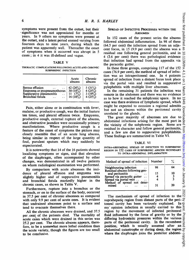
TABLE VTHORACIC COMPLICATIONS FOLLOWING ACUTE AND CHRONIC
SUBPHRENIC INFECTION
Acute Chronicabscess abscess
Serous effusion .. .. 42 (26%) 5 (31%)Empyema or pyopneumothorax 30 (18°/) 4 (25%/)Suppurative pneumonitis .. 11 (7%) 3 (19%)Bronchial fistula .. .. 16 (10%) 4 (25%)
Pain, either alone or in combination with fever,
malaise, or productive cough, was the initial featureten times, and pleural effusion twice. Empyema,productive cough, external rupture of the abscess,and obstructive jaundice were occasionally primarymanifestations. Where productive cough is a
feature of the onset of symptoms the picture may
closely resemble that of an acute lung abscess,being similar in respect of the large quantity offoul, purulent sputum which may suddenly beexpectorated.
It is noteworthy that 14 of the 16 patients showedlocalizing symptoms or signs, and that elevationof the diaphragm, often accompanied by otherchanges, was demonstrated in all twelve patientson whom radiological examination was performed.By comparison with acute abscesses the inci-
dence of pleural effusion and empyema was
slightly higher and of suppurative pneumonitisand bronchial fistula markedly higher in thechronic cases, as shown in Table V.
Furthermore, rupture into a bronchus or thestomach, or on to the surface of the body, occurredin 37.5 per cent of chronic abscesses as comparedwith only 9.9 per cent of acute ones. It is evidentthat undrained abscesses point to a surface andtend to evacuate themselves there.
All the chronic abscesses were drained, and 37.5per,cent of the patients died. The mortality ofacute cases which were drained in this series was
25.2 per cent. The chronic abscess appears, there-fore, to be a somewhat more lethal condition thanthe acute variety, though the figures are too smallto be conclusive.
SPREAD OF INFECTIVE PROCESSES WITHIN THEABDOMEN
In 152 cases of the present series the abscessfollowed abdominal inflammation. In 98 of these(64.5 per cent) the infection spread from an adja-cent focus, in 15 (9.9 per cent) the abscess was aresidual one following general peritonitis, and in4 (2.6 per cent) there was pathological evidencethat infection had spread from the appendix viathe paracolic gutter.
In these three groups, comprising 117 of the 152cases (76.9 per cent), the method of spread of infec-tion was an intraperitoneal one. In 4 patientsspread of infection from a distant focus took placevia the portal vein and resulted in suppurativepylephebitis with multiple liver abscesses.
In the remaining 31 patients the infection wasremote in the abdomen and there was no evidenceas to how it reached the subphrenic space. In nocase was there evidence of lymphatic spread, whichmight be expected to occasion a regional adenitisbut not an abscess within the peritoneal cavityunrelated to the glands.The great majority of abscesses are due to
abdominal infections arising for the most part inthe vicinity of the subphrenic region. Some areresidual in character and follow general peritonitis,and a few are due to suppurative pylephlebitis.These findings are summarized in Table VI.
TABLE VIINTRA-ABDOMINAL SPREAD OF INFECTION TO SUBPHRENICREGION IN 152 CASES OF SUBPHRENIC ABSCESS SECONDARY
TO INTRA-ABDOMINAL INFLAMMATION
Method of spread of infection Number %
Neighbouring infection .. 98 64Residual abscess following gen-
eral peritonitis .. 15 10Spread via paracolic gutter .. 4 3Spread via portal vein .. .. 4 3Method of spread not deter-mined .. .. .. 31 20
The mechanism of spread of infection to thesuprahepati9 region from distant parts of the peri-toneal cavity has been variously explained. Inmy opinion infection is usually carried to thisregion by the movement of infected peritonealfluid influenced by the force of gravity or by thediffering hydrostatic pressuires within the variousparts of the peritoneal cavity. In the recumbentposition; which is usually assumed after anabdominal catastrophe -or during sleep, the regionwhere the'diaphragm joins the posterior abdomi-
6
on January 9, 2022 by guest. Protected by copyright.
http://thorax.bmj.com
/T
horax: first published as 10.1136/thx.4.1.1 on 1 March 1949. D
ownloaded from
SUBPHRENIC ABSCESS
nal wall is a low point to which fluid will natur-ally gravitate and from which it is soon spreadby the constant movement occurring between thediaphragm and its related viscera. The effect ofthis movement can readily be observed by placinga moistened cover-slip on a microscope slide. Ifa drop of dye is placed nearby and the cover slipmoved to and fro on the slide, the dye will rapidlyspread between the two surfaces. Mass movementof fluid occurs in serous cavities in accordancewith the laws of gravity. This has been proved inthe case of pleural effusions by Blair (personalcommunication), who has shown by x-ray studiesthat the pleural fluid moves quickly to new posi-tions with changes of posture. It is legitimate toassume that peritoneal fluid behaves similarlybefore adhesions have formed.
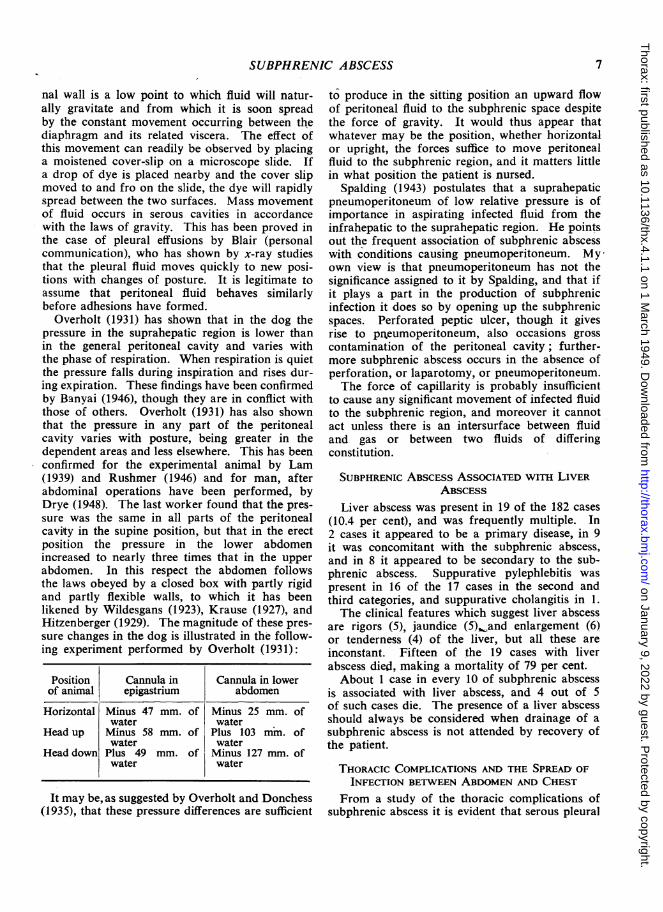
Overholt (1931) has shown that in the dog thepressure in the suprahepatic region is lower thanin the general peritoneal cavity and varies withthe phase of respiration. When respiration is quietthe pressure falls during inspiration and rises dur-ing expiration. These findings have been confirmedby Banyai (1946), though they are in conflict withthose of others. Overholt (1931) has also shownthat the pressure in any part of the peritonealcavity varies with posture, being greater in thedependent areas and less elsewhere. This has beenconfirmed for the experimental animal by Lam(1939) and Rushmer (1946) and for man, afterabdominal operations have been performed, byDrye (1948). The last worker found that the pres-sure was the same in all parts of the peritonealcavity in the supine position, but that in the erectposition the pressure in the lower abdomenincreased to nearly three times that in the upperabdomen. In this respect the abdomen followsthe laws obeyed by a closed box with partly rigidand partly flexible walls, to which it has beenlikened by Wildesgans (1923), Krause (1927), andHitzenberger (1929). The magnitude of these pres-sure changes in the dog is illustrated in the follow-ing experiment performed by Overholt (1931):
Position Cannula in Cannula in lowerof animal epigastrium abdomen
Horizontal Minus 47 mm. of Minus 25 mm. ofwater water
Head up Minus 58 mm. of Plus 103 mm. ofwater water
Head down Plus 49 mm. of Minus 127 mm. ofwater water
It may be, as suggested by Overholt and Donchess(1935), that these pressure differences are sufficient
to produce in the sitting position an upward flowof peritoneal fluid to the subphrenic space despitethe force of gravity. It would thus appear thatwhatever may be the position, whether horizontalor upright, the forces suffice to move peritonealfluid to the subphrenic region, and it matters littlein what position the patient is nursed.
Spalding (1943) postulates that a suprahepaticpneumoperitoneum of low relative pressure is ofimportance in aspirating infected fluid from theinfrahepatic to the suprahepatic region. He pointsout the frequent association of subphrenic abscesswith conditions causing pneumoperitoneum. Myown view is that pneumoperitoneum has not thesignificance assigned to it by Spalding, and that ifit plays a part in the production of subphrenicinfection it does so by opening up the subphrenicspaces. Perforated peptic ulcer, though it givesrise to prneumoperitoneum, also occasions grosscontamination of the peritoneal cavity; further-more subphrenic abscess occurs in the absence ofperforation, or laparotomy, or pneumoperitoneum.The force of capillarity is probably insufficient
to cause any significant movement of infected fluidto the subphrenic region, and moreover it cannotact unless there is an intersurface between fluidand gas or between two fluids of differingconstitution.
SUBPHRENIC ABSCESS ASSOCIATED WiTH LIVERABSCESS
Liver abscess was present in 19 of the 182 cases(10.4 per cent), and was frequently multiple. In2 cases it appeared to be a primary disease, in 9it was concomitant with the subphrenic abscess,and in 8 it appeared to be secondary to the sub-phrenic abscess. Suppurative pylephlebitis waspresent in 16 of the 17 cases in the second andthird categories, and suppurative cholangitis in 1.The clinical features which suggest liver abscess
are rigors (5), jaundice (5), and enlargement (6)or tenderness (4) of the liver, but all these areinconstant. Fifteen of the 19 cases with liverabscess died, making a mortality of 79 per cent.About 1 case in every 10 of subphrenic abscess
is associated with liver abscess, and 4 out of 5of such cases die. The presence of a liver abscessshould always be considered when drainage of asubphrenic abscess is not attended by recovery ofthe patient.
THORACIC COMPLICATIONS AND THE SPREAD OFINFECTION BETWEEN ABDOMEN AND CHEST
From a study of the thoracic complications ofsubphrenic abscess it is evident that serous pleural
7
on January 9, 2022 by guest. Protected by copyright.
http://thorax.bmj.com
/T
horax: first published as 10.1136/thx.4.1.1 on 1 March 1949. D
ownloaded from
H. R. S. HARLEY
effusion should be considered separately fromintra-thoracic suppurations.A serous effusion, either clear or cloudy in
appearance, was proved to be present by aspira-tion, operation, or necropsy in 47 cases (25.8 percent). In 12 other cases the presence of an effu-sion was suggested by x-ray examination, but wasnot otherwise confirmed. Serous effusion is thusa common sequela of subphrenic infection.
It is noteworthy that in 6 cases the effusion wascontralateral and in 2 it was bilateral. It is impor-tant to know that contralateral effusions may occur,because of the difficulty they may occasion indiagnosis.
ln both bilateral cases malignant disease waspresent, in the stomach or the caecum, and per-foration of the bowel occurred, with fatal peri-tonitis. In neither case was the subphrenic abscessrecognized or drained.
In all the cases with contralateral or bilateraleffusion the subphrenic abscess was unilateral, andin 7 of the 8 cases it was situated on the right side.In 6 cases the abscess was intraperitoneal, in oneextraperitoneal, and in one both intra- and extra-peritoneal.
In 20 of the 47 cases the pleural fluid wasexamined bacteriologically. In 17 of these noorganisms were demonstrated either in films or incultures. In 1 case organisms were seen in fims,but cultures were sterile. In only 2 cases didorganisms grow in culture.Examination of the protein content in 5 cases,
and of the cellular content in 15, suggested that thenature of the pleural fluid was inflammatory. Theprotein content varied between 1.2 per cent and3.3 per cent. The chief cellular constituents werered cells, neutrophils, and lymphocytes. A dif-ferential cell count was made in 12 cases. In 8 ofthese, neutrophils predominated over lymphocytes,and in 3 lymphocytes predominated over neutro-phils. In 1 case the two types of cell were presentin about equal proportions.No cause within the chest was found to account
for the serous effusion in 39 of the 47 cases. In8 cases a possible cause was present in the lungor pleura, such as fibrino-purulent pleurisy or sup-purative pneumonitis. It is my belief that whereno obvious cause can be found in the chest theserous effusion is an expression of the reaction ofthe subpleural vessels to a neighbouring focus ofinfection. These vessels are subject to -inflam-matory vasodilatation, and the outpouring ofserous fluid into the pleura is analogous to theoedema which occurs around an acute inflamma-tory focus elsewhere. An effusion of similar
character occurs -in a joint when acute inflam-mation develops in its neighbourhood.When suppuration in the chest follows in con-
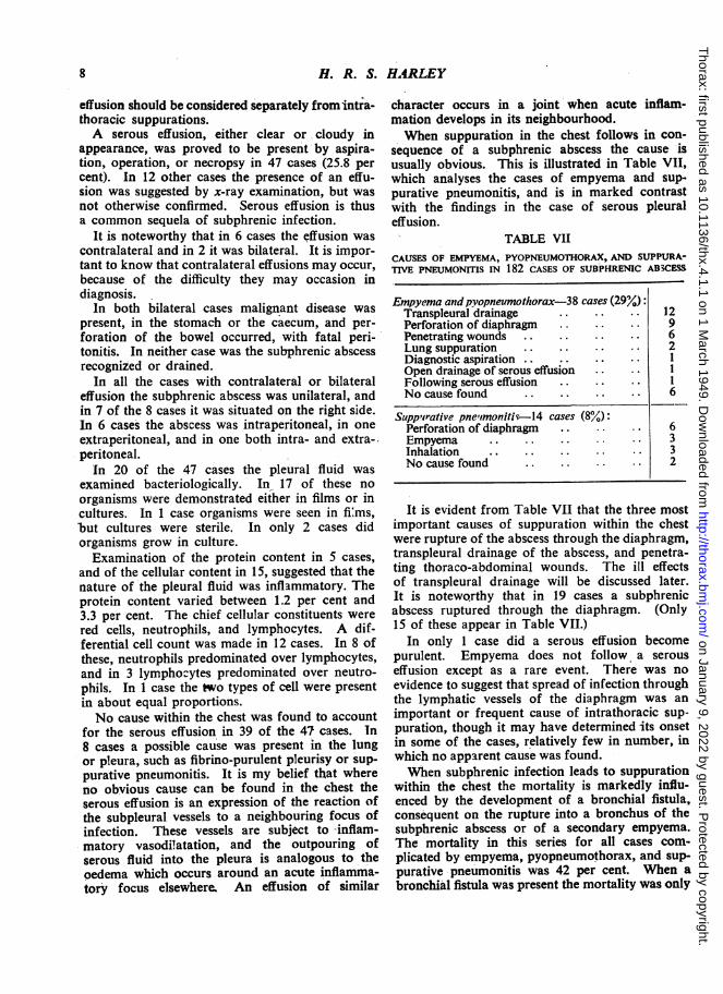
sequence of a subphrenic abscess the cause isusually obvious. This is illustrated in Table VII,which analyses the cases of empyema and sup-purative pneumonitis, and is in marked contrastwith the findings in the case of serous pleuraleffusion.
TABLE VIICAUSES OF EMPYEMA, PYOPNEUMOTHORAX, AND SUPPURA-TIVE PNEUMONITIS IN 182 CASES OF SUBPIPHRENIC ABSCESS
Empyema and pyopneumothorax-38 cases (29%)Transpleural drainage .. .. .. 12Perforation of diaphragm .. .. .. 9Penetrating wounds .. .. .. .. 6Lung suppuration .. .. .. .. 2Diagnostic aspiration .. .. .. .. 1Open drainage of serous effusion .. ..Following serous effusion .. .. INo cause found .. .. .. .. 6
Suppirative pneimonitiv-14 cases (8%):Perforation of diaphragm .. .. .. 6Empyema .. .. .. .. .. 3Inhalation .. .. .. .. .. 3No cause found .. .. .. .. 2
It is evident from Table VII that the three mostimportant causes of suppuration within the chestwere rupture of the abscess through the diaphragm,transpleural drainage of the abscess, and penetra-ting thoraco-abdominal wounds. The ill effectsof transpleural drainage will be discussed later.It is notewQrthy that in 19 cases a subphrenicabscess ruptured through the diaphragm. (Only15 of these appear in Table VII.)
In only 1 case did a serous effusion becomepurulent. Empyema does not follow a serouseffusion except as a rare event. There was noevidence to suggest that spread of infection throughthe lymphatic vessels of the diaphragm was animportant or frequent cause of intrathoracic sup-puration, though it may have determined its onsetin some of the cases, relatively few in number, inwhich no apparent cause was found.When subphrenic infection leads to suppuration
within the chest the mortality is markedly influ-enced by the development of a bronchial fistula,consequent on the rupture into a bronchus of thesubphrenic abscess or of a secondary empyema.The mortality in this series for all cases com-plicated by empyema, pyopneumothorax, and sup-purative pneumonitis was 42 per cent. When abronchial fistula was present the mortality was only
8
on January 9, 2022 by guest. Protected by copyright.
http://thorax.bmj.com
/T
horax: first published as 10.1136/thx.4.1.1 on 1 March 1949. D
ownloaded from
SUBPHRENIC ABSCESS
Oe(.opha(geal groove
Ffissuire for liga-melz.s 1'
I1{ Stl! I Nmrneium-t venosuin
Papillairv process Liawinientum terns lieptatis
PLATE Ia.-Posterior and inferior surface of liver, showing attachments of coronary andright triangular ligaments. (From Gray's Anatomy, 28th edition, Fig. 1204.)
Gall bladder
Ca dlle lobe Riglit itrianzgulart ligaewntLeft tr'ian,gutla-r ligamiiient
PLATE Ib.-Superior, anterior, and right lateral surfaces of liver, showingattachments of falciform ligament, superior layer of coronary ligament,and right and left triangular ligaments. (From Gray's Anatomy, 28thedition, Fig. 1203.) The two figures on this page are reproduced by kindpermission of Messrs. Longmans, Green and Co.
A4
9
on January 9, 2022 by guest. Protected by copyright.
http://thorax.bmj.com
/T
horax: first published as 10.1136/thx.4.1.1 on 1 March 1949. D
ownloaded from
H. R. S. HARLEY
S S -,
',.1- i t.':PI
PLATE II.-Posterior abdominal wall showing attachments of coronaryand right and left triangular ligaments. (From Gray's Anatomy, 28thedition, Fig. 1163.) Reproduced by kind permission of Messrs. Longmans,Green and Co.
10
on January 9, 2022 by guest. Protected by copyright.
http://thorax.bmj.com
/T
horax: first published as 10.1136/thx.4.1.1 on 1 March 1949. D
ownloaded from
SUBPHRENIC ABSCESS
(a)
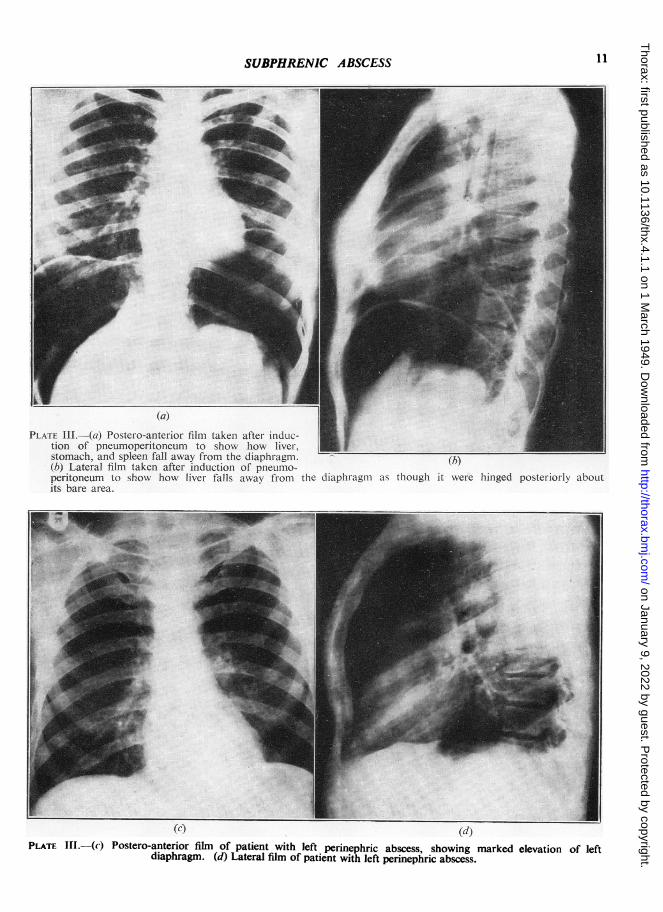
PLATl 111.-(a) Postero-anterior fiinl tatkeni after indiuc-tion of pneumoperitoneuni to show how livelr.stomach, and spleen fall aLway from the diaphr-agm.(b) Lateral film taken after induLction of pneLumo-peritoneum to show how liver falls away from the diiaphlr-agmii aIS thouLghits bare area.
(h)it were lhinlged posteriorlvy abott
(C.) (c1)PLATE IHI.-(c) Postero-anterior film of patient with left perinephric abscess, showing marked elevation of left
diaphragm. (d) Lateral film of patient with left perinephric abscess.
11
on January 9, 2022 by guest. Protected by copyright.
http://thorax.bmj.com
/T
horax: first published as 10.1136/thx.4.1.1 on 1 March 1949. D
ownloaded from
H. R. S. HARLEY
Ie \ (/ *..-
ciihcI&cll5\>I 1~iiTijWhijI)}nTTl l I,;P
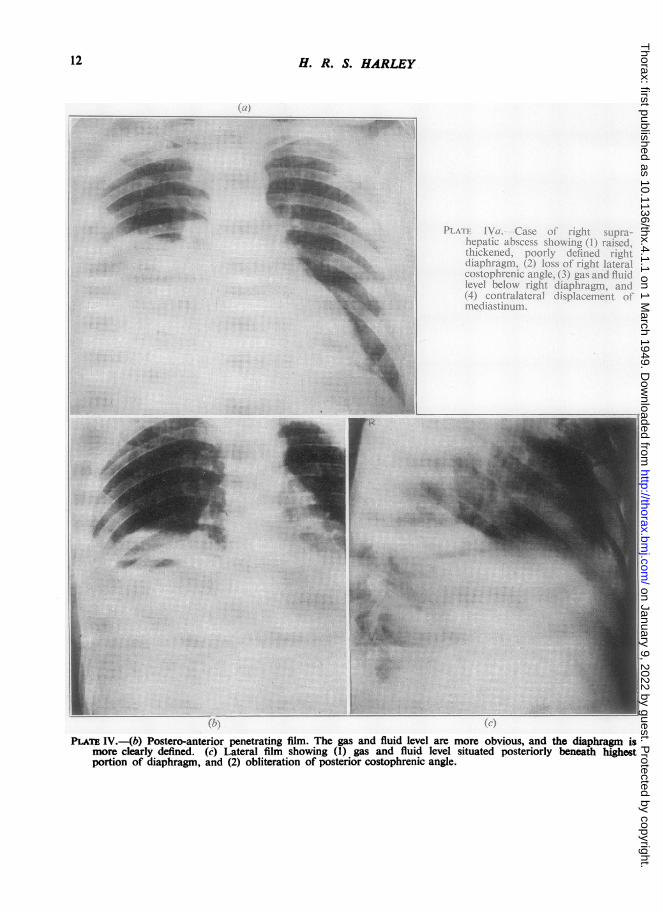
PLAm IV.-4b) Postero-anterior penetrating film. The gas and fluid level are more obvious, and the diaphragm ismore clearly defined. (c) Lateral film showing (1) gas and fluid level situated posteriorly beneath highestportion of diaphragm, and (2) obliteration of posterior costophrenic angle.
12
:: ...M,:-
M:
on January 9, 2022 by guest. Protected by copyright.
http://thorax.bmj.com
/T
horax: first published as 10.1136/thx.4.1.1 on 1 March 1949. D
ownloaded from
SUBPHRENIC ABSCESS
(a)
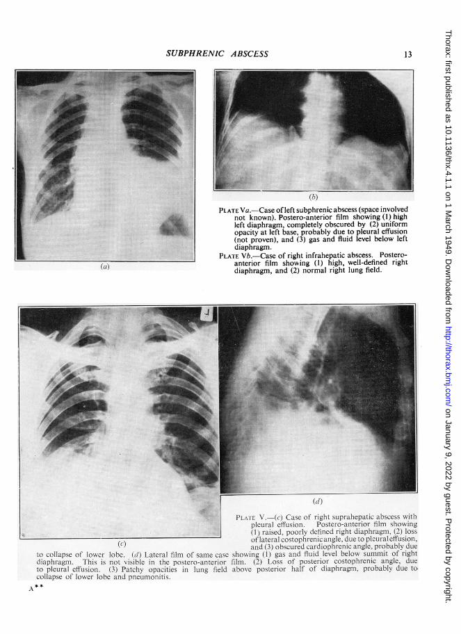
(h)PLATE Va. Case of left subphrenic abscess (space involved
not known). Postero-anterior film showing (1) highleft diaphragm, completely obscured by (2) uniformopacity at left base, probably due to pleural effusion(not proven), and (3) gas and fluid level below leftdiaphragm.
PLATE Vb.-Case of right infrahepatic abscess. Postero-anterior film showing (1) high, well-defined rightdiaphragm, and (2) normal right lung field.
4
(d1)
PL..Ir V -(c) Case of right suprahepatic abscess withlpleural effLsion. Postero-anterior film showing
L_____ <(I ) raised, poorly defined right diaphragmn. (2) loss
(c,) oflateralcostoplhrenicanigle.duetopleur-aleffusion,and (3) obscurcd cardiophrenic angle. probablv due
to collapse of lower lobe. (tW) Lateral film of same case showing (1) gas and fluid level below, summit of rightdiapliragni. This is not visible itn the postero-anteerior film. (2) Loss of posterior costophrenic angle. dueto pleural effusion. (3) Patchy opacities in lung field above posterior half of diaphragm. probablv due tocollapse of lower lobe and pneumonitis.
A * *
13
r ..'.Aw...s5 " .:0"kilak..;=
AOFMI1:
on January 9, 2022 by guest. Protected by copyright.
http://thorax.bmj.com
/T
horax: first published as 10.1136/thx.4.1.1 on 1 March 1949. D
ownloaded from
H. R. S. HARLEY
ciSsC
00
r_,,20 ¢5E_ to
3-uz
Er Q
CL
3.~ 0 0~
oopto CJ
0.
clE
u t- C)o-
ct0CZQ
7a w
t-o.r O00.
Co 0.~
CR:
m
U0-UE).Q _CZ
N,
.0 -.
14
on January 9, 2022 by guest. Protected by copyright.
http://thorax.bmj.com
/T
horax: first published as 10.1136/thx.4.1.1 on 1 March 1949. D
ownloaded from
SUBPHRENIC ABSCESS
(a)
ON
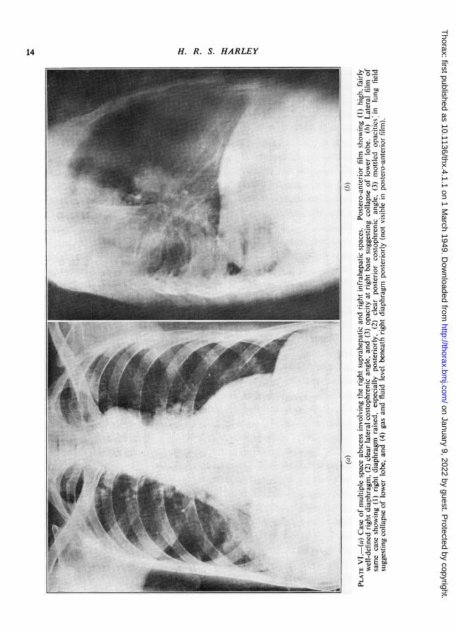
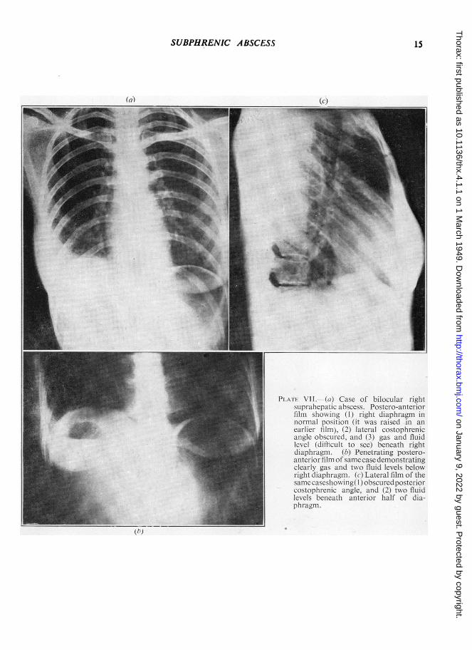
PLAIT VIl--I. (ai) Case of biloCUlar rightsuLpralhepatic abscess. Posteiro-anlteriorfilmii showing (1) i-ighlt diaplhr-agnm innlormial positioIn (it Nas r-aised in aneat-liel- film). (2) lateral costophrenicanlgle obsculed. anid (3) gas and fluidlevel (dithfcult to see) beneath rightdiaphragmn. (h) Penetrating postero-anter-ior hIm of samecase demonstratingclearly gas and two fluid levels belowright diaphl-agmii. (c) Later-al film of thesame caseshlo ing( I()obscul-ed posterior-costoplhreniic angle. and (2) tx-o fluidlevels benacatlh anterior half of dia-phrag-nm.
(h)
15
W,. I
-=
iiiimi.'., "'la.
on January 9, 2022 by guest. Protected by copyright.
http://thorax.bmj.com
/T
horax: first published as 10.1136/thx.4.1.1 on 1 March 1949. D
ownloaded from
H. R. S. HARLEY
::w . 1::.A..i..
I.i|IE
¶..
I'I~~~~~~~~~~~~~~~~~~~~~~~~~~~~~~~~~~~~~~~~~~~~~~~~~~~~~~~~~~~~~~~~~~~~~~~~~~~~~~~~~~
til). C*') ( )O,- ighII &iphrtuagfli_
*1')st~>>11)X.i^.!'1 : : ll 811(5;N illt' C I..I.
Hk)> x l Io Ivi t>t[ttTI. iII. l at<t
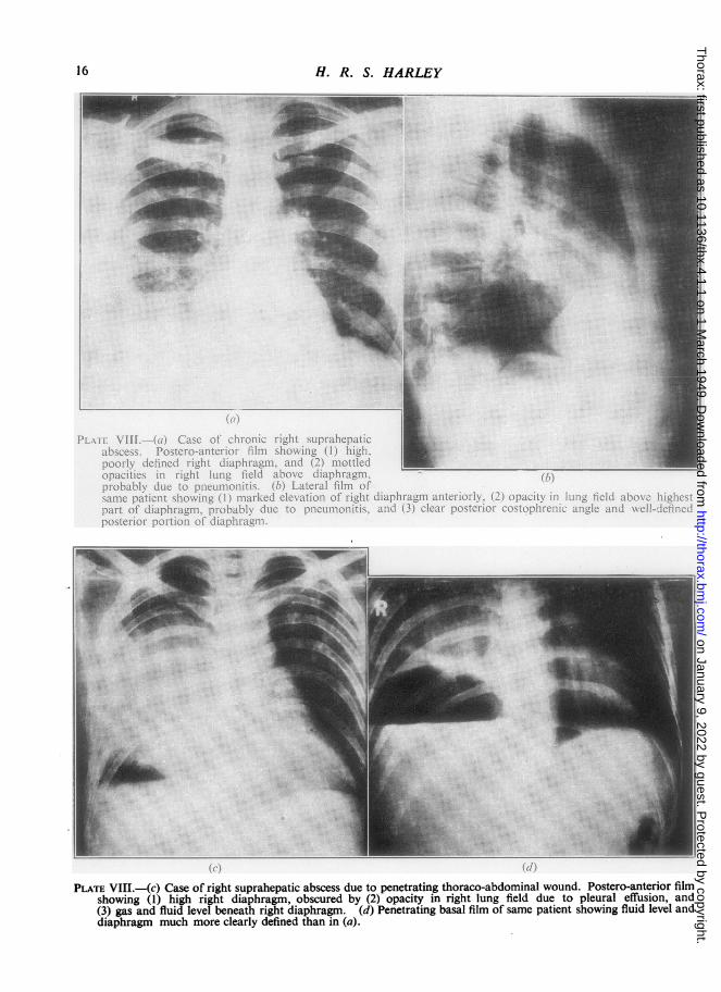
PLATE VIII.-(C) Case of right suprahepatic abscess due to penetrating thoraco-abdominal wound. Postero-anterior filmshowing (1) high right diaphragm, obscured by (2) opacity in right lung field due to pleural effusion, and(3) gas and fluid level beneath right diaphragm. (d) Penetrating basal film of same patient showing fluid level anddiaphragm much more clearly defined than in (a).
16
on January 9, 2022 by guest. Protected by copyright.
http://thorax.bmj.com
/T
horax: first published as 10.1136/thx.4.1.1 on 1 March 1949. D
ownloaded from
SUBPHRENIC ABSCESS
(a)
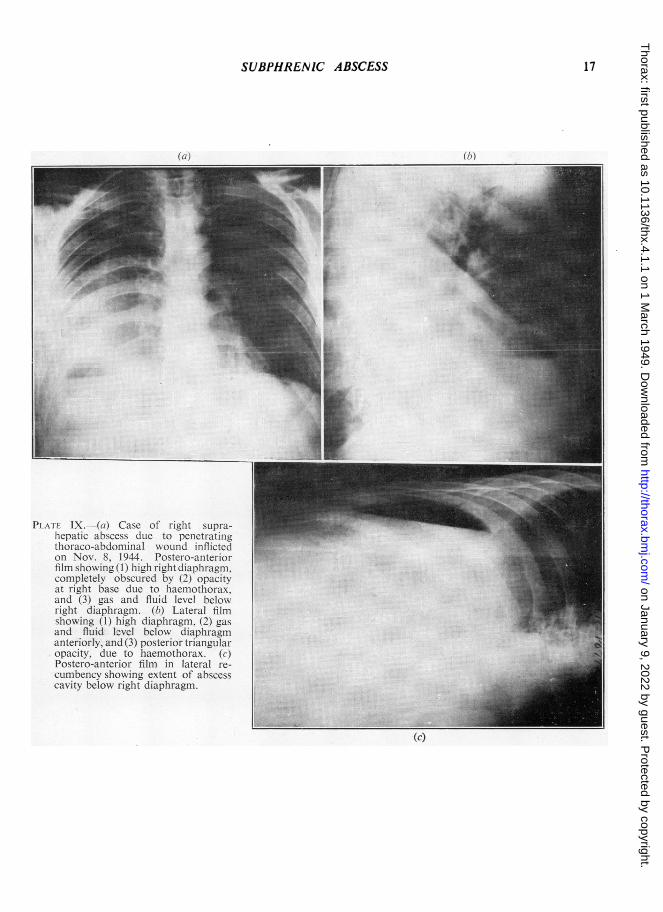
PLATF IX.-(a) Case of right supra-hepatic abscess due to penetratinigthoraco-abdominal wk ound inflictedon Nov. 8. 1944. Postero-aniter-ior-filmn showing (1) high right diaphragm.completely obscured by (2) opacitvat right base due to laemothorax.and (3) gas and fluid level belowright diaphragm. (h) Lateral filmshowing (1) high diaphragm, (2) gasanid fluid level below diaphragmanteriorly, and (3) posterior triangular-opacity, due to haemothorax. (c)Postero-anterior film in lateral re-cumbency showing extent of abscesscavity below right diaphragm.
(1;)
17
on January 9, 2022 by guest. Protected by copyright.
http://thorax.bmj.com
/T
horax: first published as 10.1136/thx.4.1.1 on 1 March 1949. D
ownloaded from
18 H. R. S. HARLEY
I ID'I.'_1\
IV.1 1!. IC
I~~~ ~~~~~~~~~~~~~~~~~~~~~~~~~~ ILmi~~~~~~~~~~~~~~~~~~~~~~~~~~~~~~~~~~.,1 11 LII
1.~~~~~~~~~~~~~~~~~~~~~~~~~~~~~~~~^'.i : se | | . 9 \ r ( ; I >|1t<'|r;i|tIL:J
|: it .' 1t -1 l Wl 'h 11! , \;E
on January 9, 2022 by guest. Protected by copyright.
http://thorax.bmj.com
/T
horax: first published as 10.1136/thx.4.1.1 on 1 March 1949. D
ownloaded from
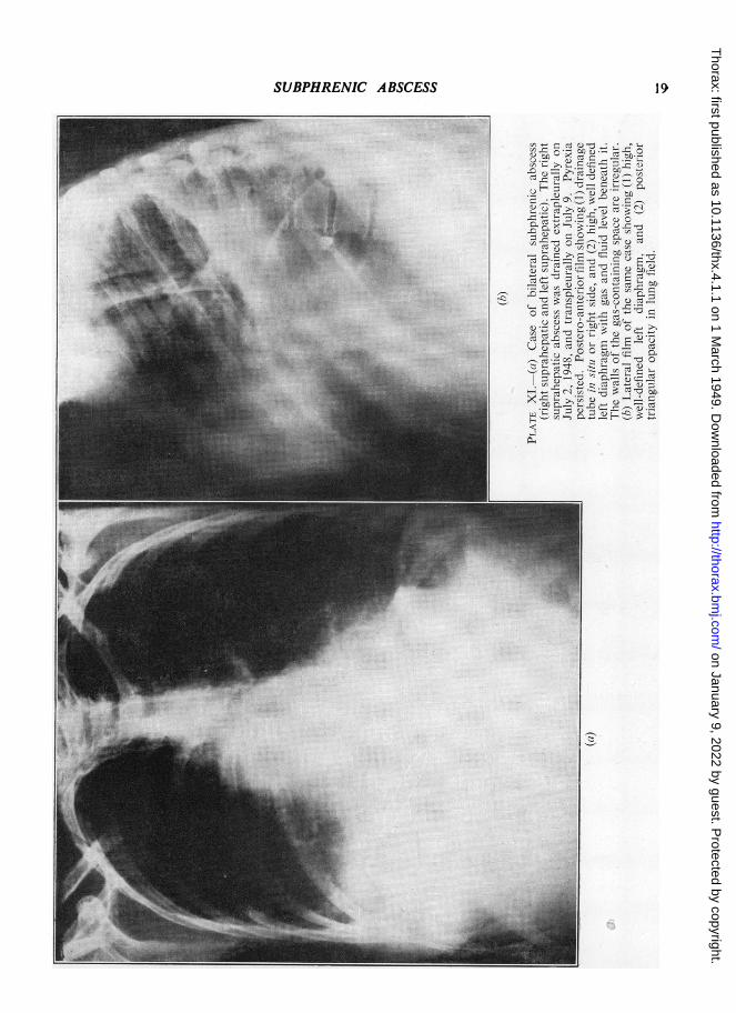
SUBPHRENIC ABSCESS 19
......~~~~~~~~~~~~~~~~~~~~~~~~~~~~~~~~ ~~~~~~~~~~~~~~~~~~~~~~~~~~~~~~~~~~~~~~~~~~~~~~~~~
on January 9, 2022 by guest. Protected by copyright.
http://thorax.bmj.com
/T
horax: first published as 10.1136/thx.4.1.1 on 1 March 1949. D
ownloaded from
on January 9, 2022 by guest. Protected by copyright.
http://thorax.bmj.com
/T
horax: first published as 10.1136/thx.4.1.1 on 1 March 1949. D
ownloaded from
SUBPHRENIC ABSCESS
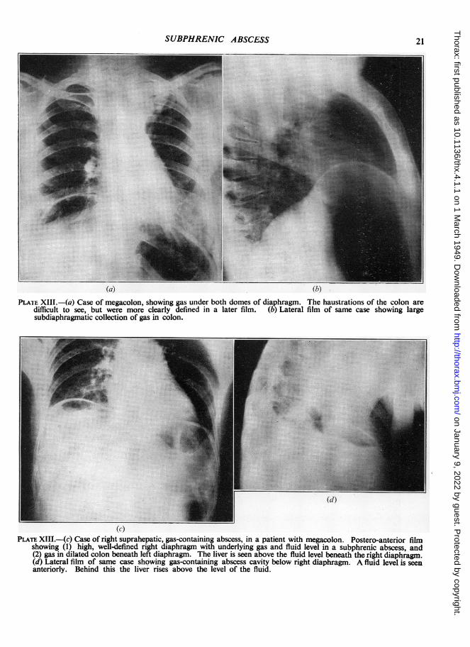
(a) (h)PLATE XIII.-(a) Case of megacolon, showing gas under both domes of diaphragm. The haustrations of the colon are
difficult to see, but were more clearly defined in a later film. (b) Lateral film of same case showing largesubdiaphragmatic collection of gas in colon.
(c)PLATE XIII.-(c) Case of right suprahepatic, gas-containing abscess, in a patient with megacolon. Postero-anterior film
showing (1) high, well-defined right diaphragm with underlying gas and fluid level in a subphrenic abscess, and(2) gas in dilated colon beneath left diaphragm. The liver is seen above the fluid level beneath the right diaphragm.(d) Lateral film of same case showing gas-containing abscess cavity below right diaphragm. A fluid level is seenanteriorly. Behind this the liver rises above the level of the fluid.
21
on January 9, 2022 by guest. Protected by copyright.
http://thorax.bmj.com
/T
horax: first published as 10.1136/thx.4.1.1 on 1 March 1949. D
ownloaded from
H. R. S. HARLEY
o
5.0
o o
I .a) .g
~co
0. o
Y >e
o C
A0!
- . 0toP
F* .iE °
)o~
Oa)
aX
-
Cd
......11.11---- ....- .. .................. ...
22
.,A
X: :.
:...Y :;& :,
on January 9, 2022 by guest. Protected by copyright.
http://thorax.bmj.com
/T
horax: first published as 10.1136/thx.4.1.1 on 1 March 1949. D
ownloaded from
SUBPHRENIC ABSCESS 23
_s s_ .. o =KEfl R m _ _ W_ . _.4j§:lt 1121 flEJ -___B52!£ L ,g58:. 'Xfl B£ __ _Ig!wt .. ,J 1 - 1 1 a 2. / ,iL _ _ _._w _
_ _
m. t,,t, ,._:. X 4__:s w
... . .. :.£_:wat __ iM_., :: _:: ':sfl' go_g s __lle :. '.:£1 *_
:B: _
_r e,
_ $:t_! .3 _8 _, . 3 =E. x. oB... ,-. -, __..... . } . -F A'. a aY' :s* a. - b p: X- e R
-B--ll < o-
E Ch s w" } . *- W
WF :}, _vd:BS. Os.s VeX_ 9ei o 3_ ''' I gzo
, n|EL t aL =
.X1 o 8s ':C
_-.vea*-£ * | .. i.<-E . I . sillr h r | __P X n e
T § . .o_- | *_ ds. ::: 1_: | | s
'! . A1s tJ
_ _
m. - a V.. .: a .. ,*_it'::: . ££ . , B' I _l Cx. S_:,:._ _
1EF '}_!Sr - -.. . sft #wFF .... . ,, ... 1 s3 =. . i ,.S-g.} ... , I Q* e i. . . * r ... j j j E - we | ; C:;. 1 Y Xo%£:Wr; ... _- £ _| ................................................ : =.. 1 ....................... v O
I 2XP 11 i hs
.... _§ a M.,,}.: | FsS££t_N£ :.i: i .. >_ . o S D a | *_X.t ij.-6h I; : | ':. ' I X;_Rg zi svA I tV!__ I _
1 U Y-S1
__; _I v E_ w_ 6 I YE :;_ ;_ < YPF . iM£££££££. ... i_ l .............. M _ | ^- ;£S' '__::} eN:e. ::. |
_r I Y E'' | o gt I OM
I SeI un.S
0<_O
l_ ¢: E_C;
I O Ys I B
,* =:i | B .
* o E.R X oht. ei t Tzffi I a >... ...... I >r.o_ Rs Q.__I .oo sC_I E; Y......... .... .. } l: - :S .... :: -l E <£8 °, s ; .ii.i .: ... ; .. ...... . w_>_ I ................ > on January 9, 2022 by guest. P
rotected by copyright.http://thorax.bm
j.com/
Thorax: first published as 10.1136/thx.4.1.1 on 1 M
arch 1949. Dow
nloaded from
H. R. S. HARLEY
17.65 per cent, but in the absence of a bronchial
fistula the mortality was 53.8 per cent. It appears
that the presence of a bronchial fistula exerts a
benign influence, presumably by allowing drainageof infection to occur.
The rarity of the spread of infection from thechest to the subphrenic region contrasts with thefrequency of spread in the reverse direction. In
the large series, comprising 3,533 cases, reviewedby Ochsner and De Bakey (1938), in only 2.5 per
cent did the infection originate in the chest, andin their own 75 cases the origin was intrathoracicin only one. In a study of 337 cases of empyemaBeye (1932) found only one instance of infectiontraversing the diaphragm to produce a subphrenicabscess, and in this case the diaphragm was injuredduring the operation for drainage of the empyema.
In the records of the Brompton Hospital the onlycase of subphrenic abscess complicating thoracicdisease which I could find was one reported byRoberts and Nelson (1933), in which right lowerlobectomy performed for bronchiectasis was fol-lowed by empyema and later by subphrenicabscess. The authors ascribed the subphrenicabscess to injury of the diaphragm at the time oflobectomy.
In the present series only 2 cases followed a
thoracic infection; one occurred in consequence
of perforation of the diaphragm in a case of pul-monary actinomycosis; the other followed an
empyema for no obvious reason. The clinicalrecord of the case suggests that the empyema was
the causal lesion, but an empyema may be thefirst clinical manifestation of a chronic and silentsubphrenic abscess, and such may well have beenthe sequence of events.
Subphrenic abscess so rarely complicatesempyema thoracis that when the two condi-tions are present together the presumption isstrongly in favour of the first being the primaryone. That infection should so frequently spreadfrom the abdomen to the chest, and so infrequentlyspread in the opposite direction, is, I think, attri-
butable to two circumstances. The first is ill-
considered surgery, in particular transpleuraldrainage of subphrenic infection. The second is
the process of compression to which a subphrenicabscess is subjected during the inspiratory phaseof respiration, when the diaphragm descends and
in consequence of which an attrition of its fibres
occurs which predisposes to its rupture. Down-
ward spread of the abscess is impeded by the
abdominal viscera, in particular the liver, or bythe formation of limiting peritoneal adhesions.
An abscess of the lung usually ruptures into a
bronchus or the pleura, while an empyema is freeto extend into the whole of the pleural cavity.
In my opinion, these factors are more importantthan spread of infection by the diaphragmaticlymphatics, even though the lymphatic drainage inthis region is almost wholly from abdomen tochest, and notwithstanding the fact that particu-late matter and bacteria have been proved to passwith great rapidity from peritoneal cavity to medi-astinal lymph nodes.* These are matters of verygreat interest, but they do not, I believe, explainthe peculiarities of the spread of infection acrossthe diaphragm.
RADIOLOGICAL DIAGNOSIS
Radiological examination was performed in 135cases and screen examination in 73. Both methodsare essential and are helpful in diagnosis, althoughLehman and Archer point out that the cardinalradiological signs when present are not necessarilydiagnostic and their absence does not exclude sub-phrenic abscess.
In the present series radiological examinationrevealed no abnormality in 12 cases (8.9 per cent),including 5 of suprahepatic infection involving asingle space. Two of the latter occurred on theright side and three on the left, the percentageincidence being 3.1 and 15.8 respectively. Themore frequent absence of radiological changes onthe left side as compared with the right is worthyof note.
Other conditions, both above and below the dia-phragm, may cause changes similar to those result-ing from subphrenic abscess. Elsberg (1901),Barnard (1908), Schwartz (1930), and Lehman andArcher (1937) all state that liver abscess may pro-duce changes indistinguishable from those of sub-phrenic abscess, while Pancoast (1926), Delario(1934), and Overholt and Donchess (1935) referto the difficulty in distinguishing between the twoconditions. The last three authorities maintainthat a large perinephric abscess may give rise toa high diaphragm with some limitation of excur-sion, but Neuhof and Schlossmann (1942), in a
series of 65 cases of perinephric abscess, foundonly 1 instance in which the diaphragm was raisedThis was the case of a child of 9 years old whohad a very large abscess, and the elevation ofthe diaphragm developed late. Radiological
* (Allen, 1936; Bizzozero and Salvioli, 1876; Bolton, 1921;Brown, 1928; Buxton and Torrey, 1906; Cunningham, 1922. 1926;Florey, 1927; Florey and Witts, 1928; Hahn and others, 1944;Higgins and others, 1930; Higgins and Graham, 1929; Higgins andLemon, 1931; Higgins and Murphy, 1928; Kolossow, 1893; Levi,1927; MacCallum, 1903; Menville and Ant, 1934; Muscatello.1895; von Recklinghausen, 1862, 1863.)
24
on January 9, 2022 by guest. Protected by copyright.
http://thorax.bmj.com
/T
horax: first published as 10.1136/thx.4.1.1 on 1 March 1949. D
ownloaded from
SUBPHRENIC ABSCESS
examination of the chest was performed in 13of 50 cases of perinephric abscess studied by theauthor. In 3 of the 13 patients the diaphragm wasraised and showed restricted movement. Theradiographs of one of these three patients areshown in Plate IIc and d. Aspiration of thechest withdrew serous fluid from 3 patients, andin two others a radiological diagnosis of pleuraleffusion was made. Delario (1934) points out thatthe radiological diagnosis of perinephric abscessmay be suggested by such features as obliterationof the outer edges of the psoas shadow, or thedemonstration of enlargement of the apparentkidney shadow, stones, or hydronephrosis.
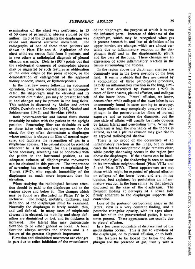
In the first few weeks following an abdominaloperation, even when convalescence is uncompli-cated, the diaphragm may be elevated and itsmovement diminished, gas may be present beneathit, and changes may be present in the lung fields.This subject is discussed by Muller and others(1929), Overholt and Donchess (1935), Neuhof andSchlossmann (1942), and Howkins (1948).Both postero-anterior and lateral films should
invariably be taken with the patient in the uprightposition. Penetrating films are desirable as wellas those taken with standard exposures for thechest, for they often demonstrate a diaphragmwhose position is obscure in films of normal expo-sure, and facilitate the recognition of gas in asubphrenic abscess. The patient should be screenedwhenever he is fit enough for this examination.When necessary, screening can be conducted withthe patient in the horizontal position, as anadequate estimate of diaphragmatic movementscan be obtained in this posture. The importanceof screening has recently been re-emphasized byThorek (1947), who regards immobility of thediaphragm as much more important than itselevation.When studying the radiological features atten-
tion should be paid to the diaphragm and to theregions above and below it. The changes whichmay be found are illustrated in Plates IV to Xinclusive. The height, mobility, thickness, anddefinition of the diaphragm must be examined.Normally the diaphragm is freely mobile, thin,and well defined. In many cases of subphrenicabscess it is elevated, its mobility and sharp defi-nition are diminished or lost, and its thickness isincreased. The lateral film may demonstrateelevation to be either general or local. A localelevation always overlies the abscess and is afeature of the greatest diagnostic importance.
Elevation and diminished movement are changesin part due to reflex inhibition of the musculature
of the diaphragm, the purpose of which is to restthe inflamed parts. Increase of thickness of thediaphiagm, which may be recognized when gasis present beneath it, and loss of definition of itsupper border, are changes which are almost cer-tainly due to inflammatory reaction in the dia-phragm itself and in the diaphragmatic pleuraand the base of the lung. These changes are anexpression of acute inflammatory reaction in thetissues surrounding the abscess.
In the region above the diaphragm changes arecommonly seen in the lower portions of the lungfield. It seems probable that they are caused bya combination of three pathological processes,namely an inflammatory reaction in the lung, simi-lar to that described by Pancoast (1926) incases of liver abscess, pleural effusion, and collapseof the lower lobes. Pleural effusion, we know,occurs often, while collapse of the lower lobes is notuncommonly found in cases coming to necropsy.A large effusion may completely obscure the dia-phragm in the postero-anterior films of normalexposure and so confuse the diagnosis, but thetrue state of affairs will usually be made obviousby taking lateral and penetrating films. When thediaphragm is high the mechanics of the thorax isaltered, so that a pleural effusion may give rise toan atypical radiological shadow.There is no proof of the occurrence of an
inflammatory reaction in the lungs, but in somecases the lateral costophrenic angle remains clear,while patchy shadowing occurs in the region justabove the diaphragm. If the abscess can be local-ized radiologically the shadowing is seen to occurin its immediate neighbourhood (Plate VIlla andb and Plate XIV). These appearances are notthose which might be expected of pleural effusionor collapse of the lower lobes, and are, in myopinion, best explained by postulating an inflam-matory reaction in the lung similar to that alreadydiscussed in. the case of the diaphragm. Thefrequent finding at necropsy of a lower lobefirmly adherent- to the diaphragm supports thiscontention.
Loss of the posterior costophrenic angle in thelateral view is a very constant finding, and aposterior triangular opacity, with its apex aboveand behind in the paravertebral gutter, is some-times present. These appearances are usually dueto pleural effusion.
In a few cases contralateral displacement of themediastinum occurs. This is due to elevation ofthe diaphragm, or to pleural effusion, or to both.The features to be looked for below the dia-
phragm are the presence of gas, usually with a
25
on January 9, 2022 by guest. Protected by copyright.
http://thorax.bmj.com
/T
horax: first published as 10.1136/thx.4.1.1 on 1 March 1949. D
ownloaded from
2 H. R. S. HARLEY
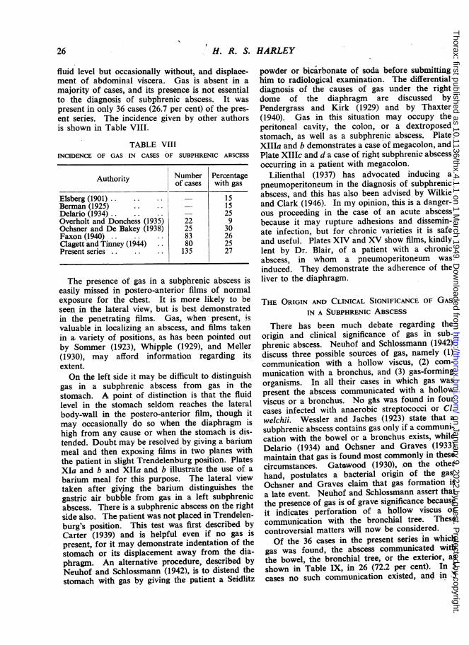
fluid level but occasionally without, and displaee-ment of abdominal viscera. Gas is absent in amajority of cases, and its presence is not essentialto the diagnosis of subphrenic abscess. It waspresent in only 36 cases (26.7 per cent) of the pres-ent series. The incidence given by other authorsis shown in Table VIII.
TABLE VIIIINCIDENCE OF GAS IN CASES OF SUBPHRENIC ABSCESS
Authority Number Percentageof cases with gas
Elsberg (1901) .. .. .. - 15Berman (1925) . 2515Delario (1934)..25Overholt and Donchess (1935) 22 9Ochsner and De Bakey (1938) 25 30Faxon (1940) .. .. .. 83 26Clagett and Tinney (1944) .. 80 25Present series .. .. .. 135 27
The presence of gas in a subphrenic abscess iseasily missed in postero-anterior films of normalexposure for the chest. It is more likely to be
seen in the lateral view, but is best demonstratedin the penetrating films. Gas, when present, isvaluable in localizing an abscess, and films takenin a. variety of positions, as has been pointed out
by Sommer (1923), Whipple (1929), and Meller(1930), may afford information regarding itsextent.On the left side it may be difficult to distinguish
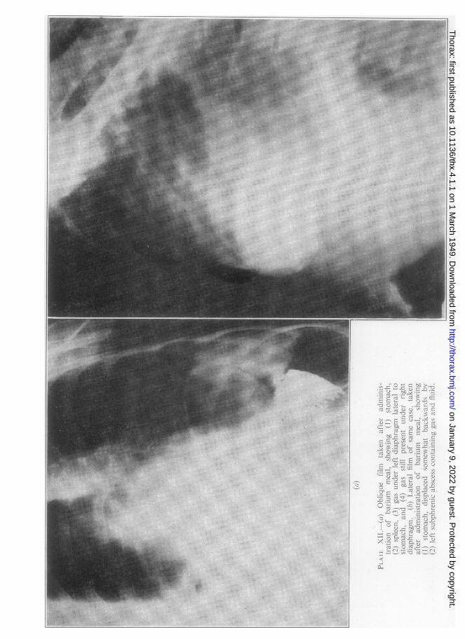
gas in a subphrenic abscess from gas in thestomach. A point of distinction is that the fluidlevel in the stomach seldom reaches the lateralbody-wall in the postero-anterior film, though itmay occasionally do so when the diaphragm ishigh from any cause or when the stomach is dis-tended. Doubt may be resolved by giving a bariummeal and then exposing films in two planes withthe patient in slight Trendelenburg position. PlatesXIa and b and XIIa and b illustrate the use of a
barium meal for this purpose. The lateral viewtaken after giving the barium distinguishes thegastric air bubble from gas in a left subphrenicabscess. There is a subphrenic abscess on the rightside also. The patient was not placed in Trendelen-burg's position. This test was first described byCarter (1939) and is helpful even if no gas is
present, for it may demonstrate indentation of thestomach or its displacement away from the dia-phragm. An alternative procedure, described byNeuhof and Schlossmann (1942), is to distend thestomach with gas by giving the patient a Seidlitz
powder or bicarbonate of soda before submittinghim to radiological examination. The differentialdiagnosis of the causes of gas under the rightdome of the diaphragm are discussed byPendergrass and Kirk (1929) and by Thaxter(1940). Gas in this situation may occupy theperitoneal cavity, the colon, or a dextroposedstomach, as well as a subphrenic abscess. PlateXIIIa and b demonstrates a case of megacolon, andPlate XIlIc and d a case of right subphrenic abscessoccurring in a patient with megacolon.
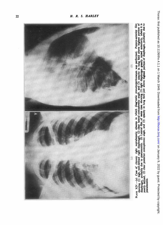
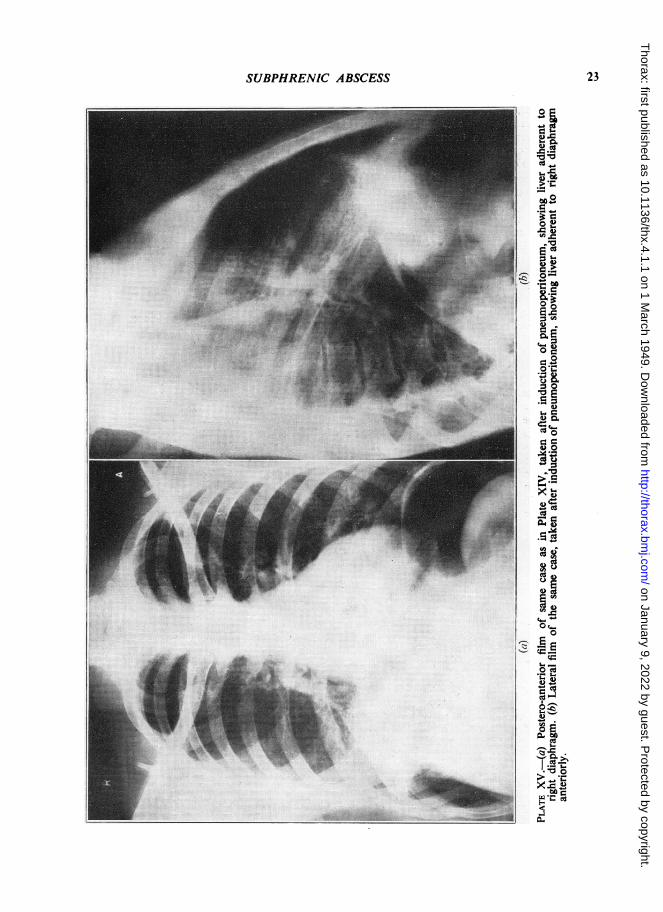
Lilienthal (1937) has advocated inducing apneumoperitoneum in the diagnosis of subphrenicabscess, and this has also been advised by Wilkieand Clark (1946). In my opinion, this is a danger-ous proceeding in the case of an acute abscessbecause it may rupture adhesions and dissemin-ate infection, but for chronic varieties it is safeand useful. Plates XIV and XV show films, kindlylent by Dr. Blair, of a patient with a chronicabscess, in whom a pneumoperitoneum wasinduced. They demonstrate the adherence of theliver to the diaphragm.
THE ORIGIN AND CLINICAL SIGNIFICANCE OF GASIN A SUBPHRENIC ABSCESS
There has been much debate regarding theorigin and clinical significance of gas in sub-phrenic abscess. Neuhof and Schlossmann (1942)discuss three possible sources of gas, namely (1)communication with a hollow viscus, (2) com-munication with a bronchus, and (3) gas-formingorganisms. In all their cases in which gas was
present the abscess communicated with a hollowviscus or a bronchus. No gas was found in fourcases infected with anaerobic streptococci or Cl.welchii. Wessler and Jaches (1923) state that a
subphrenic abscess contains gas only if a communi-cation with the bowel' or a bronchus exists, whileDelario (1934) and Ochsner and Graves (1933)maintain that gas is found most commonly in thesecircumstances. Gat&wood (1930), on the otherhand, postulates a bacterial origin of the gas.Ochsner and Graves claim that gas formation isa late event. Neuhof and Schlossmann assert thatthe presence of gas is of grave significance becauseit indicates perforation of a hollow viscus or
communication with the bronchial tree. Thesecontroversial matters will now be considered.Of the 36 cases in the present series in which
gas was found, the abscess com-municated withthe bowel, the bronchial tree, or the exterior, as
shown in Table IX, in 26 (72.2 per cent). In 5cases no such communication existed, and in -5
-26
on January 9, 2022 by guest. Protected by copyright.
http://thorax.bmj.com
/T
horax: first published as 10.1136/thx.4.1.1 on 1 March 1949. D
ownloaded from
SUBPHRENIC ABSCESS
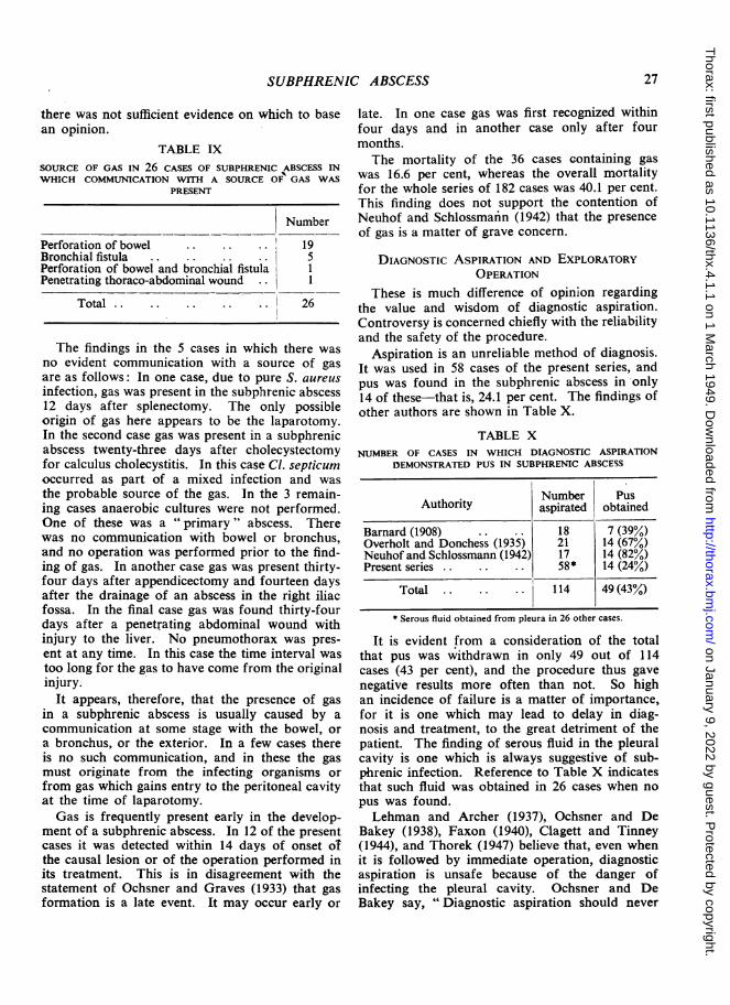
there was not sufficient evidence on which to basean opinion.
TABLE IXSOURCE OF GAS IN 26 CASES OF SUBPHRENIC ABSCESS INWHICH COMMUNICATION WITH A SOURCE OF GAS WAS
PRESENT
Number
Perforation of bowel .. .. .. 19Bronchial fistula .. .. .. .. 5Perforation of bowel and bronchial fistula IPenetrating thoraco-abdominal wound ..
Total . .. .. .. .. 26
The findings in the 5 cases in which there was
no evident communication with a source of gas
are as follows: In one case, due to pure S. aureus
infection, gas was present in the subphrenic abscess12 days after splenectomy. The only possibleorigin of gas here appears to be the laparotomy.In the second case gas was present in a subphrenicabscess twenty-three days after cholecystectomyfor calculus cholecystitis. In this case Cl. septicumoccurred as part of a mixed infection and was
the probable source of the gas. In the 3 remain-ing cases anaerobic cultures were not performed.One of these was a "primary" abscess. Therewas no communication with bowel or bronchus,and no operation was performed prior to the find-ing of gas. In another case gas was present thirty-four days after appendicectomy and fourteen daysafter the drainage of an abscess in the right iliacfossa. In the final case gas was found thirty-fourdays after a penetrating abdominal wound withinjury to the liver. No pneumothorax was pres-
ent at any time. In this case the time interval was
too long for the gas to have come from the originalinjury.
It appears, therefore, that the presence of gas
in a subphrenic abscess is usually caused by a
communication at some stage with the bowel, or
a bronchus, or the exterior. In a few cases thereis no such communication, and in these the gas
must originate from the infecting organisms or
from gas which gains entry to the peritoneal cavityat the time of laparotomy.Gas is frequently present early in the develop-
ment of a subphrenic abscess. In 12 of the presentcases it was detected within 14 days of onset ot
the causal lesion or of the operation performed inits treatment. This is in disagreement with thestatement of Ochsner and Graves (1933) that gas
formation is a late event. It may occur early or
late. In one case gas was first recognized withinfour days and in another case only after fourmonths.The mortality of the 36 cases containing gas
was 16.6 per cent, whereas the overall mortalityfor the whole series of 182 cases was 40.1 per cent.This finding does not support the contention ofNeuhof and Schlossmafin (1942) that the presenceof gas is a matter of grave concern.
DIAGNOSTIC ASPIRATION AND EXPLORATORYOPERATION
These is much difference of opinion regardingthe value and wisdom of diagnostic aspiration.Controversy is concerned chiefly with the reliabilityand the safety of the procedure.
Aspiration is an unreliable method of diagnosis.It was used in 58 cases of the present series, andpus was found in the subphrenic abscess in only14 of these-that is, 24.1 per cent. The findings ofother authors are shown in Table X.
TABLE XNUMBER OF CASES IN WHICH DIAGNOSTIC ASPIRATION
DEMONSTRATED PUS IN SUBPHRENIC ABSCESS
Number PusAuthority aspirated obtained
Barnard (1908) .. 18 7 (39%)Overholt and Donchess (1935) 21 14 (67%)Neuhof and Schlossmann (1942) 17 14 (82%)Present series .. .. .. 58* 14 (24%)
Total .. .. .. 114 49 (43%)
* Serous fluid obtained from pleura in 26 other cases.
It is evident from a consideration of the totalthat pus was withdrawn in only 49 out of 114cases (43 per cent), and the procedure thus gavenegative results more often than not. So highan incidence of failure is a matter of importance,for it is one which may lead to delay in diag-nosis and treatment, to the great detriment of thepatient. The finding of serous fluid in the pleuralcavity is one which is always suggestive of sub-phrenic infection. Reference to Table X indicatesthat such fluid was obtained in 26 cases when nopus was found.Lehman and Archer (1937), Ochsner and De
Bakey (1938), Faxon (1940), Clagett and Tinney(1944), and Thorek (1947) believe that, even whenit is followed by immediate operation, diagnosticaspiration is unsafe because of the danger ofinfecting the pleural cavity. Ochsner and DeBakey say, " Diagnostic aspiration should never
27
on January 9, 2022 by guest. Protected by copyright.
http://thorax.bmj.com
/T
horax: first published as 10.1136/thx.4.1.1 on 1 March 1949. D
ownloaded from
2H. R. S. HARLEY
be attempted, and in cases in which the diagnosisremains doubtful, exploratory operation shouldpreferably be undertaken."
Barnard (1908), Gatewood (1930), Bogart (1934),Janz (1934), and Szacsvay (1934) maintain thataspiration is safe, but only if performed immedi-ately before operation. Doherty and Rowlands(1931) and Overholt and Donchess (1935) depre-cate aspiration, but should it be performed, theyadvocate the use of the extra-serous route.
Neuhof and Schlossmann (1942) stress the diffi-culty in diagnosing subphrenic infection on theleft side, and conclude that "exploratory aspira-tion is imperative in all cases with vague clinicaland inconclusive features suggesting suppurationin the left subphrenic space." They performaspiration at the time of operation.
Precise statements regarding the frequency withwhioh infection of the pleural and peritoneal cavi-ties follows aspiration are scanty. In the presentseries there was evidence that infection of thepleura occurred in consequence of aspiration inonly one of the 58 cases in which it was performed.Lehman and Archer (1937) report two cases.
Empyema followed aspiration in one of the 21cases recorded by Overholt and Donchess (1935).In one of Barnard's (1908) cases no pus was
obtained at aspiration, but three hours later thepatient died and at necropsy it was found thatone and a half pints of turbid offensive pus hadpassed through the puncture track from the rightsubphrenic space into the p1eura, and that the lung
was collapsed. In none of Neuhof and Sch1oss-mann's (1942) cases, in all of which the abscessWas situated on the left side, did pleural infectionensue. Aspiration was responsible for pleuralinfection in 3 of the 114 cases represented inTable X.From these observations it is evident that the
pleura may be infected in consequence of diag-nostic aspiration, whether pus is withdrawn ornot, but in only a small percentage of cases doesthis event occur.
Aspiration is a method of diagnosis which, inmy opinion, should not be employed, chieflybecause it is unreliable but partly because of therisk of pleural infection which it entails, thoughthis risk is admittedly small. If aspiration fails,exploratory operation will alone exclude the diag-nosis; on the other hand, if pus is withdrawn an
operation will be required for the drainage of theabscess.
In those cases in which a study of the clinicaland radiological findings leaves the diagnosis in
doubt, the wisest course is to perform an explora-
tory operation under local anaesthesia by theextra-serous route, if necessary both from in frontand from behind.
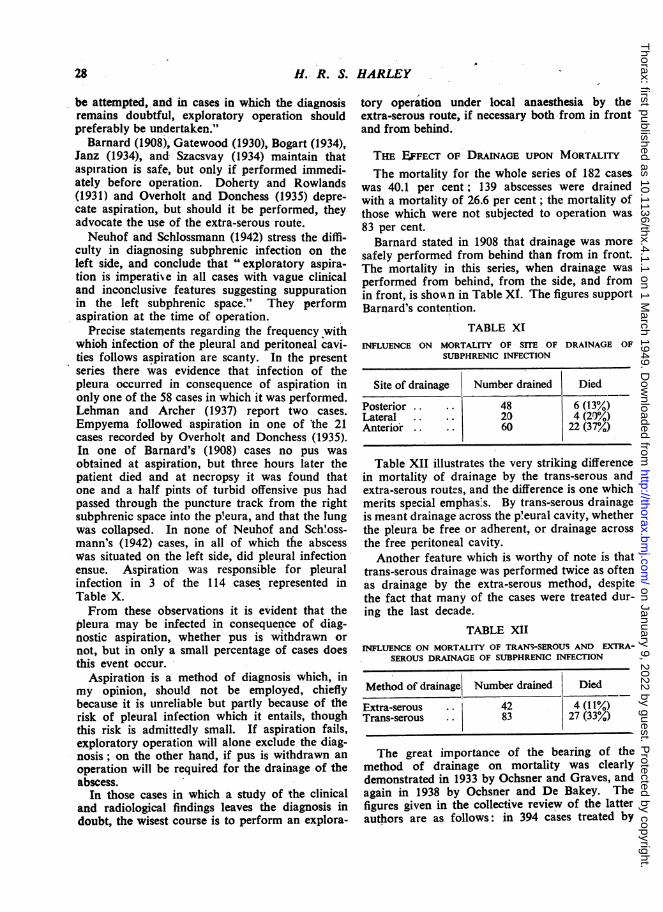
THE E.FFEcr OF DRAINAGE UPON MORTALMThe mortality for the whole series of 182 cases
was 40.1 per cent; 139 abscesses were drainedwith a mortality of 26.6 per cent; the mortality ofthose which were not subjected to operation was83 per cent.Barnard stated in 1908 that drainage was more
safely performed from behind than from in front.The mortality in this series, when drainage wasperformed from behind, from the side, and fromin front, is shoA n in Table XI. The figures supportBarnard's contention.
TABLE XIINFLUENCE ON MORTALITY OF SITE OF DRAINAGE OF
SUBPHRENIC INFECTION
Site of drainage Number drained Died
Posterior .. .. 48 6 (13%)Lateral .. .. 20 4 (29%)Anterior 60.... 22 (37%)
Table XII illustrates the very striking differencein mortality of drainage by the trans-serous andextra-serous routes, and the difference is one whichmerits special emphas:s. By trans-serous drainageis mea-nt drainage across the p'eural cavity, whetherthe pleura be free or adherent, or drainage acrossthe free peritoneal cavity.Another feature which is worthy of note is that
trans-serous drainage was performed twice as oftenas drainage by the extra-serous method, despitethe fact that many of the cases were treated dur-ing the last decade.
TABLE XIIINFLUIENCE ON MORTALITY OF TRANS-SEROUS AND EXTRA-
SEROUS DRAINAGE OF SUBPHRENIC INFECTION
Method of drainage Number drained Died
Extra-serousTrans-serous
42 4 (1 1%)83 27 (33%)
The great importance of the bearing of themethod of drainage on mortality was clearlydemonstrated in 1933 by Ochsner and Graves, andagain in 1938 by Ochsner and De Bakey. Thefigures given in the collective review of the latterauthors are as follows: in 394 cases treated by
28
on January 9, 2022 by guest. Protected by copyright.
http://thorax.bmj.com
/T
horax: first published as 10.1136/thx.4.1.1 on 1 March 1949. D
ownloaded from
SUBPHRENIC ABSCESS
trans-serous drainage the mortality was' 36.2 per
cent, whereas in 211 cases treated by the extra-serous method the mortality was 20.8 per cent; in37 cases drained extra-serously by Ochsner andDe Bakey themselves the mortality was only10.8 per cent. The importance of the extra-serousroute has been stressed more recently by Clagettand Tinney (1944), Zaveleta (1945), Lewenstein(1946), and Thorek (1947).That the proportion of cases treated by trans-
serous drainage in the present series is so highis evidence that the superiority of the extra-serousmethod is even yet not fully appreciated. It was
possible to determine the method of drainage usedin 74 patients treated since the beginning of 1939.In 60 per cent of these the trans-serous methodwas employed. Prior to 1939 the trans-serousroute was utilized in 74.5 per cent of cases.
A matter which also requires emphasis is thatadherence of the pleura is no guarantee againstthe spread of infection after trans-pleural drain-age. This has been stressed by Doherty andRowlands (1931). In the present series there was
one death amongst the four patients on whomdrainage was performed through a p'eural cavitywhich was stated to have been obliterated byadhesions. Barnard (1908) recorded 5 cases inwhich pleural adhesions gave way when the dia-phragm descended after the abscess had beendrained. This happened twice at operation'andthree times a day or two later.The extra-serous method of drainage should, I
believe, invariably be adopted in the treatment ofsubphrenic abscess, even when the serosa isadherent. By its employment the mortality ofoperation should be reduced to the neighbourhoodof 10 per cent.The entire subphrenic region can be reached by
the posterior and anterior extra-serous routesdescribed by Nather- and Ochsner (1923) and byClairmont and Meyer (1926). Which of theseroutes be chosen will depend upon the location ofthe abscess. If it is equally accessible from behindor in front then the posterior route should beselected.
SUMMARY
1. A series of 182 cases of subphrenic abscessis reviewed.
2. Only one intraperitoneal subphrenic space isrecognized above the right lobe of the liver.
3. Marked clinical differences were foundbetween suprahepatic and infrahepatic infection
on the right side. On the left side these differenceswere less striking.
4. Infection occurred on the left side in fiftycases. It presented certain ciinical peculiarities,and was attended by a high mortality.
5. A brief account of multiple-space infection isgiven and the high mortality noted, especiallywhen the abscess is bilateral.
6. Primary subphrenic abscess occurred inseventeen cases, and was attended by a relativelylow mortality.
7. Chronic subphrenic abscess is defined, andsixteen cases of the condition are discussed.
8. A majority of the cases of subphrenic abscesssecondary to intra-abdominal inflammation fol-lowed neighbouring infection. Many of those inwhich the inflammation was more remote fromthe subphrenic region were residual abscesses fol-lowing general peritonitis. No evidence wasobtained to suggest that spread of infectionoccurred via the abdominal lymphatic vessels.
9. Nineteen cases of subphrenic abscess in asso-ciation with liver abscess are presented, and thehigh mortality is noted.
10. Serous pleural effusion occurred in about25 per cent of cases. The cause of the effusionwas seldom in the chest, and the fluid rarelybecame purulent.
11. Intrathoracic suppuration occurred in slightlymore than 25 per cent of cases, and was usuallydue to some such cause as perforation of the d'ia-phragm or transpleural drainage of the sub-phrenic abscess. No evidence -was obtained tosuggest that infection commonly spreads from thesubphrenic region to the chest via the lymphaticpathways.
12. When a bronchial fistula was present in addi-tion to intrathoracic suppuration, the mortalitywas significantly diminished.
13. The radiological diagnosis is discussed indetail and certain difficulties are considered.
14. Gas was demonstrated in the abscess in26.7 p2r cent of cases examined radiological'y. Inmost cases it was caused by communicationbetween the abscess and the bowel, the bronchialtree, or the exterior. The mortality of the casesin which gas was present was lower than averagefor the series.
15. Diagnostic aspiration is not advised becauseof its unreliability, and also because of the dangerof infecting the pleura. This danger is less thanmight be expected.
29
on January 9, 2022 by guest. Protected by copyright.
http://thorax.bmj.com
/T
horax: first published as 10.1136/thx.4.1.1 on 1 March 1949. D
ownloaded from
H. R. S. HARLEY
16. Trans-serous drainage is attended by a muchhigher mortality than extra-serous drainage andis therefore condemned.
The clinical records and x-ray films were suppliedthrough the courtesy of Drs. L. G. Blair and G. Simon,Mr. F. J. Sambrook Gower, and the Medical Com-mittees of the following hospitals: Brompton; CharingCross; Guy's; Harefield County; Horton Emergency;King Edward VII, Ealing; London; LondonChest; Middlesex; St. Bartholomew's; St. George's;St. Stephen's; St. Thomas's; University College;Westminster. To all of these my grateful thanks aredue. I am indebted to Mr. N. R. Barrett and Mr.Grant Massie for their help and guidance, and toProf. H. T. Flint for his advice.
REFERENCESAllen, L. (1936). Anat. Rec., 67, 89.Banyai, A. L. (1946). "Pneumoperitoneum Treat-
ment." Henry Kimpton, London.Barnard, H. L. (1908). Brit. med. J., 1, 371, 429.Berman, J. K. (1925). J. Indiana med. Ass., 18, 217.Beye, H. L. (1932). J. thorac. Surg., 1, 655.Bizzozero, G., and Salvioli (1876). G. Acad. Med.
Torino, 19, 466.Bogart, L. M. (1934). Amer. J. Suirg., 26, 467.Bolton, C. (1921). J. Path. Bact., 24, 429.Brown, K. P. (1928). Brit. J. Surg., 15, 538.Buxton, B. H., and Torrey, J. C. (1906). J. med. Res.,
15, 3.Carter, B. N. (1939). Ohio St. med. J., 35, 833.Clagett, 0. T., and Tinney, W. S. (1944). Amer. J.
Surg., 66, 189.Clairmont', P., and Meyer, M. (1926). Acta chir.
scand., 60, 55.Cunningham, R. S. (1922). Amer. J. Physiol., 62, 248,
253.Cunningham, R. S. (1926). Physiol. Rev., 6, 242.Delario, A. J. (1934). Amer. J. Roentgen., 31, 177.Doherty, W. D., and Rowlands, R. P. (1931). Brit.
med. J., 1, 168.Drye, J. C. (1948). Surg. Gynec. Obstet., 87, 472.Elsberg, C. A. (1901). Ann. Surg., 34, 729.Faxon, H. H. (1940). New Engl. J. Med., 222, 289.Fifield, L. R., and Love, R. J. McN. (1926). Brit. J.
Surg., 13, 683.Florey, H. (1927). Brit. J. exp. Path., 8, 479.Florey, H., and Witts, L. J. (1928). Lancet, 1, 1323.Gatewood, B. (1930). Amer. J. med. Sci., 180, 398.Hahn, P. F., Miller, L. L., Robscheit-Robbins, F. S.,
Bale, W. F., and Whipple, G. H. (1944). J. exp.med., 80, 77.
Higgins, G. M., Beaver, M. G., and Lemon, W. S.(1930). Amer. J. Anat., 45, 137.
Higgins, G. M., and Graham, A. S. (1929). Arch. Surg.,Chicago, 19, 453.
Higgins, G. M., and Lemon, W. S. (1931). Amer. J.med. Sci., 181, 697.
Higgins, G. M., and Murphy, G. T. (1928). Anat.Rec., 40, 15.
Hitzenberger, K. (1929). Klin. Wschr., 8, 961.Howkins, J. (1948). Lancet, 2, 85.Janz, G. (1934). Arch. klin. Chir., 178, 482.Kolossow, A. (1893). Arch. mikr. Anat., 42, 318.Krause, N. J. (1927). Arch. klin. Chir., 144, 201.Lam, C. R. (1939). Arch. Surg., 39, 1006.Lehman, E. P., and Archer, V. W. (1937). Sth. Surg.,
6, 407.Levi, D. (1927). Brit. J. exp. Path., 8, 355.Lewenstein, H. J. (1946). J. Mt. Sinai Hosp., 13, 20.Lilienthal, H. Discussion of Lehman, E. P. (1937).
Ann. Surg., 106, 522.MacCallum, W. G. (1903). Bull. Johns Hopk. Hosp.,
14, 105.Meller,.O. (1930). Rontgen Praxis, 2, 1071.Menville, L. J., and Ane, J. N. (1934). Radiology,
23, 327.Mitchell, G. A. G. (1940). Brit. J. Surg., 28, 291.Muller, G. P., Overholt, R. H., and Pendergrass, E. P.
(1929). Arch. Surg., 19, 1322.Muscatello, G. (1895). Virchows Arch., 142, 327.Nather, C., and Ochsner, E. W. A. (1923). Surg.
Gynec. Obstet., 37, 665.Neuhof, H., and Schlossmann, N. C. (1942). Surg.
Gynec. Obstet., 75, 751.Ochsner, A., and De Bakey, M. (1938). Surg. Gynec.
Obstet., 66, 426. (Internat. Abstr. Surg).Ochsner. A., and Graves, A. M. (1933). Ann. Surg.,
98, 961.Overholt, R. H. (1931). Arch. Surg., Chicago, 22,
691.Overholt, R. H., and Donchess, J. C. (1935). New
Engi. J. Med., 213, 294.Pancoast, H. K. (1926). Amer. J. Roentgen., 16, 303.Pendergrass, E. P., and Kirk, E. (1929). Anmer. J.
Roentgen., 22, 238.Re&klinghausen, F. von (1862). " Die Lymphgefasse
und ihre Beziehung zum Bindegewebe." Berlin.Recklinghausen, F. von (1863). Virchows Arch., 26,
172.Roberts, J. E. H., and Nelson, H. P. (1933). Brit. J.
Surg., 21, 277.Rushmer, R. F. (1946). Amer. J. Physiol., 147, 242.Schwartz, J. (1930). Arch. Surg., 20, 317.Siperstein, D. M., and Sansby, J. M. (1923). Amer.
J. Dis. Child., 25, 107.Sommer, J. (1923). Zbl. Chir., 50, 215.Spalding, J. E. (1946). Lancet, 1, 643.Szacsvay, L. von (1934). Beitr. klin. Chir., 160, 591.Thaxter, L. T. (1940). Amer. J. Roentgen., 44, 853.Thorek, P. (1947). Surgery, 21, 739.Truesdale, P. E. (1933). Ann. Surg., 98, 846.Wessler, H., and Jaches, L. (1923). Quoted by Neuhof,
H., and Schlossmann, N. C..(1942). Surg. Gynec.Obstet., 75, 751. %
Whipple, A. 0. (1926). Amer. J. Surg., 40, 1.Wildesgans, H. (1923). Mitt. Grenzgeb. Med. Chir.,
37, 308.Wilkie, A. L., and Clark, J. C. (1946). Cznad. med.
Ass. J., 55, 296.Zaveleta, D. E. (1945). Prensa meld. argent., 32, 78.
30
on January 9, 2022 by guest. Protected by copyright.
http://thorax.bmj.com
/T
horax: first published as 10.1136/thx.4.1.1 on 1 March 1949. D
ownloaded from


