Embed Size (px)
Citation preview

Seediscussions,stats,andauthorprofilesforthispublicationat:https://www.researchgate.net/publication/273331534
Successfultissueengineeringofcompetentallogeneicvenousvalves
ARTICLE·MARCH2015
DOI:10.1016/j.jvsv.2014.12.002
READS
84
8AUTHORS,INCLUDING:
VijayKumarKuna
UniversityofGothenburg
11PUBLICATIONS74CITATIONS
SEEPROFILE
AntonioRosales
OsloUniversityHospital
17PUBLICATIONS104CITATIONS
SEEPROFILE
HenrikBackdahl
SPTechnicalResearchInstituteofSweden
17PUBLICATIONS783CITATIONS
SEEPROFILE
SuchitraHolgersson
UniversityofGothenburg
64PUBLICATIONS1,595CITATIONS
SEEPROFILE
Availablefrom:VijayKumarKuna
Retrievedon:05February2016
FrommGOanV
This(4Swan
AuthNneduO
Successful tissue engineering of competentallogeneic venous valvesVijay Kumar Kuna, MSc,a Antonio Rosales, MD,b Jonny Hisdal, PhD,b Eivind K. Osnes, MD, PhD,b
Jon O. Sundhagen, MD,b Henrik Bäckdahl, PhD,c Suchitra Sumitran-Holgersson, PhD,a andJørgen J. Jørgensen, MD, PhD,b,d Gothenburg and Borås, Sweden; and Aker and Oslo, Norway
Objective: The purpose of this study was to evaluate whethertissue-engineered human allogeneic vein valves have a normalclosure time (competency) and tolerate reflux pressure in vitro.Methods: Fifteen human allogeneic femoral vein segmentscontaining valves were harvested from cadavers. Valve closuretime and resistance to reflux pressure (100 mm Hg) wereassessed in an in vitro model to verify competency of the veinvalves. The segments were tissue engineered using the tech-nology of decellularization (DC) and recellularization (RC).The decellularized and recellularized vein segments werecharacterized biochemically, immunohistochemically, andbiomechanically.Results: Four of 15 veins with valves were found to be incom-petent immediately after harvest. In total, 2 of 4 segments withincompetent valves and 10 of 11 segments with competentvalves were further decellularized using detergents and DNAse.DC resulted in significant decrease in host DNA compared withcontrols. DC scaffolds, however, retained major extracellularmatrix proteins and mechanical integrity. RC resulted in suc-cessful repopulation of the lumen and valves of the scaffold withendothelial and smooth muscle cells. Valve mechanical param-eters were similar to the native tissue even after DC. Eight of 10
the Laboratory for Transplantation and Regenerative Medicine, Depart-ent of Surgery, Sahlgrenska Academy at the University of Gothenburg,othenburga; the Department of Vascular Surgery, Oslo Vascular Centre,slo University Hospital, Akerb; the Department of Chemistry, Materials,d Surfaces, SP Technical Research Institute of Sweden, Boråsc; and theascular Department, University of Oslo, Oslo.d
study was financed by the Swedish Government LUA ALF grants41421), the Swedish Heart and Lung Foundation (20130505), theedish Research Council K2013-65X-22347-01-3, and the Inga-Brittd Arne Lundberg foundation (2009-362) to S.S.H.or conflict of interest: S.S.H. is a cofounder and board member ofovaHep AB, a company that has licensed the technology of tissue engi-ering of blood vessels. There is a patent pending related to the proce-re of tissue engineering, for which A.R. and J.H. as scientists at theslo University Hospital are coinventors.
veins with competent valves remained competent even after DCand RC, whereas the two incompetent valves remained incom-petent even after DC and RC. The valve closure time to refluxpressure of the tissue-engineered veins was <0.5 second.Conclusions: Tissue-engineered veins with valves provide a validtemplate for future preclinical studies and eventual clinical ap-plications. This technique may enable replacement of diseasedincompetent or damaged deep veins to treat axial reflux and thusreduce ambulatory venous hypertension. (J Vasc Surg: Venousand Lym Dis 2015;-:1-10.)
Clinical Relevance: The use of natural, human scaffolds toproduce tissue-engineered venous segments containing func-tioning valves will revolutionize the surgical correction of deepvenous reflux in patients with chronic venous insufficiency andleg ulcer. Reconstructive deep venous surgery in the form ofvalvuloplasty, transplantation, and neovalve construction hasmet limitations in the rare availability of valves to be repaired,lack of donor sites, and inadequate conditions to create newvalves. This tissue-engineered procedure produces the func-tioning unit “valve-conduit,” and surgery will be used only toimplant it.
Chronic venous insufficiency (CVI) describes a condi-tion that affects the venous system of the lower extremities,in which persistent ambulatory venous hypertension is themain pathophysiologic factor leading to pain, edema, skinchanges, and ulcerations.1 The more serious consequencesof CVI, such as venous ulcers, have an estimated prevalenceof 0.1% to 1.0%.2,3 The overall prognosis of venous ulcers ispoor.4 Risk factors found to be associated with CVI includeage, sex, family history of varicose veins, obesity, preg-nancy, and phlebitis.5,6 The financial burden of venous ul-cer disease on the health care system is an estimated $1
billion spent annually on treatment of chronic wounds inthe United States or #2% of the total health care budgetin Western countries.
The conventional treatment of CVI with compressionstockings combined with superficial surgery seems toimprove venous hemodynamics but achieves only a 65% ul-cer healing rate after 24 weeks, with a recurrence rate of12%/year.7 Reconstructive deep venous surgery, such asvalvuloplasty, autotransplantation, and neovalve construc-tion, has proved to be an option, improving ulcer healingrates and providing an ulcer-free period in patients for
Additional material for this article may be found online at www.jvsvenous.org.Reprint requests: Suchitra Sumitran-Holgersson, PhD, Laboratory forTransplantation and Regenerative Medicine, Sahlgrenska Science Park,Medicinaregatan 8A, 2nd Fl, S-413 46, Gothenburg, Sweden (e-mail:[email protected]).
The editors and reviewers of this article have no relevant financial relation-ships to disclose per the Journal policy that requires reviewers to declinereview of any manuscript for which they may have a conflict of interest.
2213-333XCopyright � 2015 The Authors. Published by Elsevier Inc. on behalf of theSociety for Vascular Surgery. This is an open access article under the CCBY-NC-ND license (http://creativecommons.org/licenses/by-nc-nd/4.0/).
http://dx.doi.org/10.1016/j.jvsv.2014.12.002
1

Fig 1. Picture of the setup and in vitro model for functional testing of veins. A, The system is circulated with room-temperature saline containing an ultrasound contrast agent for enhancement of the Doppler signals. A peristaltic pump(a) pumps the saline through the whole circuit; a mechanical valve (b) enables flow through the vein (c) during outputfrom the pump. An ultrasound probe (d) is used for visualization of the vein (e) and evaluation of the flow through thevein valve. Reflux pressure at the valve site is adjusted by the height of the reservoir (f) above the vein. B, Enlargedpicture of a valve-bearing venous segment mounted in the circuit and placed in a container filled with saline to facilitatevisualization by ultrasound.
JOURNAL OF VASCULAR SURGERY: VENOUS AND LYMPHATIC DISORDERS2 Kuna et al --- 2015
whom conventional treatment has failed. Axillary or saphe-nous transplantation to treat axial reflux in these patientshas been reported to yield ulcer healing rates of up to70%. However, the durability of these procedures remainsan issue, taking into consideration that the average age ofpatients in reported materials is about 50 years, and onlyhalf of the transplants remained functional after 4 years.8-11
In addition, the demanding surgical technique needed haslimited the use of reconstructive deep venous surgery tovery few centers, and although reports have been producedthat show good results, no randomized clinical trials haveyet been published. Experimental efforts to understandthe venous valve function to be able to create mechanicaland bioprosthetic valves also exist. The challenges encoun-tered are to avoid immune reaction, thrombogenicity, andmigration in the vein.12-16
Thus, given the prevalence and socioeconomic impactof CVI and the poor treatment modalities available for it,alternative, more effective therapeutic options need to beexplored. Regenerative medicine may offer novel strategiesto treat these patients. Tissue-engineered venous segmentscontaining competent valves might represent a more dura-ble solution for this young group of patients. In addition,the required surgical skills will not be as demanding (inter-position of a vein segment), which might help spread thispossibility of treating severe CVI cases.
Perfusion decellularization (DC) and recellularization(RC) of tissues and organs is believed to be a successfulplatform technology for creating scaffolding materials fortissue engineering and regenerative medicine.17,18 DC isthe process of removal of all host cells and nuclear materialby physical, chemical, and enzymatic methods.19 Thus,whole organ acellular matrices provide an attractive scaffold
for the repopulation with cells for an engineered tissue ororgan because of the physiologic resemblance to the originaltissue, including intact three-dimensional anatomic architec-ture, preserved spatial array of extracellular matrix (ECM)components, vascular network, and biomechanical proper-ties. Creating regenerated donor organs by repopulatingorgan scaffolds with patient-specific mature or stemcell-derived populations would allow us to personalize trans-plantation medicine and reduce the need for long-termimmunosuppressive therapy. Using this approach, werecently tissue engineered human veins using autologousstem cells from the patients and successfully transplantedthree pediatric patients with portal vein thrombosis.20,21 Inthe present study, we have further expanded the applicationof this technology to tissue engineer human veins containingvalves.Wehypothesized that vein segmentswith valves tissueengineered by the technology of DC and RCwith stem cellswould yield a blood vessel with physiologic properties resem-bling the original tissue and that the valves would retain theircompetency, making them resistant to reflux.
METHODS
The Swedish and Norwegian Institutional Review Boardsand Ethics Committees approved all research protocols.
Harvesting of valve-bearing veins. All veins (N ¼15) used in this study were harvested at Oslo UniversityHospital, Norway. Vein segments about 8 cm (range,5.5-10 cm), including the common femoral vein, profundafemoral vein, and femoral vein, were harvested from adulthuman cadavers by using vascular surgical technique andcarefully ligating all side branches. Valves were identified,and 15 segments were cut with a margin of 3 to 4 cm oneach end of the valves. Sterile saline solution was injected

Fig 2. Gross morphology and microscopic view of the decellularized valve-containing vein segments. Grossmorphology of (A) native vein and (B) decellularized vein after 14 cycles. Hematoxylin and eosin (HE) staining of (C)decellularized vein after 14 cycles showing preserved tissue architecture and absence of blue-black nuclei and (D) anative vein (positive control) showing presence of nuclei. DAPI staining of native vein showed several nuclei (E), butnot in (F) decellularized vein. Immunohistochemical staining of decellularized veins showed absence of HLA class I(G) and class II (H) antigen expression. Normal vein (positive control) stained positive for HLA class I (I, brown) butnot HLA class II (J). K, Negative control.
JOURNAL OF VASCULAR SURGERY: VENOUS AND LYMPHATIC DISORDERSVolume -, Number - Kuna et al 3
manually, and functioning valves were identified by theretainment of solution above the valves. It was also possibleto see the valve leaflets closing. We had approximately 8-cm segments of the veins with a valve in the middle.Four of 15 veins with valves were found to be incompetentimmediately after harvest. All vein segments were thor-oughly rinsed in phosphate-buffered saline (PBS) contain-ing 0.5% penicillin, 0.5% streptomycin, and 0.5%amphotericin B and preserved at 4�C. The samples weretransported within 1 week on ice to the Laboratory forTransplantation and Regenerative Medicine at theSahlgrenska University Hospital, Gothenburg, Sweden. Intotal, 2 incompetent veins and 1 competent vein withvalves were used for the biomechanical tests, whereas 10competent and 2 incompetent veins with valves were usedfor DC and RC.
Functional in vitro testing of valve-bearing veins.This test was performed at the Oslo Vascular Centre, OsloUniversity Hospital, Norway. A custom-made test setupwas used to assess the functionality of the veins before andafter RC (Fig 1, A). The in vitro test setup used in thepresent study for evaluating the functionality is a modifica-tion of the one used by Geselschap et al.22 The aim of oursetup was basically to have a model that would allow us toassess venous valve function only at a determined pressure.
The vein was mounted vertically in the in vitro flow cir-cuit and perfused with room-temperature saline. The veinwas connected to the fluid circuit by conic connectors,secured with sutures at both ends (Fig 1, B). A commercialperistaltic pump (Model No. 700044; Baxter HealthcareCorp, Deerfield, Ill) delivered intermittent flow to thecircuit.
A mechanical valve was used to regulate the flow direc-tion through the circuit during outflow from the pump,and when the same valve was switched to open position,it achieved backflow in the vein until the valve closed.Reflux pressure in the vein was adjusted by the height ofthe column of fluid above the valve. The choice of reverseflow pressure level at 100 mm Hg was based on the esti-mated distance between the heart and the popliteal fossaand supported by the mathematical model published byFragomeni et al.23 Ultrasound Doppler technique wasused to detect potential reflux at the site of the venousvalve (9 MHz linear probe, Vivid E9; GE Healthcare, Bos-ton, Mass). To optimize the visualization with ultrasound,the vein segment was submerged in a plastic container filledwith saline. A contrast agent (SonoVue; Bracco Diagnos-tics, Cranbury, NJ) was administered in the saline solutionto enhance the echogenicity and to enable recordings ofthe flow and flow direction in the circuit through the

Fig 3. Extracellular matrix (ECM) in decellularized veins. A, Masson trichrome (MT) staining of normal vein (positivecontrol) showing presence of nuclei (black), cytoplasm (red/pink), and collagen (blue). B, In the decellularized vein, nonuclei were found, indicating lack of endothelial and smooth muscle cells, but with abundant collagen still present. C,Graphs showing decrease in amount of collagen (n ¼ 5), significant increase in elastin (n ¼ 5;P ¼ .03), and significantdecrease in glycosaminoglycans (GAGs; n ¼ 5;P ¼ .007), respectively, after 14 decellularization (DC) cycles incomparison to native (N).D, Graph showing significant decrease in amount of DNA after DC (P ¼ .0002). Scale bar Aand B, upper panel ¼ 50 mm, and lower panel ¼ 25 mm.
JOURNAL OF VASCULAR SURGERY: VENOUS AND LYMPHATIC DISORDERS4 Kuna et al --- 2015
vein valve. The time from flow reversal until cessation offlow was used as a measure of valve closure time. In addi-tion, the vein diameter was measured at the site of theleaflet at a reflux pressure at 100 mm Hg.
DC and characterization of veins. The processes ofDC, RC, and associated analysis were carried out in Goth-enburg, Sweden. DC was carried out as described by usearlier using 1% Triton, 1% tri-n-butyl phosphate (TnBP),and 4 mg/L DNAse.20 In brief, the veins were washedfor 72 hours in distilled water. One end of the vein wassealed with 4-0 suture, and the vein was filled with thedecellularizing solution; the other end was clamped. Thevein was placed in a container with the same solution andagitated on a shaker for 4 hours at 170 rpm and 37�C. Allthe solutions used for DC contained 0.02% sodium azide(71290; Sigma-Aldrich, Seelze, Germany) and 0.18%EDTA (ED2SS; Sigma-Aldrich). Triton (X-100; Sigma-Aldrich) and TnBP (A16084; Alfa Aesar, Karlsruhe,Germany) solutions were prepared in distilled water;DNase was prepared in PBS containing calcium and mag-nesium (D8662; Sigma-Aldrich). After every detergent, theveins were washed with distilled water for 10 minutes. Theprocedure from Triton to DNase is termed one cycle. Thedecellularized vein segments were evaluated for residualDNA content, staining with hematoxylin and eosin (HE)and Masson trichrome (MT) by standard procedures foracellularity and quantification of various ECM proteins.Immunohistochemistry for detection of HLA class I andclass II antigens was performed by standard procedure.
See the Supplementary Methods (online only) for de-tails of DNA quantification, ECM staining and
quantification, acid- and pepsin-soluble collagen quantifi-cation, sulfated glycosaminoglycan (GAG) quantification,and soluble elastin quantification.
RC of veins. On the day of RC, 20 to 25 mL of pe-ripheral venous blood was collected from healthy donors(age group, 25-35 years) in sterile heparin-coated Vacu-tainer tubes and transported to the laboratory as soon aspossible (within 2 hours). Healthy donors were individualsand personnel with no obvious diseases or disorders andwith no venous reflux disease. The volume of bloodrequired was determined by the length of the vessel and ofthe pipes used in the bioreactor.
The entire RC process was performed under sterile condi-tions, and all perfusions were carried out in an incubator at37�C supplied with 5% CO2. Before RC, the veins wereperfused with heparin (387107; LEO Pharma, Malmö, Swe-den) at a concentration of 50 IU/mL PBS for 2 hours. Theheparin was drained off, and whole blood was immediatelyperfused for 48 hours at 2 mL/min speed. The blood wasthen drained off, and the vein was rinsed with PBS containing1% penicillin-streptomycin-amphotericin until blood wascompletely removed. The vein was subsequently perfused4 days with endothelial and 4 days with smoothmusclemedia.The complete endothelial medium was prepared withMCDB131 (10372; Life Technologies, Stockholm, Sweden)basalmediumsupplementedwith10%heat-inactivatedhumanAB serum (34005100; Life Technologies), 1% glutamine(25030; Lonza, Copenhagen, Denmark), 1% penicillin-streptomycin-amphotericin, and EGM-2 SingleQuot kit(CC4176; Lonza) that contained ascorbic acid, hydrocorti-sone, transferrin, insulin, recombinant human vascular

Fig 4. Characterization of recellularized valve containing vein segments. A, Gross picture of a recellularized veinshowing valves. B and C,Hematoxylin and eosin (HE)-stained microscopic pictures of a recellularized vein and valve atlow and high magnification, respectively. The pictures show presence of continuous cells at endothelial lining on bothvein and valve (arrow). D, HE and Masson trichrome (MT) staining of normal (positive control) and recellularizedvalves showing presence of nuclei in all. E, CD31 staining of a recellularized vein showing continuous endotheliallining. F, Alpha smooth muscle actin staining of valve showing presence of smooth muscle cells in the valve. G, Alphasmooth muscle actin staining showing smooth muscle cells in media of a recellularized vein.
JOURNAL OF VASCULAR SURGERY: VENOUS AND LYMPHATIC DISORDERSVolume -, Number - Kuna et al 5
endothelial growth factor (VEGF), human fibroblast growthfactor, human epithelial growth factor, heparin, and genta-micin sulfate. The complete smooth muscle medium was pre-pared using 500mLMedium231 (M231; Life Technologies)supplied with 10% heat-inactivated human AB serum, 1%penicillin-streptomycin-amphotericin, and 20 mL of smoothmuscle growth supplement (S00725; Life Technologies).Vein scaffolds were recellularized for a total of 10 days.21
See the Supplementary Methods (online only) for de-tails of characterization of recellularized veins, biomechan-ical analysis, sterility control test, and statistics.
RESULTS
Functional in vitro testing of cadaveric valve-bearing veins. A total of 15 femoral vein specimenswere harvested from human cadavers. The median time be-tween death and harvest was 3 days (2-6) and betweendeath and testing, 6 days (5-7). Four of 15 veins withvalves were incompetent immediately after harvest. In gen-eral, the median diameter of the vein specimens was9.8 mm (7.5-14) and 9.8 mm (8-14.2) after RC. The 11vein segments with competent valves (normal closuretime #0.5 second at a pressure of 100 mm Hg)24 and
the 4 vein segments with incompetent valves were thentransported to Sweden. In Sweden, further examination ofthe four incompetent valves showed presence of holes inthe valves of two veins, mechanical damage in one, and noapparent problems with the fourth. This was examined byturning the veins inside-out. Two of the four incompetentveins with valves were used for biomechanical testing; theother two were used for DC and RC. Further, one veinwith competent valves was used for biomechanical testing,whereas 10 of 11 were used for DC and RC. Thus, in total,3 of 15 veins were used for biomechanical studies, whereas12 of 15 were used for DC and RC.
We found that 8 of 10 segments with competent valvesretained their competency after the RC process, whereas 2showed reflux already after DC. In both cases, the valves inthe segments were damaged mechanically while the speci-mens were handled during DC. In addition, the two seg-ments with incompetent valves already at the start werealso found to be incompetent even after DC and RC.Thus, RC did not repair the damaged valves.
DC, characterization, and functional in vitrotesting of decellularized valve-bearing veins. Treatmentwith 1% Triton and 1% TnBP successfully decellularized the
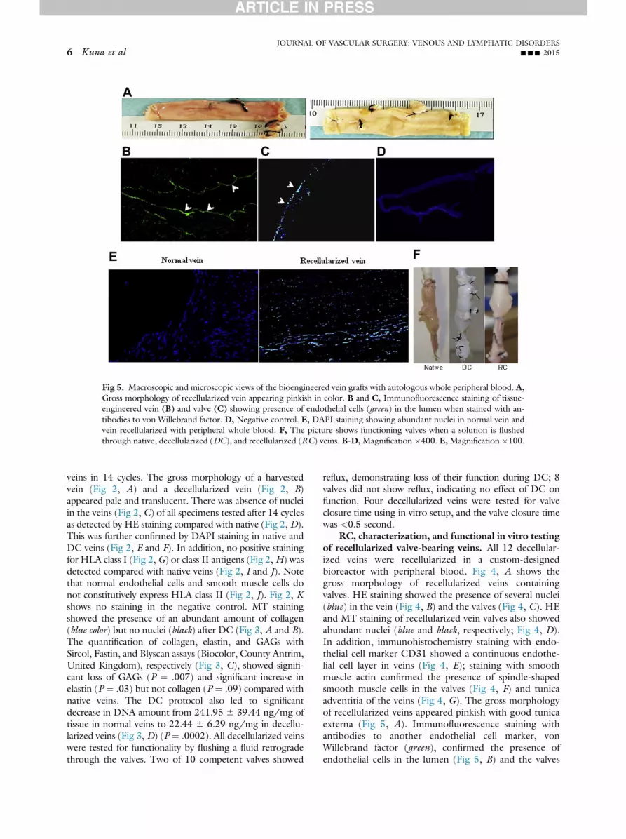
Fig 5. Macroscopic and microscopic views of the bioengineered vein grafts with autologous whole peripheral blood. A,Gross morphology of recellularized vein appearing pinkish in color. B and C, Immunofluorescence staining of tissue-engineered vein (B) and valve (C) showing presence of endothelial cells (green) in the lumen when stained with an-tibodies to von Willebrand factor. D, Negative control. E, DAPI staining showing abundant nuclei in normal vein andvein recellularized with peripheral whole blood. F, The picture shows functioning valves when a solution is flushedthrough native, decellularized (DC), and recellularized (RC) veins. B-D,Magnification �400. E,Magnification �100.
JOURNAL OF VASCULAR SURGERY: VENOUS AND LYMPHATIC DISORDERS6 Kuna et al --- 2015
veins in 14 cycles. The gross morphology of a harvestedvein (Fig 2, A) and a decellularized vein (Fig 2, B)appeared pale and translucent. There was absence of nucleiin the veins (Fig 2, C) of all specimens tested after 14 cyclesas detected by HE staining compared with native (Fig 2, D).This was further confirmed by DAPI staining in native andDC veins (Fig 2, E and F). In addition, no positive stainingfor HLA class I (Fig 2, G) or class II antigens (Fig 2,H) wasdetected compared with native veins (Fig 2, I and J). Notethat normal endothelial cells and smooth muscle cells donot constitutively express HLA class II (Fig 2, J). Fig 2, Kshows no staining in the negative control. MT stainingshowed the presence of an abundant amount of collagen(blue color) but no nuclei (black) after DC (Fig 3, A and B).The quantification of collagen, elastin, and GAGs withSircol, Fastin, and Blyscan assays (Biocolor, County Antrim,United Kingdom), respectively (Fig 3, C), showed signifi-cant loss of GAGs (P ¼ .007) and significant increase inelastin (P ¼ .03) but not collagen (P ¼ .09) compared withnative veins. The DC protocol also led to significantdecrease in DNA amount from 241.95 6 39.44 ng/mg oftissue in normal veins to 22.44 6 6.29 ng/mg in decellu-larized veins (Fig 3, D) (P ¼ .0002). All decellularized veinswere tested for functionality by flushing a fluid retrogradethrough the valves. Two of 10 competent valves showed
reflux, demonstrating loss of their function during DC; 8valves did not show reflux, indicating no effect of DC onfunction. Four decellularized veins were tested for valveclosure time using in vitro setup, and the valve closure timewas <0.5 second.
RC, characterization, and functional in vitro testingof recellularized valve-bearing veins. All 12 decellular-ized veins were recellularized in a custom-designedbioreactor with peripheral blood. Fig 4, A shows thegross morphology of recellularized veins containingvalves. HE staining showed the presence of several nuclei(blue) in the vein (Fig 4, B) and the valves (Fig 4, C). HEand MT staining of recellularized vein valves also showedabundant nuclei (blue and black, respectively; Fig 4, D).In addition, immunohistochemistry staining with endo-thelial cell marker CD31 showed a continuous endothe-lial cell layer in veins (Fig 4, E); staining with smoothmuscle actin confirmed the presence of spindle-shapedsmooth muscle cells in the valves (Fig 4, F) and tunicaadventitia of the veins (Fig 4, G). The gross morphologyof recellularized veins appeared pinkish with good tunicaexterna (Fig 5, A). Immunofluorescence staining withantibodies to another endothelial cell marker, vonWillebrand factor (green), confirmed the presence ofendothelial cells in the lumen (Fig 5, B) and the valves

Fig 6. Functional testing of tissue-engineered valve-containing veins. A, Two-dimensional pictures from ultrasoundshow a longitudinal projection of the vein segment: a closed valve (a); the gradual opening of the valve duringantegrade flow (b-d); and the closing of the valve with retrograde flow (e and f). B, The yellow Doppler scale gives thevalve closure time in seconds.
JOURNAL OF VASCULAR SURGERY: VENOUS AND LYMPHATIC DISORDERSVolume -, Number - Kuna et al 7
(Fig 5, C) compared with the negative control (Fig 5, D).Fig 5, E depicts the DAPI staining of a native and recel-lularized vein showing the presence of blue nuclei in bothsections. Testing of valve function showed that freshlyharvested, decellularized and recellularized veins distendwhen flushed with fluid through a syringe (Fig 5, F).Ultrasound testing demonstrated the gradual opening ofthe valve during antegrade flow and closing of the valvewith retrograde flow (Fig 6, A), and the Doppler scalerevealed the valve closure time in seconds (Fig 6, B;Video, online only). Only 8 of 12 recellularized veinswere functional. The veins that lost function whiledecellularized and the veins nonfunctional from thebeginning did not show function.
Biomechanical testing of the valves. In total, sixrecellularized veins (n ¼ 12 valves) and three normal veins(n ¼ 6 valves) were tested. The vein valves were graduallytorn apart starting from the suture. The tear progressedhorizontally, giving rise to peaks when the fibers of thevalve broke. Eventually one of the ends of the valve wastorn from the vein wall, as indicated by the last peak inthe force/elongation diagram (Fig 7, A). Forces at firstbreak (peak) were above 0.8 N for all vein valves thatproved to function in the functional in vitro test (Fig 7, B).
The median force was not significantly different betweennormal and RC functional valves (Fig 7, C).
Sterility control test. In both culture media of all theveins, no marked increase in absorbance was recorded at14 days of culture. The optical density at 600 nm measuredin the spectrophotometer for fresh media (0.006) wassimilar to that of perfused media (0.010), which was thesame as the negative control (0.010). The optical densityof the positive control was high (0.135). No colonies ofbacteria or fungal growth were seen on the tryptone soyaagar plates, confirming sterility of the vein segments.
DISCUSSION
For the first time, we demonstrate the successful pres-ervation of valve function of tissue-engineered humanvalve-bearing vein segments in an in vitro setting. Themain aim of the study was to demonstrate that the technol-ogy of DC and RC generates a graft with a competentvalve, provided the native vein already has a functioningvalve. The present study also demonstrates the successfulre-endothelialization of decellularized human vein seg-ments and vein valves with a simple peripheral blood sam-ple that enabled significantly the formation of extracellularmonolayers. Although transplant donors would provide

Fig 7. Mechanical analyses of the recellularized (RC) valves. A, Representative diagram of the deformation behaviorfor native and RC vein valves. The valves were gradually torn apart horizontally, resulting in a series of peaks. B, Forceat first peak for each pair of valves from the same vein. Native vein valves are squares; RC valves are circles. Green colormarks functioning veins, whereas red veins do not function. The blue line represents a cutoff for how high force at firstpeak a vein needs to have to function. C, Box plots of median force of working valves indicating no significantdifferences.
JOURNAL OF VASCULAR SURGERY: VENOUS AND LYMPHATIC DISORDERS8 Kuna et al --- 2015
fresher tissue, we for the first time explored the use of ca-davers as a source of the tissue. Interestingly, the majorityof the vein segments taken from human cadavers 4 to6 days after demise still had functioning grafts and couldbe used for tissue engineering. This promising observationcan help increase the pool of blood vessel donors. Becauseof the lack of transplant donors and limited access to thesetypes of donors, we are currently developing a program forusing cadaver veins. The ability of the decellularizing pro-cess to leave “footprints” for RC may open the possibilityfor brain-dead, heart-beating transplant donors and xeno-graft tissue.
We found it important for the specimen to be har-vested with precise vascular surgical technique, and the dis-tance between the actual vein valve and the ends should beat least 4 cm. In a future clinical study, we intend to test thespecimens after each of the following procedures: (1) har-vesting, (2) DC, and (3) RC. We believe that the in vitromodel can be further simplified by connecting the veinsegment directly to a fluid reservoir that is placed at1.36 meters (100 mm Hg) high to assess the competenceof the veins.
Mechanical analysis of the valves showed that in mostof the functioning valves, the force at first peak was above0.8 N for both vein valves in a pair, indicating that 0.8 Nmay be the cutoff criterion required to define strength offunctional valves. However, larger numbers of sampleswill have to be tested to confirm this interesting finding,and such studies are ongoing in our laboratory.
Interestingly and importantly, the vein specimen’sstructure and function do not seem to be affected by a
window of time of up to 7 days from death to harvestand testing. Thus, the pool of cadaver donors for bloodvessels can be increased enormously. The histologic resultsshowed that DC with Triton/TnBP/DNase was complete,as adjudged by DNA quantification, which was <50 ng/mg weight. It is reported that >50 ng/mg dry weight isnormally required to evoke a potential immunologicactivity.25
We found that perfusion of peripheral whole blood re-sults in the formation of a clear extracellular monolayer andpresence of smooth muscle cells in the media. The use of asimple peripheral blood sample to recellularize the vein seg-ments is a clear advantage over the more tedious approaches,such as isolation and expansion of mature cells or stem cellsfrombonemarrowor peripheral blood. In our recent study,21
we reported that the major cell types in circulating peripheralblood that contribute to re-endothelialization and smoothmuscle cell repopulation are VEGFR2þ/CD45þ cells and asmall fraction of VEGFR2þ/CD14þ cells. Furthermore,function and strength were also preserved in the recellular-ized vein valves as indicated by results obtained using thein vitro test model and biomechanical test. Thus, the presentresults are further supported by our recently published clinicalproof-of-concept study, in which we successfully transplantedthree pediatric patients with tissue-engineered veins usingautologous peripheral whole blood. Interestingly, none ofthe four incompetent veins with valves regained their compe-tency after RC, indicating that cells and elastin on the valvesare not critical factors for valve functionality. However, agreater number of incompetent valves need to be tissue engi-neered to fully understand the problems involved. According

JOURNAL OF VASCULAR SURGERY: VENOUS AND LYMPHATIC DISORDERSVolume -, Number - Kuna et al 9
to our observations, maintenance of the mechanical propertymay be one of the most important factors for competency.
Patients with CVI who prove refractory to conven-tional treatment may benefit from reconstructive deepvalve reconstruction. However, this is a treatment that isnot widespread and readily available because of complexworkup and demanding surgical technique. Both venousvalve transfer and valve construction have a durabilitythat could be improved because the average age of thesepatients is 50years.8-11 Venous valvuloplasty has been re-ported to provide competency in z60% and ulcer-freerecurrence in z60% at 30 months.26 Other proceduresfor reconstruction of nonfunctioning venous valves result-ing from post-thrombotic valve destruction include trans-position, transplantation, cryopreserved vein valveallografts, and neovalve construction. Cryopreserved veinvalve allografts have also been used but are limited becauseof frequent complications, such as early thrombosis andpoor patency and competency.27 Thus, reconstructivedeep venous surgery in the form of valvuloplasty, trans-plantation, and neovalve construction has its limitations.However, the use of natural, human scaffolds to producetissue-engineered venous segments containing functioningvalves may revolutionize the surgical correction of deepvenous reflux in patients with CVI and leg ulcer.
Although the results are promising, this is an in vitrostudy and cannot predict the outcome for future humanimplantations. Other limitations are the small number ofpatients studied and the limited number of biomechanicalstudies performed on decellularized valves. It would alsobe good in future studies to include tests such as burst pres-sure and compliance. Tissue-engineered valve-containingvein segments may be a therapeutic option in selected pa-tients in whom deep venous reflux and venous hyperten-sion are the main pathophysiologic features leading torecurrent leg ulcer. We believe that the results of the pre-sent study are encouraging and constitute the basisrequired to start a clinical pilot study.
CONCLUSIONS
In the future, personalized tissue-engineered veins withcompetent valves may enable the replacement of incompe-tent or destroyed deep vein valves in patients with CVI.
We wish to thank Pradeep B. Patil for design of thebioreactor used in the study.
AUTHOR CONTRIBUTIONS
Conception and design: AR, JH, SH, JJAnalysis and interpretation: VK, AR, JH, SH, JJData collection: VK, AR, JH, EO, JS, HBWriting the article: VK, AR, SHCritical revision of the article: VK, AR, SH, JJFinal approval of the article: VK, AR, JH, EO, JS, HB, SH,
JJStatistical analysis: VK, HBObtained funding: SHOverall responsibility: SH
VK and AR contributed equally to this article and shareco-first authorship.
REFERENCES
1. Simka M, Majewski E. The social and economic burden of venous legulcers: focus on the role of micronized purified flavonoid fractionadjuvant therapy. Am J Clin Dermatol 2003;4:573-81.
2. Nelzen O, Bergqvist D, Lindhagen A. The prevalence of chronic lower-limb ulceration has been underestimated: results of a validated popu-lation questionnaire. Br J Surg 1996;83:255-8.
3. Rhodes JM, Gloviczki P, Canton LG, Rooke T, Lewis BD, Lindsey JR.Factors affecting clinical outcome following endoscopic perforator veinablation. Am J Surg 1998;176:162-7.
4. Callam MJ, Harper DR, Dale JJ, Ruckley CV. Chronic ulcer of the leg:clinical history. Br Med J (Clin Res Ed) 1987;294:1389-91.
5. Scott TE, LaMorte WW, Gorin DR, Menzoian JO. Risk factors forchronic venous insufficiency: a dual case-control study. J Vasc Surg1995;22:622-8.
6. Jawien A. The influence of environmental factors in chronic venousinsufficiency. Angiology 2003;54(Suppl 1):S19-31.
7. Wright DD. The ESCHAR trial: should it change practice? PerspectVasc Surg Endovasc Ther 2009;21:69-72.
8. Raju S, Fredericks RK, Neglen PN, Bass JD. Durability of venous valvereconstruction techniques for “primary” and postthrombotic reflux.J Vasc Surg 1996;23:357-66; discussion: 366-7.
9. Rosales A, Jorgensen JJ, Slagsvold CE, Stranden E, Risum O,Kroese AJ. Venous valve reconstruction in patients with secondarychronic venous insufficiency. Eur J Vasc Endovasc Surg 2008;36:466-72.
10. Masuda EM, Kistner RL. Long-term results of venous valve recon-struction: a four- to twenty-one-year follow-up. J Vasc Surg 1994;19:391-403.
11. Lehtola A, Oinonen A, Sugano N, Alback A, Lepantalo M. Deepvenous reconstructions: long-term outcome in patients with primary orpost-thrombotic deep venous incompetence. Eur J Vasc Endovasc Surg2008;35:487-93.
12. Lurie F, Kistner RL, Eklof B, Kessler D. Mechanism of venous valveclosure and role of the valve in circulation: a new concept. J Vasc Surg2003;38:955-61.
13. Tien WH, Chen HY, Berwick ZC, Krieger J, Chambers S, Dabiri D,et al. Role of sinus in prosthetic venous valve. Eur J Vasc Endovasc Surg2014;48:98-104.
14. Tien WH, Chen HY, Berwick ZC, Krieger J, Chambers S, Dabiri D,et al. Characterization of a bioprosthetic bicuspid venous valve hemo-dynamics: implications for mechanism of valve dynamics. Eur J VascEndovasc Surg 2014;48:459-64.
15. Tien WH, Chen HY, Berwick ZC, Krieger J, Chambers S, Dabiri D,et al. Hemodynamic coupling of a pair of venous valves. J Vasc SurgVenous Lymphat Disord 2014;2:303-14.
16. Pavcnik D, Uchida B, Kaufman J, Hinds M, Keller FS, Rosch J.Percutaneous management of chronic deep venous reflux: review ofexperimental work and early clinical experience with bioprostheticvalve. Vasc Med 2008;13:75-84.
17. Badylak SF. Regenerative medicine and developmental biology: therole of the extracellular matrix. Anat Rec B New Anat 2005;287:36-41.
18. Badylak SF. The extracellular matrix as a biologic scaffold material.Biomaterials 2007;28:3587-93.
19. Gilbert TW, Sellaro TL, Badylak SF. Decellularization of tissues andorgans. Biomaterials 2006;27:3675-83.
20. Olausson M, Patil PB, Kuna VK, Chougule P, Hernandez N, Methe K,et al. Transplantation of an allogeneic vein bioengineered with autol-ogous stem cells: a proof-of-concept study. Lancet 2012;380:230-7.
21. Olausson M, Kuna VK, Travnikova G, Bäckdahl H, Patil PB,Saalman R, et al. In vivo application of tissue-engineered veins usingautologous peripheral whole blood: a proof of concept study. EBio-Medicine 2014;1:72-9.
22. Geselschap JH, van Zuiden JM, Toonder IM, Wittens CH. In vitroevaluation of a new autologous valve-stent for deep venous incompe-tence. J Endovasc Ther 2006;13:762-9.

JOURNAL OF VASCULAR SURGERY: VENOUS AND LYMPHATIC DISORDERS10 Kuna et al --- 2015
23. Fragomeni G, Merola A, De Franciscis S, Amato F. A haemodynamicmodel of the venous network of the lower limbs. Conf Proc IEEE EngMed Biol Soc 2007;2007:1002-5.
24. Meissner MH, Gloviczki P, Bergan J, Kistner RL, Morrison N,Pannier F, et al. Primary chronic venous disorders. J Vasc Surg2007;46(Suppl S):54S-67S.
25. Crapo PM, Gilbert TW, Badylak SF. An overview of tissue and wholeorgan decellularization processes. Biomaterials 2011;32:3233-43.
26. Raju S, Berry MA, Neglen P. Transcommissural valvuloplasty: tech-nique and results. J Vasc Surg 2000;32:969-76.
27. Neglen P, Raju S. Venous reflux repair with cryopreserved vein valves.J Vasc Surg 2003;37:552-7.
Submitted Sep 8, 2014; accepted Dec 22, 2014.
Additional material for this article may be found onlineat www.jvsvenous.org.

JOURNAL OF VASCULAR SURGERY: VENOUS AND LYMPHATIC DISORDERSVolume -, Number - Kuna et al 10.e1
SUPPLEMENTARY METHODS (online only)
DNA quantification. DNA was extracted from sevennative and nine decellularized veins by use of a commer-cially available kit; 20 mg of tissue was collected, andDNA extraction was carried out according to the manufac-turer’s instructions (69506; Qiagen, Sollentuna, Sweden).Extracted DNA was quantified at 260-nm wavelength byuse of a NanoDrop (ND-1000; Thermo Fisher Scientific,Wilmington, Del).
Extracellular matrix (ECM) staining and quantifi-cation. Briefly, to demonstrate collagen and the connec-tive tissues, the formalin-fixed vein tissue sections wererefixed overnight at room temperature in Bouin fixative,followed by staining with the Masson trichrome staining kit(No. 25088; Polysciences Inc, Warrington, Pa). The dyesemployed during the staining procedure stained thecollagen fibers blue, the nuclei black, and the cytoplasmand muscle fibers red.
Acid- and pepsin-soluble collagen quantification.The acid- and pepsin-soluble collagen content in the ECMwas measured by a Sircol soluble collagen assay kit (S1000;Biocolor, County Antrim, United Kingdom). To extractacid- and pepsin-soluble collagen, the 25 mg of native anddecellularized tissue (n ¼ 5) was digested with 0.5 M aceticacid containing 0.1 mg/mL pepsin (P7012; Sigma-Aldrich,Seelze, Germany) for 48 hours at 4�C. The soluble collagenwas incubatedwith 1mLof Sircol dye reagent for 30minutesat room temperature. The collagen-dye complex wasprecipitated by centrifugation at 10,000 g for 10 minutes,and the supernatantwas removed. The pellets were dissolvedin 1 mL of alkali reagent, and the relative absorbance wasmeasured in a 96-well plate at 555 nmby amicroplate reader(PowerWave XS; BioTek Instruments, Winooski, Vt).
Sulfated glycosaminoglycan (GAG) quantification.The sulfated GAG content in the ECM was measured with aBlyscan sulfatedGAG assay kit (B1000; Biocolor). To extractsulfated GAGs, 25mg of native and decellularized tissue (n¼5) was digested with a 0.2 M sodium phosphate buffer (pH,6.8) containing 250 mg/mL of papain (P3125; Sigma-Aldrich), 5 mM cysteine hydrochloride (C3290000; Sigma-Aldrich), and 10 mM EDTA (ED2SS; Sigma-Aldrich) for 4to 6 hours (until the tissue was completely dissolved) at 65�C.The suspension was centrifuged at 10,000 g for 10 minutes.The extracted sulfated GAGs (100 mL) were mixed with1 mL of Blyscan dye and shaken for 30 minutes. The pre-cipitate was collected by centrifugation for 10 minutes andthen dissolved in 0.5 mL of dissociation reagent. Theabsorbance was measured in a 96-well plate at 656 nm by amicroplate reader.
Soluble elastin quantification. The soluble elastincontent in the ECM was measured by a Fastin elastin assay
kit (F2000; Biocolor). To extract soluble elastin, 20 mg ofnative and decellularized tissue (n ¼ 5) was hydrolyzedwith 0.25 M oxalic acid at 100�C for 4 to 5 hours (untilthe tissue was completely dissolved). The insoluble residueswere separated by centrifugation. The supernatant wascollected, and the sediment underwent an additionalextraction under the same conditions. The extracted solu-ble elastin was mixed with 1 mL of Fastin dye and shakenfor 90 minutes. The precipitate was collected by centrifuga-tion for 10 minutes and then dissolved in 250 mL of disso-ciation reagent. The absorbance was measured in a 96-wellplate at 513 nm by a microplate reader.
Characterization of recellularized veins. To visualizethe presence of endothelial cells, antibodies to CD31(1:100, ab9498; Abcam, Cambridge, UK) and von Wille-brand factor (1:100, SC73268; Santa Cruz Biotechnology,Heidelberg, Germany) were selected and stained byimmunohistochemistry and immunofluorescence; smoothmuscle actin (1:50, ab7817; Abcam) was stained by immu-nohistochemistry to visualize smooth muscle cells.
Biomechanical analysis. All biomechanical analyseswere performed at the SP Technical Research Institute,Borås, Sweden. After the veins were cut open with surgicalscissors, the mechanical properties of the vein valve wereevaluated by tearing the valve in the horizontal directionwith the help of a 4-0 nonabsorbable monofilament suturemade of polypropylene attached to the grips of an Instron5566 (Instron, Norwood, Mass). A preload of 0.1 N with atest speed of 20 mm/min was used. The accuracy of thetensile tester is 0.5% in force and 0.5% in elongation, basedon calibrations performed regularly according to ISO7500-1:2004 and ISO 9513:1999. Force at first peak wasmeasured, and median force was calculated for eachsample.
Sterility control test. Sterility during recellularizationwas evaluated by collecting 1 mL of perfused endothelialand smooth muscle media collected after every 2 days dur-ing culture and tested for microbial contaminants. About500 mL of collected media was added to fluid thioglycollatebroth, plated on tryptone soya agar plates, and incubated at37�C for 14 days. The medium exposed to outside air wasused as the positive control, and only the medium was usedas the negative control. The growth of fungi and aerobicand anaerobic bacteria was visualized and also measuredin a spectrophotometer for absorbance at 600 nm. Differ-ences in absorbance were noted.
Statistics. Results are presented as median and range.The error bars represent the standard error mean for orig-inal value. Mann-Whitney U tests were performed tocompare the effects of decellularization and recellulariza-tion on the ECM and valves. P # .05 was considered to bea significant difference.