Embed Size (px)
Citation preview
UNIVERSITY OF BOTSWANA
Department of Chemistry
Superoxide Dismutase; structure, function and possible role
in cancer prevention
in living cells.
STUDENT NAME: Mogaadile Keorapetse Marrieter P.
SUPERVISOR: Dr N.M Nnyepi
A LITERATURE SURVEY REPORT (CHE 352) SUBMITTED IN
PARTIAL FULFILLMENT OF THE REQUIREMENT FOR THE BSc
DEGREE (SINGLE MAJOR) IN CHEMISTRY
April 2008
Gaborone
DEDICATIONS
I will like to dedicate this project to all of the people who
gave me support, the nation of Botswana at large, cancer patients
and Botswana Cancer Association to have a clear vision on cancer
and have a light about it as it has nowadays became a silent
killer which have no cure. Also to dedicate it to my friend who
later this year on March 2008 lost his younger brother because of
blood cancer. My friend Boago “Stix” Sitwane this is to remember
you my man, may your soul rest in peace.
LIST OF UNCOMMON SYMBOLS AND ABBREVIATIONS INCLUDING
NOTATIONS OF SI UNITS
SOD = Superoxide Dismutase
SOD-1 = Superoxide Dismutase-1
SOD-2 = Superoxide Dismutase-2
SOD-3 = Superoxide Dismutase-3
UVB = Ultra violet light B
CuZn-SOD = CopperZinc Superoxide Dismutase
Mn-SOD = Manganese Superoxide Dismutase
EC-SOD = Extra cellular Superoxide Dismutase
GSH = monomeric glutathione
GS-SG = Glutathione Disulphide
NADPH = Nicotinamide adenine dinucleotide phosphate
GSR = Glutathione Reductase
ACKNOWLEDGEMENTS
Thanks to all people who helped me in preparing this literature
project. It was not an easy task to take it alone as it requires
too much research which is based on reading, consulting various
books, internet sources and also journals. All of this requires
much time. In binding the information, a logistic order is
required and thereafter proof reading which helps in correction
of omitted words, sentences which are not clear, grammar and
spellings. All this requires help from various people most
importantly who link with the subject topic. I will start by
thanking my supervisor Dr. N.M Nnyepi who contributed a lot in
this research, for his patience and motivating words. For proof
reading, I will like to thank my medical Doctor Dr Magdi (From
Princess marina hospital), my dad Tlhobogang Mogaadile, Mrs
Kalane, my relative and Lecture at University Of Botswana.
I thank you all, those who supported me that I can make this and
with that from me I say take it up ladies and gentlemen, le ka
moso.
Keorapetse Marrieter P. Mogaadile
ABSTRACT
The enzyme superoxide dismutase (an antioxidant) is an enzyme
which catalyses the dismutation reaction of superoxide to ground
state oxygen and hydrogen peroxide. This enzyme was found by
Irwin Fridovich and Joe McCord. The are three types of superoxide
with superoxide dismutase-1 and superoxide dismutase-3 having
copper and zinc and superoxide dismutase-2 being a manganese
superoxide, however, superoxide dismutase-3 differs from
superoxide dismutase-1.With the help of enzymes involved, such as
Catalase and Glutathione reductase, complete reaction can be
carried out leaving all products which are not harmful to the
body. However no clear studies have been done on whether the role
of superoxide dismutase radicals have any role in aging or life
span but it have been clearly shown that these radicals play an
important role in carcinogenesis as they mutate cancer-related
genes. Superoxide dismutase however follows a very complicated
mechanism due to more enymes being involved. Superoxide Dismutase
plays a role in cancer treatment even though this is not clearly
outstated, however, the enzyme increase the rate of the
dismutation reaction as dismutation reaction can occur out of the
enzyme but the reaction will be relatively slow which can
increase possible activation of cancer cells.
CONTENTS
1. Introduction------------------------------------------------
-------------------------1
1.1 Definition of Superoxide
dismutase-----------------------------------------1
1.2 Discovery of the enzyme superoxide
dismutase---------------------------1
1.3 Role of free radicals of the
enzyme------------------------------------------3
1.3.1 Role of free radicals in
aging----------------------------------------3
1.3.2 Role of free radicals in
carcinogenesis-----------------------------4
2. Main
Finding-----------------------------------------------------
-------------------6
2.1 Structure of the
enzyme(SOD)----------------------------------------------
-6
2.2 Types of
SOD------------------------------------------------------
-------------6
2.2.1 Superoxide Dismutase-1 (SOD-
1)----------------------------------6
2.2.2 Superoxide Dismutase-2 (SOD-
2)----------------------------------8
2.2.3 Superoxide Dismutase-3 (SOD-
3)----------------------------------10
2.3 Dismutation
reaction-------------------------------------------------
----------12
2.3.1 Reaction of Iron and Hydrogen peroxide (Fenton
reagent)------12
2.3.2 Action of enzyme Glutathione
Peroxidase-------------------------13
2.3.3 Action of enzyme
Catalase-------------------------------------------
15
2.4 Reaction catalysed by the enzyme proposed
mechanism-----------------18
2.5 Possible application to cancer treatment or
management-----------------21
3. Conclusions-------------------------------------------------
-------------------------23
4. References--------------------------------------------------
--------------------------24
5. Appendix----------------------------------------------------
-------------------------27
INTRODUCTION
Definition of the Enzyme Superoxide Dismutase
Superoxide Dismutase (abb SOD) is an enzyme which catalyses the
dismutation (disproportionation) of superoxide (O2-) into oxygen
(O2) and hydrogen peroxide (H2O2), which indicates that SOD is an
antioxidant. An antioxidant is a molecule that prevents the
oxidation of other molecules. SOD therefore provides defense
against oxidation in nearly all oxygen-metabolizing cells exposed
to oxygen1. In short SOD can be described as a naturally occuring
enzyme that removes the superoxide (O2-) from living cells2. This
is to say,SOD catalyzes or facilitates the convention of
superoxide (O2-) radical to molecular oxygen as superoxide
radical is harmful or toxic to biological system within the
living cells3. The dismutation reaction can be represented as
shown below;
2O2- + 2H+ → O2 + H2O2 (1)
When SOD comes in contact with O2-, it reacts with it to form
hydrogen peroxide (H2O2). The stoichiometry of this reaction is
that for each 2 superoxide radicals (O2-) encounted by the SOD, 1
H2O2 is formed.4
Discovery of Superoxide Dismutase
SOD was discovered by Irwin Fridovich and Joe McCord,which prior
were known as several metalloproteins with unknown function (for
example, CuZnSOD copper-zinc-SOD was known as erythrocuprein).
Fridovich was born in New York City in 1929. As a child he was
enthralled by what he called the "miracle of insect
metamorphosis" , which led him to an interest in biology (Kresge
N et.al 2006). However, he found that the descriptions provided
by biology were not enough and that he wanted an explanation at
the molecular level. Thus, when he entered the City College of
New York he majored in both chemistry and biology. During his
senior year he enrolled in a biochemistry course taught by
Abraham Mazur. Fridovich found Mazur to be an effective teacher,
and he became interested in biochemistry. Abe Mazur made it clear
that biochemistry was the highest use of chemistry and could lead
to real, testable explanations for the processes of life.
Toward the end of his senior year Fridovich was offered a job
doing research at Cornell Medical School. After graduating in
1951 he spent a year at Cornell isolating vasopressor material
from hog kidneys. When the year was almost over Mazur suggested
Fridovich consider graduate school. When asked where he should
apply, Mazur replied that Fridovich had already been accepted
into the Department of Biochemistry at Duke University School of
Medicine.
After graduating, Fridovich stayed on at Duke as an Instructor in
Biochemistry. He worked his way up the academic ladder at Duke
and became an Assistant Professor (1961), Associate Professor
(1966), and Professor (1971).In 1976 he was named James B.Duke
Professor of Biochemistry, a title he still holds today as an
emeritus professor.
Throughout his time at Duke, Fridovich has continued to focus on
the biology of oxidation. His initial identification of
hypoxanthine led him to further investigate the ability of
xanthine oxidase to catalyze sulfite oxidation. He discovered
that the enzyme could do so, but only while oxidizing its
substrates hypoxanthine or xanthine. Since tens of thousands of
sulfites were oxidized per electron pair removed from
hypoxanthine, he deduced that the reaction was occurring by a
free radical chain. Other studies looking at the oxygen
dependence of cytochrome c reduction suggested that O2- was
conducting electrons from xanthine oxidase to cytochrome c.
Eventually, Fridovich's graduate student, Joe M. McCord, showed
that xanthine oxidase was releasing O2- into free solution where
it could initiate sulfite oxidation, reduce cytochrome c, or be
intercepted by protein inhibitors of cytochrome c. It became
clear to Fridovich that inhibitors of cytochrome c must be acting
catalytically to eliminate O2- by catalyzing the following
dismutation reaction;
O2- + O2
- + 2H+ → H2O2 + O2 (2)
Using bovine erythrocytes obtained from a gallon of blood from a
slaughterhouse,Fridovich and McCord purified the enzyme that
catalyzed the dismutation of superoxide radicals,superoxide
dismutase (SOD). Fridovich and McCord found that their purified
enzyme contained copper,which was required for its activity,and
that it existed in many tissues including bovine heart,brain and
liver. They also showed that SOD is identical to the copper-
containing erythrocuprein and hemocuprein.
Soon,SODs were isolated from a variety of eukaryotes and
prokaryotes. All of the eukaryotic SODs(1) contained copper and
zinc whereas the prokaryotic SODs(2) contained manganese.
Surprisingly,the mitochondrial SOD contained manganese.Along with
Fred Yost, Fridovich also isolated an iron-containing SOD
(3).Howard M. Steinman and coworkers (Kresge N et.al 2006) later
determined the complete amino acid sequence of the CuZn
dismutase. Steinman and Robert L. Hill (Kresge N et.al 2006)
determined the amino acid sequence of the first 29 residues from
the amino terminus of the mitochondrial manganese dismutase, the
bacterial manganese dismutase, and the bacterial iron dismutase.
Several years later,with Hosni M. Hassan, Fridovich investigated
the effect of paraquat (methyl viologen) on Escherichia coli.They
showed that the herbicide caused a marked increase in the rate of
biosynthesis of Mn-SOD.The cells that had augmented levels of Mn-
SOD also showed an increase in resistance to the toxicities of
oxygen and the quinone streptonigrin.Because paraquat also
increased cyanide-resistant respiration,it seemed likely that it
engaged in a cycle of alternate reduction and autoxidation within
E. coli to increase the production of O2- while providing a
cyanide-insensitive route for electron flow to O2.
Almost 40 years after his initial isolation of SOD, Fridovich is
still actively involved in SOD research at Duke.In his current
investigations he has been seeking efficient, stable, and non-
toxic mimics of SOD activity. He has also been tabulating the
biological targets for O2- and the controls on the biosynthesis of
SODs. [5]
ROLE OF FREE RADICALS
Role of free radicals in aging
The free radical theory of aging is that organisms age because
cells accumulate free radical damage with the passage of time.In
general,a "free radical" is any molecule that has a single
unpaired electron in an outer shell.For most biological
structures free radical damage is closely associated with
oxidation damage.Oxidation and reduction are redox chemical
reactions.Oxidation does not necessarily involve oxygen,after
which it was named,but is most easily described as the loss of
electrons from the atoms and molecules forming such biological
structures.The inverse reaction, reduction, occurs when a
molecule gains electrons.As the name suggests,antioxidants like
vitamin C prevent oxidation and are often electron donators.[6]
In biochemistry,the free radicals of interest are often referred
to as reactive oxygen species (ROS) because the most biologically
significant free radicals are oxygen-centered.But not all free
radicals are ROS and not all ROS are free radicals.For
example,the free radicals superoxide and hydroxyl radical are
ROS,but the ROS hydrogen peroxide (H2O2) is not a free radical
species,however the term "Free-radical theory of aging" usually
refers to these compounds as well.[6]
Genetic manipulations that increase CuZn-SOD activity have only a
slight,if any,effect on maximum life span in several species,they
do increase resistance to oxidative stress.However,increasing
both CuZn-SOD and catalase does significantly increase maximum
life span.Decreased SOD in a variety of species increases their
vulnerability to oxidative stress,and in the case of genetically
altered CuZn-SOD,leads to premature death of motor neurons in
humans.However superoxide is among the most abundant reactive
oxygen species (ROS) produced by the mitochondria,and is involved
in cellular signaling pathways.Superoxide and other ROS can
damage cellular macromolecules and levels of oxidative damage
products are positively correlated with aging.Superoxide
dismutase (SOD) enzymes catalyze the breakdown of superoxide into
hydrogen peroxide and water and are therefore central regulators
of ROS levels.[7]
Role of Free radicals in Carcinogenesis
Carcinogenesis meaning literally, the creation of cancer is the
process by which normal cells are transformed or converted into
cancer cells.Oxidant carcinogens interact with multiple cellular
targets including membranes,proteins and nucleic acids.They cause
structural damage to DNA and have the potential to mutate cancer-
related genes.At the same time,oxidants activate signal
transduction pathways and alter the expression of growth and
differentiation-related genes.The carcinogenic action of oxidants
results from the superposition of these genetic and epigenetic
effects. Cancer is,ultimately,a disease of genes.In order for
cells to start dividing uncontrollably,genes which regulate cell
growth must be damaged.Proto-oncogenes are genes which promote
cell growth and mitosis,a process of cell division,and tumor
suppressor genes discourage cell growth,or temporarily halts cell
division from occurring in order to carry out DNA repair.
Typically,a series of several mutations to these genes are
required before a normal cell transforms into a cancer cell.All
cells possess elaborate antioxidant defense systems that consist
of interacting low and high molecular weight components.Among
them,superoxide dismutases (SOD) play a central role.Studies with
mouse epidermal cells have demonstrated that the balance between
several antioxidant enzymes rather than the activity of a single
component determines the degree of
protection.Unexpectedly,increased levels of Cu,Zn-SOD alone in
stable transfectants resulted in sensitization to oxidative
chromosomal aberrations and DNA (Deoxyribonucleic) strand
breaks.Carcinogenesis is caused by this mutation of the genetic
material of normal cells,which upsets the normal balance between
proliferation and cell death.This results in uncontrolled cell
division and tumor formation.The uncontrolled and often rapid
proliferation of cells can lead to benign tumors,some types of
these may turn into malignant tumors (cancer).Benign tumors do
not spread to other parts of the body or invade other tissues,and
they are rarely a threat to life unless they compress vital
structures or are physiologically active for instance,producing a
hormone.Malignant tumors can invade other organs,spread to
distant locations (metastasis) and become life threatening.
More than one mutation is necessary for carcinogenesis.In fact,a
series of several mutations to certain classes of genes is
usually required before a normal cell will transform into a
cancer cell.Only mutations in those certain types of genes which
play vital roles in cell division,apoptosis (cell death),and DNA
repair will cause a cell to lose control of its cell
proliferation.
The cellular antioxidant defense also affects the action of UVB
light (290-320 nm) that represents the most potent carcinogenic
wavelength range of the solar spectrum.UVB light(ultra violet
light B) is known to exert its action in part through oxidative
mechanisms.[8]
MAIN FINDINGS
SUPEROXIDE DISMUTASE
Structure of the Enzyme Superoxide Dismutase
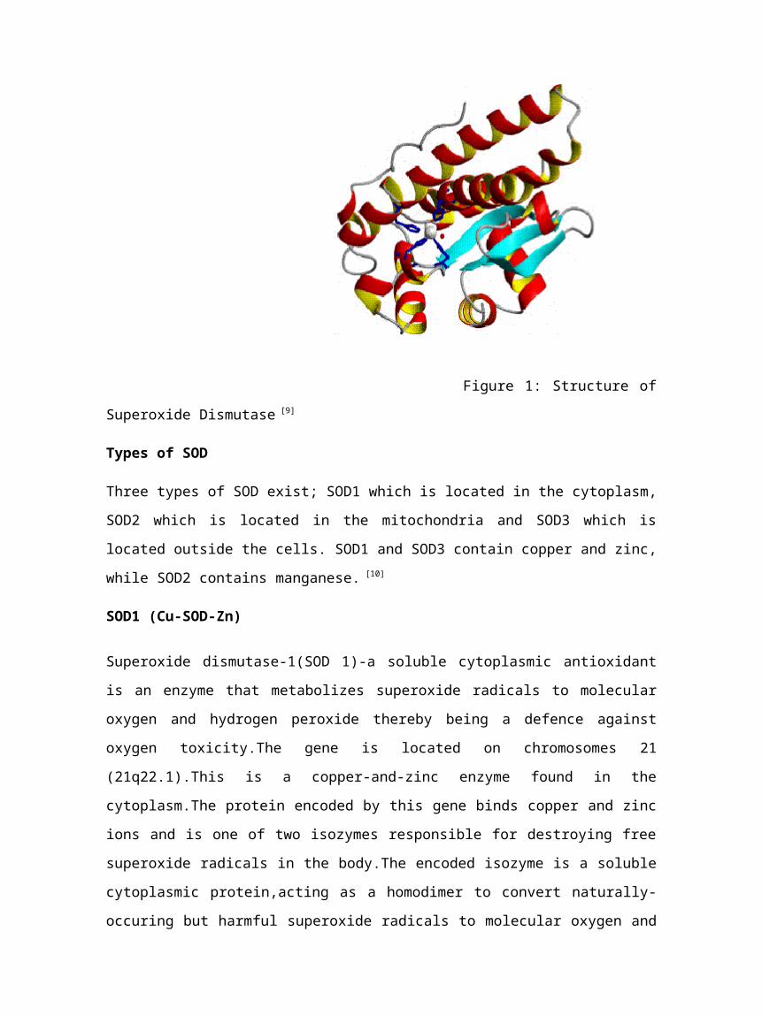
Figure 1: Structure of
Superoxide Dismutase [9]
Types of SOD
Three types of SOD exist; SOD1 which is located in the cytoplasm,
SOD2 which is located in the mitochondria and SOD3 which is
located outside the cells. SOD1 and SOD3 contain copper and zinc,
while SOD2 contains manganese. [10]
SOD1 (Cu-SOD-Zn)
Superoxide dismutase-1(SOD 1)-a soluble cytoplasmic antioxidant
is an enzyme that metabolizes superoxide radicals to molecular
oxygen and hydrogen peroxide thereby being a defence against
oxygen toxicity.The gene is located on chromosomes 21
(21q22.1).This is a copper-and-zinc enzyme found in the
cytoplasm.The protein encoded by this gene binds copper and zinc
ions and is one of two isozymes responsible for destroying free
superoxide radicals in the body.The encoded isozyme is a soluble
cytoplasmic protein,acting as a homodimer to convert naturally-
occuring but harmful superoxide radicals to molecular oxygen and
hydrogen peroxide.The other isozyme is a mitochondrial
protein.Mutations in this gene have been implicated as causes of
familial amyotrophic lateral sclerosis.The enzyme is found in the
eukaryotes as a cytoplasmic form,in plants as a chloroplast
form,also extracellular form in some eukaryotes and periplamic
form in the prokaryotes.Two patterns of the enzyme exist,the
first one contains histidine residues that bind the copper atom
and the second which is located in the C-terminal section of the
enzyme is involved in a disulphide bond.[11]The Figure 4 is of an
SOD1;
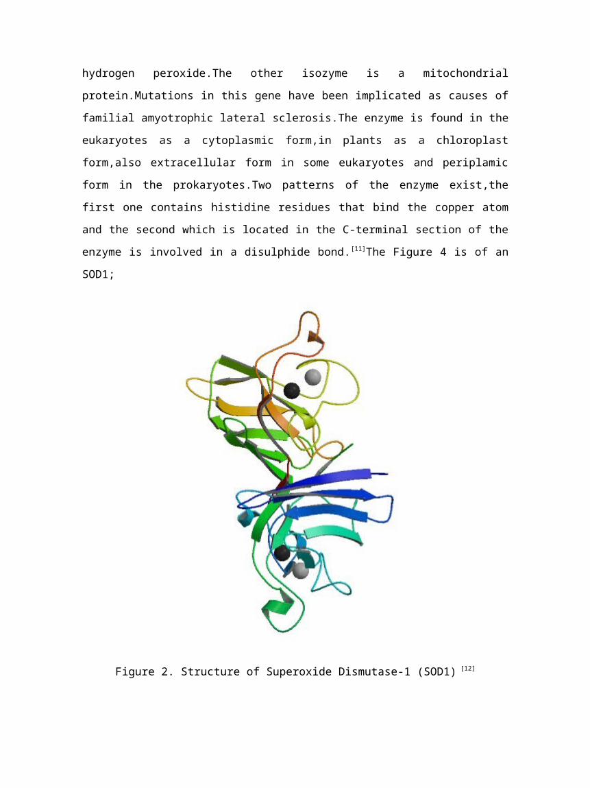
Figure 2. Structure of Superoxide Dismutase-1 (SOD1) [12]
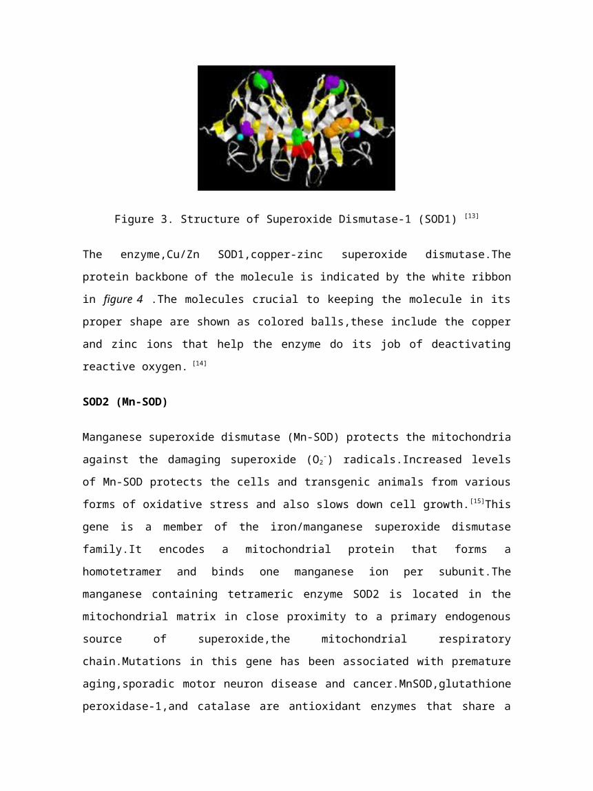
Figure 3. Structure of Superoxide Dismutase-1 (SOD1) [13]
The enzyme,Cu/Zn SOD1,copper-zinc superoxide dismutase.The
protein backbone of the molecule is indicated by the white ribbon
in figure 4 .The molecules crucial to keeping the molecule in its
proper shape are shown as colored balls,these include the copper
and zinc ions that help the enzyme do its job of deactivating
reactive oxygen. [14]
SOD2 (Mn-SOD)
Manganese superoxide dismutase (Mn-SOD) protects the mitochondria
against the damaging superoxide (O2-) radicals.Increased levels
of Mn-SOD protects the cells and transgenic animals from various
forms of oxidative stress and also slows down cell growth.[15]This
gene is a member of the iron/manganese superoxide dismutase
family.It encodes a mitochondrial protein that forms a
homotetramer and binds one manganese ion per subunit.The
manganese containing tetrameric enzyme SOD2 is located in the
mitochondrial matrix in close proximity to a primary endogenous
source of superoxide,the mitochondrial respiratory
chain.Mutations in this gene has been associated with premature
aging,sporadic motor neuron disease and cancer.MnSOD,glutathione
peroxidase-1,and catalase are antioxidant enzymes that share a
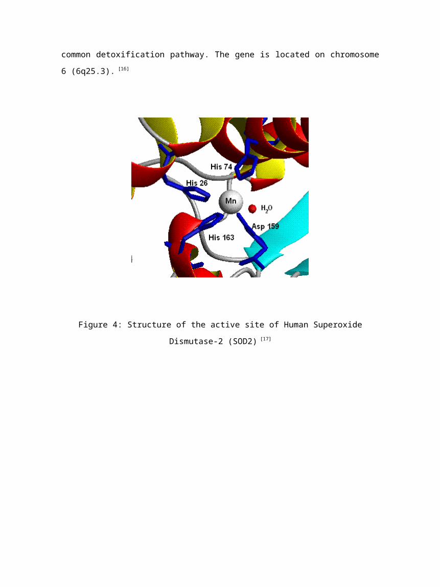
common detoxification pathway. The gene is located on chromosome
6 (6q25.3). [16]
Figure 4: Structure of the active site of Human Superoxide
Dismutase-2 (SOD2) [17]
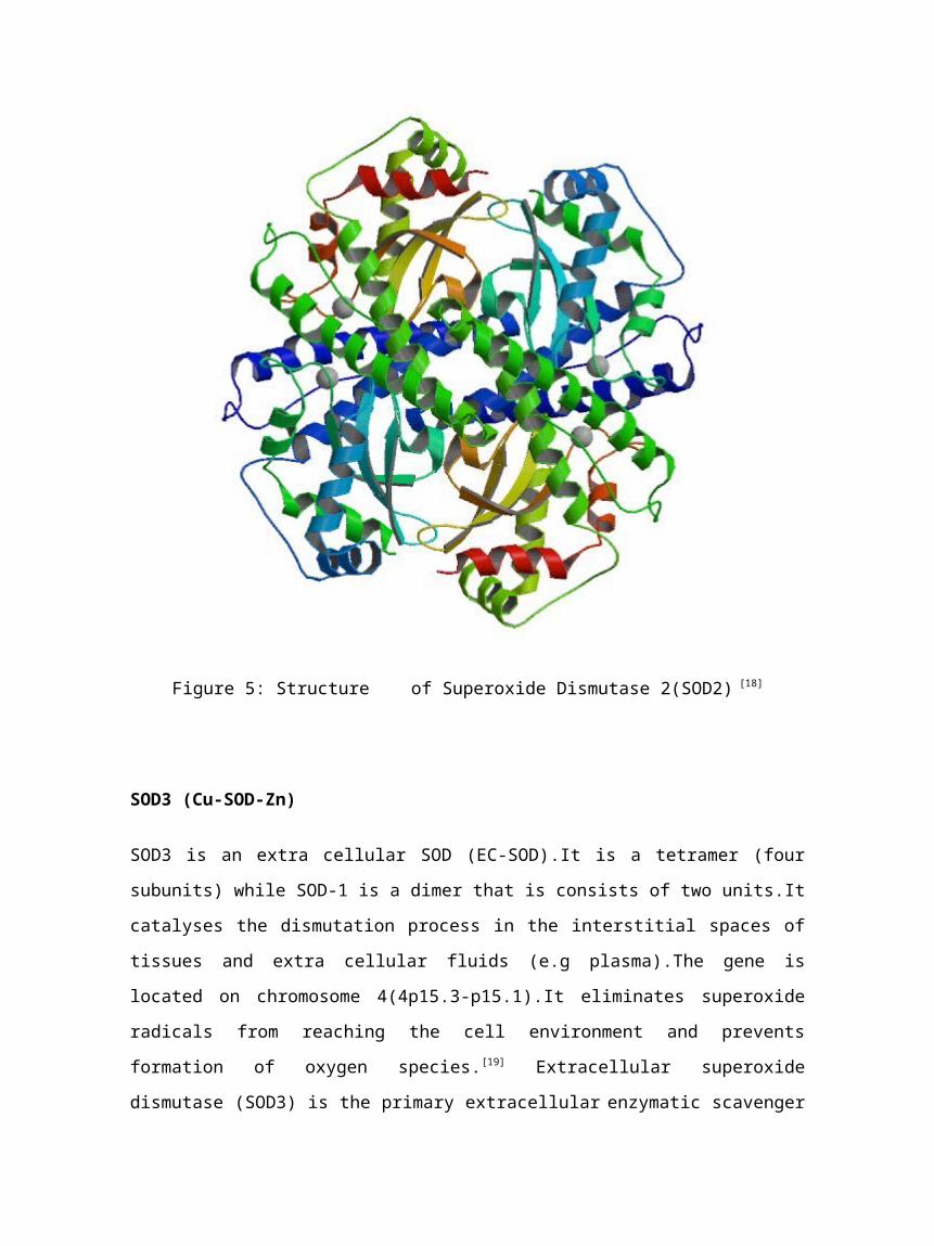
Figure 5: Structure of Superoxide Dismutase 2(SOD2) [18]
SOD3 (Cu-SOD-Zn)
SOD3 is an extra cellular SOD (EC-SOD).It is a tetramer (four
subunits) while SOD-1 is a dimer that is consists of two units.It
catalyses the dismutation process in the interstitial spaces of
tissues and extra cellular fluids (e.g plasma).The gene is
located on chromosome 4(4p15.3-p15.1).It eliminates superoxide
radicals from reaching the cell environment and prevents
formation of oxygen species.[19] Extracellular superoxide
dismutase (SOD3) is the primary extracellular enzymatic scavenger
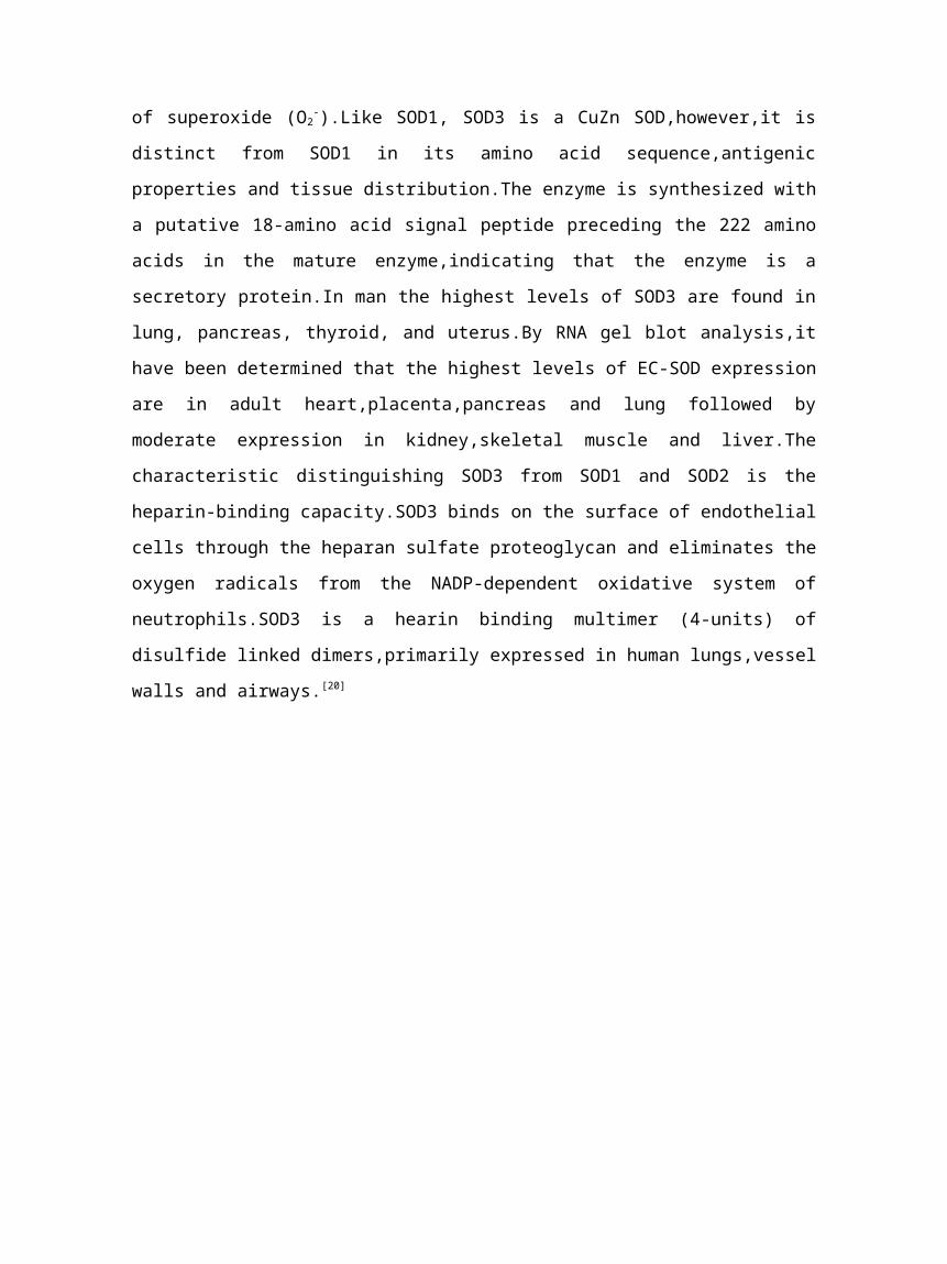
of superoxide (O2-).Like SOD1, SOD3 is a CuZn SOD,however,it is
distinct from SOD1 in its amino acid sequence,antigenic
properties and tissue distribution.The enzyme is synthesized with
a putative 18-amino acid signal peptide preceding the 222 amino
acids in the mature enzyme,indicating that the enzyme is a
secretory protein.In man the highest levels of SOD3 are found in
lung, pancreas, thyroid, and uterus.By RNA gel blot analysis,it
have been determined that the highest levels of EC-SOD expression
are in adult heart,placenta,pancreas and lung followed by
moderate expression in kidney,skeletal muscle and liver.The
characteristic distinguishing SOD3 from SOD1 and SOD2 is the
heparin-binding capacity.SOD3 binds on the surface of endothelial
cells through the heparan sulfate proteoglycan and eliminates the
oxygen radicals from the NADP-dependent oxidative system of
neutrophils.SOD3 is a hearin binding multimer (4-units) of
disulfide linked dimers,primarily expressed in human lungs,vessel
walls and airways.[20]
Figure 6: Superoxide Dismutase 3 [21]
Dismutation Reaction in general
The SOD-catalysed dismutation of superoxide dismutation may be
represented in the following two half equations;
M(n+1)+−SOD + O2- →Mn+ −SOD + O2 (3)
M+−SOD + O2- + 2H+ →M(n+1) −SOD + H2O2 (4)
Where M is Cu (n=1), Mn (n=2), Fe (n=2) and Ni (n=2),oxidation
state of metal oscillates between n and n+1 [22]
The reaction however can be represented as below
2O2- + 2H+ → O2 + H2O2 [23] (5)
Action of Iron as a catalyst (Fenton Chemistry or Reagent)
Peroxide formed can also be dangerous to the cells,more
especially as it transforms to a hydroxyl radical (OH.) when
reacting with Fe2+ (Fenton reaction),a combination of H2O2 and Fe2+
is known as Fenton's reagent[24].Fenton chemistry was developed in
1890s by Henry John Horstman Fenton.In this type of
reaction,ferrous iron(II), Fe2+ is oxidized by hydrogen peroxide
(H2O2) to ferric iron (III), Fe3+,a hydroxyl radical (OH.) and a
hydroxyl anion (OH-).A hydroxyl radical (OH.) is the neutral form
of the hydroxide ion (OH-).A hydroxyl is a molecule of oxygen and
hydrogen in a covalent bond.This hydroxyl radical is highly
reactive and short lived.In addition to the Fenton reaction,
iron (III) can then be reduced back to Fe2+,a peroxide radical
(OOH.) and a proton by the same hydrogen peroxide via a
disproportionation process[25].The reaction can be represented as
below;
Fe2+ + H2O2 → Fe3+ + OH. + OH- (6)
Fe3+ + H2O2 → Fe2+ + OOH. + H+ (7)
Note: OH- + H+ → H2O (8)
The net reaction results in two molecules of H2O2 hydrogen
peroxide being converted to water and two hydroxyl radicals. This
clearly shows that iron is truly catalytic.The generated hydroxyl
radicals then are engaged in secondary reactions.[26] Below is the
net reaction;
2H2O2 → OH. + OOH. + H2O (9) (net reaction)
However in the net equation it is stated that;
2H2O2 → 2OH. + H2O [27] (10)
Two hydrogen peroxide give two hydroxyl radicals and water.
Action of Glutathione Peroxidase
However,this reaction (10) is dangerous as the hydroxyl radical
generated can engage in secondary reactions like convention of
benzene to phenol and can also destroy organic compounds e.g
trichloroethylene.The conversion of hydrogen peroxide to generate
hydroxyl radicals is disturbed by glutathione peroxidase which
reduces hydrogen peroxide by energy transfer from reactive
peroxide to a very small sulfur-containing protein called
glutathione. [28]
Figure7. Structure of Enzyme Glutathione Peroxidase [29]
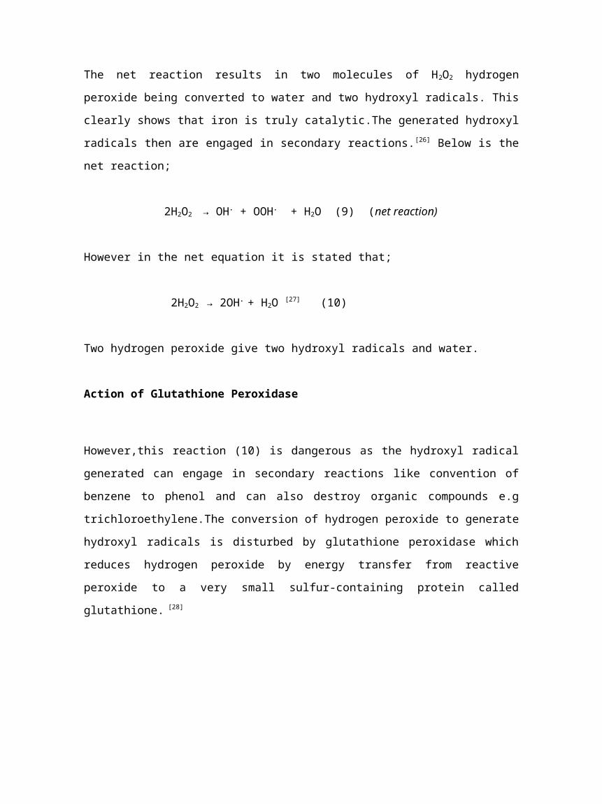
Figure 8: Structure of Glutathione
Glutathione peroxidase is an enzyme found in the cytoplasm of
nearly all mammalian tissues,whose preferred substrate is
hydrogen peroxide. Glutathione peroxidase is the general name of
an enzyme family with peroxidase activity whose main biological
role is to protect the organism from oxidative damage. The
biochemical function of glutathione peroxidase is to reduce lipid
hydroperoxides to their corresponding alcohols and to reduce free
hydrogen peroxide to water. [30]
In this type of reaction, glutathione Peroxidase catalyze H2O2 as
given below;
2GSH + H2O2 → GS−SG + 2H2O (11)
GSH is monomeric glutathione and GS−SG is glutathione disulphide,
formed by oxidation of glutathione through loss of the thiolic
hydrogens and formation of an S-S bond.
Then Glutathione Reductase (in figure 9 below) regenerates the
enzyme by reducing the oxidized glutathione to complete the cycle
as below;
GS−SG + NADPH + H+ → 2GSH + NADP+ (12)
NADPH is the reduced form of nicotinamide adenine dinucleotide
phosphate.
Glutathione reductase,also known as GSR,is a human gene.It is an
enzyme which reduces glutathione disulfide (GSSG) to the
sulfidryl form GSH,which is an important cellular antioxidant. [31]
For every one mole of GSSG one mole of NADPH is required.
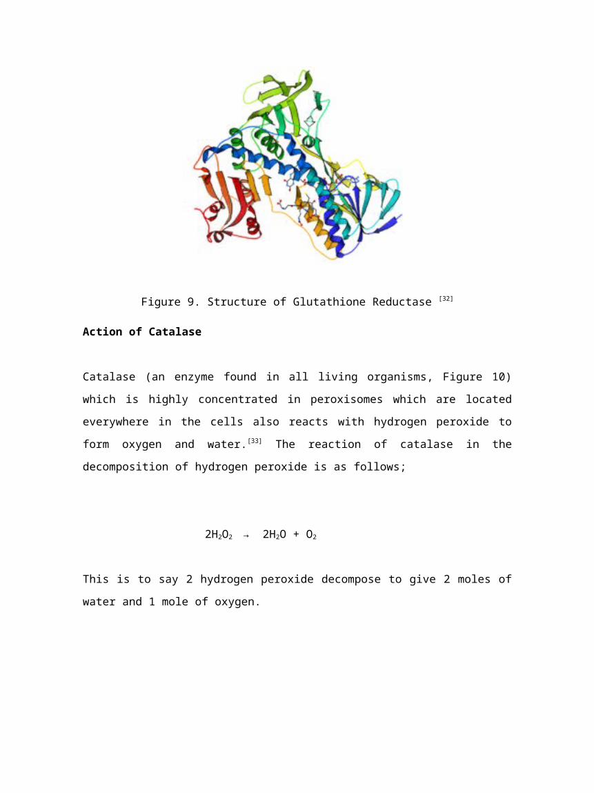
Figure 9. Structure of Glutathione Reductase [32]
Action of Catalase
Catalase (an enzyme found in all living organisms, Figure 10)
which is highly concentrated in peroxisomes which are located
everywhere in the cells also reacts with hydrogen peroxide to
form oxygen and water.[33] The reaction of catalase in the
decomposition of hydrogen peroxide is as follows;
2H2O2 → 2H2O + O2
This is to say 2 hydrogen peroxide decompose to give 2 moles of
water and 1 mole of oxygen.
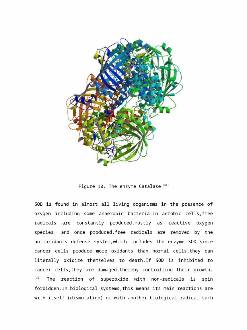
Figure 10. The enzyme Catalase [34]
SOD is found in almost all living organisms in the presence of
oxygen including some anaerobic bacteria.In aerobic cells,free
radicals are constantly produced,mostly as reactive oxygen
species, and once produced,free radicals are removed by the
antioxidants defense system,which includes the enzyme SOD.Since
cancer cells produce more oxidants than normal cells,they can
literally oxidize themselves to death.If SOD is inhibited to
cancer cells,they are damaged,thereby controlling their growth.[19] The reaction of superoxide with non-radicals is spin
forbidden.In biological systems,this means its main reactions are
with itself (dismutation) or with another biological radical such
as nitric oxide (NO).The superoxide anion radical (O2-)
spontaneously dismutes to O2 and hydrogen peroxide (H2O2) quite
rapidly (~105 M-1 s-1 at pH 7).SOD is biologically necessary
because superoxide reacts even faster with certain targets such
as NO radical,which makes peroxynitrite.Similarly,the dismutation
rate is second order with respect to initial superoxide
concentration.Thus,the half-life of superoxide,although very
short at high concentrations (e.g. 0.05 seconds at 0.1mM) is
actually quite long at low concentrations (e.g. 14 hours at 0.1
nM).In contrast, the reaction of superoxide with SOD is first
order with respect to superoxide
concentration.Moreover,superoxide dismutase has the fastest
turnover number (reaction rate with its substrate) of any known
enzyme (~109 M-1 s-1), this reaction being only limited by the
frequency of collision between itself and superoxide.That is the
reaction rate is "diffusion limited".[35]
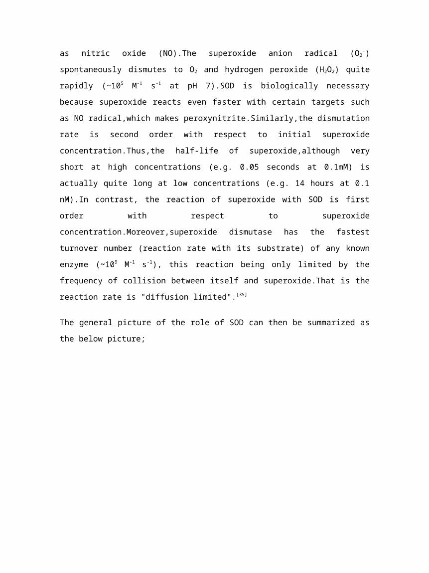
The general picture of the role of SOD can then be summarized as
the below picture;
Figure 11: A picture showing overview of all dismutation
reactions and steps taken [36]
REACTION CATALYZED BY THE ENZYME-PROPOSED MECHANISM
Both copper ions and copper-containing enzymes have been shown to
catalyze NO release from GSNO.It have been observed that copper-
zinc superoxide dismutase (Cu,ZnSOD) in the presence of H2O2
caused a rapid decomposition of GSNO,forming oxidized glutathione
(GSSG) and (*)NO.The cupric ions (Cu(2+) released from Cu,ZnSOD
were bound to the glutamate moiety of GSNO,yielding a 2:1 (GSNO)
(2)Cu(2+) complex.Strong chelators of cupric ions,such as
histidine and diethylenetriaminepentaacetic acid,inhibited the
formation of (GSNO)(2)Cu(2+) complex,GSSG and (*)NO.GSSG alone
inhibited Cu(2+)-induced decomposition of GSNO.This effect is
attributed to complexation of copper by GSSG.Binding of copper to
GSNO is obligatory for NO release from GSNO,however,the rate of
this reaction was considerably slow due to binding of Cu(2+) by
GSSG.The glutamate moiety in GSNO and GSSG controls copper-
catalyzed NO release from GSNO.Cu,ZnSOD and H2O enhanced
peroxidation of unsaturated lipid that was inhibited by GSNO.The
antioxidant function of GSNO is related to the sequestering of
copper by GSNO and its ability to slowly release NO. [37]
Further mechanism can be represented as below Figures;
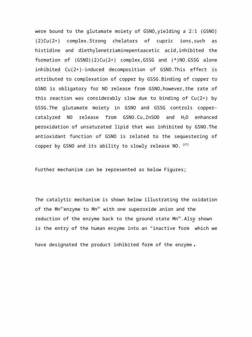
The catalytic mechanism is shown below illustrating the oxidation
of the Mn3+enzyme to Mn2+ with one superoxide anion and the
reduction of the enzyme back to the ground state Mn3+.Also shown
is the entry of the human enzyme into an “inactive form” which we
have designated the product inhibited form of the enzyme.
NADH
Succinate2e- Succinate Dehydrogenase
(3Fe-S)
CO M PLEX II
NADHDehydrogenase
(5Fe-S)
CO M PLEX I
UQ cyt. b cyt. c1CO M PLEX III
cyt. c cyt. a-a3e- e-CO M PLEX IV
O2O2
-.
e-
e-
4e- O2
H2O
O2O2
-.
Amobarbital
AntimycinAM yxothiazole
HQNO
2e-
Figure 12: Kinetic mechanism of Mn-SOD [38]
Kinetic M echanism of M n-SOD
P-M n3+O 2·- P-M n3+: O2·-
P-M n2+
O 2k1k-1
k2
P-M n2+: O 2·-H 2O 2O 2·-
k-3k3
k-4k4
Inactive formk-5 k5
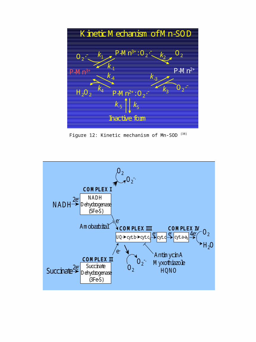
Figure 13: Mechanism involved in NADH [38]
This anti-oxidant enzyme functions primarily to protect
mitochondrial components from superoxide liberated as a normal
byproduct of respiration.Complex I and Complex III release
superoxide radical as a consequence of normal respiration,with
estimates of 1-5% of the oxygen consumed being liberated as
superoxide anion.MnSOD is therefore the cells primary defense
against free radical mediated damage.In addition,stimulated
levels of the enzyme have evolved to address increased free
radical production during an inflammatory episode. [38]
POSSIBLE APPLICATION OF SUPEROXIDE DISMUTASE IN CANCER TREATMENT
Cancer may affect people at all ages but risk for the more common
varieties tends to increase with age.Critics of the role of
superoxide in cancer have pointed out that it is not known
whether the loss of Mn-SOD is a cause or effect of cancer.After
all,there are numerous enzyme changes that occur in cancer (both
additions and deletions).In contrast to most other enzymes,the
principal role of SOD seems to be to act as a protective
enzyme.Thus,its absence can lead to widespread metabolic
consequences.In order to understand these consequences,it is
necessary to consider what is known about the chemistry of the
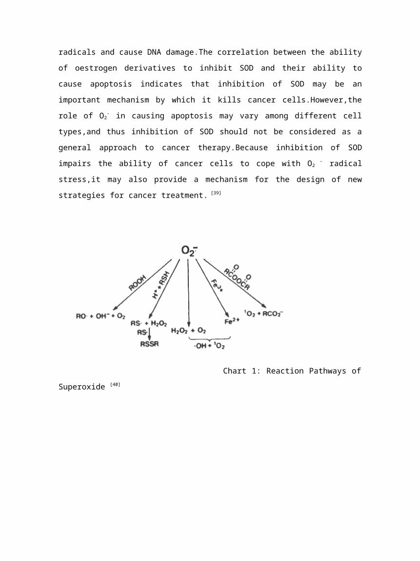
superoxide radical.Proposed pathways are shown in Chart 1
below.It should be emphasized that there is considerable evidence
for each of these pathways,but none have been proven beyond
doubt.Three pathways are of particular interest in the field of
cancer.Superoxide can oxidize SH groups to S−S via RS (Pathway 1)
dismute to form H2O2 + ground-state oxygen (O2) (Pathway 2),react
with ferric ion to form ferrous ion (Pathway 3).Each of these
pathways can probably lead to large changes in cell
metabolism.Considering Pathway 1 initially,sulfidryl groups are
constituents of many proteins.Oxidation of these groups to
disulfides can result in protein conformational changes with
possible activation or inactivation of key enzymes.Thesecond
important pathway,dismutation to form H2O2 and ground-state
oxygen O2, occurs either with or without SOD.Dismutation occurs
much faster in the presence of SOD than in its absence.
The last pathway may lead to perhaps the most widespread
changes.Superoxide can denote electrons to metals to change their
oxidation state.For instance,it has been shown that O2- can react
with Fe3+ to produce Fe2+.Reactions of this sort can lead to vast
metabolic consequences due to large changes in the oxidation-
reduction potential of the cell.Many enzymatic reactions require
metals as cofactors,and a change in their oxidation-reduction
state will surely affect these reactions.Fridovich has recently
shown that O2- is apparently a very diffusible substance able to
migrate large distances and even through membranes if they do not
contain SOD.Thus,in the cancer cell,which is low in SOD,changes
may occur far from where the O2- was originally produced.The
evidence that SOD plays a much important role in cancer
treatment.SODs catalyses the dismutation of O2-,an essential step
in eliminating the toxic free radical.This critical function
makes SOD an attractive target for pharmacological
intervention.Although several small molecules,including cyanide
ion (CN-),hydroxyl ion (OH -) and azide ion (N3-),were found to
inhibit SOD by competing with O2- at the catalytic site,these
chemicals are highly toxic and their potential for cancer therapy
is limited.Inhibition of SOD leads to free-radical-mediated
damage to mitochondrial membranes and the release of cytochrome
c.This is probably a major pathway,although other mechanisms such
as inhibition of tubulin polymerization and angiogenesis may also
contribute to its activity.O2- may also give rise to hydroxyl
radicals and cause DNA damage.The correlation between the ability
of oestrogen derivatives to inhibit SOD and their ability to
cause apoptosis indicates that inhibition of SOD may be an
important mechanism by which it kills cancer cells.However,the
role of O2- in causing apoptosis may vary among different cell
types,and thus inhibition of SOD should not be considered as a
general approach to cancer therapy.Because inhibition of SOD
impairs the ability of cancer cells to cope with O2 - radical
stress,it may also provide a mechanism for the design of new
strategies for cancer treatment. [39]
Chart 1: Reaction Pathways of
Superoxide [40]
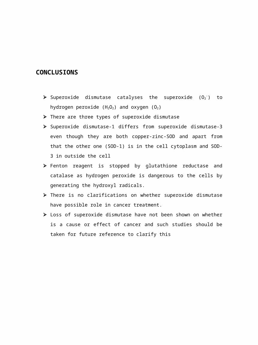
CONCLUSIONS
Superoxide dismutase catalyses the superoxide (O2-) to
hydrogen peroxide (H2O2) and oxygen (O2)
There are three types of superoxide dismutase
Superoxide dismutase-1 differs from superoxide dismutase-3
even though they are both copper-zinc-SOD and apart from
that the other one (SOD-1) is in the cell cytoplasm and SOD-
3 in outside the cell
Fenton reagent is stopped by glutathione reductase and
catalase as hydrogen peroxide is dangerous to the cells by
generating the hydroxyl radicals.
There is no clarifications on whether superoxide dismutase
have possible role in cancer treatment.
Loss of superoxide dismutase have not been shown on whether
is a cause or effect of cancer and such studies should be
taken for future reference to clarify this
References
1. Superoxide dismutase, February 23,2008,MediaWiki,Wikimedia
Foundation, Inc
http://en.wikipedia.org/wiki/superoxide_dismutase,retrieved
28 February 2008
2. Superoxide Dismutase,April 2001,Karl Harrison,
http:www.3dchem.com/molecules.asp,retrieved 08-03-08.
3. Superoxide Dismutase,11 March 2008,The Center for Cancer
Education,University of Newcastle,
http://cancerweb.ncl.ac.uk/cgi-bin/omd?
superoxide+dismutase,retrieved 15 March 2008
4. Griendling K.K. and Weber D.S.,2003, The Yin/Yang of
superoxide dismutase mimetics: potential cardiovascular
therapies? British Journal of Pharmacology, 139, 1059–1060
5. Kresge N.,Hill R.L. and Simoni R.D., June 2, 2006,Forty
Years of Superoxide Dismutase Research: the Work of Irwin
Fridovich, Journal of Biochemistry,Vol. 281(22),01-03
6. What are free radicals,Free radicals and aging,Copyright
1999,The National Health Museum,
http://65.79.226.34/LC/ST/bgfreerad.html,retrieved 17 March
2008
7. Cause of Aging,Reactive oxygen Species,11 March
2008,MediaWiki,Wikimedia Foundation Inc,
http://en.wikipedia.org/wiki/Reactive_oxygen_species,retriev
ed 18 March 2008
8. Amstad P.,Cerutti P.,Ghosh R.,Oya Y.,December 1994,The role
of the cellular antioxidant defense in oxidant
carcinogenesis, Environ Health Perspect,Vol 102(Supply
10),123–129
9. Image:SOD.gif,28 November 2006,Mediawiki,Wikimedia
Foundation Inc,
http://en.wikipedia.org/wiki/Image:SOD.gif,retrieved 27
February 2008
10. General,Types,Superoxide dismutase, 23 February
2008,Mediawiki,Wikimedia Foundation Inc,
http://en.wikipedia.org/wiki/Superoxide_dismutase,retrived
01 March 2008
11. Copper/Zinc superoxide dismutase signatures,April 2006,
Swiss Institute of
Bioinformatics,http://www.expasy.org/prosite/PDOC00082,retri
eved 01 March 2008
12. Image:PBB Protein SOD1 image.jpg,9 October 2007,
Mediawiki,Wikimedia Foundation Inc,
http://en.wikipedia.org/wiki/Image:PBB_Protein_SOD1_image.jp
g,retrieved 01 March 2008
13. SOD1 (copper zinc superoxide dismutase 1) and
ALS,2004,The ALS Association,
http://www.alsa.org/research/article.cfm?,retrieved 03 March
2008
14. SOD1 (copper zinc superoxide dismutase 1) and
ALS,2004,The ALS Association,
http://www.alsa.org/research/article.cfm?,retrieved 03 March
2008
15. Carlson E.J.,Carra S and Epotein C.J.,October
2001,Strain-dependent high-level expression of a transgene
for manganese superoxide dismutase is associated with growth
retardation and decreased fertility, Free Radic. Biol.
Med.,Vol 31(8),1018-1030
16. SOD2,6 April 2008, Mediawiki,Wikimedia Foundation Inc,
http://en.wikipedia.org/wiki/SOD2,retrived 7 April 2008
17. Human,Superoxide dismutase,23 February
2008,Mediawiki,Wikimedia Foundation Inc,
http://en.wikipedia.org/wiki/Superoxide_dismutase,retrieved
28 February 2008
18. Image:PBB Protein SOD2 image.jpg,1 November
2007,Mediawiki,Wikimedia Foundation
Inc,http://en.wikipedia.org/wiki/Image:PBB_Protein_SOD2_imag
e.jpg,retrieved 28 February 2008
19. Human,Superoxide Dismutase,23 February
2008,Mediawiki,Wikimedia Foundation Inc,
http://en.wikipedia.org/wiki/Superoxide_dismutase,retrieved
29 February 2008
20. Superoxide Dismutase, Extracellular EC-SOD,Superoxide
Dismutase,Elevated Extracellular,Included,Superoxide
Dismutase 3; SOD3,30 May 2007,
http://www.ncbi.nlm.nih.gov/entrez/dispomim.cgi?
id=185490,retrieved 29 February 2008
21. Image:PBB Protein SOD1 image.jpg,9 October 2007,
Mediawiki,Wikimedia Foundation Inc,
http://en.wikipedia.org/wiki/Image:PBB_Protein_SOD1_image.jp
g,retrieved 01 March 2008
22. Reaction,Superoxide Dismutase,23 February
2008,Mediawiki,Wikimedia Foundation Inc,
http://en.wikipedia.org/wiki/Superoxide_dismutase,retrieved
28 February 2008
23. Superoxide: superoxide oxidoreductase, 2008,
Worthington Biochemical Corporation, http://www.worthington-
biochem.com/SODBE/default.html,retrieved 28 February 2008
24. Decomposition,Hydrogen Peroxide, 14 April
2008,MediaWiki,Wikimedia Foundation Inc,
http://en.wikipedia.org/wiki/Hydrogen_peroxide,retrieved 14
April 2008
25. Fenton's reagent, 21 February 2008,MediaWiki,Wikimedia
Foundation Inc, http://en.wikipedia.org/wiki/Fenton
%27s_reagent ,retrieved 28 February 2008
26. Ashraf S.S., Alhadrami S and Rauf M.A.,2006,
Degradation of Methyl Red using Fenton's reagent and the
effect of various salts,Science Direct,Vol 69(1-2),74-78
27. Fenton's reagent, 21 February 2008,MediaWiki,Wikimedia
Foundation Inc, http://en.wikipedia.org/wiki/Fenton
%27s_reagent ,retrieved 28 February 2008
28. Glutathione Peroxidase,20 March
2008,MediaWiki,Wikimedia Foundation Inc,
http://en.wikipedia.org/wiki/glutathione_peroxidase,retrieve
d on 22 March 2008
29. Image:GlutPeroxidase-1GP1.png,20 March
2008,MediaWiki,Wikimedia Foundation
Inc,http://en.wikipedia.org/wiki/image:GlutPeroxidase-
1GP1.png, retrieved;28 March 2008
30. Glutathione peroxidase, 20 March 2008,
MediaWiki,Wikimedia Foundation Inc,
http://en.wikipedia.org/wiki/Glutathione_peroxidase,retrieve
d 28 March 2008
31. Glutathione reductase,20 March 2008,
MediaWiki,Wikimedia Foundation Inc,
http://en.wikipedia.org/wiki/Glutathione_reductase,retrieved
28 March 2008
32. Image:Glutathione reductase.png, 7 May 2007,
MediaWiki,Wikimedia Foundation Inc,
http://en.wikipedia.org/wiki/Image:Glutathione_reductase.png
,retrieved 28 March 2008
33. Catalase,12 April 2008,MediaWiki,Wikimedia Foundation
Inc, http://en.wikipedia.org/wiki/Catalase,retrieved 13
April 2008
34. Image:PBB Protein CAT image.jpg, 7 November 2007,
MediaWiki,Wikimedia Foundation Inc,
http://en.wikipedia.org/wiki/Image:PBB_Protein_CAT_image.jpg
, retrieved 28 March 2008
35. Biochemistry,Superoxide dismutase,23 February
2008,Mediawiki,Wikimedia Foundation Inc,
http://en.wikipedia.org/wiki/Superoxide_dismutase,retrieved
29 March 2008
36. Cell Damage,2008, Sigma-Aldrich Co.,
http://www.sigmaaldrich.com/Area_of_Interest/Biochemicals/En
zyme_Explorer/Cell_Signaling_Enzymes/
Superoxide_Dismutase.html,retrieved 29 March 2008
37. Antholine W.E.,Goss S.P.A.,Hogg N.,1 December
1999,Mechanism of Superoxide Dismutase/H2O2-Mediated Nitric
Oxide Release from S-Nitrosoglutathione— Role of
Glutamate,Archieves of Biochemistry and Biophysics,Vol
372(1),8-15
38. The enzyme,its reaction and mechanism,Manganese
Superoxide Dismutase (MnSOD),
http://www.ufbi.ufl.edu/~hnick/Manganese%20Superoxide
%20Dismutase.htm,retrieved 8 April 2008
39. Feng L.,Huang P.,Keating M.J.,21 September
2000,Superoxide dismutase as a target for the selective
killing of cancer cells,Nature journal,407,390-395
40. Buettner G.R. and Oberley L.W.,April 1979,Role of
Superoxide Dismutase in cancer,A review,Cancer
Research,39,1141-1149
APPENDIX
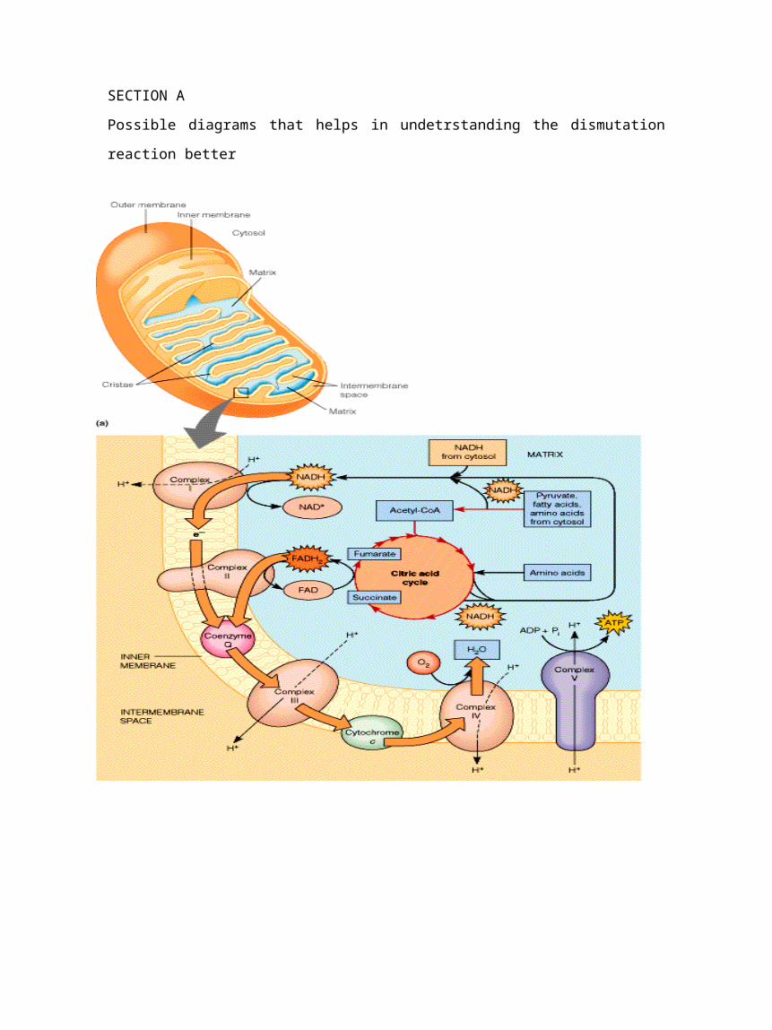
SECTION A
Possible diagrams that helps in undetrstanding the dismutation
reaction better


![Coordination behavior of sulfathiazole. Crystal structure of [Cu (sulfathiazole) (py)3Cl] superoxide dismutase activity](https://img.pdfslide.net/doc/110x75/63344a87b94d623842029d59/coordination-behavior-of-sulfathiazole-crystal-structure-of-cu-sulfathiazole.jpg)


