Embed Size (px)
Citation preview
ORIGINAL RESEARCH
Suppression of the hERG potassium channel responseto premature stimulation by reduction in extracellularpotassium concentrationDario Melgari*, Chunyun Du*, Aziza El Harchi, Yihong Zhang & Jules C. Hancox
School of Physiology and Pharmacology and Cardiovascular Research Laboratories, University of Bristol, Medical Sciences Building, Bristol, BS8
1TD, UK
Keywords
Human ether-�a-go-go-related gene,
hyperkalemia, hypokalemia, long QT,
potassium, potassium channels, QT interval.
Correspondence
Jules C. Hancox, School of Physiology and
Pharmacology and Cardiovascular Research
Laboratories, University of Bristol, Medical
Sciences Building, Bristol, BS8 1TD, UK.
Tel: +44 (0)117 3312292
Fax: +44 (0)117 3312288
E-mail: [email protected]
Funding Information
Funding from the British Heart Foundation
(FS/11/59; PG/10/96; PG/12/69; PG/13/68)
and Heart Research UK (RG2594) is gratefully
acknowledged.
Received: 14 August 2014; Accepted: 29
August 2014
doi: 10.14814/phy2.12165
Physiol Rep, 2(10), 2014, e12165,
doi: 10.14814/phy2.12165
*These authors contributed equally to this
study.
Abstract
Potassium channels encoded by human ether-�a-go-go-related gene (hERG)
mediate the cardiac rapid delayed rectifier K+ current (IKr), which participates
in ventricular repolarization and has a protective role against unwanted
premature stimuli late in repolarization and early in diastole. Ionic current
carried by hERG channels (IhERG) is known to exhibit a paradoxical depen-
dence on external potassium concentration ([K+]e), but effects of acute [K+]echanges on the response of IhERG to premature stimulation have not been
characterized. Whole-cell patch-clamp measurements of hERG current were
made at 37°C from hERG channels expressed in HEK293 cells. Under conven-
tional voltage-clamp, both wild-type (WT) and S624A pore-mutant IhERG dur-
ing depolarization to +20 mV and subsequent repolarization to �40 mV were
decreased when superfusate [K+]e was decreased from 4 to 1 mmol/L. When
[K+]e was increased from 4 to 10 mmol/L, pulse current was increased and
tail IhERG was decreased. Increasing [K+]e produced a +10 mV shift in volt-
age-dependent inactivation of WT IhERG and slowed inactivation time course,
while lowering [K+]e from 4 to 1 mmol/L produced little change in inactiva-
tion voltage dependence, but accelerated inactivation time course. Under
action potential (AP) voltage-clamp, lowering [K+]e reduced the amplitude of
IhERG during the AP and suppressed the maximal IhERG response to premature
stimuli. Raising [K+]e increased IhERG early during the AP and augmented the
IhERG response to premature stimuli. Our results are suggestive that during
hypokalemia not only is the contribution of IKr to ventricular repolarization
reduced but its ability to protect against unwanted premature stimuli also
becomes impaired.
Introduction
Repolarization of cardiac action potentials (APs) depends
on the interplay between inward and outward conduc-
tances during the AP plateau, with key roles identified for
several potassium ion channels (Tamargo et al. 2004).
hERG (human ether-�a-go-go-related gene) encodes a protein
that underlies the pore-forming subunit of potassium
channels mediating the rapid delayed rectifier current, IKr(Sanguinetti et al. 1995; Trudeau et al. 1995). Due to fast
voltage-dependent inactivation, IKr/hERG channels pass lit-
tle current at the peak of the ventricular action potential
(AP), but mediate greater current as the AP plateau
proceeds, peaking before the final rapid repolarization
phase of the AP (Hancox et al. 1998; Zhou et al. 1998),
which is mediated by a different potassium current (the
inward rectifier, IK1; Shimoni et al. 1992; Mitcheson and
Hancox 1999). Loss-of-function mutations in hERG are
associated with the LQT2 form of the Long QT Syndrome
(LQTS; Modell and Lehmann 2006), while gain-of-function
ª 2014 The Authors. Physiological Reports published by Wiley Periodicals, Inc. on behalf of
the American Physiological Society and The Physiological Society.
This is an open access article under the terms of the Creative Commons Attribution License,
which permits use, distribution and reproduction in any medium, provided the original work is properly cited.
2014 | Vol. 2 | Iss. 10 | e12165Page 1
Physiological Reports ISSN 2051-817X
hERG mutations are associated with the SQT1 variant of
the short QT syndrome (SQTS; Brugada et al. 2004; Sun
et al. 2011).
When hERG was initially identified, the magnitude of
hERG current (IhERG) was demonstrated to have an anom-
alous dependence on extracellular K+ concentration
([K+]e), with low-[K+]e reducing outward IhERG amplitude
and raised [K+]e augmenting the current (Sanguinetti et al.
1995). These changes were the opposite of those expected
due merely to changes in electrochemical gradient and
were observed also for native IKr (Sanguinetti and
Jurkiewicz 1992; Yang and Roden 1996). This anomalous
[K+]e dependence of IKr was subsequently proposed to
arise from the rectification properties of the IKr channel
and specifically that rapid inactivation underlies this effect
(Yang et al. 1997), most likely because external K+ ions
interact with the pore and influence the channel’s rapid
collapse-of-pore type inactivation (Smith et al. 1996). This
property of IKr/hERG has clinical significance as, on the
one hand, hypokalemia can exacerbate effects of QT inter-
val prolonging, hERG-blocking drugs (Hancox et al. 2008)
whilst, on the other hand, potassium supplementation has
been reported to improve repolarization in some LQT2
patients (Compton et al. 1996; Etheridge et al. 2003).
In addition to their role in ventricular AP repolariza-
tion, due to comparatively slow deactivation kinetics, IKr/
hERG channels can also contribute to net membrane con-
ductance early in diastole and may play a protective role
against premature beats (Smith et al. 1996; Lu et al.
2001). Consistent with this, using the “AP clamp” tech-
nique, Lu et al. (2001) demonstrated that premature
stimuli applied late during AP repolarization or early in
diastole elicit rapid outward IhERG transients that would
be anticipated to oppose premature depolarization. Subse-
quent studies have demonstrated that this property can
be altered by LQTS gene mutation (Lu et al. 2003) or aci-
dosis (Du et al. 2010). As both the magnitude and inacti-
vation properties of IKr/hERG are considered sensitive to
[K+]e, a question of significance is whether or not the
putative protective role of hERG against premature stim-
ulation is altered by [K+]e? Accordingly, the aim of this
study was to address this question through a combination
of conventional and AP voltage-clamp experiments on
recombinant hERG channels.
Methods
Wild-type and S624A hERG channelsconstructs
Human Embryonic Kidney (HEK-293) cells stably
expressing wild-type (WT) hERG channels construct were
donated by Prof Craig January (Zhou et al. 1998). The
S624A mutant was generated using QuickChange� (Strat-
agene, La Jolla, CA) mutagenesis as described previously
(El Harchi et al. 2012). hERG 1b in pcDNA3.1 was
donated by Prof Gail Robertson.
Cells maintenance and transfection
HEK-293 cells stably expressing WT hERG or transiently
expressing S624A-mutant constructs were maintained and
passaged as described previously (Zhang et al. 2011; El
Harchi et al. 2012). Cells were transfected 24–48 h after
plating in 40 mm petri dishes. Transient transfections
were conducted using LipofectamineTM LTX (Life Tech-
nologies, Carlsbad, CA) following the instructions pro-
vided by the manufacturer. To mark successful
transfections, 0.5 lg of S624A-mutant construct were
always cotransfected with 1.0 lg of green fluorescent pro-
tein (GFP, in pCMX donated by Dr. Jeremy Tavare, Uni-
versity of Bristol, UK). For experiments on coexpressed
hERG1a/1b, 0.25 lg of the hERG 1a construct were
cotransfected with the same amount of hERG 1b, together
with 0.5 lg of CD8 as a transfection marker. Successfully
transfected cells were detected using Dynabeads� (Invitro-
gen). After transfection cells were incubated at 37°C (5%
CO2) for 6 h before plating them on small dry-heat steril-
ized glass coverslips. Electrophysiological experiments
were conducted after at least 24 h of further incubation
at 37°C (5% CO2). Throughout the Results section, hERG
refers to hERG1a, except for data in Figure 6, which were
obtained from coexpressed hERG1a/1b.
Electrophysiological recording
Coverslips with plated cells were placed in a recording
chamber mounted on an inverted microscope (Nikon
Diaphot, Kingston upon Thames, UK). The chamber tem-
perature was kept at 37°C and cells were continuously
superfused with a standard Tyrode’s solution containing
(in mmol/L): 140 NaCl, 4 KCl, 2.5 CaCl2, 1 MgCl2, 10
glucose, 5 HEPES (titrated to pH 7.4 with NaOH) (Zhang
et al. 2011; El Harchi et al. 2012; Du et al. 2014). Patch-
pipettes (Schott #8250 glass; A-M Systems Inc., Sequim,
WA) were pulled (Narishige, PP 830, Tokyo, Japan) and
polished (Narishige, MF 83) to a final resistance between
2 and 4 MΩ. Patch-pipettes were dialyzed with an intra-
cellular solution containing (in mmol/L): 130 KCl, 1
MgCl2, 5 EGTA, 5 MgATP, 10 HEPES (titrated to pH 7.2
with KOH) (Zhang et al. 2011; El Harchi et al. 2012; Du
et al. 2014). IhERG was recorded using an Axopatch 1D or
200B amplifier (Axon Instruments, now Molecular
Devices) and a CV-4/100 or CV203BU head-stage. Volt-
age-clamp commands were generated with Clampex 8 or
Clampex 9.2 (Axon Instruments, now Molecular Devices).
2014 | Vol. 2 | Iss. 10 | e12165Page 2
ª 2014 The Authors. Physiological Reports published by Wiley Periodicals, Inc. on behalf of
the American Physiological Society and The Physiological Society.
hERG and Extracellular Potassium D. Melgari et al.
Pipette series resistance was compensated between 70%
and 80%. Data were acquired through a Digidata 1200B
or a Digidata 1320A (Axon Instruments, now Molecular
Devices). Data digitization rates were 10–25 kHz during
all protocols and an appropriate bandwidth of 2–10 kHz
was set on the amplifier.
Potassium solutions
The standard Tyrode’s solution described earlier was mod-
ified to simulate hypo- and hyperkalemic conditions. Low
[K+]e solution was made by lowering the KCl in the Ty-
rode’s solution from 4 to 1 mmol/L, while the raised
[K+]e solution contained 10 mmol/L KCl. In both cases,
the NaCl concentration was adjusted accordingly to main-
tain the same total external [K+] + [Na+]: when [K+]e was
reduced to 1 mmol/L, [Na+]e was increased by 3 mmol/L
and when [K+]e was increased to 10 mmol/L, [Na+]e was
reduced by 6 mmol/L. All the solutions were warmed at
37°C and superfused over the cells using a homemade,
multibarreled perfusion system that allowed rapid
exchange of extracellular solutions (Levi et al. 1996).
Data analysis
All data analysis was performed using Clampfit 10.3 and
10.2 (Axon Instruments, now Molecular Devices), Prism
v4.03 and Excel 2003 and 2007. All data are presented as
the mean � SEM.
The effect of different external potassium concentra-
tions on IhERG “pulse” and “tail” currents was determined
using the equation:
Effect ¼ 1� IhERG�Altered½Kþ�IhERG�Control
(1)
where IhERG-Altered[K+] and IhERG-Control represent “pulse”
or “tail” currents in altered (hypo or hyperkalemia) and
normal external potassium concentration. In both altered
potassium conditions, a steady-state was reached within
�2 min and therefore no run-down correction was
needed.
The voltage dependence of inactivation was assessed
using a three-step protocol (Fig. 2A, inset) and by fitting
the normalized peak currents with the equation:
I=IMAX ¼ 1� ð1þ exp½ðV0:5 � VmÞ=k�Þ (2)
where I is amplitude of the peak current elicited by the
third depolarizing step of the protocol after a brief 2 msec
conditioning step (Vm) that relieves the inactivation
caused by the first depolarizing step. IMAX is the maximal
current amplitude during the third pulse observed during
the protocol, and V0.5 and k are the half-maximal inacti-
vation voltage and the slope factor for the fit to the plot-
ted relation.
To calculate the time constant of inactivation the tran-
sient current elicited by the third step of the three-step
protocol after a 2 msec step to �120 mV was fitted with
a mono-exponential equation:
y ¼ A� expð�x=sÞ þ C (3)
where y is the current amplitude at time x, s is the time
constant for the decay of the transient current, A represent
the total fitted current, and C is the residual unfitted cur-
rent component after the decline of the transient current.
Similarly, the time constants of deactivation were
assessed by fitting the decaying tail current elicited by a
standard IhERG protocol (Fig. 1) with a double-exponen-
tial function:
y ¼ As � expð�x=ssÞ þ Af � expð�x=sf Þ þ C (4)
where y is the current amplitude at time x, ss and sf arethe slow and the fast time constants of the slow and fast
components of tail current deactivation. As and Af repre-
sent the total current fitted by the fast and the slow com-
ponents and C is the residual unfitted current.
Statistical analysis was performed using a paired,
unpaired t-test or a two-way ANOVA (analysis of variance)
with Bonferroni post-test, as appropriate. P values less than
0.05 were considered to be statistically significant.
Results
Effects of altering [K+]e on IhERG elicited by astandard square pulse protocol
In initial experiments, the effects of reducing [K+]e from
4 to 1 mmol/L and elevating it from 4 to 10 mmol/L
were assessed using a conventional voltage protocol,
employed in a number of prior studies of IhERG from our
laboratory (e.g., Du et al. 2010, 2011, 2013), in which
membrane potential was stepped from �80 to +20 mV
for 2 sec and then repolarized to �40 mV, in order to
observe IhERG tails (see lower panel of Fig. 1Aii). A brief
(50 msec) depolarization was incorporated before the
protocol in order to monitor instantaneous leak current
at �40 mV, which was used as a reference level for mea-
suring tail current amplitude (Du et al. 2010, 2011,
2013). Figure 1Ai shows IhERG elicited in 4 mmol/L [K+]eand, in the same cell, 2 min after switching to 1 mmol/L
[K+]e superfusate. This intervention resulted in reduced
IhERG during both the +20 mV step and during the
�40 mV repolarization step. In 31 cells, the mean reduc-
tion in IhERG during the +20 mV step was 31.5 � 1.0%,
while the IhERG tail on repolarization was reduced by
ª 2014 The Authors. Physiological Reports published by Wiley Periodicals, Inc. on behalf ofthe American Physiological Society and The Physiological Society.
2014 | Vol. 2 | Iss. 10 | e12165Page 3
D. Melgari et al. hERG and Extracellular Potassium
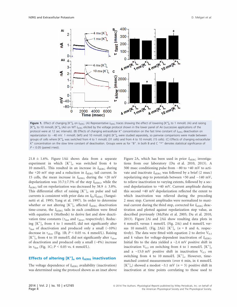
21.8 � 1.6%. Figure 1Aii shows data from a separate
experiment in which [K+]e was switched from 4 to
10 mmol/L. This resulted in an increase in IhERG during
the +20 mV step and a reduction in IhERG tail current. In
15 cells, the mean increase in IhERG during the +20 mV
depolarization was 33.7�7.5% of the step IhERG, while the
IhERG tail on repolarization was decreased by 38.9 � 3.8%.
This differential effect of raising [K+]e on pulse and tail
currents is consistent with prior data on IKr/IhERG (Sangui-
netti et al. 1995; Yang et al. 1997). In order to determine
whether or not altering [K+]e affected IhERG deactivation
time-course, the IhERG tails in each condition were fitted
with equation 4 (Methods) to derive fast and slow deacti-
vation time constants (sfast and sslow, respectively). Reduc-ing [K+]e from 4 to 1 mmol/L did not significantly alter
sfast of deactivation and produced only a small (~10%)
decrease in sslow (Fig. 1B; P < 0.05 vs. 4 mmol/L). Raising
[K+]e from 4 to 10 mmol/L did not significantly alter sslowof deactivation and produced only a small (~4%) increase
in sfast (Fig. 1C; P < 0.05 vs. 4 mmol/L).
Effects of altering [K+]e on IhERG inactivation
The voltage dependence of IhERG availability (inactivation)
was determined using the protocol shown as an inset above
Figure 2A, which has been used in prior IhERG investiga-
tions from our laboratory (Du et al. 2010, 2013). A
500 msec conditioning pulse from �80 to +40 mV to acti-
vate and inactivate IhERG was followed by a brief (2 msec)
repolarizing step to potentials between +50 and �140 mV,
to relieve inactivation to varying extents, followed by a sec-
ond depolarization to +40 mV. Current amplitude during
this second +40 mV depolarization reflected the extent to
which inactivation was relieved during the preceding
2 msec step. Current amplitudes were normalized to maxi-
mal current during the third step, corrected for IhERG deac-
tivation and plotted against repolarization step value, as
described previously (McPate et al. 2005; Du et al. 2010,
2013). Figure 2Ai and 2Aii show resulting data plots in
4 mmol/L versus 1 mmol/L (Fig. 2Ai) and 4 mmol/L ver-
sus 10 mmol/L (Fig. 2Aii) [K+]e (n = 8 and 6, respec-
tively). The data were fitted with equation 2 to derive V0.5
and k values for voltage-dependent inactivation of IhERG.
Initial fits to the data yielded a ~2.4 mV positive shift in
inactivation V0.5 on switching from 4 to 1 mmol/L [K+]eand a ~13.0 mV positive shift in inactivation V0.5 on
switching from 4 to 10 mmol/L [K+]e. However, time-
matched control measurements (over 6 min, in 4 mmol/L
[K+]e) showed a modest ~3.1 mV (n = 5) positive shift in
inactivation at time points correlating to those used to
Ai B
C
Aii
Figure 1. Effect of changing [K+]e on IhERG. (A) Representative IhERG traces showing the effect of lowering [K+]e to 1 mmol/L (Ai) and raising
[K+]e to 10 mmol/L [K+]e (Aii) on WT IhERG elicited by the voltage protocol shown in the lower panel of Aii (successive applications of the
protocol were at 12 sec intervals). (B) Effects of changing extracellular K+ concentration on the fast time constant of IhERG deactivation on
repolarization to �40 mV. 1 mmol/L (left) and 10 mmol/L (right) [K+]e were studied separately, so pairwise comparisons were made between
groups of cells where [K+]e was switched from 4 to 1 mmol/L (31 cells) and from 4 to 10 mmol/L (15 cells). (C) Effects of changing extracellular
K+ concentration on the slow time constant of deactivation. Groups were as for “B”. In both B and C “*” denotes statistical significance of
P < 0.05 (paired t-test).
2014 | Vol. 2 | Iss. 10 | e12165Page 4
ª 2014 The Authors. Physiological Reports published by Wiley Periodicals, Inc. on behalf of
the American Physiological Society and The Physiological Society.
hERG and Extracellular Potassium D. Melgari et al.
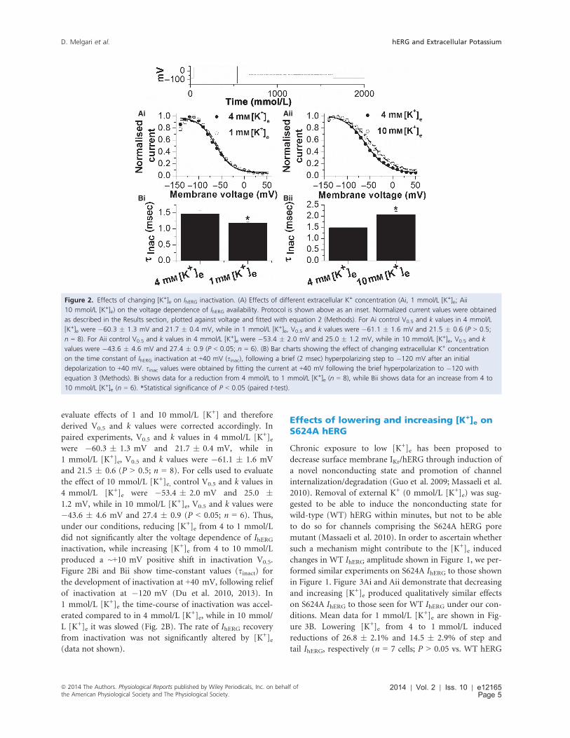
evaluate effects of 1 and 10 mmol/L [K+] and therefore
derived V0.5 and k values were corrected accordingly. In
paired experiments, V0.5 and k values in 4 mmol/L [K+]ewere �60.3 � 1.3 mV and 21.7 � 0.4 mV, while in
1 mmol/L [K+]e, V0.5 and k values were �61.1 � 1.6 mV
and 21.5 � 0.6 (P > 0.5; n = 8). For cells used to evaluate
the effect of 10 mmol/L [K+]e, control V0.5 and k values in
4 mmol/L [K+]e were �53.4 � 2.0 mV and 25.0 �1.2 mV, while in 10 mmol/L [K+]e, V0.5 and k values were
�43.6 � 4.6 mV and 27.4 � 0.9 (P < 0.05; n = 6). Thus,
under our conditions, reducing [K+]e from 4 to 1 mmol/L
did not significantly alter the voltage dependence of IhERGinactivation, while increasing [K+]e from 4 to 10 mmol/L
produced a ~+10 mV positive shift in inactivation V0.5.
Figure 2Bi and Bii show time-constant values (sinact) for
the development of inactivation at +40 mV, following relief
of inactivation at �120 mV (Du et al. 2010, 2013). In
1 mmol/L [K+]e the time-course of inactivation was accel-
erated compared to in 4 mmol/L [K+]e, while in 10 mmol/
L [K+]e it was slowed (Fig. 2B). The rate of IhERG recovery
from inactivation was not significantly altered by [K+]e(data not shown).
Effects of lowering and increasing [K+]e onS624A hERG
Chronic exposure to low [K+]e has been proposed to
decrease surface membrane IKr/hERG through induction of
a novel nonconducting state and promotion of channel
internalization/degradation (Guo et al. 2009; Massaeli et al.
2010). Removal of external K+ (0 mmol/L [K+]e) was sug-
gested to be able to induce the nonconducting state for
wild-type (WT) hERG within minutes, but not to be able
to do so for channels comprising the S624A hERG pore
mutant (Massaeli et al. 2010). In order to ascertain whether
such a mechanism might contribute to the [K+]e induced
changes in WT IhERG amplitude shown in Figure 1, we per-
formed similar experiments on S624A IhERG to those shown
in Figure 1. Figure 3Ai and Aii demonstrate that decreasing
and increasing [K+]e produced qualitatively similar effects
on S624A IhERG to those seen for WT IhERG under our con-
ditions. Mean data for 1 mmol/L [K+]e are shown in Fig-
ure 3B. Lowering [K+]e from 4 to 1 mmol/L induced
reductions of 26.8 � 2.1% and 14.5 � 2.9% of step and
tail IhERG, respectively (n = 7 cells; P > 0.05 vs. WT hERG
Ai
Bi
Aii
Bii
Figure 2. Effects of changing [K+]e on IhERG inactivation. (A) Effects of different extracellular K+ concentration (Ai, 1 mmol/L [K+]e; Aii
10 mmol/L [K+]e) on the voltage dependence of IhERG availability. Protocol is shown above as an inset. Normalized current values were obtained
as described in the Results section, plotted against voltage and fitted with equation 2 (Methods). For Ai control V0.5 and k values in 4 mmol/L
[K+]e were �60.3 � 1.3 mV and 21.7 � 0.4 mV, while in 1 mmol/L [K+]e, V0.5 and k values were �61.1 � 1.6 mV and 21.5 � 0.6 (P > 0.5;
n = 8). For Aii control V0.5 and k values in 4 mmol/L [K+]e were �53.4 � 2.0 mV and 25.0 � 1.2 mV, while in 10 mmol/L [K+]e, V0.5 and k
values were �43.6 � 4.6 mV and 27.4 � 0.9 (P < 0.05; n = 6). (B) Bar charts showing the effect of changing extracellular K+ concentration
on the time constant of IhERG inactivation at +40 mV (sinac), following a brief (2 msec) hyperpolarizing step to �120 mV after an initial
depolarization to +40 mV. sinac values were obtained by fitting the current at +40 mV following the brief hyperpolarization to �120 with
equation 3 (Methods). Bi shows data for a reduction from 4 mmol/L to 1 mmol/L [K+]e (n = 8), while Bii shows data for an increase from 4 to
10 mmol/L [K+]e (n = 6). *Statistical significance of P < 0.05 (paired t-test).
ª 2014 The Authors. Physiological Reports published by Wiley Periodicals, Inc. on behalf ofthe American Physiological Society and The Physiological Society.
2014 | Vol. 2 | Iss. 10 | e12165Page 5
D. Melgari et al. hERG and Extracellular Potassium
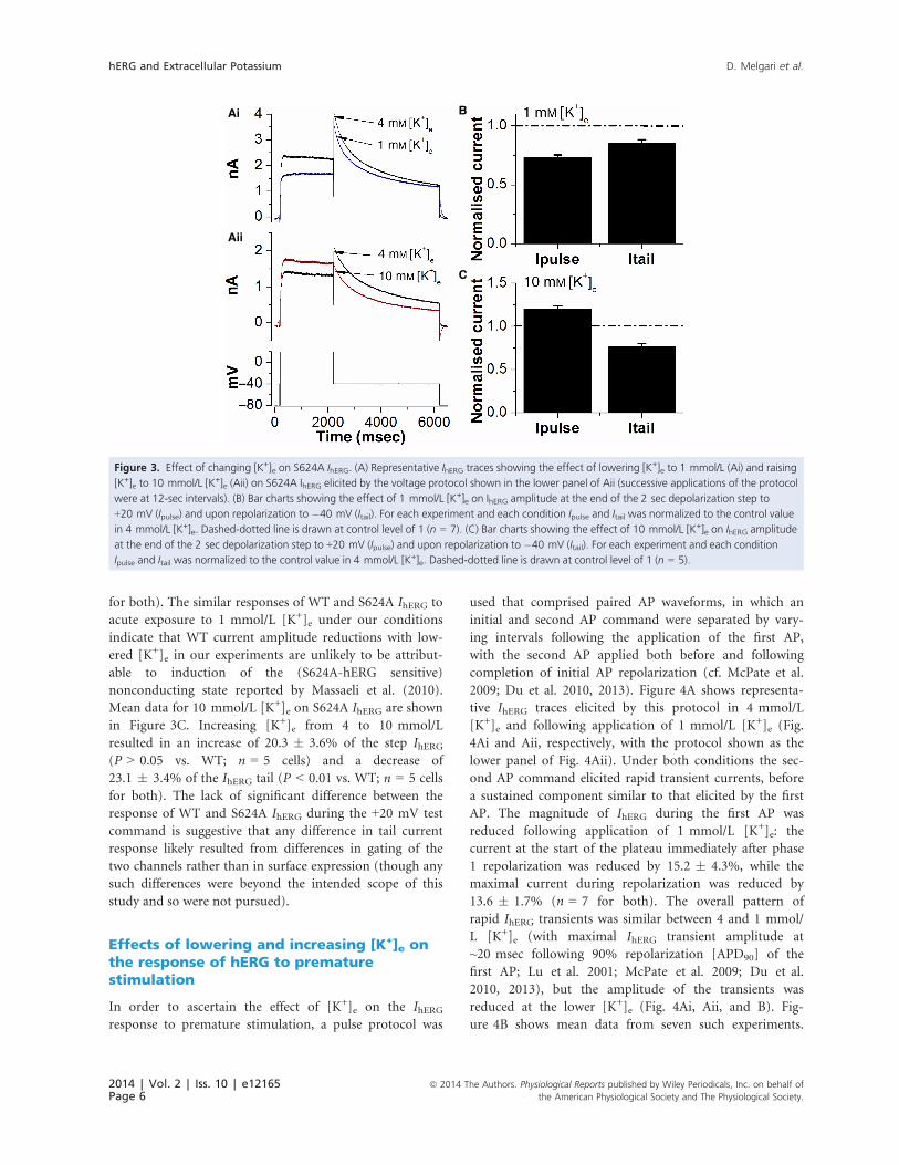
for both). The similar responses of WT and S624A IhERG to
acute exposure to 1 mmol/L [K+]e under our conditions
indicate that WT current amplitude reductions with low-
ered [K+]e in our experiments are unlikely to be attribut-
able to induction of the (S624A-hERG sensitive)
nonconducting state reported by Massaeli et al. (2010).
Mean data for 10 mmol/L [K+]e on S624A IhERG are shown
in Figure 3C. Increasing [K+]e from 4 to 10 mmol/L
resulted in an increase of 20.3 � 3.6% of the step IhERG(P > 0.05 vs. WT; n = 5 cells) and a decrease of
23.1 � 3.4% of the IhERG tail (P < 0.01 vs. WT; n = 5 cells
for both). The lack of significant difference between the
response of WT and S624A IhERG during the +20 mV test
command is suggestive that any difference in tail current
response likely resulted from differences in gating of the
two channels rather than in surface expression (though any
such differences were beyond the intended scope of this
study and so were not pursued).
Effects of lowering and increasing [K+]e onthe response of hERG to prematurestimulation
In order to ascertain the effect of [K+]e on the IhERGresponse to premature stimulation, a pulse protocol was
used that comprised paired AP waveforms, in which an
initial and second AP command were separated by vary-
ing intervals following the application of the first AP,
with the second AP applied both before and following
completion of initial AP repolarization (cf. McPate et al.
2009; Du et al. 2010, 2013). Figure 4A shows representa-
tive IhERG traces elicited by this protocol in 4 mmol/L
[K+]e and following application of 1 mmol/L [K+]e (Fig.
4Ai and Aii, respectively, with the protocol shown as the
lower panel of Fig. 4Aii). Under both conditions the sec-
ond AP command elicited rapid transient currents, before
a sustained component similar to that elicited by the first
AP. The magnitude of IhERG during the first AP was
reduced following application of 1 mmol/L [K+]e: the
current at the start of the plateau immediately after phase
1 repolarization was reduced by 15.2 � 4.3%, while the
maximal current during repolarization was reduced by
13.6 � 1.7% (n = 7 for both). The overall pattern of
rapid IhERG transients was similar between 4 and 1 mmol/
L [K+]e (with maximal IhERG transient amplitude at
~20 msec following 90% repolarization [APD90] of the
first AP; Lu et al. 2001; McPate et al. 2009; Du et al.
2010, 2013), but the amplitude of the transients was
reduced at the lower [K+]e (Fig. 4Ai, Aii, and B). Fig-
ure 4B shows mean data from seven such experiments.
Ai B
C
Aii
Figure 3. Effect of changing [K+]e on S624A IhERG. (A) Representative IhERG traces showing the effect of lowering [K+]e to 1 mmol/L (Ai) and raising
[K+]e to 10 mmol/L [K+]e (Aii) on S624A IhERG elicited by the voltage protocol shown in the lower panel of Aii (successive applications of the protocol
were at 12-sec intervals). (B) Bar charts showing the effect of 1 mmol/L [K+]e on IhERG amplitude at the end of the 2 sec depolarization step to
+20 mV (Ipulse) and upon repolarization to �40 mV (Itail). For each experiment and each condition Ipulse and Itail was normalized to the control value
in 4 mmol/L [K+]e. Dashed-dotted line is drawn at control level of 1 (n = 7). (C) Bar charts showing the effect of 10 mmol/L [K+]e on IhERG amplitude
at the end of the 2 sec depolarization step to +20 mV (Ipulse) and upon repolarization to �40 mV (Itail). For each experiment and each condition
Ipulse and Itail was normalized to the control value in 4 mmol/L [K+]e. Dashed-dotted line is drawn at control level of 1 (n = 5).
2014 | Vol. 2 | Iss. 10 | e12165Page 6
ª 2014 The Authors. Physiological Reports published by Wiley Periodicals, Inc. on behalf of
the American Physiological Society and The Physiological Society.
hERG and Extracellular Potassium D. Melgari et al.
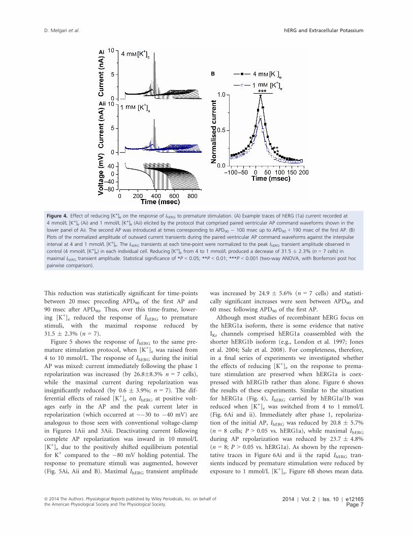
This reduction was statistically significant for time-points
between 20 msec preceding APD90 of the first AP and
90 msec after APD90. Thus, over this time-frame, lower-
ing [K+]e reduced the response of IhERG to premature
stimuli, with the maximal response reduced by
31.5 � 2.3% (n = 7).
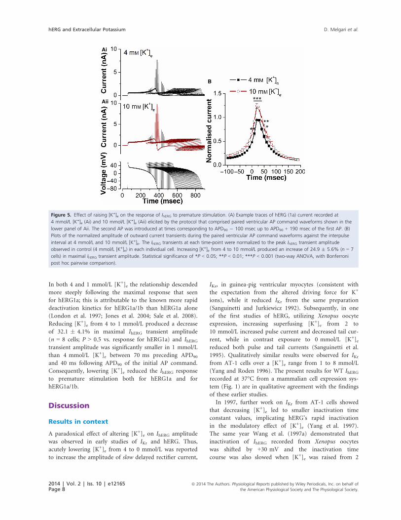
Figure 5 shows the response of IhERG to the same pre-
mature stimulation protocol, when [K+]e was raised from
4 to 10 mmol/L. The response of IhERG during the initial
AP was mixed: current immediately following the phase 1
repolarization was increased (by 26.8�8.3% n = 7 cells),
while the maximal current during repolarization was
insignificantly reduced (by 0.6 � 3.9%; n = 7). The dif-
ferential effects of raised [K+]e on IhERG at positive volt-
ages early in the AP and the peak current later in
repolarization (which occurred at ~�30 to �40 mV) are
analogous to those seen with conventional voltage-clamp
in Figures 1Aii and 3Aii. Deactivating current following
complete AP repolarization was inward in 10 mmol/L
[K+]e due to the positively shifted equilibrium potential
for K+ compared to the �80 mV holding potential. The
response to premature stimuli was augmented, however
(Fig. 5Ai, Aii and B). Maximal IhERG transient amplitude
was increased by 24.9 � 5.6% (n = 7 cells) and statisti-
cally significant increases were seen between APD90 and
60 msec following APD90 of the first AP.
Although most studies of recombinant hERG focus on
the hERG1a isoform, there is some evidence that native
IKr channels comprised hERG1a coassembled with the
shorter hERG1b isoform (e.g., London et al. 1997; Jones
et al. 2004; Sale et al. 2008). For completeness, therefore,
in a final series of experiments we investigated whether
the effects of reducing [K+]e on the response to prema-
ture stimulation are preserved when hERG1a is coex-
pressed with hERG1b rather than alone. Figure 6 shows
the results of these experiments. Similar to the situation
for hERG1a (Fig. 4), IhERG carried by hERG1a/1b was
reduced when [K+]e was switched from 4 to 1 mmol/L
(Fig. 6Ai and ii). Immediately after phase 1, repolariza-
tion of the initial AP, IhERG was reduced by 20.8 � 5.7%
(n = 8 cells; P > 0.05 vs. hERG1a), while maximal IhERGduring AP repolarization was reduced by 23.7 � 4.8%
(n = 8; P > 0.05 vs. hERG1a). As shown by the represen-
tative traces in Figure 6Ai and ii the rapid IhERG tran-
sients induced by premature stimulation were reduced by
exposure to 1 mmol/L [K+]e. Figure 6B shows mean data.
Ai
Aii
B
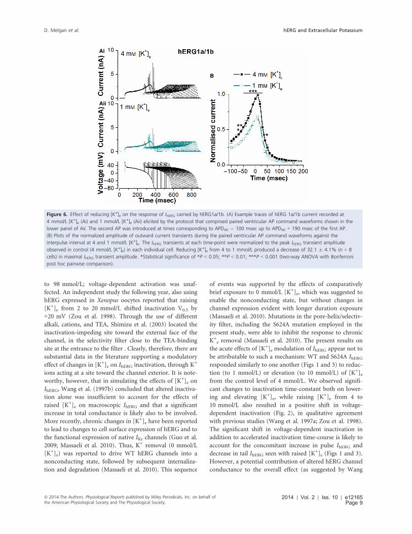
Figure 4. Effect of reducing [K+]e on the response of IhERG to premature stimulation. (A) Example traces of hERG (1a) current recorded at
4 mmol/L [K+]e (Ai) and 1 mmol/L [K+]e (Aii) elicited by the protocol that comprised paired ventricular AP command waveforms shown in the
lower panel of Aii. The second AP was introduced at times corresponding to APD90 � 100 msec up to APD90 + 190 msec of the first AP. (B)
Plots of the normalized amplitude of outward current transients during the paired ventricular AP command waveforms against the interpulse
interval at 4 and 1 mmol/L [K+]e. The IhERG transients at each time-point were normalized to the peak IhERG transient amplitude observed in
control (4 mmol/L [K+]e) in each individual cell. Reducing [K+]e from 4 to 1 mmol/L produced a decrease of 31.5 � 2.3% (n = 7 cells) in
maximal IhERG transient amplitude. Statistical significance of *P < 0.05; **P < 0.01; ***P < 0.001 (two-way ANOVA, with Bonferroni post hoc
pairwise comparison).
ª 2014 The Authors. Physiological Reports published by Wiley Periodicals, Inc. on behalf ofthe American Physiological Society and The Physiological Society.
2014 | Vol. 2 | Iss. 10 | e12165Page 7
D. Melgari et al. hERG and Extracellular Potassium
In both 4 and 1 mmol/L [K+]e the relationship descended
more steeply following the maximal response that seen
for hERG1a; this is attributable to the known more rapid
deactivation kinetics for hERG1a/1b than hERG1a alone
(London et al. 1997; Jones et al. 2004; Sale et al. 2008).
Reducing [K+]e from 4 to 1 mmol/L produced a decrease
of 32.1 � 4.1% in maximal IhERG transient amplitude
(n = 8 cells; P > 0.5 vs. response for hERG1a) and IhERGtransient amplitude was significantly smaller in 1 mmol/L
than 4 mmol/L [K+]e between 70 ms preceding APD90
and 40 ms following APD90 of the initial AP command.
Consequently, lowering [K+]e reduced the IhERG response
to premature stimulation both for hERG1a and for
hERG1a/1b.
Discussion
Results in context
A paradoxical effect of altering [K+]e on IhERG amplitude
was observed in early studies of IKr and hERG. Thus,
acutely lowering [K+]e from 4 to 0 mmol/L was reported
to increase the amplitude of slow delayed rectifier current,
IKs, in guinea-pig ventricular myocytes (consistent with
the expectation from the altered driving force for K+
ions), while it reduced IKr from the same preparation
(Sanguinetti and Jurkiewicz 1992). Subsequently, in one
of the first studies of hERG, utilizing Xenopus oocyte
expression, increasing superfusing [K+]e from 2 to
10 mmol/L increased pulse current and decreased tail cur-
rent, while in contrast exposure to 0 mmol/L [K+]ereduced both pulse and tail currents (Sanguinetti et al.
1995). Qualitatively similar results were observed for IKrfrom AT-1 cells over a [K+]e range from 1 to 8 mmol/L
(Yang and Roden 1996). The present results for WT IhERGrecorded at 37°C from a mammalian cell expression sys-
tem (Fig. 1) are in qualitative agreement with the findings
of these earlier studies.
In 1997, further work on IKr from AT-1 cells showed
that decreasing [K+]e led to smaller inactivation time
constant values, implicating hERG’s rapid inactivation
in the modulatory effect of [K+]e (Yang et al. 1997).
The same year Wang et al. (1997a) demonstrated that
inactivation of IhERG recorded from Xenopus oocytes
was shifted by +30 mV and the inactivation time
course was also slowed when [K+]e was raised from 2
Ai
Aii
B
Figure 5. Effect of raising [K+]e on the response of IhERG to premature stimulation. (A) Example traces of hERG (1a) current recorded at
4 mmol/L [K+]e (Ai) and 10 mmol/L [K+]e (Aii) elicited by the protocol that comprised paired ventricular AP command waveforms shown in the
lower panel of Aii. The second AP was introduced at times corresponding to APD90 � 100 msec up to APD90 + 190 msec of the first AP. (B)
Plots of the normalized amplitude of outward current transients during the paired ventricular AP command waveforms against the interpulse
interval at 4 mmol/L and 10 mmol/L [K+]e. The IhERG transients at each time-point were normalized to the peak IhERG transient amplitude
observed in control (4 mmol/L [K+]e) in each individual cell. Increasing [K+]e from 4 to 10 mmol/L produced an increase of 24.9 � 5.6% (n = 7
cells) in maximal IhERG transient amplitude. Statistical significance of *P < 0.05; **P < 0.01; ***P < 0.001 (two-way ANOVA, with Bonferroni
post hoc pairwise comparison).
2014 | Vol. 2 | Iss. 10 | e12165Page 8
ª 2014 The Authors. Physiological Reports published by Wiley Periodicals, Inc. on behalf of
the American Physiological Society and The Physiological Society.
hERG and Extracellular Potassium D. Melgari et al.
to 98 mmol/L; voltage-dependent activation was unaf-
fected. An independent study the following year, also using
hERG expressed in Xenopus oocytes reported that raising
[K+]e from 2 to 20 mmol/L shifted inactivation V0.5 by
+20 mV (Zou et al. 1998). Through the use of different
alkali, cations, and TEA, Shimizu et al. (2003) located the
inactivation-impeding site toward the external face of the
channel, in the selectivity filter close to the TEA-binding
site at the entrance to the filter . Clearly, therefore, there are
substantial data in the literature supporting a modulatory
effect of changes in [K+]e on IhERG inactivation, through K+
ions acting at a site toward the channel exterior. It is note-
worthy, however, that in simulating the effects of [K+]e on
IhERG, Wang et al. (1997b) concluded that altered inactiva-
tion alone was insufficient to account for the effects of
raised [K+]e on macroscopic IhERG and that a significant
increase in total conductance is likely also to be involved.
More recently, chronic changes in [K+]e have been reported
to lead to changes to cell surface expression of hERG and to
the functional expression of native IKr channels (Guo et al.
2009; Massaeli et al. 2010). Thus, K+ removal (0 mmol/L
[K+]e) was reported to drive WT hERG channels into a
nonconducting state, followed by subsequent internaliza-
tion and degradation (Massaeli et al. 2010). This sequence
of events was supported by the effects of comparatively
brief exposure to 0 mmol/L [K+]e, which was suggested to
enable the nonconducting state, but without changes in
channel expression evident with longer duration exposure
(Massaeli et al. 2010). Mutations in the pore-helix/selectiv-
ity filter, including the S624A mutation employed in the
present study, were able to inhibit the response to chronic
K+e removal (Massaeli et al. 2010). The present results on
the acute effects of [K+]e modulation of IhERG appear not to
be attributable to such a mechanism: WT and S624A IhERGresponded similarly to one another (Figs 1 and 3) to reduc-
tion (to 1 mmol/L) or elevation (to 10 mmol/L) of [K+]efrom the control level of 4 mmol/L. We observed signifi-
cant changes to inactivation time-constant both on lower-
ing and elevating [K+]e, while raising [K+]e from 4 to
10 mmol/L also resulted in a positive shift in voltage-
dependent inactivation (Fig. 2), in qualitative agreement
with previous studies (Wang et al. 1997a; Zou et al. 1998).
The significant shift in voltage-dependent inactivation in
addition to accelerated inactivation time-course is likely to
account for the concomitant increase in pulse IhERG and
decrease in tail IhERG seen with raised [K+]e (Figs 1 and 3).
However, a potential contribution of altered hERG channel
conductance to the overall effect (as suggested by Wang
Ai
Aii
B
Figure 6. Effect of reducing [K+]e on the response of IhERG carried by hERG1a/1b. (A) Example traces of hERG 1a/1b current recorded at
4 mmol/L [K+]e (Ai) and 1 mmol/L [K+]e (Aii) elicited by the protocol that comprised paired ventricular AP command waveforms shown in the
lower panel of Aii. The second AP was introduced at times corresponding to APD90 � 100 msec up to APD90 + 190 msec of the first AP.
(B) Plots of the normalized amplitude of outward current transients during the paired ventricular AP command waveforms against the
interpulse interval at 4 and 1 mmol/L [K+]e. The IhERG transients at each time-point were normalized to the peak IhERG transient amplitude
observed in control (4 mmol/L [K+]e) in each individual cell. Reducing [K+]e from 4 to 1 mmol/L produced a decrease of 32.1 � 4.1% (n = 8
cells) in maximal IhERG transient amplitude. *Statistical significance of *P < 0.05; **P < 0.01; ***P < 0.001 (two-way ANOVA with Bonferroni
post hoc pairwise comparison).
ª 2014 The Authors. Physiological Reports published by Wiley Periodicals, Inc. on behalf ofthe American Physiological Society and The Physiological Society.
2014 | Vol. 2 | Iss. 10 | e12165Page 9
D. Melgari et al. hERG and Extracellular Potassium
et al. 1997a) cannot be ruled out, given that single hERG
channel conductance is known to vary with [K+]e (2 pS at
5 mmol/L and 10 pS at 100 mmol/L in Kiehn et al.
[1996]). Although IhERG is known to be sensitive to [Na+]e(Namaguchi et al. 2000; Mullins et al. 2002) and changes
to [K+]e in the present study were compensated by con-
comitant alterations to [Na+]e, the modulatory effects of
[Na+]e on IhERG amplitude are most marked for [Na+]econcentrations substantially below 100 mmol/L (Namagu-
chi et al. 2000; Mullins et al. 2002) and so are unlikely to
contribute significantly to observed effects of altering [K+]ein our experiments.
The response to premature stimulationunder AP clamp
The profile of WT IhERG seen here in normal (4 mmol/L)
[K+]e both during imposed AP clamp commands and in
response to premature AP stimuli (Figs 4 and 5) is compa-
rable to that found in prior studies that have used similar
paired AP clamp protocols, with maximal IhERG transient
amplitude occurring when premature stimuli were applied
shortly after the point of 90% complete repolarization of
the first AP (APD90) (Lu et al. 2001, 2003; McPate et al.
2009; Du et al. 2010). Application of premature stimuli
between 100 msec before APD90 of the initial AP and
190 msec after APD90 was sufficient to reveal the normal
biphasic relationship of IhERG transient amplitude with
time late in repolarization/early in diastole (Lu et al. 2001,
2003; McPate et al. 2009; Du et al. 2010). In our experi-
ments, reduced [K+]e decreased IhERG both during the ini-
tial AP command and during the transient responses to the
second AP command waveform. To our knowledge, our
data constitute the first direct AP clamp demonstration of
modification by [K+]e of the IhERG response to premature
stimulation. We have shown previously a suppression of
the IhERG response to premature stimuli in the context of
extracellular acidosis, an effect that was associated with
marked acceleration of IhERG deactivation (Du et al. 2010).
However, in the case of low [K+]e, the fast component of
deactivation was unaffected by reducing [K+]e from 4 to
1 mmol/L (Fig. 1) and so the altered response to prema-
ture stimuli in late repolarization/early diastole is unlikely
to be accounted for by changes to IhERG deactivation.
Rather, enhanced inactivation and reduced net conduc-
tance are likely to account for the reduced response to pre-
mature stimuli. It is significant that coexpressed hERG1a/
1b showed a similar suppression of the IhERG response to
premature stimuli with low [K+]e to that of hERG1a alone
(Figs 4 and 6). Thus, whether native IKr results from het-
eromeric hERG1a and hERG1b (London et al. 1997; Jones
et al. 2004; Sale et al. 2008) or from hERG1a alone, it is
safe to conclude that the channel’s protective role against
premature depolarization at time-points comparable to
those studied here is likely to be significantly reduced in
circumstances with reduced [K+]e.
The characteristic resurgent IhERG tail during conven-
tional voltage-clamp results from rapid recovery of IhERGfrom inactivation on membrane potential repolarization.
Concomitant increases in IhERG pulse current and
decreases in tail current with raised [K+]e (Figs 1 and 3;
Sanguinetti et al. 1995; Yang and Roden 1996) are both
consequences of attenuated inactivation. The effect of
10 mmol/L [K+]e on IhERG during the AP waveform seen
here reflects dynamic changes in IhERG gating during the
AP, such that peak IhERG during repolarization (which
typically occurs between ~�30 and �40 mV; Hancox
et al. 1998; McPate et al. 2005) was little changed, but
IhERG early during the AP was increased. Thus, an
increased contribution of IKr to repolarization might be
anticipated early during the ventricular AP under situa-
tions of hyperkalemia. Our data are also suggestive of an
increased ability of hERG to resist premature depolariza-
tion for a short period early in diastole.
Potential physiological significance
In the setting of experimental acute coronary occlusion or
ischemia, [K+]e accumulation to values exceeding
10 mmol/L has been reported (Hill and Gettes 1980;
Weiss and Shine 1982). Consequently, our data with
raised [K+]e have relevance in terms of suggesting an
altered role of IKr both early during the ventricular AP
plateau and late in repolarization/early in diastole (as
considered earlier). If pathological ischemia/K+ accumula-
tion is localized, then the localized effect of raised [K+]eon hERG/IKr could contribute to heterogeneity in repolar-
ization and in tissue sensitivity to premature excitation.
On the other hand, global hypokalemia is strongly associ-
ated with risk of arrhythmia and is known to exacerbate
the risk of acquired (drug-induced) LQTS and associated
Torsades de Pointes (TdP) (Viskin 1999; Zeltser et al.
2003). In profound hypokalemia levels close to 1 mmol/L
(1.2 mmol/L) have been reported (Garcia et al. 2008).
Thus, while the reduction in [K+]e from 4 to 1 mmol/L
can fairly be considered to represent an extreme in terms
of clinically relevant hypokalemia, our findings constitute
a valuable proof-of-concept demonstration: acute hypoka-
lemia not only reduces the contribution of IhERG/IKr to
ventricular repolarization but can also impair the chan-
nel’s protective role against premature excitation. In
chronic hypokalemia, these acute effects can be expected
to be synergistic with decreased surface expression of IKr/
hERG channels consequent to sustained low [K+]e (Guo
et al. 2009; Massaeli et al. 2010), to contribute to the
overall effect. In the additional presence of a hERG/IKr
2014 | Vol. 2 | Iss. 10 | e12165Page 10
ª 2014 The Authors. Physiological Reports published by Wiley Periodicals, Inc. on behalf of
the American Physiological Society and The Physiological Society.
hERG and Extracellular Potassium D. Melgari et al.
blocking drug, these effects can be anticipated to combine
with pharmacological suppression of IhERG in augmenting
the overall arrhythmic risk. Conversely, restoration of a
normal [K+]e in hypokalemic patients can be anticipated
to restore both the role of hERG/IKr in normal ventricular
repolarization and its protective role early in diastole. It is
feasible that acute effects of raising [K+]e on IhERG may
contribute to the beneficial actions of potassium
supplementation therapy (raising serum potassium by
~1 mmol/L) in patients with hERG mutation-linked
congenital LQTS (Compton et al. 1996; Etheridge et al.
2003), although the effects of long-term potassium sup-
plementation in that setting are perhaps more likely to
involve [K+]e linked changes to cell surface channel
expression (Guo et al. 2009; Massaeli et al. 2010).
Conflict of Interest
None declared.
References
Brugada, R., K. Hong, R. Dumaine, J. Cordeiro, F. Gaita,
M. Borggrefe, et al. 2004. Sudden death associated with
short-QT syndrome linked to mutations in HERG.
Circulation 109:30–35.
Compton, S. J., R. L. Lux, M. R. Ramsey, K. R. Strelich,
M. C. Sanguinetti, L. S. Green, et al. 1996. Genetically
defined therapy of inherited long-QT syndrome – correction
of abnormal repolarisation by potassium. Circulation
94:1018–1022.
Du, C. Y., I. Adeniran, H. Cheng, Y. H. Zhang, H. A. El,
M. J. McPate, et al. 2010. Acidosis impairs the protective
role of hERG K+ channels against premature stimulation.
J. Cardiovasc. Electrophysiol. 21:1160–1169.
Du, C. Y., A. El Harchi, Y. H. Zhang, C. H. Orchard, and
J. C. Hancox. 2011. Pharmacological inhibition of hERG is
modulated by extracellular but not intracellular acidosis.
J. Cardiovasc. Electrophysiol. 22:1163–1170.
Du, C., A. El Harchi, H. Zhang, and J. C. Hancox. 2013.
Modification by KCNE1 variants of the hERG potassium
channel response to premature stimulation and to
pharmacological inhibition. Physiol. Rep. 1:e00175.
Du, C., Y. Zhang, H. A. El, C. E. Dempsey, and J. C. Hancox.
2014. Ranolazine inhibition of hERG potassium channels:
drug-pore interactions and reduced potency against
inactivation mutants. J. Mol. Cell. Cardiol. 74C:220–230.
El Harchi, A., Y. H. Zhang, L. Hussein, C. E. Dempsey, and
J. C. Hancox. 2012. Molecular determinants of hERG
potassium channel inhibition by disopyramide. J. Mol. Cell.
Cardiol. 52:185–195.
Etheridge, S. P., S. J. Compton, M. Tristani-Firouzi, and
J. W. Mason. 2003. A new oral therapy for long QT
syndrome: long-term oral potassium improves repolarisation
in patients with HERG mutations. J. Am. Coll. Cardiol.
42:1777–1782.
Garcia, E., N. Nakhleh, D. Simmons, and C. Ramsay. 2008.
Profound hypokalemia: unusual presentation and
management in a 12-year-old boy. Pediatr. Emerg. Care
24:157–160.
Guo, J., H. Massaeli, J. Xu, Z. Jia, J. T. Wigle, N. Mesaeli,
et al. 2009. Extracellular K+ concentration controls cell
surface density of IKr in rabbit hearts and of the HERG
channel in human cell lines. J. Clin. Invest. 119:2745–2757.
Hancox, J. C., A. J. Levi, and H. J. Witchel. 1998. Time course
and voltage dependence of expressed HERG current
compared with native ‘rapid’ delayed rectifier K current
during the cardiac ventricular action potential. Pflugers
Arch. 436:843–853.
Hancox, J. C., M. J. McPate, A. El Harchi, and Y. H. Zhang.
2008. The hERG potassium channel and hERG screening for
drug-induced torsades de pointes. Pharmacol. Ther.
119:118–132.
Hill, J. L., and L. S. Gettes. 1980. Effect of acute coronary
artery occlusion on local myocardial extracellular K+ activity
in swine. Circulation 61:768–778.
Jones, E. M., E. C. Roti Roti, J. Wang, and G. A. Robertson.
2004. Cardiac IKr channels minimally comprise hERG 1a
and 1b subunits. J. Biol. Chem. 279:44690–44694.
Kiehn, J., A. E. Lacerda, B. Wible, and A. M. Brown. 1996.
Molecular physiology and pharmacology of HERG. Single
channel currents and block by dofetilide. Circulation
94:2572–2579.
Levi, A. J., J. C. Hancox, F. C. Howarth, J. Croker, and
J. Vinnicombe. 1996. A method for making rapid changes of
superfusate whilst maintaining temperature at 37°C. PflugersArch. 432:930–937.
London, B., M. C. Trudeau, K. P. Newton, A. K. Bayer,
N. G. Copeland, D. J. Gilbert, et al. 1997. Two isoforms of
the mouse ether-a-go-go related gene coassemble form
channels with properties similar to the rapidly activating
component of the cardiac delayed rectifier K current. Circ.
Res. 81:870–878.
Lu, Y., M. P. MahautSmith, A. Varghese, C. L. H. Huang,
P. R. Kemp, and J. I. Vandenberg. 2001. Effects of premature
stimulation on HERG channels. J. Physiol. 537:843–851.
Lu, Y., M. P. Mahaut-Smith, C. L. Huang, and J. I.
Vandenberg. 2003. Mutant MiRP1 subunits modulate HERG
K+ channel gating: a mechanism for pro-arrhythmia in long
QT syndrome type 6. J. Physiol. 551:253–262.
Massaeli, H., J. Guo, J. Xu, and S. Zhang. 2010. Extracellular
K+ is a prerequisite for the function and plasma membrane
stability of HERG channels. Circ. Res. 106:1072–1082.
McPate, M. J., R. S. Duncan, J. T. Milnes, H. J. Witchel, and
J. C. Hancox. 2005. The N588K-HERG K+ channel mutation
in the ‘short QT syndrome’: mechanism of gain-in-function
determined at 37°C. Biochem. Biophys. Res. Commun.
334:441–449.
ª 2014 The Authors. Physiological Reports published by Wiley Periodicals, Inc. on behalf ofthe American Physiological Society and The Physiological Society.
2014 | Vol. 2 | Iss. 10 | e12165Page 11
D. Melgari et al. hERG and Extracellular Potassium
McPate, M. J., H. Zhang, I. Ideniran, J. M. Cordeiro,
H. J. Witchel, and J. C. Hancox. 2009. Comparative effects
of the short QT N588K mutation at 37°C on hERG K+
channel current during ventricular, Purkinje fibre and atrial
action potentials: an action potential clamp study. J. Physiol.
Pharmacol. 60:23–41.
Mitcheson, J. S., and J. C. Hancox. 1999. An investigation of
the role played by the E-4031-sensitive (rapid delayed
rectifier) potassium current in isolated rabbit
atrioventricular nodal and ventricular myocytes. Pflugers
Arch. 438:843–850.
Modell, S. M., and M. H. Lehmann. 2006. The long QT
syndrome family of cardiac ion channelopathies: a HuGE
review. Genet. Med. 8:143–155.
Mullins, F. M., S. Z. Stepanovic, R. R. Desai, A. L. Jr George,
and J. R. Balser. 2002. Extracellular sodium interacts with
the hERG channel at an outer pore site. J. Gen. Physiol.
120:517–537.
Namaguchi, H., J. P. Jr Johnson, C. I. Petersen, and
J. R. Balser. 2000. A sensitive mechanism for cation
modulation of a potassium current. Nat. Neurosci. 3:429–430.
Sale, H., J. Wang, T. J. O’Hara, D. J. Tester, P. Phartiyal,
J. Q. He, et al. 2008. Physiological properties of hERG 1a/1b
heteromeric currents and a hERG 1b-specific mutation
associated with Long-QT syndrome. Circ. Res. 103:e81–e95.
Sanguinetti, M. C., and N. K. Jurkiewicz. 1992. Role of
external Ca and K in gating of cardiac delayed rectifier
currents. Pflugers Arch. 420:180–186.
Sanguinetti, M. C., C. Jiang, M. E. Curran, and M. T. Keating.
1995. A mechanistic link between an inherited and an
acquired cardiac arrhythmia: HERG encodes the IKrpotassium channel. Cell 81:299.
Shimizu, H., C. Toyoshima, and S. Oiki. 2003. Interaction
between tetraethylammonium and permeant cations at the
inactivation gate of the HERG potassium channel. Jpn.
J. Physiol. 53:25–34.
Shimoni, Y., R. B. Clark, and W. R. Giles. 1992. Role of an
inwardly rectifying potassium current in rabbit ventricular
action potential. J. Physiol. 448:709–727.
Smith, P. L., T. Baukrowitz, and G. Yellen. 1996. The inward
rectification mechanism of the HERG cardiac potassium
channel. Nature 379:833–836.
Sun, Y., X. Q. Quan, S. Fromme, R. H. Cox, P. Zhang,
L. Zhang, et al. 2011. A novel mutation in the KCNH2 gene
associated with short QT syndrome. J. Mol. Cell. Cardiol.
50:433–441.
Tamargo, J., R. Caballero, R. Gomez, C. Valenzuela, and E.
Delpon. 2004. Pharmacology of cardiac potassium channels.
Cardiovasc. Res. 62:9–33.
Trudeau, M. C., J. W. Warmke, B. Ganetzky, and
G. A. Robertson. 1995. HERG, an inward rectifier in the
voltage-gated potassium channel family. Science 269:92–95.
Viskin, S. 1999. Long QT syndromes and torsade de pointes.
Lancet 354:1625–1633.
Wang, S., S. Liu, M. J. Morales, H. C. Strauss, and
R. L. Rasmusson. 1997a. A quantitative analysis of the
activation and inactivation kinetics of HERG expressed in
Xenopus oocytes. J. Physiol. 502:45–60.
Wang, S., M. J. Morales, S. Liu, H. C. Strauss, and
R. L. Rasmusson. 1997b. Modulation of HERG affinity for
E-4031 by [K]o and C-type inactivation. FEBS Lett. 417:
43–47.
Weiss, J., and K. I. Shine. 1982. [K+]o accumulation and
electrophysiological alterations during early myocardial
ischemia. Am. J. Physiol. 243:H318–H327.
Yang, T., and D. M. Roden. 1996. Extracellular potassium
modulation of drug block of IKr. Implications for torsade de
pointes and reverse use-dependence. Circulation 93:407–411.
Yang, T., D. J. Snyders, and D. M. Roden. 1997. Rapid
inactivation determines the rectification and [Ko]
dependence of the rapid component of the delayed rectifier
K current in cardiac cells. Circ. Res. 80:782–789.
Zeltser, D., D. Justo, A. Halkin, V. Prokhorov, K. Heller, and
S. Viskin. 2003. Torsade de pointes due to noncardiac
drugs: most patients have easily identifiable risk factors.
Medicine (Baltimore) 82:282–290.
Zhang, Y. H., C. K. Colenso, R. B. Sessions, C. E. Dempsey,
and J. C. Hancox. 2011. The hERG K+ channel S4 domain
L532P mutation: characterization at 37 degrees C. Biochim.
Biophys. Acta 1808:2477–2487.
Zhou, Z., Q. Gong, B. Ye, Z. Fan, J. C. Makielski, G. A.
Robertson, et al. 1998. Properties of HERG channels stably
expressed in HEK 293 cells studied at physiological
temperature. Biophys. J. 74:230–241.
Zou, A., Q. P. Xu, and M. C. Sanguinetti. 1998. A mutation in
the pore region of HERG K channels expressed in Xenopus
oocytes reduces rectification by shifting the voltage
dependence of inactivation. J. Physiol. 509:129–137.
2014 | Vol. 2 | Iss. 10 | e12165Page 12
ª 2014 The Authors. Physiological Reports published by Wiley Periodicals, Inc. on behalf of
the American Physiological Society and The Physiological Society.
hERG and Extracellular Potassium D. Melgari et al.


![[Premature pubarche is not always an innocent phenomenon]](https://img.pdfslide.net/doc/110x75/635b7c7bc383d7d1580514b8/premature-pubarche-is-not-always-an-innocent-phenomenon.jpg)
