Embed Size (px)
Citation preview

Sl
Va
b
a
ARRAA
KSHESOE
1
fydabieorttdrpa
(
h0
Sensors and Actuators B 230 (2016) 485–492
Contents lists available at ScienceDirect
Sensors and Actuators B: Chemical
jo ur nal home page: www.elsev ier .com/ locate /snb
urface plasmon resonance based fiber optic ethanol sensor usingayers of silver/silicon/hydrogel entrapped with ADH/NAD
ivek Semwala, Anand M. Shrivastava, Roli Vermab, Banshi D. Guptaa,∗
Physics Department, Indian Institute of Technology Delhi, New Delhi 110016, IndiaSchool of Chemistry, Reymond and Beverly Sackler Faculty of Exact Sciences, Tel Aviv University, Israel
r t i c l e i n f o
rticle history:eceived 30 October 2015eceived in revised form 21 January 2016ccepted 18 February 2016vailable online 23 February 2016
eywords:urface plasmon resonanceydrogel entrapment
a b s t r a c t
In this study an approach for the fabrication and characterization of a surface plasmon resonance (SPR)based fiber optic sensor for the detection of ethanol in 0–10 mM concentration range has been reported.The sensing probe has been prepared by coating films of silver and silicon over the unclad portion ofan optical fiber followed by immobilization of enzyme (ADH) and coenzyme (NAD) entrapped hydrogellayer. The sensor works on spectral interrogation mode of operation. The resonance wavelength has beenfound to decrease with the increase in ethanol concentration. For the concentration range 0–10 mM, a30 nm shift in resonance wavelength has been observed. The maximum sensitivity of the sensor has beenfound to be 21.70 nm/mM near zero concentration of ethanol solution. The calculated values of limit of
thanolensorptical fibernzyme
detection (LOD) and limit of quantification (LOQ) of the sensor are 15.34 �M and 29.63 �M respectively. Inaddition, the sensor has high selectivity, repeatability and stability. Further, the sensor has various otheradvantages such as ease of operation, low cost, and fast response. Since the probe has been fabricated onoptical fiber, it can work for online monitoring and remote sensing of analyte without any interferenceof electromagnetic fields.
© 2016 Elsevier B.V. All rights reserved.
. Introduction
Ethanol is an alcoholic compound which is mostly found inood, drinks and beverages. The fermentation of fruit sugar byeast gives the ethanol and this is the natural source for its pro-uction. It plays a very important role in clinical and forensicnalysis in order to analyze human body fluids, e.g., blood, urine,reath, serum, saliva, sweat etc. The concentration range of ethanol
n ripe fruits is 8.68–156.25 mM. Similarly the concentrations ofndogenous ethanol in healthy human blood as well as in vari-us metabolic disorders patient like diabetes, hepatitis, cirrhosisange from 0–0.017 mM [1,2]. These concentrations are very smallo have any medical and forensic implication. Hence the detec-ion of ethanol in above concentration range is important. For theetection of ethanol, a number of techniques/methods have beeneported in the literature such as amperometric, gas chromatogra-
hy and liquid chromatography [3–5]. These techniques/methodsre slow, expensive and take lot of time for analysis. To overcome∗ Corresponding author.E-mail addresses: [email protected], [email protected]
B.D. Gupta).
ttp://dx.doi.org/10.1016/j.snb.2016.02.084925-4005/© 2016 Elsevier B.V. All rights reserved.
the problems of various ethanol sensors, development of a fast,cheap and having low response time sensor is required.
In the last few years, hydrogels have been used in variousbiosensing applications because hydrogel provides admirable envi-ronment for enzymes, coenzymes and other biomolecules andmaintains their activity [6]. Hydrogels are the cross-linked poly-mer networks which have the ability of swelling/shrinking in anaqueous medium. The swelling/shrinking of hydrogels dependsupon its cross-linking mechanism. According to its mechanism, it isdivided into two categories: (i) physically cross-linked hydrogels,and (ii) chemically cross-linked hydrogels. Physically cross-linkedhydrogels are formed by non-covalent network between thepolymer and cross-linking agent, while chemically cross-linkedhydrogels are formed by covalent network. With the help of inter-action between polymer and cross-linking agent, one can findthe swelling/shrinking ratio of the hydrogel. Highly cross-linkedhydrogels have small void size into their matrix while loosely cross-linked hydrogels have large void size. The swelling ability in highlycross-linked hydrogels is less as compared to loosely cross-linkedhydrogels [7].
In the past few decades, surface plasmon resonance (SPR) hasemerged a very promising and useful tool for the detection of var-ious analytes, biological substances, bacterial detection and foodsafety etc. [8,9], due to its advantage of rapid and label free sensing.

4 d Actu
Htioasntaos(nfprwtdwltroGwcib
baoIahWiwgwssaaifibib
2
2
dFanL7((o
86 V. Semwal et al. / Sensors an
igh index prism and optical fiber have been used for the excita-ion of surface plasmons at the metal-dielectric (sensing medium)nterface using Kretschmann configuration. The SPR based fiberptic sensors have many advantages over prism based sensors suchs stability, miniaturized probe, low cost, can be used for remoteensing and online monitoring applications [10,11]. The SPR phe-omenon is highly sensitive to the change of dielectric nature ofhe sensing medium which, in biosensors, occurs due to the inter-ction between sensing layer and analyte around it. The resonanceccurs when the wave vectors of surface plasmons (charge den-ity oscillation at metal/sensing layer) and the evanescent waveexponentially decay wave at core/metal region due to total inter-al reflection) in optical fiber configuration match. At resonance, a
raction of light guided into the fiber is transferred to the surfacelasmons resulting in a sharp dip at particular wavelength, calledesonance wavelength, in the transmitted spectrum. The resonanceavelength depends on the dielectric constant/refractive index of
he sensing layer. Change in refractive index of the sensing layerue to the presence of analyte to be sensed changes the resonanceavelength. Thus, by observing the change in resonance wave-
ength, the confirmation/measurement of anlayte present aroundhe sensing medium can be made [12–14]. Our group has previouslyeported a study on the SPR based glucose sensor using glucosexidase (GOx) entrapped hydrogel layer as a sensing region [15].lucose oxidase plays an important role of enzyme which interactsith glucose present around the hydrogel layer resulting in the
hange in the dielectric nature of hydrogel. This causes a reductionn the effective refractive index of the hydrogel layer and hence thelue shift in resonance wavelength.
In the present study fabrication and characterization of a SPRased fiber optic ethanol sensor utilizing coating of hydrogel (poly-crylamide) entrapped with enzyme (ADH) and coenzyme (NAD)ver silver coated unclad core of the fiber have been carried out.n contrast to glucose sensor [15], in the present study, we havelso entrapped coenzyme (NAD) of enzyme (ADH) in the polymericydrogel to see the kinetics of enzymatic behavior in its presence.hen ethanol comes into the domain of hydrogel, due to its activ-
ty with enzyme and coenzyme, the swelling of hydrogel occurshich results in the decrease in the refractive index of the hydro-
el layer. This change is reflected in the decrease in the resonanceavelength in the SPR spectrum. To enhance the sensitivity of the
ensor a nano meter thick film of silicon (Si) is introduced betweenilver and hydrogel layer and its thickness has been optimized tochieve the best performance of the sensor. The effect of pH vari-tion of ethanol solution has also been investigated. The sensors best suited for 0–5 mM ethanol concentration range. The speci-city, repeatability and the response time of the sensor have alsoeen studied. The performance of the sensor has been compared
n terms of limit of detection with those reported in the literatureased on different techniques/methods.
. Materials and methods
.1. Reagents
Highly multimode, plastic clad silica optical fiber of coreiameter 600 �m and having NA of 0.37 was purchased fromiberguide Industries. N-tetramethylethylenediamine (TEMED),crylamide/bisacrylamide pre-mix powder (29:1) and ammo-ium persulphate (APS) were purchased from Sisco Researchaboratories Pvt., Ltd. Phosphate-buffer solution (0.1 M and pH
.0) was prepared by sodium dihydrogen phosphate dihydratedNaH2PO4·2H2O) and disodium hydrogen phosphate dihydratedNa2HPO4·2H2O) in Millipore® water. These reagents werebtained from Merck India. Alcohol dehydrogenase (ADH) enzymeators B 230 (2016) 485–492
and its coenzyme nicotinamide adenine dinucleotide (NAD) werepurchased from TCI chemicals (India) Pvt., Ltd. This enzyme andcoenzyme are active for ethanol. For selectivity experiments,sucrose and benzene were obtained from Merck India. Ethanol andpropanol were purchased from Fisher Scientific. Silicon (Si) waspurchased from Sisco Research Laboratories Pvt., Ltd. Silver (Ag)wire (99.99% pure) was purchased from a local vendor. No furtherpurification was carried out before performing the experiments.
2.2. Fabrication of probe
Two steps method was used for the fabrication of sensing probe.In the first step, about 1 cm length of the fiber was unclad from themiddle portion of the 17 cm long fiber using a sharp blade. Theunclad portion was cleaned with acetone and de-ionized water.Coatings of Ag and Si layers were performed over unclad portionof the fiber using thermal evaporation coating unit with a vac-uum pressure of 5 × 10⁻⁶ mbar. The vacuum was achieved by atwo step process. In first process unit was operated in the rough-ing mode using rotary pump to achieve the chamber pressure of5 × 10−3 mbar and for obtaining high vacuum of 5 × 10⁻⁶ mbar, theunit was operated in backing mode using diffusion pump. For all theprobes fabricated the thickness of Ag layer was chosen as 40 nmwhile the thickness of Si layer was varied (2, 4, 6, 8, 10 nm). Thethickness of Ag film was kept 40 nm because it is the optimizedthickness of silver film in SPR sensors as reported in the litera-ture [16,17]. The thickness of the coated film was controlled bythickness monitor with quartz crystal setup having resolution of0.1 nm, which was inbuilt in the coating unit. The rate of film depo-sition was kept around 0.04 nm/s to maintain the film uniformity.The thermal evaporating coating unit had inbuilt gear system torotate the fiber with uniform speed to maintain the uniformity ofAg/Si thin films over unclad core of the fiber. In the second step, apolymer matrix of hydrogel entrapped with enzyme and its coen-zyme was prepared to coat over the Ag/Si coated region of thefiber. For the immobilization of enzyme and coenzyme, the gelentrapment method was used [18,19]. The entrapment method wasbased on the occlusion of an enzyme within a constraining struc-ture (lattice of polymeric matrix) to be tight enough such that itcould prevent the release of enzyme from the matrix while allow-ing the transport of substrate/product. In the present study weentrapped the enzyme as well as coenzyme in a definite regionof space (polymeric matrix). For immobilization, a polymer matrixof polyacrylamide gel was used. This was prepared by the mixingof 10 mg ADH and 10 mg NAD in 3.5 ml of phosphate buffer solu-tion (0.1 M, pH 7). The solution was further mixed with 1400 �l of30 wt% acrylamide/bisacrylamide (29:1) using a magnetic stirrerin the presence of N2 gas. The purging of nitrogen gas was used toremove the oxygen from the solution. The mixed solution was againmixed with 140 �l APS (10 wt %) and 28 �l TEMED. APS works asreaction initiator and TEMED works as catalyst. The mixture waskept for 10 min to polymerize. This completed the gel prepara-tion process with entrapment of enzyme and coenzyme. The Ag/Sicoated region was further coated with prepared gel using dip coat-ing method. Before coating the gel layer over Ag/Si film the probewas dipped into the phosphate buffer to make the silicon surfacehydrophilic for binding. For dip coating of hydrogel the fiber wasdipped vertically in the hydrogel and removed gently from it after30 min to ensure the uniformity of the hydrogel film. The thicknessof the hydrogel layer was measured using an optical profilometer
and was found to be 2 �m. Finally, the probe was left overnight at5 ◦C to dry before starting experiments. This completed the sens-ing probe fabrication process. SEM images of unclad fiber and gelcoated fiber are shown in Fig. 1(a) and (b) respectively.
V. Semwal et al. / Sensors and Actu
Fig. 1. SEM image of (a) unclad core of the fiber, and (b) sensing region of the fiberhs
2
uraowswmfofot
2
bttis
aving coatings of Ag/Si/gel entrapped with ADH and NAD. The images are at thecale of 100 �m.
.3. Preparation of ethanol samples
Phosphate buffer solution of 0.1 M concentration and pH 7.0 wassed for the preparation of ethanol samples. For the sample prepa-ation, a stock solution of ethanol was prepared by mixing 1 ml ofbsolute ethanol in 100 ml of phosphate buffer. The concentrationf stock solution thus prepared was 0.171 M. The stock solutionas further diluted to prepare the ethanol solutions. The ethanol
olutions of varying in concentration range from 0.1 mM to 10 mMere prepared. The refractive index of the all the solutions wereeasured by the Abbe refractometer having resolution of 0.001 and
ound to be approximately the same. To investigate the pH effectf ethanol solution on sensing probe, the sample solutions of dif-erent pH values were prepared. For selectivity test, the solutionsf methanol, propanol, benzene and sucrose in phosphate buffer inhe concentration range 0–10 mM were also prepared.
.4. Experimental setup
The experimental setup of the SPR-based fiber optic ethanoliosensor is shown in Fig. 2. It consists of a flow cell which has
he ability of pouring and removing of samples. Both the ends ofhe fiber optic probe were cleaved using a tungsten cutter for max-mum guidance of light. The probe was then fixed in the flow celluch that the sensing region of probe was in the middle of the flowators B 230 (2016) 485–492 487
cell and the ethanol sample can interact with the sensing regionof the probe. A three dimensional movement stage was used tohold the flow cell with the sensing probe such that maximum lightcan be guided into the fiber. A polychromatic light from a tung-sten halogen lamp was launched from the one end of the fiber.The incident light guided through the optical fiber was collected atthe other end by a spectrometer (AvaSpec-3648) connected with acomputer to record the transmitted spectrum of the sample in theflow cell. The SPR spectra were recorded within 40 s after pouringthe ethanol samples in the flow cell. Further, all the measurementswere recorded under static condition.
3. Results and discussion
3.1. Sensing principle
The basic principle behind the ethanol sensing is the change inrefractive index of the polymer matrix due to the presence of theethanol solution around the matrix. When ethanol sample comesin the domain of its enzyme and coenzyme entrapped in polyacry-lamide gel, it reacts and converts into acetaldehyde which resultsin the change in the dielectric nature of the matrix. In addition tothis, the swelling of hydrogel occurs due to the presence of aqueousethanol solution which also plays an important role in the change inthe dielectric nature of the polymer matrix. The following reactionof ethanol with ADH and NAD takes place in the matrix:
When ethanol comes near the vicinity of NAD+, it reacts andproduces acetaldehyde and NADH. ADH works as a catalyst ofthe reaction. One molecule of NAD+ is used to convert ethanol toacetaldehyde by proton transfer. Formation of acetaldehyde resultsin the change in the dielectric constant of the polymer [20–23].Further, acetaldehyde again reacts with NADH in the presence ofADH, causes the formation of ethanol and NAD+ due to the reac-tion reversibility. For the varying ethanol concentration in aqueousmedium around the matrix, the reaction between ethanol andenzyme in the presence of coenzyme results in the formation ofvarying concentration of acetaldehyde as product and hence thevariation in the dielectric constant of the polymer matrix occurs asthe concentration of ethanol is varied. This variation in dielectricconstant is determined in the form of shift in resonance wave-length. Thus by calculating the shift in resonance wavelength,ethanol concentration in the solution can be determined.
3.2. Optimization of Silicon layer thickness
To achieve the best performance of the sensor, nanometer thicklayer of silicon is introduced between the silver and hydrogel layer.To optimize its thickness, as discussed in the probe fabrication sec-tion, probes with varying thicknesses of silicon layer from 2 nm to10 nm were fabricated. Experiments were performed on each probefor 0 mM and 10 mM ethanol concentration samples. From the SPRspectrum of each probe, the shift in resonance wavelength for theethanol concentration change from 0 mM to 10 mM was calculated.Fig. 3 shows the plot between shift in resonance wavelength andthe thickness of the silicon layer. It may be noted from the fig-ure that the maximum shift of 30 nm in resonance wavelengthoccurs for 8 nm thick silicon layer. The origin behind introducingthe nanometer thick silicon layer between Ag and hydrogel layers
was the enhancement of the sensitivity of the Ag/hydrogel probe[24]. The silicon layer introduced between Ag and hydrogel spreadssurface plasmons along the dielectric layer (hydrogel) and whenthe thickness of the silicon layer is small, it supports the guided
488 V. Semwal et al. / Sensors and Actuators B 230 (2016) 485–492
Fig. 2. Schematic diagram of the experimental s
Ft
wfone[s8pe
3
cc
matic reaction of ethanol with ADH/NAD and the change in the local
ig. 3. Variation of shift in resonance wavelength for the change in ethanol concen-ration from 0 mM to 10 mM as function of silicon layer thickness.
aves and enhances the electric field at the silicon–hydrogel inter-ace. Due to these the interaction volume and hence the sensitivityf the sensor increases [25]. The requirement of optimized thick-ess of the silicon layer is due to the fact that the enhancement oflectric field strongly depends on the thickness of the silicon layer26]. Another advantage of the silicon layer is that it protects theilver layer from oxidation and the sensor from poor stability. Thus,
nm is the optimized thickness of the silicon layer for the sensorrobe. The same is used for the probe characterization and furtherxperiments.
.3. Probe characterization
After optimizing the silicon layer thickness, the probe havingonfiguration of core/Ag (40 nm)/Si (8 nm)/Gel (NAD/ADH) washaracterized. For the characterization, the probe was fixed into
etup used for the probe characterization.
the flow cell and for each ethanol solution respective SPR spectrumwas recorded using the experimental setup shown in Fig. 2. Ethanolsolutions of varying concentration were poured into the flow cellone by one and for each concentration respective SPR spectrumwas recorded. Fig. 4(a) shows the SPR response corresponding todifferent concentrations of the ethanol solution. Shift in SPR diphas been observed for the varying concentration of ethanol solu-tion. As discussed in Section 3.1, the shift in SPR dip occurs dueto the change in the effective dielectric constant of the sensing(hydrogel) layer. This change in dielectric constant occurs due to theenzymatic reaction (between ethanol and NAD) and the swellingof the hydrogel (due to presence of aqueous solution). From eachSPR curve, resonance wavelength (wavelength corresponding tominimum transmitted power) was determined. A plot of the varia-tion in resonance wavelength with ethanol concentration has beenshown in Fig. 4(b). Figure shows about a 30 nm shift in resonancewavelength for the change in ethanol concentration from 0 mM to10 mM. It may be noted that the resonance wavelength decreasesas the concentration of ethanol solution increases. The blue shift inresonance wavelength is due to the change in the dielectric behav-ior of the sensing medium (hydrogel). The change in the dielectricnature of the sensing medium is basically due to the two reasons.First, when ethanol (refractive index = 1.361) comes in the inter-action volume of hydrogel (refractive index = 1.375) the swellingof hydrogel occurs which decreases the bulk refractive index ofthe polymeric region because the refractive index of ethanol smallin comparison to the hydrogel [27]. Second, ethanol moleculesinteract with the entrapped enzymes and co-enzymes resulting inthe formation of acetaldehyde (refractive index = 1.332) which hasrefractive index lower than that of ethanol. This also decreases theeffective refractive index of the polymeric sensing medium. Hence,the observed blue shift of resonance wavelength is due to the enzy-
density of the polymeric medium [15]. The saturation of the plotin Fig. 4(b) at higher concentration of ethanol solution is due tothe limited amount of enzyme in the hydrogel layer and hence the

V. Semwal et al. / Sensors and Actuators B 230 (2016) 485–492 489
F th, an1
dcratiaifip
snasfsTceset
ig. 4. (a) Surface plasmon resonance response spectrum, (b) resonance waveleng0 mM.
ecrease in the available ADH sites per ethanol molecule as the con-entration of ethanol increases. Thus the rate of change in effectiveefractive index decreases as the concentration of ethanol increasesnd is responsible for the saturation of the curve in Fig. 4(b). Fromhe figure it is clear that the resonance wavelength for increas-ng concentration of ethanol decays exponentially and saturatesfter about 5 mM ethanol concentration. Thus the sensor operatesn the ethanol concentration range of 0–5 mM. The error bars in thegure show the standard deviation in resonance wavelength aftererforming experiments number of times.
Sensitivity is the most important performance parameter of aensing device. In our case it is defined as the change in the reso-ance wavelength per unit change in the concentration of sensingnalyte. It has been calculated by taking the derivative of the curvehown in Fig. 4(b). Fig. 4(c) shows the sensitivity variation as aunction of ethanol concentration. Figure shows the decrease in theensitivity as the ethanol concentration in the solution increases.he maximum sensitivity of 21.70 nm/mM is obtained at near zerooncentration of the ethanol. It decreases with the increase in the
thanol concentration and saturates after 5 mM concentration. Thishows the sensor’s applicability for very low concentrations ofthanol and no utility for concentrations greater than 5 mM. Satura-ion of the sensitivity at higher concentration is due to saturation ofd (c) sensitivity of the sensor for ethanol concentration in the range from 0 mM to
resonance wavelength at higher ethanol concentration. Error barsin the figure are due to the error in Fig. 4(b).
To check the effect of various layers and enzymes/coenzymeson the shift in resonance wavelength and hence on the perfor-mance of the sensor, eight probes with different configurationswere fabricated and characterized. Fig. 5 shows the observed shift inresonance wavelength for different probe configurations. To see theeffect of coenzyme (NAD) in the sensor performance, a probe with-out coenzyme entrapment was prepared and characterized. Theshift of 17 nm in resonance wavelength was observed for the changein ethanol concentration from 0 mM to 10 mM. The smaller shiftin resonance wavelength as compared to the finalized probe wasdue to the decrease in the activity of the enzyme in the absence ofcoenzyme. A probe was also prepared without the additional siliconlayer but with coenzyme to investigate the effect of silicon in theoptimized sensing probe. After the characterization of the probe,the shift in resonance wavelength was found to be 18 nm. Thereason behind this performance of the probe has already been men-tioned above. The probe having configuration of Ag/gel (enzyme)
gives even a smaller shift of 8 nm in resonance wavelength dueto less enzymatic activity and the absence of silicon layer in theprobe fabrication. The probes with various other configurationssuch as Ag, Ag/Si, Ag/gel (without enzyme and coenzyme), Ag/Si/gel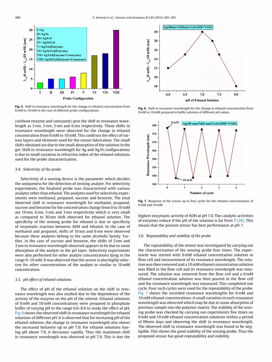
490 V. Semwal et al. / Sensors and Actuators B 230 (2016) 485–492
F0
(lrcisgiu
3
teaiosaasombt3awrtc
3
naobFsetii
Fig. 6. Shift in resonance wavelength for the change in ethanol concentration from0 mM to 10 mM prepared in buffer solution of different pH values.
ig. 5. Shift in resonance wavelength for the change in ethanol concentration from mM to 10 mM in the case of different probe configurations.
without enzyme and coenzyme) give the shift in resonance wave-ength as 3 nm, 5 nm, 5 nm and 6 nm respectively. These shifts inesonance wavelength were observed for the change in ethanoloncentration from 0 mM to 10 mM. This confirms the effect of var-ous layers and elements used for the sensor fabrication. The smallhifts obtained are due to the small absorption of the solution in theel. Shift in resonance wavelength for Ag and Ag/Si configurationss due to small variation in refractive index of the ethanol solutionssed for the probe characterization.
.4. Selectivity of the probe
Selectivity of a sensing device is the parameter which decideshe uniqueness for the detection of sensing analyte. For selectivityxperiments, the finalized probe was characterized with variousnalytes other than ethanol. The analytes used for selectivity exper-ments were methanol, propanol, sucrose and benzene. The totalbserved shift in resonance wavelength for methanol, propanol,ucrose and benzene for the concentration change from 0 to 10 mMre 10 nm, 6 nm, 5 nm and 3 nm respectively which is very smalls compared to 30 nm shift observed for ethanol solution. Thepecificity of the sensing probe for ethanol is due to specificityf enzymatic reaction between ADH and ethanol. In the case ofethanol and propanol, shifts of 10 nm and 6 nm were observed
ecause these analytes belong to the same alcoholic family. Fur-her, in the case of sucrose and benzene, the shifts of 5 nm and
nm in resonance wavelength observed appears to be due to somebsorption of the analyte in the gel layer. Selectivity experimentsere also performed for other analyte concentrations lying in the
ange 0–10 mM. It was observed that the sensor is also highly selec-ive for other concentrations of the analyte in similar to 10 mMoncentration.
.5. pH effect of ethanol solutions
The effect of pH of the ethanol solution on the shift in reso-ance wavelength was also studied due to the dependence of thectivity of the enzyme on the pH of the solvent. Ethanol solutionsf 0 mM and 10 mM concentrations were prepared in phosphateuffer of varying pH in the range 6.0–8.0 to study the effect of pH.ig. 6 shows the observed shift in resonance wavelength for ethanololutions of different pH. It is observed that for increasing pH of the
thanol solution, the change in resonance wavelength also showshe increased behavior up to pH 7.0. For ethanol solutions hav-ng pH above 7.0, it decreases rapidly. Thus the maximum shiftn resonance wavelength was observed at pH 7.0. This is due theFig. 7. Response of the sensor up to four cycles for the ethanol concentrations of0 mM and 10 mM.
highest enzymatic activity of ADH at pH 7.0. The catalytic activitiesof enzymes reduce if the pH of the solution is far from 7 [28]. Thismeans that the present sensor has best performance at pH 7.
3.6. Repeatability and stability of the probe
The repeatability of the sensor was investigated by carrying outthe characterization of the sensing probe four times. The exper-iment was started with 0 mM ethanol concentration solution inflow cell and measurement of its resonance wavelength. The solu-tion was then removed and a 10 mM ethanol concentration solutionwas filled in the flow cell and its resonance wavelength was mea-sured. The solution was removed from the flow cell and a 0 mMethanol concentration solution was then poured in the flow celland the resonance wavelength was measured. This completed onecycle. Four such cycles were used for the repeatability of the probe.Fig. 7 shows the recorded resonance wavelengths for 0 mM and10 mM ethanol concentrations. A small variation in each resonancewavelength was observed which may be due to some absorption ofprevious sample into the polymer matrix. The stability of the sens-ing probe was checked by carrying out experiments five times on0 mM and 10 mM ethanol concentration solutions within a period
of fifteen days and observing the shift in resonance wavelength.The observed shift in resonance wavelength was found to be neg-ligible. This shows the good stability of the sensing probe. Thus theproposed sensor has good repeatability and stability.
V. Semwal et al. / Sensors and Actuators B 230 (2016) 485–492 491
Table 1Limit of detections and operating range of various approaches for ethanol detection.
S. No. LOD Operating range Technique Reference
1 600 �M 1.6–160 mM Chemiluminescenceon nanosized ZrO2 [33]2 29.7 ± 1.5 �M 0–3 mM Electrode and mediator film [34]3 32 �M 1.5–79 mM Electrocatalytic oxidation [35]
3
bts
C
wfpt01abtstmv
3
Lzpis
C
waebc
wsrrowulSgbdstw
4 16.8 �M 0–60 mM
5 6 �M 0–20.4 mM
6 15.34 �M 0–10 mM
.7. Limit of detection (LOD)
LOD of the sensor is the lowest analyte concentration which cane detect by the sensor [29]. In our case, it has been calculated fromhe ratio of the resolution of spectrometer to the sensitivity of theensor near zero concentration. It can be expressed as
LOD = ��
S(1)
here �� stands for the resolution of the spectrometer and S standsor the sensitivity of the sensor near zero concentration. In theresent study the sensitivity of the sensor near zero concentra-ion is 21.70 nm/mM and the spectrometer has the resolution of.333 nm. After calculating, LOD of the present sensor is found to be5.34 �M. To check the sensor performance over the other sensingpproaches for ethanol detection, LODs of various approaches haveeen compared in Table 1. The LOD of our sensor has been foundo be the least except that reported in Ref. [30]. In this study, theensing approach of ethanol detection is based on cyclic voltamme-ry technique which is quite complex as compared to our sensing
ethod. Thus our method makes the sensor advantageous to pre-iously reported studies.
.8. Limit of quantification (LOQ)
LOQ of the sensor is slightly different from the LOD of the sensor.OQ of the sensor is the actual lowest analyte concentration (nearero concentration) that can be detected by the sensor. It shows theresence of the analyte in the detectable limit of the sensor [31]. It
s calculated as the ratio of standard deviation error with the sensorensitivity near zero concentration and can be expressed as
LOQ = ��
S(2)
here �� is the standard deviation in resonance wavelengthnd S stands for the sensor sensitivity near zero concentration ofthanol. Standard deviation in resonance wavelength is found toe 0.643 nm near 0 mM ethanol concentration solution. Thus thealculated value of LOQ of the present sensor is 29.63 �M.
As mentioned in Section 2.4, the SPR spectra were recordedithin 40 s after pouring the ethanol sample in the flow cell. SPR
pectra recorded after 40 s do not show any further change in theesonance wavelength which confirms that the equilibrium haseached within 40 s after pouring the sample in the flow cell. Inther words, the response time of the sensor is less than 40 s. Weould also like to mention that for the experiments, we have usednpolarized light and hence influence of the polarization of input
ight does not occur. As far as the effect of temperature on thePR spectrum is concerned we would like to mention that hydro-els are sensitive to the change in the thermal environment. It haseen reported that the degree of swelling of hydrogels strongly
epends on the surrounding temperature [32] which can affect theensor performance. However, we would like to mention that, inhe present study, our aim was to develop an ethanol sensor toork at room temperature and hence all the experiments wereMCP electrode [36]Cyclic voltammetry [30]Surface plasmon resonance Present study
performed at room temperature. Therefore, no effect of tempera-ture was included.
4. Conclusion
Fabrication and characterization of a SPR based fiber opticethanol sensor using gel entrapment technique have been carriedout. The sensing probe is prepared by coating the layer of silver,silicon and gel entrapped with enzyme and its coenzyme overthe unclad portion of the fiber. The operation of sensor has beenchecked for the ethanol concentration range from 0 mM to 10 mM.To achieve the best performance of the sensor, the thickness of thesilicon layer has been optimized and has been found to be 8 nm. Thesensor has maximum sensitivity of 21.70 nM/mM near zero con-centration of ethanol solution. Using control experiments we haveshown that the coenzyme increases the sensitivity of the sensorby about 33%. This is because of the improvement of the enzymaticactivity in the presence of coenzyme. It has been observed that sen-sor is sensitive ethanol for the concentration in the range 0–5 mM.The values of limit of detection and limit of quantification of thepresent sensor are 15.34 �M and 29.63 �M respectively. Selectiv-ity experiments have also been performed on the proposed probe.The probe has been found to be highly specific for ethanol solutions.In addition, the sensor has the best performance for ethanol solu-tions having pH 7.0. The sensors have numerous advantages suchas high sensitivity, compact size, fast response, high selectivity, lowcost and capability of online monitoring and remote sensing.
Acknowledgments
The present work is partially supported by the Council of Scien-tific and Industrial Research (CSIR), India. Vivek Semwal and AnandM. Shrivastav are thankful to DST (India) and UGC (India) for pro-viding research fellowships, respectively. Roli Verma thanks to theCouncil of Higher Education of the Government of the State of Israelfor PBC post-doctoral fellowship.
References
[1] A.H. Tobías, A.J. Sánchez, E. Pina, H.R. Rosas, Natural alcohol exposure: isethanol the main substrate for alcohol dehydrogenases in animals? Chem.Biol. Interact. 191 (2011) 14–25.
[2] B.K. Logan, Endogenous ethanol ‘auto-brewery syndrome’ as a drunk-drivingdefence challenge, Med. Sci. Law 40 (2000) 206–215.
[3] P. Clarkson, I.H.L. Ormrod, F.R. Sharpe, Determination of ethanol in beer bydirect injection gas chromatography: a comparison of six identical systems, J.Inst. Brew. 101 (1995) 191–193.
[4] H. Lidén, A.R. Vijayakumar, L. Gorton, G.M. Varga, Rapid alcoholdetermination in plasma and urine by column liquid chromatography withbiosensor detection, J. Pharm. Biomed. Anal. 17 (1998) 1111–1128.
[5] C. Tsai, J.D. Huang, C.C. Chiu, Amperometric ethanol biosensor based onpoly(vinyl alcohol)–multiwalled carbon nanotube–alcohol dehydrogenasebiocomposite, Biosens. Bioelectron. 22 (2007) 3051–3056.
[6] K.T. Carlson, L.A. Setton, A. Chilkoti, Swelling and mechanical behaviors ofchemically cross-linked hydrogels of elastin-like polypeptides,Biomacromolecules 4 (2003) 572–580.
[7] D. Buenger, F. Topuz, J. Groll, Hydrogels in sensing applications, Prog. Polym.Sci. 37 (2012) 1678–1719.
[8] R. Verma, S.K. Srivastava, B.D. Gupta, Surface-plasmon-resonance-basedfiber-optic sensor for the detection of low-density lipoprotein, IEEE Sens. J. 12(2012) 3460–3466.

4 d Actu
[
[
[
[
[
[
[
[
[
[
[
[
[
[
[
[
[
[[
[
[
[[
[
[
[
[
92 V. Semwal et al. / Sensors an
[9] Y.B. Amram, R.T. Vered, M. Riskin, Z.G. Wang, I. Willner, Ultrasensitive andselective detection of alkaline-earth metal ions using ion-imprinted Au NPscomposites and surface plasmon resonance spectroscopy, Chem. Sci. 3 (2012)162–167.
10] B.D. Gupta, R.K. Verma, Surface plasmon resonance-based fiber optic sensors:principle, probe designs, and some applications, J. Sens. 2009 (2009) 979761.
11] J. Homola, S.S. Yee, G. Gauglitz, Surface plasmon resonance sensors: review,Sens. Actuators B 54 (1999) 3–15.
12] N. Cennamo, A. Dona, P. Pallavicini, G. D’Agostino, G. Dacarro, L. Zeni, M.Pesavento, Sensitive detection of 2,4,6-trinitrotoluene by tridimensionalmonitoring of molecularly imprinted polymer with optical fiber andfive-branched gold nanostars, Sens. Actuators B 208 (2015) 291–298.
13] A.K. Sharma, R. Jha, B.D. Gupta, Fiber-optic sensors based on surface plasmonresonance: a comprehensive review, IEEE Sens. J. 7 (2007) 1118–1129.
14] F. Downes, C.M. Taylor, Optical fibre surface plasmon resonance sensor basedon a palladium–yttrium alloy, Procedia Eng. 120 (2015) 602–605.
15] S. Singh, B.D. Gupta, Fabrication and characterization of a surface plasmonresonance based fiber optic sensor using gel entrapment technique for thedetection of low glucose concentration, Sens. Actuators B 177 (2013) 589–595.
16] E. Fontana, Thickness optimization of metal films for the development ofsurface-plasmon-based sensors for nonabsorbing media, Appl. Opt. 45 (2006)7632–7642.
17] Y. Chen, R. Zheng, Y. Lu, P. Wang, H. Ming, Fiber-optic surface plasmonresonant sensor with low-index anti-oxidation coating, Chin. Opt. Lett. 9(2011) 100605.
18] K.F. O’Driscoll, Techniques of enzyme entrapment in gels, Meth. Enzymol. 44(1976) 169–183.
19] P. Pal, D. Nandi, T.N. Mishra, Immobilization of alcohol dehydrogenaseenzyme in a Langmuir–Blodgett film of stearic acid: its application as anethanol sensor, Thin Solid Films 239 (1994) 138–143.
20] B.S. Walters, T.J. Nilelsen, M.A. Arnold, Fiber-optic biosensor for ethanol,based on an internal enzyme concept, Talanta 35 (1988) 151–155.
21] A.K. Williams, J.T. Hupp, Sol–gel-encapsulated alcohol dehydrogenase as aversatile, environmentally stabilized sensor for alcohols and aldehydes, J. Am.Chem. Soc. 120 (1998) 4366–4371.
22] F. Salimi, M. Negahdary, G. Mazaheri, H.A. Dastjerdi, Y.G. Kakavandi, S. Javadi,S.H. Inanloo, M.M. Route, M.H. Shokoohnia, A. Sayad, A novel alcoholbiosensor based on alcohol dehydrogenase and modified electrode with ZrO2
nanoparticles, Int. J. Electrochem. Sci. 7 (2012) 7225–7234.23] A.A. Sauve, NAD+ and vitamin B3: from metabolism to therapies, J. Pharma.
Exp. Therap. 324 (2008) 883–893.24] A. Shalabney, I. Abdulhalim, Electromagnetic fields distribution in multilayer
thin film structures and the origin of sensitivity enhancement in surfaceplasmon resonance sensors, Sens. Actuators A 159 (2010) 24–32.
25] A. Lahav, A. Shalabaney, I. Abdulhalim, Surface plasmon sensor with enhancesensitivity using top nano dielectric layer, J. Nanophotonics 3 (2009) 031501.
26] A.M. Shrivastav, B.D. Gupta, SPR and molecular imprinting based fiber opticmelamine sensor with high sensitivity and low limit of detection, IEEE J. Sel.Topics Quantum Electron. 22 (2015) 6900207.
27] http://www.raeco.com/training/refractive-index-values.htm.28] C.J. Dickenson, F.M. Dickinson, A study of the pH- and
temperature-dependence of the reactions of yeast alcohol dehydrogenasewith ethanol, acetaldehyde and butyraldehyde as substrates, Biochem. J. 147
(1975) 303–311.29] S.K. Mishra, B.D. Gupta, Surface plasmon resonance based fiber optic pHsensor utilizing Ag/ITO/Al/hydrogel layers, Analyst 138 (2013) 2640–2646.
30] B. Tao, J. Zhang, S. Hui, L. Wan, An amperometric ethanol sensor based on aPd–Ni/SiNWs electrode, Sens. Actuators B 142 (2009) 298–303.
ators B 230 (2016) 485–492
31] J. Homola, Surface Plasmon Resonance Based Sensors, Springer, 2006.32] T. Tanaka, Collapse of gels and the critical endpoint, Phys. Rev. Lett. 40 (1978)
820–823.33] Z. Zhang, C. Zhang, X. Zhang, Development of a chemiluminescence ethanol
sensor based on nanosized ZrO2, Analyst 127 (2002) 792–796.34] M.M. Barsan, C.M.A. Brett, An alcohol oxidase biosensor using PNR redox
mediator at carbon film electrodes, Talanta 74 (2008) 1505–1510.35] E.T. Hayes, B.K. Bellingham, H.B. Mark Jr., A. Galal, An amperometric aqueous
ethanol sensor based on the electrocatalytic oxidation at a cobalt–nickeloxide electrode, Electrochim. Acta 41 (1996) 337–344.
36] J. Shi, P. Ci, F. Wang, H. Peng, P. Yang, L. Wang, Q. Wang, P.K. Chu,Pd/Ni/Si-microchannel-plate-based amperometric sensor for ethanoldetection, Electrochim. Acta 56 (2011) 4197–4202.
Biographies
Vivek Semwal received his M.Sc. degree in Physics in 2013 from H.N.B. GarhwalUniversity, Uttrakhand (India). Since January 2015, Mr. Semwal is a full time Ph.D.student at the Physics Department, Indian Institute of Technology Delhi. Mr. Semwalis a DST (India) research fellow and is a student member of the Optical Society ofAmerica.
Anand Mohan Shrivastav received his M.Sc. degree in Physics in 2012 from C.S.J.M.University Kanpur (India). Since July 2013, Mr. Shrivastav is a full time Ph.D. studentat the Physics Department, Indian Institute of Technology Delhi. Mr. Shrivastav isa UGC (India) research fellow and is a student member of the Optical Society ofAmerica.
Roli Verma received her M.Sc. degree in Physics in 2006 and B.Ed. in 2007 fromC.S.J.M. University Kanpur (India) and Ph.D. in 2014 from Indian Institute of Tech-nology Delhi. Since April 2014, Ms. Verma is a post doctoral fellow at Reymond andBeverly Sackler Faculty of Exact Sciences, Tel Aviv University, Israel Ms. Verma is amember of Optical Society of America.
Banshi D. Gupta received the M.Sc. degree from Aligarh Muslim University, Ali-garh, India, in 1975, and the Ph.D. degree from the Indian Institute of TechnologyDelhi, New Delhi, India, in 1979, both in physics. In 1978, he joined the Indian Insti-tute of Technology Delhi where he is currently a Professor of physics. In addition,he was with the University of Guelph, Guelph, ON, Canada, during 1982–1983, theUniversity of Toronto, Toronto, ON, in 1985, Florida State University, Tallahassee,FL, USA, in 1988, the University of Strathclyde, Glasgow, U.K., in 1993, and the Uni-versity of Birmingham, Birmingham, U.K., in 2010. He has published more than 130research papers in international journals and 80 papers in conferences. He is theauthor of books Fiber Optic Sensors: Principles and Applications (New Delhi, India:NIPA, 2006) and Fiber Optic Sensors Based on Plasmonics (Singapore: World Scien-tific, 2015), and is the Coeditor of the Proceedings of SPIE (USA), (vol. 3666, 1998and vol. 8173, 2010) and Advances in Contemporary Physics and Energy (Supple-ment Volume) (New Delhi, India: Allied). His current areas of research interestsinclude plasmonic biosensors, fiber-optic sensors, and nanotechnology. He receivedthe 1991 Gowri Memorial Award of the Institution of Electronics and Telecommu-nication Engineers, India, and the ICTP Associateship by the International Center forTheoretical Physics (ICTP), Trieste, Italy, in 1992, which he held for eight consecutive
years. In this capacity, he visited ICTP in 1994 and 1996. He has delivered invitedtalks on plasmonics sensors in various international conferences held in USA, China,Australia, Korea, and India. He is in the Editorial Board of the Journal of Sensors.He is a Regular Member of the Optical Society of America and a Life Member of theOptical Society of India.


