Embed Size (px)
Citation preview
Surface Plasmon Resonance Biosensors for Detection of Foodborne Pathogens and Toxins
Jiří Homola, Kateřina Hegnerová, Milan Vala
Institute of Photonics and Electronics AS CR, Chaberská 57, 18251 Prague, Czech Republic
ABSTRACT
In the last decade surface plasmon resonance (SPR) biosensors have made great strides both in terms of technology and its applications. SPR biosensors have become a central tool for study of molecular interactions and have been widely used for detection of chemical and biological analytes. Food analysis belongs to major areas of potential applications of SPR biosensors. Therefore, numerous SPR biosensors for detection of analytes implicated in food safety (e.g. pathogens, toxins, drug residues, vitamins, hormones, chemical contaminants, and allergens) have been developed. This paper reviews recent developments in the field of SPR biosensors for food safety, in particular, for detection of foodborne pathogens and toxins.
Keywords: surface plasmon resonance, foodborne pathogens, toxins, food safety
1. INTRODUCTION Optical sensors for detection of chemical and biological species represent a rapidly evolving field with potential applications in numerous important areas such as genomics, proteomics, medical diagnostics, environmental monitoring, food analysis, agriculture, and security. Label-free optical biosensors present unique technology that enables direct observation of molecular interaction in real-time [1]. Label-free optical biosensors measure refractive index changes induced by the molecular interaction and are typically based on interferometric transducers [2-4] and transducers based on spectroscopy of guided modes of dielectric waveguides [5, 6] or metal-dielectric waveguides - surface plasmon resonance sensors [7-15].
In the last decade SPR biosensors have been increasingly used for rapid label-free detection of chemical and biological species [9, 15]. Analytes implicated in food safety and security (e.g. pathogens, toxins, drug residues, vitamins, hormones, antibodies, chemical contaminants and allergens) have received most attention [15]. SPR biosensors have been also applied for detection of analytes of interest in medical diagnostics (antibodies, hormones, cancer markers, allergy markers and drugs) and environmental protection (pesticides, aromatic hydrocarbons, phenols, and polychlorinated biphenyls).
In this paper, we review the state of the art in the applications of SPR for food safety and present examples of SPR biosensors for detection of foodborne pathogens and toxins.
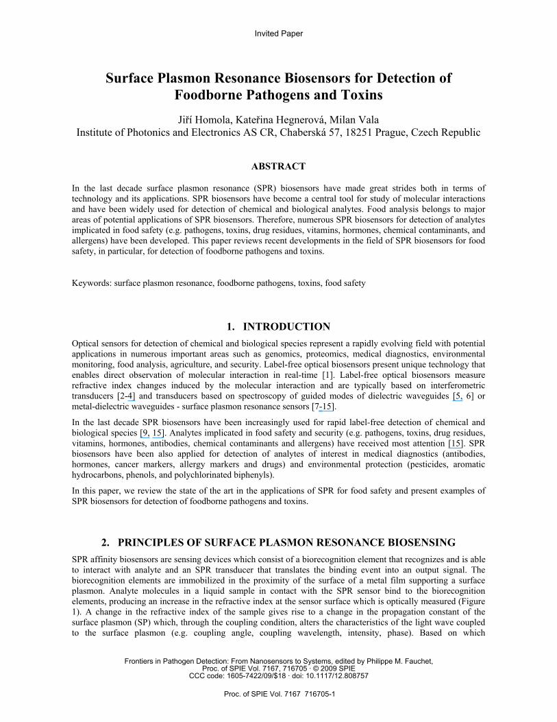
2. PRINCIPLES OF SURFACE PLASMON RESONANCE BIOSENSING SPR affinity biosensors are sensing devices which consist of a biorecognition element that recognizes and is able to interact with analyte and an SPR transducer that translates the binding event into an output signal. The biorecognition elements are immobilized in the proximity of the surface of a metal film supporting a surface plasmon. Analyte molecules in a liquid sample in contact with the SPR sensor bind to the biorecognition elements, producing an increase in the refractive index at the sensor surface which is optically measured (Figure 1). A change in the refractive index of the sample gives rise to a change in the propagation constant of the surface plasmon (SP) which, through the coupling condition, alters the characteristics of the light wave coupled to the surface plasmon (e.g. coupling angle, coupling wavelength, intensity, phase). Based on which
Invited Paper
Frontiers in Pathogen Detection: From Nanosensors to Systems, edited by Philippe M. Fauchet,Proc. of SPIE Vol. 7167, 716705 · © 2009 SPIE
CCC code: 1605-7422/09/$18 · doi: 10.1117/12.808757
Proc. of SPIE Vol. 7167 716705-1
characteristic of the light wave modulated by a surface plasmon is measured, SPR sensors are classified as sensors with angular, wavelength, intensity, or phase modulation.
SPR Metal
Light Beam
BiomolecularRecognition
Element
Analyte
Surface Plasmon
Figure 1. Concept of SPR biosensing.
2.1 Detection formats
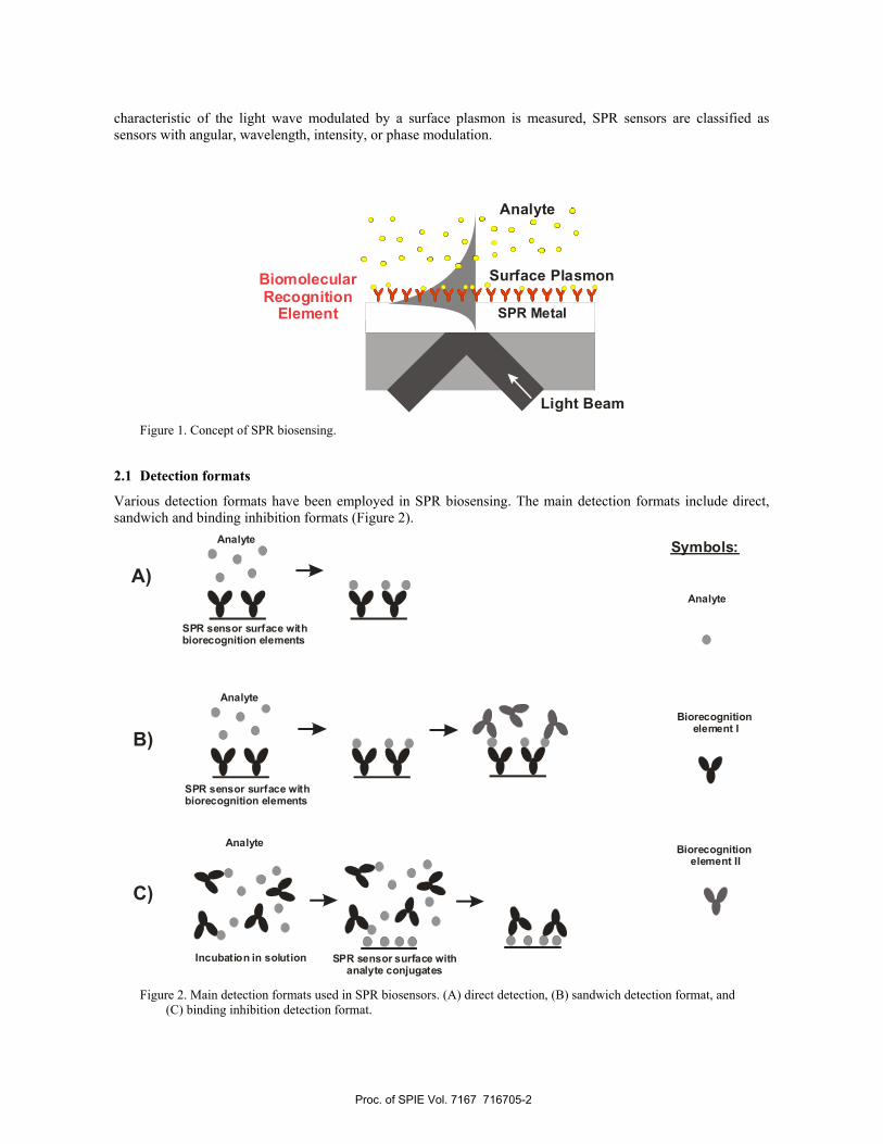
Various detection formats have been employed in SPR biosensing. The main detection formats include direct, sandwich and binding inhibition formats (Figure 2).
SPR sensor surface with biorecognition elements
Analyte
Analyte
SPR sensor surface withanalyte conjugates
Analyte
SPR sensor surface with biorecognition elements
Incubation in solution
A)
C)
B)
Symbols:
Analyte
Biorecognition element I
Biorecognition element II
Figure 2. Main detection formats used in SPR biosensors. (A) direct detection, (B) sandwich detection format, and
(C) binding inhibition detection format.
Proc. of SPIE Vol. 7167 716705-2
In the direct detection format, a sample containing target molecules is brought into contact with the sensor surface coated with respective biomolecular recognition elements (e.g. antibodies). Binding of analyte molecules to antibodies produces an increase in the refractive index at the sensor surface. The SPR sensor instrument translates this change into a change in sensor response (Figure 2a). A sandwich assay consists of two steps. In the first step, the analyte molecules bind to antibodies immobilized on the sensor surface as in the direct detection format. In the second step, the sensor is incubated with a solution containing secondary antibodies, which bind to the previously captured analyte, enhancing the specific sensor response (Figure 2b). Another two-step detection format, binding inhibition detection format, is based on the competitive reaction of analyte in solution and analyte immobilized on the surface with free antibodies. In the first step of detection format, a sample is mixed with antibodies and analyte in the sample bind to antibodies, thus blocking their binding sites. Then, the sample with antibodies is brought to sensor surface so that the unbound antibodies can bind to analyte molecules immobilized on the sensor surface (Figure 2c). In this detection format, the SPR biosensor measures the concentration of unbound antibodies, which can be used to calculate the concentration of target analyte. Direct detection is usually preferred in applications when binding of analyte at appropriate concentrations produces a sufficient direct response. If necessary, the lowest detection limits of the direct SPR sensors can be improved by using a sandwich assay. Detection of small molecules is usually performed using the binding inhibition detection format.
3. SPR SENSORS FOR FOOD SAFETY The main groups of food-safety-related analytes targeted by SPR biosensor technology include toxins [16-28], pathogens [29-44], antibiotics [45-49], vitamins [50-53], hormons [54, 55], and allergens [56, 57]. In the following chapters we shall focus on detection of foodborne pathogens and toxins which present the most important target group.
3.1 Detection of Toxins
Toxins implicated in food safety include mainly toxins produced by bacteria, fungi and algae.
Staphylococcal enterotoxin B (SEB) is an exotoxin excreted by the Staphylococcus aureus bacterium. Detection of SEB was demonstrated by Nedelkov, et al. in 2000 [16]. They used the Biacore X SPR sensor and SEB antibody immobilized in carboxymethyldextran layer via amine coupling chemistry and demonstrated detection of SEB in milk and mushroom samples at levels down to 1 ng/ml. SPR-based detection of staphylococcal enterotoxin B (SEB) in milk was demonstrated by Homola et al. The SPR biosensor was based on prism coupling and wavelength modulation and covalent attachment of antibodies on a mixed self-assembled monolayer via activated carboxyl groups. In sandwich detection mode, the lowest detection limit was determined to be 0.5 ng/ml [17]. In 2002, Furlong’s group detected SEB using an SPR sensor developed by Texas Instruments and antibodies immobilized on the sensor surface via a gold binding peptide [18]. SEB was detected in buffer and seawater using direct detection or sandwich format with one or two amplification antibodies. The limit of detection for direct detection was 5.6 ng/ml in buffer and 28 ng/ml in seawater. Using one-step amplification, concentrations of 0.6 ng/ml and 1.4 ng/ml were detected in buffer and seawater, respectively. Medina used inhibition assay to detect SEB in milk and achieved the limit of detection as low as 0.3 ng/ml [19, 20]. Later, Medina expanded the work to another food matrix - egg [21].
Marine toxin, Domoic acid (DA), is a small molecule which causes amnesic shellfish poisoning. DA was detected by Lotierzo et al. who used a commercial SPR sensor BIAcore 3000 and a molecularly imprinted polymer as a biorecognition element [22]. Detection was performed in a competitive binding format in which free DA competed with its conjugate with horseradish peroxidase. The sensor was demonstrated to be able to detect DA in buffer down to 5 ng/ml [22]. DA was also detected using an SPR sensor based on prism coupling and wavelength modulation and inhibition assay [23]. DA was immobilized on a mixed SAM of oligo (ethylene glycol) (OEG)-containing alkanethiolates using amine coupling chemistry. The lowest limit of detection of DA was established at 0.1 ng/ml. A portable SPR biosensor for the detection of DA was reported by Furlong’s group
Proc. of SPIE Vol. 7167 716705-3
[24]. The system used affinity-purified antibodies and competition-based detection format. The detection of DA was performed in phosphate buffered saline and in diluted clam extracts. The limit of detection was established at the level of 3 ppb (10 nM) [24].
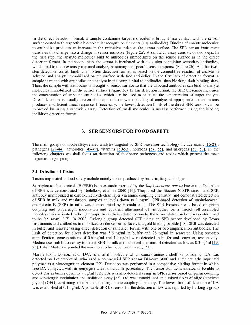
Another example of small toxin targeted by SPR biosensors technology is Tetrodotoxin (TTX) [25]. TTX is found in a range of organisms including certain species of puffer fish and amphibians and is most commonly associated with human puffer fish poisoning. In the following section, we report our SPR biosensor for TTX. For detection of TTX, we used a four-channel SPR sensor (PLASMON IV) based on spectroscopy of surface plasmons developed in our laboratory, antibodies as a biorecognition element and binding inhibition assay. Prior to detection of TTX, the cleaned gold surface was functionalized with a mixed self-assembled monolayer (SAM) consisting of hydroxyl terminated oligo-ethylene glycol (OEG) alkanethiol (OH-OEG-AT) and amine terminated OEG alkanethiol (NH2-OEG-AT). The substrate was incubated in this ethanolic SAM solution (with final concentration 200 µM) with triethylamine overnight at room temperature. After the formation of the mixed SAM, the TTX was covalently linked to the amine groups on the NH2-OEG-AT via formaldehyde reaction. In order to characterize the immobilized TTX surface, detection of pure anti-TTX with concentrations from 0.125 µg/ml to 32 µg/ml in PBS buffer was measured. Based on the results, the optimum concentration of antibody for detection of TTX was determined to be 1µg/ml. In the first step of the detection assay, the pre-incubated samples containing antibody (anti-TTX; 1 µg/ml) and TTX at various known concentrations (0.01; 0.1; 1; 10; 100; 1000; 10,000 ng/ml) were flowed across the TTX-coated surface for 15 minutes to obtained TTX calibration curve (Figure 3). Subsequently, the surface was washed with PBS, 50 mM NaOH and PBS. In each experiment, three sensing channels were used to analyze samples, while the fourth was used as a reference channel (only anti-TTX at a concentration of 1 µg/ml was injected in this channel). Subsequently, TTX was detected in blind samples (PBS and 10% PFM = puffer fish matrix) using the same procedure as described above. The measurements were repeated using different sensing channels and different SPR chips. The error in determination of the concentration of TTX was expressed as a standard deviation of the calculated concentration. The standard deviation of the concentrations determined by the sensor was determined to be within 20 per cent.
0,01 0,1 1 10 100 1000 10000
0,0
0,2
0,4
0,6
0,8
1,0
1,2
LOD
Nor
mal
ized
sen
sor r
espo
nse
TTX concentration [ng/ml]
PBS 10% PFM
(puffer fish matrix)
Figure 3: Calibration curve for TTX in PBS and 10% PFM.
3.2 Detection of Pathogens
The first SPR sensor detection of bacterial cells was published by Fratamico et al. in 1998 who demonstrated SPR biosensor for E. coli O157:H7 [29]. They used a sandwich assay using immobilized monoclonal antibody (MAb) and a secondary polyclonal antibody (PAb). Antibodies were immobilized by three methods. Capture of the MAb Fc region by amine-coupled protein A or protein G did not enhance detection compared to amine-
Proc. of SPIE Vol. 7167 716705-4
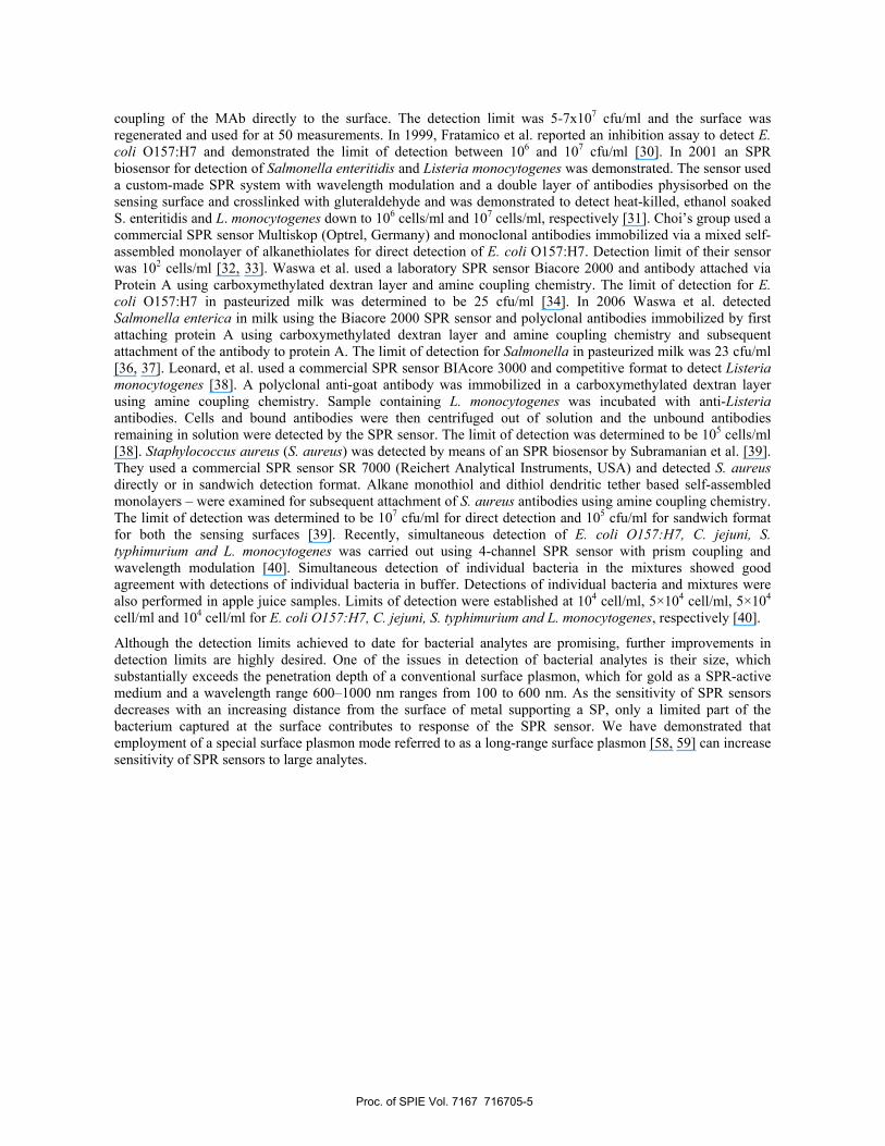
coupling of the MAb directly to the surface. The detection limit was 5-7x10⁷ cfu/ml and the surface was regenerated and used for at 50 measurements. In 1999, Fratamico et al. reported an inhibition assay to detect E. coli O157:H7 and demonstrated the limit of detection between 106 and 107 cfu/ml [30]. In 2001 an SPR biosensor for detection of Salmonella enteritidis and Listeria monocytogenes was demonstrated. The sensor used a custom-made SPR system with wavelength modulation and a double layer of antibodies physisorbed on the sensing surface and crosslinked with gluteraldehyde and was demonstrated to detect heat-killed, ethanol soaked S. enteritidis and L. monocytogenes down to 106 cells/ml and 107 cells/ml, respectively [31]. Choi’s group used a commercial SPR sensor Multiskop (Optrel, Germany) and monoclonal antibodies immobilized via a mixed self-assembled monolayer of alkanethiolates for direct detection of E. coli O157:H7. Detection limit of their sensor was 102 cells/ml [32, 33]. Waswa et al. used a laboratory SPR sensor Biacore 2000 and antibody attached via Protein A using carboxymethylated dextran layer and amine coupling chemistry. The limit of detection for E. coli O157:H7 in pasteurized milk was determined to be 25 cfu/ml [34]. In 2006 Waswa et al. detected Salmonella enterica in milk using the Biacore 2000 SPR sensor and polyclonal antibodies immobilized by first attaching protein A using carboxymethylated dextran layer and amine coupling chemistry and subsequent attachment of the antibody to protein A. The limit of detection for Salmonella in pasteurized milk was 23 cfu/ml [36, 37]. Leonard, et al. used a commercial SPR sensor BIAcore 3000 and competitive format to detect Listeria monocytogenes [38]. A polyclonal anti-goat antibody was immobilized in a carboxymethylated dextran layer using amine coupling chemistry. Sample containing L. monocytogenes was incubated with anti-Listeria antibodies. Cells and bound antibodies were then centrifuged out of solution and the unbound antibodies remaining in solution were detected by the SPR sensor. The limit of detection was determined to be 105 cells/ml [38]. Staphylococcus aureus (S. aureus) was detected by means of an SPR biosensor by Subramanian et al. [39]. They used a commercial SPR sensor SR 7000 (Reichert Analytical Instruments, USA) and detected S. aureus directly or in sandwich detection format. Alkane monothiol and dithiol dendritic tether based self-assembled monolayers – were examined for subsequent attachment of S. aureus antibodies using amine coupling chemistry. The limit of detection was determined to be 107 cfu/ml for direct detection and 105 cfu/ml for sandwich format for both the sensing surfaces [39]. Recently, simultaneous detection of E. coli O157:H7, C. jejuni, S. typhimurium and L. monocytogenes was carried out using 4-channel SPR sensor with prism coupling and wavelength modulation [40]. Simultaneous detection of individual bacteria in the mixtures showed good agreement with detections of individual bacteria in buffer. Detections of individual bacteria and mixtures were also performed in apple juice samples. Limits of detection were established at 104 cell/ml, 5×10⁴ cell/ml, 5×10⁴ cell/ml and 104 cell/ml for E. coli O157:H7, C. jejuni, S. typhimurium and L. monocytogenes, respectively [40].
Although the detection limits achieved to date for bacterial analytes are promising, further improvements in detection limits are highly desired. One of the issues in detection of bacterial analytes is their size, which substantially exceeds the penetration depth of a conventional surface plasmon, which for gold as a SPR-active medium and a wavelength range 600–1000 nm ranges from 100 to 600 nm. As the sensitivity of SPR sensors decreases with an increasing distance from the surface of metal supporting a SP, only a limited part of the bacterium captured at the surface contributes to response of the SPR sensor. We have demonstrated that employment of a special surface plasmon mode referred to as a long-range surface plasmon [58, 59] can increase sensitivity of SPR sensors to large analytes.
Proc. of SPIE Vol. 7167 716705-5
10 100 10000
2
4
6
8
10 cSP (50nm Au) LRSP (25nm Au, 1200nm Teflon AF)
SPR
shi
ft [n
m]
Biolayer thickness [nm]
Glass Substraten=1.51
Teflon AF~1.31n
G o l d F i l m ~ 25nm
AqueousSample,n=1.33
OpticalWave
Evanescent Wave
Long-RangeSP
Gold Film 50nm
AqueousSample,n=1.33
OpticalWave
ConventionalSP
Glass Substraten=1.51
Evanescent Wave
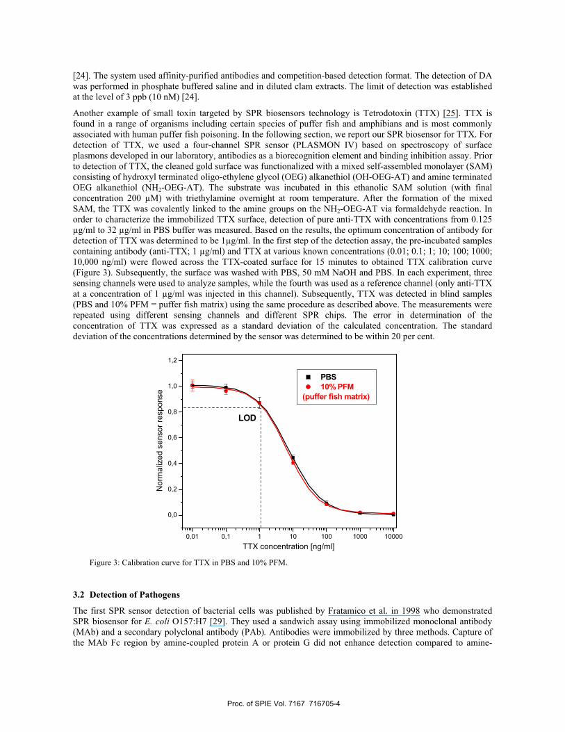
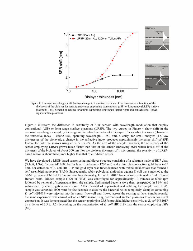
Figure 4: Resonant wavelength shift due to a change in the refractive index of the biolayer as a function of the thickness of the biolayer for sensing structures employing conventional (cSP) or long-range (LRSP) surface plasmons (left). Scheme of sensing structures supporting long-range (upper right) and conventional (lower right) surface plasmons.
Figure 4 illustrates the difference in sensitivity of SPR sensors with wavelength modulation that employ conventional (cSP) or long-range surface plasmons (LRSP). The two curves in Figure 4 show shift in the resonant wavelength caused by a change in the refractive index of a biolayer of a variable thickness (change in the refractive index – 0.0005RIU, operating wavelength – 750 nm). Clearly, for small analytes (i.e. low thicknesses of the biolayer), a change in the refractive index produces approximately the same shift of SPR feature for both the sensors using cSPs or LRSPs. As the size of the analyte increases, the sensitivity of the sensor employing LRSPs grows much faster than that of the sensor employing cSPs which levels off at the thickness of the biolayer of about 500 nm. For the biolayer thickness of 1 micrometer, the sensitivity of LRSP-based sensor is about three times higher than that of cSP-based sensor.
We have developed a LRSP-based sensor using multilayer structure consisting of a substrate made of BK7 glass (Schott, USA), Teflon AF 1600 buffer layer (thickness - 1200 nm) and a thin plasmon-active gold layer (~25 nm). For detection of E. coli HB101P, the gold layer was functionalized with mixed alkanethiols that formed a self-assembled monolayer (SAM). Subsequently, rabbit polyclonal antibodies against E. coli were attached to the SAM by means of NHS/EDC amine coupling chemistry. E. coli HB101P bacteria were obtained in 1ml of Luria Bertani broth. Diluted sample (1:20 in PBM) was centrifuged for approximately 10 minutes at 6000 rpm followed by removal of supernatant from the sample. Sedimented bacteria were then resuspended in PBM and sedimented by centrifugation once more. After removal of supernatant and refilling the sample with PBM, sample was vortexed (1000 rpm) for few seconds to dissolve the bacterial pellet completely. Samples containing E. coli HB101P were injected into the sensor flow-cell and flowed across the sensing surface. Simultaneously, the same experiment was carried out on an SPR sensor using conventional surface plasmons to allow for direct comparison. It was demonstrated that the sensor employing LRSPs provided higher sensitivity to E. coli HB101P by a factor of 3.5 to 5.5 (depending on the concentration of E. coli HB101P) than the sensor employing cSPs [60].
Proc. of SPIE Vol. 7167 716705-6
4. CONCLUSIONS Rapid and sensitive detection of analytes implicated in food safety and security presents an important goal for SPR biosensor research. Therefore, numerous SPR sensor platforms have been developed for detection of foodborne pathogens and toxins. Small and medium size analytes have been detected at sub-ng/ml levels. Large analytes, such as bacteria, have been proven to be challenging to detect at levels lower than 104 cell/ml. We demonstrate that SPR sensors employing long-range surface plasmons have potential to enhance sensitivity of SPR sensors to large analytes and thus enhance their utility for detection of bacterial analytes.
5. ACKNOWLEDGEMENTS This research was supported by the US Food and Drug Administration and the Grant Agency of the Academy of Sciences of the Czech Republic (contract # KAN200670701).
REFERENCES [1] F. S. Ligler, and C. A. R. Taitt, Optical biosensors: present and future, Elsevier, Amsterdam
Oxford(2002). [2] R. G. Heideman, and P. V. Lambeck, “Remote opto-chemical sensing with extreme sensitivity: design,
fabrication and performance of a pigtailed integrated optical phase-modulated Mach-Zehnder interferometer system,” Sensors and Actuators B, 61(1-3), 100-127 (1999).
[3] K. Schmitt, B. Schirmer, C. Hoffmann et al., “Interferometric biosensor based on planar optical waveguide sensor chips for label-free detection of surface bound bioreactions,” Biosensors and Bioelectronics, 22(11), 2591-2597 (2007).
[4] H.-M. Schmitt, A. Brecht, J. Piehler et al., “An integrated system for optical biomolecular interaction analysis,” Biosensors and Bioelectronics, 12(8), 809-816 (1997).
[5] R. Cush, J. M. Cronin, W. J. Stewart et al., “The resonant mirror: a novel optical biosensor for direct sensing of biomolecular interactions Part I: Principle of operation and associated instrumentation,” Biosensors and Bioelectronics, 8(7-8), 347-354 (1993).
[6] D. Clerc, and W. Lukosz, “Direct immunosensing with an integrated-optical output grating coupler,” Sensors and Actuators B, 40(1), 53-58 (1997).
[7] B. D. Malhotra, and A. P. F. Turner, Perspectives in biosensors, JAI, Amsterdam(2003). [8] L. Gorton, Biosensors and modern biospecific analytical techniques, Elsevier, Amsterdam; Boston(2005). [9] J. Homola, Surface plasmon resonance based sensors, Springer, Berlin(2006). [10] H. Nakamura, and I. Karube, “Current research activity in biosensors,” Analytical and Bioanalytical
Chemistry, 377(3), 446-468 (2003). [11] M. Bally, M. Halter, J. Voros et al., “Optical microarray biosensing techniques,” Surface and Interface
Analysis, 38(11), 1442-1458 (2006). [12] D. G. M. Rebecca L. Rich, “Survey of the year 2005 commercial optical biosensor literature,” Journal of
Molecular Recognition, 19(6), 478-534 (2006). [13] C. Boozer, G. Kim, S. Cong et al., “Looking towards label-free biomolecular interaction analysis in a
high-throughput format: a review of new surface plasmon resonance technologies,” Current Opinion in Biotechnology, 17(4), 400-405 (2006).
[14] K. S. Phillips, and Q. Cheng, “Recent advances in surface plasmon resonance based techniques for bioanalysis,” Analytical and Bioanalytical Chemistry, 387(5), 1831-1840 (2007).
[15] J. Homola, “Surface plasmon resonance sensors for detection of chemical and biological species,” Chemical Reviews, 108(2), 462-493 (2008).
[16] D. Nedelkov, A. Rasooly, and R. W. Nelson, “Multitoxin biosensor-mass spectrometry analysis: a new approach for rapid, real-time, sensitive analysis of staphylococcal toxins in food,” International Journal of Food Microbiology, 60(1), 1-13 (2000).
Proc. of SPIE Vol. 7167 716705-7
[17] J. Homola, J. Dostalek, S. F. Chen et al., “Spectral surface plasmon resonance biosensor for detection of staphylococcal enterotoxin B in milk,” International Journal of Food Microbiology, 75(1-2), 61-69 (2002).
[18] A. N. Naimushin, S. D. Soelberg, D. K. Nguyen et al., “Detection of Staphylococcus aureus enterotoxin B at femtomolar levels with a miniature integrated two-channel surface plasmon resonance (SPR) sensor,” Biosensors & Bioelectronics, 17(6-7), 573-584 (2002).
[19] M. B. Medina, “A biosensor method for a competitive immunoassay detection of staphylococcal enterotoxin B (SEB) in milk,” Journal of Rapid Methods and Automation In Microbiology, 13(1), 37-55 (2005).
[20] M. B. Medina, “Detection of staphylococcal enterotoxin B (SEB) with surface plasmon resonance biosensor,” Journal of Rapid Methods and Automation In Microbiology, 11(3), 225-243 (2003).
[21] M. B. Medina, “A biosensor method for detection of staphylococcal enterotoxin A in raw whole egg,” Journal of Rapid Methods and Automation in Microbiology, 14(2), 119-132 (2006).
[22] M. Lotierzo, O. Y. F. Henry, S. Piletsky et al., “Surface plasmon resonance sensor for domoic acid based on grafted imprinted polymer,” Biosensors & Bioelectronics, 20(2), 145-152 (2004).
[23] Q. M. Yu, S. F. Chen, A. D. Taylor et al., “Detection of low-molecular-weight domoic acid using surface plasmon resonance sensor,” Sensors and Actuators B, 107(1), 193-201 (2005).
[24] R. C. Stevens, S. D. Soelberg, B. T. L. Eberhart et al., “Detection of the toxin domoic acid from clam extracts using a portable surface plasmon resonance biosensor,” Harmful Algae, 6(2), 166-174 (2007).
[25] A. D. Taylor, J. Ladd, S. Etheridge et al., “Quantitative detection of tetrodotoxin (TTX) by a surface plasmon resonance (SPR) sensor,” Sensors and Actuators B, 130(1), 120-128 (2008).
[26] S. J. Daly, G. J. Keating, P. P. Dillon et al., “Development of surface plasmon resonance-based immunoassay for aflatoxin B-1,” Journal of Agricultural and Food Chemistry, 48(11), 5097-5104 (2000).
[27] L. Dunne, S. Daly, A. Baxter et al., “Surface plasmon resonance-based inummoassay for the detection of affatoxin B-1 using single-chain antibody fragments,” Spectroscopy Letters, 38(3), 229-245 (2005).
[28] A. J. Tudos, E. R. Lucas-van den Bos, and E. C. A. Stigter, “Rapid surface plasmon resonance-based inhibition assay of deoxynivalenol,” Journal of Agricultural and Food Chemistry, 51(20), 5843-5848 (2003).
[29] P. M. Fratamico, T. P. Strobaugh, M. B. Medina et al., “Detection of Escherichia coli O157 : H7 using a surface plasmon resonance biosensor,” Biotechnology Techniques, 12(7), 571-576 (1998).
[30] P. M. Fratamico, T. P. Strobaugh, M. B. Medina et al., A Surface Plasmon Resonance Biosensor for Real-Time Immunologic Detection of Escherichia Coli O157:H7, Kluwer Academic, New York(1999).
[31] V. Koubova, E. Brynda, L. Karasova et al., “Detection of foodborne pathogens using surface plasmon resonance biosensors,” Sensors and Actuators B, 74(1-3), 100-105 (2001).
[32] B. K. Oh, Y. K. Kim, Y. M. Bae et al., “Detection of Escherichia coli O157 : H7 using immunosensor based on surface plasmon resonance,” Journal of Microbiology and Biotechnology, 12(5), 780-786 (2002).
[33] B. K. Oh, W. Lee, W. H. Lee et al., “Nano-scale probe fabrication using self-assembly technique and application to detection of Escherichia coli O157 : H7,” Biotechnology and Bioprocess Engineering, 8(4), 227-232 (2003).
[34] J. W. Waswa, C. Debroy, and J. Irudayaraj, “Rapid detection of Salmonella enteritidis and Escherichia coli using surface plasmon resonance biosensor,” Journal of Food Process Engineering, 29(4), 373-385 (2006).
[35] A. Subramanian, J. Irudayaraj, and T. Ryan, “A mixed self-assembled monolayer-based surface plasmon immunosensor for detection of E-coli O157 : H7,” Biosensors & Bioelectronics, 21(7), 998-1006 (2006).
[36] B. K. Oh, Y. K. Kim, K. W. Park et al., “Surface plasmon resonance immunosensor for the detection of Salmonella typhimurium,” Biosensors & Bioelectronics, 19(11), 1497-1504 (2004).
[37] B. K. Oh, W. Lee, Y. K. Kim et al., “Surface plasmon resonance immunosensor using self-assembled protein G for the detection of Salmonella paratyphi,” Journal of Biotechnology, 111(1), 1-8 (2004).
[38] P. Leonard, S. Hearty, J. Quinn et al., “A generic approach for the detection of whole Listeria monocytogenes cells in contaminated samples using surface plasmon resonance,” Biosensors & Bioelectronics, 19(10), 1331-1335 (2004).
[39] A. Subramanian, J. Irudayaraj, and T. Ryan, “Mono and dithiol surfaces on surface plasmon resonance biosensors for detection of Staphylococcus aureus,” Sensors and Actuators B, 114(1), 192-198 (2006).
Proc. of SPIE Vol. 7167 716705-8
[40] A. D. Taylor, J. Ladd, Q. M. Yu et al., “Quantitative and simultaneous detection of four foodborne bacterial pathogens with a multi-channel SPR sensor,” Biosensors & Bioelectronics, 22(5), 752-758 (2006).
[41] B. K. Oh, W. Lee, B. S. Chun et al., “Surface plasmon resonance immunosensor for the detection of Yersinia enterocolitica,” Colloids and Surfaces A-Physicochemical and Engineering Aspects, 257-58, 369-374 (2005).
[42] J. Y. Jyoung, S. H. Hong, W. Lee et al., “Immunosensor for the detection of Vibrio cholerae O1 using surface plasmon resonance,” Biosensors & Bioelectronics, 21(12), 2315-2319 (2006).
[43] C. D. Kang, S. W. Lee, T. H. Park et al., “Performance enhancement of real-time detection of protozoan parasite, Cryptosporidium oocyst by a modified surface plasmon resonance (SPR) biosensor,” Enzyme and Microbial Technology, 39(3), 387-390 (2006).
[44] F. Zezza, M. Pascale, G. Mule et al., “Detection of Fusarium culmorum in wheat by a surface plasmon resonance-based DNA sensor,” Journal of Microbiological Methods, 66(3), 529-537 (2006).
[45] G. Cacciatore, M. Petz, S. Rachid et al., “Development of an optical biosensor assay for detection of [beta]-lactam antibiotics in milk using the penicillin-binding protein 2x*,” Analytica Chimica Acta, 520(1-2), 105-115 (2004).
[46] H. M. Ashwin, S. L. Stead, J. C. Taylor et al., “Development and validation of screening and confirmatory methods for the detection of chloramphenicol and chloramphenicol glucuronide using SPR biosensor and liquid chromatography-tandem mass spectrometry,” Analytica Chimica Acta, 529(1-2), 103-108 (2005).
[47] J. Ferguson, A. Baxter, P. Young et al., “Detection of chloramphenicol and chloramphenicol glucuronide residues in poultry muscle, honey, prawn and milk using a surface plasmon resonance biosensor and Qflex((R)) kit chloramphenicol,” Analytica Chimica Acta, 529(1-2), 109-113 (2005).
[48] V. Dumont, A. C. Huet, I. Traynor et al., “A surface plasmon resonance biosensor assay for the simultaneous determination of thiamphenicol, florefenicol, florefenicol amine and chloramphenicol residues in shrimps,” Analytica Chimica Acta, 567(2), 179-183 (2006).
[49] N. Moeller, E. Mueller-Seitz, O. Scholz et al., “A new strategy for the analysis of tetracycline residues in foodstuffs by a surface plasmon resonance biosensor,” European Food Research and Technology, 224(3), 285-292 (2007).
[50] I. Caelen, A. Kalman, and L. Wahlstrom, “Biosensor-based determination of riboflavin in milk samples,” Analytical Chemistry, 76(1), 137-143 (2004).
[51] S. A. Haughey, A. A. O'Kane, G. A. Baxter et al., “Determination of pantothenic acid in foods by optical biosensor immunoassay,” Journal of AOAC International, 88(4), 1008-1014 (2005).
[52] H. E. Indyk, E. A. Evans, M. C. B. Caselunghe et al., “Determination of biotin and folate in infant formula and milk by optical biosensor-based immunoassay,” Journal of AOAC International, 83(5), 1141-1148 (2000).
[53] H. E. Indyk, B. S. Persson, M. C. B. Caselunghe et al., “Determination of vitamin B-12 in milk products and selected foods by optical biosensor protein-binding assay: Method comparison,” Journal of AOAC International, 85(1), 72-81 (2002).
[54] E. H. Gillis, J. P. Gosling, J. M. Sreenan et al., “Development and validation of a biosensor-based immunoassay for progesterone in bovine milk,” Journal of Immunological Methods, 267(2), 131-138 (2002).
[55] E. H. Gillis, I. Traynor, J. P. Gosling et al., “Improvements to a surface plasmon resonance-based immunoassay for the steroid hormone progesterone,” Journal of AOAC International, 89(3), 838-842 (2006).
[56] I. Mohammed, W. M. Mullett, E. P. C. Lai et al., “Is biosensor a viable method for food allergen detection?,” Analytica Chimica Acta, 444(1), 97-102 (2001).
[57] I. M. Yman, A. Eriksson, M. A. Johansson et al., “Food allergen detection with biosensor immunoassays,” Journal of AOAC International, 89(3), 856-861 (2006).
[58] D. Sarid, “Long-range surface-plasma waves on very thin metal-films,” Physical Review Letters, 47(26), 1927-1930 (1981).
[59] G. G. Nenninger, P. Tobiska, J. Homola et al., “Long-range surface plasmons for high-resolution surface plasmon resonance sensors,” Sensors and Actuators B, 74(1-3), 145-151 (2001).
Proc. of SPIE Vol. 7167 716705-9

[60] M. Vala, S. Etheridge, J. A. Roach et al., “Long-range surface plasmons for sensitive detection of bacterial analytes,” Sensors and Actuators B, in press.
Proc. of SPIE Vol. 7167 716705-10


