Embed Size (px)
Citation preview

Surgical Management of Neurovascular Bundle inUterine Fibroid Pseudocapsule
Andrea Tinelli, MD, Antonio Malvasi, MD, Brad S. Hurst, MD, Daniel A. Tsin, MD,Fausto Davila, MD, Guillermo Dominguez, MD, Domenico Dell’edera, MD, Carlo Cavallotti, MD,
Roberto Negro, MD, Sarah Gustapane, MD, Chris M. Teigland, MD, Liselotte Mettler, MD
ABSTRACT
The uterine fibroid pseudocapsule is a fibro-neurovascu-lar structure surrounding a leiomyoma, separating it fromnormal peripheral myometrium. The fibroid pseudocap-sule is composed of a neurovascular network rich inneurofibers similar to the neurovascular bundle surround-ing a prostate. The nerve-sparing radical prostatectomyhas several intriguing parallels to myomectomy. It mayserve either as a useful model in modern fibroid surgicalremoval, or it may accelerate our understanding of therole of the fibrovascular bundle and neurotransmitters inthe healing and restoration of reproductive potential afterintracapsular myomectomy. Surgical innovations, such aslaparoscopic or robotic myomectomy applied to the intra-capsular technique with magnification of the fibroidpseudocapsule surrounding a leiomyoma, originated from
the radical prostatectomy method that highlighted a care-ful dissection of the neurovascular bundle to preservesexual functioning after prostatectomy. Gentle uterineleiomyoma detachment from the pseudocapsule neuro-vascular bundle has allowed a reduction in uterine bleed-ing and uterine musculature trauma with sparing of thepseudocapsule neuropeptide fibers. This technique hashad a favorable impact on functionality in reproductionand has improved fertility outcomes. Further researchshould determine the role of the myoma pseudocapsuleneurovascular bundle in the formation, growth, andpathophysiological consequences of fibroids, includingpain, infertility, and reproductive outcomes.
Key Words: Fibroid pseudocapsule, Uterine leiomyoma,Myoma pseudocapsule, Myomectomy, Prostatectomy, Neu-rovascular bundle, Laparoscopy, Uterine rupture, Fertility,Sterility, Reproduction, Labor, Neurotransmitters, Neuropep-tides, Intraoperative complications, Postoperative compli-ance, Surgical outcome.
INTRODUCTION
Uterine fibroids affect 25% of women. They are the mostcommon benign tumors of the female reproductive tract,and arise from the smooth-muscle cells of the uterus andmay be single or multiple. Hysterectomy has been themost common treatment modality for symptomatic fi-broids in the past. Based on data from 1990 to 1997, thepresence of uterine fibroids was the main indication forhysterectomy in the United States. Myomectomy, the sur-gical removal of fibroids without hysterectomy, is thesecond most common surgical procedure for this condi-tion.1 Generally fibroids are benign with few symptoms,but larger fibroids can compress surrounding organs,leading to urinary, digestive, or sexual symptoms. Fibroidsmay negatively affect fertility, especially when large tu-mors are present or when the uterine cavity is distorted.1
Several treatments are available to remove fibroids andalleviate symptoms, such as conservative surgery, medicaltherapy,2 or various novel radiological interventions.3 De-spite the frequency with which fibroids are diagnosed andtreated, there remains considerable uncertainty and con-
Department of Obstetrics and Gynaecology, Vito Fazzi Hospital, Lecce, Italy (Dr.Tinelli).
Department of Obstetrics and Gynaecology, Santa Maria Hospital, Bari, Italy (Dr.Malvasi).
Assisted Reproduction Center, Carolinas Medical Center, Charlotte, North Carolina,USA (Dr. Hurst).
The Mount Sinai Hospital of Queens, Long Island City, New York, USA (Dr. Tsin).
Universidad Autonoma de Mexico, Facultad de Estudios Superiores. Iztacala, Mexico(Dr. Davila).
Fundacion Hospitalaria, Buenos Aires, Argentina (Dr. Dominguez).
Unit of Cytogenetic and Molecular Genetics, Madonna delle Grazie Hospital,Matera, Italy (Dr. Dell’edera).
Department of Human Anatomy, Sapienza University, Rome, Italy (Dr. Cavallotti).
Department of Endocrinology, Vito Fazzi Hospital, Lecce, Italy (Dr. Negro).
Department of Obstetrics and Gynaecology, SS. Annunziata Hospital, Chieti, Italy(Dr. Gustapane).
Department of Urology, Carolinas Medical Center, Charlotte, North Carolina, USAand Division of Gastrointestinal and Minimally Invasive Surgery, Department ofSurgery, Carolinas Medical Center, Charlotte, North Carolina, USA (Dr. Teigland).
Kiel School of Gynaecological Endoscopy, Department of Obstetrics and Gynae-cology, University Hospitals Schleswig-Holstein, Campus Kiel, Germany (Dr. Mettler).
Address correspondence to: Dr. Andrea Tinelli, MD, Department of Obstetrics andGynaecology, Division of Experimental Endoscopic Surgery, Imaging, MinimallyInvasive Therapy and Technology, Vito Fazzi Hospital, Piazza Muratore, 73100Lecce, Italy. Telephone: �39/339/2074078, Fax: �39/0832/661511, E-mail:[email protected]
DOI: 10.4293/108680812X13291597716302
© 2012 by JSLS, Journal of the Society of Laparoendoscopic Surgeons. Published bythe Society of Laparoendoscopic Surgeons, Inc.
JSLS (2012)16:119–129 119
SCIENTIFIC PAPER

troversy among clinicians and women regarding the bestway to manage them. Myomectomy is the most commonconservative treatment in gynecology, performed by clas-sical open surgery or by laparoscopy.4 There is supportfor performing myomectomy by removing the fibroidfrom its surrounding structure, the fibroid pseudocap-sule,5 an “intracapsular myomectomy.” It is performed bystretching and extracting fibroid tissue directly from thesurrounding fibromuscular skeleton, breaking up the fi-brous bridges. The importance of intracapsular myomec-tomy was described in a recent review.6 Research on thepseudocapsule started with Ito et al,7 who performed ahistopathologic evaluation of a uterine fibroid and itspseudocapsule, to determine the scientific reason for lessblood loss during an intracapsular myomectomy. Theydemonstrated that a pseudocapsule is formed by extracel-lular matrix around the myoma, separating fibroid fromnormal myometrium. The authors maintained that a fi-broid is anchored to the pseudocapsule by connectivebridges, but lacks its own true vascular pedicle.7 AlsoDapunt et al8 showed a vascular network surroundingmyoma, as a pseudocapsule, so that if the detachment ofthe myoma occurred into the pseudocapsule, less bleed-ing occurred during myomectomy. Fox et al9 studied fi-broids by ultrastructural microscopy and showed an ana-tomical structure different than the normal myometrium:fibroids had a well-defined regular outline and a sur-rounding pseudocapsule of compressed muscle fibers.The hypothesis of a fibroid pseudocapsule was also as-serted by Vizza et al,10 who demonstrated that the pseudo-capsule contains fibers that tend to bulge out from thesurrounding myometrium and have a firm, whorled, ortrabeculate surface. Furthermore, ultrasonographic evalu-ations have been performed on myomas and their con-necting structures: the pseudocapsule appeared as anechogenic line around the myoma, with a wall �1cm andwith reinforcement of distal echoes.11 Additional histolog-ical investigations of the fibroid vascular pseudocapsulehas led to a better understanding of the role in the modernminimally invasive myomectomy.12 Macroscopic evalua-tion of the pseudocapsule and adjacent myometriumshowed parallel arrays of extremely dense capillaries andlarger vessels that form the capsule. This is separated fromthe myometrial vasculature by a narrow avascular cleft.Pseudocapsule vessels from the surrounding myometriumformed clusters in the center of the vascular network creatinga sort of pedicle, and the veins surrounding the myomacirculated under the pseudocapsule in a plexus.12 Moreover,biochemical growth factors in the pseudocapsule vesselscause intense angiogenesis in the pseudocapsule, probablypromoted by the fibroids. The angiogenesis of the myoma
pseudocapsule likely leads to the formation of a “protective“vascular capsule responsible for the supply of blood to thegrowing tumor.13 However, studies have demonstrated adysregulation of various growth factors and their receptors inuterine myomas.12,13
DATABASE
The idea of a neurovascular bundle surrounding myo-mas inside a pseudocapsule derives from a multidisci-plinary discussion among gynecologists and urologistson similarities of a myoma pseudocapsule with theprostate capsule. For reducing the probability of impo-tence associated with prostate cancer treatment, urolo-gists generally preserve neurovascular bundles sur-rounding the prostate. Anatomically, neurovascularbundles are situated on the periphery of the prostate.Neurovascular bundles are placed in the lateral pelvicfascia, deep, lateral, and cephalad to Denonvilliers’fascia, prostatic fascia, and the levator fascia.14 As evi-denced by Takenaka et al15 and Costello et al,16 cavern-ous branches connect the capsular arteries and veins ina spray-like distribution to form neurovascular bundles20mm to 30mm distal to the junction of the bladder andprostate. At the apex of the prostate, branches of thenerves to the cavernous bodies and striated sphincteralso have a spray-like distribution both anteriorly andposteriorly. This anatomical framework has been de-fined as neurovascular bundles by Walsh et al.17 Theneurovascular bundles pass through 2 distinct fascialplanes that surround the prostate called “the prostaticfascia and levator fascia”: the nerves cross in the neu-rovascular bundle and innervate the corpora cavernosa,rectum, prostate, and levator ani musculature. The last3 receive a blood supply from vessels running in theneurovascular bundle. Nowadays, in most men whoundergo radical prostatectomy, it is common to pre-serve both neurovascular bundles. If nerve sparing isperformed correctly, the prostatic fascia must remainintact. When tumor extends through the capsule, itseldom penetrates more than 1mm to 2mm. This in-volved tissue can often be removed with preservationof the neurovascular bundles. Even in extraprostaticextension, it is possible to partially excise the neuro-vascular bundle, preserve potency, and achieve nega-tive margins of excision.18
The prostatic neurovascular bundle provides both somaticand autonomic innervations for urinary continence. Theexcision of neurovascular bundles causes more inconti-nence and impotence than when the neurovascular bun-dles are preserved.19 To spare the prostatic neurovascular
Surgical Management of Neurovascular Bundle in Uterine Fibroid Pseudocapsule, Tinelli A et al.
JSLS (2012)16:119–129120
bundles, laparoscopic and robotic-assisted prostatectomyare both useful,20 because magnification facilitates a moreaccurate dissection with less traction.21 Surgically, a mid-line vertical incision is made into Denonvilliers’ fascia—along the entire gland extending up the urethra—and thefascia is sharply retracted to the right and left to exposethe posterior prostatic capsule. This incision must com-pletely release the neurovascular bundles to allow correctrestoration of the retractor blades and to avoid a tractioninjury to the bundles. Blunt or sharp dissection is neededto release the bundles far enough laterally to achieveadequate exposure to the posterior prostate base. Al-though these nerves are microscopic,22 their anatomiclocation can be ascertained by using of the capsular ves-sels as a landmark. An opening in the levator fascia isperformed by sharp incision along the anterolateral sur-face of the prostate starting at the base of the prostate andproceeding toward the apex. This maneuver releases thebundle laterally, making it easier for the next step inwhich the bundle is released posteriorly at the apex.
Once the superficial fascia has been released, the neuro-vascular bundles can be identified by the presence of athin “groove” on the posterolateral edge of the prostate.The interfascial plane (ie, between the levator and pros-tatic fascia) is developed by using blunt dissection with afine curved dissector and gentle diathermocoagulation.Dissection continues in close approximation to the surfaceof the prostatic fascia to optimize quantitative cavernousnerve preservation. If bleeding occurs from periprostaticvessels, insufflation pressure can be increased and pres-sure applied to the source of bleeding with hemostaticgauze. Hemostasis with high wattage diathermocoagula-tion or ultrasonic heat energy should be avoided duringdissection near the neurovascular bundles, because theseenergy sources have been shown to be injurious to cav-ernous nerve function in the canine model.23 The neuro-vascular bundles may be identified through the use ofultrasound Doppler.24 Based on these findings concerningthe importance of the prostatic capsule and the importantand physiologic role of nerve-sparing techniques for pros-tatectomy, the potential importance of the peripheral neu-rovascular bundle of uterine leiomyoma was revisited.
ANATOMY OF THE PSEUDOCAPSULE
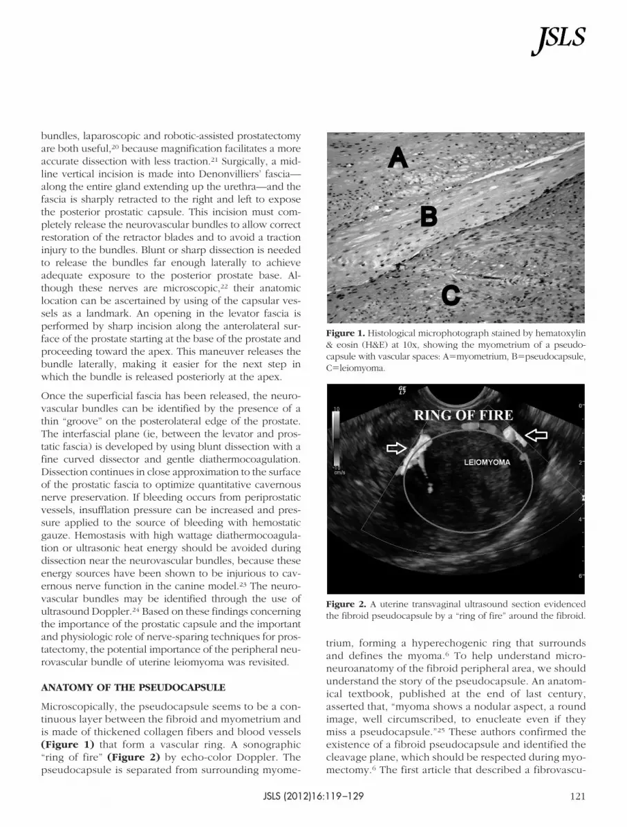
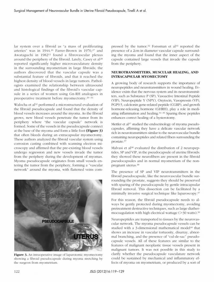
Microscopically, the pseudocapsule seems to be a con-tinuous layer between the fibroid and myometrium andis made of thickened collagen fibers and blood vessels(Figure 1) that form a vascular ring. A sonographic“ring of fire” (Figure 2) by echo-color Doppler. Thepseudocapsule is separated from surrounding myome-
trium, forming a hyperechogenic ring that surroundsand defines the myoma.6 To help understand micro-neuroanatomy of the fibroid peripheral area, we shouldunderstand the story of the pseudocapsule. An anatom-ical textbook, published at the end of last century,asserted that, “myoma shows a nodular aspect, a roundimage, well circumscribed, to enucleate even if theymiss a pseudocapsule.”25 These authors confirmed theexistence of a fibroid pseudocapsule and identified thecleavage plane, which should be respected during myo-mectomy.6 The first article that described a fibrovascu-
Figure 1. Histological microphotograph stained by hematoxylin& eosin (H&E) at 10x, showing the myometrium of a pseudo-capsule with vascular spaces: A�myometrium, B�pseudocapsule,C�leiomyoma.
Figure 2. A uterine transvaginal ultrasound section evidencedthe fibroid pseudocapsule by a “ring of fire” around the fibroid.
JSLS (2012)16:119–129 121
lar system over a fibroid as “a mass of proliferatingarteries” was in 1944.26 Farrer-Brown in 1970,27 andAwataguchi in 198212 found a fibrovascular plexusaround the periphery of the fibroid. Lately, Casey et al28
reported significantly higher microvasculature densityin the surrounding myometrium in large fibroids; theauthors discovered that the vascular capsule was asubstantial feature of fibroids, and that it reached thehighest density of blood vessels in large tumors. Pathol-ogists examined the relationship between ultrasoundand histological findings of the fibroid’s vascular cap-sule in a series of women using Gn-RH analogues inpreoperative treatment before myomectomy.29–31
Walocha et al32 performed a microstructural evaluation ofthe fibroid pseudocapsule and found that the density ofblood vessels increases around the myoma. As the fibroidgrows, new blood vessels penetrate the tumor from itsperiphery where “the vascular capsule” network isformed. Some of the vessels in the pseudocapsule connectat the base of the myoma and form a little foot (Figure 3)that often bleeds during an extracapsular myomectomy.These authors analyzed the fibroid vascular system usingcorrosion casting combined with scanning electron mi-croscopy and affirmed that the pre-existing blood vesselsundergo regression and new vessels invade the tumorfrom the periphery during the development of myomas.Myoma pseudocapsule originates from small vessels en-tering the tumor from the periphery, forming a ”vascularnetwork” around the myoma, with flattened veins com-
pressed by the tumor.32 Forssman et al33 reported thepresence of a 2cm in diameter vascular capsule surround-ing the myoma and found that the inner aspect of thecapsule contained large vessels that invade the capsulefrom the periphery.
NEUROTRANSMITTERS, MUSCULAR HEALING, ANDINTRACAPSULAR MYOMECTOMY
A growing body of research supports the importance ofneuropeptides and neurotransmitters in wound healing. Ev-idence exists that the nervous system and its neurotransmit-ters, such as Substance P (SP), Vasoactive Intestinal Peptide(VIP), Neuropeptide Y (NPY), Oxytocin, Vasopressin (VP),PGP9.5, calcitonin gene-related peptide (CGRP), and growthhormone-releasing hormone (GHRH), play a role in medi-ating inflammation and healing.34–36 Sparing these peptidesenhances correct healing of a hysterotomy.
Mettler et al37 studied the endocrinology of myoma pseudo-capsules, affirming they have a delicate vascular networkrich in neurotransmitters similar to the neurovascular bundlecontaining neuropeptides and related fibers surrounding theprostate.37
Malvasi et al38 evaluated the distribution of 2 neuropep-tides, SP and VIP, in the pseudocapsule of uterine fibroids:they showed these neurofibers are present in the fibroidpseudocapsules and in normal myometrium of the non-pregnant uterus.38
The presence of SP and VIP neurotransmitters in thefibroid pseudocapsule, like the neurovascular bundle sur-rounding the prostate, suggests they should be preservedwith sparing of the pseudocapsule by gentle intracapsularfibroid removal. This dissection can be facilitated by aminimally invasive surgical technique like laparoscopy.37
For this reason, the fibroid pseudocapsule needs to al-ways be gently protected during myomectomy, avoidingpretreatment destructive techniques, such as large diather-mocoagulation with high electrical wattage (�30 watts).39
Neuropeptides are transported to tissues by the neurovas-cular network. The myoma pseudocapsule vessels can bestudied with a 3-dimensional mathematical model40 thatshows an increase in vascular tortuosity, disarray, abnor-mal branching, and the presence of “cul-de-sac” pseudo-capsule vessels. All of these features are similar to thefeatures of malignant neoplastic tissue vessels present inmalignant tumors. It was not possible in this study toclarify whether the pseudocapsule vasculature networkcould be sustained by mechanical and inflammatory ef-fects of myoma on myometrium, or produced by a sort of
Figure 3. An intraoperative image of laparotomic myomectomyshowing a fibroid pseudocapsule during myoma stretching bythe surgeon from myometrium.
Surgical Management of Neurovascular Bundle in Uterine Fibroid Pseudocapsule, Tinelli A et al.
JSLS (2012)16:119–129122

neoplastic type of neoangiogenesis due to the fibroidgrowth, or even by a normal muscle and tissue healingprocess, such as a neurovascular preparative reaction ofthe female body to a fibroid expulsion (pedunculated fi-broids), necrosis or degeneration.
In humans, difficulties in obtaining serial samples of hys-terotomic scar during myomectomy or cesarean deliveryare a major barrier to our understanding of the eventsinvolved in the postmyomectomy and postpartum remod-eling processes of the uterine wound after a cesareandelivery. Thus, little is known about healing of the uterinescar tissue after surgical injuries, such as myomectomyand cesarean deliveries. However, irrespective of the ul-timate result, wound healing is a dynamic, interactiveprocess involving neuromediators, angiogenetic factors,neuropeptides, blood cells, extracellular matrix, and pa-renchymal cells that follows 3 complex and overlappingphases: inflammation, tissue formation, and tissue remod-eling.41 And this healing process is also involved in post-operative adhesion development, as a consequence ofmyomectomy and associated with a high risk of de-novoadhesion formation, that may decrease fertility.42
The authors applied their surgical method in a series ofpatients to study adhesions following intracapsular lapa-roscopic and abdominal myomectomy (6cm to 8cm), withor without an anti-adhesion barrier, using nonsystematicsecond-look surgery.43 When an adhesion barrier was notused, a significant rate of adhesions occurred in laparot-omy (28.1%) compared to laparoscopy (22.6%). Filmy andorganized adhesions were predominant with an adhesionbarrier, and cohesive adhesions were more common with-out an adhesion barrier. Based on these data, an adhesionbarrier should always be used after laparoscopic intracap-sular myomectomy in women wanting pregnancy, be-cause it appears to promote correct healing and reducesadhesion formation.
ENDOSCOPICALLY TAILORED MICRO-SURGERYAPPLIED TO PROSTATECTOMY ANDMYOMECTOMY
A radical prostatectomy must be balanced between achiev-ing cancer control (negative surgical margins) and preserva-tion of the neurovascular bundle to protect sexual and post-surgical function. This can be accomplished by anendoscopically tailored microsurgery, which spares nerveinjuries. Likewise, myomectomy should be performed by anintracapsular method, because the uterus is fully innervedand each myomectomy site could lead to uterine neuro-anatomical damage.
Pelvic innervations in both men and women are derivedfrom the superior hypogastric plexus, sympathetic chain,parasympathetic fibers (S2-4), and the sacral plexus(S1-S5). The superior hypogastric plexus is the downwardcontinuation of the inferior mesenteric plexus over thelower aorta and sacral promontory. Right and left hypo-gastric nerves supply the inferior hypogastric plexi overthe posterior-lateral pelvis, passing behind the commoniliac arteries before entering the utero-sacral and cardinalligaments. In women, this system innervates the uterusand upper vagina. The uterus receives its primary inner-vations from the utero-vaginal plexus (Frankenhauser’splexus), which is located near the transverse cervical lig-ament lateral to the cervix. Nerve fibers are distributedthroughout the myometrium with the branches of theuterine artery. There are nerve fibers throughout the basalthird of the endometrium and a significant plexus at theendometrial-myometrial interface.44
Based on these neuroanatomical findings, the authorsdeveloped and standardized their intracapsular myomec-tomy technique (as urologists standardized prostatectomy)to preserve neurovascular bundles.
In urological surgery, the goal is to preserve the neuro-vascular bundle, which is located outside the prostaticcapsule, to prevent erectile dysfunction (impotency) andurinary incontinence. Nerve-sparing techniques for ana-tomic radical prostatectomy (Figure 4) developed in re-cent years have helped to reduce complications related toinjuries to the neurovascular bundle. A commonly usedapproach for neurovascular bundle preservation involvesincision of the lateral prostatic fascia, partial mobilizationof the neurovascular bundle from the apical third of thegland, and then urethral transection. Subsequent elevationof the apex of the prostate, generally with a Foley catheterfor traction ensures lateralization of the neurovascularbundle and posterior dissection of the prostate. Earlyrelease of the neurovascular bundle from the apex of theprostate before beginning the posterior dissection reducesthe time to recovery and improves function.45
The clinical rationale for intracapsular myomectomy canbe applied to all myomectomies. It is relevant, because aproper laparoscopic myomectomy dramatically improvesfertility, reduces blood loss, shortens hospital stay, andminimizes therapeutic antibiotic administration.46
Laparoscopic myomectomies are performed under gen-eral anesthesia via endotracheal intubation with a stan-dardized 4-port approach: 1 port for the laparoscope and3 lower quadrant ancillary ports (1 suprapubic central10-mm port and 2 lateral 5-mm ports). The 10-mm central
JSLS (2012)16:119–129 123

suprapubic port is often changed to 15mm to 20mm forthe introduction of a morcellator at the end of the proce-dure. All patients receive an intrauterine manipulator priorto laparoscopy, to better mobilize the uterus. Intracapsularlaparoscopic myomectomy of the submucosa and intra-mural fibroids is generally performed without injection ofan ischemic solution into the myometrium. The visceralperitoneum is incised in the midline longitudinal plane, bymonopolar scissors or crochet needle electrode, proceed-ing in depth into myometrium to reach the correct planeunder the myometrium, to detect the myoma pseudocap-sule with the fibroid below.
Once the myoma pseudocapsule is identified, it is ex-posed by atraumatic clamp or by irrigator cannula, toprovide a panoramic laparoscopic view of the pseudocap-sule of all subserous-intramural leiomyomas. Then, thesurgeon affects the pseudocapsule by a longitudinal cut,using monopolar scissors or a Hook electrode at low wattage(30 watt), to expose the myoma surface (Figure 5). Thefibroid is then secured by a myoma screw or Collinslaparoscopic forceps to perform the traction necessary forits enucleation, helped by an irrigator cannula inserted inthe space under the myoma pseudocapsule and fibroid.Hemostasis of small vessel bleeding is achieved by a
low-wattage bipolar clamp or by Hook electrode or mo-nopolar scissors, always at 30 watts, to free the base of themyoma and the connective bridges from the pseudocap-
Figure 4. A laparoscopic radical prostatectomy image by da Vinci robot, showing the prostatic vein and neurovascular bundlesurrounding the prostate.
Figure 5. A laparoscopic image showing fibroid enucleatingfrom myometrium by monopolar Hook electrode gentle cut ofpseudocapsule by low-wattage energy, to expose the myomasurface, with a magnification of the pseudocapsule during my-oma removal.
Surgical Management of Neurovascular Bundle in Uterine Fibroid Pseudocapsule, Tinelli A et al.
JSLS (2012)16:119–129124

sule. In this way, complete minimal traumatic fibroid re-moval from its pseudocapsule is accomplished with min-imal blood loss sparing of the pseudocapsule. In case ofpseudo-pedunculated myomas, the pedicle is coagulatedby bipolar forceps and cut by laparoscopic scissors or cutafter placement of loops or staples. In cases of deepintramural myomas, chromopertubation is always appliedvia a cervical cannula not only to check tubal patency butalso to facilitate the direct recognition of an inadvertentlyopened uterine cavity.
The myometrium closure is performed with a single (forsubserous fibroids) or double layer (for intramurals)suture, including overlying serosa, with a round CT-1curved needle, using intra- or extracorporeal knots. Insubserosal myomectomies, the edges of the uterinedefect are approximated with introflecting U-invertedstitches (myometrium/serosa-serosa/myometrium direc-tion) with intramyometrial knot, at 1-cm increments from theedge of the incision (as a “baseball-type” suture). Closure isby surgeon’s choice: interrupted closure or traditional unidi-rectional running suture, started at the end of one of thehysterotomy sides.
Deep intramural fibroids require a 2-layer myometrial clo-sure with introflecting sutures, covered by a “baseball-type” suture. If the uterine cavity is accidentally openedduring fibroid enucleating, 2 to 3 deep myometrial singleor continuous sutures is applied to the uterine cavityedges. After hysterorraphy, fibroids are usually morcel-lated for ease of removal.
CONTROVERSIES SURROUNDINGLAPAROENDOSCOPIC MYOMECTOMY
The general myomectomy dogma is that “each surgical fi-broid enucleation needs to be gently performed to enhancethe correct healing process of the uterine musculature and tosuccessively facilitate the correct restoration of the uterinemusculature anatomical-functional.” During myomectomy,as stated in the literature,6,37–40,43 the fibroid pseudocapsuleneurovascular bundle needs to always be protected, avoid-ing destructive surgical procedures, such as extensive andhigh-wattage diathermocoagulation (�30 watt) or excessivetissue manipulation or trauma. This method of myomectomymaximally respects the fibroid pseudocapsule neurovascularbundle, rich in neurofibers that are involved in correctsuccessful scar healing. It is thought that iatrogenic my-oma pseudocapsule damage alters neurotransmitter func-tion in the repair process, with a negative effect on uterinehealing. Thus, the surgical sequels of an uncorrected myo-mectomy is damage to the pseudocapsule neurovascular
bundle, a reduction in the presence of neurofibers at thehysterotomy site, a deterioration in uterine musculaturehealing, and a deficit either in myometrial neurotransmis-sion or in muscular impulse and contractility, with a finalreduction in uterine musculature functionality.
There are many issues with modern-day myomectomy.One of the first questions concerns the current use oftechniques that minimize blood loss and occlude the uter-ine blood supply during myomectomy, such as tourniquetmethods,47,48 uterine artery embolization3,49 or ligation,50
intrauterine injection of ischemic solutions,51,52 or gonad-otrophin-releasing hormone (GnRH) analogues.48,53
All these methods reduce blood flow into the pseudocap-sule, by mechanical compression (the tourniquet methods),vascular occlusion, or a pharmacological blood flow de-crease.
In the authors’ opinion, the main problem with theseapproaches is that their use could mask the musculaturevascularization for vessel collapse, making selective andgentle pseudocapsule vessel hemostasis difficult duringmyomectomy and favoring successive intramyometrial he-matomas, detected by ultrasound,54,55 with impairment ofthe muscular healing at the hysterotomy site. On thecontrary, during surgery, the fibroid pseudocapsule neu-rovascular bundle needs to be well exposed, by endo-scopic magnification, and carefully protected, avoidingdestructive procedures, such as large-scale diathermoco-agulation at high wattage, favoring incorrect restoration ofthe uterine musculature.
Analyzing the literature on ischemic solutions to injectinto the myometrium,52 studies show that vasopressindecreases blood loss at the time of myomectomy bylaparotomy compared with placebo or a tourniquet. Onthe other hand, other studies have found no differencebetween the use of vasopressin or a tourniquet at myo-mectomy performed by laparotomy.52 In clinical prac-tice, probably either technique will decrease blood losscompared with no intervention, so the greater magnifi-cation afforded by the laparoscope may allow for moreprecise treatment of blood vessels. In addition, the CO2
pneumoperitoneum associated with laparoscopy maytamponade small vessels and, cumulatively, result inless blood loss.
Vasopressin injection unfortunately has been reported tocause pulmonary edema and, with intravenous injection,even death, while loop ligation may ultimately compro-mise uterine function and may reduce fertility or increasecomplications during pregnancy.50
JSLS (2012)16:119–129 125

Moreover, the exclusion of preoperative GnRH-analogue treatment is due to the reported increased riskof fibroid recurrence, a possible delay in the diagnosisof leiomyosarcoma, a risk of massive hemorrhage fromdegeneration,56 and, primarily, to avoid distortion ofthe myoma pseudocapsule. The GnRH-analogue treat-ment decreases the size of the myoma, causing conflu-ent nodular hyaline degeneration and hydropic degen-eration necrosis, masking the correct cleavage planbetween myoma and pseudocapsule and making myomahooking difficult.57 For this reason, the use of ischemic so-lutions and GnRH-analogues before surgery is useless andmay even be harmful.
It is likely that the suturing technique is of secondaryimportance when an intracapsular myomectomy is per-formed well. The problem with stitches has never beenapproached properly with regards to the myomectomy,because of the lack of a well-stated scientific rationale. Asreported in many articles,6,37–40,43 the myoma pseudocap-sule is a fibro neurovascular structure, a neurovascularbundle, probably created by the uterus to cope with thedevelopment and growth of fibroids. When the surgeongently removes a myoma through the pseudocapsule, heor she preserves the muscle surrounding the myoma,returning it to normal healthy uterine tissue. The samething happens in the lower uterine segment during preg-nancy: after delivery, it disappears. For these reasons, thehysterotomy after intracapsular myomectomy needs to beclosed by simply introflecting muscle edges in 1 or 2layers. Sutures in several layers are not necessary. Moresuture serves as a foreign body to produce inflammatoryreactions, submesothelial fibrosis, and regenerative meso-thelial hyperplasia.58 Intracapsular fibroid removal, whichspares neurovascular fibers and neuropeptides, helps pre-serve uterine muscle that is not traumatized and ready forproper healing. And, if the myoma is enucleated entirelythrough the fibrovascular capsule opening, using blunt orsharp dissection on the surrounding myometrium, with agentle selective low energy hemostasis on pseudocapsulevessels, the myometrial bed collapses without excessivebleeding once the myoma is removed. This is the rationalefor not putting too much importance on suturing tech-nique. Technique itself that preserves myometrial integrityand allows the restoration of uterine musculature withmagnification of the pseudocapsule neurovascular bundleusing laparoscopy or robotic-assisted surgery is a sort ofendoscopic tailored microsurgery.
The neurovascular bundle surrounding the prostate is richin neurotransmitters and needs to be preserved duringradical prostatectomy to maintain urinary and sexual func-
tion. The uterine leiomyoma pseudocapsule has a neuro-vascular bundle rich in neurofibers and neuropeptides.Intracapsular myomectomy respects the leiomyomapseudocapsule, permits better healing of the uterine scar,leading to neurotransmitter sparing at the myomectomysite, which is extremely important for successive repro-ductive functionality of the uterine musculature.
NEED OF FUTURE RESEARCH
Much still needs to be learned concerning the presence ofneuropeptides and neurotransmitter activity in the neuro-vascular bundle of fibroid pseudocapsules in each part ofthe uterus, the body, the isthmus, and cervix. Studies arealso needed of pseudocapsule anomalous vascularization,which is similar to malignant neoplastic tissue vesselspresent in malignant tumors. More research needs to bedirected at the impact of hormonal patterns on the origin,growth, and recurrence of the fibroid pseudocapsule, theinfluence of drugs, embolospheres, and magnetic reso-nance-guided focused ultrasound on the pseudocapsuleand their effects on uterine healing. Increasing knowledgeof pseudocapsule angiogenesis and neurovascular fiberscould play an important role in understanding the origin,recurrence, and correct treatment of uterine leiomyomas.Therefore, more research is needed in both fertile and post-menopausal patients to demonstrate the anatomic pattern,fibroid behavior, and the involvement of the pseudocapsulein a hormonal environment. What has been said shouldstimulate research on the influence of angiogenic growthfactors on fibroid pseudocapsule and on the neuroanatomi-cal significance of the fibroid pseudocapsule, to explain thepathobiology of fibroid and pseudocapsule formation, thehormonal influence on pseudocapsule growth and neuro-vascular development, and the pseudocapsule during thepostoperative course. This study suggests the need forfurther investigation of the fibroid pseudocapsule and itsneurovascular bundle to determine the role of the myomapseudocapsule in the formation, growth, and pathophys-iologic consequences of uterine fibroids including pain,infertility, and poor reproductive outcomes.
CONCLUSION
In recent years, new advancements in endoscopic sur-gery, including the use of laparoscopic or robotic surgery infibroid removal, have been considered a true alternative tolaparotomy with numerous advantages, such as short hospi-talization, decreased need for postoperative analgesia, lessintraoperative blood loss, and good outcomes in subsequentpregnancy.59 Recent evidence on neurofibers and neuropep-
Surgical Management of Neurovascular Bundle in Uterine Fibroid Pseudocapsule, Tinelli A et al.
JSLS (2012)16:119–129126

tides in the fibroid pseudocapsule suggests a new horizon inendoscopic gynecological surgery, which can be called in-tracapsular fibroid nerve-sparing laparoscopic “microsur-gery,” or intracapsular fibroid nerve-sparing robotic-assisted“nanosurgery,” in case of robotic magnification. A great ad-vantage of intracapsular laparoscopic myomectomy is thereproducibility of its application for all laparoscopic myo-mectomies as a safe, feasible, and minimally invasive tech-nique. Intracapsular myomectomy enhances myometriumintegrity peripheral to the fibroid site, by preserving theneurovascular bundle and neurotransmitters surroundingfibroids, for uterine healing and restoration of the myo-metrium after surgery. Moreover, allowing correct myo-metrial healing, intracapsular myomectomy should pre-serve reproductive outcomes and normal labor anddelivery, for less bleeding, better neurovascular bundlesparing for scar quality, and postoperative adhesion re-duction. The authors propose that their intracapsular myo-mectomy should always be recommended to maximizethe potential for future fertility and to minimize the risk oflabor dystocia or uterine rupture during pregnancy orlabor.
References:
1. Lethaby A, Vollenhoven B. Fibroids (uterine myomatosis,leiomyomas). Clin Evid (Online). 2011;pii:0814.
2. Mettler L, Schollmeyer T, Tinelli A, Malvasi A, Alkatout I.Complications of uterine fibroids and their management, surgicalmanagement of fibroids, laparoscopy and hysteroscopy versushysterectomy, haemorrhage, adhesions and complications. Ob-stet Gynecol Int. 2012; Article ID 791248 doi:10.1155/2012/791248.
3. Stovall DW. Alternatives to hysterectomy: focus on globalendometrial ablation, uterine fibroid embolization, and magneticresonance-guided focused ultrasound. Menopause. 2011;18(4):443–450.
4. Sami Walid M, Heaton RL. The role of laparoscopic myo-mectomy in the management of uterine fibroids. Curr OpinObstet Gynecol. 2011;23:273–277.
5. Mais V, Ajossa S, Guerriero S, Mascia M, Solla E, Melis GB.Laparoscopic versus abdominal myomectomy. A prospective,randomized trial to evaluate benefits in early outcome. Am JObstet Gynecol. 1996;174:654–658.
6. Tinelli A, Malvasi A, Rahimi S, et al. Myoma pseudocapsule:a new-old endocrino-anatomical entity to exploit in uterine myo-mectomy. Gynecol Endocrinol. 2009;25(10):661–667.
7. Ito F, Kawamura N, Ichimura T, Tsujimura A, Ishiko O, OgitaS. Ultrastructural comparison of uterine leiomyoma cells from
the same myoma nodule before and after gonadotropin-releas-ing hormone agonist treatment. Fertil Steril. 2001;75:l25–130.
8. Dapunt O. Studies on the structure of the myoma capsule.Arch Gynecol. 1965;202:492–494.
9. Fox H, Buckley CH. Benign neoplasms of the female genitaltract. In: Pathology for Gynaecologists. Arnold ed. London, UK,1982;8:91–97.
10. Vizza E, Motta PM. The skeleton fibrous and muscular of theuterus. In: Atti LXXVII Congresso SIGO. Rome: CIC Int Ed, 2001;47–49.
11. Hsu WC, Hwang JS, Chang WC, Huang SC, Sheu BC, TorngPL. Prediction of operation time for laparoscopic myomectomyby ultrasound measurements. Surg Endosc. 2007;21(9):1600–1606.
12. Awataguchi K. Studies on the angioarchitecture of uterinemyoma. Nippon Ika Daigaku Zasshi. 1982;49:225–232.
13. Stewart EA, Nowak RA. Leiomyoma-related bleeding: a clas-sic hypothesis updated for the molecular era. Hum Reprod Up-date 1996;2(4):295–306.
14. Yucel S, Erdogru T, Baykara M. Recent neuroanatomicalstudies on the neurovascular bundle of the prostate and caver-nosal nerves: clinical reflections on radical prostatectomy. AsianJ Androl. 2005;7(4):339–349.
15. Takenaka A, Murakami G, Soga H, Han SH, Arai Y, FujisawaM. Anatomical analysis of the neurovascular bundle supplyingpenile cavernous tissue to ensure a reliable nerve graft afterradical prostatectomy. J Urol. 2004;172(3):1032–1035.
16. Costello AJ, Brooks M, Cole OJ. Anatomical studies of theneurovascular bundle and cavernosal nerves. BJU Int. 2004;94(7):1071–1076.
17. Walsh PC. The discovery of the cavernous nerves and de-velopment of nerve sparing radical retropubic prostatectomy.J Urol. 2007;177(5):1632–1635.
18. Hernandez DJ, Epstein JI, Trock BJ, Tsuzuki T, Carter HB,Walsh PC. Radical retropubic prostatectomy. How often do ex-perienced surgeons have positive surgical margins when there isextraprostatic extension in the region of the neurovascular bun-dle? J Urol. 2005;173(2):446–449.
19. Shah SJ, Goyal V, Sachar R, Nath AK, Jain N, Kapadia K.Seminal vesicle sparing laparoscopic radical prostatectomy usinga low-energy source: Better continence and potency. IndianJ Urol. 2009;25(2):199–202.
20. Han M, Kim C, Mozer P, et al. Tandem-robot assisted lapa-roscopic radical prostatectomy to improve the neurovascularbundle visualization: a feasibility study. Urology. 2011;77(2):502–506.
21. Walz J, Burnett AL, Costello AJ, et al. A critical analysis of thecurrent knowledge of surgical anatomy related to optimization of
JSLS (2012)16:119–129 127

cancer control and preservation of continence and erection incandidates for radical prostatectomy. Eur Urol. 2010;57(2):179–192.
22. Lee SE, Hong SK, Han JH, et al. Significance of neurovascu-lar bundle formation observed on preoperative magnetic reso-nance imaging regarding postoperative erectile function afternerve-sparing radical retropubic prostatectomy. Urology. 2007;69(3):510–514.
23. Tinelli A, Hurst BS, Hudelist G, Tsin DA, Stark M, Mettler L,Guido M, Malvasi A. Laparoscopic myomectomy focusing on themyoma pseudocapsule: technical and outcome reports. HumReprod. 2012;27(2):427–35.
24. Ukimura O, Gill IS. Real-time transrectal ultrasound guid-ance during nerve sparing laparoscopic radical prostatectomy:pictorial essay. J Urol. 2006;175(4):1311–1319.
25. Pasetto N, Baschieri L, Giacomello F, Ticconi C. Patologiabenigna dell’utero. In: Candiani GB, Danesino V, Gastaldi C. LaClinica Ostetrica e Ginecologica. Milan, Italy; Masson Editore.1992;1242–1256.
26. Faulkner RL. The blood vessels of the myomatous uteri. Am JObstet Gynaecol. 1944;47:185–197.
27. Farrer-Brown G, Beilby JOW, Tarbit MH. The vascular pat-terns in myomatous uteri. J Obstet Gynaecol. 1970;77:967–975.
28. Casey R, Rogers PA, Vollenhoven BJ. An immunohistochem-ical analysis of fibroid vasculature. Hum Reprod. 2000;15(7):1469–1475.
29. Campo S, Garcea N. Laparoscopic myomectomy in pre-menopausal women with and without preoperative treatmentusing gonadotrophin-releasing hormone analogues. Hum Re-prod. 1999;14:44–48.
30. Lethaby A, Vollenhoven B, Sowter M. Pre-operative Gn-RHanalogue therapy before hysterectomy or myomectomy for uter-ine fibroids. Cochrane Database Syst Rev. 2001;2:CD000547.
31. Pinkerton JV. Pharmacological therapy for abnormal uterinebleeding. Menopause. 2011;18(4):459–467.
32. Walocha JA, Litwin JA, Miodonski A. Vascular system ofintramural leiomyomata revealed by corrosion casting and scan-ning electron microscopy. Human Reprod. 2003;18:1088–1093.
33. Forssman L. Blood flow in myomatous uteri as measured byintraarterial 133-Xenon. Acta Obstet Gynecol Scand. 1976;55:21–24.
34. Hanna KR, Katz AJ. An update on wound healing and thenervous system. Ann Plast Surg. 2011;67(1):49–52.
35. Henderson J, Terenghi G, Ferguson MW. The reinnervationand revascularisation pattern of scarless murine fetal wounds. JAnat. 2011;218(6):660–667.
36. Gouin JP, Carter CS, Pournajafi-Nazarloo H, et al. Maritalbehavior, oxytocin, vasopressin, and wound healing. Psycho-neuroendocrinology. 2010;35(7):1082–1090.
37. Mettler L, Tinelli A, Hurst BS, et al. Neurovascular bundle infibroid pseudocapsule and its neuroendocrinologic implications.Expert Rev Endocrinol Metab. 2011;6:715–722.
38. Malvasi A, Tinelli A, Cavallotti C, et al. Distribution of Sub-stance P (SP) and Vasoactive Intestinal Peptide (VIP) neuropep-tides in pseudocapsules of uterine fibroids. Peptides. 2011;32(2):327–332.
39. Tinelli A, Malvasi A, Hudelist G, et al. Laparoscopic intra-capsular myomectomy: comparison of single versus multiplefibroids removal. An institutional experience. J LaparoendoscAdv Surg Tech A. 2010;20(8):705–711.
40. Malvasi A, Tinelli A, Rahimi S, et al. A three-dimensionalmorphological reconstruction of uterine leiomyoma pseudocap-sule vasculature by the Allen-Cahn mathematical model. BiomedPharmacother. 2011;65(5):359–363.
41. Vikhareva Osser O, Valentin L. Risk factors for incompletehealing of the uterine incision after caesarean section. BJOG.2010;117(9):1119–1126.
42. Kubinova K, Mara M, Horak P, Kuzel D, Dohnalova A.Reproduction after myomectomy: Comparison of patients withand without second-look laparoscopy. Minim Invasive Ther Al-lied Technol. 2011 Jul 11. [Epub ahead of print]
43. Tinelli A, Malvasi A, Guido M, et al. Adhesions formationafter intracapsular myomectomy with or without adhesion bar-rier. Fertil Steril. 2011;95(5):1780–1785.
44. Quinn MJ, Kirk N. Differences in uterine innervation athysterectomy. Am J Obstet Gynecol. 2002;187(6):1515–1520.
45. Masterson TA, Serio AM, Mulhall JP, Vickers AJ, EasthamJA. Modified technique for neurovascular bundle preservationduring radical prostatectomy: association between techniqueand recovery of erectile function. BJU Int. 2008;101(10):1217–1222.
46. Hurst BS, Matthews ML, Marshburn PB. Laparoscopic myo-mectomy for symptomatic uterine myomas. Fertil Steril. 2005;83(1):1–23.
47. Ikechebelu JI, Ezeama CO, Obiechina NJ. The use of tour-niquet to reduce blood loss at myomectomy. Niger J Clin Pract.2010;13(2):154–158.
48. Al-Shabibi N, Chapman L, Madari S, Papadimitriou A,Papalampros P, Magos A. Prospective randomised trial com-paring gonadotrophin-releasing hormone analogues with tri-ple tourniquets at open myomectomy. BJOG. 2009;116(5):681–687.
49. Malartic C, Morel O, Fargeaudou Y, et al. Conservativetwo-step procedure including uterine artery embolizationwith embosphere and surgical myomectomy for the treatmentof multiple fibroids: Preliminary experience. Eur J Radiol.2012;81:1–5.
Surgical Management of Neurovascular Bundle in Uterine Fibroid Pseudocapsule, Tinelli A et al.
JSLS (2012)16:119–129128

50. Chang WC, Chou LY, Chang DY, et al. Simultaneous lapa-roscopic uterine artery ligation and laparoscopic myomectomyfor symptomatic uterine myomas with and without in situ mor-cellation. Hum Reprod. 2011;26(7):1735–1740.
51. Riess ML, Ulrichs JG, Pagel PS, Woehlck HJ. Severe vaso-spasm mimics hypotension after high-dose intrauterine vaso-pressin. Anesth Analg. 2011;113:1103–1105.
52. Frishman GN, Jurema MW. Myomas and myomectomy. JMinimal Invasive Gynecol. 2005;12:443–456.
53. Chen I, Motan T, Kiddoo D. Gonadotropin-releasing hor-mone agonist in laparoscopic myomectomy: systematic reviewand meta-analysis of randomized controlled trials. J Minim In-vasive Gynecol. 2011;18(3):303–309.
54. Seinera P, Gaglioti P, Volpi E, Cau MA, Todros T. Ultrasoundevaluation of uterine wound healing following laparoscopicmyomectomy: preliminary results. Hum Reprod. 1999;14(10):2460–2463.
55. Chang WC, Chang DY, Huang SC, et al. Use of three-dimen-sional ultrasonography in the evaluation of uterine perfusionand healing after laparoscopic myomectomy. Fertil Steril. 2009;92(3):1110–1115.
56. Sankaran S, Manyonda IT. Medical management of fibroids.Best Pract Res Clin Obstet Gynaecol. 2008;22(4):655–676.
57. De Falco M, Staibano S, Mascolo M, et al. Leiomyomapseudocapsule after pre-surgical treatment with gonadotropin-releasing hormone agonists: relationship between clinical fea-tures and immunohistochemical changes. Eur J Obstet GynecolReprod Biol. 2009;144(1):44–47.
58. Malvasi A, Tinelli A, Farine D, et al. Effects of visceralperitoneal closure on scar formation at cesarean delivery. Int JGynaecol Obstet. 2009;105(2):131–135.
59. Munro MG, Critchley HO, Fraser IS. Outcomes from leiomy-oma therapies: comparison with normal controls. Obstet Gyne-col. 2011;117(4):987, author reply 987–988.
JSLS (2012)16:119–129 129


![Chemoprevention of fibroid tumors by [−]-epigallocatechin-3-gallate in quail](https://img.pdfslide.net/doc/110x75/631aee3c2784ca2fc00514f1/chemoprevention-of-fibroid-tumors-by-epigallocatechin-3-gallate-in-quail.jpg)


