Embed Size (px)
Citation preview
Clin Podiatr Med Surg
25 (2008) 95–120
Surgical Reconstruction of the CharcotRearfoot and Ankle
Patrick R. Burns, DPM, FACFASa,b,c,*,Dane K. Wukich, MDa,b,c
aFoot and Ankle Division, University of Pittsburgh School of Medicine, Roesch-Taylor
Medical Building, North Suite 7100, 2100 Jane Street, Pittsburgh, PA 15203, USAbPodiatric Surgical Training Program, University of Pittsburgh Medical Center South
Side Hospital, 2000 Mary Street, Pittsburgh, PA 15203, USAcUniversity of Pittsburgh Medical Center, Comprehensive Foot and Ankle Center,
Roesch-Taylor Medical Building, North Suite 7100, 2100 Jane Street,
Pittsburgh, PA 15203, USA
Destructive changes in the joints of patients who have neuropathy weredescribed by Jean-Martin Charcot [1] in 1868 and later were described inthe diabetic foot and ankle in 1936 by Jordan [2]. Peripheral neuropathyis universally present in patients who develop Charcot fracture/dislocation,and the number of patients who have diabetes and associated peripheralneuropathy is on the rise [3,4].
Other factors play a role, however. An area currently under investigationat the cellular level is receptor activator of nuclear factor kappa B ligand.Normal regulation and counterbalance of osteoclasts and osteoblasts maybe altered in the patient who has peripheral neuropathy and may causethe loss in structural integrity and subsequent failure [5]. The history andpathophysiology are reviewed more thoroughly in other articles in this issue.
Although the exact mechanism is not known, the ultimate goal of treat-ment, whether nonoperative or operative, is to achieve a stable, plantigradefoot. Achieving this status is challenging in the rearfoot and ankle. Statisti-cally, Charcot deformity affects the rearfoot and ankle less often than themidfoot, but the resultant deformities typically are more severe and difficultto stabilize conservatively. The resultant instability in the ankle leads toa limb-threatening deformity, and surgical intervention and salvage are
* Corresponding author. Foot and Ankle Division, University of Pittsburgh School of
Medicine, Roesch-Taylor Medical Building, North Suite 7100, 2100 Jane Street, Pittsburgh,
PA 15203.
E-mail address: [email protected] (P.R. Burns).
0891-8422/08/$ - see front matter � 2008 Elsevier Inc. All rights reserved.
doi:10.1016/j.cpm.2007.10.008 podiatric.theclinics.com
96 BURNS & WUKICH
more common. To date, few data on the best course of treatment are avail-able, but the use of limb-salvage techniques is on the rise. With increasingknowledge of the disease and with technological advances in internal and ex-ternal fixation, limb salvage is becoming more consistent. This article dis-cusses basic techniques in deformity planning and current uses of internaland external fixation techniques for rearfoot and ankle limb salvage.
Diagnosis, staging, and classification
In many cases the traumatic event that initiates Charcot changes goesunrecognized. This lack of recognition can delay the diagnosis and can affecttreatment. At least 25% of cases have improper or delayed diagnosis, andsome studies show even higher rates [6,7].
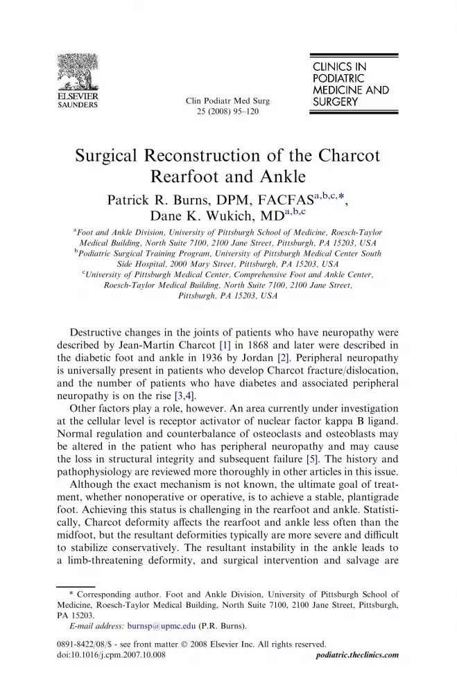
Radiographic findings of Charcot deformity are well known. Destructionof bone and joint, debris, dislocation, and osteopenia are common findings;however, they may not be present initially. Clinical suspicion should remainhigh when warmth, edema, and erythema present in the neuropathic patient(Fig. 1). Most often this presentation is unilateral and commonly is misdiag-nosed as gout, infection, or deep venous thrombosis. Bilateral events are lesslikely but are reported in approximately 10.4% of patients [8]. In any event,foot and ankle surgeons must continually educate patients and other healthcare providers about these subtle findings and Charcot deformity.
Infection must be ruled out in patients who have an open ulcer or a his-tory of previous surgery. Without a portal of entry, it is unlikely that theincreased erythema and temperature localized to the foot or ankle wouldresult from infection. A thorough examination of the skin for loss of skinintegrity and evaluating the patient for fevers, chills, and a change in glyce-mic control is paramount. Patients who have acute Charcot deformity man-ifest local signs of inflammation without the typical systemic signs ofincreased white cell count or fever. A simple test of elevating the leg for
Fig. 1. Typical clinical presentation of erythema and edema seen with a foot with acute Charcot
deformity.

97SURGICAL RECONSTRUCTION OF CHARCOT REARFOOT
10 minutes should show decrease in the edema in a patient who has Charcotdeformity, whereas the edema will persist in infection [9].
If the patient has a wound, the proper steps need to be taken. Local care,off-loading techniques, imaging studies, deep cultures, and biopsies aid inthe diagnosis. Nonhealing ulcers or those that show no improvement withstandard care should be examined more thoroughly, and surgical interven-tion often is necessary (Fig. 2). Intraoperative debridement allows propercultures and specimens to be sent for analysis to determine the presenceand number of white cells. Bone can be sampled as well for diagnosis ofosteomyelitis. Charcot deformity and infection can coexist, and proper diag-nosis is needed to guide treatment. Intraoperative frozen sections and infec-tious disease consultations help guide antibiotic therapy.
Vascular status must also be evaluated properly. Noninvasive studies arenow recommended yearly in patients who have diabetes, independent ofsubjective complaints. In the patient who has neuroarthropathy, the anklebrachial index typically demonstrates adequate flow. Studies show the anklebrachial index in patients who have Charcot deformity average 0.65 duringthe process [10–13]. This finding still needs to be regarded with caution,because diabetic patients have both macro- and microvascular disease, mak-ing the interpretation of these tests challenging.
The classic radiograph of Charcot deformity is identified easily becausefew disease processes have such a profound effect with widespread disloca-tion and debris (Fig. 3). Osteomyelitis is a common misdiagnosis because ofthe bone destruction and periosteal reaction. Differentiation of Charcotdeformity from osteomyelitis may be difficult radiographically, especiallywhen a wound is present. There continues to be debate about the best mo-dality for differentiating osteomyelitis from Charcot arthropathy. MRI isvery sensitive in showing marrow edema and abscess, but others believeindium-labeled white cell scans are more reliable for distinguishing betweenthe two conditions [10,11,14].
Fig. 2. Talar dislocation and ulceration associated with acute Charcot deformity of the ankle.

98 BURNS & WUKICH
Radiographs are critical for staging, classification, and defining treatmentoptions. Traditionally, surgery during the acute stage has not been recom-mended because of poor results. Staging systems became useful in treatmentprotocols, and Eichenholtz [15] in 1966 was the first to classify three stages.Charcot deformity progresses through what he determined were well-defineddevelopmental, coalescence, and remodeling stages. These stages were easilyidentifiable and aided in treatment planning. He believed that surgery wouldhave better results if performed either in early stage 1 or late stage 3, pre-sumably because of relatively less inflammation and overall increased stabil-ity and structural integrity of bone.
Harris and Brand [16] followed this idea and suggested that arthrodesis beperformed early in the disease process. Similarly, Newman [17] found thatearly arthrodesis and a period of immobilization of the involved joint couldprevent further deformity. This practice was not widely adopted, however;others noted hardware failure and infection, so early surgical interventionhas not been recommended for treating the initial stages. Traditionally stage1 has been treated with conservative measures including total contact castingand off-loading. Surgery was reserved for the more predictable later stage 3.There are no comparative or prospective studies for definitive guidance.
The concept of Charcot stage 0 was later added by Shibata and colleagues[18] for the ‘‘at risk’’ patient who has neuropathy and injury. Presumably,these patients have a much higher chance of progressing to the destructivestages, even though initial radiographs are negative. If treated appropriately,the Charcot cycle would not progress to the destructive stage. This area hasseen much interest recently, particularly in diabetic ankle fractures [19].
Fig. 3. (A) Lateral and (B) oblique radiographs of classic findings of Charcot deformity includ-
ing dislocation, destruction, and debris.

99SURGICAL RECONSTRUCTION OF CHARCOT REARFOOT
Because of increased complication rates, current recommendations are formore stable constructs, prolonged non–weight-bearing, and increased numberof office visits to attempt tominimize the possibility of aCharcot event (Fig. 4).
Classification based on anatomy, inclusive of the entire foot and ankle,has been described by Brodsky [9,20] and by Sanders and Frykberg [21].Schon and colleagues [22] also classified Charcot deformity with a focus onthe midtarsus alone. The classifications based on location aid in treatmentplans and prognosis. Deformities become less stable and more often requireintervention with longer immobilization periods as they move proximal. TheBrodsky and Sanders/Frykberg classifications differ in the actual ‘‘type’’ andlocation, but the overall statistics remain similar. Clearly the incidence ofCharcot deformity in the midfoot makes up the majority in both systems(60% to 70% in Brodsky type 1, which includes the tarsometatarsal and na-viculocuneiform joints). The Sanders/Frykberg classification including thesame joints is considered type II and type III, comprising about 55% of cases.Although the classification systems do not match perfectly in location orjoints involved, they do show overall similarities in the percentages of mid-foot involvement. Corresponding types involving the ankle from both sys-tems include Brodsky type 3a at 10% and Sanders/Frykberg type IV at 9%.
Hindfoot and ankle literature
Outcomes and evidenced-based medicine regarding Charcot ankle andhindfoot deformity is minimal. Most literature consists of case series or
Fig. 4. (A) Lateral and (B) anteroposterior radiographs of a diabetic ankle fracture which
converted to an acute Charcot deformity with subsequent hardware failure and deformity.

100 BURNS & WUKICH
expert opinion. Midfoot Charcot deformity has been the focus of most stud-ies of treatment and outcomes, presumably because of its greater incidence.Nonoperative treatments show acceptable outcomes in much midfoot Char-cot literature, possibly because of the greater inherent stability of the region.Acute rearfoot and ankle Charcot deformity is less stable and is more likelyto require surgery, but it also is overall less common than midfoot disease.There are no studies comparing conservative versus surgical treatment in theankle or rearfoot, and the majority of the literature regarding rearfoot andankle Charcot deformity is anecdotal or small, retrospective studies.
Today, increasing numbers of surgeons are beginning to advocate earlierintervention for Charcot changes [23,24]. In 2000, Simon and colleagues [25]published an arthrodesis study that demonstrated successful restoration ofanatomic alignment with the use of open reduction internal fixation and fu-sion in the acute developmental stage of Charcot breakdown. Stable fusionwas achieved in all 14 patients in this study of midfoot Charcot deformity.To date there are no similar studies concerning the acute event in the rear-foot or ankle, and more studies are needed before these types of surgery areconsidered routine or standard in the acute patient.
In midfoot disease, Pinzur [26,27] found approximately 60% of 147patients at 1-year follow-up required no surgery and could be managedwith shoe and brace modifications. The remaining 60 patients in the studyrequired corrective surgery, mainly consisting of triple arthrodesis toaddress a nonplantigrade foot. In this retrospective review, eight amputa-tions were performed on this group in the 1-year period. Pinzur [28] alsoreported on 237 patients in which the diagnosis of Charcot deformity wasmade over a 10-year period. Over that time period 120 patients required143 operations. Surgeries ranged from debridement to major amputation.The article did not elaborate on location or classifications to stratify out-comes but did state that 55 ankle or hindfoot fusions were performed alongwith 21 major amputations. It did not give data to correlate location withthese events or other complication rates specific to the ankle or hindfoot.This benchmark analysis was aimed more at projecting costs for the healthcare system to allocate resources appropriately.
Saltzman and colleagues [8] reviewed 127 limbs to determine survivorshipand amputation rate. The actual number of major amputations was 15 of127, giving an annual risk of 2.72 per 100 patients on a survivorship curve.This finding was similar to Pinzur’s [28] benchmark review of 237 patients inwhich the overall amputation rate was 9%. In Saltzman’s article [8] thelocation of Charcot deformity did not seem to affect the rate of amputation,but the numbers were too low to be significant. As expected, patients whohad recurrent ulceration did show a significant increase in amputationrate, as did patients who presented initially with an ulceration comparedwith those who had an intact skin envelope. This study included Charcotdeformity at every level, 32 of which were of the ankle or hindfoot requiringsix major amputations. A majority of the surgery performed addressed

101SURGICAL RECONSTRUCTION OF CHARCOT REARFOOT
midfoot pathology. A total of 53 non-amputation surgeries were performed,including two ankle fusions and three hindfoot fusions to address unstabledeformities. There were no amputations in this surgically treated group.
Zarutsky and colleagues [29] reported on 43 patients undergoing salvageankle arthrodesis with the use of external fixation. Eleven of those patientsunderwent surgery for Charcot reconstruction. The data and complicationsdid not separate out the patients who had Charcot deformity from theothers, but a major complication rate of 51.2% was noted for the series.Complications included abscess and non-union, and one of the two be-low-knee amputations reported in the series occurred in a patient whohad Charcot deformity. Although this study was not exclusively of Charcotreconstruction, the complexity of external fixation for these salvage caseswas expressed.
Papa and colleagues [13] reported on 29 patients; of these a large percent-age had rearfoot and ankle involvement. The series reported on salvage witharthrodesis in 21 ankle and 6 subtalar joints with longstanding neuroarthr-opathy. It may be that this study contains a high number of rearfoot andankle patients because these patients were less stable and so were less likelyto respond to the typical conservative treatments, but this possibility wasnot discussed. The patients were operated on in later stages of the disease,and a variety of rearfoot and ankle fusions were performed as needed.Most received internal fixation with only four external fixators. The articledoes state ankle brachial index findings were a mean of 0.86, and some ofthe fusions were performed in the presence of a nonhealing wound. Sixty-six percent achieved a solid fusion at 5 months. The most likely fusion todevelop a pseudoarthrosis was tibiocalcaneal, which fused in only 4 of 11procedures. One patient was lost to follow-up, one patient underwentbelow-knee amputation, one patient had recurrent ulceration, and theremaining patients were successfully stabilized long term with bracing.
Internal fixation with intramedullary nail fixation in the treatment ofCharcot ankle and rearfoot deformity was reviewed retrospectively by Car-avaggi and colleagues [30]. Bracing had failed in 14 patients who had severeankle instability. All ulcerations were healed before surgery was performed,and all surgeries took place in the later remodeling phase of Charcot defor-mity. In the end, the authors showed a 92.8% salvage rate, with unions in 10of 14 patients. There were three fibrous unions that developed after hard-ware failure in the calcaneus and one below-knee amputation for osteomy-elitis. This case series shows good results in patients who had late-stageCharcot deformity, affording the stability required in the ankle and rearfootfor ambulation with bracing.
There was a similar finding by Pelton and colleagues [31] using intrame-dullary retrograde nailing for salvage of ankle and rearfoot deformity. Tenof the 33 patients in the series had the diagnosis of Charcot deformity. Theunion rate for the entire series was 88%. There were four non-unions, two ofwhich were in patients who had Charcot deformity, making non-union in

102 BURNS & WUKICH
the small Charcot subset 2 of 10. These non-unions remained stable andbraceable, leading the authors to state they believe this technique is favor-able in the Charcot reconstruction because of the increased stiffness com-pared with lag screws, especially when the distal screws are insertedposterior-to-anterior through the calcaneus.
Charcot deformity related to leprosy was reviewed by Shibata and col-leagues [18]. In this series, 26 fusions were performed with intramedullaryrod (17 ankle and 9 tibiocalcaneal) after talectomy. Most were performedin the later stages. Four, however, were performed in stage 0, and onewas performed in stage 1. The authors achieved fusion in 73% (ie, 19attempts); all their failures in the later stage 3 patients.
Fabrin and colleagues [32] reviewed 12 feet in 11 patients over a 12-yearperiod in whom external fixation was used for management of Charcotankle deformity. Most studies still lean toward internal fixation, but a recenttrend toward external fixation can be seen. This study stated that externalfixation was used because of the presence of ulceration and unstable defor-mity. There were seven tibiotalar fusions and five tibiocalcaneal fusions.Only one of these failed, leading to below-knee amputation, with 6 of the12 going on to union. The remaining fibrous unions were stable and brace-able. Four of the five fibrous unions were seen in the tibiocalcaneal patients.A Charnley-type device was used for fixation and was kept in place for 6weeks, followed by an additional 6 weeks of total contact casting. Theresults in these salvages are similar to those reported for internal fixationtechniques and are encouraging. This study in late-stage Charcot deformitywith open wounds gives options to those trying to salvage these difficultpatients. At the University of Pittsburgh, external fixators and non–weight-bearing periods are much longer, but a similar trend of a higherrate of fibrous union for tibiocalcaneal fusions is seen.
Stuart and Morrey [33] reported on ankle fusion in 13 diabetic patients,all of whom had a history of ankle trauma. Nine of the patients had evi-dence of neuroarthropathy radiographically. Only seven ankles achievedunion. Three patients underwent amputations, two developed non-unions,and one patient died. External fixation was used in nine cases and internalfixation in four. Complication rates were high, with 20 re-operations, lead-ing the author to conclude that caution should be used in the neuropathicankle. There was no correlation between location or method of fixationand failure, but neuropathy did correlate with poor results.
Ligament laxity and soft tissue instability were discussed by Brink and col-leagues [34], along with a case study concerning a talar dislocation with Char-cot deformity. This case was treated with a triple arthrodesis. The relevancewas the mention and discussion of the soft tissue structures and their role.Clearly Charcot deformity is not only a bone disorder, and although it iseasy to see the changes radiographically, failure occurs on many levels.
Moore and colleagues [35] reviewed 19 intramedullary rods for anklearthrodesis. The study included rheumatoid and posttraumatic ankles, but

103SURGICAL RECONSTRUCTION OF CHARCOT REARFOOT
the largest group had Charcot ankle deformities (seven patients). Overallthere were five pseudarthroses, one infection, and one broken rod for theentire review population. Most patients required bracing afterwards, butnone of the pseudarthroses had symptoms. The authors concluded thatthe use of an intramedullary rod should be considered for salvage, especiallyin cases of bone loss and osteopenia such as neuropathic arthropathy.
Myerson and colleagues [36] reviewed internal fixation of the neuropathicankle using an adolescent condylar blade plate with allograft bone in 30patients, 26 of whom had diabetic neuroarthropathy with talar fragmenta-tion. Many of these patients were offered amputation as an alternative,many had undergone multiple prior surgeries, and eight had previous oste-omyelitis. Many of these patients had ulcerations at the time of surgery. Thesurgery was performed with removal of remaining talus and placement ofmorcelized bone graft with antibiotic powder to fill the void. A rigid platethen was applied. Fusion was achieved in 28 of 30 patients on average of16 weeks. Besides the two non-unions, there were two stress fractures atthe proximal plate and three superficial infections. This review was a contin-uation of a report of Alvarez and colleagues’ [37] use of a condylar plate forfusion of a neuropathic ankle in seven patients. The mixture of allograft andantibiotics was used in all seven of these patients and resulted in 100%union at 5.2 months.
Principles of therapy
Although there is much debate about treatment types and timing, the endgoal is the same. Stable chronic patients are monitored and off-loaded asneeded (Fig. 5). If a patient presents in the acute stages of neuroarthropathy,conversion to the later more ‘‘quiet’’ stages is a primary concern. Duringthat time, maintaining structure or limiting further collapse is of extreme im-portance. This goal can be accomplished initially by total contact casting,the use of Charcot restraint orthotic walkers, or plain non–weight-bearing.Each of these modalities has its own difficulties, from nonadherence to in-structions to iatrogenic ulceration. It can be quite difficult for some patientsto use crutches and perform a three-point gait, and doing so can predisposethe contralateral limb to high pressures and even Charcot events [38].
If reasonable alignment is maintained, therapy is continued withoutchange. On the other hand, if significant collapse continues during the initialvisits, a decision needs to be made about correction and the timing ofcorrection, which can reduce the chance of ulceration or aid in its healing.Relieving the deformity and therefore its pressure is a common reason forperforming Charcot reconstruction. Reduction during the pliable acutestage and maintaining the reconstruction with external fixation until coales-cence is becoming more common in attempted treatment.
Stability aids in wound healing and also may help in conversion to laterstages of Charcot deformity. This conversion may be augmented with several

104 BURNS & WUKICH
additional modalities. The natural history of Charcot deformity is to con-tinue through its stages, with or without intervention. The questions are,what is the time frame involved, and how can it be expedited?
The use of electricity to aid in bone healing was described in the late1800s, and bone’s piezogenic properties were discovered in the late 1950s[39]. Later work in the 1960s showed electricity could induce new bone for-mation [40].
In the recent years, bone stimulation with pulsed electrical fields and low-intensity ultrasound has been described to aid in the repair of bone. Thistechnique has been used mostly in cases of non-union or fusions. Therehave been few studies of bone stimulation in the foot and ankle and evenfewer with regards to Charcot arthropathy [41–43]. A few studies do existwith Hanft [41] reporting on 31 patients who had acute stage 1 Charcotdeformity. Two groups were formed, 10 control patients and 21 study pa-tients. Time to consolidation was reduced in the study patients (11 weeks,versus 23.8 weeks for the control group), and there was less bony destruc-tion in the study group. The groups were not broken down by the locationof the Charcot event.
Acting on anecdotal reports or with expert opinion, many foot and anklesurgeons use a form of bone stimulation during the acute Charcot processwith or without surgery, but evidence-based research is lacking.
Bisphosphonate therapy, which inhibits osteoclastic bone resorption,may be another adjunct to overall Charcot management. Studies of the
Fig. 5. Clinical pictures of a Charcot ankle deformity (A) before and (B) after surgical correc-
tion with accommodative shoes and bracing.

105SURGICAL RECONSTRUCTION OF CHARCOT REARFOOT
use of a few of the bisphosphonates in patients who have Charcot deformityhave shown some promising results [44–46]. During treatment, patients re-ceiving these therapies showed decreases in markers of osteoclastic activityas well as local temperatures. How this experience correlates with clinicalpractice or with the location of deformity is not yet known.
Basic surgical principles
Preventing further deformity is a key element in the treatment of patientswho have Charcot deformity. In many cases the acute Charcot event, iftreated appropriately, can maintain reasonable alignment, and surgery orulceration from pressure areas in the midfoot can be avoided [26]. Thisexperience has not been described in rearfoot and ankle literature.
Acute or chronic deformity with instability often requires surgical stabili-zation. Acute Charcot deformity in the hindfoot and ankle leads to greaterinstability than in the midfoot; therefore the potential for major complicationis higher. In the acute patient who has severe deformity and collapse, externalfixation is used commonly, whether or not there is concomitant ulceration.Stability will aid the repair of soft tissues, much as in open trauma situations,and the external fixator allows access for local care. The fixator also mayallow the correction and maintenance of deformity during the initial stages.
Often the acute deformity can be corrected on the table, held temporarilywith large Steinman pins, and stabilized with an external fixator. Other casesrequire the use of olive wires and motors to help pull segments (Fig. 6). Stillothers with complex deformity and multiple centers of rotation of angula-tion (CORA) can be treated with advanced technology such as the Taylor’sSpatial Frame (Smith and Nephew, Inc., Memphis, Tennessee), whichallows continual computer-aided adjustment. To date no level 1 evidencesupports these treatments or theories, although some articles describingtechniques are available [47].
Internal fixation in the acute setting has been attempted, but hardwarefailure is common, especially if the fragmentation stage has occurred, pre-sumably because of insufficient bone strength and the continuation of defor-mity. External fixation does allow further distribution of the body weight,fixation away from the acute diseased bone, and possibly more stable fixa-tion in poor-quality bone. External fixation wires and pins also can be dou-bled to increase fixation and can be revised much more readily if needed.
With chronic deformity, ulceration is common. The first goal is to healthe ulceration with standard treatments including off-loading and localwound care. In the event of nonhealing or recurrent wounds, surgery canbe performed [13]. Often the deformity itself is the main cause for the non-healing ulceration and thus must be addressed. Preoperative planning,including templates and CORA measurements, becomes crucial. Osteoto-mies and bone wedge resection can correct deformity, with the surgeon’schoice of fixation and local plastic techniques to aid in closure.

106 BURNS & WUKICH
Surgical techniques
Equinus
The Charcot rearfoot and ankle deformity can be a difficult problem, fre-quently associated with multiple locations of deformity. One of the mostcommon issues, and the easiest to identify, is the equinus contracture ofthe Achilles tendon. There is controversy about whether equinus has a causalrelationship with Charcot joint deformities. Equinus increases stress on themedial arch and forefoot, but many patients who have equinus do not prog-ress to Charcot arthropathy. In general, it is agreed that equinus contractureplays some role, and most Charcot reconstructions are performed with con-comitant lengthening of the Achilles tendon or gastrocnemius.
Calcaneal inclination on lateral radiographs is a common way to identifyequinus and can be used to assess equinus objectively. The normal calcanealinclination is about 20� from the weight-bearing surface. In Charcot footand ankle deformities, this angle is decreased significantly or even has a neg-ative value (Fig. 7). As a sequela of the Achilles tendon contracture, the cal-caneus loses its normal position, resulting in altered biomechanics andadjacent ligament and joint failure. In theory, lengthening of the Achillestendon should help recover calcaneal inclination and take stress off thecompensating joints. Lengthening of the Achilles tendon has been shownto decrease its power, decrease pressures on the midfoot and forefoot,and increase available dorsiflexion [48]. With these findings, there is no
Fig. 6. (A) Acute Charcot deformity with subluxation of the ankle joint. (B) Closed reduction
was performed with application of external fixation. An olive wire locked to a threaded rod was
used to pull the talus medially reducing the ankle joint.
107SURGICAL RECONSTRUCTION OF CHARCOT REARFOOT
measurable change in functional limitations in patients after Achilleslengthening [49]. Such findings lend support to the theory that midfootcollapse in many patients who have Charcot deformity is initiated byequinus.
Most of the changes that occur after lengthening are temporary, lasting 7to 8 months, but at 2-year follow-up Mueller and colleagues [49] found therecurrence of midfoot ulcerations was 38%, compared with to 81% for totalcontact casting. Even though power and pressures returned many ulcera-tions remained healed.
A percutaneous technique with three-stab incisions is often preferred(Fig. 8). The initial incision is made parallel with the tendon, beginningjust medial to its border and 2 cm proximal to the insertion. The blade isinserted and turned 90�, and the tendon is cut percutaneously by feel.Two similar stab incisions are made at 2-cm intervals more proximal alongthe tendon, alternating lateral and medial. The foot is dorsiflexed during theprocess until lengthening is achieved. (One must avoid overcorrection,which leads to a calcaneal gait. In the insensate patient, that gait can createthe further complication of plantar heel ulcerations.) These incisions aresmall, and closure is not required.
Management of the chronic stage
Once conservative measures have failed to control the deformity, healulcerations, or provide a stable extremity amenable to bracing, surgicalintervention is warranted. Osseous intervention in the rearfoot and ankleis challenging. Corrective osteotomies or tendon work alone will not give
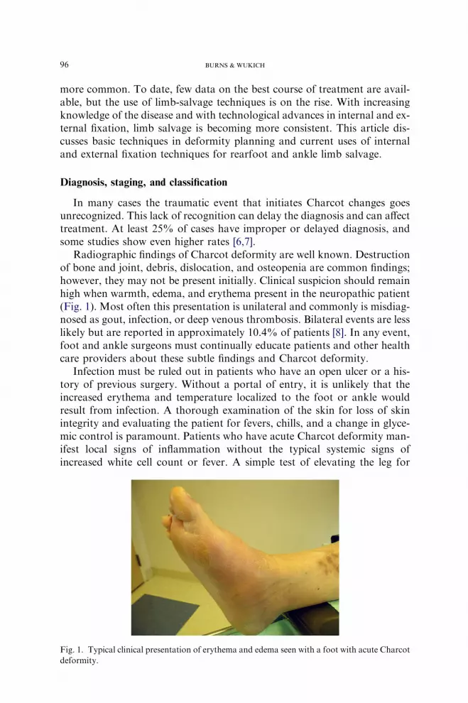
Fig. 7. (A) Complication of failed internal fixation. (B) Broken skinny wire of an external
fixator.

108 BURNS & WUKICH
the needed long-term stability. As in other joints with Charcot deformities,arthrodesis is the treatment of choice, but achieving a solid fusion can bechallenging. In a review of arthrodesis of the Charcot knee deformity,Drennan and colleagues [50] found that important factors for successinclude (1) careful removal of all cartilage and debris, (2) debridement tobleeding subchondral bone, (3) meticulous fashioning of bone surfaces forcontact, (4) complete debridement of all synovial and scarred capsule, and(5) stable internal fixation.
There are many options for rearfoot and ankle fusions. Once the jointsare prepared, unhealthy bone has been debrided, and the deformity hasbeen corrected, the area is stabilized. Internal fixation uses large screws,plates, and intramedullary fixation. Increased stability can be achievedwith locked plates, reconstruction plates, or blade plates.
Correction of osseous deformities requires preoperative and intraopera-tive planning and often requires templates (Fig. 9). In the operating room,the patient must be positioned properly. With rearfoot and ankle proce-dures, a bump under the ipsilateral hip with the patient in a supine positionis most common. This position allows the leg to be in a more neutral posi-tion for deformity correction and also makes the lateral side more accessiblefor surgical approach to the ankle. The leg should be prepped and drapedabove the knee. Fixation for these deformities requires access to the entireleg. The knee is also a landmark for rotation of the lower extremity. Toreduce the chance of malunion during arthrodesis, external rotation shouldalign the second toe and the tibial crest. A thigh tourniquet commonly isused as well. In many cases, the tourniquet is elevated during the dissectionand deformity correction to aid in visualization and is released once tempo-rary fixation is in place.
Correction can be acute, staged, or partial with plans for continued grad-ual correction. There are no level 1 studies to guide these plans, but anunderstanding of biomechanics and knowledge of anatomy are crucial.Many of the resultant deformities are unique, but restoration of normal
Fig. 8. Radiographs of (A) normal and (B) abnormal calcaneal inclination angle caused by
Charcot deformity.

109SURGICAL RECONSTRUCTION OF CHARCOT REARFOOT
anatomic relationships continues to be the cornerstone of management. It ishelpful to draw out basic anatomy and common angles on the radiographsto determine the axis of rotation and apex of deformity. Knowledge of theserelationships will guide the location and size of wedge resections. During theprocedure, Kirschner wires are placed along the lines of the previouslyplanned osteotomies and act as a guide. The saw and osteotomes then canfollow along the wires, removing the appropriate bone.
Charcot ankle deformity, although representing only 5% of the defor-mities, results in unstable varus and valgus malalignment. Acute correctionsof varus can compromise the tarsal tunnel. During acute varus corrections,prophylactic release of the tarsal tunnel should be considered. In general,any large correction of angle or length can cause vessels to kink, leadingto ischemia. When there is large, acute loss of height, as in Charcot ankledeformity and talectomy, a more gradual, staged correction may benecessary.
Incisions are large. For the rearfoot and ankle, a utilitarian lateral inci-sion often is used. It begins approximately 6 cm from the tip of the lateralmalleolus, courses along the lateral border of the fibula, and then makesa gentle curve over the sinus tarsi and calcaneal cuboid joint. This incisionallows access to much of the rearfoot and ankle complex. The fibula, if pres-ent, is removed 5 cm proximal to the ankle joint and can be used as graft ifhealthy. The ankle and subtalar joints are visualized easily, and talectomycan be performed if needed.
Fig. 9. Percutaneous Achilles lengthening.
110 BURNS & WUKICH
A second medial incision may be used. It typically is positioned justanterior to the medial malleolus and courses between the tibialis anteriorand posterior tendons. This incision allows access to the talonavicular jointand medial gutter of the ankle for medialization of the talus for intramedul-lary rod fixation. Both incisions can communicate anteriorly, with dissectioncarried across the distal tibia. A malleable retractor can be used here to pro-tect tissues during corrective osteotomies (Fig. 10).
After more ‘‘normal’’ relationships are established and deformity hasbeen corrected, Steinman pins are used for temporary fixation across therearfoot and ankle. Calcaneal inclination is one important key. Once therearfoot and ankle are reduced, it gives a building block for the remainingmidfoot, and ankle deformities can be addressed. Depending on final fixa-tion, a half-pin is inserted into the calcaneus using fluoroscopy. It entersposteriorly following the normal inclination angle. After the Achilles length-ening, the half-pin can be used to pull the Achilles into a more anatomic po-sition (Fig. 11). At this time a transfixation pin is placed and aids in holdingthe rearfoot. It enters the inferior calcaneus and is driven proximallythrough the talus, if present, and into the anterior cortex of the tibia(Fig. 12). These pins can be incorporated later into an external fixator, ifused. Together these pins provide stability of the rearfoot during furthercorrection. From this point the remaining fixation depends on surgeon pref-erence, clinical circumstances, available healthy bone, and comfort level. In-ternal fixation is common. At the University of Pittsburgh Medical Schoolexternal fixation is used frequently and may be used as an adjunct. External
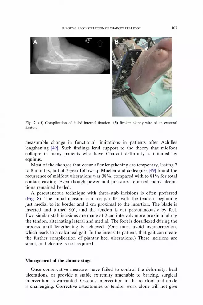
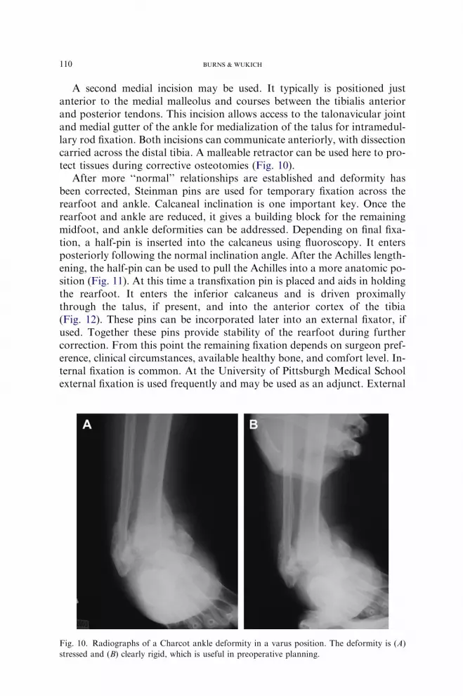
Fig. 10. Radiographs of a Charcot ankle deformity in a varus position. The deformity is (A)
stressed and (B) clearly rigid, which is useful in preoperative planning.

111SURGICAL RECONSTRUCTION OF CHARCOT REARFOOT
fixation also can be used alone, especially in the face of open ulceration orthe acute Charcot process, where fixation is needed away from the unhealthyneuropathic bone.
Whatever the type of fixation, the use of larger, sturdier, and even dou-bled hardware is common. For internal fixation, locking plates are available.With these plates, all components are locked together at a fixed angle, todisperse force better. For failure to occur, the entire construct must fail,not just one screw. Extra wires, half-pins, or full rings are some of the easiestways to increase the strength of external fixation.
External fixation does have drawbacks. In a review of a consecutive seriesfrom the University of Pittsburgh Medical School, 60% to 70% of compli-cations were minor and did not require a change in treatment, but theymake this technique labor intensive and challenging.
Fig. 11. Standard (A) lateral and (B) medial incisions for reconstruction of Charcot rearfoot
and ankle deformity with a malleable retractor for protection of soft tissue.
Fig. 12. Although for a midfoot reconstruction, this photograph shows the calcaneal half-pin
used posteriorly to improve the inclination angle. A transfixation pin then was placed from
inferior calcaneus into the tibia proximally.

112 BURNS & WUKICH
Management of the acute stage
If the deformity is considered too unstable for conservative managementin the acute phase, surgical options exist. Many principles remain the same.The patient in the operating room must be prepared similarly, and Achilleslengthening is performed in the standard fashion. Preoperative planning ofdeformity is essential. As in chronic deformities, the calcaneus is reducedunder fluoroscopy and is pinned as described previously. The remaining cor-rection of midfoot to rearfoot and rearfoot to ankle relationships can thenbe made.
In the acute phase, this correction may be achieved with manipulationand stabilized with Steinman pins. Fluoroscopy guides reduction, and fixa-tion is applied. Other options include external fixation techniques with olivewires, washers, and motors. These devices can help reduce and then stabilizethe dislocations (Fig. 13). Many of these corrections are made percutane-ously to avoid making large incisions in unfavorable skin conditions.
Acute Charcot deformity can be reduced and maintained by external fix-ation, but long-term results are not available. As the patient continuesthrough the Charcot stages, consolidation may occur and be held in placewith the fixator, but the achievement of long-term stability usually requiresdefinitive arthrodesis. Early surgical intervention, however, affords easierreduction in the supple phases so that staged arthrodesis is more anatomicand possibly easier to perform.
Fig. 13. Radiograph of a patient who has a chronic Charcot deformity undergoing reconstruc-
tion. The ankle and rearfoot were corrected and then held in place with a transfixation Steinman
pin from the plantar aspect of the calcaneus into the anterior tibial cortex.

113SURGICAL RECONSTRUCTION OF CHARCOT REARFOOT
Whatever the fixation modality, long periods of non–weight-bearing andfrequent office visits are necessary. A good support system for both thepatient and surgeon are essential, because follow-up is labor intensive.
Case illustrations
Case 1
A 41-year-old woman who had type 2 diabetes and a history of increasingdeformity over the past several months was referred for evaluation (Fig. 14).She had been treated by another physician with serial debridements and off-loading after obvious Charcot neuroarthropathy. Swelling, warmth, anderythema eventually calmed, and the wound finally closed, but during thepostoperative follow-up the deformity could not be braced successfully.She eventually had a recurrence of ulceration that would not heal.
On clinical examination, she had a rigid rearfoot varus of 40� and a non-infected ulcer on the lateral weight-bearing foot. Radiographs showedremodeling stages of Charcot deformity with obvious varus deformity ofthe ankle and minimal talus remaining.
Surgically, the goal was to remove the varus and achieve a plantigradefoot and ankle to off-load the lateral column. A small acute correction of15� was performed with an osteotomy through standard incisions. An exter-nal fixator was applied to correct the remaining deformity gradually to limitneurovascular compromise. The remaining varus was corrected over thenext 3 weeks.
Fig. 14. Lateral radiograph of (A) an acute Charcot deformity with collapse and (B) percuta-
neous treatment with olive wires to improve medial column arch height.
114 BURNS & WUKICH
Once a rectus ankle was achieved, long-term stability was achieved witha tibial-calcaneal fusion. An intramedullary rod was placed upon removal ofthe external fixator. The patient continues to wear custom shoes withoutrecurrence. It has been the author’s (PRB) experience that ankle and tib-ial-calcaneal fusions using an external fixation alone frequently are fibrousunions and revert back to some deformity once the fixator is removed.Therefore placement of an intramedullary rod after correction has becomecommon.
Case 2
A 45-year-old man who had type 2 diabetes was well known to the author(PRB), who had treated the patient for previous Charcot events in the bothfeet (Fig. 15). A new, acute Charcot event with ulceration and drainage ofthe left ankle brought him through the emergency room.
Clinically he had a warm, erythematous ankle with obvious instability.The ulceration laterally with significant drainage was cultured. He wasadmitted, and radiographs and MRI revealed an acute Charcot process ofthe ankle, destruction of the talus, and abscess. This condition was treatedwith incision and drainage, biopsy, antibiotic beads, intravenous antibiotics,and external fixation for stability. After months of treatment with multipledebridements, biopsies, and antibiotics, a tibial-calcaneal fusion with bonegraft was performed with intramedullary rod fixation. The graft was placedto limit acute loss of height after destruction of the talus. To date the patientremains stable in custom shoes and external upright braces.
Case 3
A 49-year-old man who had type 2 diabetes presented with a 6-week his-tory of swelling and redness (Fig. 16). He had been treated for gout by hisprimary care physician after presenting following a ‘‘sprain.’’ He wasreferred because of continued symptoms. Clinically no open wounds werenoted, but significant erythema and edema around ankle was noted. Theankle had obvious instability and deformity on examination. Radiographsrevealed a subluxed ankle and dislocated midfoot with Charcot changes.(Fig. 17)
He was admitted, and an external fixator was placed. The fixator allowedstability during the acute Charcot process but also permitted reduction ofthe dislocation and subluxation with minimal incisions. Once distracted,olive wires and motors were used to reduce the deformities.
During the next few months the ankle became infected, and the fixatorwas adjusted as needed with debridement of the ankle joint. Biopsies weretaken, and appropriate antibiotics were given. The fixator was continuallycompressed over the ankle during this process along with the appropriateantibiotics. It was clear the talus would be lost, and the goal was a tibial-calcaneal fusion. As the patient progressed to later stages of Charcot

115SURGICAL RECONSTRUCTION OF CHARCOT REARFOOT
Fig. 15. (A) Preoperative radiograph of a chronic deformity. (B) Immediate postoperative
radiograph and (C) clinical pictures. (D) Radiograph and (E) clinical pictures 3 weeks after
gradual correction. (F, G) Final radiographs with intramedullary rod for fixation.

116 BURNS & WUKICH
Fig. 16. (A, B) Preoperative radiographs of acute Charcot deformity. (C) MRI of abscess and
Charcot deformity surrounding the talus. (D) External fixator during serial debridement and
talectomy. (E, F) Postoperative radiographs after graft and intramedullary rod placement.

117SURGICAL RECONSTRUCTION OF CHARCOT REARFOOT
deformity, consolidation was noted, and the patient remained stable. Thefixator was periodically compressed during the process and produced a sta-ble fusion that is braced without further surgery required.
Summary
Charcot arthropathy of the rearfoot and ankle is a complex disorder. Todate there are no evidence-based, universally agreed upon treatment proto-cols. Many propose earlier intervention of these acute deformities, but theirtreatment is challenging. As the number of patients who have these
Fig. 17. (A, B) Preoperative radiographs of acute dislocation. (C) Radiograph and (D) clinical
pictures of postoperative reduction with external fixator. Subsequent radiographs (E) after de-
bridement with continued compression and stability with (F) stable outcome.
118 BURNS & WUKICH
deformities continues to increase, surgeons’ skill levels and experience growas well. With increased technical skill, knowledge, and advances in fixation,these deformities are becoming more manageable. In the future this experi-ence should afford the general community with evidenced-based protocols.These deformities are demanding and require a good support system forboth the surgeon and patient, but a stable, braceable limb is obtainable.
Further readings
Sanders LJ, Frykberg RG. The high risk foot in diabetes mellitus. New York: Churchill-
Livingstone; 1991.
Pinzur MS, Sage R, Stuck R, et al. A treatment algorithm for neuropathic (Charcot) midfoot
deformity. Foot Ankle 1993;14(4):189–97.
References
[1] Charcot JM. [Sur quelques arthropathies. Qui paraissent dependre d’une lesion. Du cerveau
ou de la moelle epiniere]. Arch Physiol Norm Pathol 1868;1:161–78 [in French].
[2] JordanWR. Neuritic manifestations in diabetes mellitus. Arch Intern Med 1936;57:307–66.
[3] Armstrong DG, Lavery LA. Elevated peak plantar pressures in patients who have Charcot
arthropathy. J Bone Joint Surg Am 1998;80(3):365–9.
[4] Lavery LA, Armstrong DG, Wunderlich RP, et al. Diabetic foot syndrome: evaluating the
prevalence and incidence of foot pathology in Mexican American and non-Hispanic whites
from a diabetes disease management cohort. Diabetes Care 2003;26(5):1435–8.
[5] JeffcoateWJ, Game F, Cavanagh PR. The role of proinflammatory cytokines in the cause of
neuropathic osteoarthropathy (acute Charcot foot) in diabetes. Lancet 2005;366(9502):
2058–61.
[6] Schon LC, Marks RM. The management of neuroarthropathic fracture-dislocations in the
diabetic patient. Orthop Clin North Am 1995;26:375–92.
[7] ChantelauE. The perils of procrastination: effect of early vs. delayed detection and treatment
of incipient Charcot fracture. Diabet Med 2005;22:1707–12.
[8] SaltzmanCL, HagyML, Zimmerman B, et al. How effective is intensive nonoperative initial
treatment of patients with diabetes and Charcot arthropathy of the feet? 2005;(435):
185–90.
[9] Brodsky JW. The diabetic foot. In:MannRA,CoughlinMJ, editors. Surgery of the foot and
ankle. St. Louis (MO): Mosby-Year Book; 1993. p. 877–958.
[10] Myerson MS, Edwards WH. Management of neuropathic fractures in the foot and ankle.
J Am Acad Orthop Surg 1999;7:8–18.
[11] Myerson MS. Diabetic neuroarthropathy. In: Myerson MS, editor. Foot and ankle disor-
ders. Philadelphia: W.B. Saunders; 2000. p. 439–65.
[12] MyersonMS, Papa J, Eaton K, et al. The total-contact cast for management of neuropathic
plantar ulceration of the foot. J Bone Joint Surg Am 1992;74:261–9.
[13] Papa J, Myerson MS, Girard P. Salvage, with arthrodesis, in intractable diabetic neuro-
pathic arthropathy of the foot and ankle. J Bone Joint Surg Am 1993;75-A(7):1056–66.
[14] Levine SE, Myerson MS. Management of ulceration and infection in the diabetic foot. In:
Myerson MS, editor. Foot and ankle disorders. Philadelphia: W.B. Saunders; 2000.
p. 411–38.
[15] Eichenholtz SN. Charcot joints. Springfield (IL): Charles C. Thomas; 1966.
[16] Harris JR, Brand PW. Patterns of disintegration of the tarsus in the anaesthetic foot. J Bone
Joint Surg Br 1966;48(1):4–16.

119SURGICAL RECONSTRUCTION OF CHARCOT REARFOOT
[17] Newman JH. Non-infective diseases of the diabetic foot. J Bone Joint Surg Br 1981;63-B(4):
593–6.
[18] Shibata T, Tada K, Hashizume C. The result of arthrodesis of the ankle for leprotic neuro-
arthropathy. J Bone Joint Surg Am 1990;72(5):749–56.
[19] Prisk VR, Wukich DK. Ankle fractures in diabetics. Foot Ankle Clin 2006;11(4):
849–63.
[20] Brodsky JW, Rouse AM. Exostectomy for symptomatic bony prominences in diabetic
Charcot feet. Clin Orthop Relat Res 1993;296:21–6.
[21] Sanders LJ, Frykberg RG. Diabetic neuropathic osteoarthropathy. The Charcot foot. In:
Frykberg RG, editor. The high risk foot in diabetes mellitus. New York: Churchill Living-
ston; 1991. p. 297.
[22] Schon LC, Weinfeld SB, Horton GA, et al. Radiographic and clinical classification of
acquired midtarsus deformities. Foot Ankle Int 1998;19(6):394–404.
[23] Wang JC. Use of external fixation in the reconstruction of the Charcot foot and ankle. Clin
Podiatr Med Surg 2003;20(1):97–117.
[24] Pinzur MS, Stuck R, Sage R, et al. Benchmark analysis on diabetics at high risk for lower
extremity amputation. Foot Ankle Int 1996;17(11):695–700.
[25] Simon SR, Tejwani SG,WilsonDL, et al. Arthrodesis as an early alternative to nonoperative
management of Charcot arthropathy of the diabetic foot. J Bone Joint Surg Am 2000;
82A(7):939–50.
[26] Pinzur MS. Surgical versus accommodative treatment for Charcot arthropathy of the
midfoot. Foot Ankle Int 2004;25(8):545–9.
[27] Pinzur MS, Lio T, Posner M. Treatment of Eichenhotlz stage 1 Charcot foot arthropathy
with a weightbearing total contact cast. Foot Ankle Int 2006;27(5):324–9.
[28] Pinzur MS. Benchmark analysis of diabetic patients with neuropathic (Charcot) foot defor-
mity. Foot Ankle Int 1999;20(9):564–7.
[29] Zarutsky E, Rush SM, Schuberth JM. The use of circular wire external fixation in the treat-
ment of salvage ankle arthrodesis. J Foot Ankle Surg 2005;44(1):22–31.
[30] Caravaggi C, Cimmino M, Caruso S, et al. Intramedullary compressive nail fixation for the
treatment of severe Charcot deformity of the ankle and rearfoot. J Foot Ankle Surg 2006;
45(1):20–4.
[31] Pelton K, Hofer JK, Thordarson DB. Tibiotalocaneal arthrodesis using a dynamically
locked retrograde intramedullary nail. Foot Ankle Int 2006;27(10):759–63.
[32] Fabrin J, Larsen K, Holstein PE. Arthrodesis with external fixation in the unstable or mis-
aligned Charcot ankle in patients with diabetes mellitus. Int J Low Extrem Wounds 2007;
6(2):102–7.
[33] Stuart MJ, Morrey BF. Arthrodesis of the diabetic neuropathic ankle joint. Clin Orthop
Relat Res 1990;253:209–11.
[34] BrinkDS, EickmeierKM, LevitskyDR, et al. Subtalar and talonavicular joint dislocation as
presentation of diabetic neuropathic arthropathy with salvage by triple arthrodesis. J Foot
Ankle Surg 1994;33(6):583–9.
[35] Moore TJ, PrinceR, PochatkoD, et al. Retrograde intramedullary nailing for ankle arthrod-
esis. Foot Ankle Int 1995;16(7):433–6.
[36] Myerson MS, Alvarez RG, Lam PW. Tibiocalcaneal arthrodesis for the management of
severe ankle and hindfoot deformities. Foot Ankle Int 2000;21(8):643–50.
[37] Alvarez RG, Barbour TM, Perkins TD. Tibiocalcaneal arthrodesis for nonbraceable neuro-
pathic ankle deformity. Foot Ankle Int 1994;15(7):354–9.
[38] Lesko P, Maurer RC. Talonavicular dislocations and midfoot arthropathy in neuropathic
diabetic feet. Natural course and principles of treatment. Clin Orthop Relat Res
1989;(240):226–31.
[39] Fukuda E, Yasuda I. On the piezoelectric effect of bone. J Physiol Soc Japan 1957;12:
1158–64.

120 BURNS & WUKICH
[40] Bassett CA, Becker RO. Generation of electric potentials by bone in response to mechanical
stress. Science 1962;137:1063–4.
[41] Hanft JR, Goggin JP, Landsman A, et al. The role of combined magnetic field bone growth
stimulation as an adjunct in the treatment of neuropathy/Charcot joint: an expanded pilot
study. J Foot Ankle Surg 1998;37(6):510–5.
[42] Bier RR, EstersohnHS. A new treatment for Charcot joint in the diabetic foot. J AmPodiatr
Med Assoc 1987;77(2):63–9.
[43] Grady JF, O’Connor KJ, Axe TM, et al. Use of electrostimulation in the treatment of
diabetic neuroarthropathy. J Am Podiatr Med Assoc 2000;90(6):287–94.
[44] Selby PL, Young MJ, Boulton AJ. Bisphosphonates: a new treatment for diabetic Charcot
neuroarthropathy? Diabetic Med 1994;11(1):28–31.
[45] JudeEB, Selby PL, Burgess J, et al. Bisphosphonates in the treatment ofCharcot neuroarthr-
opathy: a double-blind randomized controlled trial. Diabetologia 2001;44(11):2032–7.
[46] Pitocco D, Ruotolo V, Caputo S, et al. Six-month treatment with alendronate in acute
Charcot neuroarthropathy. Diabetes Care 2005;28(5):1214–5.
[47] Roukis TS, Zgonis T. The management of acute Charcot fracture-dislocations with the
Taylor’s spatial external fixation system. Clin Podiatr Med Surg 2006;23(2):467–83.
[48] Maluf KS, Mueller MJ, Strube MJ, et al. Tendon Achilles lengthening for the treatment of
neuropathic ulcers causes a temporary reduction in forefoot pressure associated with
changes in plantar flexor power rather than ankle motion during gait. J Biomech 2004;
37(6):897–906.
[49] Mueller MJ, Sinacore DR, Hastings MK, et al. Impact of Achilles tendon lengthening on
functional limitations and perceived disability in people with a neuropathic plantar ulcer.
Diabetes Care 2004;27(7):1559–64.
[50] Drennan DB, Fahey JJ, Maylahn DJ. Important factors in achieving arthrodesis of the
Charcot knee. J Bone Joint Surg Am 1971;53-A(6):1180–93.


