Embed Size (px)
Citation preview

Anastasius MoumtzoglouP. & A. Kyriakou Children’s Hospital, Greece
Anastasia KastaniaAthens University of Economics and Business, Greece
E-Health Technologies and Improving Patient Safety: Exploring Organizational Factors

E-health technologies and improving patient safety : exploring organizational factors / Anastasius Moumtzoglou and Anastasia Kastania, editors. pages cm Summary: “This book presents an overview of information and communication technologies and their impact on the field of both patient safety and e-health and highlights a better understanding of their interaction”-- Provided by publisher. Includes bibliographical references and index. ISBN 978-1-4666-2657-7 (hardcover) -- ISBN 978-1-4666-2688-1 (ebook) (print) -- ISBN 978-1-4666-2719-2 (print & perpetual access) (print) 1. Medical informatics. 2. Patients--Safety measures. I. Moumtzoglou, Anastasius, 1959- editor of compilation. II. Kastania, Anastasia, 1965- editor of compilation. R858.E34 2013 610.285--dc23 2012029409
British Cataloguing in Publication DataA Cataloguing in Publication record for this book is available from the British Library.
All work contributed to this book is new, previously-unpublished material. The views expressed in this book are those of the authors, but not necessarily of the publisher.
Managing Director: Lindsay JohnstonEditorial Director: Joel GamonBook Production Manager: Jennifer YoderPublishing Systems Analyst: Adrienne FreelandDevelopment Editor: Myla MerkelAssistant Acquisitions Editor: Kayla WolfeTypesetter: Deanna Jo ZombroCover Design: Nick Newcomer
Published in the United States of America by Medical Information Science Reference (an imprint of IGI Global)701 E. Chocolate AvenueHershey PA 17033Tel: 717-533-8845Fax: 717-533-8661 E-mail: [email protected] site: http://www.igi-global.com
Copyright © 2013 by IGI Global. All rights reserved. No part of this publication may be reproduced, stored or distributed in any form or by any means, electronic or mechanical, including photocopying, without written permission from the publisher.Product or company names used in this set are for identification purposes only. Inclusion of the names of the products or companies does not indicate a claim of ownership by IGI Global of the trademark or registered trademark.
Library of Congress Cataloging-in-Publication Data

227
Copyright © 2013, IGI Global. Copying or distributing in print or electronic forms without written permission of IGI Global is prohibited.
Chapter 15
DOI: 10.4018/978-1-4666-2657-7.ch015
George I. LambrouUniversity of Athens, Greece
Apostolos ZaravinosUniversity of Athens, Greece
Maria AdamakiUniversity of Athens, Greece
Spiros VlahopoulosUniversity of Athens, Greece
Systems Modeling of Proliferation Mechanisms
in Childhood Acute Lymphoblastic Leukemia
ABSTRACT
Acute Lymphoblastic Leukemia (ALL) is the most common neoplasm in children, but the mechanisms underlying leukemogenesis are poorly understood, despite the existence of several theories regarding the mechanics of leukemic cell proliferation. However, with the advent of new biological principles, it appears that a systems approach could be used in an effective search of global patterns in biological systems, so as to be able to model the phenomenon of proliferation and gain a better understanding of how cells may progress from a healthy to a diseased state. This chapter reviews the current knowledge on proliferation dynamics, along with a discussion of the several existing theories on leukemogenesis and their comparison with the theories governing general oncogenesis. Furthermore, the authors pres-ent some “in-house” experimental data that support the view that it is possible to model leukemic cell proliferation and explain how this has been performed in in vitro experiments.
228
Systems Modeling of Proliferation Mechanisms in Childhood Acute Lymphoblastic Leukemia
INTRODUCTION
Acute Lymphoblastic Leukemia-Disease Description and Preliminaries
Acute leukemia mainly appears during childhood but it can also occur in adolescence manifesting a poor prognosis regardless of age. Progress in childhood leukemia has been immense the last decades with an overall survival rate exceeding 75% (Carroll, et al., 2003). Still though there is an approximate 20% that relapses, which in many cases can prove fatal. Acute lymphoblastic leu-kemia, as most cases of childhood neoplasms, is characterized by chromosomal aberrations (Gagos & Irminger-Finger, 2005). In the majority of cases leukemia appears to have a greater incidence of chromosomal abnormalities compared to solid tumors (Saha, Young, & Freemont, 1998). Also, gene expression is aberrantly regulated and in certain cases fusion genes form, that are similarly aberrantly expressed. It has been reported that those genes, involved in leukemia progression, are very potent regulators of cell proliferation, differentiation, cell cycle progression and anti-apoptosis (Saha, et al., 1998). In addition, it has been observed that certain types of leukemia are resistant to chemotherapeutics while others are more sensitive. In other words, depending on the type of leukemia, different chemotherapy inten-sities might be required. Therefore, it is impera-tive to mention that classifying patients into risk categories may improve the therapeutic outcome. Hence, recurrence of the disease requires usually more intense chemotherapeutic protocols and in several cases bone marrow transplantation. The prognostic factors have also been well charac-terized in childhood leukemia; including white blood cell count at presentation (diagnosis), age and gender, immunophenotype, as well as the presence of CNS blast presentation or certain chromosomal aberrations (Carroll, et al., 2003). Treatment of childhood leukemia is successful
for the majority of patients, mainly due to the use of classical chemotherapeutics. Recently, individualized treatments have also been applied, as in the case of BCR/ABL positive (also known as Philadelphia positive (Ph+)) leukemia, using imatinib mesylate, a new agent specific for the particular gene fusion.
Yet, the question that might rise is why leu-kemia and especially childhood leukemia? Acute lymphoblastic leukemia (ALL) is the most frequent occurring malignancy among childhood cancers (Severson & Ross, 1999). It originates from the undifferentiated lymphoblast, which abnormally ceases to develop into the mature lymphoid cell giving rise to a tumour. Hence, one of the most interesting characteristics of leukemia is its trait of clonal expansion. That is, the almost uniform phenotype of cells giving rise to the tumour. But, what does this has to do with leukemogenesis? Necessarily, leukemia as a disease has a starting point and a diagnosis point. Between those two there is an immense lack of knowledge. This does not apply only to leukemia but to any neoplasm in general or even inflammation. For example, as mentioned, absolute lymphocyte count is a prog-nostic factor in childhood leukemia (De Angulo, Yuen, Palla, Anderson, & Zweidler-McKay, 2008). In that sense, cell counts thus proliferation, is tightly connected to disease prognosis. The next question that would arise is: what is the connection between the first steps of disease emergence and the presentation stage. That is the most difficult part to answer since we simply do not, and cannot, have the slightest clue about what happens between that time and the present. A necessary approach to this phenomenon would be the modeling ap-proach. That is the understanding and prediction of the phenomenon on a physical and systems basis. In order for such an approach to succeed it must entail a range of “crafts” ranging from mechanics, systems theory and thermodynamics to mathematical analysis and chaos dynamics.

229
Systems Modeling of Proliferation Mechanisms in Childhood Acute Lymphoblastic Leukemia
The Merging of Biological Sciences with the Exact Sciences
As we have mentioned numerous times before, until recently, one of the main approaches in scientific research was the study of molecules in a sequential order. That is, we were able to dis-cover the function and roles of genes or protein in pairs or maximal three at the same time. The last decade, this has changed. With the advent of high-throughput methodologies, the study of thousands of factors (and even millions meanwhile) has been plausible. Microarrays and deep-sequencing are two examples of such methodologies. Yet, we must admit that this revolution was facilitated with the development of the personal computer. From the time point that computational power became publicly available has revolutionized the way science has preceded with its steps. On the other hand, biology was a science that was based, and to a great extent still is, on observation. This of course was true for all natural sciences, yet other disciplines such as the physical or chemical sciences were more mature since they were able to describe their phenomena through a common mathematical language. For example, it is of great certainty that a falling weight will go down towards the gravitational force of the earth and as a matter of fact we are in position, knowing the initial height of the falling body, to calculate the exact velocity just before it crashes to the ground. Also, given the time interval we are able to calculate, again with great certainty, the exact position of the falling body from its initial point. The amazing thing is that if we took consecutive measurements on the falling weight we would find very similar results and we would obtain for our results a very high confidence interval. On the other hand, if we take cultured cells (cell lines) and need to know their number at a given time point, it is impossible to do so, if we know, with great certainty, the initial cell number. As in the case of the falling body, if we could perform several experiments with the same cells and the
same conditions, most probably we would come up with, sometimes, very different results. The conclusion from these two examples is that physi-cal phenomena can be described mathematically, which does not mean that these phenomena are simple but their simplified forms can be calculated with high certainty. This is not true for biological phenomena. Even the simplest observations, such as how a cell population increases in numbers, are not trivial and it is almost impossible to make predictions about a cell population. In the advent of the 21st century a new trend has emerged in biological sciences. This was a systems approach to biological phenomena. The idea was not new, since it has been described previously that there is a need for the life sciences to be integrated with the physical and mathematical sciences. The first merge occurred between biology and chemistry which gave rise to biochemistry, since all life molecules are chemicals and their interactions are chemical reactions. The difficult part is the merge of the biological sciences with the physical and mathematical sciences. This has taken place from the emergence of two new disciplines namely systems biology and physical biology. Both very young and in their infancy, yet very powerful as we think it will prove to be. Systems biology has borrowed its name from the engineers and their systems theory concepts. Physical biology, on the other hand, could be mentioned that has taken its name from the merge of physics and biology and in a way it is another discipline similar to physical chemistry. This short review on biology and its branches was considered significant, since it would give us an idea why we need such ideas such as mathematical modelling of biological phenom-ena. In concordance, this brief introduction gave us an idea about the necessity of modelling cell proliferation. It can be considered a very simple phenomenon, yet it proved to be complicated and tedious to understand.
Modelling biological systems is not new. At-tempts have emerged from the advent of the 20th century. However, systems biology and physical

230
Systems Modeling of Proliferation Mechanisms in Childhood Acute Lymphoblastic Leukemia
biology have come to give a more concise meaning to the concept of modelling. Both, that is systems and physical biology, can be considered a relatively young disciplines. It was in the advent of the 21st century that the first coordinated attempts became reality. However, the term had been previously mentioned both as a necessity and as a discipline. We couldn’t phrase it in better words than Mihajlo Mesarović in 1968:
…in spite of the considerable interest and efforts, the application of systems theory in biology has not quite lived up to expectations…one of the main reasons for the existing lag is that systems theory has not been directly concerned with some of the problems of vital importance in biology…
…The real advance in the application of systems theory to biology will come about only when the biologists start asking questions which are based on the system-theoretic concepts rather than using these concepts to represent in still another way the phenomena which are already explained in terms of biophysical or biochemical principles. …then we will not have the application of engineering principles to biological problems but rather a field of systems biology with its own identity and in its own right… (Mesarovic, 1968) (adapted from the book Systems Biology-Dynamic Pathway Model-ing by Olaf Wolkenhauer (Wolkenhauer, 2010)).
But in order to further define the field of sys-tems biology we should refer to an older reference from Henri Poincare, who proposed a solution to the three-body problem in the 19th century:
…life is a relationship among molecules and not a property of any molecule…
…Science is built up of facts, as a house is with stones. But a collection of facts is no more a sci-ence than a heap of stones is a house… (Wolken-hauer, 2010).
In general, natural sciences have matured with time especially with the integration of mathemat-ics. For example, the disciplines of physics and chemistry have benefited from the mathematical discipline and matured enough to create a common language for describing phenomena. Similarly, biological sciences could greatly benefit by in-tegrating knowledge from the mathematical and engineering disciplines. Today a huge amount of work is performed over the collection of data into large databases such as the Gene Ontology database, the KEGG database and others (Chang, Nagarajan, Magee, Milbrandt, & Stormo, 2006; Chatziioannou, Moulos, & Kolisis, 2009; Kane-hisa, 2002; Kanehisa & Goto, 2000; Ogata, et al., 1999; Rubinstein & Simon, 2005; Salwinski, et al., 2004; Wingender, Dietze, Karas, & Knuppel, 1996; Wolkenhauer, 2010; Zhang, Schmoyer, Kirov, & Snoddy, 2004).
BACKGROUND
In the case of cancer, tumorigenesis is the in-terconnection of thousands of factors acting simultaneously that brings about the malignant phenotype. Before we proceed to the discussion of cell proliferation in leukemia and the efforts attempted by several researchers we should try to mention and briefly understand, the main theories of tumorigenesis, which includes leukemogenesis and carcinogenesis in general.
Carcinogenesis in General
The Genetic Basis of Cancer as a Tumorigenic Theory
A very popular theory of carcinogenesis is that the cause of the disease is the accumulation of genetic aberrations throughout an individual’s life. If an adequate number of such genetic aberrations ac-cumulates then there is the occurrence of cancer (A. Knudson, 2001); actually, this theory was initially

231
Systems Modeling of Proliferation Mechanisms in Childhood Acute Lymphoblastic Leukemia
proposed by Nordling in 1953 (Nordling, 1953), and further on it was formulated by Knudson, which was called the two-hit hypothesis. With his hypothesis he considered that fact that several mutations are required for the immergence of can-cer, also in children, and he further proposed that there is a possibility that tumorigenesis can occur in two consecutive events (hits) (A. G. Knudson, Jr., 1971). Initially, evidence was given for reti-noblastoma, where he proposed that at least for that type of tumour (retinoblastoma) it can occur with two mutational events (A. G. Knudson, Jr., 1971). Yet other equally significant theories have been suggested, such as the multistage oncogenesis theory (Moolgavkar, 1978; Ritter, Wilson, Pompei, & Burmistrov, 2003).
Another proposed carcinogenetic model was that of Vogelstein et al. concerning colorectal cancer (Vogelstein, et al., 1988). It appeared that at least five mutations were adequate for the tumor to occur. This model was based upon the observation that the RAS gene mutations increased proportionally to the size of the malignant pheno-type. However, it was not possible to present the multi-factorial character of the disease.
Chemical Carcinogenesis
Further on, several other models have been de-veloped for carcinogenesis including those of the mouse skin model (Zoumpourlis, Solakidi, Papathoma, & Papaevangeliou, 2003). In this par-ticular study, normal epithelial mouse cells were administered a single dose of the polycyclic aro-matic hydrocarbon 7,12-dimethylbenz[a]anthra-cene (DMBA), followed by weekly applications of the phorbol ester 12-O-tetradecanoylphorbol-13-acetate (TPA). This leads to the development of numerous benign papilloma, some of which progressed to malignant squamous cell carcinomas 20-40 weeks after the first exposure to carcinogens.
Evolutionary Theories of Carcinogenesis
A very interesting theory poses that tumours are di-rectly connected to the industrialization of modern life. Thus, it has been suggested that cancer is not actually a disease but an evolutionary adaptation of the cells in order to survive certain environmental changes/threats. In addition, it has been proposed that tumour cells are an evolution of normal cells in order to overcome environmental challenge and stress (Iwasa & Michor). Furthermore, it has been reported that this evolutionary change might control the heterogeneity of tumour tissues. Since, genetic as well epigenetic mechanisms control the evolution of cancer then it is possible that the network of events that leads to cancer will remain unsolved due to the complexity of the phenomenon. Modeling approaches have been proposed with respect to the explanation of the evolutionary character of tumor emergence and progression (Iwasa & Michor).
The Oldest Carcinogenetic Theory
However, to be more precise one of the first carcinogenetic theories was proposed from the beginning of the first organized observations on cancer. In particular, a common characteristic of almost all types of tumours is the known Warburg effect described by Otto Warburg in 1924. This simple observation remains until today a funda-mental trait of tumour biology i.e. that tumours perform a shift from oxidative phosphorylation to anaerobic glycolysis for their energy needs (Warburg, Posener, & Negelein, 1924). At the time, Warburg considered this as the leading aeti-ology of cancer and of note that it was considered as such until the observation or finding that it is one of the side-effects of cancer. It is now known that the avoidance of the oxidative phosphoryla-tion pathway, it is the one that leads to excessive lactate production causing this deteriorating effect cancerous cell have on the surrounding tissue. Yet,

232
Systems Modeling of Proliferation Mechanisms in Childhood Acute Lymphoblastic Leukemia
this property of cancer cells has been exploited and several drugs are in clinical trials function-ing as inhibitors of the anaerobic fermentation of glucose. The observation at that time was a break-through in the understanding of the disease. Yet, it was misleading as it was not the cause that it was observed but the result.
However, it is of great interest to mention that despite all the efforts towards the understanding of the tumour phenomenon, its origins and ae-tiology are largely unknown and are still being investigated.
ISSUES
Theories of carcinogenesis were developed mainly with the adult organism in mind. Considering the fact that genetics, chemicals and evolution came into play they all require a very important factor: that is time. Time is required in order for all neces-sary changes to take place and cause cancer. Hence, a question that rises is whether these theories could be applied to childhood neoplasms, since these models cannot be applied directly, as the elapsed time of exposure to the carcinogenetic factor is not adequate to explain the appearance of cancer. As we will mention further on, there are sev-eral hypotheses regarding childhood neoplasms. Similarly, leukemogenetic models although they have been mentioned, they have not been studied and this subject remains obscure. The biological mechanisms that lead to leukemia are until today largely unknown. We can, however, divide leu-kemogenesis into two major categories: de novo leukemia and environmentally-related leukemia.
Environmentally-Related Leukemia: Chemical, Radiation, and Infectious Factors Proposed as Affecting Leukemogenesis
Mostly theories on the ontology of leukemia were based upon the characteristics that leukemic cells
have as far as their cytogenetic characteristics are concerned. In the early studies on the aetiology of leukemia several factors have been investigated. In several reports, it has been investigated whether environmental factors were implied in the emer-gence of leukemia. For example, investigations included the study of electromagnetic fields, as in the case of residency in nearby high voltage power plants (Hatch, et al., 1998; Kleinerman, et al., 1997; Linet, et al., 1997; Michaelis, et al., 1998; Petridou, et al., 1997). In other cases, research was focused on whether exposure to environmental pollutants could imply a high risk for the emergence of leukemia, as in the case of exposure to benzene and its derivatives (Snyder, 2004). In addition, several other chemicals have been proposed as factors triggering leukemia (Irons & Stillman, 1996b; Pyatt, Aylward, & Hays, 2007). Other factors, proposed as potentially leu-kemogenetic, include infections (M. F. Greaves, 1988; Hehlmann, et al., 1983; Kinlen, 1988; Risser, Horowitz, & McCubrey, 1983), autoimmunity and familial history of cancer (Hayes, et al., 1997; Sandler & Ross, 1997) among others. Hitherto, there are still under debate the reasons underlying leukemogenesis. The aforementioned aetiologies, we already presented in the previous section, would fit in the first theories of carcinogenesis. However, a question that still rises is regarding the adequate time for mutations to occur, since the evolution of a cell to cancerous cell probably takes time and more than one step are required for a neoplasm to occur.
Induction of leukemia in vitro has been also attempted as in several protocols uses hydroqui-none, a benzene derivative, for the development of acute myeloid leukemia (AML) from normal hematopoietic progenitor cells of human bone mar-row (Irons, Colagiovanni, & Stillman, 1996; Irons & Stillman, 1996a, 1996b). Benzene metabolism takes place in the liver, where the main metabolites are phenol and catechol. Liver metabolism con-verts phenol into hydroquinone, which is in turn converted to 1,4-benzoquinone and semi-Quinone,

233
Systems Modeling of Proliferation Mechanisms in Childhood Acute Lymphoblastic Leukemia
substances that are highly toxic and whose action is believed to resemble that of alkylating agents and topoisomerase inhibitors. Another protocol uses the organophosphate pesticide isofenphos (IFP) to cause myeloid metaplasia in human lymphocytes and progressive development to AML (Williams, Boros, Kolanko, Jackman, & Eggers, 2004). It was demonstrated that exposure of human lymphocytes to IFP induces DNA mutation beyond endogenous repair capacity and disrupts cholinergic nuclear signalling affectively constructing the mutator phenotype of leukemogenesis.
Environmentally-Related Leukemia: Evolutionary Theories of Leukemogenesis
In the previous section, we have mentioned that one of the explanations attempted to be given to tumori-genesis, is through the evolutionary processes that cells probably possess. As it has been reviewed by Greaves (2010) there are three main attributions to cancer propagation: a) a fixed and hierarchically positioned subset of stem cells resembling normal hematopoietic stem cells, b) a non-deterministic or stochastic process with plasticity of “stem-ness”. At this point we would like to add another possibility, that is, the existence of a deterministic yet chaotic mechanism of propagation. By saying chaotic, we do not mean the disarrangement of cell propagation but we refer to the mathematical meaning of chaos as a non-linearity. It is possible that if such phenomena occur due to evolutionary driving forces then this could be controlled by chaos i.e. non-linear dynamics and c) activity of a genetically dominant sub-clone (M. Greaves). The model of evolutionary cancer propagation is based on the idea that transformed cells pass through a series of “bottlenecks”. These are selec-tive processes that “decide” in a way, which cells will progress to a next phase of proliferation until a final population is reached that will cause the tumour to emerge. In general, cancer cells could be considered as elements participating in a Dar-
winian process. The criteria of selection could be the presence or even acquisition of mutations, or even epigenetic changes. Once more, regarding childhood leukemia, and childhood neoplasms in general, the question remains on the adequate time for such phenomena to evolve. Either evo-lutionary processes are so rapid that the disease emerges in the early ages; that is the first years of life, or they pre-exist in utero as it has been previously reported for the MLL/ALL cases (M. Greaves, 2005).
Environmentally-Related Leukemia: Therapy-Related Leukemia
The current therapeutic approaches to haema-tological primary tumours (multiple myeloma, Hodgkin’s disease, non-Hodgkin’s lymphoma, polycythemia vera, essential thrombocythemia) and primary solid tumours with chemotherapeutic agents (mechlorethamine, cyclophosphamide, carmustine, lomustine, dacarbazine, cisplatin, carboplatin, ifosfamide, nitrosourea) can lead to the development of acute leukemia (therapy-related acute leukemia) (Dedrick & Morrison, 1992; Leone, Mele, Pulsoni, Equitani, & Pagano, 1999; Park & Koeffler, 1996). Alkylating drugs are essentially genotoxic agents that are administered as part of therapeutic regimens; these alkylating substances, regardless of metabolic activation, produce an active metabolite that attaches to certain parts of the DNA causing covalent dam-ages. As a first line defence, the cell recognizes these damages and activates signal transduction mechanisms in an attempt to correct them; if it fails to do so, it causes inhibition of the cell cycle via activation of cell cycle check points. This hap-pens as a means of delaying cellular amplification and gaining extra time to repair the DNA damages in order to avoid the production of “abnormal” daughter cells. If this also fails, then the cell resorts to inducing cell death mechanisms, which is also the desirable effect of chemotherapy. However, all cells have a threshold point defence, which means

234
Systems Modeling of Proliferation Mechanisms in Childhood Acute Lymphoblastic Leukemia
that under certain conditions the cell will divide and multiply regardless of the amount of DNA damage it carries, thus giving rise to abnormal progeny. At this point mutagenesis and, subse-quently, carcinogenesis are induced. It is, there-fore, of fundamental importance to investigate, in the leukemogenesis model caused secondarily by chemotherapeutic drugs, the mechanism of overall cell response to DNA damage, a mechanism that essentially determines the cell’s chemosensitivity to alkylating/genotoxic agents, and the probability of adverse effects.
de novo: Cytogenetic Aberrations as Leukemogenetic Factors
Chromosomal abnormalities detected at diag-nosis are associated with a high percentage of childhood leukemia. The early detection of such aberrations is of significant importance, not only for the classification of the leukemic subtype, but also for the prognosis and therapeutic outcome of patients. On the other hand, leukemia involving the BCR-ABL fusion gene, present in about 4% of children with ALL, are characterised by a high incidence of relapses, whereas leukemia involving the TEL-AML1 fusion are associated with a good prognosis (M. Greaves, 1999a, 1999b). Studies on monozygotic twins with concordant leukemia (Ford, et al., 1998; Ford, et al., 1997; M. Greaves, 1999a, 1999b) and retrospective scrutiny of neona-tal blood spots (Guthrie cards) of leukemia patients (Gale, et al., 1997; Rowley, 1998; Wiemels, Kang, & Greaves, 2009) have demonstrated that for a significant number of childhood patients leukemia starts at the embryonic stage. Further scrutiny of Guthrie spots showed that over 70% of children with ALL up to 13 years of age had clonotypic markers of the disease at birth (Fasching, et al., 2000; Gale, et al., 1997; Wiemels, et al., 2009; Yagi, et al., 2000); the molecular markers present at birth were exactly the same as the ones detected in the leukemic cells a few years later. In another study it was shown that the particular fusion (TEL-AML1)
is also detected in healthy children at birth with a frequency of 1% (Mori, et al., 2002). This 1% represents 100 times the risk of ALL with TEL-AML1 fusion gene, indicating that the frequency of conversion of the pre-leukemic clone to overt disease is low (M. Greaves, 2002a, 2002b). This conclusion, in connection with the fact that other subtypes of leukemia (e.g. the ones characterised by the BCR-ABL fusion or MLL translocations) do not show clonotypic markers at birth (Kantarjian, O’Brien, Cortes, et al., 2008; Kantarjian, O’Brien, Ravandi, et al., 2008; Kebriaei, Anastasi, & Lar-son, 2002; Stubbs & Armstrong, 2007; Stubbs, et al., 2008), has led scientists to believe that a gene fusion alone cannot be the causative effect for the development of leukemia. Therefore, post-natal exposure and additional genetic events are required for clinically overt leukemia to take place.
Returning to the initial discussion on cytoge-netic aberrations, it has been proposed that for certain leukemic types, cytogenetic aberrations are the leading effects that cause the disease. Yet, what is happening with the rest of the diseased cells that do not carry those aberrations? For example, in the case of infant acute leukemia, 80% of cases carry the MLL fusion gene, which is also observed in newly formed, secondary leukemia (Severson & Ross, 1999). Thus, what really happens with the rest of 20%? The phenotype is the same at least as far as the disease is concerned, which means that all cells possess the ability to proliferate in an uncontrollable manner and propagate on expense of the hosting body. Moreover, in leukemia addi-tional aberrations are found in certain percentages of diseased cells and not necessarily in all of them. Subsequently, maybe those effects observed are not the etiological grounds in leukemia but random manifestations of the same, yet another, aetiol-ogy. This “dead-end” brings us to the discussion of the previous section concerning the existence of cancer stem cells (CSCs). Equally, with the existence of a cancer stem cell is the existence of leukemia stem cells (LSCs).

235
Systems Modeling of Proliferation Mechanisms in Childhood Acute Lymphoblastic Leukemia
It is meanwhile an established value that can-cer tissues and in concordance leukemic cells are heterogeneous (Dick, 2008). Even in the case of leukemia, where it is considered to be a clonal expansion of cells, it probably is not as such. It is in fact a mixture of progenitor cells but prob-ably with very diverse properties and genetic characteristics. Cytogenetic abnormalities and aberrant gene expression have been considered as prominent candidates for leukemogenesis such as the HOX family of proteins (Sitwala, Dandekar, & Hess, 2008), MEIS and AML families of genes, respectively (Mikhail, et al., 2002; Zeisig, et al., 2004). Further reports on hematopoietic stem cells (HSCs) have revealed the existence of several populations among them, such as quiescent, pro-liferating and leukemia initializing cells (Forsberg, et al.). Especially, as far as MLL-ALL is concerned there are some special issues that have to be ad-dressed. There is a tight connection between the MLL/Trithorax rearrangements and the regulation of the HOX cluster of genes. It appears that this relation is evolutionary conserved, which gives the ability to MLL proteins to de-regulate HOX genes (Marschalek). In infant leukemia, a very interesting issue is the latency of leukemia devel-opment. MLL leukemia occurs immediately after birth or can be delayed for years. This leads to the hypothesis that additional mutations or epigenetic transformations are required in order for leukemia to occur. The time of leukemia development is of crucial importance and the question that arises is what is the actual time that it takes for leuke-mia to progress from the point that an LSC or a leukemia-initiating cell appears.
The reports on leukemia cytogenetics are nu-merous as they also are their causal effects on the disease. The reader could be referred to some in-teresting reviews for in-depth information (Bacher, Haferlach, Fehse, Schnittger, & Kroger; Bassan & Hoelzer; Bejar, Levine, & Ebert; Ebert, Hunger, Raetz, Loh, & Mullighan; Marcucci, Haferlach, & Dohner) as the topic has been extensively covered by other chapters of our scientific group.
de novo: The Stem Cell Theories
There is a great debate on the existence of a rare cancer stem cell population in childhood ALL, which, provides a key target for novel therapeutic targets. The presence of LSCs has been clearly shown in AML by xenograft transplantation stud-ies. It has been demonstrated that cells with the ability to initiate human leukemia in immune-de-ficient NOD/scid mice were exclusively presented within the CD34+CD38- stem cell fraction (le Viseur, et al., 2008). This means that in AML, such cells could provide novel targets for therapeutics as well as the evidence for changes, in the way we think of cancer biology. Immunophenotyping is a very powerful method to determine leukemic blast maturation stage and classification. Considering that the LSC exists in the very beginning as the first of its kind, consequently this would give rise to clones that would proliferate but not necessarily retain the properties of initial mother cell. This was found to be inaccurate when cells from differ-ent phenotypic maturation stages were implanted into mice, which were able to initiate leukemia (le Viseur, et al., 2008). This might imply two things: firstly, that immunophenotypic maturation is in-dependent of the initiating LSC and maturation progresses randomly in a non-regulated pattern and secondly that during proliferation, several daughter cells retain the ability and properties of the maternal LSC. However, several studies have reported contradictory results. More specifically, in a previous report, only cells with an immature stem cell-like immunophenotype were able to initiate leukemia in nude mice (Cobaleda, et al., 2000; Cox, et al., 2004), while two other studies have suggested that more mature blast cells were able to engraft leukemia in an in vivo model (Cas-tor, et al., 2005; Hong, et al., 2008). Of note, both heterogeneous reports are very interesting, since neither of them is wrong and the existence of LSC is independent of cell maturation.
In previous considerations, childhood ALL was considered to be chemo-curable due to the fact that
236
Systems Modeling of Proliferation Mechanisms in Childhood Acute Lymphoblastic Leukemia
it was thought to originate from stem cells that were functionally transient and chemosensitive, while adult leukemia was considered to originate from a more primitive stem cell population with extensive self-renewal capabilities (M. F. Greaves, 1993).
CONTROVERSIES
On the Mechanics of Haematopoiesis and Leukemogenesis
Let us return to a short description of haematopoi-esis and leukemia. From a developmental point of view, progenitor hematopoietic cells originate from the embryonic mesoderm. Blood cells are derived from the lateral mesoderm, which gives rise to the splachnic mesoderm and this, in turn, to the hemangioblastic tissue. In particular, dur-ing embryogenesis, blood cells originate from two sites: the first is thought to be the ventral mesoderm near the yolk sac, which gives rise to the intra-embryonic hematopoietic precursors, whereas the hematopoietic cells that last through-out the entire life time of an organism are derived from the mesodermal area surrounding the aorta (Godin & Cumano, 2005).
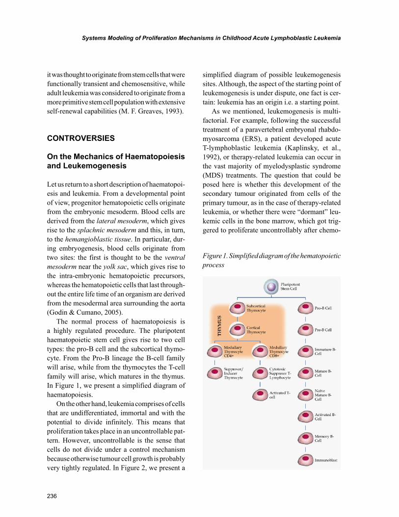
The normal process of haematopoiesis is a highly regulated procedure. The pluripotent haematopoietic stem cell gives rise to two cell types: the pro-B cell and the subcortical thymo-cyte. From the Pro-B lineage the B-cell family will arise, while from the thymocytes the T-cell family will arise, which matures in the thymus. In Figure 1, we present a simplified diagram of haematopoiesis.
On the other hand, leukemia comprises of cells that are undifferentiated, immortal and with the potential to divide infinitely. This means that proliferation takes place in an uncontrollable pat-tern. However, uncontrollable is the sense that cells do not divide under a control mechanism because otherwise tumour cell growth is probably very tightly regulated. In Figure 2, we present a
simplified diagram of possible leukemogenesis sites. Although, the aspect of the starting point of leukemogenesis is under dispute, one fact is cer-tain: leukemia has an origin i.e. a starting point.
As we mentioned, leukemogenesis is multi-factorial. For example, following the successful treatment of a paravertebral embryonal rhabdo-myosarcoma (ERS), a patient developed acute T-lymphoblastic leukemia (Kaplinsky, et al., 1992), or therapy-related leukemia can occur in the vast majority of myelodysplastic syndrome (MDS) treatments. The question that could be posed here is whether this development of the secondary tumour originated from cells of the primary tumour, as in the case of therapy-related leukemia, or whether there were “dormant” leu-kemic cells in the bone marrow, which got trig-gered to proliferate uncontrollably after chemo-
Figure 1. Simplified diagram of the hematopoietic process

237
Systems Modeling of Proliferation Mechanisms in Childhood Acute Lymphoblastic Leukemia
therapy. In the case where cells from one type of cancer develop into another, it probably means that the tumour cell possesses two traits: first it is able to migrate and second it is able to differ-entiate to another type of cell manifesting stem cell properties. This is in accordance with the stem cell theory of cancer origin, as cancer stem cells keep their ability to differentiate, migrate and even give rise to a new malignancy with almost totally new traits. Furthermore, it has been re-ported that therapy-related leukemia can occur due to the use of chemotherapeutics (Dedrick & Morrison, 1992; Park & Koeffler, 1996). This phenomenon has not been thoroughly investi-gated and the mechanisms underlying it are still obscure. However, it points out the fact that car-
cinogenesis is a complicated process, implicating a number of mechanisms and not just single events in a cell’s life time. If we were to accept the hy-pothesis that two different tumour cells can co-exist in different locations as accurate, then we could accept the notion that stem cells play the main role in carcinogenesis and tumour growth. On the other hand, an interesting report by Kelly et al. revealed that, at least in part, the presence of cancer stem cells is not necessary for tumour growth to be sustained (Kelly, Dakic, Adams, Nutt, & Strasser, 2007).
An interesting reference highlights the fact that stem cells are probably found in different types of tumours, thus suggesting that stem cells are impli-cated in the aetiology of tumour maintenance and
Figure 2. Part of the hematopoietic process, with some of the possible origins of leukemia. Arrows in-dicate the possible stages where leukemia can emerge.

238
Systems Modeling of Proliferation Mechanisms in Childhood Acute Lymphoblastic Leukemia
growth (Nicolis, 2007). However, there is evidence that even normal, already differentiated cells can be transformed to tumorigenic (Nicolis, 2007). Based on this observation, there was a case reporting a child manifesting four different tumour types se-quentially (Perilongo, et al., 1993). It could be that stem cells originating from the same tissue possess similar defects but enough alterations to be able to give rise to five different types of tumour. Another point on which attention should be drawn is the regulation of genes through transcription factors (TFs). Knowledge of gene regulatory networks is considered to be of crucial importance for the understanding of diseases such as cancer, as it may lead to new therapeutic approaches (Chang, et al., 2006). The knowledge of common transcriptional regulatory networks could potentially lead to a universal treatment for diverse diseases such as cancer, and it is through this possibility that the need for computational methods in the study of carcinogenesis becomes apparent.
PROBLEMS
On the Physical Nature of Leukemogenesis and Proliferation
The data that we obtain about the disease is only at presentation, i.e. at diagnosis. There are no hints of what has happened before that. We do not know how the cells have progressed from the healthy to the diseased state and in what velocity or ac-celeration or deceleration (to use some physical terms for start). How is it then possible to gain knowledge on these issues of leukemia and to make the disease more comprehendible? The answer to this is derived from the previous paragraphs where we state that biology should interact deeply with the classical natural sciences and try to set laws and theorems regarding its phenomena. Once we are able to model the phenomenon of prolifera-tion, we might gain access to its mechanisms and understand its nature.
To the best of our knowledge, there are no considerations concerning the merging of physical theories and applications regarding the clarifica-tion of leukemogenesis. From a perspective, it is highly unlikely that such theory could be applied to the system called leukemogenesis. Yet, there are some physical aspects that are not taken into consideration, at least up until now. The Warburg effect that we mentioned in the previous sec-tion, where the current observation made was that tumour cells had defective mitochondria, thus leading to the malfunction of oxidative phosphorylation. The net reaction for energy production in cellular metabolism gives twelve molecules of ATP for one molecule of glucose. In cancer cells this is not accurate. Each mole of glucose being consumed gives two moles of ATP and two moles of lactate. This immediately gives a hint towards the different energetic needs that each cells has, i.e. the Leukemic Stem Cell (LSC) and the Hematopoietic Stem Cell (HSC). From a thermodynamics point of view, proliferation and survival leads to entropy minimization, while cell death leads to entropy maximization. Also, proliferating cells would produce more work than less proliferating cells. There is a clear difference between the two cell types, that is normal and tumour cells. No matter what the processes of carcinogenesis (leukemogenesis) are, those cells require energy to proliferate and they too obey to the laws of mass and energy conservation. If we represent cell proliferation as a series of equations, we would describe normal cells in a certain form and in the case of tumour cells, since the same global laws apply, we would have an imbalanced equation that should be solved.
In addition, at this point, we could combine the aspects mentioned in the previous section of evolutionary theories, which are time and sto-chasticity or non-determinism. Time is a physical quantity that is not irrelevant to the progress of leukemia. As we mentioned previously, there is a time interval between the point of emergence of the first cell and the presentation of the dis-

239
Systems Modeling of Proliferation Mechanisms in Childhood Acute Lymphoblastic Leukemia
ease. This latency is probably crucial not only to understand the disease but also to cure it. A fast proliferating disease follows different biological pathways than a slow proliferating disease or even a disease that moves near a steady-state condition. Also, bearing that gene expression is a process that it is regulated very tightly even in the case of neoplasms, one might say with certainty that any details from tight regulation would lead to non-viable conditions. Since tumour cells are viable, in a contradictory sense, they do possess a tight gene regulation.
The debate becomes more interesting when it comes to the aspects of stochastic processes and non-determinism. This is a very complicated topic and we would attempt to give some thoughts on the subject. Yet, it is immensely interesting to deal with it. From the classical physics point of view, phenomena are causal. That is, for each observa-tion there is a causal effect that brought about the phenomenon. The question now is whether this phenomenon is random or deterministic. If we consider the quantum aspect of things, this is a random process that yet with statistical certainty, will be within the limits of viability. To bring this topic to leukemogenesis, let us ask wonder what are the chances, if any, for a cell to undergo mutations that would be lethal and instead of lethality, to get exactly the opposite that is potential for infinitesimal propagation. Tumours are such an example. On one hand, the observed mutations could be random but the result is not. The regulation and biology of tumor cells implies that random phenomena could lead to non-random effects. By combining the aspects of thermodynamics on these thoughts, we could hypothesize that random phenomena usually lead to the maximization of entropy as well as increase in dispersed events, but in the case of tumours, dispersed and random events lead to entropy minimization since they lead to cell proliferation, or in other words to order. This leads to a para-dox, since randomness leads to not-order but in the case of tumours randomness leads to entropy
minimization, thus order. Therefore, if such phe-nomena i.e. leukemogenesis and tumorigenesis were purely stochastic and non-deterministic, they would contradict the physical laws to which they also obey. Therefore, it is possible that two events occur simultaneously; certainty and probability. This issue has been marvellously addressed by Max Planck in a lecture given at the University of Berlin in his talk entitled “Dynamische und Statistische Gesetzmässigkeit” (“Dynamical and Statistical Regularity”) in 1914. In that lecture, the same example was presented in terms of fluid dynamics and energy transfer. Thus, Planck im-plied that the movement of a heavier fluid will move towards the gravitational forces, which is a certainty, while heat will travel upwards from the heater to the colder layers, which is a prob-ability. This is a very complicated aspect, which has not yet been addressed in the literature as far as biological systems are concerned, and in particular tumorigenesis, while here we attempt to give some thoughts to the aspect and leave it open for future investigations.
SOLUTIONS AND RECOMMENDATIONS
A Mathematical Formulation of the Physical Approach
Let us use an example from unpublished data, in order to see how the above mentioned approach could be used to understand the link between thermodynamics and proliferation. In the present model the attempt was to simulate cell proliferation with respect to glucose consumption. This example concerns the treatment of a leukemic cell line (CCRF-CEM), in particular a T-cell leukemia, with glucocorticoids and subsequent measurements of metabolic and proliferation factors. Symbols and definitions are also summarized in Table 1.
The model presumes that the fraction of cells linked to a certain phenotypic effect can be derived

240
Systems Modeling of Proliferation Mechanisms in Childhood Acute Lymphoblastic Leukemia
from the previous total cell population so, let Ne,t+1 be the cell population under a certain effect. This effect can be, for the present analysis, either vi-ability or cell death. Therefore, the total population estimate under the impact of a given effect will be given by:
N k Ne t e t e t,( ) , ,+ = ⋅
1, (1)
where ke t,
is a generalized nonlinear coefficient of the effect e in the population N
e t,at this in-
stance.At the same time, apart from cell proliferation,
we have to account for metabolic factors that change over time and probably influence the course of proliferation. In the case of metabolic factors, the rate of change in concentration is defined as the rate of the respective reaction which is:
udC
dtmm= (2)
where um is the reaction rate of the substrate used as nutrient i.e. glucose, Cm is the substrate’s con-centration and t is time. However, in the present case two of the substances accounted for were glucose and lactic acid. It is known that glucose is transformed into two lactic acid molecules based on the reaction:
C6H12O6→2CH3CHOHCOOH+2ATP, (3)
Or to be more precise, Equation 3 can be written as:
C6H12O6+2NAD++2ADP+2Pi→2H2O+2CH3COCO2
-+2NADH+2ATP+2H3O+, (4)
This is due to the formation of two molecules of pyruvate from the anaerobic catabolism of glucose and the subsequent formation of two molecules of lactate in the cytosol. However, this reaction represents a lump reaction, namely one that represents the algebraic sum of many reac-tions. With many intermediates in between and
Table 1. Symbols and abbreviations
Symbol Quantity Units
Nt Total cell population at time t cells/ul ∙103
Ne Cell population under an effect, e can take the following values: cells/ul ∙103
k The factor by which total population proliferates from time t to time t+1.
Ke,t The factor by which cell population under a certain effect proliferates from time t to time t+1. e takes values as mentioned above in the same table
CG Glucose concentration mg/dlt
CLA Lactic Acid concentration mg/dlt
CALP Alkaline Phosphatase concentration IU/lt
CLDH Lactate Dehydrogenase IU/lt
km The factor by which metabolic factors are produced or consumed. from time t to time t+1. m can take the following values:
G: Glucose
LA: Lactic Acid
ALP: Alkaline Phosphatase
LDH: Lactate Dehydrogenase
um Reaction rate (reaction kinetics) M/sec

241
Systems Modeling of Proliferation Mechanisms in Childhood Acute Lymphoblastic Leukemia
therefore kinetic rules such as Michaelis-Menten or Le Chatelier’s/Van’t Hoff cannot be directly applied to these. However, under the assumption that there is not significant biochemical cross-talk of these intermediates with other external metabolic pools, the lumping of the reactions to a single one is plausible as is the case of lactate production through the catalysis of pyruvate. The substrates of this reaction can be measured. In addition, we have included an estimate of the reaction efficiency as we will describe further on which is given by:
α =C
Cobserved
theoretical
, (5)
where, α is the efficiency of the reaction and 0<α<1, Cobserved is the observed/measured yield of the reaction and Ctheoretical is the expected reaction yield if we were dealing with a perfect reaction. In particular, in the system we are referring to we have calculated that the reaction efficiency reached 95%, which is impressive for an in vitro cell system.
Since we are dealing with thermodynamical systems then we have to use the efficiencies of the reaction in our calculations. In reality, biological systems are far from thermodynamic equilibrium, therefore they are dissipative. In other words, they exchange matter and energy from their en-vironment. This makes them very complicated systems hence:
LDH concentration can be accounted only from cells that have been lysed and not from the total population. Although the LDH concentration can be numerically calculated, it would still not be a reliable numerical approximation. Therefore, we used the same principle as in the case of cell population. The concentration C of a metabolite or substance at time t+1 can be written as:
C k Cm t m t m t,( ) , ,+ = ⋅
1, (6)
Applying mass balance equations (Chatzi-ioannou, Palaiologos, & Kolisis, 2003) for the metabolic pools with respect to time we have,
dC
dtk Cmm t m t
= ⋅, ,
, (7)
where km t,
is a generalized coefficient of the net effect observed in the pool C
m t, at time t. This
resembles a modification of the Lotka-Voltera-Kolmogorov equations which were initially used for the description of reaction dynamics and further expanded to population dynamics (Lotka, 1910, 1920). The Lotka-Voltera functions were derived from the Verhulst logistic equation (Ver-hulst, 1838). Though succinct, this mathematical formulation introduces through the use of factorkm t,
, non- linearity. Coefficient km t,
bears a critical biological significance in the model. Pre-suming that the effects in this study are directly linked to glucocorticoid exposure, k=f(Cp), where p stands for prednisolone, the glucocorticoid used in the present study, the effects observed depend solely on the drug’s concentration. In order to approximate the values, i.e. numerically solve our functions, we have used phase-space maps of the measured data.
Further on, for the net energy consumption we have used the linear relation which has been previously described by Stouthamer and Betten-haussen (1973) which is given by:
r Y mATP x ATP ATP= ⋅ +
,µ , (8)
where rATP is the production rate of ATPs from the available energy resources, Yx,ATP is the growth-associated ATPs, μ is the specific cell growth rate, which is a function of the substrate’s concentration (μ=f(Cs)) and mATP is the nongrowth-related ATP consumption. In our case the substrate is glucose and we consider it, at least for the time being, the sole substrate on which cells gain their energy
242
Systems Modeling of Proliferation Mechanisms in Childhood Acute Lymphoblastic Leukemia
needs from. However, the relation μ=f(Cs) does not apply since the concentration of the substrate is the same for all experimental setups i.e. all cells start from the same initial glucose concentration. The difference in our case is that what changes is the prednisolone concentration. Therefore, the relation would be μ=f(Cp). This means that μ is a function of prednisolone concentration. Hence, Equation 8 could be translated as: the total consumption of ATP is equal to the ATP needed for proliferation plus the ATP needed for maintenance.
This relation was initially proposed for mi-crobial growth (Zeng & Deckwer, 1995), yet it can be possible that the same principle could be applied to eukaryotic cells, as we also mention in this work.
In , Equation 8, the first term i.e. Yx,ATP, can be divided further into three subterms, so we get:
Y Y Y Yx ATP x ATP growth x ATP lysis x ATP leak, , , , , , ,
= + + , (9)
where, Yx,ATP is the ATP consumption for po-lymerization, biosynthesis and growth overall, Yx,ATP,lysis is the ATP usage for repolymerization of degraded molecules and Yx,ATP,leak includes all ATP consumption due to leaks, futile cycles and growth-associated maintenance.
A basic question that we have dealt with was whether cell division, could be regarded as expan-sion (Wex) or non-expansion (Wnon-ex) work. This was a very fundamental question since it influ-ences all forth-coming assumptions. Hence, on one hand it is reasonable to think of cell proliferation in terms of molecule polymerization. However, cells do divide and therefore the system expands. If N0 cells are present at time t0 and N at time t and N>N0 then the total volume occupied by cells in the surroundings has increased therefore we have an expansion. So, the work performed by nutrient consumption is the sum of expansion and non-expansion work.
An interesting approach to the concept of bioenergetics, has been previously described by Westerhoff et al. (1981), where they referred to the term mosaic non-equilibrium thermodynamics as a way to describe the different types of energy resources in bacterial photosynthetic energy pro-duction (Westerhoff, Hellingwerf, Arents, Scholte, & Van Dam, 1981). In that sense, it is a concept that we agree to, since it is most probable that eukaryotic organisms could potentially use vari-ous types of energy sources for their sustainment.
In our paradigm (system of study) one of the first observations made was the fact that glucose uptake has been lowered down from lower to higher glucocorticoid concentrations. As a matter of fact, it appeared that this was regulated with a threshold in glucocorticoid concentration, 10uM in this particular case. This was observed from two sides. The first was the measurements of glucose and the second the measurement of its metabolite in the case of the Warburg effect which is the lactic acid.
Returning to the unknown factor k, this is a non-linear factor that is solely depended upon environmental conditions. That is what makes very hard to estimate such a factor, since the slightest changes in the environment could have an effect on cell proliferation. The only method that such a factor can be estimated is numerically. Therefore, we have assumed that k is a nonlinear factor. The first aim was to determine the dynamics of the factor k i.e. how it changes as a function of concentration. Glucose measurements were taken from cell culture supernatants (CG) (Figure 3). We assumed that glucose entering the cell was transformed as a total into ATPs and pyruvate. Since cells presumably follow a lactic acid fer-mentation cycle, pyruvate should be transformed into lactate through LDH. In addition, the enzyme LDH (Lactate Dehydrogenase) was measured as a function of the total population of necrotic cells (CLDH) (data not shown). It is important to note that LDH is released from the cells only if cell

243
Systems Modeling of Proliferation Mechanisms in Childhood Acute Lymphoblastic Leukemia
lysis takes place, thus allowing the contents of the cytosol to be released in the extracellular medium.
At the same time, the measured lactate (CLA) was considered to be diffused from both living and apoptotic cells and also released from ne-crotic cells due to cell membrane lysis (Figure 4).
Finally, we accounted for three possible cell fates: progression of proliferation (Nv), necrosis (Nn) and apoptosis (Nta). One of the first correla-tions calculated was that of the measured LDH and the respective number of necrotic cells. We would be expected to observe a positive correlation between the two factors. It appeared that LDH
Figure 3. Glucose concentration measurements under the effect of the glucocorticoid prednisolone (unpublished data)
Figure 4. Lactate metabolite concentration with respect to time and under the influence of the gluco-corticoid prednisolone (unpublished data)

244
Systems Modeling of Proliferation Mechanisms in Childhood Acute Lymphoblastic Leukemia
concentration and necrotic cell population indeed showed a positive correlation in two particular cases: untreated cells and cells treated with a large dose of prednisolone (700uM) (data not shown). This effect could be interpreted as follows: that all other glucocorticoid concentrations beside ne-crotic cell death, also lead to the rupture of the cell membrane and cell lysis. Interestingly, the largest concentration that would be expected to have a lytic effect due to the overdose per se, showed a negative correlation, exactly matching that of cells with no glucocorticoid treatment. As mentioned earlier, the only way to estimate the nonlinear factor k is through numerical approximations, thus plotting conditions at time t+1 vs. conditions at time t. In other words we have attempted to model cell populations over time as a function of the drug’s concentrations. In order to connect the factors: cell population, k and metabolites we have attempted simulations in the 3D space, the data have been fitted with two polynomial equations of 4th order for both the x and y variables. In Figure 5, several examples of such simulations are pre-sented. Simulation of experimental data is a useful approach to find patterns. In the present case, the dynamics of proliferation manifested interesting dynamics, although the dataset was small. Mea-sured factors were simulated with 4th order poly-nomial equations. A novel factor that has been introduced by this analysis is the consideration of the physical quantities velocity and acceleration, which were calculated from the existent data. In particular, we have calculated the velocity of the total population, defining velocity as the change rate in cell population dN/dt. In addition, we have similarly calculated the cell population acceleration defined as d2N/d2t. The results returned some in-teresting dynamics as it appeared that there was a correlation between the concentrations of the glucocorticoid, time and the physical quantities defined above. Also, it appeared that there was a correlation between the nonlinear factor k we mentioned before, the cell populations and gluco-corticoid concentrations.
In the present analysis the Jacobian matrix J determines the transition dynamics of the system from one state to the next. In a previous work the use of Jacobian matrices was used for the deter-mination of the possible dynamics of a system at a metabolic state (Grimbs, Selbig, Bulik, Holzhut-ter, & Steuer, 2007). There is a great amount of mathematical formulations concerning biological systems dating back in the early 19th century but the whole idea of integrating biological systems with analytical or stochastic formulations is still in its infancy (Lotka, 1910, 1920; Ramakrishnan, DuBois, Almon, Pyszczynski, & Jusko, 2002; Verhulst, 1838).
Since we observed that glucose uptake was inhibited due to prednisolone treatment and it is known from the literature that leukemic cells and especially these cells follow by letter the War-burg effect, we have seen that the consumption of glucose is related to the production of lactic acid. The expected effect would be that most of glucose would become lactic acid and another part would enter either the mitochondria for entering oxidative phosphorylation or enter the pentose-phosphate pathway for de novo biosynthesis of molecules. We have concluded from those data that almost all glucose taken from the cell has been used exclusively for energy production. This is a subject to be further investigated.
In the system of study we are referring to, we have dealt with the following paradox, in the past: cells treated with low to medium prednisolone doses presented a higher proliferation rate as compared to control. On the other hand higher prednisolone doses manifested similar prolifera-tion dynamics as compared to control. At the same time glucose measurements showed the control and low to medium prednisolone doses manifested a rapid consumption of glucose within 72h while higher doses manifested a much slower consump-tion. Yet, the proliferation potential was similar for both untreated cells as well as higher doses of prednisolone.

245
Systems Modeling of Proliferation Mechanisms in Childhood Acute Lymphoblastic Leukemia
Figure 5. Simulations of cell population and metabolites measurements: total cell population vs. time and glucocorticoid concentrations (A), cell population velocity vs. time and glucocorticoid concentra-tion (B), cell death population acceleration vs. time and glucocorticoid concentration (C), velocity of glucose consumption vs. time and glucocorticoid concentration (D), velocity of lactate production vs. time and glucocorticoid concentration (E), correlation of glucose concentration, lactate concentration and glucocorticoid concentration (F), total cell population acceleration vs. time and glucocorticoid concentration (G), simulation of early apoptotic, necrotic and apoptotic populations (H). Simulations of viable cell population vs. glucocorticoid concentrations and the factor k (I), simulation of the k factors for the necrotic cell populations (Kn), total apoptotic populations (Kta) and viable populations (Kv) (J).
246
Systems Modeling of Proliferation Mechanisms in Childhood Acute Lymphoblastic Leukemia
At this point several hypotheses could be formed. First, high prednisolone doses changed the membrane’s permeability and uptake of glucose as well as prednisolone ceased which leaves open the question of how cells obtain the required energy for further growth. Second, prednisolone does not change membrane permeability but it causes a blocking of the glucose receptors in order for the cell to take in glucose and third, it causes a shift away from the Warburg effect taking place in leukemic cells and towards autophagy which explains the proliferation effect. It has been previ-ously described in numerous reports that there is a strong and well documented relation between tumor progression and metabolism i.e. glucose uptake (Gatenby & Gillies, 2004; Kim, Gao, & Dang, 2007; Yun, et al., 2009) and impaired meta-bolic pathways. Yet, since solid tumor metabolism is relatively well documented not as many studies have been dealing with leukemia (Boag, et al., 2006; Hulleman, et al., 2009).
FUTURE RESEARCH DIRECTIONS
Some Remarks on Modelling Leukemia’s Proliferation Dynamics
From this discussion, it is evident that to be able to improve a certain condition, one first needs to understand the phenomenon or, in other words, to appreciate the governing rules of the system. Hence, returning to our previous discussion on systems biology, it works towards understand-ing and defining the rules governing biological phenomena.
In order to obtain a more complete view of leukemia one needs to identify and understand the progressive stages of the disease. For example, the development of an in vitro model of leukemogen-esis would permit the study of the leukemic stages in the laboratory. For this purpose, two methods are available: the use of animals in vivo or the use of cell lines in vitro. Cell lines seem to offer
a more direct and practical means for studying the disease since, apart from being economical in both time and money, they also allow for ex-periments and experimental repetitions on human biological material.
Leukemia, from the point of diagnosis onwards, has become a basic area of study and research, mainly due to the great variety of pathological characteristics and the availability of biological and clinical samples. On the contrary, the study of the mechanisms that lead to such a complicated phenomenon, as is the phenotype of leukemia, presents practical difficulties due to lack of knowledge on the pre-neoplasmatic state of blood cells. In order for the leukemogenic mechanisms of childhood to be elucidated, a great number of infants should be followed up from their birth and up until some of these would develop haemato-logical neoplasm.
With the introduction of the basic concepts of systems approaches to biological phenomena and some developmental aspects of hematopoietic and leukemic cells we could say that this is a very complicated phenomenon and there is still an on-going debate on the aetiologies and dynamics of the disease.
The major challenge of computational and systems biology is to make contributions to the description of population and reaction dynamics (Grimbs, et al., 2007). This is applied to both systems under no external influences but also to systems under the influence of external stimuli, such as pharmacological interventions (which was our present example) or environmental stresses. In the case of the cell system mentioned here as an example, the most interesting observation was that the system was resistant to glucocorticoids and therefore our attempt was in fact to model dynamics of cellular growth and metabolism in resistant cases. Future research directions could point towards describing drug effects as a function of time or concentration and towards predicting the outcome of certain treatments or even towards improving the state of treatment in

247
Systems Modeling of Proliferation Mechanisms in Childhood Acute Lymphoblastic Leukemia
such a way that it would be more effective. We suggest that the transition of the cell system we have studied from one state to the next follows complicated dynamics, manifesting in almost all cases oscillatory behaviour. The use of mathemati-cal and modelling tools for the discovery of such mechanisms is a unique method for understanding complicated biological systems. Many research efforts are dedicated to the improvement of the existing or to the development of new pharma-ceuticals. Modelling approaches could assist in such efforts as they would provide with a more in-depth understanding of biological systems. The general idea is to be able to predict the future states of a system based on the present ones. This has proved to be a difficult task since biological systems follow nonlinear behaviour and, unlike physical systems, there are only a few generaliza-tions that can be formulated.
Into Deep Waters: Cell Proliferation and Chaos Dynamics
Several studies have been occupied with the complex dynamic behaviour of animal popula-tions (Mackey & Glass, 1977; May, 1974, 1976; Romond, Rustici, Gonze, & Goldbeter, 1999). However, very little is known about the dynam-ics of tumour cell proliferation (Wolfrom, et al., 2000) and even less is known about the state of proliferation dynamics during oncogenesis; that is until cells reach an adequate population to be diagnosed. The data that can be collected from tumours, regarding their dynamic nature, can only happen after a tumour has been diagnosed, which usually is too late for the patient, as all the progress-determining steps have taken place. Therefore, in vitro systems provide an excellent opportunity to study effects that are impossible to measure in vivo. Most importantly, they en-able the study of long-term behaviour, which is required when it comes to reaching conclusions with regards to non-linearity and chaotic system behaviour. This, in particular, is impossible to
happen, even with primary cultures of cells, since they are short-lived (15-20 days) when untrans-formed, and the only way is the use of established cell lines obtained from different organisms. For that reason developing a modelling approach to simulate the in vivo conditions as best as possible, is considered crucial.
Hence, the nature of proliferation dynamics may give insight into the way that cells not only proliferate but also differentiate. Population dynamics have been the subject of study among various groups. It has already been shown that even cells that grow under normal conditions can manifest proliferation dynamics of non-linear nature (Laurent, Deschatrette, & Wolfrom; Wol-from, et al., 2000). In addition, other groups have demonstrated that this non-linear behavior can also exist under the influence of drugs (Guerroui, De-schatrette, & Wolfrom, 2005), or similarly, under the influence of environmental factors. Any new knowledge on the mechanisms underlying cell proliferation is of major importance, and even the smallest of indications towards a certain direction could enable us to further discover differences in the mechanisms distinguishing between health and disease. This issue is especially important in tumors, the incidence of which is approaching that of an epidemic.
New questions, also, could be posed regarding the nature of the dynamics of a cell population under the influence of a drug. If certain physical measures, such as proliferation, are observed on the phenotypic level, how are they translated on the molecular/genomic level? For example, if a cell population increases its rate of proliferation, does it mean that the genes required for this effect transcribe faster than usual? An interesting report by Mar et al. (2009) suggested that gene expression takes place in quanta, i.e. that it happens discretely and not continuously (Mar & Quackenbush, 2009; Mar, Rubio, & Quackenbush, 2006). Also, in two other reports it was suggested that gene expres-sion follows oscillatory patterns, which makes things even more complicated with regards to

248
Systems Modeling of Proliferation Mechanisms in Childhood Acute Lymphoblastic Leukemia
the proliferation rate, be it growth acceleration or deceleration (Chabot, Pedraza, Luitel, & van Oudenaarden, 2007; Degenhardt, et al., 2009). This means that cells cannot simply transit from one state to another in terms of growth rate. Should the hypothesis of oscillatory modulation of gene expression, which implies non-linearity, stand correct, then a much more complicated regula-tory pattern is required by a cell so as to change its state, as a function of environmental stimuli.
The same applies for critical aspects of the metabolism of cancer cells and in particular, leukemic cells. Already in 1924, Warburg et al. observed that a shift occurred in tumors from oxidative phosphorylation to aerobic glycolysis, known as the Warburg effect (Warburg, et al., 1924). It is known, that metabolites, or metabolic molecules, do not only participate in metabolic processes related solely with energy production and thermodynamical conservation of the cell, but also mediate numerous signal transduction related functions.
In continuation of the complexity of biological systems as dynamic systems, it has been reported previously that biological systems exhibit very complicated dynamics (Mackey & Glass, 1977; Wolfrom, et al., 2000). The knowledge on how to determine a present state from the previous ones is critical within many areas, or applications, varying from cancer to insect population control. However, it has been proven a very tedious work to discover laws underlying biological systems, since on one hand it is not easy to model such systems due to their complexity, and on the other hand, biological dynamical systems possess significant adaptation capabilities. In a previous work by Wolfrom et al. (2000) a hypothesis was tested on adherent Fao rat hepatoma cells and it was found that these cells manifest aperiodic oscillations in their proliferation rate, giving evidence for deterministic chaos. Testing this hypothesis on another cell system, appeared to show similar results. We have previously described a study with the acute lymphoblastic leukemia cell line CCRF-
CEM, which provided an excellent substrate for modelling proliferation dynamics. In this study it has been shown that leukemic cells also follow non-linear dynamics, implying the existence of chaos. Cell cultures in vitro are considered to manifest a linear pattern of growth. Given the fact that the logistic equation takes into account limited nutrients and space, it predicts that a cell population would reach a steady-state within a certain time and eventually die out. In this study we have introduced a new constant to the cell model by making nutrient readily available but keeping space, in terms of total volume, constant. Under these conditions we drew the proliferation curve, which manifested aperiodic oscillations, whereas the proliferation rate manifested aperiodic oscillations most clearly. In addition, when the analysis expanded to the return map of prolifera-tion rate a nonlinear behavior, was observed and we found geometrically that the fixed points on this curve manifest a period-3 orbit which implies chaos dynamics. This work showed evidence for deterministic chaos in the proliferative behaviour of leukemia cells in vitro. Since this is a very com-plicated phenomenon it requires a lot more effort to understand the mechanisms underlying those dynamics. The implications from the understand-ing of these systems are tremendous. It will give us insight to the mechanisms of disease progression, such as in cancer, and enable building advanced models for the disease, which combine important features of both in vitro and in vivo systems. It is known that cancer starts and progresses slowly, at least before clinical presentation.
CONCLUSION
Cancer is a disease of the genome, where several genetic and epigenetic changes are required in order to occur. One of the main problems with the stipulations of theories of leukemogenesis is the fact that the only hints available, at least for in vivo cases, are the ones at diagnosis. More

249
Systems Modeling of Proliferation Mechanisms in Childhood Acute Lymphoblastic Leukemia
specifically, at diagnosis, the observed cell popu-lation consists of a mixture of phenotypes and genotypes, which in turn represents the outcome of the events that occurred from the very first steps of leukemogenesis. Hence, we do not have any hints on what happened from the beginning of the emergence of the first leukemic cells, no matter if this was a stem cell or not, until the presenta-tion of the disease, composing real challenge to discover what happened before the time of our observations. In general, systems and physical approaches to leukemogenesis and carcinogenesis could prove extremely useful on the elucidation of the disease and also its cure.
The expected outcome of such investigations would be the development of repeatable models of leukemogenesis. This will have as a consequence the better understanding of the mechanisms that lead to leukemia and possibly the identification of molecular markers in relation to the model. In this way, the different stages or “milestones” in the development of the disease and their specific transcriptional characteristics could be identified.
Neoplasms are responsible for a significant rate of mortality in the general population and this automatically puts them in high priority when it comes to studying the mechanisms that could ultimately lead to their treatment. Scientific research on the other hand cannot deliver and provide solutions if the mechanisms that lead to leukemia are not first identified and elucidated. One need to know the causes of such a complex phenomenon in order to be able to go ahead with proposals for treatment and the mechanistic view of the biological system that composes leukemia seems to be a necessary prerequisite.
Special attention should be given to the non-linear character of cell proliferation. This could be the key in understanding all above mentioned phenomena. Studies on this topic are scarce and more efforts should be the focus towards that direction.
REFERENCES
Bacher, U., Haferlach, T., Fehse, B., Schnittger, S., & Kroger, N. (2011). Minimal residual disease diagnostics and chimerism in the post-transplant period in acute myeloid leukemia. TheScien-tificWorldJournal, 11, 310–319. doi:10.1100/tsw.2011.16
Bassan, R., & Hoelzer, D. (2011). Modern therapy of acute lymphoblastic leukemia. Journal of Clinical Oncology, 29(5), 532–543. doi:10.1200/JCO.2010.30.1382
Bejar, R., Levine, R., & Ebert, B. L. (2011). Unraveling the molecular pathophysiology of myelodysplastic syndromes. Journal of Clini-cal Oncology, 29(5), 504–515. doi:10.1200/JCO.2010.31.1175
Boag, J. M., Beesley, A. H., Firth, M. J., Freitas, J. R., Ford, J., & Hoffmann, K. (2006). Altered glucose metabolism in childhood pre-B acute lymphoblastic leukemia. Leukemia, 20(10), 1731–1737. doi:10.1038/sj.leu.2404365
Carroll, W. L., Bhojwani, D., Min, D. J., Raetz, E., Relling, M., & Davies, S. (2003). Pediatric acute lymphoblastic leukemia. Hematology, 2003, 102–131. doi:10.1182/asheducation-2003.1.102
Castor, A., Nilsson, L., Astrand-Grundstrom, I., Buitenhuis, M., Ramirez, C., & Anderson, K. (2005). Distinct patterns of hematopoietic stem cell involvement in acute lymphoblastic leuke-mia. Nature Reviews Medicine, 11(6), 630–637. doi:10.1038/nm1253
Chabot, J. R., Pedraza, J. M., Luitel, P., & van Oudenaarden, A. (2007). Stochastic gene expres-sion out-of-steady-state in the cyanobacterial circadian clock. Nature, 450(7173), 1249–1252. doi:10.1038/nature06395

250
Systems Modeling of Proliferation Mechanisms in Childhood Acute Lymphoblastic Leukemia
Chang, L. W., Nagarajan, R., Magee, J. A., Mil-brandt, J., & Stormo, G. D. (2006). A systematic model to predict transcriptional regulatory mecha-nisms based on overrepresentation of transcription factor binding profiles. Genome Research, 16(3), 405–413. doi:10.1101/gr.4303406
Chatziioannou, A., Moulos, P., & Kolisis, F. N. (2009). Gene ARMADA: An integrated multi-analysis platform for microarray data implemented in MATLAB. BMC Bioinformatics, 10, 354. doi:10.1186/1471-2105-10-354
Chatziioannou, A., Palaiologos, G., & Kolisis, F. N. (2003). Metabolic flux analysis as a tool for the elucidation of the metabolism of neurotransmitter glutamate. Metabolic Engineering, 5(3), 201–210. doi:10.1016/S1096-7176(03)00029-6
Cobaleda, C., Gutierrez-Cianca, N., Perez-Losada, J., Flores, T., Garcia-Sanz, R., & Gonzalez, M. (2000). A primitive hematopoietic cell is the target for the leukemic transformation in human philadelphia-positive acute lymphoblastic leuke-mia. Blood, 95(3), 1007–1013.
Cox, C. V., Evely, R. S., Oakhill, A., Pamphilon, D. H., Goulden, N. J., & Blair, A. (2004). Char-acterization of acute lymphoblastic leukemia progenitor cells. Blood, 104(9), 2919–2925. doi:10.1182/blood-2004-03-0901
De Angulo, G., Yuen, C., Palla, S. L., Anderson, P. M., & Zweidler-McKay, P. A. (2008). Absolute lymphocyte count is a novel prognostic indicator in ALL and AML: Implications for risk stratifica-tion and future studies. Cancer, 112(2), 407–415. doi:10.1002/cncr.23168
Dedrick, R. L., & Morrison, P. F. (1992). Carcino-genic potency of alkylating agents in rodents and humans. Cancer Research, 52(9), 2464–2467.
Degenhardt, T., Rybakova, K. N., Tomaszewska, A., Mone, M. J., Westerhoff, H. V., & Bruggeman, F. J. (2009). Population-level transcription cycles derive from stochastic timing of single-cell tran-scription. Cell, 138(3), 489–501. doi:10.1016/j.cell.2009.05.029
Dick, J. E. (2008). Stem cell concepts renew cancer research. Blood, 112(13), 4793–4807. doi:10.1182/blood-2008-08-077941
Ebert, B. L. (2010). Genetic deletions in AML and MDS. Best Practice & Research. Clinical Haematology, 23(4), 457–461. doi:10.1016/j.beha.2010.09.006
Fasching, K., Panzer, S., Haas, O. A., Marschalek, R., Gadner, H., & Panzer-Grumayer, E. R. (2000). Presence of clone-specific antigen receptor gene rearrangements at birth indicates an in utero origin of diverse types of early childhood acute lym-phoblastic leukemia. Blood, 95(8), 2722–2724.
Ford, A. M., Bennett, C. A., Price, C. M., Bruin, M. C., Van Wering, E. R., & Greaves, M. (1998). Fetal origins of the TEL-AML1 fusion gene in identical twins with leukemia. Proceedings of the National Academy of Sciences of the United States of America, 95(8), 4584–4588. doi:10.1073/pnas.95.8.4584
Ford, A. M., Pombo-de-Oliveira, M. S., McCarthy, K. P., MacLean, J. M., Carrico, K. C., & Vincent, R. F. (1997). Monoclonal origin of concordant T-cell malignancy in identical twins. Blood, 89(1), 281–285.
Forsberg, E. C., Passegue, E., Prohaska, S. S., Wagers, A. J., Koeva, M., & Stuart, J. M. (2010). Molecular signatures of quiescent, mobilized and leukemia-initiating hematopoietic stem cells. PLoS ONE, 5(1), e8785. doi:10.1371/journal.pone.0008785

251
Systems Modeling of Proliferation Mechanisms in Childhood Acute Lymphoblastic Leukemia
Gagos, S., & Irminger-Finger, I. (2005). Chromo-some instability in neoplasia: Chaotic roots to continuous growth. The International Journal of Biochemistry & Cell Biology, 37(5), 1014–1033. doi:10.1016/j.biocel.2005.01.003
Gale, K. B., Ford, A. M., Repp, R., Borkhardt, A., Keller, C., & Eden, O. B. (1997). Backtracking leukemia to birth: Identification of clonotypic gene fusion sequences in neonatal blood spots. Proceed-ings of the National Academy of Sciences of the United States of America, 94(25), 13950–13954. doi:10.1073/pnas.94.25.13950
Gatenby, R. A., & Gillies, R. J. (2004). Why do cancers have high aerobic glycolysis? Nature Reviews. Cancer, 4(11), 891–899. doi:10.1038/nrc1478
Godin, I., & Cumano, A. (2005). Of birds and mice: Hematopoietic stem cell development. The International Journal of Developmental Biology, 49(2-3), 251–257. doi:10.1387/ijdb.041945ig
Greaves, M. (1999a). Autoimmune thrombophilic syndromes. Haematologica, 84(Suppl EHA-4), 32-35.
Greaves, M. (1999b). Molecular genetics, natural history and the demise of childhood leukemia. European Journal of Cancer, 35(14), 1941–1953. doi:10.1016/S0959-8049(99)00296-8
Greaves, M. (2002a). Cancer causation: The Darwinian downside of past success? The Lancet Oncology, 3(4), 244–251. doi:10.1016/S1470-2045(02)00716-7
Greaves, M. (2002b). Childhood leukemia. British Medical Journal, 324(7332), 283–287. doi:10.1136/bmj.324.7332.283
Greaves, M. (2005). In utero origins of childhood leukemia. Early Human Development, 81(1), 123–129. doi:10.1016/j.earlhumdev.2004.10.004
Greaves, M. (2010). Cancer stem cells: Back to Darwin? Seminars in Cancer Biology, 20(2), 65–70. doi:10.1016/j.semcancer.2010.03.002
Greaves, M. F. (1988). Speculations on the cause of childhood acute lymphoblastic leukemia. Leu-kemia, 2(2), 120–125.
Greaves, M. F. (1993). Stem cell origins of leu-kemia and curability. British Journal of Cancer, 67(3), 413–423. doi:10.1038/bjc.1993.81
Grimbs, S., Selbig, J., Bulik, S., Holzhutter, H. G., & Steuer, R. (2007). The stability and robustness of metabolic states: Identifying stabilizing sites in metabolic networks. Molecular Systems Biology, 3, 146. doi:10.1038/msb4100186
Guerroui, S., Deschatrette, J., & Wolfrom, C. (2005). Prolonged perturbation of the oscillations of hepatoma Fao cell proliferation by a single small dose of methotrexate. Pathologie Biologie, 53(5), 290–294. doi:10.1016/j.patbio.2004.12.026
Hatch, E. E., Linet, M. S., Kleinerman, R. A., Tarone, R. E., Severson, R. K., & Hartsock, C. T. (1998). Association between childhood acute lymphoblastic leukemia and use of electrical ap-pliances during pregnancy and childhood. Epi-demiology (Cambridge, Mass.), 9(3), 234–245. doi:10.1097/00001648-199805000-00006
Hayes, R. B., Yin, S. N., Dosemeci, M., Li, G. L., Wacholder, S., & Travis, L. B. (1997). Benzene and the dose-related incidence of hematologic neoplasms in China. Chinese Academy of Pre-ventive Medicine--National Cancer Institute Benzene Study Group. Journal of the National Cancer Institute, 89(14), 1065–1071. doi:10.1093/jnci/89.14.1065
Hehlmann, R., Schetters, H., Kreeb, G., Erfle, V., Schmidt, J., & Luz, A. (1983). RNA-tumorviruses, oncogenes, and their possible role in human car-cinogenesis. Klinische Wochenschrift, 61(24), 1217–1231. doi:10.1007/BF01540470

252
Systems Modeling of Proliferation Mechanisms in Childhood Acute Lymphoblastic Leukemia
Hong, D., Gupta, R., Ancliff, P., Atzberger, A., Brown, J., & Soneji, S. (2008). Initiating and cancer-propagating cells in TEL-AML1-associ-ated childhood leukemia. Science, 319(5861), 336–339. doi:10.1126/science.1150648
Hulleman, E., Kazemier, K. M., Holleman, A., VanderWeele, D. J., Rudin, C. M., & Broekhuis, M. J. (2009). Inhibition of glycolysis modulates prednisolone resistance in acute lymphoblastic leukemia cells. Blood, 113(9), 2014–2021. doi:10.1182/blood-2008-05-157842
Hunger, S. P., Raetz, E. A., Loh, M. L., & Mul-lighan, C. G. (n.d.). Improving outcomes for high-risk ALL: Translating new discoveries into clinical care. Pediatric Blood & Cancer, 56(6), 984–993. doi:10.1002/pbc.22996
Irons, R. D., Colagiovanni, D. B., & Stillman, W. S. (1996). Murine thymic lymphoma is associated with a species-specific hematopoietic progenitor cell subpopulation. Toxicology, 113(1-3), 59–67. doi:10.1016/0300-483X(96)03428-2
Irons, R. D., & Stillman, W. S. (1996a). Impact of benzene metabolites on differentiation of bone marrow progenitor cells. Environmental Health Perspectives, 104(Suppl 6), 1247–1250.
Irons, R. D., & Stillman, W. S. (1996b). The pro-cess of leukemogenesis. Environmental Health Perspectives, 104(Suppl 6), 1239–1246.
Iwasa, Y., & Michor, F. (2011). Evolutionary dy-namics of intratumor heterogeneity. PLoS ONE, 6(3), e17866. doi:10.1371/journal.pone.0017866
Kanehisa, M. (2002). The KEGG database. Novartis Foundation Symposium, 247, 91-101; discussion 101-103, 119-128, 244-152.
Kanehisa, M., & Goto, S. (2000). KEGG: Kyoto encyclopedia of genes and genomes. Nucleic Acids Research, 28(1), 27–30. doi:10.1093/nar/28.1.27
Kantarjian, H., O’Brien, S., Cortes, J., Wierda, W., Faderl, S., & Garcia-Manero, G. (2008). Therapeutic advances in leukemia and myelodys-plastic syndrome over the past 40 years. Cancer, 113(7Suppl), 1933–1952. doi:10.1002/cncr.23655
Kantarjian, H., O’Brien, S., Ravandi, F., Cortes, J., Shan, J., & Bennett, J. M. (2008). Proposal for a new risk model in myelodysplastic syndrome that accounts for events not considered in the original International Prognostic Scoring System. Cancer, 113(6), 1351–1361. doi:10.1002/cncr.23697
Kaplinsky, C., Frisch, A., Cohen, I. J., Goshen, Y., Jaber, L., & Yaniv, I. (1992). T-cell acute lym-phoblastic leukemia following therapy of rhabdo-myosarcoma. Medical and Pediatric Oncology, 20(3), 229–231. doi:10.1002/mpo.2950200310
Kebriaei, P., Anastasi, J., & Larson, R. A. (2002). Acute lymphoblastic leukemia: Diagnosis and classification. Best Practice & Research. Clini-cal Haematology, 15(4), 597–621. doi:10.1053/beha.2002.0224
Kelly, P. N., Dakic, A., Adams, J. M., Nutt, S. L., & Strasser, A. (2007). Tumor growth need not be driven by rare cancer stem cells. Science, 317(5836), 337. doi:10.1126/science.1142596
Kim, J. W., Gao, P., & Dang, C. V. (2007). Effects of hypoxia on tumor metabolism. Cancer and Metastasis Reviews, 26(2), 291–298. doi:10.1007/s10555-007-9060-4
Kinlen, L. (1988). Evidence for an infective cause of childhood leukemia: Comparison of a Scottish new town with nuclear reprocessing sites in Brit-ain. Lancet, 2(8624), 1323–1327. doi:10.1016/S0140-6736(88)90867-7
Kleinerman, R. A., Linet, M. S., Hatch, E. E., Wacholder, S., Tarone, R. E., & Severson, R. K. (1997). Magnetic field exposure assessment in a case-control study of childhood leukemia. Epi-demiology (Cambridge, Mass.), 8(5), 575–583. doi:10.1097/00001648-199709000-00017

253
Systems Modeling of Proliferation Mechanisms in Childhood Acute Lymphoblastic Leukemia
Knudson, A. (2001). Alfred Knudson and his two-hit hypothesis. (Interview by Ezzie Hutchin-son). The Lancet Oncology, 2(10), 642–645. doi:10.1016/S1470-2045(01)00524-1
Knudson, A. G. Jr. (1971). Mutation and cancer: Statistical study of retinoblastoma. Proceedings of the National Academy of Sciences of the United States of America, 68(4), 820–823. doi:10.1073/pnas.68.4.820
Laurent, M., Deschatrette, J., & Wolfrom, C. M. (2010). Unmasking chaotic attributes in time se-ries of living cell populations. PLoS ONE, 5(2), e9346. doi:10.1371/journal.pone.0009346
le Viseur, C., Hotfilder, M., Bomken, S., Wilson, K., Rottgers, S., & Schrauder, A. (2008). In child-hood acute lymphoblastic leukemia, blasts at different stages of immunophenotypic maturation have stem cell properties. Cancer Cell, 14(1), 47–58. doi:10.1016/j.ccr.2008.05.015
Leone, G., Mele, L., Pulsoni, A., Equitani, F., & Pagano, L. (1999). The incidence of secondary leukemias. Haematologica, 84(10), 937–945.
Linet, M. S., Hatch, E. E., Kleinerman, R. A., Robison, L. L., Kaune, W. T., & Friedman, D. R. (1997). Residential exposure to magnetic fields and acute lymphoblastic leukemia in children. The New England Journal of Medicine, 337(1), 1–7. doi:10.1056/NEJM199707033370101
Lotka, A. J. (1910). Contribution to the theory of periodic reactions. Journal of Physical Chemistry, 14(3), 271–274. doi:10.1021/j150111a004
Lotka, A. J. (1920). Analytical note on certain rhythmic relations in organic systems. Proceed-ings of the National Academy of Sciences of the United States of America, 6(7), 410–415. doi:10.1073/pnas.6.7.410
Mackey, M. C., & Glass, L. (1977). Oscillation and chaos in physiological control systems. Science, 197(4300), 287–289. doi:10.1126/science.267326
Mar, J. C., & Quackenbush, J. (2009). Decompo-sition of gene expression state space trajectories. PLoS Computational Biology, 5(12), e1000626. doi:10.1371/journal.pcbi.1000626
Mar, J. C., Rubio, R., & Quackenbush, J. (2006). Inferring steady state single-cell gene expression distributions from analysis of mesoscopic samples. Genome Biology, 7(12), R119. doi:10.1186/gb-2006-7-12-r119
Marcucci, G., Haferlach, T., & Dohner, H. (n.d.) Molecular genetics of adult acute myeloid leu-kemia: Prognostic and therapeutic implications. Journal of Clinical Oncology, 29(5), 475-486.
Marschalek, R. (2010). Mechanisms of leukemo-genesis by MLL fusion proteins. British Journal of Haematology, 152(2), 141–154. doi:10.1111/j.1365-2141.2010.08459.x
May, R. M. (1974). Biological populations with nonoverlapping generations: Stable points, stable cycles, and chaos. Science, 186(4164), 645–647. doi:10.1126/science.186.4164.645
May, R. M. (1976). Simple mathematical mod-els with very complicated dynamics. Nature, 261(5560), 459–467. doi:10.1038/261459a0
Mesarovic, M. (1968). Systems theory and biol-ogy-The view of a theoretician. Systems Theory and Biology, 351, 59–87. doi:10.1007/978-3-642-88343-9_3
Michaelis, J., Schuz, J., Meinert, R., Zemann, E., Grigat, J. P., & Kaatsch, P. (1998). Com-bined risk estimates for two German popula-tion-based case-control studies on residential magnetic fields and childhood acute leukemia. Epidemiology (Cambridge, Mass.), 9(1), 92–94. doi:10.1097/00001648-199801000-00018
Mikhail, F. M., Serry, K. A., Hatem, N., Mou-rad, Z. I., Farawela, H. M., & El Kaffash, D. M. (2002). AML1 gene over-expression in childhood acute lymphoblastic leukemia. Leukemia, 16(4), 658–668. doi:10.1038/sj.leu.2402399

254
Systems Modeling of Proliferation Mechanisms in Childhood Acute Lymphoblastic Leukemia
Moolgavkar, S. H. (1978). The multistage theory of carcinogenesis and the age distribution of cancer in man. Journal of the National Cancer Institute, 61(1), 49–52.
Mori, H., Colman, S. M., Xiao, Z., Ford, A. M., Healy, L. E., & Donaldson, C. (2002). Chromo-some translocations and covert leukemic clones are generated during normal fetal development. Proceedings of the National Academy of Sci-ences of the United States of America, 99(12), 8242–8247. doi:10.1073/pnas.112218799
Nicolis, S. K. (2007). Cancer stem cells and “stemness” genes in neuro-oncology. Neurobio-logical Disorders, 25(2), 217–229. doi:10.1016/j.nbd.2006.08.022
Nordling, C. O. (1953). A new theory on cancer-inducing mechanism. British Journal of Cancer, 7(1), 68–72. doi:10.1038/bjc.1953.8
Ogata, H., Goto, S., Sato, K., Fujibuchi, W., Bono, H., & Kanehisa, M. (1999). KEGG: Kyoto ency-clopedia of genes and genomes. Nucleic Acids Research, 27(1), 29–34. doi:10.1093/nar/27.1.29
Park, D. J., & Koeffler, H. P. (1996). Therapy-related myelodysplastic syndromes. Seminars in Hematology, 33(3), 256–273.
Perilongo, G., Felix, C. A., Meadows, A. T., Nowell, P., Biegel, J., & Lange, B. J. (1993). Sequential development of Wilms tumor, T-cell acute lymphoblastic leukemia, medulloblastoma and myeloid leukemia in a child with type 1 neu-rofibromatosis: a clinical and cytogenetic case report. Leukemia, 7(6), 912–915.
Petridou, E., Trichopoulos, D., Kravaritis, A., Pourtsidis, A., Dessypris, N., & Skalkidis, Y. (1997). Electrical power lines and childhood leukemia: A study from Greece. International Journal of Cancer, 73(3), 345–348. doi:10.1002/(SICI)1097-0215(19971104)73:3<345::AID-IJC7>3.0.CO;2-#
Pyatt, D. W., Aylward, L. L., & Hays, S. M. (2007). Is age an independent risk factor for chemically induced acute myelogenous leukemia in chil-dren? Journal of Toxicology and Environmental Health. Part B, Critical Reviews, 10(5), 379–400. doi:10.1080/15287390600975061
Ramakrishnan, R., DuBois, D. C., Almon, R. R., Pyszczynski, N. A., & Jusko, W. J. (2002). Fifth-generation model for corticosteroid phar-macodynamics: application to steady-state re-ceptor down-regulation and enzyme induction patterns during seven-day continuous infusion of methylprednisolone in rats. Journal of Pharma-cokinetics and Pharmacodynamics, 29(1), 1–24. doi:10.1023/A:1015765201129
Risser, R., Horowitz, J. M., & McCubrey, J. (1983). Endogenous mouse leukemia viruses. Annual Review of Genetics, 17, 85–121. doi:10.1146/annurev.ge.17.120183.000505
Ritter, G., Wilson, R., Pompei, F., & Burmis-trov, D. (2003). The multistage model of cancer development: Some implications. Toxicologi-cal Indications of Health, 19(7-10), 125–145. doi:10.1191/0748233703th195oa
Romond, P. C., Rustici, M., Gonze, D., & Gold-beter, A. (1999). Alternating oscillations and chaos in a model of two coupled biochemical oscilla-tors driving successive phases of the cell cycle. Annals of the New York Academy of Sciences, 879, 180–193. doi:10.1111/j.1749-6632.1999.tb10419.x
Rowley, J. D. (1998). Backtracking leukemia to birth. Nature Reviews Medicine, 4(2), 150–151. doi:10.1038/nm0298-150
Rubinstein, R., & Simon, I. (2005). MILANO--Custom annotation of microarray results using automatic literature searches. BMC Bioinformat-ics, 6, 12. doi:10.1186/1471-2105-6-12

255
Systems Modeling of Proliferation Mechanisms in Childhood Acute Lymphoblastic Leukemia
Saha, V., Young, B. D., & Freemont, P. S. (1998). Translocations, fusion genes, and acute leukemia. Journal of Cellular Biochemistry. Supplement, 30-31, 264–276. doi:10.1002/(SICI)1097-4644(1998)72:30/31+<264::AID-JCB32>3.0.CO;2-U
Salwinski, L., Miller, C. S., Smith, A. J., Pettit, F. K., Bowie, J. U., & Eisenberg, D. (2004). The database of interacting proteins: 2004 update. Nucleic Acids Research, 32(Database issue), D449–D451. doi:10.1093/nar/gkh086
Sandler, D. P., & Ross, J. A. (1997). Epidemiology of acute leukemia in children and adults. Seminars in Oncology, 24(1), 3–16.
Severson, R. K., & Ross, J. A. (1999). The causes of acute leukemia. Current Opinion in Oncology, 11(1), 20–24. doi:10.1097/00001622-199901000-00005
Sitwala, K. V., Dandekar, M. N., & Hess, J. L. (2008). HOX proteins and leukemia. International Journal of Clinical and Experimental Pathology, 1(6), 461–474.
Snyder, R. (2004). Xenobiotic metabolism and the mechanism(s) of benzene toxicity. Drug Me-tabolism Reviews, 36(3-4), 531–547. doi:10.1081/DMR-200033445
Stubbs, M. C., & Armstrong, S. A. (2007). Therapeutic implications of leukemia stem cell development. Clinical Cancer Research, 13(12), 3439–3442. doi:10.1158/1078-0432.CCR-06-3090
Stubbs, M. C., Kim, Y. M., Krivtsov, A. V., Wright, R. D., Feng, Z., & Agarwal, J. (2008). MLL-AF9 and FLT3 cooperation in acute myelogenous leukemia: Development of a model for rapid therapeutic assessment. Leukemia, 22(1), 66–77. doi:10.1038/sj.leu.2404951
Verhulst, P. H. (1838). Notice sur la loi que la population poursuit dans son accroissement. Cor-resp. Mathématique et Physique, 10, 113–121.
Vogelstein, B., Fearon, E. R., Hamilton, S. R., Kern, S. E., Preisinger, A. C., & Leppert, M. (1988). Genetic alterations during colorectal-tumor development. The New England Journal of Medicine, 319(9), 525–532. doi:10.1056/NEJM198809013190901
Warburg, O., Posener, K., & Negelein, E. (1924). Ueber den Stoffwechsel der Tumoren. Bioche-mische Zeitschrift, 152, 319–344.
Westerhoff, H. V., Hellingwerf, K. J., Arents, J. C., Scholte, B. J., & Van Dam, K. (1981). Mo-saic nonequilibrium thermodynamics describes biological energy transduction. Proceedings of the National Academy of Sciences of the United States of America, 78(6), 3554–3558. doi:10.1073/pnas.78.6.3554
Wiemels, J., Kang, M., & Greaves, M. (2009). Backtracking of leukemic clones to birth. Methods in Molecular Biology (Clifton, N.J.), 538, 7–27. doi:10.1007/978-1-59745-418-6_2
Williams, R. D., Boros, L. G., Kolanko, C. J., Jackman, S. M., & Eggers, T. R. (2004). Chromo-somal aberrations in human lymphocytes exposed to the anticholinesterase pesticide isofenphos with mechanisms of leukemogenesis. Leukemia Research, 28(9), 947–958. doi:10.1016/j.leu-kres.2003.12.014
Wingender, E., Dietze, P., Karas, H., & Knuppel, R. (1996). TRANSFAC: a database on transcrip-tion factors and their DNA binding sites. Nucleic Acids Research, 24(1), 238–241. doi:10.1093/nar/24.1.238
Wolfrom, C., Chau, N. P., Maigne, J., Lambert, J. C., Ducot, B., & Guerroui, S. (2000). Evidence for deterministic chaos in aperiodic oscillations of proliferative activity in long-term cultured Fao hepatoma cells. Journal of Cell Science, 113(Pt 6), 1069–1074.
Wolkenhauer, O. (2010). Systems biology-Dy-namic pathway modelling. Rostock.
256
Systems Modeling of Proliferation Mechanisms in Childhood Acute Lymphoblastic Leukemia
Yagi, T., Hibi, S., Tabata, Y., Kuriyama, K., Teramura, T., & Hashida, T. (2000). Detection of clonotypic IGH and TCR rearrangements in the neonatal blood spots of infants and children with B-cell precursor acute lymphoblastic leukemia. Blood, 96(1), 264–268.
Yun, J., Rago, C., Cheong, I., Pagliarini, R., An-genendt, P., & Rajagopalan, H. (2009). Glucose deprivation contributes to the development of KRAS pathway mutations in tumor cells. Sci-ence, 325(5947), 1555–1559. doi:10.1126/sci-ence.1174229
Zeisig, B. B., Milne, T., Garcia-Cuellar, M. P., Schreiner, S., Martin, M. E., & Fuchs, U. (2004). Hoxa9 and Meis1 are key targets for MLL-ENL-mediated cellular immortalization. Molecular and Cellular Biology, 24(2), 617–628. doi:10.1128/MCB.24.2.617-628.2004
Zeng, A. P., & Deckwer, W. D. (1995). A kinetic model for substrate and energy consumption of microbial growth under substrate-sufficient con-ditions. Biotechnology Progress, 11(1), 71–79. doi:10.1021/bp00031a010
Zhang, B., Schmoyer, D., Kirov, S., & Snoddy, J. (2004). GOTree Machine (GOTM): A Web-based platform for interpreting sets of interesting genes using Gene Ontology hierarchies. BMC Bioin-formatics, 5, 16. doi:10.1186/1471-2105-5-16
Zoumpourlis, V., Solakidi, S., Papathoma, A., & Papaevangeliou, D. (2003). Alterations in signal transduction pathways implicated in tumour progression during multistage mouse skin carci-nogenesis. Carcinogenesis, 24(7), 1159–1165. doi:10.1093/carcin/bgg067


