Embed Size (px)
Citation preview

Angelo BenedettiMaristella Tassi, Alessandra Gamberucci and Emanuele Giurisato, Deirdre P. McIntosh, Attendantly to Raft ClusteringIndependently of Cell Signaling andSubset of Plasma Membrane Rafts, T Cell Receptor Can Be Recruited to aFUNCTION AND BIOGENESIS:MEMBRANE TRANSPORT STRUCTURE
doi: 10.1074/jbc.M210758200 originally published online December 22, 20022003, 278:6771-6778.J. Biol. Chem.
10.1074/jbc.M210758200Access the most updated version of this article at doi:
.JBC Affinity SitesFind articles, minireviews, Reflections and Classics on similar topics on the
Alerts:
When a correction for this article is posted•
When this article is cited•
to choose from all of JBC's e-mail alertsClick here
http://www.jbc.org/content/278/9/6771.full.html#ref-list-1
This article cites 56 references, 30 of which can be accessed free at
by guest on January 27, 2014http://w
ww
.jbc.org/D
ownloaded from
by guest on January 27, 2014
http://ww
w.jbc.org/
Dow
nloaded from

T Cell Receptor Can Be Recruited to a Subset of Plasma MembraneRafts, Independently of Cell Signaling and Attendantly toRaft Clustering*
Received for publication, October 22, 2002, and in revised form, December 9, 2002Published, JBC Papers in Press, December 22, 2002, DOI 10.1074/jbc.M210758200
Emanuele Giurisato, Deirdre P. McIntosh‡, Maristella Tassi, Alessandra Gamberucci,and Angelo Benedetti§
From the Dipartimento di Fisiopatologia e Medicina Sperimentale, Universita degli Studi di Siena,Viale Aldo Moro No. 1, 53100-Siena, Italy
The constitutive/inducible association of the T cellreceptor (TCR) with isolated detergent-resistant, lipidraft-derived membranes has been studied in Jurkat Tlymphocytes. Membranes resistant to 1% Triton X-100contained virtually no CD3�, part of the TCR complex,irrespective of cell stimulation. On the other hand, mem-branes resistant either to a lower Triton X-100 concen-tration (i.e. 0.2%) or to the less hydrophobic detergentBrij 58 (1%) contained (i) a low CD3� amount (approxi-mate 2.7% of total) in resting cells and (ii) a several timeshigher amount of the TCR component, after T cell stim-ulation with either antigen-presenting cells or with phy-tohemagglutinin. It appeared that CD3/TCR was consti-tutively associated with and recruited to a raft-derivedmembrane subset because (i) all three membrane prep-arations contained a similar amount of the raft markertyrosine kinase Lck but no detectable amounts of theconventional membrane markers, CD45 phosphataseand transferrin receptor; (ii) a larger amount of partic-ulate membranes were resistant to solubilization with0.2% Triton X-100 and Brij 58 than to solubilization with1% Triton X-100; and (iii) higher cholesterol levels werepresent in membranes resistant to either the lower Tri-ton X-100 concentration or to Brij 58, as compared withthose resistant to 1% Triton X-100. The recruitment ofCD3 to the raft-derived membrane subset appeared (i) tooccur independently of cell signaling events, such asprotein-tyrosine phosphorylation and Ca2� mobiliza-tion/influx, and (ii) to be associated with clustering ofplasma membrane rafts induced by multiple cross-linkingof either TCR or the raft component, ganglioside GM1. Wesuggest that during T cell stimulation a lateral reorgani-zation of rafts into polarized larger domains can deter-mine the recruitment of TCR into these domains, whichfavors a polarization of the signaling cascade.
The plasma membranes (PM)1 of many cell types containdomains rich in cholesterol and sphingolipids, which have come
to be referred to as lipid rafts (1, 2). It is thought that rafts mayform because of the segregation of their components from thebulk of the glycerol-based phospholipid PM because of theorientation and tight packing of the long, largely saturated acylchains of the sphingolipids. This phase separation of the mem-brane results in patches of molecules, which form the rafts,existing in the liquid-ordered phase (lo) but surrounded by andco-existing with the phospholipids in the bulk PM that are inthe liquid disordered phase (ld). In many cell types rafts areorganized into structurally distinct invaginations of the PMcalled caveolae (3, 4). However in other types, including lym-phocytes, the rafts are thought to exist as islands of tightlypacked sphingolipid and cholesterol-based structures that, likecaveolae, can be isolated from the rest of the PM by purificationmethods based on their detergent insolubility at low tempera-tures (5, 6). These PM domains have been therefore calleddetergent-resistant membranes (DRMs), detergent-insolubleglycolipid-enriched complexes, and Triton-insoluble floatingfractions (reviewed in Refs. 2 and 7).
The comparative analysis of the detergent-insoluble do-mains, caveolae, and the bulk PM has provided an inventory ofproteins apparently residing in each (6, 8). However, doubtshave been raised as to whether the detergent treatment itselfmay modify the lipid rafts or destabilize certain proteins resi-dent therein (7). On the other hand, a recent report (9) stronglysuggests that use of different detergents can result in differentDRMs, which contain different proteins and likely correspondto different cholesterol-based PM lipid rafts.
In T lymphocytes, many proteins involved in signal trans-duction have been constitutively or inducibly recovered inDRMs (reviewed in Ref. 10). Among these are glycosylphos-phatidylinositol-anchored proteins, the Src family protein-ty-rosine kinases Lck and Fyn (10, 11), the transmembraneadapter protein linker for activation of T cells (12), and avariety of co-stimulatory and co-receptor proteins (Refs. 10 and13–16 and the references therein).
A large body of evidence supports a crucial role for rafts inthe signaling events activated by the T cell receptor (TCR)engagement (see Ref. 17 for a recent review). Uncertainties,however, still exist concerning the constitutive or inducibleassociation of TCR to rafts/DRMs, as well as to the mechanismsunderlining the possible recruitment of the receptor complex torafts upon T cell stimulation. Neither a constitutive nor aninducible (after treatment with antibodies to CD3) associationof the TCR/CD3 complex to DRMs was found in Jurkat T cells
* This work was supported by grants from the Italian Ministry ofInstruction, University and Scientific Research Cofin 2001 (to A. B.),University of Siena Progetto Giovani Ricercatori (to E. G.), and theItalian Space Agency (to A. B.). The costs of publication of this articlewere defrayed in part by the payment of page charges. This article musttherefore be hereby marked “advertisement” in accordance with 18U.S.C. Section 1734 solely to indicate this fact.
‡ Present address: Leukocyte Biology Section, Biomedical SciencesDivision, Imperial College, School of Medicine, London, UK.
§ To whom correspondence should be addressed. Tel.: 39-0577-234021; Fax: 39-0577-234009; E-mail: [email protected].
1 The abbreviations used are: PM, plasma membrane; TCR, T cellreceptor; DRM, detergent-resistant membrane; Lck, lymphocyte-spe-
cific protein-tyrosine kinase; PP2, 4-amino-5-(4-cholrophenyl)-7-(t-bu-tyl)pirazolo[3,4-d]pyrimidine; SEE, staphylococcal enterotoxin E; CTB,cholera toxin B subunit; PHA, phytohemagglutinin; Mes, 4-morpho-lineethanesulfonic acid; Pipes, 1,4-piperazinediethanesulfonic acid.
THE JOURNAL OF BIOLOGICAL CHEMISTRY Vol. 278, No. 9, Issue of February 28, pp. 6771–6778, 2003© 2003 by The American Society for Biochemistry and Molecular Biology, Inc. Printed in U.S.A.
This paper is available on line at http://www.jbc.org 6771
by guest on January 27, 2014http://w
ww
.jbc.org/D
ownloaded from

(18, 19). No constitutive association to DRMs of TCR� has beenalso found both in Th1 and Th2 lymphocytes, whereas TCR�was recruited to DRMs only in Th1 cells challenged with anti-gen-presenting cells (20). In a murine T cell hybrydoma, it hasbeen demonstrated that the TCR complex is excluded fromDRM domain before and after TCR stimulation, although aportion of the TCR� component appeared to be constitutivelyassociated with DRMs (21). Another report (22) has shown therecruitment of TCR/CD3 to DRMs, which is dependent on bothreceptor engagement and the activity of Src family kinases.While this work was being completed, evidence was been pro-vided for the constitutive presence of a minor portion of TCR/CD3 in a subset of DRMs prepared from splenic and thymic Tlymphocytes (23). On the other hand, microscopical evidencehas been provided for the recruitment of TCR to PM rafts as aconsequence of raft clustering (18).
Here we have preliminarily assessed a suitable experimentalprotocol to prepare cholesterol-based DRMs in Jurkat T lym-phocytes. Taking advantage of this assessment, we have theninvestigated on the possible constitutive/inducible associationof TCR/CD3 with DRMs. As a main result, we report thatTCR/CD3 can be recruited to DRMs/rafts upon T cell stimula-tion. Moreover, we provide evidence that clustering of TCR/rafts can be a determinant of the recruitment, independently ofcell signaling activation.
EXPERIMENTAL PROCEDURES
Cells—Jurkat cells, Jurkat-derived JCaM 1.6 cells (purchased fromthe American Type Culture Collection, Manassas, VA) and the Epstein-Barr virus transformed human B cells (EBV-B, kindly supplied byChiron-Biocine, Siena, Italy) were grown in RPMI 1640 (Invitrogen)supplemented with 10% heat-inactivated fetal calf serum (Invitrogen),2 mM L-glutamine, 100 units/ml penicillin, and 100 �g/ml streptomycin(Sigma). In the case of EBV-B cells, 50 �M 2-mercaptoethanol was alsoincluded in the culture medium. The cells were harvested 48–60 hafter transplantation.
Cell Treatments—To stimulate Jurkat cells with antigen-presentingcells, 12.5 � 106 Jurkat cells (in 0.5 ml of RPMI 1640 at 37 °C) weremixed with 12.5 � 106 EBV-B cells (in 1 ml of RPMI 1640 at 37 °C), themixture was rapidly centrifuged at 400 � g, and pelletted cells (in thepresence of supernatant) were incubated for 30 min at 37 °C. Prior toadding to Jurkat T cell suspension, EBV-B cells were preincubated (5 �106/ml, in RPMI 1640) with or without 1 �g/ml of staphylococcal ente-rotoxin E (SEE) for 1.5 h at 37 °C and then washed twice with RPMI1640 to remove free SEE. Phytohemagglutinin (PHA) stimulation wasperformed by treating cells with 10 �g/ml of the lectin in RPMI 1640supplemented with 0.1% fetal calf serum for 30 min at 37 °C. Treatmentwith anti-CD3 antibodies was performed by incubating cells (10 �106/ml, in RPMI 1640 supplemented with 0.1% bovine serum albumin)in the presence of 5 �g/ml of the anti-CD3 antibody, TR66, for 5 min at37 °C. To cross-link CD3-TR66 complexes (18), TR66-treated cells(washed and resuspended in fresh RPMI 1640) were subsequentlytreated (10 min at 37 °C) with an antibody to TR66 (anti-mouse IgG, 8�g of protein/ml). To induce GM1 cross-linking, cells (10 � 106/ml inRPMI 1640 supplemented with 0.1% bovine serum albumin) weretreated with cholera toxin B subunit (CTB; 0.1 �g/ml) for 1 min at37 °C, washed twice with RPMI 1640, and subsequently treated with ananti-CTB antibody for 5 min at 37 °C.
Preparation of Detergent-resistant Membrane Fractions (DRMs)—Cells (25 � 106) were washed twice with ice-cold RPMI 1640 andhomogenized in 1.5 ml of ice-cold MBS (0.15 M NaCl, 25 mM Mes, pH6.5) containing the detergent (i.e. 1% Triton X-100, 0.2% Triton X-100,or 1% Brij 58) and a mixture of protease inhibitors (1 �g/ml leupeptin,1 �g/ml aprotinin, and 1 mM phenylmethylsulfonyl fluoride). The ho-mogenates were incubated for 1 h on ice under gentle shacking and thencentrifuged for 5 min at 400 � g to remove nuclei and debris. Thesupernatants were then adjusted to 45% sucrose by the addition of anequal volume of 90% sucrose/MBS, placed in the bottom of ultracentri-fuge tubes, and overlaid with 5 ml of 35% sucrose and 4 ml of 5% sucrose(24). The gradients were centrifuged at 187,000 � g in a SW41 rotor(Beckman) for 20 h at 4 °C. Ten fractions (1 ml each) were collected fromthe top of the gradients (fractions 1–10), and the residual volume of thecentrifuge tube (1.5–1.7 ml) was recovered as fraction 11. The proteincontent of fractions was determined by a modified Lowry assay (Bio-
Rad). Aliquots (50–100 �l) of the sucrose gradient fractions were with-drawn to measure light scattering and cholesterol content (see below).Because fractions 9–11 contained the bulk of solubilized cell materials,they were subsequently pooled for futher analysis. The proteins con-tained in fractions 1–8, as well as in the pooled fractions 9–11, wererecovered by trichloroacetic acid/deoxycholic acid precipitation as re-ported in Ref. 25. The proteins were then dissolved in SDS-PAGEbuffer, and half (fractions 1–8) or 1⁄12 (pooled fractions 9–11) of thesolutions were loaded onto 5–15% gradient polyacrylamide gels andblotted onto nitrocellulose. The immunoblots were probed with thedifferent antibodies and analyzed by enhanced chemiluminescence(Amersham Biosciences). Scanning densitometry was performed withinthe linear range of preflashed x-ray film with a Bio-Rad VERSADOCmod.1000 imaging densitometer.
Tyrosine Phosphorylation Assay—The cells were lysed for 30 min at4°C in 1% Nonidet P-40 buffer (20 mM Tris-HCl, pH 7.5, 150 mM NaCl,1 mM MgCl2, 1 mM EGTA) in the presence of protease and phosphataseinhibitors (10 �g/ml aprotinin, 10 �g/ml leupeptin, 1 mM phenylmeth-ylsulfonyl fluoride, 50 mM NaF, 10 mM Na4P2O7, and 1 mM NaVO4). Thesamples were centrifuged (at 13,000 � g for 10 min) and postnuclearsupernatants were subjected to SDS-PAGE and anti-phosphotyrosineimmunoblotting.
Light Scattering Assay—Aliquots (0.1 ml) of the sucrose gradientfractions (see above) were diluted with 1.9 ml of 10 mM K-Pipes, pH 7.0,containing 1 mM EGTA. Light scattering intensity of each fraction wasmeasured at 400 nm at right angles to the incoming light beam (whoseintensity was 80% reduced with the aid of a grid) using a fluorimeter(Perkin-Elmer model 650-10S) equipped with a temperature-controlledcuvette holder (22 °C) (26, 27).
Cholesterol Determination—The cholesterol content of sucrose gradi-ent fractions was measured enzymatically, essentially as reported inRef. 28. Briefly, 50–100 �l of the fractions were reacted (for 30 min at37 °C in the dark) in 1.5 ml of KPi buffer (0.1 M, pH 7.4) containing 2 mM
sodium cholate, 0.66 mg/ml of p-hydroxyphenilacetic acid, 0.1 UI/ml ofcholesterol oxidase, and 1 UI/ml of horseradish peroxidase. Parallelsamples without cholesterol oxidase were also run as blanks. The finalproduct of the coupled reactions, oxidized p-hydroxyphenilacetic acidderivative, was measured fluorimetrically (excitation and emissionwavelengths, 325 and 415 nm, respectively).
[Ca2�]i Measurements—The cells were loaded with fura-2 (acetoxym-ethyl ester), and cytosolic free Ca2� concentration ([Ca2�]i) was meas-ured as described in Ref. 29. To minimize the leakage of intracellularfura-2, the assay temperature was 30 °C, and 200 �M sulfinpyrazonewas included in the medium (29).
Microscopical Analysis—Cells suspensions (2 � 106 cell/ml in serum-free RPMI 1640) were treated with 0.15 �M BODIPY FL-labeled C5-ganglioside for 2 min at 22 °C. The cells were rapidly harvested bycentrifuging at 1000 � g for 15 s, resuspended in 0.1 ml of serum-freeRPMI 1640, placed on a coverslip, and immediately observed with a realtime confocal microscope (Bio-Rad DCV 250 mounted on a NikonEclipse 300 inverted microscope). The images were acquired with acooled CCD camera (Princeton Inst.) and a Metamorph� imagingsystem.
Materials—Triton X-100, PHA, polyclonal antibodies to cholera toxinB subunit and horseradish peroxidase (type 4A) were obtained fromSigma. Brij 58 was obtained from Fluka. 4-Amino-5-(4-cholrophenyl)-7-(t-butyl)pirazolo[3,4-d]pyrimidine (PP2) and cholesterol oxidase werefrom Calbiochem. Fura-2 (acetoxymethyl ester) and BODIPY FL-la-beled C5-ganglioside GM1 were from Molecular Probes. Polyclonal an-tibodies to CD3� and monoclonal antibodies to Lck and CD45 wereobtained from Santa Cruz Biotechnology. Monoclonal antibodies totransferrin receptor and the anti-phosphotyrosine fragment, directlyconjugated with horseradish peroxidase (RC20:HRP), were from BDTransduction Laboratories. SEE was obtained from Toxin Technology.Anti-CD3 (clone TR66) and CTB were a gift from Chiron-Biocine (Siena,Italy). All other chemicals were of analytical grade.
RESULTS
Characterization of DRMs Prepared under Different Deter-gent Solubilization Conditions—A classic method to prepareDRMs is based on the disruption of cells with 1% Triton X-100at 0–4 °C. However, alternative detergents/conditions havebeen also used to prepare DRMs, particularly in lymphocytes(13, 20, 22, 23, 30–37). As a prelude to examining the consti-tutive or inducible association of CD3/TCR to DRMs, we havecharacterized DRMs prepared from (unstimulated) Jurkat cells
Recruitment of TCR to Lipid Rafts6772
by guest on January 27, 2014http://w
ww
.jbc.org/D
ownloaded from

treated with 1% Triton X-100, 0.2% Triton X-100, or 1% Brij 58at 0–4 °C and separated by sucrose gradient ultra centrifuga-tion. Fig. 1 shows the distribution of particulate membranes(evaluated by light scattering), total proteins, and cholesterolcontent across the density gradient in the three experimentalconditions of membrane solubilization. Particulate membraneswere largely recovered in fractions 4 and 5 (Fig. 1A) in all cases,indicating that these fractions contain DRMs. Actually frac-tions 4 and 5 contained an opaque band, which equilibrated byflotation at 10–25% sucrose (data not shown), independently ofthe detergent treatment employed. A relatively high protein(Fig. 1B) and cholesterol (Fig. 1C) content was also found infractions 4 and 5. Notably, in the cases of both 0.2% TritonX-100 and 1% Brij 58, the amount of protein and cholesterol in
the DRM-containing fractions was significantly higher than inthe case of cell solubilization with 1% Triton X-100 (Fig. 1, Band C). Also the content in particulate membranes was appar-ently higher in DRMs prepared from cells treated with thelower concentration of Triton X-100 or Brij 58. However, light-scattering intensity may be influenced by factors other than theconcentration of membranes, such as, for example, the size ofthe membrane particles (26, 27). The fact that not only theprotein but also the cholesterol content is higher in DRMsprepared with 0.2% Triton X-100 or Brij 58, indicates thatthese DRMs can be considered cholesterol-based and can beregarded as raft-derived. Consistently, either the transferrinreceptor or CD45 proteins, which are assumed to be located inthe conventional lipid environment of the PM (22, 36, 38), were
FIG. 1. Characterization of DRMs prepared under different detergent solubilization conditions. Jurkat cells (25 � 106 cells) werelysed in a medium containing 1% Triton X-100, 0.2% Triton X-100, or 1% Brij 58, the lysates were then fractionated by sucrose densitycentrifugation, and 11 fractions were collected from the top of the gradients as described under “Experimental Procedures.” Light-scatteringintensity (A), protein (B), and cholesterol content (C) of each fraction were determined as detailed under “Experimental Procedures.” D, proteinsderived from 12.5 � 106 or 2 � 106 Jurkat cells, in the case of fractions 3–8 or the solubilized materials (pooled fractions 9–11), respectively, wereanalyzed by SDS-PAGE and Western blotting and probed with antibodies recognizing the indicated proteins; the positions of molecular massmarkers (in kilodaltons) are shown; fractions 1 and 2, which showed no immunoreactivity, are not shown for clarity. In B, protein levels representedby continuous and dotted lines correspond to the wider and smaller abscissa scales, respectively. In A–C data are the means � S.E. of four to sixdifferent experiments. In D a representative experiment of three is shown. TX-100, Triton X-100; Ab, antibody.
Recruitment of TCR to Lipid Rafts 6773
by guest on January 27, 2014http://w
ww
.jbc.org/D
ownloaded from

found to be virtually undetectable in the DRM-containing frac-tions (Fig. 1D). On the other hand, the Lck protein that isknown to be largely localized in DRMs from Jurkat cells (10,11) was found to be associated with the DRM-containing frac-tions at a very similar extent in all of the solubilization proto-cols employed (Fig. 1D).
A Minor Portion of TCR/CD3 Is Constitutively Associatedwith 0.2% Triton X-100- and Brij 58-resistant Membranes—Inthe case of cell membrane solubilization with 1% Triton X-100,virtually no CD3� protein was detected in DRMs (Fig. 2A),which is in agreement with previous observations (19).However, a minor portion of total CD3� protein was immunor-evealed in DRMs prepared from Jurkat cells upon solubiliza-tion with 0.2% Triton X-100 and Brij 58 (Fig. 2). The percent-ages of the CD3� protein in DRM-containing fractions(fractions 4 and 5) were 2.8 � 0.4 and 2.7 � 0.4 (mean � S.E.),in the case of cell solubilization with 0.2% Triton X-100 and 1%Brij 58, respectively. It was previously observed that cell mem-brane solubilization with 1% Brij 58 at 0–4 °C (22) or with 1%Brij 98 at 37 °C (23) resulted in the recovery of a portion of cellCD3/TCR in DRMs.
Recruitment of TCR/CD3 to 0.2% Triton X-100 and Brij 58-resistant Membranes—To investigate whether or not TCR/CD3can be dynamically recruited to DRMs as a consequence of Tcell stimulation, Jurkat cells have been treated with the mito-genic lectin PHA or EBV-B cells prepulsed with SEE as a modelfor antigen-presenting cells. Both treatments did result in a
marked increase in the amount of CD3� protein associated withDRMs prepared with 0.2% Triton X-100 (compare Fig. 3 withFig. 2). The percentages of the CD3� protein present in DRMsof cells stimulated with PHA or SEE-pulsed EBV-B cells (frac-tions 4 and 5) were 18.7 � 2.2 and 12.6 � 0.9 (means � S.E.).The amount of CD3� protein recovered in DRMs of Jurkat cellstreated with control EBV-B cells (without SEE treatment) wasvery similar to that of resting Jurkat cells (compare Fig. 3 withFig. 2). Experiments with Brij 58, which were performed in thecase of PHA stimulation (not shown), gave analogous results;the percentage of CD3� protein associated with DRMs (fraction4 � fraction 5) was 19.7 � 1.7 (mean � S.E., n � 4). In bothabove stimulatory conditions, virtually no immunodetectableCD3� protein was found in DRMs prepared by cell membranesolubilization with 1% Triton X-100 (data not shown).
Recruitment of TCR/CD3 to 0.2% Triton X-100-resistantMembranes Does Not Require Activation of Cell Signaling—Challenging T cells with antigen-presenting cells or PHA re-sults in the ligation of TCR and of a variety of co-stimulatoryproteins as well. A downstream key event is the activation of
FIG. 2. Associations of CD3� with DRMs prepared by differentdetergent treatments of resting Jurkat cells. The cells (25 � 106
cells) were lysed in a medium containing 1% Triton X-100, 0.2% TritonX-100, or 1% Brij 58, and the lysates were then fractionated by sucrosedensity centrifugation as described under “Experimental Procedures.”A, proteins derived from 12.5 � 106 or 2 � 106 Jurkat cells, in the caseof fractions 3–8 or the solubilized materials (pooled fractions 9–11),respectively, were analyzed by SDS-PAGE and Western blotting andprobed with antibodies recognizing the CD3� protein; fractions 1 and 2,which showed no immunoreactivity, are not shown for clarity. B, West-ern blots like those shown in A were quantified using scanning densi-tometry. Normalized densitometry data are presented as the percent-ages of total intensity, and they represent the means � S.E. of fourdifferent experiments; the values for the pooled fractions 9–11 are notshown for clarity. TX-100, Triton X-100; Ab, antibody.
FIG. 3. Recruitment of CD3� to DRMs in Jurkat cells stimu-lated with SEE-prepulsed EBV-B cells or with PHA. Jurkat cellswere treated with control EBV-B cells, SEE-prepulsed EBV-B cells orPHA, and lysed in a medium containing 0.2% Triton X-100, and thelysates were then fractionated by sucrose density centrifugation asdescribed under “Experimental Procedures.” A, proteins derived from12.5 � 106 or 2 � 106 Jurkat cells, in the case of fractions 3–8 or thesolubilized materials (pooled fractions 9–11), respectively, were ana-lyzed by SDS-PAGE and Western blotting and probed with antibodiesrecognizing the C CD3� protein. B, Western blots like those shown in Awere quantified using scanning densitometry. Normalized densitome-try data are presented as the percentages of total intensity, and theyrepresent the means � S.E. of three to five different experiments; thevalues for the pooled fractions 9–11 are not shown for clarity.Ab, antibody.
Recruitment of TCR to Lipid Rafts6774
by guest on January 27, 2014http://w
ww
.jbc.org/D
ownloaded from

the Src kinase Lck, which in turn causes tyrosine phosphoryl-ation and Ca2� signaling. To investigate on the role of cellsignaling in the recruitment of TCR/CD3 to DRMs, we em-ployed the Jurkat-derived cell line JCaM 1.6, which lacks theactivity of the Lck tyrosine kinase (39). As shown in Fig. 4A,PHA treatment of JCaM 1.6 cells resulted in a recruitment ofthe CD3� protein to DRMs (prepared with 0.2% Triton X-100),at an extent that was comparable with that observed in PHA-treated Jurkat cells (Fig. 3A). As expected, PHA stimulationcaused virtually no increase in tyrosine phosphorylation andCa2� signaling in JCaM 1.6 cells (Fig. 4, B and C), whereas itresulted in a robust increase in tyrosine phosphorylation andCa2� signaling in Jurkat cells (Fig. 4, B and C). Ca2� signalingwas presumably due to both mobilization of cell Ca2� storesand influx of extracellular Ca2�, because it was evaluated inthe presence of (1 mM) extracellular Ca2� (29).
Src kinases other than Lck, such as Fyn, may also be in-volved in T cell signaling (40–42). In further experiments, wetherefore investigated whether or not the CD3� protein is re-cruited to DRMs obtained from Jurkat cells pretreated with the
selective inhibitor of Src kinases, PP2 (43). As shown in Fig. 5A,an evident PHA-induced recruitment of the CD3� protein toDRMs was also present in PP2-treated Jurkat cells. As ex-pected, PHA stimulation caused little or no increase in tyrosinephosphorylation and Ca2� signaling in PP2-treated cells (Fig.5, B and C). In addition, the CD3� protein was recruited toDRMs irrespective of its phosphorylation status. Indeed, theCD3� protein associated with DRMs (after PHA stimulation)was apparently not phosphorylated in PP2-treated cells,whereas it was phosphorylated in PHA-treated controlcells (Fig. 5A).
In the experiments shown in Figs. 3–5, the cells were stim-ulated with PHA for 30 min (see “Experimental Procedures”).However, comparable amounts of CD3� protein were foundassociated with DRMs either in control Jurkat cells or in PP2-treated Jurkat and JCaM 1.6 cells, at later times of PHAtreatment (60–90 min) of PHA stimulation (data not shown).This suggests that the stability of the association of CD3/TCRwith DRMs over time does not require cell signaling activation,
FIG. 4. Recruitment of CD3� to DRMs (A), cell protein-tyrosinephosphorylation (B), and Ca2� signaling (C) in JCaM 1.6 cellsstimulated with PHA. A, JCaM 1.6 cells were treated with PHA andlysed in a medium containing 0.2% Triton X-100, and the lysates werethen fractionated by sucrose density centrifugation, as described under“Experimental Procedures.” proteins derived from 12.5 � 106 or 2 � 106
cells, in the case of fractions 3–8 or the solubilized materials (pooledfractions 9–11), respectively, were analyzed by SDS-PAGE and West-ern blotting and probed with antibodies recognizing the CD3� protein.B, total lysated of JCaM 1.6 and Jurkat cells were analyzed by SDS-PAGE and Western blotting and probed using an anti-phosphotyrosineantibody, as described under “Experimental procedures”; the positionsof molecular mass markers (in kilodaltons) are shown. C, variations in[Ca2�]i induced by PHA addition (10 �g/ml) to JCaM 1.6 and Jurkatcells were measured as described under “Experimental Procedures.”Ab, antibody.
FIG. 5. Recruitment of CD3� to DRMs (A), cell protein-tyrosinephosphorylation (B), and Ca2� signaling (C) in Jurkat cells pre-treated with the Src kinase inhibitor PP2 and stimulated withPHA. A, cells were pretreated with 10 �M PP2 (for 10 min at 37 °C),stimulated with PHA, and lysed in a medium containing 0.2% TritonX-100 (also including 50 mM NaF, 10 mM Na4P2O7 and 1 mM NaVO4, toallow phosphotyrosine detection), and the lysates were then fraction-ated by sucrose density centrifugation, as described under “Experimen-tal Procedures”; proteins derived from 12.5 � 106 or 2 � 106 Jurkatcells, in the case of fractions 3–8 or the solubilized materials (pooledfractions 9–11), respectively, were analyzed by SDS-PAGE and West-ern blotting; the blot membranes were probed with an anti-phosphoty-rosine antibody (antibody to PY) and then reprobed with antibodies tothe CD3� protein. B, total lysated of Jurkat cells were analyzed bySDS-PAGE and Western blotting and probed using an anti-phosphoty-rosine antibody, as described under “Experimental Procedures”; thepositions of molecular mass markers (in kilodaltons) are shown. C,variations in [Ca2�]i induced by PHA addition (10 �g/ml) to PP2-pretreated Jurkat cells were measured as described under “Experimen-tal Procedures.” Ab, antibody.
Recruitment of TCR to Lipid Rafts 6775
by guest on January 27, 2014http://w
ww
.jbc.org/D
ownloaded from

at least in the case of PHA stimulation.In all of the experimental conditions described above, no
immunodetectable CD3� protein was found in DRMs preparedwith 1% Triton X-100 (data not shown).
Antibody-mediated Multiple Cross-linking of GM1 and CD3Can Determine the Recruitment of TCR/CD3 to 0.2% TritonX-100-resistant Membranes—It is well known that clusters ofTCR are formed at the site of contact between T cells andantigen-presenting cells (44), as well as on the PM surface oflectin-treated T cells (45). It also has been shown that cross-linking of either the raft component GM1 or TCR results in theco-clustering of the ganglioside and the receptor (18). There-fore, in subsequent experiments, we investigated whether ornot the cross-linking of either GM1 or CD3 can induce therecruitment of TCR/CD3 to DRMs.
To induce GM1 cross-linking, the cells were treated with CTBas a ligand for GM1, and then the GM1-CTB complex wascross-linked with an antibody to CTB (18, 46). This treatmentresulted in a marked increase in the amount of the CD3�protein associated with DRMs (Fig. 6A). In the cells treatedwith CTB alone, the amount of the CD3� protein associatedwith DRMs was comparable with that observed in control (un-stimulated) cells (Fig. 2A).
Cross-linking of TCR/CD3 was performed by treating thecells with the antibody to CD3, TR66, and subsequently withantibodies to TR66. As can be seen in Fig. 6B, cross-linking ofCD3 resulted in a marked increase in the amount of the CD3�protein associated with DRMs. In the cells treated with TR66alone, the amount of the CD3� protein present in DRMs wascomparable with that observed in control (unstimulated)cells (Fig. 2A).
In the two experimental conditions as above, virtually noimmunodetectable CD3� protein was found in DRMs preparedwith 1% Triton X-100 (data not shown).
Recruitment of TCR/CD3 to 0.2% Triton X-100-resistant
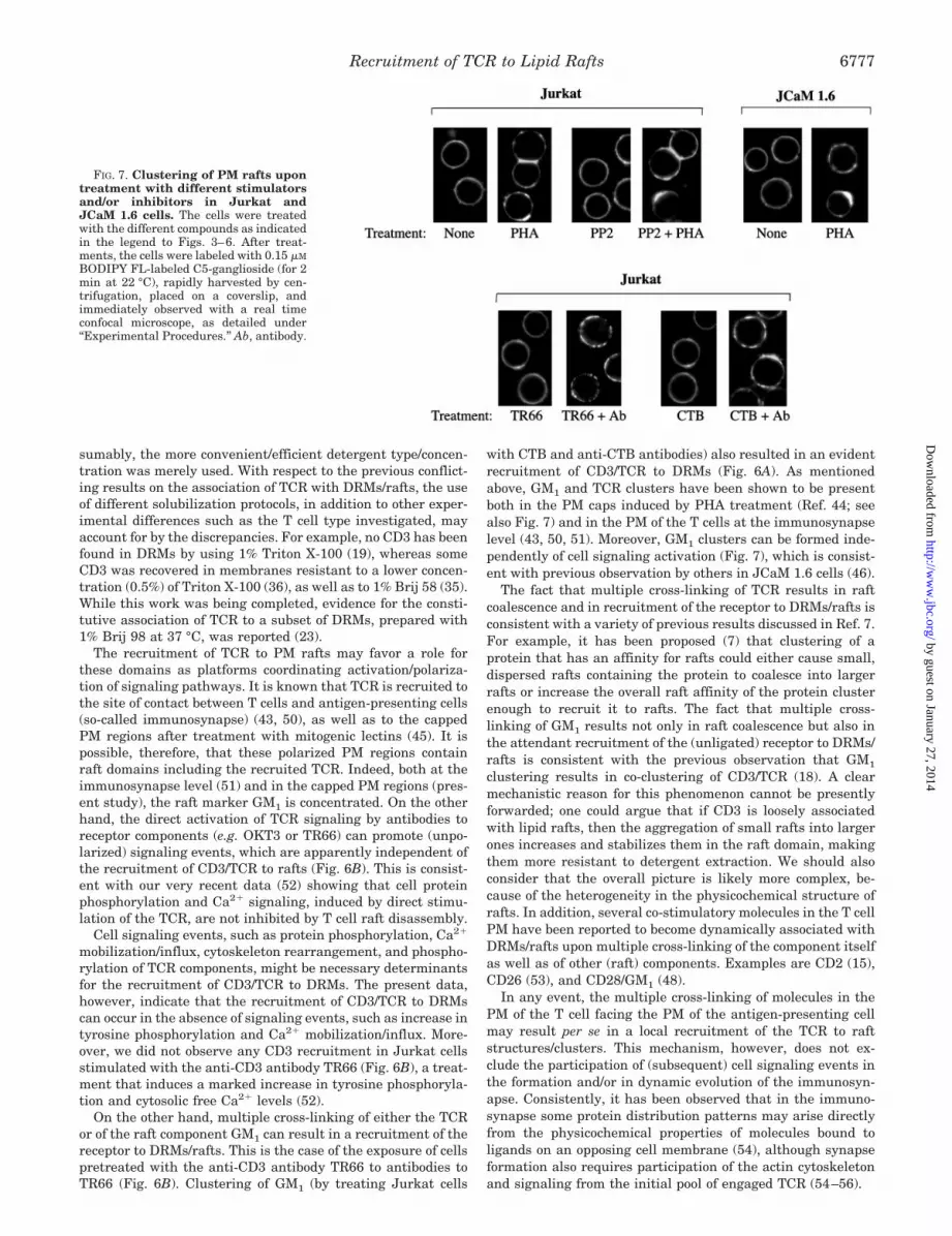
Membranes Is Associated with Clustering of PM Rafts—In afinal set of experiments, we investigated whether or not therecruitment of CD3 to DRMs (resistant to 0.2% Triton X-100) isparalleled by lipid raft clustering by microscopical observationof PM rafts probed with a fluorescent analogue of GM1. Indeed,previous microscopic observations have shown that fluorescentGM1 probes uniformly label the PM of resting (unstimulated)Jurkat cells but selectively stain PM patches in cells treatedwith cross-linking antibodies to GM1 or CD3 (18). The logicalexplanation is that the GM1 analogue inserts in PM lipid rafts;lipid rafts in resting cells, however, are too small (<70 nm indiameter (4–6)) to be resolvable by light microscopy, whereastheir aggregates (patches) are resolvable by light microscopy(18, 46, 47).
As demonstrated in Fig. 7, clusters of rafts were formed inthe experimental conditions, in which we observed the recruit-ment of the CD3� protein to DRMs. This appeared to be thecase independently of activation of cell signaling. Indeed, PHAtreatment of JCaM 1.6 or PP2-pretreated Jurkat cells resultedin both the recruitment of CD3� protein to DRMs (Figs. 4 and5) and raft clustering (Fig. 7) but in no evident activation oftyrosine phosphorylation and Ca2� signaling (Figs. 4 and 5).On the other hand, treating Jurkat cells with the antibody toCD3, TR66, caused neither the recruitment of the CD3� proteinto DRMs (Fig. 6A) nor clustering of PM rafts (Fig. 7). Instead,as expected on the basis of previous reports (22, 48), TR66stimulation caused a marked increase in tyrosine phosphoryl-ation and Ca2� signaling also in the present experimentalconditions (data not shown).
DISCUSSION
Although previous microscopical evidence suggest that theTCR is present in PM rafts (18), a variety of biochemical stud-ies gave conflicting results with respect to the constitutive/inducible association of the receptor with DRMs (18–23). Thepresent data show that a relatively low amount of the TCRcomponent CD3� is constitutively associated with a DRM sub-set and that this amount can be largely increased as a result ofT cell stimulation in a cell signaling-independent manner.
These data were gained by using 0.2% Triton X-100 or 1%Brij 58 to solubilize “conventional” nonraft membranes. On theother hand, we observed that DRMs prepared with the “classic”concentration of Triton X-100, i.e. 1%, did not contain anydetectable CD3 amount. It could be argued that 1% TritonX-100, but not the less hydrophobic detergent Brij 56 or a lowerconcentration of Triton X-100 itself, simply extracts CD3/TCRfrom rafts. However, solubilization with either Brij 58 or thelower concentration of Triton X-100 also resulted in a higherrecovery of membranes as well as of cholesterol and proteins inthe DRM-containing fractions. Therefore, a logical explanationis that the CD3/TCR complex is contained in a subset of cho-lesterol-enriched membranes that are not resistant to 1%Triton X-100 but are resistant to a lower Triton X-100 concen-tration or to Brij 58. The idea that heterogeneity in cholesterol-based DRMs and/or PM raft domains exists is not unprece-dented. For example, the co-existence within a membranedomain, such as the apical plasma membrane, of differentcholesterol-based lipid rafts has recently been proposed (9).Moreover, evidence for structural diversity of the PM domainsoccupied by functionally different glycosylphosphatidylinositol-anchored proteins has been previously forwarded (49).
It should be noted that a variety of previous studies on theassociation of signaling proteins to rafts/DRMs in T cells hasbeen based on the use of detergents other that Triton X-100 (13,14, 20, 22, 23, 31–35) or of Triton X-100 concentrations lowerthan 1% (14, 30, 36, 37). The aim of these studies, however, wasnot related to the possible heterogeneity in DRMs/rafts; pre-
FIG. 6. Multiple cross-linking of GM1 (A) or CD3 (B) results inthe recruitment of CD3� to DRMs. A, Jurkat cells were pretreatedwith the GM1 ligand, CTB, and then treated with or without antibodiesto CTB to cross-link GM1/CTB complexes as described under “Experi-mental Procedures.” B, Jurkat cells were pretreated with the antibodyto CD3, TR66, and then treated with or without antibodies to TR66, asdescribed under “Experimental Procedures.” The cells were lysed in amedium containing 0.2% Triton X-100, and the lysates were then frac-tionated by sucrose density centrifugation, as described under “Exper-imental Procedures”; proteins derived from 12.5 � 106 or 2 � 106 cells,in the case of fractions 3–8 or the solubilized materials (pooled fractions9–11), respectively, were analyzed by SDS-PAGE and Western blottingand probed with antibodies recognizing the CD3� protein. Ab, antibody
Recruitment of TCR to Lipid Rafts6776
by guest on January 27, 2014http://w
ww
.jbc.org/D
ownloaded from
sumably, the more convenient/efficient detergent type/concen-tration was merely used. With respect to the previous conflict-ing results on the association of TCR with DRMs/rafts, the useof different solubilization protocols, in addition to other exper-imental differences such as the T cell type investigated, mayaccount for by the discrepancies. For example, no CD3 has beenfound in DRMs by using 1% Triton X-100 (19), whereas someCD3 was recovered in membranes resistant to a lower concen-tration (0.5%) of Triton X-100 (36), as well as to 1% Brij 58 (35).While this work was being completed, evidence for the consti-tutive association of TCR to a subset of DRMs, prepared with1% Brij 98 at 37 °C, was reported (23).
The recruitment of TCR to PM rafts may favor a role forthese domains as platforms coordinating activation/polariza-tion of signaling pathways. It is known that TCR is recruited tothe site of contact between T cells and antigen-presenting cells(so-called immunosynapse) (43, 50), as well as to the cappedPM regions after treatment with mitogenic lectins (45). It ispossible, therefore, that these polarized PM regions containraft domains including the recruited TCR. Indeed, both at theimmunosynapse level (51) and in the capped PM regions (pres-ent study), the raft marker GM1 is concentrated. On the otherhand, the direct activation of TCR signaling by antibodies toreceptor components (e.g. OKT3 or TR66) can promote (unpo-larized) signaling events, which are apparently independent ofthe recruitment of CD3/TCR to rafts (Fig. 6B). This is consist-ent with our very recent data (52) showing that cell proteinphosphorylation and Ca2� signaling, induced by direct stimu-lation of the TCR, are not inhibited by T cell raft disassembly.
Cell signaling events, such as protein phosphorylation, Ca2�
mobilization/influx, cytoskeleton rearrangement, and phospho-rylation of TCR components, might be necessary determinantsfor the recruitment of CD3/TCR to DRMs. The present data,however, indicate that the recruitment of CD3/TCR to DRMscan occur in the absence of signaling events, such as increase intyrosine phosphorylation and Ca2� mobilization/influx. More-over, we did not observe any CD3 recruitment in Jurkat cellsstimulated with the anti-CD3 antibody TR66 (Fig. 6B), a treat-ment that induces a marked increase in tyrosine phosphoryla-tion and cytosolic free Ca2� levels (52).
On the other hand, multiple cross-linking of either the TCRor of the raft component GM1 can result in a recruitment of thereceptor to DRMs/rafts. This is the case of the exposure of cellspretreated with the anti-CD3 antibody TR66 to antibodies toTR66 (Fig. 6B). Clustering of GM1 (by treating Jurkat cells
with CTB and anti-CTB antibodies) also resulted in an evidentrecruitment of CD3/TCR to DRMs (Fig. 6A). As mentionedabove, GM1 and TCR clusters have been shown to be presentboth in the PM caps induced by PHA treatment (Ref. 44; seealso Fig. 7) and in the PM of the T cells at the immunosynapselevel (43, 50, 51). Moreover, GM1 clusters can be formed inde-pendently of cell signaling activation (Fig. 7), which is consist-ent with previous observation by others in JCaM 1.6 cells (46).
The fact that multiple cross-linking of TCR results in raftcoalescence and in recruitment of the receptor to DRMs/rafts isconsistent with a variety of previous results discussed in Ref. 7.For example, it has been proposed (7) that clustering of aprotein that has an affinity for rafts could either cause small,dispersed rafts containing the protein to coalesce into largerrafts or increase the overall raft affinity of the protein clusterenough to recruit it to rafts. The fact that multiple cross-linking of GM1 results not only in raft coalescence but also inthe attendant recruitment of the (unligated) receptor to DRMs/rafts is consistent with the previous observation that GM1
clustering results in co-clustering of CD3/TCR (18). A clearmechanistic reason for this phenomenon cannot be presentlyforwarded; one could argue that if CD3 is loosely associatedwith lipid rafts, then the aggregation of small rafts into largerones increases and stabilizes them in the raft domain, makingthem more resistant to detergent extraction. We should alsoconsider that the overall picture is likely more complex, be-cause of the heterogeneity in the physicochemical structure ofrafts. In addition, several co-stimulatory molecules in the T cellPM have been reported to become dynamically associated withDRMs/rafts upon multiple cross-linking of the component itselfas well as of other (raft) components. Examples are CD2 (15),CD26 (53), and CD28/GM1 (48).
In any event, the multiple cross-linking of molecules in thePM of the T cell facing the PM of the antigen-presenting cellmay result per se in a local recruitment of the TCR to raftstructures/clusters. This mechanism, however, does not ex-clude the participation of (subsequent) cell signaling events inthe formation and/or in dynamic evolution of the immunosyn-apse. Consistently, it has been observed that in the immuno-synapse some protein distribution patterns may arise directlyfrom the physicochemical properties of molecules bound toligands on an opposing cell membrane (54), although synapseformation also requires participation of the actin cytoskeletonand signaling from the initial pool of engaged TCR (54–56).
FIG. 7. Clustering of PM rafts upontreatment with different stimulatorsand/or inhibitors in Jurkat andJCaM 1.6 cells. The cells were treatedwith the different compounds as indicatedin the legend to Figs. 3–6. After treat-ments, the cells were labeled with 0.15 �M
BODIPY FL-labeled C5-ganglioside (for 2min at 22 °C), rapidly harvested by cen-trifugation, placed on a coverslip, andimmediately observed with a real timeconfocal microscope, as detailed under“Experimental Procedures.” Ab, antibody.
Recruitment of TCR to Lipid Rafts 6777
by guest on January 27, 2014http://w
ww
.jbc.org/D
ownloaded from

Acknowledgment—We are grateful to Antonella Viola for helpfuldiscussion.
REFERENCES
1. Simons, K., and Ikonen, E. (1997) Nature 387, 569–5722. Brown, D. A., and London, E. (1998) Annu. Rev. Cell Dev. Biol. 14, 111–1363. Anderson, R. G. (1993) Proc. Natl. Acad. Sci. U. S. A. 90, 10909–109134. Schnitzer, J. E., McIntosh, D. P., Dvorak, A. M., Liu, J., and Oh, P. (1995)
Science 269, 1435–14395. Brown, D. A., and London, E. (1997) Biochem. Biophys. Res. Commun. 240,
11–176. Simons, K., and Toomre, D. (2000) Nat. Rev. Mol. Cell Biol. 1, 31–397. Brown, D. A., and London, E. (2000) J. Biol. Chem. 275, 17221–172248. Anderson, R. G. (1998) Annu. Rev. Biochem. 67, 199–2259. Roper, K., Corbeil, D., and Huttner, W. B. (2000) Nat. Cell Biol. 2, 582–592
10. Cherukuri, A., Dykstra, M., and Pierce, S. K. (2001) Immunity 14, 657–66011. Rodgers, W., and Rose, J. K. (1996) J. Cell Biol. 135, 1515–152312. Zhang, W., Trible, R. P., and Samelson, L. E. (1998) Immunity 9, 239–24613. Zubiaur, M., Fernandez, O., Ferrero, E., Salmeron, J., Malissen, B., Malavasi,
F., and Sancho, J. (2002) J. Biol. Chem. 277, 13–2214. Ilangumaran, S., Briol, A., and Hoessli, D. C. (1998) Blood 91, 3901–390815. Yang, H., and Reinherz, E. L. (2001) J. Biol. Chem. 276, 18775–1878516. Yashiro-Ohtani, Y., Zhou, X. Y., Toyo-Oka, K., Tai, X. G., Park, C. S.,
Hamaoka, T., Abe, R., Miyake, K., and Fujiwara, H. (2000) J. Immunol.164, 1251–1259
17. Galbiati, F., Razani, B., and Lisanti, M. P. (2001) Cell 106, 403–41118. Janes, P. W., Ley, S. C., and Magee, A. I. (1999) J. Cell Biol. 147, 447–46119. Janes, P. W., Ley, S. C., Magee, A. I., and Kabouridis, P. S. (2000) Semin.
Immunol. 12, 23–3420. Balamuth, F., Leitenberg, D., Unternaehrer, J., Mellman, I., and Bottomly, K.
(2001) Immunity 15, 729–73821. Kosugi, A., Saitoh, S. I., Noda, S., Yasuda, K., Hayashi, F., Ogata, M., and
Hamaoka, T. (1999) Int. Immunol. 11, 1395–140122. Montixi, C., Langlet, C., Bernard, A. M., Thimonier, J., Dubois, C., Wurbel,
M. A., Chauvin, J. P., Pierres, M., and He, H. T. (1998) EMBO J. 17,5334–5348
23. Drevot, P., Langlet, C., Guo, X. J., Bernard, A. M., Colard, O., Chauvin, J. P.,Lasserre, R., and He, H. T. (2002) EMBO J. 21, 1899–1908
24. Sargiacomo, M., Sudol, M., Tang, Z., and Lisanti, M. (1993) J. Cell Biol. 122,789–807
25. Bensadoun, A., and Weinstein, D. (1976) Anal. Biochem. 70, 241–25026. Meissner, G. (1988) Methods Enzymol., 157, 417–43727. Fulceri, R., Bellomo, G., Gamberucci, A., Scott, H. M., Burchell A., and
Benedetti, A. (1992) Biochem. J. 286, 813–81728. Gamble, W., Vaughan, M., Kruth, H. S., and Avigan, J. (1978) J. Lipid Res. 19,
1068–107029. Gamberucci, A., Innocenti, B., Fulceri, R., Banhegyi, G., Giunti, R., Pozzan, T.,
and Benedetti, A (1994) J. Biol. Chem. 269, 23597–23602
30. Foger, N., Marhaba, R., and Zoller, M., (2000) J. Cell Sci. 114, 1169–117831. Rebres, R. A., Green, J. M., Reinhold, M. I., Ticchioni, M., and Brown, E. J.
(2001) J. Biol. Chem. 276, 7672–768032. Moran, M., and Miceli, C. M. (1998) Immunity 9, 787–79633. Marmor, M. D., and Julius, M. (2001) Blood 98, 1489–149734. Brdicka, T., Cerny, J., and Horejsi, V. (1998) Biochem. Biophys. Res. Commun.
248, 356–36035. Stulnig, M. T., Berger, M., Sigmund, T., Raederstorff, D., Stockinger, H., and
Waldhausl, W. (1998) J. Cell Biol. 143, 637–64436. Xavier, R., Brennan, T., Li, Q., McCormack, C., and Seed, B. (1998) Immunity
8, 723–73237. Khoshnan, A., Bae, D., Tindell. A. C., and Nel, A. E. (2000) J. Immunol. 165,
6933–694038. Smart, E. J., Ying, Y. S., Mineo, C., and Anderson, R. G. (1995) Proc. Natl.
Acad. Sci. U. S. A. 92, 10104–1010839. Van Oers, N. S. (1999) Semin. Immunol. 11, 227–23740. Straus, D. B., and Weiss, A. (1992) Cell 70, 585–59341. Samelson, L. E., Philips, A. F., Luong, E. T., and Klausner, R. D. (1990) Proc.
Natl. Acad. Sci. U. S. A. 87, 4358–436242. Denny, M. F., Patai, B., and Straus, D. B. (2000) Mol. Cell. Biol. 20, 1426–143543. Hanke, J. H., Gardner, J. P., Dow, R. L., Changelian, P. S., Brissette, W. H.,
Weringer, E. J., Pollok, B. A., and Connelly, P. A. (1996) J. Biol. Chem. 271,695–701
44. Grakoui, A., Bromley, S. K., Sumen, C., Davis, M. M. Shaw, A. S., Allen, P. M.,and Dustin, M. L. (1999) Science 285, 221–227
45. Khan, A. A., Steiner, J. P., Klein, M. G., Schneider, M. F., and Snyder, S. H.(1992) Science 257, 815–818
46. Fra, A. M., Williamson, E., Simons, K., and Parton, R. G. (1994) J. Biol. Chem.269, 30745–30748
47. Harder, T., and Simons, K. (1999) Eur. J. Immunol. 29, 556–56248. Viola, A., Schroeder, S., Sakakibara, Y., and Lanzavecchia, A. (1999) Science
283, 680–68249. Madore, N., Smith, K. L., Graham, C. H., Jen, A., Brady, K., Hall, S., and
Morris, R. (1999) EMBO J. 18, 6917–692650. Tomas, E. M., Chau, T. A., and Madrenas, J. (2002) Immunol. Lett. 83,
143–14751. Burack, W. R., Lee, K. H., Holdorf, A. D., Dustin, M. L., and Shaw, A. S. (2002)
J. Immunol. 169, 2837–284152. Pizzo, P., Giurisato, E., Tassi, M., Benedetti, A., Pozzan, T., and Viola, A.
(2002) Eur. J. Immunol. 32, 3082–309153. Ishii, T., Ohnuma, K., Murakami, A., Takasawa, N., Kobayashi, S., Dang,
N. H., Schlossman, S. F., and Morimoto, C. (2001) Proc. Natl. Acad. Sci.U. S. A. 98, 12138–12143
54. Qi, S. Y., Groves, J. T., and Chakraborty, A. K. (2001) Proc. Natl. Acad. Sci.U. S. A. 98, 6548–6553
55. Dustin, M. L., and Cooper, J. A. (2000) Nat. Immunol. 1, 23–2956. Wulfing, C., and Davis, M. M. (1998) Science 282, 2266–2269
Recruitment of TCR to Lipid Rafts6778
by guest on January 27, 2014http://w
ww
.jbc.org/D
ownloaded from


