Embed Size (px)
Citation preview
Targeted mutations of breast cancer susceptibility gene homologs in mice: lethal phenotypes of Brcal, Brca2, Brcal /Brca2, Brcal /p53, and Brca2/p53 nullizygous embryos T h o m a s Ludwig, 1 Deborah L. Chapman, 2 Virginia E. Papaioannou, 2 and Argiris Efstratiadis 2'3
Departments of 1Anatomy and Cell Biology and ~Genetics and Development, Columbia University, New York, New York 10032 USA
Mutations of the human BRCA1 and BRCA2 genes encoding tumor suppressors have been implicated in inherited predisposition to breast and other cancers. Disruption of the homologous mouse genes Brcal and Brca2 by targeting showed that they both have indispensable roles during embryogenesis, because nullizygous embryos become developmentally retarded and disorganized, and die early in development. In Brcal mutants, the onset of abnormalities is earlier by one day and their phenotypic features and time of death are highly variable, whereas the phenotype of Brca2 null embryos is more uniform, and they all survive for at least 8.5 embryonic days. Observations with Brcal/Brca2 double nullizygotes raise the possibility that the two developmental pathways could be linked. Interestingly, the impact of the Brcal or Brca2 null mutation is less severe in a p53 null background.
[Key Words: Brcal; Brca2; p53; mouse mutant; embryonic lethality]
Received February 11, 1997; revised version accepted April 11, 1997.
Two genes of unknown biochemical function, BRCA1 (Miki et al. 1994) and BRCA2 (Wooster et al. 1995; Tav- tigian et al. 1996), have been implicated in inherited pre- disposition to female breast cancer that is a causal factor for the disease in -5%-10% of all patients (for review, see Claus et al. 1991; Easton et al. 1993; Szabo and King 1995; Cannon-Albright and Skolnick 1996; Marcus et al. 1996; Stratton and Wooster 1996). Mutations of BRCA1 also predispose to ovarian cancer (see, e.g., Easton et al. 1995; Gayther et al. 1995; Johannsson et al. 1996; Serova et al. 1996), whereas mutations of BRCA2 are involved in male breast cancer and apparently in pancreatic can- cer (Schutte et al. 1995; Goggins et al. 1996; Teng et al. 1996). Loss of heterozygosity (LOH) consistently affect- ing the wild-type allele in tumors from patients carrying BRCA1 or BRCA2 mutations (Smith et al. 1992; Neu- hausen and Marshall 1994; Collins et al. 1995) indicated that these genes encode tumor suppressors. Moreover, BRCA1 antisense strategies using cultured ceils resulted either in acceleration of proliferation (Thompson et al. 1995) or cell transformation (Rao et al. 1996). Most of the mutations affecting the BRCA1 gene (Szabo and King
3Corresponding author. E-MAIL [email protected]; FAX (212) 923-2090.
1995; Cannon-Albright and Skolnick 1996; Feunteun and Lenoir 1996; Stratton and Wooster 1996; Xu and So- lomon 1996) or the BRCA2 gene (Wooster et al. 1995; Couch et al. 1996; Neuhausen et al. 1996; Phelan et al. 1996; Tavtigian et al. 1996; Teng et al. 1996; Thorlacius et al. 1996) result in truncated protein products. In spo- radic tumors, somatic BRCA1 or BRCA2 mutations oc- cur infrequently (Lancaster et al. 1996; Miki et al. 1996; Stratton and Wooster 1996; Teng et al. 1996).
The major BRCA1 mRNA (Miki et al. 1994; Xu et al. 1995; Brown et al. 1996; Smith et al. 1996) encodes a nuclear phosphoprotein (X. Chen et al. 1996; C.F. Chen et al. 1996) of 1863 amino acid residues, but differential splicing events generate additional mRNAs that are only partially characterized (C.F. Chen et al. 1996; Thakur et al. 1997; Wilson et al. 1997). The amino-terminal portion of the BRCA1 protein contains a RING finger domain (Freemont 1993; Saurin et al. 1996) that should be im- portant for function, because it is invariant between the human and mouse genes, otherwise exhibiting only 58% sequence identity at the amino acid level (Abel et al. 1995; Bennett et al. 1995; Lane et al. 1995; Sharan et al. 1995). The carboxy-terminal region of BRCA1, which acts as a transcriptional transactivator when fused with the GAL4 DNA-binding domain (Chapman and Verma 1996; Monteiro et al. 1996), contains two copies of a
1226 GENES & DEVELOPMENT 11:1226-1241 © 1997 by Cold Spring Harbor Laboratory Press ISSN 0890-9369[97 $5.00
Cold Spring Harbor Laboratory Press on July 26, 2016 - Published by genesdev.cshlp.orgDownloaded from
BrcaI and Brca2 mouse mutants
domain (BRCT; Koonin et al. 1996) exhibiting weak similarity with the mammalian p53-binding protein 53BP1; with the yeast RAD9 protein that is involved in cell cycle arrest at checkpoints on DNA damage; and with other proteins (Koonin et al. 1996; Bork et al. 1997; Callebaut and Mornon 1997), including BARD1 (Wu et al. 1996) that interacts with BRCA1 in vivo. The highly charged protein of 3418 residues encoded by BRCA2 is novel and, like BRCA1, not well conserved between hu- mans and mice (59% sequence identity at the amino acid level; Connor et al. 1997; Sharan and Bradley 1997). Eight variable internal repeats of unknown significance have been identified in the region ecoded by BRCA2 exon 11 (Bork et al. 1996), which are also present in the homolo- gous proteins of six other mammals, including mouse (Bingell et al. 1997).
To provide clues about the normal function of Brcal and Brca2, we have disrupted these genes in mice by targeted mutagenesis. Our results demonstrate that ei- ther of these mutations results in early embryonic le- thality in homozygous mutants, while heterozygous and double heterozygous mice are indistinguishable from wild type. We have also assessed the combined effects of the two mutations in double nullizygous embryos, and have examined potential relationships between the Brcal or Brca2 and p53 functions. While this work was in progress, three reports appeared (Gowen et al. 1996; Hakem et al. 1996; Liu et al. 1996) describing Brcal tar- geted mutations. Similarities and differences between these studies and our results are discussed.
R e s u l t s
Targeted disruption of the mouse Brcal and Brca2 genes
To generate a null mutation of the Brcal gene, we used a replacement vector that would delete a DNA frag- ment consisting of exon 2, encoding a part of the con- served RING finger motif, and portions of its flanking introns (see Materials and Methods). The targeted allele was designated Brcal ~x2. Similarly, we disrupted the Brca2 gene by replacing a segment of exon 11 with a neo cassette. Because of a frame-shift in the targeted allele (Brca2~×11), a stop codon is encountered in the neo se- quence. Therefore, any processed Brca2 transcript would generate a truncated protein product that should be in- active.
Genotyping of progeny (129/Sv x C57BL/6J) heterozy- gous for either the Brcal or the Brca2 mutation showed that these mice were indistinguishable from their wild- type littermates. Brca 1 heterozygous females are still tu- mor-free at the age of 15 months, whereas of the Brca2 heterozygotes, the oldest of which are eight months old, only a single four-month-old female developed a squa- mous cell carcinoma of the skin in the thoracic region (data not shown). However, the appearance of this carci- noma cannot be formally attributed to loss of Brca2 tu- mor suppressing activity, as PCR analysis of DNA ex-
tracted from histological sections and corresponding exclusively to tumor tissue indicated that Brca2 hetero- zygosity was maintained, and sequence analysis was not performed to determine whether the nontargeted allele was mutated.
Embryonic lethality of nullizygous Brcal and Brca2 mutants: comparative analysis
Genotyping of 183 progeny derived from intercrosses of heterozygous Brcal(+/-) mice showed that 59 were wild type, whereas the remaining 124 were heterozygous for the mutation. In analogous crosses between Brca2(+/-) heterozygotes, 160 pups were analyzed, 58 of which were wild type and 102 were heterozygous. In both cases, the complete absence of nullizygous mutants among these offspring, whereas heterozygous and wild-type progeny were obtained at the expected ratio, indicated that in the homozygous state the Brcal and Brca2 null mutations result in embryonic lethality.
To analyze the lethal phenotypes, pregnant females from matings between heterozygous Brcal or Brca2 mu- tants were sacrificed at different times post coitum, and embryos were examined in histological sections of dis- sected decidua and then genotyped. The gross morphol- ogy of additional embryos, some of which were geno- typed, was also examined. The data are summarized in Table 1. In both the histological and the gross morpho- logical analyses, we observed a high incidence of empty decidua in the Brcal series. Although genotyping was precluded, it is likely that most, if not all, of these empty decidua corresponded to Brcal null mutants, because their occurrence in the Brca2 series was very rare (Table 1). Moreover, a number of decidua contained only giant cells and extraembryonic membranes, which on geno- typing were found to be Brcal mutants (eight cases; five shown in Table 1). Therefore, the decidua with or with- out embryonic remnants are considered as one of the aspects of the variable Brcal null phenotype. In contrast, the phenotypic features of the Brca2 nullizygotes are overall quite uniform, especially before embryonic day 9.5 (E9.5).
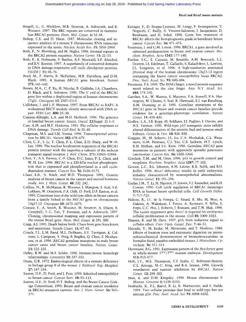
At the earliest age examined (E5.5), the wild-type and heterozygous embryos (hereafter referred to indiscrimi- nately as "normal," for brevity) were small egg cylinders with distinct embryonic and extraembryonic regions, which had initiated the formation of a proamniotic cav- ity. Moreover, the visceral endoderm was differentiated into extraembryonic (columnar) and embryonic (squa- mous) layers (Fig. 1A). This characteristic regional differ- ence in the appearance of endodermal cells was not evident in Brcal nullizygous mutants, which were rec- ognizable morphologically as small and shorter-than- normal egg cylinders without a visible demarcation of embryonic and extraembryonic regions (Fig. 1B). A pro- amniotic cavity was present in only one of five examined mutant embryos (data not shown). The primary giant cells, Reichert's membrane and distal endoderm of the mutants did not differ from those of the controls.
GENES & DEVELOPMENT 1227
Cold Spring Harbor Laboratory Press on July 26, 2016 - Published by genesdev.cshlp.orgDownloaded from
Ludwig et al.
Table 1. Embryonic phenotypes
Normal Abnormal
Genotype + +/+ +/- -/- N.D. +/- - /- N.D.
Giant cells + membranes only
-/- N.D.
Empty decidua (N.D.)
A. Brcal Histology
E5.5 3 5 E6.5 2 5 E7.5 6 9 7 E8.5 2 5
Gross morphology E6.5 46 E7.5 4 15 60 E8.5 6 E9.5 6 10 23
B. Brca2 Histology
E5.5 15 E6.5 4 9 2 E7.5 6 11 4 E8.5 7 11 2
Gross morphology E7.5 22 E8.5 17 E9.5 3 11
5 1 1 2 4 2 1 1 1 4 1 1 1
8 3 15
2 6 3
2 8 2 5 2
6 14
5
6 1 5
2 2 6
The genotyping data do not correspond to a random sample of embryos. However, if only a subset of the data from complete litters are considered, the expected Mendelian ratios are observed (not shown). (N.D.) Genotype not determined.
At E6.5, the Brcal mutan t embryos were two-layered cylinders that were about half of the normal size (Fig. 1, cf. C and D). Although the boundary between embryonic and extraembryonic regions was poorly demarcated, dif- ferentiation of the primary endoderm into columnar and squamous epi thel ium had occurred. The embryonic por- tion was disproportionately smaller (Fig. 1D) and, in con- trast to the normal embryos, the mutants lacked amni- otic folds. The primary giant cells associated wi th the mutan t embryos were prominent and very large (at the upper size l imi t of the normal range). Of four Brca2 mu- tant embryos examined at this age, two were indistin- guishable from their normal li t termates, whereas the other two were slightly shorter-than-normal egg cylin- ders wi th expanded proamniotic cavities (see Fig. 1E). This reduction in size was disproportionate for the ex- t raembryonic and embryonic regions, the latter being af- fected more severely, as in the case of the Brcal nulli- zygotes.
The normal embryos that were examined at E7.5 had gastrulated and possessed a third (mesodermal) germ layer and, wi th one exception (1/22 in the Brcal series), fused amniot ic folds {Fig. IF). Some of the normal em- bryos in the Brcal series and all of the normal embryos in the Brca2 series exhibited head-folds. The Brcal nul- lizygotes were small egg cylinders that lacked amniot ic folds, whereas presence of mesoderm was not readily vis- ible. However, large primary giant cells, a small ectopla- cental cone, Reichert 's membrane wi th distal endoderm,
extraembryonic ectoderm and columnar endoderm, and a meager epiblast and squamous endoderm surrounding a proamniotic cavity containing some pyknotic nuclei and cell debris were discernible (Fig. 1G). The phenotype of one of the mutants was more severe. It consisted of a loose network of giant cells, and a cluster of cells sur- rounded by a small sac wi th the appearance of distal endoderm (data not shown). In contrast to the Brcal mu- tants, the Brca2 nullizygotes were clearly three-layered egg cylinders wi th mesoderm present in the amniot ic folds and in the disproportionately reduced embryonic region (Fig. 1H). However, the amniot ic folds were not fused and the proamniotic cavity, containing cellular de- bris and some pyknotic nuclei, communica ted wi th the ectoplacental cavity. The embryos were surrounded by large giant cells and had a prominent ectoplacental cone and extraembryonic ectoderm. Despite the presence of pyknotic nuclei in the proamniotic cavities of some Brcal or Brca2 embryos that occasionally exceeded the level seen in controls, signs of excessive cell death in embryonic regions were not observed in any of the mu- tants at this or any other age.
At E8.5, the normal embryos in the Brcal series had head-folds and up to eight somites, whereas five geno- typed Brcal mutants were variable in shape and devel- opmental stage (Fig. II-K). One of them consisted only of giant cells, whereas in the embryonic region of three other mutants , the columnar cells of the stubby epiblast were surrounded by squamous endoderm, but there was
1228 GENES & DEVELOPMENT
Cold Spring Harbor Laboratory Press on July 26, 2016 - Published by genesdev.cshlp.orgDownloaded from
Brcal and Brca2 mouse mutants
e5.5 A ~.~.
.
e6.5 e7.5 e8.5
Jh "-
arm ~ ~--m
= ' 2 : J ~: • "
•
'.,4~, ~ " ,~,~ • , " e . . . . . -;I "''~ :
e8.5 bl L":,~;,/.:'::~ b2
• "" . ,
b 1M .Y:::'. b2
al
g ~ . . . . " m . : ' " v
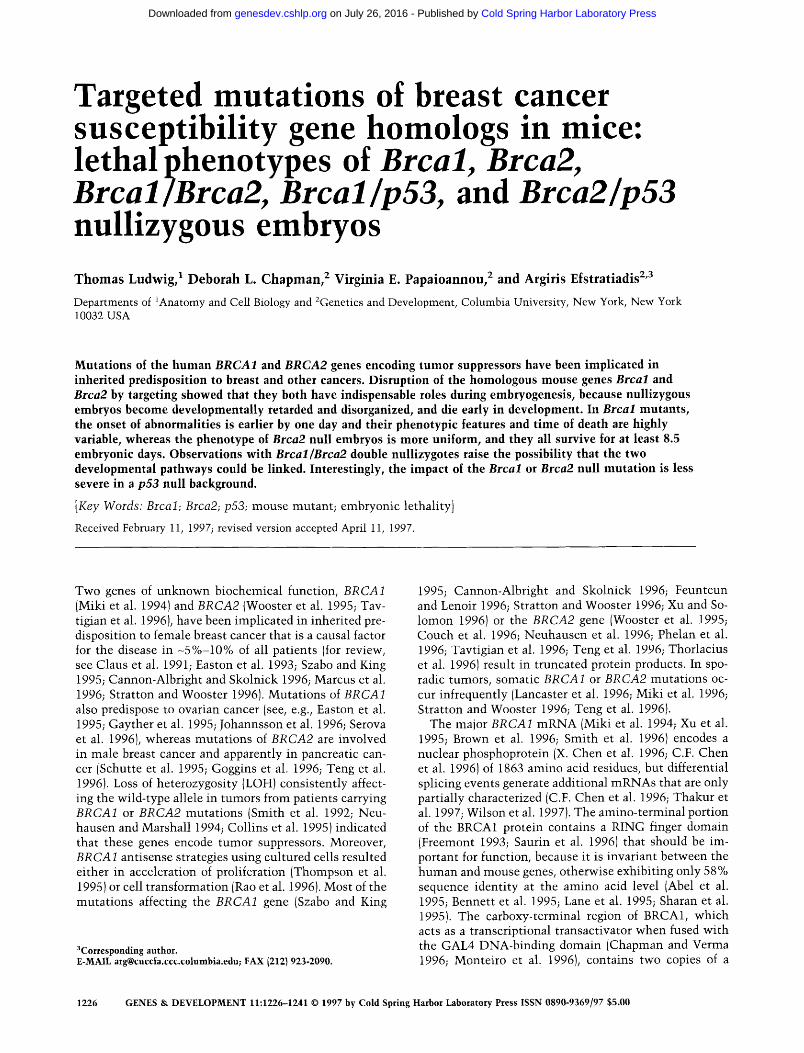
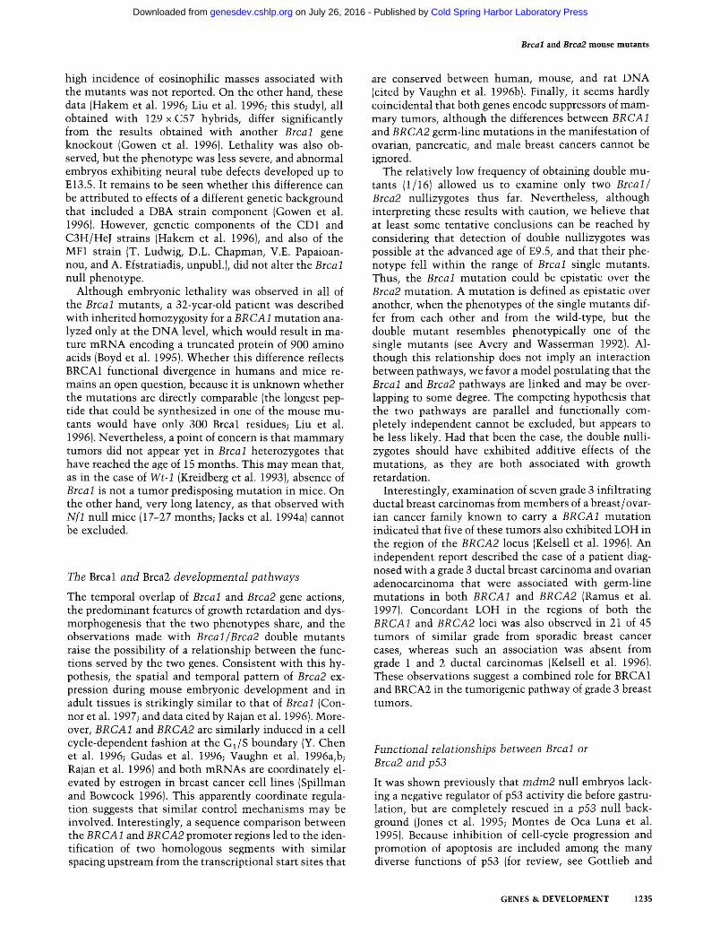
Figure 1. Histological comparisons of nor- mal and Brcal and Brca2 nullizygous em- bryos. Sections of normal embryos (n) and Brcal (bl) and Brca2 (b2) mutants are dis- played in five columns corresponding to the embryonic ages indicated on top. (A) Nor- mal (Brcal heterozygous) elongating egg cylinder with the beginning of a proamni- otic cavity (pac). In this and subsequent panels, an arrowhead marks the boundary between the extraembryonic and embry- onic regions. At this boundary, the endo- derm changes from columnar to squamous epithelium. (B) Brcal nullizygous embryo. Endoderm surrounding the primitive ecto- derm is evident, but the egg cylinder has not elongated and there is not yet any dis- tinction between the extraembryonic and embryonic regions. (C) Wild-type egg cylin- der with proamniotic cavity (pac); ectopla- cental cone (epc). (D,E) Brcal and Brca2 nullizygous egg cylinders with prominent proamniotic cavities. (F) Brcal heterozy- gous embryo with three distinct germ lay- ers: (m)mesoderm; (am) amnion; (ch) cho- rion; and (ys)yolk sac. (G) Brcal nullizy- gous embryo. There are no amniotic folds and the proamniotic cavity, which contains cellular debris, does not extend to the proxi- mal region of the extraembryonic ecto- derm. (H) Brca2 nullizygous embryo. The
amniotic folds (af) have formed, but not fused (*). Mesoderm (m) is evident in both the embryonic and extraembryonic regions. Cellular debris is present in the proamniotic cavity. (I) An arrow indicates the remains of a severely affected Brcal nullizygous embryo consisting of giant cells and a mass of undifferentiated tissue• A large eosinophilic mass (e) is present mesometrially. (J) A less severly affected Brcal nullizygous embryo. A loose network of giant cells (gc) surrounds Reichert's membrane and distal endoderm. The extraembryonic ectoderm (ee) has the appearance of a solid cellular mass, although a chorion (ch) has formed and extraembryonic mesoderm is present. In the embryonic region, the epiblast and endoderm surround a small amniotic cavity. (K) Sagittal section of the most advanced Brcal nullizygote observed in this study. The heart (h), head-fold (hf), posterior neural plate (*) and extraembryonic membranes are shown. An eosinophilic mass (e) is present antimesometrially. (L) Brca2 nullizygous embryo showing development of an ectoplacental cone (epc) and amniotic folds (af), but with a persistent proamniotic canal (*). The embryonic region is underdevel- oped, but contains mesoderm (m). (M) Transverse section of a Brca2 nullizygous embryo with an allantois (al), a diffuse amnion (am), and three germ layers in the underdeveloped embryonic region. (N) Sagittal section of a wild-type embryo with head-folds (hf), heart (h), somites (s), neural plate (*), and the allantois (al) attached to the chorion (ch). Scale bar, 100 ]~m (A,B); 180 ]um (C-E); 250 ]am (F-J,L); 400 pm (K,M); and 500 pm (N).
no visible mesoderm. However, extraembryonic meso- derm was detectable, whereas the extraembryonic ecto- derm, instead of being hollow, had the appearance of a solid cellular mass (Fig. 1J). In two of these mutan ts a distinct chorion was formed, therefore creating a small ectoplacental cavity. The ectoplacental cone was very small wi th little or no placental development. The em- bryos were surrounded by a loose ne twork of giant cells with large nuclei (Fig. 1J, K). The fifth Brca l mutan t was more advanced than any other examined; it contained an expanded yolk sac with blood islands, an amnion and an allantois, whereas the embryonic region, al though se- verely reduced in size, was quite organized and had head- folds, a neural tube closed in one region, and a heart (Fig. 1K). The normal embryos in the Brca2 series had turned and contained somites (Fig. IN), whereas the Brca2 nul- lizygotes were much smaller and lacked somites (Fig.
1L,M). Despite their reduced size, all of these mutan ts possessed embryonic mesoderm and had an anterior/ posterior axis. Several of them were forming head-folds and exhibited rudimentary heart differentiation. All mu- tant embryos had a yolk sac with blood islands, a promi- nent allantois, and a well-developed chorion. The devel- opment of the amnion, however, was poor, wi th a loose mesodermal layer (Fig. 1M), and, in one of the mutan ts (1/7), the proamniotic canal persisted (Fig. 1L). The em- bryos were surrounded by giant cells and had developing placentas wi th ectoplacental cones.
Gross morphological analysis was consistent wi th the previous observations and indicated that at E9.5 the range of phenotypes of Brca 1 mutants was similar to that observed at E8.5, except that the most advanced embryos had developed an axial embryonic region and possessed an allantois and an expanded yolk sac with blood islands
GENES & DEVELOPMENT 1229
Cold Spring Harbor Laboratory Press on July 26, 2016 - Published by genesdev.cshlp.orgDownloaded from
Ludwig et al.
(Table 1). In contrast to this phenotypic range of Brcal mutants, the E9.5 Brca2 nullizygotes were all similar, and exhibited a reduced embryonic region with head- folds, a large allantois, and an expanded yolk sac with visible blood. One of these mutants had three to four somites, whereas another had a beating heart.
Of 60 genotyped Brcal mutant embryos, nine (15%) were associated with eosinophilic masses present in the implantation crypt either on the mesometrial or on the antimesometrial side, which were infiltrated with ma- ternal neutrophils and possibly with endothelial cells (see Fig. II, K). This material had the appearance of a fi- brin-like deposit probably formed after hemorrhage of uterine vessels. Twenty additional eosinophilic masses were associated with nongenotyped abnormal embryos or with empty decidua. In the Brca2 series, on the other hand, the appearance of these deposits was very infre- quent, and observed in association with a single empty deciduum and only 1/32 (3%) genotyped Brca2 mutant embryos.
Cell proliferation assay in Brca2 mutan t s
Previously, the growth deficiency of Brcal nullizygotes was correlated with decreased cell proliferation (Hakem et al. 1996; Liu et al. 1996). To ascertain whether the same occurs in Brca2 mutants, pregnant females from heterozygous crosses were injected with 5-bromo-2'-de- oxyuridine (BrdU) 1 hr before sacrifice, and decidua re- covered at E6.5 and E7.5 were processed for histology. Incorporation of BrdU into DNA was assayed by count- ing labeled and unlabeled nuclei in the ectoplacental cone, the extraembryonic region and the embryonic re- gion in representative sagitta] sections close to the mid- line, whereas other sections were used for genotyping. Statistical differences in the averaged percentages of BrdU-positive nuclei were not detected between the three embryonic regions examined either in mutants or in controls (Student's t-test; P < 0.05). Therefore, for comparison, overall averages for the embryos of each group were used. At E6.5, there was no difference be- tween the mean values of the two groups; 67.5 + 2.7% and 68.4 _+ 1.8% of the nuclei were labeled in controls and mutants, respectively (three genotyped embryos in each group). The percentage of BrdU-positive cells was within the normal range even in one of the embryos of the latter group that was morphologically distinguish- able as a mutant. At E7.5, however, the corresponding values were significantly different: 67.6_+2.3% and 52.3 +_ 2.0% for controls and mutants, respectively (two embryos in each group). We conclude that, at the time when the Brca2 phenotype becomes unequivocally de- tectable by morphology, growth retardation is correlated with relative hypoproliferation.
Expression of marker genes
Previous reports suggested that the Brcal mutants fail to form mesoderm (Hakem et al. 1996; Liu et al. 1996). In
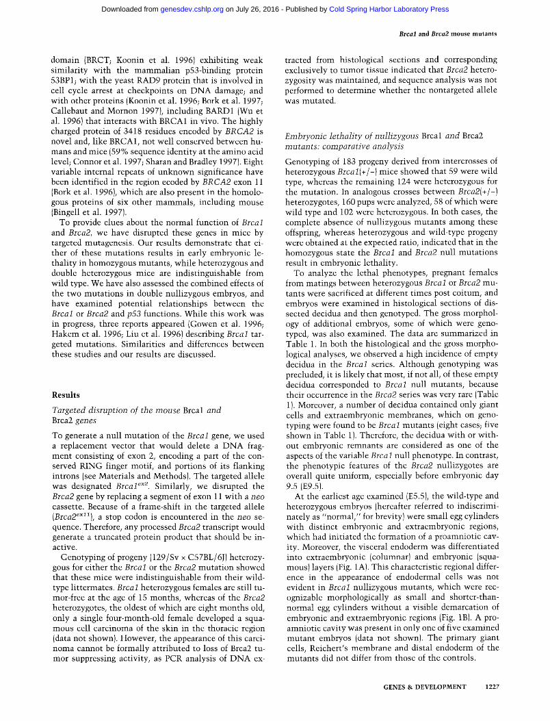
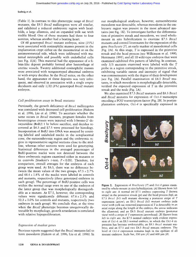
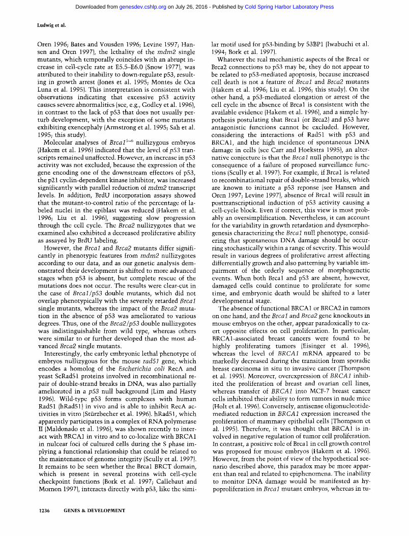
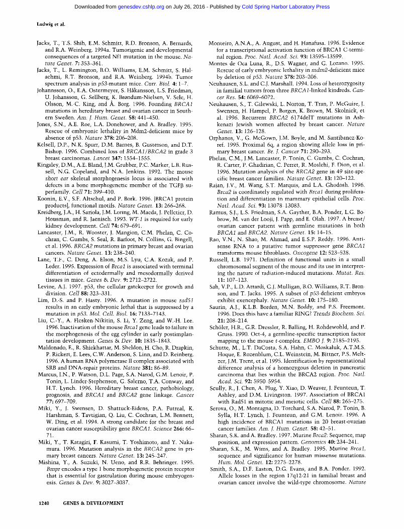
our morphological analyses, however, extraembryonic mesoderm was detectable, whereas mesoderm in the em- bryonic region was present in the most advanced mu- tants (see Fig. 1K). To investigate further the differentia- tion of primitive streak and mesoderm, we used whole- mount in situ hybridization to examine E7.5 Brcal mutants and control littermates for the expression of the gene Brachyury (T), an early marker of mesodermal cells (Fig. 2A). At this stage, T is expressed in the primitive streak and the head process (see Wilkinson et al. 1990; Herrmann 1991 ), and all 10 wild-type embryos that were examined exhibited this pattern of labeling. In contrast, only 3/5 mutants examined were labeled with the T probe in a region corresponding to the primitive streak, exhibiting variable extent and intensity of signal that was commensurate with the degree of their development (see Fig. 2A). Parallel examination of E8.5 Brca2 mu- tants, in which mesoderm is morphologically detectable, verified the expected expression of T in the primitive streak and the node (Fig. 2A).
We also examined E7.5 Brca2 mutants and E8.5 Brcal and Brca2 mutants for expression of Oct-4 transcripts encoding a POU transcription factor (Fig. 2B). In postim- plantation embryos, Oct-4 is specifically expressed in
Figure 2. Expression of Brachyury (T) and Oct-4 genes exam- ined by whole-mount in situ hybridization. (A) Shown from left to right are: A normal (n) E7.5 embryo expressing T (brown stripe} in the primitive streak (ps) and the head process (hp); an E7.5 Brcal (bl) null mutant showing a posterior stripe of T expression (arrow); an E8.5 Brca2 (b2) mutant embryo (side view) with yolk sac removed (expression of T is detectable in an axial stripe along the length of the embryo; the arrow indicates the allantois); and an E8.5 Brca2 mutant embryo (posterior view) with a stripe of T expression (arrowhead). (B) Shown from left to right are: An E7.5 normal embryo with evident expres- sion of Oct-4; an E8.5 normal embryo, in which the Oct-4 ex- pression has become undetectable; an E8.5 Brcal mutant em- bryo; and an E7.5 and two E8.5 Brca2 mutant embryos. The level of Oct-4 expression remains high in the epiblast of all mutant embryos. Scale bar, 350/am (A) and 600 ~m (B).
1230 GENES & DEVELOPMENT
Cold Spring Harbor Laboratory Press on July 26, 2016 - Published by genesdev.cshlp.orgDownloaded from
Brcal and Brca2 mouse mutants
the epiblast at high levels, but, as development progresses, the level of expression declines, and after E8.5 becomes restricted to the primordial germ cells (Sch61er et al. 1990; Yeom et al. 1996). All of the normal specimens examined showed this characteristic pattern of labeling, including confinement of signal to germ cells in the most advanced embryos. In the developmentally retarded mutant embryos of both types, however, Oct-4 signal remained positive in the embryonic portion of the egg cylinders (Fig. 2B).
In vitro outgrowth of blastocysts
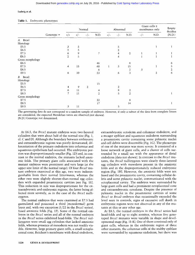
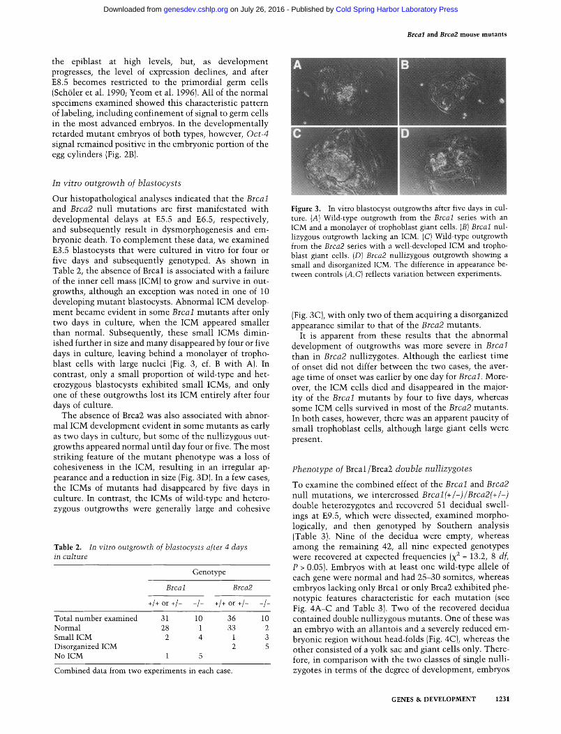
Our histopathological analyses indicated that the Brcal and Brca2 null mutations are first manifestated with developmental delays at E5.5 and E6.5, respectively, and subsequently result in dysmorphogenesis and em- bryonic death. To complement these data, we examined E3.5 blastocysts that were cultured in vitro for four or five days and subsequently genotyped. As shown in Table 2, the absence of Brcal is associated with a failure of the inner cell mass (ICM) to grow and survive in out- growths, although an exception was noted in one of 10 developing mutant blastocysts. Abnormal ICM develop- ment became evident in some Brcal mutants after only two days in culture, when the ICM appeared smaller than normal. Subsequently, these small ICMs dimin- ished further in size and many disappeared by four or five days in culture, leaving behind a monolayer of tropho- blast cells with large nuclei (Fig. 3, cf. B with A). In contrast, only a small proportion of wild-type and het- erozygous blastocysts exhibited small ICMs, and only one of these outgrowths lost its ICM entirely after four days of culture.
The absence of Brca2 was also associated with abnor- mal ICM development evident in some mutants as early as two days in culture, but some of the nullizygous out- growths appeared normal until day four or five. The most striking feature of the mutant phenotype was a loss of cohesiveness in the ICM, resulting in an irregular ap- pearance and a reduction in size (Fig. 3D). In a few cases, the ICMs of mutants had disappeared by five days in culture. In contrast, the ICMs of wild-type and hetero- zygous outgrowths were generally large and cohesive
T a b l e 2. In vitro outgrowth of blastocysts after 4 days in culture
Genotype
Brca 1 Brca2
+/+ or +/- -/- +/+ or +/- -/-
Total number examined 31 10 36 10 Normal 28 1 33 2 Small ICM 2 4 1 3 Disorganized ICM 2 5 No ICM 1 5
Combined data from two experiments in each case.
Figure 3. In vitro blastocyst outgrowths after five days in cul- ture. (A) Wild-type outgrowth from the Brcal series with an ICM and a monolayer of trophoblast giant cells. (B) Brcal nul- lizygous outgrowth lacking an ICM. (C) Wild-type outgrowth from the Brca2 series with a well-developed ICM and tropho- blast giant cells. (D) Brca2 nullizygous outgrowth showing a small and disorganized ICM. The difference in appearance be- tween controls (A,C) reflects variation between experiments.
(Fig. 3C), with only two of them acquiring a disorganized appearance similar to that of the Brca2 mutants.
It is apparent from these results that the abnormal development of outgrowths was more severe in Brcal than in Brca2 nullizygotes. Although the earliest time of onset did not differ between the two cases, the aver- age time of onset was earlier by one day for Brcal. More- over, the ICM cells died and disappeared in the major- ity of the Brcal mutants by four to five days, whereas some ICM cells survived in most of the Brca2 mutants. In both cases, however, there was an apparent paucity of small trophoblast ceils, although large giant cells were present.
Phenotype of Brcal/Brca2 double nullizygotes
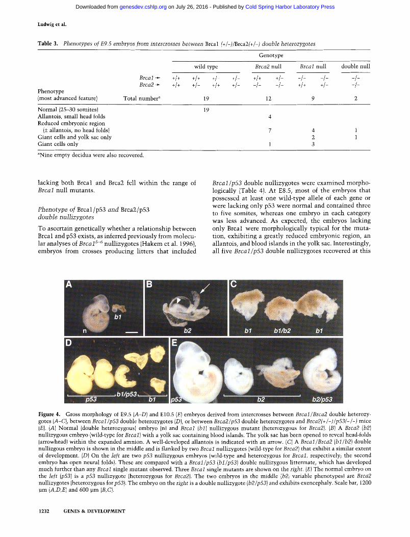
To examine the combined effect of the Brcal and Brca2 null mutations, we intercrossed Brcal(+/-)/Brca2(+/-) double heterozygotes and recovered 51 decidual swell- ings at E9.5, which were dissected, examined morpho- logically, and then genotyped by Southern analysis (Table 3). Nine of the decidua were empty, whereas among the remaining 42, all nine expected genotypes were recovered at expected frequencies (x 2 = 13.2, 8 df, P > 0.05). Embryos with at least one wild-type allele of each gene were normal and had 25-30 somites, whereas embryos lacking only Brcal or only Brca2 exhibited phe- notypic features characteristic for each mutation (see Fig. 4A-C and Table 3). Two of the recovered decidua contained double nullizygous mutants. One of these was an embryo with an allantois and a severely reduced em- bryonic region without head-folds (Fig. 4C), whereas the other consisted of a yolk sac and giant cells only. There- fore, in comparison with the two classes of single nulli- zygotes in terms of the degree of development, embryos
GENES & DEVELOPMENT 1231
Cold Spring Harbor Laboratory Press on July 26, 2016 - Published by genesdev.cshlp.orgDownloaded from
Ludwig et al.
Tab le 3. Phenotypes of E9.5 embryos from intercrosses between Brcal (+/-)/Brca2(+/-) double heterozygotes
Genotype
wild type Brca2 null Brcal null double null
Brcal+ +t+ +1+ +I- +1- +1+ +1- -I- - l- Brca2-~ +I+ +t- +t+ +1- -t- - t- +1+ +1-
Phenotype (most advanced feature) Total number a 19 12 9
-/- - / -
Normal (25-30 somites) Allantois, small head folds Reduced embryonic region
(+ allantois, no head folds) Giant cells and yolk sac only Giant cells only
19
4 1 2 1 3
aNine empty decidua were also recovered.
lacking both Brcal and Brca2 fell wi thin the range of Brcal null mutants .
Phenotype of Brcal /p53 and Brca2/p53 double nullizygotes
To ascertain genetically whether a relationship between Brcal and p53 exists, as inferred previously from molecu- lar analyses of Brcal s-6 nullizygotes (Hakem et al. 1996), embryos from crosses producing litters that included
Brcal/p53 double nullizygotes were examined morpho- logically (Table 4). At E8.5, most of the embryos that possessed at least one wild-type allele of each gene or were lacking only p53 were normal and contained three to five somites, whereas one embryo in each category was less advanced. As expected, the embryos lacking only Brcal were morphologically typical for the muta- tion, exhibiting a greatly reduced embryonic region, an allantois, and blood islands in the yolk sac. Interestingly, all five Brcal/p53 double nullizygotes recovered at this
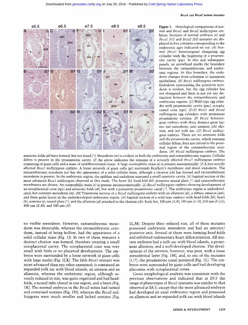
Figure 4. Gross morphology of E9.5 (A-D) and El0.5 (E) embryos derived from intercrosses between Brcal/Brca2 double heterozy- gotes (A-C), between Brcal/p53 double heterozygotes (D), or between Brca2/p53 double heterozygotes and Brca2(+/-)/p53(--/-) mice (E). (A) Normal (double heterozygous) embryo (n) and Brcal (bl) nullizygous mutant (heterozygous for Brca2). (B) A Brca2 (b2) nullizygous embryo (wild-type for Brcal) with a yolk sac containing blood islands. The yolk sac has been opened to reveal head-folds (arrowhead) within the expanded amnion. A well-developed allantois is indicated with an arrow. (C) A Brcal/Brca2 (bl/b2) double nullizgous embryo is shown in the middle and is flanked by two Brcal nullizygotes (wild-type for Brca2) that exhibit a similar extent of development. (D) On the left are two p53 nullizygous embryos (wild-type and heterozygous for Brcal, respectively; the second embryo has open neural folds). These are compared with a Brcal/p53 (bl/p53) double nullizygous littermate, which has developed much further than any Brcal single mutant observed. Three Brcal single mutants are shown on the right. (E) The normal embryo on the left (p53) is a p53 nullizygote (heterozygous for Brca2). The two embryos in the middle (b2; variable phenotypes) are Brca2 nullizygotes (heterozygous for p53). The embryo on the right is a double nullizygote (b2/p53) and exhibits exencephaly. Scale bar, 1200 ~m (A,D,E) and 600 pm (B,C).
1232 GENES & DEVELOPMENT
Cold Spring Harbor Laboratory Press on July 26, 2016 - Published by genesdev.cshlp.orgDownloaded from

Brcal and Brca2 m o u s e m u t a n t s
Table 4. Results of intercrosses between Brcal (+/-)/p53(+/-) double heterozygotes and of crosses between double heterozygotes and Brcal (+/-)/p53(-/-) mice
Phenotype (most advanced feature)
Genotype
wild type p53 null Brcal null double null
Srca~ ~ +1+ +1+ +I- +1- +1+ +1- -I- -I- -I- p53 + +1+ +1- +1+ +1- -I- -I- 4+ +1- -I-
A. E8.5
Total number a 7 Normal (3-5 somites) 5 Normal (head fold; 0-3 somites) 1 Retarded (egg cylinder) Reduced embryonic region Yolk sac only 1
B. E9.5
Total number b 22 Normal (15-20 somites) 20 Exencephalic c ( 15-20 somites) 1 Retarded (5 somites) Abnormal head fold (1-8 somites) Reduced embryonic region
(allantois, streak) Reduced embryonic region
(egg cylinder) Giant cells and yolk sac only Giant cells only 1
10 3 5 9
4 1 1
17 13 3 1
11 4
aAn additional embryo consisting of giant cells only was not genotyped. Three empty decidua were also recovered. BTwo empty decidua were also recovered. CExencephaly (i.e., protrusion of the brain above the skull) results from failure of neural tube closure. The term is used here for embryos exhibiting open midbrain and hindbrain.
age were more advanced than their Brcal null litter- mates. One of them was an egg cylinder with a constric- tion in the junction between the embryonic and extra- embryonic regions, whereas the remaining four, al- though developmentally retarded in comparison with their most advanced normal littermates, appeared to be normal and had reached the head-fold stage. Two of these four embryos possessed one to three somites. At E9.5 (see Fig. 4D and Table 4), most of the embryos possessing at least one wild-type allele of each gene or lacking only p53 were normal with 15-20 somites, whereas a minority of these embryos were exencephalic (exhibiting an open and flattened midbrain and hind- brain). The occurrence of exencephaly in a subset of p53 nullizygotes has been reported previously (Armstrong et al. 1995; Sah et al. 1995). We also observed a retarded p53 nullizygote with only five somites, whereas only giant cells were recovered from another embryo (hetero- zygous for Brcal and wild-type for p53). All embryos lacking Brcal exhibited a range of typical abnormal phe- notypes, whereas four Brcal/p53 double nullizygous mutants were further advanced than any of the embryos lacking only Brcal, although they were overtly retarded and abnormal in comparison with the controls and the p53 nullizygotes. The heart, a few somites, and large
head-folds had formed in the double mutants, whereas the neural folds had a zigzag or kinky appearance. In several of the embryos the allantois was fused with the chorion.
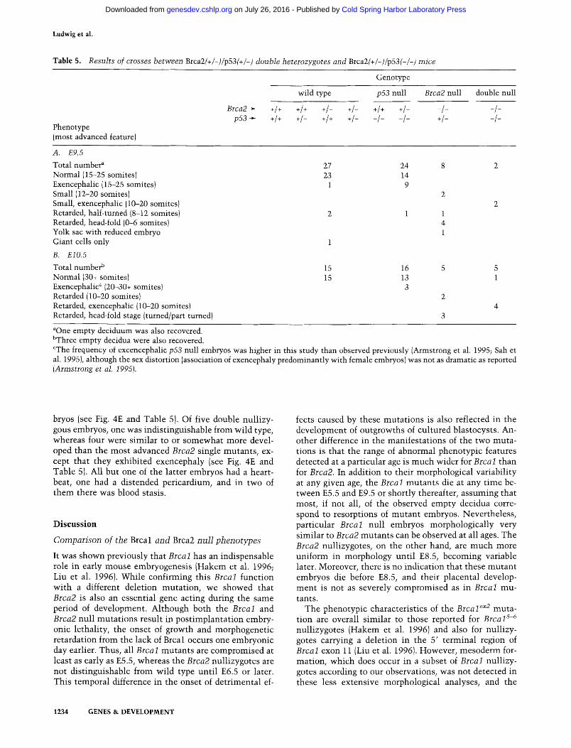
A similar analysis was performed by examining litters of embryos that included Brca2/p53 double nullizy- gotes, to determine if there is also a relationship between Brca2 and p53 (Table 5). The majority of E9.5 embryos possessing at least one wild-type allele of each gene or lacking only p53 were normal with 15-25 somites, al- though some of them were exencephalic. Of eight em- bryos lacking Brca2 (all heterozygous for p53), two were smaller than their most advanced littermates, but other- wise normal with 12-20 somites, whereas the remaining six were abnormal with variable phenotypes. Two double nullizygous embryos recovered at this age were similar to the most advanced Brca2 single mutants, ex- cept that they exhibited exencephaly. At El0.5, all of the embryos with at least one wild-type allele of each gene and most of the embryos lacking only p53 were normal with more than 30 somites, whereas a few p53 nullizy- gotes were exencephalic. All five embryos lacking only Brca2 (heterozygous for p53) were abnormal, ranging from fairly well-formed embryos about one third the size of normal littermates to retarded, head-fold stage em-
GENES & DEVELOPMENT 1233
Cold Spring Harbor Laboratory Press on July 26, 2016 - Published by genesdev.cshlp.orgDownloaded from
Ludwig et al.
Table 5. Results of crosses between Brca2(+/-)/p53(+/-) double heterozygotes and Brca2(+/-)/p53(-/-) mice
Phenotype (most advanced feature)
Genotype
wild type p53 null
Brca2 -~ +/+ +/+ +/- +/- +/+ +/- p53 ->- +l+ +!- +1+ +1- -1- -1-
Brca2 null
- /- +/-
double null
- / - - / -
A. E9.5
Total number a Normal (15-25 somites) Exencephalic (15-25 somites) Small (12-20 somites) Small, exencephalic ( 10-20 somites) Retarded, half-turned (8-12 somites) Retarded, head-fold (0-6 somites) Yolk sac with reduced embryo Giant cells only
B. ElO.5
Total number b Normal (30+ somites) Exencephalic ¢ (20-30+ somites) Retarded (10-20 somites) Retarded, exencephalic (10-20 somites) Retarded, head-fold stage (turned/part turned)
27 24 23 14
1 9
2 1
15 16 5 5 15 13 1
3
aOne empty deciduum was also recovered. bThree empty decidua were also recovered. CThe frequency of excencephalic p53 null embryos was higher in this study than observed previously (Armstrong et al. 1995; Sah et al. 1995), although the sex distortion (association of exencephaly predominantly with female embryos) was not as dramatic as reported (Armstrong et al. 1995).
bryos (see Fig. 4E and Table 5). Of five double nullizy- gous embryos, one was indist inguishable from wild type, whereas four were s imilar to or somewhat more devel- oped than the most advanced Brca2 single mutants , ex- cept that they exhibited exencephaly (see Fig. 4E and Table 5). All but one of the latter embryos had a heart- beat, one had a distended pericardium, and in two of them there was blood stasis.
D i s c u s s i o n
Comparison of the Brcal and Brca2 null phenotypes
It was shown previously that Brca 1 has an indispensable role in early mouse embryogenesis (Hakem et al. 1996; Liu et al. 1996). While confirming this Brcal function with a different deletion mutat ion, we showed that Brca2 is also an essential gene acting during the same period of development. Although both the Brcal and Brca2 null mutat ions result in post implantat ion embry- onic lethality, the onset of growth and morphogenetic retardation from the lack of Brcal occurs one embryonic day earlier. Thus, all Brcal mutants are compromised at least as early as E5.5, whereas the Brca2 nullizygotes are not dist inguishable from wild type unt i l E6.5 or later. This temporal difference in the onset of detr imental ef-
fects caused by these mutat ions is also reflected in the development of outgrowths of cultured blastocysts. An- other difference in the manifestat ions of the two muta- tions is that the range of abnormal phenotypic features detected at a particular age is much wider for Brca 1 than for Brca2. In addition to their morphological variabili ty at any given age, the Brcal mutants die at any t ime be- tween E5.5 and E9.5 or shortly thereafter, assuming that most, if not all, of the observed empty decidua corre- spond to resorptions of mutan t embryos. Nevertheless, particular Brcal null embryos morphologically very s imilar to Brca2 mutants can be observed at all ages. The Brca2 nullizygotes, on the other hand, are much more uniform in morphology unt i l E8.5, becoming variable later. Moreover, there is no indication that these mutan t embryos die before E8.5, and their placental develop- ment is not as severely compromised as in Brcal mu- tants.
The phenotypic characteristics of the Brcal ~x2 muta- tion are overall s imilar to those reported for Brcal s-6 nullizygotes (Hakem et al. 1996) and also for nullizy- gotes carrying a deletion in the 5' terminal region of Brcal exon 11 (Liu et al. 1996). However, mesoderm for- mation, which does occur in a subset of Brcal nullizy- gores according to our observations, was not detected in these less extensive morphological analyses, and the
1234 GENES & DEVELOPMENT
Cold Spring Harbor Laboratory Press on July 26, 2016 - Published by genesdev.cshlp.orgDownloaded from
Brcal and Brca2 m o u s e m u t a n t s
high incidence of eosinophilic masses associated with the mutants was not reported. On the other hand, these data (Hakem et al. 1996; Liu et al. 1996; this study), all obtained with 129 x C57 hybrids, differ significantly from the results obtained with another Brcal gene knockout (Gowen et al. 1996). Lethality was also ob- served, but the phenotype was less severe, and abnormal embryos exhibiting neural tube defects developed up to E13.5. It remains to be seen whether this difference can be attributed to effects of a different genetic background that included a DBA strain component (Gowen et al. 1996). However, genetic components of the CD1 and C3H/HeJ strains (Hakem et al. 1996), and also of the MF1 strain (T. Ludwig, D.L. Chapman, V.E. Papaioan- nou, and A. Efstratiadis, unpubl.), did not alter the Brca 1 null phenotype.
Although embryonic lethality was observed in all of the Brcal mutants, a 32-year-old patient was described with inherited homozygosity for a BR CA 1 mutation ana- lyzed only at the DNA level, which would result in ma- ture mRNA encoding a truncated protein of 900 amino acids (Boyd et al. 1995). Whether this difference reflects BRCA1 functional divergence in humans and mice re- mains an open question, because it is unknown whether the mutations are directly comparable (the longest pep- tide that could be synthesized in one of the mouse mu- tants would have only 300 Brcal residues; Liu et al. 1996). Nevertheless, a point of concern is that mammary tumors did not appear yet in Brcal heterozygotes that have reached the age of 15 months. This may mean that, as in the case of Wt-1 (Kreidberg et al. 1993), absence of Brca 1 is not a tumor predisposing mutation in mice. On the other hand, very long latency, as that observed with Nfl null mice (17-27 months; Jacks et al. 1994a) cannot be excluded.
The Brcal and Brca2 developmental pa thways
The temporal overlap of Brcal and Brca2 gene actions, the predominant features of growth retardation and dys- morphogenesis that the two phenotypes share, and the observations made with Brcal /Brca2 double mutants raise the possibility of a relationship between the func- tions served by the two genes. Consistent with this hy- pothesis, the spatial and temporal pattern of Brca2 ex- pression during mouse embryonic development and in adult tissues is strikingly similar to that of Brcal (Con- nor et al. 1997; and data cited by Rajah et al. 1996). More- over, BRCA1 and BRCA2 are similarly induced in a cell cycle-dependent fashion at the G1/S boundary (Y. Chen et al. 1996; Gudas et al. 1996; Vaughn et al. 1996a,b; Rajan et al. 1996) and both mRNAs are coordinately el- evated by estrogen in breast cancer cell lines (Spillman and Bowcock 1996). This apparently coordinate regula- tion suggests that similar control mechanisms may be involved. Interestingly, a sequence comparison between the BR CA 1 and BR CA2 promoter regions led to the iden- tification of two homologous segments with similar spacing upstream from the transcriptional start sites that
are conserved between human, mouse, and rat DNA (cited by Vaughn et al. 1996b). Finally, it seems hardly coincidental that both genes encode suppressors of mam- mary tumors, although the differences between BRCA1 and BR CA2 germ-line mutations in the manifestation of ovarian, pancreatic, and male breast cancers cannot be ignored.
The relatively low frequency of obtaining double mu- tants (1/16) allowed us to examine only two Brca l / Brca2 nullizygotes thus far. Nevertheless, although interpreting these results with caution, we believe that at least some tentative conclusions can be reached by considering that detection of double nullizygotes was possible at the advanced age of E9.5, and that their phe- notype fell within the range of Brcal single mutants. Thus, the Brcal mutation could be epistatic over the Brca2 mutation. A mutation is defined as epistatic over another, when the phenotypes of the single mutants dif- fer from each other and from the wild-type, but the double mutant resembles phenotypically one of the single mutants (see Avery and Wasserman 1992). Al- though this relationship does not imply an interaction between pathways, we favor a model postulating that the Brcal and Brca2 pathways are linked and may be over- lapping to some degree. The competing hypothesis that the two pathways are parallel and functionally com- pletely independent cannot be excluded, but appears to be less likely. Had that been the case, the double nulli- zygotes should have exhibited additive effects of the mutations, as they are both associated with growth retardation.
Interestingly, examination of seven grade 3 infiltrating ductal breast carcinomas from members of a breast/ovar- ian cancer family known to carry a BRCA1 mutation indicated that five of these tumors also exhibited LOH in the region of the BRCA2 locus (Kelsell et al. 1996). An independent report described the case of a patient diag- nosed with a grade 3 ductal breast carcinoma and ovarian adenocarcinoma that were associated with germ-line mutations in both BRCA1 and BRCA2 (Ramus et al. 1997). Concordant LOH in the regions of both the BRCA1 and BRCA2 loci was also observed in 21 of 45 tumors of similar grade from sporadic breast cancer cases, whereas such an association was absent from grade 1 and 2 ductal carcinomas (Kelsell et al. 1996). These observations suggest a combined role for BRCA1 and BRCA2 in the tumorigenic pathway of grade 3 breast tumors.
Functional relationships between Brcal or Brca2 and p53
It was shown previously that m d m 2 null embryos lack- ing a negative regulator of p53 activity die before gastru- lation, but are completely rescued in a p53 null back- ground (Jones et al. 1995; Montes de Oca Luna et al. 1995). Because inhibition of cell-cycle progression and promotion of apoptosis are included among the many diverse functions of p53 (for review, see Gottlieb and
GENES & DEVELOPMENT 1235
Cold Spring Harbor Laboratory Press on July 26, 2016 - Published by genesdev.cshlp.orgDownloaded from
Ludwig et al.
Oren 1996; Bates and Vousden 1996; Levine 1997; Han- sen and Oren 1997), the lethality of the mdm2 single mutants, which temporally coincides with an abrupt in- crease in cell-cycle rate at E5.5-E6.0 (Snow 1977), was attributed to their inability to down-regulate p53, result- ing in growth arrest (Jones et al. 1995; Montes de Oca Luna et al. 1995). This interpretation is consistent with observations indicating that excessive p53 activity causes severe abnormalities (see, e.g., Godley et al. 1996), in contrast to the lack of p53 that does not usually per- turb development, with the exception of some mutants exhibiting exencephaly (Armstrong et al. 1995; Sah et al. 1995; this study).
Molecular analyses of B r c a l s-6 nullizygous embryos (Hakem et al. 1996) indicated that the level of p53 tran- scripts remained unaffected. However, an increase in p53 activity was not excluded, because the expression of the gene encoding one of the downstream effectors of p53, the p21 cyclin-dependent kinase inhibitor, was increased significantly with parallel reduction of mdm2 transcript levels. In addition, BrdU incorporation assays showed that the mutant-to-control ratio of the percentage of la- beled nuclei in the epiblast was reduced (Hakem et al. 1996; Liu et al. 1996), suggesting slow progression through the cell cycle. The Brca2 nullizygotes that we examined also exhibited a decreased proliferative ability as assayed by BrdU labeling.
However, the Brcal and Brca2 mutants differ signifi- cantly in phenotypic features from mdm2 nullizygotes according to our data, and as our genetic analysis dem- onstrated their development is shifted to more advanced stages when p53 is absent, but complete rescue of the mutations does not occur. The results were clear-cut in the case of Brcal/p53 double mutants, which did not overlap phenotypically with the severely retarded Brcal single mutants, whereas the impact of the Brca2 muta- tion in the absence of p53 was ameliorated to various degrees. Thus, one of the Brca2/p53 double nullizygotes was indistinguishable from wild type, whereas others were similar to or further developed than the most ad- vanced Brca2 single mutants.
Interestingly, the early embryonic lethal phenotype of embryos nullizygous for the mouse rad51 gene, which encodes a homolog of the Escherichia coli RecA and yeast ScRad51 proteins involved in recombinational re- pair of double-strand breaks in DNA, was also partially ameliorated in a p53 null background (Lim and Hasty 1996). Wild-type p53 forms complexes with human Rad51 (hRad51) in vivo and is able to inhibit RecA ac- tivities in vitro (St~irzbecher et al. 1996). hRad51, which apparently participates in a complex of RNA polymerase II (Maldonado et al. 1996), was shown recently to inter- act with BRCA1 in vitro and to co-localize with BRCA1 in nulcear foci of cultured cells during the S phase im- plying a functional relationship that could be related to the maintenance of genome integrity (Scully et al. 1997). It remains to be seen whether the Brcal BRCT domain, which is present in several proteins with cell-cycle checkpoint functions (Bork et al. 1997; Callebaut and Mornon 1997), interacts directly with p53, like the simi-
lar motif used for p53-binding by 53BP1 (Iwabuchi et al. 1994; Bork et al. 1997).
Whatever the real mechanistic aspects of the Brcal or Brca2 connections to p53 may be, they do not appear to be related to p53-mediated apoptosis, because increased cell death is not a feature of Brcal and Brca2 mutants (Hakem et al. 1996; Liu et al. 1996; this study). On the other hand, a p53-mediated elongation or arrest of the cell cycle in the absence of Brcal is consistent with the available evidence (Hakem et al. 1996), and a simple hy- pothesis postulating that Brcal (or Brca2) and p53 have antagonistic functions cannot be excluded. However, considering the interactions of Rad51 with p53 and BRCA1, and the high incidence of spontaneous DNA damage in cells (see Carr and Hoekstra 1995), an alter- native conjecture is that the Brca 1 null phenotype is the consequence of a failure of proposed surveillance func- tions (Scully et al. 1997). For example, if Brcal is related to recombinational repair of double-strand breaks, which are known to initiate a p53 reponse (see Hansen and Oren 1997; Levine 1997), absence of Brcal will result in posttranscriptional induction of p53 activity causing a cell-cycle block. Even if correct, this view is most prob- ably an oversimplification. Nevertheless, it can account for the variability in growth retardation and dysmorpho- genesis characterizing the Brcal null phenotype, consid- ering that spontaneous DNA damage should be occur- ring stochastically within a range of severity. This would result in various degrees of proliferative arrest affecting differrentially growth and also patterning by variable im- pairment of the orderly sequence of morphogenetic events. When both Brcal and p53 are absent, however, damaged cells could continue to proliferate for some time, and embryonic death would be shifted to a later developmental stage.
The absence of functional BRCA1 or BRCA2 in tumors on one hand, and the Brca 1 and Brca2 gene knockouts in mouse embryos on the other, appear paradoxically to ex- ert opposite effects on cell proliferation. In particular, BRCAl-associated breast cancers were found to be highly proliferating tumors (Eisinger et al. 1996), whereas the level of BRCA1 mRNA appeared to be markedly decreased during the transition from sporadic breast carcinoma in situ to invasive cancer (Thompson et al. 1995). Moreover, overexpression of BRCA1 inhib- ited the proliferation of breast and ovarian cell lines, whereas transfer of BRCA1 into MCF-7 breast cancer cells inhibited their ability to form tumors in nude mice (Holt et al. 1996). Conversely, antisense oligonucleotide- mediated reduction in BRCA1 expression increased the proliferation of mammary epithelial cells (Thompson et al. 1995). Therefore, it was thought that BRCA1 is in- volved in negative regulation of tumor cell proliferation. In contrast, a positive role of Brcal in cell growth control was proposed for mouse embryos (Hakem et al. 1996). However, from the point of view of the hypothetical sce- nario described above, this paradox may be more appar- ent than real and related to epiphenomena. The inability to monitor DNA damage would be manifested as hy- poproliferation in Brcal mutant embryos, whereas in tu-
1236 GENES & D E V E L O P M E N T
Cold Spring Harbor Laboratory Press on July 26, 2016 - Published by genesdev.cshlp.orgDownloaded from
Brcal and Brca2 mouse mutants
mors, it could allow accumulat ion of additional muta- tions promoting abnormal overgrowth, if the lack of BRCA1 is involved in tumor progression, rather than ini- tiation, as proposed (Scully et al. 1997).
Compar ison of the Brcal and pnlf5 m u t a n t pheno types
Comparison of our data wi th the features of other mouse mutat ions resulting in post implantat ion lethali ty (see Copp 1995) showed that the Brcal nullizygous embryos exhibit a striking resemblance with the early embryonic lethal phenotype of homozygotes carrying the radiation- induced deletion muta t ion 5RD3OOH or se 1 (s_hort-ear le- thal; Russell 1971; Dunn 1972) that includes the se (short-ear) locus encoding Bmp-5 (bone morphogenetic protein 5; Kingsley et al. 1992) on mouse chromosome 9. The locus associated with the early embryonic lethali ty identified by the se 1 mutat ion is designated pnl f5 (prena- tal lethal function 5; Imai and Kingsley 1994). The his- tological appearance of the pnlf5 nullizygotes at around E8.0, which exhibit large giant cells, a severely reduced embryonic region, and an ectoplacental cavity filled with a solid mass of extraembryonic ectodermal cells (Dunn 1972), is almost indistinguishable in morphology from Brcal mutants at E8.5. The segment of mouse chromo- some 9 where pnlf5 resides is syntenic wi th the q12-13 region of human chromosome 6 (DeBry and Seldin 1996). Interestingly, the presence of a tumor suppressor gene at 6q13 has been postulated because of a high incidence of
allelic imbalance in this region in malignant breast tu- mors (Orphanos et al. 1995).
M a t e r i a l s a n d m e t h o d s
Construction of replacement vectors
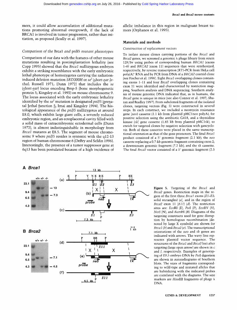
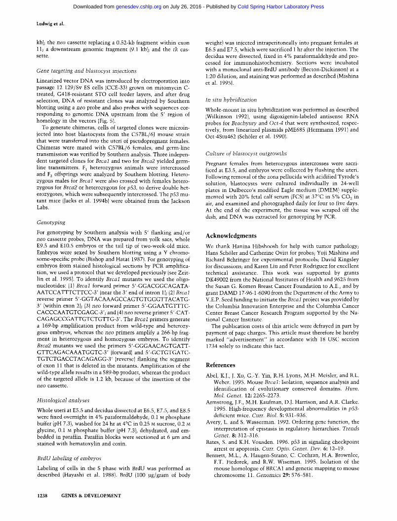
To isolate mouse clones carrying portions of the Brcal and Brca2 genes, we screened a genomic X phage library from strain 129/Sv using probes of corresponding human BRCA1 (exons 1-6) and BRCA2 (exon 11)sequences that were synthesized, respectively, by reverse transcription (RT)-PCR from HeLa cell poly(A) ÷ RNA and by PCR from DNA of a BRCA2 cosmid clone (see Fischer et al. 1996). Eight Brcal overlapping clones contain- ing exons 1-11 and four Brca2 overlapping clones containing exon 11 were identified and characterized by restriction map- ping, Southern analysis and DNA sequencing. Southern analy- sis of mouse genomic DNA indicated that, as in humans, the Brca2 gene is unique in mice (see also Connor et al. 1997; Sha- ran and Bradley 1997). From subcloned fragments of the isolated clones, targeting vectors (Fig. 5) were constructed in several steps. In each construct, we included a neomycin resistance gene (neo) cassette (1.1 kb from plasmid pMClneo polyA), for positive selection using the antibiotic G418, and a thymidine kinase (tk) gene cassette (1.85 kb from plasmid pMCltk), to enrich for targeted clones by negative selection with gancyclo- vir. Both of these cassettes were placed in the same transcrip- tional orientation as that of the gene promoters. The final Brcal product consisted of a 5' genomic fragment (2.1 kb); the neo cassette replacing a 0.7-kb genomic fragment containing exon 2; a downstream genomic fragment (7.2 kb); and the tk cassette. The final Brca2 vector consisted of a 5' genomic fragment (2.5
A Brcal
+ / + + / - - / -
23.1 - - 1 3
9.4 7.3
6.5
4.3
B Brca2
23.1
9.4
6.5
4.3
+ / + + / - - / -
~ i l - 7.1
- - 4.5
E P (a) I I
(c)
13 kb
V N E B E
I I J, 1/71 ~ 2
I X l l (b ) l I I I ' '
pro~e
X
EE
I F.3 I I
,
' F,I B3
P 7.3 kb I
H l k b
: 7.1 kb I
P BB P
(d )
' X ' ~ X l ( e ) ' I I
pro'T)e f~
( f ) El l
Figure 5. Targeting of the Brcal and Brca2 genes. Restriction maps in the re- gion of the first three Brcal exons (El-E3; solid rectangles) (a), and in the region of Brca2 exon 11 (Ell) (d). The restriction sites are: EcoRI (E), PstI (P), EcoRV (V), NotI (N), and BamHI (B). Diagrams of the targeting constructs used for gene disrup- tion by homologous recombination (de- noted by large X symbols) are shown for Brcal (b) and Brca2 (e). The transcriptional orientations of the neo and tk genes are indicated with arrows. The wavy line rep- resents plasmid vector sequence. The structures of the Brcal and Brca2 loci after targeting (large open arrow) are shown in c and f, respectively. Examples of genotyp- ing of E9.5 embryo DNA by PstI digestion are shown in autoradiograms of Southern blots. The sizes of fragments correspond- ing to wild-type and mutated alleles that are hybridizing with the indicated probes are correlated with the diagrams. The size markers are HindIII fragments of phage X DNA.
GENES & DEVELOPMENT 1237
Cold Spring Harbor Laboratory Press on July 26, 2016 - Published by genesdev.cshlp.orgDownloaded from
Ludwig et al.
kb); the neo cassette replacing a 0.52-kb fragment within exon 11; a downstream genomic fragment (9.1 kb); and the tk cas- sette.
Gene targeting and btastocyst injections
Linearized vector DNA was introduced by electroporation into passage 12 129/Sv ES cells (CCE-33) grown on mitomycin C- treated, G418-resistant STO cell feeder layers, and after drug selection, DNA of resistant clones was analyzed by Southern blotting using a neo probe and also probes with sequences cor- responding to genomic DNA upstream from the 5' region of homology in the vectors (Fig. 5).
To generate chimeras, cells of targeted clones were microin- jected into host blastocysts from the C57BL/6J mouse strain that were transferred into the uteri of pseudopregnant females. Chimeras were mated with C57BL/6 females, and germ-line transmission was verified by Southern analysis. Three indepen- dent targeted clones for Brcal and two for Brca2 yielded germ- line transmitters. F 1 heterozygous animals were intercrossed and F 2 offsprings were analyzed by Southern blotting. Hetero- zygous males for Brcal were also crossed with females hetero- zygous for Brca2 or heterozygous for p53, to derive double het- erozygotes, which were subsequently intercrossed. The p53 mu- tant mice (Jacks et al. 1994b) were obtained from the Jackson Labs.
Genotyping
For genotyping by Southern analysis with 5' flanking and/or neo cassette probes, DNA was prepared from yolk sacs, whole E9.5 and El0.5 embryos or the tail tip of two-week-old mice. Embryos were sexed by Southern blotting using a Y chromo- some-specific probe (Bishop and Hatat 1987). For genotyping of embryos from stained histological sections by PCR amplifica- tion, we used a protocol that we developed previously (see Zeit- lin et al. 1995). To identify Brcal mutants we used the oligo- nucleotides: (1)Brcal forward primer 5'-GGACGGCAGATA- AATCCATTTCTTCC-3' (near the 3' end of intron 1); (2) Brcai reverse primer 5'-GGTACAAAGCCAGTGTGGGTTACATG- 3' (within exon 2); (3) neo forward primer 5'-GGAATGTTTC- CACCCAATGTCGAGC-3'; and (4) neo reverse primer 5'-CAT- CAGAGCCGATTGTCTGTTG-3'. The Brcal primers generate a 169-bp amplification product from wild-type and heterozy- gous embryos, whereas the neo primers amplify a 266-bp frag- ment in heterozygous and homozygous embryos. To identify Brca2 mutants we used the primers 5'-GGGAACAGTGATT- GTTCAGACAAATGGTC-3' (forward) and 5'-GCTGTGATC- TGTCTGACCTACAGAGG-3' (reverse) flanking the segment of exon 11 that is deleted in the mutants. Amplification of the wild-type allele results in a 589-bp product, whereas the product of the targeted allele is 1.2 kb, because of the insertion of the neo cassette.
Histological analyses
Whole uteri at E5.5 and decidua dissected at E6.5, E7.5, and E8.5 were fixed overnight in 4% paraformaldehyde, 0.1 M phosphate buffer (pH 7.3}, washed for 24 hr at 4°C in 0.25 M sucrose, 0.2 M glycine, 0.1 M phosphate buffer (pH 7.3), dehydrated, and em- bedded in paraffin. Paraffin blocks were sectioned at 6 ~m and stained with hematoxylin and eosin.
BrdU labeling of embryos
Labeling of cells in the S phase with BrdU was performed as described (Hayashi et al. 1988). BrdU (100 ~g/gram of body
weight) was injected intraperitoneally into pregnant females at E6.5 and E7.5, which were sacrificed 1 hr after the injection. The decidua were dissected, fixed in 4% paraformaldehyde and pro- cessed for immunohistochemistry. Sections were incubated with a monoclonal anti-BrdU antibody (Becton-Dickinson) at a 1:20 dilution, and staining was performed as described (Mishina et al. 1995).
In situ hybridization
Whole-mount in situ hybridization was performed as described (Wilkinson 1992), using digoxigenin-labeled antisense RNA probes for Brachyury and Oct-4 that were synthesized, respec- tively, from linearized plasmids pME685 (Herrmann 1991)and Oct-4Stu462 (Sch61er et al. 1990).
Culture of blastocyst outgrowths
Pregnant females from heterozygous intercrosses were sacri- ficed at E3.5, and embryos were collected by flushing the uteri. Following removal of the zona pellucida with acidified Tyrode's solution, blastocysts were cultured individually in 24-well plates in Dulbecco's modified Eagle medium (DMEM) supple- mented with 20% fetal calf serum (FCS) at 37°C in 5% CO2 in air, and examined and photographed daily for four to five days. At the end of the experiment, the tissue was scraped off the dish, and DNA was extracted for genotyping by PCR.
A c k n o w l e d g m e n t s
We thank Hanina Hibshoosh for help with tumor pathology; Hans Sch61er and Catherine Ovitt for probes; Yuji Mishina and Richard Behringer for experimental protocols; David Kingsley for discussions; and Karen Liu and Peter Rodriguez for excellent technical assistance. This work was supported by grants DK49002 from the National Institutes of Health and 9625 from the Susan G. Komen Breast Cancer Foundation to A.E., and by grant DAMD 17-96-1-6090 from the Department of the Army to V.E.P. Seed funding to initiate the Brcal project was provided by the Columbia Innovation Enterprise and the Columbia Cancer Center Breast Cancer Research Program supported by the Na- tional Cancer Institute.
The publication costs of this article were defrayed in part by payment of page charges. This article must therefore be hereby marked "advertisement" in accordance with 18 USC section 1734 solely to indicate this fact.
R e f e r e n c e s
Abel, K.J., J. Xu, G.-Y. Yin, R.H. Lyons, M.H. Meisler, and B.L. Weber. 1995. Mouse Brcal: Isolation, sequence analysis and identification of evolutionary conserved domains. Hum. Mol. Genet. 12: 2265-2273.
Armstrong, J.F., M.H. Kaufman, D.J. Harrison, and A.R. Clarke. 1995. High-frequency developmental abnormalities in p53- deficient mice. Curt. Biol. 5" 931-936.
Avery, L. and S. Wasserman. 1992. Ordering gene function, the interpretation of epistasis in regulatory hierarchies. Trends Genet. 8" 312-316.
Bates, S. and K.H. Vousden. 1996. p53 in signaling checkpoint arrest or apoptosis. Curr. Opin. Genet. Dev. 6" 12-19.
Bennett, M.L., A. Haugen-Strano, C. Cochran, H.A. Brownlee, F.T. Fiedorek, and R.W. Wiseman. 1995. Isolation of the mouse homologue of BRCA1 and genetic mapping to mouse chromosome 11. Genomics 29: 576-581.
1238 GENES & DEVELOPMENT
Cold Spring Harbor Laboratory Press on July 26, 2016 - Published by genesdev.cshlp.orgDownloaded from
Brcal and Brca2 mouse mutants
Bingell, G., G. Micklem, M.R. Stratton, A. Ashworth, and R. Wooster. 1997. The BRC repeats are conserved in mamma- lian BRCA2 proteins. Hum. Mol. Genet. 6" 53-58.
Bishop, C.E. and D. Hatat. 1987. Molecular cloning and se- quence analysis of a mouse Y chromosome RNA transcript expressed in the testis. Nucleic Acids Res. 15: 2959-2969.
Bork, P., N. Blomberg, and M. Nigles. 1996. Internal repeats in the BRCA2 protein sequence. Nature Genet. 13- 22-23.
Bork, P., K. Hofmann, P. Bucher, A.F. Neuwald, S.F. Altschul, and E.V. Koonin. 1997. A superfamily of conserved domains in DNA damage-responsive cell cycle checkpoint proteins. FASEB J. 11: 68-76.
Boyd, M., F. Harris, R. McFarlane, H.R. Davidson, and D.M. Black. 1995. A human BRCA1 gene knockout. Nature 375: 541-542.
Brown, M.A., C.-F. Xu, H. Nicolai, B. Griffiths, J.A. Chambers, D. Black, and E. Solomon. 1996. The 5' end of the BRCA1 gene lies within a duplicated region of human chromosome 17q21. Oncogene 12: 2507-2513.
Callebaut, I. and J.-P. Mornon. 1997. From BRCA1 to RAPI: A widespread BRCT module closely associated with DNA re- pair. FEBS Lett. 400: 25-30.
Cannon-Albright, L.A. and M.H. Skolnick. 1996. The genetics of familial breast cancer. Semin. OncoI. 23(Suppl. 2): 1-5.
Carr, A.M. and M.F. Hoekstra. 1995. The cellular responses to DNA damage. Trends Cell Biol. 5" 32-40.
Chapman, M.S. and I.M. Verma. 1996. Transcriptional activa- tion by BRCA1. Nature 382" 678-679.
Chen, C.-F., S. Li, Y. Chen, P.-L. Chen, Z.D. Sharp, and W.-H. Lee. 1996. The nuclear localization sequences of the BRCA1 protein interact with the importin-a subunit of the nulcear transport signal receptor. J. Biol. Chem. 271: 32863-32868.
Chen, Y., A.A. Farmer, C.-F. Chen, D.C. Jones, P.-L. Chen, and W.-H. Lee. 1996. BRCA1 is a 220-kDa nuclear phosphopro- tein that is expressed and phosphorylated in a cell cycle- dependent manner. Cancer Res. 56:3168-3172.
Claus, E.B., N. Risch, and W.D. Thompson. 1991. Genetic analysis of breast cancer in the cancer and steroid hormone study. Am. J. Hum. Genet. 48: 232-242.
Collins, N., R. McManus, R. Wooster, J. Mangion, S. Seal, S.R. Lakhani, W. Ormiston, P.A. Daly, D. Ford, D.F. Easton, et al. 1995. Consistent loss of the wild type allele in breast cancers from a family linked to the BRCA2 gene on chromosome 13q12-13. Oncogene 10: 1673-1675.
Connor, F., A. Smith, R. Wooster, M. Stratton, A. Dixon, E. Campbell, T.-C. Tait, T. Freeman, and A. Ashworth. 1997. Cloning, chromosomal mapping and expression pattern of the mouse Brca2 gene. Hum. MoI. Genet. 6: 291-300.
Copp, A.J. 1995. Death before birth: Clues from gene knockouts and mutations. Trends Genet. 11: 87-93.
Couch, F.J., L.M. Farid, M.L. DeShano, S.V. Tavtigian, K. Cal- zone, L. Campeau, Y. Peng, B. Bogden, Q. Chen, S. Neuhau- sen, et al. 1996. BRCA2 germline mutations in male breast cancer cases and breast cancer families. Nature Genet. 13: 123-125.
DeBry, R.W. and M.F. Seldin. 1996. Human/mouse homology relationships. Genomics 33: 337-351.
Dunn, G.R. 1972. Embryological effects of a minute deficiency in linkage group II of the mouse. J. Embryol. Exp. Morphol. 27: 147-154.
Easton, D.F., D. Ford, and J. Peto. 1993. Inherited susceptibility to breast cancer. Cancer Surv. 18: 95-113.
Easton, D.F., D. Ford, D.T. Bishop, and the Breast Cancer Link- age Consortium. 1995. Breast and ovarian cancer incidence in BRCAl-mutation carriers. Am. J. Hum. Genet. 56" 265- 271.
Eisinger, F., D. Stoppa-Lyonnet, M. Longy, F. Kerangueven, T. Noguchi, C. Bailly, A. Vincent-Salomon, J. Jacquemier, D. Birnbaum, and H. Sobol. 1996. Germ line mutation of BRCA1 affects the histoprognostic grade in hereditary breast cancer. Cancer Res. 56: 471-474.
Feunteun, J. and G.M. Lenoir. 1996. BRCA1, a gene involved in inherited predisposition to breast and ovarian cancer. Bio- chim. Biophys. Acta 1242: 177-180.
Fischer, S.G., E. Cayanis, M. Bonaldo, A.M. Bowcock, L.L. Deaven, I.S. Edelman, T. Gallardo, S. Kalachikov, L. Lawton, J.L. Longmire, et al. 1996. A high-resolution annotated physical map of the human chromosome 13q12-13 region containing the breast cancer susceptibility locus BRCA2. Proc. Natl. Acad. Sci. 93: 690-694.
Freemont, P.S. 1993. The RING finger, a novel protein sequence motif related to the zinc finger. Ann. N.Y. Acad. Sci. 684: 174-192.
Gayther, S.A., W. Warren, S. Mazoyer, P.A. Russell, P.A. Har- rington, M. Chiano, S. Seal, R. Hamoudi, E.J. van Rensburg, A.M. Dunning et al. 1995. Germline mutations of the BRCA1 gene in breast and ovarian cancer families provide evidence for a genotype-phenotype correlation. Nature Genet. 11: 428-433.
Godley, L.A., J.B. Kopp, M. Eckhaus, J.J. Paglino, J. Owens, and H.E. Varmus. 1996. Wild-type p53 transgenic mice exhibit altered differentiation of the ureteric bud and possess small kidneys. Genes & Dev. 10: 836-850.
Goggins, M., M. Schutte, J.L. Lu, C.A. Moskaluk, C.L. Wein- stein, G.M. Petersen, C.J. Yeo, C.E. Jackson, H.T. Lynch, R.H. Hruban, and S.E. Kern. 1996. Germline BRCA2 gene mutations in patients with apparently sporadic pancreatic carcinomas. Cancer Res. 56: 5360-5364.
Gottlieb, T.M. and M. Oren. 1996. p53 in growth control and neoplasia. Biochim. Biophys. Acta 1287: 77-102.
Gowen, L.C., B.L. Johnson, A.M. Latour, K.K. Sulik, and B.H. Koller. 1996. Brcal deficiency results in early embryonic lethality characterized by neuroepithelial abnormalities. Nature Genet. 12: 191-194.
Gudas, J.M., T. Li, H. Nguyen, D. Jensen, F.J. Rauscher, and K.H. Cowan. 1996. Cell cycle regulation of BRCA1 messenger RNA in human breast epithelial cells. Cell Growth Differ. 7:717-723.
Hakem, R., J.L. de la Pompa, C. Sirard, R. Mo, M. Woo, A. Hakem, A. Wakeham, J. Potter, A. Reitmair, F. Billia, E. Firpo, C.C. Hui, J. Roberts, J. Rossant, and T.W. Mak. 1996. The tumor suppressor gene Brca 1 is required for embryonic cellular proliferation in the mouse. Cell 85: 1009-1023.
Hansen, R. and M. Oren. 1997. p53; from inductive signal to cellular effect. Curr. Opin. Genet. Dev. 7" 46-51.
Hayashi, Y., M. Koike, M. Matsutani, and T. Hoshino. 1988. Effects of fixation time and enzymatic digestion on immu- nohistochemical demonstration of bromodeoxyuridine in formalin-fixed, paraffin-embedded tissues. J. Histochem. Cy- tochem. 36: 511-514.
Herrmann, B.G. 1991. Expression pattern of the Brachyury gene in whole-mount TWi~/T wi'~ mutant embryos. Development 113: 913-917.
Holt, J.T., M.E. Thompson, C.I. Szabo, C. Robinson-Benion, C.L. Arteaga, M.-C. King, and R.A. Jensen. 1996. Growth retardation and tumour inhibition by BRCA1. Nature Genet. 12: 298-302.
Imai, K. and D.M. Kingsley. 1994. Mouse chromosome 9. Mature. Genome 5: S139-S153.
Iwabuchi, K., P.L. Bartel, B. Li, R. Marraccino, and S. Fields. 1994. Two cellular proteins that bind to wild-type but not mutant p53. Proc. Natl. Acad. Sci. 91: 6098-6102.
GENES & DEVELOPMENT 1239
Cold Spring Harbor Laboratory Press on July 26, 2016 - Published by genesdev.cshlp.orgDownloaded from
Ludwig et al.
Jacks, T., T.S. Shih, E.M. Schmitt, R.D. Bronson, A. Bernards, and R.A. Weinberg. 1994a. Tumorigenic and developmental consequences of a targeted Nfl mutation in the mouse. Na- ture Genet. 7" 353-361.
Jacks, T., L. Remington, B.O. Williams, E.M. Schmitt, S. Hal- achmi, R.T. Bronson, and R.A. Weinberg. 1994b. Tumor spectrum analysis in p53-mutan t mice. Curr. Biol. 4: 1-7.
Johannsson, O., E.A. Ostermeyer, S. Hakansson, L.S. Friedman, U. Johansson, G. Sellberg, K. Brondum-Nielsen, V. Sele, H. Olsson, M.-C. King, and A. Borg. 1996. Founding BRCA1 mutations in hereditary breast and ovarian cancer in South- ern Sweden. Am. J. Hum. Genet. 58: 441-450.
Jones, S.N., A.E. Roe, L.A. Donehower, and A. Bradley. 1995. Rescue of embryonic lethality in Mdm2-deficient mice by absence of p53. Nature 378: 206-208.
Kelsell, D.P., N.K. Spurr, D.M. Barnes, B. Gusterson, and D.T. Bishop. 1996. Combined loss of B R C A 1 / B R C A 2 in grade 3 breast carcinomas. Lancet 347" 1554-1555.
Kingsley, D.M., A.E. Bland, J.M. Grubber, P.C. Marker, L.B. Rus- sell, N.G. Copeland, and N.A. Jenkins. 1992. The mouse short ear skeletal morphogenesis locus is associated with defects in a bone morphogenetic member of the TGF~ su- perfamily. Cell 71: 399-410.
Koonin, E.V., S.F. Altschul, and P. Bork. 1996. [BRCA1 protein products], functional motifs. Nature Genet. 13: 266-268.
Kreidberg, J.A., H. Sariola, J.M. Loring, M. Maeda, J. pelletier, D. Housman, and R. Jaenisch. 1993. WT-1 is required for early kidney development. Cell 74: 679-691.
Lancaster, J.M., R. Wooster, J. Mangion, C.M. Phelan, C. Co- chran, C. Gumbs, S. Seal, R. Barfoot, N. Collins, G. Bingell, et al. 1996. BRCA2 mutations in primary breast and ovarian cancers. Nature Ge~et. 13: 238-240.
Lane, T.F., C. Deng, A. Elson, M.S. Lyu, C.A. Kozak, and P. Leder. 1995. Expression of Brcal is associated with terminal differentiation of ectodermally and mesodermally derived tissues in mice. Genes & Dev. 9: 2712-2722.
Levine, A.J. 1997. p53, the cellular gatekeeper for growth and division. Cell 88" 323-331.
Lim, D.-S. and P. Hasty. 1996. A mutation in mouse rad51 results in an early embryonic lethal that is suppressed by a mutation in p53. Mol. Ceil. Biol. 16: 7133-7143.
Liu, C.-Y., A. Flesken-Nikitin, S. Li, Y. Zeng, and W.-H. Lee. 1996. Inactivation of the mouse Brcal gene leads to failure in the morphogenesis of the egg cylinder in early postimplan- tation development. Genes & Dev. 10: 1835-1843.
Maldonado, E., R. Shiekhattar, M. Sheldon, H. Cho, R. Drapkin, P. Rickert, E. Lees, C.W. Anderson, S. Linn, and D. Reinberg. 1996. A human RNA polymerase II complex associated with SRB and DNA-repair proteins. Nature 381: 86-89.
Marcus, J.N., P. Watson, D.L. Page, S.A. Narod, G.M. Lenoir, P. Tonin, L. Linder-Stephenson, G. Salerno, T.A. Conway, and H.T. Lynch. 1996. Hereditary breast cancer, pathobiology, prognosis, and BRCA1 and BRCA2 gene linkage. Cancer 77" 697-709.
Miki, Y., J. Swensen, D. Shattuck-Eidens, P.A. Futreal, K. Harshman, S. Tavtigian, Q. Liu, C. Cochran, L.M. Bennett, W. Ding, et al. 1994. A strong candidate for the breast and ovarian cancer susceptibility gene BRCAI . Science 266: 66- 71.
Miki, Y., T. Katagiri, F. Kasumi, T. Yoshimoto, and Y. Naka- mura. 1996. Mutation analysis in the BRCA2 gene in pri- mary breast cancers. Nature Genet. 13" 245-247.
Mishina, Y., A. Suzuki, N. Ueno, and R.R. Behringer. 1995. Brnpr encodes a type 1 bone morphogenetic protein receptor that is essential for gastrulation during mouse embryogen- esis. Genes & Dev. 9" 3027-3037.
Monteiro, A.N.A., A. August, and H. Hanafusa. 1996. Evidence for a transcriptional activation function of BRCA1 C-termi- nal region. Proc. Natl. Acad. Sci. 93: 13595-13599.
Montes de Oca Luna, R., D.S. Wagner, and G. Lozano. 1995. Rescue of early embryonic lethality in mdm2-def ic ien t mice by deletion of p53. Nature 378: 203-206.
Neuhausen, S.L. and C.J. Marshall. 1994. Loss of heterozygosity in familial tumors from three BRCAl - l i nked kindreds. Can- cer Res. 54: 6069-6072.
Neuhausen, S., T. Gilewski, L. Norton, T. Tran, P. McGuire, J. Swensen, H. Hampel, P. Borgen, K. Brown, M. Skolnick, et al. 1996. Recurrent BRCA2 6174delT mutations in Ash- kenazi Jewish women affected by breast cancer. Nature Genet. 13: 126-128.
Orphanos, V., G. McGown, J.M. Boyle, and M. Santibanez-Ko- ref. 1995. Proximal 6q, a region showing allele loss in pri- mary breast cancer. Br. J. Cancer 71" 290-293.
Phelan, C.M., J.M. Lancaster, P. Tonin, C. Gumbs, C. Cochran, R. Carter, P. Ghadirian, C. Perret, R. Moslehi, F. Dion, et al. 1996. Mutation analysis of the BRCA2 gene in 49 site-spe- cific breast cancer families. Nature Genet. 13: 120-122.
Rajan, J.V., M. Wang, S.T. Marquis, and L.A. Ghodosh. 1996. Brca2 is coordinately regulated with Brcal during prolifera- tion and differentiation in mammary epithelial cells. Proc. Natl. Acad. Sci. 93: 13078-13083.
Ramus, S.J., L.S. Friedman, S.A. Gayther, B.A. Ponder, L.G. Bo- brow, M. van der Looji, J. Papp, and E. Olah. 1997. A breast/ ovarian cancer patient with germline mutations in both BRC AI and BRCA2. Nature Genet. 15: 14-15.
Rao, V.N., N. Shao, M. Ahmad, and E.S.P. Reddy. 1996. Anti- sense RNA to a putative tumor suppressor gene BRCA1 transforms mouse fibroblasts. Oncogene 12: 523-528.
Russell, L.B. 1971. Definition of functional units in a small chromosomal segment of the mouse and its use in interpret- ing the nature of radiation-induced mutations. Mutat. Res. l l : 107-123.
Sah, V.P., L.D. Attardi, G.J. Mulligan, B.O. Williams, R.T. Bron- son, and T. Jacks. 1995. A subset of p53-deficient embryos exhibit exencephaly. Nature Genet. 10: 175-180.
Saurin, A.J., KL.B. Borden, M.N. Boddy, and P.S. Freemont. 1996. Does this have a familiar RING? Trends Biochem. Sci. 21: 208-214.
Sch61er, H.R., G.R. Dressier, R. Balling, H. Rohdewohld, and P. Gruss. 1990. Oct-4, a germline-specific transcription factor mapping to the mouse t-complex. EMBO J. 9" 2185-2195.
Schutte, M., L.T. DaCosta, S.A. Hahn, C. Moskaluk, A.T.M.S. Hoque, E. Rozenblum, C.L. Weinstein, M. Bittner, P.S. Melt- zer, J.M. Trent, et al. 1995. Identification by representational difference analysis of a homozygous deletion in pancreatic carcinoma that lies within the BRCA2 region. Proc. Natl. Acad. Sci. 92" 5950-5954.
Scully, R., J. Chen, A. Plug, Y. Xiao, D. Weaver, J. Feunteun, T. Ashley, and D.M. Livingston. 1997. Association of BRCA1 with Rad51 in mitotic and meiotic cells. Cell 88" 265-275.
Serova, O., M. Montagna, D. Torchard, S.A. Narod, P. Tonin, B. Sylla, H.T. Lynch, J. Feunteun, and G.M. Lenoir. 1996. A high incidence of BRCA1 mutations in 20 breast-ovarian cancer families. Am. J. Hum. Genet. 58" 42-51.
Sharan, S.K. and A. Bradley. 1997. Murine Brca2: Sequence, map position, and expression pattern. Genomics 40: 234-241.
Sharan, S.K., M. Wims, and A. Bradley. 1995. Murine Brcal, sequence and significance for human missense mutations. Hum. Mol. Genet. 12: 2275-2278.
Smith, S.A., D.F. Easton, D.G. Evans, and B.A. Ponder. 1992. Allele losses in the region 17q12-21 in familial breast and ovarian cancer involve the wild-type chromosome. Nature
1240 GENES & DEVELOPMENT
Cold Spring Harbor Laboratory Press on July 26, 2016 - Published by genesdev.cshlp.orgDownloaded from
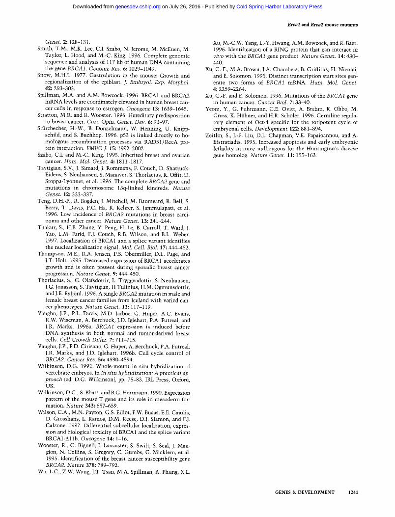
Brcal and Brca2 mouse mutants
Genet. 2: 128-131. Smith, T.M., M.K. Lee, C.I. Szabo, N. Jerome, M. McEuen, M.
Taylor, L. Hood, and M.-C. King. 1996. Complete genornic sequence and analysis of 117 kb of human DNA containing the gene BRCA1. Genome Res. 6: 1029-1049.
Snow, M.H.L. 1977. Gastrulation in the mouse: Growth and regionalization of the epiblast. I. Embryol. Exp. Morphol. 42: 293-303.
Spillman, M.A. and A.M. Bowcock. 1996. BRCA1 and BRCA2 mRNA levels are coordinately elevated in human breast can- cer cells in response to estrogen. Oncogene 13: 1639-1645.
Stratton, M.R. and R. Wooster. 1996. Hereditary predisposition to breast cancer. Curr. Opin. Genet. Dev. 6: 93-97.
St~irzbecher, H.-W., B. Donzelmann, W. Henning, U. Knipp- schild, and S. Buchhop. 1996. p53 is linked directly to ho- mologous recombination processes via RAD51/RecA pro- tein interaction. EMBO J. 15: 1992-2002.
Szabo, C.I. and M.-C. King. 1995. Inherited breast and ovarian cancer. Hum. Mol. Genet. 4: 1811-1817.
Tavtigian, S.V., J. Simard, J. Rommens, F. Couch, D. Shattuck- Eidens, S. Neuhausen, S. Marajver, S. Thorlacius, K. Offit, D. Stoppa-Lyonnet, et al. 1996. The complete BRCA2 gene and mutations in chromosome 13q-linked kindreds. Nature Genet. 12: 333-337.
Teng, D.H.-F., R. Bogden, J. Mitchell, M. Baumgard, R. Bell, S. Berry, T. Davis, P.C. Ha, R. Kehrer, S. Jammulapati, et al. 1996. Low incidence of BRCA2 mutations in breast carci- noma and other cancer. Nature Genet. 13: 241-244.
Thakur, S., H.B. Zhang, Y. Peng, H. Le, B. Carroll, T. Ward, J. Yao, L.M. Farid, F.J. Couch, R.B. Wilson, and B.L. Weber. 1997. Localization of BRCA1 and a splice variant identifies the nuclear localization signal. MoI. Cell. Biol. 17" 444-452.
Thompson, M.E., R.A. Jensen, P.S. Obermiller, D.L. Page, and J.T. Holt. 1995. Decreased expression of BRCA1 accelerates growth and is often present during sporadic breast cancer progression. Nature Genet. 9: 444-450.
Thorlacius, S., G. Olafsdottir, L. Tryggvadottir, S. Neuhausen, J.G. Jonasson, S. Tavtigian, H Tulinius, H.M. Ogmunsdottir, and J.E. Eyfj6rd. 1996. A single BRCA2 mutation in male and female breast cancer families from Iceland with varied can- cer phenotypes. Nature Genet. 13:117-119.
Vaughn, J.P., P.L. Davis, M.D. Jarboe, G. Huper, A.C. Evans, R.W. Wiseman, A. Berchuck, J.D. Iglehart, P.A. Futreal, and J.R. Marks. 1996a. BRCA1 expression is induced before DNA synthesis in both normal and tumor-derived breast cells. Cell Growth Differ. 7:711-715.
Vaughn, J.P., F.D. Cirisano, G. Huper, A. Berchuck, P.A. Futreal, J.R. Marks, and J.D. Iglehart. 1996b. Cell cycle control of BRCA2. Cancer Res. 56: 4590-4594.
Wilkinson, D.G. 1992. Whole-mount in situ hybridization of vertebrate embryos. In In situ hybridization: A practical ap- proach (ed. D.G. Wilkinson), pp. 75-83. IRL Press, Oxford, UK.
Wilkinson, D.G., S. Bhatt, and B.G. Herrmann. 1990. Expression pattern of the mouse T gene and its role in mesoderm for- mation. Nature 343: 657-659.
Wilson, C.A., M.N. Payton, G.S. Elliot, F.W. Buaas, E.E. Cajulis, D. Grosshans, L. Ramos, D.M. Reese, D.J. Slamon, and F.J. Calzone. 1997. Differential subcellular localization, expres- sion and biological toxicity of BRCA1 and the splice variant BRCAI-&llb. Oncogene 14: 1-16.
Wooster, R., G. Bignell, J. Lancaster, S. Swift, S. Seal, J. Man- gion, N. Collins, S. Gregory, C. Gumbs, G. Micklem, et al. 1995. Identification of the breast cancer susceptibility gene BRCA2. Nature 378: 789-792.
Wu, L.C., Z.W. Wang, J.T. Tsan, M.A. Spillman, A. Phung, X.L.
Xu, M.-C.W. Yang, L.-Y. Hwang, A.M. Bowcock, and R. Baer. 1996. Identification of a RING protein that can interact in vivo with the BRCA1 gene product. Nature Genet. 14: 430- 440.
Xu, C.-F., M.A. Brown, J.A. Chambers, B. Griffiths, H. Nicolai, and E. Solomon. 1995. Distinct transcription start sites gen- erate two forms of BRCA1 mRNA. Hum. Mol. Genet. 4" 2259-2264.
Xu, C.-F. and E. Solomon. 1996. Mutations of the BRCA1 gene in human cancer. Cancer Biol. 7: 33-40.
Yeom, Y., G. Fuhrmann, C.E. Ovitt, A. Brehm, K. Ohbo, M. Gross, K. Hiibner, and H.R. Sch61er. 1996. Germline regula- tory element of Oct-4 specific for the totipotent cycle of embryonal cells. Development 122: 881-894.
Zeitlin, S., J.-P. Liu, D.L. Chapman, V.E. Papaioannou, and A. Efstratiadis. 1995. Increased apoptosis and early embryonic lethality in mice nullizygous for the Huntington's disease gene homolog. Nature Genet. l l : 155-163.
GENES & DEVELOPMENT 1241
Cold Spring Harbor Laboratory Press on July 26, 2016 - Published by genesdev.cshlp.orgDownloaded from
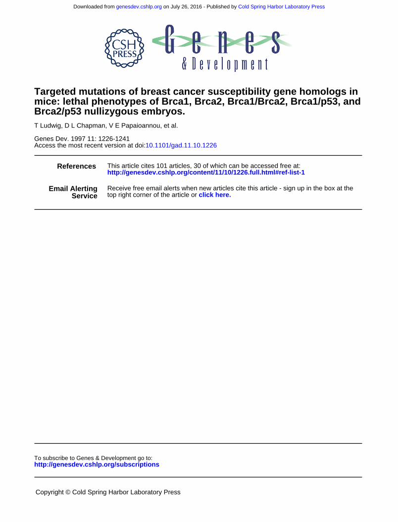
10.1101/gad.11.10.1226Access the most recent version at doi: 1997 11: 1226-1241 Genes Dev.
T Ludwig, D L Chapman, V E Papaioannou, et al. Brca2/p53 nullizygous embryos.
andmice: lethal phenotypes of Brca1, Brca2, Brca1/Brca2, Brca1/p53, Targeted mutations of breast cancer susceptibility gene homologs in
References
http://genesdev.cshlp.org/content/11/10/1226.full.html#ref-list-1
This article cites 101 articles, 30 of which can be accessed free at:
ServiceEmail Alerting
click here.top right corner of the article orReceive free email alerts when new articles cite this article - sign up in the box at the
http://genesdev.cshlp.org/subscriptionsgo to: Genes & Development To subscribe to
Copyright © Cold Spring Harbor Laboratory Press
Cold Spring Harbor Laboratory Press on July 26, 2016 - Published by genesdev.cshlp.orgDownloaded from


