Embed Size (px)
Citation preview
TbpoeffimFbiomeefia
v
o
S
t
J
t
J
c
L
c
C
v
S
D
9
©
0
d
ARTICLE IN PRESS
J Oral Maxillofac Surgxx:xxx, 2010Temporomandibular Joint CondylarAbnormality: Evaluation, Treatment
Planning, and Surgical ApproachJaqueline S. Venturin, DDS,* Werner H. Shintaku, DDS, MS,†
Yuko Shigeta, DDS, PhD,‡ Takumi Ogawa, DDS, PhD,§
Bach Le, DDS, MD,� and Glenn T. Clark, DDS, MS¶
epsrcmpfg
motmrdamdfaihtm
mtcontt
(earcims
he cartilage of the mandibular condyle is locatedeneath the fibrous articular layer and undergoes atro-hic changes, assuming endochondral bone growthr adaptive growth, according to the absence or pres-nce of functional demand. Normal condylar growthollows a sequence of transitory stages that are de-ned by molecules synthesized by undifferentiatedesenchymal cells and differentiating chondrocytes.1
or example, Sox 9 transcription factor is expressedy cells in the proliferative layer and chondrocytes. It
s a potent activator of type II collagen, the main typef collagen that forms the framework of the cartilageatrix in the growing condyle.2 Type X collagen is
xpressed only by hypertrophic chondrocytes, and itsxpression precedes the onset of endochondral ossi-cation. It is involved in the growth, development,nd remodeling of the articular cartilage.3,4 Vascular
*Assistant Professor, Division of Health Promotion, Disease Pre-
ention and Epidemiology, University of Southern California School
f Dentistry, Los Angeles, CA
†Resident, Dental Diagnostic Science, University of Texas Health
cience Center at San Antonio Dental School, San Antonio, TX.
‡Assistant Professor, Department of Fixed Prosthodontic Den-
istry, Tsurumi University School of Dental Medicine, Yokohama,
apan.
§Assistant Professor, Department of Fixed Prosthodontic Den-
istry, Tsurumi University School of Dental Medicine, Yokohama,
apan.
�Clinical Associate Professor, Department of Oral and Maxillofa-
ial Surgery, University of Southern California School of Dentistry,
os Angeles, CA.
¶Program Director and Professor, Orofacial Pain and Oral Medi-
ine Center, Division of Diagnostic Sciences, University of Southern
alifornia School of Dentistry, Los Angeles, CA.
Address correspondence and reprint requests to Dr Venturin: Di-
ision of Health Promotion, Disease Prevention and Epidemiology,
pecial Patients’ Care, University of Southern California School of
entistry, 925 West 34th Street, Room DEN 4202, Los Angeles, CA
0089; e-mail: [email protected]
2010 American Association of Oral and Maxillofacial Surgeons
278-2391/10/xx0x-0$36.00/0
doi:10.1016/j.joms.2009.08.002
1
ndothelial growth factor is expressed by hypertro-hic chondrocytes, and its maximal level of expres-ion precedes the maximal level of bone formation. Itegulates the neovascularization of the hypertrophicartilage and influences the removal of the cartilageatrix. The invading blood vessels bring osteogenicrogenitor mesenchymal cells into the mineralization
ront that later differentiate into osteoblasts that en-age in osteogenesis.5
Both during jaw growth and after it has stopped, theandibular condyle is continuously under the influence
f extrinsic forces, which can be physiologic, func-ional, or excessive, such as with macrotraumas andicrotraumas. It is also important to consider the natu-
al aging process, the genetic mechanisms that mightetermine the reaction of the normal tissues to becomebnormal, and even the idiopathic apposition of abnor-al or tumor cells.6,7 Alterations or disruptions in the
evelopmental process can happen in the early stages oformation and can be isolated malformations or a part ofsyndrome.8 Alterations in growth produce anomalies
n size, described as condylar hypoplasia or condylaryperplasia, abnormalities of shape such as a bifid orrifid condyle, and tumors, which can be benign oralignant.When a patient presents with progressive facial asym-etry, a thorough clinical evaluation and diagnostic
ests with proper imaging techniques are indicated. Thelinical differential diagnosis for these growth disordersf the condyle or ramus includes developmental andeoplastic conditions such as condylar hyperplasia, os-eochondroma, chondroblastoma, osteoma, osteoblas-oma, and chondrosarcoma (Table 1).9-13
Panoramic radiography and computed tomographyCT) of the temporomandibular joint (TMJ) will show annlarged condyle and a longer mandibular neck on theffected side; however, they cannot provide informationegarding whether the abnormal growth is still active. Aomparison of follow-up images might provide enoughnformation to assess the tumor growth. However, if a
ore immediate answer is needed, techniques such askeletal scintigraphy using technetium-99m methylene
iphosphate, which can be used with single photon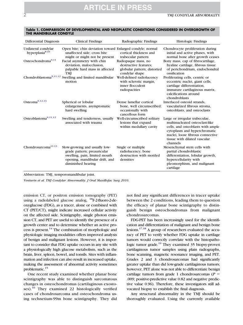
euoCosgcpotabmmp
scsci
nbtgc
clrtlcbGghc.tv
U
O
C
O
O
C
A
V g 2010
2 TMJ CONDYLAR ABNORMALITY
ARTICLE IN PRESS
mission CT, or positron emission tomography (PET)sing a radiolabeled glucose analog, 18F-2-fluoro-2-de-xyglucose (FDG), as a tracer, alone or combined withT (PET/CT), might indicate increased cellular activityn the affected side. Scintigraphy, single photon emis-ion CT, and PET are useful to identify the presence of arowth center and to determine whether an active pro-ess is present.14 The combination of morphologic andhysiologic imaging modalities offers improved analysisf benign and malignant lesions. However, it is impor-ant to consider that FDG uptake occurs in any site withphysiologically high glucose metabolism, such as therain, liver, spleen, bowel, and tonsils. Sites with inflam-ation and infection can also result in increased uptake,aking the assessment of abnormal activity sometimesroblematic.15
One recent study examined whether planar bonecintigraphy was able to distinguish sarcomatoushanges in osteochondromas (cartilaginous exosto-es).16 They examined 22 histologically verifiedases of chondrosarcoma and osteochondroma us-
Table 1. COMPARISON OF DEVELOPMENTAL AND NEOTHE MANDIBULAR CONDYLE
Differential Diagnosis Clinical Findings
nilateral condylarhyperplasia8-10
Open bite; chin deviation towardunaffected side; cross bitemight or might not be present
E
steochondroma9,11 Facial asymmetry with chindeviation, malocclusion,palpable hard mass in affectedTMJ
R
hondroblastoma9,11,12 Swelling and limited mandibularmotion
W
steoma9,12,13 Spherical or lobularenlargements, asymptomatichard swelling
D
steoblastoma9,11,12 Swelling and tenderness, usuallyassociated with trauma
W
hondrosarcoma11-13 Slow-growing and usually low-grade pattern; preauricularswelling, pain, limited mouthopening, mandibular drift, anddiminished hearing
S
bbreviation: TMJ, temporomandibular joint.
enturin et al. TMJ Condylar Abnormality. J Oral Maxillofac Sur
ng technetium-99m bone scintigraphy. They did t
ot find any significant differences in tracer uptakeetween the 2 conditions, leading them to questionhe efficacy of planar bone scintigraphy to distin-uish benign osteochondromas from malignanthondrosarcomas.FDG-PET has been increasingly used for the identifi-
ation and differentiation of malignant and benign boneesions.17,18 A group of researchers evaluated the accu-acy of PET to verify whether FDG uptake in cartilageumors would correctly correlate with the histopatho-ogic tumor grade.19 They examined 35 biopsy-provenartilaginous tumor samples using plain radiographs,one scanning, magnetic resonance imaging, and PET.rades 2 and 3 chondrosarcomas had significantlyreater uptake than did low-grade cartilaginous tumors;owever, PET alone was not able to differentiate benignartilage tumors from grade 1 chondrosarcomas (P �009; positive-predictive value 0.82 and negative predic-ive value 0.96). Therefore, these investigators still ad-ocated biopsy to establish the final diagnosis.Any structural abnormality in the TMJ should be
IC CONDITIONS CONSIDERED IN OVERGROWTH OF
iographic Findings Histologic Findings
d condyle; normalcal thickness andcular pattern
Chondrocyte proliferation duringinitial and active phases, withnormal bone after growth ceases
aque mass, nouctive features;ular pattern; distortedylar shape
Bony mass, cap of fibrocartilage,hyaline cartilage, fibrous tissueof perichondrium, endochondralossification
fined radiolucencysclerotic margin,
r flocculentpacities
Proliferating cells, centric oreccentric nuclei, giant cells,cartilage differentiation,immature cartilaginous matrix,calcifications aroundchondroblasts
lamellar cortical, well circumscribed;sionally withellous form
Interlaced osteoid strands,vascularized fibrous stroma,osteoblasts, and osteoclasts
rcumscribed solitaryns that expandin medullary cavity
Large or irregular trabeculae,multinucleated osteoclast-likecells, and osteoblasts with amplecytoplasm and hyperchromaticnuclei, loose fibrous connectivetissue with dilated vascularchannels
or multiplelucency; boneuction with mottledities
Mesenchymal stem cells withpartial chondroblasticdifferentiation, lobular growth,hypercellularity withpleomorphism, and malignantcartilage
.
PLAST
Rad
nlargecortitrabe
adiopdestrglobcondell-dewithinneradio
enseboneoccacancell-cilesiowith
ingleradiodestrdens
horoughly evaluated. Using the currently available

tgt3cmvdd
R
bhhTatsim
siwtr
cptrottmo
aoaitadauadscetapebshi
fwm5pTtm
V2
VENTURIN ET AL 3ARTICLE IN PRESS
echnology, the imaging systems are able to supply aood amount of information for the assessment ofhese abnormalities. Multiplanar reconstructions and-dimensional models using the data acquired byone-beam CT (CBCT) or CT scans contribute infor-ation not only for detailed evaluation, but also pro-
ide an accurate visual perspective for the efficientiagnosis and treatment planning with a better pre-iction of the final outcome.
eport of a Case
CHIEF COMPLAINT AND HISTORY OFPRESENT ILLNESSA 20-year-old man presented with a chief complaint of
ilateral TMJ pain, facial asymmetry, and bite shifting thatad progressively increased within a 2-year period andad become more evident during the previous 4 months.he patient reported that before that period he had hadcomfortable bite that had been achieved with orthodon-
ic treatment during childhood. He also reported epi-odic clicking in the left TMJ, sporadic ringing and stuff-ness in the left ear, daily headaches, and episodic
FIGURE 1. Patient’s facial profile at first appointment.
enturin et al. TMJ Condylar Abnormality. J Oral Maxillofac Surg010.
igraines that had became aggravated throughout theV2
ame period. No history of trauma, no relevant concom-tant systemic problem, and no other joint presented
ith signs of abnormality. However, he did present withhe habits of tooth clenching and grinding and was al-eady using a night guard.
CLINICAL EXAMINATIONThe first step in the assessment of facial asymmetry is to
onduct a thorough head and neck examination. In ouratient, facial asymmetry was evident, with chin deviationo the left side, and enlargement noted on the right preau-icular region (Fig 1). No lymphadenopathy was noted. Thetoscopic examination findings were normal for the audi-ory canal, pinna, and tympanic membrane. Muscle palpa-ion revealed moderate to severe pain bilaterally on theasticatory and postural muscles, with severe pain noted
n palpation of the bilateral TMJs.The intraoral examination revealed no dental attrition
nd generally good oral health. Significant malocclusion wasbserved secondary to mandible posture deviation to thenterior, and lateral to the left position. Only the left centralncisors were in contact in the maximal intercuspal posi-ion, resulting in a bilateral posterior open bite with annterior edge-to-edge relation, and the midline with a man-ibular deviation to the left of 4 mm. The teeth were wellligned within the arches, and the alveolar bone in bothpper and lower arches presented with the anatomic char-cteristics preserved. This indicated that no deformities hadeveloped in a natural attempt to accommodate the occlu-ion to this abnormal jaw position, contributing to theonclusion that the patient’s situation had been recentlystablished (Fig 2). Six months later, the clinical examina-ion revealed changes in the mandibular position with annterior cross bite and increased pain in the TMJ area. Theatient could not move his jaw backward to the initialdge-to-edge position possible at the initial examination,ecause this movement was interrupted by rigid ending andevere pain in the right TMJ. These findings suggested thatis condition was an ongoing process. This information is
mportant for the treatment planning (Fig 3).The mandibular mobility examination revealed pain-
ree opening of 40 mm. Active opening reached 55 mm,hich induced pain on the left side of the face. Excursiveovements were more affected, and he could move onlymm to the right, 2 mm to the left, and 2 mm to the
rotrusive, with all movements provoking pain in the leftMJ. No TMJ noises were present during the examina-
ion. The midline was deviated to the left when theouth was closed. However, on opening, it acquired a
FIGURE 2. Occlusal condition at first appointment.
enturin et al. TMJ Condylar Abnormality. J Oral Maxillofac Surg010.

mrt
siocnc
tVvwrwoecwdp
mm
ti
icdaaim
tnawmb
V2
Fr
V2
Fr
V2
Ft
4 TMJ CONDYLAR ABNORMALITY
ARTICLE IN PRESS
ore centered position. The left condyle was able tootate and translate, but the right condyle was only ableo perform translational movement.
PANORAMIC RADIOGRAPHIC EXAMINATIONBecause the clinical findings were highly indicative of
tructural involvement of the right TMJ, the second stepn our workup was panoramic radiography. The pan-ramic radiograph showed a right condyle with an in-reased size and abnormal shape. The left condyle had aormal size and shape, with no evidence of degenerativehanges (Fig 4).
CBCT WITH 3D RECONSTRUCTIONFor a better assessment of the involved structures, the
hird step was to acquire a CBCT scan (Newtom 3G,erona, Italy) of the jaws and face (volume size 20 cm,oxel size 0.3 mm). An irregular, globular-shaped massas noted on the anterior aspect of the lateral pole of the
ight condyle (Fig 5). The internal structure of the lesionas of a uniform greater density with the medial one halff the condyle having normal bone density. The articularminence revealed moderate bone remodeling. In thelosed position, owing to the overgrowth, the condyleas displaced downward and forward outside the man-ibular fossa. On opening, the condyle translated wellast the articular eminence.On the left side, the cortical outlines of the condyle,
andibular fossa, and articular eminence were within nor-al limits. In the closed position, the condyle was concen-
FIGURE 3. Occlusal condition 6 months after initial visit.
enturin et al. TMJ Condylar Abnormality. J Oral Maxillofac Surg010.
IGURE 4. Initial radiographic assessment with panoramicadiography.
enturin et al. TMJ Condylar Abnormality. J Oral Maxillofac Surg010.
V2
rically located within the mandibular fossa, and, on open-ng, the condyle translated past the eminence.
The 3D reconstruction of the CBCT data using specificmaging software (Amira; Visage Imaging, San Diego, CA)onfirmed the extensive abnormal growth of the right con-yle, with an irregular contour and expansion suggestive ofbenign tumor lesion rather than condylar hyperplasia. Thebsence of aggressive infiltration and bone destruction re-nforced the indication of a benign process rather than a
alignant condition (Fig 6).
PET/CT SCANThe fourth step in our workup involved PET/CT to verify
he lesion activity. Multi-axial images through the head,eck, chest, abdomen, and pelvis were obtained for reviewfter CT attenuation correction. No abnormal hyperactivityas demonstrated in the right condyle to suggest a highlyetabolically active condition (Fig 7). Normal tonsillar,
owel, and urinary uptake was observed, as well as normal
IGURE 5. Axial CBCT reconstruction. Note irregular outline ofight condyle.
enturin et al. TMJ Condylar Abnormality. J Oral Maxillofac Surg010.
IGURE 6. Three-dimensional reconstruction. Note difference be-ween condylar outlines.
enturin et al. TMJ Condylar Abnormality. J Oral Maxillofac Surg010.

up
s
spcmtgm(
Fi
V2
V
V2
VENTURIN ET AL 5ARTICLE IN PRESS
ptake in the brown fat in the neck and mediastinal andaravertebral regions.
PREOPERATIVE SIMULATION: 3D IMAGINGRECONSTRUCTION AND FABRICATION OFACRYLIC MODELSTo better analyze the altered structures and plan the
urgical procedure, we used 3D images to develop a virtual
IGURE 7. PET/CT scan showing lack of tracer uptake in region ofnterest.
enturin et al. TMJ Condylar Abnormality. J Oral Maxillofac Surg010.
FIGURE 8. Simulation models. Three-dimensional mo
enturin et al. TMJ Condylar Abnormality. J Oral Maxillofac Surg 2010
imulation model in which we could remove the lesion,reserve a portion of the condyle, and then place theondyle back into the fossa to determine whether thisanipulation would provide reasonable interdigitation of
he teeth (Fig 8). Derived from the CBCT data, stereolitho-raphic models were fabricated with and without the tu-orous mass. This also enabled surgical excision simulation
Fig 9).
owing A, tumor lesion and B, after surgical exicision.
FIGURE 9. Stereolithographic model of right TMJ.
enturin et al. TMJ Condylar Abnormality. J Oral Maxillofac Surg010.
dels sh
.

imilcoorswtch
rw
eAfdis(upd
D
ia(
V2
V2
V2
6 TMJ CONDYLAR ABNORMALITY
ARTICLE IN PRESS
THERAPYThe patient was admitted to University of Southern Cal-
fornia University Hospital for surgical removal of the tu-or. The electrocardiographic and chest radiographic find-
ngs and routine urine and blood values were within normalimits. The tumor resection and reshaping of the mandibularondyle was planned using 3D imaging. The precise amountf resection needed to achieve the patient’s preoperativecclusion was determined using 3D reconstruction of theelationship between the patient’s condylar tumor and re-ulting malocclusion (Figs 6, 8). A preauricular approachas created to access the condylar head. A high condylec-
omy was performed as planned (Fig 10). A piece of theondyle (with the tumor) was removed (Fig 11) and sent foristologic identification.
The occlusion was verified clinically after conservativeesection and reshaping of the condyle. Guiding elasticsere maintained for 2 weeks postoperatively. Microscopic
FIGURE 10. Condylectomy of right condyle.
enturin et al. TMJ Condylar Abnormality. J Oral Maxillofac Surg010.
FIGURE 11. Tumor specimen sent for histologic evaluation.
enturin et al. TMJ Condylar Abnormality. J Oral Maxillofac Surg010.
V2
xamination confirmed the diagnosis of osteochondroma.t 6 months of follow-up, the patient presented with good
acial symmetry and a stable occlusal relationship. The man-ibular function was excellent, with a maximal incisal open-
ng of 35 to 40 mm. A panoramic radiograph revealed goodymmetry of the mandible and a favorable condylar shapeFig 12). The radiographic follow-up data were obtainedsing small-volume CBCT (Accuitomo; J Morita, Kyoto, Ja-an) for increased image resolution and a reduced radiationose to the patient (Fig 13).
iscussion
Considering the history of the illness and the clin-cal and radiographic findings from the panoramicnd CBCT images, we suspected a benign neoplasmmost likely an osteochondroma). Osteochondroma is
FIGURE 12. Panoramic radiograph after surgical procedure.
enturin et al. TMJ Condylar Abnormality. J Oral Maxillofac Surg010.
FIGURE 13. CBCT scan of right TMJ after condylectomy.
enturin et al. TMJ Condylar Abnormality. J Oral Maxillofac Surg010.

adabftchhsnTscmtei
cafatwaccldaflowcle
tglsrtTofiscotpHp
lt
rrpaiepaaosomd7
ituatcTgotwppo
twtccvttwott
R
VENTURIN ET AL 7ARTICLE IN PRESS
benign bone tumor that arises from the endochon-ral bone and can present as a solitary lesion (75%) ors multiple lesions (25%). It represents 35% of allenign tumors and 8.5% of all bone tumors. It is rarelyound in the oral and maxillofacial region. However,he occurrence in the condyle seems to be the mostommon site in the facial region. Osteochondromaas been suggested to have a female predilection andas a wide age range of 11 to 69 years. However,ome reports have also reported no gender predomi-ance and a peak age of about the fourth decade.20,21
he risk of malignant transformation is about 1% forolitary lesions. However, we have found no cases ofonversion to malignancy reported for tumors in theandibular condyle region. The risk of recurrence of
his benign lesion is low after surgical removal; how-ver, the only recurrence reported in published stud-es resulted from incomplete excision.22
The signs and symptoms associated with osteo-hondroma of the condyle vary according to the sizend location of the growth. The lesion usually arisesrom the medial anterior aspect of the condylar necknd extends to the condylar head. Radiographically,he lesion can be visualized as a radiopaque massithout lytic features, with a globular pattern with
ltered condylar morphology. The histopathologicharacteristics consist of a proliferative bony massovered by a cap of fibrocartilage and hyaline carti-age surrounded by the fibrous tissue of the perichon-rium, and endochondral ossification in the deepestspect of the cartilage.12,17,23 The chondrocytes canorm rows that are perpendicular to the surface of theesion, and they can overlie a zone of endochondralssification, resulting in fusion of the cancellous boneith the normal underlying bone. Calcification of the
artilage and ossification might be seen, and olderesions will have a thinner rim of cartilage owing toxtensive replacement by bone.24
Given the dimensions reached by the growth, theherapeutic approach recommended is typically sur-ery. Several techniques have been suggested in pub-ished reports, and the decision must take into con-ideration the shape and size of the lesion, anyesulting deformities or impingement on other struc-ures, and the functional morbidity it has caused.20,25
he procedures can involve conservative approachesr more extensive measures according to the surgicalndings, but complete excision of the lesion is con-idered the standard. Some surgeons have used aondylectomy (77% of the time), followed by excisionf the lesion, preserving the condyle (23% of theime).19 The condylectomy can be partial or com-lete.26,27 Operating in the TMJ area is challenging.owever, the data reported to date have not sup-
orted corrective orthognathic surgery of the maxil-ary and/or mandibular arches without accessing theumor to confirm the histopathologic diagnosis.
Surgery of small benign tumors of the condyleequires a subperiosteal resection through a preau-icular access. Larger lesions with breaching of theeriosteum require condylectomy. Attempts to userthroscopic surgery in specific situations are undernvestigation but have not been proved.24 After tumorxcision, with or without condylar preservation, com-lementary correction of the facial and/or occlusalsymmetry might be required and can involve a sep-rate orthognathic procedure and subsequent orth-dontic treatment. In cases of full condylectomy andubcondylar process extension, a costochondral graftr artificial joint prosthetic reconstruction is recom-ended. The prognosis is good, and the published
ata have demonstrated no evidence of recurrence atmonths, 20 months, and 5 years.18,20,28
The aim of the present report was to describe themportance of using a comprehensive model to assesshe progressive facial asymmetry and to emphasize these of 3D reconstruction in planning the tumor surgerynd how it affects the treatment planning, operativeime, and prognosis. The treatment planning for ourase was all done using 3D CBCT reconstructed images.his included determination of the anatomy of the sur-ical area, the surgical access method, and the amountf surgical resection needed. Using 3D reconstruction,he tumor was conservatively resected and the condyleas left intact, leaving no postoperative sequelae. All theatient’s preoperative problems resolved, and patientresented with 3-mm midline deviation to the right onpening at 6 months of follow-up.A comprehensive evaluation of our patient involved
he history reported and the clinical findings, correlatedith the panoramic, CBCT, and PET/CT findings. Using
hese data, we recommended surgical management, in-luding tumor excision, with condylar reshaping or highondylectomy. We used the CBCT data to create a 3Dirtual model that could be manipulated and reposi-ioned such that we could visualize the final postopera-ive occlusion. Although the clinical and imaging dataere suggestive of a benign lesion, the diagnosis couldnly be confirmed by histopathologic examination ofhe surgical specimen, confirming the diagnosis of os-eochondroma of the TMJ condyle.
eferences1. Shen G, Hagg U, Rabie AB, et al: Identification of temporal
pattern of mandibular condylar growth: A molecular and bio-chemical experiment. Orthod Craniofac Res 8:114, 2005
2. Rabie AB, Hagg U: Factors regulating mandibular condylargrowth. Am J Orthod Dentofac Orthop 122:401, 2002
3. Shen G: The role of type X collagen in facilitating and regulat-
ing endochondral ossification of articular cartilage. OrthodCraniofac Res 8:11, 2005
1
1
1
1
1
1
1
1
1
1
2
2
2
2
2
2
2
2
2
8 TMJ CONDYLAR ABNORMALITY
ARTICLE IN PRESS
4. Kwan KM, Pang MK, Zhou S, et al: Abnormal compartmental-ization of cartilage matrix components in mice lacking collagenX: Implications for function. J Cell Biol 136:459, 1997
5. Bi W, Deng JM, Zhang Z, et al: Sox9 is required for cartilageformation. Nat Genet 22:85, 1999
6. Hylander WL: Functional anatomy and biomechanics of themasticatory apparatus, in Laskin DM, Greene CS, Hylander WL(eds): Temporomandibular Disorders: An Evidence-Based Ap-proach to Diagnosis and Treatment (ed 1). Chicago, Quintes-sence Publishing, 2006, p 3
7. Stegenga B, Bont LGM: TMJ growth, adaptive modeling andremodeling, and compensatory mechanisms, in Laskin DM,Greene CS, Hylander WL (eds): Temporomandibular Disorders:An Evidence-Based Approach to Diagnosis and Treatment(ed 1). Chicago, Quintessence Publishing, 2006, p 53
8. Neville BW, Damm DD, Allen CM, et al: Developmental defectsof the oral and maxillofacial region, in Neville BW, Damm DD,Allen CM, et al (eds): Oral and Maxillofacial Pathology (ed 2).Philadelphia, WB Saunders, 2002, p 1
9. Petrikowski CG: Diagnostic imaging of the temporomandibularjoint, in White SC, Pharoah MJ (eds): Oral Radiology (ed 5).Philadelphia, Mosby, 2004, p 538
0. Troulis MJ, Kaban LB: Congenital and developmental anomalies,in Laskin DM, Green, CS, Hylander WL (eds): Temporomandibu-lar Disorders: An Evidence-Based Approach to Diagnosis andTreatment. Chicago, Quintessence Publishing, 2006, p 421
1. Neville BW, Damm DD, Allen CM, et al: Bone pathology, inNeville BW, Damm DD, Allen CM, et al (eds): Oral and Maxillofa-cial Pathology (ed 2). Philadelphia, WB Saunders, 2002, p 533
2. Stern D: Benign and malignant tumors, in Laskin DM, Green,CS, Hylander WL (eds): Temporomandibular Disorders: AnEvidence-Based Approach to Diagnosis and Treatment. Chi-cago, Quintessence Publishing, 2006, p 319
3. Batra PS, Estrem SA, Zitsch RP, et al: Chondrosarcoma of thetemporomandibular joint. Otolaryngol Head Neck Surg 120:961, 1999
4. Rosenbaum SJ, Lind T, Antoch G, et al: False-positive FDG PETuptake—The role of PET/CT. Eur Radiol 16:1054, 2006
5. Henderson MJ, Wastie ML, Bromige M, et al: Technetium-99mbone scintigraphy and mandibular condylar hyperplasia. ClinRadiol 41:411, 1990
6. Hendel HW, Daugaard S, Kjaer A: Utility of planar bone scin-tigraphy to distinguish benign osteochondromas from malig-
nant chondrosarcomas. Clin Nucl Med 27:622, 20027. Cham DK, Conti PS: Normal physiology and variants: A primer,in Conti PS, Cham DK (eds): PET-CT: A Case-Based Approach.New York, Springer, 2005, p 3
8. Dehdashti F, Siegel BA, Griffeth LK, et al: Benign versusmalignant intraosseous lesions: Discrimination by means ofPET with 2-[F-18] fluoro-2-deoxy-D-glucose. Radiology 200:243, 1996
9. Lee FY, Yu J, Chang SS, et al: Diagnostic value and limitationsof fluorine-18 fluorodeoxyglucose positron emission tomogra-phy for cartilaginous tumors of bone. J Bone Joint Surg Am86A:2677, 2004
0. Saito T, Utsunomiya T, Furutani M, et al: Osteochondroma ofthe mandibular condyle: A case report and review of theliterature. J Oral Sci 43:293, 2001
1. Khochtali H, Bouzaiene M, Yacoubi MT, et al: Osteochondromaof the mandibular condyle: Apropos of a case. Rev StomatolChir Maxillofac 94:87, 1993
2. Vezeau PJ, Fridrich KL, Vincent SD: Osteochondroma of themandibular condyle: Literature review—A report of two atyp-ical cases. J Oral Maxillofac Surg 53:954, 1995
3. Koga M, Toyofuku S, Nakamura Y, et al: Osteochondroma inthe mandibular condyle that caused facial asymmetry: A casereport. Cranio 24:67, 2006
4. Karras CS, Wolford LM, Cottrell DA: Concurrent osteochon-droma of the mandibular condyle and ipsilateral cranial baseresulting in temporomandibular joint ankylosis: Report of caseand review of the literature. J Oral Maxillofac Surg 54:640,1996
5. Iizuka T, Schroth G, Laeng RH, et al: Osteochondroma of themandibular condyle: Report of a case. J Oral Maxillofac Surg54:495, 1996
6. Wolford LM, Mehra P, Franco P: Use of conservative condylec-tomy for treatment of osteochondroma of the mandibular con-dyle. J Oral Maxillofac Surg 60:262, 2002
7. Clayman L: Surgical management of benign and malignantneoplasms, in Laskin DM, Greene CS, Hylander WL (eds):Temporomandibular Disorders: An Evidence-Based Approachto Diagnosis and Treatment. Chicago, Quintessence Publish-ing, 2006, p 509
8. Holmlund AB, Gynther GW, Reinholt FP: Surgical treatment ofosteochondroma of the mandibular condyle in the adult: A
5-year follow-up. Int J Oral Maxillofac Surg 33:549, 2004


