Embed Size (px)
Citation preview
UNIVERSIDAD COMPLUTENSE DE MADRID
FACULTAD DE FARMACIA
TESIS DOCTORAL
Identificación, expresión y caracterización de peroxidasas ligninolíticas de interés en genomas de basidiomicetos
MEMORIA PARA OPTAR AL GRADO DE DOCTORA
PRESENTADA POR
Elena Fernández Fueyo
Directores
Ángel Tomás Martínez Ferrer Francisco Javier Ruiz Dueñas
Madrid, 2014 © Elena Fernández Fueyo, 2014
Identificación, expresión y
caracterización de peroxidasas
ligninolíticas de interés en genomas
de basidiomicetos
Elena Fernández Fueyo

Identificación, expresión y caracterización de peroxidasas
ligninolíticas de interés en genomas de basidiomicetos
Tesis doctoral
Elena Fernández Fueyo
Centro de Investigaciones Biológicas
CSIC Universidad Complutense de Madrid
Facultad de Farmacia Madrid, 2014

Identificación, expresión y caracterización de peroxidasas
ligninolíticas de interés en genomas de basidiomicetos
Tesis doctoral para optar al grado de
Doctora por la Universidad Complutense de Madrid presentada por:
Elena Fernández Fueyo
DIRECTORES:
Dr. Angel Tomás Martínez Ferrer Profesor de Investigación, CSIC
Dr. Francisco Javier Ruíz Dueñas
Investigador, CSIC


A Isra
2v1a


Cuando llegues a la cima de una montaña sigue subiendo

ESTRUCTURA DE LA TESIS
El presente trabajo, para obtener el Título de Doctor, se estructura en diez Capítulos que incluyen una Introducción (Cap. 1), cinco Artículos científicos ya publicados (Caps. 2-6), dos Artículos en proceso de revisión (Caps. 7 y 8), una Discusión general (Cap. 9), Conclusiones (Cap. 10) y un Anexo conteniendo Material suplementario (Cap. 11), tal como se detalla a continuación:
• Resumen en español Páginas 1-2
• Resumen en Inglés Páginas 3-4
• Capítulo 1: Introducción
Páginas 5-35
• Capítulo 2: "Pleurotus ostreatus heme peroxidases: An in silico analysis from the genome sequence to the enzyme molecular structure" Ruiz-Dueñas, F. J., E. Fernández, M. J. Martínez, and A. T. Martínez. C. R. Biol. 334:795-805. 2011.
Páginas 37-49
• Capítulo 3: "Ligninolytic peroxidase genes in the oyster mushroom genome: Heterologous expression, molecular structure, catalytic and stability properties and lignin-degrading ability" Fernández-Fueyo, E., F. J. Ruiz-Dueñas, M. J. Martínez, A. Romero, K. E. Hammel, F. J. Medrano, and A. T. Martínez. Biotechnol. Biofuels 7:2. 2014. Páginas 51-75
• Capítulo 4: "Ligninolytic peroxidase gene expression by Pleurotus ostreatus: Differential regulation in lignocellulose medium and effect of temperature and pH" Fernández-Fueyo, E., R. Castanera, F. J. Ruiz-Dueñas, M. F. López-Lucendo, L. Ramírez, A. G. Pisabarro, and A. T. Martínez. Fungal Genet. Biol. 2014. Páginas 77-90
• Capítulo 5: "Comparative genomics of Ceriporiopisis subvermispora and Phanerochaete chrysosporium provide insight into selective ligninolysis" Fernández-Fueyo, E., F. J. Ruiz-Dueñas, P. Ferreira, D. Floudas, D. S. Hibbett, P. Canessa, L. Larrondo, T. Y. James, D. Seelenfreund, S. Lobos, R. Polanco, M. Tello, Y. Honda, T. Watanabe, T. Watanabe, J. S. Ryu, C. P. Kubicek, M. Schmoll, J. Gaskell, K. E. Hammel, F. J. St.John, A. Vanden Wymelenberg, G. Sabat, S. S. Bondurant, K. Syed, J. Yadav, H. Doddapaneni, V. Subramanian, J. L. Lavín, J. A. Oguiza, G. Perez, A. G. Pisabarro, L. Ramírez, F. Santoyo, E. Master, P. M. Coutinho, B. Henrissat, V. Lombard, J. K. Magnuson, U. Kües, C. Hori, K. Igarashi, M. Samejima, B. W. Held, K. Barry, K. LaButti, A. Lapidus, E. Lindquist, S. Lucas, R. Riley, A. Salamov, D. Hoffmeister, D. Schwenk, Y. Hadar, O. Yarden, R. P. de Vries, A. Wiebenga, J. Stenlid, D. C. Eastwood, I. V. Grigoriev, R. Berka, R. A. Blanchette, P. Kersten, A. T. Martínez, R. Vicuña, and D. Cullen. Proc. Natl. Acad. Sci. USA 109:5458-5463. 2012. Páginas 91-99
• Capítulo 6: "Lignin-degrading peroxidases from genome of selective ligninolytic fungus Ceriporiopsis subvermispora" Fernández-Fueyo, E., F. J. Ruiz-Dueñas, Y. Miki, M. J. Martínez, K. E. Hammel, and A. T. Martínez. J. Biol. Chem. 287:16903-16906. 2012. Páginas 101-116
• Capítulo 7: "Catalytic and stability properties of manganese peroxidase subfamilies: A study based on the Ceriporiopsis subvermispora genome" Fernández-Fueyo, E., S Acebes. , F. J. Ruiz-Dueñas, , M. J. Martínez, A. Romero, F. J. M , V Guallar and A. T. Martínez (enviado). Páginas 117-133
• Capítulo 8: "Engineering a lignin-degrading peroxidase acting at very acidic pH Fernández-Fueyo, E., F. J. Ruiz-Dueñas and A. T. Martínez. (enviado). Páginas 135-150
• Capítulo 9: Discusión Páginas 151-159

• Capítulo 10: Conclusiones Páginas161-163
• Capítulo 11: Anexo (con Material Suplementario)
Páginas 165-227

Resumen
La secuenciación de los genomas de Pleurotus ostreatus y Ceriporiopsis subvermispora nos ha permitido investigar las propiedades bioquímicas, estructurales y operacionales de las diferentes hemoperoxidasas ligninolíticas presentes en dos hongos modelo de interés biotecnológico, por su capacidad para degradar preferentemente la lignina frente a la celulosa en diferentes sustratos lignocelulósicos.
Las constantes cinéticas de las diferentes peroxidasas de clase-II (de la superfamilia de peroxidasas vegetales-fúngicas-procariotas) del genoma de P. ostreatus, tras su expresión heteróloga en Escherichia coli, revelan un repertorio formado por tres peroxidasas versátiles (VPs) y seis manganeso peroxidasas (MnPs), confirmando la ausencia de lignina peroxidasa (LiP), a la que hasta ahora se ha asociado la capacidad para degradar la lignina, en los hongos ligninolíticos. Este estudio muestra por primera vez la presencia en una MnP del triptófano catalítico superficial conservado en todas las VPs y LiPs. Esta anomalía se explica por una mutación durante la evolución de estas peroxidasas que interrumpió la vía de transporte electrónico desde la superficie hasta el grupo hemo, como se demostró por mutagénesis dirigida reversa. Los estudios estructurales-funcionales de las seis MnPs de P. ostreatus revelan que éstas constituyen una nueva subfamilia, caracterizada por su actividad independiente de Mn sobre algunos sustratos y por la presencia de una cola en el extremo C-terminal más corta que en las típicas MnPs largas de P. chrysosporium. Se ha cristalizado un miembro de esta nueva subfamilia, siendo la primera MnP corta cristalizada. Además, usando dímeros modelo y lignina sintética marcados radioactivamente se ha mostrado por primera vez la capacidad de las VPs para degradar la lignina. Estos resultados confirman que la VP desempeña en P. ostreatus y en otras especies de agaricales el papel desempeñando por la LiP en P. chrysosporium y otras especies de poliporales.
En general las diferentes isoenzimas de MnP y de VP del genoma de P. ostreatus no presentan grandes diferencias en sus constantes catalíticas pero, sorprendentemente, si muestran grandes diferencias en su estabilidad a la temperatura (con valores de T50 de 43-63 ºC, tras 10 min a pH 5) y al pH (con actividades residuales de 0-96% a pH 3 y de 0-57% a pH 9, en ambos casos tras 4 h a 4 ºC). La isoenzima más estable al pH y la más estable a la temperatura de P. ostreatus se cristalizaron y su estructura fue resulta a 1.0-1.1 Å para intentar identificar las bases estructurales de estas propiedades. La mayor abundancia de residuos básicos, y de puentes de hidrogeno e interacciones salinas, en la superficie de la proteina se relaciona con la estabilidad de una de estas peroxidasas, como se ha confirmado en estudios posteriores de mutagénesis dirigida. Para investigar el significado biológico de la duplicación de genes y la existencia de múltiples MnPs y VPs, se diseñaron cebadores específicos para cada isoenzima y se analizaron por RT-qPCR las diferencias de expresión cuando se modifican las condiciones de temperatura y pH en cultivos de P. ostreatus sobre material lignocelulósico, en combinación con estudios proteómicos y medidas de actividad. No se encontró una correlación entre los resultados secretómicos y de transcripción de la isoenzima más expresada, debido a una secreción deficiente puesta de manifiesto mediante la comparación del secretoma y el proteoma intracelular. Sin embargo, los genes de algunas de las enzimas más estables mostraron niveles de transcripción relativa mayores en las condiciones más extremas de pH y temperatura ensayadas. Estos resultados muestran una regulación ambiental de la expresión de las isoenzimas, pero también ponen de manifiesto la necesidad de realizar estudios cuidadosos para establecer relaciones entre los datos transcriptómicos y la producción extracelular de enzimas por estos hongos.
Aunque Phanerochaete chrysosporium ha sido durante años el hongo ligninolítico modelo, su patrón de degradacion simultanea de la lignocelulosa, impide su uso en
1
Resumen
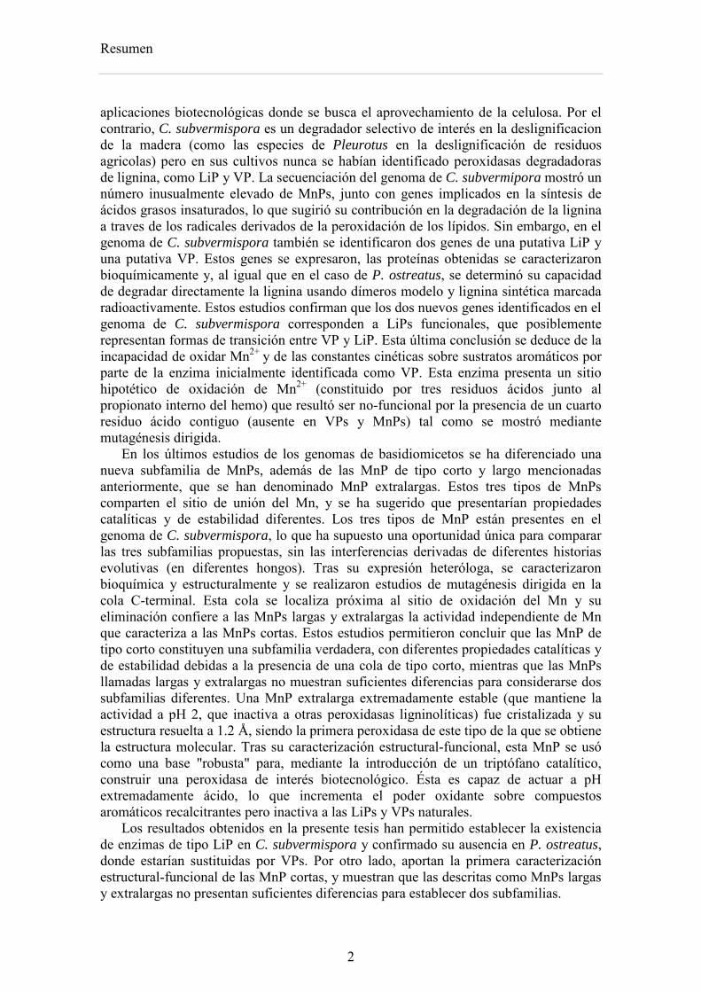
aplicaciones biotecnológicas donde se busca el aprovechamiento de la celulosa. Por el contrario, C. subvermispora es un degradador selectivo de interés en la deslignificacion de la madera (como las especies de Pleurotus en la deslignificación de residuos agricolas) pero en sus cultivos nunca se habían identificado peroxidasas degradadoras de lignina, como LiP y VP. La secuenciación del genoma de C. subvermipora mostró un número inusualmente elevado de MnPs, junto con genes implicados en la síntesis de ácidos grasos insaturados, lo que sugirió su contribución en la degradación de la lignina a traves de los radicales derivados de la peroxidación de los lípidos. Sin embargo, en el genoma de C. subvermispora también se identificaron dos genes de una putativa LiP y una putativa VP. Estos genes se expresaron, las proteínas obtenidas se caracterizaron bioquímicamente y, al igual que en el caso de P. ostreatus, se determinó su capacidad de degradar directamente la lignina usando dímeros modelo y lignina sintética marcada radioactivamente. Estos estudios confirman que los dos nuevos genes identificados en el genoma de C. subvermispora corresponden a LiPs funcionales, que posiblemente representan formas de transición entre VP y LiP. Esta última conclusión se deduce de la incapacidad de oxidar Mn2+ y de las constantes cinéticas sobre sustratos aromáticos por parte de la enzima inicialmente identificada como VP. Esta enzima presenta un sitio hipotético de oxidación de Mn2+ (constituido por tres residuos ácidos junto al propionato interno del hemo) que resultó ser no-funcional por la presencia de un cuarto residuo ácido contiguo (ausente en VPs y MnPs) tal como se mostró mediante mutagénesis dirigida.
En los últimos estudios de los genomas de basidiomicetos se ha diferenciado una nueva subfamilia de MnPs, además de las MnP de tipo corto y largo mencionadas anteriormente, que se han denominado MnP extralargas. Estos tres tipos de MnPs comparten el sitio de unión del Mn, y se ha sugerido que presentarían propiedades catalíticas y de estabilidad diferentes. Los tres tipos de MnP están presentes en el genoma de C. subvermispora, lo que ha supuesto una oportunidad única para comparar las tres subfamilias propuestas, sin las interferencias derivadas de diferentes historias evolutivas (en diferentes hongos). Tras su expresión heteróloga, se caracterizaron bioquímica y estructuralmente y se realizaron estudios de mutagénesis dirigida en la cola C-terminal. Esta cola se localiza próxima al sitio de oxidación del Mn y su eliminación confiere a las MnPs largas y extralargas la actividad independiente de Mn que caracteriza a las MnPs cortas. Estos estudios permitieron concluir que las MnP de tipo corto constituyen una subfamilia verdadera, con diferentes propiedades catalíticas y de estabilidad debidas a la presencia de una cola de tipo corto, mientras que las MnPs llamadas largas y extralargas no muestran suficientes diferencias para considerarse dos subfamilias diferentes. Una MnP extralarga extremadamente estable (que mantiene la actividad a pH 2, que inactiva a otras peroxidasas ligninolíticas) fue cristalizada y su estructura resuelta a 1.2 Å, siendo la primera peroxidasa de este tipo de la que se obtiene la estructura molecular. Tras su caracterización estructural-funcional, esta MnP se usó como una base "robusta" para, mediante la introducción de un triptófano catalítico, construir una peroxidasa de interés biotecnológico. Ésta es capaz de actuar a pH extremadamente ácido, lo que incrementa el poder oxidante sobre compuestos aromáticos recalcitrantes pero inactiva a las LiPs y VPs naturales.
Los resultados obtenidos en la presente tesis han permitido establecer la existencia de enzimas de tipo LiP en C. subvermispora y confirmado su ausencia en P. ostreatus, donde estarían sustituidas por VPs. Por otro lado, aportan la primera caracterización estructural-funcional de las MnP cortas, y muestran que las descritas como MnPs largas y extralargas no presentan suficientes diferencias para establecer dos subfamilias.
2
Summary
The sequenced genomes of P. ostreatus and C. subvermispora allowed us to investigate the biochemical, structural and operational properties of the different ligninolytic hemeperoxidases present in two model fungi of biotechnological interest, due to their ability to degrade lignin preferentially when growing on lignocellulosic substrates.
The kinetic constants of the different class-II peroxidases (of the superfamily of plant-fungal-prokaryotic peroxidases) from the P. ostreatus genome, after their heterologous expression in Escherichia coli, revealed a repertoire formed by three VPs and six MnPs and confirmed the absence of LiP, which until now has been associated with the ability of white-rot fungi to degrade lignin. The study also showed for the first time the presence in a MnP of the conserved catalytic tryptophan that oxidizes the bulky lignin polymer at the surface of VPs and LiPs. This anomaly was explained by a natural mutation in peroxidase evolution that interrupts the electron transfer pathway from the surface to the activated heme cofactor, as shown by reversed mutagenesis. The structural-functional study of the six MnPs revealed that they correspond to a novel subfamily characterized by their Mn-independent activity on some substrates and the presence of a C-terminal tail shorter than in typical (long) MnPs from Phanerochaete chrysosporium. One member of this new subfamily has been crystallized, being the first short MnP crystallized to date. Moreover, studies using 14C-labeled lignin and model dimers showed for the first time the lignin degrading ability of VPs. The above results confirm that VP plays in P. ostreatus and other species of Agaricales the role of LiP in P. chrysosporium and other species of the order Polyporales.
In general the different MnP and VP isoenzymes from P. ostreatus genome present slightly differences in their catalytic constants but, surprisingly, they show significant differences in their stability against temperature (with T50 values of 43-63 ºC, after 10 min at pH 5) and pH (with residual activities of 0-96% at pH 3 and 0-57 % at pH 9, after 4 h at 4 ºC). To take advantage from all the above findings, the two isoenzymes from the P. ostreatus genome with the highest thermal and pH stabilities were crystallized, with the aim of identifying the structural bases of the above properties. The high abundance of basic residues and salt-bridge/H-bond interactions at the surface of the protein was related to the high stability of one of these peroxidases, as confirmed by directed mutagenesis. To investigate the biological meaning of these differences, specific primers were designed for each isoenzyme and their differential transcription was analyzed by quantitative PCR, by varying the temperature and pH conditions of cultures grown in a lignocellulose medium, combined with secretomic and activity studies. Although the genes of some of the most stable isoenzymes showed higher relative transcription levels at the most extreme temperature and pH conditions assayed, no correlation between the transcriptomic and secretomic results was observed for the most expressed MnP gene. This was due to impaired secretion as shown by the abundance of this isoenzyme in the intracellular proteome. These results showed the environmental regulation of isoenzyme gene expression, but also evidenced the need for careful studies to establish correlations between transcriptomic data and production of extracellular enzymes by these fungi.
Although P. chrysosporium has been the model ligninolytic fungus for years, the simultaneous degradation of wood lignin and polysaccharides prevents its use in biotechnological applications, where the use of cellulose is intended. In contrast, C. subvermispora is a selective lignin degrader of interest in wood delignification (similar to Pleurotus species in delignification of agricultural wastes) but its production of lignin-degrading peroxidases (LiPs and VPs) remained unknown. The sequenced C.
3
Summary
subvermipora genome revealed an unusually high number of MnPs, together with genes implicated in the synthesis of unsaturated fatty acids, suggesting their contribution in lignin degradation via free radicals derived from lipid peroxidation. However, in the C. subvermispora genome two genes of one putative VP and one putative LiP were also found. These genes were heterologously expressed, the obtained proteins were biochemically characterized and, as in the case of P. ostreatus peroxidases, their ability to degrade lignin was demonstrated using a radiolabeled model dimer and synthetic lignin. These studies confirm that these two new identified genes in C. subvermispora genome correspond to functionally competent LiPs, which probably represent VP-to-LiP transitional forms. This conclusion was based on the kinetics constant on aromatic substrates and the inability to oxidize Mn2+ of the enzyme initially identified as a VP. This enzyme presents an hypothetical Mn2+-oxidizing site (formed by three acidic residues close to the internal heme propionate) which was non-functional due to the presence of a neighbor forth acidic residue (absent in VPs and MnPs) as demonstrated by site directed mutagenesis.
In addition to the above mentioned short and long MnP subfamilies, a new subfamily of the so-called extralong MnPs has been distinguished in recent surveys of basidiomicete genomes. The three different MnP types share a Mn-binding site, but it has been suggested that they would differ in their catalytic and stability properties. Interestingly, the genome of C. subvermispora revealed the joint presence of short, long and extralong MnPs (in addition to the above mentioned LiPs) offering a unique opportunity to compare the three proposed subfamilies. After their heterologous expression, we performed a biochemical and structural characterization, together with directed mutagenesis studies of the C-terminal tail. This tail is located at the vicinity of the conserved Mn-oxidation site and its removal from extralong and long MnPs confers them the Mn-independent activity characteristic of short MnPs. It was concluded that the short forms represent a true MnP subfamily, whose different catalytic and stability properties are related to the presence of the tail, while the so-called long and extralong MnPs did not show enough differences to be classified as two separate subfamilies. A highly stable extralong MnP (maintaining its activity at pH 2, which inactivate other peroxidases) was crystallized and its structure was solved at 1.2 Å, being the first extralong MnP crystallized to date. After its structural-functional characterization, this MnP was used as a robust scaffold to obtain stable high redox-potential peroxidases of biotechnological interest, by introducing an exposed catalytic tryptophan. This enzyme was able to act at extremely acidic pH that increases its oxidizing power against recalcitrant aromatic compounds but inactivates wild-type LiPs and VPs.
The results obtained in this thesis have allowed to establish the existence of LiP type enzymes in C. subvermispora and confirm their absence in P. ostreatus, where they are substituted by VPs. On the other hand, the first structural-functional characterization of short-MnP has been performed and the results obtained for long and extralong MnPs show that they do not present enough differences to be classified as two different peroxidase subfamilies.
4

Capítulo 1
Introducción
5

6
1. Introducción
1. Biorrefinerias La biomasa lignocelulosica es la materia prima renovable más abundante en la tierra, con una producción anual de unos 200 billones de toneladas. La bioconversión de la biomasa lignocelulósica (residuos agrícolas, madera, cultivos para la producción energía y otros) en biocombustibles y otros productos de valor añadido, ofrece numerosas ventajas geopolíticas y ambientales, así como beneficios económicos (Ragauskas et al., 2006).
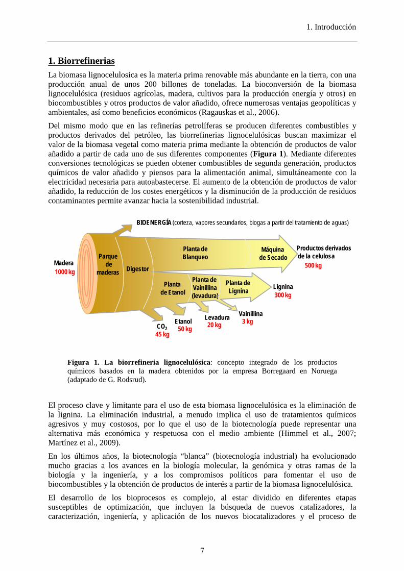
Del mismo modo que en las refinerías petrolíferas se producen diferentes combustibles y productos derivados del petróleo, las biorrefinerias lignocelulósicas buscan maximizar el valor de la biomasa vegetal como materia prima mediante la obtención de productos de valor añadido a partir de cada uno de sus diferentes componentes (Figura 1). Mediante diferentes conversiones tecnológicas se pueden obtener combustibles de segunda generación, productos químicos de valor añadido y piensos para la alimentación animal, simultáneamente con la electricidad necesaria para autoabastecerse. El aumento de la obtención de productos de valor añadido, la reducción de los costes energéticos y la disminución de la producción de residuos contaminantes permite avanzar hacia la sostenibilidad industrial.
Figura 1. La biorrefineria lignocelulósica: concepto integrado de los productos químicos basados en la madera obtenidos por la empresa Borregaard en Noruega (adaptado de G. Rodsrud).
El proceso clave y limitante para el uso de esta biomasa lignocelulósica es la eliminación de la lignina. La eliminación industrial, a menudo implica el uso de tratamientos químicos agresivos y muy costosos, por lo que el uso de la biotecnología puede representar una alternativa más económica y respetuosa con el medio ambiente (Himmel et al., 2007; Martínez et al., 2009).
En los últimos años, la biotecnología “blanca” (biotecnología industrial) ha evolucionado mucho gracias a los avances en la biología molecular, la genómica y otras ramas de la biología y la ingeniería, y a los compromisos políticos para fomentar el uso de biocombustibles y la obtención de productos de interés a partir de la biomasa lignocelulósica.
El desarrollo de los bioprocesos es complejo, al estar dividido en diferentes etapas susceptibles de optimización, que incluyen la búsqueda de nuevos catalizadores, la caracterización, ingeniería, y aplicación de los nuevos biocatalizadores y el proceso de
Vainillina
Lignina
Productos derivadosde la celulosa
EtanolCO2
BIOENERGÍA (corteza, vapores secundarios, biogas a partir del tratamiento de aguas)
Madera1000 kg
50 kg45 kg
3 kg
300 kg
500 kg
Levadura20 kg
Parquede
maderas Digestor
Planta de Etanol
Planta deVainillina(levadura)
Planta deLignina
Planta de Blanqueo
Máquinade Secado
7
1. Introducción
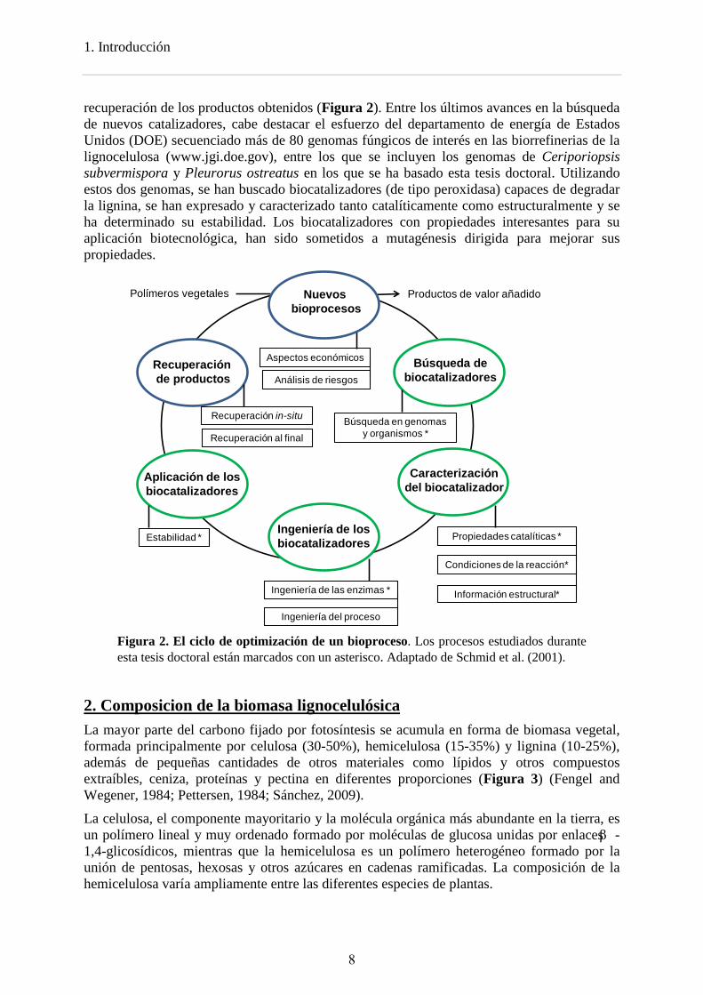
recuperación de los productos obtenidos (Figura 2). Entre los últimos avances en la búsqueda de nuevos catalizadores, cabe destacar el esfuerzo del departamento de energía de Estados Unidos (DOE) secuenciado más de 80 genomas fúngicos de interés en las biorrefinerias de la lignocelulosa (www.jgi.doe.gov), entre los que se incluyen los genomas de Ceriporiopsis subvermispora y Pleurorus ostreatus en los que se ha basado esta tesis doctoral. Utilizando estos dos genomas, se han buscado biocatalizadores (de tipo peroxidasa) capaces de degradar la lignina, se han expresado y caracterizado tanto catalíticamente como estructuralmente y se ha determinado su estabilidad. Los biocatalizadores con propiedades interesantes para su aplicación biotecnológica, han sido sometidos a mutagénesis dirigida para mejorar sus propiedades.
Polímeros vegetales Productos de valor añadido
Búsqueda de biocatalizadores
Caracterización del biocatalizador
Recuperaciónde productos
Aplicación de los biocatalizadores
Ingeniería de los biocatalizadores
Condiciones de la reacción*
Información estructural*
Propiedades catalíticas *
Ingeniería de las enzimas *
Ingeniería del proceso
Estabilidad *
Búsqueda en genomasy organismos *
Aspectos económicos
Análisis de riesgos
Recuperación in-situ
Recuperación al final
Nuevosbioprocesos
Figura 2. El ciclo de optimización de un bioproceso. Los procesos estudiados durante esta tesis doctoral están marcados con un asterisco. Adaptado de Schmid et al. (2001).
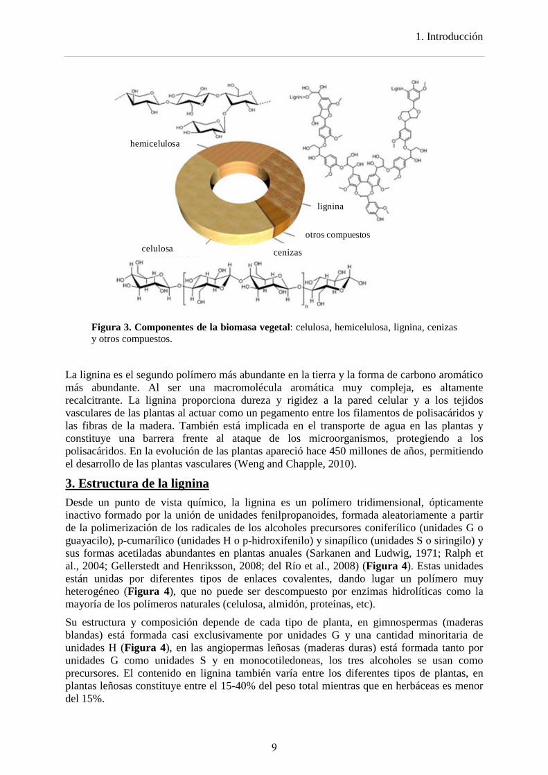
2. Composicion de la biomasa lignocelulósica La mayor parte del carbono fijado por fotosíntesis se acumula en forma de biomasa vegetal, formada principalmente por celulosa (30-50%), hemicelulosa (15-35%) y lignina (10-25%), además de pequeñas cantidades de otros materiales como lípidos y otros compuestos extraíbles, ceniza, proteínas y pectina en diferentes proporciones (Figura 3) (Fengel and Wegener, 1984; Pettersen, 1984; Sánchez, 2009).
La celulosa, el componente mayoritario y la molécula orgánica más abundante en la tierra, es un polímero lineal y muy ordenado formado por moléculas de glucosa unidas por enlaces β -1,4-glicosídicos, mientras que la hemicelulosa es un polímero heterogéneo formado por la unión de pentosas, hexosas y otros azúcares en cadenas ramificadas. La composición de la hemicelulosa varía ampliamente entre las diferentes especies de plantas.
8
1. Introducción
hemicelulosa
celulosa
lignina
cenizas
otros compuestos
Figura 3. Componentes de la biomasa vegetal: celulosa, hemicelulosa, lignina, cenizas y otros compuestos.
La lignina es el segundo polímero más abundante en la tierra y la forma de carbono aromático más abundante. Al ser una macromolécula aromática muy compleja, es altamente recalcitrante. La lignina proporciona dureza y rigidez a la pared celular y a los tejidos vasculares de las plantas al actuar como un pegamento entre los filamentos de polisacáridos y las fibras de la madera. También está implicada en el transporte de agua en las plantas y constituye una barrera frente al ataque de los microorganismos, protegiendo a los polisacáridos. En la evolución de las plantas apareció hace 450 millones de años, permitiendo el desarrollo de las plantas vasculares (Weng and Chapple, 2010).
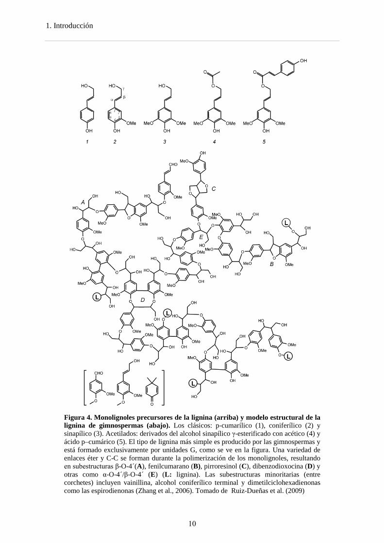
3. Estructura de la lignina Desde un punto de vista químico, la lignina es un polímero tridimensional, ópticamente inactivo formado por la unión de unidades fenilpropanoides, formada aleatoriamente a partir de la polimerización de los radicales de los alcoholes precursores coniferílico (unidades G o guayacilo), p-cumarílico (unidades H o p-hidroxifenilo) y sinapílico (unidades S o siringilo) y sus formas acetiladas abundantes en plantas anuales (Sarkanen and Ludwig, 1971; Ralph et al., 2004; Gellerstedt and Henriksson, 2008; del Río et al., 2008) (Figura 4). Estas unidades están unidas por diferentes tipos de enlaces covalentes, dando lugar un polímero muy heterogéneo (Figura 4), que no puede ser descompuesto por enzimas hidrolíticas como la mayoría de los polímeros naturales (celulosa, almidón, proteínas, etc). Su estructura y composición depende de cada tipo de planta, en gimnospermas (maderas blandas) está formada casi exclusivamente por unidades G y una cantidad minoritaria de unidades H (Figura 4), en las angiopermas leñosas (maderas duras) está formada tanto por unidades G como unidades S y en monocotiledoneas, los tres alcoholes se usan como precursores. El contenido en lignina también varía entre los diferentes tipos de plantas, en plantas leñosas constituye entre el 15-40% del peso total mientras que en herbáceas es menor del 15%.
9
1. Introducción
Figura 4. Monolignoles precursores de la lignina (arriba) y modelo estructural de la lignina de gimnospermas (abajo). Los clásicos: p-cumarílico (1), coniferílico (2) y sinapílico (3). Acetilados: derivados del alcohol sinapílico γ-esterificado con acético (4) y ácido p–cumárico (5). El tipo de lignina más simple es producido por las gimnospermas y está formado exclusivamente por unidades G, como se ve en la figura. Una variedad de enlaces éter y C-C se forman durante la polimerización de los monolignoles, resultando en subestructuras β-O-4´(A), fenilcumarano (B), pirroresinol (C), dibenzodioxocina (D) y otras como α-O-4´/β-O-4´ (E) (L: lignina). Las subestructuras minoritarias (entre corchetes) incluyen vainillina, alcohol coniferílico terminal y dimetilciclohexadienonas como las espirodienonas (Zhang et al., 2006). Tomado de Ruiz-Dueñas et al. (2009)
10
1. Introducción
4. Organismos degradadores de lignina La degradación de la lignina es un paso clave en el reciclado del carbono terrestre, al permitir la degradación de los polisacáridos vegetales. A lo largo de la evolución, sólo un grupo de basidiomicetes junto con algunos ascomicetes y bacterias, ha desarrollado la habilidad de degradar (o modificar) la lignina (Martínez et al., 2005). La aparición de estos basidiomicetes ligninolíticos se correlaciona con el descenso brusco de la acumulación de carbono orgánico en el suelo y la formación de carbón al final del Carbonífero (Floudas et al., 2012).
En función de los patrones de degradación, los hongos se dividen en cuatro grupos, hongos de la podredumbre blanca, hongos de la podredumbre parda, hongos de la podredumbre blanda y hongos que tiñen la madera (Liese, 1970; Blanchette, 1991; Schwarze et al., 2000; Martínez et al., 2005; 2011). Estos dos últimos grupos son ascomicetes que sólo son capaces de modificar la madera en condiciones ambientales especiales (podredumbre blanda) o colonizarla a través de los vasos o canales existentes (hongos que tiñen la madera).
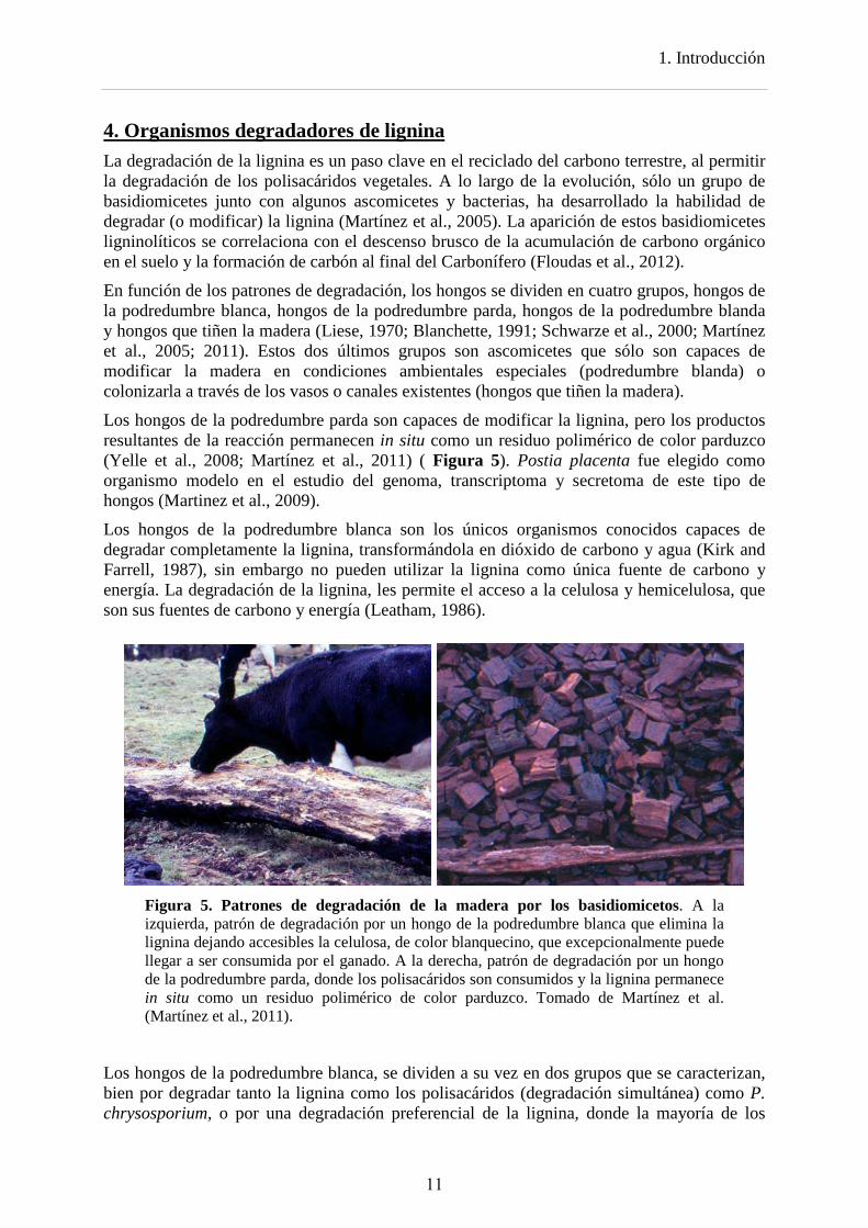
Los hongos de la podredumbre parda son capaces de modificar la lignina, pero los productos resultantes de la reacción permanecen in situ como un residuo polimérico de color parduzco (Yelle et al., 2008; Martínez et al., 2011) ( Figura 5). Postia placenta fue elegido como organismo modelo en el estudio del genoma, transcriptoma y secretoma de este tipo de hongos (Martinez et al., 2009).
Los hongos de la podredumbre blanca son los únicos organismos conocidos capaces de degradar completamente la lignina, transformándola en dióxido de carbono y agua (Kirk and Farrell, 1987), sin embargo no pueden utilizar la lignina como única fuente de carbono y energía. La degradación de la lignina, les permite el acceso a la celulosa y hemicelulosa, que son sus fuentes de carbono y energía (Leatham, 1986).
Figura 5. Patrones de degradación de la madera por los basidiomicetos. A la izquierda, patrón de degradación por un hongo de la podredumbre blanca que elimina la lignina dejando accesibles la celulosa, de color blanquecino, que excepcionalmente puede llegar a ser consumida por el ganado. A la derecha, patrón de degradación por un hongo de la podredumbre parda, donde los polisacáridos son consumidos y la lignina permanece in situ como un residuo polimérico de color parduzco. Tomado de Martínez et al. (Martínez et al., 2011).
Los hongos de la podredumbre blanca, se dividen a su vez en dos grupos que se caracterizan, bien por degradar tanto la lignina como los polisacáridos (degradación simultánea) como P. chrysosporium, o por una degradación preferencial de la lignina, donde la mayoría de los
11
1. Introducción
polisacáridos quedan intactos (degradación selectiva) como en el caso de C. subvermispora (Blanchette et al., 1985; Blanchette et al., 1997; del Río et al., 2001).
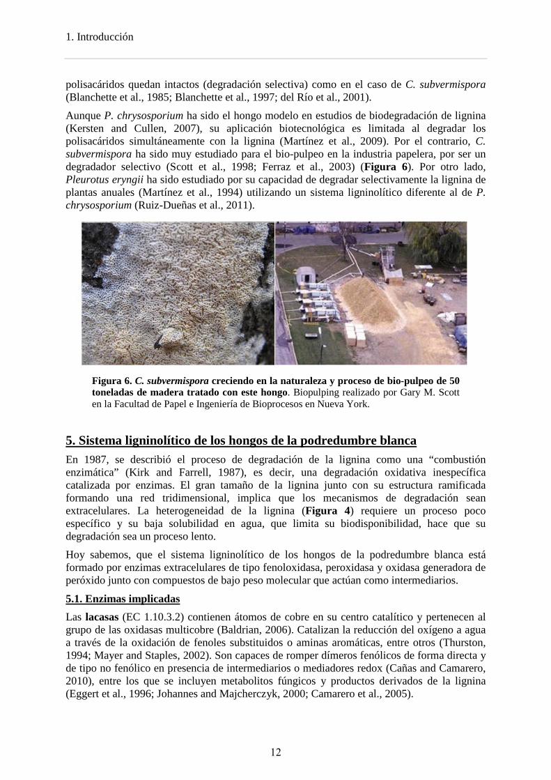
Aunque P. chrysosporium ha sido el hongo modelo en estudios de biodegradación de lignina (Kersten and Cullen, 2007), su aplicación biotecnológica es limitada al degradar los polisacáridos simultáneamente con la lignina (Martínez et al., 2009). Por el contrario, C. subvermispora ha sido muy estudiado para el bio-pulpeo en la industria papelera, por ser un degradador selectivo (Scott et al., 1998; Ferraz et al., 2003) (Figura 6). Por otro lado, Pleurotus eryngii ha sido estudiado por su capacidad de degradar selectivamente la lignina de plantas anuales (Martínez et al., 1994) utilizando un sistema ligninolítico diferente al de P. chrysosporium (Ruiz-Dueñas et al., 2011).
Figura 6. C. subvermispora creciendo en la naturaleza y proceso de bio-pulpeo de 50 toneladas de madera tratado con este hongo. Biopulping realizado por Gary M. Scott en la Facultad de Papel e Ingeniería de Bioprocesos en Nueva York.
5. Sistema ligninolítico de los hongos de la podredumbre blanca En 1987, se describió el proceso de degradación de la lignina como una “combustión enzimática” (Kirk and Farrell, 1987), es decir, una degradación oxidativa inespecífica catalizada por enzimas. El gran tamaño de la lignina junto con su estructura ramificada formando una red tridimensional, implica que los mecanismos de degradación sean extracelulares. La heterogeneidad de la lignina (Figura 4) requiere un proceso poco específico y su baja solubilidad en agua, que limita su biodisponibilidad, hace que su degradación sea un proceso lento.
Hoy sabemos, que el sistema ligninolítico de los hongos de la podredumbre blanca está formado por enzimas extracelulares de tipo fenoloxidasa, peroxidasa y oxidasa generadora de peróxido junto con compuestos de bajo peso molecular que actúan como intermediarios.
5.1. Enzimas implicadas Las lacasas (EC 1.10.3.2) contienen átomos de cobre en su centro catalítico y pertenecen al grupo de las oxidasas multicobre (Baldrian, 2006). Catalizan la reducción del oxígeno a agua a través de la oxidación de fenoles substituidos o aminas aromáticas, entre otros (Thurston, 1994; Mayer and Staples, 2002). Son capaces de romper dímeros fenólicos de forma directa y de tipo no fenólico en presencia de intermediarios o mediadores redox (Cañas and Camarero, 2010), entre los que se incluyen metabolitos fúngicos y productos derivados de la lignina (Eggert et al., 1996; Johannes and Majcherczyk, 2000; Camarero et al., 2005).
12
1. Introducción
Están presentes tanto en hongos de la podredumbre parda como de podredumbre blanca (Floudas et al., 2012), aunque en el caso de P. chrysosporium no se ha detectado en su genoma ninguna lacasa en sentido estricto (Martinez et al., 2004).
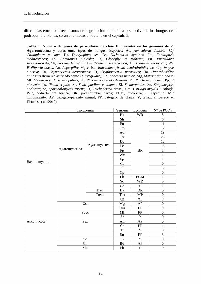
Las peroxidasas ligninolíticas son un grupo de hemoperoxidasas incluidas en la clase II de la superfamilia de peroxidasas vegetales-fúngicas-procariotas. Estas enzimas presentan un grupo hemo de tipo protoporfirina IX unido de forma no covalente y localizado en una cavidad interna conectada con el solvente a través de un canal principal, que permite el acceso tanto de peróxido de hidrógeno como de pequeños sustratos (Banci, 1997; Dunford, 1999). Catalizan la oxidación de gran variedad de compuestos orgánicos e inorgánicos a expensas de H2O2 (Dunford, 1999). Las peroxidasas ligninolíticas se dividen en tres familias - lignina peroxidasas (LiP), manganeso peroxidasas (MnP) y peroxidasas versátiles (VP) - y se caracterizan por su elevado potencial redox. Se ha demostrado la capacidad de la LiP de degradar la lignina in vitro (Hammel et al., 1993) y recientemente de la VP (como parte de esta tesis doctoral) (Fernández-Fueyo et al., 2014). La MnP puede degradar la lignina a través de la peroxidación de los lípidos (Bao et al., 1994), ya que su acción directa (Wariishi et al., 1991) está por confirmar. De acuerdo con el papel central de las peroxidasas en la degradación de la lignina, los hongos de la podredumbre blanca tienen entre 5 y 26 copias de genes que codifican para peroxidasas ligninolíticas (14 copias de media) (Tabla 1) y, como se ha mencionado anteriormente, no están presentes ni en hongos de la podredumbre parda ni en hongos patógenos, a diferencia de las lacasas (Floudas et al., 2012) .
El H2O2 desempeña un papel central en el ataque a la madera por parte de los hongos. Es el sustrato oxidante de las peroxidasas ligninolíticas (Ruiz-Dueñas and Martínez, 2009). Además, en presencia de Fe2+, puede atacar la pared celular a través del radical hidroxilo (OH●) procedente de la reacción de Fenton (Fe2+ + H2O2 → Fe3+ + H2O +OH●) en hongos de la podredumbre parda y blanca (Forney et al., 1982; Faison and Kirk, 1983; Guillén et al., 2000; Gómez-Toribio et al., 2009). En 1983, Faison y Kirk (1983) demostraron que el H2O2 se produce simultáneamente con el sistema ligninolítico, y que la adicción de catalasa a los cultivos fúngicos, disminuye la capacidad de degradar la lignina. Se han propuesto numerosas enzimas extracelulares que pueden estar implicadas en la producción de H2O2 extracelular en los hongos de la podredumbre blanca, como la glioxal-oxidasa (Kersten and Kirk, 1987), la aril-alcohol oxidasa (AAO) (Guillén et al., 1992a; Guillén et al., 1992b), la metanol oxidasa (Ozimek et al., 2005)y la piranosa oxidasa (Daniel et al., 1994). También existen enzimas intracelulares productoras de H2O2 (Greene and Gould, 1984; Eriksson et al., 1986), pero se desconoce la existencia de un sistema transportador para el H2O2 producido. Los sustratos de estas enzimas durante la degradación de la lignocelulosa incluyen tanto compuestos derivados de la lignina como metabolitos aromáticos producidos por los hongos, entre los que se incluyen aldehídos, ácidos aromáticos de tipo fenólico y aldehídos aromáticos derivados de las ruptura de enlaces Cα-Cβ o sintetizados de novo por el hongo (Kirk and Farrell, 1987; Shimada and Higuchi, 1991). La celobiosa deshidogenasa (CDH) podría estar implicada en la degradación de la lignocelulosa por su capacidad para reducir el Fe3+ a Fe2+ y formar H2O2 (reactivo de Fenton) (Henriksson et al., 2000)
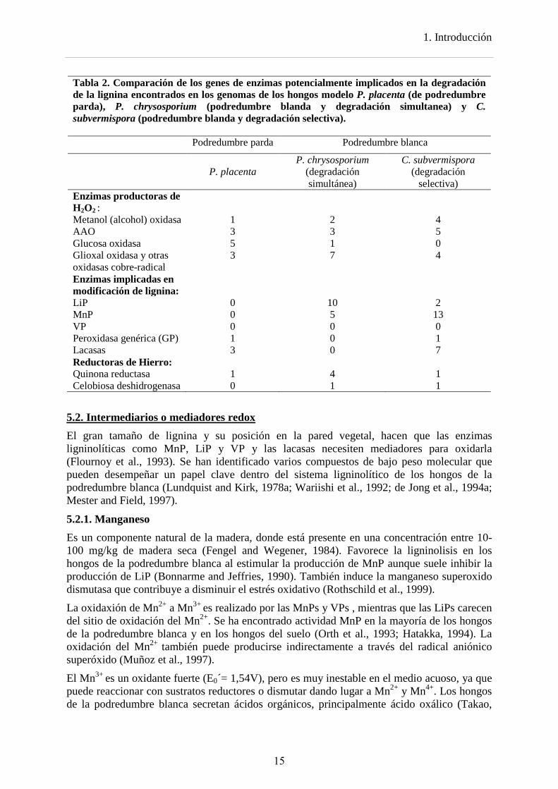
La comparación de los genomas, trascriptomas y secretomas de los hongos modelo P. placenta (de podredumbre parda), P. chrysosporium (podredumbre blanda y degradación simultanea) y C. subvermispora (podredumbre blanda y degradación selectiva) reflejan las diferencias en los patrones de degradación lignocelulósica. La comparación de sus genomas (Tabla 2) indica que durante la evolución de los hongos de la podredumbre parda se concentraron o perdieron familias de genes que son importantes en los hongos de la podredumbre blanca como las peroxidasas ligninolíticas y la celobiosa deshidrogenasa. Las
13
1. Introducción
diferencias entre los mecanismos de degradación simultánea o selectiva de los hongos de la podredumbre blanca, serán analizadas en detalle en el capítulo 5.
Tabla 1. Número de genes de peroxidasas de clase II presentes en los genomas de 20 Agaromicotina y otros once tipos de hongos. Especies: Ad, Auricularia delicata; Cp, Coniophora puteana; Da, Dacryopinax sp., Ds, Dichomitus squalens; Fm, Fomitiporia mediterranea; Fp, Fomitopsis pinicola; Gt, Gloeophyllum trabeum; Pu, Punctularia strigosozonata; Sh, Stereum hirsutum; Tm, Tremella mesenterica, Tv, Trametes versicolor; Wc, Wolfiporia cocos, An, Aspergillus niger; Bd, Batrachochytrium dendrobatidis; Cc, Coprinopsis cinerea; Cn, Cryptococcus neoformans; Cr, Cryphonectria parasitica; Ha, Heterobasidion annosum(ahora reclasificado como H. irregulare); Lb, Laccaria bicolor; Mg, Malassezia globosa; Ml, Melampsora laricis-populina; Pb, Phycomyces blakesleeanus; Pc, P. chrysosporium; Pp, P. placenta; Ps, Pichia stipitis; Sc, Schizophyllum commune; Sl, S. lacrymans; Sn, Stagonospora nodorum; Sr, Sporobolomyces roseus; Tr, Trichoderma reesei; Um, Ustilago maydis. Ecología: WR, podredumbre blanca; BR, podredumbre parda; ECM, micorriza; S, saprófito; MP, micoparasito; AP, patógeno/parasito animal; PP, patógeno de planta; Y, levadura. Basado en Floudas et al (2012). Taxonomía Genoma Ecología Nº de PODs Basidiomycota
Agaromycotina
Agaromycetes
Ha WR 8 Sh 6 Pu 11 Fm 17 Ad 19 Tv 26 Ds 12 Pc 16 Pp BR 1 Wc 1 Fp 1 Gt 0 Sl 0 Cp 0 Lb ECM 1 Sc WR 0 Cc S 1
Dac Da BR 0 Trem Tm MP 0
Cn AP 0 Ust Mg AP 0
Um PP 0 Pucc Ml PP 0
Sr Y 0 Ascomycota Pez An AP 0
Cr PP 1 Tr S 0 Sn PP 5
Sc Ps Y 0 Ch Bd AP 0 Mu Pb S 0
14
1. Introducción
Tabla 2. Comparación de los genes de enzimas potencialmente implicados en la degradación de la lignina encontrados en los genomas de los hongos modelo P. placenta (de podredumbre parda), P. chrysosporium (podredumbre blanda y degradación simultanea) y C. subvermispora (podredumbre blanda y degradación selectiva). Podredumbre parda Podredumbre blanca
P. placenta
P. chrysosporium (degradación simultánea)
C. subvermispora (degradación
selectiva) Enzimas productoras de H2O2 :
Metanol (alcohol) oxidasa 1 2 4 AAO 3 3 5 Glucosa oxidasa 5 1 0 Glioxal oxidasa y otras oxidasas cobre-radical
3 7 4
Enzimas implicadas en modificación de lignina:
LiP 0 10 2 MnP 0 5 13 VP 0 0 0 Peroxidasa genérica (GP) 1 0 1 Lacasas 3 0 7 Reductoras de Hierro: Quinona reductasa 1 4 1 Celobiosa deshidrogenasa 0 1 1
5.2. Intermediarios o mediadores redox El gran tamaño de lignina y su posición en la pared vegetal, hacen que las enzimas ligninolíticas como MnP, LiP y VP y las lacasas necesiten mediadores para oxidarla (Flournoy et al., 1993). Se han identificado varios compuestos de bajo peso molecular que pueden desempeñar un papel clave dentro del sistema ligninolítico de los hongos de la podredumbre blanca (Lundquist and Kirk, 1978a; Wariishi et al., 1992; de Jong et al., 1994a; Mester and Field, 1997).
5.2.1. Manganeso Es un componente natural de la madera, donde está presente en una concentración entre 10-100 mg/kg de madera seca (Fengel and Wegener, 1984). Favorece la ligninolisis en los hongos de la podredumbre blanca al estimular la producción de MnP aunque suele inhibir la producción de LiP (Bonnarme and Jeffries, 1990). También induce la manganeso superoxido dismutasa que contribuye a disminuir el estrés oxidativo (Rothschild et al., 1999).
La oxidaxión de Mn2+ a Mn3+ es realizado por las MnPs y VPs , mientras que las LiPs carecen del sitio de oxidación del Mn2+. Se ha encontrado actividad MnP en la mayoría de los hongos de la podredumbre blanca y en los hongos del suelo (Orth et al., 1993; Hatakka, 1994). La oxidación del Mn2+ también puede producirse indirectamente a través del radical aniónico superóxido (Muñoz et al., 1997).
El Mn3+ es un oxidante fuerte (E0´= 1,54V), pero es muy inestable en el medio acuoso, ya que puede reaccionar con sustratos reductores o dismutar dando lugar a Mn2+ y Mn4+. Los hongos de la podredumbre blanca secretan ácidos orgánicos, principalmente ácido oxálico (Takao,
15
1. Introducción
1965), que forman quelatos con el Mn3+ estabilizándolo con un potencial rédox algo menor (E0´= 0,9-1,2V). Estos quelatos actúan como oxidantes estables difusibles de compuestos fenólicos, aminas aromáticas y ácidos carboxílicos (Paszczynski et al., 1986; Wariishi and Gold, 1989; Wariishi et al., 1991; Hammel et al., 1993). Estos quelatos también son capaces de oxidar ácidos grasos insaturados y sus derivados formando radicales lipídicos de tipo peroxilo (Kapich and Shishbina, 1995), que son altamente reactivos y pueden reaccionar con otras moléculas de lípidos insaturados para propagar la oxidación, produciendo un proceso de peroxidación lipídica en cadena.
Se ha descrito como la peroxidación de acidos grasos insaturados por parte de las MnP permite la ruptura oxidativa de fenantreno, hidrocarburos policíclicos aromáticos y de un dímero modelo no fenólico de lignina (Moen and Hammel, 1994; Bao et al., 1994). Este mecanismo parece estar implicado en la degradación de la lignina por algunos hongos basidiomicetos de la podredumbre blanca (Fernández-Fueyo et al., 2012a).
5.2.2. Alcohol veratrílico (VA) Es un metabolito aromático de los basidiomicetos que se sintetiza de novo a partir de glucosa (Shimada and Takahashi, 1991). Es el sustrato típico de las LiPs aunque también es oxidado con menor eficiencia por VPs (Fernández-Fueyo et al., 2012b). Es secretado por algunos hongos como P. chrysosporium y Bjerkandera adusta durante la oxidación de la lignina (Lundquist and Kirk, 1978b; Shimada et al., 1981). Existe cierta controversia sobre el papel del alcohol veratrílico en la degradación de la lignina. Se ha demostrado que su presencia permite a la LiP oxidar compuestos que no es capaz de oxidar por si sola, como el Reactive Black 5 o alcohol anisílico (Harvey et al., 1986). Algunos autores proponen que el radical cationico (VA+● ) formado tras la oxidación del alcohol (por la sustracción de un electrón) es el que oxida estos compuestos, actuando como mediador rédox (Harvey et al., 1986; Paszczynski and Crawford, 1991; Tien and Ma, 1997), pudiendo actuar como mediador en la despolimerización de la lignina. Por otra parte, se ha descrito como el alcohol anisílico reduce el compuesto I de la LiP, pero no el compuesto II. Por lo tanto, el alcohol veratrílico podría actuar completando el ciclo de la enzima, al reaccionar con el compuesto II, permitiendo la regeneración de la enzima al estado de reposo y evitando la formación de compuesto III (Koduri and Tien, 1994). De acuerdo a esto, se ha propuesto que el alcohol veratrilico actuaría como un agente reductor previniendo la inactivación de la enzima por peróxido (Collins et al., 1997).
Aunque la VP no requiere de mediadores redox para la oxidación de colorantes de alto potencial redox como el Reactive Black 5 (Heinfling et al., 1998), la presencia del alcohol veratrílico es necesaria para la oxidación de la lignina in vitro (Fernández-Fueyo et al., 2014) al igual que en el caso de la LiP (Hammel et al., 1993).
5.2.3. Metabolitos clorados Los hongos de podredumbre blanca producen una gran variedad de compuestos halogenados (de Jong et al., 1994c; de Jong and Field, 1997), siendo los más comunes los metabolitos anisílicos clorados y las hidroquinonas cloradas (de Jong et al., 1994c; Field et al., 1995). Los primeros pueden tener importancia en la degradación de la lignina en condiciones fisiológicas, al ser sustratos de la AAO, productora de H2O2 en hongos de la podredumbre blanca como P. ostreatus y B. adusta (de Jong et al., 1994b). Estos compuestos clorados tienen Km extremadamente bajas para la AAO de los hongos que los producen en comparación con los equivalentes no clorados.
16
1. Introducción
Estudios previos, han descrito que el 1,4 dimetoxibenceno puede remplazar al VA como mediador (Joshi and Gold, 1996). Mientras que este compuesto no es un metabolito de los hongos de la podredumbre blanca, el 2-cloro-1,4-dimetoxibenceno y 2,6-cloro-1,4-dimetoxibenceno, que están estructuralmente relacionados, han sido detectados en cultivos de B. adusta, (Spinnler et al., 1994). El 2-cloro-1,4-dimetoxybenceno es oxidado por la LiP de P. chrysosporium (Teunissen and Field, 1998b), produciendo su radical catiónico, que actúa como un mediador difusible oxidando sustratos que la LiP no es capaz de oxidar por si sola (Teunissen et al., 1998; Teunissen and Field, 1998a; 1998b). Solamente concentraciones catalíticas (10-50 µM) de este compuesto son necesarias para mediar en la oxidación de los sustratos, lo que sugiere la importancia de este metabolito fúngico en la degradación de la lignina.
5.2.4. Mediadores de las lacasas Las lacasas tienen un potencial redox relativamente bajo (≤0.8 V), por lo que su acción quedaría restringida a la oxidación de las unidades fenólicas de lignina (Kawai et al., 1987). Sin embargo, en presencia de mediadores redox, la actividad catalítica de las lacasas puede ser expandida hacia sustratos no fenólicos más difícilmente oxidables, en lo que se denomina sistema lacasa-mediador.
Los mediadores, mediante mecanismos de oxidación no enzimáticos, oxidan compuestos que bien sea por su elevado tamaño molecular o por su alto potencial redox, en principio no son sustratos de las lacasas. Bourbonnais y Paice (1990) fueron los primeros en describir la oxidación de dímeros no fenólicos de lignina en presencia de lacasa y el mediador ABTS (2,2'-azino-bis(3-etilbenzotiazolin-6-sulfonato)).
Los mediadores sintéticos más eficaces son compuestos que portan un grupo NOH como el 1-hidroxibenzotriazol, el ácido violúrico o la N-hidroxiacetanilida (Böhmer et al., 1998; Johannes et al., 1998; Galli and Gentili, 2004). Entre los mediadores de origen natural, destacan el ácido 3-hidroxiantranílico y el ácido 4-hidroxibenzoico, que son metabolitos secundarios de Pycnoporus cinnabarinus y de T. versicolor, respectivamente (Eggert et al., 1996; Johannes and Majcherczyk, 2000). Recientemente, Camarero et al (2005) han descrito la capacidad de ciertos compuestos fenólicos liberados durante la biodegradación de la lignina, como la acetosiringona, el siringilaldehido, la vainillina y el ácido p-cumárico, para actuar como mediadores redox de lacasa.
6. Enzimas de tipo peroxidasa 6.1 Características generales Las peroxidasas (EC 1.11.1) son un tipo de enzimas muy extendidas en toda la escala filogenética, que catalizan la oxidación de gran variedad de compuestos orgánicos e inorgánicos a expensas de H2O2 u otros hidroperóxidos (Dunford, 1999). Los análisis filogenéticos revelan que los genes que codifican para peroxidasas tiene un origen procariota muy antiguo, que probablemente se remonta a la aparición del oxígeno atmosférico.
Se pueden dividir en dos clases según su sitio activo: las hemoperoxidasas, que contienen un grupo hemo como cofactor, presentan una gran diversidad funcional y están presenten en la mayoría de los organismos; y las peroxidasas sin grupo hemo, entre las que se encuentran pequeñas familias de proteínas heterogéneas (algunas haloperoxidasas, alquil-
17
1. Introducción
hidroperoxidasas, NADH peroxidasas, tiol peroxidasas, glutation peroxidasas, peroxirredoxinas y manganeso catalasas) (Passardi et al., 2007).
En función de sus diferencias bioquímicas y estructurales, las hemoperoxidasas se han clasificado tradicionalmente en dos superfamilias: la superfamilia de peroxidasas animales (Kimura and Ikeda-Saito, 1988) y la superfamilia de peroxidasas vegetales-fúngicas-procariotas (no animales) (Welinder, 1992). Recientemente se han añadido dos nuevas superfamilias: de hemotiolato peroxidasas y DyPs (Ruiz-Dueñas and Martínez, 2010; Hofrichter et al., 2010) abundantes en genomas de basidiomicetos (Floudas et al., 2012). Estas superfamilias surgieron independientemente, tal y como se deduce de sus diferencias de secuencia, plegamiento general, unión del grupo prostético y coordinación del hierro, entre otras características.
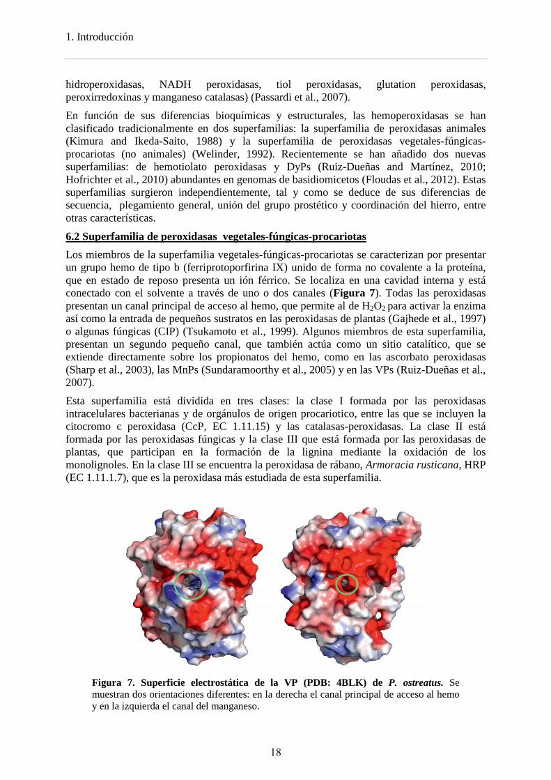
6.2 Superfamilia de peroxidasas vegetales-fúngicas-procariotas Los miembros de la superfamilia vegetales-fúngicas-procariotas se caracterizan por presentar un grupo hemo de tipo b (ferriprotoporfirina IX) unido de forma no covalente a la proteína, que en estado de reposo presenta un ión férrico. Se localiza en una cavidad interna y está conectado con el solvente a través de uno o dos canales (Figura 7). Todas las peroxidasas presentan un canal principal de acceso al hemo, que permite al de H2O2 para activar la enzima así como la entrada de pequeños sustratos en las peroxidasas de plantas (Gajhede et al., 1997) o algunas fúngicas (CIP) (Tsukamoto et al., 1999). Algunos miembros de esta superfamilia, presentan un segundo pequeño canal, que también actúa como un sitio catalítico, que se extiende directamente sobre los propionatos del hemo, como en las ascorbato peroxidasas (Sharp et al., 2003), las MnPs (Sundaramoorthy et al., 2005) y en las VPs (Ruiz-Dueñas et al., 2007).
Esta superfamilia está dividida en tres clases: la clase I formada por las peroxidasas intracelulares bacterianas y de orgánulos de origen procariotico, entre las que se incluyen la citocromo c peroxidasa (CcP, EC 1.11.15) y las catalasas-peroxidasas. La clase II está formada por las peroxidasas fúngicas y la clase III que está formada por las peroxidasas de plantas, que participan en la formación de la lignina mediante la oxidación de los monolignoles. En la clase III se encuentra la peroxidasa de rábano, Armoracia rusticana, HRP (EC 1.11.1.7), que es la peroxidasa más estudiada de esta superfamilia.
Figura 7. Superficie electrostática de la VP (PDB: 4BLK) de P. ostreatus. Se muestran dos orientaciones diferentes: en la derecha el canal principal de acceso al hemo y en la izquierda el canal del manganeso.
18
1. Introducción
La peroxidasas de la clase II (PODs) se divididen en cuatro familias: GPs, LiPs, MnPs y VPs (Fernández-Fueyo et al., 2012b). Las tres últimas familias constituyen el grupo de las peroxidasas ligninolíticas por su implicación en la degradación de la lignina y como se ha mencionado anteriormente, son exclusivas de los hongos de la podredumbre blanca.
7. Peroxidasas ligninolíticas: Las peroxidasas ligninolíticas son proteínas globulares de 30-40 kDa, y alrededor de 330 aminoácidos. Son extracelulares y presentan un péptido señal en el N-terminal que dirige la secreción a través del retículo endoplasmático. Su grado de glicosilación está en torno al 5%. Generalmente, el aminoácido tirosina está ausente en la secuencia, ya que sería susceptible de oxidación en las condiciones oxidativas que caracterizan a la degradación de la lignina. Sin embargo, en la LiP de Trametopsis cervina (Miki et al., 2011; 2013), una tirosina desempeña el mismo papel catalítico que el triptófano en otras LiPs y VPs.
A
I
B
B’ ’ CD
E
GH
J
F
B’a
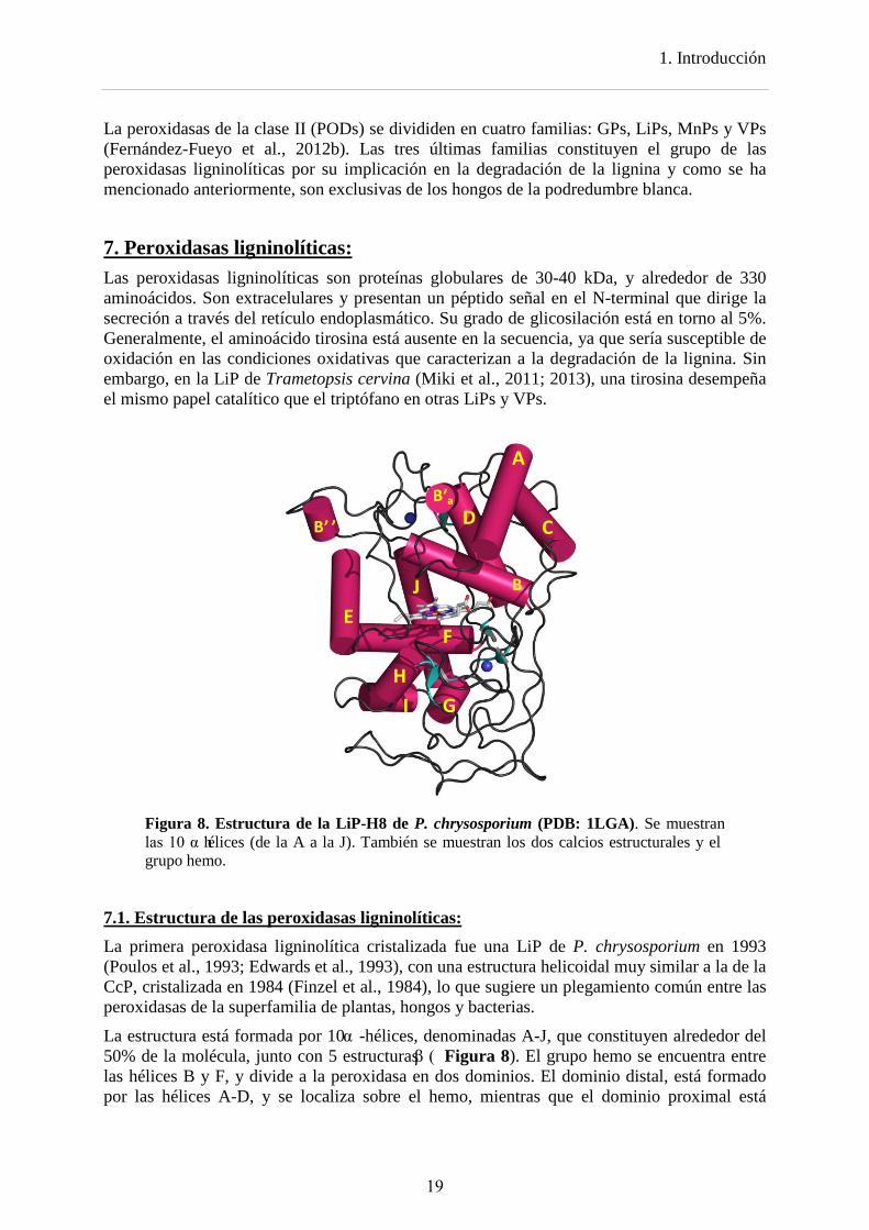
Figura 8. Estructura de la LiP-H8 de P. chrysosporium (PDB: 1LGA). Se muestran las 10 α hélices (de la A a la J). También se muestran los dos calcios estructurales y el grupo hemo.
7.1. Estructura de las peroxidasas ligninolíticas: La primera peroxidasa ligninolítica cristalizada fue una LiP de P. chrysosporium en 1993 (Poulos et al., 1993; Edwards et al., 1993), con una estructura helicoidal muy similar a la de la CcP, cristalizada en 1984 (Finzel et al., 1984), lo que sugiere un plegamiento común entre las peroxidasas de la superfamilia de plantas, hongos y bacterias.
La estructura está formada por 10 α -hélices, denominadas A-J, que constituyen alrededor del 50% de la molécula, junto con 5 estructuras β ( Figura 8). El grupo hemo se encuentra entre las hélices B y F, y divide a la peroxidasa en dos dominios. El dominio distal, está formado por las hélices A-D, y se localiza sobre el hemo, mientras que el dominio proximal está
19
1. Introducción
formado por las hélices E-F, junto con la mayor parte de la estructura β. El nombre de estos dominios se deben a que albergan las denominadas histidinas proximal y distal, mencionadas más adelante. Presentan cuatro (VPs y MnP cortas) o cinco puentes disulfuro (MnPs largas y extralargas) que le confieren rigidez y dos iones calcios imprescindibles en el mantenimiento de la estructura.
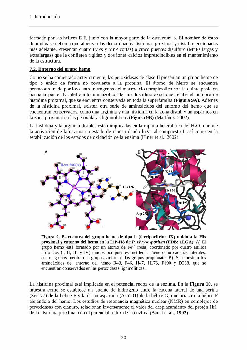
7.2. Entorno del grupo hemo Como se ha comentado anteriormente, las peroxidasas de clase II presentan un grupo hemo de tipo b unido de forma no covalente a la proteína. El átomo de hierro se encuentra pentacoordinado por los cuatro nitrógenos del macrociclo tetrapirrolico con la quinta posición ocupada por el Nε del anillo imidazolico de una histidina axial que recibe el nombre de histidina proximal, que se encuentra conservada en toda la superfamilia (Figura 9A). Además de la histidina proximal, existen otra serie de aminoácidos del entorno del hemo que se encuentran conservados, como una arginina y una histidina en la zona distal, y un aspártico en la zona proximal en las peroxidasas ligninoliticas (Figura 9B) (Martínez, 2002).
La histidina y la arginina distales están implicadas en la ruptura heterolítica del H2O2 durante la activación de la enzima en estado de reposo dando lugar al compuesto I, así como en la estabilización de los estados de oxidación de la enzima (Hiner et al., 2002).
Figura 9. Estructura del grupo hemo de tipo b (ferriporfirina IX) unido a la His proximal y entorno del hemo en la LiP-H8 de P. chrysosporium (PDB: 1LGA). A) El grupo hemo está formado por un átomo de Fe3+ (rosa) coordinado por cuatro anillos pirrólicos (I, II, III y IV) unidos por puentes metileno. Tiene ocho cadenas laterales: cuatro grupos metilo, dos grupos vinilo y dos grupos propionato. B). Se muestran los aminoácidos del entorno del hemo R43, F46, H47, H176, F190 y D238, que se encuentran conservados en las peroxidasas ligninolíticas.
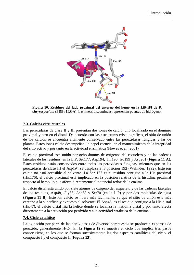
La histidina proximal está implicada en el potencial redox de la enzima. En la Figura 10, se muestra como se establece un puente de hidrógeno entre la cadena lateral de una serina (Ser177) de la hélice F y la de un aspártico (Asp201) de la hélice G, que arrastra la hélice F alejándola del hemo. Los estudios de resonancia magnética nuclear (NMR) en complejos de peroxidasas con cianuro, relacionan inversamente el valor del desplazamiento del protón Hε1 de la histidina proximal con el potencial redox de la enzima (Banci et al., 1992).
20
1. Introducción
Figura 10. Residuos del lado proximal del entorno del hemo en la LiP-H8 de P. chrysosporium (PDB: 1LGA). Las líneas discontinuas representan puentes de hidrógeno.
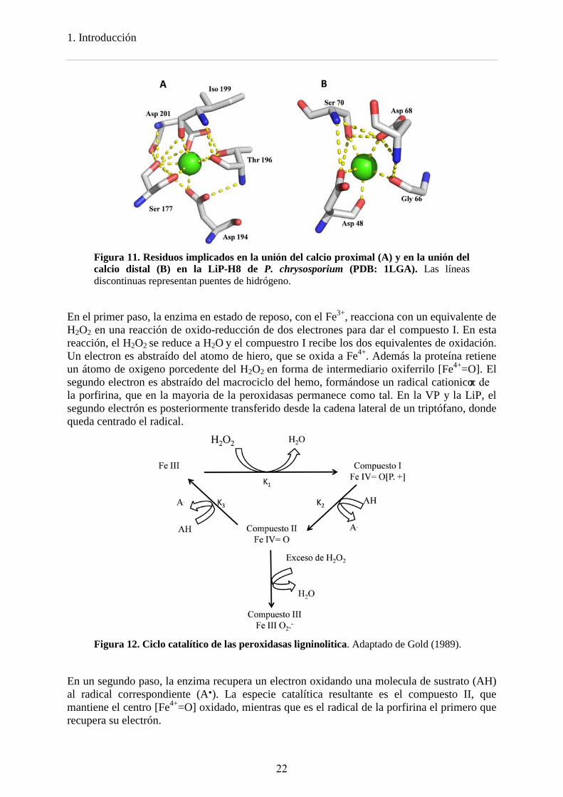
7.3. Calcios estructurales Las peroxidasas de clase II y III presentan dos iones de calcio, uno localizado en el dominio proximal y otro en el distal. De acuerdo con las estructuras cristalográficas, el sitio de unión de los calcios se encuentra altamente conservado entre las peroxidasas fúngicas y las de plantas. Estos iones calcio desempeñan un papel esencial en el mantenimiento de la integridad del sitio activo y por tanto en la actividad enzimática (Howes et al., 2001).
El calcio proximal está unido por ocho átomos de oxígenos del esqueleto y de las cadenas laterales de los residuos, en la LiP, Ser177, Asp194, Thr196, Iso199 y Asp201 (Figura 11 A). Estos residuos están conservados entre todas las peroxidasas fúngicas, mientras que en las peroxidasas de clase III el Asp194 se desplaza a la posición 193 (Welinder, 1992). Este ión calcio no está accesible al solvente. La Ser 177 es el residuo contiguo a la His proximal (His176), el calcio proximal está implicado en la posición relativa de la histidina proximal respecto al hemo, lo que afecta directamente al potencial redox de la enzima.
El calcio distal está unido por siete átomos de oxigeno del esqueleto y de las cadenas laterales de los residuos, Asp48, Gly66, Asp68 y Ser70 (en la LiP) y por dos moléculas de agua (Figura 11 B). Este ión calcio se libera más fácilmente, ya que el sitio de unión está más cercano a la superficie y expuesto al solvente. El Asp48, es el residuo contiguo a la His distal (His47), el calcio distal fija la hélice donde se localiza la histidina distal y por tanto afecta directamente a la activación por peróxido y a la actividad catalítica de la enzima.
7.4. Ciclo catalítico La oxidación por parte de las peroxidasas de diversos compuestos se produce a expensas de peróxido, generalmente H2O2. En la Figura 12 se muestra el ciclo que implica tres pasos consecutivos, en los que se forman sucesivamente las dos especies catalíticas del ciclo, el compuesto I y el compuesto II (Figura 13).
21
1. Introducción
Figura 11. Residuos implicados en la unión del calcio proximal (A) y en la unión del calcio distal (B) en la LiP-H8 de P. chrysosporium (PDB: 1LGA). Las líneas discontinuas representan puentes de hidrógeno.
En el primer paso, la enzima en estado de reposo, con el Fe3+, reacciona con un equivalente de H2O2 en una reacción de oxido-reducción de dos electrones para dar el compuesto I. En esta reacción, el H2O2 se reduce a H2O y el compuestro I recibe los dos equivalentes de oxidación. Un electron es abstraído del atomo de hiero, que se oxida a Fe4+. Además la proteína retiene un átomo de oxigeno porcedente del H2O2 en forma de intermediario oxiferrilo [Fe4+=O]. El segundo electron es abstraído del macrociclo del hemo, formándose un radical cationico π de la porfirina, que en la mayoria de la peroxidasas permanece como tal. En la VP y la LiP, el segundo electrón es posteriormente transferido desde la cadena lateral de un triptófano, donde queda centrado el radical.
Figura 12. Ciclo catalítico de las peroxidasas ligninolitica. Adaptado de Gold (1989).
En un segundo paso, la enzima recupera un electron oxidando una molecula de sustrato (AH) al radical correspondiente (A●). La especie catalítica resultante es el compuesto II, que mantiene el centro [Fe4+=O] oxidado, mientras que es el radical de la porfirina el primero que recupera su electrón.
22
1. Introducción
A B
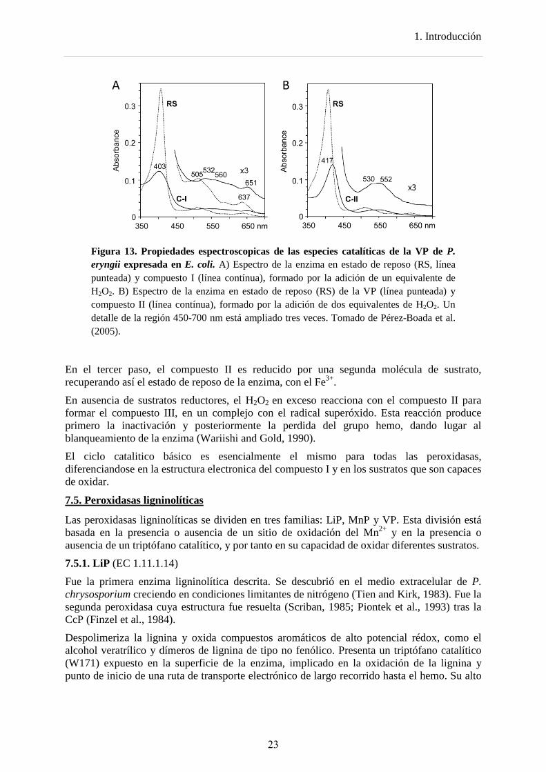
Figura 13. Propiedades espectroscopicas de las especies catalíticas de la VP de P. eryngii expresada en E. coli. A) Espectro de la enzima en estado de reposo (RS, línea punteada) y compuesto I (línea contínua), formado por la adición de un equivalente de H2O2. B) Espectro de la enzima en estado de reposo (RS) de la VP (línea punteada) y compuesto II (línea contínua), formado por la adición de dos equivalentes de H2O2. Un detalle de la región 450-700 nm está ampliado tres veces. Tomado de Pérez-Boada et al. (2005).
En el tercer paso, el compuesto II es reducido por una segunda molécula de sustrato, recuperando así el estado de reposo de la enzima, con el Fe3+.
En ausencia de sustratos reductores, el H2O2 en exceso reacciona con el compuesto II para formar el compuesto III, en un complejo con el radical superóxido. Esta reacción produce primero la inactivación y posteriormente la perdida del grupo hemo, dando lugar al blanqueamiento de la enzima (Wariishi and Gold, 1990).
El ciclo catalitico básico es esencialmente el mismo para todas las peroxidasas, diferenciandose en la estructura electronica del compuesto I y en los sustratos que son capaces de oxidar.
7.5. Peroxidasas ligninolíticas
Las peroxidasas ligninolíticas se dividen en tres familias: LiP, MnP y VP. Esta división está basada en la presencia o ausencia de un sitio de oxidación del Mn2+ y en la presencia o ausencia de un triptófano catalítico, y por tanto en su capacidad de oxidar diferentes sustratos.
7.5.1. LiP (EC 1.11.1.14)
Fue la primera enzima ligninolítica descrita. Se descubrió en el medio extracelular de P. chrysosporium creciendo en condiciones limitantes de nitrógeno (Tien and Kirk, 1983). Fue la segunda peroxidasa cuya estructura fue resuelta (Scriban, 1985; Piontek et al., 1993) tras la CcP (Finzel et al., 1984).
Despolimeriza la lignina y oxida compuestos aromáticos de alto potencial rédox, como el alcohol veratrílico y dímeros de lignina de tipo no fenólico. Presenta un triptófano catalítico (W171) expuesto en la superficie de la enzima, implicado en la oxidación de la lignina y punto de inicio de una ruta de transporte electrónico de largo recorrido hasta el hemo. Su alto
23
1. Introducción
potencial redox, se debe a que el hierro del anillo de porfirina es más deficiente en electrones que el de las peroxidasas clásicas.
Este triptófano catalítico esta conservado entre todas las LiPs, con la excepción de una LiP de T. cervina, que presenta un residuo de tirosina que desempeña el mismo papel, localizado en una posición diferente (Miki et al., 2003; 2011; 2013).
7.5.2. MnP (EC 1.11.1.13)
Se descubrió en 1984 en P. chrysosporium por Kuwahara et al. (1984) y fue la tercera peroxidasa cuya estructura cristalográfica fue resuelta (Sundaramoorthy et al., 1994). Su presencia entre los hongos de la podredumbre blanca está más extendida que la de la LiPs o las VPs
No presentan un triptófano catalítico, pero si un sitio de oxidación de manganeso, que consiste en un pequeño canal de acceso formado por tres residuos ácidos, situado directamente sobre el propionato interno del hemo (propionato del anillo pirrólico III) (Figura 7), oxidando Mn2+ a Mn3+.
Gracias a la gran cantidad de genomas secuenciados en los últimos años, más de 200 secuencias de MnPs están disponibles, y de acuerdo a estos datos genómicos, esta familia ha sido dividida en tres subfamilias, cortas, largas y extralargas en función de la longitud del C-terminal. Las peroxidasas caracterizadas en P. chrysosporium son de tipo largo, oxidando solo Mn2+, mientras que las peroxidasas de tipo corto, con mayor identidad de secuencia con las VP que con las MnPs largas y extralargas, son capaces de oxidar otros sustratos como el ABTS y el DMP. Estas nuevas subfamilias propuestas se analizan en detalle en el capítulo 7.
7.5.3. VP (EC 1.11.1.16)
Se describió en P. eryngii como una MnP con actividad independiente de Mn (Martínez et al., 1996) y tras su clonación se describió como una nueva enzima (Ruiz-Dueñas et al., 1999; Camarero et al., 1999). También se ha encontrado en Bjerkandera spp. (Mester and Field, 1998). Se caracteriza por combinar las propiedades catalíticas de la LiP y la MnP, al presentar tanto un triptófano catalítico, responsable de la oxidación de sustratos de alto potencial redox, como un sitio de oxidación del Mn2+. A diferencia de las LiPs, oxida el alcohol VA con menor eficiencia, pero es capaz de oxidar el RB5 sin necesidad de ningún mediador.
7.6. Evolución de las peroxidasas ligninolíticas Hasta hace poco los estudios filogenéticos de los hongos se basaban en el análisis de diferentes regiones del DNA ribosomal y un limitado número de genes (James et al., 2006). Sin embargo, el aumento en la secuenciación de genomas fúngicos ha permitido que los estudios evolutivos actuales utilicen genomas completos (Fitzpatrick et al., 2006).
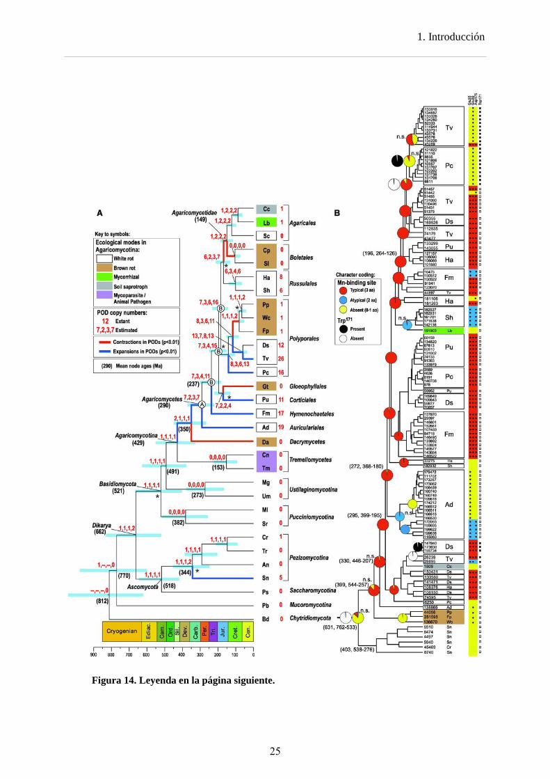
Los estudios de Floudas et al (2012), en el que se analizan 31 genomas fúngicos, y el de Ruiz-Dueñas et al (2013) en el que se analizan 10 genomas de Polyporales, han reconstruido la historia evolutiva de las peroxidasas ligninolíticas. Los análisis de diversificación génica confirman que la tasa de duplicación de genes de PODs es más elevada en la línea de los hongos de la podredumbre blanca que en las otras líneas, de modo que a partir de una única copia de un gen de POD en el ancestro común de los Basidiomycota, ésta se expandiría dando lugar a 26 peroxidasas en T. versicolor, 16 en P. chrysosporium o 12 en D. squalens, entre otros ejemplos. Por el contrario, esta única copia se perdería o se mantendría, en las líneas que han dado lugar a los hongos micorrizógenos, de podredumbre parda y micoparásitos (Figura 14 A).
24
1. Introducción
Figura 14. Leyenda en la página siguiente.
25
1. Introducción
Estos estudios revelan que el ancestro de todas las peroxidasas de clase II carecía de sitio de unión al Mn2+ y de triptófano catalítico, es decir, sería una enzima no ligninolítica de tipo GP. Los mismos análisis muestran que la primera peroxidasa ligninolítica fue MnP derivada de una GP ancestral (Figura 14 B), hecho que concuerda con la presencia de MnP en todos los hongos de podredumbre blanca.
Las peroxidasas tipo VP habrían evolucionado a partir de una MnP ancestral que adquirió un triptófano catalítico. Durante la evolución posterior, alguna VP habría perdido uno de los aminoácidos del sitio de unión del Mn, dando lugar a las VPs atípicas (que sólo presentan dos de los tres aminoácidos implicados en la unión del Mn). Este proceso habría ido más lejos en alguna otra VP ancestral, que habría perdido la funcionalidad del sitio de oxidación del Mn dando lugar a las LiPs.
Lo anterior no excluye líneas de evolución secundaria, como alguna de las mostradas en la presente tesis.
Figura 14. Filogenia de 31 basidiomicetos y otros hongos a partir de sus genomas (A) e historia evolutiva de las peroxidasas ligninolíticas (LiP, MnP y VP) y GPs (B). A) La filogenia de los hongos (cronograma) fue obtenida con BEAST partir de 26 genes. Las barras azules son intervalos del 95% para las edades de los nodos, cuyo promedio (en millones de años) se indica en los paréntesis. Las ramas azules y rojas indican expansión y contracción, respectivamente, en el número de genes de PODs inferido utilizando CAFE. Los números rojos junto a la clave de las especies indican el número real de genes de cada una. Los números en rojo, separados por comas, son los números de copias de genes de PODs estimados con CAFE, Notung (usando dos umbrales diferentes) y DrML. El nodo A sería el antepasado de los Agaricomycetes; los nodos B corresponden a ancestros de otros grupos de Agaricomycetes. Los asteriscos indican los nodos que no reciben apoyo en todos los análisis. Véase la Tabla 1 para las abreviaturas de las especies. B) Filogenia de los genes de PODs estimada en BEAST con reconstrucciones del estado ancestral para el sitio de unión de manganeso (rojo, azul y amarillo) y el triptófano implicado en la degradación de la lignina (negro y blanco) estimados con BayesTraits. Las casillas de la derecha indican la presencia de residuos funcionales. Las edades medias de los nodos, entre paréntesis, están seguidas por sus intervalos de confianza del 95%. Tomado de Floudas et al (2012).
26
1. Introducción
Referencias
Baldrian, P. 2006. Fungal laccases - occurrence and properties. FEMS Microbiol. Rev. 30:215-242.
Banci, L. 1997. Structural properties of peroxidases. J. Biotechnol. 53:253-263. Banci, L., I. Bertini, E. A. Pease, M. Tien, and P. Turano. 1992. 1H NMR investigation of
manganese peroxidase from Phanerochaete chrysosporium. A comparison with other peroxidases. Biochemistry 31:10009-10017.
Bao, W. L., Y. Fukushima, K. A. Jensen, M. A. Moen, and K. E. Hammel. 1994. Oxidative degradation of non-phenolic lignin during lipid peroxidation by fungal manganese peroxidase. FEBS Lett. 354:297-300.
Blanchette, R. A. 1991. Delignification by wood-decay fungi. Annu. Rev. Phytopathol. 29:381-398.
Blanchette, R. A., E. W. Krueger, J. E. Haight, M. Akhtar, and D. E. Akin. 1997. Cell wall alterations in loblolly pine wood decayed by the white- rot fungus, Ceriporiopsis subvermispora. J. Biotechnol. 53:203-213.
Blanchette, R. A., L. Otjen, M. J. Effland, and W. E. Eslyn. 1985. Changes in structural and chemical components of wood delignified by fungi. Wood Sci. Technol. 19:35-46.
Böhmer, S., K. Messner, and E. Srebotnik. 1998. Oxidation of phenanthrene by a fungal laccase in the presence of 1-hydroxybenzotriazole and unsaturated lipids. Biochem. Biophys. Res. Commun. 244:233-238.
Bonnarme, P. and T. W. Jeffries. 1990. Mn(II) regulation of lignin peroxidases and manganese-dependent peroxidases from lignin-degrading white rot fungi. Appl. Environ. Microbiol. 56:210-217.
Bourbonnais, R. and M. G. Paice. 1990. Oxidation of non-phenolic substrates. An expanded role for laccase in lignin biodegradation. FEBS Lett. 267:99-102.
Camarero, S., D. Ibarra, M. J. Martínez, and A. T. Martínez. 2005. Lignin-derived compounds as efficient laccase mediators for decolorization of different types of recalcitrant dyes. Appl. Environ. Microbiol. 71:1775-1784.
Camarero, S., S. Sarkar, F. J. Ruiz-Dueñas, M. J. Martínez, and A. T. Martínez. 1999. Description of a versatile peroxidase involved in natural degradation of lignin that has both Mn-peroxidase and lignin-peroxidase substrate binding sites. J. Biol. Chem. 274:10324-10330.
Cañas, A. I. and S. Camarero. 2010. Laccases and their natural mediators: Biotechnological tools for sustainable eco-friendly processes. Biotechnol. Adv. 28:694-705.
Collins, P. J., J. A. Field, P. Teunissen, and A. D. W. Dobson. 1997. Stabilization of lignin peroxidases in white rot fungi by tryptophan. Appl. Environ. Microbiol. 63:2543-2548.
Daniel, G., J. Volc, and E. Kubátová. 1994. Pyranose oxidase, a major source of H2O2 during wood degradation by Phanerochaete chrysosporium, Trametes versicolor, and Oudemansiella mucida. Appl. Environ. Microbiol. 60:2524-2532.
de Jong, E., A. E. Cazemier, J. A. Field, and J. A. M. de Bont. 1994a. Physiological role of chlorinated aryl alcohols biosynthesized de novo by the white rot fungus Bjerkandera sp strain BOS55. Appl. Environ. Microbiol. 60:271-277.
de Jong, E. and J. A. Field. 1997. Sulfur tuft and turkey tail: Biosynthesis and biodegradation of organohalogens by basidiomycetes. Annu. Rev. Microbiol. 51:375-414:375-414.
de Jong, E., J. A. Field, and J. A. M. de Bont. 1994b. Aryl alcohols in the physiology of ligninolytic fungi. FEMS Microbiol. Rev. 13:153-188.
de Jong, E., J. A. Field, H. E. Spinnler, J. B. P. A. Wijnberg, and J. A. M. de Bont. 1994c. Significant biogenesis of chlorinated aromatics by fungi in natural environments. Appl. Environ. Microbiol. 60:264-270.
27
1. Introducción
del Río, J. C., A. Gutiérrez, M. J. Martínez, and A. T. Martínez. 2001. Py-GC-MS study of
Eucalyptus globulus wood treated with different fungi. J. Anal. Appl. Pyrolysis 58/59:441-453.
del Río, J. C., J. Rencoret, G. Marques, A. Gutiérrez, D. Ibarra, J. I. Santos, J. Jiménez-Barbero, and A. T. Martínez. 2008. Highly acylated (acetylated and/or p-coumaroylated) native lignins from diverse herbaceous plants. J. Agric. Food Chem. 56:9525-9534.
Dunford, H. B. 1999. Heme peroxidases. Wiley-VCH, New York. Edwards, S. L., R. Raag, H. Wariishi, M. H. Gold, and T. L. Poulos. 1993. Crystal structure of
lignin peroxidase. Proc. Natl. Acad. Sci. USA 90:750-754. Eggert, C., U. Temp, J. F. D. Dean, and K.-E. L. Eriksson. 1996. A fungal metabolite
mediates degradation of non-phenolic lignin structures and synthetic lignin by laccase. FEBS Lett. 391:144-148.
Eriksson, K.-E., B. Pettersson, J. Volc, and V. Musílek. 1986. Formation and partial characterization of glucose-2-oxidase, a H2O2 producing enzyme in Phanerochaete chrysosporium. Appl. Microbiol. Biotechnol. 23:257-262.
Faison, B. D. and T. K. Kirk. 1983. Relationship between lignin degradation and production of reduced oxygen species by Phanerochaete chrysosporium. Appl. Environ. Microbiol. 46:1140-1145.
Fengel, D. and G. Wegener. 1984. Wood: Chemistry, ultrastructure, reactions. De Gruyter, Berlin.
Fernández-Fueyo, E., F. J. Ruiz-Dueñas, P. Ferreira, D. Floudas, D. S. Hibbett, P. Canessa, L. Larrondo, T. Y. James, D. Seelenfreund, S. Lobos, R. Polanco, M. Tello, Y. Honda, T. Watanabe, T. Watanabe, J. S. Ryu, C. P. Kubicek, M. Schmoll, J. Gaskell, K. E. Hammel, F. J. St.John, A. Vanden Wymelenberg, G. Sabat, S. S. Bondurant, K. Syed, J. Yadav, H. Doddapaneni, V. Subramanian, J. L. Lavín, J. A. Oguiza, G. Perez, A. G. Pisabarro, L. Ramírez, F. Santoyo, E. Master, P. M. Coutinho, B. Henrissat, V. Lombard, J. K. Magnuson, U. Kües, C. Hori, K. Igarashi, M. Samejima, B. W. Held, K. Barry, K. LaButti, A. Lapidus, E. Lindquist, S. Lucas, R. Riley, A. Salamov, D. Hoffmeister, D. Schwenk, Y. Hadar, O. Yarden, R. P. de Vries, A. Wiebenga, J. Stenlid, D. C. Eastwood, I. V. Grigoriev, R. Berka, R. A. Blanchette, P. Kersten, A. T. Martínez, R. Vicuña, and D. Cullen. 2012a. Comparative genomics of Ceriporiopisis subvermispora and Phanerochaete chrysosporium provide insight into selective ligninolysis. Proc. Natl. Acad. Sci. USA 109:5458-5463.
Fernández-Fueyo, E., F. J. Ruiz-Dueñas, M. J. Martínez, A. Romero, K. E. Hammel, F. J. Medrano, and A. T. Martínez. 2014. Ligninolytic peroxidase genes in the oyster mushroom genome: Heterologous expression, molecular structure, catalytic and stability properties and lignin-degrading ability. Biotechnol. Biofuels 7:2.
Fernández-Fueyo, E., F. J. Ruiz-Dueñas, Y. Miki, M. J. Martínez, K. E. Hammel, and A. T. Martínez. 2012b. Lignin-degrading peroxidases from genome of selective ligninolytic fungus Ceriporiopsis subvermispora. J. Biol. Chem. 287:16903-16906.
Ferraz, A., A. M. Cordova, and A. Machuca. 2003. Wood biodegradation and enzyme production by Ceriporiopsis subvermispora during solid-state fermentation of Eucalyptus grandis. Enzyme Microb. Technol. 32:59-65.
Field, J. A., F. J. M. Verhagen, and E. de Jong. 1995. Natural organohalogen production by basidiomycetes. Trends Biotechnol. 13:451-456.
Finzel, B. C., T. L. Poulos, and J. Kraut. 1984. Crystal structure of yeast cytochrome c peroxidase refined at 1.7 Å resolution. J. Biol. Chem. 259:13027-13036.
28
1. Introducción
Fitzpatrick, D. A., M. E. Logue, J. E. Stajich, and G. Butler. 2006. A fungal phylogeny based
on 42 complete genomes derived from supertree and combined gene analysis. BMC Evolutionary Biology 6.
Floudas, D., M. Binder, R. Riley, K. Barry, R. A. Blanchette, B. Henrissat, A. T. Martínez, R. Otillar, J. W. Spatafora, J. S. Yadav, A. Aerts, I. Benoit, A. Boyd, A. Carlson, A. Copeland, P. M. Coutinho, R. P. de Vries, P. Ferreira, K. Findley, B. Foster, J. Gaskell, D. Glotzer, P. Górecki, J. Heitman, C. Hesse, C. Hori, K. Igarashi, J. A. Jurgens, N. Kallen, P. Kersten, A. Kohler, U. Kües, T. K. A. Kumar, A. Kuo, K. LaButti, L. F. Larrondo, E. Lindquist, A. Ling, V. Lombard, S. Lucas, T. Lundell, R. Martin, D. J. McLaughlin, I. Morgenstern, E. Morin, C. Murat, M. Nolan, R. A. Ohm, A. Patyshakuliyeva, A. Rokas, F. J. Ruiz-Dueñas, G. Sabat, A. Salamov, M. Samejima, J. Schmutz, J. C. Slot, F. St.John, J. Stenlid, H. Sun, S. Sun, K. Syed, A. Tsang, A. Wiebenga, D. Young, A. Pisabarro, D. C. Eastwood, F. Martin, D. Cullen, I. V. Grigoriev, and D. S. Hibbett. 2012. The Paleozoic origin of enzymatic lignin decomposition reconstructed from 31 fungal genomes. Science 336:1715-1719.
Flournoy, D. S., J. A. Paul, T. K. Kirk, and T. L. Highley. 1993. Changes in the size and volume of pores in sweetgum wood during simultaneous rot by Phanerochaete chrysosporium Burds. Holzforschung 47:297-301.
Forney, L. J., C. A. Reddy, M. Tien, and S. D. Aust. 1982. The involvement of hydroxyl radical derived from hydrogen peroxide in lignin degradation by the white rot fungus Phanerochaete chrysosporium. J. Biol. Chem. 257:11455-11462.
Gajhede, M., D. J. Schuller, A. Henriksen, A. T. Smith, and T. L. Poulos. 1997. Crystal structure of horseradish peroxidase C at 2.15 Å resolution. Nature Struct. Biology 4:1032-1038.
Galli, C. and P. Gentili. 2004. Chemical messengers: mediated oxidations with the enzyme laccase. J. Phys. Org. Chem. 17:973-977.
Gellerstedt, G. and G. Henriksson. 2008. Lignins: Major sources, structure and properties, p. 201-224. In M. Belgacem and A. Gandini (eds.), Monomers, polymers and composites from renewable resources. Elsevier, Amsterdam.
Gold, M. H., H. Wariishi, and K. Valli. 1989. Extracellular peroxidases involved in lignin degradation by the white-rot basidiomycetes Phanerochaete chrysosporium, p. 127-140. In J. R. Whitaker and P. E. Sonnet (eds.), Biocatalystists in Agricultural Biotechnology. ACS, Washington, DC.
Gómez-Toribio, V., A. B. García-Martín, M. J. Martínez, A. T. Martínez, and F. Guillén. 2009. Induction of extracellular hydroxyl radical production by white-rot fungi through quinone redox cycling. Appl. Environ. Microbiol. 75:3944-3953.
Greene, R. V. and J. M. Gould. 1984. Fatty acyl-coenzyme A oxidase activity and H2O2 production in Phanerochaete chrysosporium mycelia. Biochem. Biophys. Res. Commun. 118:437-443.
Guillén, F., V. Gómez-Toribio, M. J. Martínez, and A. T. Martínez. 2000. Production of hydroxyl radical by the synergistic action of fungal laccase and aryl alcohol oxidase. Arch. Biochem. Biophys. 383:142-147.
Guillén, F., A. T. Martínez, and M. J. Martínez. 1992a. Aryl-alcohol oxidase from Pleurotus eryngii: substrate specificity and H2O2-producing system, p. 371-376. In M. Kuwahara and M. Shimada (eds.), Biotechnology in pulp and paper industry. UNI Pub. Co., Ltd., Tokyo.
Guillén, F., A. T. Martínez, and M. J. Martínez. 1992b. Substrate specificity and properties of the aryl-alcohol oxidase from the ligninolytic fungus Pleurotus eryngii. Eur. J. Biochem. 209:603-611.
29
1. Introducción
Hammel, K. E., K. A. Jensen, M. D. Mozuch, L. L. Landucci, M. Tien, and E. A. Pease. 1993.
Ligninolysis by a purified lignin peroxidase. J. Biol. Chem. 268:12274-12281. Harvey, P. J., H. E. Schoemaker, and J. M. Palmer. 1986. Veratryl alcohol as a mediator and
the role of radical cations in lignin biodegradation by Phanerochaete chrysosporium. FEBS Lett. 195:242-246.
Hatakka, A. 1994. Lignin-modifying enzymes from selected white-rot fungi - Production and role in lignin degradation. FEMS Microbiol. Rev. 13:125-135.
Heinfling, A., F. J. Ruiz-Dueñas, M. J. Martínez, M. Bergbauer, U. Szewzyk, and A. T. Martínez. 1998. A study on reducing substrates of manganese-oxidizing peroxidases from Pleurotus eryngii and Bjerkandera adusta. FEBS Lett. 428:141-146.
Henriksson, G., G. Johansson, and G. Pettersson. 2000. A critical review of cellobiose dehydrogenases. J. Biotechnol. 78:93-113.
Himmel, M. E., S. Y. Ding, D. K. Johnson, W. S. Adney, M. R. Nimlos, J. W. Brady, and T. D. Foust. 2007. Biomass recalcitrance: Engineering plants and enzymes for biofuels production. Science 315:804-807.
Hiner, A. N. P., E. L. Raven, R. N. F. Thorneley, F. García-Canovas, and J. N. Rodríguez-López. 2002. Mechanisms of compound I formation in heme peroxidases. J. Inorg. Biochem. 91:27-34.
Hofrichter, M., R. Ullrich, M. J. Pecyna, C. Liers, and T. Lundell. 2010. New and classic families of secreted fungal heme peroxidases. Appl. Microbiol. Biotechnol. 87:871-897.
Howes, B. D., A. Feis, L. Raimondi, C. Indiani, and G. Smulevich. 2001. The critical role of the proximal calcium ion in the structural properties of horseradish peroxidase. J. Biol. Chem. 276:40704-40711.
James, T. Y., F. Kauff, C. L. Schoch, P. Matheny, V. Hofstetter, C. J. Cox, G. Celio, C. Gueidan, E. Fraker, J. Miadlikowska, H. Lumbsch, A. Rauhut, V. Reeb, A. Arnold, A. Amtoft, J. E. Stajich, K. Hosaka, G. H. Sung, D. Johnson, B. O'Rourke, M. Crockett, M. Binder, J. M. Curtis, J. C. Slot, Z. Wang, A. W. Wilson, A. Schuessler, J. E. Longcore, K. O'Donnell, S. Mozley-Standridge, D. Porter, P. M. Letcher, M. J. Powell, J. W. Taylor, M. M. White, G. W. Griffith, D. R. Davies, R. A. Humber, J. B. Morton, J. Sugiyama, A. Y. Rossman, J. D. Rogers, D. H. Pfister, D. Hewitt, K. Hansen, S. Hambleton, R. A. Shoemaker, J. Kohlmeyer, B. Volkmann-Kohlmeyer, R. A. Spotts, M. Serdani, P. W. Crous, K. W. Hughes, K. Matsuura, E. Langer, G. Langer, W. A. Untereiner, R. Lucking, B. Buedel, D. M. Geiser, A. Aptroot, P. Diederich, I. Schmitt, M. Schultz, R. Yahr, D. S. Hibbett, F. Lutzoni, D. J. McLaughlin, J. W. Spatafora, and R. Vilgalys. 2006. Reconstructing the early evolution of Fungi using a six-gene phylogeny. Nature 443:818-822.
Johannes, C. and A. Majcherczyk. 2000. Natural mediators in the oxidation of polycyclic aromatic hydrocarbons by laccase mediator systems. Appl. Environ. Microbiol. 66:524-528.
Johannes, C., A. Majcherczyk, and A. Hüttermann. 1998. Oxidation of acenaphthene and acenaphthylene by laccase of Trametes versicolor in a laccase-mediator system. J. Biotechnol. 61:151-156.
Joshi, D. K. and M. H. Gold. 1996. Oxidation of dimethoxylated aromatic compounds by lignin peroxidase from Phanerochaete chrysosporium. Eur. J. Biochem. 237:45-57.
Kapich, A. N. and L. N. Shishbina. 1995. Lipid peroxidation and its regulation in the mycelium of xylotrophic basidiomycetes. Microbiology-Engl. Tr. 64:266-271.
Kawai, S., T. Umezawa, M. Shimada, T. Higuchi, K. Koide, T. Nishida, N. Morohoshi, and T. Haraguchi. 1987. Cα-Cβ cleavage of phenolic β-1 lignin substructure model compound by laccase of Coriolus versicolor. Mokuzai Gakkaishi 33:792-797.
30
1. Introducción
Kersten, P. and D. Cullen. 2007. Extracellular oxidative systems of the lignin-degrading
basidiomycete Phanerochaete chrysosporium. Fungal Genet. Biol. 44:77-87. Kersten, P. J. and T. K. Kirk. 1987. Involvement of a new enzyme, glyoxal oxidase, in
extracellular H2O2 production by Phanerochaete chrysosporium. J. Bacteriol. 169:2195-2201.
Kimura, S. and M. Ikeda-Saito. 1988. Human myeloperoxidase and thyroid peroxidase, two enzymes with separate and distinct physiological functions, are evolutionarily related members of the same gene family. Proteins 3:113-120.
Kirk, T. K. and R. L. Farrell. 1987. Enzymatic "combustion": The microbial degradation of lignin. Annu. Rev. Microbiol. 41:465-505.
Koduri, R. S. and M. Tien. 1994. Kinetic analysis of lignin peroxidase - Explanation for the mediation phenomenon by veratryl alcohol. Biochemistry 33:4225-4230.
Kuwahara, M., J. K. Glenn, M. A. Morgan, and M. H. Gold. 1984. Separation and characterization of two extracellular H2O2-dependent oxidases from ligninolytic cultures of Phanerochaete chrysosporium. FEBS Lett. 169:247-250.
Leatham, G. F. 1986. The ligninolytic activities of Lentinus edodes and Phanerochaete chrysosporium. Appl. Microbiol. Biotechnol. 24:51-58.
Liese, W. 1970. Ultrastructural aspects of woody tissue disintegration. Annu. Rev. Phytopathol. 8:231-258.
Lundquist, K. and Kirk, T. K. De novo synthesis and decomposition of veratryl alcohol by a lignin degrading basidiomicete. Phytochemistry 17, 1676. 1978a.
Ref Type: Generic Lundquist, K. and T. K. Kirk. 1978b. De novo synthesis and decomposition of veratryl
alcohol by a lignin degrading basidiomycete. Phytochemistry 17:1676. Martínez, A. T. 2002. Molecular biology and structure-function of lignin-degrading heme
peroxidases. Enzyme Microb. Technol. 30:425-444. Martínez, A. T., S. Camarero, F. Guillén, A. Gutiérrez, C. Muñoz, E. Varela, M. J. Martínez,
J. M. Barrasa, K. Ruel, and M. Pelayo. 1994. Progress in biopulping of non-woody materials: Chemical, enzymatic and ultrastructural aspects of wheat-straw delignification with ligninolytic fungi from the genus Pleurotus. FEMS Microbiol. Rev. 13:265-274.
Martínez, A. T., J. Rencoret, L. Nieto, J. Jiménez-Barbero, A. Gutiérrez, and J. C. del Río. 2011. Selective lignin and polysaccharide removal in natural fungal decay of wood as evidenced by in situ structural analyses. Environ. Microbiol. 13:96-107.
Martínez, A. T., F. J. Ruiz-Dueñas, M. J. Martínez, J. C. del Río, and A. Gutiérrez. 2009. Enzymatic delignification of plant cell wall: from nature to mill. Curr. Opin. Biotechnol. 20:348-357.
Martínez, A. T., M. Speranza, F. J. Ruiz-Dueñas, P. Ferreira, S. Camarero, F. Guillén, M. J. Martínez, A. Gutiérrez, and J. C. del Río. 2005. Biodegradation of lignocellulosics: Microbiological, chemical and enzymatic aspects of fungal attack to lignin. Int. Microbiol. 8:195-204.
Martinez, D., J. Challacombe, I. Morgenstern, D. S. Hibbett, M. Schmoll, C. P. Kubicek, P. Ferreira, F. J. Ruiz-Dueñas, A. T. Martínez, P. Kersten, K. E. Hammel, A. Vanden Wymelenberg, J. Gaskell, E. Lindquist, G. Sabat, S. S. Bondurant, L. F. Larrondo, P. Canessa, R. Vicuña, J. Yadav, H. Doddapaneni, V. Subramanian, A. G. Pisabarro, J. L. Lavín, J. A. Oguiza, E. Master, B. Henrissat, P. M. Coutinho, P. Harris, J. K. Magnuson, S. E. Baker, K. Bruno, W. Kenealy, P. J. Hoegger, U. Kues, P. Ramaiya, S. Lucas, A. Salamov, H. Shapiro, H. Tu, C. L. Chee, M. Misra, G. Xie, S. Teter, D. Yaver, T. James, M. Mokrejs, M. Pospisek, I. V. Grigoriev, T. Brettin, D. Rokhsar, R. Berka, and D. Cullen. 2009. Genome, transcriptome, and secretome analysis of wood decay fungus Postia
31
1. Introducción
placenta supports unique mechanisms of lignocellulose conversion. Proc. Natl. Acad. Sci. USA 106:1954-1959.
Martinez, D., L. F. Larrondo, N. Putnam, M. D. Gelpke, K. Huang, J. Chapman, K. G. Helfenbein, P. Ramaiya, J. C. Detter, F. Larimer, P. M. Coutinho, B. Henrissat, R. Berka, D. Cullen, and D. Rokhsar. 2004. Genome sequence of the lignocellulose degrading fungus Phanerochaete chrysosporium strain RP78. Nat. Biotechnol. 22:695-700.
Martínez, M. J., F. J. Ruiz-Dueñas, F. Guillén, and A. T. Martínez. 1996. Purification and catalytic properties of two manganese-peroxidase isoenzymes from Pleurotus eryngii. Eur. J. Biochem. 237:424-432.
Mayer, A. M. and R. C. Staples. 2002. Laccase: new functions for an old enzyme. Phytochemistry 60:551-565.
Mester, T. and J. A. Field. 1997. Optimization of manganese peroxidase production by the white rot fungus Bjerkandera sp. strain BOS55. FEMS Microbiol. Lett. 155:161-168.
Mester, T. and J. A. Field. 1998. Characterization of a novel manganese peroxidase-lignin peroxidase hybrid isozyme produced by Bjerkandera species strain BOS55 in the absence of manganese. J. Biol. Chem. 273:15412-15417.
Miki, Y., F. R. Calviño, R. Pogni, S. Giansanti, F. J. Ruiz-Dueñas, M. J. Martínez, R. Basosi, A. Romero, and A. T. Martínez. 2011. Crystallographic, kinetic, and spectroscopic study of the first ligninolytic peroxidase presenting a catalytic tyrosine. J. Biol. Chem. 286:15525-15534.
Miki, Y., R. Pogni, S. Acebes, F. Lucas, E. Fernández-Fueyo, M. C. Baratto, M. I. Fernández, V. de los Ríos, F. J. Ruiz-Dueñas, A. Sinicropi, R. Basosi, K. E. Hammel, V. Guallar, and A. T. Martínez. 2013. Formation of a tyrosine adduct involved in lignin degradation by Trametopsis cervina lignin peroxidase: A novel peroxidase activation mechanism. Biochem. J. 452:575-584.
Miki, Y., H. Wariishi, and H. Tanaka. 2003. Lignin peroxidase from the white-rot basidiomycete Trametes cervina. Proc. 12th ISWPC, Madison, 9-12 June II:95.
Moen, M. A. and K. E. Hammel. 1994. Lipid peroxidation by the manganese peroxidase of Phanerochaete chrysosporium is the basis for phenanthrene oxidation by the intact fungus. Appl. Environ. Microbiol. 60:1956-1961.
Muñoz, C., F. Guillén, A. T. Martínez, and M. J. Martínez. 1997. Laccase isoenzymes of Pleurotus eryngii: Characterization, catalytic properties and participation in activation of molecular oxygen and Mn2+ oxidation. Appl. Environ. Microbiol. 63:2166-2174.
Orth, A. B., D. J. Royse, and M. Tien. 1993. Ubiquity of lignin-degrading peroxidases among various wood-degrading fungi. Appl. Environ. Microbiol. 59:4017-4023.
Ozimek, P., M. Veenhuis, and I. J. van der Klei. 2005. Alcohol oxidase: A complex peroxisomal, oligomeric flavoprotein. FEMS Yeast Res. 5:975-983.
Passardi, F., Theiler, G., Zamocky, M., Cosio, C., Rouhier, N., Teixera, F., Margis-Pinheiro, M., Ionnidis, V., Penel, C., Falquet, L., and Dunand, C. Peroxibase: The peroxidase database. Phytochemistry 68, 1605-1611. 2007.
Ref Type: Generic Paszczynski, A. and R. L. Crawford. 1991. Degradation of azo compounds by ligninase from
Phanerochaete chrysosporium: Involvement of veratryl alcohol. Biochem. Biophys. Res. Commun. 178:1056-1063.
Paszczynski, A., V.-B. Huynh, and R. Crawford. 1986. Comparison of ligninase-I and peroxidase-M2 from the white-rot fungus Phanerochaete chrysosporium. Arch. Biochem. Biophys. 244:750-765.
Pérez-Boada, M., F. J. Ruiz-Dueñas, R. Pogni, R. Basosi, T. Choinowski, M. J. Martínez, K. Piontek, and A. T. Martínez. 2005. Versatile peroxidase oxidation of high redox potential
32
1. Introducción
aromatic compounds: Site-directed mutagenesis, spectroscopic and crystallographic investigations of three long-range electron transfer pathways. J. Mol. Biol. 354:385-402.
Pettersen, R. C. 1984. The chemical composition of wood, p. 57-126. In R. M. Powell (ed.), The Chemistry of Solid Wood. ACS.
Piontek, K., T. Glumoff, and K. Winterhalter. 1993. Low pH crystal structure of glycosylated lignin peroxidase from Phanerochaete chrysosporium at 2.5 Å resolution. FEBS Lett. 315:119-124.
Poulos, T. L., S. L. Edwards, H. Wariishi, and M. H. Gold. 1993. Crystallographic refinement of lignin peroxidase at 2 Å. J. Biol. Chem. 268:4429-4440.
Ragauskas, A. J., C. K. Williams, B. H. Davison, G. Britovsek, J. Cairney, C. A. Eckert, W. J. Frederick, J. P. Hallett, D. J. Leak, C. L. Liotta, J. R. Mielenz, R. Murphy, R. Templer, and T. Tschaplinski. 2006. The path forward for biofuels and biomaterials. Science 311:484-489.
Ralph, J., K. Lundquist, G. Brunow, F. Lu, H. Kim, P. F. Schatz, J. M. Marita, R. D. Hatfield, S. A. Ralph, J. H. Christensen, and W. Boerjan. 2004. Lignins: Natural polymers from oxidative coupling of 4-hydroxyphenylpropanoids. Phytochem. Rev. 3:29-60.
Rothschild, N., A. Levkowitz, Y. Hadar, and C. G. Dosoretz. 1999. Manganese deficiency can replace high oxygen levels needed for lignin peroxidase formation by Phanerochaete chrysosporium. Appl. Environ. Microbiol. 65:483-488.
Ruiz-Dueñas, F. J., E. Fernández, M. J. Martínez, and A. T. Martínez. 2011. Pleurotus ostreatus heme peroxidases: An in silico analysis from the genome sequence to the enzyme molecular structure. C. R. Biol. 334:795-805.
Ruiz-Dueñas, F. J., T. Lundell, D. Floudas, L. G. Nagy, J. M. Barrasa, D. S. Hibbett, and A. T. Martínez. 2013. Lignin-degrading peroxidases in Polyporales: An evolutionary survey based on ten sequenced genomes. Mycologia 105:1428-1444.
Ruiz-Dueñas, F. J. and A. T. Martínez. 2009. Microbial degradation of lignin: How a bulky recalcitrant polymer is efficiently recycled in nature and how we can take advantage of this. Microbial Biotechnol. 2:164-177.
Ruiz-Dueñas, F. J. and A. T. Martínez. 2010. Structural and functional features of peroxidases with a potential as industrial biocatalysts, p. 37-59. In E. Torres and M. Ayala (eds.), Biocatalysts based on heme peroxidases. Springer-Verlag, Berlin.
Ruiz-Dueñas, F. J., M. J. Martínez, and A. T. Martínez. 1999. Molecular characterization of a novel peroxidase isolated from the ligninolytic fungus Pleurotus eryngii. Mol. Microbiol. 31:223-236.
Ruiz-Dueñas, F. J., M. Morales, M. Pérez-Boada, T. Choinowski, M. J. Martínez, K. Piontek, and A. T. Martínez. 2007. Manganese oxidation site in Pleurotus eryngii versatile peroxidase: A site-directed mutagenesis, kinetic and crystallographic study. Biochemistry 46:66-77.
Sánchez, C. 2009. Lignocellulosic residues: biodegradation and bioconversion by fungi. Biotechnol. Adv. 27:185-194.
Sarkanen, K. V. and C. H. Ludwig. 1971. Lignins. Occurrence, formation, structure and reactions. Wiley-Interscience, New York.
Schmid, A., J. S. Dordick, B. Hauer, A. Kiener, M. Wubbolts, and B. Witholt. 2001. Industrial biocatalysis today and tomorrow. Nature 409:258-268.
Schwarze, F. W. M. R., J. Engels, and C. Mattheck. 2000. Fungal strategies of decay in trees. Springer, Berlin.
Scott, G. M., M. Akhtar, M. J. Lenz, and R. E. Swaney. 1998. Engineering, scale-up, and economic aspects of fungal pretreatment of wood chips, p. 341-384. In R. A. Young and
33
1. Introducción
M. Akhtar (eds.), Environmentally friendly technologies for the pulp and paper industry. TAPPI Press, Atlanta.
Scriban, R. 1985. Biotecnologia. Editorial El Manual Moderno; Mexico. Sharp, K. H., M. Mewies, P. C. Moody, and E. L. Raven. 2003. Crystal structure of the
ascorbate peroxidase-ascorbate complex. Nat. Struct. Biol. 10:303-307. Shimada, M. and T. Higuchi. 1991. Microbial, enzymatic and biomimetic degradation of
lignin, p. 557-619. In D. N. S. Hon and N. Shiraishi (eds.), Wood and cellulosic chemistry. Marcel Dekker, N.Y.
Shimada, M., F. Nakatsubo, T. K. Kirk, and T. Higuchi. 1981. Biosynthesis of the secondary metabolite veratryl alcohol in relation to lignin degradation by Phanerochaete chrysosporium. Arch. Microbiol. 129:321-324.
Shimada, M. and M. Takahashi. 1991. Biodegradation of cellulosic materials, p. 621-663. In D. N. S. Hon and N. Shiraishi (eds.), Wood and cellulosic chemistry. Marcel Dekker, N.Y.
Spinnler, H. E., E. de Jong, G. Mauvais, E. Semon, and J.-L. Le Quéré. 1994. Production of halogenated compounds by Bjerkandera adusta. Appl. Microbiol. Biotechnol. 42:212-221.
Sundaramoorthy, M., K. Kishi, M. H. Gold, and T. L. Poulos. 1994. The crystal structure of manganese peroxidase from Phanerochaete chrysosporium at 2.06-Å resolution. J. Biol. Chem. 269:32759-32767.
Sundaramoorthy, M., H. L. Youngs, M. H. Gold, and T. L. Poulos. 2005. High-resolution crystal structure of manganese peroxidase: substrate and inhibitor complexes. Biochemistry 44:6463-6470.
Takao, S. 1965. Organic acid production by Basidiomycetes. I Screening of acid-producing strains. Arch. Microbiol. 13:732-737.
Teunissen, P. J. M. and J. A. Field. 1998a. 2-Chloro-1,4-dimethoxybenzene as a mediator of lignin peroxidase catalyzed oxidations. FEBS Lett. 439:219-223.
Teunissen, P. J. M. and J. A. Field. 1998b. 2-Chloro-1,4-dimethoxybenzene as a novel catalytic cofactor for oxidation of anisyl alcohol by lignin peroxidase. Appl. Environ. Microbiol. 64:830-835.
Teunissen, P. J. M., D. W. Sheng, G. V. B. Reddy, P. Moënne-Loccoz, J. A. Field, and M. H. Gold. 1998. 2-Chloro-1,4-dimethoxybenzene cation radical: Formation and role in the lignin peroxidase oxidation of anisyl alcohol. Arch. Biochem. Biophys. 360:233-238.
Thurston, C. F. 1994. The structure and function of fungal laccases. Microbiology 140:19-26. Tien, M. and T. K. Kirk. 1983. Lignin-degrading enzyme from the hymenomycete
Phanerochaete chrysosporium Burds. Science 221:661-663. Tien, M. and D. Ma. 1997. Oxidation of 4-methoxymandelic acid by lignin peroxidase.
Mediation by veratryl alcohol. J. Biol. Chem. 272:8912-8917. Tsukamoto, K., H. Itakura, K. Sato, K. Fukuyama, S. Miura, S. Takahashi, H. Ikezawa, and T.
Hosoya. 1999. Binding of salicylhydroxamic acid and several aromatic donor molecules to Arthromyces ramosus peroxidase, investigated by X-ray crystallography, optical difference spectroscopy, NMR relaxation, molecular dynamics, and kinetics. Biochemistry 38:12558-12568.
Wariishi, H. and M. H. Gold. 1989. Lignin peroxidase compound III: Formation, inactivation, and conversion to the native enzyme. FEBS Lett. 243:165-168.
Wariishi, H. and M. H. Gold. 1990. Lignin peroxidase compound III. Mechanism of formation and decomposition. J. Biol. Chem. 265:2070-2077.
Wariishi, H., K. Valli, and M. H. Gold. 1991. In vitro depolymerization of lignin by manganese peroxidase of Phanerochaete chrysosporium. Biochem. Biophys. Res. Commun. 176:269-275.
34
1. Introducción
Wariishi, H., K. Valli, and M. H. Gold. 1992. Manganese(II) oxidation by manganese
peroxidase from the basidiomycete Phanerochaete chrysosporium. Kinetic mechanism and role of chelators. J. Biol. Chem. 267:23688-23695.
Welinder, K. G. 1992. Superfamily of plant, fungal and bacterial peroxidases. Curr. Opin. Struct. Biol. 2:388-393.
Weng, J. K. and C. Chapple. 2010. The origin and evolution of lignin biosynthesis. New Phytol. 187:273-285.
Yelle, D. J., J. Ralph, F. Lu, and K. E. Hammel. 2008. Evidence for cleavage of lignin by a brown rot basidiomycete. Environ. Microbiol. 10:1844-1849.
Zhang, L. M., G. Gellerstedt, J. Ralph, and F. C. Lu. 2006. NMR studies on the occurrence of spirodienone structures in lignins. J. Wood Chem. Technol. 26:65-79.
35

36
Capítulo 2
"Pleurotus ostreatus heme peroxidases: An in silico analysis from the genome sequence to the enzyme molecular structure" Ruiz-Dueñas, F. J., E. Fernández, M. J. Martínez, and A. T. Martínez. C. R. Biol. 334:795-805. 2011.
37

38
Re
Plge
Fra
Cent
1. I
ed
patwotheandthebaspla
lum
phaGenDOiop
por
C. R. Biologies xxx (2011) xxx–xxx
A R
Keyw
Pleu
Gen
In si
Hem
Lign
Hem
Dye
*
atm
G Model
CRASS3-3032; No. of Pages 11
Plth
163
doi:
view/Revue
eurotus ostreatus heme peroxidases: An in silico analysis from thenome sequence to the enzyme molecular structure
ncisco J. Ruiz-Duenas *, Elena Fernandez, Marıa Jesus Martınez, Angel T. Martınez *
ro de Investigaciones Biologicas, CSIC, Ramiro de Maeztu 9, 28040 Madrid, Spain
ntroduction
A series of basidiomycete genomes have been complet-during last years corresponding to plant and animalhogens, and soil and mycorrhizal species, together withod-rotting fungi. The latter have raised much interest in
search for peroxidases with industrial interest in bulk fine chemistry, food and other sectors. They include
genomes of Phanerochaete chrysosporium [1] (the firstidiomycete genome to be sequenced) and Postia
centa [2], the recently published genome of Schizophyl-
commune [3], and many others that are in differentses of the sequencing process (up to 53 only at the Jointome Institute, JGI, of the US Department of Energy,
E) including those of Pleurotus ostreatus and Ceripor-
sis subvermispora (both to be published soon), Pycno-
us cinnabarinus (to be released) and Phlebiopsis gigantea.
Among the above basidiomycetes, several white-rotfungi from the genus Pleurotus have been described asbeing able to degrade lignin selectively [4]. The limitedattack to cellulose makes them very interesting in differentbiotechnological applications related to the use of plantbiomass, including the integrated lignocellulose biorefi-neries for the future production of chemicals, materialsand biofuels. This fact, together with the understanding ofthe regulation of the ligninolytic system constituted byheme peroxidases and other enzymes [5], as well as theincreasing interest in Pleurotus as an edible mushroom(with a world production near that of Agaricus bisporus [6])were the main reasons to include a representative of thisgenus among the organisms to be sequenced by the DOE, ina project coordinated by Dr A.G. Pisabarro from the PublicUniversity of Navarre (Spain).
Ligninolytic peroxidases from P. ostreatus and otherwhite-rot fungi have been widely studied over the last years[7–9]. However, only in the case of P. chrysosporium acomplete catalog of these enzymes can be obtained from itsgenome sequence. On the other hand, new superfamilies ofheme peroxidases, namely heme-thiolate peroxidases and
T I C L E I N F O
ords:
rotus ostreatus
ome sequence
lico analysis
e peroxidases
inolytic peroxidases
e-thiolate peroxidases
-decolorizing peroxidases
A B S T R A C T
An exhaustive screening of the Pleurotus ostreatus genome was performed to search for
nucleotide sequences of heme peroxidases in this white-rot fungus, which could be useful
for different biotechnological applications. After sequence identification and manual
curation of the corresponding genes and cDNAs, the deduced amino acid sequences were
converted into structural homology models. A comparative study of these sequences and
their structural models with those of known fungal peroxidases revealed the complete
inventory of heme peroxidases of this fungus. This consists of cytochrome c peroxidase and
ligninolytic peroxidases, including manganese peroxidase and versatile peroxidase but not
lignin peroxidase, as representative of the ‘‘classical’’ superfamily of plant, fungal, and
bacterial peroxidases; and members of two relatively ‘‘new’’ peroxidase superfamilies,
namely heme-thiolate peroxidases, here described for the first time in a fungus from the
genus Pleurotus, and dye-decolorizing peroxidases, already known in P. ostreatus but still
to be thoroughly explored and characterized.
� 2011 Academie des sciences. Published by Elsevier Masson SAS. All rights reserved.
Corresponding authors.
E-mail addresses: [email protected] (F.J. Ruiz-Duenas),
[email protected] (A.T. Martınez).
Contents lists available at ScienceDirect
Comptes Rendus Biologies
ww w.s c ien c edi r ec t . c om
ease cite this article in press as: F.J. Ruiz-Duenas, et al., Pleurotus ostreatus heme peroxidases: An in silico analysis frome genome sequence to the enzyme molecular structure, C. R. Biologies (2011), doi:10.1016/j.crvi.2011.06.004
1-0691/$ – see front matter � 2011 Academie des sciences. Published by Elsevier Masson SAS. All rights reserved.
10.1016/j.crvi.2011.06.004
39
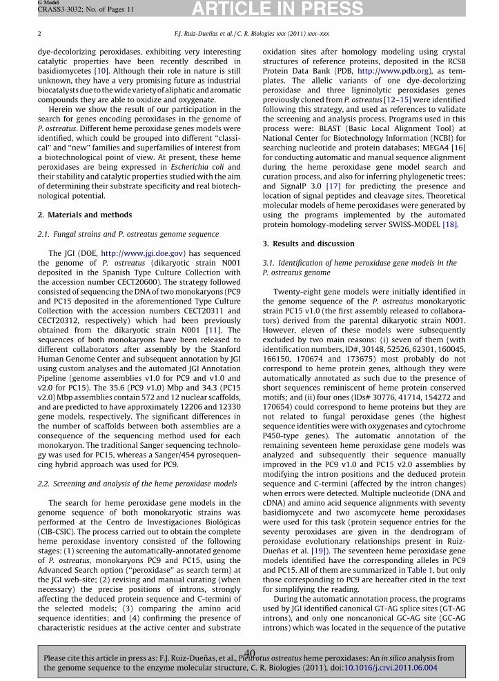
F.J. Ruiz-Duenas et al. / C. R. Biologies xxx (2011) xxx–xxx2
G Model
CRASS3-3032; No. of Pages 11
dye-decolorizing peroxidases, exhibiting very interestingcatalytic properties have been recently described inbasidiomycetes [10]. Although their role in nature is stillunknown, they have a very promising future as industrialbiocatalysts due to the widevarietyof aliphatic and aromaticcompounds they are able to oxidize and oxygenate.
Herein we show the result of our participation in thesearch for genes encoding peroxidases in the genome ofP. ostreatus. Different heme peroxidase genes models wereidentified, which could be grouped into different ‘‘classi-cal’’ and ‘‘new’’ families and superfamilies of interest froma biotechnological point of view. At present, these hemeperoxidases are being expressed in Escherichia coli andtheir stability and catalytic properties studied with the aimof determining their substrate specificity and real biotech-nological potential.
2. Materials and methods
2.1. Fungal strains and P. ostreatus genome sequence
The JGI (DOE, http://www.jgi.doe.gov) has sequencedthe genome of P. ostreatus (dikaryotic strain N001deposited in the Spanish Type Culture Collection withthe accession number CECT20600). The strategy followedconsisted of sequencing the DNA of two monokaryons (PC9and PC15 deposited in the aforementioned Type CultureCollection with the accession numbers CECT20311 andCECT20312, respectively) which had been previouslyobtained from the dikaryotic strain N001 [11]. Thesequences of both monokaryons have been released todifferent collaborators after assembly by the StanfordHuman Genome Center and subsequent annotation by JGIusing custom analyses and the automated JGI AnnotationPipeline (genome assemblies v1.0 for PC9 and v1.0 andv2.0 for PC15). The 35.6 (PC9 v1.0) Mbp and 34.3 (PC15v2.0) Mbp assemblies contain 572 and 12 nuclear scaffolds,and are predicted to have approximately 12206 and 12330gene models, respectively. The significant differences inthe number of scaffolds between both assemblies are aconsequence of the sequencing method used for eachmonokaryon. The traditional Sanger sequencing technolo-gy was used for PC15, whereas a Sanger/454 pyrosequen-cing hybrid approach was used for PC9.
2.2. Screening and analysis of the heme peroxidase models
The search for heme peroxidase gene models in thegenome sequence of both monokaryotic strains wasperformed at the Centro de Investigaciones Biologicas(CIB-CSIC). The process carried out to obtain the completeheme peroxidase inventory consisted of the followingstages: (1) screening the automatically-annotated genomeof P. ostreatus, monokaryons PC9 and PC15, using theAdvanced Search option (‘‘peroxidase’’ as search term) atthe JGI web-site; (2) revising and manual curating (whennecessary) the precise positions of introns, stronglyaffecting the deduced protein sequence and C-termini ofthe selected models; (3) comparing the amino acidsequence identities; and (4) confirming the presence ofcharacteristic residues at the active center and substrate
oxidation sites after homology modeling using crystalstructures of reference proteins, deposited in the RCSBProtein Data Bank (PDB, http://www.pdb.org), as tem-plates. The allelic variants of one dye-decolorizingperoxidase and three ligninolytic peroxidases genespreviously cloned from P. ostreatus [12–15] were identifiedfollowing this strategy, and used as references to validatethe screening and analysis process. Programs used in thisprocess were: BLAST (Basic Local Alignment Tool) atNational Center for Biotechnology Information (NCBI) forsearching nucleotide and protein databases; MEGA4 [16]for conducting automatic and manual sequence alignmentduring the heme peroxidase gene model search andcuration process, and also for inferring phylogenetic trees;and SignalP 3.0 [17] for predicting the presence andlocation of signal peptides and cleavage sites. Theoreticalmolecular models of heme peroxidases were generated byusing the programs implemented by the automatedprotein homology-modeling server SWISS-MODEL [18].
3. Results and discussion
3.1. Identification of heme peroxidase gene models in the
P. ostreatus genome
Twenty-eight gene models were initially identified inthe genome sequence of the P. ostreatus monokaryoticstrain PC15 v1.0 (the first assembly released to collabora-tors) derived from the parental dikaryotic strain N001.However, eleven of these models were subsequentlyexcluded by two main reasons: (i) seven of them (withidentification numbers, ID#, 30148, 52526, 62301, 160045,166150, 170674 and 173675) most probably do notcorrespond to heme protein genes, although they wereautomatically annotated as such due to the presence ofshort sequences reminiscent of heme protein conservedmotifs; and (ii) four ones (IDs# 30776, 41714, 154272 and170654) could correspond to heme proteins but they arenot related to fungal peroxidase genes (the highestsequence identities were with oxygenases and cytochromeP450-type genes). The automatic annotation of theremaining seventeen heme peroxidase gene models wasanalyzed and subsequently their sequence manuallyimproved in the PC9 v1.0 and PC15 v2.0 assemblies bymodifying the intron positions and the deduced proteinsequence and C-termini (affected by the intron changes)when errors were detected. Multiple nucleotide (DNA andcDNA) and amino acid sequence alignments with seventybasidiomycete and two ascomycete heme peroxidaseswere used for this task (protein sequence entries for theseventy peroxidases are given in the dendrogram ofperoxidase evolutionary relationships present in Ruiz-Duenas et al. [19]). The seventeen heme peroxidase genemodels identified have the corresponding alleles in PC9and PC15. All of them are summarized in Table 1, but onlythose corresponding to PC9 are hereafter cited in the textfor simplifying the reading.
During the automatic annotation process, the programsused by JGI identified canonical GT-AG splice sites (GT-AGintrons), and only one noncanonical GC-AG site (GC-AGintrons) which was located in the sequence of the putative
Please cite this article in press as: F.J. Ruiz-Duenas, et al., Pleurotus ostreatus heme peroxidases: An in silico analysis fromthe genome sequence to the enzyme molecular structure, C. R. Biologies (2011), doi:10.1016/j.crvi.2011.06.004
40
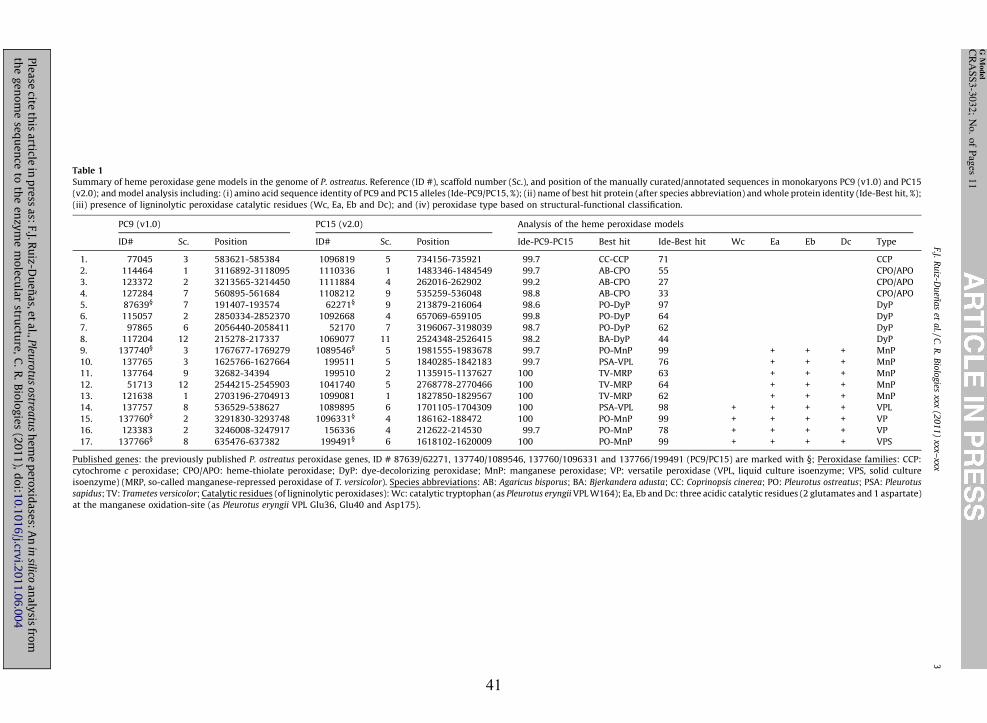
Table 1
Summary of heme peroxidase gene models in the genome of P. ostreatus. Reference (ID #), scaffold number (Sc.), and position of the manually curated/annotated sequences in monokaryons PC9 (v1.0) and PC15
(v2.0); and model analysis including: (i) amino acid sequence identity of PC9 and PC15 alleles (Ide-PC9/PC15, %); (ii) name of best hit protein (after species abbreviation) and whole protein identity (Ide-Best hit, %);
(iii) presence of ligninolytic peroxidase catalytic residues (Wc, Ea, Eb and Dc); and (iv) peroxidase type based on structural-functional classification.
PC9 (v1.0) PC15 (v2.0) Analysis of the heme peroxidase models
ID# Sc. Position ID# Sc. Position Ide-PC9-PC15 Best hit Ide-Best hit Wc Ea Eb Dc Type
1. 77045 3 583621-585384 1096819 5 734156-735921 99.7 CC-CCP 71 � � � � CCP
2. 114464 1 3116892-3118095 1110336 1 1483346-1484549 99.7 AB-CPO 55 � � � � CPO/APO
3. 123372 2 3213565-3214450 1111884 4 262016-262902 99.2 AB-CPO 27 � � � � CPO/APO
4. 127284 7 560895-561684 1108212 9 535259-536048 98.8 AB-CPO 33 � � � � CPO/APO
5. 87639§ 7 191407-193574 62271§ 9 213879-216064 98.6 PO-DyP 97 � � � � DyP
6. 115057 2 2850334-2852370 1092668 4 657069-659105 99.8 PO-DyP 64 � � � � DyP
7. 97865 6 2056440-2058411 52170 7 3196067-3198039 98.7 PO-DyP 62 � � � � DyP
8. 117204 12 215278-217337 1069077 11 2524348-2526415 98.2 BA-DyP 44 � � � � DyP
9. 137740§ 3 1767677-1769279 1089546§ 5 1981555-1983678 99.7 PO-MnP 99 � + + + MnP
10. 137765 3 1625766-1627664 199511 5 1840285-1842183 99.7 PSA-VPL 76 � + + + MnP
11. 137764 9 32682-34394 199510 2 1135915-1137627 100 TV-MRP 63 � + + + MnP
12. 51713 12 2544215-2545903 1041740 5 2768778-2770466 100 TV-MRP 64 � + + + MnP
13. 121638 1 2703196-2704913 1099081 1 1827850-1829567 100 TV-MRP 62 � + + + MnP
14. 137757 8 536529-538627 1089895 6 1701105-1704309 100 PSA-VPL 98 + + + + VPL
15. 137760§ 2 3291830-3293748 1096331§ 4 186162-188472 100 PO-MnP 99 + + + + VP
16. 123383 2 3246008-3247917 156336 4 212622-214530 99.7 PO-MnP 78 + + + + VP
17. 137766§ 8 635476-637382 199491§ 6 1618102-1620009 100 PO-MnP 99 + + + + VPS
Published genes: the previously published P. ostreatus peroxidase genes, ID # 87639/62271, 137740/1089546, 137760/1096331 and 137766/199491 (PC9/PC15) are marked with §; Peroxidase families: CCP:
cytochrome c peroxidase; CPO/APO: heme-thiolate peroxidase; DyP: dye-decolorizing peroxidase; MnP: manganese peroxidase; VP: versatile peroxidase (VPL, liquid culture isoenzyme; VPS, solid culture
isoenzyme) (MRP, so-called manganese-repressed peroxidase of T. versicolor). Species abbreviations: AB: Agaricus bisporus; BA: Bjerkandera adusta; CC: Coprinopsis cinerea; PO: Pleurotus ostreatus; PSA: Pleurotus
sapidus; TV: Trametes versicolor; Catalytic residues (of ligninolytic peroxidases): Wc: catalytic tryptophan (as Pleurotus eryngii VPL W164); Ea, Eb and Dc: three acidic catalytic residues (2 glutamates and 1 aspartate)
at the manganese oxidation-site (as Pleurotus eryngii VPL Glu36, Glu40 and Asp175).
F.J. R
uiz-D
uen
as
et a
l. /
C.
R.
Bio
log
ies xxx
(20
11
) xxx–
xxx
3
G M
od
el
CR
AS
S3
-30
32
; N
o.
of
Pag
es 1
1
Ple
ase
cite th
is a
rticle in
pre
ss a
s: F.J.
Ru
iz-Du
en
as,
et
al.,
Pleu
rotu
s o
streatu
s h
em
e p
ero
xid
ase
s: A
n in
silico a
na
lysis
from
the
ge
no
me
seq
ue
nce
to th
e e
nzy
me
mo
lecu
lar
structu
re,
C.
R.
Bio
log
ies
(20
11
), d
oi:1
0.1
01
6/j.crv
i.20
11
.06
.00
4
41
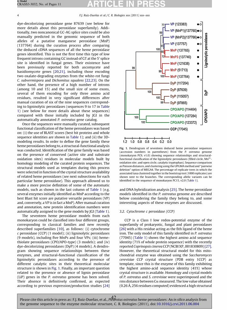
F.J. Ruiz-Duenas et al. / C. R. Biologies xxx (2011) xxx–xxx4
G Model
CRASS3-3032; No. of Pages 11
dye-decolorizing peroxidase gene 87639 (see below formore details about this peroxidase superfamily). Addi-tionally, two noncanonical GC-AG splice sites could be alsomanually predicted in the genomic sequence of bothalleles of a putative manganese peroxidase (MnP)(137764) during the curation process after comparingthe deduced cDNA sequences of all the heme peroxidasegenes identified. This is not the first time this type of lowfrequent introns containing GC instead of GT at the 5’ splicesite is identified in fungal genes. Their existence havebeen previously reported for both ascomycete andbasidiomycete genes [20,21], including those encodingtwo oxalate-degrading enzymes from the white-rot fungiC. subvermispora and Dichomitus squalens [22,23]. On theother hand, the presence of a high number of introns(among 10 and 15) and the small size of some exons,several of them encoding for only three amino acidresidues, resulted in very significant differences aftermanual curation of six of the nine sequences correspond-ing to ligninolytic peroxidases (sequences 9 to 17 in Table1) (see below for more details about these sequences)compared with those initially included by JGI in theautomatically annotated P. ostreatus gene catalog.
Once the sequences were manually curated, subsequentfunctional classification of the heme peroxidases was basedon: (i) the use of BLAST scores (best hit proteins and wholesequence identities are shown in Table 1); and (ii) the 3D-modeling results. In order to define the gene family theseheme peroxidases belong to, a structural-functional analysiswas conducted. Identification of the gene family was basedon the presence of conserved (active site and substrateoxidation sites) residues in molecular models built byhomology modeling of the curated protein sequences. Thestructural models used as templates during this processwere selected in function of the crystal structure availabilityof related heme peroxidases (see next subsections for eachparticular heme peroxidase). This approach allowed us tomake a more precise definition of some of the automaticmodels, such as shown in the last column of Table 1 (e.g.several enzymes initially identified as MnP according to thebest Blast hit score are putative versatile peroxidases (VP)and, conversely, a VP is in fact a MnP). After manual curationand annotation, new protein identification numbers wereautomatically assigned to the gene models by JGI (Table 1).
The seventeen heme peroxidase models from eachmonokaryon could be classified into four different groups,corresponding to classical families and new recentlydescribed superfamilies [10], as follows: (i) cytochromec peroxidase (CCP) (1 model); (ii) ligninolytic peroxidases(9 models), including five MnPs and four VPs; (iii) heme-thiolate peroxidases (CPO/APO-type) (3 models); and (iv)dye-decolorizing peroxidases (DyP) (4 models). A dendro-gram showing sequence relationships between theseenzymes, and structural-functional classification of theligninolytic peroxidases according to the presence ofdifferent catalytic sites in their theoretical molecularstructure is shown in Fig. 1. Finally, an important questionrelated to the presence or absence of lignin peroxidase(LiP) genes in the P. ostreatus genome has been solved.Their absence is definitively confirmed, as expected
and DNA hybridization analysis [25]. The heme peroxidasemodels identified in the P. ostreatus genome are describedbelow considering the family they belong to, and someinteresting aspects of these enzymes are discussed.
3.2. Cytochrome c peroxidase (CCP)
CCP is a Class I low redox-potential enzyme of thesuperfamily of prokaryotic, fungal and plant peroxidases[26] with a His residue acting as the fith ligand of the hemeiron. The only model of this family identified in P. ostreatus
(77045) (Table 1) shows the highest amino acid sequenceidentity (71% of whole protein sequence) with the recentlyreported Coprinopsis cinerea CCP (NCBI XP_001830809) [27].However, the theoretical structural model for this mito-chondrial enzyme was obtained using the Saccharomyces
cerevisiae CCP crystal structure (PDB entry 1CCP) astemplate, since this is the enzyme of this family exhibitingthe highest amino-acid sequence identity (41%) whosecrystal structure is available. Homology and crystal modelsof P. ostreatus and S. cerevisiae were superimposed and therms distance between Ca measured. The low value obtained
Fig. 1. Dendrogram of seventeen deduced heme peroxidase sequences
(accession numbers in parentheses) from the P. ostreatus genome
(monokaryon PC9, v1.0) showing sequence relationships and structural-
functional classification of the ligninolytic peroxidases (filled circle, Mn2+-
oxidation site; and open circle, catalytic tryptophan). Sequence comparison
as Poisson distances, and clustering using the UPGMA method and ‘‘pairwise
deletion’’ option of MEGA4. The percentage of replicate trees in which the
associated taxa clustered together in the bootstrap test (1000 replicates) are
shown next to the branches. The corresponding allelic variants can be
identified in the sequence of monokaryon PC15, v2.0 (Table 1).
(0.26 A, 256 residues computed) evidenced a high structural
according to previous expression/production studies [24]Please cite this article in press as: F.J. Ruiz-Duenas, et al., Pleurotus ostreatus heme peroxidases: An in silico analysis fromthe genome sequence to the enzyme molecular structure, C. R. Biologies (2011), doi:10.1016/j.crvi.2011.06.004
42
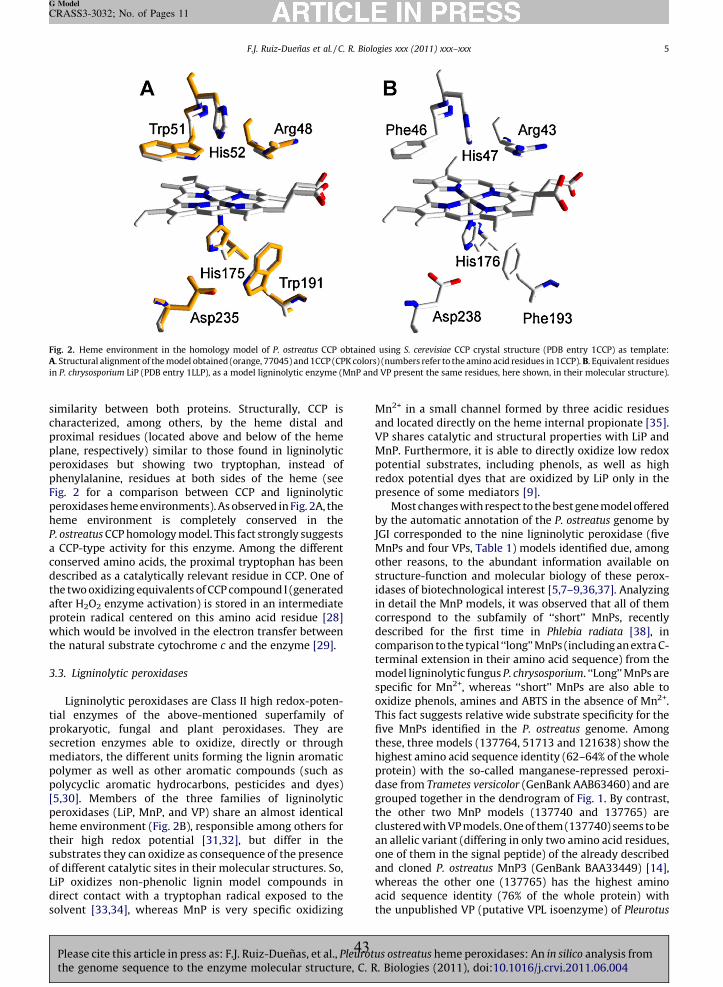
simchaproplaperpheFig.perhemP. o
a Ccondestheafteprowhthe
3.3.
tialprosecmepolpol[5,3perhemthesubof dLiPdiresolv
Fig.
A. St
in P
F.J. Ruiz-Duenas et al. / C. R. Biologies xxx (2011) xxx–xxx 5
G Model
CRASS3-3032; No. of Pages 11
Plth
ilarity between both proteins. Structurally, CCP isracterized, among others, by the heme distal andximal residues (located above and below of the hemene, respectively) similar to those found in ligninolyticoxidases but showing two tryptophan, instead ofnylalanine, residues at both sides of the heme (see
2 for a comparison between CCP and ligninolyticoxidases heme environments). As observed in Fig. 2A, the
e environment is completely conserved in thestreatus CCP homology model. This fact strongly suggestsCP-type activity for this enzyme. Among the differentserved amino acids, the proximal tryptophan has beencribed as a catalytically relevant residue in CCP. One of
two oxidizing equivalents of CCP compound I (generatedr H2O2 enzyme activation) is stored in an intermediatetein radical centered on this amino acid residue [28]ich would be involved in the electron transfer between
natural substrate cytochrome c and the enzyme [29].
Ligninolytic peroxidases
Ligninolytic peroxidases are Class II high redox-poten- enzymes of the above-mentioned superfamily ofkaryotic, fungal and plant peroxidases. They areretion enzymes able to oxidize, directly or throughdiators, the different units forming the lignin aromaticymer as well as other aromatic compounds (such asycyclic aromatic hydrocarbons, pesticides and dyes)0]. Members of the three families of ligninolyticoxidases (LiP, MnP, and VP) share an almost identical
e environment (Fig. 2B), responsible among others forir high redox potential [31,32], but differ in thestrates they can oxidize as consequence of the presenceifferent catalytic sites in their molecular structures. So,
oxidizes non-phenolic lignin model compounds inct contact with a tryptophan radical exposed to theent [33,34], whereas MnP is very specific oxidizing
Mn2+ in a small channel formed by three acidic residuesand located directly on the heme internal propionate [35].VP shares catalytic and structural properties with LiP andMnP. Furthermore, it is able to directly oxidize low redoxpotential substrates, including phenols, as well as highredox potential dyes that are oxidized by LiP only in thepresence of some mediators [9].
Most changes with respect to the best gene model offeredby the automatic annotation of the P. ostreatus genome byJGI corresponded to the nine ligninolytic peroxidase (fiveMnPs and four VPs, Table 1) models identified due, amongother reasons, to the abundant information available onstructure-function and molecular biology of these perox-idases of biotechnological interest [5,7–9,36,37]. Analyzingin detail the MnP models, it was observed that all of themcorrespond to the subfamily of ‘‘short’’ MnPs, recentlydescribed for the first time in Phlebia radiata [38], incomparison to the typical ‘‘long’’ MnPs (including an extra C-terminal extension in their amino acid sequence) from themodel ligninolytic fungus P. chrysosporium. ‘‘Long’’ MnPs arespecific for Mn2+, whereas ‘‘short’’ MnPs are also able tooxidize phenols, amines and ABTS in the absence of Mn2+.This fact suggests relative wide substrate specificity for thefive MnPs identified in the P. ostreatus genome. Amongthese, three models (137764, 51713 and 121638) show thehighest amino acid sequence identity (62–64% of the wholeprotein) with the so-called manganese-repressed peroxi-dase from Trametes versicolor (GenBank AAB63460) and aregrouped together in the dendrogram of Fig. 1. By contrast,the other two MnP models (137740 and 137765) areclustered with VP models. One of them (137740) seems to bean allelic variant (differing in only two amino acid residues,one of them in the signal peptide) of the already describedand cloned P. ostreatus MnP3 (GenBank BAA33449) [14],whereas the other one (137765) has the highest aminoacid sequence identity (76% of the whole protein) withthe unpublished VP (putative VPL isoenzyme) of Pleurotus
2. Heme environment in the homology model of P. ostreatus CCP obtained using S. cerevisiae CCP crystal structure (PDB entry 1CCP) as template:
ructural alignment of the model obtained (orange, 77045) and 1CCP (CPK colors) (numbers refer to the amino acid residues in 1CCP). B. Equivalent residues
. chrysosporium LiP (PDB entry 1LLP), as a model ligninolytic enzyme (MnP and VP present the same residues, here shown, in their molecular structure).
ease cite this article in press as: F.J. Ruiz-Duenas, et al., Pleurotus ostreatus heme peroxidases: An in silico analysis frome genome sequence to the enzyme molecular structure, C. R. Biologies (2011), doi:10.1016/j.crvi.2011.06.004
43
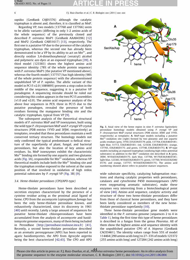
F.J. Ruiz-Duenas et al. / C. R. Biologies xxx (2011) xxx–xxx6
G Model
CRASS3-3032; No. of Pages 11
sapidus (GenBank CAJ01576) although the catalytictryptophan is absent and, therefore, it is classified as MnP.
Regarding VP, two models (137760 and 137766) seemto be allelic variants (differing in only 1-2 amino acids ofthe whole sequence) of the previously cloned anddescribed P. ostreatus MnP1 (GenBank AAA84396) [12]and MnP2 (GenBank CAB51617) [13], respectively. Thefirst one is a putative VP due to the presence of the catalytictryptophan, whereas the second one has already beendemonstrated to be a VP by its ability to act on Mn2+, anddirectly oxidize 3,4-dimethoxybenzyl (veratryl) alcoholand polymeric azo dyes at an exposed tryptophan [39]. Athird model (123383) shows the highest amino acidsequence identity (78% of the whole protein sequence)with P. ostreatus MnP1 (the putative VP mentioned above),whereas the fourth model (137757) has high identity (98%of the whole protein sequence) with the aforementionedunpublished VP of P. sapidus. The allelic variant of thismodel in PC15 v2.0 (1089895) presents a stop codon in themiddle of the sequence, suggesting it is a putative VPpseudogene. A sequencing mistake should be ruled outconsidering this codon appears in the two PC15 assemblies(v1.0 and v2.0). The amino acid sequence analysis of theabove four sequences in PC9, three in PC15 due to theputative pseudogen, revealed the presence of bothresidues forming the manganese binding site and thecatalytic tryptophan, typical from VP [9].
The subsequent analysis of the theoretical structuralmodels of P. ostreatus MnP and VP isoenzymes, built usingthe wild type P. chrysosporium MnP and P. eryngii VP crystalstructures (PDB entries 1YYD and 3FJW, respectively) astemplates, revealed that these peroxidases maintain a wellconserved tertiary structure. The homology models notonly confirmed the typical predominant a-helical struc-ture of the superfamily of plant, fungal, and bacterialperoxidases, but also the location of key amino acidresidues. So, MnP isoenzymes exhibit the characteristicMn2+ binding site formed by one aspartic and two glutamicacids (Fig. 3A), responsible for Mn2+ oxidation, whereas VPtheoretical models include both the Mn2+ binding site andthe tryptophan residue exposed to the solvent at the sameposition of that involved in oxidation of high redoxpotential substrates by P. eryngii VP (Fig. 3B) [9].
3.4. Heme-thiolate peroxidases (CPO/APO-type)
Heme-thiolate peroxidases have been described assecretion enzymes characterized by the presence of acysteine residue acting as the proximal axial ligand ofheme. CPO from the ascomycete Leptoxyphium fumago hasbeen the only heme-thiolate peroxidase known, andexhaustively characterized, since its discovery in 1961[40] until recently. Lately a large amount of sequences forputative heme-thiolate chloroperoxidases have beenaccumulated from the analysis of ascomycete and basid-iomycete genome sequences, many of them being includedin PeroxiBase (http://peroxibase.toulouse.inra.fr) [41].Recently, a second heme-thiolate peroxidase describedas an aromatic peroxygenase (APO) has been reported inagaric basidiomycetes, the APO from Agrocybe aegerita
wide substrate specificity, catalyzing halogenation reac-tions and sharing catalytic properties with peroxidases,catalases and cytochrome P450 monooxygenases (APOeven oxygenating aromatic substrates), make theseenzymes very interesting from a biotechnological pointof view [44]. Amino acid sequence, catalytic activity andstructure of these two heme-thiolate peroxidases differfrom those of classical heme peroxidases, and they havebeen lately considered as members of the new heme-thiolate peroxidase superfamily [10].
Three heme-thiolate peroxidase gene models wereidentified in the P. ostreatus genome (sequences 2 to 4 inTable 1), being the first time this type of heme peroxidasesis described in a fungus from the genus Pleurotus. All ofthem show the highest amino acid sequence identity withthe unpublished putative CPO of A. bisporus (GenBankCAC03461). The identity values range from 55% of model114464 (290 amino acids long) to 27–33% of models 123372
Fig. 3. Axial view of the heme region in nine P. ostreatus ligninolytic
peroxidase homology models obtained using P. eryngii VP and
P. chrysosporium MnP crystal structures (PDB entries 3FJW and 1YYD,
respectively) as templates: A. MnP-type models including a putative
Mn2+-oxidation site (right) formed by two glutamic and one aspartic
acidic residues (grey, 1YYD, E35/E39/D179; pink, 137764, E36/E40/D181;
light blue, 51713, E36/E40/D181; red, 121638, E36/E40/D181; orange,
137765, E36/E40/D175; and green, 137740, E36/E40/D175). B. VP-type
models including an exposed tryptophan (oxidizing high redox-potential
substrates) in addition to the Mn2+-oxidation site described in A (grey,
3FJW, W164/E36/E40/D175; dark blue, 137766, W170/E36/E40/D181;
light blue, 123383, W164/E36/E40/D175; green, 137760, W165/E36/E40/
D176; and orange, 137757, W164/E36/E40/D175). The amino acid
numbering showed above refers to putative mature sequences.
(255 amino acids long) and 127284 (242 amino acids long)
being the best characterized [42,43]. The CPO and APOPlease cite this article in press as: F.J. Ruiz-Duenas, et al., Pleurotus ostreatus heme peroxidases: An in silico analysis fromthe genome sequence to the enzyme molecular structure, C. R. Biologies (2011), doi:10.1016/j.crvi.2011.06.004
44
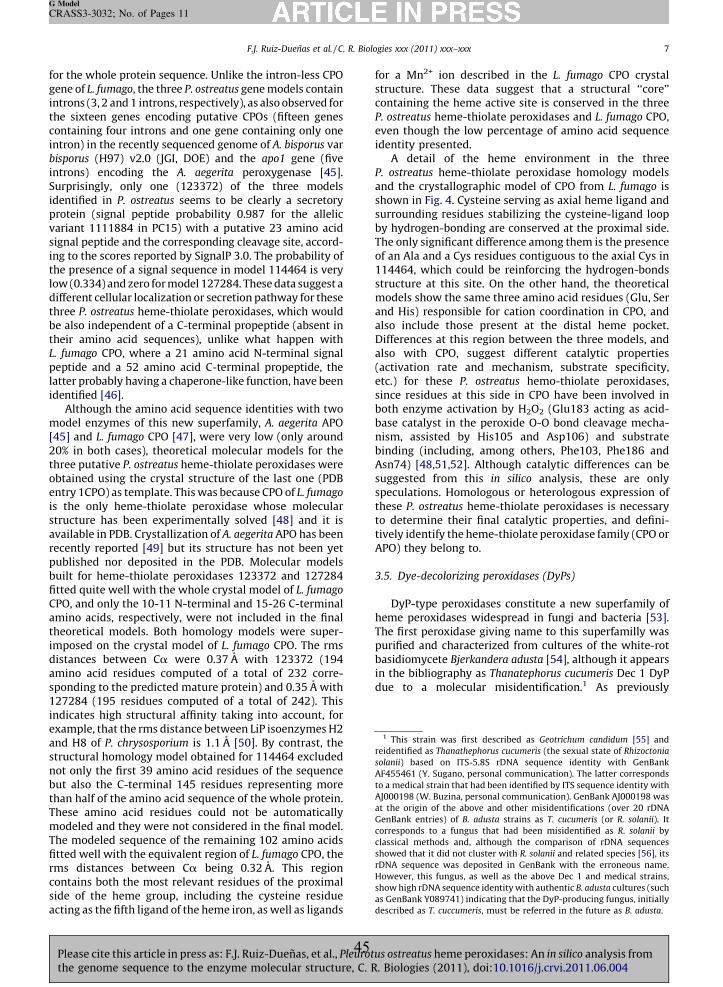
for
genintrtheconintrbisp
intrSurideprovarsigningthelowdiffthrbe
theL. f
peplattide
mo[4520%throbtentis tstruavarecpubbuifittCPOamtheimpdistamspo127indexaandstrunotbutthaThemoThefittrmsconsideact
F.J. Ruiz-Duenas et al. / C. R. Biologies xxx (2011) xxx–xxx 7
G Model
CRASS3-3032; No. of Pages 11
Plth
the whole protein sequence. Unlike the intron-less CPOe of L. fumago, the three P. ostreatus gene models containons (3, 2 and 1 introns, respectively), as also observed for
sixteen genes encoding putative CPOs (fifteen genestaining four introns and one gene containing only oneon) in the recently sequenced genome of A. bisporus varorus (H97) v2.0 (JGI, DOE) and the apo1 gene (fiveons) encoding the A. aegerita peroxygenase [45].prisingly, only one (123372) of the three modelsntified in P. ostreatus seems to be clearly a secretorytein (signal peptide probability 0.987 for the alleliciant 1111884 in PC15) with a putative 23 amino acidal peptide and the corresponding cleavage site, accord-
to the scores reported by SignalP 3.0. The probability of presence of a signal sequence in model 114464 is very
(0.334) and zero for model 127284. These data suggest aerent cellular localization or secretion pathway for theseee P. ostreatus heme-thiolate peroxidases, which wouldalso independent of a C-terminal propeptide (absent inir amino acid sequences), unlike what happen withumago CPO, where a 21 amino acid N-terminal signaltide and a 52 amino acid C-terminal propeptide, theer probably having a chaperone-like function, have beenntified [46].Although the amino acid sequence identities with twodel enzymes of this new superfamily, A. aegerita APO] and L. fumago CPO [47], were very low (only around
in both cases), theoretical molecular models for theee putative P. ostreatus heme-thiolate peroxidases wereained using the crystal structure of the last one (PDBry 1CPO) as template. This was because CPO of L. fumago
he only heme-thiolate peroxidase whose molecularcture has been experimentally solved [48] and it is
ilable in PDB. Crystallization of A. aegerita APO has beenently reported [49] but its structure has not been yetlished nor deposited in the PDB. Molecular models
lt for heme-thiolate peroxidases 123372 and 127284ed quite well with the whole crystal model of L. fumago
, and only the 10-11 N-terminal and 15-26 C-terminalino acids, respectively, were not included in the finaloretical models. Both homology models were super-osed on the crystal model of L. fumago CPO. The rmsances between Ca were 0.37 A with 123372 (194
ino acid residues computed of a total of 232 corre-nding to the predicted mature protein) and 0.35 A with284 (195 residues computed of a total of 242). Thisicates high structural affinity taking into account, formple, that the rms distance between LiP isoenzymes H2
H8 of P. chrysosporium is 1.1 A [50]. By contrast, thectural homology model obtained for 114464 excluded
only the first 39 amino acid residues of the sequence also the C-terminal 145 residues representing moren half of the amino acid sequence of the whole protein.se amino acid residues could not be automaticallydeled and they were not considered in the final model.
modeled sequence of the remaining 102 amino acidsed well with the equivalent region of L. fumago CPO, the
distances between Ca being 0.32 A. This regiontains both the most relevant residues of the proximal
of the heme group, including the cysteine residue
for a Mn2+ ion described in the L. fumago CPO crystalstructure. These data suggest that a structural ‘‘core’’containing the heme active site is conserved in the threeP. ostreatus heme-thiolate peroxidases and L. fumago CPO,even though the low percentage of amino acid sequenceidentity presented.
A detail of the heme environment in the threeP. ostreatus heme-thiolate peroxidase homology modelsand the crystallographic model of CPO from L. fumago isshown in Fig. 4. Cysteine serving as axial heme ligand andsurrounding residues stabilizing the cysteine-ligand loopby hydrogen-bonding are conserved at the proximal side.The only significant difference among them is the presenceof an Ala and a Cys residues contiguous to the axial Cys in114464, which could be reinforcing the hydrogen-bondsstructure at this site. On the other hand, the theoreticalmodels show the same three amino acid residues (Glu, Serand His) responsible for cation coordination in CPO, andalso include those present at the distal heme pocket.Differences at this region between the three models, andalso with CPO, suggest different catalytic properties(activation rate and mechanism, substrate specificity,etc.) for these P. ostreatus hemo-thiolate peroxidases,since residues at this side in CPO have been involved inboth enzyme activation by H2O2 (Glu183 acting as acid-base catalyst in the peroxide O-O bond cleavage mecha-nism, assisted by His105 and Asp106) and substratebinding (including, among others, Phe103, Phe186 andAsn74) [48,51,52]. Although catalytic differences can besuggested from this in silico analysis, these are onlyspeculations. Homologous or heterologous expression ofthese P. ostreatus heme-thiolate peroxidases is necessaryto determine their final catalytic properties, and defini-tively identify the heme-thiolate peroxidase family (CPO orAPO) they belong to.
3.5. Dye-decolorizing peroxidases (DyPs)
DyP-type peroxidases constitute a new superfamily ofheme peroxidases widespread in fungi and bacteria [53].The first peroxidase giving name to this superfamilly waspurified and characterized from cultures of the white-rotbasidiomycete Bjerkandera adusta [54], although it appearsin the bibliography as Thanatephorus cucumeris Dec 1 DyPdue to a molecular misidentification.1 As previously
1 This strain was first described as Geotrichum candidum [55] and
reidentified as Thanathephorus cucumeris (the sexual state of Rhizoctonia
solanii) based on ITS-5.8S rDNA sequence identity with GenBank
AF455461 (Y. Sugano, personal communication). The latter corresponds
to a medical strain that had been identified by ITS sequence identity with
AJ000198 (W. Buzina, personal communication). GenBank AJ000198 was
at the origin of the above and other misidentifications (over 20 rDNA
GenBank entries) of B. adusta strains as T. cucumeris (or R. solanii). It
corresponds to a fungus that had been misidentified as R. solanii by
classical methods and, although the comparison of rDNA sequences
showed that it did not cluster with R. solanii and related species [56], its
rDNA sequence was deposited in GenBank with the erroneous name.
However, this fungus, as well as the above Dec 1 and medical strains,
show high rDNA sequence identity with authentic B. adusta cultures (such
as GenBank Y089741) indicating that the DyP-producing fungus, initially
ribed as T. cuccumeris, must be referred in the future as B. adusta.
ing as the fifth ligand of the heme iron, as well as ligands descease cite this article in press as: F.J. Ruiz-Duenas, et al., Pleurotus ostreatus heme peroxidases: An in silico analysis frome genome sequence to the enzyme molecular structure, C. R. Biologies (2011), doi:10.1016/j.crvi.2011.06.004
45
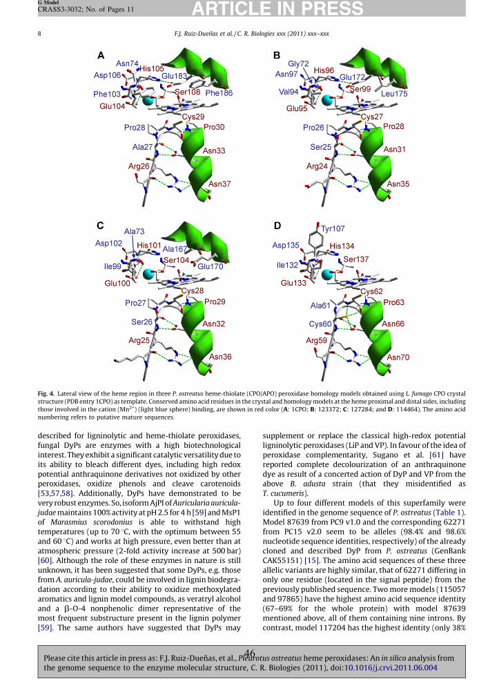
F.J. Ruiz-Duenas et al. / C. R. Biologies xxx (2011) xxx–xxx8
G Model
CRASS3-3032; No. of Pages 11
described for ligninolytic and heme-thiolate peroxidases,fungal DyPs are enzymes with a high biotechnologicalinterest. They exhibit a significant catalytic versatility due toits ability to bleach different dyes, including high redoxpotential anthraquinone derivatives not oxidized by otherperoxidases, oxidize phenols and cleave carotenoids[53,57,58]. Additionally, DyPs have demonstrated to bevery robust enzymes. So, isoform AjPI of Auricularia auricula-
judae maintains 100% activity at pH 2.5 for 4 h [59] and MsP1of Marasmius scorodonius is able to withstand hightemperatures (up to 70 8C, with the optimum between 55and 60 8C) and works at high pressure, even better than atatmospheric pressure (2-fold activity increase at 500 bar)[60]. Although the role of these enzymes in nature is stillunknown, it has been suggested that some DyPs, e.g. thosefrom A. auricula-judae, could be involved in lignin biodegra-dation according to their ability to oxidize methoxylatedaromatics and lignin model compounds, as veratryl alcoholand a b-O-4 nonphenolic dimer representative of themost frequent substructure present in the lignin polymer[59]. The same authors have suggested that DyPs may
supplement or replace the classical high-redox potentialligninolytic peroxidases (LiP and VP). In favour of the idea ofperoxidase complementarity, Sugano et al. [61] havereported complete decolourization of an anthraquinonedye as result of a concerted action of DyP and VP from theabove B. adusta strain (that they misidentified asT. cucumeris).
Up to four different models of this superfamily wereidentified in the genome sequence of P. ostreatus (Table 1).Model 87639 from PC9 v1.0 and the corresponding 62271from PC15 v2.0 seem to be alleles (98.4% and 98.6%nucleotide sequence identities, respectively) of the alreadycloned and described DyP from P. ostreatus (GenBankCAK55151) [15]. The amino acid sequences of these threeallelic variants are highly similar, that of 62271 differing inonly one residue (located in the signal peptide) from thepreviously published sequence. Two more models (115057and 97865) have the highest amino acid sequence identity(67–69% for the whole protein) with model 87639mentioned above, all of them containing nine introns. Bycontrast, model 117204 has the highest identity (only 38%
Fig. 4. Lateral view of the heme region in three P. ostreatus heme-thiolate (CPO/APO) peroxidase homology models obtained using L. fumago CPO crystal
structure (PDB entry 1CPO) as template. Conserved amino acid residues in the crystal and homology models at the heme proximal and distal sides, including
those involved in the cation (Mn2+) (light blue sphere) binding, are shown in red color (A: 1CPO; B: 123372; C: 127284; and D: 114464). The amino acid
numbering refers to putative mature sequences.
Please cite this article in press as: F.J. Ruiz-Duenas, et al., Pleurotus ostreatus heme peroxidases: An in silico analysis fromthe genome sequence to the enzyme molecular structure, C. R. Biologies (2011), doi:10.1016/j.crvi.2011.06.004
46
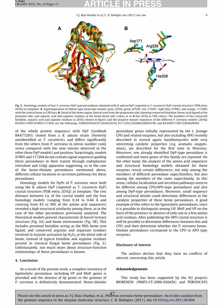
of
BAAmisfromsevoth978theretiof
diffper
usicrydisthomcovrevcasthestruincligainvhempreUnfrela
4. C
lignproP. o
Fig.
2D3
with
prox
hist
876
F.J. Ruiz-Duenas et al. / C. R. Biologies xxx (2011) xxx–xxx 9
G Model
CRASS3-3032; No. of Pages 11
Plth
the whole protein sequence) with DyP (GenBank77283) cloned from a B. adusta strain (formerlyidentified as T. cucumeris), and differs significantly
the others from P. ostreatus in intron number (onlyen compared with the nine introns observed in theer three DyP models) and position. Surprisingly, models65 and 117204 do not contain signal sequences guidingse peroxidases in their transit through endoplasmicculum and Golgi apparatus suggesting, as in the casethe heme-thiolate peroxidases mentioned above,erent cellular locations or secretion pathways for theseoxidases.Homology models for DyPs of P. ostreatus were builtng the B. adusta DyP (reported as T. cucumeris DyP)stal structure (PDB entry 2D3Q) as template. The rmsance between Ca of the crystal model and the fourology models (ranging from 0.34 to 0.44 A and
ering from 83 to 90% of the amino acid sequences)ealed a high structural similarity among them, as in thee of the other peroxidases previously analyzed. Theoretical models present characteristic b-barrel tertiarycture (Fig. 5A) and heme environment (Fig. 5B). This
ludes proximal histidine acting as the fifth heme ironnd, and conserved arginine and aspartate residuesolved in enzyme activation by H2O2 at the distal side of
e, instead of typical histidine and arginine residuessent in classical fungal heme peroxidases (Fig. 2).ortunately, not much more about structure-functiontionships of these peroxidases is known.
onclusion
As a result of the present work, a complete inventory ofinolytic peroxidase, including VP and MnP, genes isvided and the absence of lip genes in the genome ofstreatus is definitively demonstrated. Heme-thiolate
peroxidase genes initially represented by the L. fumago
CPO and related enzymes, but also including APO recentlydescribed in several agaric basidiomycetes with veryinteresting catalytic properties (e.g. aromatic oxygen-ation), are described for the first time in Pleurotus.Moreover, one already identified DyP-type peroxidase isconfirmed and more genes of this family are reported. Onthe other hand, the analysis of the amino acid sequencesand structural homology models obtained for theseenzymes reveal certain differences, not only among themembers of different peroxidase super/families, but alsoamong the members of the same super/family. In thissense, cellular localization and secretion pathways seem tobe different among CPO/APO-type peroxidases and alsoamong DyP-type peroxidases. Moreover, small sequenceand structural details could determine differences in thecatalytic properties of these heme peroxidases. A goodexample of this refers to the ligninolytic peroxidases, sinceit is possible to distinguish among LiP, MnP and VP on thebasis of the presence or absence of only one or a few aminoacid residues. After publishing the APO crystal structure itwill be possible to determine differences between this andCPO, and then determine whether the P. ostreatus heme-thiolate peroxidases correspond to the CPO or APO typeenzymes.
Disclosure of interest
The authors declare that they have no conflicts ofinterest concerning this article.
Acknowledgements
This study has been supported by the EU projectsBIORENEW (NMP2-CT-2006-026456) and PEROXICATS
5. Homology models of four P. ostreatus DyP-type peroxidases obtained with B. adusta DyP (reported as T. cucumeris DyP) crystal structure (PDB entry
Q) as template: A. Superimposition of ribbon-type molecular models (grey, 2D3Q; green, 87639; red, 115057; light blue, 97865; and orange, 117204)
the central heme as CPK bars. B. Detail of the heme region (lateral view from the propionate side) showing conserved histidine (heme axial ligand at the
imal side) and aspartic acid and arginine residues at the heme distal side (colors as in A but 2D3Q in CPK colors). The numbers of the conserved
idine, aspartic acid and arginine residues in 2D3Q (shown in figure) and the putative mature sequences of the different P. ostreatus models (2D3Q/
39/115057/97865/117204) are the following: H308/H356/H351/H343/H334, D171/D213/D208/D200/D196, and R329/R377/R372/R364/R360.
ease cite this article in press as: F.J. Ruiz-Duenas, et al., Pleurotus ostreatus heme peroxidases: An in silico analysis frome genome sequence to the enzyme molecular structure, C. R. Biologies (2011), doi:10.1016/j.crvi.2011.06.004
47
F.J. Ruiz-Duenas et al. / C. R. Biologies xxx (2011) xxx–xxx10
G Model
CRASS3-3032; No. of Pages 11
(KBBE-2010-4-265397) and the Spanish project BIO2008-01533. The work conducted by the US Department ofEnergy Joint Genome Institute is supported by the Office ofScience of the US Department of Energy under ContractNo. DE-AC02-05CH11231. Antonio G. Pisabarro (PublicUniversity of Navarre) is acknowledged for coordinatingthe P. ostreatus genome project, and Yasushi Sugano (TokyoInstitute of Technology) and Walter Buzina (MedicalUniversity of Graz) for their collaboration in the reapprais-al of the molecular identification of the DyP-producingB. adusta strain (formerly reported as T. cucumeris Dec 1).E.F. thanks CSIC for a JAE fellowship and F.J.R.-D. thanks theSpanish MICINN for a Ramon y Cajal contract.
References
[1] D. Martınez, L.F. Larrondo, N. Putnam, M.D. Gelpke, K. Huang, J. Chap-man, K.G. Helfenbein, P. Ramaiya, J.C. Detter, F. Larimer, P.M. Coutinho,B. Henrissat, R. Berka, D. Cullen, D. Rokhsar, Genome sequence of thelignocellulose degrading fungus Phanerochaete chrysosporium strainRP78, Nat. Biotechnol. 22 (2004) 695–700.
[2] D. Martınez, J. Challacombe, I. Morgenstern, D.S. Hibbett, M. Schmoll,C.P. Kubicek, P. Ferreira, F.J. Ruiz-Duenas, A.T. Martınez, P. Kersten,K.E. Hammel, A. Vanden Wymelenberg, J. Gaskell, E. Lindquist,G. Sabat, S.S. Bondurant, L.F. Larrondo, P. Canessa, R. Vicuna, J. Yadav,H. Doddapaneni, V. Subramanian, A.G. Pisabarro, J.L. Lavın, J.A. Oguiza,E. Master, B. Henrissat, P.M. Coutinho, P. Harris, J.K. Magnuson, S.E.Baker, K. Bruno, W. Kenealy, P.J. Hoegger, U. Kues, P. Ramaiya, S. Lucas,A. Salamov, H. Shapiro, H. Tu, C.L. Chee, M. Misra, G. Xie, S. Teter,D. Yaver, T. James, M. Mokrejs, M. Pospisek, I.V. Grigoriev, T. Brettin,D. Rokhsar, R. Berka, D. Cullen, Genome, transcriptome, and secretomeanalysis of wood decay fungus Postia placenta supports unique mech-anisms of lignocellulose conversion, Proc. Natl. Acad. Sci. U. S. A. 106(2009) 1954–1959.
[3] R.A. Ohm, J.F. de Jong, L.G. Lugones, A. Aerts, E. Kothe, J.E. Stajich, R.P. deVries, E. Record, A. Levasseur, S.E. Baker, K.A. Bartholomew, P.M.Coutinho, S. Erdmann, T.J. Fowler, A.C. Gathman, V. Lombard, B. Hen-rissat, N. Knabe, U. Kues, W.W. Lilly, E. Lindquist, S. Lucas, J.K. Magnu-son, F. Piumi, M. Raudaskoski, A. Salamov, J. Schmutz, F.W.M.R.Schwarze, P.A. vanKuyk, J.S. Horton, I.V. Grigoriev, H.A.B. Wosten,Genome sequence of the model mushroom Schizophyllum commune,Nat. Biotechnol. 28 (2010) 957–1010.
[4] A.T. Martınez, S. Camarero, F. Guillen, A. Gutierrez, C. Munoz, E. Varela,M.J. Martınez, J.M. Barrasa, K. Ruel, M. Pelayo, Progress in biopulping ofnon-woody materials: chemical, enzymatic and ultrastructural aspectsof wheat-straw delignification with ligninolytic fungi from the genusPleurotus, FEMS Microbiol. Rev. 13 (1994) 265–274.
[5] F.J. Ruiz-Duenas, A.T. Martınez, Microbial degradation of lignin: how abulky recalcitrant polymer is efficiently recycled in nature and how wecan take advantage of this, Microbial. Biotechnol. 2 (2009) 164–177.
[6] C. Sanchez, Cultivation of Pleurotus ostreatus and other edible mush-rooms, Appl. Microbiol. Biotechnol. 85 (2010) 1321–1337.
[7] K.E. Hammel, D. Cullen, Role of fungal peroxidases in biological lig-ninolysis, Curr. Opin. Plant Biol. 11 (2008) 349–355.
[8] A.T. Martınez, Molecular biology and structure-function of lignin-degrading heme peroxidases, Enzyme Microb. Technol. 30 (2002)425–444.
[9] F.J. Ruiz-Duenas, M. Morales, E. Garcıa, Y. Miki, M.J. Martınez, A.T.Martınez, Substrate oxidation sites in versatile peroxidase and otherbasidiomycete peroxidases, J. Exp. Bot. 60 (2009) 441–452.
[10] M. Hofrichter, R. Ullrich, M.J. Pecyna, C. Liers, T. Lundell, New and classicfamilies of secreted fungal heme peroxidases, Appl. Microbiol. Biotech-nol. 87 (2010) 871–897.
[11] L.M. Larraya, G. Perez, M.M. Penas, J.J.P. Baars, T.S.P. Mikosch, A.G.Pisabarro, L. Ramırez, Molecular karyotype of the white rot fungusPleurotus ostreatus, Appl. Environ. Microbiol. 65 (1999) 3413–3417.
[12] Y. Asada, A. Watanabe, T. Irie, T. Nakayama, M. Kuwahara, Structures ofgenomic and complementary DNAs coding for Pleurotus ostreatusmanganese (II) peroxidase, Biochim. Biophys. Acta 1251 (1995)205–209.
[13] P. Giardina, G. Palmieri, B. Fontanella, V. Rivieccio, G. Sannia, Manga-nese peroxidase isoenzymes produced by Pleurotus ostreatus grown onwood sawdust, Arch. Biochem. Biophys. 376 (2000) 171–179.
[14] T. Irie, Y. Honda, H.-C. Ha, T. Watanabe, M. Kuwahara, Isolation of cDNA
isoenzyme from the white rot basidiomycete Pleurotus ostreatus, J.Wood Sci. 46 (2000) 230–233.
[15] V. Faraco, A. Piscitelli, G. Sannia, P. Giardina, Identification of a newmember of the dye-decolorizing peroxidase family from Pleurotusostreatus, World J. Microbiol. Biotechnol. 23 (2007) 889–893.
[16] K. Tamura, J. Dudley, M. Nei, S. Kumar, MEGA4: molecular evolutionarygenetics analysis (MEGA) software version 4.0, Mol. Biol. Evol. 24(2007) 1596–1599.
[17] J.D. Bendtsen, H. Nielsen, G. von Heijne, S. Brunak, Improved predictionof signal peptides: SignalP 3.0, J. Mol. Biol. 340 (2004) 783–795.
[18] L. Bordoli, F. Kiefer, K. Arnold, P. Benkert, J. Battey, T. Schwede, Proteinstructure homology modeling using SWISS-MODEL workspace, Nat.Protoc. 4 (2009) 1–13.
[19] F.J. Ruiz-Duenas, A.T. Martınez, Structural and functional features ofperoxidases with a potential as industrial biocatalysts, in: E. Torres, M.Ayala (Eds.), Biocatalysts based on heme peroxidases, Springer-Verlag,Berlin, 2010, pp. 37–59.
[20] D.M. Kupfer, S.D. Drabenstot, K.L. Buchanan, H.S. Lai, H. Zhu, D.W. Dyer,B.A. Roe, J.W. Murphy, Introns and splicing elements of five diversefungi, Eukaryot. Cell 3 (2004) 1088–1100.
[21] M. Rep, R.G.E. Duyvesteijn, L. Gale, T. Usgaard, B.J.C. Cornelissen, L.J. Ma,T.J. Ward, The presence of GC-AG introns in Neurospora crassa and othereuascomycetes determined from analyses of complete genomes: Impli-cations for automated gene prediction, Genomics 87 (2006) 338–347.
[22] M.R. Makela, K. Hilden, A. Hatakka, T.K. Lundell, Oxalate decarboxylaseof the white-rot fungus Dichomitus squalens demonstrates a novelenzyme primary structure and non-induced expression on wood andin liquid cultures, Microbiol. Sgm 155 (2009) 2726–2738.
[23] M.R. Escutia, L. Bowater, A. Edwards, A.R. Bottrill, M.R. Burrell, R.Polanco, R. Vicuna, S. Bornemann, Cloning and Sequencing of twoCeriporiopsis subvermispora bicupin oxalate oxidase allelic isoforms:implications for the reaction specificity of oxalate oxidases and dec-arboxylases, Appl. Environ. Microbiol. 71 (2005) 3608–3616.
[24] F. Pelaez, M.J. Martınez, A.T. Martınez, Screening of 68 species ofbasidiomycetes for enzymes involved in lignin degradation, Mycol.Res. 99 (1995) 37–42.
[25] E. Varela, A.T. Martınez, M.J. Martınez, Southern blot screening forlignin peroxidase and aryl-alcohol oxidase genes in 30 fungal species, J.Biotechnol. 83 (2000) 245–251.
[26] K.G. Welinder, Superfamily of plant, fungal and bacterial peroxidases,Curr. Opin. Struct. Biol. 2 (1992) 388–393.
[27] J.E. Stajich, S.K. Wilke, D. Ahren, C.H. Au, B.W. Birren, M. Borodovsky, C.Burns, B. Canback, L.A. Casselton, C.K. Cheng, J.X. Deng, F.S. Dietrich, D.C.Fargo, M.L. Farman, A.C. Gathman, J. Goldberg, R. Guigo, P.J. Hoegger,J.B. Hooker, A. Huggins, T.Y. James, T. Kamada, S. Kilaru, C. Kodira, U.Kues, D. Kupfert, H.S. Kwan, A. Lomsadze, W.X. Li, W.W. Lilly, L.J. Ma, A.J.Mackey, G. Manning, F. Martin, H. Muraguchi, D.O. Natvig, H. Palmerini,M.A. Ramesh, C.J. Rehmeyer, B.A. Roe, N. Shenoy, M. Stanke, V. TerHovhannisyan, A. Tunlid, R. Velagapudi, T.J. Vision, Q.D. Zeng, M.E.Zolan, P.J. Pukkila, Insights into evolution of multicellular fungi fromthe assembled chromosomes of the mushroom Coprinopsis cinerea(Coprinus cinereus), Proc. Natl. Acad. Sci. U. S. A. 107 (2010) 11889–11894.
[28] M. Sivaraja, D.B. Goodin, M. Smith, B.M. Hoffman, Identification byENDOR of Trp191 as the free-radical site in cytochrome-c peroxidasecompound ES, Science 245 (1989) 738.
[29] H. Pelletier, J. Kraut, Crystal structure of a complex between electrontransfer partners, cytochrome c peroxidase and cytochrome c, Science258 (1992) 1748–1755.
[30] Q. Husain, M. Husain, Y. Kulshrestha, Remediation and treatment oforganopollutants mediated by peroxidases: a review, Crit. Rev. Bio-technol. 29 (2009) 94–119.
[31] L. Banci, I. Bertini, P. Turano, M. Tien, T.K. Kirk, Proton NMR investiga-tion into the basis for the relatively high redox potential of ligninperoxidase, Proc. Natl. Acad. Sci. U. S. A. 88 (1991) 6956–6960.
[32] L. Banci, S. Camarero, A.T. Martınez, M.J. Martınez, M. Perez-Boada, R.Pierattelli, F.J. Ruiz-Duenas, NMR study of Mn(II) binding by the newversatile peroxidase from the white-rot fungus Pleurotus eryngii, J. Biol.Inorg. Chem. 8 (2003) 751–760.
[33] T. Mester, K. Ambert-Balay, S. Ciofi-Baffoni, L. Banci, A.D. Jones, M. Tien,Oxidation of a tetrameric nonphenolic lignin model compound bylignin peroxidase, J. Biol. Chem. 276 (2001) 22985–22990.
[34] A.T. Smith, W.A. Doyle, P. Dorlet, A. Ivancich, Spectroscopic evidence foran engineered, catalytically active Trp radical that creates the uniquereactivity of lignin peroxidase, Proc. Natl. Acad. Sci. U. S. A. 106 (2009)16084–16089.
[35] M. Sundaramoorthy, M.H. Gold, T.L. Poulos, Ultrahigh (0.93 angstrom)resolution structure of manganese peroxidase from Phanerochaetechrysosporium: Implications for the catalytic mechanism, J. Inorg.
Biochem. 104 (2010) 683–690. and genomic fragments encoding the major manganese peroxidasePlease cite this article in press as: F.J. Ruiz-Duenas, et al., Pleurotus ostreatus heme peroxidases: An in silico analysis fromthe genome sequence to the enzyme molecular structure, C. R. Biologies (2011), doi:10.1016/j.crvi.2011.06.004
48
[36]
[37]
[38]
[39]
[40]
[41]
[42]
[43]
[44]
[45]
[46]
[47]
[48]
[49]
F.J. Ruiz-Duenas et al. / C. R. Biologies xxx (2011) xxx–xxx 11
G Model
CRASS3-3032; No. of Pages 11
Plth
D. Cullen, P.J. Kersten, Enzymology and molecular biology of lignindegradation, in: R. Brambl, G.A. Marzluf (Eds.), Mycota III: biochemistryand molecular biology, Springer, Berlin, 2004, pp. 249–273.
M.H. Gold, H.L. Youngs, M.D. Gelpke, Manganese peroxidase, Met. IonsBiol. Syst. 37 (2000) 559–586.
K. Hilden, A.T. Martınez, A. Hatakka, T. Lundell, The two manganeseperoxidases Pr-MnP2 and Pr-MnP3 of Phlebia radiata, a lignin-degrad-ing basidiomycete, are phylogenetically and structurally divergent,Fungal Genet. Biol. 42 (2005) 403–419.
T. Tsukihara, Y. Honda, R. Sakai, T. Watanabe, T. Watanabe, Mechanismfor oxidation of high-molecular-weight substrates by a fungal versatileperoxidase, MnP2, Appl. Environ. Microbiol. 74 (2008) 2873–2881.
P.D. Shaw, L.P. Hager, Biological chlorination. 6. Chloroperoxidase -Component of beta-ketoadipate chlorinase system, J. Biol. Chem. 236(1961) 1626.
D. Koua, L. Cerutti, L. Falquet, C.J.A. Sigrist, G. Theiler, N. Hulo, C.Dunand, PeroxiBase: a database with new tools for peroxidase familyclassification, Nucleic Acids Res. 37 (2009) D261–D266.
R. Ullrich, J. Nuske, K. Scheibner, J. Spantzel, M. Hofrichter, Novelhaloperoxidase from the agaric basidiomycete Agrocybe aegerita oxi-dizes aryl alcohols and aldehydes, Appl. Environ. Microbiol. 70 (2004)4575–4581.
D.H. Anh, R. Ullrich, D. Benndorf, A. Svatos, A. Muck, M. Hofrichter, Thecoprophilous mushroom Coprinus radians secretes a haloperoxidasethat catalyzes aromatic peroxygenation, Appl. Environ. Microbiol. 73(2007) 5477–5485.
M. Hofrichter, R. Ullrich, Heme-thiolate haloperoxidases: versatilebiocatalysts with biotechnological and environmental significance,Appl. Microbiol. Biotechnol. 71 (2006) 276–288.
M.J. Pecyna, R. Ullrich, B. Bittner, A. Clemens, K. Scheibner, R. Schubert, M.Hofrichter, Molecular characterization of aromatic peroxygenase fromAgrocybe aegerita, Appl. Microbiol. Biotechnol. 84 (2009) 885–897.
A. Conesa, G. Weelink, C.A.M.J. van den Hondel, P.J. Punt, C-terminalpropeptide of the Caldariomyces fumago chloroperoxidase: an intramo-lecular chaperone? FEBS Lett. 503 (2001) 117–120.
M.J. Nuell, G.H. Fang, M.J. Axley, P. Kenigsberg, L.P. Hager, Isolation andnucleotide sequence of the chloroperoxidase gene from Caldariomycesfumago, J. Bacteriol. 170 (1988) 1007–1011.
M. Sundaramoorthy, J. Terner, T.L. Poulos, The crystal structure ofchloroperoxidase: a heme peroxidase-cytochrome P450 functionalhybrid, Structure 3 (1995) 1367–1377.
K. Piontek, R. Ullrich, C. Liers, K. Diederichs, D.A. Plattner, M. Hofrichter,Crystallization of a 45 kDa peroxygenase/peroxidase from the
mushroom Agrocybe aegerita and structure determination by SADutilizing only the haem iron, Acta Crystallogr. F 66 (2010) 693–698.
[50] F.J. Ruiz-Duenas, M.J. Martınez, A.T. Martınez, Molecular characteriza-tion of a novel peroxidase isolated from the ligninolytic fungus Pleur-otus eryngii, Mol. Microbiol. 31 (1999) 223–236.
[51] M. Sundaramoorthy, J. Terner, T.L. Poulos, Stereochemistry of thechloroperoxidase active site: crystallographic and molecular-modelingstudies, Chem. Biol. 5 (1998) 461–473.
[52] K. Kuhnel, W. Blankenfeldt, J. Terner, I. Schlichting, Crystal structuresof chloroperoxidase with its bound substrates and complexedwith formate, acetate, and nitrate, J. Biol. Chem. 281 (2006) 23990–23998.
[53] Y. Sugano, DyP-type peroxidases comprise a novel heme peroxidasefamily, Cell Mol. Life Sci. 66 (2009) 1387–1403.
[54] S.J. Kim, M. Shoda, Purification and characterization of a novel peroxi-dase from Geotrichum candidum Dec 1 involved in decolorization ofdyes, Appl. Environ. Microbiol. 65 (1999) 1029–1035.
[55] Y. Sugano, K. Sasaki, M. Shoda, cDNA cloning and genetic analysis of anovel decolorizing enzyme, peroxidase gene dyp from Geotrichumcandidum Dec 1, J. Biosci. Bioeng. 87 (1999) 411–417.
[56] A. Johanson, H.C. Turner, G.J. Mckay, A.E. Brown, A PCR-based methodto distinguish fungi of the rice sheath-blight complex, Rhizoctoniasolani, R-oryzae and R-oryzae-sativae, FEMS Microbiol. Lett. 162(1998) 289–294.
[57] Y. Sugano, Y. Matsushima, K. Tsuchiya, H. Aoki, M. Hirai, M. Shoda,Degradation pathway of an anthraquinone dye catalyzed by a uniqueperoxidase DyP from Thanatephorus cucumeris Dec 1, Biodegradation20 (2009) 433–440.
[58] K. Zelena, B. Hardebusch, B. Hulsdau, R.G. Berger, H. Zorn, Generation ofnorisoprenoid flavors from carotenoids by fungal peroxidases, J. Agric.Food Chem. 57 (2009) 9951–9955.
[59] C. Liers, C. Bobeth, M. Pecyna, R. Ullrich, M. Hofrichter, DyP-likeperoxidases of the jelly fungus Auricularia auricula-judae oxidize non-phenolic lignin model compounds and high-redox potential dyes, Appl.Microbiol. Biotechnol. 85 (2010) 1869–1879.
[60] M. Puhse, R.T. Szweda, Y.Y. Ma, C. Jeworrek, R. Winter, H. Zorn,Marasmius scorodonius extracellular dimeric peroxidase - Exploringits temperature and pressure stability, BBA Proteins Proteomics1794 (2009) 1091–1098.
[61] Y. Sugano, Y. Matsushima, M. Shoda, Complete decolorization of theanthraquinone dye Reactive blue 5 by the concerted action of twoperoxidases from Thanatephorus cucumeris Dec 1, Appl. Microbiol.Biotechnol. 73 (2006) 862–871.
ease cite this article in press as: F.J. Ruiz-Duenas, et al., Pleurotus ostreatus heme peroxidases: An in silico analysis frome genome sequence to the enzyme molecular structure, C. R. Biologies (2011), doi:10.1016/j.crvi.2011.06.004
49

50
Capítulo 3
"Ligninolytic peroxidase genes in the oyster mushroom genome: Heterologous expression, molecular structure, catalytic and stability properties and lignin-degrading ability" Fernández-Fueyo, E., F. J. Ruiz-Dueñas, M. J. Martínez, A. Romero, K. E. Hammel, F. J. Medrano, and A. T. Martínez. Biotechnol. Biofuels 7:2. 2014.
51

52
Fernández-Fueyo et al. Biotechnology for Biofuels 2014, 7:2http://www.biotechnologyforbiofuels.com/content/7/1/2
RESEARCH Open Access
Ligninolytic peroxidase genes in the oystermushroom genome: heterologous expression,molecular structure, catalytic and stabilityproperties, and lignin-degrading abilityElena Fernández-Fueyo1, Francisco J Ruiz-Dueñas1, María Jesús Martínez1, Antonio Romero1, Kenneth E Hammel2*,Francisco Javier Medrano1* and Angel T Martínez1*
Abstract
Background: The genome of Pleurotus ostreatus, an important edible mushroom and a model ligninolytic organismof interest in lignocellulose biorefineries due to its ability to delignify agricultural wastes, was sequenced with thepurpose of identifying and characterizing the enzymes responsible for lignin degradation.
Results: Heterologous expression of the class II peroxidase genes, followed by kinetic studies, enabled theirfunctional classification. The resulting inventory revealed the absence of lignin peroxidases (LiPs) and the presenceof three versatile peroxidases (VPs) and six manganese peroxidases (MnPs), the crystal structures of two of them(VP1 and MnP4) were solved at 1.0 to 1.1 Å showing significant structural differences. Gene expansion supportsthe importance of both peroxidase types in the white-rot lifestyle of this fungus. Using a lignin model dimerand synthetic lignin, we showed that VP is able to degrade lignin. Moreover, the dual Mn-mediated andMn-independent activity of P. ostreatus MnPs justifies their inclusion in a new peroxidase subfamily. The availabilityof the whole POD repertoire enabled investigation, at a biochemical level, of the existence of duplicated genes.Differences between isoenzymes are not limited to their kinetic constants. Surprising differences in their activity T50and residual activity at both acidic and alkaline pH were observed. Directed mutagenesis and spectroscopic/structural information were combined to explain the catalytic and stability properties of the most interesting isoenzymes,and their evolutionary history was analyzed in the context of over 200 basidiomycete peroxidase sequences.
Conclusions: The analysis of the P. ostreatus genome shows a lignin-degrading system where the role generally played byLiP has been assumed by VP. Moreover, it enabled the first characterization of the complete set of peroxidase isoenzymesin a basidiomycete, revealing strong differences in stability properties and providing enzymes of biotechnological interest.
Keywords: Genome, Pleurotus ostreatus, Ligninolytic peroxidase genes, Heterologous expression, Crystal structure, Catalyticproperties, Thermal stability, pH stability, Gene duplication, Peroxidase evolution
BackgroundPleurotus ostreatus, the oyster mushroom, is the secondmost consumed edible mushroom worldwide, just afterAgaricus bisporus, with a current production accountingfor over 25% of mushroom world production (that is,around 3 million metric tons/year, with a market value
* Correspondence: [email protected]; [email protected];[email protected] Forest Products Laboratory, One Gifford Pinchot Drive, Madison, WI53726, USA1Centro de Investigaciones Biológicas, Consejo Superior de InvestigacionesCientíficas (CSIC), Ramiro de Maeztu 9, E-28040 Madrid, Spain
© 2014 Fernández-Fueyo et al.; licensee BioMeCreative Commons Attribution License (http:/distribution, and reproduction in any mediumDomain Dedication waiver (http://creativecomarticle, unless otherwise stated.
53
of several thousand million euros per year) [1]. Inaddition to their food properties, Pleurotus species alsohave medicinal properties due to their production ofanticholesterolemic statins [2] and antitumor polysac-charides [3], among other bioactive molecules. China isthe main producer of P. ostreatus and related species.Sawdust and cereal straw are the usual substrates forPleurotus production, together with other agricultural,forest, and/or industrial lignocellulosic wastes.From an ecophysiological point of view, Pleurotus spe-
cies belong to the group of fungi causing a so-called
d Central Ltd. This is an Open Access article distributed under the terms of the/creativecommons.org/licenses/by/2.0), which permits unrestricted use,, provided the original work is properly cited. The Creative Commons Publicmons.org/publicdomain/zero/1.0/) applies to the data made available in this
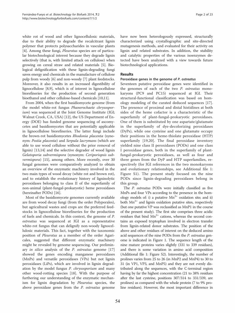
Fernández-Fueyo et al. Biotechnology for Biofuels 2014, 7:2 Page 2 of 23http://www.biotechnologyforbiofuels.com/content/7/1/2
white rot of wood and other lignocellulosic materials,due to their ability to degrade the recalcitrant ligninpolymer that protects polysaccharides in vascular plants[4]. Among these fungi, Pleurotus species are of particu-lar biotechnological interest because they degrade ligninselectively (that is, with limited attack on cellulose) whengrowing on cereal straw and related materials [5]. Bio-logical delignification with these lignin-degrading fungisaves energy and chemicals in the manufacture of cellulosepulp from woody [6] and non-woody [7] plant feedstocks.Moreover, it also results in an increased digestibility oflignocellulose [8,9], which is of interest in lignocellulosebiorefineries for the production of second generationbioethanol and other cellulose-based chemicals [10,11].From 2004, when the first basidiomycete genome (from
the model white-rot fungus Phanerochaete chrysospor-ium) was sequenced at the Joint Genome Institute (JGI;Walnut Creek, CA, USA) [12], the US Department of En-ergy (DOE) has funded genome sequencing of ascomy-cetes and basidiomycetes that are potentially applicablein lignocellulose biorefineries. The latter fungi includethe brown-rot basidiomycetes Rhodonia placenta (syno-nym: Postia placenta) and Serpula lacrymans (which areable to use wood cellulose without the prior removal oflignin) [13,14] and the selective degrader of wood ligninGelatoporia subvermispora (synonym: Ceriporiopsis sub-vermispora) [15], among others. More recently, over 30fungal genomes were comparatively analyzed to obtainan overview of the enzymatic machinery involved in thetwo main types of wood decay (white rot and brown rot),and to establish the evolutionary history of ligninolyticperoxidases belonging to class II of the superfamily ofnon-animal (plant-fungal-prokaryotic) heme peroxidases(hereinafter PODs) [16].Most of the basidiomycete genomes currently available
are from wood decay fungi (from the order Polyporales),but agricultural wastes and crops are the preferred feed-stocks in lignocellulose biorefineries for the productionof fuels and chemicals. In this context, the genome of P.ostreatus was sequenced at JGI as a representativewhite-rot fungus that can delignify non-woody lignocel-lulosic materials. This fact, together with the taxonomicposition of Pleurotus as a member of the order Agari-cales, suggested that different enzymatic machinerymight be revealed by genome sequencing. Our prelimin-ary in silico analysis of the P. ostreatus genome [17]showed the genes encoding manganese peroxidases(MnPs) and versatile peroxidases (VPs) but not ligninperoxidases (LiPs), which are involved in lignin degrad-ation by the model fungus P. chrysosporium and manyother wood-rotting species [18]. With the purpose offurthering our understanding of the enzymatic mechan-ism for lignin degradation by Pleurotus species, theabove peroxidase genes from the P. ostreatus genome
54
have now been heterologously expressed, structurallycharacterized using crystallographic and site-directedmutagenesis methods, and evaluated for their activity onlignin and related substrates. In addition, the stabilityand catalytic properties of the various isoenzymes de-tected have been analyzed with a view towards futurebiotechnological applications.
ResultsPeroxidase genes in the genome of P. ostreatusSeventeen putative peroxidase genes were identified inthe genomes of each of the two P. ostreatus mono-karyons (PC9 and PC15) sequenced at JGI. Theirstructural-functional classification was based on hom-ology modeling of the curated deduced sequences [17].The presence of proximal and distal histidines at bothsides of the heme cofactor is a characteristic of thesuperfamily of plant-fungal-prokaryotic peroxidases.One of them is substituted by one aspartate/glutamatein the superfamily of dye-decolorizing peroxidases(DyPs), while one cysteine and one glutamate occupytheir positions in the heme-thiolate peroxidase (HTP)superfamily [19,20]. The final peroxidase inventoryyielded nine class II peroxidases (PODs) and one classI peroxidase genes, both in the superfamily of plant-fungal-prokaryotic peroxidases, as well as four andthree genes from the DyP and HTP superfamilies, re-spectively (for JGI references in the two monokaryonsand evolutionary relationships, see Additional file 1:Figure S1). The present study focused on the ninePODs since lignin-degrading peroxidases belong inthis group.The P. ostreatus PODs were initially classified as five
MnPs and four VPs according to the presence in the hom-ology models of: i) a putative Mn2+ oxidation site; and ii)both Mn2+ and lignin oxidation putative sites, respectively(but one putative VP was reclassified as MnP1 in the courseof the present study). The first site comprises three acidicresidues that bind Mn2+ cations, whereas the second con-tains an exposed tryptophan involved in electron transferfrom lignin-related donor substrates. The position of theabove and other residues of interest on the deduced aminoacid sequences of the nine PODs from the P. ostreatus gen-ome is indicated in Figure 1. The sequence length of thenine mature proteins varies slightly (331 to 339 residues),and there is some variation in amino acid composition(Additional file 1: Figure S2). Interestingly, the number ofprolines varies from 25 to 26 (in MnP2 and MnP4) to 30 to31 (in VP1, VP3, and MnP5) and they are not evenly dis-tributed along the sequences, with the C-terminal regionhaving by far the highest concentration (21 to 38% residuesafter the last cysteine, positions 307/314 to 331/339, areprolines) as compared with the whole protein (7 to 9% pro-line residues). However, the most important difference in
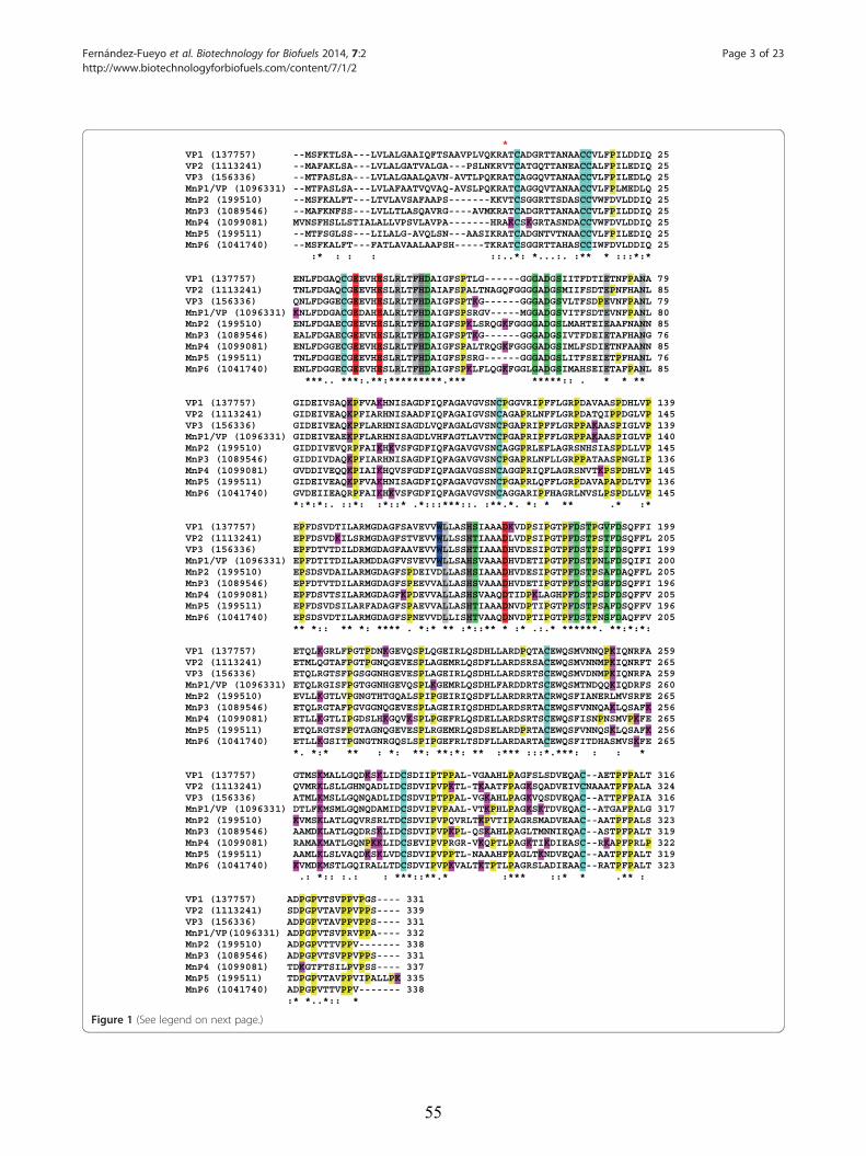
*VP1 (137757) --MSFKTLSA---LVLALGAAIQFTSAAVPLVQKRATCADGRTTANAACCVLFPILDDIQ 25 VP2 (1113241) --MAFAKLSA---LVLALGATVALGA---PSLNKRVTCATGQTTANEACCALFPILEDIQ 25 VP3 (156336) --MTFASLSA---LVLALGAALQAVN-AVTLPQKRATCAGGQVTANAACCVLFPILEDLQ 25MnP1/VP (1096331) --MTFASLSA---LVLAFAATVQVAQ-AVSLPQKRATCAGGQVTANAACCVLFPLMEDLQ 25 MnP2 (199510) --MSFKALFT---LTVLAVSAFAAPS-------KKVTCSGGRTTSDASCCVWFDVLDDIQ 25MnP3 (1089546) --MAFKNFSS---LVLLTLASQAVRG----AVMKRATCADGRTTANAACCVLFPILDDIQ 25 MnP4 (1099081) MVNSFHSLLSTIALALLVPSVLAVPA-------HRAKCSKGRTASNDACCVWFDVLDDIQ 25MnP5 (199511) --MTFSGLSS---LILALG-AVQLSN---AASIKRATCADGNTVTNAACCVLFPILEDIQ 25 MnP6 (1041740) --MSFKALFT---FATLAVAALAAPSH-----TKRATCSGGRTTAHASCCIWFDVLDDIQ 25
:* : : : ::..*: *...:. :** * :::*:*
VP1 (137757) ENLFDGAQCGEEVHESLRLTFHDAIGFSPTLG------GGGADGSIITFDTIETNFPANA 79 VP2 (1113241) TNLFDGAQCGEEVHESLRLTFHDAIAFSPALTNAGQFGGGGADGSMIIFSDTEPNFHANL 85 VP3 (156336) QNLFDGGECGEEVHESLRLTFHDAIGFSPTKG------GGGADGSVLTFSDPEVNFPANL 79MnP1/VP (1096331) KNLFDDGACGEDAHEALRLTFHDAIGFSPSRGV-----MGGADGSVITFSDTEVNFPANL 80 MnP2 (199510) ENLFDGAECGEEVHESLRLTFHDAIGFSPKLSRQGKFGGGGADGSLMAHTEIEAAFNANN 85 MnP3 (1089546) EALFDGAECGEEVHESLRLTFHDAIGFSPTKG------GGGADGSIVTFDEIETAFHANG 76 MnP4 (1099081) ENLFDGGECGEEVHESLRLTFHDAIGFSPALTRQGKFGGGGADGSIMLFSDIETNFAANN 85 MnP5 (199511) TNLFDGGECGEEVHESLRLTFHDAIGFSPSRG------GGGADGSLITFSEIETPFHANL 76MnP6 (1041740) ENLFDGGECGEEVHESLRLTFHDAIGFSPKLFLQGKFGGLGADGSIMAHSEIETAFPANL 85
***.. ***:.**:*********.*** *****:: . * * **
VP1 (137757) GIDEIVSAQKPFVAKHNISAGDFIQFAGAVGVSNCPGGVRIPFFLGRPDAVAASPDHLVP 139 VP2 (1113241) GIDEIVEAQKPFIARHNISAADFIQFAGAIGVSNCAGAPRLNFFLGRPDATQIPPDGLVP 145 VP3 (156336) GIDEIVEAQKPFLARHNISAGDLVQFAGALGVSNCPGAPRIPFFLGRPPAKAASPIGLVP 139MnP1/VP (1096331) GIDEIVEAEKPFLARHNISAGDLVHFAGTLAVTNCPGAPRIPFFLGRPPAKAASPIGLVP 140 MnP2 (199510) GIDDIVEVQRPFAIKHKVSFGDFIQFAGAVGVSNCAGGPRLEFLAGRSNHSIASPDLLVP 145 MnP3 (1089546) GIDDIVDAQKPFIARHNISAGDFIQFAGAVGVSNCPGAPRLNFLLGRPPATAASPNGLIP 136 MnP4 (1099081) GVDDIVEQQKPIAIKHQVSFGDFIQFAGAVGSSNCAGGPRIQFLAGRSNVTKPSPDHLVP 145 MnP5 (199511) GIDEIVEAQKPFVAKHNISAGDFIQFAGAVGVSNCPGAPRLQFFLGRPDAVAPAPDLTVP 136MnP6 (1041740) GVDEIIEAQRPFAIKHKVSFGDFIQFAGAVGVSNCAGGARIPFHAGRLNVSLPSPDLLVP 145
*:*:*:. ::*: :*::* .*:::***::. :**.*. *: * ** .* :*
VP1 (137757) EPFDSVDTILARMGDAGFSAVEVVWLLASHSIAAADKVDPSIPGTPFDSTPGVFDSQFFI 199VP2 (1113241) EPFDSVDKILSRMGDAGFSTVEVVWLLSSHTIAAADLVDPSIPGTPFDSTPSTFDSQFFL 205 VP3 (156336) EPFDTVTDILDRMGDAGFAAVEVVWLLSSHTIAAADHVDESIPGTPFDSTPSIFDSQFFI 199MnP1/VP (1096331) EPFDTITDILARMDDAGFVSVEVVWLLSAHSVAAADHVDETIPGTPFDSTPNLFDSQIFI 200MnP2 (199510) EPSDSVDAILARMGDAGFSPDEIVDLLASHSIAAADHVDESIPGTPFDSTPSAFDAQFFL 205 MnP3 (1089546) EPFDTVTDILARMGDAGFSPEEVVALLASHSVAAADHVDETIPGTPFDSTPGEFDSQFFI 196 MnP4 (1099081) EPFDSVTSILARMGDAGFKPDEVVALLASHSVAAQDTIDPKLAGHPFDSTPSDFDSQFFV 205 MnP5 (199511) EPFDSVDSILARFADAGFSPAEVVALLASHTIAAADNVDPTIPGTPFDSTPSAFDSQFFV 196MnP6 (1041740) EPSDSVDTILARMGDAGFSPNEVVDLLISHTVAAQDNVDPTIPGTPFDSTPNSFDAQFFV 205
** *:: ** *: **** . *:* ** :*::** * :* .:.* ******. **:*:*:
VP1 (137757) ETQLKGRLFPGTPDNKGEVQSPLQGEIRLQSDHLLARDPQTACEWQSMVNNQPKIQNRFA 259 VP2 (1113241) ETMLQGTAFPGTPGNQGEVESPLAGEMRLQSDFLLARDSRSACEWQSMVNNMPKIQNRFT 265VP3 (156336) ETQLRGTSFPGSGGNHGEVESPLAGEIRLQSDHLLARDSRTSCEWQSMVDNMPKIQNRFA 259MnP1/VP (1096331) ETQLRGISFPGTGGNHGEVQSPLKGEMRLQSDHLFARDDRTSCEWQSMTNDQQKIQDRFS 260 MnP2 (199510) EVLLKGTLVPGNGTHTGQALSPIPGEIRIQSDFLLARDRRTACRWQSFIANERLMVSRFE 265 MnP3 (1089546) ETQLRGTAFPGVGGNQGEVESPLAGEIRIQSDHDLARDSRTACEWQSFVNNQAKLQSAFK 256 MnP4 (1099081) ETLLKGTLIPGDSLHKGQVKSPLPGEFRLQSDELLARDSRTSCEWQSFISNPNSMVPKFE 265 MnP5 (199511) ETQLRGTSFPGTAGNQGEVESPLRGEMRLQSDSELARDPRTACEWQSFVNNQSKLQSAFK 256MnP6 (1041740) ETLLKGSITPGNGTNRGQSLSPIPGEFRLTSDFLLARDARTACEWQSFITDHASMVSKFE 265
*. *:* ** : *: **: **:*: ** :*** :::*.***: : : *
VP1 (137757) GTMSKMALLGQDKSKLIDCSDIIPTPPAL-VGAAHLPAGFSLSDVEQAC--AETPFPALT 316 VP2 (1113241) QVMRKLSLLGHNQADLIDCSDVIPVPKTL-TKAATFPAGKSQADVEIVCNAAATPFPALA 324 VP3 (156336) ATMLKMSLLGQNQADLIDCSDVIPTPPAL-VGKAHLPAGKVQSDVEQAC--ATTPFPAIA 316MnP1/VP (1096331) DTLFKMSMLGQNQDAMIDCSDVIPVPAAL-VTKPHLPAGKSKTDVEQAC--ATGAFPALG 317 MnP2 (199510) KVMSKLATLGQVRSRLTDCSDVIPVPQVRLTKPVTIPAGRSMADVEAAC--AATPFPALS 323 MnP3 (1089546) AAMDKLATLGQDRSKLIDCSDVIPVPKPL-QSKAHLPAGLTMNNIEQAC--ASTPFPALT 319 MnP4 (1099081) RAMAKMATLGQNPKKLIDCSEVIPVPRGR-VKQPTLPAGKTIKDIEASC--RKAPFPRLP 322 MnP5 (199511) AAMLKLSLVAQDKSKLVDCSDVIPVPPTL-NAAAHFPAGLTKNDVEQAC--AATPFPALT 319MnP6 (1041740) KVMDKMSTLGQIRALLTDCSDVIPVPKVALTKTPTLPAGRSLADIEAAC--RATPFPALT 323
.: *:: :.: : ***::**.* :*** ::* * .** :
VP1 (137757) ADPGPVTSVPPVPGS---- 331 VP2 (1113241) SDPGPVTAVPPVPPS---- 339 VP3 (156336) ADPGPVTAVPPVPPS---- 331MnP1/VP(1096331) ADPGPVTSVPRVPPA---- 332 MnP2 (199510) ADPGPVTTVPPV------- 338 MnP3 (1089546) ADPGPVTSVPPVPPS---- 331 MnP4 (1099081) TDKGTFTSILPVPSS---- 337 MnP5 (199511) TDPGPVTAVPPVIPALLPK 335MnP6 (1041740) ADPGPVTTVPPV------- 338
:* *..*:: *
Figure 1 (See legend on next page.)
Fernández-Fueyo et al. Biotechnology for Biofuels 2014, 7:2 Page 3 of 23http://www.biotechnologyforbiofuels.com/content/7/1/2
55
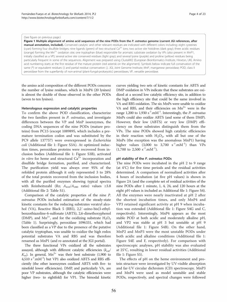
(See figure on previous page.)Figure 1 Multiple alignment of amino acid sequences of the nine PODs from the P. ostreatus genome (current JGI references, aftermanual annotation, included). Conserved catalytic and other relevant residues are indicated with different colors including: eight cysteines(cyan) forming four disulfide bridges; nine ligands (green) of two structural Ca2+ ions; two active site histidines (dark gray); three acidic residues(orange) forming the Mn2+ oxidation site; one tryptophan (blue) responsible for aromatic substrate oxidation by VPs (also present in MnP1,initially classified as a VP); several active site conserved residues (light gray); and several lysine (purple) and proline (yellow) residues beingparticularly frequent in some of the sequences. Alignment was prepared using ClustalW2 (European Bioinformatics Institute, Hinxton, UK). Aminoacid numbering starts at the first residue of the mature protein (red asterisk on the alignment). Symbols below indicate full conservation of thesame (*) or equivalent residues (:) and partial residue conservation (.). JGI, Joint Genome Institute; MnP, manganese peroxidase; POD, class IIperoxidase from the superfamily of non-animal (plant-fungal-prokaryotic) peroxidases; VP, versatile peroxidase.
Fernández-Fueyo et al. Biotechnology for Biofuels 2014, 7:2 Page 4 of 23http://www.biotechnologyforbiofuels.com/content/7/1/2
the amino acid composition of the different PODs concernsthe number of lysine residues, which in MnP4 (20 lysines)is almost the double of those observed in the other PODs(seven to ten lysines).
Heterologous expression and catalytic propertiesTo confirm the above POD classification, characterizethe two families present in P. ostreatus, and investigatedifferences between the VP and MnP isoenzymes, thecoding DNA sequences of the nine PODs (mature pro-teins) from PC15 (except 1089895, which includes a pre-mature termination codon and was substituted by thePC9 allele 137757) were overexpressed in Escherichiacoli (Additional file 1: Figure S3A). At optimized induc-tion times, peroxidase proteins were recovered from in-clusion bodies (Additional file 1: Figure S3B), activatedin vitro for heme and structural Ca2+ incorporation anddisulfide bridge formation, purified, and characterized.The purification yield was always over 95% of therefolded protein although it only represented 3 to 28%of the total protein recovered from the inclusion bodies,with all the purified enzymes showing typical spectrawith Reinheitszahl (Rz; A410/A280 ratio) values ≥3.8(Additional file 2: Table S1).Comparison of the catalytic properties of the nine P.
ostreatus PODs included estimation of the steady-statekinetic constants for the reducing substrates veratryl alco-hol (VA), Reactive Black 5 (RB5), 2,2′-azino-bis(3-ethyl-benzothiazoline-6-sulfonate (ABTS), 2,6-dimethoxyphenol(DMP), and Mn2+, and for the oxidizing substrate H2O2
(Table 1). Surprisingly, peroxidase 1096331, which hadbeen classified as a VP due to the presence of the putativecatalytic tryptophan, was unable to oxidize the high redoxpotential substrates VA and RB5 and was thereforerenamed as MnP1 (and re-annotated at the JGI portal).The three functional VPs oxidized all the substrates
assayed, although with different catalytic efficiencies (kcat/Km). In general, Mn2+ was their best substrate (1,900 to4,510 s-1.mM-1) but VP1 also oxidized ABTS and RB5 effi-ciently (the other isoenzymes oxidized ABTS with five- toninefold lower efficiencies). DMP, and particularly VA, arepoor VP substrates, although the catalytic efficiencies werehigher (two- to eightfold) for VP1. The bimodal kinetic
56
curves yielding two sets of kinetic constants for ABTS andDMP oxidation in VPs indicate that these substrates are oxi-dized at a second low catalytic efficiency site, in addition tothe high efficiency site that could be the same involved inVA and RB5 oxidation. The six MnPs were unable to oxidizeVA and RB5, and their efficiencies on Mn2+ were in therange 1,200 to 1,930 s-1.mM-1. Interestingly, the P. ostreatusMnPs could also oxidize ABTS (and some of them DMP).However, their low (ABTS) or very low (DMP) effi-ciency on these substrates distinguish them from theVPs. The nine PODs showed high catalytic efficienciesin their reaction with H2O2, with all but one of theMnPs (the exception was the anomalous MnP1) havinghigher values (3,000 to 3,700 s-1.mM-1) than VPs(1,700 to 2,500 s-1.mM-1).
pH stability of the P. ostreatus PODsThe nine PODs were incubated in the pH 2 to 9 range(at 4°C) for five time periods and the residual activitiesdetermined. A comparison of normalized activities after4 hours of incubation (at five pH values) is shown inFigure 2A (and the complete set of residual activities of thenine PODs after 1 minute, 1, 4, 24, and 120 hours at theeight pH values is included as Additional file 1: Figure S4).All the enzymes were nearly inactivated at pH 2 afterthe shortest incubation times, and only MnP4 andVP3 retained significant activity at pH 9 when incuba-tion was extended (Additional file 1: Figure S4G and C,respectively). Interestingly, MnP4 appears as the moststable POD at both acidic and moderately alkaline pH,and VP2 was stable at pH 3 but unstable at pH 9(Additional file 1: Figure S4B). On the other hand,MnP2 and MnP3 were the most unstable PODs underboth acidic and alkaline conditions (Additional file 1:Figure S4E and F, respectively). For comparison withspectroscopic analyses, pH stability was also evaluatedat 25°C, resulting in lower residual activities (Additionalfile 1: Figure S5).The effects of pH on the heme environment and pro-
tein structure were investigated by UV-visible absorptionand far-UV circular dichroism (CD) spectroscopy. MnP3and MnP4 were used as model unstable and stablePODs, respectively, and spectral changes were followed
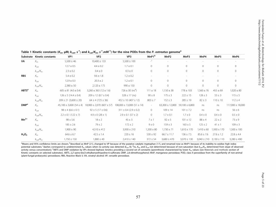
Table 1 Kinetic constants (Km, μM; kcat, s-1; and kcat/Km, s
-1.mM-1) for the nine PODs from the P. ostreatus genomea
Substrate Kinetic constants VP1 VP2 VP3 MnP1b MnP2 MnP3 MnP4 MnP5 MnP6
VA Km 5,500 ± 46 10,400 ± 133 5,500 ± 100 -c - - - - -
kcat 12.7 ± 0.5 4.4 ± 0.2 1.7 ± 0.1 0 0 0 0 0 0
kcat/Km 2.3 ± 0.2 0.4 ± 0 0.3 ± 0 0 0 0 0 0 0
RB5 Km 5.4 ± 0.2 9.6 ± 1.8 1.2 ± 0.2 - - - - - -
kcat 12.9 ± 0.3 20.3 ± 2 1.2 ± 0.1 0 0 0 0 0 0
kcat/Km 2,380 ± 50 2,120 ± 175 990 ± 150 0 0 0 0 0 0
ABTSe Km 605 ± 81 (4.0 ± 0.4) 3,260 ± 363 (12 ± 1.6) 726 ± 30 (nsd) 111 ± 18 1,150 ± 38 778 ± 103 1,560 ± 76 455 ± 69 1,020 ± 80
kcat 126 ± 5 (14.4 ± 0.4) 209 ± 12 (8.7 ± 0.4) 328 ± 17 (ns) 90 ± 8 175 ± 3 222 ± 15 128 ± 3 53 ± 3 115 ± 5
kcat/Km 209 ± 21 (3,600 ± 20) 64 ± 4 (725 ± 36) 452 ± 10 (407 ± 12) 803 ± 7 152 ± 3 285 ± 19 82 ± 3 110 ± 10 112 ± 4
DMPe Km 45,100 ± 3,600 (54 ± 4) 18,900 ± 2,070 (607 ± 57) 108,000 ± 13,000 (51 ± 14) - 63,300 ± 12,800 59,100 ± 6,800 ns ns 117,000 ± 18,000
kcat 98 ± 4 (6.6 ± 0.1) 92 ± 5 (17 ± 0.6) 311 ± 0.4 (2.9 ± 0.2) 0 109 ± 14 101 ± 7.2 ns ns 56 ± 6
kcat/Km 2.2 ± 0.1 (122 ± 7) 4.9 ± 0 (28 ± 1) 2.9 ± 0.1 (57 ± 2) 0 1.7 ± 0.1 1.7 ± 0 0.4 ± 0 0.4 ± 0 0.5 ± 0
Mn2+ Km 98 ± 5.6 18 ± 2 45 ± 3 7 ± 1 92 ± 5 101 ± 12 88 ± 4 22 ± 2 73 ± 9
kcat 185 ± 2.6 79 ± 2 172 ± 2 9 ± 0 159 ± 3 163 ± 5 125 ± 2 41 ± 1 109 ± 3
kcat/Km 1,900 ± 90 4,510 ± 412 3,930 ± 210 1,200 ± 80 1,730 ± 71 1,610 ± 170 1,410 ± 60 1,930 ± 170 1,500 ± 100
H2O2 Km 64.6 ± 6.7 42.5 ± 1.4 220 ± 16 530 ± 92 66.7 ± 11.7 136 ± 7.5 85.6 ± 7.6 27.6 ± 1.2 22.8 ± 4.4
kcat/Km 1,750 ± 150 1,880 ± 40 2,410 ± 140 372 ± 54 3,680 ± 470 3,070 ± 130 3,040 ± 210 3,130 ± 110 3,280 ± 490aMeans and 95% confidence limits are shown; bdescribed as MnP [21], changed to VP because of the putative catalytic tryptophan [17], and renamed now as MnP1 because of its inability to oxidize high redoxpotential substrates; cdashes correspond to undetermined Km values when no activity was detected (kcat 0);
dns, Km, and kcat not determined because of non-saturation (but kcat/Km determined from slope of observedactivity versus concentration); eABTS and DMP oxidation by VPs showed biphasic kinetics providing a second set of constants (parenthesis) characterized by lower Km values (see Materials and methods section ofKinetic constants on selected substrates). ABTS, 2,2′-azino-bis(3-ethylbenzothiazoline-6-sulfonate; DMP, 2,6-dimethoxyphenol; MnP, manganese peroxidase; POD, class II peroxidase from the superfamily of non-animal(plant-fungal-prokaryotic) peroxidases; RB5, Reactive Black 5; VA, veratryl alcohol; VP, versatile peroxidase.
Fernández-Fueyoet
al.Biotechnologyfor
Biofuels2014,7:2
Page5of
23http://w
ww.biotechnologyforbiofuels.com
/content/7/1/2
57
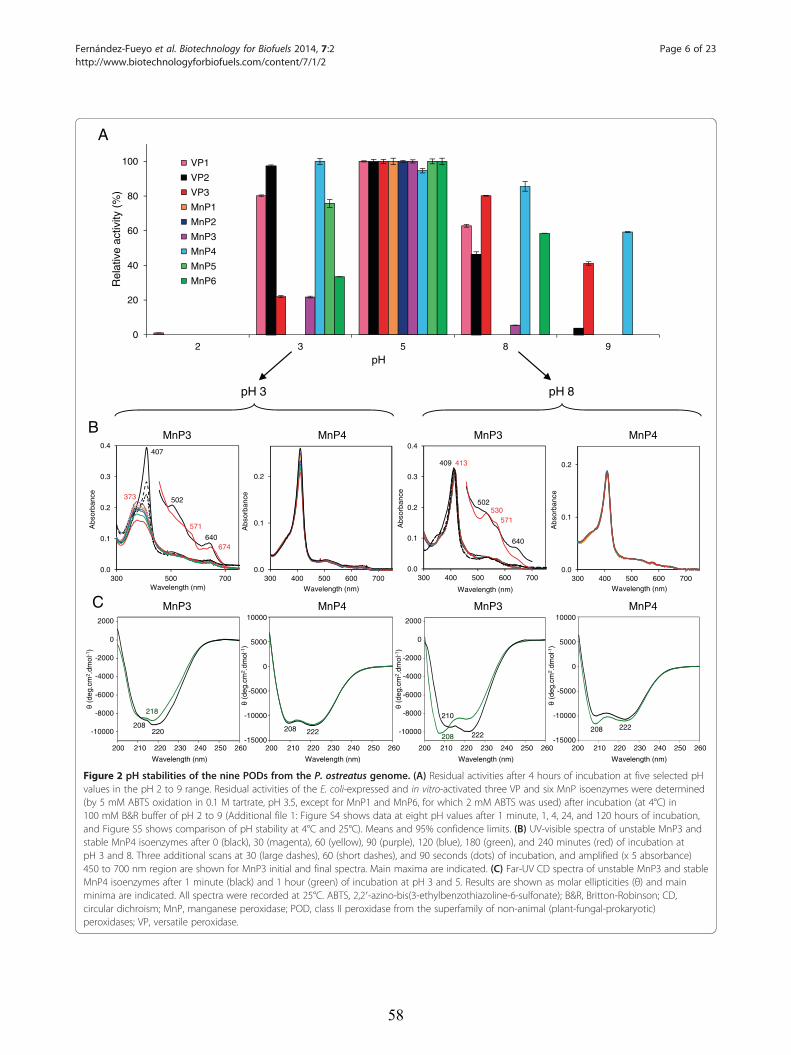
0.0
0.1
0.2
0.3
0.4
300 400 500 600 700
Wavelength (nm)
409
502
640
413
530571
0.0
0.1
0.2
300 400 500 600 700
Abs
orba
nce
Wavelength (nm)
0.0
0.1
0.2
0.3
0.4
300 500 700
Abs
orba
nce
Wavelength (nm)
407
502
640
373
571
674
0.0
0.1
0.2
300 400 500 600 700
Abs
orba
nce
Wavelength (nm)
Abs
orba
nce
-10000
-8000
-6000
-4000
-2000
0
2000
-15000
-10000
-5000
0
5000
10000
-10000
-8000
-6000
-4000
-2000
0
2000
-15000
-10000
-5000
0
5000
10000
200 210 220 230 240 250 260
Wavelength (nm)
2000
0
-2000
-4000
-6000
-8000
-10000
(deg
.cm
2 .dm
ol-1
)
200 210 220 230 240 250 260
Wavelength (nm)
200 210 220 230 240 250 260
Wavelength (nm)
200 210 220 230 240 250 260
Wavelength (nm)
2000
0
-2000
-4000
-6000
-8000
-10000
(deg
.cm
2 .dm
ol-1
)
10000
5000
0
-5000
-10000
-15000
(deg
.cm
2 .dm
ol-1
)
10000
5000
0
-5000
-10000
-15000
(deg
.cm
2 .dm
ol-1
)
208220 208 222 222
208 222
0
20
40
60
80
100
2 3 5 8 9
VP1
VP2
VP3
MnP1
MnP2
MnP3
MnP4
MnP5
MnP6 Rel
ativ
e ac
tivity
(%
)
pH
pH 3
B MnP4MnP3
pH 8
MnP4MnP3
A
C
208
210218
MnP4MnP3 MnP4MnP3
Figure 2 pH stabilities of the nine PODs from the P. ostreatus genome. (A) Residual activities after 4 hours of incubation at five selected pHvalues in the pH 2 to 9 range. Residual activities of the E. coli-expressed and in vitro-activated three VP and six MnP isoenzymes were determined(by 5 mM ABTS oxidation in 0.1 M tartrate, pH 3.5, except for MnP1 and MnP6, for which 2 mM ABTS was used) after incubation (at 4°C) in100 mM B&R buffer of pH 2 to 9 (Additional file 1: Figure S4 shows data at eight pH values after 1 minute, 1, 4, 24, and 120 hours of incubation,and Figure S5 shows comparison of pH stability at 4°C and 25°C). Means and 95% confidence limits. (B) UV-visible spectra of unstable MnP3 andstable MnP4 isoenzymes after 0 (black), 30 (magenta), 60 (yellow), 90 (purple), 120 (blue), 180 (green), and 240 minutes (red) of incubation atpH 3 and 8. Three additional scans at 30 (large dashes), 60 (short dashes), and 90 seconds (dots) of incubation, and amplified (x 5 absorbance)450 to 700 nm region are shown for MnP3 initial and final spectra. Main maxima are indicated. (C) Far-UV CD spectra of unstable MnP3 and stableMnP4 isoenzymes after 1 minute (black) and 1 hour (green) of incubation at pH 3 and 5. Results are shown as molar ellipticities (θ) and mainminima are indicated. All spectra were recorded at 25°C. ABTS, 2,2′-azino-bis(3-ethylbenzothiazoline-6-sulfonate); B&R, Britton-Robinson; CD,circular dichroism; MnP, manganese peroxidase; POD, class II peroxidase from the superfamily of non-animal (plant-fungal-prokaryotic)peroxidases; VP, versatile peroxidase.
Fernández-Fueyo et al. Biotechnology for Biofuels 2014, 7:2 Page 6 of 23http://www.biotechnologyforbiofuels.com/content/7/1/2
58
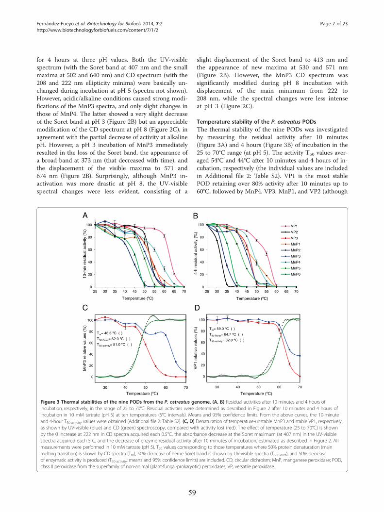
Fernández-Fueyo et al. Biotechnology for Biofuels 2014, 7:2 Page 7 of 23http://www.biotechnologyforbiofuels.com/content/7/1/2
for 4 hours at three pH values. Both the UV-visiblespectrum (with the Soret band at 407 nm and the smallmaxima at 502 and 640 nm) and CD spectrum (with the208 and 222 nm ellipticity minima) were basically un-changed during incubation at pH 5 (spectra not shown).However, acidic/alkaline conditions caused strong modi-fications of the MnP3 spectra, and only slight changes inthose of MnP4. The latter showed a very slight decreaseof the Soret band at pH 3 (Figure 2B) but an appreciablemodification of the CD spectrum at pH 8 (Figure 2C), inagreement with the partial decrease of activity at alkalinepH. However, a pH 3 incubation of MnP3 immediatelyresulted in the loss of the Soret band, the appearance ofa broad band at 373 nm (that decreased with time), andthe displacement of the visible maxima to 571 and674 nm (Figure 2B). Surprisingly, although MnP3 in-activation was more drastic at pH 8, the UV-visiblespectral changes were less evident, consisting of a
0
20
40
60
80
100
25 30 35 40 45 50 55 60 65 70
30 40 50 60 70
0
20
40
60
80
100
Tm= 46.6 ºC ( )
T50-Soret= 62.0 ºC ( )
T50-activity= 51.0 ºC ( )
Temperature (ºC)
MnP
3 re
lativ
e va
lues
(%
)
DCTemperature ( C)
10-m
in r
esid
ual a
ctiv
ity (
%)
A B
Figure 3 Thermal stabilities of the nine PODs from the P. ostreatus geincubation, respectively, in the range of 25 to 70°C. Residual activities wereincubation in 10 mM tartrate (pH 5) at ten temperatures (5°C intervals). Meaand 4-hour T50-activity values were obtained (Additional file 2: Table S2). (C, D)as shown by UV-visible (blue) and CD (green) spectroscopy, compared witby the θ increase at 222 nm in CD spectra acquired each 0.5°C, the absorbspectra acquired each 5°C, and the decrease of enzyme residual activity afmeasurements were performed in 10 mM tartrate (pH 5). T50 values corresponmelting transition) is shown by CD spectra (Tm), 50% decrease of heme Soretof enzymatic activity is produced (T50-activity; means and 95% confidence limitsclass II peroxidase from the superfamily of non-animal (plant-fungal-prokaryot
59
slight displacement of the Soret band to 413 nm andthe appearance of new maxima at 530 and 571 nm(Figure 2B). However, the MnP3 CD spectrum wassignificantly modified during pH 8 incubation withdisplacement of the main minimum from 222 to208 nm, while the spectral changes were less intenseat pH 3 (Figure 2C).
Temperature stability of the P. ostreatus PODsThe thermal stability of the nine PODs was investigatedby measuring the residual activity after 10 minutes(Figure 3A) and 4 hours (Figure 3B) of incubation in the25 to 70°C range (at pH 5). The activity T50 values aver-aged 54°C and 44°C after 10 minutes and 4 hours of in-cubation, respectively (the individual values are includedin Additional file 2: Table S2). VP1 is the most stablePOD retaining over 80% activity after 10 minutes up to60°C, followed by MnP4, VP3, MnP1, and VP2 (although
0
20
40
60
80
100
25 30 35 40 45 50 55 60 65 70
VP1
VP2
VP3
MnP1
MnP2
MnP3
MnP4
MnP5
MnP6
30 40 50 60 70
0
20
40
60
80
100
Tm= 59.0 ºC ( )
T50-Soret= 64,7 ºC ( )
T50-activity= 62.8 ºC ( )
Temperature (ºC)
VP
1 re
lativ
e va
lues
(%
)
Temperature (ºC)
4-h
resi
dual
act
ivity
(%
)
nome. (A, B) Residual activities after 10 minutes and 4 hours ofdetermined as described in Figure 2 after 10 minutes and 4 hours ofns and 95% confidence limits. From the above curves, the 10-minuteDenaturation of temperature-unstable MnP3 and stable VP1, respectively,h activity lost (red). The effect of temperature (25 to 70°C) is shownance decrease at the Soret maximum (at 407 nm) in the UV-visibleter 10 minutes of incubation, estimated as described in Figure 2. Allding to those temperatures where 50% protein denaturation (mainband is shown by UV-visible spectra (T50-Soret), and 50% decrease) are included. CD, circular dichroism; MnP, manganese peroxidase; POD,ic) peroxidases; VP, versatile peroxidase.
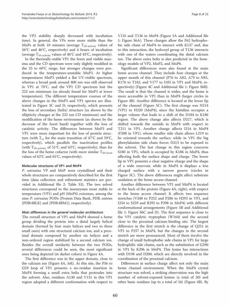
Fernández-Fueyo et al. Biotechnology for Biofuels 2014, 7:2 Page 8 of 23http://www.biotechnologyforbiofuels.com/content/7/1/2
the VP2 stability sharply decreased with incubationtime). In general, the VPs were more stable than theMnPs at both 10 minutes (average T50-activity values of56°C and 46°C, respectively) and 4 hours of incubation(average T50-activity values of 46°C and 43°C, respectively).In the thermally-stable VP1 the Soret and visible max-
ima and the CD spectrum were only slightly modified inthe 25 to 60°C range, but stronger changes were pro-duced in the temperature-unstable MnP3. At highertemperatures MnP3 yielded a flat UV-visible spectrum,whereas a broad peak around 400 nm was still observedin VP1 at 70°C, and the VP1 CD spectrum lost the222 nm minimum (as already found for MnP3 at lowertemperature). The different temperature courses of theabove changes in the MnP3 and VP1 spectra are illus-trated in Figure 3C and D, respectively, which presentsthe loss of secondary (helix) structure (as shown by theellipticity changes at the 222 nm CD minimum) and themodification of the heme environment (as shown by thedecrease of the Soret band) together with the loss ofcatalytic activity. The differences between MnP3 andVP1 were more important for the loss of protein struc-ture (with Tm for the main transition of 47°C and 59°C,respectively), which parallels the inactivation profiles(with T50-activity of 51°C and 63°C, respectively), than forthe loss of the heme cofactor (with more similar T50-Soret
values of 62°C and 65°C, respectively).
Molecular structures of VP1 and MnP4P. ostreatus VP and MnP were crystallized and theirwhole structures are comparatively described for the firsttime (data collection and refinement statistics are pro-vided in Additional file 2: Table S3). The two solvedstructures correspond to the isoenzymes most stable totemperature (VP1) and pH (MnP4) extremes, among thenine P. ostreatus PODs (Protein Data Bank, PDB, entries[PDB:4BLK] and [PDB:4BM1], respectively).
Main differences in the general molecular architectureThe overall structure of VP1 and MnP4 showed a hemegroup dividing the protein into a distal largely helicaldomain (formed by four main helices and two to threesmall ones) with one structural calcium ion, and a prox-imal domain composed by another six helices and anon-ordered region stabilized by a second calcium ion.Besides the overall similarity between the two PODs,several differences could be seen, the most significantones being depicted (in darker colors) in Figure 4A.The first difference was in the upper domain, close to
the calcium ion (Figure 4A, left). At this site, the P56 toG59 loop of VP1 presents a six-residue insertion inMnP4 forming a small extra helix that protrudes intothe solvent. Also, residues A130 and V131 in this VP1region adopted a different conformation with respect to
60
V135 and T136 in MnP4 (Figure 5A and Additional file1: Figure S6A). These changes allow the F62 hydropho-bic side chain of MnP4 to interact with K137 and, dueto this interaction, the hydroxyl group of T136 interactswith one of the waters coordinating the distal calciumion. The above extra helix is also predicted in the hom-ology models of VP2, MnP2, and MnP6.Significant differences were also found at the main
heme access channel. They include four changes at theupper mouth of this channel (P76 to A82, A79 to N85,K176 to T182, and V177 to I183 in VP1 and MnP4, re-spectively) (Figure 4C and Additional file 1: Figure S6B).The result is that the channel is wider, and the heme ismore accessible in VP1 than in MnP4 (larger circles inFigure 4B). Another difference is located at the lower lipof the channel (Figure 5C). The first change was N214(VP1) to H220 (MnP4), since the histidine occupies alarger volume that leads to a shift of the D184 to K186region. The above change also affects D217, which isshifted towards the outside in MnP4 with respect toT211 in VP1. Another change affects I214 in MnP4(F208 in VP1), whose smaller side chain allows L219 tobe oriented towards the surface, while in VP1 the bigphenylalanine side chain forces D213 to be exposed tothe solvent. The last change in this region concernsS180 in VP1, which is occupied by K186 in MnP4, thusaffecting both the surface shape and charge. The lowerlip in VP1 presents a clear negative charge and the shapeof a wide reservoir, while in MnP4 it displays a lesscharged surface with a narrow groove (circles inFigure 5C). The above differences might affect substrateoxidation at the heme access channel.Another difference between VP1 and MnP4 is located
at the back of the protein (Figure 4A, right), with respectto the heme access channel. It affects two sequencestretches (V248 to P252 and P286 to H293 in VP1, andI254 to S259 and R292 to P298 in MnP4) with differentconformational arrangements (Figure 5B and Additionalfile 1: Figure S6C and D). The first sequence is close tothe VP1 catalytic tryptophan (W164) and the secondclose to the proximal calcium-binding site. The largestdifference in the first stretch is the change of Q251 inVP1 to P257 in MnP4, but the changes in the secondstretch are more pronounced. Most of them involve thechange of small hydrophobic side chains in VP1 for largehydrophilic side chains, such as the substitution of G290in VP1 by K296 in MnP4. This lysine has interactionswith D198 and D200, which are directly involved in thecoordination of the proximal calcium.Differences in surface charge affect not only the main
heme channel environment. When the MnP4 crystalstructure was solved, a striking observation was the highnumber of solvent-exposed lysines (a total of 20) andother basic residues (up to a total of 34) (Figure 4B). By
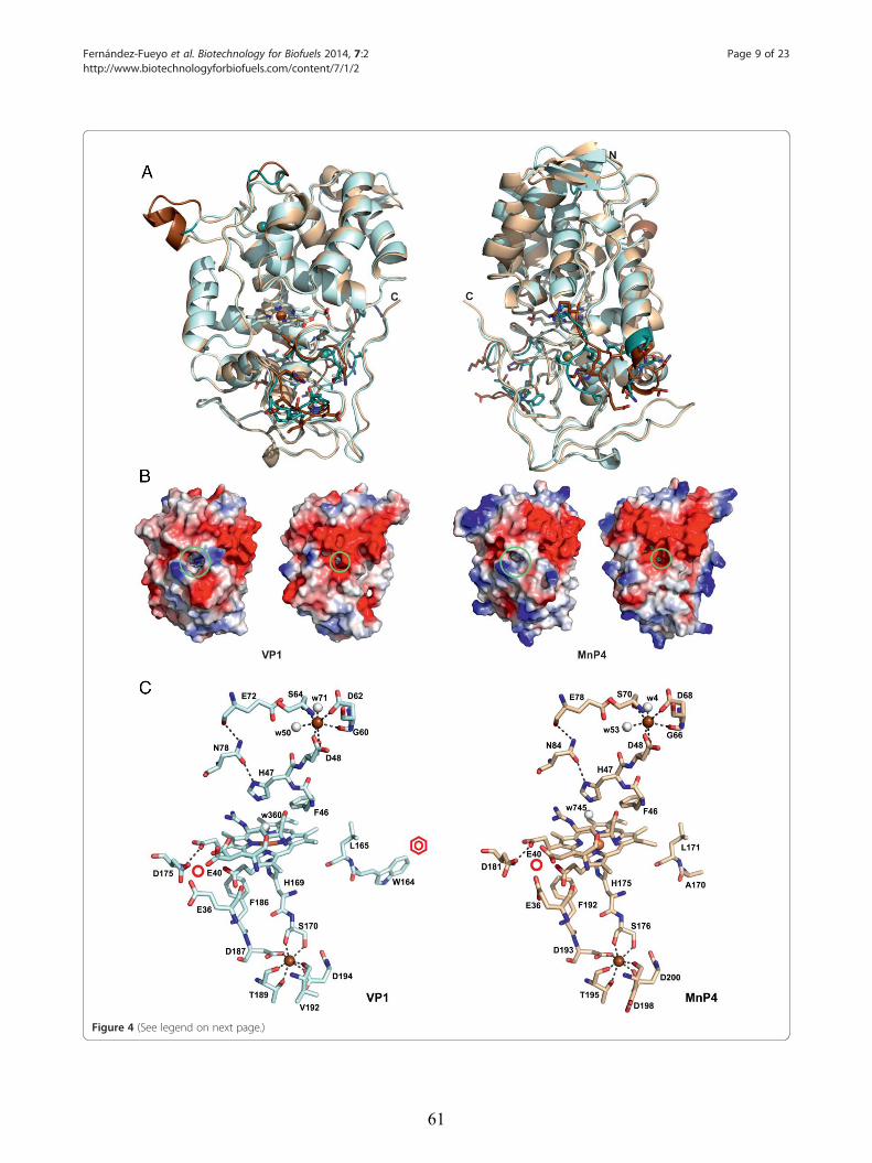
Figure 4 (See legend on next page.)
Fernández-Fueyo et al. Biotechnology for Biofuels 2014, 7:2 Page 9 of 23http://www.biotechnologyforbiofuels.com/content/7/1/2
61
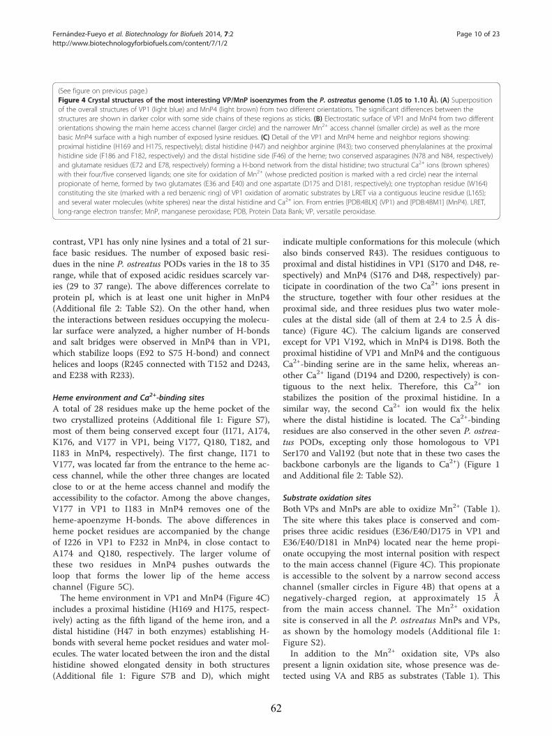
(See figure on previous page.)Figure 4 Crystal structures of the most interesting VP/MnP isoenzymes from the P. ostreatus genome (1.05 to 1.10 Å). (A) Superpositionof the overall structures of VP1 (light blue) and MnP4 (light brown) from two different orientations. The significant differences between thestructures are shown in darker color with some side chains of these regions as sticks. (B) Electrostatic surface of VP1 and MnP4 from two differentorientations showing the main heme access channel (larger circle) and the narrower Mn2+ access channel (smaller circle) as well as the morebasic MnP4 surface with a high number of exposed lysine residues. (C) Detail of the VP1 and MnP4 heme and neighbor regions showing:proximal histidine (H169 and H175, respectively); distal histidine (H47) and neighbor arginine (R43); two conserved phenylalanines at the proximalhistidine side (F186 and F182, respectively) and the distal histidine side (F46) of the heme; two conserved asparagines (N78 and N84, respectively)and glutamate residues (E72 and E78, respectively) forming a H-bond network from the distal histidine; two structural Ca2+ ions (brown spheres)with their four/five conserved ligands; one site for oxidation of Mn2+ (whose predicted position is marked with a red circle) near the internalpropionate of heme, formed by two glutamates (E36 and E40) and one aspartate (D175 and D181, respectively); one tryptophan residue (W164)constituting the site (marked with a red benzenic ring) of VP1 oxidation of aromatic substrates by LRET via a contiguous leucine residue (L165);and several water molecules (white spheres) near the distal histidine and Ca2+ ion. From entries [PDB:4BLK] (VP1) and [PDB:4BM1] (MnP4). LRET,long-range electron transfer; MnP, manganese peroxidase; PDB, Protein Data Bank; VP, versatile peroxidase.
Fernández-Fueyo et al. Biotechnology for Biofuels 2014, 7:2 Page 10 of 23http://www.biotechnologyforbiofuels.com/content/7/1/2
contrast, VP1 has only nine lysines and a total of 21 sur-face basic residues. The number of exposed basic resi-dues in the nine P. ostreatus PODs varies in the 18 to 35range, while that of exposed acidic residues scarcely var-ies (29 to 37 range). The above differences correlate toprotein pI, which is at least one unit higher in MnP4(Additional file 2: Table S2). On the other hand, whenthe interactions between residues occupying the molecu-lar surface were analyzed, a higher number of H-bondsand salt bridges were observed in MnP4 than in VP1,which stabilize loops (E92 to S75 H-bond) and connecthelices and loops (R245 connected with T152 and D243,and E238 with R233).
Heme environment and Ca2+-binding sitesA total of 28 residues make up the heme pocket of thetwo crystallized proteins (Additional file 1: Figure S7),most of them being conserved except four (I171, A174,K176, and V177 in VP1, being V177, Q180, T182, andI183 in MnP4, respectively). The first change, I171 toV177, was located far from the entrance to the heme ac-cess channel, while the other three changes are locatedclose to or at the heme access channel and modify theaccessibility to the cofactor. Among the above changes,V177 in VP1 to I183 in MnP4 removes one of theheme-apoenzyme H-bonds. The above differences inheme pocket residues are accompanied by the changeof I226 in VP1 to F232 in MnP4, in close contact toA174 and Q180, respectively. The larger volume ofthese two residues in MnP4 pushes outwards theloop that forms the lower lip of the heme accesschannel (Figure 5C).The heme environment in VP1 and MnP4 (Figure 4C)
includes a proximal histidine (H169 and H175, respect-ively) acting as the fifth ligand of the heme iron, and adistal histidine (H47 in both enzymes) establishing H-bonds with several heme pocket residues and water mol-ecules. The water located between the iron and the distalhistidine showed elongated density in both structures(Additional file 1: Figure S7B and D), which might
62
indicate multiple conformations for this molecule (whichalso binds conserved R43). The residues contiguous toproximal and distal histidines in VP1 (S170 and D48, re-spectively) and MnP4 (S176 and D48, respectively) par-ticipate in coordination of the two Ca2+ ions present inthe structure, together with four other residues at theproximal side, and three residues plus two water mole-cules at the distal side (all of them at 2.4 to 2.5 Å dis-tance) (Figure 4C). The calcium ligands are conservedexcept for VP1 V192, which in MnP4 is D198. Both theproximal histidine of VP1 and MnP4 and the contiguousCa2+-binding serine are in the same helix, whereas an-other Ca2+ ligand (D194 and D200, respectively) is con-tiguous to the next helix. Therefore, this Ca2+ ionstabilizes the position of the proximal histidine. In asimilar way, the second Ca2+ ion would fix the helixwhere the distal histidine is located. The Ca2+-bindingresidues are also conserved in the other seven P. ostrea-tus PODs, excepting only those homologous to VP1Ser170 and Val192 (but note that in these two cases thebackbone carbonyls are the ligands to Ca2+) (Figure 1and Additional file 2: Table S2).
Substrate oxidation sitesBoth VPs and MnPs are able to oxidize Mn2+ (Table 1).The site where this takes place is conserved and com-prises three acidic residues (E36/E40/D175 in VP1 andE36/E40/D181 in MnP4) located near the heme propi-onate occupying the most internal position with respectto the main access channel (Figure 4C). This propionateis accessible to the solvent by a narrow second accesschannel (smaller circles in Figure 4B) that opens at anegatively-charged region, at approximately 15 Åfrom the main access channel. The Mn2+ oxidationsite is conserved in all the P. ostreatus MnPs and VPs,as shown by the homology models (Additional file 1:Figure S2).In addition to the Mn2+ oxidation site, VPs also
present a lignin oxidation site, whose presence was de-tected using VA and RB5 as substrates (Table 1). This
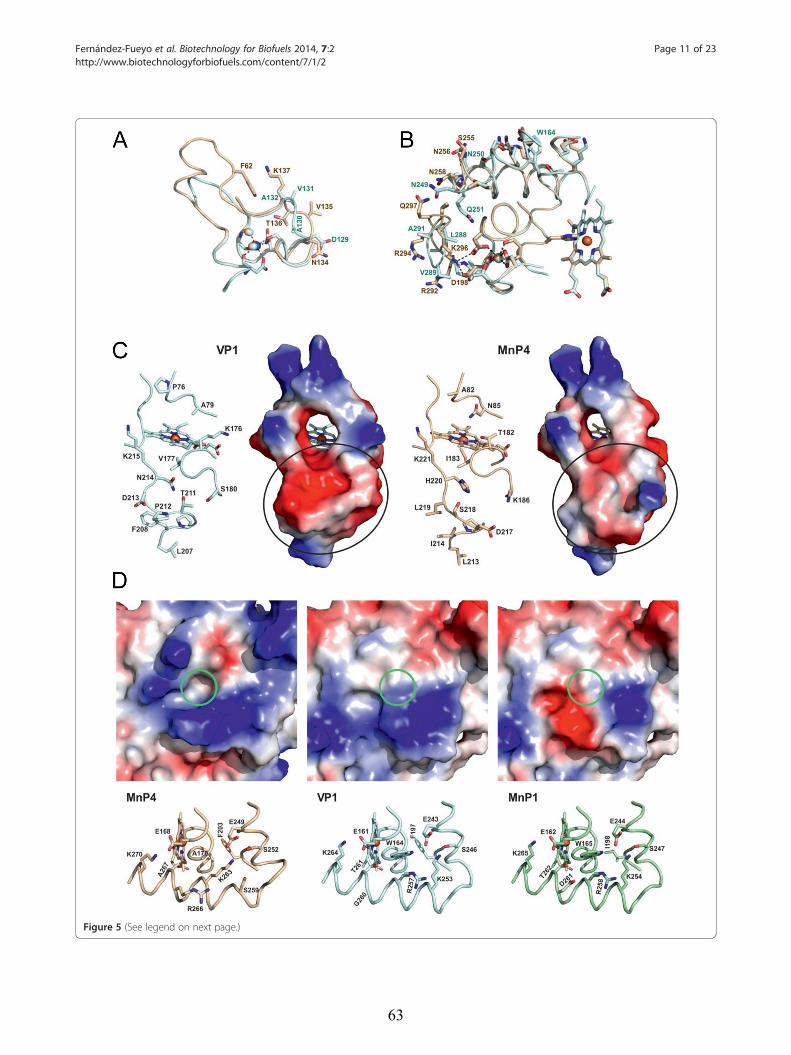
Figure 5 (See legend on next page.)
Fernández-Fueyo et al. Biotechnology for Biofuels 2014, 7:2 Page 11 of 23http://www.biotechnologyforbiofuels.com/content/7/1/2
63
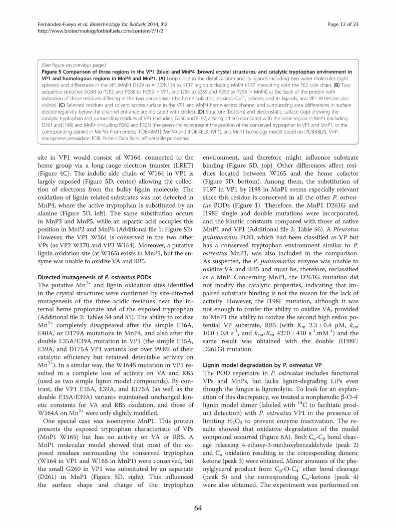
(See figure on previous page.)Figure 5 Comparison of three regions in the VP1 (blue) and MnP4 (brown) crystal structures; and catalytic tryptophan environment inVP1 and homologous regions in MnP4 and MnP1. (A) Loop close to the distal calcium and its ligands including two water molecules (lightspheres) and differences in the VP1/MnP4 D129 to A132/N134 to K137 region including MnP4 K137 interacting with the F62 side chain. (B) Twosequence stretches (V248 to P252 and P286 to H293 in VP1, and I254 to S259 and R292 to P298 in MnP4) at the back of the protein withindication of those residues differing in the two peroxidases (the heme cofactor, proximal Ca2+, spheres, and its ligands, and VP1 W164 are alsovisible). (C) Selected residues and solvent access surface in the VP1 and MnP4 heme access channel and surrounding area (differences in surfaceelectronegativity below the channel entrance are indicated with circles). (D) Structure (bottom) and electrostatic surface (top) showing thecatalytic tryptophan and surrounding residues of VP1 (including G260 and F197, among others) compared with the same region in MnP1 (includingD261 and I198) and MnP4 (including R266 and F203) (the green circles represent the position of the conserved tryptophan in VP1 and MnP1, or thecorresponding alanine in MnP4). From entries [PDB:4BM1] (MnP4) and [PDB:4BLK] (VP1), and MnP1 homology model based on [PDB:4BLK]. MnP,manganese peroxidase; PDB, Protein Data Bank; VP, versatile peroxidase.
Fernández-Fueyo et al. Biotechnology for Biofuels 2014, 7:2 Page 12 of 23http://www.biotechnologyforbiofuels.com/content/7/1/2
site in VP1 would consist of W164, connected to theheme group via a long-range electron transfer (LRET)(Figure 4C). The indolic side chain of W164 in VP1 islargely exposed (Figure 5D, center) allowing the collec-tion of electrons from the bulky lignin molecule. Theoxidation of lignin-related substrates was not detected inMnP4, where the active tryptophan is substituted by analanine (Figure 5D, left). The same substitution occursin MnP3 and MnP5, while an aspartic acid occupies thisposition in MnP2 and MnP6 (Additional file 1: Figure S2).However, the VP1 W164 is conserved in the two otherVPs (as VP2 W170 and VP3 W164). Moreover, a putativelignin oxidation site (at W165) exists in MnP1, but the en-zyme was unable to oxidize VA and RB5.
Directed mutagenesis of P. ostreatus PODsThe putative Mn2+ and lignin oxidation sites identifiedin the crystal structures were confirmed by site-directedmutagenesis of the three acidic residues near the in-ternal heme propionate and of the exposed tryptophan(Additional file 2: Tables S4 and S5). The ability to oxidizeMn2+ completely disappeared after the simple E36A,E40A, or D179A mutations in MnP4, and also after thedouble E35A/E39A mutation in VP1 (the simple E35A,E39A, and D175A VP1 variants lost over 99.8% of theircatalytic efficiency but retained detectable activity onMn2+). In a similar way, the W164S mutation in VP1 re-sulted in a complete loss of activity on VA and RB5(used as two simple lignin model compounds). By con-trast, the VP1 E35A, E39A, and E175A (as well as thedouble E35A/E39A) variants maintained unchanged kin-etic constants for VA and RB5 oxidation, and those ofW164A on Mn2+ were only slightly modified.One special case was isoenzyme MnP1. This protein
presents the exposed tryptophan characteristic of VPs(MnP1 W165) but has no activity on VA or RB5. AMnP1 molecular model showed that most of the ex-posed residues surrounding the conserved tryptophan(W164 in VP1 and W165 in MnP1) were conserved, butthe small G260 in VP1 was substituted by an aspartate(D261) in MnP1 (Figure 5D, right). This influencedthe surface shape and charge of the tryptophan
64
environment, and therefore might influence substratebinding (Figure 5D, top). Other differences affect resi-dues located between W165 and the heme cofactor(Figure 5D, bottom). Among them, the substitution ofF197 in VP1 by I198 in MnP1 seems especially relevantsince this residue is conserved in all the other P. ostrea-tus PODs (Figure 1). Therefore, the MnP1 D261G andI198F single and double mutations were incorporated,and the kinetic constants compared with those of nativeMnP1 and VP1 (Additional file 2: Table S6). A Pleurotuspulmonarius POD, which had been classified as VP buthas a conserved tryptophan environment similar to P.ostreatus MnP1, was also included in the comparison.As suspected, the P. pulmonarius enzyme was unable tooxidize VA and RB5 and must be, therefore, reclassifiedas a MnP. Concerning MnP1, the D261G mutation didnot modify the catalytic properties, indicating that im-paired substrate binding is not the reason for the lack ofactivity. However, the I198F mutation, although it wasnot enough to confer the ability to oxidize VA, providedto MnP1 the ability to oxidize the second high redox po-tential VP substrate, RB5 (with Km 2.3 ± 0.4 μM, kcat10.0 ± 0.8 s-1, and kcat/Km 4270 ± 410 s-1.mM-1) and thesame result was obtained with the double (I198F/D261G) mutation.
Lignin model degradation by P. ostreatus VPThe POD repertoire in P. ostreatus includes functionalVPs and MnPs, but lacks lignin-degrading LiPs eventhough the fungus is ligninolytic. To look for an explan-ation of this discrepancy, we treated a nonphenolic β-O-4’lignin model dimer (labeled with 14C to facilitate prod-uct detection) with P. ostreatus VP1 in the presence oflimiting H2O2 to prevent enzyme inactivation. The re-sults showed that oxidative degradation of the modelcompound occurred (Figure 6A). Both Cα-Cβ bond cleav-age releasing 4-ethoxy-3-methoxybenzaldehyde (peak 2)and Cα oxidation resulting in the corresponding dimericketone (peak 3) were obtained. Minor amounts of the phe-nylglycerol product from Cβ-O-C4’ ether bond cleavage(peak 5) and the corresponding Cα-ketone (peak 4)were also obtained. The experiment was performed on
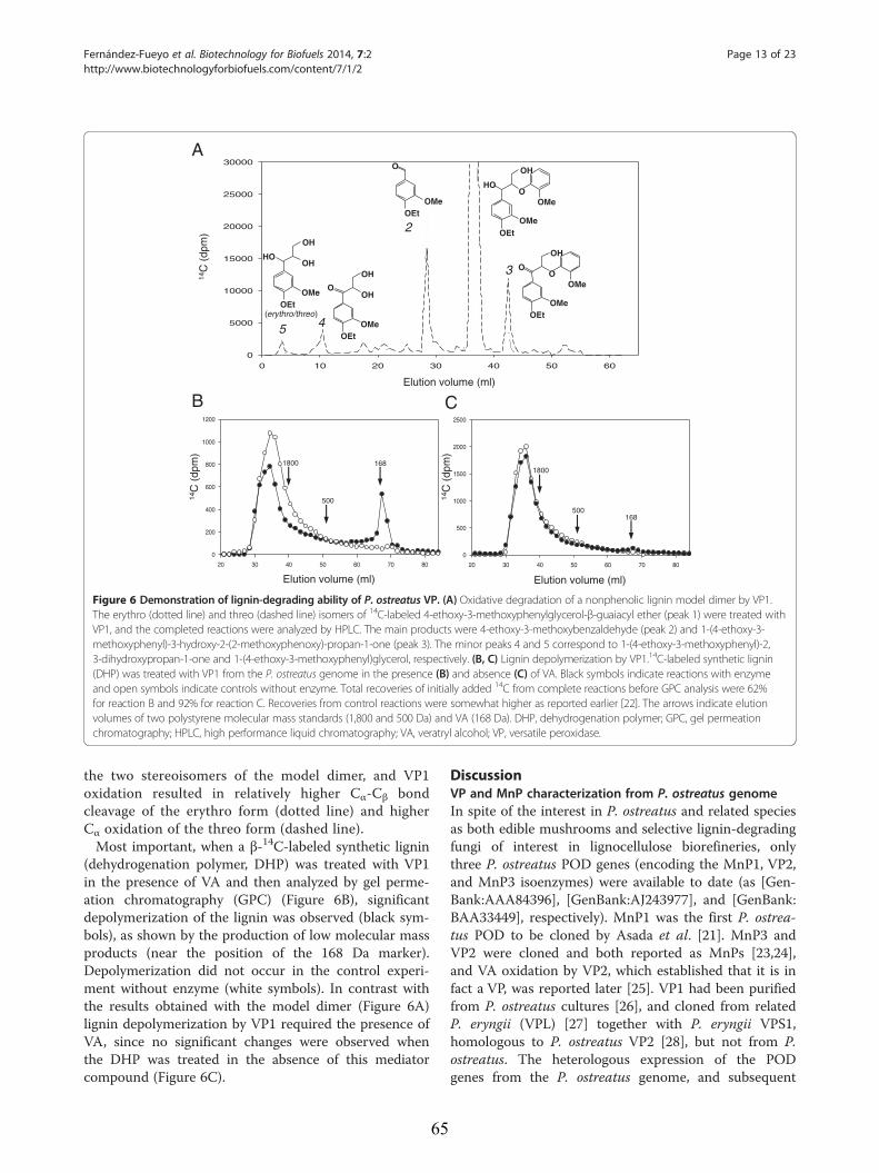
20 30 40 50 60 70 80
0
200
400
600
800
1000
1200
20 30 40 50 60 70 80
0
500
1000
1500
2000
2500
Elution volume (ml)
14C
(dp
m)
168
500
1800
B
Elution volume (ml)
14C
(dp
m)
168500
1800
C
0 10 20 30 40 50 600
5000
10000
15000
20000
25000
30000
5 4
3
2
O
OMeOEt
OH
OMeO
O
OMeOEt
HO
OMeOEt
OH
OH
O
OMeOEt
OH
OH
HO
OMeOEt
OH
OMeO
(erythro/threo)
Elution volume (ml)
14C
(dp
m)
A
Figure 6 Demonstration of lignin-degrading ability of P. ostreatus VP. (A) Oxidative degradation of a nonphenolic lignin model dimer by VP1.The erythro (dotted line) and threo (dashed line) isomers of 14C-labeled 4-ethoxy-3-methoxyphenylglycerol-β-guaiacyl ether (peak 1) were treated withVP1, and the completed reactions were analyzed by HPLC. The main products were 4-ethoxy-3-methoxybenzaldehyde (peak 2) and 1-(4-ethoxy-3-methoxyphenyl)-3-hydroxy-2-(2-methoxyphenoxy)-propan-1-one (peak 3). The minor peaks 4 and 5 correspond to 1-(4-ethoxy-3-methoxyphenyl)-2,3-dihydroxypropan-1-one and 1-(4-ethoxy-3-methoxyphenyl)glycerol, respectively. (B, C) Lignin depolymerization by VP1.14C-labeled synthetic lignin(DHP) was treated with VP1 from the P. ostreatus genome in the presence (B) and absence (C) of VA. Black symbols indicate reactions with enzymeand open symbols indicate controls without enzyme. Total recoveries of initially added 14C from complete reactions before GPC analysis were 62%for reaction B and 92% for reaction C. Recoveries from control reactions were somewhat higher as reported earlier [22]. The arrows indicate elutionvolumes of two polystyrene molecular mass standards (1,800 and 500 Da) and VA (168 Da). DHP, dehydrogenation polymer; GPC, gel permeationchromatography; HPLC, high performance liquid chromatography; VA, veratryl alcohol; VP, versatile peroxidase.
Fernández-Fueyo et al. Biotechnology for Biofuels 2014, 7:2 Page 13 of 23http://www.biotechnologyforbiofuels.com/content/7/1/2
the two stereoisomers of the model dimer, and VP1oxidation resulted in relatively higher Cα-Cβ bondcleavage of the erythro form (dotted line) and higherCα oxidation of the threo form (dashed line).Most important, when a β-14C-labeled synthetic lignin
(dehydrogenation polymer, DHP) was treated with VP1in the presence of VA and then analyzed by gel perme-ation chromatography (GPC) (Figure 6B), significantdepolymerization of the lignin was observed (black sym-bols), as shown by the production of low molecular massproducts (near the position of the 168 Da marker).Depolymerization did not occur in the control experi-ment without enzyme (white symbols). In contrast withthe results obtained with the model dimer (Figure 6A)lignin depolymerization by VP1 required the presence ofVA, since no significant changes were observed whenthe DHP was treated in the absence of this mediatorcompound (Figure 6C).
65
DiscussionVP and MnP characterization from P. ostreatus genomeIn spite of the interest in P. ostreatus and related speciesas both edible mushrooms and selective lignin-degradingfungi of interest in lignocellulose biorefineries, onlythree P. ostreatus POD genes (encoding the MnP1, VP2,and MnP3 isoenzymes) were available to date (as [Gen-Bank:AAA84396], [GenBank:AJ243977], and [GenBank:BAA33449], respectively). MnP1 was the first P. ostrea-tus POD to be cloned by Asada et al. [21]. MnP3 andVP2 were cloned and both reported as MnPs [23,24],and VA oxidation by VP2, which established that it is infact a VP, was reported later [25]. VP1 had been purifiedfrom P. ostreatus cultures [26], and cloned from relatedP. eryngii (VPL) [27] together with P. eryngii VPS1,homologous to P. ostreatus VP2 [28], but not from P.ostreatus. The heterologous expression of the PODgenes from the P. ostreatus genome, and subsequent
Fernández-Fueyo et al. Biotechnology for Biofuels 2014, 7:2 Page 14 of 23http://www.biotechnologyforbiofuels.com/content/7/1/2
kinetic studies, reveal a peroxidase array consisting ofthree VPs and six MnPs. In all but one (isoenzymeMnP1) the catalytic properties of the purified enzymesagree with the presence in the homology models of a pu-tative Mn2+ oxidation site, and for the VPs they agreewith the presence in the models of an exposed trypto-phan likely responsible for oxidation of high redox po-tential substrates and lignin [29,30].The P. ostreatus VPs are highly efficient at oxidizing
Mn2+ and RB5, and their lower catalytic efficiency onVA is related to the low apparent affinity for this sub-strate. The low redox potential substrate ABTS is alsooxidized with high efficiency, but this is not the case forDMP. The kinetic constants obtained are similar tothose reported for P. eryngii and Bjerkandera adusta VPon the same substrates [31]. The P. ostreatus VPs notonly show wider substrate specificity than MnPs, but theircatalytic efficiencies on Mn2+ are also higher, which hasnot been previously reported. The Mn2+-independent ac-tivity of VP on phenols and dyes can be related to the ex-istence of two more catalytic sites in addition to the Mn2+
oxidation site. The first site, characterized by its high cata-lytic efficiency, corresponds to the same exposed trypto-phan involved in VA/RB5 oxidation as shown by directedmutagenesis, while the second site has been assigned tothe main heme channel [32]. In contrast with the bestknown MnPs from P. chrysosporium, whose activity is al-ways mediated by Mn3+ [33], the P. ostreatus MnPs alsohave Mn-independent activity on ABTS and (exceptMnP1) on DMP. This activity has already been reportedfor P. ostreatus MnP3 [23] and for Agrocybe praecox MnP[34], and is expected to be present in many other hypo-thetical MnPs reported from genomes as members of anew subfamily of short MnPs [16], which would also in-clude all the P. ostreatus MnPs.Concerning MnP1, previously classified as a VP [17],
some differences in the environment of the nonfunc-tional tryptophan (W165) are responsible for its inabilityto oxidize high redox potential substrates, as shown bythe I198F variant being able to oxidize RB5. This muta-tion introduced a phenylalanine residue that could beimportant for correct positioning of the neighboringproximal histidine (which modulates the redox potentialof the enzyme) and/or for LRET from W165 to the hemecofactor [18]. In addition to the presence of W165,MnP1 also has a close evolutionary relationship (Additionalfile 1: Figure S1) and the highest sequence identity(70%) with VP3 (for sequence identity between differentPODs, see Additional file 2: Table S7). It has been re-cently shown that MnPs represent an ancestral peroxid-ase type in Agaricomycetes from which VPs and LiPsoriginated [16]. Our results on P. ostreatus MnP1 (andthe above P. pulmonarius MnP) suggest a more compli-cated picture of POD evolution, where some MnP-type
66
peroxidases might derive secondarily from an ancestralVP-type enzyme.
VP and short MnP crystal structuresThe VP1 and MnP4 crystal structures were solved asrepresentative for the two POD families present in the P.ostreatus genome. Several aspects of the crystal structureof P. eryngii VP were discussed when studying the cata-lytic sites of this enzyme [29,32,35], although a compara-tive description of the whole VP structure was notreported. On the other hand, the molecular structure oflong MnP from P. chrysosporium has been reported atdifferent resolutions [36-38], but no crystal structure fora short MnP was available before the present study.As already described for P. eryngii VPL [29], the main
structural feature of the P. ostreatus VP1 crystal struc-ture is the combination of the Mn oxidation site ofMnPs [36] and the high redox potential substrate oxida-tion site of LiPs [39]. Differences in the side chain pos-ition of the Mn-binding E36 (in VP1 and MnP4) wouldbe related to the mobility of this residue at the oxidationsite entrance. On the other hand, the catalytic W164 en-vironment in VP1 is less acidic than the correspondingregion in P. chrysosporium LiP, the latter probably con-tributing to stabilize the VA cation radical [40]. A char-acteristic of ligninolytic peroxidases is their narrowheme access channel, which has been related to the needto protect the enzyme from inactivation by substrateradicals generated at the heme pocket [18,29]. However,one of the main characteristics of the VP1 structure,when compared with MnP4, is the broader main hemeaccess channel (whose lower lip has the shape of a widereservoir), which could be related to the reported exis-tence of a third substrate oxidation site in VP [32]. Thefact that only low redox potential substrates (and notVA) are oxidized at the main heme access channelcould explain the lack of enzyme inactivation in theseVP reactions.Concerning short MnP, whose crystal structure is now
solved for the first time (from P. ostreatus MnP4), itsmain structural difference from P. chrysosporium longMnP concerns the existence of a more exposed Mn oxi-dation site, due to the absence of the C-terminal exten-sion that in P. chrysosporium MnP (with a total of 357residues) is partially fixed by a fifth disulfide bridge.Whether this difference, which is used to define the newsubfamily of short MnPs from genomes [16], underliesthe low but significant Mn-independent activity (onABTS) exhibited by these and other short MnPs remainsto be confirmed. Other characteristics of the MnP4structure, such as the high number of exposed basic res-idues and H-bonds/salt-bridges, and the differences inthe environment of the two structural Ca2+ ions, are dis-cussed below in connection with the PODs’ pH stability.
Fernández-Fueyo et al. Biotechnology for Biofuels 2014, 7:2 Page 15 of 23http://www.biotechnologyforbiofuels.com/content/7/1/2
Catalytic properties of POD isoenzymesThe existence of multiple isoforms (isoenzymes) is awell-known phenomenon among degradative enzymessecreted by fungi, including peroxidases. LiP isoenzymeswere first isolated from P. chrysosporium cultures[41,42]. DNA cloning showed that some of them wereencoded by different genes [43], but post-translationalmodifications are at the origin of other isoforms [44].The number of POD ‘true isoenzymes’ increased whenmore LiP and MnP genes were cloned from other Poly-porales. Studies in Agaricales reported cloning of threePOD isoenzymes from P. ostreatus [21,23,24]. However,a correlation between cloned genes and purified isoen-zymes was not clearly established, and characterizationof the different isoenzymes was lacking with a few ex-ceptions [41,42,45]. Therefore, the number and charac-teristics of the POD isoenzymes produced by white-rotfungi remained an open matter to date.The availability of genomes provides evidence on the
large and widespread duplication of POD genes inwhite-rot basidiomycetes [12,15,17,46]. This availability,together with the possibility to optimize the in vitro acti-vation of E. coli-expressed PODs [47], enabled us to ob-tain the complete set of POD isoenzymes encoded bythe P. ostreatus genome for biochemical comparisons,an important complement to differential expressionstudies [48]. Substrate specificity was used for functionalclassification into the VP and MnP families. Moreover,kinetic constants revealed some quantitative differencesbetween the members (isoenzymes) from each family in-cluding: i) the higher efficiency of VP1 oxidizing aro-matics and dyes, and also its lower efficiency relative toVP2 and VP3 at oxidizing Mn2+; ii) the non-saturationkinetics exhibited by MnP3 and MnP4 in DMP oxida-tion, as compared with MnP3 to MnP6 (MnP1 had noactivity); and iii) the unusual properties of MnP1, includ-ing its higher ABTS oxidation efficiency (in the range ofVP isoenzymes), low H2O2 reaction efficiency, and in-ability to oxidize DMP. The unique properties of isoen-zyme MnP1 can be related to its separate evolutionaryorigin, whereas the structural/evolutionary basis of otherdifferences in catalytic properties are still to be deter-mined. However, the most interesting differences be-tween the P. ostreatus VP and MnP isoenzymes concernthe temperature and pH stability properties discussedbelow.
Different stabilities of the POD isoenzymesP. ostreatus is not a thermophilic fungus [49]. However,a comparison of the temperature stabilities of the nineP. ostreatus PODs reveals some thermostable isoen-zymes, as shown by T50-activity values that, after 10 mi-nutes of incubation, ranged from 53°C (VP2 and VP3) to63°C (VP1) in the case of VPs; and from 43°C (MnP6) to
67
57°C (MnP4) in the case of MnPs. These thermostabil-ities are significant in high redox potential peroxidases,which are generally isolated from wood-rotting mesophi-lic basidiomycetes. Among peroxidases, the mosttemperature- and pH-stable forms have been isolatedfrom palm species [50] but plant peroxidases lack thestructural adaptations enabling oxidation of recalcitrantaromatics. Moreover, the above VP and MnP T50-activity
ranges (10°C and 14°C, respectively) are notable for iso-enzymes from the same organism (and attain 23°C and16°C, respectively, after 4 hours of incubation). Some in-formation on the mechanisms of thermal stability/inacti-vation was provided by CD and UV-visible spectroscopy.A rough correlation between activity loss, structuremelting (shown by CD spectroscopy), and partial loss ofheme (shown by visible spectroscopy of the Soret band)was observed during inactivation of the most stable iso-enzyme VP1, whereas the unstable MnP3 was fully inac-tivated in parallel with structure melting, and the loss ofthe heme cofactor was delayed. This indicates that PODthermal inactivation is due to an unspecific effect oftemperature on the protein structure, and the cofactorloss appears as a secondary phenomenon.Wood lignin degradation takes place at acidic pH due
to secretion of organic acids by white-rot basidiomycetes[18]. The redox potential of heme peroxidases increasesat low pH, providing LiP and VP the capability tooxidize the recalcitrant lignin polymer, which optimallytakes place around pH 3. Although partial stability underacidic conditions is therefore requisite for these en-zymes, inactivation inexorably takes place below theabove pH value [51]. Moreover, PODs are inherently un-stable to slightly alkaline pH due to loss of the structuralCa2+ ions [52,53]. This does not represent a problem innatural biodegradation of lignin but can be a drawbackfor industrial applications. The P. ostreatus VPs andMnPs are quickly inactivated at pH ≤2 and pH ≥9, butthe residual activities of the different isoenzymesstrongly varied after incubation at both pH 3 (20 to 95%for VPs, and 0 to 95% for MnPs, after 4 hours at 4°C)and pH 8 (45 to 80% for VPs, and 0 to 80% for MnPs).Among the enzymes investigated, isoenzyme MnP4 isespecially resistant to alkaline inactivation, maintainingsignificant activity after 1 hour at pH 9 (over 75%),pH 10 (over 50%), and even pH 11 (over 30%). Theacidic pH effect on the CD and UV-visible spectra of P.ostreatus PODs is reminiscent of that observed duringthermal inactivation. By contrast, at alkaline pH the hemecofactor remains quantitatively linked to the protein, evenwhen complete inactivation and structure loss was pro-duced (as found for MnP3). The displacements of theSoret band (from 407 to 413 nm) and other visible max-ima (with a marked peak appearing at 530 nm) agree withformation of a bis-histidyl heme iron complex [52].
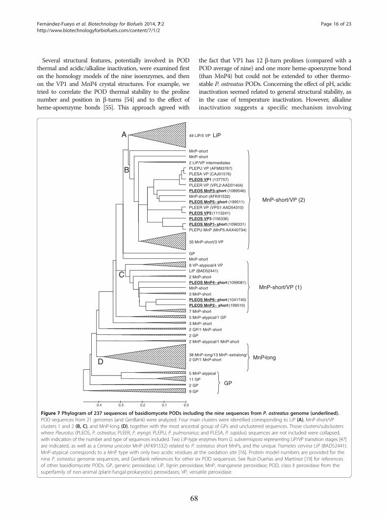
Fernández-Fueyo et al. Biotechnology for Biofuels 2014, 7:2 Page 16 of 23http://www.biotechnologyforbiofuels.com/content/7/1/2
Several structural features, potentially involved in PODthermal and acidic/alkaline inactivation, were examined firston the homology models of the nine isoenzymes, and thenon the VP1 and MnP4 crystal structures. For example, wetried to correlate the POD thermal stability to the prolinenumber and position in β-turns [54] and to the effect ofheme-apoenzyme bonds [55]. This approach agreed with
49 Li
MnPMnP
2 LiPPLEPPLESPLEOPLEEPLEOMnP-PLEOPLEEPLEOPLEOPLEOPLEP
35 M
GPMnP
8 VPLiP (
2 MnPLEOMnP
3 MnPLEOPLEO
7 Mn
5 Mn
3 Mn
2 GP
2 GP
2 Mn
38 M2 GP
5 Mn
11 G
2 GP
9 GP
0.00.10.20.30.4
A
B
C
D
Figure 7 Phylogram of 237 sequences of basidiomycete PODs includiPOD sequences from 21 genomes (and GenBank) were analyzed. Four maiclusters 1 and 2 (B, C), and MnP-long (D), together with the most ancestrawhere Pleurotus (PLEOS, P. ostreatus; PLEER, P. eryngii; PLEPU, P. pulmonariuswith indication of the number and type of sequences included. Two LiP-typeare indicated, as well as a Cerrena unicolor MnP (AFK91532) related to P. oMnP-atypical corresponds to a MnP type with only two acidic residues atnine P. ostreatus genome sequences, and GenBank references for other sof other basidiomycete PODs. GP, generic peroxidase; LiP, lignin peroxidasuperfamily of non-animal (plant-fungal-prokaryotic) peroxidases; VP, vers
68
the fact that VP1 has 12 β-turn prolines (compared with aPOD average of nine) and one more heme-apoenzyme bond(than MnP4) but could not be extended to other thermo-stable P. ostreatus PODs. Concerning the effect of pH, acidicinactivation seemed related to general structural stability, asin the case of temperature inactivation. However, alkalineinactivation suggests a specific mechanism involving
P/5 VP
-short-short
/VP intermediatesU VP (AFM93767)A VP (CAJ01576)S VP1 (137757)R VP (VPL2:AAD01404)S MnP3-short (1089546)
short (AFK91532)S MnP5- short (199511)R VP (VPS1:AAD54310)S VP2 (1113241)S VP3 (156336)S MnP1- short (1096331)U MnP (MnP5:AAX40734)
nP-short/3 VP
-short
-atypical/4 VPBAD52441)
P-shortS MnP4- short (1099081)
-short
P-shortS MnP6- short (1041740)S MnP2- short (199510)
P-short
P-atypical/1 GP
P- short
/1 MnP-short
P-atypical/1 MnP-short
nP-long/13 MnP- extralong//1 MnP-short
P-atypical
P
LiP
MnP-short/VP (2)
MnP-short/VP (1)
MnP-long
GP
ng the nine sequences from P. ostreatus genome (underlined).n clusters were identified corresponding to LiP (A), MnP-short/VPl group of GPs and unclustered sequences. Those clusters/subclusters; and PLESA, P. sapidus) sequences are not included were collapsed,enzymes from G. subvermispora representing LiP/VP transition stages [47]streatus short MnPs, and the unique Trametes cervina LiP (BAD52441).the oxidation site [16]. Protein model numbers are provided for theix POD sequences. See Ruiz-Dueñas and Martínez [19] for referencesse; MnP, manganese peroxidase; POD, class II peroxidase from theatile peroxidase.
Fernández-Fueyo et al. Biotechnology for Biofuels 2014, 7:2 Page 17 of 23http://www.biotechnologyforbiofuels.com/content/7/1/2
formation of a bis-histidyl heme iron complex due to lossof structural Ca2+ ions [53]. In this context, two of themain differences in the crystal structure of the pH-stableMnP4, compared with VP1, affect regions neighbor tothe Ca2+-binding sites, such as the loop containing theMnP4 extra helix in the upper domain and the two differ-ent sequence stretches at the lower domain. In both cases,MnP4 has an extra interaction in the second coordinationsphere of the Ca2+ ions (due to presence of T136 andK296) that could increase its alkaline stability. The higheststability of MnP4 could be also due to some interactionslacking in VP1 that stabilize loops and connect helices andloops at the molecular surface. Another noteworthy char-acteristic of MnP4 is the high number of surface-exposedlysines (up to a total of 20) and other alkaline residues(up to a total of 35) compared with the other P. ostreatusPODs, which would result in a higher net positive chargeof the protein and increased stability at acidic pH. Involve-ment of the above surface interactions and alkaline residuesin MnP4 pH stability is being confirmed by site-directedmutagenesis studies in course (unpublished data).
Evolutionary relationships of basidiomycete PODsThe evolutionary relationships of P. ostreatus and otherbasidiomycete PODs are shown in Figure 7. The mostbasal sequences correspond to generic peroxidases(GPs), which gave rise to MnPs by progressive incorpor-ation of the three residues forming the Mn oxidation site[16]. Long (and extra long) MnPs from P. chrysosporiumand other fungi form cluster D, separated from the restof PODs. A few more GPs and atypical MnPs areunclustered (between clusters C and D) and the rest ofthe sequences are intermixed, except for the well-defined cluster A that includes all the LiPs, and a fewVPs related to the common VP-type ancestor of bothfamilies [16].The P. ostreatus PODs are in the two other clusters,
both containing short MnP and VP sequences, in agree-ment with higher sequence identity of short MnPs withVPs than with long MnPs from P. chrysosporium andother fungi (see Additional file 2: Table S7). Cluster Cincludes P. ostreatus MnP2, MnP4, and MnP6, togetherwith other short MnPs and a group of VPs. Cluster B isconstituted by: i) a Pleurotus group formed by the othersix genome PODs and five more sequences from Gen-Bank; ii) a second group of short MnPs/VPs; and iii)unclustered short MnPs and two LiP/VP intermediates[47]. Inside the Pleurotus group, P. ostreatus VP1 isclosely related to P. eryngii VPL, the best characterizedVP [29,35] (97% identity), and to the only Pleurotus sapi-dus and P. pulmonarius VPs, suggesting that the four se-quences correspond to the same isoenzyme in fourrelated species. Moreover, P. ostreatus VP2 clusters withP. eryngii VPS1, the second cloned VP [28] (98%
69
identity), revealing that both are also the same isoen-zyme, and the same is true of P. ostreatus MnP1 and aP. pulmonarius MnP (96% sequence identity). As dis-cussed, the structural characteristics of the two latterMnPs suggest a secondary origin from an ancestral VP-type enzyme that, according to the phylogram, could berelated to P. ostreatus VP3.
P. ostreatus PODs: demonstration of ligninolysisPOD involvement in fungal ligninolysis is supported bythe presence of LiP, VP, and/or MnP encoding genes inall the currently available genomes of lignin-degradingbasidiomycetes (white-rot fungi) and their absence fromall the genomes of basidiomycetes that degrade cellulosewithout removing lignin significantly (brown-rot fungi)[16]. Oxidation of a non-phenolic lignin model dimer byDyP has been reported [56], although in other cases itsdirect action seems restricted to the minor phenolicunits [57] as in the case of laccases [4]. Some HTPs canalso degrade non-phenolic lignin model dimers [58] butthey seem unable to act on polymeric lignin [59]. More-over, the genes of these two peroxidase superfamilies(and laccases) are not restricted to the genomes ofwhite-rot fungi, as in the case of PODs [16].In P. chrysosporium, LiP is the enzyme principally re-
sponsible for cleavage of the predominant and most re-calcitrant nonphenolic structures in lignin [60], whereasclassical MnPs contribute Mn3+ that can act as a diffus-ible oxidant of minor phenolic lignin moieties [61]. VPwas described as a fourth POD family combining someof the catalytic properties of LiPs, MnPs, and fungal GPs(which act on the same substrates that plant peroxidasesoxidize) [27,28]. The chimeric nature of VPs raised ques-tions about their role in ligninolysis, and no studies ontheir lignin-degrading ability have been available untilnow.The present genomic study establishes the absence of
LiP in a ligninolytic white-rot fungus, P. ostreatus, in ac-cordance with the in silico analysis of its genome, whichindicated the presence of only VP and MnP genes.Moreover, we demonstrate for the first time the lignino-lytic capabilities of one of these VPs, showing that it isable to: i) cleave the most frequent interunit linkages inlignin (β-O-4’ ether structures); and ii) depolymerize lig-nin. These capabilities are similar to those reported forP. chrysosporium LiP in terms of reactions and productsobtained [22]. Concerning MnPs, we also provide evi-dence on the dual Mn-mediated and Mn-independent ac-tivity of the so-called short MnPs found in P. ostreatus,whose presence had been reported in several fungal ge-nomes [16] without the corresponding catalytic studies.We show the existence, in P. ostreatus, of a POD rep-
ertoire constituted by VPs and MnPs, with the formerperoxidases assuming the role played by LiPs in P.
Fernández-Fueyo et al. Biotechnology for Biofuels 2014, 7:2 Page 18 of 23http://www.biotechnologyforbiofuels.com/content/7/1/2
chrysosporium (and most other white-rot fungi). Thisdifferent enzymatic machinery is most probably relatedto the different phylogenetic position of the two fungi,which are in the orders Agaricales and Polyporales, re-spectively. It seems that the POD evolutionary history inthe Agaricales did not include the final transition fromVP to LiP enzymes via loss of the Mn2+ oxidation site,as occurred in the Polyporales [46].
ConclusionsAs a result of the current genomic screening we wereable to: i) show the presence in the model agaric P.ostreatus of a peroxidase repertoire in which VPs playthe role that LiPs do in white-rot polypores; ii) describethe catalytic properties of P. ostreatus MnPs, as repre-sentatives of a new peroxidase subfamily; iii) establishthe evolutionary relationships of the above enzymes withthe other basidiomycete PODs; iv) describe the firstcrystal structure of a short MnP, which was comparedwith the solved VP structure; and v) demonstrate the ex-istence of strongly divergent thermal and pH stabilitiesamong a wide array of POD isoenzymes encoded by du-plicated genes.
Materials and methodsFungal strains and genome sequencingMonokaryons PC9 (CECT20311) and PC15 (CECT20312)were isolated from P. ostreatus N001 (CECT20600),and their genomic DNA sequences obtained at JGI ina project coordinated by AG Pisabarro (Public Universityof Navarre, Pamplona, Spain). The 35.6 Mbp (PC9v1.0) and 34.3 Mbp (PC15 v2.0) assemblies are pre-dicted to include 12,206 and 12,330 gene models, re-spectively (the results are available for searching athttp://genome.jgi.doe.gov/PleosPC15_2/PleosPC15_2.home.html and http://genome.jgi.doe.gov/PleosPC9_1/PleosPC9_1.home.html).
Genome screening and analysis of peroxidase modelsThe final inventory of heme peroxidase genes in theP. ostreatus genome was obtained by: i) screening theautomatically annotated genomes; ii) revising and manu-ally curating the positions of introns, and the N and C ter-mini, using SignalP 3.0 (Center for Biological SequenceAnalysis, Kongens Lyngby, Denmark) for predicting sig-nal peptides; iii) comparing the predicted amino acidsequences with related peroxidases, after multiple align-ment with MEGA5 (Center for Evolutionary Medicineand Informatics, Tempe, AZ, USA); and iv) confirmingthe presence of characteristic residues at the hemepocket and substrate oxidation sites, after homologymodeling at the Swiss-Model server (Protein Structure Bio-informatics Group, Swiss Institute of Bioinformatics and theBiozentrum of the University of Basel, Basel, Switzerland)
70
using the crystal structures of P. eryngii VPL ([PDB:3FJW])and P. ostreatus MnP4 (this study) as templates. Finally, therevised POD sequences from the sequenced genome werecompared with all the basidiomycete POD sequences avail-able (up to a total of 237 sequences from 21 genomes andGenBank) and phylograms were constructed with MEGA5,using Poisson-corrected distances and an unweighted pairgroup method with arithmetic mean (UPGMA) clustering(bootstrap consensus trees were inferred from 1,000replicates).
Gene synthesisThe revised mature protein-coding sequences of thenine POD genes (models 156336, 199510, 199511,1041740, 1089546, 1096331, 1099081, and 1113241 fromPC15, and model 137757 from PC9) were synthesized byATG:biosynthetics (Merzhausen, Germany) after verify-ing that all the codons had previously been used for ex-pressing other genes in the same E. coli strains (andsubstituting them when required). A MnP-encodinggene from P. pulmonarius ([GenBank:AAX40734]) wasalso synthesized for comparison.
Directed mutagenesisI198F and D261G mutations were introduced in the P.ostreatus MnP1 (109633) gene by PCR using the expres-sion plasmid pFLAG1-109633 (see below) as template,and the QuikChange kit from Stratagene (La Jolla, CA,USA). The 5′- CG CCA AAC CTT TTC GAT TCACAA TTC TTC ATC GAG ACG C −3′ (I198F) and 5′-C CGC TTC TCC GGA ACG CTG TTC AAG ATGTCG −3′ (D261G) direct primers (mutated codons initalics), and the reverse primers bearing the complemen-tary sequences were synthesized. The PCR reaction(50 μl volume) was carried out in an Eppendorf(Hamburg, Germany) Mastercycler pro S thermal cyclerusing 20 ng of template DNA, 500 μM each dNTP,125 ng direct and reverse primers, 2.5 units of PfuTurbopolymerase (Stratagene), and the manufacturer’s buffer.Reaction conditions included: i) a start cycle of 1 minuteat 95°C; ii) 18 cycles of 50 seconds at 95°C, 50 secondsat 55°C, and 10 minutes at 68°C; and iii) a final cycle of10 minutes at 68°C. The mutated gene was expressed inE. coli and purified as were the wild type genes.
E. coli expressionThe nine P. ostreatus genome coding sequencesand the only P. pulmonarius mature POD coding se-quences, together with the two P. ostreatus MnP1(10963331) mutated sequences, were cloned in the expres-sion vectors pFLAG1 (International Biotechnologies Inc,Kodak, CT, USA) or pET23a (+) (Novagen, Darmstadt,Germany) and the resulting plasmids (pET23a-156336,pET23a-1099081, pET23a-1041740, pFLAG1-137757,

Fernández-Fueyo et al. Biotechnology for Biofuels 2014, 7:2 Page 19 of 23http://www.biotechnologyforbiofuels.com/content/7/1/2
pFLAG1-199511, pFLAG1-199510, pFLAG1-1089546,pFLAG1-1113241, pFLAG1-1096331, and pFLAG1-AAX40734) were used for expression.Peroxidases were produced in E. coli W3110 (pFLAG1
plasmids) and BL21(DE3)pLysS (pET23a plasmids). Cellswere grown for 3 hours in Terrific Broth, induced with1 mM isopropyl-β-D-thiogalactopyranoside (IPTG), andgrown further for 4 hours. The apoenzyme accumulatedin inclusion bodies, as observed by SDS-PAGE, and wassolubilized with 8 M urea. In vitro refolding was per-formed using 0.16 M urea, 5 mM Ca2+, 20 μM hemin,0.5 mM oxidized glutathione, 0.1 mM dithiothreitol, and0.1 mg/ml protein, at pH 9.5 [62]. For P. ostreatus MnP6(1041740) refolding was obtained using 0.1 M urea,5 mM Ca2+, 20 μM hemin, 1.5 mM oxidized glutathione,0.1 mM dithiothreitol, and 0.1 mg/ml protein, at pH 8. En-zymes were purified by Resource Q chromatography usinga 0 to 300 mM NaCl gradient (2 ml.min-1, 20 minutes) in10 mM sodium tartrate (pH 5.5) containing 1 mM CaCl2(except for MnP4-1099081, for which pH 6 was used).
Crystallization, data collection, and refinementCrystallization trials were carried out by the sitting dropvapor diffusion method, in 96-well plates using Wizardscreens I to III (Emerald Bio, Bainbridge Island, WA,USA) and JBScreen Kits 1-10 (Jena Biosciences, Jena,Germany) at 22°C. Drops consisted of 0.2 μl of proteinsolution (10 mg/ml in 10 mM sodium tartrate, pH 5.0)and 0.2 μl of reservoir solution. Crystals of VP1belonged to three crystal forms in two space groups.Form I belonged to the P43 group and was obtained inthe five following conditions: i) 0.1 M sodium acetate(pH 4.6) containing 8% PEG 4000; ii) 20% PEG 3350 and0.2 M ammonium chloride; iii) 20% PEG 3350 and0.2 M ammonium formate; iv) 0.1 M sodium HEPESbuffer (pH 7.5) containing 1.6 M ammonium sulfate and2% PEG 1000; and v) 0.1 M sodium HEPES buffer(pH 7.5) containing 0.2 M sodium acetate and 20% PEG3000. Form II also belonged to the P43 group and wasobtained in 0.1 M imidazole (pH 8.0) containing1.0 M K/Na tartrate and 0.2 M NaCl. Form III belongedto the P21 group and was obtained in 0.1 M sodiumacetate (pH 4.5) containing 20% PEG 1000 and 0.2 Mzinc acetate. Four different conditions gave two differentMnP4 crystal forms. Form I belonged to the P1 groupand was obtained in 0.1 M sodium citrate (pH 5.5) con-taining 2.0 M ammonium sulfate. Finally, form IIbelonged to the C2 group and was obtained in: i) 0.1 Msodium acetate (pH 4.6) containing 2.0 M ammoniumsulfate; ii) 0.1 M Tris-HCl buffer (pH 7.0) containing2.0 M ammonium sulfate and 0.2 M lithium sulfate; andiii) 0.1 M sodium CAPS buffer (pH 10.5) containing1.2 M NaH2PO4/0.8 M K2HPO4 and 0.2 M lithium sul-fate. Crystals were mounted in nylon loops and flash-
71
frozen in liquid N2 in the mother liquor containing vari-ous cryoprotectants.X-ray diffraction data were collected at 100 K at the
X06DA and X06SA beam lines at the Swiss Light Source(Villigen, Switzerland) using a wavelength of 1.0000 Å, andPilatus 2 M and 6 M detectors, respectively. Diffractiondata were indexed, integrated, merged, and scaled usingXDS and XSCALE. Only the highest resolution structurefor each POD (crystal form I) is presented (their data col-lection statistics are shown in Additional file 2: Table S3).The structures of VP1 and MnP4 were solved by mo-
lecular replacement using the crystal structure of P.eryngii VPL (3FMU) as the search model and the pro-gram AutoMR of the PHENIX package (LawrenceBerkeley Laboratory, Berkeley, CA, USA). The finalmodels were obtained by successive refinement roundsfollowed by manual building with Coot using σAweighted 2Fo-Fc and Fo-Fc electron density maps. Solv-ent molecules were introduced in the refinement, as im-plemented in the PHENIX package, and visuallyinspected. A total of 5% of reflections was used to calcu-late the Rfree value throughout the refinement process.The VP1 final model contained all but the last residue ofthe sequence (S331) (which did not present any electrondensity), one heme cofactor, two Ca2+ ions, and 452 watermolecules; and the MnP4 final model contained all the337 residues of the sequence, one heme cofactor, two Ca2+
ions, and 1,212 water molecules (representative details ofthe VP1 and MnP4 electron density maps, correspondingto the heme pocket, are shown in Additional file 1: FigureS7). The structures were validated with MolProbity (TheRichardson Laboratory, Duke University, Durham, NC,USA). Refinement and final model statistics are shown inAdditional file 2: Table S3. Figures were produced withPyMOL (Schrödinger, Portland, OR, USA). The coordi-nates and structure factors have been deposited with thePDB accession codes [PDB:4BLK] and [PDB:4BM1].
Kinetic constants on selected substratesAbsorbance changes during substrate oxidation in 0.1 Mtartrate (at various pH values) were recorded at 25°C ina Biomate5 spectrophotometer (Thermo Scientific, Wal-tham, MA, USA) using approximately 0.01 μM enzymeconcentration, estimated from the ε406 of each isoen-zyme (Additional file 2: Table S1). The reactions wereinitiated by H2O2 (0.1 mM) addition. Oxidation of Mn2+
was followed at pH 5 by monitoring Mn3+.tartrate com-plex (ε238 6.5 mM-1.cm-1) formation. VA oxidation wasfollowed at pH 3 for veratraldehyde (ε310 9.3 mM-1.cm-1)formation. RB5, ABTS, and DMP oxidation were assayedat pH 3.5, and monitored for RB5 disappearance (ε59830 mM-1.cm-1) and formation of ABTS cation radical(ε436 29.3 mM-1.cm-1) and dimeric coerulignone (ε46955 mM-1.cm-1), respectively. ABTS and DMP oxidation
Fernández-Fueyo et al. Biotechnology for Biofuels 2014, 7:2 Page 20 of 23http://www.biotechnologyforbiofuels.com/content/7/1/2
by VP showed double kinetics, with sigmoidal activitycurves at increasing substrate concentration (Additionalfile 2: Figure S8), that enabled calculation of two sets ofkinetic constants. Kinetic constants for enzyme activa-tion by H2O2 were determined using 5 mM ABTS(except for MnP1 and MnP6, for which 2 mM wasused). Means and standard errors for Michaelis constant(Km) and enzyme turnover (kcat) values were obtained bynonlinear least-squares fitting to the Michaelis–Mentenmodel. Fitting of these constants to the normalizedequation v = (kcat/Km)[S]/(1 + [S]/Km) yielded the cata-lytic efficiency values (kcat/Km) with their correspondingstandard errors.
pH inactivation studiesTo study the effect of preincubation at different pHvalues on activity, the nine PODs were dissolved(0.05 μM) in Britton-Robinson (B&R) buffer with a pHrange from 2 to 9, and kept at 4°C for different timeperiods. Activity was determined by oxidation of a satur-ating concentration of ABTS (5 mM, except for MnP1-1096331 and MnP6-1041740, for which 2 mM was used)in 0.1 M tartrate (pH 3.5) under the conditions de-scribed above. Residual activities were measured after 1minute (to evaluate the initial survival of the enzyme ateach pH value), and 1, 4, 24, and 120 hours of incuba-tion. The highest activity after 1 minute (at any pH) wastaken as 100% activity, and the percentage of residual ac-tivity at the different times and pH conditions was calcu-lated according to this maximal value. The sameexperiment was repeated with the more stable (MnP4)and the less stable enzyme (MnP3) by keeping them at25°C instead of at 4°C.
Thermal inactivation studiesTo study the effect of enzyme preincubation at differenttemperatures on the activity, the nine PODs (0.05 μM) in10 mM tartrate (pH 5) were incubated at 5°C for 10 mi-nutes or 4 hours in the temperature range of 25 to 70°C.Residual activity was determined at 25°C, as describedabove, and that obtained after 25°C preincubation wastaken as 100%. Temperature stability was presented as 10-minute and 4-hour T50-activity values, that is, thetemperature at which 50% of the activity was lost after in-cubation for the above time periods.
CD and UV-visible absorption spectroscopyThe effect of pH and temperature on the structure andcofactor binding of different PODs was followed by CDand UV-visible absorption spectroscopies. Far-UV (190to 250 nm) CD measurements were carried out on a J-720 spectropolarimeter (Jasco, Oklahoma City, OK,USA) equipped with a peltier temperature controllerand a thermostated cell holder using a 0.01 cm path
72
length quartz cell. The effect of three pHs (3, 5, and 8)on the CD spectra was estimated at different incubationtimes (1 minute, 1 hour, and 4 hours) at a protein con-centration of approximately 50 μM in 0.1 mM B&R buf-fer, at 25°C. The spectra from five averaged scans werecorrected for the baseline contribution of the buffer andthe observed ellipticities were converted into mean resi-due ellipticities (θ). The effect of temperature on CDspectra was analyzed at a protein concentration of ap-proximately 5 μM in 0.01 mM phosphate (pH 6). Ther-mal denaturation was estimated by increasing thetemperature from 20 to 70°C at 20°C.h-1, and recordingthe CD signal at 222 nm. Tm represents the temperatureat the midpoint of the unfolding transition. UV-visible(300 to 800 nm) absorption spectra of the two proteinsafter incubation at different pHs and temperatures wereobtained on an 8453E diode-array spectrophotometer(Agilent, Santa Clara, CA, USA), using a 1 cm path lengthquartz cell. The effect of three pHs (3, 5, and 8) after dif-ferent incubation times (0, 30, 60, 90, 120, 180, and 240minutes) was estimated at a protein concentration of ap-proximately 3.5 μM in 0.1 mM B&R buffer, at 25°C. TheUV-visible spectra of different PODs were also collectedin the range of 25 to 70°C, after 10 minutes of incubationat 5°C intervals, using a protein concentration of approxi-mately 2 μM in 10 mM tartrate (pH 5). T50-Soret representsthe temperature at the midpoint of the heme-loss transi-tion, as estimated by the Soret band intensity at 407 nm.
Oxidative degradation of a lignin model dimerRing-14C-labeled (1.0 mCi.mmol-1) 4-ethoxy-3-methoxy-phenylglycerol-β-guaiacyl ether was prepared, and itserythro and threo isomers were chromatographicallyseparated, as described earlier [63]. The radiolabeled ery-thro or threo dimer was treated with VP1 (137757) fromP. ostreatus in 10 mM sodium acetate (pH 3.0) at 25°Cfor 1 hour. The products formed were analyzed byreversed-phase HPLC using a Gilson (Middleton, WI,USA) system equipped with a C-18 column (Luna C18(2), Phenomenex, Macclesfield, UK; 150 by 4.6 mm,5 μm particle size), and methanol:water as mobile phase(35:65 for 15 minutes, followed by 50:50) at a flow rateof 1 ml.min-1. Elution was monitored at 255 nm, and the14C-content in collected fractions (0.5 ml) was measuredin a liquid scintillation counter. HPLC in conjunctionwith gas chromatography–mass spectrometry (GC-MS)was used in parallel analyses with unlabeled dimers toconfirm the identity of the products obtained.
Enzymatic depolymerization of synthetic ligninA radiolabeled syringyl-guaiacyl DHP (with a syringyl/guaiacyl ratio of approximately 4:1) was prepared by co-polymerization of β-[14C]-sinapyl alcohol (0.01 mCi.mmol-1)and unlabeled coniferyl alcohol using horseradish
Fernández-Fueyo et al. Biotechnology for Biofuels 2014, 7:2 Page 21 of 23http://www.biotechnologyforbiofuels.com/content/7/1/2
peroxidase and fractionated on a 1.8 × 30 cm column ofSephadex LH-20 in N,N-dimethylformamide. The high mo-lecular mass fractions excluded from the column (>1 kDa)were pooled for use in depolymerization experiments [64].Enzymatic depolymerization by the same peroxidase
used in the dimer degradation assays was investigated in10 mM Na acetate (pH 4.5) containing 0.25% Tween 20,1.5 × 104 dpm (188 μg) DHP, and 0.01 μM enzyme in afinal volume of 40 ml, in the presence or absence of10 mM VA. Reactions were conducted at 25°C by addingH2O2 (7.5 mM in experiments with VA, and 0.3 mM inexperiments without VA) over 24 hours with a syringepump [22]. Control reactions without enzyme were runfor comparison. The reaction mixtures were then con-centrated by rotary vacuum evaporation, redissolved inN,N-dimethylformamide containing 0.1 M LiCl, and cen-trifuged as described earlier [22]. Molecular mass distri-butions of the supernatant fractions were assessed byGPC on a 1.8 × 30 cm column of Sephadex LH20, usingN,N′-dimethylformamide containing 0.1 M LiCl as themobile phase. Fractions (2 ml) were collected andassayed for 14C in a liquid scintillation counter.
Additional files
Additional file 1: Sequence comparison, amino acid composition,heterologous expression, pH stability (4°C and 25°C), molecularstructure (VP1 and MnP4 differences and heme pockets), and ABTSsigmoidal kinetics (VP1) for different PODs from the P. ostreatusgenome. Figure S1. Phylogram of heme peroxidase sequences from thegenomes of two P. ostreatus monokaryons. Figure S2. Amino acidcomposition of the PODs from the P. ostreatus genome (predictedmature proteins). Figure S3. E. coli expression of P. ostreatus genomeperoxidases (SDS-PAGE). Figure S4. pH 2 to 9 stability of the nine PODsfrom the P. ostreatus genome at different incubation times. Figure S5.Influence of temperature on the pH stability of PODs from theP. ostreatus genome. Figure S6. Stereo views of some of the maindifferences between VP1 and MnP4 crystal structures. Figure S7. Partial2Fo-Fc electron density map, contoured at the 1.1 σ level, of hemecofactor, neighbor residues and several water molecules, and position ofsurrounding heme pocket residues, in the VP1 and MnP4 crystals. Figure S8.Sigmoidal curve for ABTS oxidation by VP (isoenzyme VP1) enablingcalculation of two sets of kinetic constants.
Additional file 2: Gene inventory, isoenzyme structural properties,crystallographic data, kinetic constants (native VP1, MnP4 andMnP1, and mutated variants), and sequence identities for differentPODs from the P. ostreatus genome. Table S1. Inventory of peroxidasegenes in the genomes of P. ostreatus monokaryons PC9 and PC15 andsome characteristics of the purified PODs from E. coli expression. Table S2.Structural properties potentially related to temperature/pH stability in thenine PODs from the P. ostreatus genome, together with experimentally-determined thermal stability (T50-activity) and pH stability range. Table S3.Crystallographic data collection and refinement statistics of P. ostreatus VP1and MnP4. Table S4. Kinetic constants of W165, E35A, E39A, D175A, andE35A/E39A variants of P. ostreatus VP1 oxidizing VA, RB5, and Mn2+, comparedwith native VP1. Table S5. Kinetic constants of E36A, E40A, D179A, andE36A/E40A variants of P. ostreatus MnP4 oxidizing Mn2+, compared withnative MnP4. Table S6. Kinetic constants of two variants in the environmentof Trp165 of P. ostreatus MnP1, compared with the native MnP1, and a relatedMnP from P. pulmonarius oxidizing VA, RB5, ABTS, DMP, and Mn2+. Table S7.Amino acid sequence identities between the nine PODs from the P. ostreatusgenome, P. chrysosporium LiP and MnP, and two P. eryngii VPs.
73
AbbreviationsABTS: 2,2'-azino-bis(3-ethylbenzothiazoline-6-sulfonate); B&R: Britton-Robinson; CAPS: N-cyclohexyl-3-aminopropanesulfonic acid; CD: Circulardichroism; DHP: Dehydrogenation polymer (lignin); DMP: 2,6-dimethoxyphenol; dNTP: Deoxyribonucleotide triphosphates;DOE: Department of Energy; DyP: Dye-decolorizing peroxidase; GC-MS: Gaschromatography–mass spectrometry; GP: Generic peroxidase; GPC: Gelpermeation chromatography; HEPES: 4-(2-hydroxyethyl)-1-piperazineethanesulfonic acid; HPLC: High performance liquidchromatography; HTP: Heme-thiolate peroxidase; IPTG: Isopropyl-β-D-thiogalactopyranoside; JGI: Joint Genome Institute; kcat: Catalytic constant;Km: Michaelis constant; LiP: Lignin peroxidase; LRET: Long-range electrontransfer; MnP: Manganese peroxidase; PCR: Polymerase chain reaction;PDB: Protein Data Bank; PEG: Polyethylene glycol; POD: Class II peroxidasefrom the superfamily of non-animal (plant-fungal-prokaryotic) peroxidases;RB5: Reactive Black 5; Rz: Reinheitszahl; UPGMA: Unweighted pair groupmethod with arithmetic mean; VA: Veratryl alcohol; VP: Versatile peroxidase.
Competing interestsThe authors declare that they have no competing interests.
Authors’ contributionsATM and FJR-D conceived and designed the experiments. FJM carried outthe crystallographic study. EF-F, FJR-D, and FJM performed the experiments.EF-F, FJR-D, FJM, AR, MJM, KEH, and ATM analyzed the data. ATM, FJM, andKEH wrote the paper. All authors read and approved the final manuscript.
AcknowledgementsThis work was supported by the PEROXICATS (KBBE-2010-4-265397) and INDOX(KBBE-2013-7-613549) grants of the European Union (to ATM), by grantsBIO2011-26694 (to FJR-D) and BFU2011-24615 (to AR) of the Spanish Ministry ofEconomy and Competitiveness (MINECO), and by US DOE grant DE-AI02-07ER64480 (to KEH). The work conducted by the US DOE JGI is supported by theOffice of Science of the US DOE under contract number DE-AC02-05CH11231.AG Pisabarro (Public University of Navarre, Pamplona, Spain) is acknowledged forcoordinating the P. ostreatus genome project. We thank Michael D Mozuch forassistance with the lignin depolymerization studies. EF-F acknowledges a Junta deAmpliación de Estudios fellowship of the CSIC, co-funded by the European SocialFund, and FJR-D acknowledges a MINECO Ramón y Cajal contract.
Received: 26 September 2013 Accepted: 16 December 2013Published: 3 January 2014
References1. Sánchez C: Cultivation of Pleurotus ostreatus and other edible
mushrooms. Appl Microbiol Biotechnol 2010, 85:1321–1337.2. Gunde-Cimerman N, Cimerman A: Pleurotus fruiting bodies contain the
inhibitor of 3-hydroxy-3-methylglutaryl-coenzyme-A reductase-lovastatin.Exp Mycol 1995, 19:1–6.
3. Lavi I, Levinson D, Peri I, Tekoah Y, Hadar Y, Schwartz B: Chemicalcharacterization, antiproliferative and antiadhesive properties ofpolysaccharides extracted from Pleurotus pulmonarius mycelium andfruiting bodies. Appl Microbiol Biotechnol 2010, 85:1977–1990.
4. Ruiz-Dueñas FJ, Martínez AT: Microbial degradation of lignin: how a bulkyrecalcitrant polymer is efficiently recycled in nature and how we cantake advantage of this. Microbial Biotechnol 2009, 2:164–177.
5. Martínez AT, Camarero S, Guillén F, Gutiérrez A, Muñoz C, Varela E, MartínezMJ, Barrasa JM, Ruel K, Pelayo M: Progress in biopulping of non-woodymaterials: chemical, enzymatic and ultrastructural aspects of wheat-straw delignification with ligninolytic fungi from the genus Pleurotus.FEMS Microbiol Rev 1994, 13:265–274.
6. Young RA, Akhtar M: Environmentally Friendly Technologies for the Pulp andPaper Industry. New York: John Wiley and Sons; 1998.
7. Camarero S, Barrasa JM, Pelayo M, Martínez AT: Evaluation of Pleurotusspecies for wheat-straw biopulping. J Pulp Paper Sci 1998, 24:197–203.
8. Valmaseda M, Almendros G, Martínez AT: Chemical transformation ofwheat straw constituents after solid-state fermentation with selectedlignocellulose-degrading fungi. Biomass and Bioenergy 1991, 1:261–266.
9. Salvachúa D, Prieto A, Lopez-Abelairas M, Lú-Chau T, Martínez AT, MartínezMJ: Fungal pretreatment: an alternative in second-generation ethanolfrom wheat straw. Bioresource Technol 2011, 102:7500–7506.
Fernández-Fueyo et al. Biotechnology for Biofuels 2014, 7:2 Page 22 of 23http://www.biotechnologyforbiofuels.com/content/7/1/2
10. Ragauskas AJ, Williams CK, Davison BH, Britovsek G, Cairney J, Eckert CA,Frederick WJ, Hallett JP, Leak DJ, Liotta CL, Mielenz JR, Murphy R,Templer R, Tschaplinski T: The path forward for biofuels andbiomaterials. Science 2006, 311:484–489.
11. Martínez AT, Ruiz-Dueñas FJ, Martínez MJ, del Río JC, Gutiérrez A: Enzymaticdelignification of plant cell wall: from nature to mill. Curr Opin Biotechnol2009, 20:348–357.
12. Martinez D, Larrondo LF, Putnam N, Gelpke MD, Huang K, Chapman J,Helfenbein KG, Ramaiya P, Detter JC, Larimer F, Coutinho PM, Henrissat B,Berka R, Cullen D, Rokhsar D: Genome sequence of the lignocellulosedegrading fungus Phanerochaete chrysosporium strain RP78. NatBiotechnol 2004, 22:695–700.
13. Martinez D, Challacombe J, Morgenstern I, Hibbett D, Schmoll M, KubicekCP, Ferreira P, Ruiz-Duenas FJ, Martinez AT, Kersten P, Hammel KE, VandenWymelenberg A, Gaskell J, Lindquist E, Sabat G, Bondurant SS, Larrondo LF,Canessa P, Vicuna R, Yadav J, Doddapaneni H, Subramanian V, Pisabarro AG,Lavín JL, Oguiza JA, Master E, Henrissat B, Coutinho PM, Harris P, MagnusonJK, et al: Genome, transcriptome, and secretome analysis of wood decayfungus Postia placenta supports unique mechanisms of lignocelluloseconversion. Proc Natl Acad Sci U S A 2009, 106:1954–1959.
14. Eastwood DC, Floudas D, Binder M, Majcherczyk A, Schneider P, Aerts A,Asiegbu FO, Baker SE, Barry K, Bendiksby M, Blumentritt M, Coutinho PM,Cullen D, de Vries RP, Gathman A, Goodell B, Henrissat B, Ihrmark K,Kauserud H, Kohler A, LaButti K, Lapidus A, Lavin JL, Lee YH, Lindquist E, LillyW, Lucas S, Morin E, Murat C, Oguiza JA, et al: The plant cell wall-decomposing machinery underlies the functional diversity of forestfungi. Science 2011, 333:762–765.
15. Fernandez-Fueyo E, Ruiz-Dueñas FJ, Ferreira P, Floudas D, Hibbett DS,Canessa P, Larrondo LF, James TY, Seelenfreund D, Lobos S, Polanco R, TelloM, Honda Y, Watanabe T, Watanabe T, Ryu JS, Kubicek CP, Schmoll M,Gaskell J, Hammel KE, St John FJ, Vanden Wymelenberg A, Sabat G, SplinterBonDurant S, Syed K, Yadav JS, Doddapaneni H, Subramanian V, Lavín JL,Oguiza JA, et al: Comparative genomics of Ceriporiopisis subvermisporaand Phanerochaete chrysosporium provide insight into selectiveligninolysis. Proc Natl Acad Sci U S A 2012, 109:5458–5463.
16. Floudas D, Binder M, Riley R, Barry K, Blanchette RA, Henrissat B, Martínez AT,Otillar R, Spatafora JW, Yadav JS, Aerts A, Benoit I, Boyd A, Carlson A, CopelandA, Coutinho PM, de Vries RP, Ferreira P, Findley K, Foster B, Gaskell J, Glotzer D,Górecki P, Heitman J, Hesse C, Hori C, Igarashi K, Jurgens JA, Kallen N, KerstenP, et al: The Paleozoic origin of enzymatic lignin decompositionreconstructed from 31 fungal genomes. Science 2012, 336:1715–1719.
17. Ruiz-Dueñas FJ, Fernández E, Martínez MJ, Martínez AT: Pleurotus ostreatusheme peroxidases: an in silico analysis from the genome sequence tothe enzyme molecular structure. C R Biol 2011, 334:795–805.
18. Martínez AT: Molecular biology and structure-function of lignin-degrading heme peroxidases. Enzyme Microb Technol 2002,30:425–444.
19. Ruiz-Dueñas FJ, Martínez AT: Structural and functional features of peroxidaseswith a potential as industrial biocatalysts. In Biocatalysts Based on HemePeroxidases. Edited by Torres E, Ayala M. Berlin: Springer; 2010:37–59.
20. Sugano Y, Muramatsu R, Ichiyanagi A, Sato T, Shoda M: DyP, a uniquedye-decolorizing peroxidase, represents a novel heme peroxidase family.J Biol Chem 2007, 282:36652–36658.
21. Asada Y, Watanabe A, Irie T, Nakayama T, Kuwahara M: Structures ofgenomic and complementary DNAs coding for Pleurotus ostreatusmanganese (II) peroxidase. Biochim Biophys Acta 1995, 1251:205–209.
22. Hammel KE, Jensen KA, Mozuch MD, Landucci LL, Tien M, Pease EA: Ligninolysisby a purified lignin peroxidase. J Biol Chem 1993, 268:12274–12281.
23. Giardina P, Palmieri G, Fontanella B, Rivieccio V, Sannia G: Manganeseperoxidase isoenzymes produced by Pleurotus ostreatus grown on woodsawdust. Arch Biochem Biophys 2000, 376:171–179.
24. Irie T, Honda Y, Ha H-C, Watanabe T, Kuwahara M: Isolation of cDNA andgenomic fragments encoding the major manganese peroxidaseisoenzyme from the white rot basidiomycete Pleurotus ostreatus.J Wood Sci 2000, 46:230–233.
25. Kamitsuji H, Honda Y, Watanabe T, Kuwahara M: Production and inductionof manganese peroxidase isozymes in a white-rot fungus Pleurotusostreatus. Appl Microbiol Biotechnol 2004, 65:287–294.
26. Sarkar S, Martínez AT, Martínez MJ: Biochemical and molecularcharacterization of a manganese peroxidase isoenzyme from Pleurotusostreatus. Biochim Biophys Acta 1997, 1339:23–30.
74
27. Ruiz-Dueñas FJ, Martínez MJ, Martínez AT: Molecular characterization of anovel peroxidase isolated from the ligninolytic fungus Pleurotus eryngii.Mol Microbiol 1999, 31:223–236.
28. Camarero S, Sarkar S, Ruiz-Dueñas FJ, Martínez MJ, Martínez AT: Descriptionof a versatile peroxidase involved in natural degradation of lignin thathas both Mn-peroxidase and lignin-peroxidase substrate binding sites.J Biol Chem 1999, 274:10324–10330.
29. Ruiz-Dueñas FJ, Morales M, García E, Miki Y, Martínez MJ, Martínez AT:Substrate oxidation sites in versatile peroxidase and otherbasidiomycete peroxidases. J Exp Bot 2009, 60:441–452.
30. Mester T, Ambert-Balay K, Ciofi-Baffoni S, Banci L, Jones AD, Tien M:Oxidation of a tetrameric nonphenolic lignin model compound by ligninperoxidase. J Biol Chem 2001, 276:22985–22990.
31. Heinfling A, Ruiz-Dueñas FJ, Martínez MJ, Bergbauer M, Szewzyk U, MartínezAT: A study on reducing substrates of manganese-oxidizing peroxidasesfrom Pleurotus eryngii and Bjerkandera adusta. FEBS Lett 1998, 428:141–146.
32. Morales M, Mate MJ, Romero A, Martínez MJ, Martínez AT, Ruiz-Dueñas FJ:Two oxidation sites for low redox-potential substrates: a directedmutagenesis, kinetic and crystallographic study on Pleurotus eryngiiversatile peroxidase. J Biol Chem 2012, 287:41053–41067.
33. Palma C, Martínez AT, Lema J, Martínez MJ: Different fungal manganese-oxidizing peroxidases: a comparison between Bjerkandera sp. andPhanerochaete chrysosporium. J Biotechnol 2000, 77:235–245.
34. Steffen KT, Hofrichter M, Hatakka A: Purification and characterization ofmanganese peroxidases from the litter-decomposing basidiomycetesAgrocybe praecox and Stropharia coronilla. Enzyme Microb Technol 2002,30:550–555.
35. Ruiz-Dueñas FJ, Pogni R, Morales M, Giansanti S, Mate MJ, Romero A,Martínez MJ, Basosi R, Martínez AT: Protein radicals in fungal versatileperoxidase: catalytic tryptophan radical in both compound I andcompound II and studies on W164Y, W164H and W164S variants. J BiolChem 2009, 284:7986–7994.
36. Sundaramoorthy M, Kishi K, Gold MH, Poulos TL: The crystal structure ofmanganese peroxidase from Phanerochaete chrysosporium at 2.06-Åresolution. J Biol Chem 1994, 269:32759–32767.
37. Sundaramoorthy M, Youngs HL, Gold MH, Poulos TL: High-resolutioncrystal structure of manganese peroxidase: substrate and inhibitorcomplexes. Biochemistry 2005, 44:6463–6470.
38. Sundaramoorthy M, Gold MH, Poulos TL: Ultrahigh (0.93 angstrom)resolution structure of manganese peroxidase from Phanerochaetechrysosporium: Implications for the catalytic mechanism. J Inorg Biochem2010, 104:683–690.
39. Poulos TL, Edwards SL, Wariishi H, Gold MH: Crystallographic refinement oflignin peroxidase at 2 Å. J Biol Chem 1993, 268:4429–4440.
40. Khindaria A, Yamazaki I, Aust SD: Stabilization of the veratryl alcoholcation radical by lignin peroxidase. Biochemistry 1996, 35:6418–6424.
41. Glumoff T, Harvey PJ, Molinari S, Goble M, Frank G, Palmer JM, Smit JDG,Leisola MSA: Lignin peroxidase from Phanerochaete chrysosporium.Molecular and kinetic characterization of isozymes. Eur J Biochem 1990,187:515–520.
42. Farrell RL, Murtagh KE, Tien M, Mozuch MD, Kirk KT: Physical and enzymaticproperties of lignin peroxidase isoenzimes from Phanerochaetechrysosporium. Enzyme Microb Technol 1989, 11:322–328.
43. Cullen D: Recent advances on the molecular genetics of ligninolyticfungi. J Biotechnol 1997, 53:273–289.
44. Rothschild N, Hadar Y, Dosoretz CG: Lignin peroxidase isozymes fromPhanerochaete chrysosporium can be enzymatically dephosphorylated.Appl Environ Microbiol 1997, 63:857–861.
45. Johansson T, Welinder KG, Nyman PO: Isozymes of lignin peroxidase andmanganese(II) peroxidase from the white-rot basidiomycete Trametesversicolor. II. Partial sequences, peptide maps, and amino acid andcarbohydrate compositions. Arch Biochem Biophys 1993, 300:57–62.
46. Ruiz-Dueñas FJ, Lundell T, Floudas D, Nagy LG, Barrasa JM, Hibbett DS, MartínezAT: Lignin-degrading peroxidases in Polyporales: an evolutionary surveybased on ten sequenced genomes. Mycologia 2013, 105:1428–1444.
47. Fernández-Fueyo E, Ruiz-Dueñas FJ, Miki Y, Martínez MJ, Hammel KE, MartínezAT: Lignin-degrading peroxidases from genome of selective ligninolyticfungus Ceriporiopsis subvermispora. J Biol Chem 2012, 287:16903–16906.
48. Salame TM, Knop D, Levinson D, Yarden O, Hadar Y: Redundancy amongmanganese peroxidases in Pleurotus ostreatus. Appl Environ Microbiol2013, 79:2405–2415.
Fernández-Fueyo et al. Biotechnology for Biofuels 2014, 7:2 Page 23 of 23http://www.biotechnologyforbiofuels.com/content/7/1/2
49. Zervakis G, Philippoussis A, Ioannidou S, Diamantopoulou P: Myceliumgrowth kinetics and optimal temperature conditions for the cultivationof edible mushroom species on lignocellulosic substrates. Folia MicrobiolPrague 2001, 46:231–234.
50. Sakharov IY: Palm tree peroxidases. Biochemistry Engl Tr 2004, 69:823–829.51. Tuisel H, Sinclair R, Bumpus JA, Ashbaugh W, Brock BJ, Aust SD: Lignin
peroxidase H2 from Phanerochaete chrysosporium: purification,characterization and stability to temperature and pH. Arch BiochemBiophys 1990, 279:158–166.
52. Youngs HL, Moënne-Loccoz P, Loehr TM, Gold MH: Formation of a bis(histidyl) heme iron complex in manganese peroxidase at high pH andrestoration of the native enzyme structure by calcium. Biochemistry2000, 39:9994–10000.
53. George SJ, Kvaratskhelia M, Dilworth MJ, Thorneley RNF: Reversible alkalineinactivation of lignin peroxidase involves the release of both the distaland proximal site calcium ions and bishistidine co-ordination of thehaem. Biochem J 1999, 344:237–244.
54. Fu HL, Grimsley GR, Razvi A, Scholtz JM, Pace CN: Increasing proteinstability by improving b-turns. Proteins 2009, 77:491–498.
55. Zakharova GS, Uporov IV, Tishkov VI: Horseradish peroxidase: modulationof properties by chemical modification of protein and heme. Biochemistry(Mosc) 2011, 76:1391–1401.
56. Liers C, Bobeth C, Pecyna M, Ullrich R, Hofrichter M: DyP-like peroxidasesof the jelly fungus Auricularia auricula-judae oxidize nonphenolic ligninmodel compounds and high-redox potential dyes. Appl MicrobiolBiotechnol 2010, 85:1869–1879.
57. Ahmad M, Roberts JN, Hardiman EM, Singh R, Eltis LD, Bugg TDH:Identification of DypB from Rhodococcus jostii RHA1 as a ligninperoxidase. Biochemistry 2011, 50:5096–5107.
58. Kinne M, Poraj-Kobielska M, Ullrich R, Nousiainen P, Sipila J, Scheibner K,Hammel KE, Hofrichter M: Oxidative cleavage of non-phenolic beta-O-4lignin model dimers by an extracellular aromatic peroxygenase.Holzforschung 2011, 65:673–679.
59. Hofrichter M, Ullrich R, Pecyna MJ, Liers C, Lundell T: New and classicfamilies of secreted fungal heme peroxidases. Appl Microbiol Biotechnol2010, 87:871–897.
60. Tien M, Kirk TK: Lignin-degrading enzyme from the hymenomycetePhanerochaete chrysosporium Burds. Science 1983, 221:661–663.
61. Wariishi H, Valli K, Gold MH: Manganese(II) oxidation by manganeseperoxidase from the basidiomycete Phanerochaete chrysosporium. Kineticmechanism and role of chelators. J Biol Chem 1992, 267:23688–23695.
62. Pérez-Boada M, Doyle WA, Ruiz-Dueñas FJ, Martínez MJ, Martínez AT, Smith AT:Expression of Pleurotus eryngii versatile peroxidase in Escherichia coli andoptimisation of in vitro folding. Enzyme Microb Technol 2002, 30:518–524.
63. Srebotnik E, Jensen KA, Hammel KE: Fungal degradation of recalcitrantnonphenolic lignin structures without lignin peroxidase. Proc Natl AcadSci U S A 1994, 91:12794–12797.
64. Shary S, Ralph SA, Hammel KE: New insights into the ligninolytic capability ofa wood decay ascomycete. Appl Environ Microbiol 2007, 73:6691–6694.
doi:10.1186/1754-6834-7-2Cite this article as: Fernández-Fueyo et al.: Ligninolytic peroxidase genesin the oyster mushroom genome: heterologous expression, molecularstructure, catalytic and stability properties, and lignin-degrading ability.Biotechnology for Biofuels 2014 7:2.
75
Submit your next manuscript to BioMed Centraland take full advantage of:
• Convenient online submission
• Thorough peer review
• No space constraints or color figure charges
• Immediate publication on acceptance
• Inclusion in PubMed, CAS, Scopus and Google Scholar
• Research which is freely available for redistribution
Submit your manuscript at www.biomedcentral.com/submit

76
Capítulo 4
"Ligninolytic peroxidase gene expression by Pleurotus ostreatus: Differential regulation in lignocellulose medium and effect of temperature and pH" Fernández-Fueyo, E., R. Castanera, F. J. Ruiz-Dueñas, M. F. López-Lucendo, L. Ramírez, A. G. Pisabarro, and A. T. Martínez. Fungal Genet. Biol. 2014.
77

78
Fungal Genetics and Biology xxx (2014) xxx–xxx
Contents lists available at ScienceDirect
Fungal Genetics and Biology
journal homepage: www.elsevier .com/locate /yfgbi
Ligninolytic peroxidase gene expression by Pleurotus ostreatus:Differential regulation in lignocellulose medium and effect oftemperature and pH
http://dx.doi.org/10.1016/j.fgb.2014.02.0031087-1845/� 2014 The Authors. Published by Elsevier Inc.This is an open access article under the CC BY-NC-ND license (http://creativecommons.org/licenses/by-nc-nd/3.0/).
⇑ Corresponding author.E-mail address: [email protected] (A.T. Martínez).
Please cite this article in press as: Fernández-Fueyo, E., et al. Ligninolytic peroxidase gene expression by Pleurotus ostreatus: Differential regulationnocellulose medium and effect of temperature and pH. Fungal Genet. Biol. (2014), http://dx.doi.org/10.1016/j.fgb.2014.02.003
79
Elena Fernández-Fueyo a, Raul Castanera b, Francisco J. Ruiz-Dueñas a, María F. López-Lucendo a,Lucía Ramírez b, Antonio G. Pisabarro b, Angel T. Martínez a,⇑a Centro de Investigaciones Biológicas, CSIC, Ramiro de Maeztu 9, E-28006 Madrid, Spainb Department of Agrarian Production, Universidad Pública de Navarra, E-31006 Pamplona, Spain
a r t i c l e i n f o a b s t r a c t
Article history:Available online xxxx
Keywords:Pleurotus ostreatusManganese peroxidasesVersatile peroxidasesSecretome analysisQuantitative PCRDifferential expression
Pleurotus ostreatus is an important edible mushroom and a model lignin degrading organism, whose gen-ome contains nine genes of ligninolytic peroxidases, characteristic of white-rot fungi. These genes encodesix manganese peroxidase (MnP) and three versatile peroxidase (VP) isoenzymes. Using liquid chroma-tography coupled to tandem mass spectrometry, secretion of four of these peroxidase isoenzymes(VP1, VP2, MnP2 and MnP6) was confirmed when P. ostreatus grows in a lignocellulose medium at25 �C (three more isoenzymes were identified by only one unique peptide). Then, the effect of environ-mental parameters on the expression of the above nine genes was studied by reverse transcription-quan-titative PCR by changing the incubation temperature and medium pH of P. ostreatus cultures pre-grownunder the above conditions (using specific primers and two reference genes for result normalization). Thecultures maintained at 25 �C (without pH adjustment) provided the highest levels of peroxidase tran-scripts and the highest total activity on Mn2+ (a substrate of both MnP and VP) and Reactive Black 5(a VP specific substrate). The global analysis of the expression patterns divides peroxidase genes intothree main groups according to the level of expression at optimal conditions (vp1/mnp3 > vp2/vp3/mnp1/mnp2/mnp6 > mnp4/mnp5). Decreasing or increasing the incubation temperature (to 10 �C or37 �C) and adjusting the culture pH to acidic or alkaline conditions (pH 3 and 8) generally led to down-regulation of most of the peroxidase genes (and decrease of the enzymatic activity), as shown when thetranscription levels were referred to those found in the cultures maintained at the initial conditions. Tem-perature modification produced less dramatic effects than pH modification, with most genes being down-regulated during the whole 10 �C treatment, while many of them were alternatively upregulated (often6 h after the thermal shock) and downregulated (12 h) at 37 �C. Interestingly, mnp4 and mnp5 were theonly peroxidase genes upregulated under alkaline pH conditions. The differences in the transcription lev-els of the peroxidase genes when the culture temperature and pH parameters were changed suggest anadaptive expression according to environmental conditions. Finally, the intracellular proteome wasanalyzed, under the same conditions used in the secretomic analysis, and the protein product of thehighly-transcribed gene mnp3 was detected. Therefore, it was concluded that the absence of MnP3 fromthe secretome of the P. ostreatus lignocellulose cultures was related to impaired secretion.
� 2014 The Authors. Published by Elsevier Inc. This is an open access article under the CC BY-NC-NDlicense (http://creativecommons.org/licenses/by-nc-nd/3.0/).
1. Introduction
Pleurotus ostreatus, the oyster mushroom, is the second inimportance edible fungus worldwide (Sánchez, 2010). From anecophysiological point of view, Pleurotus species belong to thegroup of fungi causing the so-called white rot of wood and other
lignocellulosic materials, due to their ability to degrade the recalci-trant lignin polymer that protects polysaccharides in vascularplants (Ruiz-Dueñas and Martínez, 2009). Among these fungi, Pleu-rotus species are of biotechnological interest because some of themdegrade lignin selectively (i.e., with a limited attack on cellulose)when growing on cereal straw and related materials (Martínezet al., 1994). Biological delignification with lignin-degrading fungisaves energy and chemicals in the manufacture of cellulose pulpfrom woody (Young and Akhtar, 1998) and non-woody (Camarero
in lig-
2 E. Fernández-Fueyo et al. / Fungal Genetics and Biology xxx (2014) xxx–xxx
et al., 1998) plant feedstocks, and can be also of interest in the pro-duction of second generation bioethanol (Salvachúa et al., 2011).
Since the sequencing of white-rot Phanerochaete chrysosporiumgenome in 2004 (Martinez et al., 2004), many other basidiomycetegenomes have been sequenced, up to a total of 90 by the JGI(http://www.jgi.doe.gov), as those of Postia placenta (first brown-rot fungal genome) (Martinez et al., 2009), Ceriporiopsis subvermis-pora (first selective white-rot fungal genome) (Fernández-Fueyoet al., 2012) and P. ostreatus. According to the genomic data, lignin-olytic peroxidases – including lignin peroxidase (LiP), manganeseperoxidase (MnP) and versatile peroxidase (VP) – are exclusive oflignin-degrading white-rot basidiomycetes, being absent frompolysaccharide-degrading brown-rot basidiomycetes (Floudaset al., 2012). The above distribution of ligninolytic peroxidasegenes in basidiomycete genomes confirms its central role in ligninbiodegradation. In vitro degradation of lignin was first reported forLiP (Tien and Kirk, 1983; Hammel et al., 1993) and recently for VP(Fernández-Fueyo et al., 2014), being ligninolytic enzymes largelyinvestigated because of their biotechnological interest (Martínezet al., 2009).
The existence of multiple isoforms (isoenzymes) is a well-known phenomenon among degradative enzymes secreted by fun-gi, already described years ago for ligninolytic peroxidases (Farrellet al., 1989; Glumoff et al., 1990). However, the biochemical andoperational differences between isoenzymes remain largelyunknown. The availability of genomes provides evidence on thelarge and widespread duplication of peroxidase genes in white-rot basidiomycetes (Martinez et al., 2004; Floudas et al., 2012;Fernández-Fueyo et al., 2012; Ruiz-Dueñas et al., 2013). Our preli-minary in silico analysis of the P. ostreatus genome (Ruiz-Dueñaset al., 2011) indicated the presence of nine genes encoding fiveMnP and four VP isoenzymes, and the absence of genes encodingLiP and generic peroxidases (a fourth non-ligninolytic peroxidasefamily at the class II of the superfamily of plant-fungal-prokaryoticperoxidases) (Ruiz-Dueñas and Martínez, 2010). The heterologousexpression of these nine genes showed the existence of three VPsand six MnPs in P. ostreatus (including an unusual MnP that hadbeen in silico classified as a VP) with significant differences in theirpH and temperature stabilities suggesting environmental regula-tion (Fernández-Fueyo et al., 2014).
Media composition and fungal growth conditions stronglyaffect the production of ligninolytic enzymes and the extent oflignin degradation, e.g. no ligninolytic peroxidase activity isproduced by Pleurotus species in the synthetic medium used toproduce these enzymes in P. chrysosporium, while activity wasfound in peptone and lignocellulose cultures (Martínez et al.,1996). Transcriptional regulation of vp and mnp genes has beeninvestigated in Pleurotus species (Ruiz-Dueñas et al., 1999; Cohenet al., 2001). Differential expression is also characteristic amongthe members of a gene family, including ligninolytic peroxidaseisoenzymes (Salame et al., 2010; MacDonald et al., 2011;Wymelenberg et al., 2011). Recently, the effect of natural(lignocellulosic) vs. simple (glucose) C sources has been addressedin different transcriptomic studies on sequenced wood-rottingbasidiomycetes (Martinez et al., 2009; Sato et al., 2009;Wymelenberg et al., 2011; Fernández-Fueyo et al., 2012). Reversetranscription followed by quantitative PCR (RT-qPCR) representsthe most powerful technology to quantitatively amplify traceamounts of mRNA (Heid et al., 1996; Pfaffl, 2004). Moreover,RT-qPCR is considered the gold standard for measuring geneexpression (Qin et al., 2006) because of its high sensitivity andspecificity, robust reproducibility, and wide dynamic range (Pfaffland Hageleit, 2001). However, this technique requires the carefulselection and validation of reference genes (internal standards),which are processed in parallel with the target gene (Ling andSalvaterra, 2011). Moreover, it is critical to determine the
Please cite this article in press as: Fernández-Fueyo, E., et al. Ligninolytic peroxnocellulose medium and effect of temperature and pH. Fungal Genet. Biol. (20
80
amplification efficiency (Pfaffl, 2001; Ramakers et al., 2003), whichis used in mathematical models for the accurate estimation of theexpression levels.
In the present study, we analyze the ligninolytic peroxidasessecreted by P. ostreatus when grown in a culture medium withlignocellulose as the sole carbon (and nitrogen) source, by activityestimation and isoenzyme identification by nanoflow liquid chro-matography coupled to tandem mass spectrometry (nLC-MS/MS)of a whole secretome hydrolyzate. Then, we designed specificprimers for each of the corresponding transcripts, and usedRT-qPCR to quantify the differential expression of the nine perox-idase genes when the cultures were transferred to extreme pH (3and 8) and temperature (10 �C and 37 �C), compared with thosemaintained at optimal conditions (25 �C and pH 5.5), using tworeference genes that were validated for the present qPCRexperiments. Finally, an intracellular proteomic study wasperformed to explain some differences between the results fromthe previous transcriptomic and secretomic analyses.
2. Material and methods
2.1. P. ostreatus strain and genome
Monokaryotic P. ostreatus PC9 (CECT-20311) was used in thisstudy. This strain is a protoclone obtained by dedikaryotizationfrom the commercial dikaryon N001 (CECT-20600) (Larraya et al.,1999). The fungus was maintained in 2% malt extract agar.
The genomic sequence of P. ostreatus PC9 is available at the JGIwebsite (http://genome.jgi-psf.org/PleosPC9_1), together with thatof the second monokaryon PC15, after sequencing in a projectcoordinated by A.G. Pisabarro. The manual annotation of nineligninolytic peroxidase gene models in the PC9 genome, includingintron positions and N/C termini, has been already described(Ruiz-Dueñas et al., 2011).
In addition, 1 kb promoter fragment from each of the nineP. ostreatus (PC9) ligninolytic peroxidase genes was manuallyanalyzed looking for the presence of conserved regulatory andother promoter elements (Janusz et al., 2013) (see SupplementalInformation Section S1, for the specific sequences searched).
2.2. Fungal growth in lignocellulose medium
Lignocellulose cultures were carried out at 25 �C on 5 g of amixture of milled wheat straw and small poplar chips (particle size<4 mm, ratio 1:1) soaked with 35 mL of distilled water (unadjustedpH 5.5) in glass flasks at a surface to volume ratio of 1 cm�1 (here-inafter referred as standard conditions). Inocula consisted of 15 mLof homogenized actively growing mycelium from liquid culturesgrown at 200 rpm in the dark in M7GY medium, comprising (perliter) 2 g ammonium tartrate, 0.5 g MgSO4�7 H20, 1 g KH2PO4,0.5 g KCl, 10 g glucose, and 1 ml element’s trace solution (0.1 gNa2B4O7�H2O, 0.07 g ZnSO4, 0.01 g CuSO4�5 H2O, 0.01 g MnSO4�4H2O, 0.05 g FeSO4�7 H2O, 0.01 g (NH4)6Mo7O2�4 H2O per liter)(Castanera et al., 2012).
To study the expression of the nine ligninolytic peroxidasegenes when temperature turned to be nearly limiting for fungalgrowth, twenty-four 7-day-old flask cultures were transferred to10 �C or 37 �C (twelve flasks per condition) and incubated for addi-tional 24 h to study the effect of temperature change on the tran-script levels along the time (twelve additional flasks weremaintained at 25 �C and used as reference). In a similar way, othertwenty-four 7-day-old cultures were adjusted to pH 3 or 8 (twelveflasks per condition) by adding 0.2 M Brighton&Robin buffer pH 2or 9, respectively, and incubated for 24 h to study the effect ofextreme pH on the transcription levels (the twelve flasks
idase gene expression by Pleurotus ostreatus: Differential regulation in lig-14), http://dx.doi.org/10.1016/j.fgb.2014.02.003

E. Fernández-Fueyo et al. / Fungal Genetics and Biology xxx (2014) xxx–xxx 3
maintained at 25 �C, without pH adjustment, were used as refer-ence). In both cases, three replicate flasks were sampled after 1,6, 12 and 24 h, combined and analyzed as described below.
2.3. Enzymatic activities
For enzymatic activity measurements, three technical replicates(1 mL each) were collected from each biological replicate and centri-fuged at 15,000 rpm for 5 min at 4 �C to remove debris. Peroxidaseactivity was determined using a Biomate5 (Thermo Scientific) spec-trophotometer for following the oxidation of 3 mM Mn2+, 10 lMReactive Black 5 (RB5) and 5 mM 2,20-azino-bis(3-ethylbenzothiaz-oline-6-sulfonic acid) (ABTS) in 0.1 M sodium tartrate, at differentpH values, in the presence/absence of 0.1 mM H2O2. Mn2+ oxidationwas followed at pH 5 for the formation of Mn3+ tartrate complex(e238 6.5 mM�1 cm�1). RB5 and ABTS oxidation were assayed at pH3.5 by monitoring RB5 disappearance (e598 30 mM�1 cm�1) and for-mation of ABTS cation radical (e436 29.3 mM�1 cm�1), respectively.Reactions were at 25 �C being initiated by the addition of H2O2.One enzymatic activity unit was defined as the amount of enzymethat oxidizes 1 lmol of substrate in 1 min.
2.4. Analysis of P. ostreatus extracellular and intracellular proteomes
For secretomic analysis, total extracellular proteins in the fil-trate of a P. ostreatus culture grown in lignocellulose medium forseven days under standard conditions were concentrated, dialyzedagainst 10 mM sodium acetate (pH 4.3), and centrifuged. The solu-ble proteins were concentrated and impurities removed by a shortpolyacrylamide gel electrophoresis (PAGE) run, followed by Colloi-dal Blue Kit (Invitrogen) staining. The total protein band was cutand destained using 50 mM ammonium bicarbonate in 50% aceto-nitrile (ACN), reduced with 10 mM dithiothreitol for 30 min at56 �C, alkylated with 55 mM iodoacetamide in obscurity for30 min (24 �C) and digested with 12.5 ng ll�1 trypsin in 50 mmammonium bicarbonate, overnight at 30 �C. Peptides wereextracted at 37 �C using 100% ACN and them 0.5% trifluoroaceticacid, dried, cleaned using ZipTip with 0.6 ll C18 resin (Millipore),and reconstituted in 5 ll of 0.1% formic acid in 2% ACN.
The tryptic peptides were analyzed in an LTQ-Orbitrap Velosmass spectrometer (Thermo Scientific) coupled to a nanoEasyhigh-performance liquid chromatography (HPLC) equipment(Proxeon). Peptides were first trapped onto a C18-A1 ASY-Column2 cm precolumn (Thermo Scientific), and then eluted onto a Bio-sphere C18 column (75 lm inner diameter, 15 cm long and 3 lmparticle size) (NanoSeparations) using a 130 min gradient from 0to 45% buffer-B (buffer-A: 0.1% formic acid in 2% ACN; buffer B:0.1% formic acid in pure ACN) at a flow rate of 250 nl min�1. Massspectra were acquired in the positive ion mode and data dependentmanner selecting the 20 most intense ions for fragmentation usingCID (collision induced dissociation). Full scan MS spectra (m/z 300–1600) were acquired in the Orbitrap with a target value of1,000,000 at a resolution of 30,000 (at m/z 400) and MS2 spectrawere acquired in the linear ion trap with a target value of 10,000and normalized collision energy of 35%. Precursor ion charge statescreening and monoisotopic precursor selection were enabled.Singly charged ions and unassigned charge states were rejected.Dynamic exclusion was enabled with a repeat count of 1 andexclusion duration of 30 s.
Acquired spectra were searched against the P. ostreatus PC9genomic database from JGI (PleosPC9_1_GeneModels_Filtered-models2_aa) using Sequest search engine through ProteomeDiscoverer (version 1.4). As for the search parameters, precursorand fragment mass tolerance were set to 10 ppm and 0.8 Da,respectively. Carbamidomethylation of cysteines was set as a fixedmodification and oxidation of methionines was set as a dynamic
Please cite this article in press as: Fernández-Fueyo, E., et al. Ligninolytic peroxnocellulose medium and effect of temperature and pH. Fungal Genet. Biol. (20
81
modification. Two missed cleavages were allowed. Identified pep-tides were validated using Percolator algorithm with a q-valuethreshold of 0.01.
The intracellular proteome from the P. ostreatus lignocelluloseculture under standard conditions was also analyzed. With thispurpose, the mycelium was harvested, ground in a sterile mortarwith liquid N2, and resuspended in 0.1 M TRIS, pH 7.0. The suspen-sion was homogenized for 10 min and centrifuged at 15,000 rpmfor 15 min. The supernatant was processed as described abovefor the extracellular secretome.
2.5. Nucleic acid extraction, reverse transcription, primer design andreal-time qPCR
The mycelia from 7-day-old P. ostreatus lignocellulose cultures,maintained at standard (25 �C, pH 5.5) and extreme temperature(10 �C and 37 �C) and pH (pH 3 and pH 8) conditions for 1, 6, 12or 24 h, were harvested, frozen and ground in a sterile mortar withliquid N2. Total RNA was extracted from �200 mg of deep frozentissue using Fungal RNA E.Z.N.A Kit (Omega Bio-Tek, Norcross,GA) and its integrity estimated by denaturing electrophoresis on1% (w/v) agarose gels. Nucleic acid concentrations were measuredusing a Nanodrop™ 2000 (Thermo Scientific, Wilmington, DE), andthe purity of the total RNA was estimated by the 260/280 nmabsorbance ratio. Samples were DNAse treated using 1 U of RQ1DNase (Promega, Madison, WI) per lg of RNA.
One lg of total RNA was reverse-transcribed into cDNA in a20 ll volume using the iScript cDNA Synthesis kit (Bio-Rad, Alcob-endas, Spain). A set of specific primers was designed for amplifica-tion of the transcripts from the nine ligninolytic peroxidase genesidentified in the genome (Table 1). Primers corresponding to thereference gene panel were designed using the filtered model tran-scripts sequences of PC9 (http://www.jgi.doe.gov) and the Primer-QuestSM tool (Integrated DNA Technologies, Madrid, Spain). PCRproducts were purified and sequenced to confirm primerspecificity.
RT-qPCR experiments were performed using a CFX96 (Bio-Rad)thermal cycler. SYBR green fluorescent dye was used to detectproduct amplification. Each reaction was set to a final volume of20 ll and contained 1X IQ SYBR green Supermix from Bio-Rad,300 nM forward and reverse primers, and 1 ll of 1:25 dilution ofRT product in nuclease-free water. The amplification programwas the following: 5 min at 95 �C, 40 cycles of 15 s at 95 �C and30 s at 63 �C, followed by 1 min at 95 �C, and a final melting curvewith increments of 0.5 �C every 5 s in a linear gradient of 78–95 �C.High temperature fluorescence acquisition (78 �C) was performedto eliminate the impact of PCR artifacts in cDNA quantification,whose absence was confirmed by melting-curve analysis. Baselinecorrection and crossing-point (Cp) acquisition were performedusing the Bio-Rad CFXManager. The reactions were performed intriplicate in 96-well microtiter plates, and no-template controlswere included for each master mix (one for each primer set). Theamplification efficiencies were sample-estimated by linear regres-sion from a Window-of-Linearity set in the exponential phase ofthe fluorescence history, which was plotted in log scale using theLinReg tool (Ramakers et al., 2003).
2.6. Selection of reference genes, quantification of RT-qPCR data, andstatistical analyses
Taking advantage from previous studies (Castanera et al., 2012,2013) ten genes of different functional classes were selected asreference candidates on the basis of their expression stability inP. ostreatus. The expression of the ten genes was evaluated infive samples corresponding to our experimental conditions.GeNorm (Vandesompele et al., 2002) and NormFinder
idase gene expression by Pleurotus ostreatus: Differential regulation in lig-14), http://dx.doi.org/10.1016/j.fgb.2014.02.003
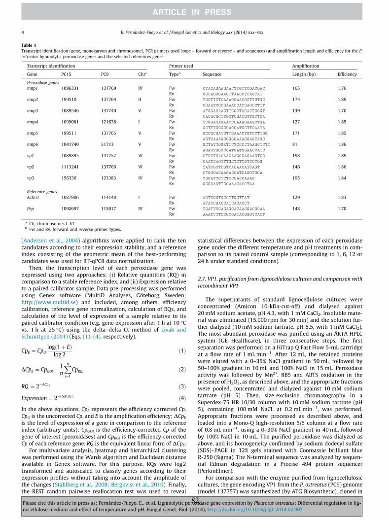
Table 1Transcript identification (gene, monokaryon and chromosome), PCR primers used (type – forward or reverse – and sequences) and amplification length and efficiency for the P.ostreatus ligninolytic peroxidase genes and the selected references genes.
Transcript identification Primer used Amplification
Gene PC15 PC9 Chra Typeb Sequence Length (bp) Efficiency
Peroxidase genesmnp1 1096331 137760 IV Fw CTACAGAAGAACTTGTTCGACGAC 165 1.76
Rv GGCAGGGAAGTTAACCTCAGTAT
mnp2 199510 137764 II Fw TGCTTCTCAAAGGAACGCTTGTGC 174 1.89Rv CGAATCGCGAAACCATGAGCCTTT
mnp3 1089546 137740 V Fw ATGGACAAGTTGGCTACACTCGGT 139 1.70Rv CACACGCTTGCTCGATGTTGTTCA
mnp4 1099081 121638 I Fw TCGGACAGAACCCAAAGAAGCTGA 127 1.85Rv GCTTTGCGGCAGGATGCTTCAATA
mnp5 199511 137765 V Fw GCCGCGATGTTGAAACTGTCTTTGG 171 1.85Rv AGTCAAAGCGGGGAAAGGAGTAGC
mnp6 1041740 51713 V Fw GCTATTGGATTCTCCCCTAAGCTCTT 81 1.86Rv AGAGTGGGCCATGATGGAACCATC
vp1 1089895 137757 VI Fw CTCCTGACAACAAGGGAGAAGTCC 198 1.89Rv CAATCAGTTTGCTCTTGTCCTGG
vp2 1113241 137766 VI Fw TATCGCTCGTCACAACATCAGT 146 1.86Rv CTGGGACAAGACCATCAGGTGGA
vp3 156336 123383 IV Fw TGGATTCTCTCCCACCAAAG 195 1.84Rv GGGCAGTTGGAAACACCTAA
Reference genesActin1 1087906 114148 I Fw AGTCGGTGCCTTGGTTAT 129 1.83
Rv ATACCGACCATCACACCT
Pep 1092697 115017 IV Fw TGATTCCAGAGGACAAGGACGCAA 148 1.70Rv AAATCTTCCGCGATACGGGTCACT
a Ch, chromosomes I–VI.b Fw and Rv, forward and reverse primer types.
4 E. Fernández-Fueyo et al. / Fungal Genetics and Biology xxx (2014) xxx–xxx
(Andersen et al., 2004) algorithms were applied to rank the tencandidates according to their expression stability, and a referenceindex consisting of the geometric mean of the best-performingcandidates was used for RT-qPCR data normalization.
Then, the transcription level of each peroxidase gene wasexpressed using two approaches: (i) Relative quantities (RQ) incomparison to a stable reference index, and (ii) Expression relativeto a paired calibrator sample. Data pre-processing was performedusing Genex software (MultiD Analyses, Göteborg, Sweden;http://www.multid.se) and included, among others, efficiencycalibration, reference gene normalization, calculation of RQs, andcalculation of the level of expression of a sample relative to itspaired calibrator condition (e.g. gene expression after 1 h at 10 �Cvs. 1 h at 25 �C) using the delta–delta Ct method of Livak andSchmittgen (2001) (Eqs. (1)–(4), respectively).
CpE ¼ CpUlogð1þ EÞ
log 2ð1Þ
DCpE ¼ CpGOI �1n
Xn
i¼1
CpRGi ð2Þ
RQ ¼ 2�DCpE ð3Þ
Expression ¼ 2�ðDDCpEÞ ð4Þ
In the above equations, CpE represents the efficiency corrected Cp.CpU is the uncorrected Cp, and E is the amplification efficiency; DCpE
is the level of expression of a gene in comparison to the referenceindex (arbitrary units); CpGOI is the efficiency-corrected Cp of thegene of interest (peroxidases) and CpRGi is the efficiency-correctedCp of each reference gene. RQ is the equivalent linear form of DCpE.
For multivariate analysis, heatmap and hierarchical clusteringwas performed using the Wards algorithm and Euclidean distanceavailable in Genex software. For this purpose, RQs were log2transformed and autoscaled to classify genes according to theirexpression profiles without taking into account the amplitude ofthe changes (Stahlberg et al., 2008; Bergkvist et al., 2010). Finally,the REST random pairwise reallocation test was used to reveal
Please cite this article in press as: Fernández-Fueyo, E., et al. Ligninolytic peroxnocellulose medium and effect of temperature and pH. Fungal Genet. Biol. (20
82
statistical differences between the expression of each peroxidasegene under the different temperature and pH treatments in com-parison to its paired control sample (corresponding to 1, 6, 12 or24 h under standard conditions).
2.7. VP1. purification from lignocellulose cultures and comparison withrecombinant VP1
The supernatants of standard lignocellulose cultures wereconcentrated (Amicon 10-kDa-cut-off) and dialysed against20 mM sodium acetate, pH 4.3, with 1 mM CaCl2. Insoluble mate-rial was eliminated (15,000 rpm for 30 min) and the solution fur-ther dialysed (10 mM sodium tartrate, pH 5.5, with 1 mM CaCl2).The most abundant peroxidase was purified using an ÄKTA HPLCsystem (GE Healthcare), in three consecutive steps. The firstseparation was performed on a HiTrap Q Fast Flow 5-mL cartridgeat a flow rate of 1 mL min�1. After 12 mL, the retained proteinswere eluted with a 0–35% NaCl gradient in 50 mL, followed by50–100% gradient in 10 mL and 100% NaCl in 15 mL. Peroxidaseactivity was followed by Mn2+, RB5 and ABTS oxidation in thepresence of H2O2, as described above, and the appropriate fractionswere pooled, concentrated and dialyzed against 10 mM sodiumtartrate (pH 5). Then, size-exclusion chromatography in aSuperdex-75 HR 10/30 column with 10 mM sodium tartrate (pH5), containing 100 mM NaCl, at 0.2 mL min�1, was performed.Appropriate fractions were processed as described above, andloaded into a Mono-Q high-resolution 5/5 column at a flow rateof 0.8 mL min�1, using a 0–30% NaCl gradient in 40 mL, followedby 100% NaCl in 10 mL. The purified peroxidase was dialyzed asabove, and its homogeneity confirmed by sodium dodecyl sulfate(SDS)–PAGE in 12% gels stained with Coomassie brilliant blueR-250 (Sigma). The N-terminal sequence was analyzed by sequen-tial Edman degradation in a Procise 494 protein sequencer(PerkinElmer).
For comparison with the enzyme purified from lignocellulosiccultures, the gene encoding VP1 from the P. ostreatus (PC9) genome(model 137757) was synthesized (by ATG Biosynthetic), cloned in
idase gene expression by Pleurotus ostreatus: Differential regulation in lig-14), http://dx.doi.org/10.1016/j.fgb.2014.02.003
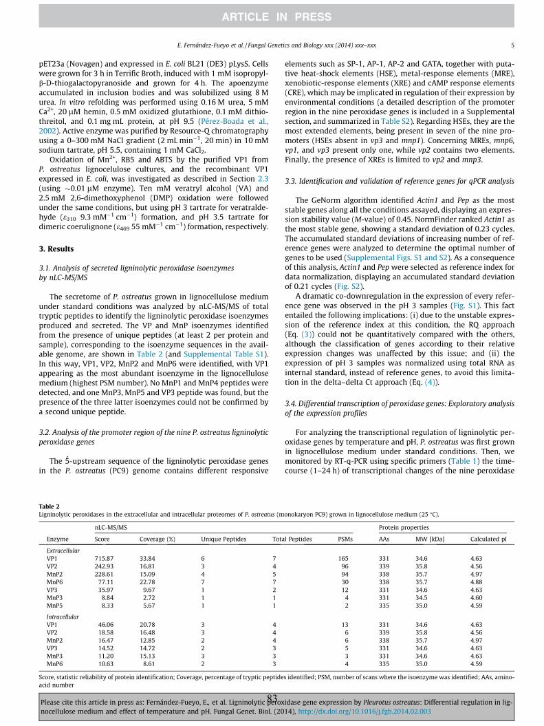
E. Fernández-Fueyo et al. / Fungal Genetics and Biology xxx (2014) xxx–xxx 5
pET23a (Novagen) and expressed in E. coli BL21 (DE3) pLysS. Cellswere grown for 3 h in Terrific Broth, induced with 1 mM isopropyl-b-D-thiogalactopyranoside and grown for 4 h. The apoenzymeaccumulated in inclusion bodies and was solubilized using 8 Murea. In vitro refolding was performed using 0.16 M urea, 5 mMCa2+, 20 lM hemin, 0.5 mM oxidized glutathione, 0.1 mM dithio-threitol, and 0.1 mg mL protein, at pH 9.5 (Pérez-Boada et al.,2002). Active enzyme was purified by Resource-Q chromatographyusing a 0–300 mM NaCl gradient (2 mL min�1, 20 min) in 10 mMsodium tartrate, pH 5.5, containing 1 mM CaCl2.
Oxidation of Mn2+, RB5 and ABTS by the purified VP1 fromP. ostreatus lignocelulose cultures, and the recombinant VP1expressed in E. coli, was investigated as described in Section 2.3(using �0.01 lM enzyme). Ten mM veratryl alcohol (VA) and2.5 mM 2,6-dimethoxyphenol (DMP) oxidation were followedunder the same conditions, but using pH 3 tartrate for veratralde-hyde (e310 9.3 mM�1 cm�1) formation, and pH 3.5 tartrate fordimeric coerulignone (e469 55 mM�1 cm�1) formation, respectively.
3. Results
3.1. Analysis of secreted ligninolytic peroxidase isoenzymesby nLC-MS/MS
The secretome of P. ostreatus grown in lignocellulose mediumunder standard conditions was analyzed by nLC-MS/MS of totaltryptic peptides to identify the ligninolytic peroxidase isoenzymesproduced and secreted. The VP and MnP isoenzymes identifiedfrom the presence of unique peptides (at least 2 per protein andsample), corresponding to the isoenzyme sequences in the avail-able genome, are shown in Table 2 (and Supplemental Table S1).In this way, VP1, VP2, MnP2 and MnP6 were identified, with VP1appearing as the most abundant isoenzyme in the lignocellulosemedium (highest PSM number). No MnP1 and MnP4 peptides weredetected, and one MnP3, MnP5 and VP3 peptide was found, but thepresence of the three latter isoenzymes could not be confirmed bya second unique peptide.
3.2. Analysis of the promoter region of the nine P. ostreatus ligninolyticperoxidase genes
The 5-upstream sequence of the ligninolytic peroxidase genesin the P. ostreatus (PC9) genome contains different responsive
Table 2Ligninolytic peroxidases in the extracellular and intracellular proteomes of P. ostreatus (m
nLC-MS/MS
Enzyme Score Coverage (%) Unique Peptides Tota
ExtracellularVP1 715.87 33.84 6 7VP2 242.93 16.81 3 4MnP2 228.61 15.09 4 5MnP6 77.11 22.78 7 7VP3 35.97 9.67 1 2MnP3 8.84 2.72 1 1MnP5 8.33 5.67 1 1
IntracellularVP1 46.06 20.78 3 4VP2 18.58 16.48 3 4MnP2 16.47 12.85 2 4VP3 14.52 14.72 2 3MnP3 11.20 15.13 3 3MnP6 10.63 8.61 2 3
Score, statistic reliability of protein identification; Coverage, percentage of tryptic peptideacid number
Please cite this article in press as: Fernández-Fueyo, E., et al. Ligninolytic peroxnocellulose medium and effect of temperature and pH. Fungal Genet. Biol. (20
83
elements such as SP-1, AP-1, AP-2 and GATA, together with puta-tive heat-shock elements (HSE), metal-response elements (MRE),xenobiotic-response elements (XRE) and cAMP response elements(CRE), which may be implicated in regulation of their expression byenvironmental conditions (a detailed description of the promoterregion in the nine peroxidase genes is included in a Supplementalsection, and summarized in Table S2). Regarding HSEs, they are themost extended elements, being present in seven of the nine pro-moters (HSEs absent in vp3 and mnp1). Concerning MREs, mnp6,vp1, and vp3 present only one, while vp2 contains two elements.Finally, the presence of XREs is limited to vp2 and mnp3.
3.3. Identification and validation of reference genes for qPCR analysis
The GeNorm algorithm identified Actin1 and Pep as the moststable genes along all the conditions assayed, displaying an expres-sion stability value (M-value) of 0.45. NormFinder ranked Actin1 asthe most stable gene, showing a standard deviation of 0.23 cycles.The accumulated standard deviations of increasing number of ref-erence genes were analyzed to determine the optimal number ofgenes to be used (Supplemental Figs. S1 and S2). As a consequenceof this analysis, Actin1 and Pep were selected as reference index fordata normalization, displaying an accumulated standard deviationof 0.21 cycles (Fig. S2).
A dramatic co-downregulation in the expression of every refer-ence gene was observed in the pH 3 samples (Fig. S1). This factentailed the following implications: (i) due to the unstable expres-sion of the reference index at this condition, the RQ approach(Eq. (3)) could not be quantitatively compared with the others,although the classification of genes according to their relativeexpression changes was unaffected by this issue; and (ii) theexpression of pH 3 samples was normalized using total RNA asinternal standard, instead of reference genes, to avoid this limita-tion in the delta–delta Ct approach (Eq. (4)).
3.4. Differential transcription of peroxidase genes: Exploratory analysisof the expression profiles
For analyzing the transcriptional regulation of ligninolytic per-oxidase genes by temperature and pH, P. ostreatus was first grownin lignocellulose medium under standard conditions. Then, wemonitored by RT-q-PCR using specific primers (Table 1) the time-course (1–24 h) of transcriptional changes of the nine peroxidase
onokaryon PC9) grown in lignocellulose medium (25 �C).
Protein properties
l Peptides PSMs AAs MW [kDa] Calculated pI
165 331 34.6 4.6396 339 35.8 4.5694 338 35.7 4.9730 338 35.7 4.8812 331 34.6 4.63
4 331 34.5 4.602 335 35.0 4.59
13 331 34.6 4.636 339 35.8 4.566 338 35.7 4.975 331 34.6 4.633 331 34.6 4.634 335 35.0 4.59
s identified; PSM, number of scans where the isoenzyme was identified; AAs, amino-
idase gene expression by Pleurotus ostreatus: Differential regulation in lig-14), http://dx.doi.org/10.1016/j.fgb.2014.02.003
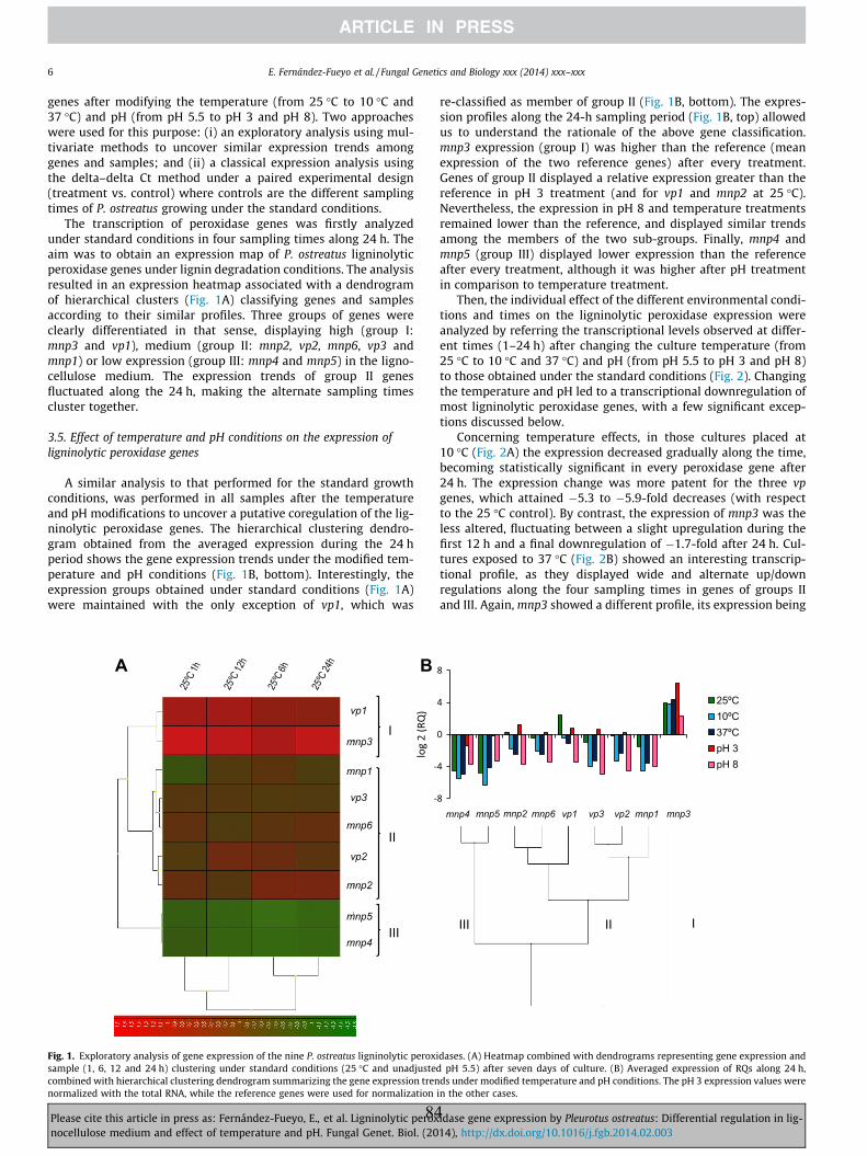
6 E. Fernández-Fueyo et al. / Fungal Genetics and Biology xxx (2014) xxx–xxx
genes after modifying the temperature (from 25 �C to 10 �C and37 �C) and pH (from pH 5.5 to pH 3 and pH 8). Two approacheswere used for this purpose: (i) an exploratory analysis using mul-tivariate methods to uncover similar expression trends amonggenes and samples; and (ii) a classical expression analysis usingthe delta–delta Ct method under a paired experimental design(treatment vs. control) where controls are the different samplingtimes of P. ostreatus growing under the standard conditions.
The transcription of peroxidase genes was firstly analyzedunder standard conditions in four sampling times along 24 h. Theaim was to obtain an expression map of P. ostreatus ligninolyticperoxidase genes under lignin degradation conditions. The analysisresulted in an expression heatmap associated with a dendrogramof hierarchical clusters (Fig. 1A) classifying genes and samplesaccording to their similar profiles. Three groups of genes wereclearly differentiated in that sense, displaying high (group I:mnp3 and vp1), medium (group II: mnp2, vp2, mnp6, vp3 andmnp1) or low expression (group III: mnp4 and mnp5) in the ligno-cellulose medium. The expression trends of group II genesfluctuated along the 24 h, making the alternate sampling timescluster together.
3.5. Effect of temperature and pH conditions on the expression ofligninolytic peroxidase genes
A similar analysis to that performed for the standard growthconditions, was performed in all samples after the temperatureand pH modifications to uncover a putative coregulation of the lig-ninolytic peroxidase genes. The hierarchical clustering dendro-gram obtained from the averaged expression during the 24 hperiod shows the gene expression trends under the modified tem-perature and pH conditions (Fig. 1B, bottom). Interestingly, theexpression groups obtained under standard conditions (Fig. 1A)were maintained with the only exception of vp1, which was
A B
I
II
III
vp1
mnp3
mnp1
vp3
mnp6
vp2
mnp2
mnp5
mnp4
Fig. 1. Exploratory analysis of gene expression of the nine P. ostreatus ligninolytic peroxisample (1, 6, 12 and 24 h) clustering under standard conditions (25 �C and unadjustecombined with hierarchical clustering dendrogram summarizing the gene expression trennormalized with the total RNA, while the reference genes were used for normalization
Please cite this article in press as: Fernández-Fueyo, E., et al. Ligninolytic peroxnocellulose medium and effect of temperature and pH. Fungal Genet. Biol. (20
84
re-classified as member of group II (Fig. 1B, bottom). The expres-sion profiles along the 24-h sampling period (Fig. 1B, top) allowedus to understand the rationale of the above gene classification.mnp3 expression (group I) was higher than the reference (meanexpression of the two reference genes) after every treatment.Genes of group II displayed a relative expression greater than thereference in pH 3 treatment (and for vp1 and mnp2 at 25 �C).Nevertheless, the expression in pH 8 and temperature treatmentsremained lower than the reference, and displayed similar trendsamong the members of the two sub-groups. Finally, mnp4 andmnp5 (group III) displayed lower expression than the referenceafter every treatment, although it was higher after pH treatmentin comparison to temperature treatment.
Then, the individual effect of the different environmental condi-tions and times on the ligninolytic peroxidase expression wereanalyzed by referring the transcriptional levels observed at differ-ent times (1–24 h) after changing the culture temperature (from25 �C to 10 �C and 37 �C) and pH (from pH 5.5 to pH 3 and pH 8)to those obtained under the standard conditions (Fig. 2). Changingthe temperature and pH led to a transcriptional downregulation ofmost ligninolytic peroxidase genes, with a few significant excep-tions discussed below.
Concerning temperature effects, in those cultures placed at10 �C (Fig. 2A) the expression decreased gradually along the time,becoming statistically significant in every peroxidase gene after24 h. The expression change was more patent for the three vpgenes, which attained �5.3 to �5.9-fold decreases (with respectto the 25 �C control). By contrast, the expression of mnp3 was theless altered, fluctuating between a slight upregulation during thefirst 12 h and a final downregulation of �1.7-fold after 24 h. Cul-tures exposed to 37 �C (Fig. 2B) showed an interesting transcrip-tional profile, as they displayed wide and alternate up/downregulations along the four sampling times in genes of groups IIand III. Again, mnp3 showed a different profile, its expression being
25ºC10ºC37ºCpH 3pH 8
mnp4
IIIII I
mnp3mnp1vp2vp3vp1mnp6mnp2mnp5
dases. (A) Heatmap combined with dendrograms representing gene expression andd pH 5.5) after seven days of culture. (B) Averaged expression of RQs along 24 h,ds under modified temperature and pH conditions. The pH 3 expression values were
in the other cases.
idase gene expression by Pleurotus ostreatus: Differential regulation in lig-14), http://dx.doi.org/10.1016/j.fgb.2014.02.003
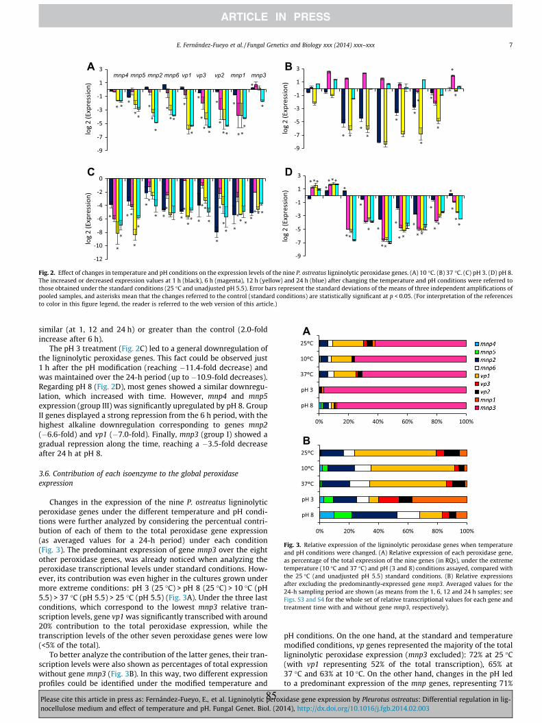
C D
BA
Fig. 2. Effect of changes in temperature and pH conditions on the expression levels of the nine P. ostreatus ligninolytic peroxidase genes. (A) 10 �C. (B) 37 �C. (C) pH 3. (D) pH 8.The increased or decreased expression values at 1 h (black), 6 h (magenta), 12 h (yellow) and 24 h (blue) after changing the temperature and pH conditions were referred tothose obtained under the standard conditions (25 �C and unadjusted pH 5.5). Error bars represent the standard deviations of the means of three independent amplifications ofpooled samples, and asterisks mean that the changes referred to the control (standard conditions) are statistically significant at p < 0.05. (For interpretation of the referencesto color in this figure legend, the reader is referred to the web version of this article.)
Fig. 3. Relative expression of the ligninolytic peroxidase genes when temperatureand pH conditions were changed. (A) Relative expression of each peroxidase gene,as percentage of the total expression of the nine genes (in RQs), under the extremetemperature (10 �C and 37 �C) and pH (3 and 8) conditions assayed, compared withthe 25 �C (and unadjusted pH 5.5) standard conditions. (B) Relative expressionsafter excluding the predominantly-expressed gene mnp3. Averaged values for the24-h sampling period are shown (as means from the 1, 6, 12 and 24 h samples; seeFigs. S3 and S4 for the whole set of relative transcriptional values for each gene andtreatment time with and without gene mnp3, respectively).
E. Fernández-Fueyo et al. / Fungal Genetics and Biology xxx (2014) xxx–xxx 7
similar (at 1, 12 and 24 h) or greater than the control (2.0-foldincrease after 6 h).
The pH 3 treatment (Fig. 2C) led to a general downregulation ofthe ligninolytic peroxidase genes. This fact could be observed just1 h after the pH modification (reaching �11.4-fold decrease) andwas maintained over the 24-h period (up to �10.9-fold decreases).Regarding pH 8 (Fig. 2D), most genes showed a similar downregu-lation, which increased with time. However, mnp4 and mnp5expression (group III) was significantly upregulated by pH 8. GroupII genes displayed a strong repression from the 6 h period, with thehighest alkaline downregulation corresponding to genes mnp2(�6.6-fold) and vp1 (�7.0-fold). Finally, mnp3 (group I) showed agradual repression along the time, reaching a �3.5-fold decreaseafter 24 h at pH 8.
3.6. Contribution of each isoenzyme to the global peroxidaseexpression
Changes in the expression of the nine P. ostreatus ligninolyticperoxidase genes under the different temperature and pH condi-tions were further analyzed by considering the percentual contri-bution of each of them to the total peroxidase gene expression(as averaged values for a 24-h period) under each condition(Fig. 3). The predominant expression of gene mnp3 over the eightother peroxidase genes, was already noticed when analyzing theperoxidase transcriptional levels under standard conditions. How-ever, its contribution was even higher in the cultures grown undermore extreme conditions: pH 3 (25 �C) > pH 8 (25 �C) > 10 �C (pH5.5) > 37 �C (pH 5.5) > 25 �C (pH 5.5) (Fig. 3A). Under the three lastconditions, which correspond to the lowest mnp3 relative tran-scription levels, gene vp1 was significantly transcribed with around20% contribution to the total peroxidase expression, while thetranscription levels of the other seven peroxidase genes were low(<5% of the total).
To better analyze the contribution of the latter genes, their tran-scription levels were also shown as percentages of total expressionwithout gene mnp3 (Fig. 3B). In this way, two different expressionprofiles could be identified under the modified temperature and
Please cite this article in press as: Fernández-Fueyo, E., et al. Ligninolytic peroxnocellulose medium and effect of temperature and pH. Fungal Genet. Biol. (20
85
pH conditions. On the one hand, at the standard and temperaturemodified conditions, vp genes represented the majority of the totalligninolytic peroxidase expression (mnp3 excluded): 72% at 25 �C(with vp1 representing 52% of the total transcription), 65% at37 �C and 63% at 10 �C. On the other hand, changes in the pH ledto a predominant expression of the mnp genes, representing 71%
idase gene expression by Pleurotus ostreatus: Differential regulation in lig-14), http://dx.doi.org/10.1016/j.fgb.2014.02.003
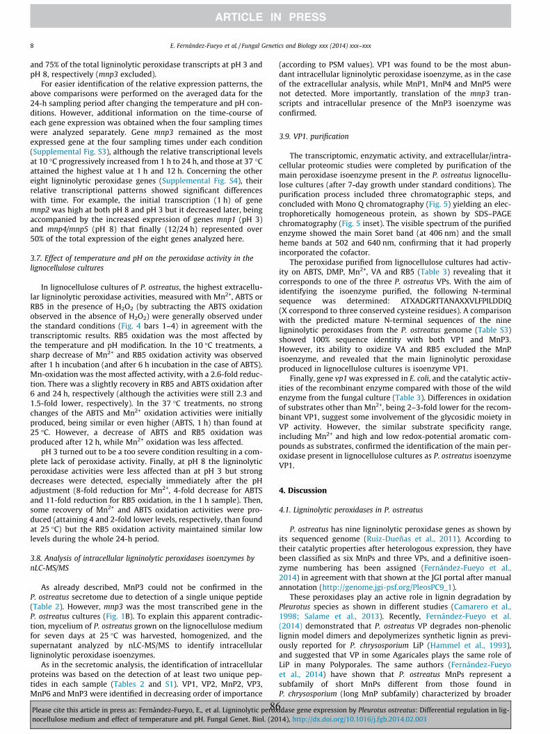
8 E. Fernández-Fueyo et al. / Fungal Genetics and Biology xxx (2014) xxx–xxx
and 75% of the total ligninolytic peroxidase transcripts at pH 3 andpH 8, respectively (mnp3 excluded).
For easier identification of the relative expression patterns, theabove comparisons were performed on the averaged data for the24-h sampling period after changing the temperature and pH con-ditions. However, additional information on the time-course ofeach gene expression was obtained when the four sampling timeswere analyzed separately. Gene mnp3 remained as the mostexpressed gene at the four sampling times under each condition(Supplemental Fig. S3), although the relative transcriptional levelsat 10 �C progressively increased from 1 h to 24 h, and those at 37 �Cattained the highest value at 1 h and 12 h. Concerning the othereight ligninolytic peroxidase genes (Supplemental Fig. S4), theirrelative transcriptional patterns showed significant differenceswith time. For example, the initial transcription (1 h) of genemnp2 was high at both pH 8 and pH 3 but it decreased later, beingaccompanied by the increased expression of genes mnp1 (pH 3)and mnp4/mnp5 (pH 8) that finally (12/24 h) represented over50% of the total expression of the eight genes analyzed here.
3.7. Effect of temperature and pH on the peroxidase activity in thelignocellulose cultures
In lignocellulose cultures of P. ostreatus, the highest extracellu-lar ligninolytic peroxidase activities, measured with Mn2+, ABTS orRB5 in the presence of H2O2 (by subtracting the ABTS oxidationobserved in the absence of H2O2) were generally observed underthe standard conditions (Fig. 4 bars 1–4) in agreement with thetranscriptomic results. RB5 oxidation was the most affected bythe temperature and pH modification. In the 10 �C treatments, asharp decrease of Mn2+ and RB5 oxidation activity was observedafter 1 h incubation (and after 6 h incubation in the case of ABTS).Mn-oxidation was the most affected activity, with a 2.6-fold reduc-tion. There was a slightly recovery in RB5 and ABTS oxidation after6 and 24 h, respectively (although the activities were still 2.3 and1.5-fold lower, respectively). In the 37 �C treatments, no strongchanges of the ABTS and Mn2+ oxidation activities were initiallyproduced, being similar or even higher (ABTS, 1 h) than found at25 �C. However, a decrease of ABTS and RB5 oxidation wasproduced after 12 h, while Mn2+ oxidation was less affected.
pH 3 turned out to be a too severe condition resulting in a com-plete lack of peroxidase activity. Finally, at pH 8 the ligninolyticperoxidase activities were less affected than at pH 3 but strongdecreases were detected, especially immediately after the pHadjustment (8-fold reduction for Mn2+, 4-fold decrease for ABTSand 11-fold reduction for RB5 oxidation, in the 1 h sample). Then,some recovery of Mn2+ and ABTS oxidation activities were pro-duced (attaining 4 and 2-fold lower levels, respectively, than foundat 25 �C) but the RB5 oxidation activity maintained similar lowlevels during the whole 24-h period.
3.8. Analysis of intracellular ligninolytic peroxidases isoenzymes bynLC-MS/MS
As already described, MnP3 could not be confirmed in theP. ostreatus secretome due to detection of a single unique peptide(Table 2). However, mnp3 was the most transcribed gene in theP. ostreatus cultures (Fig. 1B). To explain this apparent contradic-tion, mycelium of P. ostreatus grown on the lignocellulose mediumfor seven days at 25 �C was harvested, homogenized, and thesupernatant analyzed by nLC-MS/MS to identify intracellularligninolytic peroxidase isoenzymes.
As in the secretomic analysis, the identification of intracellularproteins was based on the detection of at least two unique pep-tides in each sample (Tables 2 and S1). VP1, VP2, MnP2, VP3,MnP6 and MnP3 were identified in decreasing order of importance
Please cite this article in press as: Fernández-Fueyo, E., et al. Ligninolytic peroxnocellulose medium and effect of temperature and pH. Fungal Genet. Biol. (20
86
(according to PSM values). VP1 was found to be the most abun-dant intracellular ligninolytic peroxidase isoenzyme, as in the caseof the extracellular analysis, while MnP1, MnP4 and MnP5 werenot detected. More importantly, translation of the mnp3 tran-scripts and intracellular presence of the MnP3 isoenzyme wasconfirmed.
3.9. VP1. purification
The transcriptomic, enzymatic activity, and extracellular/intra-cellular proteomic studies were completed by purification of themain peroxidase isoenzyme present in the P. ostreatus lignocellu-lose cultures (after 7-day growth under standard conditions). Thepurification process included three chromatographic steps, andconcluded with Mono Q chromatography (Fig. 5) yielding an elec-trophoretically homogeneous protein, as shown by SDS–PAGEchromatography (Fig. 5 inset). The visible spectrum of the purifiedenzyme showed the main Soret band (at 406 nm) and the smallheme bands at 502 and 640 nm, confirming that it had properlyincorporated the cofactor.
The peroxidase purified from lignocellulose cultures had activ-ity on ABTS, DMP, Mn2+, VA and RB5 (Table 3) revealing that itcorresponds to one of the three P. ostreatus VPs. With the aim ofidentifying the isoenzyme purified, the following N-terminalsequence was determined: ATXADGRTTANAXXVLFPILDDIQ(X correspond to three conserved cysteine residues). A comparisonwith the predicted mature N-terminal sequences of the nineligninolytic peroxidases from the P. ostreatus genome (Table S3)showed 100% sequence identity with both VP1 and MnP3.However, its ability to oxidize VA and RB5 excluded the MnPisoenzyme, and revealed that the main ligninolytic peroxidaseproduced in lignocellulose cultures is isoenzyme VP1.
Finally, gene vp1 was expressed in E. coli, and the catalytic activ-ities of the recombinant enzyme compared with those of the wildenzyme from the fungal culture (Table 3). Differences in oxidationof substrates other than Mn2+, being 2–3-fold lower for the recom-binant VP1, suggest some involvement of the glycosidic moiety inVP activity. However, the similar substrate specificity range,including Mn2+ and high and low redox-potential aromatic com-pounds as substrates, confirmed the identification of the main per-oxidase present in lignocellulose cultures as P. ostreatus isoenzymeVP1.
4. Discussion
4.1. Ligninolytic peroxidases in P. ostreatus
P. ostreatus has nine ligninolytic peroxidase genes as shown byits sequenced genome (Ruiz-Dueñas et al., 2011). According totheir catalytic properties after heterologous expression, they havebeen classified as six MnPs and three VPs, and a definitive isoen-zyme numbering has been assigned (Fernández-Fueyo et al.,2014) in agreement with that shown at the JGI portal after manualannotation (http://genome.jgi-psf.org/PleosPC9_1).
These peroxidases play an active role in lignin degradation byPleurotus species as shown in different studies (Camarero et al.,1998; Salame et al., 2013). Recently, Fernández-Fueyo et al.(2014) demonstrated that P. ostreatus VP degrades non-phenoliclignin model dimers and depolymerizes synthetic lignin as previ-ously reported for P. chrysosporium LiP (Hammel et al., 1993),and suggested that VP in some Agaricales plays the same role ofLiP in many Polyporales. The same authors (Fernández-Fueyoet al., 2014) have shown that P. ostreatus MnPs represent asubfamily of short MnPs different from those found inP. chrysosporium (long MnP subfamily) characterized by broader
idase gene expression by Pleurotus ostreatus: Differential regulation in lig-14), http://dx.doi.org/10.1016/j.fgb.2014.02.003
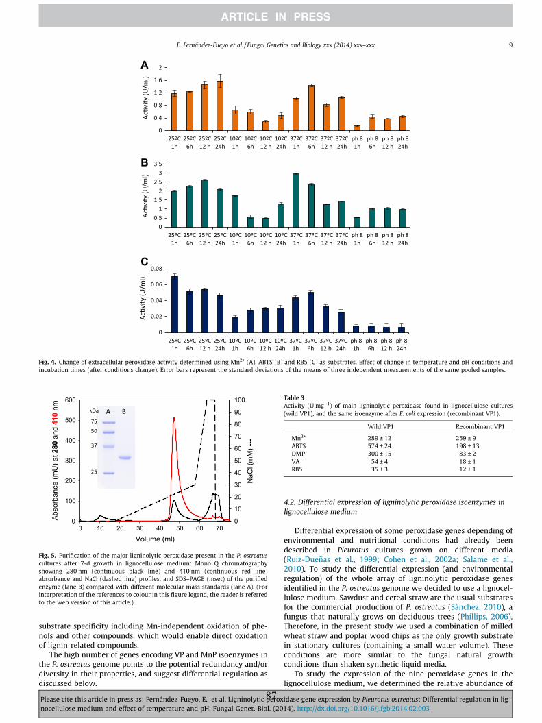
Fig. 4. Change of extracellular peroxidase activity determined using Mn2+ (A), ABTS (B) and RB5 (C) as substrates. Effect of change in temperature and pH conditions andincubation times (after conditions change). Error bars represent the standard deviations of the means of three independent measurements of the same pooled samples.
0
10
20
30
40
50
60
70
80
90
100
0
100
200
300
400
500
600
0 10 20 30 40 50 60 70
NaC
l(m
M) ---
Abso
rban
ce(m
U) a
t 280
and 410
nm
Volume (ml)
Fig. 5. Purification of the major ligninolytic peroxidase present in the P. ostreatuscultures after 7-d growth in lignocellulose medium: Mono Q chromatographyshowing 280 nm (continuous black line) and 410 nm (continuous red line)absorbance and NaCl (dashed line) profiles, and SDS–PAGE (inset) of the purifiedenzyme (lane B) compared with different molecular mass standards (lane A). (Forinterpretation of the references to colour in this figure legend, the reader is referredto the web version of this article.)
Table 3Activity (U mg�1) of main ligninolytic peroxidase found in lignocellulose cultures(wild VP1), and the same isoenzyme after E. coli expression (recombinant VP1).
Wild VP1 Recombinant VP1
Mn2+ 289 ± 12 259 ± 9ABTS 574 ± 24 198 ± 13DMP 300 ± 15 83 ± 2VA 54 ± 4 18 ± 1RB5 35 ± 3 12 ± 1
E. Fernández-Fueyo et al. / Fungal Genetics and Biology xxx (2014) xxx–xxx 9
substrate specificity including Mn-independent oxidation of phe-nols and other compounds, which would enable direct oxidationof lignin-related compounds.
The high number of genes encoding VP and MnP isoenzymes inthe P. ostreatus genome points to the potential redundancy and/ordiversity in their properties, and suggest differential regulation asdiscussed below.
Please cite this article in press as: Fernández-Fueyo, E., et al. Ligninolytic peroxnocellulose medium and effect of temperature and pH. Fungal Genet. Biol. (20
87
4.2. Differential expression of ligninolytic peroxidase isoenzymes inlignocellulose medium
Differential expression of some peroxidase genes depending ofenvironmental and nutritional conditions had already beendescribed in Pleurotus cultures grown on different media(Ruiz-Dueñas et al., 1999; Cohen et al., 2002a; Salame et al.,2010). To study the differential expression (and environmentalregulation) of the whole array of ligninolytic peroxidase genesidentified in the P. ostreatus genome we decided to use a lignocel-lulose medium. Sawdust and cereal straw are the usual substratesfor the commercial production of P. ostreatus (Sánchez, 2010), afungus that naturally grows on deciduous trees (Phillips, 2006).Therefore, in the present study we used a combination of milledwheat straw and poplar wood chips as the only growth substratein stationary cultures (containing a small water volume). Theseconditions are more similar to the fungal natural growthconditions than shaken synthetic liquid media.
To study the expression of the nine peroxidase genes in thelignocellulose medium, we determined the relative abundance of
idase gene expression by Pleurotus ostreatus: Differential regulation in lig-14), http://dx.doi.org/10.1016/j.fgb.2014.02.003
10 E. Fernández-Fueyo et al. / Fungal Genetics and Biology xxx (2014) xxx–xxx
the corresponding transcripts by RT-qPCR using selected referencegenes. In terms of studying the effect of different factors on tran-script level, RT-qPCR has been shown to be 103–104 more sensitivethan Northern blotting (Sooknanan et al., 1993). According to thepattern of expression (abundance of transcripts) at the standardgrowth conditions, the ligninolytic peroxidase genes of P. ostreatuswere divided into three different groups, being mnp3 and vp1 themost expressed genes. This agrees with previous studies usingother culture media (such as glucose-peptone medium amendedwith Mn2+). Among them, Irie et al. (2000) described MnP3 asthe major MnP isoenzyme in P. ostreatus, and Salame et al. (2012,2013) described vp1 (mnp4 for these authors) and mnp3, togetherwith mnp2 (mnp9 for these authors), as the most transcribed P.ostreatus PC9 genes. On the other hand, mnp4 and mnp5 appearedas the least expressed genes at the standard conditions used in thepresent study.
4.3. Environmental regulation of ligninolytic peroxidase isoenzymes bytemperature and pH
Two extreme temperature and pH conditions were selected tostudy the environmental regulation of the nine ligninolytic perox-idase genes in P. ostreatus, in connection with previous studies(Fernández-Fueyo et al., 2014). Previous observations had shownthat 10 �C and 37 �C are the most extreme temperatures, in whichthis fungus continues growing. On the other hand, although thehighest fungal ligninolysis and activity of ligninolytic enzymes(LiP and VP) is often produced at acidic conditions (Ruiz-Dueñasand Martínez, 2009), pH 3 always results in progressive peroxidaseinactivation, as shown for most of the P. ostreatus isoenzymes(Fernández-Fueyo et al., 2014). The same study also showed thatpH 8 inactivates these peroxidases, except the most stable isoen-zymes, due to loss of structural Ca2+ ions and formation of bis(his-tidyl) heme iron complexes. Therefore, to study environmentalregulation, P. ostreatus cultures pregrown under standard condi-tions were transferred to 10 �C and 37 �C or the medium pHadjusted to pH 3 and pH 8, and the peroxidase transcriptionallevels (during the subsequent 24 h) referred to those of culturesmaintained under unchanged conditions.
Modifications of temperature and pH led to a downregulation ofmost ligninolytic peroxidase genes, although the individual geneswere differentially affected. In general, the pH modifications pro-duced more severe effects than the temperature modifications,and vp genes were more affected than mnp genes. Among the lat-ter, mnp3 appears as the most transcribed ligninolytic peroxidasegene in all the conditions, and the least downregulated by temper-ature modification. pH 3 seems to be a too stressful condition forthe fungus, with a downregulation of more than 10 folds in allgenes after only 1 h incubation. On the other hand, while all vpgenes were dramatically downregulated at pH 8, mnp4 and mnp5were upregulated under these conditions. Interestingly, MnP4has been reported as the most stable P. ostreatus peroxidase isoen-zyme under alkaline conditions (Fernández-Fueyo et al., 2014).
Concerning temperature, all genes except mnp3 were downreg-ulated at 10 �C. Conversely, at 37 �C most of the genes were upreg-ulated after 24 h, with the exception of mnp2, mnp6 and mnp1. Thisgeneral upregulation at 37 �C could be related to HSEs, which arepresent in all the P. ostreatus peroxidase promoters except in thoseof mnp1 and vp3. The existence of HSEs has been reported in thepromoters of other ligninolytic peroxidase genes, such asP. chrysosporium mnp (Godfrey et al., 1994; Mayfield et al., 1994).No correlation between the thermal stability of the differentisoenzymes (Fernández-Fueyo et al., 2014) and the expressionlevels of the nine ligninolytic peroxidase genes was observed underthe present experimental conditions.
Please cite this article in press as: Fernández-Fueyo, E., et al. Ligninolytic peroxnocellulose medium and effect of temperature and pH. Fungal Genet. Biol. (20
88
4.4. Activity levels, proteomes, and enzyme purification fromlignocellulose cultures
In the P. ostreatus lignocellulose cultures under standard condi-tions, the maximal peroxidase activity coincided with the highestexpression of ligninolytic peroxidase genes. In most of the cases,there was also a correlation between the expression and activitylevels in the modified temperature and pH conditions. RegardingpH, no activity was detected for any substrate at pH 3, where thefungus could not survive and the peroxidase expression sharplydecreases. On the other hand, RB5 oxidation (a specific VP reaction)was the most affected activity at pH 8, in agreement with the high-er decrease of vp than mnp transcripts. In the case of temperature,the lack of activity in the cultures transferred at 10 �C, a tempera-ture at which these peroxidases are fully stable (Fernández-Fueyoet al., 2014) correlates with the strong decrease of all transcriptlevels. Thus, it seems that a constant production of the enzyme isnecessary to maintain the activity. At 37 �C, while no strongchanges in Mn2+ oxidation were produced, there was a decreasein ABTS and RB5 oxidation activities (after 12 h) which coincidedwith the stronger reduction in vp expression.
In previous studies, only VP1 (Sarkar et al., 1997; Kamitsujiet al., 2004), VP2 (Kamitsuji et al., 2004) and MnP3 (Cohen et al.,2001, 2002b) had been purified from P. ostreatus cultures, suggest-ing that they are the most abundant ligninolytic peroxidases in thedifferent culture conditions assayed. In our case, only VP1 waspurified from lignocellulose fungal cultures, highlighting the roleof VPs in lignin degradation by this fungus (Fernández-Fueyoet al., 2014). By nLC-MS/MS, a highly sensitive technique, VP2,MnP2 and MnP6 were also detected, and a single unique peptidewas found for MnP3, MnP5 and VP3 (while MnP4 and MnP1 werenot detected). In a similar study on P. ostreatus secretome (fromMn2+-amended glucose-peptone medium) only MnP3, VP1 andMnP2 were detected by Salame et al. (2013) (the two latter isoen-zymes respectively called VP4 and MnP9 by these authors).
The secretome results mostly agree with the transcript levels at25 �C except for gene mnp3 that showed the highest transcriptionlevel, but whose protein product could not be confirmed by a sec-ond unique peptide. However, the intracellular presence of MnP3was shown by the nLC-MS/MS analysis of the corresponding prote-ome, revealing a limited secretion of this isoenzyme. A similar casewas reported for a P. ostreatus laccase (POXA1b) that shows thehighest transcription levels in copper-supplemented cultures, butit is not a major extracellular protein due to inefficient secretion(it appears in the cellular extract) and the action of specific prote-ases (Palmieri et al., 2000).
Although all the ligninolytic peroxidase genes are transcribed inthe P. ostreatus lignocellulose cultures, those peroxidases beingabsent from both the intracellular/extracellular secretomes(MnP4 and MnP1) correspond to the less transcribed genes at thestandard growth conditions (together with gene mnp5). Anotherpossible reason for the absence of these two peroxidases fromthe proteomes is that MnP4 has the highest number of lysineresidues (20) followed by MnP1, a fact that would difficultnLC-MS/MS identification due to the high number and small sizeof tryptic peptides. Moreover, Salame et al. (2013) proposed thatMnP4 (called MnP7 in this study) could be anchored to themembrane, according to the prediction of a transmembranedomain that would limit its secretion.
5. Conclusions
The main ligninolytic peroxidase genes expressed in P. ostreatuscultures with lignocellulose as the sole carbon (and nitrogen)source were investigated by transcriptomic, secretomic andenzyme purification studies. Genes mnp3 and vp1 showed the
idase gene expression by Pleurotus ostreatus: Differential regulation in lig-14), http://dx.doi.org/10.1016/j.fgb.2014.02.003
E. Fernández-Fueyo et al. / Fungal Genetics and Biology xxx (2014) xxx–xxx 11
highest transcription levels, but the presence of isoenzyme MnP3in the cultures seems affected by a limited secretion (as shownby comparison of the intracellular and extracellular proteomes).However, the product of gene vp1 was the most abundant proteinin the secretome and the only peroxidase purified from theP. ostreatus lignocellulose cultures. For the rest of genes therewas also a general correlation between the level of transcriptsand the secretome abundance. Expression of the P. ostreatus sixmnp and the three vp genes was differentially affected by changesin the culture temperature and pH. Although an increase oftemperature (to 37 �C) caused a transient increase of gene expres-sion with respect to 25 �C (6 h sample), a general downregulationwas observed after 24 h at 37 �C, as well as in nearly all the sam-ples from the 10 �C treatments. Concerning the effect of pH, allthe ligninolytic peroxidase genes were downregulated at pH 3 withrespect to the standard conditions (pH 5.5). Finally, pH 8 downreg-ulated all the genes but mnp4 and mnp5, whose upregulation byalkaline pH constitutes an intriguing finding since most ligninolyt-ic peroxidases are inactivated over pH 7. However, it agrees withprevious studies that described the unusual alkaline stability ofthe MnP4 isoenzyme from the P. ostreatus genome.
Acknowledgments
This work was supported by the PEROXICATS (KBBE-2010-4-265397; www.peroxicats.org) and INDOX (KBBE-2013-7-613549;www.indoxproject.eu) EU projects, and by the BIO2011-26694and AGL2011-30495 projects of the Spanish Ministry of Economyand Competitiveness (MINECO). EF-F acknowledges a Junta deAmpliación de Estudios fellowship of the CSIC, co-funded by theEuropean Social Fund, and FJR-D acknowledges a MINECO Ramóny Cajal contract.
Appendix A. Supplementary material
Supplemental Information concerns gene promoters, secretomepeptides, N-terminus of purified peroxidase, crossing point andstability of reference genes, and expression of ligninolytic peroxi-dase genes at different times, including a section on the promoterregion of the ligninolytic peroxidase genes, the peroxidase peptidesequences identified in the secretome of P. ostreatus (Table S1), alist of putative regulatory elements in the different promoters(Table S2), the N-terminal sequence identity of the peroxidasepurified from lignocellulose cultures (Table S3), the crossing pointdistribution of ten candidate reference genes at different tempera-ture and pH conditions (Fig. S1), the expression stability of theseten genes using GeNorm and Normfinder (Fig. S2), the relativeexpression of the nine ligninolytic peroxidase genes at differenttimes after changing the incubation conditions (Fig. S3) and thesame information excluding mnp3 (Fig. S4). Supplementary dataassociated with this article can be found, in the online version, athttp://dx.doi.org/10.1016/j.fgb.2014.02.003.
References
Andersen, C.L., Jensen, J.L., Orntoft, T.F., 2004. Normalization of real-timequantitative reverse transcription-PCR data: a model-based varianceestimation approach to identify genes suited for normalization, applied tobladder and colon cancer data sets. Cancer Res. 64, 5245–5250.
Bergkvist, A., Rusnakova, V., Sindelka, R., Garda, J.M.A., Sjogreen, B., Lindh, D.,Forootan, A., Kubista, M., 2010. Gene expression profiling – clusters ofpossibilities. Methods 50, 323–335.
Camarero, S., Barrasa, J.M., Pelayo, M., Martínez, A.T., 1998. Evaluation of Pleurotusspecies for wheat–straw biopulping. J. Pulp Paper Sci. 24, 197–203.
Castanera, R., Pérez, G., Omarini, A., Alfaro, M., Pisabarro, A.G., Faraco, V., Amore, A.,Ramírez, L., 2012. Transcriptional and enzymatic profiling of Pleurotus ostreatuslaccase genes in submerged and solid-state fermentation cultures. Appl.Environ. Microbiol. 78, 4037–4045.
Please cite this article in press as: Fernández-Fueyo, E., et al. Ligninolytic peroxnocellulose medium and effect of temperature and pH. Fungal Genet. Biol. (20
89
Castanera, R., Omarini, A., Santoyo, F., Pérez, G., Pisabarro, A.G., Ramírez, L., 2013.Non-additive transcriptional profiles underlie dikaryotic superiority in Pleurotusostreatus laccase activity. PLoS ONE 8, 9.
Cohen, R., Hadar, Y., Yarden, O., 2001. Transcript and activity levels of differentPleurotus ostreatus peroxidases are differentially affected by Mn2+. Environ.Microbiol. 3, 312–322.
Cohen, R., Persky, L., Hadar, Y., 2002a. Biotechnological applications and potential ofwood-degrading mushrooms of the genus Pleurotus. Appl. Microbiol.Biotechnol. 58, 582–594.
Cohen, R., Persky, L., Hazan-Eitan, Z., Yarden, C., Hadar, Y., 2002b. Mn2+ altersperoxidase profiles and lignin degradation by the white-rot fungus Pleurotusostreatus under different nutritional and growth conditions. Appl. Biochem.Biotechnol. 102, 415–429.
Farrell, R.L., Murtagh, K.E., Tien, M., Mozuch, M.D., Kirk, K.T., 1989. Physical andenzymatic properties of lignin peroxidase isoenzymes from Phanerochaetechrysosporium. Enzyme Microb. Technol. 11, 322–328.
Fernández-Fueyo, E., Ruiz-Dueñas, F.J., Ferreira, P., Floudas, D., Hibbett, D.S.,Canessa, P., Larrondo, L., James, T.Y., Seelenfreund, D., Lobos, S., Polanco, R.,Tello, M., Honda, Y., Watanabe, T., Watanabe, T., Ryu, J.S., Kubicek, C.P., Schmoll,M., Gaskell, J., Hammel, K.E., St. John, F.J., Vanden Wymelenberg, A., Sabat, G.,Bondurant, S.S., Syed, K., Yadav, J., Doddapaneni, H., Subramanian, V., Lavín, J.L.,Oguiza, J.A., Perez, G., Pisabarro, A.G., Ramírez, L., Santoyo, F., Master, E.,Coutinho, P.M., Henrissat, B., Lombard, V., Magnuson, J.K., Kües, U., Hori, C.,Igarashi, K., Samejima, M., Held, B.W., Barry, K., LaButti, K., Lapidus, A.,Lindquist, E., Lucas, S., Riley, R., Salamov, A., Hoffmeister, D., Schwenk, D.,Hadar, Y., Yarden, O., de Vries, R.P., Wiebenga, A., Stenlid, J., Eastwood, D.C.,Grigoriev, I.V., Berka, R., Blanchette, R.A., Kersten, P., Martínez, A.T., Vicuña, R.,Cullen, D., 2012. Comparative genomics of Ceriporiopisis subvermispora andPhanerochaete chrysosporium provide insight into selective ligninolysis. Proc.Natl. Acad. Sci. USA 109, 5458–5463.
Fernández-Fueyo, E., Ruiz-Dueñas, F.J., Martínez, M.J., Romero, A., Hammel, K.E.,Medrano, F.J., Martínez, A.T., 2014. Ligninolytic peroxidase genes in the oystermushroom genome: heterologous expression, molecular structure, catalyticand stability properties and lignin-degrading ability. Biotechnol. Biofuels 7, 2.
Floudas, D., Binder, M., Riley, R., Barry, K., Blanchette, R.A., Henrissat, B., Martínez,A.T., Otillar, R., Spatafora, J.W., Yadav, J.S., Aerts, A., Benoit, I., Boyd, A., Carlson,A., Copeland, A., Coutinho, P.M., de Vries, R.P., Ferreira, P., Findley, K., Foster, B.,Gaskell, J., Glotzer, D., Górecki, P., Heitman, J., Hesse, C., Hori, C., Igarashi, K.,Jurgens, J.A., Kallen, N., Kersten, P., Kohler, A., Kües, U., Kumar, T.K.A., Kuo, A.,LaButti, K., Larrondo, L.F., Lindquist, E., Ling, A., Lombard, V., Lucas, S., Lundell, T.,Martin, R., McLaughlin, D.J., Morgenstern, I., Morin, E., Murat, C., Nolan, M.,Ohm, R.A., Patyshakuliyeva, A., Rokas, A., Ruiz-Dueñas, F.J., Sabat, G., Salamov,A., Samejima, M., Schmutz, J., Slot, J.C., St John, F., Stenlid, J., Sun, H., Sun, S., Syed,K., Tsang, A., Wiebenga, A., Young, D., Pisabarro, A., Eastwood, D.C., Martin, F.,Cullen, D., Grigoriev, I.V., Hibbett, D.S., 2012. The Paleozoic origin of enzymaticlignin decomposition reconstructed from 31 fungal genomes. Science 336,1715–1719.
Glumoff, T., Harvey, P.J., Molinari, S., Goble, M., Frank, G., Palmer, J.M., Smit, J.D.G.,Leisola, M.S.A., 1990. Lignin peroxidase from Phanerochaete chrysosporium –molecular and kinetic characterization of isozymes. Eur. J. Biochem. 187, 515–520.
Godfrey, B.J., Akileswaran, L., Gold, M.H., 1994. A reporter gene construct forstudying the regulation of manganese peroxidase gene expression. Appl.Environ. Microbiol. 60, 1353–1358.
Hammel, K.E., Jensen, K.A., Mozuch, M.D., Landucci, L.L., Tien, M., Pease, E.A., 1993.Ligninolysis by a purified lignin peroxidase. J. Biol. Chem. 268, 12274–12281.
Heid, C.A., Stevens, J., Livak, K.J., Williams, P.M., 1996. Real time quantitative PCR.Genome Res. 6, 986–994.
Irie, T., Honda, Y., Ha, H.-C., Watanabe, T., Kuwahara, M., 2000. Isolation of cDNA andgenomic fragments encoding the major manganese peroxidase isoenzyme fromthe white rot basidiomycete Pleurotus ostreatus. J. Wood Sci. 46, 230–233.
Janusz, G., Kucharzyk, K.H., Pawlik, A., Staszczak, M., Paszczynski, A.J., 2013. Fungallaccase, manganese peroxidase and lignin peroxidase: gene expression andregulation. Enzyme Microb. Technol. 52, 1–12.
Kamitsuji, H., Honda, Y., Watanabe, T., Kuwahara, M., 2004. Production andinduction of manganese peroxidase isozymes in a white-rot fungus Pleurotusostreatus. Appl. Microbiol. Biotechnol. 65, 287–294.
Larraya, L.M., Pérez, G., Peñas, M.M., Baars, J.J.P., Mikosch, T.S.P., Pisabarro, A.G.,Ramírez, L., 1999. Molecular karyotype of the white rot fungus Pleurotusostreatus. Appl. Environ. Microbiol. 65, 3413–3417.
Ling, D.J., Salvaterra, P.M., 2011. Robust RT-qPCR data normalization: validation andselection of internal reference genes during post-experimental data analysis.PLoS ONE 6, 3.
Livak, K.J., Schmittgen, T.D., 2001. Analysis of relative gene expression data usingreal-time quantitative PCR and the 2(T)(-Delta Delta C) method. Methods 25,402–408.
MacDonald, J., Doering, M., Canam, T., Gong, Y.C., Guttman, D.S., Campbell, M.M.,Master, E.R., 2011. Transcriptomic responses of the softwood-degrading white-rot fungus Phanerochaete carnosa during growth on coniferous and deciduouswood. Appl. Environ. Microbiol. 77, 3211–3218.
Martínez, A.T., Camarero, S., Guillén, F., Gutiérrez, A., Muñoz, C., Varela, E., Martínez,M.J., Barrasa, J.M., Ruel, K., Pelayo, M., 1994. Progress in biopulping of non-woody materials: Chemical, enzymatic and ultrastructural aspects of wheat–straw delignification with ligninolytic fungi from the genus Pleurotus. FEMSMicrobiol. Rev. 13, 265–274.
idase gene expression by Pleurotus ostreatus: Differential regulation in lig-14), http://dx.doi.org/10.1016/j.fgb.2014.02.003
12 E. Fernández-Fueyo et al. / Fungal Genetics and Biology xxx (2014) xxx–xxx
Martínez, M.J., Böckle, B., Camarero, S., Guillén, F., Martínez, A.T., 1996. MnPisoenzymes produced by two Pleurotus species in liquid culture and duringwheat straw solid-state fermentation. In: Jeffries, T.W., Viikari, L. (Eds.),Enzymes for Pulp and Paper Processing ACS, Washington, pp. 183–196.
Martinez, D., Larrondo, L.F., Putnam, N., Gelpke, M.D., Huang, K., Chapman, J.,Helfenbein, K.G., Ramaiya, P., Detter, J.C., Larimer, F., Coutinho, P.M., Henrissat,B., Berka, R., Cullen, D., Rokhsar, D., 2004. Genome sequence of thelignocellulose degrading fungus Phanerochaete chrysosporium strain RP78. Nat.Biotechnol. 22, 695–700.
Martínez, A.T., Ruiz-Dueñas, F.J., Martínez, M.J., del Río, J.C., Gutiérrez, A., 2009.Enzymatic delignification of plant cell wall: from nature to mill. Curr. Opin.Biotechnol. 20, 348–357.
Martinez, D., Challacombe, J., Morgenstern, I., Hibbett, D.S., Schmoll, M., Kubicek,C.P., Ferreira, P., Ruiz-Dueñas, F.J., Martínez, A.T., Kersten, P., Hammel, K.E.,Vanden Wymelenberg, A., Gaskell, J., Lindquist, E., Sabat, G., Bondurant, S.S.,Larrondo, L.F., Canessa, P., Vicuña, R., Yadav, J., Doddapaneni, H., Subramanian,V., Pisabarro, A.G., Lavín, J.L., Oguiza, J.A., Master, E., Henrissat, B., Coutinho,P.M., Harris, P., Magnuson, J.K., Baker, S.E., Bruno, K., Kenealy, W., Hoegger, P.J.,Kues, U., Ramaiya, P., Lucas, S., Salamov, A., Shapiro, H., Tu, H., Chee, C.L., Misra,M., Xie, G., Teter, S., Yaver, D., James, T., Mokrejs, M., Pospisek, M., Grigoriev, I.V.,Brettin, T., Rokhsar, D., Berka, R., Cullen, D., 2009. Genome, transcriptome, andsecretome analysis of wood decay fungus Postia placenta supports uniquemechanisms of lignocellulose conversion. Proc. Natl. Acad. Sci. USA 106, 1954–1959.
Mayfield, M.B., Godfrey, B.J., Gold, M.H., 1994. Characterization of the mnp2 geneencoding manganese peroxidase isozyme 2 from the basidiomycetePhanerochaete chrysosporium. Gene 142, 231–235.
Palmieri, G., Giardina, P., Bianco, C., Fontanella, B., Sannia, G., 2000. Copperinduction of laccase isoenzymes in the ligninolytic fungus Pleurotus ostreatus.Appl. Environ. Microbiol. 66, 920–924.
Pérez-Boada, M., Doyle, W.A., Ruiz-Dueñas, F.J., Martínez, M.J., Martínez, A.T., Smith,A.T., 2002. Expression of Pleurotus eryngii versatile peroxidase in Escherichia coliand optimisation of in vitro folding. Enzyme Microb. Technol. 30, 518–524.
Pfaffl, M.W., Hageleit, M., 2001. Validities of mRNA quantification usingrecombinant RNA and recombinant DNA external calibration curves in real-time RT-PCR. Biotechnol. Lett. 23, 275–282.
Pfaffl, M.W., 2001. A new mathematical model for relative quantification in real-time RT-PCR. Nucleic Acids Res. 29, 9.
Pfaffl, M.W., 2004. Quantification strategies in real-time PCR. In: Bustin, S.A. (Ed.),A–Z of Quantitative PCR International University Line. La Jolla, CA, pp. 87–120.
Phillips, R., 2006. Mushrooms. McMilan.Qin, L.X., Beyer, R.P., Hudson, F.N., Linford, N.J., Morris, D.E., Kerr, K.F., 2006.
Evaluation of methods for oligonucleotide array data via quantitative real-timePCR. BMC Bioinformatics 7.
Ramakers, C., Ruijter, J.M., Deprez, R.H.L., Moorman, A.F.M., 2003. Assumption-freeanalysis of quantitative real-time polymerase chain reaction (PCR) data.Neurosci. Lett. 339, 62–66.
Ruiz-Dueñas, F.J., Martínez, A.T., 2009. Microbial degradation of lignin: how a bulkyrecalcitrant polymer is efficiently recycled in nature and how we can takeadvantage of this. Microbial Biotechnol. 2, 164–177.
Ruiz-Dueñas, F.J., Martínez, A.T., 2010. Structural and functional features ofperoxidases with a potential as industrial biocatalysts. In: Torres, E., Ayala, M.
Please cite this article in press as: Fernández-Fueyo, E., et al. Ligninolytic peroxnocellulose medium and effect of temperature and pH. Fungal Genet. Biol. (20
90
(Eds.), Biocatalysts based on Heme Peroxidases, Springer-Verlag, Berlin, pp. 37–59.
Ruiz-Dueñas, F.J., Guillén, F., Camarero, S., Pérez-Boada, M., Martínez, M.J., Martínez,A.T., 1999. Regulation of peroxidase transcript levels in liquid cultures of theligninolytic fungus Pleurotus eryngii. Appl. Environ. Microbiol. 65, 4458–4463.
Ruiz-Dueñas, F.J., Fernández, E., Martínez, M.J., Martínez, A.T., 2011. Pleurotusostreatus heme peroxidases: an in silico analysis from the genome sequence tothe enzyme molecular structure. C. R. Biol. 334, 795–805.
Ruiz-Dueñas, F.J., Lundell, T., Floudas, D., Nagy, L.G., Barrasa, J.M., Hibbett, D.S.,Martínez, A.T., 2013. Lignin-degrading peroxidases in Polyporales: anevolutionary survey based on ten sequenced genomes. Mycologia 105, 1428–1444.
Salame, T.M., Yarden, O., Hadar, Y., 2010. Pleurotus ostreatus manganese-dependentperoxidase silencing impairs decolourization of Orange II. Microbial Biotechnol.3, 93–106.
Salame, T.M., Knop, D., Levinson, D., Mabjeesh, S.J., Yarden, O., Hadar, Y., 2012.Release of Pleurotus ostreatus versatile-peroxidase from Mn2+ repressionenhances anthropogenic and natural substrate degradation. PLoS ONE 7, 12.
Salame, T.M., Knop, D., Levinson, D., Yarden, O., Hadar, Y., 2013. Redundancy amongmanganese peroxidases in Pleurotus ostreatus. Appl. Environ. Microbiol. 79,2405–2415.
Salvachúa, D., Prieto, A., Lopez-Abelairas, M., Lú-Chau, T., Martínez, A.T., Martínez,M.J., 2011. Fungal pretreatment: an alternative in second-generation ethanolfrom wheat straw. Bioresource Technol. 102, 7500–7506.
Sánchez, C., 2010. Cultivation of Pleurotus ostreatus and other edible mushrooms.Appl. Microbiol. Biotechnol. 85, 1321–1337.
Sarkar, S., Martínez, A.T., Martínez, M.J., 1997. Biochemical and molecularcharacterization of a manganese peroxidase isoenzyme from Pleurotusostreatus. Biochim. Biophys. Acta 1339, 23–30.
Sato, S., Feltus, F.A., Iyer, P., Tien, M., 2009. The first genome-level transcriptome ofthe wood-degrading fungus Phanerochaete chrysosporium grown on red oak.Curr. Genetics 55, 273–286.
Sooknanan, R., Malek, L., Wang, X.H., Siebert, T., Keating, A., 1993. Detection anddirect-sequence identification of BCR-ABL messenger-RNA in Ph(+) chronicmyeloid-leukemia. Exp. Hematol. 21, 1719–1724.
Stahlberg, A., Elbing, K., Andrade-Garda, J.M., Sjogreen, B., Forootan, A., Kubista, M.,2008. Multiway real-time PCR gene expression profiling in yeast Saccharomycescerevisiae reveals altered transcriptional response of ADH-genes to glucosestimuli. BMC Genomics 9, 170.
Tien, M., Kirk, T.K., 1983. Lignin-degrading enzyme from the hymenomycetePhanerochaete chrysosporium Burds. Science 221, 661–663.
Vandesompele, J., De Preter, K., Pattyn, F., Poppe, B., Van Roy, N., De Paepe, A.,Speleman, F., 2002. Accurate normalization of real-time quantitative RT-PCRdata by geometric averaging of multiple internal control genes. Genome Biol. 3,7.
Wymelenberg, A.V., Gaskell, J., Mozuch, M., Bondurant, S.S., Sabat, G., Ralph, J.,Skyba, O., Mansfield, S.D., Blanchette, R.A., Grigoriev, I.V., Kersten, P.J., Cullen, D.,2011. Significant alteration of gene expression in wood decay fungi Postiaplacenta and Phanerochaete chrysosporium by plant species. Appl. Environ.Microbiol. 77, 4499–4507.
Young, R.A., Akhtar, M., 1998. Environmentally-Friendly Technologies for the Pulpand Paper Industry. John Wiley and Sons, New York.
idase gene expression by Pleurotus ostreatus: Differential regulation in lig-14), http://dx.doi.org/10.1016/j.fgb.2014.02.003
Capítulo 5 "Comparative genomics of Ceriporiopisis subvermispora and Phanerochaete chrysosporium provide insight into selective ligninolysis" Fernández-Fueyo, E., F. J. Ruiz-Dueñas, P. Ferreira, D. Floudas, D. S. Hibbett, P. Canessa, L. Larrondo, T. Y. James, D. Seelenfreund, S. Lobos, R. Polanco, M. Tello, Y. Honda, T. Watanabe, T. Watanabe, J. S. Ryu, C. P. Kubicek, M. Schmoll, J. Gaskell, K. E. Hammel, F. J. St.John, A. Vanden Wymelenberg, G. Sabat, S. S. Bondurant, K. Syed, J. Yadav, H. Doddapaneni, V. Subramanian, J. L. Lavín, J. A. Oguiza, G. Perez, A. G. Pisabarro, L. Ramírez, F. Santoyo, E. Master, P. M. Coutinho, B. Henrissat, V. Lombard, J. K. Magnuson, U. Kües, C. Hori, K. Igarashi, M. Samejima, B. W. Held, K. Barry, K. LaButti, A. Lapidus, E. Lindquist, S. Lucas, R. Riley, A. Salamov, D. Hoffmeister, D. Schwenk, Y. Hadar, O. Yarden, R. P. de Vries, A. Wiebenga, J. Stenlid, D. C. Eastwood, I. V. Grigoriev, R. Berka, R. A. Blanchette, P. Kersten, A. T. Martínez, R. Vicuña, and D. Cullen. Proc. Natl. Acad. Sci. USA 109:5458-5463. 2012.
91

92
Comparative genomics of Ceriporiopsis subvermisporaand Phanerochaete chrysosporium provide insight intoselective ligninolysisElena Fernandez-Fueyoa, Francisco J. Ruiz-Dueñasa, Patricia Ferreirab, Dimitrios Floudasc, David S. Hibbettc,Paulo Canessad, Luis F. Larrondod, Tim Y. Jamese, Daniela Seelenfreundf, Sergio Lobosf, Rubén Polancog, Mario Telloh,Yoichi Hondai, Takahito Watanabei, Takashi Watanabei, Jae San Ryuj, Christian P. Kubicekk,l, Monika Schmollk,Jill Gaskellm, Kenneth E. Hammelm, Franz J. St. Johnm, Amber Vanden Wymelenbergn, Grzegorz Sabato,Sandra Splinter BonDuranto, Khajamohiddin Syedp, Jagjit S. Yadavp, Harshavardhan Doddapaneniq,Venkataramanan Subramanianr, José L. Lavíns, José A. Oguizas, Gumer Perezs, Antonio G. Pisabarros, Lucia Ramirezs,Francisco Santoyos, Emma Mastert, Pedro M. Coutinhou, Bernard Henrissatu, Vincent Lombardu, Jon Karl Magnusonv,Ursula Küesw, Chiaki Horix, Kiyohiko Igarashix, Masahiro Samejimax, Benjamin W. Heldy, Kerrie W. Barryz,Kurt M. LaButtiz, Alla Lapidusz, Erika A. Lindquistz, Susan M. Lucasz, Robert Rileyz, Asaf A. Salamovz,Dirk Hoffmeisteraa, Daniel Schwenkaa, Yitzhak Hadarbb, Oded Yardenbb, Ronald P. de Vriescc, Ad Wiebengacc,Jan Stenliddd, Daniel Eastwoodee, Igor V. Grigorievz, Randy M. Berkaff, Robert A. Blanchettey, Phil Kerstenm,Angel T. Martineza, Rafael Vicunad, and Dan Cullenm,1
aCentro de Investigaciones Biológicas, Consejo Superior de Investigaciones Cientificas, E-28040 Madrid, Spain; bDepartment of Biochemistry and Molecular andCellular Biology and Institute of Biocomputation and Physics of Complex Systems, University of Zaragoza, 50018 Zaragoza, Spain; cBiology Department, ClarkUniversity, Worcester, MA 01610; dDepartment of Molecular Genetics andMicrobiology, Faculty of Biological Sciences, Pontificia Universidad Católica de Chile andMillennium Institute for Fundamental and Applied Biology, 7780344 Santiago, Chile; eDepartment of Ecology and Evolution, University ofMichigan, Ann Arbor, MI48109; fDepartment of Biochemistry and Molecular Biology, Faculty of Chemical Sciences and Pharmaceuticals, Universidad de Chile, Santiago, Chile; gDepartmentof Biological Sciences, Faculty of Biological Sciences, Universidad Andrés Bello, Santiago, Chile; hAquatic Biotechnology Center, Department of Biology, Faculty ofChemistry and Biology, Universidad de Santiago de Chile, Santiago, Chile; iLaboratory of Biomass Conversion, Research Institute for Sustainable Humanosphere,Kyoto University, Uji 611-0011, Japan; jDepartment of Ecofriendliness Research, Gyeongnam Agricultural Research and Extension Services, Gyeongnam 621-802,Korea; kResearch Area of Biotechnology and Microbiology, Institute of Chemical Engineering, Technische Universität Wien, A-1060 Vienna, Austria; lInstitute ofChemical Engineering, Austrian Center of Industrial Biotechnology, Technische Universitat Wien, A-1060 Vienna, Austria; mForest Service, Forest ProductsLaboratory, US Department of Agriculture, Madison,WI 53726; nDepartment of Bacteriology, University ofWisconsin, Madison,WI 53706; oUniversity ofWisconsinBiotechnology Center, Madison, WI 53706; pDepartment of Environmental Health, University of Cincinnati, Cincinnati, OH 45267; qDepartment of Biology,University of Iowa, Iowa City, IA 52242, rNational Renewable Energy Laboratory and Colorado School of Mines, Golden, CO 80401; sGenetics and MicrobiologyResearch Group, Public University of Navarre, 31006 Pamplona, Spain; tDepartment of Chemical Engineering, University of Toronto, Toronto, ON, CanadaM5S 3E5;uArchitecture et Fonction des Macromolécules Biologiques, Aix-Marseille Université, Centre National de la Recherche Scientifique, Unité Mixte de Recherche 7257,13288 Marseille, France; vPacific Northwest National Laboratory, Richland, WA 99352; wMolecular Wood Biotechnology and Technical Mycology, Büsgen-Institute,Georg-August-University Göttingen, Büsgenweg2, 37077 Göttingen, Germany; xDepartment of Biomaterial Sciences, University of Tokyo, Japan; yDepartment ofPlant Pathology, University of Minnesota, St. Paul, MN 55108; zUS Department of Energy Joint Genome Institute, Walnut Creek, CA 94598; aaDepartment ofPharmaceutical Biology, Friedrich-Schiller-University, 07745 Jena, Germany; bbDepartment of Plant Pathology and Microbiology, Hebrew University of Jerusalem,Rehovot 91120, Israel; ccFungal Biodiversity Centre, Centraalbureau voor Schimmelcultures, Royal Netherlands Academy of Arts and Sciences, 3584 CT Utrecht, TheNetherlands; ddDepartment of Forest Mycology and Pathology, Swedish University of Agricultural Sciences, 75007 Uppsala, Sweden; eeDepartment of Biosciences,Swansea University, Swansea SA2 8PP, United Kingdom; and ffNovozymes, Davis, CA 95618
Edited by Richard A. Dixon, The Samuel Roberts Noble Foundation, Ardmore, OK, and approved February 22, 2012 (received for review December 6, 2011)
Efficient lignin depolymerization is unique to the wood decaybasidiomycetes, collectively referred to as white rot fungi.Phanerochaete chrysosporium simultaneously degrades ligninand cellulose, whereas the closely related species, Ceriporiopsissubvermispora, also depolymerizes lignin but may do so with rel-atively little cellulose degradation. To investigate the basis forselective ligninolysis, we conducted comparative genome analysisof C. subvermispora and P. chrysosporium. Genes encoding man-ganese peroxidase numbered 13 and five in C. subvermispora andP. chrysosporium, respectively. In addition, the C. subvermisporagenome contains at least seven genes predicted to encode lac-cases, whereas the P. chrysosporium genome contains none. Wealso observed expansion of the number of C. subvermispora desa-turase-encoding genes putatively involved in lipid metabolism.Microarray-based transcriptome analysis showed substantial up-regulation of several desaturase and MnP genes in wood-con-taining medium. MS identified MnP proteins in C. subvermisporaculture filtrates, but none in P. chrysosporium cultures. Theseresults support the importance of MnP and a lignin degradationmechanism whereby cleavage of the dominant nonphenolicstructures is mediated by lipid peroxidation products. Two C. sub-vermispora genes were predicted to encode peroxidases structur-ally similar to P. chrysosporium lignin peroxidase and, followingheterologous expression in Escherichia coli, the enzymes wereshown to oxidize high redox potential substrates, but not Mn2+.Apart from oxidative lignin degradation, we also examined cellu-
lolytic and hemicellulolytic systems in both fungi. In summary, theC. subvermispora genetic inventory and expression patterns ex-hibit increased oxidoreductase potential and diminished cellulo-lytic capability relative to P. chrysosporium.
Author contributions: S.S.B., K.W.B., E.A.L., S.M.L., I.V.G., R.M.B., R.A.B., P.K., A.T.M., R.V.,and D.C. designed research; E.F.-F., J.G., A.V.W., G.S., B.W.H., K.M.L., A.L., R.R., A.A.S., andA.W. performed research; E.F.-F., F.J.R.-D., P.F., D.F., D.S.H., P.C., L.F.L., T.Y.J., D. Seelenfreund,S.L., R.P., M.T., Y. Honda, Takahito Watanabe, Takashi Watanabe, J.S.R., C.P.K., M. Schmoll,J.G., F.J.S.J., A.V.W., G.S., S.S.B., K.S., J.S.Y., H.D., V.S., J.L.L., J.A.O., G.P., A.G.P., L.R., F.S., E.M.,P.M.C., B.H., V.L., J.K.M., U.K., C.H., K.I.,M. Samejima, B.W.H., K.W.B., K.M.L., A.L., E.A.L., S.M.L.,R.R.,A.A.S.,D.H., D. Schwenk, Y.Hadar,O.Y., R.P.d.V., A.W., J.S., D.E., I.V.G., R.M.B., R.A.B., P.K.,A.T.M., R.V., and D.C. analyzed data; and E.F.-F., F.J.R.-D., P.F., D.F., D.S.H., P.C., L.F.L., T.Y.J.,D. Seelenfreund, S.L., R.P., M.T., Y. Honda, Takahito Watanabe, Takashi Watanabe, J.S.R.,C.P.K., M. Schmoll, J.G., K.E.H., F.J.S.J., G.S., S.S.B., K.S., J.S.Y., H.D., V.S., J.L.L., J.A.O., G.P.,A.G.P., L.R., F.S., E.M., P.M.C., B.H., V.L., J.K.M., U.K., C.H., K.I., M. Samejima, B.W.H., K.W.B.,K.M.L., A.L., E.A.L., S.M.L., R.R., A.A.S., D.H., D. Schwenk, Y. Hadar, O.Y., R.P.d.V., A.W., J.S.,D.E., I.V.G., R.M.B., R.A.B., P.K., A.T.M., R.V., and D.C. wrote the paper.
The authors declare no conflict of interest.
This article is a PNAS Direct Submission.
Data deposition: The annotated genome is available on an interactive web portal, http://jgi.doe.gov/Ceriporiopsis and at DNA Data Base in Japan/European Molecular BiologyLaboratory (DDBJ/EMBL)/GenBank (project accession no. AEOV00000000). The data re-ported in this paper have been deposited in the Gene Expression Omnibus (GEO) data-base, www.ncbi.nlm.nih.gov/geo (accession no. GSE34636).1To whom correspondence should be addressed. E-mail: [email protected].
This article contains supporting information online at www.pnas.org/lookup/suppl/doi:10.1073/pnas.1119912109/-/DCSupplemental.
5458–5463 | PNAS | April 3, 2012 | vol. 109 | no. 14 www.pnas.org/cgi/doi/10.1073/pnas.111991210993
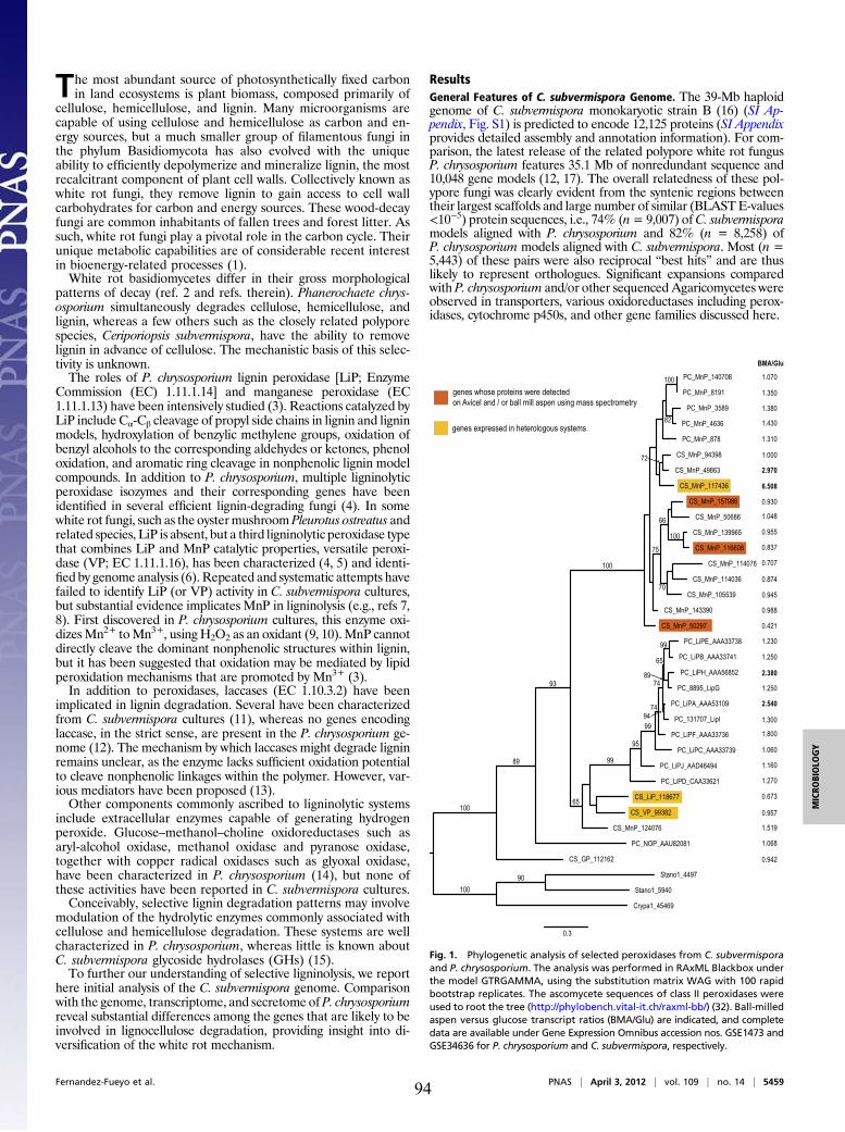
The most abundant source of photosynthetically fixed carbonin land ecosystems is plant biomass, composed primarily of
cellulose, hemicellulose, and lignin. Many microorganisms arecapable of using cellulose and hemicellulose as carbon and en-ergy sources, but a much smaller group of filamentous fungi inthe phylum Basidiomycota has also evolved with the uniqueability to efficiently depolymerize and mineralize lignin, the mostrecalcitrant component of plant cell walls. Collectively known aswhite rot fungi, they remove lignin to gain access to cell wallcarbohydrates for carbon and energy sources. These wood-decayfungi are common inhabitants of fallen trees and forest litter. Assuch, white rot fungi play a pivotal role in the carbon cycle. Theirunique metabolic capabilities are of considerable recent interestin bioenergy-related processes (1).White rot basidiomycetes differ in their gross morphological
patterns of decay (ref. 2 and refs. therein). Phanerochaete chrys-osporium simultaneously degrades cellulose, hemicellulose, andlignin, whereas a few others such as the closely related polyporespecies, Ceriporiopsis subvermispora, have the ability to removelignin in advance of cellulose. The mechanistic basis of this selec-tivity is unknown.The roles of P. chrysosporium lignin peroxidase [LiP; Enzyme
Commission (EC) 1.11.1.14] and manganese peroxidase (EC1.11.1.13) have been intensively studied (3). Reactions catalyzed byLiP include Cα-Cβ cleavage of propyl side chains in lignin and ligninmodels, hydroxylation of benzylic methylene groups, oxidation ofbenzyl alcohols to the corresponding aldehydes or ketones, phenoloxidation, and aromatic ring cleavage in nonphenolic lignin modelcompounds. In addition to P. chrysosporium, multiple ligninolyticperoxidase isozymes and their corresponding genes have beenidentified in several efficient lignin-degrading fungi (4). In somewhite rot fungi, such as the oystermushroomPleurotus ostreatus andrelated species, LiP is absent, but a third ligninolytic peroxidase typethat combines LiP and MnP catalytic properties, versatile peroxi-dase (VP; EC 1.11.1.16), has been characterized (4, 5) and identi-fied by genome analysis (6). Repeated and systematic attempts havefailed to identify LiP (or VP) activity in C. subvermispora cultures,but substantial evidence implicates MnP in ligninolysis (e.g., refs 7,8). First discovered in P. chrysosporium cultures, this enzyme oxi-dizesMn2+ toMn3+, using H2O2 as an oxidant (9, 10).MnP cannotdirectly cleave the dominant nonphenolic structures within lignin,but it has been suggested that oxidation may be mediated by lipidperoxidation mechanisms that are promoted by Mn3+ (3).In addition to peroxidases, laccases (EC 1.10.3.2) have been
implicated in lignin degradation. Several have been characterizedfrom C. subvermispora cultures (11), whereas no genes encodinglaccase, in the strict sense, are present in the P. chrysosporium ge-nome (12). The mechanism by which laccases might degrade ligninremains unclear, as the enzyme lacks sufficient oxidation potentialto cleave nonphenolic linkages within the polymer. However, var-ious mediators have been proposed (13).Other components commonly ascribed to ligninolytic systems
include extracellular enzymes capable of generating hydrogenperoxide. Glucose–methanol–choline oxidoreductases such asaryl-alcohol oxidase, methanol oxidase and pyranose oxidase,together with copper radical oxidases such as glyoxal oxidase,have been characterized in P. chrysosporium (14), but none ofthese activities have been reported in C. subvermispora cultures.Conceivably, selective lignin degradation patterns may involve
modulation of the hydrolytic enzymes commonly associated withcellulose and hemicellulose degradation. These systems are wellcharacterized in P. chrysosporium, whereas little is known aboutC. subvermispora glycoside hydrolases (GHs) (15).To further our understanding of selective ligninolysis, we report
here initial analysis of the C. subvermispora genome. Comparisonwith the genome, transcriptome, and secretome of P. chrysosporiumreveal substantial differences among the genes that are likely to beinvolved in lignocellulose degradation, providing insight into di-versification of the white rot mechanism.
ResultsGeneral Features of C. subvermispora Genome. The 39-Mb haploidgenome of C. subvermispora monokaryotic strain B (16) (SI Ap-pendix, Fig. S1) is predicted to encode 12,125 proteins (SI Appendixprovides detailed assembly and annotation information). For com-parison, the latest release of the related polypore white rot fungusP. chrysosporium features 35.1 Mb of nonredundant sequence and10,048 gene models (12, 17). The overall relatedness of these pol-ypore fungi was clearly evident from the syntenic regions betweentheir largest scaffolds and large number of similar (BLASTE-values<10−5) protein sequences, i.e., 74% (n= 9,007) ofC. subvermisporamodels aligned with P. chrysosporium and 82% (n = 8,258) ofP. chrysosporium models aligned with C. subvermispora. Most (n =5,443) of these pairs were also reciprocal “best hits” and are thuslikely to represent orthologues. Significant expansions comparedwith P. chrysosporium and/or other sequenced Agaricomycetes wereobserved in transporters, various oxidoreductases including perox-idases, cytochrome p450s, and other gene families discussed here.
1.430
1.380
1.350
1.310
1.070
2.970
6.508
1.000
1.048
0.955
0.988
0.945
0.930
0.874
0.707
0.837
0.421
2.540
2.380
1.800
1.300
1.270
1.250
1.250
1.230
1.160
1.060
1.519
0.957
0.673
0.942
BMA/Glu
1.068
0.3
genes whose proteins were detectedon Avicel and / or ball mill aspen using mass spectrometry
genes expressed in heterologous systems
GP
Fig. 1. Phylogenetic analysis of selected peroxidases from C. subvermisporaand P. chrysosporium. The analysis was performed in RAxML Blackbox underthe model GTRGAMMA, using the substitution matrix WAG with 100 rapidbootstrap replicates. The ascomycete sequences of class II peroxidases wereused to root the tree (http://phylobench.vital-it.ch/raxml-bb/) (32). Ball-milledaspen versus glucose transcript ratios (BMA/Glu) are indicated, and completedata are available under Gene Expression Omnibus accession nos. GSE1473 andGSE34636 for P. chrysosporium and C. subvermispora, respectively.
Fernandez-Fueyo et al. PNAS | April 3, 2012 | vol. 109 | no. 14 | 5459
MICRO
BIOLO
GY
94
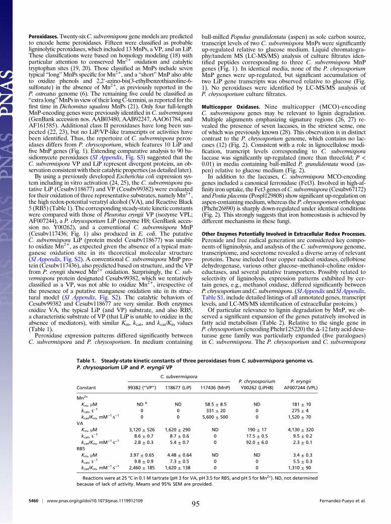
Peroxidases. Twenty-sixC. subvermispora genemodels are predictedto encode heme peroxidases. Fifteen were classified as probableligninolytic peroxidases, which included 13MnPs, a VP, and an LiP.These classifications were based on homology modeling (18) withparticular attention to conserved Mn2+ oxidation and catalytictryptophan sites (19, 20). Those classified as MnPs include seventypical “long”MnPs specific for Mn2+, and a “short”MnP also ableto oxidize phenols and 2,2′-azino-bis(3-ethylbenzothiazoline-6-sulfonate) in the absence of Mn2+, as previously reported in theP. ostreatus genome (6). The remaining five could be classified as“extra long”MnPs in viewof their longC-termini, as reported for thefirst time in Dichomitus squalens MnPs (21). Only four full-lengthMnP-encoding genes were previously identified in C. subvermispora(GenBank accession nos. AAB03480, AAB92247, AAO61784, andAF161585). Additional class II peroxidases have long been sus-pected (22, 23), but no LiP/VP-like transcripts or activities havebeen identified. Thus, the repertoire of C. subvermispora perox-idases differs from P. chrysosporium, which features 10 LiP andfive MnP genes (Fig. 1). Extending comparative analysis to 90 ba-sidiomycete peroxidases (SI Appendix, Fig. S3) suggested that theC. subvermispora VP and LiP represent divergent proteins, an ob-servation consistent with their catalytic properties (as detailed later).By using a previously developed Escherichia coli expression sys-
tem including in vitro activation (24, 25), the C. subvermispora pu-tative LiP (Cesubv118677) and VP (Cesubv99382) were evaluatedfor their oxidation of three representative substrates, namelyMn2+,the high redox-potential veratryl alcohol (VA), and Reactive Black5 (RB5) (Table 1). The corresponding steady-state kinetic constantswere compared with those of Pleurotus eryngii VP (isozyme VPL;AF007244), a P. chrysosporium LiP (isozyme H8; GenBank acces-sion no. Y00262), and a conventional C. subvermispora MnP(Cesubv117436; Fig. 1) also produced in E. coli. The putativeC. subvermispora LiP (protein model Cesubv118677) was unableto oxidize Mn2+, as expected given the absence of a typical man-ganese oxidation site in its theoretical molecular structure(SI Appendix, Fig. S2). A conventional C. subvermispora MnP pro-tein (Cesubv117436), also predicted based on structure, and the VPfrom P. eryngii showed Mn2+ oxidation. Surprisingly, the C. sub-vermispora protein designated Cesubv99382, which we tentativelyclassified as a VP, was not able to oxidize Mn2+, irrespective ofthe presence of a putative manganese oxidation site in its struc-tural model (SI Appendix, Fig. S2). The catalytic behaviors ofCesubv99382 and Cesubv118677 are very similar. Both enzymesoxidize VA, the typical LiP (and VP) substrate, and also RB5,a characteristic substrate of VP (that LiP is unable to oxidize in theabsence of mediators), with similar Km, kcat, and kcat/Km values(Table 1).Peroxidase expression patterns differed significantly between
C. subvermispora and P. chrysosporium. In medium containing
ball-milled Populus grandidentata (aspen) as sole carbon source,transcript levels of two C. subvermispora MnPs were significantlyup-regulated relative to glucose medium. Liquid chromatogra-phy/tandem MS (LC-MS/MS) analysis of culture filtrates iden-tified peptides corresponding to three C. subvermispora MnPgenes (Fig. 1). In identical media, none of the P. chrysosporiumMnP genes were up-regulated, but significant accumulation oftwo LiP gene transcripts was observed relative to glucose (Fig.1). No peroxidases were identified by LC-MS/MS analysis ofP. chrysosporium culture filtrates.
Multicopper Oxidases. Nine multicopper (MCO)-encodingC. subvermispora genes may be relevant to lignin degradation.Multiple alignments emphasizing signature regions (26, 27) re-vealed the presence of seven laccases, in the strictest sense, oneof which was previously known (28). This observation is in distinctcontrast to the P. chrysosporium genome, which contains no lac-cases (12) (Fig. 2). Consistent with a role in lignocellulose modi-fication, transcript levels corresponding to C. subvermisporalaccase was significantly up-regulated (more than threefold; P <0.01) in media containing ball-milled P. grandidentata wood (as-pen) relative to glucose medium (Fig. 2).In addition to the laccases, C. subvermispora MCO-encoding
genes included a canonical ferroxidase (Fet3). Involved in high-af-finity iron uptake, the Fet3 genes ofC. subvermispora (Cesubv67172)and Postia placenta (Pospl129808) show significant up-regulation onaspen-containingmedium, whereas the P. chrysosporium orthologue(Phchr26890) is sharply down-regulated under identical conditions(Fig. 2). This strongly suggests that iron homeostasis is achieved bydifferent mechanisms in these fungi.
Other Enzymes Potentially Involved in Extracellular Redox Processes.Peroxide and free radical generation are considered key compo-nents of ligninolysis, and analysis of the C. subvermispora genome,transcriptome, and secretome revealed a diverse array of relevantproteins. These included four copper radical oxidases, cellobiosedehydrogenase, various other glucose–methanol–choline oxidor-eductases, and several putative transporters. Possibly related toselectivity of ligninolysis, expression patterns exhibited by cer-tain genes, e.g., methanol oxidase, differed significantly betweenP. chrysosporium andC. subvermispora. (SIAppendix andSIAppendix,Table S1, include detailed listings of all annotated genes, transcriptlevels, and LC-MS/MS identification of extracellular proteins.)Of particular relevance to lignin degradation by MnP, we ob-
served a significant expansion of the genes putatively involved infatty acid metabolism (Table 2). Relative to the single gene inP. chrysosporium (encoding Phchr125220) theΔ-12 fatty acid desa-turase gene family was particularly expanded (five paralogues)in C. subvermispora. The P. chrysosporium and C. subvermispora
Table 1. Steady-state kinetic constants of three peroxidases from C. subvermispora genome vs.P. chrysosporium LiP and P. eryngii VP
Constant
C. subvermisporaP. chrysosporiumY00262 (LiPH8)
P. eryngiiAF007244 (VPL)99382 (“VP”) 118677 (LiP) 117436 (MnP)
Mn2+
Km, μM ND b ND 58.5 ± 8.5 ND 181 ± 10kcat, s
−1 0 0 331 ± 20 0 275 ± 4kcat/Km, mM−1·s−1 0 0 5,600 ± 500 0 1,520 ± 70
VAKm, μM 3,120 ± 526 1,620 ± 290 ND 190 ± 17 4,130 ± 320kcat, s
−1 8.6 ± 0.7 8.7 ± 0.6 0 17.5 ± 0.5 9.5 ± 0.2kcat/Km, mM−1·s−1 2.8 ± 0.3 5.4 ± 0.7 0 92.0 ± 6.0 2.3 ± 0.1
RB5Km, μM 3.97 ± 0.65 4.48 ± 0.64 ND ND 3.4 ± 0.3kcat, s
−1 9.8 ± 0.9 7.3 ± 0.5 0 0 5.5 ± 0.3kcat/Km, mM−1·s−1 2,460 ± 185 1,620 ± 138 0 0 1,310 ± 90
Reactions were at 25 °C in 0.1 M tartrate (pH 3 for VA, pH 3.5 for RB5, and pH 5 for Mn2+). ND, not determinedbecause of lack of activity. Means and 95% SEM are provided.
5460 | www.pnas.org/cgi/doi/10.1073/pnas.1119912109 Fernandez-Fueyo et al.95
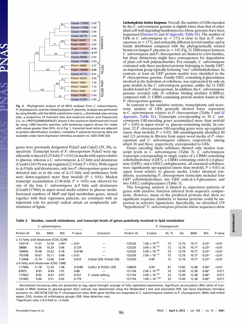
genes were previously designated Pcfad2 and Csfad2 (29, 30), re-spectively. Transcript levels of P. chrysosporium Pcfad2 were sig-nificantly reduced (0.25-fold;P< 0.01) inmedia with aspen relativeto glucose, whereas a C. subvermispora Δ-12 fatty acid desaturase(Cesubv124119) was up-regulated (2.9-fold; P< 0.01).With regardtoΔ-9 fatty acid desaturases, only two P. chrysosporium genes weredetected and, as in the case of Δ-12 fatty acid synthetases, bothwere down-regulated more than twofold (P < 0.01). Modesttranscript accumulation (1.48-fold; P = 0.03) was observed forone of the four C. subvermispora Δ-9 fatty acid desaturases(Cesubv117066) in aspen wood media relative to glucose media.Increased numbers of MnP and lipid metabolism genes, viewedtogether with their expression patterns, are consistent with animportant role for peroxyl radical attack on nonphenolic sub-structures of lignin.
Carbohydrate Active Enzymes.Overall, the number of GHs encodedby the C. subvermispora genome is slightly lower than that of otherplant cell wall degrading basidiomycetes whose genomes have beensequenced (Dataset S1 and SI Appendix, Table S1). The number ofGHs in C. subvermispora (n = 171) is close to that in P. chrys-osporium (n=177), and noticeably different in total number and infamily distribution compared with the phylogenetically relatedbrown rot fungus P. placenta (n=145; Fig. 3). Differences betweenC. subvermispora and P. chrysosporium are limited to a few families,but these distinctions might have consequences for degradationof plant cell wall polysaccharides. For example, C. subvermisporacontained only three predicted proteins belonging to family GH7,an important group typically featuring “exo” cellobiohydrolases. Incontrast, at least six GH7 protein models were identified in theP. chrysosporium genome. Family GH3, containing β-glucosidasesinvolved in the hydrolysis of cellobiose, was represented by only sixgene models in the C. subvermispora genome, unlike the 11 GH3models found in P. chrysosporium. In addition, theC. subvermisporagenome revealed only 16 cellulose binding modules (CBM1s),compared with 31 CBM1-containing protein models found in theP. chrysosporium genome.In contrast to the oxidative systems, transcriptome and secre-
tome analysis of GHs generally showed lower expressionin C. subvermispora relative to P. chrysosporium (Table 3 and SIAppendix, Table S1). Transcripts corresponding to 30 C. sub-vermispora GH-encoding genes accumulated more than twofold(P < 0.05) in aspen wood- vs. glucose-containing media. In con-trast, 52 P. chrysosporium GH-encoding genes were up-regulated(more than twofold; P < 0.05). MS unambiguously identified 60and 121 proteins in filtrates from aspen wood media of P. chrys-osporium and C. subvermispora cultures, respectively, amongwhich 18 and three, respectively, corresponded to GHs.Genes encoding likely cellulases showed only modest tran-
script levels in C. subvermispora (Table 3). C. subvermisporatranscripts corresponding to single copies of a CBM1-containingcellobiohydrolase (GH7), a CBM1-containing endo-β-1,4-gluca-nase (GH5), and a GH12 endoglucanase, all canonical cellulases,were significantly up-regulated (more than twofold; P < 0.01) inaspen wood relative to glucose media. Under identical con-ditions, accumulating P. chrysosporium transcripts included fourGH7 cellobiohydrolases, two GH5 endo-β-1,4-glucanases, andtwo GH12 endoglucanases (Table 3).The foregoing analysis is limited to expression patterns of
genes with putative function inferred from sequence compar-isons. However, many of the predicted proteins that show nosignificant sequence similarity to known proteins could be im-portant in selective ligninolysis. Specifically, we identified 139“hypothetical” C. subvermispora proteins whose sequences show
Cesubv 115068
Cesubv 84170
Cesubv 115063
Pospl1 111314
Pospl1 47589
Cesubv 108852
Pospl1 62097
Phchr1 26890
Cesubv 137686
Phchr1 10581
Pospl1 62067
Cesubv 88089
Phchr1 132237
Phchr1 10579
Pospl1 129808
Cesubv 51376
Phchr1 5406
Cesubv 67172
Cesubv 118801
LaccasesF
ET
3sother M
CO
s
0.69
0.90
0.95
0.93
1.05
0.84
3.15
6.55
5.11
6.77*
0.94
0.93
0.37
0.92
1.05
1.19
1.07
0.94
0.95
69
92
87
76
65
68
96
77
100
100
100
100
Fig. 2. Phylogenetic analysis of all MCO oxidases from C. subvermispora,P. chrysosporium, and the related polypore P. placenta. Analysis was performedby using RAxMLwith theWAG substitution matrix, γ-distributed rates amongsites, a proportion of invariant sites and empirical amino acid frequencies(i.e., m = PROTGAMMAIWAGF). Shown is themaximum-likelihood tree foundby using 1,000 heuristic searches, with bootstrap support shown for nodeswith values greater than 50%. As in Fig. 1, transcript level ratios are adjacentto protein identification numbers. Complete P. placenta microarray data areavailable under Gene Expression Omnibus accession no. GSE12540 (33).
Table 2. Number, overall relatedness, and transcript levels of genes putatively involved in lipid metabolism
C. subvermispora
Comment
P. chrysosporium
P valueProtein ID Glc BMA B/G P value Protein ID E-value ID, % Glc BMA B/G
Δ-12 fatty acid desaturase (COG 3239)124119 11.01 12.54 2.90* < 0.01 — 125220 1.00 × 10−67 72 12.76 10.77 0.25* <0.0158880 10.36 10.29 0.96 0.729 — 125220 3.00 × 10−70 72 12.76 10.77 0.25* <0.01109092 10.58 10.23 0.78 0.0149 — 125220 5.00 × 10−59 72 12.76 10.77 0.25* <0.01155708 10.67 10.11 0.68 < 0.01 — 125220 2.00 × 10−77 72 12.76 10.77 0.25* <0.01112068 12.74 12.66 0.94 0.653 Csfad2 (29); Pcfad2 (30) 125220 0.00 72 12.76 10.77 0.25* <0.01
Δ-9 fatty acid desaturase (COG 1398)117066 11.78 12.35 1.48 0.0298 CsOle1 & PcOle1 (29) 128650 0.00 81 13.82 12.48 0.40* <0.0187875 8.93 8.94 1.01 0.88 — 121154 2.00 × 10−68 33 13.49 12.38 0.46* 0.017117063 8.95 8.91 0.97 0.527 5′ needs editing 121154 2.00 × 10−62 33 13.49 12.38 0.46* 0.017121693 9.64 9.51 0.92 0.179 — 121154 1.00 × 10−154 33 13.49 12.38 0.46* 0.017
Normalized microarray data are presented as log2 signal strength average of fully replicated experiments. Significant accumulation (B/G ratio) of tran-scripts in BMA relative to glucose-grown (Glc) cultures was determined using the Moderated t test and associated FDR. See Gene Expression Omnibusaccession no. GSE14736 (33) for P. chrysosporium data. Both gene families are expanded in C. subvermispora relative to P. chrysosporium. BMA, ball-milledaspen; COG, clusters of orthologous groups; FDR, false detection rate.*Significant ratio (<0.5-fold to >2-fold).
Fernandez-Fueyo et al. PNAS | April 3, 2012 | vol. 109 | no. 14 | 5461
MICRO
BIOLO
GY
96
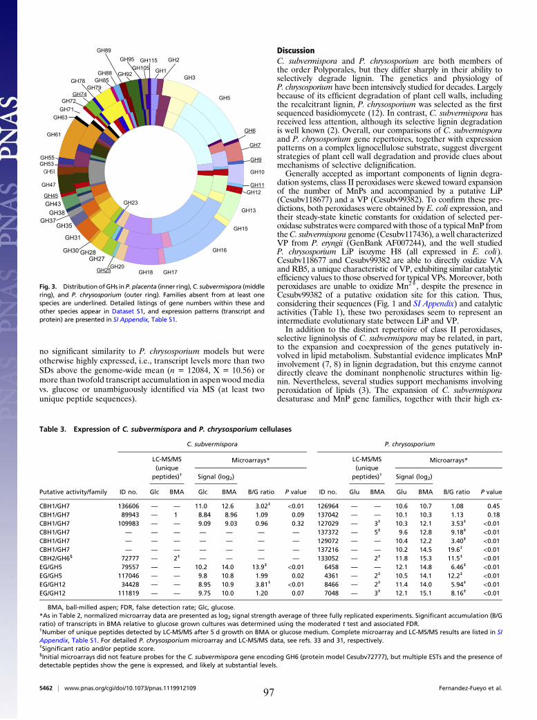
no significant similarity to P. chrysosporium models but wereotherwise highly expressed, i.e., transcript levels more than twoSDs above the genome-wide mean (n = 12084, X = 10.56) ormore than twofold transcript accumulation in aspen wood mediavs. glucose or unambiguously identified via MS (at least twounique peptide sequences).
DiscussionC. subvermispora and P. chrysosporium are both members ofthe order Polyporales, but they differ sharply in their ability toselectively degrade lignin. The genetics and physiology ofP. chrysosporium have been intensively studied for decades. Largelybecause of its efficient degradation of plant cell walls, includingthe recalcitrant lignin, P. chrysosporium was selected as the firstsequenced basidiomycete (12). In contrast, C. subvermispora hasreceived less attention, although its selective lignin degradationis well known (2). Overall, our comparisons of C. subvermisporaand P. chrysosporium gene repertoires, together with expressionpatterns on a complex lignocellulose substrate, suggest divergentstrategies of plant cell wall degradation and provide clues aboutmechanisms of selective delignification.Generally accepted as important components of lignin degra-
dation systems, class II peroxidases were skewed toward expansionof the number of MnPs and accompanied by a putative LiP(Cesubv118677) and a VP (Cesubv99382). To confirm these pre-dictions, both peroxidases were obtained by E. coli expression, andtheir steady-state kinetic constants for oxidation of selected per-oxidase substrates were compared with those of a typicalMnP fromtheC. subvermispora genome (Cesubv117436), a well characterizedVP from P. eryngii (GenBank AF007244), and the well studiedP. chrysosporium LiP isozyme H8 (all expressed in E. coli).Cesubv118677 and Cesubv99382 are able to directly oxidize VAand RB5, a unique characteristic of VP, exhibiting similar catalyticefficiency values to those observed for typical VPs. Moreover, bothperoxidases are unable to oxidize Mn2+, despite the presence inCesubv99382 of a putative oxidation site for this cation. Thus,considering their sequences (Fig. 1 and SI Appendix) and catalyticactivities (Table 1), these two peroxidases seem to represent anintermediate evolutionary state between LiP and VP.In addition to the distinct repertoire of class II peroxidases,
selective ligninolysis of C. subvermispora may be related, in part,to the expansion and coexpression of the genes putatively in-volved in lipid metabolism. Substantial evidence implicates MnPinvolvement (7, 8) in lignin degradation, but this enzyme cannotdirectly cleave the dominant nonphenolic structures within lig-nin. Nevertheless, several studies support mechanisms involvingperoxidation of lipids (3). The expansion of C. subvermisporadesaturase and MnP gene families, together with their high ex-
GH1
GH2
GH3
GH5
GH6
GH7
GH9
GH10
GH11GH12
GH13
GH15
GH16
GH17GH18GH20
GH23
GH25
GH115GH95
GH92
GH89
GH88GH85
GH79
GH61
GH63GH71GH72
GH74
GH78
GH55GH53
GH47
GH45GH43GH38
GH37GH35
GH31
GH30 GH28GH27
GH105
Fig. 3. Distribution ofGHs in P. placenta (inner ring), C. subvermispora (middlering), and P. chrysosporium (outer ring). Families absent from at least onespecies are underlined. Detailed listings of gene numbers within these andother species appear in Dataset S1, and expression patterns (transcript andprotein) are presented in SI Appendix, Table S1.
Table 3. Expression of C. subvermispora and P. chrysosporium cellulases
C. subvermispora P. chrysosporium
Microarrays* Microarrays*LC-MS/MS(unique
peptides)† Signal (log2)
LC-MS/MS(unique
peptides)† Signal (log2)
Putative activity/family ID no. Glc BMA Glc BMA B/G ratio P value ID no. Glu BMA Glu BMA B/G ratio P value
CBH1/GH7 136606 — — 11.0 12.6 3.02‡ <0.01 126964 — — 10.6 10.7 1.08 0.45CBH1/GH7 89943 — 1 8.84 8.96 1.09 0.09 137042 — — 10.1 10.3 1.13 0.18CBH1/GH7 109983 — — 9.09 9.03 0.96 0.32 127029 — 3‡ 10.3 12.1 3.53‡ <0.01CBH1/GH7 — — — — — — — 137372 — 5‡ 9.6 12.8 9.18‡ <0.01CBH1/GH7 — — — — — — — 129072 — — 10.4 12.2 3.40‡ <0.01CBH1/GH7 — — — — — — — 137216 — — 10.2 14.5 19.6‡ <0.01CBH2/GH6§ 72777 — 2‡ — — — — 133052 — 2‡ 11.8 15.3 11.5‡ <0.01EG/GH5 79557 — — 10.2 14.0 13.9‡ <0.01 6458 — — 12.1 14.8 6.46‡ <0.01EG/GH5 117046 — — 9.8 10.8 1.99 0.02 4361 — 2‡ 10.5 14.1 12.2‡ <0.01EG/GH12 34428 — — 8.95 10.9 3.81‡ <0.01 8466 — 2‡ 11.4 14.0 5.94‡ <0.01EG/GH12 111819 — — 9.75 10.0 1.20 0.07 7048 — 3‡ 12.1 15.1 8.16‡ <0.01
BMA, ball-milled aspen; FDR, false detection rate; Glc, glucose.*As in Table 2, normalized microarray data are presented as log2 signal strength average of three fully replicated experiments. Significant accumulation (B/Gratio) of transcripts in BMA relative to glucose grown cultures was determined using the moderated t test and associated FDR.†Number of unique peptides detected by LC-MS/MS after 5 d growth on BMA or glucose medium. Complete microarray and LC-MS/MS results are listed in SIAppendix, Table S1. For detailed P. chrysosporium microarray and LC-MS/MS data, see refs. 33 and 31, respectively.‡Significant ratio and/or peptide score.§Initial microarrays did not feature probes for the C. subvermispora gene encoding GH6 (protein model Cesubv72777), but multiple ESTs and the presence ofdetectable peptides show the gene is expressed, and likely at substantial levels.
5462 | www.pnas.org/cgi/doi/10.1073/pnas.1119912109 Fernandez-Fueyo et al.97
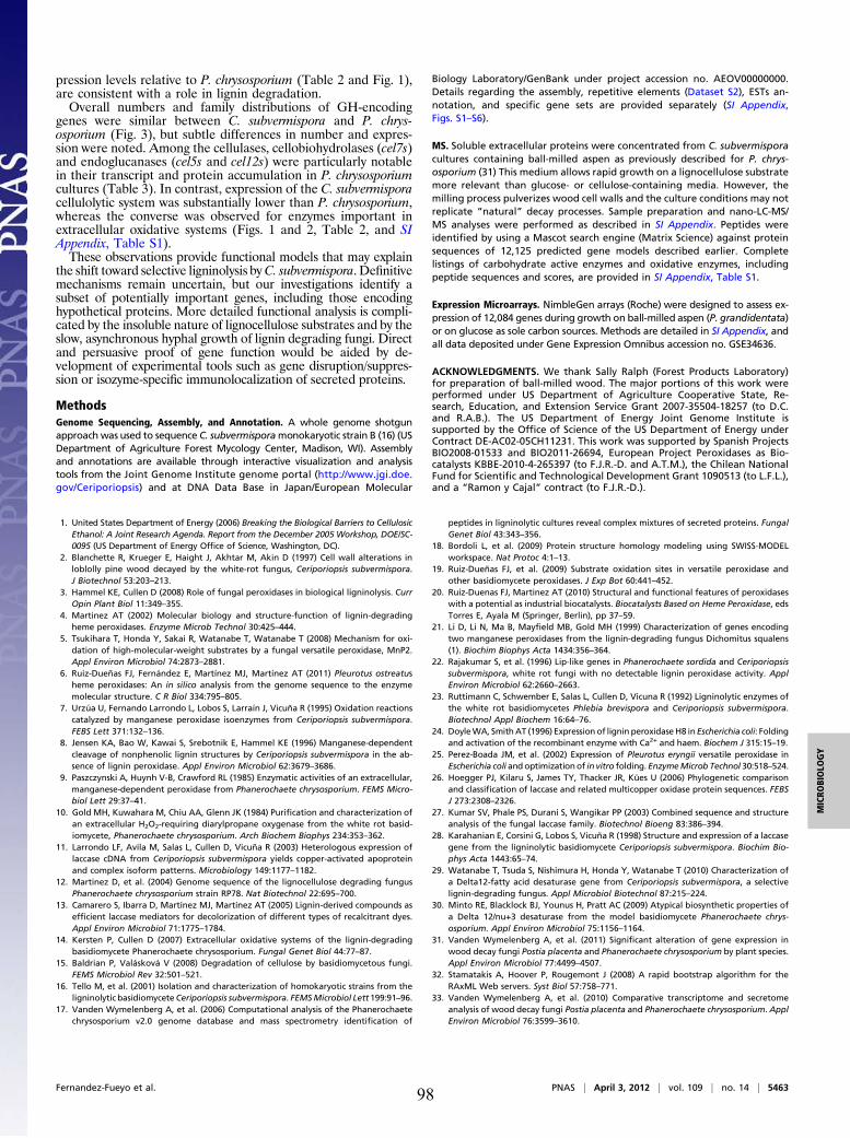
pression levels relative to P. chrysosporium (Table 2 and Fig. 1),are consistent with a role in lignin degradation.Overall numbers and family distributions of GH-encoding
genes were similar between C. subvermispora and P. chrys-osporium (Fig. 3), but subtle differences in number and expres-sion were noted. Among the cellulases, cellobiohydrolases (cel7s)and endoglucanases (cel5s and cel12s) were particularly notablein their transcript and protein accumulation in P. chrysosporiumcultures (Table 3). In contrast, expression of the C. subvermisporacellulolytic system was substantially lower than P. chrysosporium,whereas the converse was observed for enzymes important inextracellular oxidative systems (Figs. 1 and 2, Table 2, and SIAppendix, Table S1).These observations provide functional models that may explain
the shift toward selective ligninolysis byC. subvermispora. Definitivemechanisms remain uncertain, but our investigations identify asubset of potentially important genes, including those encodinghypothetical proteins. More detailed functional analysis is compli-cated by the insoluble nature of lignocellulose substrates and by theslow, asynchronous hyphal growth of lignin degrading fungi. Directand persuasive proof of gene function would be aided by de-velopment of experimental tools such as gene disruption/suppres-sion or isozyme-specific immunolocalization of secreted proteins.
MethodsGenome Sequencing, Assembly, and Annotation. A whole genome shotgunapproach was used to sequence C. subvermisporamonokaryotic strain B (16) (USDepartment of Agriculture Forest Mycology Center, Madison, WI). Assemblyand annotations are available through interactive visualization and analysistools from the Joint Genome Institute genome portal (http://www.jgi.doe.gov/Ceriporiopsis) and at DNA Data Base in Japan/European Molecular
Biology Laboratory/GenBank under project accession no. AEOV00000000.Details regarding the assembly, repetitive elements (Dataset S2), ESTs an-notation, and specific gene sets are provided separately (SI Appendix,Figs. S1–S6).
MS. Soluble extracellular proteins were concentrated from C. subvermisporacultures containing ball-milled aspen as previously described for P. chrys-osporium (31) This medium allows rapid growth on a lignocellulose substratemore relevant than glucose- or cellulose-containing media. However, themilling process pulverizes wood cell walls and the culture conditions may notreplicate “natural” decay processes. Sample preparation and nano-LC-MS/MS analyses were performed as described in SI Appendix. Peptides wereidentified by using a Mascot search engine (Matrix Science) against proteinsequences of 12,125 predicted gene models described earlier. Completelistings of carbohydrate active enzymes and oxidative enzymes, includingpeptide sequences and scores, are provided in SI Appendix, Table S1.
Expression Microarrays. NimbleGen arrays (Roche) were designed to assess ex-pression of 12,084 genes during growth on ball-milled aspen (P. grandidentata)or on glucose as sole carbon sources. Methods are detailed in SI Appendix, andall data deposited under Gene Expression Omnibus accession no. GSE34636.
ACKNOWLEDGMENTS. We thank Sally Ralph (Forest Products Laboratory)for preparation of ball-milled wood. The major portions of this work wereperformed under US Department of Agriculture Cooperative State, Re-search, Education, and Extension Service Grant 2007-35504-18257 (to D.C.and R.A.B.). The US Department of Energy Joint Genome Institute issupported by the Office of Science of the US Department of Energy underContract DE-AC02-05CH11231. This work was supported by Spanish ProjectsBIO2008-01533 and BIO2011-26694, European Project Peroxidases as Bio-catalysts KBBE-2010-4-265397 (to F.J.R.-D. and A.T.M.), the Chilean NationalFund for Scientific and Technological Development Grant 1090513 (to L.F.L.),and a “Ramon y Cajal” contract (to F.J.R.-D.).
1. United States Department of Energy (2006) Breaking the Biological Barriers to CellulosicEthanol: A Joint Research Agenda. Report from the December 2005 Workshop, DOE/SC-0095 (US Department of Energy Office of Science, Washington, DC).
2. Blanchette R, Krueger E, Haight J, Akhtar M, Akin D (1997) Cell wall alterations inloblolly pine wood decayed by the white-rot fungus, Ceriporiopsis subvermispora.J Biotechnol 53:203–213.
3. Hammel KE, Cullen D (2008) Role of fungal peroxidases in biological ligninolysis. CurrOpin Plant Biol 11:349–355.
4. Martinez AT (2002) Molecular biology and structure-function of lignin-degradingheme peroxidases. Enzyme Microb Technol 30:425–444.
5. Tsukihara T, Honda Y, Sakai R, Watanabe T, Watanabe T (2008) Mechanism for oxi-dation of high-molecular-weight substrates by a fungal versatile peroxidase, MnP2.Appl Environ Microbiol 74:2873–2881.
6. Ruiz-Dueñas FJ, Fernández E, Martínez MJ, Martínez AT (2011) Pleurotus ostreatusheme peroxidases: An in silico analysis from the genome sequence to the enzymemolecular structure. C R Biol 334:795–805.
7. Urzúa U, Fernando Larrondo L, Lobos S, Larraín J, Vicuña R (1995) Oxidation reactionscatalyzed by manganese peroxidase isoenzymes from Ceriporiopsis subvermispora.FEBS Lett 371:132–136.
8. Jensen KA, Bao W, Kawai S, Srebotnik E, Hammel KE (1996) Manganese-dependentcleavage of nonphenolic lignin structures by Ceriporiopsis subvermispora in the ab-sence of lignin peroxidase. Appl Environ Microbiol 62:3679–3686.
9. Paszczynski A, Huynh V-B, Crawford RL (1985) Enzymatic activities of an extracellular,manganese-dependent peroxidase from Phanerochaete chrysosporium. FEMS Micro-biol Lett 29:37–41.
10. Gold MH, Kuwahara M, Chiu AA, Glenn JK (1984) Purification and characterization ofan extracellular H2O2-requiring diarylpropane oxygenase from the white rot basid-iomycete, Phanerochaete chrysosporium. Arch Biochem Biophys 234:353–362.
11. Larrondo LF, Avila M, Salas L, Cullen D, Vicuña R (2003) Heterologous expression oflaccase cDNA from Ceriporiopsis subvermispora yields copper-activated apoproteinand complex isoform patterns. Microbiology 149:1177–1182.
12. Martinez D, et al. (2004) Genome sequence of the lignocellulose degrading fungusPhanerochaete chrysosporium strain RP78. Nat Biotechnol 22:695–700.
13. Camarero S, Ibarra D, Martínez MJ, Martínez AT (2005) Lignin-derived compounds asefficient laccase mediators for decolorization of different types of recalcitrant dyes.Appl Environ Microbiol 71:1775–1784.
14. Kersten P, Cullen D (2007) Extracellular oxidative systems of the lignin-degradingbasidiomycete Phanerochaete chrysosporium. Fungal Genet Biol 44:77–87.
15. Baldrian P, Valásková V (2008) Degradation of cellulose by basidiomycetous fungi.FEMS Microbiol Rev 32:501–521.
16. Tello M, et al. (2001) Isolation and characterization of homokaryotic strains from theligninolytic basidiomycete Ceriporiopsis subvermispora. FEMSMicrobiol Lett 199:91–96.
17. Vanden Wymelenberg A, et al. (2006) Computational analysis of the Phanerochaetechrysosporium v2.0 genome database and mass spectrometry identification of
peptides in ligninolytic cultures reveal complex mixtures of secreted proteins. FungalGenet Biol 43:343–356.
18. Bordoli L, et al. (2009) Protein structure homology modeling using SWISS-MODELworkspace. Nat Protoc 4:1–13.
19. Ruiz-Dueñas FJ, et al. (2009) Substrate oxidation sites in versatile peroxidase andother basidiomycete peroxidases. J Exp Bot 60:441–452.
20. Ruiz-Duenas FJ, Martinez AT (2010) Structural and functional features of peroxidaseswith a potential as industrial biocatalysts. Biocatalysts Based on Heme Peroxidase, edsTorres E, Ayala M (Springer, Berlin), pp 37–59.
21. Li D, Li N, Ma B, Mayfield MB, Gold MH (1999) Characterization of genes encodingtwo manganese peroxidases from the lignin-degrading fungus Dichomitus squalens(1). Biochim Biophys Acta 1434:356–364.
22. Rajakumar S, et al. (1996) Lip-like genes in Phanerochaete sordida and Ceriporiopsissubvermispora, white rot fungi with no detectable lignin peroxidase activity. ApplEnviron Microbiol 62:2660–2663.
23. Ruttimann C, Schwember E, Salas L, Cullen D, Vicuna R (1992) Ligninolytic enzymes ofthe white rot basidiomycetes Phlebia brevispora and Ceriporiopsis subvermispora.Biotechnol Appl Biochem 16:64–76.
24. DoyleWA, SmithAT (1996) Expression of lignin peroxidaseH8 in Escherichia coli: Foldingand activation of the recombinant enzyme with Ca2+ and haem. Biochem J 315:15–19.
25. Perez-Boada JM, et al. (2002) Expression of Pleurotus eryngii versatile peroxidase inEscherichia coli and optimizationof in vitro folding. EnzymeMicrob Technol 30:518–524.
26. Hoegger PJ, Kilaru S, James TY, Thacker JR, Kües U (2006) Phylogenetic comparisonand classification of laccase and related multicopper oxidase protein sequences. FEBSJ 273:2308–2326.
27. Kumar SV, Phale PS, Durani S, Wangikar PP (2003) Combined sequence and structureanalysis of the fungal laccase family. Biotechnol Bioeng 83:386–394.
28. Karahanian E, Corsini G, Lobos S, Vicuña R (1998) Structure and expression of a laccasegene from the ligninolytic basidiomycete Ceriporiopsis subvermispora. Biochim Bio-phys Acta 1443:65–74.
29. Watanabe T, Tsuda S, Nishimura H, Honda Y, Watanabe T (2010) Characterization ofa Delta12-fatty acid desaturase gene from Ceriporiopsis subvermispora, a selectivelignin-degrading fungus. Appl Microbiol Biotechnol 87:215–224.
30. Minto RE, Blacklock BJ, Younus H, Pratt AC (2009) Atypical biosynthetic properties ofa Delta 12/nu+3 desaturase from the model basidiomycete Phanerochaete chrys-osporium. Appl Environ Microbiol 75:1156–1164.
31. Vanden Wymelenberg A, et al. (2011) Significant alteration of gene expression inwood decay fungi Postia placenta and Phanerochaete chrysosporium by plant species.Appl Environ Microbiol 77:4499–4507.
32. Stamatakis A, Hoover P, Rougemont J (2008) A rapid bootstrap algorithm for theRAxML Web servers. Syst Biol 57:758–771.
33. Vanden Wymelenberg A, et al. (2010) Comparative transcriptome and secretomeanalysis of wood decay fungi Postia placenta and Phanerochaete chrysosporium. ApplEnviron Microbiol 76:3599–3610.
Fernandez-Fueyo et al. PNAS | April 3, 2012 | vol. 109 | no. 14 | 5463
MICRO
BIOLO
GY
98
Corrections
MICROBIOLOGYCorrection for “Comparative genomics of Ceriporiopsis sub-vermispora and Phanerochaete chrysosporium provide insight intoselective ligninolysis,” by Elena Fernandez-Fueyo, Francisco J.Ruiz-Dueñas, Patricia Ferreira, Dimitrios Floudas, David S.Hibbett, Paulo Canessa, Luis F. Larrondo, TimY. James, DanielaSeelenfreund, Sergio Lobos, Rubén Polanco, Mario Tello, YoichiHonda, Takahito Watanabe, Takashi Watanabe, Ryu Jae San,Christian P. Kubicek, Monika Schmoll, Jill Gaskell, KennethE. Hammel, Franz J. St. John, Amber Vanden Wymelenberg,Grzegorz Sabat, Sandra Splinter BonDurant, KhajamohiddinSyed, Jagjit S. Yadav, Harshavardhan Doddapaneni, Venkata-ramanan Subramanian, José L. Lavín, José A. Oguiza, GumerPerez, Antonio G. Pisabarro, Lucia Ramirez, Francisco Santoyo,Emma Master, Pedro M. Coutinho, Bernard Henrissat, VincentLombard, Jon Karl Magnuson, Ursula Kües, Chiaki Hori, Kiyo-hiko Igarashi, Masahiro Samejima, BenjaminW. Held, Kerrie W.Barry, Kurt M. LaButti, Alla Lapidus, Erika A. Lindquist, SusanM. Lucas, Robert Riley, Asaf A. Salamov, Dirk Hoffmeister,Daniel Schwenk, Yitzhak Hadar, Oded Yarden, Ronald P. deVries, Ad Wiebenga, Jan Stenlid, Daniel Eastwood, Igor V.Grigoriev, Randy M. Berka, Robert A. Blanchette, Phil Kersten,Angel T. Martinez, Rafael Vicuna, and Dan Cullen, which ap-peared in issue 14, April 3, 2012, of Proc Natl Acad Sci USA(109:5458–5463; first published March 20, 2012; 10.1073/pnas.1119912109).The authors note that the author name Ryu Jae San should
instead appear as Jae San Ryu. The corrected author line appearsbelow. The online version has been corrected.
Elena Ferandez-Fueyo, Francisco J. Ruiz-Dueñas, PatriciaFerreira, Dimitrios Floudas, David S. Hibbett, PauloCanessa, Luis F. Larrondo, Tim Y. James, DanielaSeelenfreund, Sergio Lobos, Reuben Polanco, Mario Tello,Yoichi Honda, Takahito Watanabe, Takashi Watanabe,Jae San Ryu, Christian P. Kubicek, Monika Schmoll, JillGaskell, Kenneth E. Hammel, Franz J. St. John, AmberVanden Wymelenberg, Grzegorz Sabat, Sandra SplinterBonDurant, Khajamohiddin Syed, Jagjit S. Yadav,Harshavardhan Doddapaneni, VenkataramananSubramanian, José L. Lavín, José A. Oguiza, Gumer Perez,Antonio G. Pisabarro, Lucia Ramirez, Francisco Santoyo,Emma Master, Pedro M. Coutinho, Bernard Henrissat,Vincent Lombard, Jon Karl Magnuson, Ursula Kües, ChiakiHori, Kiyohiko Igarashi, Masahiro Samejima, BenjaminW. Held, Kerrie W. Barry, Kurt M. LaButti, Alla Lapidus,Erika A. Lindquist, Susan M. Lucas, Robert Riley, Asaf A.Salamov, Dirk Hoffmeister, Daniel Schwenk, YitzhakHadar, Oded Yarden, Ronald P. de Vries, Ad Wiebenga,Jan Stenlid, Daniel Eastwood, Igor V. Grigoriev, RandyM. Berka, Robert A. Blanchette, Phil Kersten, Angel T.Martinez, Rafael Vicuna, and Dan Cullen
www.pnas.org/cgi/doi/10.1073/pnas.1206295109
EDITORIALCorrection for “Uncensored exchange of scientific results,” byJournal Editors and Authors Group, which appeared in issue 4,February 18, 2003, of Proc Natl Acad Sci USA (100:1464; firstpublished February 15, 2003; 10.1073/pnas.0630491100).Due to a printer’s error, the author name “Steven Salzburg”
should instead appear as “Steven Salzberg.” Additionally, theaffiliation for Steven Salzberg should instead appear as “TheInstitute for Genomic Research.” The corrected group authorfootnote appears below. The online version has been corrected.
*Group members: Ronald Atlas, President, ASM, and Editor, CRC Critical Reviews in Mi-crobiology; Philip Campbell, Editor, Nature; Nicholas R. Cozzarelli, Editor, PNAS; GregCurfman, Deputy Editor, New England Journal of Medicine; Lynn Enquist, Editor, Journalof Virology; Gerald Fink, Massachusetts Institute of Technology; Annette Flanagin, Man-aging Senior Editor, Journal of the American Medical Association, and President, Councilof Science Editors; Jacqueline Fletcher, President, American Phytopathological Society;Elizabeth George, Program Manager, National Nuclear Security Administration, Depart-ment of Energy; Gordon Hammes, Editor, Biochemistry; David Heyman, Senior Fellow andDirector of Science and Security Initiatives, Center for Strategic and International Studies;Thomas Inglesby, Editor, Biosecurity and Bioterrorism; Samuel Kaplan, Chair, ASM Pub-lications Board; Donald Kennedy, Editor, Science; Judith Krug, Director, Office for Intel-lectual Freedom, American Library Association; Rachel E. Levinson, Assistant Director forLife Sciences, Office of Science and Technology Policy; Emilie Marcus, Editor, Neuron;Henry Metzger, National Institute of Arthritis and Musculoskeletal and Skin Diseases,National Institutes of Health; Stephen S. Morse, Columbia University; Alison O’Brien,Editor, Infection and Immunity; Andrew Onderdonk, Editor, Journal of Clinical Microbi-ology; George Poste, Chief Executive Officer, Health Technology Networks; Beatrice Re-nault, Editor, Nature Medicine; Robert Rich, Editor, Journal of Immunology; AriellaRosengard, University of Pennsylvania; Steven Salzberg, The Institute for Genomic Re-search; Mary Scanlan, Director, Publishing Operations, American Chemical Society;Thomas Shenk, President Elect, ASM, and Past Editor, Journal of Virology; Herbert Tabor,Editor, Journal of Biological Chemistry; Harold Varmus, Memorial Sloan–Kettering CancerCenter; Eckard Wimmer, State University of New York at Stony Brook; Keith Yamamoto,Editor, Molecular Biology of the Cell.
www.pnas.org/cgi/doi/10.1073/pnas.1206993109
CELL BIOLOGYCorrection for “ATM signals to TSC2 in the cytoplasm to reg-ulate mTORC1 in response to ROS,” by Angela Alexander,Sheng-Li Cai, Jinhee Kim, Adrian Nanez, Mustafa Sahin, Kirs-teen H. MacLean, Ken Inoki, Kun-Liang Guan, Jianjun Shen,Maria D. Person, Donna Kusewitt, Gordon B. Mills, Michael B.Kastan, and Cheryl Lyn Walker, which appeared in issue 9,March 2, 2010, of Proc Natl Acad Sci USA (107:4153–4158; firstpublished February 16, 2010; 10.1073/pnas.0913860107).The authors note that in Fig. 2A, the error bars represent SEM
(mean ± SEM). In Figs. 3D and 4B, the error bars representstandard deviation (mean ± SD). These corrections do not affectthe conclusions of the article.
www.pnas.org/cgi/doi/10.1073/pnas.1206201109
8352–8353 | PNAS | May 22, 2012 | vol. 109 | no. 21 www.pnas.org99

100
Capítulo 6
"Lignin-degrading peroxidases from genome of selective ligninolytic fungus Ceriporiopsis subvermispora" Fernández-Fueyo, E., F. J. Ruiz-Dueñas, Y. Miki, M. J. Martínez, K. E. Hammel, and A. T. Martínez. J. Biol. Chem. 287:16903-16906. 2012.
101

102
Lignin-degrading Peroxidases from Genome of SelectiveLigninolytic Fungus Ceriporiopsis subvermispora*□S
Received for publication, February 25, 2012, and in revised form, March 19, 2012 Published, JBC Papers in Press, March 21, 2012, DOI 10.1074/jbc.M112.356378
Elena Fernández-Fueyo‡1, Francisco J. Ruiz-Dueñas‡2, Yuta Miki‡, María Jesús Martínez‡, Kenneth E. Hammel§3,and Angel T. Martínez‡4
From the ‡Centro de Investigaciones Biológicas, Consejo Superior de Investigaciones Científicas, Ramiro de Maeztu 9, E-28040Madrid, Spain and the §Forest Products Laboratory, United States Department of Agriculture, Madison, Wisconsin 53726
Background: The first genome of a selective lignin degrader is available.Results: Its screening shows 26 peroxidase genes, and 5 genes were heterologously expressed and the catalytic propertiesinvestigated.Conclusion: Two new peroxidases oxidize simple and dimeric lignin models and efficiently depolymerize lignin.Significance: Although lignin peroxidase and versatile peroxidase had not been reported in C. subvermispora, the genomicscreening suggests the presence of related lignin-degrading enzymes.
The white-rot fungus Ceriporiopsis subvermispora deligni-fies lignocellulose with high selectivity, but until now it hasappeared to lack the specialized peroxidases, termed ligninperoxidases (LiPs) and versatile peroxidases (VPs), that aregenerally thought important for ligninolysis. We screenedthe recently sequenced C. subvermispora genome for genesthat encode peroxidases with a potential ligninolytic role. Atotal of 26 peroxidase genes was apparent after a structural-functional classification based on homology modeling and asearch for diagnostic catalytic amino acid residues. In addi-tion to revealing the presence of nine heme-thiolate peroxi-dase superfamily members and the unexpected absence of thedye-decolorizing peroxidase superfamily, the search showedthat the C. subvermispora genome encodes 16 class IIenzymes in the plant-fungal-bacterial peroxidase superfam-ily, where LiPs and VPs are classified. The 16 encodedenzymes include 13 putative manganese peroxidases and onegeneric peroxidase but most notably two peroxidases con-taining the catalytic tryptophan characteristic of LiPs andVPs. We expressed these two enzymes in Escherichia coli anddetermined their substrate specificities on typical LiP/VPsubstrates, including nonphenolic lignin model monomersand dimers, as well as synthetic lignin. The results show thatthe two newly discovered C. subvermispora peroxidases arefunctionally competent LiPs and also suggest that they arephylogenetically and catalytically intermediate between clas-
sical LiPs and VPs. These results offer new insight into selec-tive lignin degradation by C. subvermispora.
Most of the carbon fixed by land photosynthesis is stored inthe plant polymers cellulose, hemicellulose, and lignin. Lignin isan aromatic macromolecule (1) that protects the other twopolysaccharidic polymers against biodegradation. After plantdeath, only certain specialized filamentous fungi, especially thehighly active basidiomycetes termed white-rot fungi (2), areable to degrade lignin via oxidativemechanisms. Lignin biodeg-radation is a key step in terrestrial carbon recycling, enablingsubsequent degradation of plant polysaccharides by complexmicrobial communities. Its removal is also a central issue forthe current industrial use of lignocellulosic biomass (e.g. in cel-lulose production for paper) as well as in lignocellulose biore-fineries for the sustainable production of fuels, chemicals, andmaterials from plant feedstocks (3). Industrial removal of ligninoften involves harsh chemical treatments, but lignin-degradingfungi and their enzymes may present environmentally friendlyalternatives (4).Two decay patterns are produced by white-rot fungi, being
characterized by degradation of both lignin and polysaccha-rides (simultaneous decay) and by a preferential removal oflignin leaving most of the polysaccharides unaffected (selectivedecay) (5). Among selective decay basidiomycetes, Ceriporiop-sis (Gelatoporia) subvermispora has been selected for delignifi-cation of different types of wood (6–8) in paper pulp manu-facture (9). More recently, it has also been assayed fordelignification in bioethanol production (10). The usual modelfungus in lignin biodegradation studies, Phanerochaete chrys-osporium (11), is of limited interest for such applicationsbecause it generally causes a simultaneous decay pattern (2).From the first pilot scale trials on wood biopulping with C.subvermispora, a high interest in its lignocellulolytic enzymaticmachinery has been sustained, with the double purpose ofenhancing enzyme production and discovering new or moreefficient enzymes of interest as industrial biocatalysts.
* This work was supported by the RAPERO Grant BIO2008-01533 and HIPOPGrant BIO2011-26694 from the Spanish Ministry of Economy and Compet-itiveness (to A. T. M. and F. J. R.-D., respectively), by PEROXICATS EuropeanProject Grant KBBE-2010-4-265397 (to A. T. M.), and by United StatesDepartment of Energy Grant DE-AI02-07ER64480 (to K. E. H.).
□S This article contains supplemental Figs. S1 and S2 and Table S1.1 Supported by a Junta para Ampliación de Estudios fellowship of the Consejo
Superior de Investigaciones Científicas, co-funded by the European SocialFund.
2 Supported by a Ministry of Economy and Competitiveness (MINECO) Ramóny Cajal contract.
3 To whom correspondence may be addressed. Tel.: 608-231-9528; Fax: 608-231-9262; E-mail: [email protected].
4 To whom correspondence may be addressed. Tel.: 34-918373112; Fax:34-915360432; E-mail: [email protected].
THE JOURNAL OF BIOLOGICAL CHEMISTRY VOL. 287, NO. 20, pp. 16903–16916, May 11, 2012Published in the U.S.A.
MAY 11, 2012 • VOLUME 287 • NUMBER 20 JOURNAL OF BIOLOGICAL CHEMISTRY 16903
at CS
IC - C
entro de Investigaciones Biológicas, on M
ay 16, 2012w
ww
.jbc.orgD
ownloaded from
http://www.jbc.org/content/suppl/2012/03/21/M112.356378.DC1.html Supplemental Material can be found at:
103
White-rot fungi secrete multiple isoenzymes of three hemeperoxidases with proposed roles in oxidative ligninolysis asfollows: lignin peroxidases (LiPs),5 manganese peroxidases(MnPs), and hybrid enzymes known as versatile peroxidases(VPs) that combine the structural-functional properties of LiPsand MnPs. In addition, these fungi secrete various oxidases toproduce the H2O2 required by peroxidases, and most of themalso secrete laccases (12). LiPs and VPs are able to oxidize andcleave the recalcitrant nonphenolic structures that composethe bulk of lignin, as shown by studies with dimeric ligninmodel compounds (13, 14), and also by depolymerizationexperiments with synthetic lignins in the case of LiPs (15).MnPs and VPs oxidize Mn2� to Mn3�, which can oxidize onlythe minor phenolic units in lignin (16). Likewise, laccases aredirectly able to oxidize only the phenolic units in lignin (17).Because the cleavage of nonphenolic lignin structures is cen-
tral to efficient ligninolysis, it might be expected that LiPsand/or VPs are essential participants in the process. However,among the above oxidoreductases, only laccase andMnP isoen-zymes have been found in C. subvermispora cultures (18, 19),although DNA hybridization/amplification with LiP sequenceswas reported in initial studies (20, 21), and some manganese-independent peroxidase activity has been detected (22). Theunexpected lack of LiPs or VPs in this selective delignifier hasled to various alternative hypotheses. One is that lipid peroxi-dation reactions catalyzed by MnP might generate fatty acid-derived oxyradicals that cleave lignin (23). There is some exper-imental support for this mechanism, as it can depolymerizenonphenolic synthetic lignin in vitro, but the reaction is slowand inefficient (23–25). Alternatively, the ability of laccases tooxidize nonphenolic lignin structures in the presence of variouslow molecular weight naturally occurring redox mediators hasbeen demonstrated (26). This might lead to ligninolysis if thefungus produces suchmediators as extracellular metabolites orlignin degradation products, but so far this has not beendemonstrated.The recent sequencing of the C. subvermispora genome at
the Joint Genome Institute, Department of Energy, allows anew look at this longstanding problem. This project supple-ments the first white-rot (27) and brown-rot (28) fungalgenome sequences to be obtained and reflects strong currentinterest in lignocellulose decay mechanisms that may inspirenew biotechnological applications. In this study, an inventoryof all the peroxidase genes, with special emphasis on class IIheme peroxidases (29), was obtained with manual curation.Then a structural-functional annotation of the predicted pro-teins was produced, as recently reported for the Pleurotusostreatus genome (30), and their evolutionary history wasestablished by comparison with all the basidiomycete peroxi-dase sequences currently available. Surprisingly, this annota-tion revealed the presence in C. subvermispora of two genesthat encode putative LiP/VP enzymes. To assess the signifi-cance of this finding, we have expressed these two peroxidases
heterologously (together with other C. subvermispora and ref-erence class II peroxidases) and have estimated their kineticconstants using high and low redox potential substrates. Fur-thermore, we have assessed the ligninolytic capability of thesetwo peroxidases in experiments with a nonphenolic ligninmodel dimer and with synthetic lignin.
MATERIALS AND METHODS
Fungal Strain and Genome Sequencing—The genome ofmonokaryotic C. subvermispora strain B (31) was sequenced atthe Joint Genome Institute using a shotgun approach (39.0 Mbtotal) in a project coordinated by Daniel Cullen (United StatesDepartment of Agriculture Forest Products Laboratory, Madi-son,WI) and Rafael Vicuña (Pontificia Universidad Católica deChile, Santiago, Chile). The results from gene prediction andannotation are available for searching at the Joint GenomeInstitute genome portal.Genome Screening and Analysis of Peroxidase Models—The
process to obtain the complete inventory of C. subvermisporaheme peroxidase genes consisted of the following steps: (i)screening the automatically annotated genome at the JointGenome Institute genome portal; (ii) revising and manuallycurating (when necessary) the positions of introns, and the NandC termini of the selectedmodels; (iii) comparing the aminoacid sequence identities with related proteins; and (iv) confirm-ing the presence of characteristic residues at the heme pocketand substrate oxidation sites, after homology modeling usingcrystal structures of related proteins from the RCSB ProteinData Bank as templates.Three MnP genes previously cloned from C. subvermispora
(GenBankTM accession numbers AAB03480, AAD45725, andAAO61784 and described as MnP1, MnP3, and MnP4, respec-tively) (32, 33) were identified (as models 116608, 139965, and94398, respectively, showing 98–100% identities with the cor-responding GenBankTM sequences). C. subvermispora MnP2(GenBankTM entries AAB92247 and AAD43581 with 99%sequence identity) was not localized in the genome but could bean allelic variant of gene 50297 (94% identity). The deducedamino acid sequences of theC. subvermispora heme peroxidasegene models were compared with all the basidiomycete hemeperoxidase sequences (from GenBankTM and genomes) avail-able to date (a total of 376 sequences, prokaryotic class I per-oxidases excluded), and phylogenetic trees were constructedusing Poisson-corrected distances and unweighted pair groupmethod with arithmetic mean clustering (bootstrap consensustrees were inferred from 1000 replicates).The programs used in the above analyses include the follow-
ing: (i) BLAST (Basic Local Alignment Tool) at the NationalCenter for Biotechnology Information for searching nucleotideand protein databases; (ii) MEGA5 (34) for conducting auto-matic and manual sequence alignment during the heme perox-idase gene search and curation, and for inferring evolutionarytrees; and (iii) SignalP 3.0 (35) for predicting the presence andlocation of signal peptides and cleavage sites. The latter predic-tions were validated by multiple alignment (from MEGA5) ofthe 196 class II sequences available, including proteins whoseNtermini have been sequenced. Theoretical molecular models
5 The abbreviations used are: LiP, lignin peroxidase; DHP, dehydrogenation(lignin) polymer; GPC, gel permeation chromatography; HTP, heme-thio-late peroxidase; MnP, manganese peroxidase; VP, versatile peroxidase;ABTS, 2,2�-azinobis(3-ethylbenzothiazoline-6-sulfonate).
From Genome Screening to Demonstration of Ligninolysis
16904 JOURNAL OF BIOLOGICAL CHEMISTRY VOLUME 287 • NUMBER 20 • MAY 11, 2012
at CS
IC - C
entro de Investigaciones Biológicas, on M
ay 16, 2012w
ww
.jbc.orgD
ownloaded from
104
were generated at the Swiss-Model automated protein homol-ogy-modeling server (36).Gene Synthesis and Directed Mutagenesis—The mature pro-
tein-coding sequences of five selected C. subvermispora perox-idase genes (49863, 99382, 112162, 118677, and 117436models)after the above manual curation, two VP genes from Pleurotuseryngii (GenBankTM AF007244) and Pleurotus ostreatus(genome 137757), and a P. chrysosporium LiP gene (Gen-BankTM Y00262) were synthesized by ATG:biosynthetics(Merzhausen, Germany) after verifying that all the codons hadbeen previously used for expressing other genes in the sameEscherichia coli strains (and substituting them when required).The E183K mutation was introduced in C. subvermispora
gene 99382 by PCR using the expression plasmid pFLAG1-99382 (see below) as template, and the QuikChange kit fromStratagene. The 5�-CGCGGCTGCCGACAAGGTCGATC-CTACCATCCCG-3� direct (mutated triplet underlined) andreverse primers were synthesized. The PCR (50-�l volume) wascarried out in a PerkinElmer Life Sciences GeneAmp PCR Sys-tem 240 using 20 ng of template DNA, 500 �M each dNTP, 125ng of each primer, 2.5 units of PfuTurbo DNA polymerase(Stratagene), and the manufacturer’s buffer. Reaction condi-tions were as follows: (i) a “hot start” at 95 °C for 1 min; (ii) 18cycles at 95 °C for 50 s, 55 °C for 50 s, and 68 °C for 10 min; and(iii) a final cycle at 68 °C for 10 min.E. coli Expression of Heme Peroxidase Genes—The five C.
subvermispora peroxidases, the P. eryngii and P. ostreatus VPs,and the P. chrysosporium LiP mature protein-codifyingsequences were cloned in the expression vectors pFLAG1(International Biotechnologies Inc.) or pET23a(�) (Novagen).The resulting plasmids pET23a-49863, pFLAG1-99382,pET23a-112162, pFLAG1-118677, pET23a-117436, pFLAG1-AF007244, pFLAG1-137757, and pFLAG1-Y00262 weredirectly used for expression, and for the above-describeddirected mutagenesis (pFLAG1-99382). E. coli DH5� was usedfor plasmid propagation.The different peroxidases (and the 99382 E183K variant)
were produced in E. coli W3110 (pFLAG1-derived plasmids)and BL21(DE3)pLysS (pET23a-derived plasmids). Cells weregrown for 3 h in Terrific Broth (37), induced with 1 mM isopro-pyl �-D-thiogalactopyranoside, and grown further for 4 h. Theapoenzyme accumulated in inclusion bodies, as observed bySDS-PAGE, and was solubilized using 8 M urea. Subsequent invitro refolding of C. subvermispora peroxidases and PleurotusVPswas performedusing 0.16Murea, 5mMCa2�, 20�Mhemin,0.5 mM oxidized glutathione, 0.1 mM dithiothreitol, and 0.1mg/ml protein, at pH 9.5 (38). C. subvermispora peroxidase112162 was refolded using 1 M urea, 5 mM Ca2�, 10 �M hemin,0.5 mM oxidized glutathione, 0.1 mM dithiothreitol, and 0.1mg/ml protein, at pH 9. Refolding of P. chrysosporium LiP wasperformed using 2.1 M urea, 5 mM Ca2�, 10 �M hemin, 0.7 mM
oxidized glutathione, 0.1mMdithiothreitol, and 0.2mg/ml pro-tein, at pH 8 (39). Active enzyme was purified by Resource-Qchromatography using a 0–300mMNaCl gradient (2ml�min�1,20 min) in 10 mM sodium tartrate (pH 5.5) containing 1 mM
CaCl2.Kinetic Constants on Selected Substrates—Absorbance
changes during substrate oxidation in 0.1 M tartrate (at various
pH values) by the above E. coli-expressed peroxidases wererecorded in a Thermo Scientific Biomate5 spectrophotometer(using �0.01 �M enzyme). Oxidation of Mn2� was followed atpH 5 for the formation of the Mn3��tartrate complex (�238 6.5mM�1�cm�1). Veratryl alcohol oxidation was followed at pH 3for veratraldehyde (�310 9.3 mM�1�cm�1) formation. ReactiveBlack 5, 2,2�-azinobis(3-ethylbenzothiazoline-6-sulfonate)(ABTS), and 2,6-dimethoxyphenol oxidationwere all assayed atpH 3.5. These reactions were monitored for Reactive Black 5disappearance (�598 30 mM�1�cm�1), for ABTS cation radicalformation (�436 29.3 mM�1�cm�1), and for dimeric coeruli-gnone formation (�469 5.5 mM�1 cm�1), respectively. All reac-tions were at 25 °C and were initiated by addition of 0.1 mM
H2O2. ABTS and 2,6-dimethoxyphenol oxidation by some C.subvermipora peroxidases shows double kinetics with sigmoi-dal activity curves (at increasing substrate concentrations) thatenable calculation of two sets of kinetic constants (for high andlow efficiency oxidation sites), as reported previously for VP(40, 41).Means and standard errors for affinity constant (Km) and
enzyme turnover (kcat) values were obtained by nonlinear leastsquares fitting of the experimental measurements to theMichaelis-Mentenmodel. Fitting of these constants to the nor-malized equation v� (kcat/Km)[S]/(1� [S]/Km) yielded the cat-alytic efficiency values (kcat/Km) with their correspondingstandard errors.p-Dimethoxybenzene and Ferrocytochrome c Oxidation—Spec-
tral changes during oxidation of p-dimethoxybenzene (0.2 mM)and ferrocytochrome c (15 �M) by the above peroxidases (�0.1�M) at 25 °C in the presence of 0.1mMH2O2were recordedwithan Agilent 8453 diode-array spectrophotometer, showing theformation of p-benzoquinone and ferricytochrome c, respec-tively. Ferrocytochrome c was prepared before use by reducingferricytochrome cwith sodium dithionite, followed by removalof excess dithionite on a Sephadex G-25 column. p-Benzoqui-none formation was followed at 245 nm in 20 mM sodium suc-cinate (pH 3.5). Decrease of ferrocytochrome c concentrationwas followed at 550 nm in 20 mM sodium succinate (pH 4).Controls without enzyme and without H2O2 were included.Oxidative Degradation of a Lignin Model Dimer—Unlabeled
and ring-14C-labeled (1.0 mCi mmol�1) 4-ethoxy-3-methoxy-phenylglycerol-�-guaiacyl ether were prepared, and theirerythro and threo isomers were chromatographically separated,as described earlier (42).For reactions involving product analysis, the radiolabeled
erythro or threo dimer was treated either with one of the two C.subvermispora LiP-like peroxidases (from genes 118677 and99382) or with P. chrysosporium LiPH8 in 10 mM sodium ace-tate (pH 3.0) at 25 °C for 1 h. The products formed were ana-lyzed by reversed phase high performance liquid chromatogra-phy (HPLC) using a Gilson system equipped with a C-18column (Phenomenex Luna C18(2); 150 by 4.6 mm, 5-�m par-ticle size), and methanol/water as mobile phase (35:65 for 15min, followed by 50:50) at a flow rate of 1ml�min�1. Elutionwasmonitored at 255 nm, and the 14C content in collected fractions(0.5ml) wasmeasured in a liquid scintillation counter. HPLC inconjunction with gas chromatography/mass spectrometry was
From Genome Screening to Demonstration of Ligninolysis
MAY 11, 2012 • VOLUME 287 • NUMBER 20 JOURNAL OF BIOLOGICAL CHEMISTRY 16905
at CS
IC - C
entro de Investigaciones Biológicas, on M
ay 16, 2012w
ww
.jbc.orgD
ownloaded from
105
used in parallel analyses with unlabeled dimers to confirm theidentity of the products obtained.For experiments to determine reaction kinetics, unlabeled
erythro or threo dimer was treated with C. subvermisporaperoxidase at 25 °C in the same buffer. Approximate turn-over numbers (s�1) at various dimer concentrations wereobtained from the increase in absorbance at 310 nm due tobenzylic carbonyl formation, i.e. production of 4-ethoxy-3-methoxybenzaldehyde, 1-(4-ethoxy-3-methoxyphenyl)-3-hydroxy-2-(2-methoxyphenoxy)-propan-1-one, and 1-(4-ethoxy-3-methoxyphenyl)-2,3-dihydroxypropan-1-one (�3109 mM�1�cm�1). Kinetic constants were then calculated as des-cribed above.Enzymatic Depolymerization of Synthetic Lignin—A radiola-
beled syringyl/guaiacyl dehydrogenation polymer (DHP)with asyringyl/guaiacyl ratio of �4:1 was prepared by enzymaticcopolymerization of�-[14C]sinapyl alcohol (0.01mCimmol�1)and unlabeled coniferyl alcohol using horseradish peroxidase asdescribed previously (43). The synthetic lignin was then frac-tionated on a column of Sephadex LH-20 as described (43), andthe high molecular mass fractions (approximately �21 kDa)were pooled for use in depolymerization experiments.Enzymatic depolymerization of the DHP by the same peroxi-
dases used in the dimer degradation assays was investigated in10 mM sodium acetate (pH 4.5) containing 0.25% Tween 20,1.5 � 104 dpm (188 �g) DHP, and 0.01 �M enzyme in a finalvolume of 40 ml, in the presence or absence of 10 mM veratrylalcohol. Reactionswere conducted at 25 °C by addingH2O2 (7.5mM final concentration in experiments with veratryl alcoholand 0.3 mM in experiments without veratryl alcohol) continu-ously over 24 h with a syringe pump (15). Control reactionswithout enzyme were run for comparison. The reaction mix-tures were then concentrated by rotary vacuum evaporation,redissolved in N,N�-dimethylformamide containing 0.1 M LiCl,and centrifuged as described earlier (15). Molecular mass dis-tributions of the supernatant fractionswere assessed by gel per-meation chromatography (GPC) on a 1.8 � 30-cm column ofSephadex LH20, using N,N�-dimethylformamide containing0.1 M LiCl as the mobile phase. Fractions (2 ml) were collectedand assayed for 14C in a liquid scintillation counter (15).Chemicals—p-Dimethoxybenzene, N,N�-dimethylform-
amide, dithiothreitol, 2,6-dimethoxyphenol, ferricytochromec, hemin, isopropyl �-D-thiogalactopyranoside, lithium chlo-ride, manganese sulfate, methanol, oxidized glutathione, Reac-tive Black 5, sodium dithionite, sodium tartrate, Tween 20,veratryl alcohol, and other unlabeled chemicalswere purchasedfrom Sigma, with the exception of H2O2 that was from Merck,and ABTS that was from Roche Applied Science.
RESULTS
Inventory of Heme Peroxidase Genes in the C. subvermisporaGenome—Among the 12,125 gene models identified in the C.subvermispora genome distributed in 740 main scaffolds, 26models were found to encode heme peroxidases after manualrevision of the possible candidates from automatic annotation(supplemental Table S1 provides the reference, scaffold, posi-tion, best hit, amino acid sequence identity, and peroxidasetypes encoded by these genes).
For the class II peroxidases we found (numbers 1–16 supple-mental Table S1), the classification was based on the presence/absence in the molecular models (obtained using crystal struc-tures of related enzymes as templates) of the following: (i) anexposed tryptophan (two models) putatively involved in oxida-tion of veratryl alcohol and other high redox potential aromat-ics (Wc, homologous to Trp-171 in P. chrysosporium LiP isoen-zyme-H8 and to Trp-164 in P. eryngiiVP isoenzyme-VPL) (Fig.1,A and B); and/or (ii) a putativeMn2� oxidation site (14 mod-els) formed by three acidic residues near the most internalheme propionate (Ea, Eb, and Ec, homologous to P. chrysospo-riumMnP1Glu-35, Glu-39, and Asp-179, and to P. eryngiiVPLGlu-36, Glu-40 and Asp-175, respectively) (Fig. 1, B and D).
The only C. subvermispora peroxidase showing both of theabove two catalytic sites (gene 99382) was initially classified as aputative VP (Fig. 1B). The 13 models showing only the Mn2�
oxidation site (genes 1–13 in supplemental Table S1) were clas-sified as likelyMnPs (Fig. 1D). The single peroxidase containingonly the catalytic tryptophan (gene 118677) was classified as aputative LiP (Fig. 1A). Finally, themodel lacking both oxidationsites (gene 112162) was classified as a generic peroxidase (Fig.1C), related to the Coprinopsis cinerea peroxidase (44).Among theMnPs, two types were identified (see supplemen-
tal Table S1). (i)MnP-long, represented by 12genemodels in theC. subvermispora genome, resembles typicalMnPs described inP.chrysosporium, whose amino acid sequence is longer than those ofLiPs andVPs and includes an additional disulfide bridge. (ii)MnP-short, represented by only one genemodel (gene 124076), resem-bles some Phlebia radiata MnPs. MnP-long includes five “extralong” MnPs, whose differential catalytic properties are still to bereported.An additional single model (supplemental Table S1, gene
83438) has predicted protein folding and hemepocket architec-ture similar to that of the above class II peroxidases. However, itis a class I (of prokaryotic origin but in the same superfamily)cytochrome c peroxidase and is therefore not included in thesubsequent comparisons.The other nine models (numbers 18–26 in supplemental
Table S1) correspond to putative peroxidases from the HTPsuperfamily and are most probably related to the Agrocybeaegerita peroxygenase (GenBankTM entries CAV28568 andCAV28569) (45). This superfamily is characterized by the pres-ence in themolecular structures (that of gene 80799 is shown inFig. 2A) of a cysteine residue acting as the fifth heme ligand,instead of the histidine residue present in the above peroxi-dases, among other structural details (Fig. 2B).Catalytic Properties of Peroxidases Expressed in E. coli—Among
the 15 class II genes in theC. subvermispora genome, includinggenes putatively encoding ligninolytic peroxidases, we selected118677, 99382, 117436, 49863, and 112162 for heterologousexpression, as representative for the main families identified.Our first goal was to investigate their catalytic properties onselected peroxidase substrates and in this way to confirm ormodify their putative structural/functional classifications.Additionally, we aimed to evaluate their ability to degradelignin using a simple dimericmodel compound and a polymericsynthetic lignin (DHP).
From Genome Screening to Demonstration of Ligninolysis
16906 JOURNAL OF BIOLOGICAL CHEMISTRY VOLUME 287 • NUMBER 20 • MAY 11, 2012
at CS
IC - C
entro de Investigaciones Biológicas, on M
ay 16, 2012w
ww
.jbc.orgD
ownloaded from
106
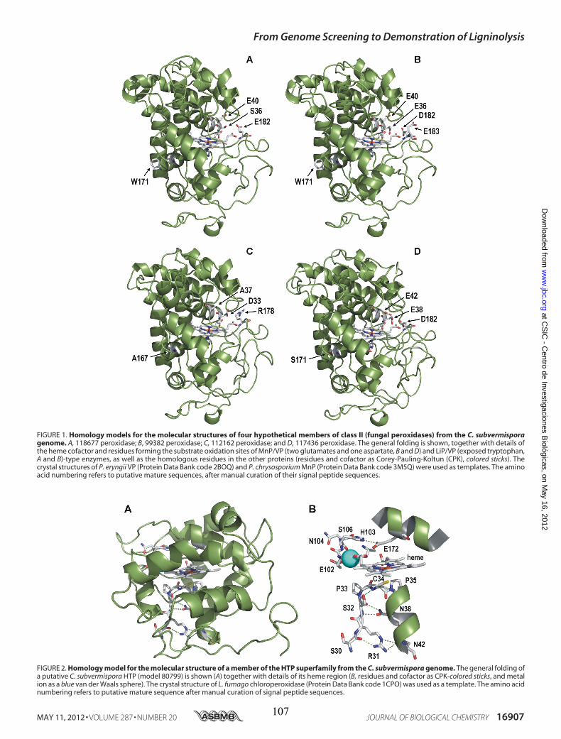
FIGURE 1. Homology models for the molecular structures of four hypothetical members of class II (fungal peroxidases) from the C. subvermisporagenome. A, 118677 peroxidase; B, 99382 peroxidase; C, 112162 peroxidase; and D, 117436 peroxidase. The general folding is shown, together with details ofthe heme cofactor and residues forming the substrate oxidation sites of MnP/VP (two glutamates and one aspartate, B and D) and LiP/VP (exposed tryptophan,A and B)-type enzymes, as well as the homologous residues in the other proteins (residues and cofactor as Corey-Pauling-Koltun (CPK), colored sticks). Thecrystal structures of P. eryngii VP (Protein Data Bank code 2BOQ) and P. chrysosporium MnP (Protein Data Bank code 3M5Q) were used as templates. The aminoacid numbering refers to putative mature sequences, after manual curation of their signal peptide sequences.
FIGURE 2. Homology model for the molecular structure of a member of the HTP superfamily from the C. subvermispora genome. The general folding ofa putative C. subvermispora HTP (model 80799) is shown (A) together with details of its heme region (B, residues and cofactor as CPK-colored sticks, and metalion as a blue van der Waals sphere). The crystal structure of L. fumago chloroperoxidase (Protein Data Bank code 1CPO) was used as a template. The amino acidnumbering refers to putative mature sequence after manual curation of signal peptide sequences.
From Genome Screening to Demonstration of Ligninolysis
MAY 11, 2012 • VOLUME 287 • NUMBER 20 JOURNAL OF BIOLOGICAL CHEMISTRY 16907
at CS
IC - C
entro de Investigaciones Biológicas, on M
ay 16, 2012w
ww
.jbc.orgD
ownloaded from
107
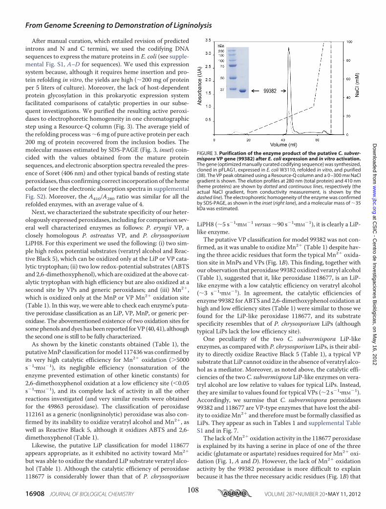
After manual curation, which entailed revision of predictedintrons and N and C termini, we used the codifying DNAsequences to express the mature proteins in E. coli (see supple-mental Fig. S1, A–D for sequences). We used this expressionsystem because, although it requires heme insertion and pro-tein refolding in vitro, the yields are high (�200 mg of proteinper 5 liters of culture). Moreover, the lack of host-dependentprotein glycosylation in this prokaryotic expression systemfacilitated comparisons of catalytic properties in our subse-quent investigations. We purified the resulting active peroxi-dases to electrophoretic homogeneity in one chromatographicstep using a Resource-Q column (Fig. 3). The average yield ofthe refolding process was�6mg of pure active protein per each200 mg of protein recovered from the inclusion bodies. Themolecular masses estimated by SDS-PAGE (Fig. 3, inset) coin-cided with the values obtained from the mature proteinsequences, and electronic absorption spectra revealed the pres-ence of Soret (406 nm) and other typical bands of resting stateperoxidases, thus confirming correct incorporation of the hemecofactor (see the electronic absorption spectra in supplementalFig. S2). Moreover, the A410/A280 ratio was similar for all therefolded enzymes, with an average value of 4.Next, we characterized the substrate specificity of our heter-
ologously expressed peroxidases, including for comparison sev-eral well characterized enzymes as follows: P. eryngii VP, aclosely homologous P. ostreatus VP, and P. chrysosporiumLiPH8. For this experiment we used the following: (i) two sim-ple high redox potential substrates (veratryl alcohol and Reac-tive Black 5), which can be oxidized only at the LiP or VP cata-lytic tryptophan; (ii) two low redox-potential substrates (ABTSand 2,6-dimethoxyphenol), which are oxidized at the above cat-alytic tryptophan with high efficiency but are also oxidized at asecond site by VPs and generic peroxidases; and (iii) Mn2�,which is oxidized only at the MnP or VP Mn2� oxidation site(Table 1). In this way, we were able to check each enzyme’s puta-tive peroxidase classification as an LiP, VP, MnP, or generic per-oxidase. The abovementioned existence of two oxidation sites forsomephenols anddyeshasbeen reported forVP (40, 41), althoughthe second one is still to be fully characterized.As shown by the kinetic constants obtained (Table 1), the
putativeMnP classification formodel 117436was confirmed byits very high catalytic efficiency for Mn2� oxidation (�5000s�1�mM�1), its negligible efficiency (nonsaturation of theenzyme prevented estimation of other kinetic constants) for2,6-dimethoxyphenol oxidation at a low efficiency site (�0.05s�1�mM�1), and its complete lack of activity in all the otherreactions investigated (and very similar results were obtainedfor the 49863 peroxidase). The classification of peroxidase112161 as a generic (nonligninolytic) peroxidase was also con-firmed by its inability to oxidize veratryl alcohol and Mn2�, aswell as Reactive Black 5, although it oxidizes ABTS and 2,6-dimethoxyphenol (Table 1).Likewise, the putative LiP classification for model 118677
appears appropriate, as it exhibited no activity toward Mn2�
but was able to oxidize the standard LiP substrate veratryl alco-hol (Table 1). Although the catalytic efficiency of peroxidase118677 is considerably lower than that of P. chrysosporium
LiPH8 (�5 s�1�mM�1 versus �90 s�1�mM�1), it is clearly a LiP-like enzyme.The putative VP classification for model 99382 was not con-
firmed, as it was unable to oxidize Mn2� (Table 1) despite hav-ing the three acidic residues that form the typical Mn2� oxida-tion site in MnPs and VPs (Fig. 1B). This finding, together withour observation that peroxidase 99382 oxidized veratryl alcohol(Table 1), suggested that it, like peroxidase 118677, is an LiP-like enzyme with a low catalytic efficiency on veratryl alcohol(�3 s�1�mM�1). In agreement, the catalytic efficiencies ofenzyme 99382 for ABTS and 2,6-dimethoxyphenol oxidation athigh and low efficiency sites (Table 1) were similar to those wefound for the LiP-like peroxidase 118677, and its substratespecificity resembles that of P. chrysosporium LiPs (althoughtypical LiPs lack the low efficiency site).One peculiarity of the two C. subvermispora LiP-like
enzymes, as compared with P. chrysosporium LiPs, is their abil-ity to directly oxidize Reactive Black 5 (Table 1), a typical VPsubstrate that LiP cannot oxidize in the absence of veratryl alco-hol as a mediator. Moreover, as noted above, the catalytic effi-ciencies of the two C. subvermispora LiP-like enzymes on vera-tryl alcohol are low relative to values for typical LiPs. Instead,they are similar to values found for typical VPs (�2 s�1�mM�1).Accordingly, we surmise that C. subvermispora peroxidases99382 and 118677 are VP-type enzymes that have lost the abil-ity to oxidizeMn2� and thereforemust be formally classified asLiPs. They appear as such in Tables 1 and supplemental TableS1 and in Fig. 7.The lack ofMn2� oxidation activity in the 118677 peroxidase
is explained by its having a serine in place of one of the threeacidic (glutamate or aspartate) residues required forMn2� oxi-dation (Fig. 1, A and D). However, the lack of Mn2� oxidationactivity by the 99382 peroxidase is more difficult to explainbecause it has the three necessary acidic residues (Fig. 1B) that
FIGURE 3. Purification of the enzyme product of the putative C. subver-mispora VP gene (99382) after E. coli expression and in vitro activation.The gene (optimized manually curated codifying sequence) was synthesized,cloned in pFLAG1, expressed in E. coli W3110, refolded in vitro, and purified(38). The VP peak obtained using a Resource-Q column and a 0 –300 mM NaClgradient is shown. The elution profiles at 280 nm (total protein) and 410 nm(heme proteins) are shown by dotted and continuous lines, respectively (theactual NaCl gradient, from conductivity measurement, is shown by thedashed line). The electrophoretic homogeneity of the enzyme was confirmedby SDS-PAGE, as shown in the inset (right lane), and a molecular mass of �35kDa was estimated.
From Genome Screening to Demonstration of Ligninolysis
16908 JOURNAL OF BIOLOGICAL CHEMISTRY VOLUME 287 • NUMBER 20 • MAY 11, 2012
at CS
IC - C
entro de Investigaciones Biológicas, on M
ay 16, 2012w
ww
.jbc.orgD
ownloaded from
108
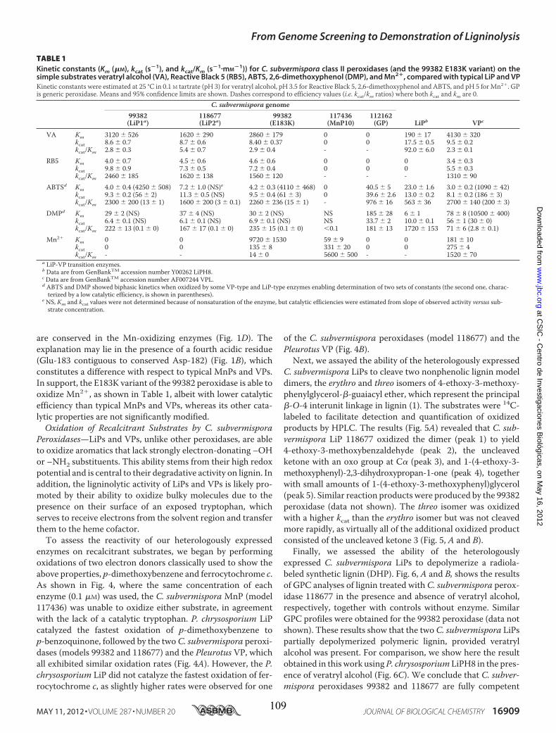
are conserved in the Mn-oxidizing enzymes (Fig. 1D). Theexplanation may lie in the presence of a fourth acidic residue(Glu-183 contiguous to conserved Asp-182) (Fig. 1B), whichconstitutes a difference with respect to typical MnPs and VPs.In support, the E183K variant of the 99382 peroxidase is able tooxidize Mn2�, as shown in Table 1, albeit with lower catalyticefficiency than typical MnPs and VPs, whereas its other cata-lytic properties are not significantly modified.Oxidation of Recalcitrant Substrates by C. subvermispora
Peroxidases—LiPs and VPs, unlike other peroxidases, are ableto oxidize aromatics that lack strongly electron-donating –OHor –NH2 substituents. This ability stems from their high redoxpotential and is central to their degradative activity on lignin. Inaddition, the ligninolytic activity of LiPs and VPs is likely pro-moted by their ability to oxidize bulky molecules due to thepresence on their surface of an exposed tryptophan, whichserves to receive electrons from the solvent region and transferthem to the heme cofactor.To assess the reactivity of our heterologously expressed
enzymes on recalcitrant substrates, we began by performingoxidations of two electron donors classically used to show theabove properties, p-dimethoxybenzene and ferrocytochrome c.As shown in Fig. 4, where the same concentration of eachenzyme (0.1 �M) was used, the C. subvermispora MnP (model117436) was unable to oxidize either substrate, in agreementwith the lack of a catalytic tryptophan. P. chrysosporium LiPcatalyzed the fastest oxidation of p-dimethoxybenzene top-benzoquinone, followed by the twoC. subvermispora peroxi-dases (models 99382 and 118677) and the Pleurotus VP, whichall exhibited similar oxidation rates (Fig. 4A). However, the P.chrysosporium LiP did not catalyze the fastest oxidation of fer-rocytochrome c, as slightly higher rates were observed for one
of the C. subvermispora peroxidases (model 118677) and thePleurotus VP (Fig. 4B).
Next, we assayed the ability of the heterologously expressedC. subvermispora LiPs to cleave two nonphenolic lignin modeldimers, the erythro and threo isomers of 4-ethoxy-3-methoxy-phenylglycerol-�-guaiacyl ether, which represent the principal�-O-4 interunit linkage in lignin (1). The substrates were 14C-labeled to facilitate detection and quantification of oxidizedproducts by HPLC. The results (Fig. 5A) revealed that C. sub-vermispora LiP 118677 oxidized the dimer (peak 1) to yield4-ethoxy-3-methoxybenzaldehyde (peak 2), the uncleavedketone with an oxo group at C� (peak 3), and 1-(4-ethoxy-3-methoxyphenyl)-2,3-dihydroxypropan-1-one (peak 4), togetherwith small amounts of 1-(4-ethoxy-3-methoxyphenyl)glycerol(peak 5). Similar reaction products were produced by the 99382peroxidase (data not shown). The threo isomer was oxidizedwith a higher kcat than the erythro isomer but was not cleavedmore rapidly, as virtually all of the additional oxidized productconsisted of the uncleaved ketone 3 (Fig. 5, A and B).Finally, we assessed the ability of the heterologously
expressed C. subvermispora LiPs to depolymerize a radiola-beled synthetic lignin (DHP). Fig. 6, A and B, shows the resultsof GPC analyses of lignin treated with C. subvermispora perox-idase 118677 in the presence and absence of veratryl alcohol,respectively, together with controls without enzyme. SimilarGPC profiles were obtained for the 99382 peroxidase (data notshown). These results show that the two C. subvermispora LiPspartially depolymerized polymeric lignin, provided veratrylalcohol was present. For comparison, we show here the resultobtained in this work using P. chrysosporium LiPH8 in the pres-ence of veratryl alcohol (Fig. 6C). We conclude that C. subver-mispora peroxidases 99382 and 118677 are fully competent
TABLE 1Kinetic constants (Km (�M), kcat (s�1), and kcat/Km (s�1�mM
�1)) for C. subvermispora class II peroxidases (and the 99382 E183K variant) on thesimple substrates veratryl alcohol (VA), Reactive Black 5 (RB5), ABTS, 2,6-dimethoxyphenol (DMP), and Mn2�, compared with typical LiP and VPKinetic constants were estimated at 25 °C in 0.1 M tartrate (pH 3) for veratryl alcohol, pH 3.5 for Reactive Black 5, 2,6-dimethoxyphenol and ABTS, and pH 5 forMn2�. GPis generic peroxidase. Means and 95% confidence limits are shown. Dashes correspond to efficiency values (i.e. kcat/km ratios) where both kcat and km are 0.
C. subvermispora genome99382(LiP1a)
118677(LiP2a)
99382(E183K)
117436(MnP10)
112162(GP) LiPb VPc
VA Km 3120 526 1620 290 2860 179 0 0 190 17 4130 320kcat 8.6 0.7 8.7 0.6 8.40 0.37 0 0 17.5 0.5 9.5 0.2kcat/Km 2.8 0.3 5.4 0.7 2.9 0.4 - - 92.0 6.0 2.3 0.1
RB5 Km 4.0 0.7 4.5 0.6 4.6 0.6 0 0 0 3.4 0.3kcat 9.8 0.9 7.3 0.5 7.2 0.4 0 0 0 5.5 0.3kcat/Km 2460 185 1620 138 1560 120 - - - 1310 90
ABTSd Km 4.0 0.4 (4250 508) 7.2 1.0 (NS)e 4.2 0.3 (4110 468) 0 40.5 5 23.0 1.6 3.0 0.2 (1090 42)kcat 9.3 0.2 (56 2) 11.3 0.5 (NS) 9.5 0.4 (61 3) 0 39.6 2.6 13.0 0.2 8.1 0.2 (186 3)kcat/Km 2300 200 (13 1) 1600 200 (3 0.1) 2260 236 (15 1) - 976 16 563 36 2700 140 (200 3)
DMPd Km 29 2 (NS) 37 4 (NS) 30 2 (NS) NS 185 28 6 1 78 8 (10500 400)kcat 6.4 0.1 (NS) 6.1 0.1 (NS) 6.9 0.1 (NS) NS 33.7 2 10.0 0.1 56 1 (30 0)kcat/Km 222 13 (0.1 0) 167 17 (0.1 0) 235 15 (0.1 0) �0.1 181 13 1720 153 71 6 (2.8 0.1)
Mn2� Km 0 0 9720 1530 59 9 0 0 181 10kcat 0 0 135 8 331 20 0 0 275 4kcat/Km - - 14 0 5600 500 - - 1520 70
a LiP-VP transition enzymes.b Data are from GenBankTM accession number Y00262 LiPH8.c Data are from GenBankTM accession number AF007244 VPL.d ABTS and DMP showed biphasic kinetics when oxidized by some VP-type and LiP-type enzymes enabling determination of two sets of constants (the second one, charac-terized by a low catalytic efficiency, is shown in parentheses).
e NS, Km and kcat values were not determined because of nonsaturation of the enzyme, but catalytic efficiencies were estimated from slope of observed activity versus sub-strate concentration.
From Genome Screening to Demonstration of Ligninolysis
MAY 11, 2012 • VOLUME 287 • NUMBER 20 JOURNAL OF BIOLOGICAL CHEMISTRY 16909
at CS
IC - C
entro de Investigaciones Biológicas, on M
ay 16, 2012w
ww
.jbc.orgD
ownloaded from
109
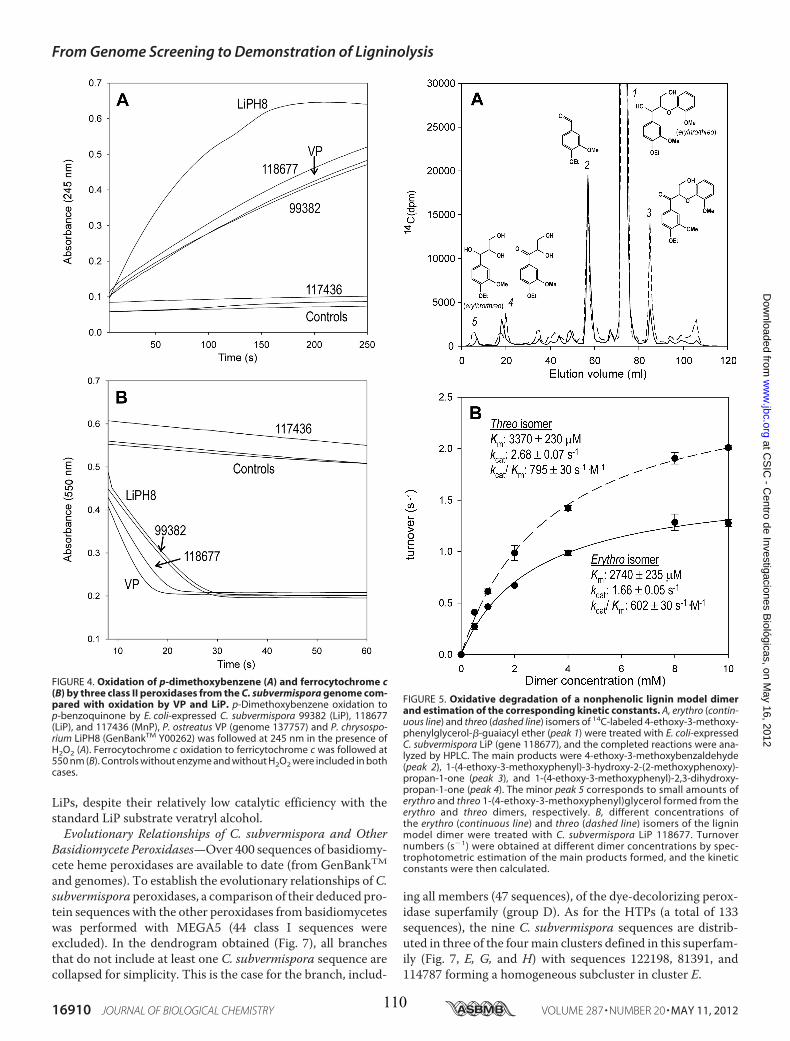
LiPs, despite their relatively low catalytic efficiency with thestandard LiP substrate veratryl alcohol.Evolutionary Relationships of C. subvermispora and Other
Basidiomycete Peroxidases—Over 400 sequences of basidiomy-cete heme peroxidases are available to date (from GenBankTMand genomes). To establish the evolutionary relationships ofC.subvermispora peroxidases, a comparison of their deduced pro-tein sequences with the other peroxidases from basidiomyceteswas performed with MEGA5 (44 class I sequences wereexcluded). In the dendrogram obtained (Fig. 7), all branchesthat do not include at least one C. subvermispora sequence arecollapsed for simplicity. This is the case for the branch, includ-
ing all members (47 sequences), of the dye-decolorizing perox-idase superfamily (group D). As for the HTPs (a total of 133sequences), the nine C. subvermispora sequences are distrib-uted in three of the fourmain clusters defined in this superfam-ily (Fig. 7, E, G, and H) with sequences 122198, 81391, and114787 forming a homogeneous subcluster in cluster E.
FIGURE 4. Oxidation of p-dimethoxybenzene (A) and ferrocytochrome c(B) by three class II peroxidases from the C. subvermispora genome com-pared with oxidation by VP and LiP. p-Dimethoxybenzene oxidation top-benzoquinone by E. coli-expressed C. subvermispora 99382 (LiP), 118677(LiP), and 117436 (MnP), P. ostreatus VP (genome 137757) and P. chrysospo-rium LiPH8 (GenBankTM Y00262) was followed at 245 nm in the presence ofH2O2 (A). Ferrocytochrome c oxidation to ferricytochrome c was followed at550 nm (B). Controls without enzyme and without H2O2 were included in bothcases.
FIGURE 5. Oxidative degradation of a nonphenolic lignin model dimerand estimation of the corresponding kinetic constants. A, erythro (contin-uous line) and threo (dashed line) isomers of 14C-labeled 4-ethoxy-3-methoxy-phenylglycerol-�-guaiacyl ether (peak 1) were treated with E. coli-expressedC. subvermispora LiP (gene 118677), and the completed reactions were ana-lyzed by HPLC. The main products were 4-ethoxy-3-methoxybenzaldehyde(peak 2), 1-(4-ethoxy-3-methoxyphenyl)-3-hydroxy-2-(2-methoxyphenoxy)-propan-1-one (peak 3), and 1-(4-ethoxy-3-methoxyphenyl)-2,3-dihydroxy-propan-1-one (peak 4). The minor peak 5 corresponds to small amounts oferythro and threo 1-(4-ethoxy-3-methoxyphenyl)glycerol formed from theerythro and threo dimers, respectively. B, different concentrations ofthe erythro (continuous line) and threo (dashed line) isomers of the ligninmodel dimer were treated with C. subvermispora LiP 118677. Turnovernumbers (s�1) were obtained at different dimer concentrations by spec-trophotometric estimation of the main products formed, and the kineticconstants were then calculated.
From Genome Screening to Demonstration of Ligninolysis
16910 JOURNAL OF BIOLOGICAL CHEMISTRY VOLUME 287 • NUMBER 20 • MAY 11, 2012
at CS
IC - C
entro de Investigaciones Biológicas, on M
ay 16, 2012w
ww
.jbc.orgD
ownloaded from
110
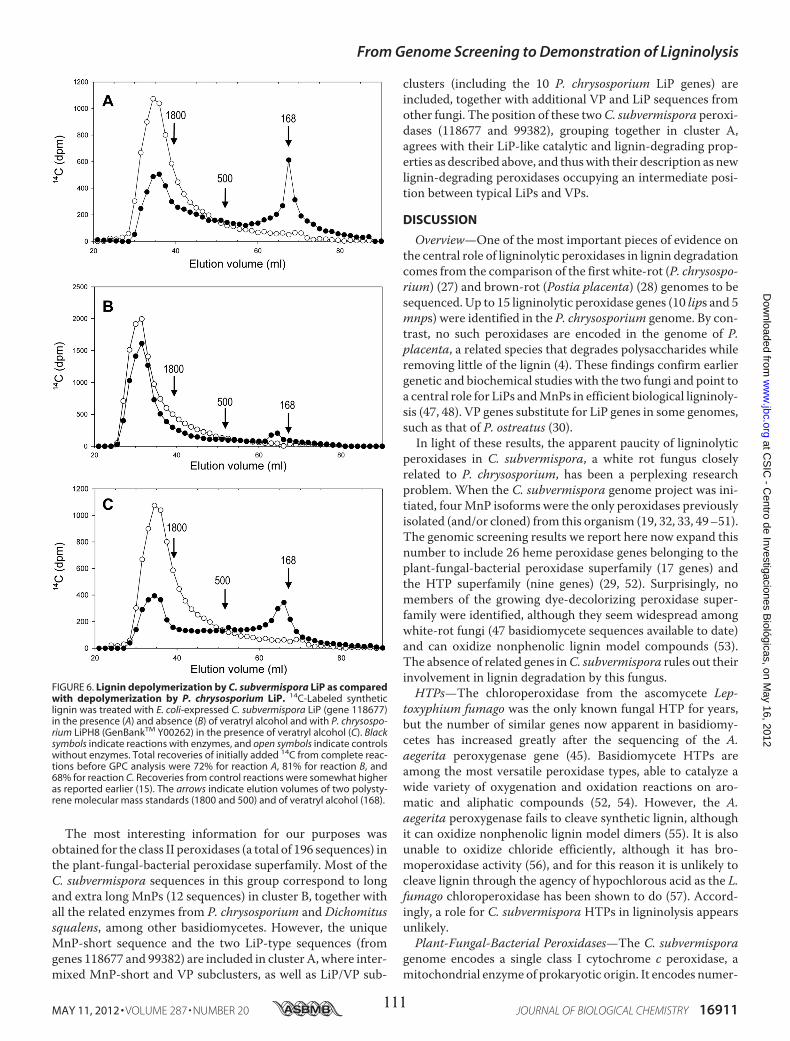
The most interesting information for our purposes wasobtained for the class II peroxidases (a total of 196 sequences) inthe plant-fungal-bacterial peroxidase superfamily. Most of theC. subvermispora sequences in this group correspond to longand extra long MnPs (12 sequences) in cluster B, together withall the related enzymes from P. chrysosporium and Dichomitussqualens, among other basidiomycetes. However, the uniqueMnP-short sequence and the two LiP-type sequences (fromgenes 118677 and 99382) are included in cluster A, where inter-mixed MnP-short and VP subclusters, as well as LiP/VP sub-
clusters (including the 10 P. chrysosporium LiP genes) areincluded, together with additional VP and LiP sequences fromother fungi. The position of these twoC. subvermispora peroxi-dases (118677 and 99382), grouping together in cluster A,agrees with their LiP-like catalytic and lignin-degrading prop-erties as described above, and thuswith their description as newlignin-degrading peroxidases occupying an intermediate posi-tion between typical LiPs and VPs.
DISCUSSION
Overview—One of the most important pieces of evidence onthe central role of ligninolytic peroxidases in lignin degradationcomes from the comparison of the first white-rot (P. chrysospo-rium) (27) and brown-rot (Postia placenta) (28) genomes to besequenced. Up to 15 ligninolytic peroxidase genes (10 lips and 5mnps) were identified in the P. chrysosporium genome. By con-trast, no such peroxidases are encoded in the genome of P.placenta, a related species that degrades polysaccharides whileremoving little of the lignin (4). These findings confirm earliergenetic and biochemical studies with the two fungi and point toa central role for LiPs andMnPs in efficient biological ligninoly-sis (47, 48). VP genes substitute for LiP genes in some genomes,such as that of P. ostreatus (30).In light of these results, the apparent paucity of ligninolytic
peroxidases in C. subvermispora, a white rot fungus closelyrelated to P. chrysosporium, has been a perplexing researchproblem. When the C. subvermispora genome project was ini-tiated, fourMnP isoforms were the only peroxidases previouslyisolated (and/or cloned) from this organism (19, 32, 33, 49–51).The genomic screening results we report here now expand thisnumber to include 26 heme peroxidase genes belonging to theplant-fungal-bacterial peroxidase superfamily (17 genes) andthe HTP superfamily (nine genes) (29, 52). Surprisingly, nomembers of the growing dye-decolorizing peroxidase super-family were identified, although they seem widespread amongwhite-rot fungi (47 basidiomycete sequences available to date)and can oxidize nonphenolic lignin model compounds (53).The absence of related genes inC. subvermispora rules out theirinvolvement in lignin degradation by this fungus.HTPs—The chloroperoxidase from the ascomycete Lep-
toxyphium fumago was the only known fungal HTP for years,but the number of similar genes now apparent in basidiomy-cetes has increased greatly after the sequencing of the A.aegerita peroxygenase gene (45). Basidiomycete HTPs areamong the most versatile peroxidase types, able to catalyze awide variety of oxygenation and oxidation reactions on aro-matic and aliphatic compounds (52, 54). However, the A.aegerita peroxygenase fails to cleave synthetic lignin, althoughit can oxidize nonphenolic lignin model dimers (55). It is alsounable to oxidize chloride efficiently, although it has bro-moperoxidase activity (56), and for this reason it is unlikely tocleave lignin through the agency of hypochlorous acid as the L.fumago chloroperoxidase has been shown to do (57). Accord-ingly, a role for C. subvermispora HTPs in ligninolysis appearsunlikely.Plant-Fungal-Bacterial Peroxidases—The C. subvermispora
genome encodes a single class I cytochrome c peroxidase, amitochondrial enzyme of prokaryotic origin. It encodes numer-
FIGURE 6. Lignin depolymerization by C. subvermispora LiP as comparedwith depolymerization by P. chrysosporium LiP. 14C-Labeled syntheticlignin was treated with E. coli-expressed C. subvermispora LiP (gene 118677)in the presence (A) and absence (B) of veratryl alcohol and with P. chrysospo-rium LiPH8 (GenBankTM Y00262) in the presence of veratryl alcohol (C). Blacksymbols indicate reactions with enzymes, and open symbols indicate controlswithout enzymes. Total recoveries of initially added 14C from complete reac-tions before GPC analysis were 72% for reaction A, 81% for reaction B, and68% for reaction C. Recoveries from control reactions were somewhat higheras reported earlier (15). The arrows indicate elution volumes of two polysty-rene molecular mass standards (1800 and 500) and of veratryl alcohol (168).
From Genome Screening to Demonstration of Ligninolysis
MAY 11, 2012 • VOLUME 287 • NUMBER 20 JOURNAL OF BIOLOGICAL CHEMISTRY 16911
at CS
IC - C
entro de Investigaciones Biológicas, on M
ay 16, 2012w
ww
.jbc.orgD
ownloaded from
111
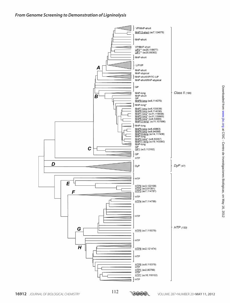
From Genome Screening to Demonstration of Ligninolysis
16912 JOURNAL OF BIOLOGICAL CHEMISTRY VOLUME 287 • NUMBER 20 • MAY 11, 2012
at CS
IC - C
entro de Investigaciones Biológicas, on M
ay 16, 2012w
ww
.jbc.orgD
ownloaded from
112
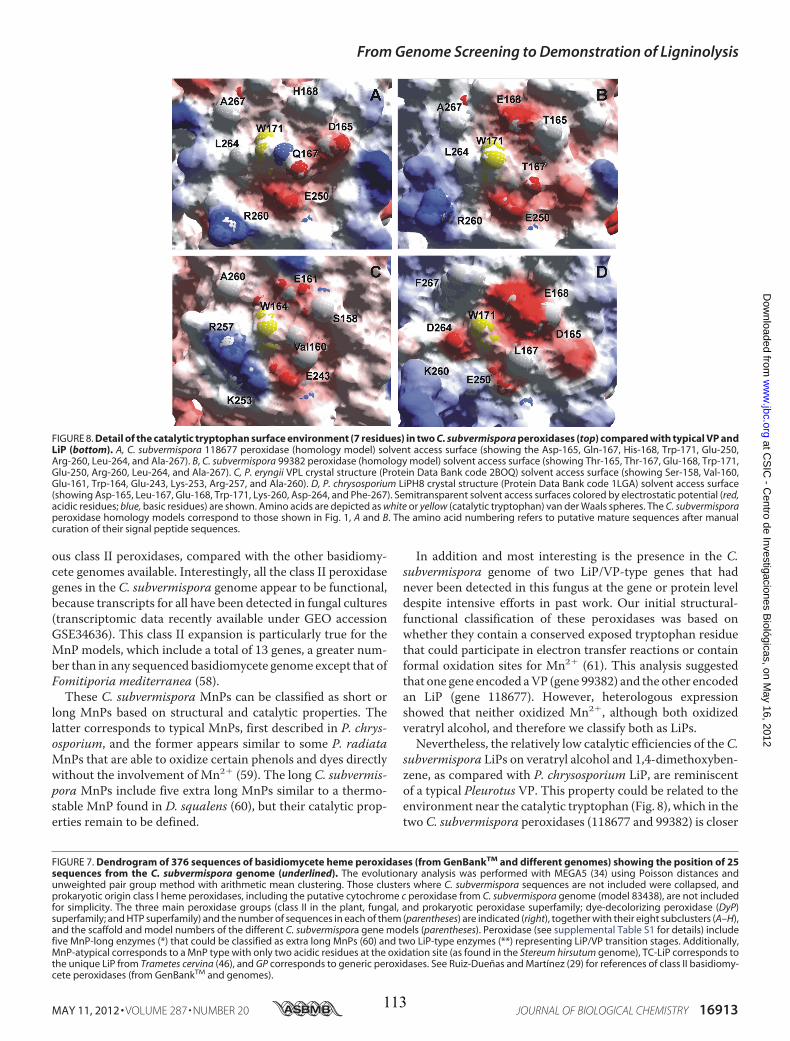
ous class II peroxidases, compared with the other basidiomy-cete genomes available. Interestingly, all the class II peroxidasegenes in the C. subvermispora genome appear to be functional,because transcripts for all have been detected in fungal cultures(transcriptomic data recently available under GEO accessionGSE34636). This class II expansion is particularly true for theMnP models, which include a total of 13 genes, a greater num-ber than in any sequenced basidiomycete genome except that ofFomitiporia mediterranea (58).These C. subvermispora MnPs can be classified as short or
long MnPs based on structural and catalytic properties. Thelatter corresponds to typical MnPs, first described in P. chrys-osporium, and the former appears similar to some P. radiataMnPs that are able to oxidize certain phenols and dyes directlywithout the involvement of Mn2� (59). The long C. subvermis-pora MnPs include five extra long MnPs similar to a thermo-stable MnP found in D. squalens (60), but their catalytic prop-erties remain to be defined.
In addition and most interesting is the presence in the C.subvermispora genome of two LiP/VP-type genes that hadnever been detected in this fungus at the gene or protein leveldespite intensive efforts in past work. Our initial structural-functional classification of these peroxidases was based onwhether they contain a conserved exposed tryptophan residuethat could participate in electron transfer reactions or containformal oxidation sites for Mn2� (61). This analysis suggestedthat one gene encoded aVP (gene 99382) and the other encodedan LiP (gene 118677). However, heterologous expressionshowed that neither oxidized Mn2�, although both oxidizedveratryl alcohol, and therefore we classify both as LiPs.Nevertheless, the relatively low catalytic efficiencies of theC.
subvermispora LiPs on veratryl alcohol and 1,4-dimethoxyben-zene, as compared with P. chrysosporium LiP, are reminiscentof a typical Pleurotus VP. This property could be related to theenvironment near the catalytic tryptophan (Fig. 8), which in thetwo C. subvermispora peroxidases (118677 and 99382) is closer
FIGURE 7. Dendrogram of 376 sequences of basidiomycete heme peroxidases (from GenBankTM and different genomes) showing the position of 25sequences from the C. subvermispora genome (underlined). The evolutionary analysis was performed with MEGA5 (34) using Poisson distances andunweighted pair group method with arithmetic mean clustering. Those clusters where C. subvermispora sequences are not included were collapsed, andprokaryotic origin class I heme peroxidases, including the putative cytochrome c peroxidase from C. subvermispora genome (model 83438), are not includedfor simplicity. The three main peroxidase groups (class II in the plant, fungal, and prokaryotic peroxidase superfamily; dye-decolorizing peroxidase (DyP)superfamily; and HTP superfamily) and the number of sequences in each of them (parentheses) are indicated (right), together with their eight subclusters (A–H),and the scaffold and model numbers of the different C. subvermispora gene models (parentheses). Peroxidase (see supplemental Table S1 for details) includefive MnP-long enzymes (*) that could be classified as extra long MnPs (60) and two LiP-type enzymes (**) representing LiP/VP transition stages. Additionally,MnP-atypical corresponds to a MnP type with only two acidic residues at the oxidation site (as found in the Stereum hirsutum genome), TC-LiP corresponds tothe unique LiP from Trametes cervina (46), and GP corresponds to generic peroxidases. See Ruiz-Dueñas and Martínez (29) for references of class II basidiomy-cete peroxidases (from GenBankTM and genomes).
FIGURE 8. Detail of the catalytic tryptophan surface environment (7 residues) in two C. subvermispora peroxidases (top) compared with typical VP andLiP (bottom). A, C. subvermispora 118677 peroxidase (homology model) solvent access surface (showing the Asp-165, Gln-167, His-168, Trp-171, Glu-250,Arg-260, Leu-264, and Ala-267). B, C. subvermispora 99382 peroxidase (homology model) solvent access surface (showing Thr-165, Thr-167, Glu-168, Trp-171,Glu-250, Arg-260, Leu-264, and Ala-267). C, P. eryngii VPL crystal structure (Protein Data Bank code 2BOQ) solvent access surface (showing Ser-158, Val-160,Glu-161, Trp-164, Glu-243, Lys-253, Arg-257, and Ala-260). D, P. chrysosporium LiPH8 crystal structure (Protein Data Bank code 1LGA) solvent access surface(showing Asp-165, Leu-167, Glu-168, Trp-171, Lys-260, Asp-264, and Phe-267). Semitransparent solvent access surfaces colored by electrostatic potential (red,acidic residues; blue, basic residues) are shown. Amino acids are depicted as white or yellow (catalytic tryptophan) van der Waals spheres. The C. subvermisporaperoxidase homology models correspond to those shown in Fig. 1, A and B. The amino acid numbering refers to putative mature sequences after manualcuration of their signal peptide sequences.
From Genome Screening to Demonstration of Ligninolysis
MAY 11, 2012 • VOLUME 287 • NUMBER 20 JOURNAL OF BIOLOGICAL CHEMISTRY 16913
at CS
IC - C
entro de Investigaciones Biológicas, on M
ay 16, 2012w
ww
.jbc.orgD
ownloaded from
113
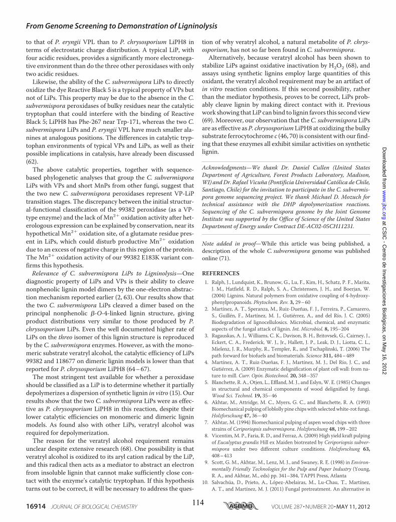
to that of P. eryngii VPL than to P. chrysosporium LiPH8 interms of electrostatic charge distribution. A typical LiP, withfour acidic residues, provides a significantly more electronega-tive environment than do the three other peroxidases with onlytwo acidic residues.Likewise, the ability of the C. subvermispora LiPs to directly
oxidize the dye Reactive Black 5 is a typical property of VPs butnot of LiPs. This property may be due to the absence in the C.subvermispora peroxidases of bulky residues near the catalytictryptophan that could interfere with the binding of ReactiveBlack 5; LiPH8 has Phe-267 near Trp-171, whereas the two C.subvermispora LiPs and P. eryngii VPL have much smaller ala-nines at analogous positions. The differences in catalytic tryp-tophan environments of typical VPs and LiPs, as well as theirpossible implications in catalysis, have already been discussed(62).The above catalytic properties, together with sequence-
based phylogenetic analyses that group the C. subvermisporaLiPs with VPs and short MnPs from other fungi, suggest thatthe two new C. subvermispora peroxidases represent VP-LiPtransition stages. The discrepancy between the initial structur-al-functional classification of the 99382 peroxidase (as a VP-type enzyme) and the lack of Mn2� oxidation activity after het-erologous expression can be explained by conservation, near itshypothetical Mn2� oxidation site, of a glutamate residue pres-ent in LiPs, which could disturb productive Mn2� oxidationdue to an excess of negative charge in this region of the protein.The Mn2� oxidation activity of our 99382 E183K variant con-firms this hypothesis.Relevance of C. subvermispora LiPs to Ligninolysis—One
diagnostic property of LiPs and VPs is their ability to cleavenonphenolic lignin model dimers by the one-electron abstrac-tion mechanism reported earlier (2, 63). Our results show thatthe two C. subvermispora LiPs cleaved a dimer based on theprincipal nonphenolic �-O-4-linked lignin structure, givingproduct distributions very similar to those produced by P.chrysosporium LiPs. Even the well documented higher rate ofLiPs on the threo isomer of this lignin structure is reproducedby the C. subvermispora enzymes. However, as with the mono-meric substrate veratryl alcohol, the catalytic efficiency of LiPs99382 and 118677 on dimeric lignin models is lower than thatreported for P. chrysosporium LiPH8 (64–67).
The most stringent test available for whether a peroxidaseshould be classified as a LiP is to determine whether it partiallydepolymerizes a dispersion of synthetic lignin in vitro (15). Ourresults show that the two C. subvermispora LiPs were as effec-tive as P. chrysosporium LiPH8 in this reaction, despite theirlower catalytic efficiencies on monomeric and dimeric ligninmodels. As found also with other LiPs, veratryl alcohol wasrequired for depolymerization.The reason for the veratryl alcohol requirement remains
unclear despite extensive research (68). One possibility is thatveratryl alcohol is oxidized to its aryl cation radical by the LiP,and this radical then acts as a mediator to abstract an electronfrom insoluble lignin that cannot make sufficiently close con-tact with the enzyme’s catalytic tryptophan. If this hypothesisturns out to be correct, it will be necessary to address the ques-
tion of why veratryl alcohol, a natural metabolite of P. chrys-osporium, has not so far been found in C. subvermispora.Alternatively, because veratryl alcohol has been shown to
stabilize LiPs against oxidative inactivation by H2O2 (68), andassays using synthetic lignins employ large quantities of thisoxidant, the veratryl alcohol requirement may be an artifact ofin vitro reaction conditions. If this second possibility, ratherthan the mediator hypothesis, proves to be correct, LiPs prob-ably cleave lignin by making direct contact with it. Previouswork showing that LiP can bind to lignin favors this second view(69). Moreover, our observation that theC. subvermispora LiPsare as effective asP. chrysosporiumLiPH8 at oxidizing the bulkysubstrate ferrocytochrome c (46, 70) is consistentwith our find-ing that these enzymes all exhibit similar activities on syntheticlignin.
Acknowledgments—We thank Dr. Daniel Cullen (United StatesDepartment of Agriculture, Forest Products Laboratory, Madison,WI) andDr. Rafael Vicuña (PontificiaUniversidadCatólica de Chile,Santiago, Chile) for the invitation to participate in the C. subvermis-pora genome sequencing project. We thank Michael D. Mozuch fortechnical assistance with the DHP depolymerization reactions.Sequencing of the C. subvermispora genome by the Joint GenomeInstitute was supported by the Office of Science of the United StatesDepartment of Energy under Contract DE-AC02-05CH11231.
Note added in proof—While this article was being published, adescription of the whole C. subvermispora genome was publishedonline (71).
REFERENCES1. Ralph, J., Lundquist, K., Brunow, G., Lu, F., Kim, H., Schatz, P. F., Marita,
J. M., Hatfield, R. D., Ralph, S. A., Christensen, J. H., and Boerjan, W.(2004) Lignins. Natural polymers from oxidative coupling of 4-hydroxy-phenylpropanoids. Phytochem. Rev. 3, 29–60
2. Martínez, A. T., Speranza, M., Ruiz-Dueñas, F. J., Ferreira, P., Camarero,S., Guillén, F., Martínez, M. J., Gutiérrez, A., and del Río, J. C. (2005)Biodegradation of lignocellulosics. Microbial, chemical, and enzymaticaspects of the fungal attack of lignin. Int. Microbiol. 8, 195–204
3. Ragauskas, A. J., Williams, C. K., Davison, B. H., Britovsek, G., Cairney, J.,Eckert, C. A., Frederick, W. J., Jr., Hallett, J. P., Leak, D. J., Liotta, C. L.,Mielenz, J. R., Murphy, R., Templer, R., and Tschaplinski, T. (2006) Thepath forward for biofuels and biomaterials. Science 311, 484–489
4. Martínez, A. T., Ruiz-Dueñas, F. J., Martínez, M. J., Del Río, J. C., andGutiérrez, A. (2009) Enzymatic delignification of plant cell wall: from na-ture to mill. Curr. Opin. Biotechnol. 20, 348–357
5. Blanchette, R. A., Otjen, L., Effland,M. J., and Eslyn,W. E. (1985) Changesin structural and chemical components of wood delignified by fungi.Wood Sci. Technol. 19, 35–46
6. Akhtar, M., Attridge, M. C., Myers, G. C., and Blanchette, R. A. (1993)Biomechanical pulping of loblolly pine chipswith selectedwhite-rot fungi.Holzforschung 47, 36–40
7. Akhtar, M. (1994) Biomechanical pulping of aspen wood chips with threestrains of Ceriporiopsis subvermispora. Holzforschung 48, 199–202
8. Vicentim,M. P., Faria, R. D., and Ferraz, A. (2009) High yield kraft pulpingof Eucalyptus grandis Hill ex Maiden biotreated by Ceriporiopsis subver-mispora under two different culture conditions. Holzforschung 63,408–413
9. Scott, G. M., Akhtar, M., Lenz, M. J., and Swaney, R. E. (1998) in Environ-mentally Friendly Technologies for the Pulp and Paper Industry (Young,R. A., and Akhtar, M., eds) pp. 341–384, TAPPI Press, Atlanta
10. Salvachúa, D., Prieto, A., López-Abelairas, M., Lu-Chau, T., Martínez,A. T., and Martínez, M. J. (2011) Fungal pretreatment. An alternative in
From Genome Screening to Demonstration of Ligninolysis
16914 JOURNAL OF BIOLOGICAL CHEMISTRY VOLUME 287 • NUMBER 20 • MAY 11, 2012
at CS
IC - C
entro de Investigaciones Biológicas, on M
ay 16, 2012w
ww
.jbc.orgD
ownloaded from
114
second-generation ethanol from wheat straw. Bioresour. Technol. 102,7500–7506
11. Kersten, P., and Cullen, D. (2007) Extracellular oxidative systems of thelignin-degrading Basidiomycete Phanerochaete chrysosporium. FungalGenet. Biol. 44, 77–87
12. Ruiz-Dueñas, F. J., and Martínez, A. T. (2009) Microbial degradation oflignin. How a bulky recalcitrant polymer is efficiently recycled in natureand how we can take advantage of this.Microbiol. Biotechnol. 2, 164–177
13. Tien, M., and Kirk, T. K. (1983) Lignin-degrading enzyme from the Hy-menomycete Phanerochaete chrysosporium Burds. Science 221, 661–663
14. Caramelo, L., Martinez, M. J., and Martinez, A. T. (1999) A search forligninolytic peroxidases in the fungus Pleurotus eryngii involving �-keto-�-thiomethylbutyric acid and lignin model dimers. Appl. Environ. Micro-biol. 65, 916–922
15. Hammel, K. E., Jensen, K. A., Jr., Mozuch, M. D., Landucci, L. L., Tien, M.,and Pease, E. A. (1993) Ligninolysis by a purified lignin peroxidase. J. Biol.Chem. 268, 12274–12281
16. Gold, M. H., Youngs, H. L., and Gelpke, M. D. (2000) Manganese peroxi-dase.Met. Ions Biol. Syst. 37, 559–586
17. Camarero, S., Galletti, G. C., and Martínez, A. T. (1994) Preferential deg-radation of phenolic lignin units by two white rot fungi. Appl. Environ.Microbiol. 60, 4509–4516
18. Salas, C., Lobos, S., Larraín, J., Salas, L., Cullen, D., and Vicuña, R. (1995)Properties of laccase isoenzymes produced by the basidiomycete Ceripo-riopsis subvermispora. Biotechnol. Appl. Biochem. 21, 323–333
19. Lobos, S., Larraín, J., Salas, L., Cullen, D., and Vicuña, R. (1994) Isoen-zymes of manganese-dependent peroxidase and laccase produced by thelignin-degrading basidiomycete Ceriporiopsis subvermispora. Microbiol-ogy 140, 2691–2698
20. Rüttimann, C., Schwember, E., Salas, L., Cullen, D., and Vicuña, R. (1992)Ligninolytic enzymes of the white rot basidiomycetes Phlebia brevisporaand Ceriporiopsis subvermispora. Biotechnol. Appl. Biochem. 16, 64–76
21. Rajakumar, S., Gaskell, J., Cullen, D., Lobos, S., Karahanian, E., andVicuna,R. (1996) Lip-like genes in Phanerochaete sordida and Ceriporiopsis sub-vermispora, white rot fungi with no detectable lignin peroxidase activity.Appl. Environ. Microbiol. 62, 2660–2663
22. Tanaka, H., Koike, K., Itakura, S., and Enoki, A. (2009) Degradation ofwood and enzyme production by Ceriporiopsis subvermispora. EnzymeMicrob. Technol. 45, 384–390
23. Jensen, K. A., Bao, W., Kawai, S., Srebotnik, E., and Hammel, K. E. (1996)Manganese-dependent cleavage of nonphenolic lignin structures by Ceri-poriopsis subvermispora in the absence of lignin peroxidase. Appl. Envi-ron. Microbiol. 62, 3679–3686
24. Srebotnik, E., Jensen, K. A., Kawai, S., and Hammel, K. E. (1997) Evidencethat Ceriporiopsis subvermispora degrades nonphenolic lignin structuresby a one-electron oxidation mechanism. Appl. Environ. Microbiol. 63,4435–4440
25. Moen, M. A., and Hammel, K. E. (1994) Lipid peroxidation by the manga-nese peroxidase of Phanerochaete chrysosporium is the basis for phenan-threne oxidation by the intact fungus. Appl. Environ. Microbiol. 60,1956–1961
26. Cañas, A. I., andCamarero, S. (2010) Laccases and their naturalmediators.Biotechnological tools for sustainable eco-friendly processes. Biotechnol.Adv. 28, 694–705
27. Martinez, D., Larrondo, L. F., Putnam,N., Gelpke,M.D., Huang, K., Chap-man, J., Helfenbein, K. G., Ramaiya, P., Detter, J. C., Larimer, F., Coutinho,P. M., Henrissat, B., Berka, R., Cullen, D., and Rokhsar, D. (2004) Genomesequence of the lignocellulose degrading fungusPhanerochaete chrysospo-rium strain RP78. Nat. Biotechnol. 22, 695–700
28. Martinez, D., Challacombe, J., Morgenstern, I., Hibbett, D., Schmoll, M.,Kubicek, C. P., Ferreira, P., Ruiz-Duenas, F. J., Martinez, A. T., Kersten, P.,Hammel, K. E., VandenWymelenberg, A., Gaskell, J., Lindquist, E., Sabat,G., Bondurant, S. S., Larrondo, L. F., Canessa, P., Vicuna, R., Yadav, J.,Doddapaneni, H., Subramanian, V., Pisabarro, A. G., Lavín, J. L., Oguiza,J. A., Master, E., Henrissat, B., Coutinho, P.M., Harris, P., Magnuson, J. K.,Baker, S. E., Bruno, K., Kenealy, W., Hoegger, P. J., Kües, U., Ramaiya, P.,Lucas, S., Salamov, A., Shapiro, H., Tu, H., Chee, C. L., Misra, M., Xie, G.,Teter, S., Yaver, D., James, T., Mokrejs, M., Pospisek, M., Grigoriev, I. V.,
Brettin, T., Rokhsar, D., Berka, R., and Cullen, D. (2009) Genome, tran-scriptome, and secretome analysis of wood decay fungus Postia placentasupports unique mechanisms of lignocellulose conversion. Proc. Natl.Acad. Sci. U.S.A. 106, 1954–1959
29. Ruiz-Dueñas, F. J., and Martínez, A. T. (2010) in Biocatalysts Based onHeme Peroxidases (Torres, E., and Ayala, M., eds) pp. 37–59, Springer-Verlag, Berlin
30. Ruiz-Dueñas, F. J., Fernández, E., Martínez, M. J., and Martínez, A. T.(2011) Pleurotus ostreatus heme peroxidases. An in silico analysis from thegenome sequence to the enzyme molecular structure. C. R. Biol. 334,795–805
31. Tello,M., Seelenfreund, D., Lobos, S., Gaskell, J., Cullen, D., andVicuña, R.(2001) Isolation and characterization of homokaryotic strains from theligninolytic basidiomycete Ceriporiopsis subvermispora. FEMSMicrobiol.Lett. 199, 91–96
32. Lobos, S., Larrondo, L., Salas, L., Karahanian, E., and Vicuña, R. (1998)Cloning and molecular analysis of a cDNA and the Cs-mnp1 gene encod-ing a manganese peroxidase isoenzyme from the lignin-degrading basidi-omycete Ceriporiopsis subvermispora. Gene 206, 185–193
33. Tello, M., Corsini, G., Larrondo, L. F., Salas, L., Lobos, S., and Vicuña, R.(2000) Characterization of three new manganese peroxidase genes fromthe ligninolytic basidiomyceteCeriporiopsis subvermispora. Biochim. Bio-phys. Acta 1490, 137–144
34. Tamura, K., Peterson, D., Peterson, N., Stecher, G., Nei,M., and Kumar, S.(2011)MEGA5.Molecular evolutionary genetics analysis usingmaximumlikelihood, evolutionary distance, and maximum parsimony methods.Mol. Biol. Evol. 28, 2731–2739
35. Bendtsen, J. D., Nielsen, H., von Heijne, G., and Brunak, S. (2004) Im-proved prediction of signal peptides. SignalP 3.0. J. Mol. Biol. 340,783–795
36. Bordoli, L., Kiefer, F., Arnold, K., Benkert, P., Battey, J., and Schwede, T.(2009) Protein structure homology modeling using SWISS-MODELworkspace. Nat. Protoc. 4, 1–13
37. Sambrook, J., and Russell, D. W. (2001)Molecular Cloning, 3rd Ed., ColdSpring Harbor Laboratory Press, Cold Spring Harbor, NY
38. Pérez-Boada, M., Doyle, W. A., Ruiz-Dueñas, F. J., Martínez, M. J., Mar-tínez, A. T., and Smith, A. T. (2002) Expression of Pleurotus eryngii versa-tile peroxidase in Escherichia coli and optimization of in vitro folding.Enzyme Microb. Technol. 30, 518–524
39. Doyle, W. A., and Smith, A. T. (1996) Expression of lignin peroxidase H8inEscherichia coli. Folding and activation of the recombinant enzymewithCa2� and heme. Biochem. J. 315, 15–19
40. Heinfling, A., Ruiz-Dueñas, F. J., Martínez, M. J., Bergbauer, M., Szewzyk,U., and Martínez, A. T. (1998) A study on reducing substrates of manga-nese-oxidizing peroxidases from Pleurotus eryngii and Bjerkandera adu-sta. FEBS Lett. 428, 141–146
41. Ruiz-Dueñas, F. J., Camarero, S., Pérez-Boada, M., Martínez, M. J., andMartínez, A. T. (2001) A new versatile peroxidase from Pleurotus.Biochem. Soc. Trans. 29, 116–122
42. Srebotnik, E., Jensen, K. A., Jr., and Hammel, K. E. (1994) Fungal degrada-tion of recalcitrant nonphenolic lignin structures without lignin peroxi-dase. Proc. Natl. Acad. Sci. U.S.A. 91, 12794–12797
43. Shary, S., Ralph, S. A., and Hammel, K. E. (2007) New insights into theligninolytic capability of a wood decay ascomycete. Appl. Environ. Micro-biol. 73, 6691–6694
44. Morita, Y., Yamashita, H.,Mikami, B., Iwamoto, H., Aibara, S., Terada,M.,andMinami, J. (1988) Purification, crystallization, and characterization ofperoxidase from Coprinus cinereus. J. Biochem. 103, 693–699
45. Pecyna, M. J., Ullrich, R., Bittner, B., Clemens, A., Scheibner, K., Schubert,R., and Hofrichter, M. (2009)Molecular characterization of aromatic per-oxygenase from Agrocybe aegerita. Appl. Microbiol. Biotechnol. 84,885–897
46. Miki, Y., Calviño, F. R., Pogni, R., Giansanti, S., Ruiz-Dueñas, F. J., Mar-tínez, M. J., Basosi, R., Romero, A., and Martínez, A. T. (2011) Crystallo-graphic, kinetic, and spectroscopic study of the first ligninolytic peroxi-dase presenting a catalytic tyrosine. J. Biol. Chem. 286, 15525–15534
47. Martínez, A. T. (2002)Molecular biology and structure-function of lignin-degrading heme peroxidases. Enzyme Microb. Technol. 30, 425–444
From Genome Screening to Demonstration of Ligninolysis
MAY 11, 2012 • VOLUME 287 • NUMBER 20 JOURNAL OF BIOLOGICAL CHEMISTRY 16915
at CS
IC - C
entro de Investigaciones Biológicas, on M
ay 16, 2012w
ww
.jbc.orgD
ownloaded from
115
48. Cullen, D. (1997) Recent advances on the molecular genetics of lignino-lytic fungi. J. Biotechnol. 53, 273–289
49. Alvarez, J. M., Canessa, P., Mancilla, R. A., Polanco, R., Santibáñez, P. A.,and Vicuña, R. (2009) Expression of genes encoding laccase and manga-nese-dependent peroxidase in the fungus Ceriporiopsis subvermispora ismediated by an ACE1-like copper-fist transcription factor. Fungal Genet.Biol. 46, 104–111
50. Larrondo, L. F., Lobos, S., Stewart, P., Cullen, D., and Vicuña, R. (2001)Isoenzyme multiplicity and characterization of recombinant manganeseperoxidases from Ceriporiopsis subvermispora and Phanerochaete chrys-osporium. Appl. Environ. Microbiol. 67, 2070–2075
51. de Souza-Cruz, P. B., Freer, J., Siikaaho, M., and Ferraz, A. (2004) Extrac-tion and determination of enzymes produced by Ceriporiopsis subvermis-pora during biopulping of Pinus taedawood chips. EnzymeMicrob. Tech-nol. 34, 228–234
52. Hofrichter, M., Ullrich, R., Pecyna, M. J., Liers, C., and Lundell, T. (2010)New and classic families of secreted fungal heme peroxidases. Appl. Mi-crobiol. Biotechnol. 87, 871–897
53. Liers, C., Bobeth, C., Pecyna, M., Ullrich, R., and Hofrichter, M. (2010)DyP-like peroxidases of the jelly fungus Auricularia auricula-judae oxi-dize nonphenolic ligninmodel compounds and high redox potential dyes.Appl. Microbiol. Biotechnol. 85, 1869–1879
54. Gutiérrez, A., Babot, E. D., Ullrich, R., Hofrichter,M.,Martínez, A. T., anddel Río, J. C. (2011) Regioselective oxygenation of fatty acids, fatty alco-hols, and other aliphatic compounds by a basidiomycete heme-thiolateperoxidase. Arch. Biochem. Biophys. 514, 33–43
55. Kinne, M., Poraj-Kobielska, M., Ullrich, R., Nousiainen, P., Sipila, J.,Scheibner, K., Hammel, K. E., and Hofrichter, M. (2011) Oxidative cleav-age of nonphenolic �-O-4 lignin model dimers by an extracellular aro-matic peroxygenase. Holzforschung 65, 673–679
56. Hofrichter, M., and Ullrich, R. (2006) Heme-thiolate haloperoxidases.Versatile biocatalysts with biotechnological and environmental signifi-cance. Appl. Microbiol. Biotechnol. 71, 276–288
57. Ortiz-Bermúdez, P., Srebotnik, E., andHammel, K. E. (2003) Chlorinationand cleavage of lignin structures by fungal chloroperoxidases. Appl. Envi-ron. Microbiol. 69, 5015–5018
58. Morgenstern, I., Robertson, D. L., and Hibbett, D. S. (2010) Characteriza-tion of three mnp genes of Fomitiporia mediterranea and report of addi-tional class II peroxidases in the order Hymenochaetales. Appl. Environ.Microbiol. 76, 6431–6440
59. Hildén, K., Martinez, A. T., Hatakka, A., and Lundell, T. (2005) The twomanganese peroxidases Pr-MnP2 and Pr-MnP3 of Phlebia radiata, alignin-degrading basidiomycete, are phylogenetically and structurally di-
vergent. Fungal Genet. Biol. 42, 403–41960. Li, D., Li, N., Ma, B., Mayfield, M. B., and Gold, M. H. (1999) Character-
ization of genes encoding two manganese peroxidases from the lignin-degrading fungus Dichomitus squalens(1). Biochim. Biophys. Acta 1434,356–364
61. Ruiz-Dueñas, F. J., Morales, M., García, E., Miki, Y., Martínez, M. J., andMartínez, A. T. (2009) Substrate oxidation sites in versatile peroxidase andother basidiomycete peroxidases. J. Exp. Bot. 60, 441–452
62. Ruiz-Dueñas, F. J., Morales, M., Mate, M. J., Romero, A., Martínez, M. J.,Smith, A. T., and Martínez, A. T. (2008) Site-directed mutagenesis of thecatalytic tryptophan environment in Pleurotus eryngii versatile peroxi-dase. Biochemistry 47, 1685–1695
63. Kirk, T. K., and Farrell, R. L. (1987) Enzymatic “combustion.” The micro-bial degradation of lignin. Annu. Rev. Microbiol. 41, 465–505
64. Bohlin, C., Persson, P., Gorton, L., Lundquist, K., and Jonsson, L. F. (2005)Product profiles in enzymic and nonenzymic oxidations of the ligninmodel compound erythro-1-(3,4-dimethoxyphenyl)2-(2-methoxyphen-oxy)-1,3-propanediol. J. Mol. Catal. B-Enzym. 35, 100–107
65. Bohlin, C., Lundquist, K., and Jonsson, L. J. (2008)Diastereomer selectivityin the degradation of a lignin model compound of the arylglycerol �-arylether type by white-rot fungi. Enzyme Microb. Technol. 43, 199–204
66. Jönsson, L., Karlsson, O., Lundquist, K., and Nyman, P. O. (1990) Ste-reospecificity in enzymic and nonenzymic oxidation of �-O-4 ligninmodel compounds. FEBS Lett. 276, 45–48
67. Cho, D. W., Parthasarathi, R., Pimentel, A. S., Maestas, G. D., Park, H. J.,Yoon, U. C., Dunaway-Mariano, D., Gnanakaran, S., Langan, P., andMariano, P. S. (2010) Nature and kinetic analysis of carbon-carbon bondfragmentation reactions of cation radicals derived from SET-oxidation oflignin model compounds. J. Org. Chem. 75, 6549–6562
68. Hammel, K. E., and Cullen, D. (2008) Role of fungal peroxidases in biolog-ical ligninolysis. Curr. Opin. Plant Biol. 11, 349–355
69. Johjima, T., Itoh, N., Kabuto, M., Tokimura, F., Nakagawa, T., Wariishi,H., and Tanaka, H. (1999) Direct interaction of lignin and lignin peroxi-dase from Phanerochaete chrysosporium. Proc. Natl. Acad. Sci. U.S.A. 96,1989–1994
70. Wariishi, H., Sheng, D., and Gold, M. H. (1994) Oxidation of ferrocyto-chrome c by lignin peroxidase. Biochemistry 33, 5545–5552
71. Fernandez-Fueyo, E., Ruiz-Duenas, F. J., Ferreira, P., Floudas, D., Hibbett,D. S., Canessa, P., Larrondo, L. F., James, T. Y., Seelenfreund, D., Lobos, S.et al. (2012) Comparative genomics of Ceriporiopsis subvermispora andPhanerochaete chrysosporium provide insight into selective ligninolysis.Proc. Natl. Acad. Sci. U.S.A. 109, 5458–5463
From Genome Screening to Demonstration of Ligninolysis
16916 JOURNAL OF BIOLOGICAL CHEMISTRY VOLUME 287 • NUMBER 20 • MAY 11, 2012
at CS
IC - C
entro de Investigaciones Biológicas, on M
ay 16, 2012w
ww
.jbc.orgD
ownloaded from
116
Capítulo 7
"Catalytic and stability properties of manganese peroxidase subfamilies: A study based on the Ceriporiopsis subvermispora genome" Fernández-Fueyo, E., S. Acebes, F. J. Ruiz-Dueñas, , M. J. Martínez, A. Romero, F. J. Medrano, V Guallar and A. T. Martínez. (enviado)
117

118
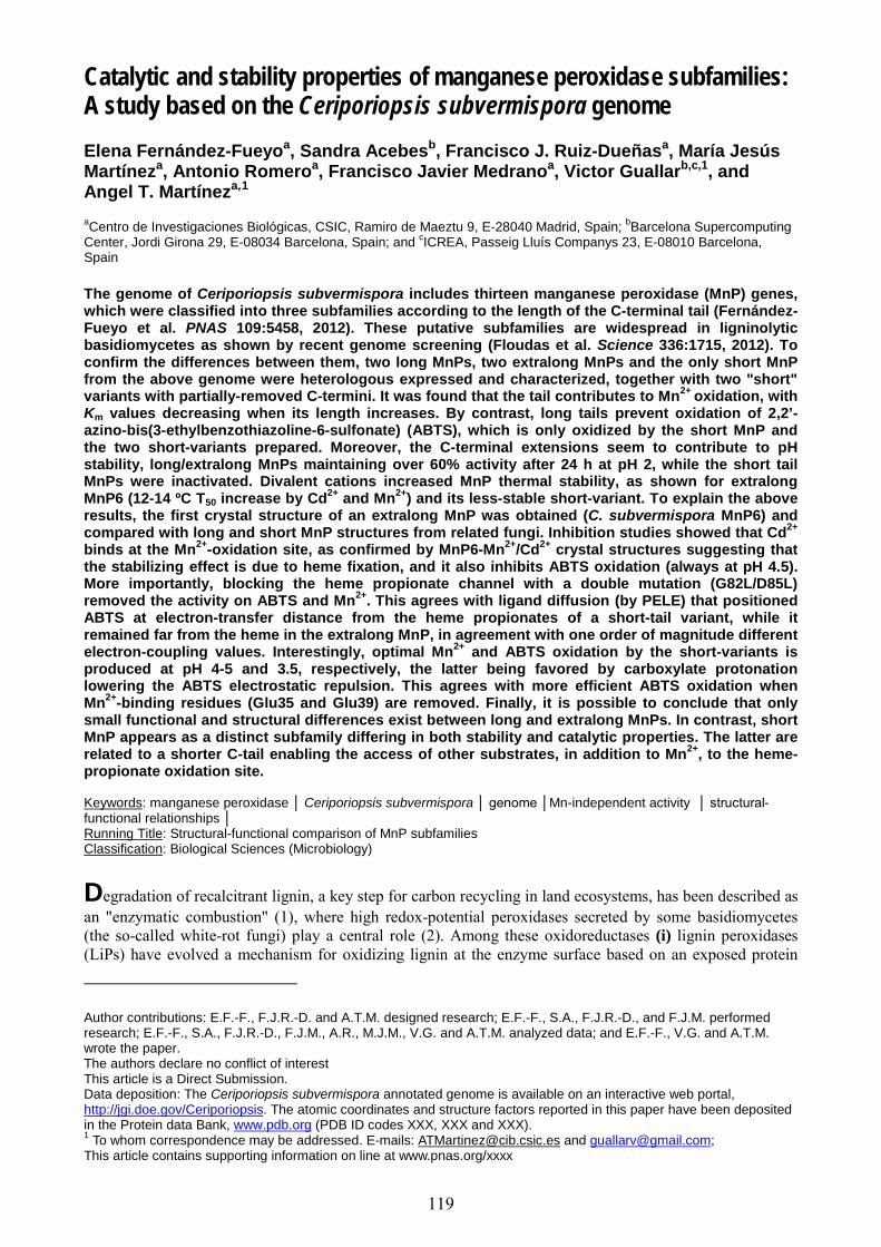
Catalytic and stability properties of manganese peroxidase subfamilies: A study based on the Ceriporiopsis subvermispora genome Elena Fernández-Fueyoa, Sandra Acebesb, Francisco J. Ruiz-Dueñasa, María Jesús Martíneza, Antonio Romeroa, Francisco Javier Medranoa, Victor Guallarb,c,1, and Angel T. Martíneza,1
aCentro de Investigaciones Biológicas, CSIC, Ramiro de Maeztu 9, E-28040 Madrid, Spain; bBarcelona Supercomputing Center, Jordi Girona 29, E-08034 Barcelona, Spain; and cICREA, Passeig Lluís Companys 23, E-08010 Barcelona, Spain The genome of Ceriporiopsis subvermispora includes thirteen manganese peroxidase (MnP) genes, which were classified into three subfamilies according to the length of the C-terminal tail (Fernández-Fueyo et al. PNAS 109:5458, 2012). These putative subfamilies are widespread in ligninolytic basidiomycetes as shown by recent genome screening (Floudas et al. Science 336:1715, 2012). To confirm the differences between them, two long MnPs, two extralong MnPs and the only short MnP from the above genome were heterologous expressed and characterized, together with two "short" variants with partially-removed C-termini. It was found that the tail contributes to Mn2+ oxidation, with Km values decreasing when its length increases. By contrast, long tails prevent oxidation of 2,2’-azino-bis(3-ethylbenzothiazoline-6-sulfonate) (ABTS), which is only oxidized by the short MnP and the two short-variants prepared. Moreover, the C-terminal extensions seem to contribute to pH stability, long/extralong MnPs maintaining over 60% activity after 24 h at pH 2, while the short tail MnPs were inactivated. Divalent cations increased MnP thermal stability, as shown for extralong MnP6 (12-14 ºC T50 increase by Cd2+ and Mn2+) and its less-stable short-variant. To explain the above results, the first crystal structure of an extralong MnP was obtained (C. subvermispora MnP6) and compared with long and short MnP structures from related fungi. Inhibition studies showed that Cd2+ binds at the Mn2+-oxidation site, as confirmed by MnP6-Mn2+/Cd2+ crystal structures suggesting that the stabilizing effect is due to heme fixation, and it also inhibits ABTS oxidation (always at pH 4.5). More importantly, blocking the heme propionate channel with a double mutation (G82L/D85L) removed the activity on ABTS and Mn2+. This agrees with ligand diffusion (by PELE) that positioned ABTS at electron-transfer distance from the heme propionates of a short-tail variant, while it remained far from the heme in the extralong MnP, in agreement with one order of magnitude different electron-coupling values. Interestingly, optimal Mn2+ and ABTS oxidation by the short-variants is produced at pH 4-5 and 3.5, respectively, the latter being favored by carboxylate protonation lowering the ABTS electrostatic repulsion. This agrees with more efficient ABTS oxidation when Mn2+-binding residues (Glu35 and Glu39) are removed. Finally, it is possible to conclude that only small functional and structural differences exist between long and extralong MnPs. In contrast, short MnP appears as a distinct subfamily differing in both stability and catalytic properties. The latter are related to a shorter C-tail enabling the access of other substrates, in addition to Mn2+, to the heme-propionate oxidation site. Keywords: manganese peroxidase │ Ceriporiopsis subvermispora │ genome │Mn-independent activity │ structural-functional relationships │ Running Title: Structural-functional comparison of MnP subfamilies Classification: Biological Sciences (Microbiology) Degradation of recalcitrant lignin, a key step for carbon recycling in land ecosystems, has been described as an "enzymatic combustion" (1), where high redox-potential peroxidases secreted by some basidiomycetes (the so-called white-rot fungi) play a central role (2). Among these oxidoreductases (i) lignin peroxidases (LiPs) have evolved a mechanism for oxidizing lignin at the enzyme surface based on an exposed protein
Author contributions: E.F.-F., F.J.R.-D. and A.T.M. designed research; E.F.-F., S.A., F.J.R.-D., and F.J.M. performed research; E.F.-F., S.A., F.J.R.-D., F.J.M., A.R., M.J.M., V.G. and A.T.M. analyzed data; and E.F.-F., V.G. and A.T.M. wrote the paper. The authors declare no conflict of interest This article is a Direct Submission. Data deposition: The Ceriporiopsis subvermispora annotated genome is available on an interactive web portal, http://jgi.doe.gov/Ceriporiopsis. The atomic coordinates and structure factors reported in this paper have been deposited in the Protein data Bank, www.pdb.org (PDB ID codes XXX, XXX and XXX). 1 To whom correspondence may be addressed. E-mails: [email protected] and [email protected]; This article contains supporting information on line at www.pnas.org/xxxx
119
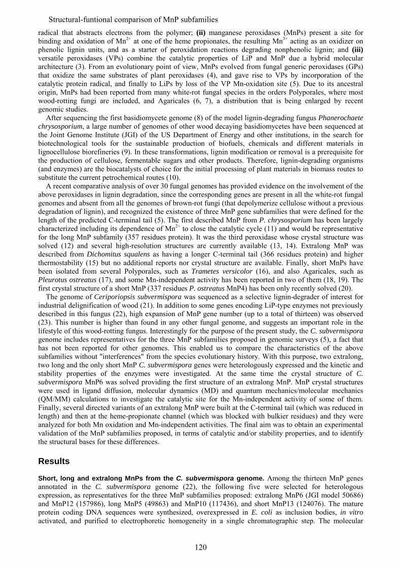
radical that abstracts electrons from the polymer; (ii) manganese peroxidases (MnPs) present a site for binding and oxidation of Mn2+ at one of the heme propionates, the resulting Mn3+ acting as an oxidizer on phenolic lignin units, and as a starter of peroxidation reactions degrading nonphenolic lignin; and (iii) versatile peroxidases (VPs) combine the catalytic properties of LiP and MnP due a hybrid molecular architecture (3). From an evolutionary point of view, MnPs evolved from fungal generic peroxidases (GPs) that oxidize the same substrates of plant peroxidases (4), and gave rise to VPs by incorporation of the catalytic protein radical, and finally to LiPs by loss of the VP Mn-oxidation site (5). Due to its ancestral origin, MnPs had been reported from many white-rot fungal species in the orders Polyporales, where most wood-rotting fungi are included, and Agaricales (6, 7), a distribution that is being enlarged by recent genomic studies. After sequencing the first basidiomycete genome (8) of the model lignin-degrading fungus Phanerochaete chrysosporium, a large number of genomes of other wood decaying basidiomycetes have been sequenced at the Joint Genome Institute (JGI) of the US Department of Energy and other institutions, in the search for biotechnological tools for the sustainable production of biofuels, chemicals and different materials in lignocellulose biorefineries (9). In these transformations, lignin modification or removal is a prerequisite for the production of cellulose, fermentable sugars and other products. Therefore, lignin-degrading organisms (and enzymes) are the biocatalysts of choice for the initial processing of plant materials in biomass routes to substitute the current petrochemical routes (10). A recent comparative analysis of over 30 fungal genomes has provided evidence on the involvement of the above peroxidases in lignin degradation, since the corresponding genes are present in all the white-rot fungal genomes and absent from all the genomes of brown-rot fungi (that depolymerize cellulose without a previous degradation of lignin), and recognized the existence of three MnP gene subfamilies that were defined for the length of the predicted C-terminal tail (5). The first described MnP from P. chrysosporium has been largely characterized including its dependence of Mn2+ to close the catalytic cycle (11) and would be representative for the long MnP subfamily (357 residues protein). It was the third peroxidase whose crystal structure was solved (12) and several high-resolution structures are currently available (13, 14). Extralong MnP was described from Dichomitus squalens as having a longer C-terminal tail (366 residues protein) and higher thermostability (15) but no additional reports nor crystal structure are available. Finally, short MnPs have been isolated from several Polyporales, such as Trametes versicolor (16), and also Agaricales, such as Pleurotus ostreatus (17), and some Mn-independent activity has been reported in two of them (18, 19). The first crystal structure of a short MnP (337 residues P. ostreatus MnP4) has been only recently solved (20). The genome of Ceriporiopsis subvermispora was sequenced as a selective lignin-degrader of interest for industrial delignification of wood (21). In addition to some genes encoding LiP-type enzymes not previously described in this fungus (22), high expansion of MnP gene number (up to a total of thirteen) was observed (23). This number is higher than found in any other fungal genome, and suggests an important role in the lifestyle of this wood-rotting fungus. Interestingly for the purpose of the present study, the C. subvermispora genome includes representatives for the three MnP subfamilies proposed in genomic surveys (5), a fact that has not been reported for other genomes. This enabled us to compare the characteristics of the above subfamilies without "interferences" from the species evolutionary history. With this purpose, two extralong, two long and the only short MnP C. subvermispora genes were heterologously expressed and the kinetic and stability properties of the enzymes were investigated. At the same time the crystal structure of C. subvermispora MnP6 was solved providing the first structure of an extralong MnP. MnP crystal structures were used in ligand diffusion, molecular dynamics (MD) and quantum mechanics/molecular mechanics (QM/MM) calculations to investigate the catalytic site for the Mn-independent activity of some of them. Finally, several directed variants of an extralong MnP were built at the C-terminal tail (which was reduced in length) and then at the heme-propionate channel (which was blocked with bulkier residues) and they were analyzed for both Mn oxidation and Mn-independent activities. The final aim was to obtain an experimental validation of the MnP subfamilies proposed, in terms of catalytic and/or stability properties, and to identify the structural bases for these differences. Results Short, long and extralong MnPs from the C. subvermispora genome. Among the thirteen MnP genes annotated in the C. subvermispora genome (22), the following five were selected for heterologous expression, as representatives for the three MnP subfamilies proposed: extralong MnP6 (JGI model 50686) and MnP12 (157986), long MnP5 (49863) and MnP10 (117436), and short MnP13 (124076). The mature protein coding DNA sequences were synthesized, overexpressed in E. coli as inclusion bodies, in vitro activated, and purified to electrophoretic homogeneity in a single chromatographic step. The molecular
120
Structural-funtional comparison of MnP subfamilies
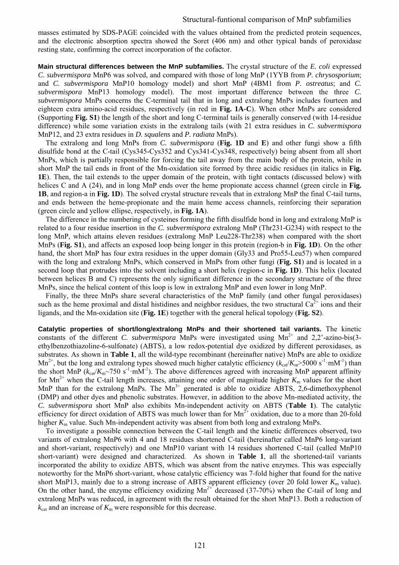
masses estimated by SDS-PAGE coincided with the values obtained from the predicted protein sequences, and the electronic absorption spectra showed the Soret (406 nm) and other typical bands of peroxidase resting state, confirming the correct incorporation of the cofactor. Main structural differences between the MnP subfamilies. The crystal structure of the E. coli expressed C. subvermispora MnP6 was solved, and compared with those of long MnP (1YYB from P. chrysosporium; and C. subvermispora MnP10 homology model) and short MnP (4BM1 from P. ostreatus; and C. subvermispora MnP13 homology model). The most important difference between the three C. subvermispora MnPs concerns the C-terminal tail that in long and extralong MnPs includes fourteen and eighteen extra amino-acid residues, respectively (in red in Fig. 1A-C). When other MnPs are considered (Supporting Fig. S1) the length of the short and long C-terminal tails is generally conserved (with 14-residue difference) while some variation exists in the extralong tails (with 21 extra residues in C. subvermispora MnP12, and 23 extra residues in D. squalens and P. radiata MnPs). The extralong and long MnPs from C. subvermispora (Fig. 1D and E) and other fungi show a fifth disulfide bond at the C-tail (Cys345-Cys352 and Cys341-Cys348, respectively) being absent from all short MnPs, which is partially responsible for forcing the tail away from the main body of the protein, while in short MnP the tail ends in front of the Mn-oxidation site formed by three acidic residues (in italics in Fig. 1E). Then, the tail extends to the upper domain of the protein, with tight contacts (discussed below) with helices C and A (24), and in long MnP ends over the heme propionate access channel (green circle in Fig. 1B, and region-a in Fig. 1D). The solved crystal structure reveals that in extralong MnP the final C-tail turns, and ends between the heme-propionate and the main heme access channels, reinforcing their separation (green circle and yellow ellipse, respectively, in Fig. 1A). The difference in the numbering of cysteines forming the fifth disulfide bond in long and extralong MnP is related to a four residue insertion in the C. subvermispora extralong MnP (Thr231-G234) with respect to the long MnP, which attains eleven residues (extralong MnP Leu228-Thr238) when compared with the short MnPs (Fig. S1), and affects an exposed loop being longer in this protein (region-b in Fig. 1D). On the other hand, the short MnP has four extra residues in the upper domain (Gly33 and Pro55-Leu57) when compared with the long and extralong MnPs, which conserved in MnPs from other fungi (Fig. S1) and is located in a second loop that protrudes into the solvent including a short helix (region-c in Fig. 1D). This helix (located between helices B and C) represents the only significant difference in the secondary structure of the three MnPs, since the helical content of this loop is low in extralong MnP and even lower in long MnP. Finally, the three MnPs share several characteristics of the MnP family (and other fungal peroxidases) such as the heme proximal and distal histidines and neighbor residues, the two structural Ca2+ ions and their ligands, and the Mn-oxidation site (Fig. 1E) together with the general helical topology (Fig. S2). Catalytic properties of short/long/extralong MnPs and their shortened tail variants. The kinetic constants of the different C. subvermispora MnPs were investigated using Mn2+ and 2,2’-azino-bis(3-ethylbenzothiazoline-6-sulfonate) (ABTS), a low redox-potential dye oxidized by different peroxidases, as substrates. As shown in Table 1, all the wild-type recombinant (hereinafter native) MnPs are able to oxidize Mn2+, but the long and extralong types showed much higher catalytic efficiency (kcat/Km>5000 s-1·mM-1) than the short MnP (kcat/Km~750 s-1·mM-1). The above differences agreed with increasing MnP apparent affinity for Mn2+ when the C-tail length increases, attaining one order of magnitude higher Km values for the short MnP than for the extralong MnPs. The Mn3+ generated is able to oxidize ABTS, 2,6-dimethoxyphenol (DMP) and other dyes and phenolic substrates. However, in addition to the above Mn-mediated activity, the C. subvermispora short MnP also exhibits Mn-independent activity on ABTS (Table 1). The catalytic efficiency for direct oxidation of ABTS was much lower than for Mn2+ oxidation, due to a more than 20-fold higher Km value. Such Mn-independent activity was absent from both long and extralong MnPs. To investigate a possible connection between the C-tail length and the kinetic differences observed, two variants of extralong MnP6 with 4 and 18 residues shortened C-tail (hereinafter called MnP6 long-variant and short-variant, respectively) and one MnP10 variant with 14 residues shortened C-tail (called MnP10 short-variant) were designed and characterized. As shown in Table 1, all the shortened-tail variants incorporated the ability to oxidize ABTS, which was absent from the native enzymes. This was especially noteworthy for the MnP6 short-variant, whose catalytic efficiency was 7-fold higher that found for the native short MnP13, mainly due to a strong increase of ABTS apparent efficiency (over 20 fold lower Km value). On the other hand, the enzyme efficiency oxidizing Mn2+ decreased (37-70%) when the C-tail of long and extralong MnPs was reduced, in agreement with the result obtained for the short MnP13. Both a reduction of kcat and an increase of Km were responsible for this decrease.
121
Structural-funtional comparison of MnP subfamilies
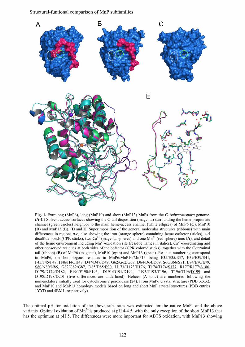
Fig. 1. Extralong (MnP6), long (MnP10) and short (MnP13) MnPs from the C. subvermispora genome. (A-C) Solvent access surfaces showing the C-tail disposition (magenta) surrounding the heme-propionate channel (green circles) neighbor to the main heme-access channel (white ellipses) of MnP6 (C), MnP10 (D) and MnP13 (E). (D and E) Superimposition of the general molecular structures (ribbons) with main differences in regions a-c, also showing the iron (orange sphere) containing heme cofactor (sticks), 4-5 disulfide bonds (CPK sticks), two Ca2+ (magenta spheres) and one Mn2+ (red sphere) ions (A), and detail of the heme environment including Mn2+-oxidation site (residue names in italics), Ca2+-coordinating and other conserved residues at both sides of the cofactor (CPK colored sticks), together with the C-terminal tail (ribbon) (B) of MnP6 (magenta), MnP10 (cyan) and MnP13 (green). Residue numbering correspond to MnP6, the homologous residues in MnP6/MnP10/MnP13 being E35/E35/E37, E39/E39/E41, F45/F45/F47, H46/H46/H48, D47/D47/D49, G62/G62/G67, D64/D64/D69, S66/S66/S71, E74/E70/E79, S80/N80/N85, G82/G82/G87, D85/D85/E90, H173/H173/H176, T174/T174/S177, R177/R177/A180, D179/D179/D182, F190/F190/F193, D191/D191/D194, T193/T193/T196, T196/T196/D199 and D198/D198/D201 (five differences are underlined). Helices (A to J) are numbered following the nomenclature initially used for cytochrome c peroxidase (24). From MnP6 crystal structure (PDB XXX), and MnP10 and MnP13 homology models based on long and short MnP crystal structures (PDB entries 1YYD and 4BM1, respectively)
The optimal pH for oxidation of the above substrates was estimated for the native MnPs and the above variants. Optimal oxidation of Mn2+ is produced at pH 4-4.5, with the only exception of the short MnP13 that has the optimum at pH 5. The differences were more important for ABTS oxidation, with MnP13 showing
122
Structural-funtional comparison of MnP subfamilies
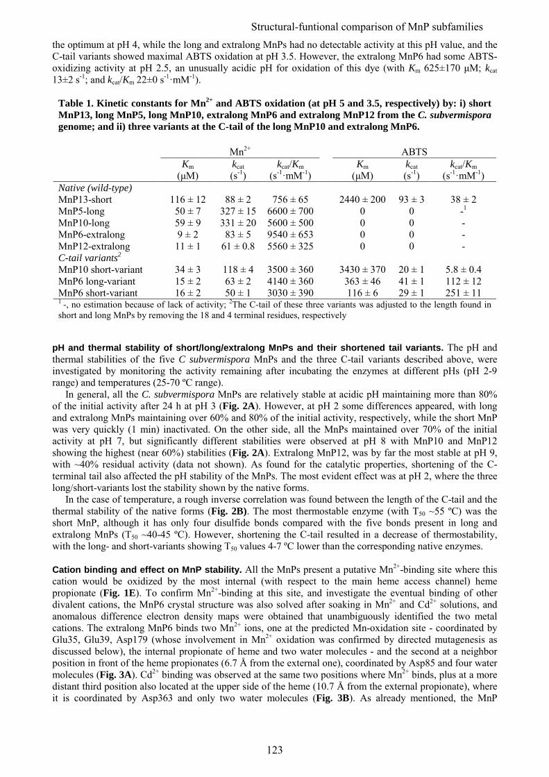
the optimum at pH 4, while the long and extralong MnPs had no detectable activity at this pH value, and the C-tail variants showed maximal ABTS oxidation at pH 3.5. However, the extralong MnP6 had some ABTS-oxidizing activity at pH 2.5, an unusually acidic pH for oxidation of this dye (with Km 625±170 μM; kcat 13±2 s-1; and kcat/Km 22±0 s-1·mM-1).
Table 1. Kinetic constants for Mn2+ and ABTS oxidation (at pH 5 and 3.5, respectively) by: i) short MnP13, long MnP5, long MnP10, extralong MnP6 and extralong MnP12 from the C. subvermispora genome; and ii) three variants at the C-tail of the long MnP10 and extralong MnP6. Mn2+ ABTS Km kcat kcat/Km Km kcat kcat/Km (μM) (s-1) (s-1·mM-1) (μM) (s-1) (s-1·mM-1) Native (wild-type) MnP13-short 116 ± 12 88 ± 2 756 ± 65 2440 ± 200 93 ± 3 38 ± 2 MnP5-long 50 ± 7 327 ± 15 6600 ± 700 0 0 -1 MnP10-long 59 ± 9 331 ± 20 5600 ± 500 0 0 - MnP6-extralong 9 ± 2 83 ± 5 9540 ± 653 0 0 - MnP12-extralong 11 ± 1 61 ± 0.8 5560 ± 325 0 0 - C-tail variants2 MnP10 short-variant 34 ± 3 118 ± 4 3500 ± 360 3430 ± 370 20 ± 1 5.8 ± 0.4 MnP6 long-variant 15 ± 2 63 ± 2 4140 ± 360 363 ± 46 41 ± 1 112 ± 12 MnP6 short-variant 16 ± 2 50 ± 1 3030 ± 390 116 ± 6 29 ± 1 251 ± 11 1 -, no estimation because of lack of activity; 2The C-tail of these three variants was adjusted to the length found in short and long MnPs by removing the 18 and 4 terminal residues, respectively
pH and thermal stability of short/long/extralong MnPs and their shortened tail variants. The pH and thermal stabilities of the five C subvermispora MnPs and the three C-tail variants described above, were investigated by monitoring the activity remaining after incubating the enzymes at different pHs (pH 2-9 range) and temperatures (25-70 ºC range). In general, all the C. subvermispora MnPs are relatively stable at acidic pH maintaining more than 80% of the initial activity after 24 h at pH 3 (Fig. 2A). However, at pH 2 some differences appeared, with long and extralong MnPs maintaining over 60% and 80% of the initial activity, respectively, while the short MnP was very quickly (1 min) inactivated. On the other side, all the MnPs maintained over 70% of the initial activity at pH 7, but significantly different stabilities were observed at pH 8 with MnP10 and MnP12 showing the highest (near 60%) stabilities (Fig. 2A). Extralong MnP12, was by far the most stable at pH 9, with ~40% residual activity (data not shown). As found for the catalytic properties, shortening of the C-terminal tail also affected the pH stability of the MnPs. The most evident effect was at pH 2, where the three long/short-variants lost the stability shown by the native forms. In the case of temperature, a rough inverse correlation was found between the length of the C-tail and the thermal stability of the native forms (Fig. 2B). The most thermostable enzyme (with T50 ~55 ºC) was the short MnP, although it has only four disulfide bonds compared with the five bonds present in long and extralong MnPs (T50 ~40-45 ºC). However, shortening the C-tail resulted in a decrease of thermostability, with the long- and short-variants showing T50 values 4-7 ºC lower than the corresponding native enzymes. Cation binding and effect on MnP stability. All the MnPs present a putative Mn2+-binding site where this cation would be oxidized by the most internal (with respect to the main heme access channel) heme propionate (Fig. 1E). To confirm Mn2+-binding at this site, and investigate the eventual binding of other divalent cations, the MnP6 crystal structure was also solved after soaking in Mn2+ and Cd2+ solutions, and anomalous difference electron density maps were obtained that unambiguously identified the two metal cations. The extralong MnP6 binds two Mn2+ ions, one at the predicted Mn-oxidation site - coordinated by Glu35, Glu39, Asp179 (whose involvement in Mn2+ oxidation was confirmed by directed mutagenesis as discussed below), the internal propionate of heme and two water molecules - and the second at a neighbor position in front of the heme propionates (6.7 Å from the external one), coordinated by Asp85 and four water molecules (Fig. 3A). Cd2+ binding was observed at the same two positions where Mn2+ binds, plus at a more distant third position also located at the upper side of the heme (10.7 Å from the external propionate), where it is coordinated by Asp363 and only two water molecules (Fig. 3B). As already mentioned, the MnP
123
Structural-funtional comparison of MnP subfamilies
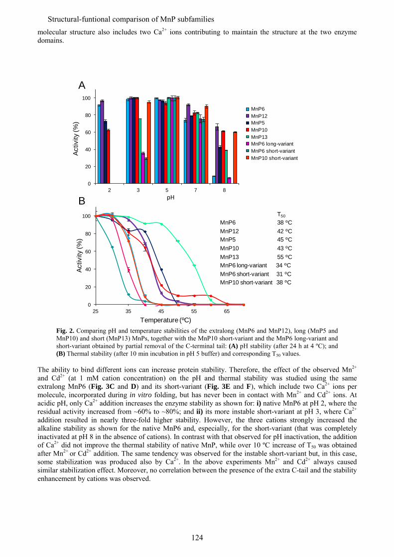
molecular structure also includes two Ca2+ ions contributing to maintain the structure at the two enzyme domains.
0
20
40
60
80
100
2 3 5 7 8
MnP-6MnP-12MnP-5MnP-10MnP-13 "long" MnP-6"short" MnP-6"short" MnP-10
Activ
ity (%
)
pH
A
MnP6MnP12MnP5MnP10MnP13MnP6 long-variantMnP6 short-variantMnP10 short-variant
0
20
40
60
80
100
25 35 45 55 65
Temperature (ºC)
Activ
ity (%
)
BT50
MnP6 38 ºCMnP12 42 ºCMnP5 45 ºCMnP10 43 ºCMnP13 55 ºCMnP6 long-variant 34 ºCMnP6 short-variant 31 ºCMnP10 short-variant 38 ºC
Fig. 2. Comparing pH and temperature stabilities of the extralong (MnP6 and MnP12), long (MnP5 and MnP10) and short (MnP13) MnPs, together with the MnP10 short-variant and the MnP6 long-variant and short-variant obtained by partial removal of the C-terminal tail: (A) pH stability (after 24 h at 4 ºC); and (B) Thermal stability (after 10 min incubation in pH 5 buffer) and corresponding T50 values.
The ability to bind different ions can increase protein stability. Therefore, the effect of the observed Mn2+ and Cd2+ (at 1 mM cation concentration) on the pH and thermal stability was studied using the same extralong MnP6 (Fig. 3C and D) and its short-variant (Fig. 3E and F), which include two Ca2+ ions per molecule, incorporated during in vitro folding, but has never been in contact with Mn2+ and Cd2+ ions. At acidic pH, only Ca2+ addition increases the enzyme stability as shown for: i) native MnP6 at pH 2, where the residual activity increased from ~60% to ~80%; and ii) its more instable short-variant at pH 3, where Ca2+ addition resulted in nearly three-fold higher stability. However, the three cations strongly increased the alkaline stability as shown for the native MnP6 and, especially, for the short-variant (that was completely inactivated at pH 8 in the absence of cations). In contrast with that observed for pH inactivation, the addition of Ca2+ did not improve the thermal stability of native MnP, while over 10 ºC increase of T50 was obtained after Mn2+ or Cd2+ addition. The same tendency was observed for the instable short-variant but, in this case, some stabilization was produced also by Ca2+. In the above experiments Mn2+ and Cd2+ always caused similar stabilization effect. Moreover, no correlation between the presence of the extra C-tail and the stability enhancement by cations was observed.
124
Structural-funtional comparison of MnP subfamilies
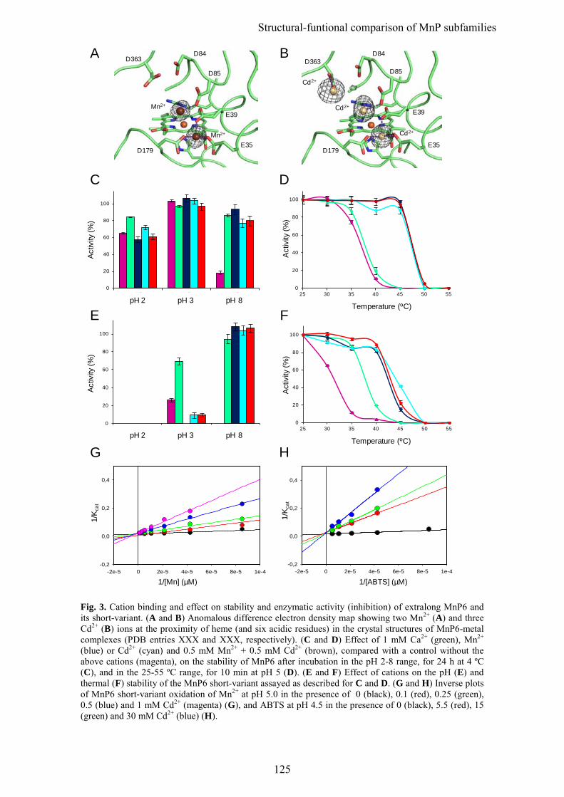
-2e-5 0 2e-5 4e-5 6e-5 8e-5 1e-4-0,2
0,0
0,2
0,4
-2e-5 0 2e-5 4e-5 6e-5 8e-5 1e-4-0,2
0,0
0,2
0,4
0
20
40
60
80
100
pH2 pH3 pH8
0
20
40
60
80
100
pH2 pH3 pH8
1/[ABTS] (µM)
1/K
cat
1/K
cat
1/[Mn] (µM)
0
20
40
60
80
100
25 30 35 40 45 50 55
Act
ivity
(%)
Temperature (ºC)
Act
ivity
(%)
pH 2 pH 3 pH 8
0
20
40
60
80
100
25 30 35 40 45 50 55
Temperature (ºC)
Act
ivity
(%)
Act
ivity
(%)
pH 2 pH 3 pH 8
Cd2+
Cd2+
Cd2+
D179
E39
E35
D85
D84D363
Mn2+
Mn2+
D179
E39
E35
D85
D84D363
G
E
A B
D
F
H
C
Fig. 3. Cation binding and effect on stability and enzymatic activity (inhibition) of extralong MnP6 and its short-variant. (A and B) Anomalous difference electron density map showing two Mn2+ (A) and three Cd2+ (B) ions at the proximity of heme (and six acidic residues) in the crystal structures of MnP6-metal complexes (PDB entries XXX and XXX, respectively). (C and D) Effect of 1 mM Ca2+ (green), Mn2+ (blue) or Cd2+ (cyan) and 0.5 mM Mn2+ + 0.5 mM Cd2+ (brown), compared with a control without the above cations (magenta), on the stability of MnP6 after incubation in the pH 2-8 range, for 24 h at 4 ºC (C), and in the 25-55 ºC range, for 10 min at pH 5 (D). (E and F) Effect of cations on the pH (E) and thermal (F) stability of the MnP6 short-variant assayed as described for C and D. (G and H) Inverse plots of MnP6 short-variant oxidation of Mn2+ at pH 5.0 in the presence of 0 (black), 0.1 (red), 0.25 (green), 0.5 (blue) and 1 mM Cd2+ (magenta) (G), and ABTS at pH 4.5 in the presence of 0 (black), 5.5 (red), 15 (green) and 30 mM Cd2+ (blue) (H).
125
Structural-funtional comparison of MnP subfamilies
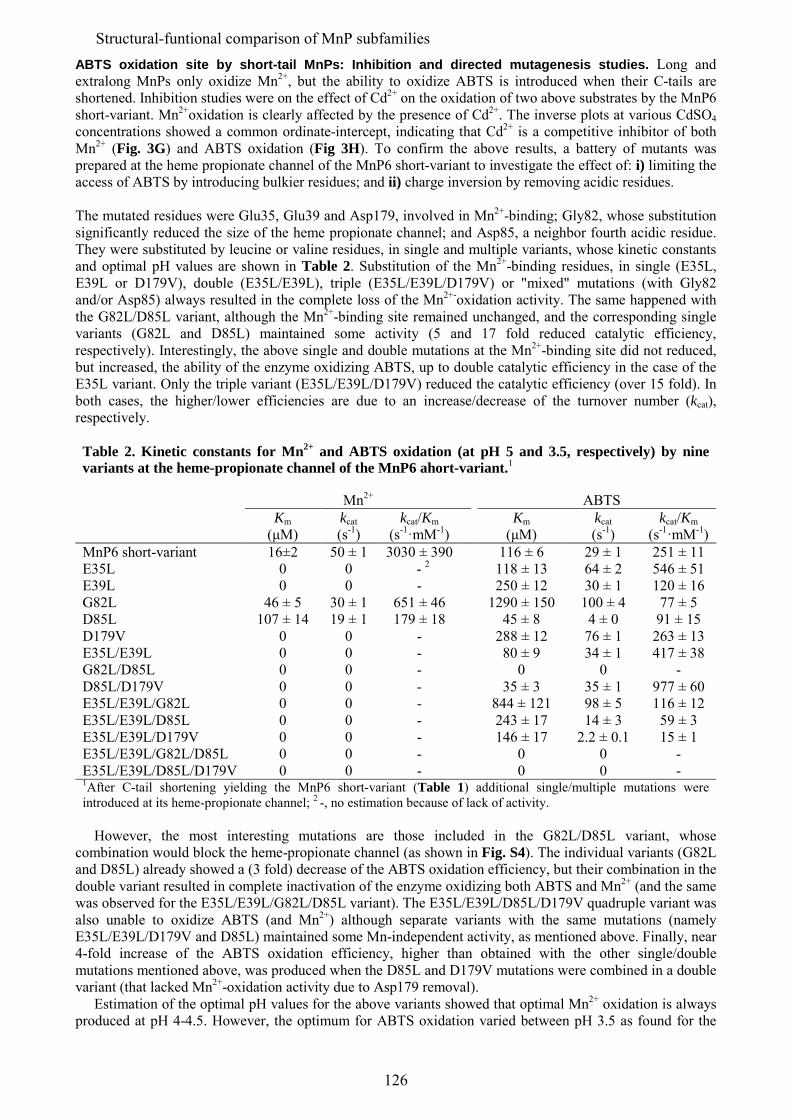
ABTS oxidation site by short-tail MnPs: Inhibition and directed mutagenesis studies. Long and extralong MnPs only oxidize Mn2+, but the ability to oxidize ABTS is introduced when their C-tails are shortened. Inhibition studies were on the effect of Cd2+ on the oxidation of two above substrates by the MnP6 short-variant. Mn2+oxidation is clearly affected by the presence of Cd2+. The inverse plots at various CdSO4 concentrations showed a common ordinate-intercept, indicating that Cd2+ is a competitive inhibitor of both Mn2+ (Fig. 3G) and ABTS oxidation (Fig 3H). To confirm the above results, a battery of mutants was prepared at the heme propionate channel of the MnP6 short-variant to investigate the effect of: i) limiting the access of ABTS by introducing bulkier residues; and ii) charge inversion by removing acidic residues. The mutated residues were Glu35, Glu39 and Asp179, involved in Mn2+-binding; Gly82, whose substitution significantly reduced the size of the heme propionate channel; and Asp85, a neighbor fourth acidic residue. They were substituted by leucine or valine residues, in single and multiple variants, whose kinetic constants and optimal pH values are shown in Table 2. Substitution of the Mn2+-binding residues, in single (E35L, E39L or D179V), double (E35L/E39L), triple (E35L/E39L/D179V) or "mixed" mutations (with Gly82 and/or Asp85) always resulted in the complete loss of the Mn2+-oxidation activity. The same happened with the G82L/D85L variant, although the Mn2+-binding site remained unchanged, and the corresponding single variants (G82L and D85L) maintained some activity (5 and 17 fold reduced catalytic efficiency, respectively). Interestingly, the above single and double mutations at the Mn2+-binding site did not reduced, but increased, the ability of the enzyme oxidizing ABTS, up to double catalytic efficiency in the case of the E35L variant. Only the triple variant (E35L/E39L/D179V) reduced the catalytic efficiency (over 15 fold). In both cases, the higher/lower efficiencies are due to an increase/decrease of the turnover number (kcat), respectively. Table 2. Kinetic constants for Mn2+ and ABTS oxidation (at pH 5 and 3.5, respectively) by nine variants at the heme-propionate channel of the MnP6 ahort-variant.1
Mn2+ ABTS Km kcat kcat/Km Km kcat kcat/Km (μM) (s-1) (s-1·mM-1) (μM) (s-1) (s-1·mM-1) MnP6 short-variant 16±2 50 ± 1 3030 ± 390 116 ± 6 29 ± 1 251 ± 11 E35L 0 0 - 2 118 ± 13 64 ± 2 546 ± 51 E39L 0 0 - 250 ± 12 30 ± 1 120 ± 16 G82L 46 ± 5 30 ± 1 651 ± 46 1290 ± 150 100 ± 4 77 ± 5 D85L 107 ± 14 19 ± 1 179 ± 18 45 ± 8 4 ± 0 91 ± 15 D179V 0 0 - 288 ± 12 76 ± 1 263 ± 13 E35L/E39L 0 0 - 80 ± 9 34 ± 1 417 ± 38 G82L/D85L 0 0 - 0 0 - D85L/D179V 0 0 - 35 ± 3 35 ± 1 977 ± 60 E35L/E39L/G82L 0 0 - 844 ± 121 98 ± 5 116 ± 12 E35L/E39L/D85L 0 0 - 243 ± 17 14 ± 3 59 ± 3 E35L/E39L/D179V 0 0 - 146 ± 17 2.2 ± 0.1 15 ± 1 E35L/E39L/G82L/D85L 0 0 - 0 0 - E35L/E39L/D85L/D179V 0 0 - 0 0 - 1After C-tail shortening yielding the MnP6 short-variant (Table 1) additional single/multiple mutations were introduced at its heme-propionate channel; 2 -, no estimation because of lack of activity.
However, the most interesting mutations are those included in the G82L/D85L variant, whose combination would block the heme-propionate channel (as shown in Fig. S4). The individual variants (G82L and D85L) already showed a (3 fold) decrease of the ABTS oxidation efficiency, but their combination in the double variant resulted in complete inactivation of the enzyme oxidizing both ABTS and Mn2+ (and the same was observed for the E35L/E39L/G82L/D85L variant). The E35L/E39L/D85L/D179V quadruple variant was also unable to oxidize ABTS (and Mn2+) although separate variants with the same mutations (namely E35L/E39L/D179V and D85L) maintained some Mn-independent activity, as mentioned above. Finally, near 4-fold increase of the ABTS oxidation efficiency, higher than obtained with the other single/double mutations mentioned above, was produced when the D85L and D179V mutations were combined in a double variant (that lacked Mn2+-oxidation activity due to Asp179 removal). Estimation of the optimal pH values for the above variants showed that optimal Mn2+ oxidation is always produced at pH 4-4.5. However, the optimum for ABTS oxidation varied between pH 3.5 as found for the
126
Structural-funtional comparison of MnP subfamilies
MnP6 short-variant and several of its propionate channel variants, and pH 4.5, as found for other of its propionate channel variants (such as D179V, E35L/E39L and D85L/D179V) (Fig. S5). Finally, under (non-physiological) pH 2.5 conditions, even the extralong MnP6 exhibited some activity on ABTS, although it disappeared when the pH was increased at the usual values. Molecular simulation of ABTS access to the heme cofactor in short and (extra)long MnPs. No crystal structures of native short MnPs (or short-variants) including an ABTS molecule could be obtained to confirm its oxidation at the heme propionate channel, suggested by directed mutagenesis and other results. However, confirmatory evidence was provided by dynamic ligand diffusion using PELE (Protein Energy Landscape Exploration) software and other molecular simulation tools, using the structure of extralong MnP6 and its short-tail form. An initial 100 ns MD simulation, following protonation at pH 3.0, was performed to equilibrate the system. In both proteins, we observed that Glh35, Glh39 and Ash179 (protonated under these pH conditions) adopt different conformational states resulting in the opening/closing of the channel connecting the surface with the heme group at the propionate level. These MD simulations also showed a high number of stable H-bonds between the tail extension and the protein body (Table S1), probably contributing to MnP6 stability in spite of its extralong C-tail. A first exploration on ABTS diffusion with PELE was performed on the whole enzyme, to identify the exposed regions were the ligand would bind preferentially (Fig. S3). In this way three binding energy minima were identified, the main one situating ABTS at 6.2 Å from the heme at the entrance of the propionate channel. To obtain an atomic detailed mechanism of ABTS’s induced fit, and of its dependency with the C-terminal size, we performed a second ligand exploration with PELE focused on the heme propionate region of both the extralong MnP6 and its short-tail form. This time, the ligand was allowed to freely explore within a 20 Å distance from the CHA heme atom. Fig. 4A shows the relative ligand’s interaction energy with respect to the distance between the heme group (carbon CHA) and the mass center of the ABTS molecule. In the case of the short-tail form (red dots) the structures with best interaction energy have the ABTS closer to the active site, adopting a favorable position for oxidation (Fig. 4D). In contrast, ABTS diffusion on the extralong MnP6 (blue dots) failed to identify and adequate structure with a good interaction energy and, in the best of the cases, the ABTS occupied a much farther position than in the short-tail form (Fig. 4C). As shown in Fig. 4E, the presence (and the dynamics) of the C-terminal tail hinders the entrance of the ligand into the active site channel. Moreover, the tail also limits the above mentioned mobility of the residues situated at the entrance of the channel in the extralong enzyme. This is shown in Fig. 4B presenting the PELE binding energy in the extralong MnP6 and its short-tail form vs the distance between Glh35 and Ash179, located at both sides of the propionate channel. As already shown in Fig. 4A, ABTS has better binding energies in the short-tail form (red dots) than in the extralong MnP (blue dots) and this is accompanied by larger Glh35-Ash179 distances resulting in much higher frequency of "open gate" conformations enabling the approach of the ligand in the short-tail form. This is shown in Figs. 4C showing ABTS approaching to the internal propionate of heme (4.0 Å) at the same time that Asph179 (and Glh35) are displaced widening the channel entrance in the short-tail form. In contrast, Ash179 and Glh35 still maintain a close position in the best ligand position on extralong MnP6 (Fig. 4D) blocking the access of ABTS to the heme. Finally, electron coupling energies were calculated to analyze ABTS oxidation by the extralong and the short-tail MnPs at the best positions predicted by PELE. The energies were one order of magnitude higher for the second MnP (Fig. 4A) in agreement with the experimental results showing that extralong MnP6 is unable to oxidize ABTS, but this ability can be introduced by shortening the C-tail in the short-variant. Discussion The most recent peroxidase inventories from basidiomycete genomes and other sources includes progressively growing numbers of genes of short (up to sixty-eight), long (up to forty) and extralong (up to thirteen) MnPs (5, 20, 25, 26). In current annotation of genomes, the three subfamilies are identified by the different extension of the C-terminal tail, but the evidence for biochemical and other differences between them is fragmentary and, more important, the structural bases for such differences remain largely unknown. The structural and functional comparison of C. subvermispora short, long and extralong MnPs, including their kinetic and stability properties, together with computational calculations on substrate diffusion and oxidation in the presence and the absence of the C-terminal tail, provide relevant answers to some of the above questions.
127
Structural-funtional comparison of MnP subfamilies
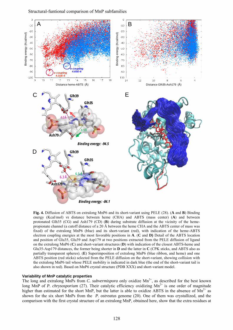
E
Distance heme-ABTS (Å)
Bin
ding
ene
rgy
(Kca
l/mol
)
Distance Glh35-Ash179 (Å)
Bin
ding
ene
rgy
(Kca
l/mol
)
e-coupling4.65E-6e-coupling
4.31E-5
A B
D
6.3 Å6.5 Å
Glh35
Glh39
Ash179
Binding energy: -84.1
4.0 Å
8.9 Å
Glh35
Glh39
Ash79
Binding energy: -94.5
C
Fig. 4. Diffusion of ABTS on extralong MnP6 and its short-variant using PELE (28). (A and B) Binding energy (Kcal/mol) vs distance between heme (CHA) and ABTS (mass center) (A) and between protonated Glh35 (CG) and Ash179 (CD) (B) during substrate diffusion at the vicinity of the heme-propionate channel (a cutoff distance of a 20 Å between the heme CHA and the ABTS center of mass was fixed) of the extralong MnP6 (blue) and its short-variant (red), with indication of the heme-ABTS electron coupling energies at the most favorable positions in A. (C and D) Detail of the ABTS location and position of Glu35, Glu39 and Asp179 at two positions extracted from the PELE diffusion of ligand on the extralong MnP6 (C) and short-variant structures (D) with indication of the closest ABTS-heme and Glu35-Asp179 distances, the former being shorter in D and the latter in C (CPK sticks, and ABTS also as partially transparent spheres). (E) Superimposition of extralong MnP6 (blue ribbon, and heme) and one ABTS position (red sticks) selected from the PELE diffusion on the short-variant, showing collision with the extralong MnP6 tail whose PELE mobility is indicated in dark blue (the end of the short-variant tail is also shown in red). Based on MnP6 crystal structure (PDB XXX) and short-variant model.
Variability of MnP catalytic properties The long and extralong MnPs from C. subvermispora only oxidize Mn2+, as described for the best known long MnP of P. chrysosporium (27). Their catalytic efficiency oxidizing Mn2+ is one order of magnitude higher than estimated for the short MnP, but the latter is able to oxidize ABTS in the absence of Mn2+ as shown for the six short MnPs from the P. ostreatus genome (20). One of them was crystallized, and the comparison with the first crystal structure of an extralong MnP, obtained here, show that the extra residues at
128
Structural-funtional comparison of MnP subfamilies
the C-terminal tail adopt a characteristic disposition surrounding the entrance of the heme-propionate channel. Such disposition could limit bulky substrates (such as ABTS) access through this channel, as revealed by ligand diffusion calculations with PELE (28). Indeed, removal of the tail from extralong MnP6 decreases the heme-ligand distance from 6.3 Å to 4.0 Å, with a 20 Kcal/mol better binding energy. Experimental confirmation for the above prediction was obtained by reducing the C-tail extension in both extralong MnP6 and long MnP10 by directed mutagenesis, which resulted in the introduction of the ability to oxidize ABTS into two peroxidases that initially lacked this activity. The above results, together with (i) ligand diffusions by PELE on the whole MnP surface showing the lowest binding energy at the heme-propionate channel and (ii) observed competitive inhibition by Cd2+ of both Mn2+ and ABTS oxidation, strongly pointed to ABTS oxidation at the heme-propionate channel of natural (MnP13) and engineered MnPs (MnP6 and MnP10 variants with a shortened tail). However, an effect of the C-tail on oxidation of ABTS at the main heme access channel, where oxidation of ABTS and other dyes and phenols has been shown in P. eryngii VP (29), could not be discarded. Therefore, additional mutations at the heme-propionate channel were performed on the (ABTS-oxidizing) MnP6 short-variant, and the simultaneous loss of both ABTS and Mn2+ oxidation activities was obtained by a double mutation (G82L/D85L) that blocked the heme-propionate channel, demonstrating that the two substrates are oxidized at the same site in short-tail MnPs. Conversely, improved ABTS oxidation was obtained by mutations reducing the negative charge at the heme-propionate channel due to reduced electrostatic repulsion with the anionic ABTS substrate. The activity on Mn2+ was lost when any of the three Mn-binding acidic residues was mutated. An easier oxidation of ABTS by the short-tail MnPs was also predicted by electron coupling calculations after ligand diffusion on the MnP6 structure before and after shortening the C-tail, the latter yielding one order of magnitude higher values. Interestingly, oxidation of a substrate other than Mn2+ (ascorbate) at the heme-propionate channel had been described in ascorbate peroxidase (30). While Mn2+ oxidation by MnPs (13) and VPs (31) imply the closure and opening of the heme-propionate channel (mainly by reorientation of the ion binding side-chains) for proper positioning of Mn2+ near the heme propionate and release of the Mn3+ formed, respectively, ABTS oxidation by the short MnPs would require more significant reorganizations enabling the approach and oxidation of this bulky substrate, as shown by the PELE simulations. Variability of MnP stability properties The C. subvermispora long and, especially, the extralong MnPs are exceptionally stable at pH 2 (over 60% and 90% residual activity, respectively, after 24 h at 4 ºC) while the short MnP was fully inactivated under the same conditions, in agreement with results obtained for the six short MnPs of P. ostreatus (20). However, the similar or even lower thermal stability of the extralong MnPs compared with the long MnPs contrasted with the previous assumption about their high thermostability, based on the initially described extralong MnPs from Dichomitus squalens (15). In fact, the highest thermal stability was shown for the short MnP, with a T50 value (55 ºC) in the range (43-57 ºC) found for the P. ostreatus short MnPs (20). However, the removal of the C-tail from long and extralong MnPs by directed mutagenesis did not result in improved thermostability (on the contrary a 5-7 ºC decrease of T50 was observed) revealing that the presence of the tail is not necessarily an instability factor, most probably because of the numerous and stable H-bond contacts with the protein body shown by MD calculations. In fact, the C-tail seems involved in the acidic stability of the C. subvermispora extralong MnPs as shown by their rapid inactivation at pH 2 when the tail is shortened by directed mutagenesis. Although the structural basis for the unique acidic stability of these MnPs is to be determined, the isoenzyme MnP6 from C. subvermispora has been used as a robust scaffold to build a lignin-degrading peroxidase of biotechnological interest acting at very acidic pH values (32). Two manganese and three cadmium ions were identifying in the anomalous difference electron density maps of the extralong MnP6 in complex with Mn2+ and Cd2+, respectively. One of them occupied the Mn-oxidation site and the others were located in neighbor positions. A similar situation has been described for the P. chrysosporium MnP (14), although the third site found in the extralong MnP does not exist due to the shorter C-tail. Those ions at the Mn-oxidation site would be responsible for the similar improvement of the MnP thermal and alkaline stability by Mn2+ and Cd2+ contributing to cofactor fixation on the protein, although a lower effect of the second Mn2+ and the two other Cd2+ ions fixing the protein structure cannot be discarded. An effect of Mn2+ and Cd2+ ions increasing the enzyme stability has been reported for long/extralong MnPs (33, 34). On the other hand, Ca2+ causes a general stabilizing effect on plant and fungal peroxidases (from the superfamily of plant-fungal-prokaryotic peroxidase) by reverting unfolding due to the loss of the two conserved structural Ca2+ ions (35, 36).
129
Structural-funtional comparison of MnP subfamilies
Evolutionary relationships of MnP subfamilies The evolutionary relationships between the C. subvermispora heme peroxidases (a total of twenty-six including one cytochrome c peroxidase) and those isolated from other basidiomycetes, up to a total of 376 sequences, were analyzed by Fernández-Fueyo et al. (22). The phylogram obtained (see Fig. S6) revealed a large cluster for the superfamily of plant-fungal-prokaryotic peroxidases (196 members), together with two other clusters of the superfamilies of dye-decolorizing peroxidases (absent from C. subvermispora) and heme-thiolate peroxidases (nine in C. subvermispora). In the first cluster, three groups corresponding to (i) short-MnPs (only one in C. subvermispora), VPs (absent from C. subvermispora) and LiPs (two in C. subvermispora), (ii) long and extralong MnPs (up to a total of twelve in C. subvermispora), and (iii) generic peroxidases (only one in C. subvermispora) are defined, together with several unclustered sequences. The separate clustering of short MnPs in the first group, and the intermixed presence of long and extralong MnPs in the second group (with high sequence identities among them, as shown in Table S2) suggest a separate origin for the short MnP subfamily, while the proposed long and extralong MnP types would not constitute two separate groups from a phylogenetic point of view. This fact, together with the significantly different catalytic and stability properties of the short MnPs (including Mn-independent activity, lower Mn2+ oxidizing efficiency and acidic stability, and higher thermal stability than the long and extralong MnPs) and the presence of a 14-22 residues shorter C-tail, strongly support their classification as a different MnP subfamily. In contrast, the so-called long and extralong MnPs cluster together in the peroxidase phylogram, their catalytic and stability properties only show gradual differences (such as the ∼5-fold lower Km and kcat values for Mn2+ oxidation and the ∼40% higher stability at pH 2 of the C. subvermispora extralong MnPs compared to the long MnPs) and variable C-tail lengths were observed. These modest differences do not justify maintaining long and extralong MnPs as two separate subfamilies. Conclusions From comparison of the different C. subvermispora MnPs and a collection of site-directed variants, together with the results from related enzymes available in the literature, we conclude that short MnPs would constitute a separate subfamily characterized by their ability to directly oxidize both Mn2+ and other substrates (such as ABTS) at the same heme access channel, located in front of the heme propionates. The access to this site in long and extralong MnPs would be limited by the C-tail that surrounds the channel preventing the approach of bulky substrates (such as ABTS) although the access of Mn2+ is not negatively affected (in fact the presence of the tail seems to improve the oxidation of Mn2+). Another remarkable characteristic of the long and, especially, extralong MnPs (of C. subvermispora) is their stability at highly acidic pH, whose structural bases remain to be identified. In contrast with that found for the short MnPs, only gradual differences were observed between the two other MnPs that do not justify their classification in two different subfamilies. Materials and Methods Genome sequencing and inventory of MnP genes. The genome of C. subvermispora was sequenced at the JGI in a project coordinated by Daniel Cullen (USDA Forest Products Laboratory, Madison, USA) and Rafael Vicuña (Pontificia Universidad Católica, Santiago, Chile). The results from gene annotation (a total of 12 125 models) are available at the JGI portal (http://genome.jgi.doe.gov/Cersu1/Cersu1.home.html). The complete inventory of C. subvermispora heme peroxidase genes was obtained as described by Fernandez-Fueyo et al. (22). The manual annotation process included, among other steps, confirmation of the presence of characteristic residues at the heme pocket and substrate oxidation sites by homology modeling of the predicted sequences, using crystal structures of related proteins as templates (37). The homology models for the different MnP isoenzymes were also used in the subsequent structural-functional studies. Gene synthesis and heterologous expression. The coding sequences of the C. subvermispora gene models encoding isoenzymes MnP5 (49863), MnP6 (50686), MnP10 (117436), MnP12 (157986) and MnP13 (124076) were designed with E. coli preferred codon usage (confirmed for other genes expressed in the BL21 strain), and synthesized by ATG:biosynthetics. The five genes were cloned in the expression vector pET23a (+) (Novagen). The resulting plasmids – pET23a-49863, pET23a-50686, pET23a-117436, pET23a-157986 and pET23a-124076 were used for expression and directed mutagenesis when required. E. coli DH5α was used for plasmid propagation, and the different peroxidases were produced in E. coli BL21 (DE3) pLys, in vitro folded and purified as previously described (22). Directed mutagenesis. Stop mutations were introduced in the MnP6 (T362stop and G348stop) and MnP10 (G344stop) encoding genes by polymerase chain reaction (PCR) (22) using the expression plasmid pET23a-50686 and pET23a-117436 (see above) as template. The 5'- CC GTC CAA TTC TCG GGC CCC GCC TAA GAC TCC CC -3', 5'- CC CTC ATT CCT CAC TGC TCC GAC TAA CTC GAG AAC TGC -3' and 5'- CCC CAT TGC AAC TAA GGC CAA GAC TGC CC -3' direct primer (and the reverse primer bearing the complementary sequence) were used, respectively. Thirteen additional variants (E35L, E39L, G82L, D85L, D179V, E35LE39L G82LD85L, D85LD179V, E35LE39LG82, E35LE39LD85L, E35LE39LD179V E35LE39LG82LD85L and E35LE39LG82LD85LD179V) were obtained by mutating the following residues (located around the heme-propionate channel) on the previously obtained MnP6 short-variant (with
130
Structural-funtional comparison of MnP subfamilies
the G348stop mutation): E35L (GAA for CTA), E39L (GAG for CTG), G82L (GGT for CTT), D85L (GAC for CTC), and D179V (GAC for GTC). Kinetics of Mn2+ and ABTS oxidation and inhibition by Cd2+. Absorbance changes during substrate oxidation in 0.1 M tartrate (of different pH values) by the five C. subvermispora MnPs and the sixteen directed variants, whose E. coli expression is described above, were recorded in a UV-160 spectrophotometer (Shimadzu). Oxidation of Mn2+ was followed at pH 5 for the formation of Mn3+·tartrate complex (ε238 6.5 mM-1·cm-1). ABTS oxidation was followed (at pH 3.5) from ABTS cation radical (ε436 29.3 mM-1·cm-1) formation. All reactions were carried out at 25 °C (using ~0.01 µM enzyme) and initiated by addition of 0.1 mM H2O2. Means and standard errors for apparent affinity constant (Km) and enzyme turnover (kcat) were obtained by nonlinear least-squares fitting to the Michaelis-Menten model. Fitting of these constants to the normalized equation v = (kcat/Km)[S]/(1+[S]/Km), where v is the reaction rate and S is the substrate concentration, yielded the catalytic efficiency values (kcat/Km) with their corresponding standard errors. Kinetic constants of MnP6 short-variant oxidation of Mn2+, at pH 5.0 in the presence of 0-1 mM Cd2+, and ABTS, at pH 4.5 in the presence of 0-30 mM Cd2+, were determined as above, to investigate possible inhibitory effects. Inhibition constants (Ki) were obtained from inverse plots. pH/temperature stability and optimal pH. To study the effect of pH on the stability of the five C subvermispora MnPs and the three short/long variants (after partial removal of the C-tail), the E. coli-expressed enzymes were dissolved in Britton-Robinson (B&R) buffer with a pH range from 2 to 9, and kept at 4 ºC. Activity was estimated by oxidation of a saturating concentration of Mn2+ (1.5 mM) in 0.1 M tartrate (pH 5) at 25 ºC. The reaction was initiated by addition of 0.1 mM H2O2, as described above. Residual activities were measured after 24-h incubation. The highest activity immediately after mixing (at any pH) was taken as 100% activity, and the percentage of residual activity at the different pH conditions was referred to this maximal value. To study the effect of temperature on enzyme stability, the five C subvermispora MnPs and the three short/long variants in 10 mM tartrate, pH 5, were incubated at 5 ºC intervals in the temperature range of 25-70 ºC. Residual activity was estimated as described above. Activity immediately after mixing at 25 ºC was considered as 100%, and the percentage of residual activity was estimated according to this value. Temperature stability was presented as 10-min T50, i.e. that temperature at which 50% of the activity was lost after 10 min incubation. The same experiments were performed in presence of 1 mM CaCl2, 1 mM MnSO4, 1 mM CdSO4, and 0,5 mM MnSO4 + 0,5 mM CdSO4, using the extralong MnP6 and its short-variant, to evaluate the effect of these cations on the pH and thermal stabilities.
The optimal pH values for substrate oxidation by the five C subvermispora MnPs and the different directed variants were determined by measuring the oxidation of saturating concentrations of Mn2+ (1.5 mM) and ABTS (5 mM) in 0.1 M B&R buffer of pH 2-6, as described above. Crystal structure of extralong MnP6 and its Mn2+/Cd2+ complexes. Crystallization trials were carried out by the sitting drop vapor diffusion method, using 96-well MRC2 plates with 50 μl reservoir solution and the commercially available screenings from Emerald (Wizard classic crystallization screens I, II and III) and Jena Biosciences (JBScreen Classic Kits 1-10). Drops consisted of 0.2 μl of protein solution (10 mg/ml in 10 mM sodium tartrate buffer at pH 5.0) and 0.2 μl of reservoir solution. Crystallization was carried out at 22°C. Crystals suitable for X-ray data collection were obtained from these initial trials using 0.1 M sodium acetate (pH 4.6) with 18% PEG 4000, and 20% glycerol (as cryoprotectant). Mn2+ and Cd2+-containing crystals were obtained by including 5 mM MnCl2 and CdCl2 in the above solution. For data collection, crystals were mounted in nylon loops and flash-frozen in liquid nitrogen in the mother liquor containing the cryoprotectant indicated above. All diffraction data were obtained at 100 K. X-ray diffraction intensities were collected at the X06DA beam line at the Swiss Light Source (Villigen, Switzerland) using a wavelength of 1.0000 Å and a Pilatus 2M detector. Diffraction data were indexed, integrated, merged and scaled using the programs XDS and XSCALE. Anomalous difference electron density maps for each dataset were calculated using SCALA from the CCP4 suite. Only the higher resolution structure for each crystal form is presented. Data collection statistics are shown in Table S3. The structure of one crystal form from both proteins was solved by molecular replacement using the crystal structure of P. chrysosporium MnP (PDB entry 3M5Q) as the search model and the program AUTOMR implemented in the PHENIX package (M2). These first models were used to solve the structure of the other crystal forms. The final models were obtained by consecutive rounds of refinement, performed with the PHENIX package (PHENIX.REFINE); followed by manual model building, performed with Coot (38) using σA weighted 2Fo-Fc and Fo-Fc electron density maps. Solvent molecules were introduced in the structure automatically in the refinement as implemented in the PHENIX package and visually inspected. A total of 5 % of reflections was used to calculate the Rfree value throughout the refinement process. The structures were validated using MolProbity (39). Refinement and final model statistics are summarized in Table S3. Figures were produced with PyMOL (Schrödinger, Portland, OR, USA). The coordinates and structure factors have been deposited with the Protein Data Bank with accession codes XXX, XXX and XXX. Computational simulations. The coordinates for C. subvermispora extralong MnP6 were taken from the Protein Data Bank (PDB) entry XXX. A terminal proline residue (not visible in the crystal structure) was added. A truncated MnP6 structure was obtained by removing the last 18 residues. The protonated states were assigned assuming systems at pH 3.0 (which corresponds with the optimum environment to oxidize the ligand) with the protein preparation wizard from Schrödinger. Therefore, the heme propionates and some residues in its access channel (Glh35, Glu39 and Ash179) were protonated. Partial charges for the heme moiety were obtained by QM/MM calculations at the B3LYP(lacvp*)/OPLS2005 level of theory with Qsite. Likewise, the ligand ABTS was optimized at DFT level, B3LYP(6-31g**) with Jaguar. 100 ns MD simulation was carried out with Desmond using the AMBER force field for both extralong MnP6 and its short-variant. Proteins were placed in an orthorhombic box of water with an ionic strength of 0.15M. A time step of 2.0 fs
131
Structural-funtional comparison of MnP subfamilies
was used throughout the simulations in combination with the SHAKE algorithm to constrain bond lengths involving hydrogen atoms. Non-bonded interactions were explicitly evaluated for distances below 9 Å. The particle mesh Ewald method was employed to treat long-range electrostatic interactions. Constant pressure and temperature (NPT ensemble) were maintained by weakly coupling the system to an external bath at 1 bar and 300 K, using the Martyna-Tobias Klein barostat and the Nose-Hoover thermostat, respectively. PELE software was used to study ABTS diffusion. This software uses a Monte Carlo scheme to describe the protein-ligand conformational dynamics. It is capable of reproducing their induced fit, including ligand migration and cavity search in a computational efficient manner (typically requiring no more than 24 hours). At each iteration the algorithm performs: 1) translation and rotation of the ligand, 2) protein perturbation following a combination of normal modes, 3) side chain prediction, and 4) overall minimization. Subsequently, the final structure is accepted or rejected based on a Metropolis criteria (for more details see, for example, https://pele.bsc.es) In this study, the ligand first explored the whole protein surface, and then was forced to explore the area close to the heme's propionates, by using a cutoff distance of a 20 Å between the heme carbon CHA and the ABTS center of mass. Chemicals. All chemicals were from Sigma-Aldrich, with the exception of H2O2 that was from Merck, and ABTS that was from Roche. ACKNOWLEDGEMENTS. This work was supported by the HIPOP (BIO2011-26694; to F.J.R.-D.) project of the Spanish Ministry of Economy and Competitiveness (MINECO) and by the PEROXICATS (KBBE-2010-4-265397; to A.T.M.) and INDOX (KBBE-2013-3-613549; to A.T.M. and V.G.) European projects. Sequencing of the Ceriporiopsis subvermispora genome by the Joint Genome Institute was supported by the Office of Science of the U.S, Department of Energy under Contract No. DE-AC02-05CH11231. E.F.F. acknowledges a JAE Fellowship of the Spanish CSIC, and F.J.R.D. acknowledges a Ramón y Cajal contract of the Spanish MINECO. 1. Kirk, T. K. & Farrell, R. L. (1987) Enzymatic "combustion": The microbial degradation of lignin. Annu. Rev.
Microbiol. 41: 465-505. 2. Ruiz-Dueñas, F. J. & Martínez, A. T. (2009) Microbial degradation of lignin: How a bulky recalcitrant polymer is
efficiently recycled in nature and how we can take advantage of this. Microbial Biotechnol. 2: 164-177. 3. Ruiz-Dueñas, F. J., Morales, M., García, E., Miki, Y., Martínez, M. J., & Martínez, A. T. (2009) Substrate oxidation
sites in versatile peroxidase and other basidiomycete peroxidases. J. Exp. Bot. 60: 441-452. 4. Ruiz-Dueñas, F. J. & Martínez, A. T. (2010) Structural and functional features of peroxidases with a potential as
industrial biocatalysts in Biocatalysts based on heme peroxidases, eds. Torres, E. & Ayala, M. (Springer-Verlag, Berlin), pp. 37-59.
5. Floudas, D., Binder, M., Riley, R., Barry, K., Blanchette, R. A., Henrissat, B., Martínez, A. T., Otillar, R., Spatafora, J. W., Yadav, J. S. et al. (2012) The Paleozoic origin of enzymatic lignin decomposition reconstructed from 31 fungal genomes. Science 336: 1715-1719.
6. Peláez, F., Martínez, M. J., & Martínez, A. T. (1995) Screening of 68 species of basidiomycetes for enzymes involved in lignin degradation. Mycol. Res. 99: 37-42.
7. Hatakka, A. (1994) Lignin-modifying enzymes from selected white-rot fungi - Production and role in lignin degradation. FEMS Microbiol. Rev. 13: 125-135.
8. Martinez, D., Larrondo, L. F., Putnam, N., Gelpke, M. D., Huang, K., Chapman, J., Helfenbein, K. G., Ramaiya, P., Detter, J. C., Larimer, F. et al. (2004) Genome sequence of the lignocellulose degrading fungus Phanerochaete chrysosporium strain RP78. Nat. Biotechnol. 22: 695-700.
9. Ragauskas, A. J., Williams, C. K., Davison, B. H., Britovsek, G., Cairney, J., Eckert, C. A., Frederick, W. J., Hallett, J. P., Leak, D. J., Liotta, C. L. et al. (2006) The path forward for biofuels and biomaterials. Science 311: 484-489.
10. Martínez, A. T., Ruiz-Dueñas, F. J., Martínez, M. J., del Río, J. C., & Gutiérrez, A. (2009) Enzymatic delignification of plant cell wall: from nature to mill. Curr. Opin. Biotechnol. 20: 348-357.
11. Gold, M. H., Youngs, H. L., & Gelpke, M. D. (2000) Manganese peroxidase. Met. Ions Biol. Syst. 37: 559-586. 12. Sundaramoorthy, M., Kishi, K., Gold, M. H., & Poulos, T. L. (1994) The crystal structure of manganese peroxidase
from Phanerochaete chrysosporium at 2.06-Å resolution. J. Biol. Chem. 269: 32759-32767. 13. Sundaramoorthy, M., Gold, M. H., & Poulos, T. L. (2010) Ultrahigh (0.93 angstrom) resolution structure of
manganese peroxidase from Phanerochaete chrysosporium: Implications for the catalytic mechanism. J. Inorg. Biochem. 104: 683-690.
14. Sundaramoorthy, M., Youngs, H. L., Gold, M. H., & Poulos, T. L. (2005) High-resolution crystal structure of manganese peroxidase: substrate and inhibitor complexes. Biochemistry 44: 6463-6470.
15. Li, D. M., Youngs, H. L., & Gold, M. H. (2001) Heterologous expression of a thermostable manganese peroxidase from Dichomitus squalens in Phanerochaete chrysosporium. Arch. Biochem. Biophys. 385: 348-356.
16. Johansson, T. & Nyman, P. O. (1996) A cluster of genes encoding major isozymes of lignin peroxidase and manganese peroxidase from the white-rot fungus Trametes versicolor. Gene 170: 31-38.
17. Asada, Y., Watanabe, A., Irie, T., Nakayama, T., & Kuwahara, M. (1995) Structures of genomic and complementary DNAs coding for Pleurotus ostreatus manganese (II) peroxidase. Biochim. Biophys. Acta 1251: 205-209.
18. Giardina, P., Palmieri, G., Fontanella, B., Rivieccio, V., & Sannia, G. (2000) Manganese peroxidase isoenzymes produced by Pleurotus ostreatus grown on wood sawdust. Arch. Biochem. Biophys. 376: 171-179.
19. Steffen, K. T., Hofrichter, M., & Hatakka, A. (2002) Purification and characterization of manganese peroxidases from the litter-decomposing basidiomycetes Agrocybe praecox and Stropharia coronilla. Enzyme Microb. Technol. 30: 550-555.
20. Fernández-Fueyo, E., Ruiz-Dueñas, F. J., Martínez, M. J., Romero, A., Hammel, K. E., Medrano, F. J., & Martínez, A. T. (2014) Ligninolytic peroxidase genes in the oyster mushroom genome: Heterologous expression, molecular structure, catalytic and stability properties and lignin-degrading ability. Biotechnol. Biofuels 7:2.
132
Structural-funtional comparison of MnP subfamilies
21. Scott, G. M., Akhtar, M., Lenz, M. J., & Swaney, R. E. (1998) Engineering, scale-up, and economic aspects of fungal pretreatment of wood chips in Environmentally friendly technologies for the pulp and paper industry, eds. Young, R. A. & Akhtar, M. (TAPPI Press, Atlanta), pp. 341-384.
22. Fernández-Fueyo, E., Ruiz-Dueñas, F. J., Miki, Y., Martínez, M. J., Hammel, K. E., & Martínez, A. T. (2012) Lignin-degrading peroxidases from genome of selective ligninolytic fungus Ceriporiopsis subvermispora. J. Biol. Chem. 287: 16903-16906.
23. Fernández-Fueyo, E., Ruiz-Dueñas, F. J., Ferreira, P., Floudas, D., Hibbett, D. S., Canessa, P., Larrondo, L., James, T. Y., Seelenfreund, D., Lobos, S. et al. (2012) Comparative genomics of Ceriporiopisis subvermispora and Phanerochaete chrysosporium provide insight into selective ligninolysis. Proc. Natl. Acad. Sci. USA 109: 5458-5463.
24. Poulos, T. L., Freer, S. T., Alden, R. A., Edwards, S. L., Skogland, U., Takio, K., Eriksson, B., Xuong, N., Yonetani, T., & Kraut, J. (1980) The crystal structure of cytochrome c peroxidase. J. Biol. Chem. 255: 575-580.
25. Ruiz-Dueñas, F. J., Lundell, T., Floudas, D., Nagy, L. G., Barrasa, J. M., Hibbett, D. S., & Martínez, A. T. (2013) Lignin-degrading peroxidases in Polyporales: An evolutionary survey based on ten sequenced genomes. Mycologia 105: 1428-1444.
26. Levasseur, A., Lomascolo, A., Chabrol, O., Ruiz-Dueñas, F. J., Uzan, E., Piumi, F., Kues, U., Ram, A. F. J., Murat, C., Haon, M. et al. (2014) The genome of the white-rot fungus Pycnoporus cinnabarinus: a new basidiomycete model with a versatile arsenal for lignin deconstruction. Genome Biol. (in press).
27. Wariishi, H., Akileswaran, L., & Gold, M. H. (1988) Manganese peroxidase from the basidiomycete Phanerochaete chrysosporium: Spectral characterization of the oxidized states and the catalytic cycle. Biochemistry 27: 5365-5370.
28. Borrelli, K. W., Vitalis, A., Alcantara, R., & Guallar, V. (2005) PELE: Protein energy landscape exploration. A novel Monte Carlo based technique. J. Chem. Theory Comput. 1: 1304-1311.
29. Morales, M., Mate, M. J., Romero, A., Martínez, M. J., Martínez, A. T., & Ruiz-Dueñas, F. J. (2012) Two oxidation sites for low redox-potential substrates: A directed mutagenesis, kinetic and crystallographic study on Pleurotus eryngii versatile peroxidase. J. Biol. Chem. 287: 41053-41067.
30. Mandelman, D., Jamal, J., & Poulos, T. L. (1998) Identification of two electron-transfer sites in ascorbate peroxidase using chemical modification, enzyme kinetics, and crystallography. Biochemistry 37: 17610-17617.
31. Ruiz-Dueñas, F. J., Morales, M., Pérez-Boada, M., Choinowski, T., Martínez, M. J., Piontek, K., & Martínez, A. T. (2007) Manganese oxidation site in Pleurotus eryngii versatile peroxidase: A site-directed mutagenesis, kinetic and crystallographic study. Biochemistry 46: 66-77.
32. Fernández-Fueyo, E., Ruiz-Dueñas, F. J., & Martínez, A. T. (2014) Engineering a lignin-degrading peroxidase acting at very acidic. (submitted).
33. Youngs, H. L., Sundaramoorthy, M., & Gold, M. H. (2000) Effects of cadmium on manganese peroxidase - Competitive inhibition of Mn-II oxidation and thermal stabilization of the enzyme. Eur. J. Biochem. 267: 1761-1769.
34. Mauk, M. R., Kishi, K., Gold, M. H., & Mauk, A. G. (1998) pH-linked binding of Mn(II) to manganese peroxidase. Biochemistry 37: 6767-6771.
35. Youngs, H. L., Moënne-Loccoz, P., Loehr, T. M., & Gold, M. H. (2000) Formation of a bis(histidyl) heme iron complex in manganese peroxidase at high pH and restoration of the native enzyme structure by calcium. Biochemistry 39: 9994-10000.
36. George, S. J., Kvaratskhelia, M., Dilworth, M. J., & Thorneley, R. N. F. (1999) Reversible alkaline inactivation of lignin peroxidase involves the release of both the distal and proximal site calcium ions and bishistidine co-ordination of the haem. Biochem. J. 344: 237-244.
37. Bordoli, L., Kiefer, F., Arnold, K., Benkert, P., Battey, J., & Schwede, T. (2009) Protein structure homology modeling using SWISS-MODEL workspace. Nat. Protoc. 4: 1-13.
38. Emsley, P. & Cowtan, K. (2004) Coot: model-building tools for molecular graphics. Acta Crystallogr. D. Biol. Crystallogr. 60: 2132.
39. Davis, I. W., Leaver-Fay, A., Chen, V. B., Block, J. N., Kapral, G. J., Wang, X., Murray, L. W., Arendall III, W. B., Snoeyink, J., Richardson, J. S. et al. (2007) MolProbity: all-atom contacts and structure validation for proteins and nucleic acids. Nucleic Acids Res. 35: W375-W385.
133
Structural-funtional comparison of MnP subfamilies

134
Capítulo 8
Engineering a lignin-degrading peroxidase acting at very acidic pH Fernández-Fueyo, E., F. J. Ruiz-Dueñas and A. T. Martínez. (enviado)
135

136
Engineering a lignin-degrading peroxidase acting at very acidic pH Elena FERNÁNDEZ-FUEYO, Francisco J. RUIZ-DUEÑAS and Angel T. MARTÍNEZ1
Centro de Investigaciones Biológicas, CSIC, Ramiro de Maeztu 9, E-28040 Madrid, Spain Short title: Engineering a versatile peroxidase acting at acidic pH Ligninolytic peroxidases are divided into three families, manganese peroxidases (MnPs), lignin peroxidases (LiPs)and versatile peroxidases (VPs). The two latter are able to degrade nonphenolic lignin model compounds, with VP oxidizing the widest range of aromatic substrates and dyes. One of the limiting issues for the industrial use of these two enzymes is their progressive inactivation under acidic pH conditions, where they exhibit the highest activities. In the screening of peroxidases from basidiomycete genomes, one MnP from Ceriporiopsis subvermispora was found to have a remarkable acidic stability. The crystal structure of this enzyme is available and, after comparison with VP and LiP structures, it was used as a robust scaffold to engineer a stable VP by introducing an exposed catalytic tryptophan, with different protein environments. The variants obtained maintained the acidic stability and strong Mn2+-oxidizing activity, and the abilities to oxidize veratryl alcohol and Reactive Black 5 (two simple VP substrates) were introduced. The different peroxidase activities were discussed in the light of the molecular structures available. The engineered peroxidases present more acidic optimal pH than the best VP from Pleurotus ostreatus enabling higher catalytic efficiency oxidizing lignin, by lowering the reaction pH, as shown using a nonphenolic model dimer. Key words: acidic pH stability, catalytic tryptophan, lignin model dimer, manganese peroxidase, versatile peroxidase, white-rot basidiomycete INTRODUCTION
Lignin removal is a key step for recycling the carbon fixed by land photosynthesis and a limiting issue for the use of lignocellulosic biomass by the pulp and paper industry and in the sustainable production of biofuels and other chemicals. Only a group of basidiomycetes, the so-called white-rot fungi, are efficient lignin degraders in nature [1]. According to genomic data, ligninolytic peroxidases - the so-called lignin peroxidase (LiP, EC1.11.1.14), manganese peroxidase (MnP, EC1.11.1.13) and versatile peroxidase (VP, EC 1.11.1.16) - are exclusive of white-rot basidiomycetes and would play a central role in lignin degradation [2]. Due to their high redox-potential, these peroxidases are enzymes of industrial interest in lignocellulose biorefineries for plant cell-wall delignification and production of cellulose, ethanol and other chemicals, functionalization of fibers, production of lignin adhesives, and modification of aromatic compounds in a variety of green chemistry and bioremediation reactions of industrial interest [3-5].
LiP and MnP were described 30-years ago in the model white-rot basidiomycete Phanerochaete chrysosporium [6;7]. VP, the third family of ligninolytic peroxidases, was described later in Pleurotus eryngii [8;9] and is also found in Bjerkandera sp [10]. LiP possess a tryptophan residue in the surface of the enzyme corresponding to Trp171 in P. chrysosporium LiP (isoenzyme H8) that enables direct oxidation of lignin via long-range electron transfer to heme [11;12]. MnP possess three residues located in an anionic pocket (Glu35, Glu39 and Asp179 in P. chrysosporium MnP, isoenzyme 1) forming a Mn-binding site [13] in which Mn2+ is oxidized to Mn3+. VP possess both the catalytic tryptophan (Trp164 in P. eryngii VP isoenzyme VPL) and the manganese binding site
Abbreviations used: DHP, dehydrogenation (lignin) polymer; DMP, 2,6-dimethoxyphenol; LiP,
lignin peroxidase; MnP, manganese peroxidase; RB5, Reactive Black 5; SDS-PAGE, sodium dodecyl sulfate-polyacrylamide gel electrophoresis; VA, veratryl alcohol; VP, versatile peroxidase. 1 To whom correspondence should be addressed (email [email protected])
137
(VPL Glu36, Glu40 and Asp175) [14;15]. Moreover, a low-efficiency site involved in oxidation of phenols is operating at the entrance of the heme distal pocket of VP [16]. On the other hand, the oxidation of phenols and dyes in the absence of manganese has been described in Pleurotus ostreatus MnPs [17] and related enzymes belonging to the subfamily of short MnPs, defined in fungal genomic screenings together with the so-called long and extralong MnP subfamilies [2;18]. In addition to its ability to oxidize Mn2+ to Mn3+, VP presents wider substrate specificity than LiP oxidizing high redox-potential aromatics and dyes, as Reactive Black 5 (RB5), that LiP oxidizes only in the presence of redox mediators [19;20]. This wider VP activity would be related to differences in the residues surrounding the exposed catalytic tryptophan which are not conserved between LiP and VP.
Wild peroxidases are not well-suited for industrial use that generally requires particular substrate specificities and application conditions (including pH, temperature, and reaction media) in addition to high expression levels. Therefore, protein engineering is often required to obtain highly expressed and efficient biocatalysts [4]. In nature, wood lignin degradation takes place at acidic pH due to secretion of organic acids by white-rot basidiomycetes [21], in agreement with the highest LiP and VP activities on aromatic compounds, which optimally takes place around pH 3 [21]. Although partial stability under acidic conditions is therefore requisite for these enzymes, inactivation inexorably takes place at acidic pH. The lack of stability under acidic pH limits the potential industrial use of these enzymes due to the necessity of carrying out reactions at pH values in which the enzymes are less active, to protect them from inactivation.
In this study, we performed a screening of ligninolytic peroxidases from the genomes of the white-rot fungi P. ostreatus and Ceriporiopsis subvermispora (up to a total of fourteen genes) for stability at acidic pH. Among them, C. subvermispora MnP6 showed a remarkable acidic stability being the most acidic stable ligninolytic peroxidases described to date. Therefore, this peroxidase isoenzyme was used as a robust protein scaffold to obtain a VP-type peroxidase by introducing an exposed catalytic tryptophan, in two different protein environments, using directed mutagenesis. The engineered peroxidase would be of interest as industrial biocatalyst, because of the combination of promiscuity oxidizing different aromatic compounds and dyes and high stability at acidic pH. MATERIAL AND METHODS Peroxidase coding sequences The mature protein-coding sequences of nine peroxidase (VP1 to VP3 and MnP1 to MnP6) genes from the P. ostreatus genomes (models 137757 from the homokaryon PC9 genome and 1113241, 156336, 1096331, 199510, 1089546, 1099081, 199511 and 1041740 from the homokaryon PC15 genome, respectively), five MnP (MnP5, MnP6, MnP10, MnP12 and MnP13) genes from the C. subvermispora genome (models 49683, 50686, 117436, 157986 and 124076, respectively) and the LiP-H8 coding sequence from the P. chrysosporium genome (model 131707), together with the S168W-environment variant from C. subvermispora MnP6, which contains the T162S, S168W, F258E, F262M, F268K, Q271N, S272R and S275G mutations, were synthesized by ATG:biosynthetics (Merzhausen, Germany) after verifying that all the codons had previously been used for expressing other genes in the same E. coli strains, and substituting them when required (the VP1 model 1089895 from the P. ostreatus PC15 monokaryon was discarded because of the presence of a premature termination codon). Directed mutagenesis The S168W variant was obtained by site-directed mutagenesis of the C. subvermispora MnP6 encoding gene by polymerase chain reaction (PCR) using the expression plasmid pET23a-50686 (see below) as template, and the QuikChangeTM kit from Stratagene. The 5'- G CCC CAG GAC AAT GTC ACA TGG ATC CTG GAG CGC- 3' direct primer (mutated codon underlined) and the reverse primer bearing the complementary sequences were synthesized. The PCR reaction (50 µl volume) was carried out in an Mastercycler proS thermal cycler (Eppendorf) using 20 ng of template DNA, 500 µM each dNTP, 125 ng direct and reverse primers, 2.5 units of PfuTurbo
138
Engineering a versatile peroxidase acting at acidic pH
polymerase (Stratagene), and the manufacturer buffer. Reaction conditions included: i) a first cycle at 95 ºC (1 min); ii) 18 cycles at 95 ºC (50 s), 55 ºC (50 s) and 68 ºC (10 min); and iii) a final cycle at 68 ºC (10 min). Heterologous expression The nine P. ostreatus, the five C. subvermispora and the only P. chrysosporium peroxidase sequences, together with the two C. subvermispora MnP6 mutated sequences (S168W and S168W-environment), were cloned in the expression vectors pFLAG1 (International Biotechnologies Inc.) or pET23a (+) (Novagen) and the resulting plasmids (pET23a-137757, pFLAG1-1113241, pET23a-156336, pFLAG1-1096331, pFLAG1-199510, pFLAG1-1089546, pET23a-1099081, pFLAG1-199511, pET23a-1041740, pET23a-49683, pET23a-50686, pET23a-50686/S168W, pET23a-50686/S168W-environment, pET23a-117436, pET23a-157986, pET23a-124076 and pFLAG1-131707) were used for expression. The peroxidases were produced in E. coli BL21(DE3)pLysS. Cells were grown for 3 h in Terrific Broth, induced with 1 mM isopropyl-β-D-thiogalactopyranoside, and grown further for 4 h. The apoenzymes, which accumulated in inclusion bodies, as shown by sodium dodecyl sulfate-polyacrylamide gel electrophoresis (SDS-PAGE), were solubilized in 8 M urea. Subsequent in vitro folding was performed using 0.16 M urea, 5 mM Ca2+, 20 µM hemin, 0.5 mM oxidized glutathione, 0.1 mM dithiothreitol and 0.1 mg/ml protein, at pH 9.5 [17;22]. Refolding of P. chrysosporium LiP-H8 was performed as previously described [23]. Active enzymes were purified by Resource-Q chromatography using a 0-300 mM NaCl gradient (2 ml·min-1, 20 min) in 10 mM sodium tartrate, pH 5.5, containing 1 mM CaCl2. Kinetic constants Steady-state kinetic constants for oxidation of representative substrates were estimated for purified native (wild-type) MnP6 from C. subvermispora, its S168W and S168W-environment variants, P. chrysosporium LiP-H8 and P. ostreatus VP1. Absorbance changes during substrate oxidation in 0.1 M tartrate (at various pH values) were recorded at 25 ºC in a Biomate5 (Thermo) spectrophotometer (using ~0.01 µM enzyme) being initiated by H2O2 (0.1 mM) addition. Oxidation of Mn2+ was followed at pH 5 by monitoring Mn3+·tartrate complex (ε238 6.5 mM-1·cm-1) formation. Veratryl alcohol (VA) oxidation and 4-O-methylsyringylglycerol-β-guaiacyl ether (nonphenolic lignin model dimer) oxidative cleavage were followed at pH 3 for veratraldehyde (ε310 9.3 mM-1.cm-1) and 3,4,5-trimethoxybenzaldehyde (ε310 6.3 mM-1.cm-1) formation, respectively. RB5, 2,2'-azino-bis(3-ethylbenzothiazoline-6-sulfonate) (ABTS) and 2,6-dimethoxyphenol (DMP) oxidation were assayed at pH 3.5, and monitored for RB5 disappearance (ε598 30 mM-1.cm-1) and formation of ABTS cation radical (ε436 29.3 mM-1.cm-1) and dimeric coerulignone (ε469 55 mM-1. cm-1), respectively. Means and standard errors for apparent affinity constant (Km) and enzyme turnover (kcat) values were obtained by nonlinear least-squares fitting to the Michaelis-Menten model. Fitting of these constants to the normalized equation v = (kcat/Km)[S]/(1+[S]/Km) yielded the catalytic efficiency values (kcat/Km) with their corresponding standard errors. pH stability and optimal pH estimation pH stability was estimated by preincubating the purified native enzymes and variants (~0.05 μM) in Britton-Robinson buffer (B&R) at different pH values (pH 2-8) for 4 or 24 hours (at 4 ºC). Residual activities were estimated by oxidation of saturating concentrations of ABTS (5 mM) or Mn2+ (1.5 mM) in 0.1 M tartrate (pH 3.5 or 5, respectively), under the standard conditions described above, immediately after mixing and after 4 h and 24-h incubation at 4ºC. For each enzyme, the highest activity after mixing (at any pH) was taken as 100% activity, and the percentage of residual activity at the different times and pH conditions was calculated according to this maximal value.
The optimal pH values for substrate oxidation by the two C. subvermispora MnP6 variants, and the native P. ostreatus VP1, were determined by measuring the oxidation of saturating
139
Engineering a versatile peroxidase acting at acidic pH
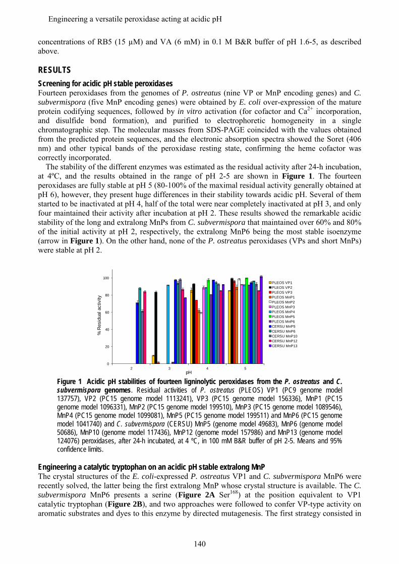
concentrations of RB5 (15 µM) and VA (6 mM) in 0.1 M B&R buffer of pH 1.6-5, as described above. RESULTS Screening for acidic pH stable peroxidases Fourteen peroxidases from the genomes of P. ostreatus (nine VP or MnP encoding genes) and C. subvermispora (five MnP encoding genes) were obtained by E. coli over-expression of the mature protein codifying sequences, followed by in vitro activation (for cofactor and Ca2+ incorporation, and disulfide bond formation), and purified to electrophoretic homogeneity in a single chromatographic step. The molecular masses from SDS-PAGE coincided with the values obtained from the predicted protein sequences, and the electronic absorption spectra showed the Soret (406 nm) and other typical bands of the peroxidase resting state, confirming the heme cofactor was correctly incorporated. The stability of the different enzymes was estimated as the residual activity after 24-h incubation, at 4ºC, and the results obtained in the range of pH 2-5 are shown in Figure 1. The fourteen peroxidases are fully stable at pH 5 (80-100% of the maximal residual activity generally obtained at pH 6), however, they present huge differences in their stability towards acidic pH. Several of them started to be inactivated at pH 4, half of the total were near completely inactivated at pH 3, and only four maintained their activity after incubation at pH 2. These results showed the remarkable acidic stability of the long and extralong MnPs from C. subvermispora that maintained over 60% and 80% of the initial activity at pH 2, respectively, the extralong MnP6 being the most stable isoenzyme (arrow in Figure 1). On the other hand, none of the P. ostreatus peroxidases (VPs and short MnPs) were stable at pH 2.
pH
% R
esid
ual a
ctiv
ity
0
20
40
60
80
100
2 3 4 5
PLEOS VP1PLEOS VP2PLEOS VP3PLEOS MnP1PLEOS MnP2PLEOS MnP3PLEOS MnP4PLEOS MnP5PLEOS MnP6CERSU MnP5CERSU MnP6CERSU MnP10CERSU MnP12CERSU MnP13
Figure 1 Acidic pH stabilities of fourteen ligninolytic peroxidases from the P. ostreatus and C. subvermispora genomes. Residual activities of P. ostreatus (PLEOS) VP1 (PC9 genome model 137757), VP2 (PC15 genome model 1113241), VP3 (PC15 genome model 156336), MnP1 (PC15 genome model 1096331), MnP2 (PC15 genome model 199510), MnP3 (PC15 genome model 1089546), MnP4 (PC15 genome model 1099081), MnP5 (PC15 genome model 199511) and MnP6 (PC15 genome model 1041740) and C. subvermispora (CERSU) MnP5 (genome model 49683), MnP6 (genome model 50686), MnP10 (genome model 117436), MnP12 (genome model 157986) and MnP13 (genome model 124076) peroxidases, after 24-h incubated, at 4 ºC, in 100 mM B&R buffer of pH 2-5. Means and 95% confidence limits.
Engineering a catalytic tryptophan on an acidic pH stable extralong MnP The crystal structures of the E. coli-expressed P. ostreatus VP1 and C. subvermispora MnP6 were recently solved, the latter being the first extralong MnP whose crystal structure is available. The C. subvermispora MnP6 presents a serine (Figure 2A Ser168) at the position equivalent to VP1 catalytic tryptophan (Figure 2B), and two approaches were followed to confer VP-type activity on aromatic substrates and dyes to this enzyme by directed mutagenesis. The first strategy consisted in
140
Engineering a versatile peroxidase acting at acidic pH
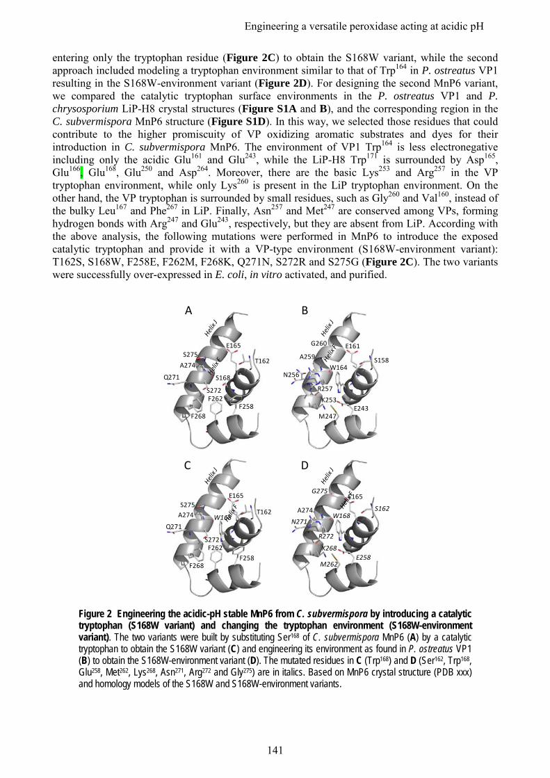
entering only the tryptophan residue (Figure 2C) to obtain the S168W variant, while the second approach included modeling a tryptophan environment similar to that of Trp164 in P. ostreatus VP1 resulting in the S168W-environment variant (Figure 2D). For designing the second MnP6 variant, we compared the catalytic tryptophan surface environments in the P. ostreatus VP1 and P. chrysosporium LiP-H8 crystal structures (Figure S1A and B), and the corresponding region in the C. subvermispora MnP6 structure (Figure S1D). In this way, we selected those residues that could contribute to the higher promiscuity of VP oxidizing aromatic substrates and dyes for their introduction in C. subvermispora MnP6. The environment of VP1 Trp164 is less electronegative including only the acidic Glu161 and Glu243, while the LiP-H8 Trp171 is surrounded by Asp165, Glu166, Glu168, Glu250 and Asp264. Moreover, there are the basic Lys253 and Arg257 in the VP tryptophan environment, while only Lys260 is present in the LiP tryptophan environment. On the other hand, the VP tryptophan is surrounded by small residues, such as Gly260 and Val160, instead of the bulky Leu167 and Phe267 in LiP. Finally, Asn257 and Met247 are conserved among VPs, forming hydrogen bonds with Arg247 and Glu243, respectively, but they are absent from LiP. According with the above analysis, the following mutations were performed in MnP6 to introduce the exposed catalytic tryptophan and provide it with a VP-type environment (S168W-environment variant): T162S, S168W, F258E, F262M, F268K, Q271N, S272R and S275G (Figure 2C). The two variants were successfully over-expressed in E. coli, in vitro activated, and purified.
A274
N271
R272
M262E258
E165
S162
K268
W168
G275
A274
Q271
S272F262
F258
E165
T162
F268
W168
S275
C D
A259
R257
M247E243
E161
S158
K253
W164
G260
N256A274
Q271
S272F262
F258
E165
T162
F268
S168
S275
A B
Figure 2 Engineering the acidic-pH stable MnP6 from C. subvermispora by introducing a catalytic tryptophan (S168W variant) and changing the tryptophan environment (S168W-environment variant). The two variants were built by substituting Ser168 of C. subvermispora MnP6 (A) by a catalytic tryptophan to obtain the S168W variant (C) and engineering its environment as found in P. ostreatus VP1 (B) to obtain the S168W-environment variant (D). The mutated residues in C (Trp168) and D (Ser162, Trp168, Glu258, Met262, Lys268, Asn271, Arg272 and Gly275) are in italics. Based on MnP6 crystal structure (PDB xxx) and homology models of the S168W and S168W-environment variants.
141
Engineering a versatile peroxidase acting at acidic pH
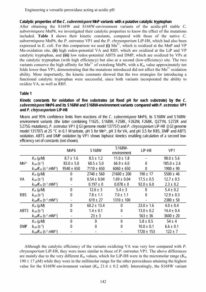
Catalytic properties of the C. subvermispora MnP variants with a putative catalytic tryptophan After obtaining the S168W and S168W-environment variants of the acidic-pH stable C. subvermispora MnP6, we investigated their catalytic properties to know the effect of the mutations included. Table 1 shows their kinetic constants, compared with those of the native C. subvermispora MnP6, the P. ostreatus VP1 and the P. chrysosporium LiP-H8, which had also been expressed in E. coli. For this comparison we used (i) Mn2+, which is oxidized at the MnP and VP Mn-oxidation site, (ii) high redox-potential VA and RB5, which are oxidized at the LiP and VP catalytic tryptophan, and (iii) low redox-potential ABTS and DMP, which are oxidized by VPs at the catalytic tryptophan (with high efficiency) but also at a second (low-efficiency) site. The two variants conserve the high affinity for Mn2+ of extralong MnP6, with a Km value approximately ten folds lower than VP1, demonstrating that the mutations introduced did not affect the Mn2+ oxidation ability. More importantly, the kinetic constants showed that the two strategies for introducing a functional catalytic tryptophan were successful, since both variants incorporated the ability to oxidize VA, as well as RB5.
Table 1 Kinetic constants for oxidation of five substrates (at fixed pH for each substrate) by the C. subvermispora MnP6 and its S168W and S168W-environment variants compared with P. ostreatus VP1 and P. chrysosporium LiP-H8 Means and 95% confidence limits from reactions of the C. subvermispora MnP6, its S168W and S168W-environment variants (the latter combining T162S, S168W, F258E, F262M, F268K, Q271N, S272R and S275G mutations), P. ostreatus VP1 (JGI genome model 137757) and P. chrysosporium LiP-H8 (JGI genome model 131707) at 25 °C in 0.1 M tartrate, pH 5 for Mn2+, pH 3 for VA, and pH 3.5 for RB5, DMP and ABTS oxidation. ABTS and DMP oxidation by VP1 shows biphasic kinetics enabling calculation of a second low-efficiency set of constants (not shown).
MnP6 S168W S168W-environment LiP-H8 VP1
Mn2+ Km (µM) 8.7 ± 1.6 8.5 ± 1.2 11.0 ± 1.8 - 98.0 ± 5.6 kcat (s-1) 83.0 ± 5.0 60.5 ± 5.0 66.9 ± 6.0 0 185.0 ± 2.6 kcat/Km (s-1.mM-1) 9540 ± 650 7110 ± 650 6060 ± 650 0 1900 ± 90
VA Km (µM) 0 2740 ± 560 21600 ± 200 190 ± 17 5500 ± 46
kcat (s-1) 0 0.54 ± 0.04 1.69 ± 0.04 17.5 ± 0.5 12.7 ± 0.5 kcat/Km (s-1.mM-1) - 0.197 ± 0 0.078 ± 0 92.0 ± 6.0 2.3 ± 0.2
RB5 Km (µM) 0 12.6 ± 3 5.4 ± 3 0 5.4 ± 0.2 kcat (s-1) 0 7.8 ± 1.1 7.0 ± 1.1 0 12.9 ± 0.3 kcat/Km (s-1.mM-1) - 619 ± 27 1310 ± 100 - 2380 ± 50
ABTS Km (µM) 0 60.2 ± 13.4 0 23.0 ± 1.6 4.0 ± 0.4 kcat (s-1) 0 1.4 ± 0.1 0 13.0 ± 0.2 14.4 ± 0.4 kcat/Km (s-1.mM-1) - 23 ± 3 - 563 ± 36 3600 ± 20
DMP Km (µM) 0 0 0 5.8 ± 0.5 54 ± 4 kcat (s-1) 0 0 0 10.0 ± 0.1 6.6 ± 0.1 kcat/Km (s-1.mM-1) - - - 1720 ± 153 122 ± 7
Although the catalytic efficiency of the variants oxidizing VA was very low compared with P.
chrysosporium LiP-H8, they were more similar to those of P. ostreatus VP1. The above differences are mainly due to the very different Km values, which for LiP-H8 were in the micromolar range (Km 190 ± 17 μM) while they were in the millimolar range for the other peroxidases attaining the highest value for the S168W-environment variant (Km 21.6 ± 0.2 mM). Interestingly, the S168W variant
142
Engineering a versatile peroxidase acting at acidic pH
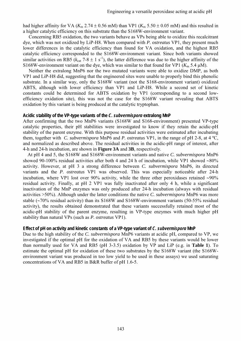
had higher affinity for VA (Km 2.74 ± 0.56 mM) than VP1 (Km 5.50 ± 0.05 mM) and this resulted in a higher catalytic efficiency on this substrate than the S168W-environment variant.
Concerning RB5 oxidation, the two variants behave as VPs being able to oxidize this recalcitrant dye, which was not oxidized by LiP-H8. When compared with P. ostreatus VP1, they present much lower differences in the catalytic efficiency than found for VA oxidation, and the highest RB5 catalytic efficiency corresponded to the S168W-environment variant. Since both variants showed similar activities on RB5 (kcat 7-8 ± 1 s-1), the latter difference was due to the higher affinity of the S168W-environment variant on the dye, which was similar to that found for VP1 (Km 5.4 μM).
Neither the extralong MnP6 nor the two mutated variants were able to oxidize DMP, as both VP1 and LiP-H8 did, suggesting that the engineered sites were unable to properly bind this phenolic substrate. In a similar way, only the S168W variant (not the S168-environment variant) oxidized ABTS, although with lower efficiency than VP1 and LiP-H8. While a second set of kinetic constants could be determined for ABTS oxidation by VP1 (corresponding to a second low-efficiency oxidation site), this was not the case for the S168W variant revealing that ABTS oxidation by this variant is being produced at the catalytic tryptophan. Acidic stability of the VP-type variants of the C. subvermispora extralong MnP After confirming that the two MnP6 variants (S168W and S168-environment) presented VP-type catalytic properties, their pH stabilities were investigated to know if they retain the acidic-pH stability of the parent enzyme. With this purpose residual activities were estimated after incubating them, together with C. subvermispora MnP6 and P. ostreatus VP1, in the range of pH 2-8, at 4 ºC, and normalized as described above. The residual activities in the acidic-pH range of interest, after 4-h and 24-h incubation, are shown in Figure 3A and 3B, respectively. At pH 4 and 5, the S168W and S168W-environment variants and native C. subvermispora MnP6 showed 90-100% residual activities after both 4 and 24 h of incubation, while VP1 showed ∼80% activity. However, at pH 3 a strong difference between C. subvermispora MnP6, its directed variants and the P. ostreatus VP1 was observed. This was especially noticeable after 24-h incubation, where VP1 lost over 90% activity, while the three other peroxidases retained ∼90% residual activity. Finally, at pH 2 VP1 was fully inactivated after only 4 h, while a significant inactivation of the MnP enzymes was only produced after 24-h incubation (always with residual activities >50%). Although under the latter conditions the native C. subvermispora MnP6 was more stable (∼70% residual activity) than its S168W and S168W-environment variants (50-55% residual activity), the results obtained demonstrated that these variants successfully retained most of the acidic-pH stability of the parent enzyme, resulting in VP-type enzymes with much higher pH stability than natural VPs (such as P. ostreatus VP1). Effect of pH on activity and kinetic constants of a VP-type variant of C. subvermispora MnP Due to the high stability of the C. subvermispora MnP6 variants at acidic pH, compared to VP, we investigated if the optimal pH for the oxidation of VA and RB5 by these variants would be lower than normally used for VA and RB5 (pH 3-3.5) oxidation by VP and LiP (e.g. in Table 1). To estimate the optimal pH for oxidation of these two substrates by the S168W variant (the S168W-environment variant was produced in too low yield to be used in these assays) we used saturating concentrations of VA and RB5 in B&R buffer of pH 1.6-5.
143
Engineering a versatile peroxidase acting at acidic pH
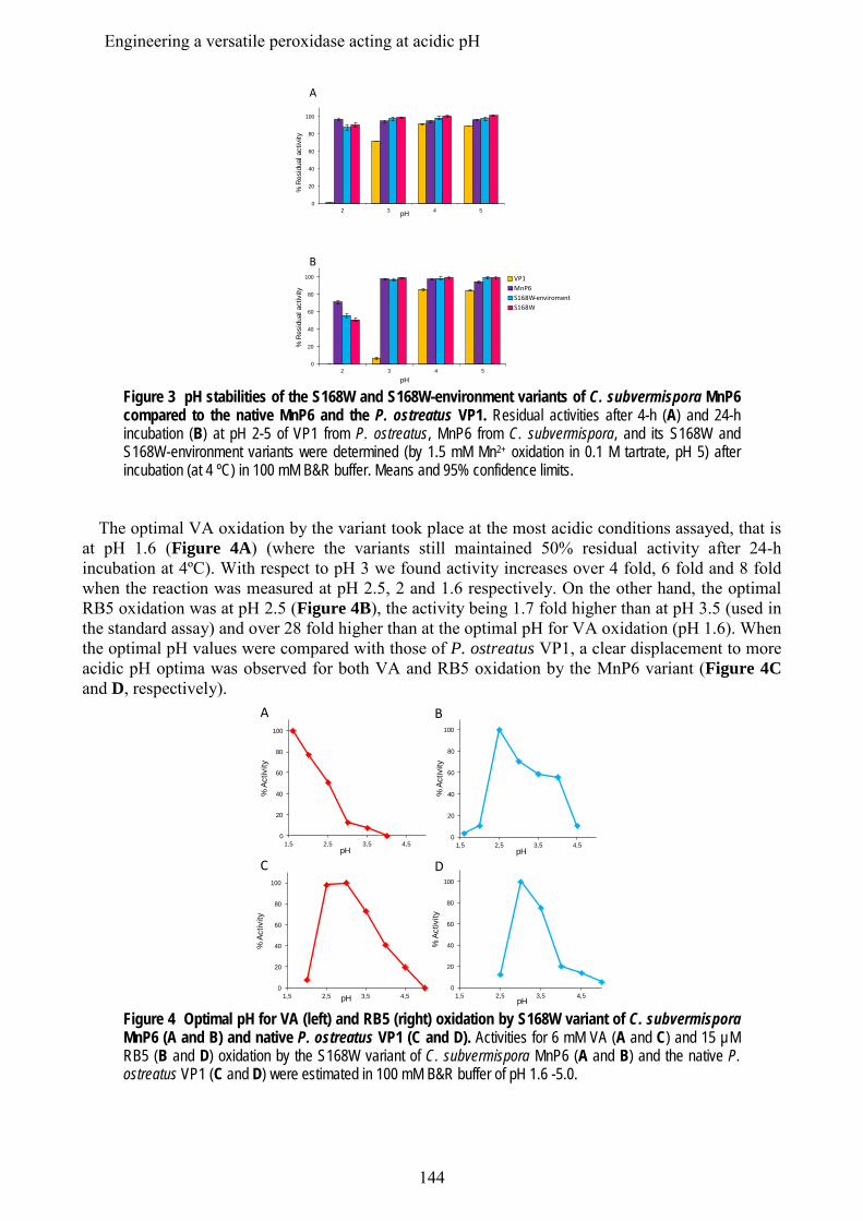
0
20
40
60
80
100
2 3 4 5
VP1MnP6S168W enviromentS168W
% R
esid
ual a
ctiv
ity
pH
B
% R
esid
ual a
ctiv
ity
pH
A
0
20
40
60
80
100
2 3 4 5
Figure 3 pH stabilities of the S168W and S168W-environment variants of C. subvermispora MnP6 compared to the native MnP6 and the P. ostreatus VP1. Residual activities after 4-h (A) and 24-h incubation (B) at pH 2-5 of VP1 from P. ostreatus, MnP6 from C. subvermispora, and its S168W and S168W-environment variants were determined (by 1.5 mM Mn2+ oxidation in 0.1 M tartrate, pH 5) after incubation (at 4 ºC) in 100 mM B&R buffer. Means and 95% confidence limits.
The optimal VA oxidation by the variant took place at the most acidic conditions assayed, that is at pH 1.6 (Figure 4A) (where the variants still maintained 50% residual activity after 24-h incubation at 4ºC). With respect to pH 3 we found activity increases over 4 fold, 6 fold and 8 fold when the reaction was measured at pH 2.5, 2 and 1.6 respectively. On the other hand, the optimal RB5 oxidation was at pH 2.5 (Figure 4B), the activity being 1.7 fold higher than at pH 3.5 (used in the standard assay) and over 28 fold higher than at the optimal pH for VA oxidation (pH 1.6). When the optimal pH values were compared with those of P. ostreatus VP1, a clear displacement to more acidic pH optima was observed for both VA and RB5 oxidation by the MnP6 variant (Figure 4C and D, respectively).
0
20
40
60
80
100
1,5 2,5 3,5 4,5
% A
ctiv
ity
pH
A
0
20
40
60
80
100
1,5 2,5 3,5 4,5pH
% A
ctiv
ity
C
0
20
40
60
80
100
1,5 2,5 3,5 4,5pH
% A
ctiv
ity
B
0
20
40
60
80
100
1,5 2,5 3,5 4,5pH
% A
ctiv
ity
D
Figure 4 Optimal pH for VA (left) and RB5 (right) oxidation by S168W variant of C. subvermispora MnP6 (A and B) and native P. ostreatus VP1 (C and D). Activities for 6 mM VA (A and C) and 15 µM RB5 (B and D) oxidation by the S168W variant of C. subvermispora MnP6 (A and B) and the native P. ostreatus VP1 (C and D) were estimated in 100 mM B&R buffer of pH 1.6 -5.0.
144
Engineering a versatile peroxidase acting at acidic pH
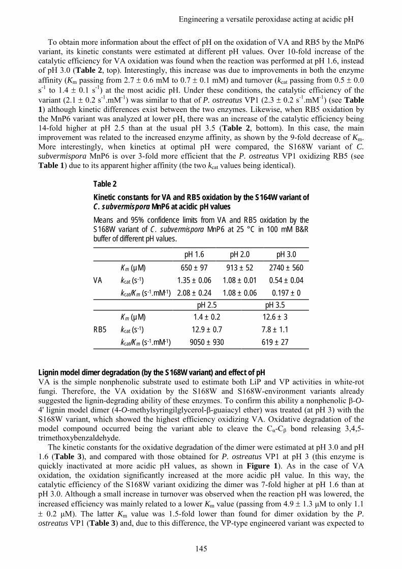
To obtain more information about the effect of pH on the oxidation of VA and RB5 by the MnP6 variant, its kinetic constants were estimated at different pH values. Over 10-fold increase of the catalytic efficiency for VA oxidation was found when the reaction was performed at pH 1.6, instead of pH 3.0 (Table 2, top). Interestingly, this increase was due to improvements in both the enzyme affinity (Km passing from 2.7 ± 0.6 mM to 0.7 ± 0.1 mM) and turnover (kcat passing from 0.5 ± 0.0 s-1 to 1.4 ± 0.1 s-1) at the most acidic pH. Under these conditions, the catalytic efficiency of the variant (2.1 ± 0.2 s-1.mM-1) was similar to that of P. ostreatus VP1 (2.3 ± 0.2 s-1.mM-1) (see Table 1) although kinetic differences exist between the two enzymes. Likewise, when RB5 oxidation by the MnP6 variant was analyzed at lower pH, there was an increase of the catalytic efficiency being 14-fold higher at pH 2.5 than at the usual pH 3.5 (Table 2, bottom). In this case, the main improvement was related to the increased enzyme affinity, as shown by the 9-fold decrease of Km. More interestingly, when kinetics at optimal pH were compared, the S168W variant of C. subvermispora MnP6 is over 3-fold more efficient that the P. ostreatus VP1 oxidizing RB5 (see Table 1) due to its apparent higher affinity (the two kcat values being identical).
Table 2 Kinetic constants for VA and RB5 oxidation by the S164W variant of C. subvermispora MnP6 at acidic pH values Means and 95% confidence limits from VA and RB5 oxidation by the S168W variant of C. subvermispora MnP6 at 25 °C in 100 mM B&R buffer of different pH values.
pH 1.6 pH 2.0 pH 3.0
VA Km (µM) 650 ± 97 913 ± 52 2740 ± 560 kcat (s-1) 1.35 ± 0.06 1.08 ± 0.01 0.54 ± 0.04 kcat/Km (s-1.mM-1) 2.08 ± 0.24 1.08 ± 0.06 0.197 ± 0
pH 2.5 pH 3.5
RB5 Km (µM) 1.4 ± 0.2 12.6 ± 3 kcat (s-1) 12.9 ± 0.7 7.8 ± 1.1 kcat/Km (s-1.mM-1) 9050 ± 930 619 ± 27
Lignin model dimer degradation (by the S168W variant) and effect of pH VA is the simple nonphenolic substrate used to estimate both LiP and VP activities in white-rot fungi. Therefore, the VA oxidation by the S168W and S168W-environment variants already suggested the lignin-degrading ability of these enzymes. To confirm this ability a nonphenolic β-O-4' lignin model dimer (4-O-methylsyringilglycerol-β-guaiacyl ether) was treated (at pH 3) with the S168W variant, which showed the highest efficiency oxidizing VA. Oxidative degradation of the model compound occurred being the variant able to cleave the Cα-Cβ bond releasing 3,4,5-trimethoxybenzaldehyde.
The kinetic constants for the oxidative degradation of the dimer were estimated at pH 3.0 and pH 1.6 (Table 3), and compared with those obtained for P. ostreatus VP1 at pH 3 (this enzyme is quickly inactivated at more acidic pH values, as shown in Figure 1). As in the case of VA oxidation, the oxidation significantly increased at the more acidic pH value. In this way, the catalytic efficiency of the S168W variant oxidizing the dimer was 7-fold higher at pH 1.6 than at pH 3.0. Although a small increase in turnover was observed when the reaction pH was lowered, the increased efficiency was mainly related to a lower Km value (passing from 4.9 ± 1.3 μM to only 1.1 ± 0.2 μM). The latter Km value was 1.5-fold lower than found for dimer oxidation by the P. ostreatus VP1 (Table 3) and, due to this difference, the VP-type engineered variant was expected to
145
Engineering a versatile peroxidase acting at acidic pH
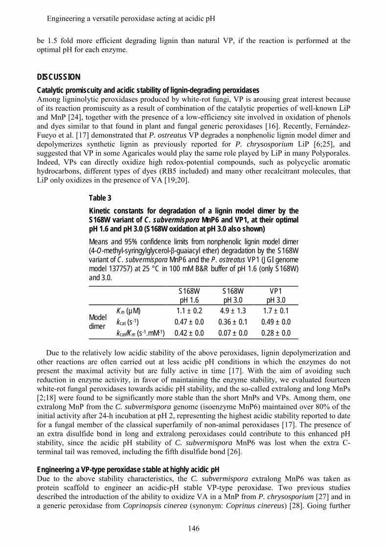
be 1.5 fold more efficient degrading lignin than natural VP, if the reaction is performed at the optimal pH for each enzyme.
DISCUSSION Catalytic promiscuity and acidic stability of lignin-degrading peroxidases Among ligninolytic peroxidases produced by white-rot fungi, VP is arousing great interest because of its reaction promiscuity as a result of combination of the catalytic properties of well-known LiP and MnP [24], together with the presence of a low-efficiency site involved in oxidation of phenols and dyes similar to that found in plant and fungal generic peroxidases [16]. Recently, Fernández-Fueyo et al. [17] demonstrated that P. ostreatus VP degrades a nonphenolic lignin model dimer and depolymerizes synthetic lignin as previously reported for P. chrysosporium LiP [6;25], and suggested that VP in some Agaricales would play the same role played by LiP in many Polyporales. Indeed, VPs can directly oxidize high redox-potential compounds, such as polycyclic aromatic hydrocarbons, different types of dyes (RB5 included) and many other recalcitrant molecules, that LiP only oxidizes in the presence of VA [19;20].
Table 3 Kinetic constants for degradation of a lignin model dimer by the S168W variant of C. subvermispora MnP6 and VP1, at their optimal pH 1.6 and pH 3.0 (S168W oxidation at pH 3.0 also shown) Means and 95% confidence limits from nonphenolic lignin model dimer (4-O-methyl-syringylglycerol-β-guaiacyl ether) degradation by the S168W variant of C. subvermispora MnP6 and the P. ostreatus VP1 (JGI genome model 137757) at 25 °C in 100 mM B&R buffer of pH 1.6 (only S168W) and 3.0.
S168W pH 1.6
S168W pH 3.0
VP1 pH 3.0
Model dimer
Km (µM) 1.1 ± 0.2 4.9 ± 1.3 1.7 ± 0.1 kcat (s-1) 0.47 ± 0.0 0.36 ± 0.1 0.49 ± 0.0 kcat/Km (s-1.mM-1) 0.42 ± 0.0 0.07 ± 0.0 0.28 ± 0.0
Due to the relatively low acidic stability of the above peroxidases, lignin depolymerization and
other reactions are often carried out at less acidic pH conditions in which the enzymes do not present the maximal activity but are fully active in time [17]. With the aim of avoiding such reduction in enzyme activity, in favor of maintaining the enzyme stability, we evaluated fourteen white-rot fungal peroxidases towards acidic pH stability, and the so-called extralong and long MnPs [2;18] were found to be significantly more stable than the short MnPs and VPs. Among them, one extralong MnP from the C. subvermispora genome (isoenzyme MnP6) maintained over 80% of the initial activity after 24-h incubation at pH 2, representing the highest acidic stability reported to date for a fungal member of the classical superfamily of non-animal peroxidases [17]. The presence of an extra disulfide bond in long and extralong peroxidases could contribute to this enhanced pH stability, since the acidic pH stability of C. subvermispora MnP6 was lost when the extra C-terminal tail was removed, including the fifth disulfide bond [26]. Engineering a VP-type peroxidase stable at highly acidic pH Due to the above stability characteristics, the C. subvermispora extralong MnP6 was taken as protein scaffold to engineer an acidic-pH stable VP-type peroxidase. Two previous studies described the introduction of the ability to oxidize VA in a MnP from P. chrysosporium [27] and in a generic peroxidase from Coprinopsis cinerea (synonym: Coprinus cinereus) [28]. Going further
146
Engineering a versatile peroxidase acting at acidic pH
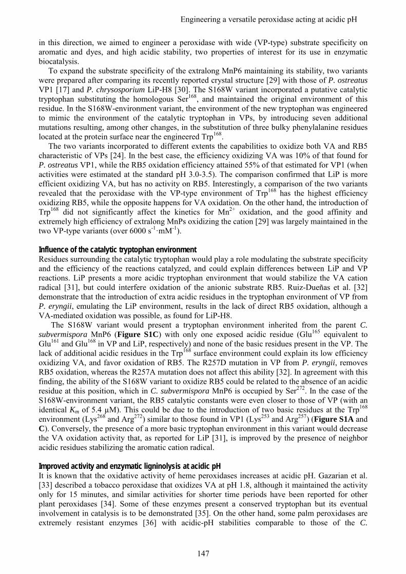
in this direction, we aimed to engineer a peroxidase with wide (VP-type) substrate specificity on aromatic and dyes, and high acidic stability, two properties of interest for its use in enzymatic biocatalysis.
To expand the substrate specificity of the extralong MnP6 maintaining its stability, two variants were prepared after comparing its recently reported crystal structure [29] with those of P. ostreatus VP1 [17] and P. chrysosporium LiP-H8 [30]. The S168W variant incorporated a putative catalytic tryptophan substituting the homologous Ser168, and maintained the original environment of this residue. In the S168W-environment variant, the environment of the new tryptophan was engineered to mimic the environment of the catalytic tryptophan in VPs, by introducing seven additional mutations resulting, among other changes, in the substitution of three bulky phenylalanine residues located at the protein surface near the engineered Trp168.
The two variants incorporated to different extents the capabilities to oxidize both VA and RB5 characteristic of VPs [24]. In the best case, the efficiency oxidizing VA was 10% of that found for P. ostreatus VP1, while the RB5 oxidation efficiency attained 55% of that estimated for VP1 (when activities were estimated at the standard pH 3.0-3.5). The comparison confirmed that LiP is more efficient oxidizing VA, but has no activity on RB5. Interestingly, a comparison of the two variants revealed that the peroxidase with the VP-type environment of Trp168 has the highest efficiency oxidizing RB5, while the opposite happens for VA oxidation. On the other hand, the introduction of Trp168 did not significantly affect the kinetics for Mn2+ oxidation, and the good affinity and extremely high efficiency of extralong MnPs oxidizing the cation [29] was largely maintained in the two VP-type variants (over 6000 s-1·mM-1). Influence of the catalytic tryptophan environment Residues surrounding the catalytic tryptophan would play a role modulating the substrate specificity and the efficiency of the reactions catalyzed, and could explain differences between LiP and VP reactions. LiP presents a more acidic tryptophan environment that would stabilize the VA cation radical [31], but could interfere oxidation of the anionic substrate RB5. Ruiz-Dueñas et al. [32] demonstrate that the introduction of extra acidic residues in the tryptophan environment of VP from P. eryngii, emulating the LiP environment, results in the lack of direct RB5 oxidation, although a VA-mediated oxidation was possible, as found for LiP-H8.
The S168W variant would present a tryptophan environment inherited from the parent C. subvermispora MnP6 (Figure S1C) with only one exposed acidic residue (Glu165 equivalent to Glu161 and Glu168 in VP and LiP, respectively) and none of the basic residues present in the VP. The lack of additional acidic residues in the Trp168 surface environment could explain its low efficiency oxidizing VA, and favor oxidation of RB5. The R257D mutation in VP from P. eryngii, removes RB5 oxidation, whereas the R257A mutation does not affect this ability [32]. In agreement with this finding, the ability of the S168W variant to oxidize RB5 could be related to the absence of an acidic residue at this position, which in C. subvermispora MnP6 is occupied by Ser272. In the case of the S168W-environment variant, the RB5 catalytic constants were even closer to those of VP (with an identical Km of 5.4 µM). This could be due to the introduction of two basic residues at the Trp168 environment (Lys268 and Arg272) similar to those found in VP1 (Lys253 and Arg257) (Figure S1A and C). Conversely, the presence of a more basic tryptophan environment in this variant would decrease the VA oxidation activity that, as reported for LiP [31], is improved by the presence of neighbor acidic residues stabilizing the aromatic cation radical. Improved activity and enzymatic ligninolysis at acidic pH It is known that the oxidative activity of heme peroxidases increases at acidic pH. Gazarian et al. [33] described a tobacco peroxidase that oxidizes VA at pH 1.8, although it maintained the activity only for 15 minutes, and similar activities for shorter time periods have been reported for other plant peroxidases [34]. Some of these enzymes present a conserved tryptophan but its eventual involvement in catalysis is to be demonstrated [35]. On the other hand, some palm peroxidases are extremely resistant enzymes [36] with acidic-pH stabilities comparable to those of the C.
147
Engineering a versatile peroxidase acting at acidic pH
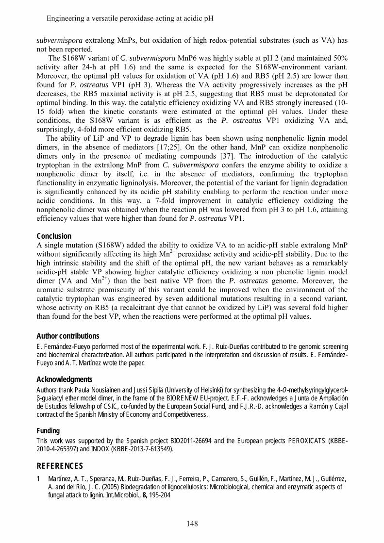
subvermispora extralong MnPs, but oxidation of high redox-potential substrates (such as VA) has not been reported.
The S168W variant of C. subvermispora MnP6 was highly stable at pH 2 (and maintained 50% activity after 24-h at pH 1.6) and the same is expected for the S168W-environment variant. Moreover, the optimal pH values for oxidation of VA (pH 1.6) and RB5 (pH 2.5) are lower than found for P. ostreatus VP1 (pH 3). Whereas the VA activity progressively increases as the pH decreases, the RB5 maximal activity is at pH 2.5, suggesting that RB5 must be deprotonated for optimal binding. In this way, the catalytic efficiency oxidizing VA and RB5 strongly increased (10-15 fold) when the kinetic constants were estimated at the optimal pH values. Under these conditions, the S168W variant is as efficient as the P. ostreatus VP1 oxidizing VA and, surprisingly, 4-fold more efficient oxidizing RB5.
The ability of LiP and VP to degrade lignin has been shown using nonphenolic lignin model dimers, in the absence of mediators [17;25]. On the other hand, MnP can oxidize nonphenolic dimers only in the presence of mediating compounds [37]. The introduction of the catalytic tryptophan in the extralong MnP from C. subvermispora confers the enzyme ability to oxidize a nonphenolic dimer by itself, i.e. in the absence of mediators, confirming the tryptophan functionality in enzymatic ligninolysis. Moreover, the potential of the variant for lignin degradation is significantly enhanced by its acidic pH stability enabling to perform the reaction under more acidic conditions. In this way, a 7-fold improvement in catalytic efficiency oxidizing the nonphenolic dimer was obtained when the reaction pH was lowered from pH 3 to pH 1.6, attaining efficiency values that were higher than found for P. ostreatus VP1. Conclusion A single mutation (S168W) added the ability to oxidize VA to an acidic-pH stable extralong MnP without significantly affecting its high Mn2+ peroxidase activity and acidic-pH stability. Due to the high intrinsic stability and the shift of the optimal pH, the new variant behaves as a remarkably acidic-pH stable VP showing higher catalytic efficiency oxidizing a non phenolic lignin model dimer (VA and Mn2+) than the best native VP from the P. ostreatus genome. Moreover, the aromatic substrate promiscuity of this variant could be improved when the environment of the catalytic tryptophan was engineered by seven additional mutations resulting in a second variant, whose activity on RB5 (a recalcitrant dye that cannot be oxidized by LiP) was several fold higher than found for the best VP, when the reactions were performed at the optimal pH values. Author contributions E. Fernández-Fueyo performed most of the experimental work. F. J. Ruiz-Dueñas contributed to the genomic screening and biochemical characterization. All authors participated in the interpretation and discussion of results. E. Fernández-Fueyo and A. T. Martínez wrote the paper. Acknowledgments Authors thank Paula Nousiainen and Jussi Sipilä (University of Helsinki) for synthesizing the 4-O-methylsyringylglycerol-β-guaiacyl ether model dimer, in the frame of the BIORENEW EU-project. E.F.-F. acknowledges a Junta de Ampliación de Estudios fellowship of CSIC, co-funded by the European Social Fund, and F.J.R.-D. acknowledges a Ramón y Cajal contract of the Spanish Ministry of Economy and Competitiveness. Funding This work was supported by the Spanish project BIO2011-26694 and the European projects PEROXICATS (KBBE-2010-4-265397) and INDOX (KBBE-2013-7-613549). REFERENCES 1 Martínez, A. T., Speranza, M., Ruiz-Dueñas, F. J., Ferreira, P., Camarero, S., Guillén, F., Martínez, M. J., Gutiérrez,
A. and del Río, J. C. (2005) Biodegradation of lignocellulosics: Microbiological, chemical and enzymatic aspects of fungal attack to lignin. Int.Microbiol., 8, 195-204
148
Engineering a versatile peroxidase acting at acidic pH
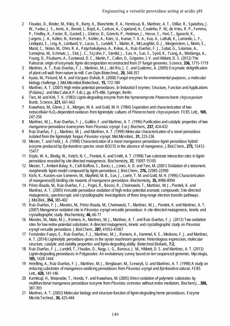
2 Floudas, D., Binder, M., Riley, R., Barry, K., Blanchette, R. A., Henrissat, B., Martínez, A. T., Otillar, R., Spatafora, J. W., Yadav, J. S., Aerts, A., Benoit, I., Boyd, A., Carlson, A., Copeland, A., Coutinho, P. M., de Vries, R. P., Ferreira, P., Findley, K., Foster, B., Gaskell, J., Glotzer, D., Górecki, P., Heitman, J., Hesse, C., Hori, C., Igarashi, K., Jurgens, J. A., Kallen, N., Kersten, P., Kohler, A., Kües, U., Kumar, T. K. A., Kuo, A., LaButti, K., Larrondo, L. F., Lindquist, E., Ling, A., Lombard, V., Lucas, S., Lundell, T., Martin, R., McLaughlin, D. J., Morgenstern, I., Morin, E., Murat, C., Nolan, M., Ohm, R. A., Patyshakuliyeva, A., Rokas, A., Ruiz-Dueñas, F. J., Sabat, G., Salamov, A., Samejima, M., Schmutz, J., Slot, J. C., St.John, F., Stenlid, J., Sun, H., Sun, S., Syed, K., Tsang, A., Wiebenga, A., Young, D., Pisabarro, A., Eastwood, D. C., Martin, F., Cullen, D., Grigoriev, I. V. and Hibbett, D. S. (2012) The Paleozoic origin of enzymatic lignin decomposition reconstructed from 31 fungal genomes. Science, 336, 1715-1719
3 Martínez, A. T., Ruiz-Dueñas, F. J., Martínez, M. J., del Río, J. C. and Gutiérrez, A. (2009) Enzymatic delignification of plant cell wall: from nature to mill. Curr.Opin.Biotechnol., 20, 348-357
4 Ayala, M., Pickard, M. A. and Vázquez-Duhalt, R. (2008) Fungal enzymes for environmental purposes, a molecular biology challenge. J.Mol.Microbiol.Biotechnol., 15, 172-180
5 Martínez, A. T. (2007) High redox potential peroxidases. In Industrial Enzymes: Structure, Function and Applications (Polaina,J. and MacCabe,A.P. Eds.), pp. 475-486. Springer, Berlin
6 Tien, M. and Kirk, T. K. (1983) Lignin-degrading enzyme from the hymenomycete Phanerochaete chrysosporium Burds. Science, 221, 661-663
7 Kuwahara, M., Glenn, J. K., Morgan, M. A. and Gold, M. H. (1984) Separation and characterization of two extracellular H2O2-dependent oxidases from ligninolytic cultures of Phanerochaete chrysosporium. FEBS Lett., 169, 247-250
8 Martínez, M. J., Ruiz-Dueñas, F. J., Guillén, F. and Martínez, A. T. (1996) Purification and catalytic properties of two manganese-peroxidase isoenzymes from Pleurotus eryngii. Eur.J.Biochem., 237, 424-432
9 Ruiz-Dueñas, F. J., Martínez, M. J. and Martínez, A. T. (1999) Molecular characterization of a novel peroxidase isolated from the ligninolytic fungus Pleurotus eryngii. Mol.Microbiol., 31, 223-236
10 Mester, T. and Field, J. A. (1998) Characterization of a novel manganese peroxidase-lignin peroxidase hybrid isozyme produced by Bjerkandera species strain BOS55 in the absence of manganese. J.Biol.Chem., 273, 15412-15417
11 Doyle, W. A., Blodig, W., Veitch, N. C., Piontek, K. and Smith, A. T. (1998) Two substrate interaction sites in lignin peroxidase revealed by site-directed mutagenesis. Biochemistry, 37, 15097-15105
12 Mester, T., Ambert-Balay, K., Ciofi-Baffoni, S., Banci, L., Jones, A. D. and Tien, M. (2001) Oxidation of a tetrameric nonphenolic lignin model compound by lignin peroxidase. J.Biol.Chem., 276, 22985-22990
13 Kishi, K., Kusters-van Someren, M., Mayfield, M. B., Sun, J., Loehr, T. M. and Gold, M. H. (1996) Characterization of manganese(II) binding site mutants of manganese peroxidase. Biochemistry, 35, 8986-8994
14 Pérez-Boada, M., Ruiz-Dueñas, F. J., Pogni, R., Basosi, R., Choinowski, T., Martínez, M. J., Piontek, K. and Martínez, A. T. (2005) Versatile peroxidase oxidation of high redox potential aromatic compounds: Site-directed mutagenesis, spectroscopic and crystallographic investigations of three long-range electron transfer pathways. J.Mol.Biol., 354, 385-402
15 Ruiz-Dueñas, F. J., Morales, M., Pérez-Boada, M., Choinowski, T., Martínez, M. J., Piontek, K. and Martínez, A. T. (2007) Manganese oxidation site in Pleurotus eryngii versatile peroxidase: A site-directed mutagenesis, kinetic and crystallographic study. Biochemistry, 46, 66-77
16 Morales, M., Mate, M. J., Romero, A., Martínez, M. J., Martínez, A. T. and Ruiz-Dueñas, F. J. (2012) Two oxidation sites for low redox-potential substrates: A directed mutagenesis, kinetic and crystallographic study on Pleurotus eryngii versatile peroxidase. J.Biol.Chem., 287, 41053-41067
17 Fernández-Fueyo, E., Ruiz-Dueñas, F. J., Martínez, M. J., Romero, A., Hammel, K. E., Medrano, F. J. and Martínez, A. T. (2014) Ligninolytic peroxidase genes in the oyster mushroom genome: Heterologous expression, molecular structure, catalytic and stability properties and lignin-degrading ability. Biotechnol.Biofuels, 7:2,
18 Ruiz-Dueñas, F. J., Lundell, T., Floudas, D., Nagy, L. G., Barrasa, J. M., Hibbett, D. S. and Martínez, A. T. (2013) Lignin-degrading peroxidases in Polyporales: An evolutionary survey based on ten sequenced genomes. Mycologia, 105, 1428-1444
19 Heinfling, A., Ruiz-Dueñas, F. J., Martínez, M. J., Bergbauer, M., Szewzyk, U. and Martínez, A. T. (1998) A study on reducing substrates of manganese-oxidizing peroxidases from Pleurotus eryngii and Bjerkandera adusta. FEBS Lett., 428, 141-146
20 Kamitsuji, H., Watanabe, T., Honda, Y. and Kuwahara, M. (2005) Direct oxidation of polymeric substrates by multifunctional manganese peroxidase isozyme from Pleurotus ostreatus without redox mediators. Biochem.J., 386, 387-393
21 Martínez, A. T. (2002) Molecular biology and structure-function of lignin-degrading heme peroxidases. Enzyme Microb.Technol., 30, 425-444
149
Engineering a versatile peroxidase acting at acidic pH
22 Pérez-Boada, M., Doyle, W. A., Ruiz-Dueñas, F. J., Martínez, M. J., Martínez, A. T. and Smith, A. T. (2002) Expression of Pleurotus eryngii versatile peroxidase in Escherichia coli and optimisation of in vitro folding. Enzyme Microb.Technol., 30, 518-524
23 Doyle, W. A. and Smith, A. T. (1996) Expression of lignin peroxidase H8 in Escherichia coli: Folding and activation of the recombinant enzyme with Ca2+ and haem. Biochem.J., 315, 15-19
24 Ruiz-Dueñas, F. J., Morales, M., García, E., Miki, Y., Martínez, M. J. and Martínez, A. T. (2009) Substrate oxidation sites in versatile peroxidase and other basidiomycete peroxidases. J.Exp.Bot., 60, 441-452
25 Hammel, K. E., Jensen, K. A., Mozuch, M. D., Landucci, L. L., Tien, M. and Pease, E. A. (1993) Ligninolysis by a purified lignin peroxidase. J.Biol.Chem., 268, 12274-12281
26 Fernández-Fueyo, E., Acebes, S., Ruiz-Dueñas, F. J., Martínez, M. J., Romero, A., Medrano, F. J., Guallar, V. and Martínez, A. T. (2014) Catalytic and stability properties of manganese peroxidase subfamilies: A study based on the Ceriporiopsis subvermispora genome. (submitted),
27 Timofeevski, S. L., Nie, G., Reading, N. S. and Aust, S. D. (1999) Addition of veratryl alcohol oxidase activity to manganese peroxidase by site-directed mutagenesis. Biochem.Biophys.Res.Commun., 256, 500-504
28 Smith, A. T. and Doyle, W. A. (2006) Engineered peroxidases with veratryl alcohol oxidase activity. Patent (International), WO/2006-114616
29 Fernández-Fueyo, E., Acebes, S., Ruiz-Dueñas, F. J., Martínez, M. J., Romero, A., Medrano, F. J., Guallar, V. and Martínez, A. T. (2014) Catalytic and stability properties of manganese peroxidase subfamilies: A structural-functional study based on the Ceriporiopsis subvermispora genome. Proc.Natl.Acad.Sci.USA, (submitted)
30 Poulos, T. L., Edwards, S. L., Wariishi, H. and Gold, M. H. (1993) Crystallographic refinement of lignin peroxidase at 2 Å. J.Biol.Chem., 268, 4429-4440
31 Khindaria, A., Yamazaki, I. and Aust, S. D. (1996) Stabilization of the veratryl alcohol cation radical by lignin peroxidase. Biochemistry, 35, 6418-6424
32 Ruiz-Dueñas, F. J., Morales, M., Mate, M. J., Romero, A., Martínez, M. J., Smith, A. T. and Martínez, A. T. (2008) Site-directed mutagenesis of the catalytic tryptophan environment in Pleurotus eryngii versatile peroxidase. Biochemistry, 47, 1685-1695
33 Gazarian, I. G., Lagrimini, M., George, S. J. and Thorneley, R. N. F. (1996) Anionic tobacco peroxidase is active at extremely low pH: Veratryl alcohol oxidation with a pH optimum of 1.8. Biochem.J., 320, 369-372
34 McEldoon, J. P., Pokora, A. R. and Dordick, J. S. (1995) Lignin peroxidase-type activity of soybean peroxidase. Enzyme Microb.Technol., 17, 359-365
35 Gazaryan, I. G., Chubar, T. A., Ignatenko, O. V., Mareeva, E. A., Orlova, M. A., Kapeliuch, Y. L., Savitsky, P. A., Rojkova, A. M. and Tishkov, V. I. (1999) Tryptophanless recombinant horseradish peroxidase: stability and catalytic properties. Biochem.Biophys.Res.Commun., 262, 297-301
36 Sakharov, I. Y. and Sakharova, T. V. (2002) Extremely high stability of African oil palm tree peroxidase. BBA Proteins Proteomics, 1598, 108-114
37 Kapich, A. N., Korneichik, T. V., Hatakka, A. and Hammel, K. E. (2010) Oxidizability of unsaturated fatty acids and of a non-phenolic lignin structure in the manganese peroxidase-dependent lipid peroxidation system. Enzyme Microb.Technol., 46, 136-140
150
Engineering a versatile peroxidase acting at acidic pH

Capítulo 9
Discusión
151

152
9. Discusión
Diversidad de peroxidasas tras el análisis genómico de basidiomicetos Como se ha comentado en la Introducción, las peroxidasas ligninolíticas, características de los basidiomicetos de podredumbre blanca (que degradan la lignina) y ausentes de los de podredumbre parda (que degradan los polisacáridos), se dividen en tres familias en función de sus propiedades catalíticas. La LiP y la MnP se describieron hace unos 30 años en P. chrysosporium (Tien and Kirk, 1983; Kuwahara et al., 1984), mientras que la VP se describió en Pleurotus enryngii, primero como una MnP "con actividad sobre alcohol veratrílico" (Martínez et al., 1996) y posteriormente como una nueva peroxidasa caracterizada por su versatilidad catalítica (Ruiz-Dueñas et al., 1999; Camarero et al., 1999), y más tarde se encontró también en Bjerkandera sp (Mester and Field, 1998).
P. chrysosporium ha sido el hongo modelo utilizado en muchos estudios bioquímicos, enzimáticos y moleculares de degradación de la lignina, por esta razón fue el primer basidiomiceto cuyo genoma fue secuenciado (Martinez et al., 2004). En diferentes estudios se ha demostrado la capacidad que tiene la LiP de este hongo para degradar dímeros modelo no-fenólicos y lignina polimérica (Tien and Kirk, 1983; Hammel et al., 1985). Debido a esto, y a estudios posteriores con LiPs de otros basidiomicetos, la presencia de LiPs se ha asociado con la capacidad de degradar la lignina natural (un polímero de escaso contenido fenólico). Sin embargo, en varios hongos con demostrada capacidad ligninolítica no había sido posible demostrar la existencia de enzimas de tipo LiP. Entre ellos se encontraban C. subvermispora y P. ostreatus, dos especies de interés biotecnológico por su capacidad para degradar selectivamente la lignina (de madera y residuos agrícolas, respectivamente), que han sido objeto de estudio en la presente Tesis.
En C. subvermispora sólo se habían detectado enzimas de tipo lacasa y MnP, a pesar de haberse realizado considerables esfuerzos para aislar enzimas de tipo LiP de los cultivos, o para clonar los correspondientes genes. La ausencia de LiPs, que en aquel momento eran las únicas enzimas con capacidad reconocida para degradar la lignina no-fenólica, llevaron a sugerir que en este hongo la degradación tendría lugar por un mecanismo alternativo, en el que estarían implicadas las MnPs (Jensen et al., 1996). La secuenciación del genoma de C. subvermipora mostró un número inusualmente elevado de MnPs, junto con genes implicados en la síntesis de ácidos grasos insaturados, lo que nos hizo sugerir que la degradación de la lignina por este hongo ocurriría a traves de los radicales libres derivados de la peroxidación de los lípidos (Fernández-Fueyo et al., 2012a). La capacidad de estos radicales para degradar la lignina no-fenólica había sido descrito anteriormente (Bao et al., 1994). Sin embargo, poco después se produjo el descubrimiento más sorprendente en el genoma de C. subvermispora al identificarse dos genes cuyas secuencias codificaban peroxidasas con el triptófano catalítico conservado en VPs y LiPs, que no habían sido previamente descritas en este hongo. Estos dos genes se clasificaron inicialmente como una putativa LiP y una putativa VP. Sin embargo su caracterización bioquímica, tras expresión heteróloga, demostró que se trataba de dos LiPs funcionales, capaces de degradar lignina sintética y un dímero modelo no-fenólico. Como consecuencia, podrían ser responsables de la degradación natural de la lignina por este hongo. Por otro lado, las peculiares constantes cinéticas de las dos nuevas enzimas, y su posición en el árbol filogenético de las peroxidasas, nos permitieron sugerir que muy posiblemente representan formas de transición entre VP y LiP (Fernández-Fueyo et al., 2012b).
153
9. Discusión
En P. ostreatus y especies relacionadas se habían encontrado MnPs y lacasas, como en C. subvermispora. Además, en estos hongos se había descrito por vez primera la VP, como una nueva familia de peroxidasas combinando las propiedades catalíticas y estructurales de LiPs y MnPs. Sin embargo, se mantenía la incógnita de si, como en el caso de C. subvermispora, pudieran también producir enzimas de tipo LiP implicadas en la degradación de la lignina. La secuenciación del genoma de P. ostreatus ha proporcionado respuesta a esta pregunta al mostrar la existencia de genes de MnPs y VPs y la ausencia de genes de LiP en este hongo (Ruiz-Dueñas et al., 2011). Mas importante, tras la expresión heteróloga de todos ellos, se encontró que la VP de P. ostreatus es capaz de oxidar dímeros modelo no-fenólicos y degradar la lignina con una eficiencia similar a la de la LiP mas estudiada (isoenzima H8) de P. chrysosporium. Estos resultados nos han permitido postular que la VP desempeñaría en P. ostreatus y probablemente en otras especies del orden Agaricales, en cuyos cultivos y genomas no se han encontrado nunca evidencias de presencia de LiP (Floudas et al., 2012), el papel desempeñando por las LiPs en P. chrysosporium y otras especies de Poliporales (Fernández-Fueyo et al., 2014d).
La clasificación de los genes de peroxidasas ligninolíticas en el creciente número de genomas de basidiomicetos disponibles (Floudas et al., 2012), se basa en la presencia o ausencia del sitio de oxidación del Mn2+ (formado por tres residuos ácidos junto al propionato interno del hemo) y/o del triptófano catalítico en las proteínas predichas. En la mayoría de los casos, los residuos conservados son funcionales tras expresión heteróloga, y la clasificación inicial - estructural y teórica - coincide con la clasificación funcional. Sin embargo, en la presente tesis hemos encontrado dos excepciones significativas correspondientes a una MnP y una VP cuya clasificación inicial tuvo que modificarse tras la determinación experimental de sus propiedades catalíticas. Así, en el genoma de P. ostreatus se encontró una MnP con un triptófano superficial homólogo al de las LiPs/VPs, por lo que el gen había sido inicialmente clasificado como un gen de LiP, pero dicho residuo resultó posteriormente no ser funcional. Esta anomalía se explica por una mutación durante la evolución de estas peroxidasas que interrumpió la ruta de transporte electrónico desde la superficie hasta el grupo hemo, como se demostró por mutagénesis dirigida reversa (Fernández-Fueyo et al., 2014d). Esta misma mutación natural pudo también localizarse en otra MnP de la especie relacionada P. pulmonarius. Por otra parte, en el genoma de C. subvermipora hubo que reclasificar, como gen de LiP, uno que la clasificación teórica había considerado codificaba una VP. Dicha reclasificación se debió a la incapacidad de la enzima para oxidar Mn2+ a pesar de presentar los tres residuos conservados que forman el sitio de unión del Mn en todas las VPs y MnPs. En este caso, la falta de actividad era debida a la presencia de un cuarto residuo ácido contiguo, como se mostró al aparecer la actividad sobre el Mn2+ cuando el residuo se eliminó por mutagénesis dirigida (Fernández-Fueyo et al., 2012b).
Estudios recientes han mostrado que la historia evolutiva general de las peroxidasas ligninolíticas se inició con una MnP primitiva que adquirió el triptófano catalítico para dar lugar a la primera VP y que, posteriormente, una VP perdió el sitio de oxidación del Mn para dar lugar a la primera LiP (esta última transición únicamente en Poliporales) (Floudas et al., 2012; Ruiz-Dueñas et al., 2013). Sin embargo, los resultados anteriores muestran la existencia más que probable de líneas evolutivas secundarias en las que, por ejemplo, una VP pudo dar lugar a una MnP, como en el caso de P. ostreatus, al mismo tiempo que nos revelan formas intermedias que explican las diferentes transiciones.
154
9. Discusión
Existencia y significado de múltiples isoformas de peroxidasas Una característica en la evolución de las enzimas que componen el sistema oxidativo de los hongos, incluyendo las peroxidasas ligninolíticas, es la existencia de duplicaciones recientes codificando diferentes isoformas. Dichas isoenzimas difieren escasamente en su secuencia y, a pesar de que existen pocos datos al respecto, deben poseer características diferentes de forma que su expresión confiera al hongo una ventaja evolutiva en determinados hábitats o sustratos. Desde hace tiempo se conoce la existencia de un número limitado de isoenzimas de LiP, MnP y VP. Sin embargo, hasta muy recientemente, no ha sido posible conocer la gran magnitud de este fenómeno de duplicación génica al disponerse de un número considerable de genomas, en los que se han realizado inventarios detallados de los diferentes tipos de genes (Floudas et al., 2012; Ruiz-Dueñas et al., 2013).
De esta forma, hasta la secuenciación del genoma de C. subvermispora solamente habían sido descritos cuatro genes de MnPs en este hongo, mientras que en su genoma hemos podido identificar quince genes de peroxidasas ligninolíticas clasificados inicialmente como trece MnPs, una LiP y una VP (Fernández-Fueyo et al., 2012a), esta última reclasificada como una LiP tras su caracterización bioquímica (Fernández-Fueyo et al., 2012b), tal como se ha discutido anteriormente. De igual forma, en P. ostreatus sólo se habían sido descrito tres genes de peroxidasas ligninolíticas, sin embargo en su genoma encontramos nueve, que inicialmente se clasificaron en cuatro genes de VP y cinco de MnP (Ruiz-Dueñas et al., 2011). Gracias a la disponibilidad de los genomas y a la activación in vitro de las peroxidasas expresadas en E. coli, hemos caracterizado bioquímicamente el inventario completo de las peroxidasas ligninolíticas presentes en P. ostreatus describiendo de esta forma seis nuevas peroxidasas ligninolíticas. Tras la caracterización funcional de todas ellas, el inventario final en P. ostreatus está formado por seis MnPs y tres VPs, ya que una de las inicialmente clasificadas como VP resultó ser una MnP funcional (Fernández-Fueyo et al., 2014d), tal como se ha discutido anteriormente.
La caracterización bioquímica de las seis isoenzimas de MnP y las tres de VP de P. ostreatus sólo mostró ligeras diferencias en sus constantes catalíticas frente a cinco sustratos representativos, lo que coincide con un estudio de Salame et al. (2013) donde describen la redundancia funcional entre las peroxidasas ligninolíticas de P. ostreatus mediante el silenciamiento génico de algunas de ellas. Sin embargo, sorprendentemente, la caracterización bioquímica de las nueve isoenzimas de P. ostreatus mostró grandes diferencias en su estabilidad a la temperatura (con valores de T50 de 43-63 ºC, tras 10 min a pH 5) y al pH (con actividades residuales de 0-96% a pH 3 y de 0-57% a pH 9, en ambos casos tras 4 h a 4 ºC). Para intentar identificar las bases estructurales de las mayores estabilidades, la isoenzima más estable al pH y la más estable a la temperatura se cristalizaron y su estructura fue resulta a 1.0-1.1 Å (Fernández-Fueyo et al., 2014d). Se ha demostrado que la mayor abundancia de residuos básicos, y de puentes de hidrogeno e interacciones salinas, en la superficie de la proteina se relaciona con la estabilidad de una de estas peroxidasas (Sáez-Jiménez et al., 2013). Para investigar el significado biológico de la duplicación de genes de peroxidasas ligninolíticas, que da lugar a múltiples isoenzimas con diferencias significativas en su estabilidad, se analizó por RT-qPCR la expresión de estos genes cuando se modifican las condiciones de temperatura y pH en cultivos de P. ostreatus creciendo sobre material lignocelulósico, en combinación con estudios proteómicos y medidas de
155
9. Discusión
actividad (Fernández-Fueyo et al., 2014b). Las condiciones óptimas de cultivo coinciden con el máximo de actividad y con el mayor nivel de expresión de los genes. Aunque todos los genes se transcriben en todas las condiciones, el estudio muestra que en condiciones óptimas se expresan dos genes principalmente, MnP3 seguida de VP1, coincidiendo con estudios previos (Irie et al., 2000; Salame et al., 2012; 2013). Al no encontrarse una correlación entre los resultados secretómicos y de transcripción de la isoenzima más expresada, se analizó el proteoma intracelular detectándose la presencia de esta proteína y demostrándose que su baja secreción era el factor limitante. Sin embargo, sí se observó que la proteína codificada por el segundo gen que más se transcribía era la más abundante en el secretoma, pudiendo ser purificada y caracterizada a partir de los cultivos. También se observó que la expresión de los genes de estas peroxidasas se ve más afectada por los cambios de pH que por los cambios de temperatura, dentro de los márgenes investigados, y que para la mayoría de las condiciones analizadas existe una correlación entre el nivel de expresión y la actividad peroxidasa detectada en los cultivos. El conjunto de los resultados muestra que algunas de las enzimas más estables presentan niveles de transcripción relativa mayores en las condiciones más extremas de pH y temperatura, lo que sugiere una regulación ambiental de la expresión de las isoenzimas relacionada con la estabilidad de las mismas. De esta forma, los resultados obtenidos aportan nuevas evidencias sobre el significado biológico de la diversidad de isoenzimas de peroxidasas ligninolíticas en P. ostreatus y probablemente en otros hongos ligninolíticos.
Estudio y posible validación de nuevas subfamilias de peroxidasas La primera MnP se describió en 1984 en P. chrysosporium (Kuwahara et al., 1984). Actualmente hay disponibles más de 200 secuencias y esto ha llevado a dividirlas en tres subfamilias - extralargas, largas y cortas - en función de las diferencias estructurales basadas en la longitud del extremo C-terminal. Cuando se analiza la identidad de secuencia protéica, se ve que, mientras que las MnPs largas y extralargas presentran entre sí identidades superiores al 75%, las MnP de tipo corto presentan mayores identidades con las VP que con las MnPs largas y extralargas, siendo estas últimas a menudo inferiores al 50%. Los tres tipos de MnPs comparten el sitio de unión del Mn pero se ha sugerido que presentarían propiedades catalíticas y de estabilidad diferentes. De esta forma, las MnP de tipo largo de P. chrysosporium no presentan actividad independiente de Mn (Palma et al., 2000), mientras que las MnP de tipo corto presentes en P. ostreatus son capaces de oxidar ABTS y DMP en ausencia de Mn2+ (Fernández-Fueyo et al., 2014d).
En el genoma de C. subvermispora se encontraron genes para los tres tipos de MnPs, lo que ha permitido comparar las tres subfamilias propuestas sin las interferencias derivadas de diferentes historias evolutivas de los hongos (Fernández-Fueyo et al., 2014a). De acuerdo con la caracterización bioquímica, las MnP de tipo corto constituyen una subfamilia verdadera con diferentes propiedades catalíticas y de estabilidad, presentando actividad independiente de Mn2+ sobre el ABTS y nula estabilidad a pH 2, mientras que las MnPs llamadas largas y extralargas no muestran suficientes diferencias entre sí para considerarse dos subfamilias diferentes. Ambas oxidan exclusivamente el Mn2+, y lo hacen de forma mucho más eficiente que las de tipo corto, y son extremadamente estables a pH 2. Estos datos bioquímicos se
156
9. Discusión
completaron con estudios estructurales y de mutagénesis dirigida, con objeto de explicar las diferencias en actividad y estabilidad observadas entre las diferentes MnPs.
Hasta la realización de esta tesis sólo se habían cristalizado MnPs de tipo largo, empezando por la de P. chrysosporium. Durante la presente tesis se ha cristalizado la primera MnP corta (de P. ostreatus), tal como se ha mencionado anteriormente, y la primera MnP extralarga (de C. subvermipora) para completar la caracterización de este tipo de peroxidasa. La comparación de las tres estructuras cristalográficas revela que la principal diferencia entre estos tres tipos es la longitud del extremo C-terminal y que la cola extra presente en las MnPs largas y extralargas se localiza próxima al sitio de oxidación del Mn.
Por mutagénisis dirigida se ha demostrado que la importante estabilidad al pH ácido de las MnPs largas y extralargas se debe a la presencia de la cola en el extremo C-terminal y que la ausencia de esta cola les confiere actividad independiente de Mn. Cerrando el canal con residuos voluminosos, se ha demostrado también que el ABTS se oxida en el canal del propionato del hemo de las MnP cortas o "recortadas", al igual que lo hace el Mn2+, lo que representa un importante hallazgo. En este mismo sentido, la eliminación de los residuos ácidos que constituyen el sitio de unión del Mn elimina la actividad sobre el Mn2+ pero mejora la actividad sobre el ABTS al reducir la repulsión electrostática. Además, usando las estructuras cristalográficas obtenidas durante esta tesis se ha estudiado el acceso del ABTS hasta el cofactor, mediante el programa PELE de "docking" dinámico que predijo el posicionamiento del sustrato en el sitio de oxidación del Mn. También se han realizado otras simulaciones computacionales, cuyos resultados coincidieron con los obtenidos por mutagénesis dirigida y otros métodos experimentales.
Finalmente, la MnP extralarga cristalizada (de C. subvermispora), que es extremadamente estable al pH ácido, se ha usado como una base "robusta" para, mediante la introducción de un triptófano catalítico, construir una peroxidasa de interés biotecnológico capaz de actuar a pH extremadamente ácido (Fernández-Fueyo et al., 2014c). Las variantes diseñadas conservan la notable estabilidad al pH ácido de la MnP inicial pero son capaces de oxidar tanto el alcohol veratrílico, como el RB5 y el Mn2+, actuando por tanto como VPs funcionales. Además, la mejora en la estabilidad al pH ácido permite incrementar el poder oxidante de estas enzimas utilizándolas a pH más ácido, lo que aumenta el potencial redox del hemo, mejorando así la actividad sobre el RB5 hasta catorce veces. De esta forma, el estudio de las diferentes MnPs del genoma de C. subvermispora permitió no sólo validar la existencia de dos subfamilias, cuyos diferencias catalíticas se explicaron en base a la información estructural obtenida, sino tambien identificar una MnP extremadamente estable, que fue utilizada para construir la peroxidasa de tipo VP/LiP más estable a pH ácido conocida utilizando el diseño racional.
Referencias Bao, W. L., Y. Fukushima, K. A. Jensen, M. A. Moen, and K. E. Hammel. 1994.
Oxidative degradation of non-phenolic lignin during lipid peroxidation by fungal manganese peroxidase. FEBS Lett. 354:297-300.
Camarero, S., S. Sarkar, F. J. Ruiz-Dueñas, M. J. Martínez, and A. T. Martínez. 1999. Description of a versatile peroxidase involved in natural degradation of lignin that
157
9. Discusión
has both Mn-peroxidase and lignin-peroxidase substrate binding sites. J. Biol. Chem. 274:10324-10330.
Fernández-Fueyo, E., S. Acebes, F. J. Ruiz-Dueñas, M. J. Martínez, A. Romero, F. J. Medrano, V. Guallar, and A. T. Martínez. 2014a. Catalytic and stability properties of manganese peroxidase subfamilies: A structural-functional study based on the Ceriporiopsis subvermispora genome. Proc. Natl. Acad. Sci. USA (submitted).
Fernández-Fueyo, E., R. Castanera, F. J. Ruiz-Dueñas, M. F. López-Lucendo, L. Ramírez, A. G. Pisabarro, and A. T. Martínez. 2014b. Ligninolytic peroxidase gene expression by Pleurotus ostreatus: Differential regulation in lignocellulose medium and effect of temperature and pH. Fungal Genet. Biol. (in press).
Fernández-Fueyo, E., F. J. Ruiz-Dueñas, P. Ferreira, D. Floudas, D. S. Hibbett, P. Canessa, L. Larrondo, T. Y. James, D. Seelenfreund, S. Lobos, R. Polanco, M. Tello, Y. Honda, T. Watanabe, T. Watanabe, J. S. Ryu, C. P. Kubicek, M. Schmoll, J. Gaskell, K. E. Hammel, F. J. St.John, A. Vanden Wymelenberg, G. Sabat, S. S. Bondurant, K. Syed, J. Yadav, H. Doddapaneni, V. Subramanian, J. L. Lavín, J. A. Oguiza, G. Perez, A. G. Pisabarro, L. Ramírez, F. Santoyo, E. Master, P. M. Coutinho, B. Henrissat, V. Lombard, J. K. Magnuson, U. Kües, C. Hori, K. Igarashi, M. Samejima, B. W. Held, K. Barry, K. LaButti, A. Lapidus, E. Lindquist, S. Lucas, R. Riley, A. Salamov, D. Hoffmeister, D. Schwenk, Y. Hadar, O. Yarden, R. P. de Vries, A. Wiebenga, J. Stenlid, D. C. Eastwood, I. V. Grigoriev, R. Berka, R. A. Blanchette, P. Kersten, A. T. Martínez, R. Vicuña, and D. Cullen. 2012a. Comparative genomics of Ceriporiopisis subvermispora and Phanerochaete chrysosporium provide insight into selective ligninolysis. Proc. Natl. Acad. Sci. USA 109:5458-5463.
Fernández-Fueyo, E., F. J. Ruiz-Dueñas, and A. T. Martínez. 2014c. Engineering and acidic pH stable versatile peroxidase from an extralong manganese peroxidase. Biochem. J. (submitted).
Fernández-Fueyo, E., F. J. Ruiz-Dueñas, M. J. Martínez, A. Romero, K. E. Hammel, F. J. Medrano, and A. T. Martínez. 2014d. Ligninolytic peroxidase genes in the oyster mushroom genome: Heterologous expression, molecular structure, catalytic and stability properties and lignin-degrading ability. Biotechnol. Biofuels 7:2.
Fernández-Fueyo, E., F. J. Ruiz-Dueñas, Y. Miki, M. J. Martínez, K. E. Hammel, and A. T. Martínez. 2012b. Lignin-degrading peroxidases from genome of selective ligninolytic fungus Ceriporiopsis subvermispora (vol 287, pg 16903, 2012). J. Biol. Chem. 287:41744.
Floudas, D., M. Binder, R. Riley, K. Barry, R. A. Blanchette, B. Henrissat, A. T. Martínez, R. Otillar, J. W. Spatafora, J. S. Yadav, A. Aerts, I. Benoit, A. Boyd, A. Carlson, A. Copeland, P. M. Coutinho, R. P. de Vries, P. Ferreira, K. Findley, B. Foster, J. Gaskell, D. Glotzer, P. Górecki, J. Heitman, C. Hesse, C. Hori, K. Igarashi, J. A. Jurgens, N. Kallen, P. Kersten, A. Kohler, U. Kües, T. K. A. Kumar, A. Kuo, K. LaButti, L. F. Larrondo, E. Lindquist, A. Ling, V. Lombard, S. Lucas, T. Lundell, R. Martin, D. J. McLaughlin, I. Morgenstern, E. Morin, C. Murat, M. Nolan, R. A. Ohm, A. Patyshakuliyeva, A. Rokas, F. J. Ruiz-Dueñas, G. Sabat, A. Salamov, M. Samejima, J. Schmutz, J. C. Slot, F. St.John, J. Stenlid, H. Sun, S. Sun, K. Syed, A. Tsang, A. Wiebenga, D. Young, A. Pisabarro, D. C. Eastwood, F. Martin, D. Cullen, I. V. Grigoriev, and D. S. Hibbett. 2012. The Paleozoic origin of enzymatic lignin decomposition reconstructed from 31 fungal genomes. Science 336:1715-1719.
158
9. Discusión
Hammel, K. E., M. Tien, B. Kalyanaraman, and T. K. Kirk. 1985. Mechanism of oxidative Cα-Cβ cleavage of a lignin model dimer by Phanerochaete chrysosporium ligninase. J. Biol. Chem. 260:8348-8353.
Irie, T., Y. Honda, H.-C. Ha, T. Watanabe, and M. Kuwahara. 2000. Isolation of cDNA and genomic fragments encoding the major manganese peroxidase isoenzyme from the white rot basidiomycete Pleurotus ostreatus. J. Wood Sci. 46:230-233.
Jensen, K. A., W. Bao, S. Kawai, E. Srebotnik, and K. E. Hammel. 1996. Manganese-dependent cleavage of non-phenolic lignin structures by Ceriporiopsis subvermispora in the absence of lignin peroxidase. Appl. Environ. Microbiol. 62:3679-3686.
Kuwahara, M., J. K. Glenn, M. A. Morgan, and M. H. Gold. 1984. Separation and characterization of two extracellular H2O2-dependent oxidases from ligninolytic cultures of Phanerochaete chrysosporium. FEBS Lett. 169:247-250.
Martinez, D., L. F. Larrondo, N. Putnam, M. D. Gelpke, K. Huang, J. Chapman, K. G. Helfenbein, P. Ramaiya, J. C. Detter, F. Larimer, P. M. Coutinho, B. Henrissat, R. Berka, D. Cullen, and D. Rokhsar. 2004. Genome sequence of the lignocellulose degrading fungus Phanerochaete chrysosporium strain RP78. Nat. Biotechnol. 22:695-700.
Martínez, M. J., F. J. Ruiz-Dueñas, F. Guillén, and A. T. Martínez. 1996. Purification and catalytic properties of two manganese-peroxidase isoenzymes from Pleurotus eryngii. Eur. J. Biochem. 237:424-432.
Mester, T. and J. A. Field. 1998. Characterization of a novel manganese peroxidase-lignin peroxidase hybrid isozyme produced by Bjerkandera species strain BOS55 in the absence of manganese. J. Biol. Chem. 273:15412-15417.
Palma, C., A. T. Martínez, J. Lema, and M. J. Martínez. 2000. Different fungal manganese-oxidizing peroxidases: A comparison between Bjerkandera sp and Phanerochaete chrysosporium. J. Biotechnol. 77:235-245.
Ruiz-Dueñas, F. J., E. Fernández, M. J. Martínez, and A. T. Martínez. 2011. Pleurotus ostreatus heme peroxidases: An in silico analysis from the genome sequence to the enzyme molecular structure. C. R. Biol. 334:795-805.
Ruiz-Dueñas, F. J., T. Lundell, D. Floudas, L. G. Nagy, J. M. Barrasa, D. S. Hibbett, and A. T. Martínez. 2013. Lignin-degrading peroxidases in Polyporales: An evolutionary survey based on ten sequenced genomes. Mycologia 105:1428-1444.
Ruiz-Dueñas, F. J., M. J. Martínez, and A. T. Martínez. 1999. Molecular characterization of a novel peroxidase isolated from the ligninolytic fungus Pleurotus eryngii. Mol. Microbiol. 31:223-236.
Sáez-Jiménez, V., E. Fernández-Fueyo, F. J. Medrano, A. T. Martínez, and F. J. Ruiz-Dueñas. 2013. Mejora de la estabilidad de la peroxidasa versátil de Pleurotus eryngii mediante diseño racional. Proc. Red Lignina, Pontevedra, 25-27 Septiembre.
Salame, T. M., D. Knop, D. Levinson, S. J. Mabjeesh, O. Yarden, and Y. Hadar. 2012. Release of Pleurotus ostreatus versatile-peroxidase from Mn2+ repression enhances anthropogenic and natural substrate degradation. PLoS One 7, 12.
Salame, T. M., D. Knop, D. Levinson, O. Yarden, and Y. Hadar. 2013. Redundancy among manganese peroxidases in Pleurotus ostreatus. Appl. Environ. Microbiol. 79:2405-2415.
Tien, M. and T. K. Kirk. 1983. Lignin-degrading enzyme from the hymenomycete Phanerochaete chrysosporium Burds. Science 221:661-663.
159

160
Capítulo 10
Conclusiones
161

162
10. Conclusiones
CONCLUSIONES
1. El análisis del genoma de C. subvermispora (Poliporales) ha permitido identificar 2 LiPs funcionales, que no habían podido ser detectadas en este hongo ligninolítico, junto con 13 MnPs
2. El estudio de las peroxidasas del genoma de P. ostreatus (Agaricales) muestra 6 MnPs y 3 VPs, siendo estas últimas capaces de degradar la lignina por lo que desempeñarían el mismo papel que la LiP en los poliporales
3. La presencia de los residuos conservados asociados a la actividad (aminoácidos de unión de Mn y triptófano superficial) no es sinónimo de la funcionalidad de los mismos (como se ha visto para las peroxidasas de P. ostreatus y C. subvermispora)
4. Las 6 MnPs y las 3 VPs de P. ostreatus presentan grandes diferencias en su estabilidad a la temperatura y al pH, que se han relacionado con diferencias estructurales, y su expresión se ve afectada por las condiciones ambientales
5. Las MnP de tipo corto constituyen una subfamilia verdadera con propiedades catalíticas diferentes (oxidación de ABTS) mientras que las MnP largas y extralargas no muestran suficientes diferencias para separarlas en dos subfamilias
6. El ABTS se oxida en el canal del propionato del hemo de las MnP cortas, como se ha demostrado por mutagénesis dirigida, y la eliminación de los residuos ácidos que unen el Mn mejora su oxidación
7. Se ha diseñado una peroxidasa de tipo VP a partir de una MnP extralarga extremadamente estable al pH ácido, lo que le confiere mejores propiedades catalíticas que las VP tradicionales
163

164
Material Suplementario
Capítulo 3
"Ligninolytic peroxidase genes in the oyster mushroom genome: Heterologous expression, molecular structure, catalytic and stability properties and lignin-degrading ability" Fernández-Fueyo, E., F. J. Ruiz-Dueñas, M. J. Martínez, A. Romero, K. E. Hammel, F. J. Medrano, and A. T. Martínez. Biotechnol. Biofuels 7:2. 2014.
165

166
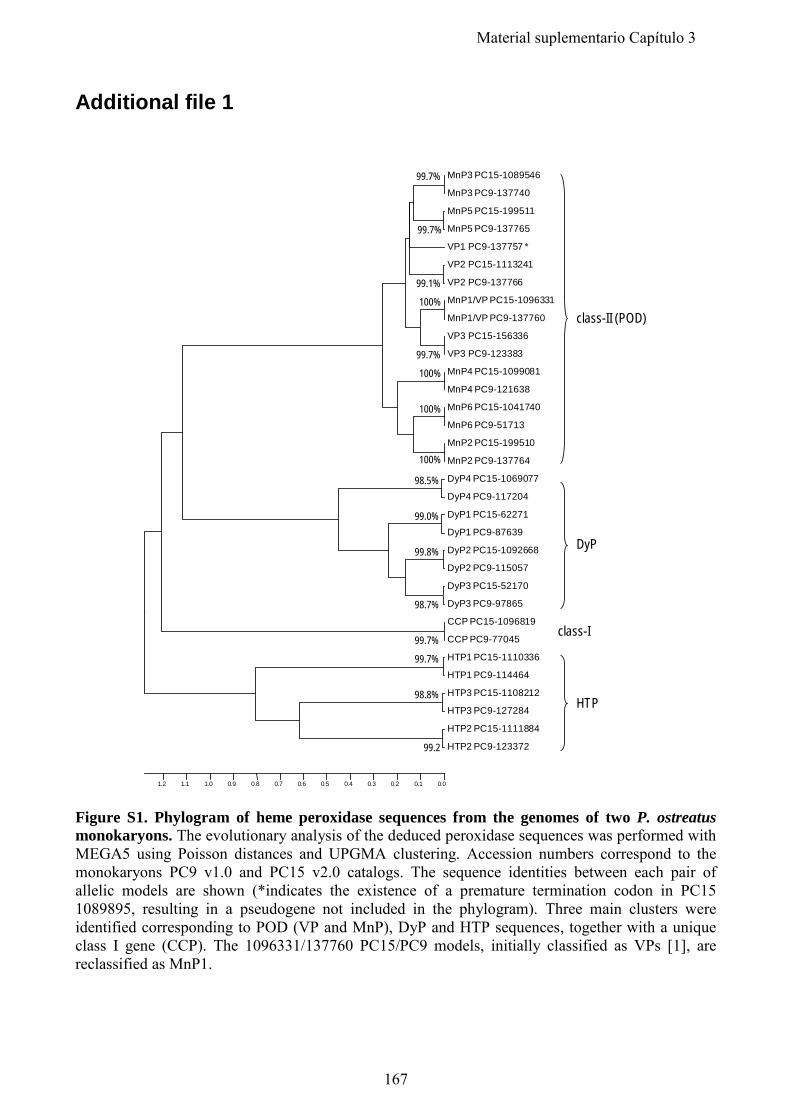
Additional file 1
Figure S1. Phylogram of heme peroxidase sequences from the genomes of two P. ostreatus monokaryons. The evolutionary analysis of the deduced peroxidase sequences was performed with MEGA5 using Poisson distances and UPGMA clustering. Accession numbers correspond to the monokaryons PC9 v1.0 and PC15 v2.0 catalogs. The sequence identities between each pair of allelic models are shown (*indicates the existence of a premature termination codon in PC15 1089895, resulting in a pseudogene not included in the phylogram). Three main clusters were identified corresponding to POD (VP and MnP), DyP and HTP sequences, together with a unique class I gene (CCP). The 1096331/137760 PC15/PC9 models, initially classified as VPs [1], are reclassified as MnP1.
MnP3 PC15-1089546
MnP3 PC9-137740
MnP5 PC15-199511
MnP5 PC9-137765
VP1 PC9-137757 *
VP2 PC15-1113241
VP2 PC9-137766
MnP1/VP PC15-1096331
MnP1/VP PC9-137760
VP3 PC15-156336
VP3 PC9-123383
MnP4 PC15-1099081
MnP4 PC9-121638
MnP6 PC15-1041740
MnP6 PC9-51713
MnP2 PC15-199510
MnP2 PC9-137764
DyP4 PC15-1069077
DyP4 PC9-117204
DyP1 PC15-62271
DyP1 PC9-87639
DyP2 PC15-1092668
DyP2 PC9-115057
DyP3 PC15-52170
DyP3 PC9-97865
CCP PC15-1096819
CCP PC9-77045
HTP1 PC15-1110336
HTP1 PC9-114464
HTP3 PC15-1108212
HTP3 PC9-127284
HTP2 PC15-1111884
HTP2 PC9-123372
98.5%
98.7%
98.8%
99.7%
99.0%
99.8%
99.2
99.7%
99.7%
100%
100%
100%
100%
99.7%
99.7%
99.1%
0.00.10.20.30.40.50.60.70.80.91.01.11.2
class-II (POD)
DyP
class-I
HTP
167
Material suplementario Capítulo 3
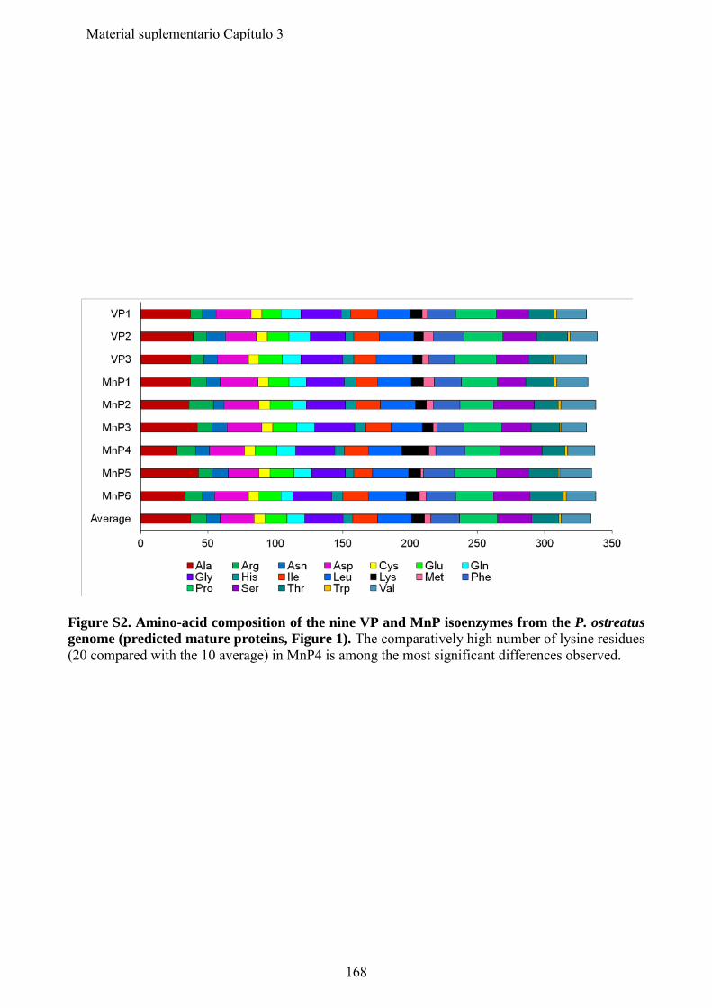
Figure S2. Amino-acid composition of the nine VP and MnP isoenzymes from the P. ostreatus genome (predicted mature proteins, Figure 1). The comparatively high number of lysine residues (20 compared with the 10 average) in MnP4 is among the most significant differences observed.
168
Material suplementario Capítulo 3
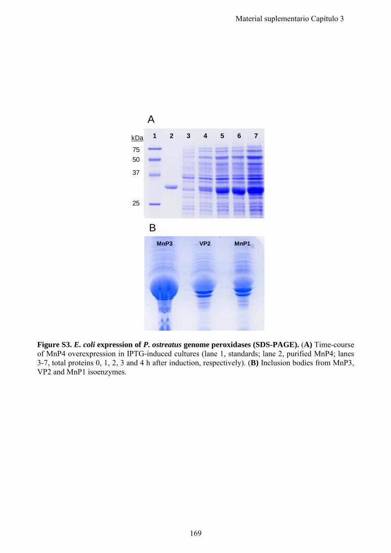
Figure S3. E. coli expression of P. ostreatus genome peroxidases (SDS-PAGE). (A) Time-course of MnP4 overexpression in IPTG-induced cultures (lane 1, standards; lane 2, purified MnP4; lanes 3-7, total proteins 0, 1, 2, 3 and 4 h after induction, respectively). (B) Inclusion bodies from MnP3, VP2 and MnP1 isoenzymes.
MnP3 VP2 MnP1
B
A1 2 3 4 5 6 7
7550
37
25
kDa
169
Material suplementario Capítulo 3
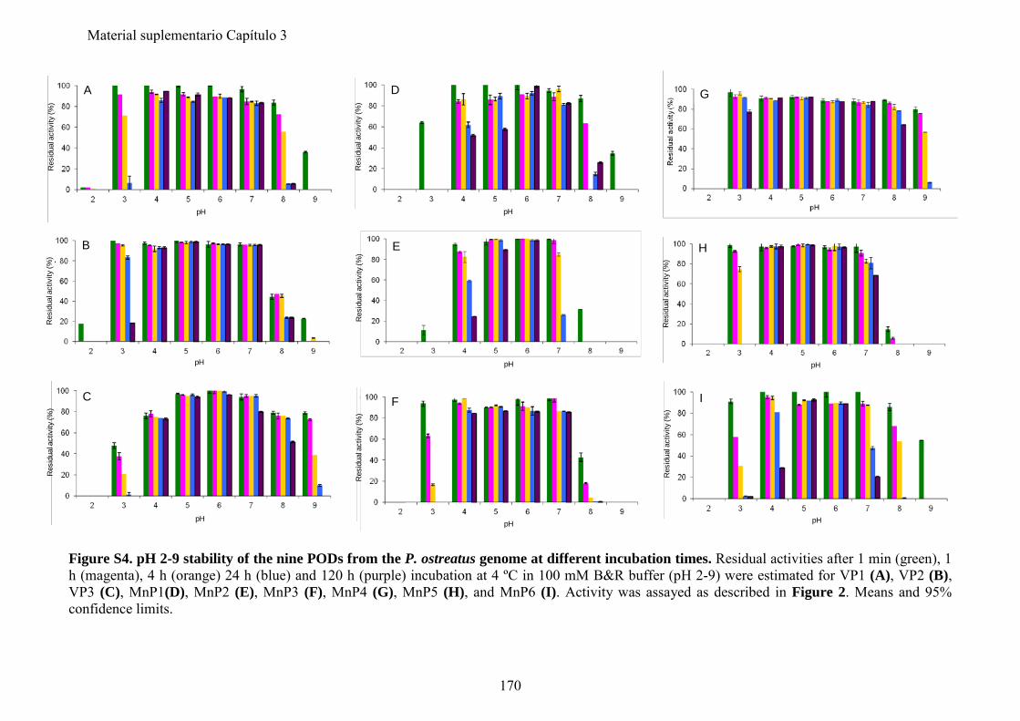
Figure S4. pH 2-9 stability of the nine PODs from the P. ostreatus genome at different incubation times. Residual activities after 1 min (green), 1 h (magenta), 4 h (orange) 24 h (blue) and 120 h (purple) incubation at 4 ºC in 100 mM B&R buffer (pH 2-9) were estimated for VP1 (A), VP2 (B), VP3 (C), MnP1(D), MnP2 (E), MnP3 (F), MnP4 (G), MnP5 (H), and MnP6 (I). Activity was assayed as described in Figure 2. Means and 95% confidence limits.
Res
idua
l act
ivity
(%)
pH
A
pH
C
Res
idua
l act
ivity
(%)
pH
B
Res
idua
l act
ivity
(%)
pH
F
Res
idua
l act
ivity
(%)
Res
idua
l act
ivity
(%)
pH
E
Res
idua
l act
ivity
(%)
pH
D
H
Res
idua
l act
ivity
(%)
pH
G
pH
I
Res
idua
l act
ivity
(%)
170
Material suplementario Capítulo 3
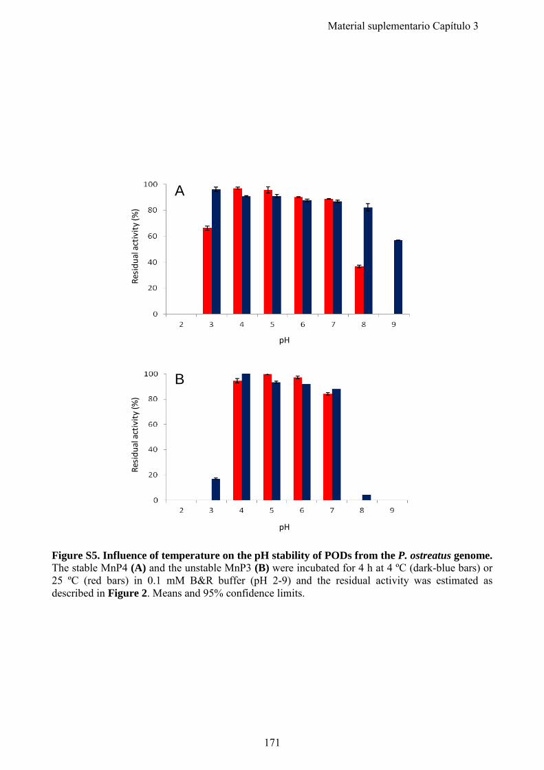
Figure S5. Influence of temperature on the pH stability of PODs from the P. ostreatus genome. The stable MnP4 (A) and the unstable MnP3 (B) were incubated for 4 h at 4 ºC (dark-blue bars) or 25 ºC (red bars) in 0.1 mM B&R buffer (pH 2-9) and the residual activity was estimated as described in Figure 2. Means and 95% confidence limits.
Resi
dual
act
ivity
(%)
pH
pH
A
Resi
dual
act
ivity
(%)
B
171
Material suplementario Capítulo 3
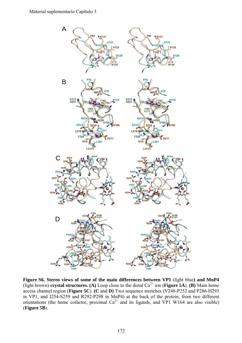
Figure S6. Stereo views of some of the main differences between VP1 (light blue) and MnP4 (light brown) crystal structures. (A) Loop close to the distal Ca2+ ion (Figure 5A). (B) Main heme access channel region (Figure 5C). (C and D) Two sequence stretches (V248-P252 and P286-H293 in VP1, and I254-S259 and R292-P298 in MnP4) at the back of the protein, from two different orientations (the heme cofactor, proximal Ca2+ and its ligands, and VP1 W164 are also visible) (Figure 5B).
A
B
C
D
172
Material suplementario Capítulo 3
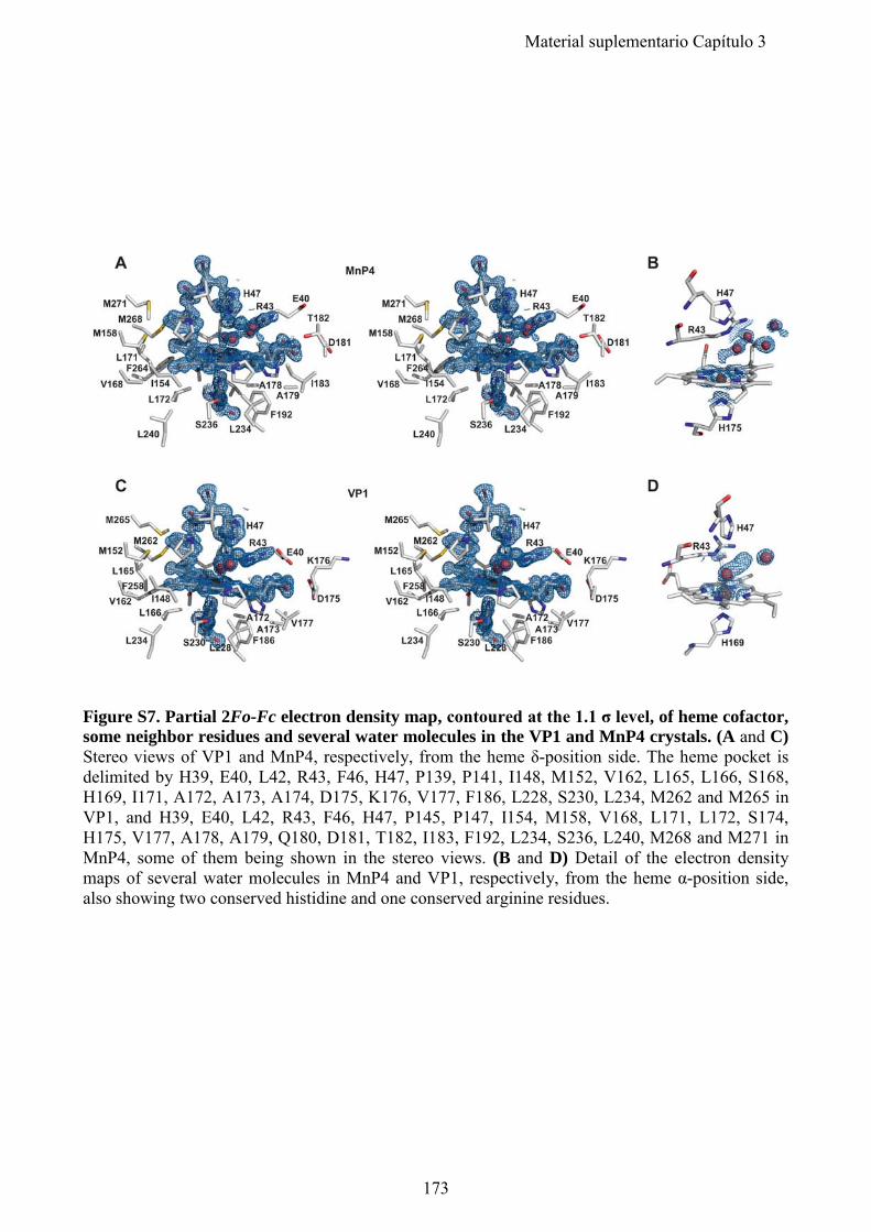
Figure S7. Partial 2Fo-Fc electron density map, contoured at the 1.1 σ level, of heme cofactor, some neighbor residues and several water molecules in the VP1 and MnP4 crystals. (A and C) Stereo views of VP1 and MnP4, respectively, from the heme δ-position side. The heme pocket is delimited by H39, E40, L42, R43, F46, H47, P139, P141, I148, M152, V162, L165, L166, S168, H169, I171, A172, A173, A174, D175, K176, V177, F186, L228, S230, L234, M262 and M265 in VP1, and H39, E40, L42, R43, F46, H47, P145, P147, I154, M158, V168, L171, L172, S174, H175, V177, A178, A179, Q180, D181, T182, I183, F192, L234, S236, L240, M268 and M271 in MnP4, some of them being shown in the stereo views. (B and D) Detail of the electron density maps of several water molecules in MnP4 and VP1, respectively, from the heme α-position side, also showing two conserved histidine and one conserved arginine residues.
173
Material suplementario Capítulo 3
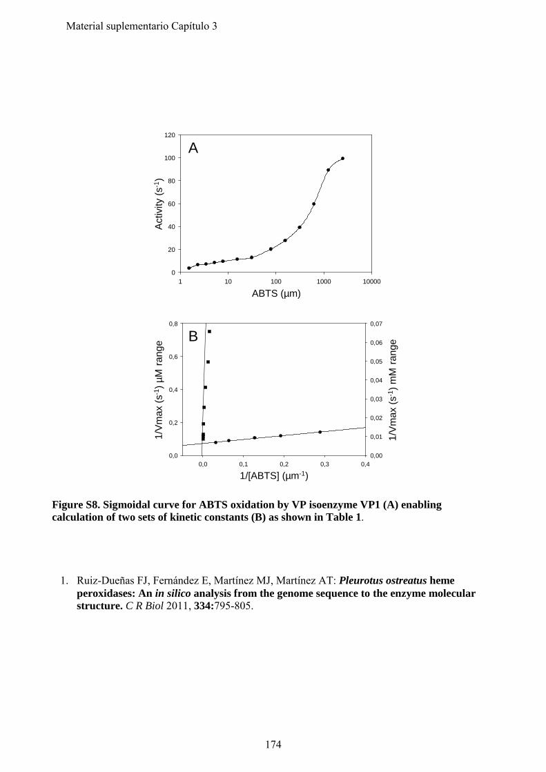
Figure S8. Sigmoidal curve for ABTS oxidation by VP isoenzyme VP1 (A) enabling calculation of two sets of kinetic constants (B) as shown in Table 1. 1. Ruiz-Dueñas FJ, Fernández E, Martínez MJ, Martínez AT: Pleurotus ostreatus heme
peroxidases: An in silico analysis from the genome sequence to the enzyme molecular structure. C R Biol 2011, 334:795-805.
1 10 100 1000 100000
20
40
60
80
100
120
0,0 0,1 0,2 0,3 0,40,0
0,2
0,4
0,6
0,8
0,00
0,01
0,02
0,03
0,04
0,05
0,06
0,07
ABTS (µm)
1/[ABTS] (µm-1)
Act
ivity
(s-1
)1/
Vm
ax(s
-1) µ
M r
ange
1/V
max
(s-1
) mM
rang
e
A
B
174
Material suplementario Capítulo 3
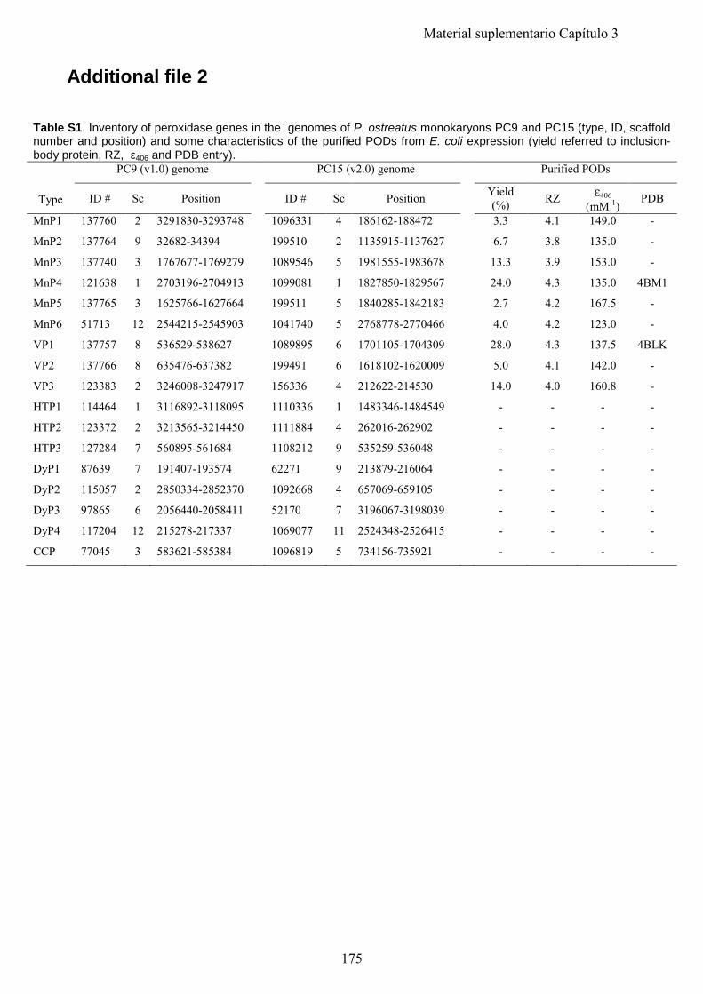
Additional file 2
Table S1. Inventory of peroxidase genes in the genomes of P. ostreatus monokaryons PC9 and PC15 (type, ID, scaffold number and position) and some characteristics of the purified PODs from E. coli expression (yield referred to inclusion-body protein, RZ, ε406 and PDB entry).
Type
PC9 (v1.0) genome PC15 (v2.0) genome Purified PODs
ID # Sc Position ID # Sc Position Yield (%) RZ ε406
(mM-1) PDB
MnP1 137760 2 3291830-3293748 1096331 4 186162-188472 3.3 4.1 149.0 -
MnP2 137764 9 32682-34394 199510 2 1135915-1137627 6.7 3.8 135.0 -
MnP3 137740 3 1767677-1769279 1089546 5 1981555-1983678 13.3 3.9 153.0 -
MnP4 121638 1 2703196-2704913 1099081 1 1827850-1829567 24.0 4.3 135.0 4BM1
MnP5 137765 3 1625766-1627664 199511 5 1840285-1842183 2.7 4.2 167.5 -
MnP6 51713 12 2544215-2545903 1041740 5 2768778-2770466 4.0 4.2 123.0 -
VP1 137757 8 536529-538627 1089895 6 1701105-1704309 28.0 4.3 137.5 4BLK
VP2 137766 8 635476-637382 199491 6 1618102-1620009 5.0 4.1 142.0 -
VP3 123383 2 3246008-3247917 156336 4 212622-214530 14.0 4.0 160.8 -
HTP1 114464 1 3116892-3118095 1110336 1 1483346-1484549 - - - -
HTP2 123372 2 3213565-3214450 1111884 4 262016-262902 - - - -
HTP3 127284 7 560895-561684 1108212 9 535259-536048 - - - -
DyP1 87639 7 191407-193574 62271 9 213879-216064 - - - -
DyP2 115057 2 2850334-2852370 1092668 4 657069-659105 - - - -
DyP3 97865 6 2056440-2058411 52170 7 3196067-3198039 - - - -
DyP4 117204 12 215278-217337 1069077 11 2524348-2526415 - - - -
CCP 77045 3 583621-585384 1096819 5 734156-735921 - - - -
175
Material suplementario Capítulo 3
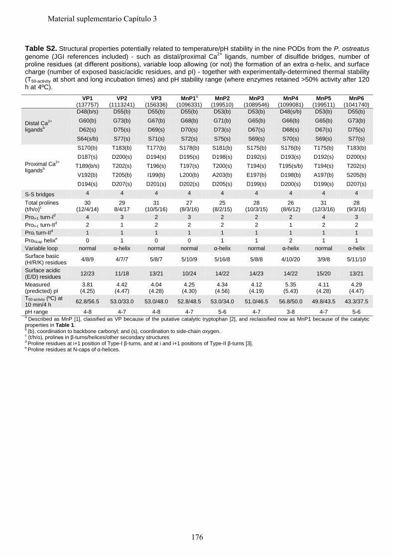
Table S2. Structural properties potentially related to temperature/pH stability in the nine PODs from the P. ostreatus genome (JGI references included) - such as distal/proximal Ca2+ ligands, number of disulfide bridges, number of proline residues (at different positions), variable loop allowing (or not) the formation of an extra α-helix, and surface charge (number of exposed basic/acidic residues, and pI) - together with experimentally-determined thermal stability (T50-activity at short and long incubation times) and pH stability range (where enzymes retained >50% activity after 120 h at 4ºC). VP1
(137757) VP2
(1113241) VP3
(156336) MnP1a
(1096331) MnP2
(199510) MnP3
(1089546) MnP4
(1099081) MnP5
(199511) MnP6
(1041740)
Distal Ca2+ ligandsb
D48(b/s) D55(b) D55(b) D55(b) D53(b) D53(b) D48(s/b) D53(b) D55(b) G60(b) G73(b) G67(b) G68(b) G71(b) G65(b) G66(b) G65(b) G73(b) D62(s) D75(s) D69(s) D70(s) D73(s) D67(s) D68(s) D67(s) D75(s)
S64(s/b) S77(s) S71(s) S72(s) S75(s) S69(s) S70(s) S69(s) S77(s)
Proximal Ca2+ ligandsb
S170(b) T183(b) T177(b) S178(b) S181(b) S175(b) S176(b) T175(b) T183(b) D187(s) D200(s) D194(s) D195(s) D198(s) D192(s) D193(s) D192(s) D200(s)
T189(b/s) T202(s) T196(s) T197(s) T200(s) T194(s) T195(s/b) T194(s) T202(s) V192(b) T205(b) I199(b) L200(b) A203(b) E197(b) D198(b) A197(b) S205(b) D194(s) D207(s) D201(s) D202(s) D205(s) D199(s) D200(s) D199(s) D207(s)
S-S bridges 4 4 4 4 4 4 4 4 4
Total prolines (t/h/o)c
30 (12/4/14)
29 8/4/17
31 (10/5/16)
27 (8/3/16)
25 (8/2/15)
28 (10/3/15)
26 (8/6/12)
31 (12/3/16)
28 (9/3/16)
Proi+1 turn-Id 4 3 2 3 2 2 2 4 3 Proi+1 turn-IId 2 1 2 2 2 2 1 2 2 Proi turn-IId 1 1 1 1 1 1 1 1 1 ProNcap helixe 0 1 0 0 1 1 2 1 1 Variable loop normal α-helix normal normal α-helix normal α-helix normal α-helix Surface basic (H/R/K) residues 4/8/9 4/7/7 5/8/7 5/10/9 5/16/8 5/8/8 4/10/20 3/9/8 5/11/10
Surface acidic (E/D) residues 12/23 11/18 13/21 10/24 14/22 14/23 14/22 15/20 13/21
Measured (predicted) pI
3.81 (4.25)
4.42 (4.47)
4.04 (4.28)
4.25 (4.30)
4.34 (4.56)
4.12 (4.19)
5.35 (5.43)
4.11 (4.28)
4.29 (4.47)
T50-activity (ºC) at 10 min/4 h 62.8/56.5 53.0/33.0 53.0/48.0 52.8/48.5 53.0/34.0 51.0/46.5 56.8/50.0 49.8/43.5 43.3/37.5
pH range 4-8 4-7 4-8 4-7 5-6 4-7 3-8 4-7 5-6 a Described as MnP [1], classified as VP because of the putative catalytic tryptophan [2], and reclassified now as MnP1 because of the catalytic properties in Table 1. b (b), coordination to backbone carbonyl; and (s), coordination to side-chain oxygen. c (t/h/o), prolines in β-turns/helices/other secondary structures d Proline residues at i+1 position of Type-I β-turns, and at i and i+1 positions of Type-II β-turns [3]. e Proline residues at N-caps of α-helices.
176
Material suplementario Capítulo 3
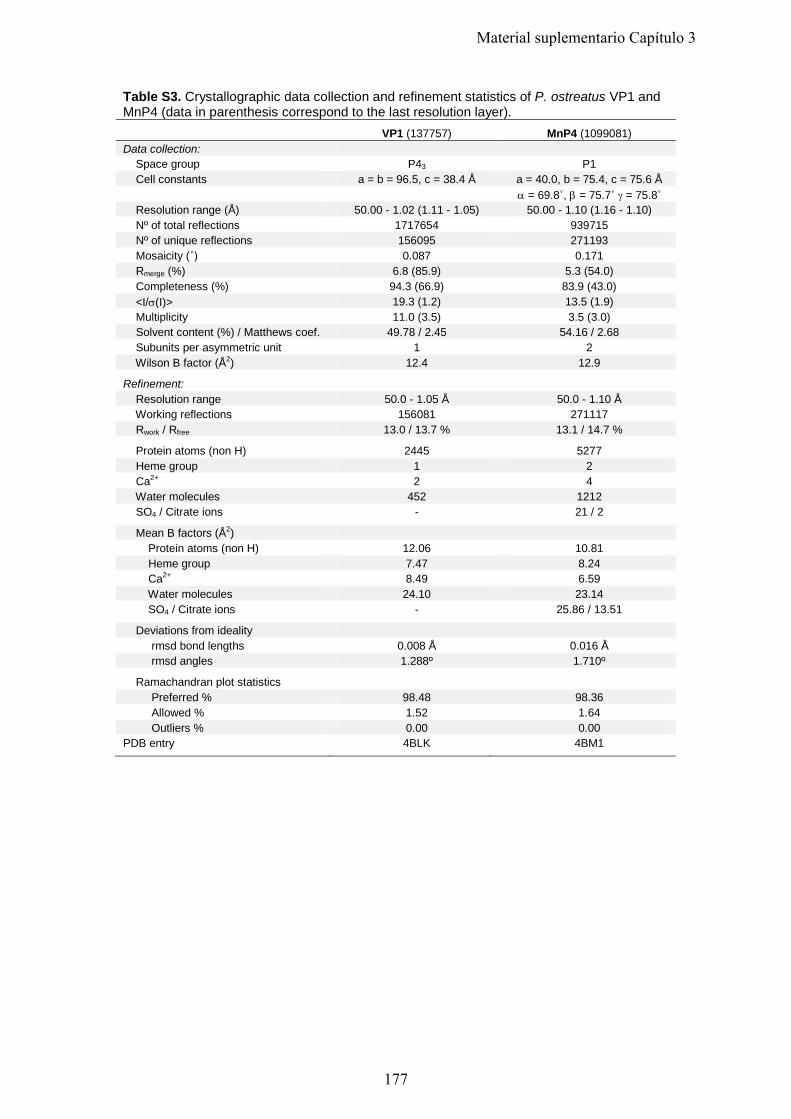
Table S3. Crystallographic data collection and refinement statistics of P. ostreatus VP1 and MnP4 (data in parenthesis correspond to the last resolution layer). VP1 (137757) MnP4 (1099081) Data collection: Space group P43 P1 Cell constants a = b = 96.5, c = 38.4 Å a = 40.0, b = 75.4, c = 75.6 Å α = 69.8˚, β = 75.7˚ γ = 75.8˚ Resolution range (Å) 50.00 - 1.02 (1.11 - 1.05) 50.00 - 1.10 (1.16 - 1.10) Nº of total reflections 1717654 939715 Nº of unique reflections 156095 271193 Mosaicity (˚) 0.087 0.171 Rmerge (%) 6.8 (85.9) 5.3 (54.0) Completeness (%) 94.3 (66.9) 83.9 (43.0) <I/σ(I)> 19.3 (1.2) 13.5 (1.9) Multiplicity 11.0 (3.5) 3.5 (3.0) Solvent content (%) / Matthews coef. 49.78 / 2.45 54.16 / 2.68 Subunits per asymmetric unit 1 2 Wilson B factor (Å2) 12.4 12.9 Refinement: Resolution range 50.0 - 1.05 Å 50.0 - 1.10 Å Working reflections 156081 271117 Rwork / Rfree 13.0 / 13.7 % 13.1 / 14.7 % Protein atoms (non H) 2445 5277 Heme group 1 2 Ca2+ 2 4 Water molecules 452 1212 SO4 / Citrate ions - 21 / 2 Mean B factors (Å2) Protein atoms (non H) 12.06 10.81 Heme group 7.47 8.24 Ca2+ 8.49 6.59 Water molecules 24.10 23.14 SO4 / Citrate ions - 25.86 / 13.51 Deviations from ideality rmsd bond lengths 0.008 Å 0.016 Å rmsd angles 1.288º 1.710º Ramachandran plot statistics Preferred % 98.48 98.36 Allowed % 1.52 1.64 Outliers % 0.00 0.00 PDB entry 4BLK 4BM1
177
Material suplementario Capítulo 3
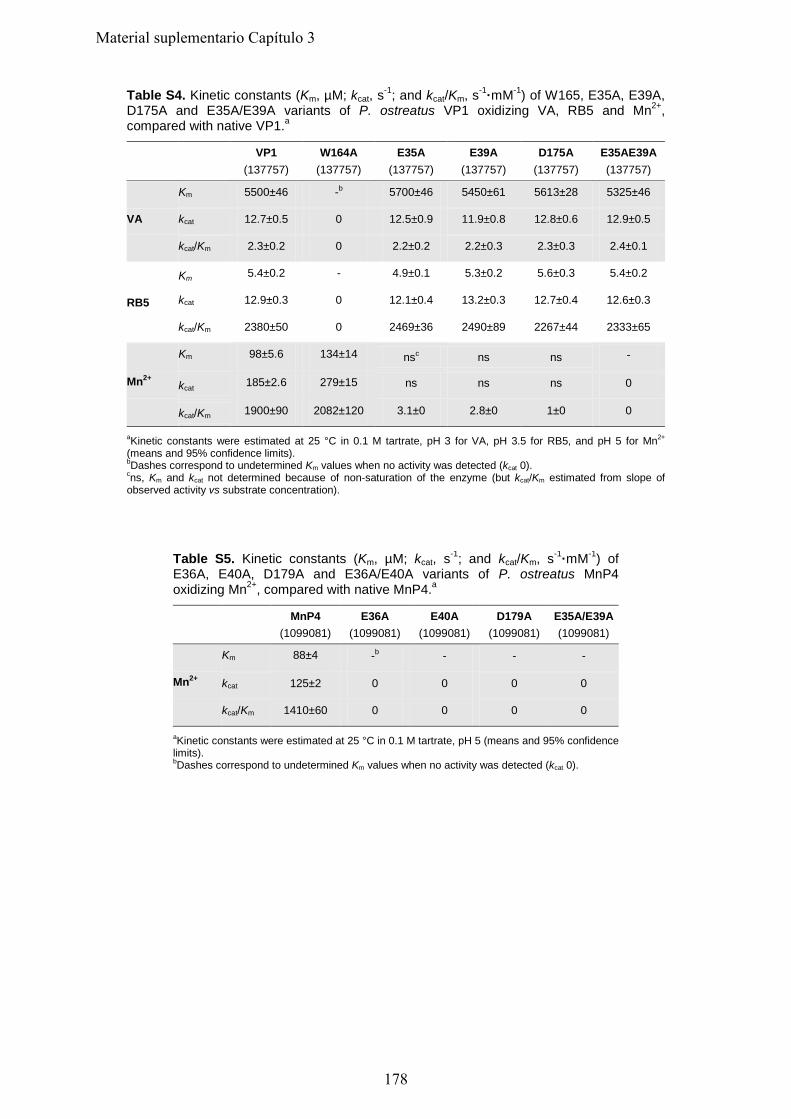
Table S4. Kinetic constants (Km, µM; kcat, s-1; and kcat/Km, s-1·mM-1) of W165, E35A, E39A, D175A and E35A/E39A variants of P. ostreatus VP1 oxidizing VA, RB5 and Mn2+, compared with native VP1.a
VP1
(137757) W164A
(137757) E35A
(137757) E39A
(137757) D175A
(137757) E35AE39A (137757)
VA
Km 5500±46 -b 5700±46 5450±61 5613±28 5325±46
kcat 12.7±0.5 0 12.5±0.9 11.9±0.8 12.8±0.6 12.9±0.5
kcat/Km 2.3±0.2 0 2.2±0.2 2.2±0.3 2.3±0.3 2.4±0.1
RB5
Km 5.4±0.2 - 4.9±0.1 5.3±0.2 5.6±0.3 5.4±0.2
kcat 12.9±0.3 0 12.1±0.4 13.2±0.3 12.7±0.4 12.6±0.3
kcat/Km 2380±50 0 2469±36 2490±89 2267±44 2333±65
Mn2+
Km 98±5.6 134±14 nsc ns ns -
kcat 185±2.6 279±15 ns ns ns 0
kcat/Km 1900±90 2082±120 3.1±0 2.8±0 1±0 0
aKinetic constants were estimated at 25 °C in 0.1 M tartrate, pH 3 for VA, pH 3.5 for RB5, and pH 5 for Mn2+ (means and 95% confidence limits). bDashes correspond to undetermined Km values when no activity was detected (kcat 0). cns, Km and kcat not determined because of non-saturation of the enzyme (but kcat/Km estimated from slope of observed activity vs substrate concentration).
Table S5. Kinetic constants (Km, µM; kcat, s-1; and kcat/Km, s-1·mM-1) of E36A, E40A, D179A and E36A/E40A variants of P. ostreatus MnP4 oxidizing Mn2+, compared with native MnP4.a
MnP4
(1099081) E36A
(1099081) E40A
(1099081) D179A
(1099081) E35A/E39A (1099081)
Mn2+
Km 88±4 -b - - -
kcat 125±2 0 0 0 0
kcat/Km 1410±60 0 0 0 0
aKinetic constants were estimated at 25 °C in 0.1 M tartrate, pH 5 (means and 95% confidence limits). bDashes correspond to undetermined Km values when no activity was detected (kcat 0).
178
Material suplementario Capítulo 3
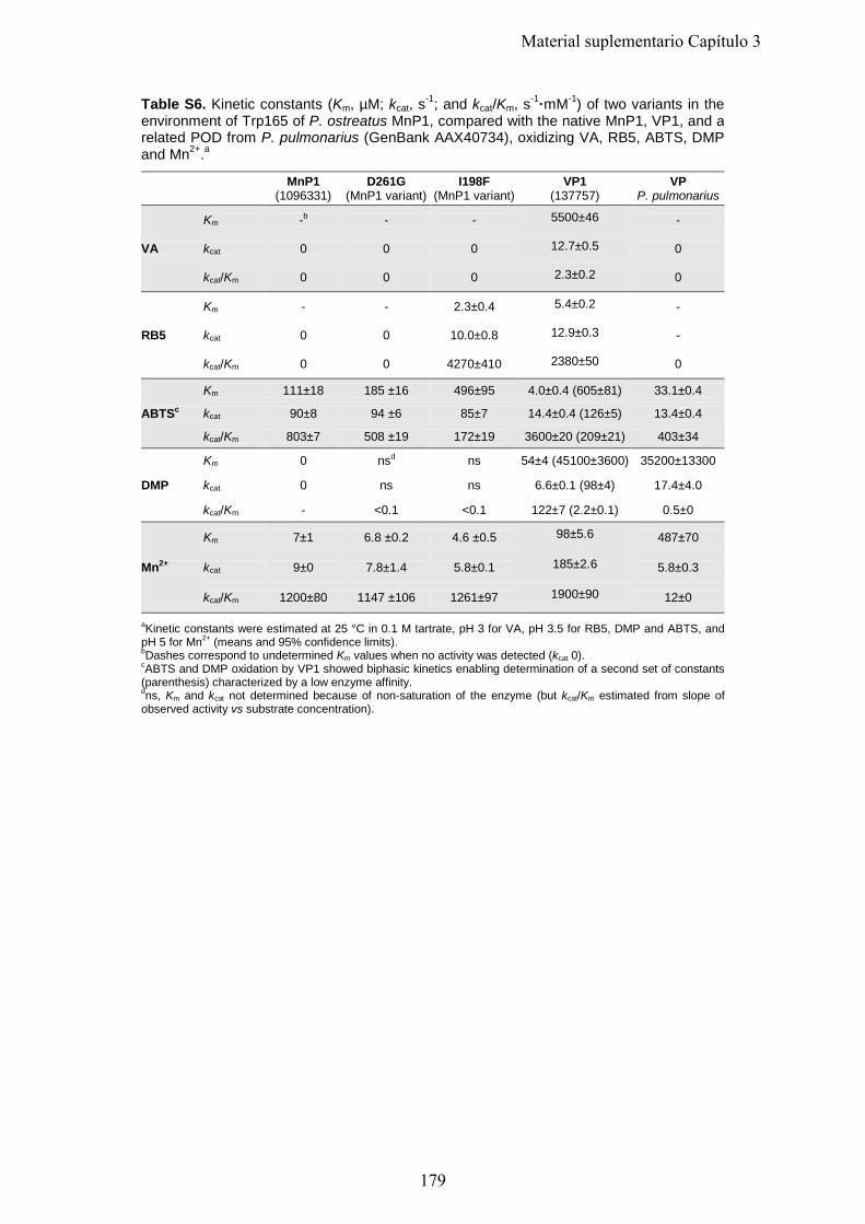
Table S6. Kinetic constants (Km, µM; kcat, s-1; and kcat/Km, s-1·mM-1) of two variants in the environment of Trp165 of P. ostreatus MnP1, compared with the native MnP1, VP1, and a related POD from P. pulmonarius (GenBank AAX40734), oxidizing VA, RB5, ABTS, DMP and Mn2+.a
MnP1 (1096331)
D261G (MnP1 variant)
I198F (MnP1 variant)
VP1 (137757)
VP P. pulmonarius
VA
Km -b - - 5500±46 -
kcat 0 0 0 12.7±0.5 0
kcat/Km 0 0 0 2.3±0.2 0
RB5
Km - - 2.3±0.4 5.4±0.2 -
kcat 0 0 10.0±0.8 12.9±0.3 -
kcat/Km 0 0 4270±410 2380±50 0
ABTSc
Km 111±18 185 ±16 496±95 4.0±0.4 (605±81) 33.1±0.4
kcat 90±8 94 ±6 85±7 14.4±0.4 (126±5) 13.4±0.4
kcat/Km 803±7 508 ±19 172±19 3600±20 (209±21) 403±34
DMP
Km 0 nsd ns 54±4 (45100±3600) 35200±13300
kcat 0 ns ns 6.6±0.1 (98±4) 17.4±4.0
kcat/Km - <0.1 <0.1 122±7 (2.2±0.1) 0.5±0
Mn2+
Km 7±1 6.8 ±0.2 4.6 ±0.5 98±5.6 487±70
kcat 9±0 7.8±1.4 5.8±0.1 185±2.6 5.8±0.3
kcat/Km 1200±80 1147 ±106 1261±97 1900±90 12±0
aKinetic constants were estimated at 25 °C in 0.1 M tartrate, pH 3 for VA, pH 3.5 for RB5, DMP and ABTS, and pH 5 for Mn2+ (means and 95% confidence limits). bDashes correspond to undetermined Km values when no activity was detected (kcat 0). cABTS and DMP oxidation by VP1 showed biphasic kinetics enabling determination of a second set of constants (parenthesis) characterized by a low enzyme affinity. dns, Km and kcat not determined because of non-saturation of the enzyme (but kcat/Km estimated from slope of observed activity vs substrate concentration).
179
Material suplementario Capítulo 3
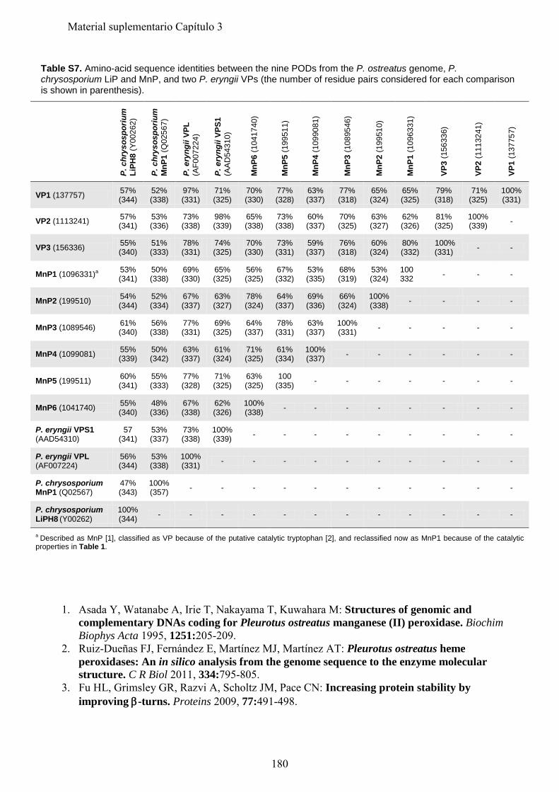
Table S7. Amino-acid sequence identities between the nine PODs from the P. ostreatus genome, P. chrysosporium LiP and MnP, and two P. eryngii VPs (the number of residue pairs considered for each comparison is shown in parenthesis).
P. c
hrys
ospo
rium
Li
PH8
(Y00
262)
P. c
hrys
ospo
rium
M
nP1
(Q02
567)
P. e
ryng
ii VP
L (A
F007
224)
P. e
ryng
ii VP
S1
(AAD
5431
0)
MnP
6 (1
0417
40)
MnP
5 (1
9951
1)
MnP
4 (1
0990
81)
MnP
3 (1
0895
46)
MnP
2 (1
9951
0)
MnP
1 (1
0963
31)
VP3
(156
336)
VP2
(111
3241
)
VP1
(137
757)
VP1 (137757) 57% (344)
52% (338)
97% (331)
71% (325)
70% (330)
77% (328)
63% (337)
77% (318)
65% (324)
65% (325)
79% (318)
71% (325)
100% (331)
VP2 (1113241) 57% (341)
53% (336)
73% (338)
98% (339)
65% (338)
73% (338)
60% (337)
70% (325)
63% (327)
62% (326)
81% (325)
100% (339) -
VP3 (156336) 55% (340)
51% (333)
78% (331)
74% (325)
70% (330)
73% (331)
59% (337)
76% (318)
60% (324)
80% (332)
100% (331) - -
MnP1 (1096331)a 53% (341)
50% (338)
69% (330)
65% (325)
56% (325)
67% (332)
53% (335)
68% (319)
53% (324)
100 332 - - -
MnP2 (199510) 54% (344)
52% (334)
67% (337)
63% (327)
78% (324)
64% (337)
69% (336)
66% (324)
100% (338) - - - -
MnP3 (1089546) 61% (340)
56% (338)
77% (331)
69% (325)
64% (337)
78% (331)
63% (337)
100% (331) - - - - -
MnP4 (1099081) 55% (339)
50% (342)
63% (337)
61% (324)
71% (325)
61% (334)
100% (337) - - - - - -
MnP5 (199511) 60% (341)
55% (333)
77% (328)
71% (325)
63% (325)
100 (335) - - - - - - -
MnP6 (1041740) 55% (340)
48% (336)
67% (338)
62% (326)
100% (338) - - - - - - - -
P. eryngii VPS1 (AAD54310)
57 (341)
53% (337)
73% (338)
100% (339) - - - - - - - - -
P. eryngii VPL (AF007224)
56% (344)
53% (338)
100% (331) - - - - - - - - - -
P. chrysosporium MnP1 (Q02567)
47% (343)
100% (357) - - - - - - - - - - -
P. chrysosporium LiPH8 (Y00262)
100% (344) - - - - - - - - - - - -
a Described as MnP [1], classified as VP because of the putative catalytic tryptophan [2], and reclassified now as MnP1 because of the catalytic properties in Table 1.
1. Asada Y, Watanabe A, Irie T, Nakayama T, Kuwahara M: Structures of genomic and
complementary DNAs coding for Pleurotus ostreatus manganese (II) peroxidase. Biochim Biophys Acta 1995, 1251:205-209.
2. Ruiz-Dueñas FJ, Fernández E, Martínez MJ, Martínez AT: Pleurotus ostreatus heme peroxidases: An in silico analysis from the genome sequence to the enzyme molecular structure. C R Biol 2011, 334:795-805.
3. Fu HL, Grimsley GR, Razvi A, Scholtz JM, Pace CN: Increasing protein stability by improving β-turns. Proteins 2009, 77:491-498.
180
Material suplementario Capítulo 3
Material Suplementario
Capítulo 4
"Ligninolytic peroxidase gene expression by Pleurotus ostreatus: Differential regulation in lignocellulose medium and effect of temperature and pH" Fernández-Fueyo, E., R. Castanera, F. J. Ruiz-Dueñas, M. F. López-Lucendo, L. Ramírez, A. G. Pisabarro, and A. T. Martínez. Fungal Genet. Biol. 2014.
181

182
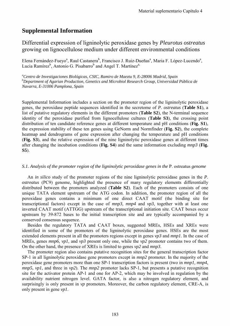
Supplemental Information Differential expression of ligninolytic peroxidase genes by Pleurotus ostreatus growing on lignocellulose medium under different environmental conditions Elena Fernández-Fueyoa, Raul Castanerab, Francisco J. Ruiz-Dueñasa, Maria F. López-Lucendoa, Lucia Ramírezb, Antonio G. Pisabarrob and Angel T. Martíneza, aCentro de Investigaciones Biológicas, CSIC, Ramiro de Maeztu 9, E-28006 Madrid, Spain bDepartment of Agarian Production, Genetics and Microbiol Research Group, Universidad Pública de Navarra, E-31006 Pamplona, Spain Supplemental Information includes a section on the promoter region of the ligninolytic peroxidase genes, the peroxidase peptide sequences identified in the secretome of P. ostreatus (Table S1), a list of putative regulatory elements in the different promoters (Table S2), the N-terminal sequence identity of the peroxidase purified from lignocellulose cultures (Table S3), the crossing point distribution of ten candidate reference genes at different temperature and pH conditions (Fig. S1), the expression stability of these ten genes using GeNorm and Normfinder (Fig. S2), the complete heatmap and dendrograms of gene expression after changing the temperature and pH conditions (Fig. S3), and the relative expression of the nine ligninolytic peroxidase genes at different times after changing the incubation conditions (Fig. S4) and the same information excluding mnp3 (Fig. S5). S.1. Analysis of the promoter region of the ligninolytic peroxidase genes in the P. ostreatus genome An in silico study of the promoter regions of the nine ligninolytic peroxidase genes in the P. ostreatus (PC9) genome, highlighted the presence of many regulatory elements differentially distributed between the promoters analyzed (Table S2). Each of the promoters consists of one unique TATA element upstream of the ATG codon. In addition, the promoter region of all the peroxidase genes contains a minimum of one direct CAAT motif (the binding site for transcriptional factors) except in the case of mnp3, mnp4 and vp3, together with at least one inverted CAAT motif (ATTGG) upstream of the transcriptional initiation site. CAAT boxes occur upstream by 39-872 bases to the initial transcription site and are typically accompanied by a conserved consensus sequence. Besides the regulatory TATA and CAAT boxes, suggested MREs, HSEs and XREs were identified in some of the promoters of the ligninolytic peroxidase genes. HSEs are the most extended elements present in all the promoters regions except in genes vp3 and mnp1. In the case of MREs, genes mnp6, vp1, and vp3 present only one, while the vp2 promoter contains two of them. On the other hand, the presence of XREs is limited to genes vp2 and mnp3. The promoter region also contains putative recognition sites for the general transcription factor SP-1 in all ligninolytic peroxidase gene promoters except in mnp2 promoter. In the majority of the peroxidase gene promoters more than one SP-1 transcription factors is present (two in mnp1, mnp4, mnp5, vp1, and three in vp2). The mnp2 promoter lacks SP-1, but presents a putative recognition site for the activator protein AP-1 and one for AP-2, which may be involved in regulation by the availability nutrient nitrogen level. GATA factor, is also a nitrogen regulatory element, and surprisingly is only present in vp promoters. Moreover, the carbon regulatory element, CRE-A, is only present in gene vp1.
183
Material suplementario Capítulo 4
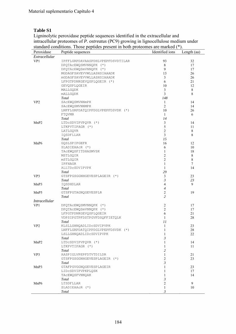
Table S1 Ligninolytic peroxidase peptide sequences identified in the extracellular and intracellular proteomes of P. ostreatus (PC9) growing in lignocellulose medium under standard conditions. Those peptides present in both proteomes are marked (*). Peroxidase Peptide sequences Identified ions Length (aa) Extracellular VP1 IPFFLGRPDAVAASPDHLVPEPFDSVDTILAR 93 32 DPQTAcEWQSMVNNQPK (*) 8 17 DPQTAcEWQSmVNNQPK (*) 9 17 MGDAGFSAVEVVWLLASHSIAAADK 13 26 mGDAGFSAVEVVWLLASHSIAAADK 3 26 LFPGTPDNKGEVQSPLQGEIR (*) 6 21 GEVQSPLQGEIR 10 12 MALLGQDK 3 8 mALLGQDK 3 8 Total 148 VP2 SAcEWQSMVNNmPK 1 14 SAcEWQSMVNNMPK 2 14 LNFFLGRPDATQIPPDGLVPEPFDSVDK (*) 10 26 FTQVMR 1 6 Total 14 MnP2 LTDcSDVIPVPQVR (*) 3 14 LTKPVTIPAGR (*) 5 11 LATLGQVR 2 8 IQSDFLLAR 3 8 Total 15 MnP6 GQSLSPIPGEFR 16 12 SLADIEAAcR (*) 6 10 TAcEWQSFITDHASMVSK 1 18 MSTLGQIR 2 8 mSTLGQIR 2 8 IPFHAGR 1 7 ALLTDcSDVIPVPK 1 14 Total 29 VP3 GTSFPGSGGNHGEVESPLAGEIR (*) 3 23 Total 3 23 MnP3 IQSDHDLAR 4 9 Total 4 MnP5 GTSFPGTAGNQGEVESPLR 2 19 Total 2 Intracellular VP1 DPQTAcEWQSMVNNQPK (*) 2 17 DPQTAcEWQSmVNNQPK (*) 2 17 LFPGTPDNKGEVQSPLQGEIR 6 21 VDPSIPGTPFDSTPGVFDSQFFIETQLK 1 28 Total 11 VP2 KLSLLGHNQADLIDcSDVIPVPK 1 23 LNFFLGRPDATQIPPDGLVPEPFDSVDK (*) 1 28 LSLLGHNQADLIDcSDVIPVPK 1 22 Total 3 MnP2 LTDcSDVIPVPQVR (*) 1 14 LTKPVTIPAGR (*) 1 11 Total 2 VP3 AASPIGLVPEPFDTVTDILDR 1 21 GTSFPGSGGNHGEVESPLAGEIR (*) 2 23 Total 3 MnP3 GTAFPGVGGNQGEVESPLAGEIR 1 23 LIDcSDVIPVPKPLQSK 1 17 TAcEWQSFVNNQAK 1 14 Total 3 MnP6 LTSDFLLAR 2 9 SLADIEAAcR (*) 1 10 Total 3
184
Material suplementario Capítulo 4
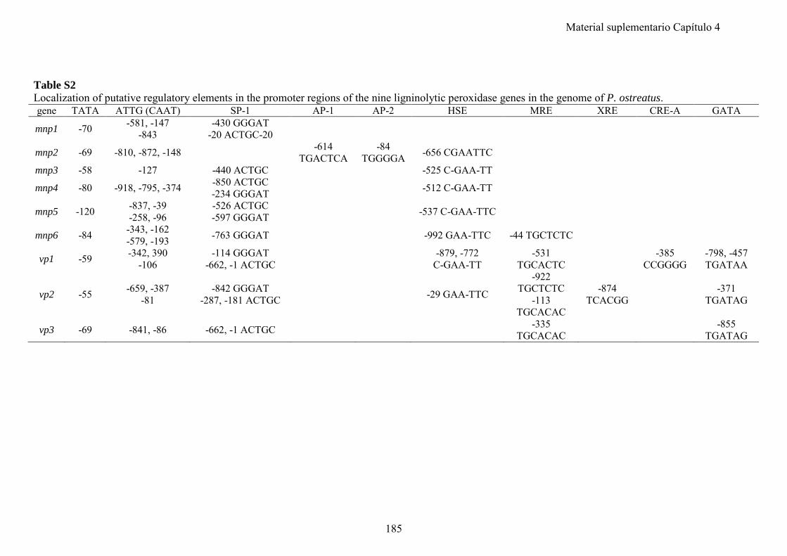
Table S2 Localization of putative regulatory elements in the promoter regions of the nine ligninolytic peroxidase genes in the genome of P. ostreatus. gene TATA ATTG (CAAT) SP-1 AP-1 AP-2 HSE MRE XRE CRE-A GATA
mnp1 -70 -581, -147 -843
-430 GGGAT -20 ACTGC-20
mnp2 -69 -810, -872, -148 -614 TGACTCA
-84 TGGGGA -656 CGAATTC
mnp3 -58 -127 -440 ACTGC -525 C-GAA-TT
mnp4 -80 -918, -795, -374 -850 ACTGC -234 GGGAT -512 C-GAA-TT
mnp5 -120 -837, -39 -258, -96
-526 ACTGC -597 GGGAT -537 C-GAA-TTC
mnp6 -84 -343, -162 -579, -193 -763 GGGAT -992 GAA-TTC -44 TGCTCTC
vp1 -59 -342, 390 -106
-114 GGGAT -662, -1 ACTGC -879, -772
C-GAA-TT -531
TGCACTC -385 CCGGGG
-798, -457 TGATAA
vp2 -55 -659, -387 -81
-842 GGGAT -287, -181 ACTGC -29 GAA-TTC
-922 TGCTCTC
-113 TGCACAC
-874 TCACGG -371
TGATAG
vp3 -69 -841, -86 -662, -1 ACTGC -335 TGCACAC -855
TGATAG
185
Material suplementario Capítulo 4
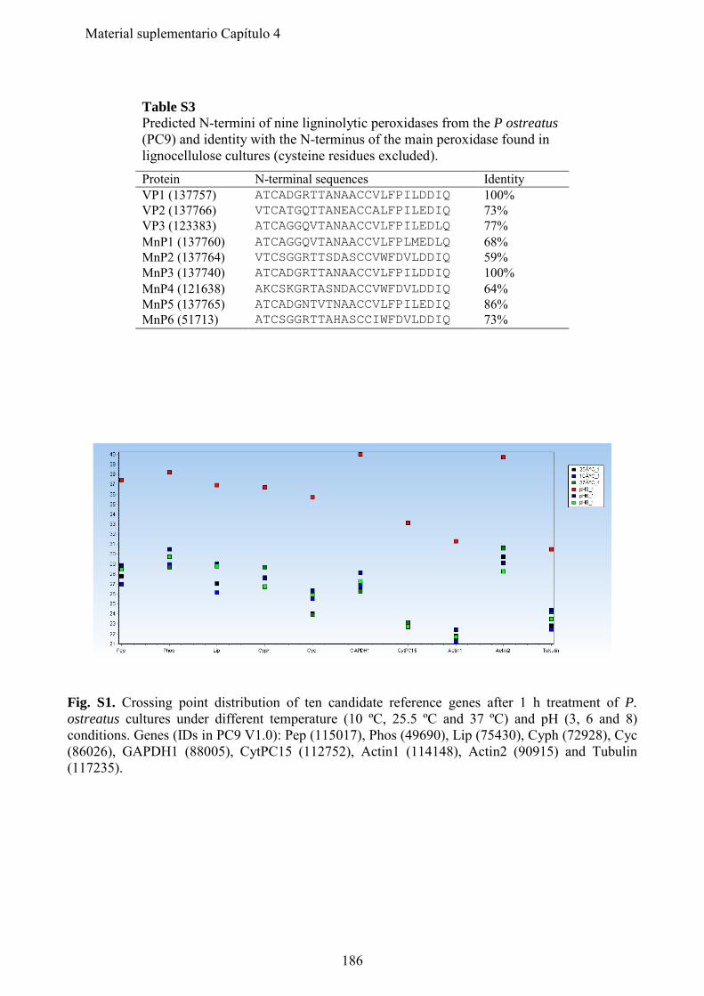
Table S3 Predicted N-termini of nine ligninolytic peroxidases from the P ostreatus (PC9) and identity with the N-terminus of the main peroxidase found in lignocellulose cultures (cysteine residues excluded). Protein N-terminal sequences Identity VP1 (137757) ATCADGRTTANAACCVLFPILDDIQ 100% VP2 (137766) VTCATGQTTANEACCALFPILEDIQ 73% VP3 (123383) ATCAGGQVTANAACCVLFPILEDLQ 77% MnP1 (137760) ATCAGGQVTANAACCVLFPLMEDLQ 68% MnP2 (137764) VTCSGGRTTSDASCCVWFDVLDDIQ 59% MnP3 (137740) ATCADGRTTANAACCVLFPILDDIQ 100% MnP4 (121638) AKCSKGRTASNDACCVWFDVLDDIQ 64% MnP5 (137765) ATCADGNTVTNAACCVLFPILEDIQ 86% MnP6 (51713) ATCSGGRTTAHASCCIWFDVLDDIQ 73%
Fig. S1. Crossing point distribution of ten candidate reference genes after 1 h treatment of P. ostreatus cultures under different temperature (10 ºC, 25.5 ºC and 37 ºC) and pH (3, 6 and 8) conditions. Genes (IDs in PC9 V1.0): Pep (115017), Phos (49690), Lip (75430), Cyph (72928), Cyc (86026), GAPDH1 (88005), CytPC15 (112752), Actin1 (114148), Actin2 (90915) and Tubulin (117235).
186
Material suplementario Capítulo 4
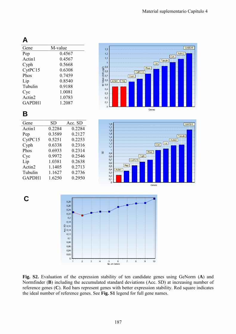
A
B
C Fig. S2. Evaluation of the expression stability of ten candidate genes using GeNorm (A) and Normfinder (B) including the accumulated standard deviations (Acc. SD) at increasing number of reference genes (C). Red bars represent genes with better expression stability. Red square indicates the ideal number of reference genes. See Fig. S1 legend for full gene names.
Gene M-value Pep 0.4567 Actin1 0.4567 Cyph 0.5668 CytPC15 0.6308 Phos 0.7459 Lip 0.8540 Tubulin 0.9188 Cyc 1.0081 Actin2 1.0783 GAPDH1 1.2087
Gene SD Acc. SD Actin1 0.2284 0.2284 Pep 0.3589 0.2127 CytPC15 0.5251 0.2253 Cyph 0.6338 0.2316 Phos 0.6933 0.2314 Cyc 0.9972 0.2546 Lip 1.0381 0.2638 Actin2 1.1405 0.2713 Tubulin 1.1627 0.2736 GAPDH1 1.6250 0.2950
187
Material suplementario Capítulo 4
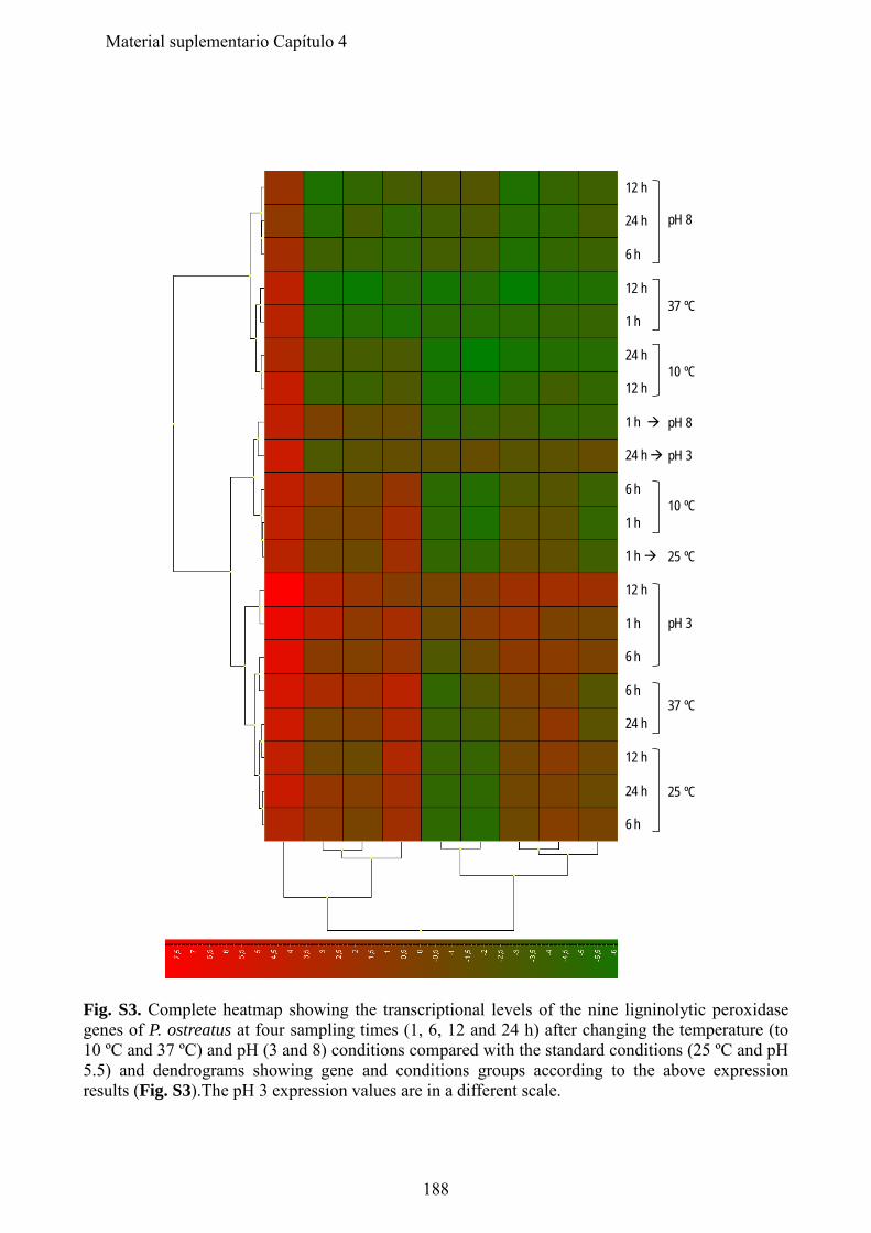
12 h
24 h
6 h
12 h
1 h
24 h
12 h
1 h
24 h
6 h
1 h
1 h
12 h
1 h
6 h
6 h
24 h
12 h
24 h
6 h
pH 8
37 ºC
10 ºC
pH 8
pH 3
10 ºC
25 ºC
pH 3
37 ºC
25 ºC
Fig. S3. Complete heatmap showing the transcriptional levels of the nine ligninolytic peroxidase genes of P. ostreatus at four sampling times (1, 6, 12 and 24 h) after changing the temperature (to 10 ºC and 37 ºC) and pH (3 and 8) conditions compared with the standard conditions (25 ºC and pH 5.5) and dendrograms showing gene and conditions groups according to the above expression results (Fig. S3).The pH 3 expression values are in a different scale.
188
Material suplementario Capítulo 4
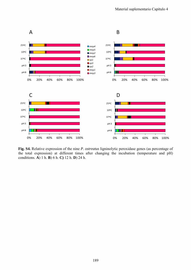
0% 20% 40% 60% 80% 100%
pH 8
pH 3
37ºC
10ºC
25ºC mnp4mnp5mnp2mnp6vp1vp3vp2mnp1mnp3
0% 20% 40% 60% 80% 100%
pH 8
pH 3
37ºC
10ºC
25ºC
0% 20% 40% 60% 80% 100%
pH 8
pH 3
37ºC
10ºC
25ºC
0% 20% 40% 60% 80% 100%
pH 8
pH 3
37ºC
10ºC
25ºC
A B
C D
Fig. S4. Relative expression of the nine P. ostreatus ligninolytic peroxidase genes (as percentage of the total expression) at different times after changing the incubation (temperature and pH) conditions. A) 1 h. B) 6 h. C) 12 h. D) 24 h.
189
Material suplementario Capítulo 4
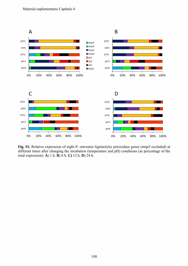
0% 20% 40% 60% 80% 100%
pH 8
pH 3
37ºC
10ºC
25ºC mnp4mnp5mnp2mnp6vp1vp3vp2mnp1
0% 20% 40% 60% 80% 100%
pH 8
pH 3
37ºC
10ºC
25ºC
0% 20% 40% 60% 80% 100%
pH 8
pH 3
37ºC
10ºC
25ºC
0% 20% 40% 60% 80% 100%
pH 8
pH 3
37ºC
10ºC
25ºC
A B
C D
Fig. S5. Relative expression of eight P. ostreatus ligninolytic peroxidase genes (mnp3 excluded) at different times after changing the incubation (temperature and pH) conditions (as percentage of the total expression). A) 1 h. B) 6 h. C) 12 h. D) 24 h.
190
Material suplementario Capítulo 4
Material Suplementario
Capítulo 5 "Comparative genomics of Ceriporiopisis subvermispora and Phanerochaete chrysosporium provide insight into selective ligninolysis" Fernández-Fueyo, E., F. J. Ruiz-Dueñas, P. Ferreira, D. Floudas, D. S. Hibbett, P. Canessa, L. Larrondo, T. Y. James, D. Seelenfreund, S. Lobos, R. Polanco, M. Tello, Y. Honda, T. Watanabe, T. Watanabe, J. S. Ryu, C. P. Kubicek, M. Schmoll, J. Gaskell, K. E. Hammel, F. J. St.John, A. Vanden Wymelenberg, G. Sabat, S. S. Bondurant, K. Syed, J. Yadav, H. Doddapaneni, V. Subramanian, J. L. Lavín, J. A. Oguiza, G. Perez, A. G. Pisabarro, L. Ramírez, F. Santoyo, E. Master, P. M. Coutinho, B. Henrissat, V. Lombard, J. K. Magnuson, U. Kües, C. Hori, K. Igarashi, M. Samejima, B. W. Held, K. Barry, K. LaButti, A. Lapidus, E. Lindquist, S. Lucas, R. Riley, A. Salamov, D. Hoffmeister, D. Schwenk, Y. Hadar, O. Yarden, R. P. de Vries, A. Wiebenga, J. Stenlid, D. C. Eastwood, I. V. Grigoriev, R. Berka, R. A. Blanchette, P. Kersten, A. T. Martínez, R. Vicuña, and D. Cullen. Proc. Natl. Acad. Sci. USA 109:5458-5463. 2012.
191

192
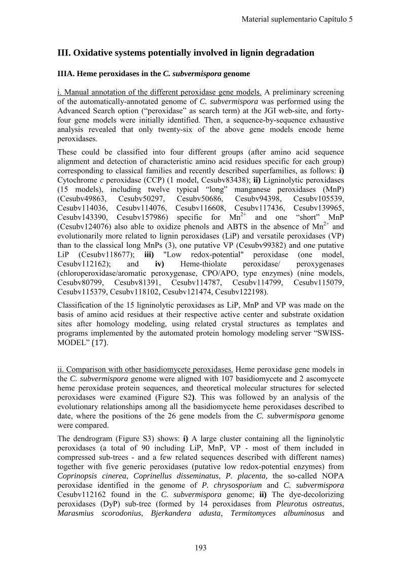
III. Oxidative systems potentially involved in lignin degradation IIIA. Heme peroxidases in the C. subvermispora genome i. Manual annotation of the different peroxidase gene models. A preliminary screening of the automatically-annotated genome of C. subvermispora was performed using the Advanced Search option (“peroxidase” as search term) at the JGI web-site, and forty-four gene models were initially identified. Then, a sequence-by-sequence exhaustive analysis revealed that only twenty-six of the above gene models encode heme peroxidases.
These could be classified into four different groups (after amino acid sequence alignment and detection of characteristic amino acid residues specific for each group) corresponding to classical families and recently described superfamilies, as follows: i) Cytochrome c peroxidase (CCP) (1 model, Cesubv83438); ii) Ligninolytic peroxidases (15 models), including twelve typical “long” manganese peroxidases (MnP) (Cesubv49863, Cesubv50297, Cesubv50686, Cesubv94398, Cesubv105539, Cesubv114036, Cesubv114076, Cesubv116608, Cesubv117436, Cesubv139965, Cesubv143390, Cesubv157986) specific for Mn2+ and one “short” MnP (Cesubv124076) also able to oxidize phenols and ABTS in the absence of Mn2+ and evolutionarily more related to lignin peroxidases (LiP) and versatile peroxidases (VP) than to the classical long MnPs (3), one putative VP (Cesubv99382) and one putative LiP (Cesubv118677); iii) "Low redox-potential" peroxidase (one model, Cesubv112162); and iv) Heme-thiolate peroxidase/ peroxygenases (chloroperoxidase/aromatic peroxygenase, CPO/APO, type enzymes) (nine models, Cesubv80799, Cesubv81391, Cesubv114787, Cesubv114799, Cesubv115079, Cesubv115379, Cesubv118102, Cesubv121474, Cesubv122198). Classification of the 15 ligninolytic peroxidases as LiP, MnP and VP was made on the basis of amino acid residues at their respective active center and substrate oxidation sites after homology modeling, using related crystal structures as templates and programs implemented by the automated protein homology modeling server “SWISS-MODEL” (17).
ii. Comparison with other basidiomycete peroxidases. Heme peroxidase gene models in the C. subvermispora genome were aligned with 107 basidiomycete and 2 ascomycete heme peroxidase protein sequences, and theoretical molecular structures for selected peroxidases were examined (Figure S2). This was followed by an analysis of the evolutionary relationships among all the basidiomycete heme peroxidases described to date, where the positions of the 26 gene models from the C. subvermispora genome were compared.
The dendrogram (Figure S3) shows: i) A large cluster containing all the ligninolytic peroxidases (a total of 90 including LiP, MnP, VP - most of them included in compressed sub-trees - and a few related sequences described with different names) together with five generic peroxidases (putative low redox-potential enzymes) from Coprinopsis cinerea, Coprinellus disseminatus, P. placenta, the so-called NOPA peroxidase identified in the genome of P. chrysosporium and C. subvermispora Cesubv112162 found in the C. subvermispora genome; ii) The dye-decolorizing peroxidases (DyP) sub-tree (formed by 14 peroxidases from Pleurotus ostreatus, Marasmius scorodonius, Bjerkandera adusta, Termitomyces albuminosus and
193
Material suplementario Capítulo 5
Polyporus sp.; iii) The group of APO/CPO-type heme-thiolate peroxidases including the Agrocybe aegerita APO and related enzymes from P. chrysosporium and Agaricus bisporus, as well as peroxidases recently reported for the first time in the genome of Pleurotus ostreatus (Ruiz-Dueñas et al., submitted) and now in the genome of C. subvermispora where nine gene models of this peroxidase superfamily were localized (unexpectedly, the related Leptoxyphium fumago CPO did not fit in this cluster); and iv) a group of evolutionarily distant peroxidases formed by CCPs, including one model from C. subvermispora not related with the previous ones.
A closer view of the ligninolytic peroxidase cluster is also shown in Figure S3. The structural-functional classification based on the presence of a Mn2+-oxidation site and/or a catalytic tryptophan, previously used to classify the C. subvermispora ligninolytic peroxidase genes, is extended here to all the 90 basidiomycete peroxidases described to date (the presence of a catalytic tyrosine in a Trametes cervina LiP is also indicated).
In this expanded dendrogram, typical “long” MnPs (from Lentinus edodes, Ceriporiopsis rivulosa, C. subvermispora, Dichomitus squalens, P. chrysosporium, Phanerochaete sordida, Phlebia radiata and an unidentified basidiomycete) form a large and homogeneous cluster clearly separated from the rest of ligninolytic peroxidases, all of them exhibiting a shorter C-terminal tail. By contrast, VP, LiP and “short” MnPs seem to have divergent and recurrent evolution pathways. Peroxidases Cesubv118677 and Cesubv99382 seem to represent transition states of these evolutionary processes, not only by their position in the dendrogram but also by their catalytic properties, as discussed below.
iii. Heterologous expression and purification of C. subvermispora peroxidases Following careful inspection and revision of automated annotations, three C. subvermispora genes were selected for Escherichia coli expression; putative VP (Cesubv99382), LiP (Cesubv118677) and MnP (Cesubv117436). The latter exhibited the highest induction levels in medium containing ball-milled aspen versus glucose as sole carbon source (main text Figure 1). The precise positions of introns were determined by comparison with other known ligninolytic peroxidase genes, and the corresponding cDNA sequences were synthesized (ATG-Biosynthetics). The synthesized cDNAs coding for the mature proteins were cloned into the pFLAG1 expression vector under the control of the IPTG-inducible tac promoter. The enzymes were overexpressed in E. coli W3110, and SDS-PAGE analysis of the cell extracts showed accumulation of the recombinant peroxidases in the insoluble fraction containing the inclusion bodies. The proteins were subsequently activated in vitro under conditions previously optimized for P. eryngii VP (18). Direct veratryl alcohol (VA) oxidation was measured to check the activity of peroxidases in the refolding mixtures confirming enzyme activation. Then, the enzymes were purified to homogeneity using an anion exchange column (Resource Q from GE Healthcare). Finally, their molar extinction coefficients at the absorption maximum of the Soret band (407 nm) were determined.
LiP from P. chrysosporium (isoenzyme LiPH8) and VP from P. eryngii (isoenzyme VPL) were also expressed in E. coli and purified for comparison with the C. subvermispora peroxidases, after E. coli expression and in vitro activation following the protocols of Doyle and Smith (19) and Pérez-Boada et al. (18) respectively.
194
Material suplementario Capítulo 5
iv. Enzyme characterization. Oxidation of three representative substrates, namely Mn2+, and high redox-potential veratryl alcohol (VA) and Reactive Black 5 (RB5), by both recombinant peroxidases were investigated under steady-state conditions. The corresponding steady-state kinetic constants were compared with those of P. eryngii VP and P. chrysosporium LiP produced by this method (main text Table 1).
As described in the main text, Cesubv118677 could not oxidize Mn2+ as expected according to the absence of a typical manganese oxidation site in its theoretical molecular structure (Figure S2A). Surprisingly, Cesubv99382, tentatively identified as a VP, was also not able to oxidize Mn2+, even though a typical manganese oxidation site is predicted in its structural model (Figure S2C).
Typical VPs and MnPs oxidizing Mn2+ do not include an acidic residue contiguous to the aspartate forming the manganese binding site, whereas Cesubv99382 presents a glutamic acid at the corresponding position. Typical LiPs, unable to oxidize Mn2+, exhibit an acidic residue at an equivalent position. Effective transformation of LiP into MnP have been obtained by removing this acidic residue, in addition to introducing the corresponding aspartic acid at the manganese binding site (20).
195
Material suplementario Capítulo 5
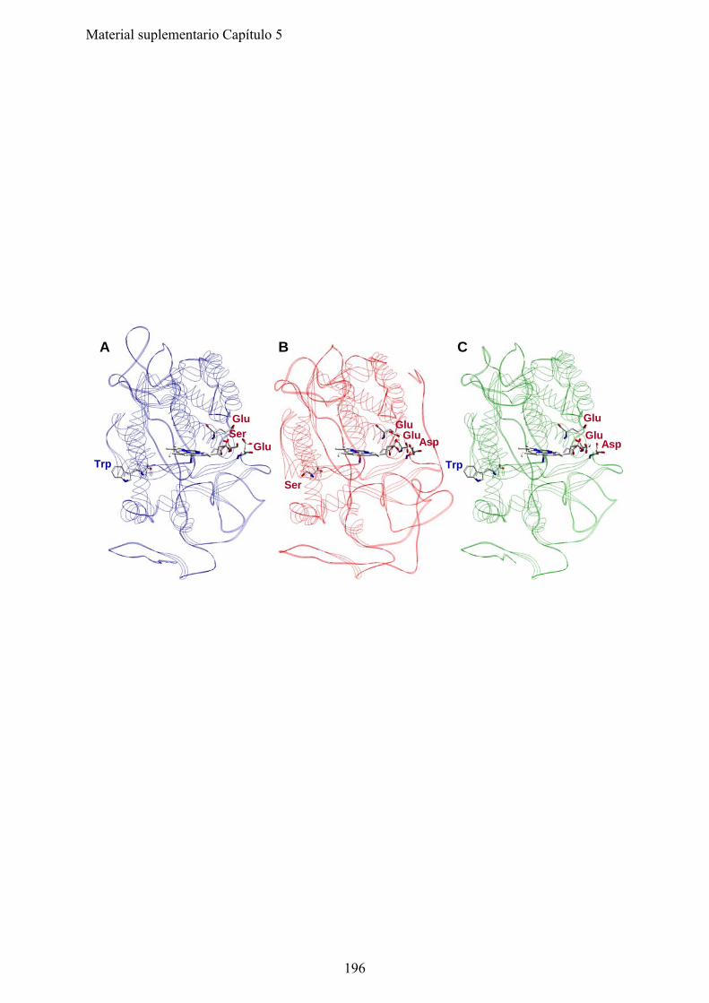
GluGlu
A
BA
Ser
Glu
C
Glu
Glu
Asp
Ser
Trp
Glu
Trp
Asp
196
Material suplementario Capítulo 5
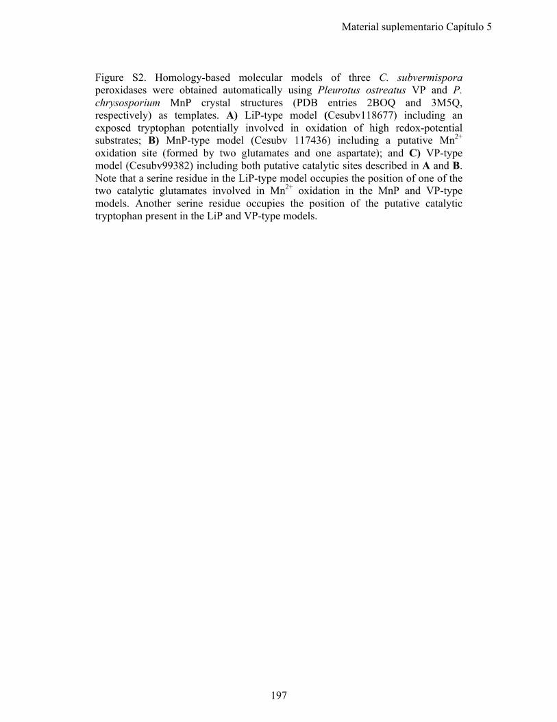
Figure S2. Homology-based molecular models of three C. subvermispora peroxidases were obtained automatically using Pleurotus ostreatus VP and P. chrysosporium MnP crystal structures (PDB entries 2BOQ and 3M5Q, respectively) as templates. A) LiP-type model (Cesubv118677) including an exposed tryptophan potentially involved in oxidation of high redox-potential substrates; B) MnP-type model (Cesubv 117436) including a putative Mn2+ oxidation site (formed by two glutamates and one aspartate); and C) VP-type model (Cesubv99382) including both putative catalytic sites described in A and B. Note that a serine residue in the LiP-type model occupies the position of one of the two catalytic glutamates involved in Mn2+ oxidation in the MnP and VP-type models. Another serine residue occupies the position of the putative catalytic tryptophan present in the LiP and VP-type models.
197
Material suplementario Capítulo 5
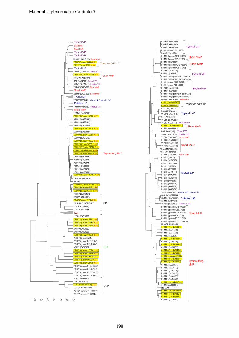
PE-VPL1 (AAD01401)PE-VPL2 (AAD01404)PE-VPL3 (CAD56164)PO-VP (genome PC9:137757)PSA-VP (CAJ01576)
Typical VP
PO-MnP (genome PC15:199511)PO-MnP (genome PC9:137765)
Short MnP
PO-MnP3 (BAA33449)PO-MnP3 (genome PC15:1089546) Short MnP
Typical VP
Short MnP
Short MnP
Typical VPTypical VP
Short MnP
CS-LiP (Cesubv118677) (0.65) CS-VP (Cesubv99382) (0 90)
Transition VP/LiP
LE-MnP1 (BAE79199)
(g )PO-MnP3 (genome PC9:137740)PE-VPS1 (AAD54310)PO-MnP2 (CAB51617)PO-MnP2(VP) (genome PC15:199491)PO-MnP2(VP) (genome PC9:137766)
Typical VP
PO-VP (genome PC15:156336)PO-VP (genome PC9:123383)PP-MnP5 (AAX40734)PO-MnP1 (AAA84396)PO-MnP1(VP) (genome PC15:1096331)PO-MnP1(VP) (genome PC9:137760)
Typical VP
Short MnPLE-MnP1 (BAE79199)CS-LiP (Cesubv118677) CS-VP (Cesubv99382) Transition VP/LiP
CS VP (Cesubv99382) (0.90) Typical LiP
Putative VPCS-MnP13 (Cesubv124076) (1.51)
Short MnP
Typical VP
Putative VP
Short MnP
Short MnP
Short MnP
Typical LiP
Unique LiP (catalytic Tyr)
Putative LiP
Putative VP
TV-LiP7 (CAA83147)
CR-MnPB (ABB83813)B-VP (AAO47909)S-MnP1 (BAE79812)TV-PGV (CAA54398)
AB-MnP (CAG27835)
TC-LiP (BAD52441)
TV-MRP (AAB63460)PCI-LiP1 (genome)PSAN-LiP1 (genome)TV-LiP12 (AAA34049)PCI-LiP2 (genome)TV-LiPGII (CAA53333)
Typical LiP
Putative VPTV-LiP7 (CAA83147)CS-MnP13 (Cesubv124076) CR-MnPB (ABB83813)
Short MnP
Typical VPB-VP (AAO47909)Putative VPS-MnP1 (BAE79812)Short MnPTV-PGV (CAA54398)
PR-MnP3 (CAC84573)TV-PGVII (CAA91043)TV-MnP2 (CAA83148)PSAN M P ( )
Short MnP
Putative VPTV-MRP (AAB63460)Short MnP
LE-MnP2 (BAG72080)CS-MnP9 (Cesubv114076) (0.72) DS-MnP2 (AAF31330)DS-MnP1 (AAF31329)PR-MnP2 (CAC85963)CS-MnP1 (Cesubv116608) (0.79) CS-MnP1 (AAB03480)CS-MnP3 (Cesubv139965) (0.87) CS-MnP3 (AAD45725)CS-MnP8 (Cesubv114036) (0.83) CS-MnP6 (Cesubv50686) (1.09) CS-MnP12 (Cesubv157986) (0.91)
PSAN-MnP (genome)PCI-MnP (genome)
Short MnPAB-MnP (CAG27835)PR-LiP3 (P20010)PR-LiP4 (AAW66483)PR-LiP1 (AAW59419)BA-LiP (1906181A)PC-LiPD (CAA33621)PC-LiPJ (AAD46494)PC-LiPC (AAA33739)PC-LiPF (AAA33736)PC-LiPH (AAA56852)PC-LiPA (AAA53109)PC-LiPB (AAA33741)PC-LiPE (AAA33738)
Typical LiP
( ) ( )CS-MnP7 (Cesubv105539 )(0.94) CS-MnP2 (Cesubv50297) (0.55) CS-MnP2 (AAD43581)PS-MnP3 (BAC06187)PC-MnP1 (AAA33744)PS-MnP1 (BAC06185)PC-MnP2 (AAA33745)PC-MnP3 (AAB39652)CS-MnP10 (Cesubv117436) (4.12) CR-MnPA (ABB83812)IZU-MnP1CS-MnP11 (Cesubv143390) (0.95) CS-MnP5 (Cesubv49863) (2.66)
Typical long MnP
PC-LiPE (AAA33738)Unique LiP (catalytic Tyr)TC-LiP (BAD52441)
GAU-MnP (ABB77244)GA-MnP1 (BAA88392)GF-MnP (ABB77243)
Putative LiP
Putative VPTV-MRP (AAB63460)PO-MnP (genome PC15:1099081)PO-MnP (genome PC9:121638)PO-MnP (genome PC15:1041740)PO-MnP (genome PC9:51713)PO-MnP (genome PC15:199510)PO-MnP (genome PC9:137764)
Short MnP
LE-MnP2 (BAG72080)CS-MnP9 (Cesubv114076) DS-MnP2 (AAF31330)
CS-MnP4 (Cesubv94398) (0.99) CS-MnP4 (AAO61784)PC-NOP (AAU82081)CS-GP (Cesubv112162) (0.95) PPL-PPLP (XP 002472503)CC-CIP (CAA50060)CD-CDP (AAZ14938)DyPLF-CPO (CAC16733)CS-HTP2 (Cesubv81391) (1.00) CS-HTP3 (Cesubv114787) (0.79) CS-HTP9 (Cesubv122198) (0.86) AA-APO (CAV28569)
GP
( )DS-MnP1 (AAF31329)PR-MnP2 (CAC85963)CS-MnP1 (Cesubv116608) CS-MnP1 (AAB03480)CS-MnP3 (Cesubv139965) CS-MnP3 (AAD45725)CS-MnP8 (Cesubv114036) CS-MnP6 (Cesubv50686) CS-MnP12 (cesubv157986) CS-MnP7 (Cesubv105539) CS-MnP2 (Cesubv50297) CS-MnP2 (AAD43581)PS-MnP3 (BAC06187)PC-MnP1 (AAA33744)
Typical longMnP
AA-APO (CAV28568)CS-HTP4 (Cesubv114799) (0.52) PC-HTP (genome:3274)PO-HTP (genome PC15:21834)PO-HTP (genome PC9:114464)AB-HTP (CAC03461)CS-HTP5 (Cesubv115079) (1.16) CS-HTP6 (Cesubv115379) (0.88) CS-HTP7 (Cesubv118102) (1.33) CS-HTP1 (Cesubv80799) (0.76) CS-HTP8 (Cesubv121474) (0.83) PO-HTP (genome PC15:162266)PO-HTP (genome PC9:127284)PO HTP (genome PC15:199695)
HTP
PS-MnP1 (BAC06185)PC-MnP2 (AAA33745)PC-MnP3 (AAB39652)CS-MnP10 (Cesubv117436) CR-MnPA (ABB83812)IZU-MnP1CS-MnP11 (Cesubv143390) CS-MnP5 (Cesubv49863) CS-MnP4 (Cesubv94398) CS-MnP4 (AAO61784)
PO-HTP (genome PC15:199695)PO-HTP (genome PC9:123372)SC-CCP (AAA88709)FN-CCP (Q6URB0)CS-CCP (Cesubv83438) (1.36) CC-CCP (XP 001830809)PO-CCP (genome PC15:199476)PO-CCP (genome PC9:77045)
CCP
0.00.20.40.60.81.01.2
198
Material suplementario Capítulo 5
Figure S3. Comparative analysis of basidiomycete peroxidases. Left, relationships among 133 basidiomycete (and two reference ascomycetes, Leptoxyphium fumago and S. cerevisiae) peroxidases (GeneBank references in parentheses, and C. subvermispora protein models in yellow). Amino-acid sequence comparisons as Poisson distances and clustering based on UPGMA and "pair-wise deletion" option of MEGA4. Compressed sub-trees are shown to facilitate the C. subvermispora peroxidases analysis. Induction levels in ball milled aspen (BMA) vs glucose as sole carbon source for each peroxidase are shown in red in brackets. Abbrev: AA, Agrocybe aegerita; AB, Agaricus bisporus; B, Bjerkandera sp; BA, Bjerkandera adusta; CC, Coprinopsis cinerea; CD, Coprinellus disseminatus; CR, Ceriporiopsis rivulosa; CS, C. subvermispora; DS, Dichomitus squalens; FN, Filobasidiella neoformans; GA, Ganoderma applanatum; GAU, Ganoderma australe; GF, Ganoderma formosanum; IZU, basidiomycete IZU-154; LE, Lentinula edodes; LF, Leptoxyphium fumago; MS, Marasmius scorodonius; P, Polyporus sp; PC, P. chrysosporium; PCI, Pycnoporus cinnabarinus; PE, Pleurotus eryngii; PO, Pleurotus ostreatus; PP, Pleurotus pulmonarius; PPL, Postia placenta; PR, Phlebia radiata; PS, Phanerochaete sordida; PSA, Pleurotus sapidus; S, Spongipellis sp; SC, Saccharomyces cerevisiae; TA, Termitomyces albuminosus; TC, Trametes cervina; and TV, Trametes versicolor. CCP, cytochrome c peroxidase; CDP, Coprinellus disseminatus peroxidase; CIP, Coprinopsis cinerea peroxidase; DyP, dye-decolorizing peroxidase; HTP, heme-thiolate peroxidase; LiP, lignin peroxidase; MnP, manganese peroxidase; MRP, manganese-repressed peroxidase from T. versicolor; NOP, P. chrysosporium hypothetical (generic) peroxidase; PGV, T. versicolor hypothetical peroxidase; PGVII, T. versicolor hypothetical peroxidase; PPLP, P. placenta peroxidase; VP, versatile peroxidase. Right side dendrogram focuses on 90 ligninolytic peroxidases showing putative evolutionary relationships and structural-functional classification. “Short” and “long” MnPs have an experimentally confirmed or a putative Mn2+-oxidation site formed by two glutamic and one aspartic residues, and differ in the length of the C-terminal tail. Typical LiPs contain an experimentally confirmed or a putative catalytic tryptophan whereas TC-LiP is a “unique” ligninolytic peroxidase with a catalytic tyrosine. Typical VPs harbor catalytic sites described above for both MnPs and Typical LiPs. Two C. subvermispora peroxidases initially annotated as LiP (Cesubv118677) and VP (Cesubv99382) are in fact a new type of “hybrid” peroxidase sharing catalytic properties with VP and LiP being also unable to oxidize Mn2+ (Main text Table 1), in spite of exhibiting (Cesubv99382) a typical Mn2+ oxidation site.
199
Material suplementario Capítulo 5

200
Material Suplementario
Capítulo 6
"Lignin-degrading peroxidases from genome of selective ligninolytic fungus Ceriporiopsis subvermispora" Fernández-Fueyo, E., F. J. Ruiz-Dueñas, Y. Miki, M. J. Martínez, K. E. Hammel, and A. T. Martínez. J. Biol. Chem. 287:16903-16906. 2012.
201

202
Revised version for The Journal of Biological Chemistry Supplemental Data Lignin-degrading peroxidases from the genome of the selective ligninolytic fungus Ceriporiopsis subvermispora Elena Fernández-Fueyo‡1, Francisco J. Ruiz-Dueñas‡2, Yuta Miki‡, María Jesús Martínez‡, Kenneth E. Hammel§3 and Angel T. Martínez‡4 From the ‡Centro de Investigaciones Biológicas, CSIC, Ramiro de Maeztu 9, E-28040 Madrid, Spain; and the §USDA Forest Service, Forest Products Laboratory, 1 Gifford Pinchot Dr, Madison, WI 53726, USA SUPPLEMENTAL TABLE AND FIGURES One supplemental table summarizing all the heme peroxidase genes found in the C. subvermispora genome and their initial structural-functional classification (Table S1) and two supplemental figures providing the nucleotide and amino-acid sequences of the five C. subvermispora genes (and a mutated variant) expressed in E. coli (Fig. S1) and their electronic absorption spectra, compared with Phanerochaete LiP and Pleurotus VP (Fig. S2) are included below.
203
Material suplementario Capítulo 6

TABLE S1
Summary of heme peroxidase genes (a total of 26) in the C. subvermispora genome, including members of the Classes I (model 17) and II (models 1-16) of the plant-fungal-prokaryotic peroxidase superfamily, and the HTP superfamily (models 18-26).
Reference (ID #), scaffold number (Sc.), position of the manually curated/annotated sequences, best hit protein after species abbreviation (protein identity in parenthesis), catalytic residues in Class II peroxidases (C-II Wc, Ea, Eb and Dc), and peroxidase type and name, based on the structural-functional classification (1).
C-II catalytic residues ID # Sc. Position Best hit Wc Ea Eb Dc Type/Name 1. 116608 11 869417-870990 CS-MnP4 (98%) - + + + MnP1 (long)* 2. 50297 6 1533603-1535152 CS-MnP2 (94%) - + + + MnP2 (long)* 3. 139965 11 865873-867462 CS-MnP3 (100%) - + + + MnP3 (long)* 4. 94398 6 474703-476192 CS-MnP1 (99%) - + + + MnP4 (long) 5. 49863 6 424184-425664 CS-MnP1 (90%) - + + + MnP5 (long) 6. 50686 6 141012-142559 CS-MnP3 (79%) - + + + MnP6 (long)* 7. 105539 6 1237989-1239477 CS-MnP3 (80%) - + + + MnP7 (long) 8. 114036 6 401273-402765 CS-MnP3 (83%) - + + + MnP8 (long) 9. 114076 6 549832-551335 CS-MnP3 (70%) - + + + MnP9 (long) 10. 117436 14 183695-185226 CR-MnP5 (89%) - + + + MnP10 (long) 11. 143390 19 448324-449814 CR-MnP5 (89%) - + + + MnP11 (long) 12. 157986 11 948929-950605 CS-MnP3 (83%) - + + + MnP12 (long)* 13. 124076 7 1607900-1609371 CR-MnP6 (91%) - + + + MnP13 (short) 14. 99382 20 496211-498070 TV-MnP7 (69%) + + + + LiP1** 15. 118677 20 490596-492455 BA-VP8 (67%) + - - - LiP2** 16. 112162 3 1562281-1563961 TC-LiP9 (42%) - - - - GP 17. 83438 6 1560404-1561943 FN-CCP 10 (65%) n/a n/a n/a n/a CCP 18. 80799 2 2721162-2722070 LB-POX11 (38%) n/a n/a n/a n/a HTP1 19. 81391 3 2160792-2162116 PT-HP12 (44%) n/a n/a n/a n/a HTP2 20. 114787 7 815278-816708 BF- POD13 (25%) n/a n/a n/a n/a HTP3 21. 114799 7 847603-846537 LB-POD14 (47%) n/a n/a n/a n/a HTP4 22. 115079 7 1856409-1857503 CC-POD15 (42%) n/a n/a n/a n/a HTP5 23. 115379 8 810181-810427 LB-POD11 (35%) n/a n/a n/a n/a HTP6 24. 118102 16 872151-873482 LB-POD11 (37%) n/a n/a n/a n/a HTP7 25. 121474 2 1700699-1701478 LB-POD16 (16%) n/a n/a n/a n/a HTP8 26. 122198 3 2164152-2165004 PN-POD17 (22%) n/a n/a n/a n/a HTP9
Protein abbreviations: CCP, cytochrome c peroxidase; GP, generic peroxidase; HP, hypothetical protein; HTP, heme-thiolate peroxidase; LiP, lignin peroxidase; MnP, manganese peroxidase; MnP-long, typical long MnP; MnP-short, short MnP; POD, putative peroxidase; VP, versatile peroxidase; *Five MnP-long enzymes that could be classified as "extralong" MnPs (2); **LiP-type enzymes representing LiP/VP transition stages. Fungal abbreviations: BA, Bjerkandera adusta; BF, Botryotinia fuckeliana; CC, C. cinerea; CR, Ceriporiopsis rivulosa; CS, C. subvermispora; FN, Filobasidiella neoformans; LB, Laccaria bicolor; PN, Phaeosphaeria nodorum; PT, Pyrenophora tritici-repentis; TC, Trametes cervina; TV, Trametes versicolor. Catalytic residues (Class II peroxidases): Wc, catalytic tryptophan; Ea, Eb and Dc, three acidic catalytic residues (2 glutamates and 1 aspartate) forming the manganese oxidation-site; n/a, not applicable (3). Best hit references: 1AAO61784, CS MnP4; 2AAB92247, CS MnP2; 3AAD45725, CS MnP3; 4AAB03480, CS MnP1; 5ABB83812, CR MnPA; 6ABB83813, CR MnPB; 7CAA54398, TV MnP-PGV; 8AAY89586, BA VP; 9BAE46585, TC LiP; 10Q6URB0, FN CCP; 11XP_001887896, LB POX; 12XP_001934218, PT HP; 13XP_001551585, BF POX; 14XP_001879054, LB POX; 15XP_001837979, CC POX; 16XP_001879928, LB POX; and 17XP_001796167, PN POX
204
Material suplementario Capítulo 6
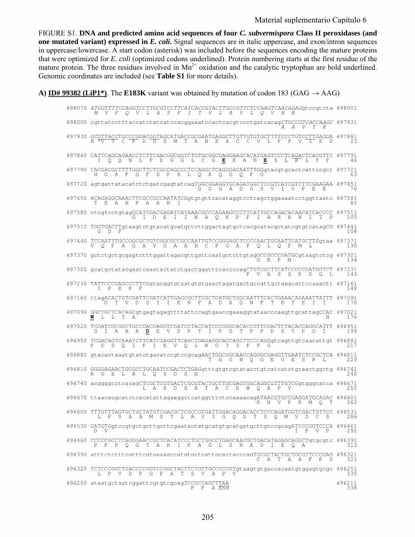
FIGURE S1. DNA and predicted amino acid sequences of four C. subvermispora Class II peroxidases (and one mutated variant) expressed in E. coli. Signal sequences are in italic uppercase, and exon/intron sequences in uppercase/lowercase. A start codon (asterisk) was included before the sequences encoding the mature proteins that were optimized for E. coli (optimized codons underlined). Protein numbering starts at the first residue of the mature protein. The three residues involved in Mn2+ oxidation and the catalytic tryptophan are bold underlined. Genomic coordinates are included (see Table S1 for more details). A) ID# 99382 (LiP1*). The E183K variant was obtained by mutation of codon 183 (GAG → AAG) 498070 ATGGTTTTCCAGGTCCTTGCGTCCTTCATCACCGTACTTGCCGTTCTCCAAGTCAACAGAGgtccgtcta 498001 M V F Q V L A S F I T V L A V L Q V N R 498000 cgttatcctttaccatctatcatccacggaaatccactcacgtccctgatcacagCTGCCGTCACCAAGC 497931 A A V T K 497930 GCGTTACCTGCCCGGACGGTAGCATGACCGCGAATGAGGCTTGTTGTGTGCTTTTCCCTGTCCTTGAGGA 497861 R *V T C P D G S M T A N E A C C V L F P V L E D 23 497860 CATTCAGCAGAACCTCTTCGACGGCGGTCTGTGCGGCGAGGAAGCACATGAGTCCCTCAGACTCACGTTC 497791 I Q Q N L F D G G L C G E E A H E S L R L T F 46 497790 CACGACGCTTTTGGCTTCTCGCCAGCCCTCCAGGCTCAGGGACAATTTGGgtacgtgcactcattccgct 497721 H D A F G F S P A L Q A Q G Q F G 63 497720 agtgattatacatctctgatcgagtatcagTGGCGGAGGTGCAGATGGCTCCGTCATCGTCTTCGAAGAA 497651 G G G A D G S V I V F E E 76 497650 ACAGAGGCAAACTTCGCCGCCAATATCGgtgtgttcacataggtcctcagctggaaaatcctggttaatc 497581 T E A N F A A N I 85 497580 ctcgtcctgtagGCATCGACGAGATCATAAACGCCCAGAAGCCCTTCATTGCCAGACACAACATCACCCC 497511 G I D E I I N A Q K P F I A R H N I T P 105 497510 TGGTGACTTgtaagtctgtacatgcatgttcttggactagtgctcacgcatacgctatcgtgtcatagCG 497441 G D F 108 497440 TCCAATTTGCCGGCGCTGTCGGCGCCGCCAATTGTCCGGGAGCTCCCCAACTGCAATTCATGCTTGgtaa 497371 V Q F A G A V G A A N C P G A P Q L Q F M L 130 497370 gctctgctgcgagtctttggattagacgttgattcaatgctcttgtagGCCGCCCGATGCgtaagtctcg 497301 G R P M 134 497300 gcatgctatacgaatcaaatactatctgactggatttcaccccagCTGTCGCTTCATCCCCCGATGGTCT 497231 P V A S S P D G L 143 497230 TATTCCCGAGCCCTTCGgtacggtgtaatgtatgaactagatgactgccgttgctgagcattccaaactt 497161 I P E P F 148 497160 ttagACACTGTCGATTCGATCATTGAGCGCTTCGCTGATGCTGGCAATTTCACTGAAACAGAAATTATTT 497091 D T V D S I I E R F A D A G N F T E T E I I 170 497090 GGCTGCTCACAGCgtgagtagagttttattccagtgaaccgaaaggtataacccaagttgcattagCCAC 497021 W L L T A H 176 497020 TCGATCGCGGCTGCCGACGAGGTCGATCCTACCATCCCGGGCACACCGTTCGACTCTACACCAGGCATTT 496951 S I A A A D E V D P T I P G T P F D S T P G I 199 496950 TCGACAGTCAAATCTTCATCGAGGTTCAGCTGAGAGGCACCAGCTTCCCAGGgtcagttgtcaacattgt 496881 F D S Q I F I E V Q L R G T S F P G 217 496880 gtacactaaatgtatctgacatccgtccgcagAACTGGCGGCAACCAGGGCGAGGTTGAATCTCCGCTCA 496811 T G G N Q G E V E S P L 229 496810 GGGGAGAACTGCGCCTGCAATCCGACTCTGAGgtttgtgtcgtatacctgtcatcatctgcaactggctg 496741 R G E L R L Q S D S E 240 496740 acggggcctcacagCTCGCTCGTGACTCGCGTACTGCCTGCGAGTGGCAGGCGTTTGTCGgtgggcatca 496671 L A R D S R T A C E W Q A F V 255 496670 ttaacacgcatctcacatattagaaggctcatggtttctccaaaacagATAACGTGCCGAGGATGCAGAC 496601 D N V P R M Q T 263 496600 TTTGTTTAGTGCTGCTATGTCGACGCTCGCCGTGATTGGACAGGACACCTCCCAGATGGTCGACTGTTCC 496531 L F S A A M S T L A V I G Q D T S Q M V D C S 286 496530 GATGTGgtccgtgctgcttgcttcgaatactatgcatgtgcatgatgcttgtccgcagATCCCGGTCCCA 496461 D V I P V P 292 496460 CCCCCGCCTCAGGGAACCGCTCACATCCCTGCTGGCCTGAGCAACGCTGACATAGAGCAGGCTgtgcgtc 496391 P P P Q G T A H I P A G L S N A D I E Q A 313 496390 atttctctttcgtttcgtgaaagccatgtgctcattgcactacccagTGCGCTACTGCTGCGTTCCCGAG 496321 C A T A A F P S 321 496320 TCTCCCGGTTGACCCCGGTCCGGCTACTTCTGTTGCCCCCGTgtaagtgtgaccacaatgtggagtgcgc 496251 L P V D P G P A T S V A P V 335 496250 ataatgctaatcggattcgtgtcgcagTCCGCCAGCTTAA 496211 P P A End 338
205
Material suplementario Capítulo 6
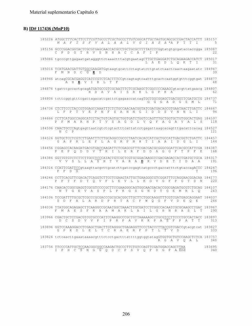
B) ID# 117436 (MnP10) 185226 ATGGCTTTCACTTCCTTCGTTGCCCTCGCTGCCCTTGTCGGCATTGCTAGTGCAGCGCCGACTACCATTT 185157 M A F T S F V A L A A L V G I A S A *A P T T I 5 185156 GCCCGGACGGTACTCGCGTGAGCAACCATGCCTGCTGCGCTTTTATCCCGgtatgtgcgataccaccgga 185087 C P D G T R V S N H A C C A F I P 22 185086 tgcccgttgagaatgatagggttctaaactttacgtgaatagCTTGCTGAGGATCTGCAGAAGACCATCT 185017 L A E D L Q K T I 31 185016 TCATGAACGATTGTGGCGAAGATGgtaagtgcattcctagtatcttgtatctaactcaactaagaatatc 184947 F M N D C G E D 39 184946 atcagCGCACGAGGTCATCCGTCTCACTTTCCgtcagtagtcaatttgcactcaatggtgtttcggtgat 184877 A H E V I R L T F 48 184876 tgatttgccactgcagATGATGCCGTCGCAATTTCTCGCAAGCTCGGCCCCAAAGCgtacgttatgaacg 184807 H D A V A I S R K L G P K A 62 184806 tcttcggtgtttcgattagacattgattctgagaaccatcagTGGTGGCGGAGCTGACGGTTCGATGCTG 184737 G G G A D G S M L 71 184736 CTCTTCCCTACCGTGGAGCCAAATTTCTCTGCCAACAACGGTATCGACGACAGCGTGAATAACTTGATTC 184667 L F P T V E P N F S A N N G I D D S V N N L I 94 184666 CCTTCATGGCCAGGCATCCTACTGTCAGTGCTGGTGATCTGGTCCAGTTTGCTGGTGCTGTGGCACTGAG 184597 P F M A R H P T V S A G D L V Q F A G A V A L S 118 184596 CAACTGCCCAgtgagttaatcgtctcgtactttcattatcctgagattaagcacagtttgacattcacag 184527 N C P 121 184526 GGTGCTCCTCGTCTTGAATTTCTTGCAGGCCGCCCTAATCACACCATCGCTGCCATTGACGGTCTGATTC 184457 G A P R L E F L A G R P N H T I A A I D G L I 144 184456 CGGAGCCACAAGATGACGTGACCAAGATTCTCGAGCGTTTCGACGACGCGGGCGGATTCACGCCATTCGA 184387 P E P Q D D V T K I L E R F D D A G G F T P F E 168 184386 GGTTGTGTCTCTTCTTGCCTCCCATACTGTCGCTCGTGCGGACAAGGTCGACGAGACCATTGATGCTGCA 184317 V V S L L A S H T V A R A D K V D E T I D A A 191 184316 CCATTCGATTCCgtaagttacgcttgcatcttgatccgagtcatgccctgaccatttctgtaccagACTC 184247 P F D S T 196 184246 CCTTCACGTTCGACACTCAGGTCTTCCTCGAAGTATTGCTGAAGGGCGTCGGATTTCCAGGAACGGACAA 184177 P F T F D T Q V F L E V L L K G V G F P G T D N 220 184176 CAACACCGGCGAGGTCGCGTCCCCGCTTCCGAAGGGCAGTGGCAACGACACCGGCGAGATGCGTCTGCAG 184107 N T G E V A S P L P K G S G N D T G E M R L Q 243 184106 TCCGATTTTGCGCTCGCCCGCGACCCGCGCACGGCTTGCTTCTGGCAAGGTTTCGTCGATGAGCAGGAAT 184037 S D F A L A R D P R T A C F W Q G F V D E Q E 266 184036 TTATGGCAGAGAGCTTCAAGGCCGCAATGGCTAAACTTGCGATCCTCGGCCACAATCGCGCAAGCCTGAC 183967 F M A E S F K A A M A K L A I L G H N R A S L T 290 183966 CGACTGCTCCGACGTCGTGCCCATTCCAAGGCCCGCTGTTAAAAAGCCTGCGTCCTTCCCTGCCACTACC 183897 D C S D V V P I P R P A V K K P A S F P A T T 313 183896 GGTCCAAAGGACCTCGAGCTGACTTGTAGGGCTGAGAGGTTCCCTACCCTTACCGTCGACCgtacgtcat 183827 G P K D L E L T C R A E R F P T L T V D 333 183826 tctcaacttgaaataaaacgtttctcctgactttattttggtggtatagGTGGTGCTGTCCAAGCTCTCA 183757 R G A V Q A L 340 183756 TTCCCCATTGCTCCAACGGCGGCCAAGACTGCCCTTCTGTCCAGTTCGATGGACCAGCTTAA 183695 I P H C S N G G Q D C P S V Q F D G P A End 360
206
Material suplementario Capítulo 6
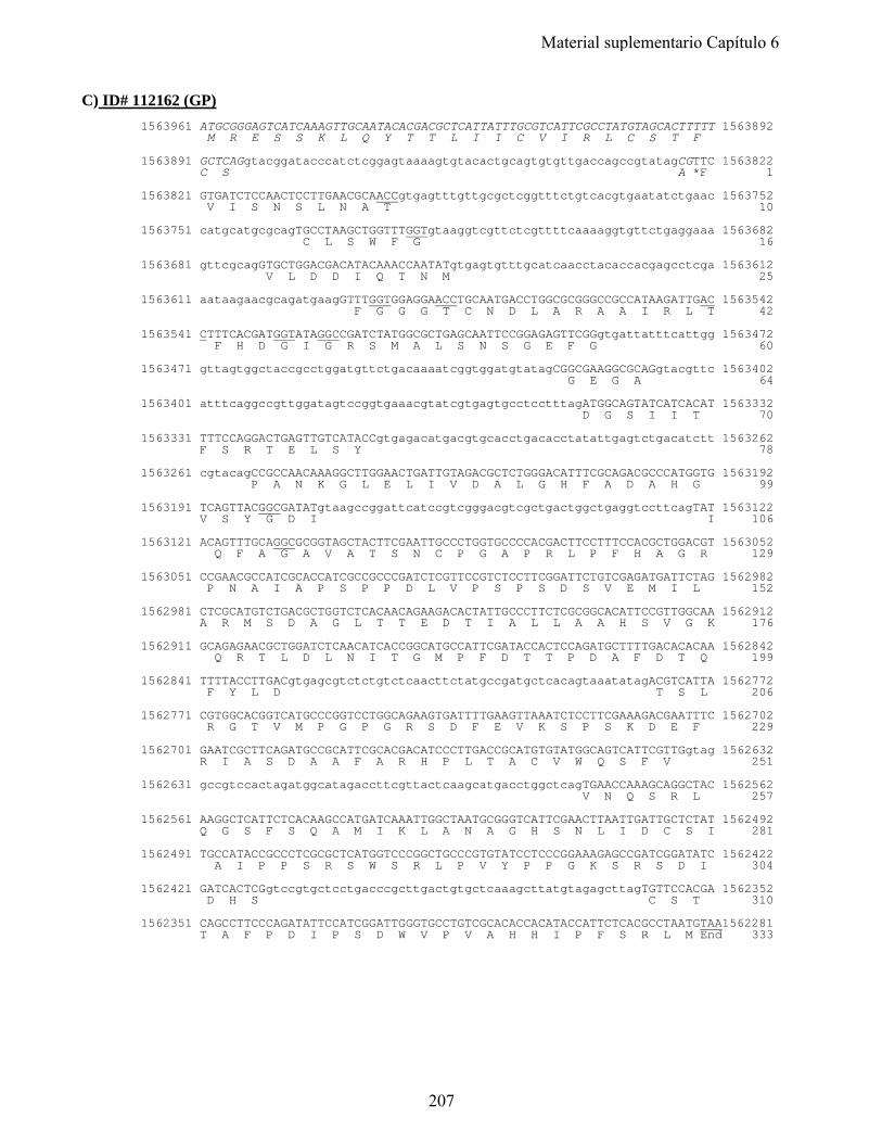
C) ID# 112162 (GP) 1563961 ATGCGGGAGTCATCAAAGTTGCAATACACGACGCTCATTATTTGCGTCATTCGCCTATGTAGCACTTTTT 1563892 M R E S S K L Q Y T T L I I C V I R L C S T F 1563891 GCTCAGgtacggatacccatctcggagtaaaagtgtacactgcagtgtgttgaccagccgtatagCGTTC 1563822 C S A *F 1 1563821 GTGATCTCCAACTCCTTGAACGCAACCgtgagtttgttgcgctcggtttctgtcacgtgaatatctgaac 1563752 V I S N S L N A T 10 1563751 catgcatgcgcagTGCCTAAGCTGGTTTGGTgtaaggtcgttctcgttttcaaaaggtgttctgaggaaa 1563682 C L S W F G 16 1563681 gttcgcagGTGCTGGACGACATACAAACCAATATgtgagtgtttgcatcaacctacaccacgagcctcga 1563612 V L D D I Q T N M 25 1563611 aataagaacgcagatgaagGTTTGGTGGAGGAACCTGCAATGACCTGGCGCGGGCCGCCATAAGATTGAC 1563542 F G G G T C N D L A R A A I R L T 42 1563541 CTTTCACGATGGTATAGGCCGATCTATGGCGCTGAGCAATTCCGGAGAGTTCGGgtgattatttcattgg 1563472 F H D G I G R S M A L S N S G E F G 60 1563471 gttagtggctaccgcctggatgttctgacaaaatcggtggatgtatagCGGCGAAGGCGCAGgtacgttc 1563402 G E G A 64 1563401 atttcaggccgttggatagtccggtgaaacgtatcgtgagtgcctcctttagATGGCAGTATCATCACAT 1563332 D G S I I T 70 1563331 TTTCCAGGACTGAGTTGTCATACCgtgagacatgacgtgcacctgacacctatattgagtctgacatctt 1563262 F S R T E L S Y 78 1563261 cgtacagCCGCCAACAAAGGCTTGGAACTGATTGTAGACGCTCTGGGACATTTCGCAGACGCCCATGGTG 1563192 P A N K G L E L I V D A L G H F A D A H G 99 1563191 TCAGTTACGGCGATATgtaagccggattcatccgtcgggacgtcgctgactggctgaggtccttcagTAT 1563122 V S Y G D I I 106 1563121 ACAGTTTGCAGGCGCGGTAGCTACTTCGAATTGCCCTGGTGCCCCACGACTTCCTTTCCACGCTGGACGT 1563052 Q F A G A V A T S N C P G A P R L P F H A G R 129 1563051 CCGAACGCCATCGCACCATCGCCGCCCGATCTCGTTCCGTCTCCTTCGGATTCTGTCGAGATGATTCTAG 1562982 P N A I A P S P P D L V P S P S D S V E M I L 152 1562981 CTCGCATGTCTGACGCTGGTCTCACAACAGAAGACACTATTGCCCTTCTCGCGGCACATTCCGTTGGCAA 1562912 A R M S D A G L T T E D T I A L L A A H S V G K 176 1562911 GCAGAGAACGCTGGATCTCAACATCACCGGCATGCCATTCGATACCACTCCAGATGCTTTTGACACACAA 1562842 Q R T L D L N I T G M P F D T T P D A F D T Q 199 1562841 TTTTACCTTGACgtgagcgtctctgtctcaacttctatgccgatgctcacagtaaatatagACGTCATTA 1562772 F Y L D T S L 206 1562771 CGTGGCACGGTCATGCCCGGTCCTGGCAGAAGTGATTTTGAAGTTAAATCTCCTTCGAAAGACGAATTTC 1562702 R G T V M P G P G R S D F E V K S P S K D E F 229 1562701 GAATCGCTTCAGATGCCGCATTCGCACGACATCCCTTGACCGCATGTGTATGGCAGTCATTCGTTGgtag 1562632 R I A S D A A F A R H P L T A C V W Q S F V 251 1562631 gccgtccactagatggcatagaccttcgttactcaagcatgacctggctcagTGAACCAAAGCAGGCTAC 1562562 V N Q S R L 257 1562561 AAGGCTCATTCTCACAAGCCATGATCAAATTGGCTAATGCGGGTCATTCGAACTTAATTGATTGCTCTAT 1562492 Q G S F S Q A M I K L A N A G H S N L I D C S I 281 1562491 TGCCATACCGCCCTCGCGCTCATGGTCCCGGCTGCCCGTGTATCCTCCCGGAAAGAGCCGATCGGATATC 1562422 A I P P S R S W S R L P V Y P P G K S R S D I 304 1562421 GATCACTCGgtccgtgctcctgacccgcttgactgtgctcaaagcttatgtagagcttagTGTTCCACGA 1562352 D H S C S T 310 1562351 CAGCCTTCCCAGATATTCCATCGGATTGGGTGCCTGTCGCACACCACATACCATTCTCACGCCTAATGTAA1562281 T A F P D I P S D W V P V A H H I P F S R L M End 333
207
Material suplementario Capítulo 6
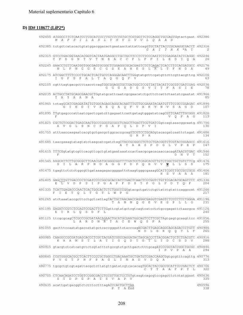
D) ID# 118677 (LiP2*) 492455 ATGGCCTTCTCAATCCTCGCATCCCTTGTCTCTGTGCTCGTGGTTCTCCAGGCTGCCAGTGgtactgaat 492386 M A F S I L A S L V S V L V V L Q A A S 492385 tctgctcatacactgtatggacggacactgaataactattattcagGTGCTATTACCCGCAAGGCGACCT 492316 G A I T R K *A T 2 492315 GTCCTGACGGTAACACGGTCACTAATGAGGCCTGCTGCTTCCTCTTCCCAATTCTCGAGGATATTCAGGC 492246 C P D G N T V T N E A C C F L F P I L E D I Q A 26 492245 GAACCTGTTCAATGGTGGCGAGTGCGGCTCGGAGGCGCACGAGTCTCTCAGACTCACCTTCCACGATGCC 492176 N L F N G G E C G S E A H E S L R L T F H D A 49 492175 ATCGGCTTTTCCCCTGCACTCACTGCCCAGGGACAATTTGGgtatgcttcgatgttcttcgttacgttcg 492106 I G F S P A L T A Q G Q F V 63 492105 cgtttagtgacgctttaacctcagTGGCGGAGGTGCTGATGGCTCCGTTATTACATTCGCGTCGATCGAG 492036 G G G A D G S V I T F A S I E 78 492035 ACTGCCTATGCAGCGAACGCTGgtatgcattcaatcgcgaacatctgcttctcattctaatatcgaatat 491966 T A Y A A N A 85 491965 tctagGCATCGAGGATATTGTGGCAGAGCAGGCACAATTTGTTGCGAAATACAATGTTTCCGCCGGAGAC 491896 G I E D I V A E Q A Q F V A K Y N V S A G D 107 491895 TTgtgagcccattaatcgactcgatcttgagaattcactgatagtgggatatcagCGTTCAATTTGCGGG 491826 F V Q F A G 113 491825 CGCTGTCGGACTGAGCAACTGCCCGGGTGCGCCTCAGCTTGACTTCGTGATCGgtcagtaaacggcaatg 491756 A V G L S N C P G A P Q L D F V I 130 491755 atttaaccaagaatcacgtgctgacgctggcaccgcagGTCGTCCTGCAGgtacacgatcaattctagat 491686 G R P A 134 491685 taacgaaagtatagtatctaagcatcgatctagCTACCGCGGCCTCTCCTGACGGTCTCGTACCGGAGCC 491616 A T A A S P D G L V P E P 147 491615 TTTCGgtatgtcgttcacgttcgctgtatgaataaatccactaacgcgacacaaccacagATAACGTGAC 491546 F D N V T 152 491545 AAGCATTCTTGCGCGCTTCAACGATGCGGGCGGTTTTGATCCTCAGCACGTTGTCTGGCTGCTGTCTTCg 491476 S I L A R F N D A G G F D P Q H V V W L L S S 175 491475 tgagttctctctggcgttgataaagagacggggattctaagtgggcgaaagGCATTCGGTTGCCGCCGCG 491406 H S V A A A 181 491405 GAGCTTGTTGACCCCTCGATCCCCGGCGCACCATTTGACTCAACTCCCGGTCTGTTCGACACGCAGTTTT 491336 E L V D P S I P G A P F D S T P G L F D T Q F 204 491335 TCATTGAGACCCAACTCACTGGCACTCTCTGGCCCGGgtacgctgattctgttatctgtattcaggccat 491266 F I E T Q L T G T L W P G 217 491265 atctaaataccgcttcctgctcattagTACTGCTAACAACCAGGGCGAGGTCGAGTCTCCCCTCCTGGGA 491196 T A N N Q G E V E S P L L G 231 491195 GAGATCCGTCTCCAGTCCGACTTCTTGgttcgtatgctcgtcagtcatcctctgccgagattctaacgca 491126 E I R L Q S D F L 240 491125 ttcgcgcagCTCGCCCGCGATAACAGAACTGCATGCGAATGGCAGTCCTTCGCTAgtgagtgcaagttcc 491056 L A R D N R T A C E W Q S F A 255 491055 gactttccaatatgaccatatgctcaccgggattctacccagACGACTTGAGCAGGCAGCAGACCCTGTT 490986 N D L S R Q Q T L F 265 490985 CAAGGCCGCGATGAGCACCCTCGCTACGATCGGCCAGGATACTAGCACCCTTACGGACTGCTCTGACGTC 490916 K A A M S T L A T I G Q D T S T L T D C S D V 288 490915 gtacgtctcatcatgttctcgtatttctgccgtctgcttgactctttgcagATCCCGGTACCGGCTGCGC 490846 I P V P A A 294 490845 CCGTGGGCACGCCTCACTTCCCCGCTGGCCTGACAAATGCTGATGTCGACCAAGCGgtgcgtttcagttg 490776 P V G T P H F P A G L T N A D V D Q A 313 490775 tgcgctgtcgtagattacgctcttatgttgatatcgtcacacagTGCACTACCGCCGCATTCCCGACTCT 490706 C T T A A F P T L 322 490705 CTCAACAGATCCTGGTCCGGCGACCTCCGTTGCTCCTGTgtaagtcagcgtccgagcttctctatggaat 490636 S T D P G P A T S V A P V 335 490635 acattgatgacggtctctttcctttagACCCACTGCTTAA 490596 P T A End 338
208
Material suplementario Capítulo 6
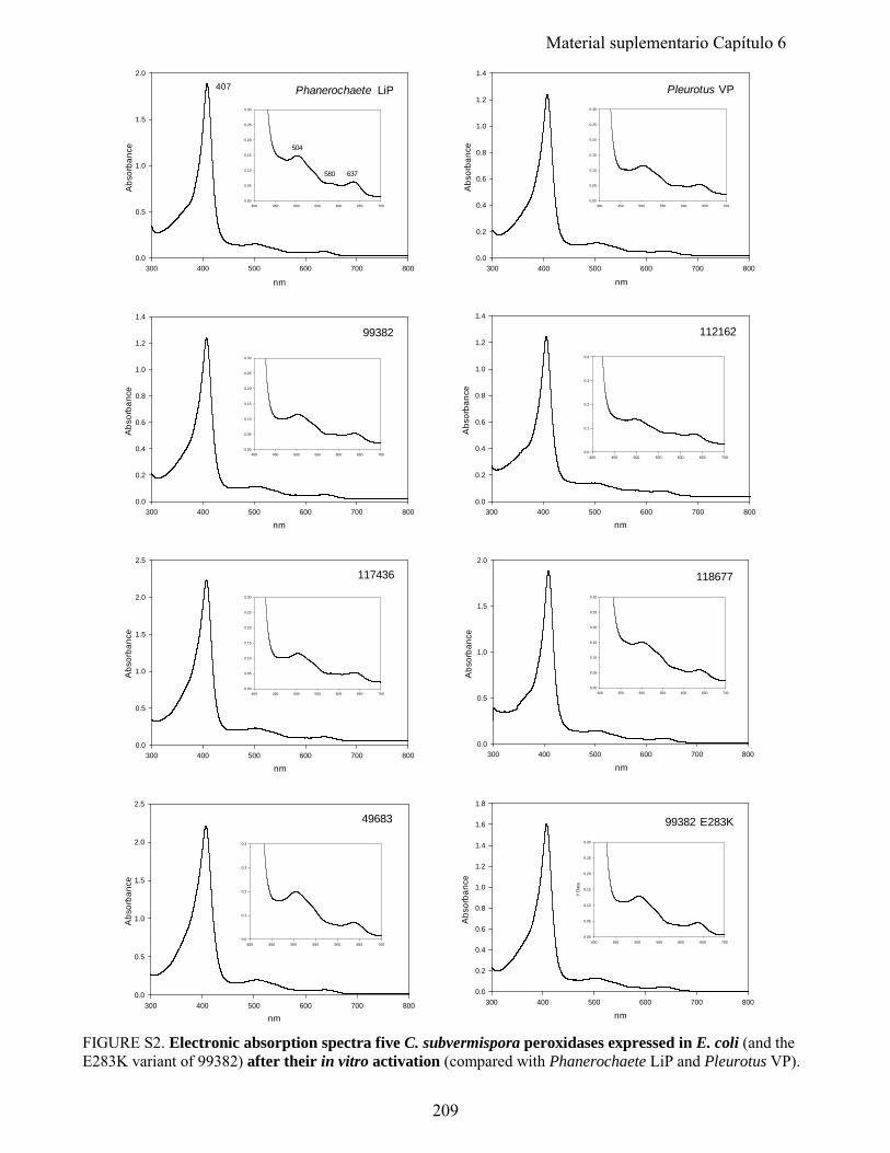
300 400 500 600 700 8000.0
0.5
1.0
1.5
2.0
2.5
400 450 500 550 600 650 7000.0
0.1
0.2
0.3
0.4
49683
nm
Abs
orba
nce
300 400 500 600 700 8000.0
0.2
0.4
0.6
0.8
1.0
1.2
1.4
nm
Abs
orba
nce
400 450 500 550 600 650 7000.00
0.05
0.10
0.15
0.20
0.25
0.30
99382
300 400 500 600 700 8000.0
0.2
0.4
0.6
0.8
1.0
1.2
1.4
1.6
1.8
99382 E283K
nm
Abs
orba
nce
400 450 500 550 600 650 700
Y D
ata
0.00
0.05
0.10
0.15
0.20
0.25
0.30
300 400 500 600 700 8000.0
0.5
1.0
1.5
2.0
2.5
117436
Abs
orba
nce
400 450 500 550 600 650 7000.00
0.05
0.10
0.15
0.20
0.25
0.30
nm
300 400 500 600 700 8000.0
0.5
1.0
1.5
2.0
Abs
orba
nce
400 450 500 550 600 650 7000.00
0.05
0.10
0.15
0.20
0.25
0.30
118677
nm
300 400 500 600 700 8000.0
0.2
0.4
0.6
0.8
1.0
1.2
1.4
Abs
orba
nce
400 450 500 550 600 650 7000.0
0.1
0.2
0.3
0.4
nm
112162
300 400 500 600 700 8000.0
0.2
0.4
0.6
0.8
1.0
1.2
1.4
Pleurotus VP
nm
Abs
orba
nce
400 450 500 550 600 650 7000.00
0.05
0.10
0.15
0.20
0.25
0.30
X Data
300 400 500 600 700 8000.0
0.5
1.0
1.5
2.0
Abs
orba
nce
407
400 450 500 550 600 650 7000.00
0.05
0.10
0.15
0.20
0.25
0.30
637
504
580
Phanerochaete LiP
nm
FIGURE S2. Electronic absorption spectra five C. subvermispora peroxidases expressed in E. coli (and the E283K variant of 99382) after their in vitro activation (compared with Phanerochaete LiP and Pleurotus VP).
209
Material suplementario Capítulo 6
REFERENCES 1. Ruiz-Dueñas, F. J. and Martínez, A. T. (2010) In Biocatalysts based on heme peroxidases (Torres, E. and Ayala, M.,
eds) pp. 37-59, Springer-Verlag, Berlin 2. Li, D. M., Li, N., Ma, B., Mayfield, M. B., and Gold, M. H. (1999) BBA Protein Struct. Mol. Enzym. 1434, 356-364 3. Ruiz-Dueñas, F. J., Morales, M., García, E., Miki, Y., Martínez, M. J., and Martínez, A. T. (2009) J. Exp. Bot. 60,
441-452
210
Material suplementario Capítulo 6
Material Suplementario
Capítulo 7
"Catalytic and stability properties of manganese peroxidase subfamilies: A study based on the Ceriporiopsis subvermispora genome" Fernández-Fueyo, E., S. Acebes, F. J. Ruiz-Dueñas, , M. J. Martínez, A. Romero, F. J. Medrano, V Guallar and A. T. Martínez. (enviado)
211

212
Supporting Information Catalytic and stability properties of manganese peroxidase subfamilies: A study based on the Ceriporiopsis subvermispora genome Elena Fernández-Fueyo, Sandra Acebes, Francisco J. Ruiz-Dueñas, María Jesús Martínez, Antonio Romero, Francisco Javier Medrano, Victor Guallar, and Angel T. Martínez Supporting Information includes H-bond involving the C-terminal tail of extralong MnP6 (100 ns MD) (Table S1), sequence identities between MnPs from C. subvermispora and other fungi (Table S2), crystallographic data of C. subvermispora MnP6 (Table S3), multiple alignment of extralong, long and short MnPs sequences from C. subvermispora and other basidiomycetes (Fig. S1), molecular structures of C. subvermispora extralong, long and short MnPs (Fig. S2), ABTS binding on extralong MnP6 as shown by PELE (Fig. S3), closure of the propionate channel in the MnP6 short-variant by the double G82L/D85L mutation (Fig. S4), effect of pH on ABTS oxidation by single and multiple variants of the MnP6 short-variant (Fig. S5), and phylogram of C. subvermispora and other basidiomycete peroxidases (Fig. S6).
Table S1. H-bonds involving the C-terminal tail residues of C. subvermispora extralong MnP6 during a 100 ns MD (300 K) C-tail residue Second residue H-bond Phe357-backbone Gln356-side chain 18.27% Phe357-backbone Asn89-side chain 23.97% Phe357-backbone Ash85-side chain* 1.82% Ala361-backbone Asn88-side chain 22.09% Ala361-backbone Asn88-side chain 10.81% Thr362-side chain Asp363-backbone 1.23% Asp363-side chain Ash84-side chain* 36.55% Asp363-side chain Lys180-side chain 19.35% Asp363-side chain Ser364-side chain 21.86% Ser364-side chain Asn81-side chain 26.99% Ser364-side chain Asn81-side chain 4.66% Ser364-side chain Lys180-side chain 2.71% Ser364-backbone Asn81-side chain 9.57% Ser364-backbone Lys180-side chain 4.51% Pro365-side chain Lys180-side chain 0.56% Pro365-backbone Lys180-side chain 0.90% *Ash, protonated aspartate
213
Material suplementario Capítulo 7
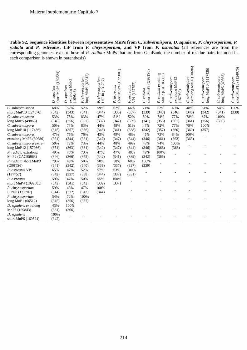
Table S2. Sequence identities between representative MnPs from C. subvermispora, D. squalens, P. chrysosporium, P. radiata and P. ostreatus, LiP from P. chrysosporium, and VP from P. ostreatus (all references are from the corresponding genomes, except those of P. radiata MnPs that are from GenBank; the number of residue pairs included in each comparison is shown in parenthesis)
D. s
qual
ens
shor
t MnP
3 (1
6952
4)
D. s
qual
ens
extra
long
MnP
3 (1
6984
3)
P. c
hrys
ospo
rium
lo
ng M
nP1
(665
12)
P. c
hrys
ospo
rium
Li
PH8
(131
707)
P. o
stre
atus
sh
ort M
nP4
(109
9081
)
P. o
stre
atus
V
P1 (1
3775
7)
P. ra
diat
a sh
ort M
nP3
(Q96
TS6)
P. ra
diat
a ex
tralo
ng
MnP
2 (C
AC
8596
3)
C. s
ubve
rmis
pora
ex
tralo
ng M
nP12
(1
5798
6)
C. s
ubve
rmis
pora
ex
tralo
ng M
nP6
(506
86)
C. s
ubve
rmis
pora
lo
ng M
nP10
(111
7436
)
C. s
ubve
rmis
pora
lo
ng M
nP5
(498
63)
C. s
ubve
rmis
pora
sh
ort M
nP13
(112
4076
)
C. subvermispora short MnP13 (124076)
68% (342)
52% (343)
52% (341)
59% (344)
62% (336)
66% (337)
71% (339)
52% (343)
49% (346)
48% (346)
51% (342)
54% (341)
100% (338)
C. subvermispora long MnP5 (49863)
53% (346)
75% (356)
83% (357)
47% (337)
51% (342)
52% (339)
50% (341)
74% (355)
77% (361)
78% (361)
87% (356)
100% (356) -
C. subvermispora long MnP10 (117436)
50% (345)
73% (357)
83% (356)
44% (346)
49% (341)
51% (338)
47% (342)
72% (357)
77% (360)
79% (360)
100% (357) - -
C. subvermispora extralong MnP6 (50686)
47% (351)
75% (344)
76% (361)
43% (347)
49% (347)
48% (344)
45% (346)
73% (361)
84% (362)
100% (365) - - -
C. subvermispora extra-long MnP12 (157986)
50% (351)
72% (363)
73% (361)
44% (342)
48% (347)
49% (344)
48% (346)
74% (366)
100% (368) - - - -
P. radiata extralong MnP2 (CAC85963)
49% (346)
78% (366)
73% (355)
47% (342)
47% (341)
48% (339)
49% (342)
100% (366) - - - - -
P. radiata short MnP3 (Q96TS6)
79% (341)
49% (342)
50% (340)
58% (339)
58% (337)
68% (337)
100% (339) - - - - - -
P. ostreatus VP1 (137757)
65% (342)
47% (337)
52% (338)
57% (344)
63% (337)
100% (331) - - - - - - -
P. ostreatus short MnP4 (1099081)
59% (342)
47% (341)
50% (342)
55% (339)
100% (337) - - - - - - - -
P. chrysosporium LiPH8 (131707)
59% (344)
43% (332)
47% (343)
100% (344) - - - - - - - - -
P. chrysosporium long MnP1 (66512)
54% (345)
72% (356)
100% (357) - - - - - - - - - -
D. squalens extralong MnP3 (169843)
43% (331)
100% (366) - - - - - - - - - - -
D. squalens short MnP6 (169524)
100% (342) - - - - - - - - - - - -
214
Material suplementario Capítulo 7
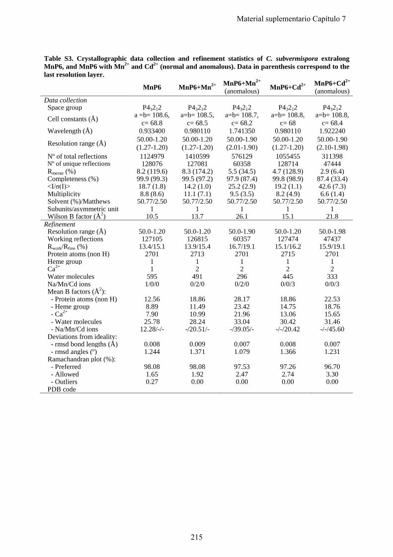
Table S3. Crystallographic data collection and refinement statistics of C. subvermispora extralong MnP6, and MnP6 with Mn2+ and Cd2+ (normal and anomalous). Data in parenthesis correspond to the last resolution layer.
MnP6 MnP6+Mn2+ MnP6+Mn2+ (anomalous) MnP6+Cd2+ MnP6+Cd2+
(anomalous) Data collection Space group P43212 P43212 P43212 P43212 P43212 Cell constants (Å) a =b= 108.6,
c= 68.8 a=b= 108.5,
c= 68.5 a=b= 108.7,
c= 68.2 a=b= 108.8,
c= 68 a=b= 108.8,
c= 68.4 Wavelength (Å) 0.933400 0.980110 1.741350 0.980110 1.922240
Resolution range (Å) 50.00-1.20 (1.27-1.20)
50.00-1.20 (1.27-1.20)
50.00-1.90 (2.01-1.90)
50.00-1.20 (1.27-1.20)
50.00-1.90 (2.10-1.98)
Nº of total reflections 1124979 1410599 576129 1055455 311398 Nº of unique reflections 128076 127081 60358 128714 47444 Rmerge (%) 8.2 (119.6) 8.3 (174.2) 5.5 (34.5) 4.7 (128.9) 2.9 (6.4) Completeness (%) 99.9 (99.3) 99.5 (97.2) 97.9 (87.4) 99.8 (98.9) 87.4 (33.4) <I/σ(I)> 18.7 (1.8) 14.2 (1.0) 25.2 (2.9) 19.2 (1.1) 42.6 (7.3) Multiplicity 8.8 (8.6) 11.1 (7.1) 9.5 (3.5) 8.2 (4.9) 6.6 (1.4) Solvent (%)/Matthews
50.77/2.50 50.77/2.50 50.77/2.50 50.77/2.50 50.77/2.50
Subunits/asymmetric unit 1 1 1 1 1 Wilson B factor (Å2) 10.5 13.7 26.1 15.1 21.8 Refinement Resolution range (Å) 50.0-1.20 50.0-1.20 50.0-1.90 50.0-1.20 50.0-1.98 Working reflections 127105 126815 60357 127474 47437 Rwork/Rfree (%) 13.4/15.1 13.9/15.4 16.7/19.1 15.1/16.2 15.9/19.1 Protein atoms (non H) 2701 2713 2701 2715 2701 Heme group 1 1 1 1 1 Ca2+ 1 2 2 2 2 Water molecules 595 491 296 445 333 Na/Mn/Cd ions 1/0/0 0/2/0 0/2/0 0/0/3 0/0/3 Mean B factors (Å2): - Protein atoms (non H) 12.56 18.86 28.17 18.86 22.53 - Heme group 8.89 11.49 23.42 14.75 18.76 - Ca2+ 7.90 10.99 21.96 13.06 15.65 - Water molecules 25.78 28.24 33.04 30.42 31.46 - Na/Mn/Cd ions 12.28/-/- -/20.51/- -/39.05/- -/-/20.42 -/-/45.60 Deviations from ideality: - rmsd bond lengths (Å) 0.008 0.009 0.007 0.008 0.007 - rmsd angles (º) 1.244 1.371 1.079 1.366 1.231 Ramachandran plot (%):
- Preferred 98.08 98.08 97.53 97.26 96.70 - Allowed 1.65 1.92 2.47 2.74 3.30 - Outliers 0.27 0.00 0.00 0.00 0.00 PDB code
215
Material suplementario Capítulo 7
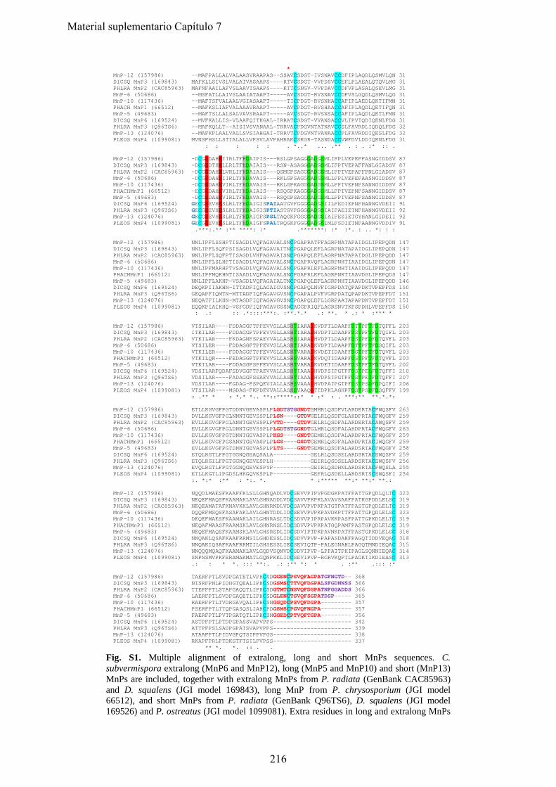
* MnP-12 (157986) --MAFPALLALVALAASVRAAPAS--SSAVCSDGT-IVSNAVCCDFIPLAQDLQSMVLQN 31 DICSQ MnP3 (169843) MAFKLLSIVSLVALATVASAAPS----RTVCSDGT-VVPDSVCCEFLPLAEALQTQVLMG 31 PHLRA MnP2 (CAC85963) MAFNFAAILAFVSLAAVTSAAPS----KTTCSNGV-VVPDAVCCDFVPLASALQSEVLMG 31 MnP-6 (50686) --MSFATLLAIVSLAAIATAAPT-----AVCSDGT-RVSNAVCCDFVSLGQDLQSMVLQG 31 MnP-10 (117436) --MAFTSFVALAALVGIASAAPT-----TICPDGT-RVSNHACCAFIPLAEDLQKTIFMN 31 PHACH MnP1 (66512) --MAFKSLIAFVALAAAVRAAPT-----AVCPDGT-RVSHAACCAFIPLAQDLQETIFQN 31 MnP-5 (49683) --MAFTSLLALSALVAVSRAAPT-----AVCSDGT-RVSNSACCAFIPLAQDLQETLFMN 31 DICSQ MnP6 (169524) --MVFKALLIS-VLAAFQITKGAL-IRRATCSDGT-VVANSACCVLIPVIQDIQENLFDG 31 PHLRA MnP3 (Q96TS6) --MAFKQLLT--AISIVSVANAAL-TRRVACPDGVNTATNAVCCSLFAVRDLIQDQLFDG 32 MnP-13 (124076) --MAFKPLAALVALLSVSIAHGAI-TRRVTCPDGVNTVANAACCPLFAVRDDIQESLFDG 32 PLEOS MnP4 (1099081) MVNSFHSLLSTIALALLVPSVLAVPAHRAKCSKGR-TASNDACCVWFDVLDDIQENLFDG 31 : : : : : . *..* ... .** . : . :* :: . MnP-12 (157986) -DCGEDAHEIIRLTFHDAIPIS---RSLGPSAGGGADGSMLIFPLVEPEFFASNGIDDSV 87 DICSQ MnP3 (169843) -DCGEDTHELLRLTFHDAIAIS---RSN-ASAGGGADGSMLIFPTVEPAFFANLGIADSV 87 PHLRA MnP2 (CAC85963) -DCGEDAHELVRLIFHDAIAIS---QSMGPSAGGGADGSMLIFPTVEPAFFPNLGIADSV 87 MnP-6 (50686) -DCGEDAHEIIRLTFHDAVAIS---RKLGPSAGGGADGSMLLFPLVEPEFAASNGIDDSV 87 MnP-10 (117436) -DCGEDAHEVIRLTFHDAVAIS---RKLGPKAGGGADGSMLLFPTVEPNFSANNGIDDSV 87 PHACHMnP1 (66512) -ECGEDAHEVIRLTFHDAIAIS---RSQGPKAGGGADGSMLLFPTVEPNFSANNGIDDSV 87 MnP-5 (49683) -DCGEDAHEVIRLTFHDAVAIS---RSQGPSAGGGADGSMLLFPTVEPNFSANNGIDDSV 87 DICSQ MnP6 (169524) GECGEEVHESLRLTFHDAIGISPAIAATGVFGGGGADGSIILFEDIEPNFHANNGVDEII 91 PHLRA MnP3 (Q96TS6) GECGEEVHESLRLTFHDAIGISPTIASTGVFGGGGADGSIAIFAEIETNFHANNGVDEII 92 MnP-13 (124076) GKCGEEVHESLRLTFHDAIGFSPSLTAQGKFGGGGADGSIAIFESIETGYHANLGIDEII 92 PLEOS MnP4 (1099081) GECGEEVHESLRLTFHDAIGFSPALTRQGKFGGGGADGSIMLFSDIETNFAANNGVDDIV 91 .***:.** :** ****: :* .*******: :* :*. : .. *: : : MnP-12 (157986) NNLIPFLSSHPTISAGDLVQFAGAVALSNCPGAPRATFFAGRPNATAPAIDGLIPEPQDN 147 DICSQ MnP3 (169843) NNLIPFLSQFPSISAGDLVQFAGAVAITNCPGAPQLEFLAGRPNATAPAIDGLIPEPQDN 147 PHLRA MnP2 (CAC85963) NNLIPFLSQFPTISAGDLVHFAGAVAISNCPGAPQLEFLAGRPNATAPAIDGLIPEPQDD 147 MnP-6 (50686) NNLIPFLSLHPTISAGDLVQFAGAVALSNCPGAPRVQFLAGRPNHTIAAIDGLIPEPQDN 147 MnP-10 (117436) NNLIPFMARHPTVSAGDLVQFAGAVALSNCPGAPRLEFLAGRPNHTIAAIDGLIPEPQDD 147 PHACHMnP1 (66512) NNLIPFMQKHNTISAADLVQFAGAVALSNCPGAPRLEFLAGRPNKTIAAVDGLIPEPQDS 147 MnP-5 (49683) NNLIPFLAKHP-VSAGDLVQFAGAIALTNCPGAPQLEFLAGRPNHTIAAVDGLIPEPQDD 146 DICSQ MnP6 (169524) DEQKPIIAKHN-ITTADFIQLAGAIGVSNCPGAPQLNVFIGRPDATQPAPDKTVPEPFDS 150 PHLRA MnP3 (Q96TS6) GEQAPFIQMTN-MTTADFIQFAGAVGVSNCPGAPALPVFVGRPDATQPAPDKTVPEPFDT 151 MnP-13 (124076) NEQATFILKHN-MTAGDFIQFAGAVGVSNCPGAPQLEFLLGRPAATAPAPDKTVPEPFDT 151 PLEOS MnP4 (1099081) EQQKPIAIKHQ-VSFGDFIQFAGAVGSSNCAGGPRIQFLAGRSNVTKPSPDHLVPEPFDS 150 : .: :: .*::::***:. :**.*.* .: **. * .: * :*** * MnP-12 (157986) VTSILAR----FDDAGGFTPFEVVSLLASHTIARADKVDPTLDAAPFDTTPFTFDTQFFL 203 DICSQ MnP3 (169843) ITKILAR----FDDAGGFTPFEVVSLLASHTIARADHVDPTLDAAPFDSTPFTFDTQIFL 203 PHLRA MnP2 (CAC85963) VTKILAR----FKDAGNFSPAEVVALLASHSIARADHVDPTLDAAPFDSTPFDFDTQVFL 203 MnP-6 (50686) VTSILER----FDDAGGFTPFEVVSLLASHTIARADKVDPTLDAAPFDTTPFTFDSQIFL 203 MnP-10 (117436) VTKILER----FDDAGGFTPFEVVSLLASHTVARADKVDETIDAAPFDSTPFTFDTQVFL 203 PHACHMnP1 (66512) VTKILQR----FEDAGGFTPFEVVSLLASHSVARADKVDQTIDAAPFDSTPFTFDTQVFL 203 MnP-5 (49683) VTKILAR----FDDAGGFSPFEVVSLLASHTVARADKVDETIDAAPFDSTPFTFDTQVFL 202 DICSQ MnP6 (169524) VDSILARFQDAFSDVGGFTPAEVVALLASHTIAAADHVDPSIPGTPFDSTPELFDTQFFI 210 PHLRA MnP3 (Q96TS6) VDSILAR----FADAGGFSSAEVVALLASHTIAAADHVDPSIPGTPFDSTPEIFDTQFFI 207 MnP-13 (124076) VDSILAR----FGDAG-FSPQEVIALLASHSVAAADHVDPAIPGTPFDSTPSDFDPQIFI 206 PLEOS MnP4 (1099081) VTSILAR----MGDAG-FKPDEVVALLASHSVAAQDTIDPKLAGHPFDSTPSDFDSQFFV 199 : .** * : *.* *.. **::*****::* * :* : . ***:** **.*.*: MnP-12 (157986) ETLLKGVGFPGTDDNVGEVASPLPLGDTSTGGNDTGMMRLQSDFVLARDERTACFWQSFV 263 DICSQ MnP3 (169843) DVLLKGVGFPGLNNNTGEVSSPLPLSN----GTDVGELRLQSDFGLAHDPRTACFWQGFV 259 PHLRA MnP2 (CAC85963) EVLLKGVGFPGLANNTGEVSSPLPVTD----GTDVGELRLQSDFALARDERTACAWQSFV 259 MnP-6 (50686) EVLLKGVGFPGLDNNTGEVSSPLPLGDTSTGGKDTGLMRLQSDFALAHDPRTACFWQGFV 263 MnP-10 (117436) EVLLKGVGFPGTDNNTGEVASPLPKGS----GNDTGEMRLQSDFALARDPRTACFWQGFV 259 PHACHMnP1 (66512) EVLLKGVGFPGSANNTGEVASPLPLGS----GSDTGEMRLQSDFALAHDPRTACIWQGFV 259 MnP-5 (49683) EVLLKGVGFPGTDNNTGEVASPLPLTS----GNDTGEMRLQSDFALARDSRTACFWQGFV 258 DICSQ MnP6 (169524) ETQLRGTLFPGTGGNQGEAQSALA-----------GELRLQSDSELARDSRTACEWQSFV 259 PHLRA MnP3 (Q96TS6) ETQLRGILFPGTGGNQGEVESPLH-----------GEIRLQSDSELARDSRTACEWQSFV 256 MnP-13 (124076) EVQLRGTLFPGTGGNQGEVESPYP-----------GEIRLQSDHNLARDSRTACFWQSLA 255 PLEOS MnP4 (1099081) ETLLKGTLIPGDSLHKGQVKSPLP-----------GEFRLQSDELLARDSRTSCEWQSFI 254 :. *:* :** : *:. *. * :***** **:* **:* **.: MnP-12 (157986) NQQDLMAESFKAAFFKLSLLGHNQADLVDCSEVVPIPVPGDGKPATFPATTGPQDLQLTC 323 DICSQ MnP3 (169843) NEQEFMAQSFKAAMAKLAVLGHNADDLVDCSAVVPKPKLAVAVSAAFPATKGPDDLELSC 319 PHLRA MnP2 (CAC85963) NEQEAMATAFKNAVKKLAVLGHNRNDLVDCSAVVPVPKPATGTPATFPASTGPQDLELTC 319 MnP-6 (50686) DQQEFMSQSFASAFAKLAVLGHNTDDLIDCSEVVPVPKPAVDKPTTFPATTGPQDLELSC 323 MnP-10 (117436) DEQEFMAESFKAAMAKLAILGHNRASLTDCSDVVPIPRPAVKKPASFPATTGPKDLELTC 319 PHACHMnP1 (66512) NEQAFMAASFRAAMSKLAVLGHNRNSLIDCSDVVPVPKPATGQPAMFPASTGPQDLELSC 319 MnP-5 (49683) NEQEFMAQSFKAAMSKLAVLGHSRSDLIDCSDVIPTPKPAVNKPATFPASTGPKDLELSC 318 DICSQ MnP6 (169524) NNQAKLQSAFKAAFRRMSILGHDESSLIDCSDVVPVP-PAPASDAHFPAGQTIDDVEQAC 318 PHLRA MnP3 (Q96TS6) NNQAKIQSAFKAAFRKMTILGHSESSLIECSEVIQTP-PALEGNAHLPAGQTMNDIEQAC 315 MnP-13 (124076) NNQQQMQAQFKAAMAKLAVLGQDVSQMVDCSDVIPVP-LPPATTPHIPAGLSQNNIEQAC 314 PLEOS MnP4 (1099081) SNPNSMVPKFERAMAKMATLGQNPKKLIDCSEVIPVP-RGRVKQPTLPAGKTIKDIEASC 313 .: : * *. ::: **:. .: :** *: * . :** .::: :* MnP-12 (157986) TAERFPTLSVDPGATETLVPHCSDGGENCPSVQFAGPATGFNGTD-- 368 DICSQ MnP3 (169843) NTSRFPNLPIDHGTQEALIPHCSDGSMSCTTVQFDGPALSFGDNNSS 366 PHLRA MnP2 (CAC85963) TTEPFPTLSTAPGAQQTLIPHCSDGTMTCNSVQFDGPATNFGGADDS 366 MnP-6 (50686) LAERFPTLSVDPGAQETLIPHCSDGLENCTSVQFSGPATDSP----- 365 MnP-10 (117436) RAERFPTLTVDRGAVQALIPHCSNGGQDCPSVQFDGPA--------- 357 PHACHMnP1 (66512) PSERFPTLTTQPGASQSLIAHCPDGSMSCPGVQFNGPA--------- 357 MnP-5 (49683) FAERFPTLPVTPGATQTLIPHCSNGGEDCPTVQFTGPA--------- 356 DICSQ MnP6 (169524) ASTPFPTLPTDPGPASSVAPVPPS----------------------- 342 PHLRA MnP3 (Q96TS6) ATTPFPSLSADPGPATSVAPVPPS----------------------- 339 MnP-13 (124076) ATAAFPTLPIDVGPQTSIPPVPGS----------------------- 338 PLEOS MnP4 (1099081) RKAPFPRLPTDKGTFTSILPVPSS----------------------- 337 ** *. *. :: . .
Fig. S1. Multiple alignment of extralong, long and short MnPs sequences. C. subvermispora extralong (MnP6 and MnP12), long (MnP5 and MnP10) and short (MnP13) MnPs are included, together with extralong MnPs from P. radiata (GenBank CAC85963) and D. squalens (JGI model 169843), long MnP from P. chrysosporium (JGI model 66512), and short MnPs from P. radiata (GenBank Q96TS6), D. squalens (JGI model 169526) and P. ostreatus (JGI model 1099081). Extra residues in long and extralong MnPs
216
Material suplementario Capítulo 7
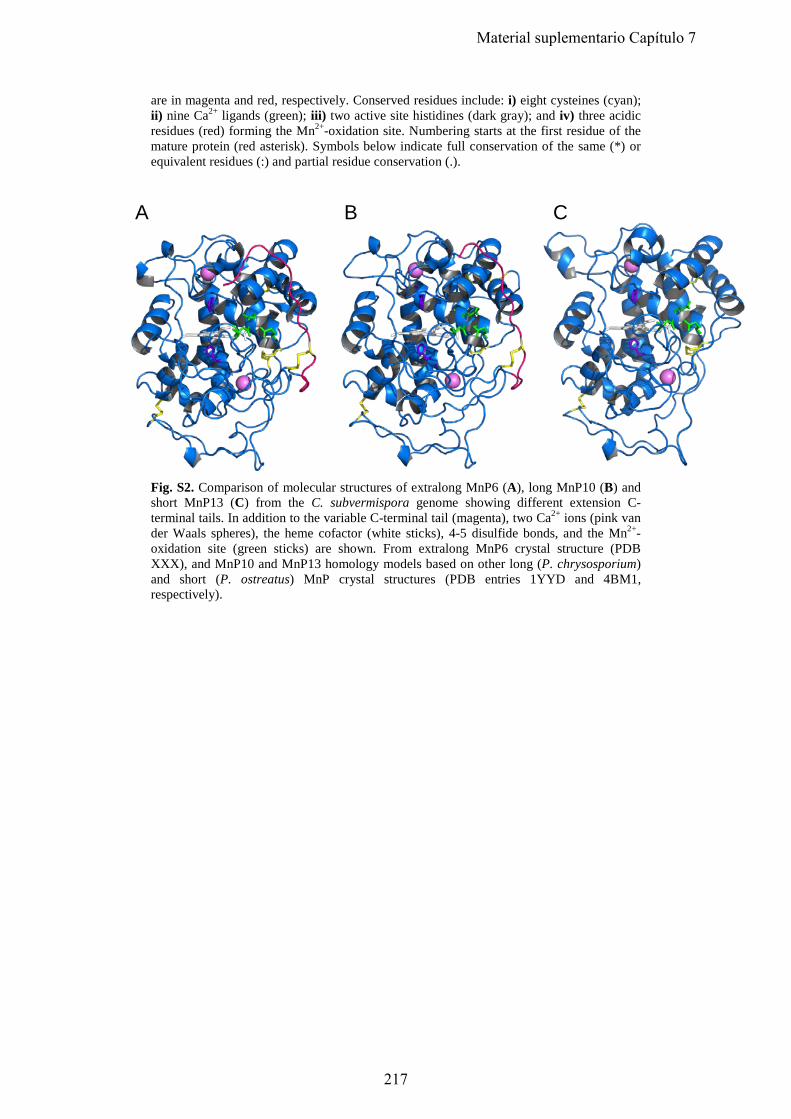
are in magenta and red, respectively. Conserved residues include: i) eight cysteines (cyan); ii) nine Ca2+ ligands (green); iii) two active site histidines (dark gray); and iv) three acidic residues (red) forming the Mn2+-oxidation site. Numbering starts at the first residue of the mature protein (red asterisk). Symbols below indicate full conservation of the same (*) or equivalent residues (:) and partial residue conservation (.).
A B C
Fig. S2. Comparison of molecular structures of extralong MnP6 (A), long MnP10 (B) and short MnP13 (C) from the C. subvermispora genome showing different extension C-terminal tails. In addition to the variable C-terminal tail (magenta), two Ca2+ ions (pink van der Waals spheres), the heme cofactor (white sticks), 4-5 disulfide bonds, and the Mn2+-oxidation site (green sticks) are shown. From extralong MnP6 crystal structure (PDB XXX), and MnP10 and MnP13 homology models based on other long (P. chrysosporium) and short (P. ostreatus) MnP crystal structures (PDB entries 1YYD and 4BM1, respectively).
217
Material suplementario Capítulo 7
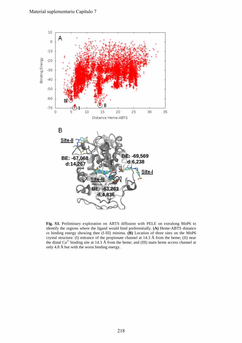
Fig. S3. Preliminary exploration on ABTS diffusion with PELE on extralong MnP6 to identify the regions where the ligand would bind preferentially. (A) Heme-ABTS distance vs binding energy showing thee (I-III) minima. (B) Location of three sites on the MnP6 crystal structure: (I) entrance of the propionate channel at 14.3 Å from the heme; (II) near the distal Ca2+ binding site at 14.3 Å from the heme; and (III) main heme access channel at only 4.8 Å but with the worst binding energy.
218
Material suplementario Capítulo 7
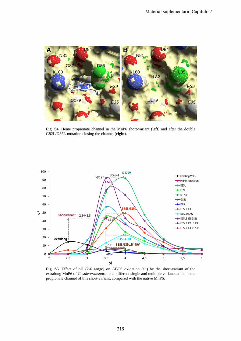
N81
K180L82
D84
L85
E39
E35D179
N81
K180G82
D84
D85
E39
E35D179
A B
Fig. S4. Heme propionate channel in the MnP6 short-variant (left) and after the double G82L/D85L mutation closing the channel (right).
s-1
0
10
20
30
40
50
60
70
80
90
100
2 2,5 3 3,5 4 4,5 5 5,5 6
50686 extral50686shortE35LE39LD179VG82LD85LE35LE39LD85LD179VE35LE39LG82LE35LE369LD85LE35LE39LD179V
pHpH
short-variant
D179V
E35L/E39L
E35L/E39L/D179Vextralong E35L/E39L
2.53.5
3.54>90 s-1
~5 s-1
~45 s-1
G82L
D85L
extralong MnP6MnP6 short-variant
Fig. S5. Effect of pH (2-6 range) on ABTS oxidation (s-1) by the short-variant of the extralong MnP6 of C. subvermispora, and different single and multiple variants at the heme propionate channel of this short-variant, compared with the native MnP6.
219
Material suplementario Capítulo 7
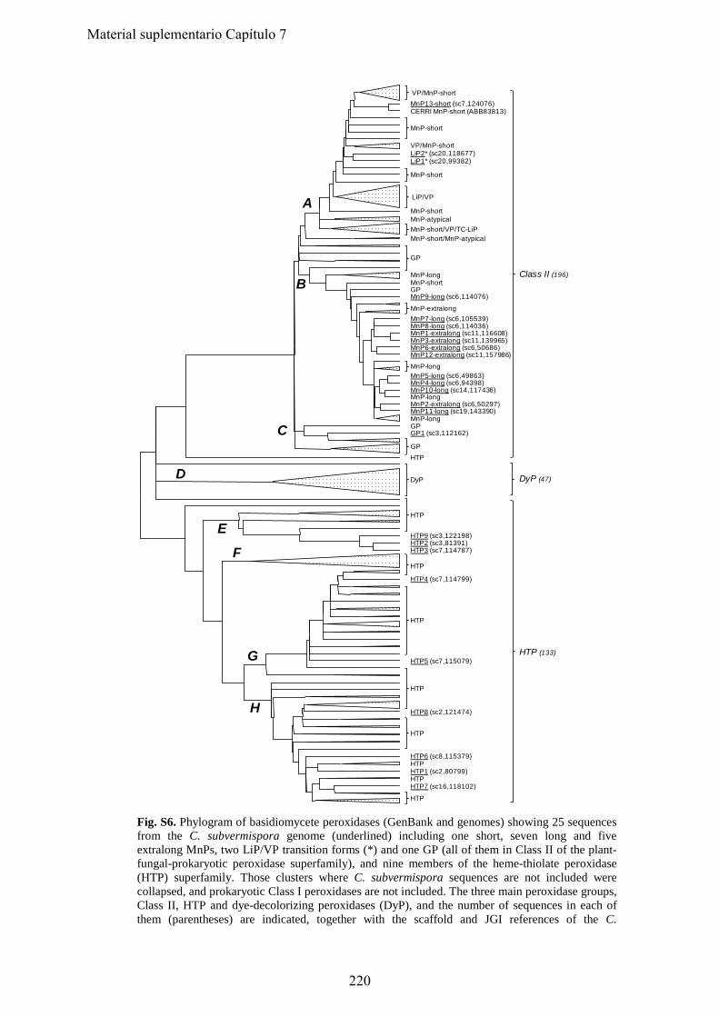
A
B
C
D
E
F
G
H
MnP13-short (sc7,124076)
VP/MnP-shortLiP2* (sc20,118677)LiP1* (sc20,99382)
MnP-short MnP-atypical
MnP-short/MnP-atypical
MnP-longMnP-shortGPMnP9-long (sc6,114076)
MnP7-long (sc6,105539)MnP8-long (sc6,114036)MnP1-extralong (sc11,116608)MnP3-extralong (sc11,139965)MnP6-extralong (sc6,50686)MnP12-extralong (sc11,157986)
MnP5-long (sc6,49863)MnP4-long (sc6,94398)MnP10-long (sc14,117436)MnP-longMnP2-extralong (sc6,50297)MnP11-long (sc19,143390)MnP-longGPGP1 (sc3,112162)
HTP
HTP9 (sc3,122198)HTP2 (sc3,81391)HTP3 (sc7,114787)
HTP4 (sc7,114799)
HTP5 (sc7,115079)
HTP8 (sc2,121474)
HTP6 (sc8,115379)HTPHTP1 (sc2,80799)HTPHTP7 (sc16,118102)
MnP-short
MnP-short
VP/MnP-short
LiP/VP
GP
MnP-extralong
MnP-long
GP
DyP
HTP
HTP
HTP
HTP
HTP
HTP
MnP-short/VP/TC-LiP
HTP (133)
Class II (196)
DyP (47)
CERRI MnP-short (ABB83813)
Fig. S6. Phylogram of basidiomycete peroxidases (GenBank and genomes) showing 25 sequences from the C. subvermispora genome (underlined) including one short, seven long and five extralong MnPs, two LiP/VP transition forms (*) and one GP (all of them in Class II of the plant-fungal-prokaryotic peroxidase superfamily), and nine members of the heme-thiolate peroxidase (HTP) superfamily. Those clusters where C. subvermispora sequences are not included were collapsed, and prokaryotic Class I peroxidases are not included. The three main peroxidase groups, Class II, HTP and dye-decolorizing peroxidases (DyP), and the number of sequences in each of them (parentheses) are indicated, together with the scaffold and JGI references of the C.
220
Material suplementario Capítulo 7
subvermispora gene models (parentheses). Additionally, MnP-atypical corresponds to a MnP type with only two acidic residues at the oxidation site; TC-LiP corresponds to the unique LiP from T. cervina; and CERRI MnP-short corresponds to a Ceriporiopsis rivulosa MnP (GenBank BBB83813) clustering together with C. subvermispora short MnP13. Adapted from (22).
221
Material suplementario Capítulo 7

222
Material Suplementario
Capítulo 8
Engineering a lignin-degrading peroxidase acting at very acidic pH Fernández-Fueyo, E., F. J. Ruiz-Dueñas and A. T. Martínez. (enviado)
223

224
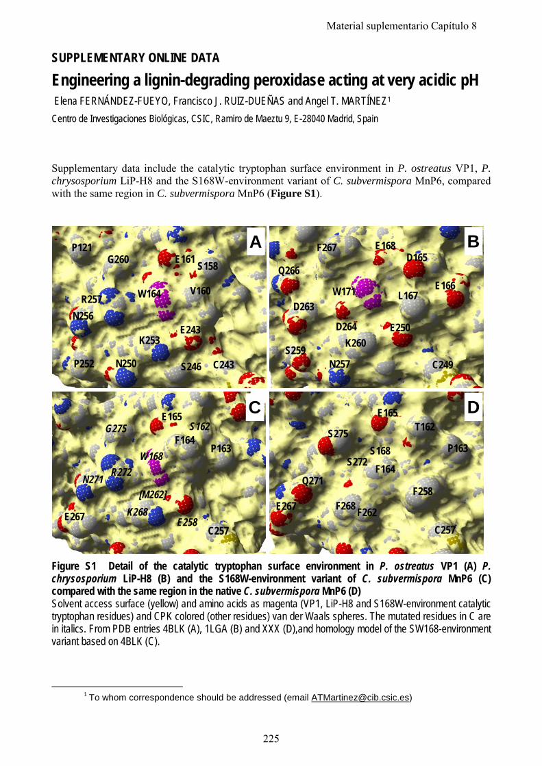
SUPPLEMENTARY ONLINE DATA
Engineering a lignin-degrading peroxidase acting at very acidic pH Elena FERNÁNDEZ-FUEYO, Francisco J. RUIZ-DUEÑAS and Angel T. MARTÍNEZ1
Centro de Investigaciones Biológicas, CSIC, Ramiro de Maeztu 9, E-28040 Madrid, Spain
Supplementary data include the catalytic tryptophan surface environment in P. ostreatus VP1, P. chrysosporium LiP-H8 and the S168W-environment variant of C. subvermispora MnP6, compared with the same region in C. subvermispora MnP6 (Figure S1).
W164
E161
R257
P121G260 S158
V160
E243K253
N250P252
N256
S246
A
C243
W171
D264
D263
Q266
F267 E168D165
E166L167
E250K260
N257S259
B
C249
C
W168
E165
N271
G275 S162
P163
E258
F164
K268E267
R272
C257
[M262]
S168
E165
Q271
S275 T162
P163
F258
F164
F268E267
D
C257F262
S272
Figure S1 Detail of the catalytic tryptophan surface environment in P. ostreatus VP1 (A) P. chrysosporium LiP-H8 (B) and the S168W-environment variant of C. subvermispora MnP6 (C) compared with the same region in the native C. subvermispora MnP6 (D) Solvent access surface (yellow) and amino acids as magenta (VP1, LiP-H8 and S168W-environment catalytic tryptophan residues) and CPK colored (other residues) van der Waals spheres. The mutated residues in C are in italics. From PDB entries 4BLK (A), 1LGA (B) and XXX (D),and homology model of the SW168-environment variant based on 4BLK (C). 1 To whom correspondence should be addressed (email [email protected])
225
Material suplementario Capítulo 8