Embed Size (px)
Citation preview

Biochimica et Biophysica Acta 1794 (2009) 297–308
Contents lists available at ScienceDirect
Biochimica et Biophysica Acta
j ourna l homepage: www.e lsev ie r.com/ locate /bbapap
The 1.4 Å crystal structure of the large and cold-active Vibrio sp. alkaline phosphatase
Ronny Helland a, Renate Lie Larsen a, Bjarni Ásgeirsson b,⁎a The Norwegian Structural Biology Centre, Department of Chemistry, University of Tromsø, N-9037 Tromsø, Norwayb Science Institute, Department of Biochemistry, University of Iceland, Dunhaga 3, IS-107 Reykjavik, Iceland
⁎ Corresponding author. Tel.: +354 525 4800; fax: +35E-mail address: [email protected] (B. Ásgeirsson).
1570-9639/$ – see front matter © 2008 Elsevier B.V. Aldoi:10.1016/j.bbapap.2008.09.020
a b s t r a c t
a r t i c l e i n f oArticle history:
Alkaline phosphatase (AP) Received 13 August 2008Received in revised form 21 September 2008Accepted 23 September 2008Available online 15 October 2008Keywords:Cold-adaptationMetalloenzymeDimerPsychrophilic bacteriaCrystallography
from the cold-adapted Vibrio strain G15-21 is among the AP variants with thehighest known kcat value. Here the structure of the enzyme at 1.4 Å resolution is reported and compared toAPs from E. coli, human placenta, shrimp and the Antarctic bacterium strain TAB5. The Vibrio AP is a dimeralthough its monomers are without the long N-terminal helix that embraces the other subunit in many otherAPs. The long insertion loop, previously noted as a special feature of the Vibrio AP, serves a similar function.The surface does not have the high negative charge density as observed in shrimp AP, but a positively chargedpatch is observed around the active site that may be favourable for substrate binding. The dimer interface hasa similar number of non-covalent interactions as other APs and the “crown"-domain is the largest observedin known APs. Part of it slopes over the catalytic site suggesting that the substrates may be small molecules.The catalytic serines are refined with multiple conformations in both monomers. One of the ligands to thecatalytic zinc ion in binding site M1 is directly connected to the crown-domain and is closest to the dimerinterface. Subtle movements in metal ligands may help in the release of the product and/or facilitate priordephosphorylation of the covalent intermediate. Intersubunit interactions may be a major factor forpromoting active site geometries that lead to the high catalytic activity of Vibrio AP at low temperatures.
© 2008 Elsevier B.V. All rights reserved.
1. Introduction
The removal or transfer of phosphoryl groups is an importantfunction in cellular metabolism and regulation. Enzymes have evolvedalong several lines to perform this function, despite the apparentsimplicity of the reaction [1]. There are several classes of phosphatasesdepending on the presence or absence of metal ion cofactors such aszinc, magnesium, or iron. Some phosphatases must be underregulation at various levels and therefore have structural featuresallowing specific interactions with selected partners. Other phospha-tases seem to be rather non-specific both in terms of substratespecificity and role. The family of alkaline phosphatases (APs) is wide-spread in nature and includes mostly members of the latter type.However, the failure of assigning a role tomany of themmay be due tolack of insight rather than the true state of affairs. Humans carry fourisozymes depending on tissue distribution. A tissue-non-specificvariant is expressed in many tissues and plays, for example, a role inbone mineralization [2]. Corresponding AP is found in lowervertebrates such as shrimp and fish. The isozymes have differentthermal stabilities and respond differently to inhibitors. Thus,structural changes within the AP family take each member along adifferent evolutionary road depending on selective pressures from theenvironment, or for that matter, lack of selective pressures [3,4]. APs
4 525 89 11.
l rights reserved.
derive their name from the basic pH optimum of catalysis (kcat), whichis above pH 10 for enzymes from animals, but closer to pH 9 forbacterial variants [5–7]. In both cases, the optimum is somewhatremoved from physiological pH. However, the vertebrate enzymes canachieve much higher catalytic rates (kcat) than APs from mesophilicbacteria [8] which compensates for the effect of working at more thantwo pH-units below their optimum conditions. In fact, they display20–30 fold higher catalytic activity than the reference AP from E. coli(ECAP) and yet the active-site structure is remarkably similar. It isclear, that this family of enzymes offers great opportunity to gaininsight into fine-tuning of enzyme function through subtledifferences.
Our interest in the AP from a cold-water Vibrio strain (VAP) hascentred on its cold-adaptation. Previous biochemical studies haveshown that APs with enhanced kcat values exist in several organismsadapted to low temperature [6,7,9,10]. Two crystal structures havebeen solved for cold-active APs [11,12], in addition to wild-type andmany mutants of ECAP [13], and the human PLAP isozyme [14]. Theseenzymes have remarkably dissimilar structural details but the coreactive-site structure is highly conserved. The bacterial APs span aspectrum in size, from a short polypeptide version found in theAntarctic bacterium strain TAB5 [10], through the medium-sized ECAPthat has similar polymer chain length to mammalian variants, andculminating in the longest polypeptide version found in a NorthAtlantic Vibrio strain under study here. The size distribution variabilityand possible differences in oligomeric state of APs is most clearly seen

298 R. Helland et al. / Biochimica et Biophysica Acta 1794 (2009) 297–308
in the Vibrio genus, where, for example, five predicted AP genes arefound in V. vulnificus YJ016 (Q7MIW5, Q8DB10, Q8DBM0, Q8DDZ2,Q8D865). All give transcripts corresponding to the longest AP versionsimilar to VAP. In contrast, the V. cholera AP gene remainedunidentified until recently, even despite elucidation of the fullgenomic sequence. A gene quite different to other known AP geneswas confirmed to produce the previously studied AP activity aftercloning, sequencing, and protein expression. [15]. In fact, theV. cholerae AP sequence did not show homology with Vibrio G15-21AP (VAP) [15,16], leading to the conclusion that AP from V. cholerawasa unique enzyme differing greatly even from APs of closely relatedbacteria [15]. V. cholerae contains one of the earliest putative exampleof an active monomeric AP [17] but the more recent study [15] didnot address the question of whether the original report of amonomeric active form was correct. In addition, the first cold-activeAP studied was apparently monomeric [18]. Our interest in solvingthe crystal structure of the Vibrio AP (VAP) was partly to provide ananswer to the question if this enzyme was also monomeric, since theshrimp AP (SAP) was reported to be active both as a monomer and adimer [6]. The sequencing of the VAP polypeptide had suggested thata long insert loop might act as a surrogate second subunit [16]. Atthat time, it was the only long-chain representative in the data banks,and still is the only representative for which biochemical data isavailable [7]. A Swiss-Prot/TrEMBL search now reveals very similarsequences in several species, often obtained in large-scale sequen-cing projects, including V. splendidus (A3UWZ7), V. harveyi(A6AR48), V. campbellii (A8T5A5), V. parahaemolyticus (Q87MR7),V. alginolyticus (Q1VEG3), and V. vulnificus (Q7MIW5), but generallyneither expression of these enzymes has been confirmed nor havetheir properties been studied.
The reaction mechanism of APs was first described in relationwiththe original ECAP structure [13] andmore recently expanded based onfurther structural information [19]. The reaction mechanism of otherAPs has been formulated with reference to the structure of ECAP. Theactive site is characterized by three metal ions that all take part in thereaction pathway, in addition to a nucleophilic serine residue and anarginine that binds the negative charges of the phosphorous-oxygens.Sequence alignments between bacterial and mammalian variantsshow 25–30% identity but the structure around the active site isremarkably similar and conserved in terms of amino acid residues.Thus, only residues D153, T155, and K328 are different whencomparing ECAP with mammalian and other bacterial APs. Further-more, overall structural features are very similar with a central beta-sheet running through the dimeric structure with helices packed oneither site. Most notable differences involve insertions and deletions,including the so-called “crown-domain”. The crown-domain notationwas introduced with the PLAP structure [14]. Cold-adapted APs are ofparticular interest for being the catalysts with highest known kcatvalues in this structural family [10,20,21]. Loops in the crown-domaincreate catalytically important conformational differences [22], allo-wing mammalian APs to be inhibited in a non-competitive manner byseveral amino acids and small peptides. One additional point ofinterest relating to the structure of APs is potential collaborationbetween subunits. This seems to depend on metal-site saturation. Ifmetals are lacking, each monomer is dependent on the other forstability and catalytic performance. When fully metalated, themammalian APs are non-cooperative allosteric enzymes (i.e. subunitsindependent, but each has regulatory subsites) [23]. Subunit interac-tions are influenced quite markedly by the N-terminal microenviron-ment, both in ECAP and mammalian APs [24], but that part is lackingin the VAP under study here.
A direct comparison of the refined ECAP structures with andwithout phosphate revealed a strong correlation between theoccupancy of the third metal-binding site and the conformation ofthe S102 nucleophile [19]. The best conformation for nucleophilicattack by S102 in ECAP requires a magnesium ion in the third metal
binding site. The other two metal sites are normally occupied by zinc.However, APs from organisms such as Thermotoga maritima and Ba-cillus subtilis require cobalt for maximal activity and function poorlywith zinc and magnesium [25]. The amino acid residues that bind thetwo zinc atoms are conserved in all known APs, whereas there is someflexibility in which residues bind the third (magnesium) ion. Thisaffects activity greatly, as numerous mutagenesis studies have shown[8,20,26]. ECAP has an aspartate (D153) where most other APs have ahistidine. Histidine may increase the affinity of the third metal site forzinc and underlie stimulating effect of magnesium seen with manyAPs due to displacement of zinc from the third site. It also explainseasy formation of triple zinc variants, for example in the crystal of SAP[11]. Furthermore, metal ion specificity is altered when the variablemetal coordinating residues in ECAP are changed to the equivalentresidues in mammalian APs (D153H and K328H). VAP has H116(equivalent to D153 in ECAP) and W274 (equivalent to K328 in ECAP)and shares this with both TAP and APs from some more thermophilicorganisms. It has been proposed that the H153/W328 (ECAPnumbering) combination of residues selected for cobalt rather thanzinc [27]. In fact, ECAP can effectively use cobalt instead of the zinc andmagnesium when mutated to contain W328. We have recentlyanalyzed metal content of wild-type VAP samples and only foundzinc and magnesium, but no cobalt. VAP was very dependent onmagnesium in buffers for maintenance of full activity, even at lowtemperature, suggesting loose association of the metal with theenzyme [20]. The X-ray structure was expected to give further infor-mation on this point.
The rate limiting step in the reaction mechanism of ECAP atalkaline pH is the release of product, i.e. non-covalently boundphosphate [5]. This conclusion has been carried over to describereaction mechanism of all APs. However, caution should be levelledtoward the possibility that the rate limiting stepmay be transferred toan earlier step in the pathway, such as the initial hydrolysis of thephosphoryl ester substrate or hydrolysis of the covalent phosphoryl–serine intermediate. Mutants have been made that show transfer ofthe rate determining step. An example is the K328C mutation in ECAP[28]. Furthermore, K328A and K328H ECAP mutants had much highertransphosphorylating than hydrolytic activity. This could not beexplained purely by greater ease in releasing phosphate from theactive site, but may be due to transfer of the slowest step to thehydrolysis of the covalently bound phosphoryl–enzyme complex [29].As previously mentioned, APs of vertebrate origin have a histidine inposition equivalent to ECAP K328, whereas VAP and a few othervariants have a tryptophan.
Our aim with the present paper is to describe the structure of VAPand review the features that may relate to its high catalytic rate andcold-adaptation. Several AP crystal structures have been obtained inrecent years with some distinct features. In addition to the originalECAP structure at 2.0 Å showing two zinc ions and onemagnesium ionin the active site [13], the structures of the PLAP at 1.8 Å [14], SAP at1.9 Å [11], and TAP at 1.95 Å [12] followed. Given the very high activityof the VAP variant, and the known sequence differences in the activesite that partly create this enhanced activity [20], we were interestedin cataloguing the structural features implicated in catalysis andcompare with the rather rich background of the other structuralrelatives.
2. Materials and methods
2.1. Crystallization, data collection and refinement of VAP
Recombinant expression of VAP is described elsewhere [20]and protein purification was essentially as described pre-viously [7]. Briefly, an initial affinity chromatography step on anL-histidyldiazobenzylphosphonic acid column was performed wherethe enzyme was eluted by adding 0.1 M potassium phosphate to the

Table 1Data collection and refinement statistics
Peak Remote
Data collectionDiffraction limit (Å) 2.0 1.4Wavelength (Å) 1.2827 0.9184Unit cell parameters (Å3)
a-axis (Å) 118.19 118.24b-axis (Å) 165.92 165.98c-axis (Å) 57.48 57.48
Space group P21212 P21212Total no. of reflections 945418 (58126) 883174 (46374)No. of unique reflections 74311 (8728) 205938 (21811)Completeness (%) 96.4 (80.1) 92.9 (68.8)Anomalous completeness (%) 96.4 (79.8) 84.0 (44.4)I/σ(I) 13.9 (13.2) 17.8 (10.1)Mean ((I)/sd(I)) 50.1 (29.8) 30.4 (10.9)Rmerge (%) 4.1 (4.9) 2.7 (7.4)Multiplicity 12.7 (6.7) 4.3 (2.1)Wilson B (Å2) 9.15 8.04Outer shell resolution (Å) 2.11–2.00 1.48-1.40
Experimental phasingPhasing power 2.98 naRCullis 0.44 naFigure of merit 0.53 na
RefinementR-factor (%) na 15.54Rfree (%) na 16.65Average B factors (Å2) na 8.05No. protein atoms na 8060No. solvent molecules
Zn2+ na 4Mg2+ na 2SO4
2− na 11Ethylene glycol na 3Water na 847
R.m.s. deviationsBond lengths (Å) na 0.007Bond angles (°) na 1.177
Diffraction-component precision indicator 0.054Ramachandran plot (%)
Most favoured na 91.7Additionally allowed na 8.3Generously allowed na 0Disallowed na 0
Outer shell values are given in parenthesis.na: not available.
Fig. 1. Ribbon representation of VAP, where molecule A is coloured blue and molecule Bis red. The conformation of unique insert II of the B molecule extending along thesurface of molecule A is clearly visible. The “crown"-domain is located on the top of thefigure and the catalytic zinc and magnesium ions located in the active site arerepresented as gold and green spheres, respectively. Sulphates and ethylene glycol arerepresented as ball-and-stick models.
299R. Helland et al. / Biochimica et Biophysica Acta 1794 (2009) 297–308
eluant. This was followed by a MonoQ anion exchange chromato-graphy where the enzyme eluted at 0.15 M in a 0–0.7 M NaCl gradient.The columns were run with buffer containing 20 mM Tris, 10 mMMgSO4, at pH 8.0, with 15% (v/v) ethylene glycol added in the MonoQstep to facilitate storage in frozen form. The protein was concentratedto 8 mg ml−1 and ethylene glycol concentration was reduced to 5% (v/v) prior to crystallization. Crystals of VAP suitable for X-ray diffractionstudies were obtained by the hanging drop vapour diffusion methodfrom reservoir solution containing 0.2 M Li2SO4, 0.1 M Tris pH 7.75,23% PEG 3350 and 3% v/v ethylene glycol at 4 C temperature. X-raydatawere collected at BL14.1 at Bessy. Data (Table 1) were processed inXDS [30], SCALA and TRUNCATE of the CCP4 program suite [31]. Thecrystal structure was solved using the single anomalous dispersion(SAD) technique on X-ray data collected to 2.0 Å on the K edge(1.2827 Å) of the catalytic zinc ions, and higher resolution data to 1.4 Åwere collected on the same crystal at a remote wavelength (0.9184 Å).SHELXD [32] was used to identify four heavy atom sites which werefurther refined with SHARP [33]. The phases were improved bysolvent flattening using SOLOMON [34]. Automatic tracing of thepolypeptide chains, using the peak data, was carried out withARP/wARP [35]. Subsequent improvement of the model was made
using the data collected on the remotewavelength bymanual refittingof side chains using O [36] based on sigmaA-weighted 2mFo-DFc andmFo-DFc electron density maps. Refinement was carried out inRefmac5 [37] of the CCP4 suite (Table 1).
Programs for structural comparison and analysis included the CCP4suite, the DaliLite server (http://www.ebi.ac.uk/DaliLite/) [38] and theSecondary Structure Matching (SSM) server (http://www.ebi.ac.uk/msd-srv/ssm/ [39]). Selection of PDB entries for structural comparisonwas based on best hits from SSM and the nature of the ions in theactive sites. Inserts and deletions were defined as regions which do, ordo not, superimpose on VAP in the structural alignment using theDaliLite server. Estimation of intramolecular hydrogen bonds wascarried out using HBPLUS [40] with a 3.4 Å distance criteria anddefault donor-acceptor angles of 90°. Analysis of the dimer interfaceincluded the use of the Protein interfaces, surfaces and assembliesservice, PISA, at European Bioinformatics Institute (http://www.ebi.ac.uk/msd-srv/prot_int/pistart.html) [41]. Calculation of electrostaticsurface potential was done using Delphi [42] with atomic partialcharges from the AMBER molecular simulation program [43].Illustrations were prepared in PyMOL (DeLano Scientific (http://pymol.sourceforge.net/)), with the exception of the structural align-ment which was generated at the ESPript server (http://espript.ibcp.fr/ESPript/ESPript/) [44].
3. Results
3.1. Overall structure
Rod-like crystals up to 0.6×0.2×0.2 mm3 grew from PEG3350,lithium sulphate, Tris pH 7.75 and ethylene glycol after three to fourweeks. The crystal structure of VAP was determined to 2.0 Å using thesingle anomalous dispersion (SAD) technique on data collected 10 eVabove the Zn K edge (1.2827 Å). Higher resolution data to 1.4 Å werecollected on the same crystal at a remote wavelength (0.9184 Å). Datacollection and refinement statistics are listed in Table 1. SHELXD [32]and SHARP [33] located four heavy atom sites, and using theexperimental phases to 2.0 Å, ARP/wARP [35] could build 930residues. Phase extension to 1.4 Å allowed auto-building of 980 ofthe 1004 residues in the VAP dimer. The final structure is refined tocrystallographic R-factor of 15.54% and Rfree of 16.65% and includestwo VAP monomers (Fig. 1) containing two zinc and one magnesium
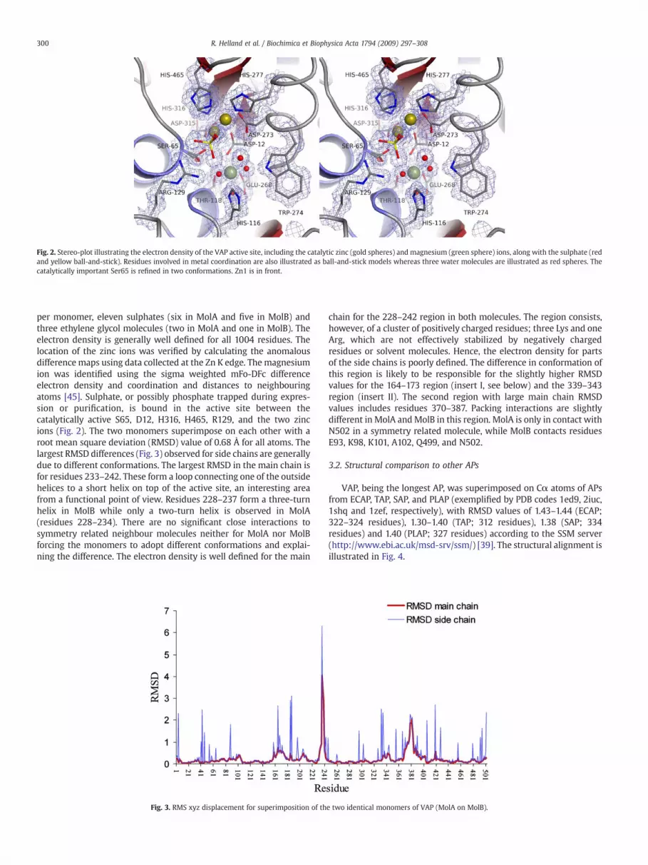
Fig. 2. Stereo-plot illustrating the electron density of the VAP active site, including the catalytic zinc (gold spheres) and magnesium (green sphere) ions, along with the sulphate (redand yellow ball-and-stick). Residues involved in metal coordination are also illustrated as ball-and-stick models whereas three water molecules are illustrated as red spheres. Thecatalytically important Ser65 is refined in two conformations. Zn1 is in front.
300 R. Helland et al. / Biochimica et Biophysica Acta 1794 (2009) 297–308
per monomer, eleven sulphates (six in MolA and five in MolB) andthree ethylene glycol molecules (two in MolA and one in MolB). Theelectron density is generally well defined for all 1004 residues. Thelocation of the zinc ions was verified by calculating the anomalousdifference maps using data collected at the Zn K edge. The magnesiumion was identified using the sigma weighted mFo-DFc differenceelectron density and coordination and distances to neighbouringatoms [45]. Sulphate, or possibly phosphate trapped during expres-sion or purification, is bound in the active site between thecatalytically active S65, D12, H316, H465, R129, and the two zincions (Fig. 2). The two monomers superimpose on each other with aroot mean square deviation (RMSD) value of 0.68 Å for all atoms. Thelargest RMSD differences (Fig. 3) observed for side chains are generallydue to different conformations. The largest RMSD in the main chain isfor residues 233–242. These form a loop connecting one of the outsidehelices to a short helix on top of the active site, an interesting areafrom a functional point of view. Residues 228–237 form a three-turnhelix in MolB while only a two-turn helix is observed in MolA(residues 228–234). There are no significant close interactions tosymmetry related neighbour molecules neither for MolA nor MolBforcing the monomers to adopt different conformations and explai-ning the difference. The electron density is well defined for the main
Fig. 3. RMS xyz displacement for superimposition of th
chain for the 228–242 region in both molecules. The region consists,however, of a cluster of positively charged residues; three Lys and oneArg, which are not effectively stabilized by negatively chargedresidues or solvent molecules. Hence, the electron density for partsof the side chains is poorly defined. The difference in conformation ofthis region is likely to be responsible for the slightly higher RMSDvalues for the 164–173 region (insert I, see below) and the 339–343region (insert II). The second region with large main chain RMSDvalues includes residues 370–387. Packing interactions are slightlydifferent in MolA andMolB in this region. MolA is only in contact withN502 in a symmetry related molecule, while MolB contacts residuesE93, K98, K101, A102, Q499, and N502.
3.2. Structural comparison to other APs
VAP, being the longest AP, was superimposed on Cα atoms of APsfrom ECAP, TAP, SAP, and PLAP (exemplified by PDB codes 1ed9, 2iuc,1shq and 1zef, respectively), with RMSD values of 1.43–1.44 (ECAP;322–324 residues), 1.30–1.40 (TAP; 312 residues), 1.38 (SAP; 334residues) and 1.40 (PLAP; 327 residues) according to the SSM server(http://www.ebi.ac.uk/msd-srv/ssm/) [39]. The structural alignment isillustrated in Fig. 4.
e two identical monomers of VAP (MolA on MolB).

Fig. 4. Structural alignment generated by pairwise comparison of the structures of VAP, ECAP, TAP, SAP, and PLAP using the DaliLite structure comparison server (http://www.ebi.ac.uk/DaliLite/). The secondary structure of VAP (top) is illustrated. Blue stars indicate residues involved in metal binding or being involved in catalysis and specificity.
301R. Helland et al. / Biochimica et Biophysica Acta 1794 (2009) 297–308

302 R. Helland et al. / Biochimica et Biophysica Acta 1794 (2009) 297–308
VAP is characterized by four inserts (Figs. 4 and 5a) having nocounterpart in the homologous APs according to pairwise struc-tural alignment using DaliLite (http://www.ebi.ac.uk/DaliLite/) [38].These regions include residues 162–175 (insert I), 325–351 (insert II),361–395 (insert III), and 405–450 (insert IV) by VAP numbering. Onlyinsert I is not related to the crown-domain, known for being ofdifferent shapes and sizes in various APs. Regions absent in VAPaccording to the structural alignment (Fig. 5b) are generally due todifferent folding of the crown-domains. These regions include aninsert of 14–15 residues in the VAP 133–134 region in SAP, PLAP, andECAP, an insert of 10–20 residues in the VAP 89–90 region in SAP andPLAP, and a seventeen residue insert in the VAP 231–232 region inECAP.
3.3. Insert I, residues 162–175
The thirteen residue insert, not being part of the crown-domain orthe dimer interface, forms a three-turn helix (residues 158–173) inVAP. The other four homologous enzymes have loops in this regionoccupying different conformations. The loops in ECAP and TAP are
Fig. 5. Superimposition of VAP (red), TAP (cyan, PDB 2iuc), ECAP (yellow, PDB 1ed9) SAP (greregions colour coded red. Loops of the other APs in the same spatial regions are coloured toabove.
shorter than in the eukaryotic APs. Attachment of the helix to theremaining enzyme is provided essentially through hydrophobicinteractions, since the hydrogen bonds found in the helix region areprimarily between main chain nitrogen and oxygen atoms of the loopitself (Supplementary Table 1).
3.4. Insert II, residues 325–351
This insert was initially believed to be located in the interfaceregion, preventing formation of a biologically active dimer. Thepresent structure shows that the twenty-six residue insert extendsalong the surface of the other monomer. The loop is characterizedwith relatively few intramolecular hydrogen bonds (twelve, Supple-mentary Table 1), where six are between main chain atoms. However,the loop, consisting of eight charged and nine non-polar residues,contributes significantly to the dimer stabilization by forming elevenintermolecular hydrogen bonds. Omitting this insert in calculation ofburied water accessible surface area (ASA) in the dimer interfacereduces the interface area from 4271 Å2 to 2627 Å2 (about 38%) permonomer (Supplementary Table 2).
en, PDB 1shq), and PLAP (magenta, PDB 1zef). (a) Loop regions including the unique VAPillustrate differences. (b) Regions with deletions in VAP are colour coded as described

303R. Helland et al. / Biochimica et Biophysica Acta 1794 (2009) 297–308
3.5. Insert III, residues 361–394
The thirty-four residue insert folds into two helices (residues368–376 and 385–395), separated by a loop including a three-residue 310 turn. About 62% of the 45 hydrogen bonds formed bythis loop are between main chain atoms, and similar to insert I, fewinclude residues not being part of the loop itself. Although the loopis part of the crown-domain it is not part of the dimer interfaceregion but rather extends on the other side over the active sitecrevasse.
3.6. Insert IV, residues 405–450
The forty-six residue insert consists mostly of loop structurecontaining two helices at the start and the end of the insert (residues402–409 and 445–456) and two short strands (residues 411–413 and428–430), forming an antiparallel sheet. More than half of thehydrogen bonds of this insert are, similar to the other inserts,between main chain atoms (Supplementary Table 2). The loopis involved in one of the two intermolecular salt-bridges D434(MolA)–R450 (MolB), resulting in two of the four intermolecular saltbridges for the dimer. Omitting residues 405–450 from the calcula-
Fig. 6. (a) Ribbon presentation of VAP looking into the active site. (b) Standard view of VAP illuElectrostatic surface potentials of VAP, TAP, ECAP, SAP and PLAP. The left view has the orientatcontoured from −10 (red) to 10 (blue) kT/q.
tion of buried ASA reduces the interface area from 4271 Å2 to 3217 Å2,about 24%, per monomer.
3.7. The active site
The active site of VAP consists of two zinc and one magnesiumions (Fig. 2). The Zn2+ ion at the M1 site is hexa-coordinated in adistorted octahedral arrangement by D273 (bidentate), H277, H465,and two oxygen atoms from Sul610 (or possibly a phosphate ion). Thezinc ion at the M2 site is penta-coordinated by D12, S65, D315, H316,and one oxygen of Sul610. The Mg2+ ion at the M3 site is octahedrallyhexa-coordinated by D12, T118, E268, and three waters. The residuesinvolved in the first sphere of metal binding are conserved in the APscompared in this study, with the exception of residues around the M3binding site. In the VAP H116 position, ECAP has an Asp while thereare His in the other enzymes, and in the VAP T118 position PLAP hasSer while the other enzymes have Thr. Lastly, the residue at the VAPW274 position is also Trp in TAP, but Lys in ECAP, and His in SAP andPLAP. The conformation of the conserved metal ligands is similar inall five enzymes and differences in metal-ligand distances aregenerally less than 0.3 Å (Supplementary Table 3). Small displace-ments could be due to different ligands within the active site of the
strating onemonomer as ribbon, and the secondwith electrostatic surface potential. (c)ion of (a) and the right view the orientation of (b). The electrostatic surface potentials are

304 R. Helland et al. / Biochimica et Biophysica Acta 1794 (2009) 297–308
crystals; namely sulphate in VAP and SAP, phosphite in PLAP and inTAP, and H2O in ECAP. Sulphate in VAP is in a position to forminteractions to both zinc ions and a bidentate interaction to R129(ECAP R166). SAP has been solved with both sulphate and phosphate(PDB 1shq and 1shn, respectively) in the active site. Both anionsoccupy positions similar to sulphate in VAP, but interestingly, SAPR162 occupies different conformations in the two structures and theside chain is rotated away when the anion is sulphate. The phosphor-atom included in the TAP structure coincides with position of thesulphur-atom in the VAP structure, and R148 in TAP occupies thesame position as R129 in VAP. Two of the oxygen atoms of phosphitein PLAP form bidentate interaction to R166. The nucleophilic S65 isrefined in two conformations in VAP.
Tyr325 from the VAP B-subunit extends into the A-subunitsubstrate binding site with the hydroxyl group 4.69 Å from Zn at theM1 position. All the other APs, except ECAP, have a tyrosine residue atan equivalent position, although this cannot be seen from thestructural alignment (Fig. 4): TAP; Y325 (B-subunit), 5.94 Å awayfrom Zn1, SAP; Y366 (B-subunit), 5.64 Å from Zn1, PLAP; Y367(symmetry related molecule), 5.43 Å from Zn1. In addition, VAPpossesses a second tyrosine, Y44 from the A-subunit, where thehydroxyl group is 5.99 Å from Zn1 (A-subunit) and 2.67 Å from thehydroxyl of the B-subunit Y325.
The catalytic efficiency of an enzymewill depend on the substrate'saccessibility to the active site. The large inserts in VAP are located closeto the rim of the depression forming the catalytic site in most APs,such that the VAP catalytic site becomes relatively deeply buried in acrevice running between inserts III and IV on one side and the wingdomain on the other (Fig. 1, Fig. 6). This crevice also contains threesulphate ions in addition to the sulphate coordinating the catalyticzinc ions, and one of them stabilizes the interaction between inserts IIIand IV. The somewhat narrow entrance to the active site appears to bepartly compensated for by a relatively large positive potential whichmay attract the negatively charged phosphate of a substrate into thecatalytic site (Fig. 6). The electrostatic attraction between oppositecharges has on several occasions been reported to be central for thecatalytic efficiency of enzymes from cold-adapted species (for review,see i.e. [46,47]). This positive potential in VAP is considerably largerthan in the other APs, but it is difficult from the figure to estimate therole of the electrostatic potential to the cold-adaptation features ofVAP.
3.8. The interface
Inserts II and IV contribute significantly to the dimer formation ofVAP. Dimerization involves 115 residues and buries 4271 Å2 of wateraccessible surface area (ASA) per monomer, or 19.7% of the total ASA(Supplementary Table 2). This is of the same order as the other APs,generally with 21–22% of the total ASA buried, but considerably morethan in the cold-adapted TAP, with 1835 Å2 or 14.1% of the total ASAburied. The small interface in TAP is attributed to the small(as visualized in Fig. 6) crown-domain together with the lack of anN-terminal helix as in PLAP and SAP or a corresponding largeN-terminal loop as in ECAP [12]. VAP also lacks the N-terminal helix,but the stabilizing effects of the helix seem to be compensated for byinsert II. Removing N-terminal amino acids prior to VAP position 1(Fig. 4) in ECAP, PLAP, and SAP, and insert II in VAP before interfaceanalysis yields a buried ASA of 2627–2839 Å2 for VAP, PLAP, and SAP,and 2200 Å2 for ECAP. Removing insert IV in VAP, further reduces theburied ASA to 2100 Å2, hence, being of the same order as the otherbacterial APs (ECAP and TAP). There is no apparent trend in thepercentage of charged residues in the interface regions of the fiveAPs, with the exception of SAP being considerably more negativelycharged overall and no ionic interactions being present in TAP overthe dimer interface. Interestingly, the percentage of non-polarresidues in the VAP interface (40%) approaches that of PLAP (45%),
while the fractions in the other three APs are in the range 30–34%(Supplementary Table 2).
4. Discussion
4.1. General
We are now in a position to compare the extra large version VAP totwo other cold-active variants, one very short version from theAntarctic bacterium strain TAB5 (TAP), the other medium sized fromNorthern shrimp (SAP). In addition, two AP variants from mesophilicorganisms (ECAP and PLAP) have previously been solved structurallythat are both medium sized in length. The quality of the structureobtained here for VAP is at a very good resolution of 1.4 Å with no partof the polypeptide unclear or excluded due to, for example, move-ments of loops. It confirms that the VAP is a dimer. It is also shownhere for the first time how the insert in the sequence (insert II;325–351), that in particular characterizes VAP, attaches themonomerstogether, thus taking the role of the N-terminal subunit connections inECAP, SAP, and PLAP. This part of the polypeptide has inherently aremarkably little secondary structure as predicted by computationalanalysis (data not shown).
4.2. Metal coordination and water in the active site
The structure confirms previous analysis [20] that themetal ions inthe active site are two zinc ions and one magnesium. Some of the APstructures have contained three zinc ions in the active sites [11,14].Still other structures have contained additional metal binding sites,such as for calcium and magnesium, proposed to be of functionalimportance [4,12]. No additional metal binding-sites were found inVAP as in some other APs, but several binding sites for sulphate andethylene glycol originating from the solvents were observed that pointto potential regulatory interactions with other molecules.
ECAP exhibits maximal activity when zinc fills the M1 andM2 sitesandmagnesium is present in siteM3 [25], but APs canworkwith othermetal ions. In fact, an AP variant from the thermophilic T. maritima hasbeen suggested to be preferentially ligated with cobalt due the factthat K328 (ECAP numbering) is a tryptophan and D153 is a histidine[25,48]. This is the samemetal ligands as in the VAP under study here.Any generalization about the preference of the M3 sites for other ionsthan magnesium if tryptophan and histidine occupy the positions inquestion is refuted by the present results and our previous directatomic absorption measurements [20].
All three metal ions take part in the catalysis and the magnesiumion is also important for stability. The latter abstracts the protonfrom the nucleophilic serine through generation of a metal-boundhydroxide ion. This mechanism is assumed to be a general one. All-zinc containing enzymes have a lower activity but can be reactivatedwith magnesium for optimal activity at alkaline pH [49]. Additionof zinc to many APs is inhibitory. The different effects may be due tosub-optimal coordination of the magnesium-bound hydroxide ion inthe initial nucleophilic attack by different ions [50]. In SAP, zinc in M3can be replaced by magnesium by extensive dialysis, and this triggersH149 to adopt a conformation in which it does not coordinate themetal ion [11], but instead superimposes with the correspondinghistidines in PLAP, TAP, and now also VAP, which all containmagnesium in M3.
The backbone positions of the VAP metal ligands fit very closely tothe structure of ECAP and the other APs. TheM1 andM2 ligands are allin a very close alignment through the backbone, and side chains alsosuperimpose well. The twoM3 ligands that are identical in all APs alsosuperimpose, but the two residues that differ between various APs areslightly out of alignment. The side chain of W274 (ECAP K328) in thesecond variable position lies in the same plane as the correspondinglysine in ECAP. The Trp rings in VAP (W274) and TAP (W260) are in the

305R. Helland et al. / Biochimica et Biophysica Acta 1794 (2009) 297–308
same plane as the corresponding histidine in PLAP (H317) and thenitrogen atoms superimposewell. In SAP, H316 differs from PLAP H317by a 45° rotation of the imidazole ring through the Cβ-Cγ bond. Theplane of the imidazole ring of VAP H116 in the first variable position isrotated about 55° relative to the plane of the carboxyl group of ECAPD153. The imidazoles of H135 in TAP and H153 in PLAP occupyconformations almost identical to VAP H116, while H149 in SAP isrotated about 50° through the Cβ-Cγ bond in the structure containingmagnesium at M3 (PDB 1shn). Despite slightly different conforma-tions of the side chains, all distances between the ligand and themagnesium ion are of the same order of around 4 Å (SupplementaryTable 3). The mutation H135D in TAP [26] (D153 in ECAP) resulted in amore stable but less active enzyme. The TAP H135 had two possibleconformations with magnesium in M3 which could not be distin-guished [12]. The published model was built with reference to PLAPwith Nɛ2 pointing toward R148 (VAP R129, ECAP R166) but it couldalso take part in the M3 water shell analogous to the function of theAsp153 in ECAP, a structure suggested by the authors to bemore likely.
At alkaline pH, the release of product is the slowest step in ECAP[5], where K328 stabilizes phosphate binding through a watermediated interaction. Weakening of this interaction increases productrelease dramatically [29]. The single K328H mutant had a lowerbinding constant for phosphate that produced a higher catalytic rate,whereas affinity for substrate was little changed. Differences in metalcoordination and strength of interaction with phosphate can explainhigher activity in the ECAP cobalt variant of the D153H/K328Wmutant compared with wild-type. VAP and TAP have a tryptophan inthe position corresponding to K328 that would suggest a similarmechanism for activity enhancement, whereas SAP and PLAP havehistidine in this position. In the D153H/K328H ECAP mutant, thehydrolysis of the covalent intermediate becomes partially rate-limiting because product release is very fast [29]. We do not know ifthis is the case for the VAP, but changing the two residues to the onescorresponding to ECAP had dramatic effect on activity as well asstability [20]. The contention that rate of product release is rate limingin all APs should be approached with caution as universal, especiallysince ECAP is rather the exception when it comes to residue choice inthe two positions referred to above.
The AP structures have water molecules at conserved positions inthe otherwise empty active sites. This is also seen in VAP, where threewaters are located around themagnesium ion in the same positions assimilar waters in the other structures. These are involved in thecatalytic function and their exact location might influence theproficiency of catalysis.
4.3. Conformation of key residues
Motional freedom of the residues involved in catalysis is closelylinked with enzyme function such as binding of substrates and releaseof products. Such considerations gain added weight when one tries todiscover the subtle differences thatmake enzyme variants particularlycold-active, as is the case for VAP. In VAP, the nucleophilic S65 (ECAPS102) is on a helix that superimposes very closely onto a correspon-ding ECAP helix. The active site Ser65 was refined in two differentconformations, illustrating a flexible nature of the side chain. Thiscoincides with S84 in TAP which had several possible positions. Themost common position (χ=60°) placed the serine oxygen atom inroughly the same conformation as in the ECAP and PLAP structureswith transition state mimics, and within the coordination sphere ofthe M2 ion.
Arg129 is another active site residue involved in the initial bindingof substrate and the release of product as mutational studies on ECAP(R166) have shown. The arginine's conformational position is knownto be different in the AP crystal structures. Two positions werepossible in SAP; “docked” (PDB 1shn) and “non-docked” (PDB 1shqand 1k7h). The presence of substrate or inhibitor was deemed a likely
prerequisite for it to adopt the docked position in SAP [11,51], and theauthors concluded that non-docked R162 might facilitate the releaseof product and be a factor in cold-active properties. The conformationof VAP R129 superimposes well on the corresponding residues inECAP, PLAP (PDB 1zef) and the “docked” conformation in SAP.Interestingly, R162 in SAP adopts the “docked” conformation whenphosphate is present at the active site and Zn at M3, but not in thepresence of sulphate and magnesium in M3. The anion-free structurehaving Zn in M3 (1k7h) adopts the “non-docked” conformation. Thus,the conformation of R162 in SAP appears to be linked to the nature ofthe anion at the active site, and not necessarily the metal at M3. R166in PLAP also adopts the “docked” conformation with phosphite at theactive site. Based on these observations, the hypothesis that the anionbridging the two zinc ions in VAP (interpreted as Sul610) could indeedbe a phosphate trapped in expression or purification is highly likely.
4.4. Surface characteristics and access to the active site
The interactions of APs with various substrates and inhibitors haverevealed some preferences that relate back to the active site structureas well as general surface characteristics. A very negatively chargedsurface has been observed in some cold-adapted enzymes, includingSAP, indicating that it may help catalysis through directing substrateapproach to the positive charge around the active site [11]. A similarnegative electrostatic surface of the cold-adapted VAP and TAP is notobserved. Instead their surfaces resemble more ECAP and PLAP. VAPhas a slightly larger positive potential area surrounding the active site(Fig. 6), possibly being involved in attracting the negatively chargedsubstrate into the active site at reduced temperatures. A specialfeature of VAP is how inserts III and IV shield the active site. The“overhang” may regulate interactions of VAP with substrates in someas yet unknownmanner through specific binding. No other substratesapart from p-nitrophenol phosphate and activity stain have been usedto assay VAP activity, so it cannot be stated yet that it is non-specific.Recently, we have discovered that VAP is inhibited by various aminoacids like vertebrate APs [23] indicating that it can accommodatesmall organic molecules in a specific manner (Ásgeirsson,unpublished).
4.5. Structural and functional features of the dimeric interface
One of the main revelations brought to light in this study is thedimeric nature of VAP. Biochemical evidence that earlier pointedtoward the existence of an active monomeric form included size-exclusion chromatography, activity staining in native electrophoresisgels [7], and attempts at cross-linking the monomers (unpublishedwork). Althoughmost well characterized APs are dimers, there are stillsome contentious reports of activemonomeric forms [17,18]. In light ofthe VAP structure presented here, this would seem increasinglyunlikely.
The dimer interface is a determinant of stability and how wellmetals bind. There is also close synergy between the monomers invarious APs; they do not function independently of one another.Cooperation between subunits has been proposed for calf intestinal APto facilitate the release of product in a half-of-sites model, involvingcommunication over the interface [5,52]. From the earliest studies onECAP, kinetic evidence was forwarded that implemented a conforma-tion change as the rate-determining step in catalysis [53] presumablyto assist in release of the non-covalently bound product, thephosphate ion. More recently, subunit communications have beendiscovered in PLAP where residues from one subunit become part ofthe active site in the second one [23]. A similar phenomenon is foundin VAP, where Tyr325 from the B-subunit extends into the A-bindingsite. Closer inspection of the APs included here reveals that all, exceptECAP, have a tyrosine from the second monomer in the active site ofthe first monomer.

306 R. Helland et al. / Biochimica et Biophysica Acta 1794 (2009) 297–308
Dissociation of the AP dimers by urea or guanidinium leads toinactivation [54–56]. The dimers in cold-adapted enzymes are heldtogether by weaker interaction than in mesophilic variants whichmayexplain part of their improved catalytic action through increasedmobility [6,55]. Cooperativity in substrate hydrolysis, or the allostericinteractions suggested for some APs, would be affected by interactionsat the interface that lies close to the two active sites. However,oligomerization appears in many cases to be driven by the need toincrease stability rather than shape activity. Thus, cold-adaptedenzymes might have weakened monomer interactions due to lack ofevolutionary pressure to enhance temperature stability. The leastextensive interface connections are seen in the smallest representa-tive of the AP family, the cold-active TAPwhere the interface has fewerH-bonds than the ECAP interface and no salt bridges. However, thepresent results show very similar number of non-covalent interactionsbetween monomers in VAP compared with much more heat-tolerantvariants (Supplementary Table 2). In most APs, the N-terminal formsan extended loop or helix that embraces the other monomer creatingincreased stability. Examples include the ECAP, SAP, and PLAP. The TAPvariant seems only to rely on the interface area, where the oppositemain walls of the two monomers touch, although the first thirtyresidues, approximately, weremissing from the structural data. The N-terminal region is not present in the VAP but instead it has a uniqueloop as part of the crown-domain (insert II) that seems to serve thesame function. Proteolytic cleavage of the N-terminal end from ECAPresulted in rearrangement of secondary structures and change intertiary and quaternary properties [57]. This unique arrangement inVAP may have some function in loosening the structure to improveactivity.
4.6. Crown-domain
The “crown”-domain is the most conspicuous structure in APs dueto its variable size and role as a connection between subunits. VAP hasthe most extensive crown of known APs and it must have a significantrole in regulating monomer/dimer interactions. The crown area isnoticeably larger in VAP compared with PLAP, the enzyme with thebest defined role for this region. Similar to PLAP and SAP, a short anti-parallel sheet is preserved there in VAP (Fig. 7). In PLAP, each crown-domain contributes three small beta strands (362–364, 391–395, and423–425) to form a six-stranded double sheet layer with the othermonomer [14]. In SAP, each crown-domain contains four small beta-
Fig. 7. Crown-domain of VAP (blue), SAP (green), and PLAP (magenta). The hairpin motifin SAP is coloured red. Zinc (gold spheres), magnesium (green sphere), and sulphaterepresenting the active site are illustrated at the bottom of the figure.
strands (360–363, 377–380, 384–388, and 390–392) forming twoadjacent hairpin motifs, where the 360–363 and 390–392 strands(strands 1 and 4) alignwith the 362–364 and 391–395 strands in PLAP,respectively, hence forming a four-stranded double sheet with theother monomer. The two strands conserved in PLAP and SAP are alsofound in VAP, but shorter (322–323 and 458–459), whereas themiddle antiparallel unit in SAP has developed into the largeintersubunit loop. Insert II occupies some of the same space as partsof the SAP hairpin strands 2 and 3, while on the opposite side of thecrown-domain, inserts III (not visible in Fig. 7) and IV extend over theactive site.
A physiological role for the crown-domain has been shown in themammalian APs. Most recently, a conservative replacement of valineby alanine in the crown-domain of the human tissue-non-specific AP(TNAP)was shown to cause defectivemineralization in a prenatal formof hypophosphatasia [58]. This mutant showed a marked reduction inactivity, with kcat/Km less than one-tenth that of thewild-type enzyme.It maintained the dimeric structure but was more susceptible toproteolysis, indicating a subtle distortion of the crown-domain. Morespecific conclusion were not possible, but residues in the crown-domain have in many cases direct effects on the enzymatic properties,in particular in relation to surface loops that differ conformationally inthe various isozymes and modulate catalytic parameters in thepresence of protein ligands [59]. In human PLAP, the allostericregulation of activity within each subunit by binding of amino acidsto the crown region is well documented [4]. Crown-domains attach toadjacent beta-strands in the central beta-sheet and may thus causeinternal movement.
In VAP, as in other APs, one of the M1 Zn-ligands (VAP H465, ECAPH412) is located toward the end of the polypeptide chain. It followsimmediately after the crown-domain and is closest to the interface ofthe ligands. The strand which it is part of forms a partial area of theinterface. The main chain of H465 forms a hydrogen bond to the sidechain of S324 at the start of insert II of the other monomer, and theH465 side chain is close to Y325 from the other monomer whichextends into the active site. Movement in the crown-domain, or at theinterface, might produce displacements in this part of the active site.Zn1 participates in the first phase of the reaction when the activatedhydroxyl of the Ser102 (ECAP) attacks the phosphorous centre of thesubstrate by bridging an oxygen atom of the substrate and facilitatingthe departure of the alcohol leaving group [19]. The role of the Zn1 ionin the second phase of the reaction mechanism is proposed to be thelowering of the pKa of a coordinating water molecule to effectivelyform the nucleophilic hydroxide ion [19]. The magnesium-coordi-nated water molecule then reprotonates the nucleophilic serine, ormay directly protonate the phosphate group, and, thus, facilitate thedeparture of the product [19]. Mobility of the active site arginine(ECAP R166, VAP R129) side chain may also facilitate the release ofphosphate from the enzyme–product non-covalent complex, which isthe slowest step for wild-type ECAP under alkaline conditions.
5. Conclusions
Several questions have been answered with the present result.Early reports on APs from an Antarctic bacteria species [18] or Vibriocholera [17] maintained that they were monomeric. Initial character-ization of the enzyme under study here pointed in the same direction.Since most other APs are inactive as monomers, it was suggested thatthe large insert in the sequence of VAP might form a surrogate secondsubunit by covering the dimerization area. The present study showsthat the VAP can form dimers and that the extra inserts in residues addto the “crown” area to form an even larger additional part thanobserved in PLAP and SAP. There is an interesting twist, however, inthe fact that the N-terminal region, a main stabilizer of dimerinteractions in ECAP, PLAP, and SAP, is absent in VAP. Instead, a largepart of the insert sequence located to the second half of the

307R. Helland et al. / Biochimica et Biophysica Acta 1794 (2009) 297–308
polypeptide comes to play the same role on the opposite site of thestructure.
Features that might be linked with cold-adaptation do not standout clearly in the three structures now available. Cold-adaptation inSAP was largely traced to extensive negative potential on its surfacethat would facilitate interaction with the solvent through hydrogenbonding and directing substrates to the active site [11]. Similartendencies in amino acid content have been observed in various cold-adapted enzymes; including fewer hydrophobic residues, higherpercentage of polar residues, lower arginine content, and fewerprolines. In TAP, cold-adaptation was linked with more abundance ofglycines, which would create backbone mobility, together with areduction in prolines. Although this notion is not universally true,importance of glycine clusters has been shown in TAP by mutagenesis[60]. More recent studies using directed evolution indicate that cold-adaptation is mostly created by substitution located close to the activesite [21]. Furthermore, global destabilization towards heating is notenough to improve kcat; other structural constrains must also befulfilled. It is interesting that the substitutions in TAP that gave lessstability (S42G, S338T) occur naturally in VAP (G11, T446). In TAP, thekcat values of these substitutions were, however, lower thanwild-type.Another study involving directed evolution of ECAP showed that theintroduction of only two mutations sufficed not only to provide abacterial enzyme with high catalytic activity comparable withmammalian APs, but almost maintained its high original thermo-stability [61]. The effect was clearly associated with D330N that lies12 Å from the centre of the catalytic pocket pointing toward theinterface and next to the M1 ligand H331. It may be uniquelyresponsible for the acceleration of the release of product into themedium producing a two- to three-fold increase in activity. Thedouble ECAP mutant D153G/D330N increased kcat a further seven-fold, giving a bacterial enzyme as active as the mammalian APs.Structural analysis showed that the Asn and Asp side chains on residue330 were fully superimposable. However, a covalent phosphoserylintermediatewas observed and some differences in the distance to theM2 zinc. Thus, the mutation made the covalent intermediateapparently more stable than the non-covalent one, probably due toacceleration of the phosphate release [61]. VAP has a glycine in theposition corresponding to D330. It remains to be tested if this site canaffect VAP catalysis by allowing some fine-tuning of distances withinthe active site. This could affect the stability of metal ion binding, andthus, strengthen interactions and/or the exact position of thephosphate containing groups the active site can accommodate.
Comparison of cold-adapted AP structural variants with mesophi-lic counterparts and mutagenesis experiments suggest that manysubtle changes in structure can be brought into play to adjust theactivity within each enzyme family. It is still difficult to pinpoint anygeneral rules. In fact, the variability in size and surface characteristicwithin cold-adapted APs is somewhat remarkable. In the VAPstructure, the most noticeable characteristic is the large crown-domain and long extension loops that connect monomers and coverthe active site. These features may contribute to minimizing thestructural inertia that cold-environments would favour. We canpropose that the catalytic rate of VAP may be affected by the natureand positioning of the ligands to the third metal site carrying themagnesium. Movement of the active site serine that interacts with thehydroxide/water molecule bound to the magnesium seems plausibledue to multiple positions in MolA and MolB, and movement of R129that binds negative charges to the shallow crevice of the catalytic siteas previoulsy mentioned is also likely involved. This may affecthydrolysis of the covalent enzyme-phosphate intermediate and/orrelease of non-covalently bound phosphate, since both steps areexperimentally known to be rate determining in APs. A strongerpositive electric field may attract substrate but may also retardproduct release, unless the negative charge is strongly subdued by saltions such as sodium. Activity of VAP is actually strongly enhanced at
NaCl concentrations from 0 to 300 mM (Ásgeirsson, unpublishedresults). The role of the positive field does not seem to affect substratebinding as indicated by the fact that Km values of cold-active APs aregenerally relatively large. That fits the theory of a more flexible activesite giving more extensively dynamic motions than in less activevariants. Motion is linked with the mass of the domains that theenzymes carry. The VAP has a larger crown-domain than other knownAPs. If this mattered for enzyme-activity, VAP would be expected tohave one of the highest kcat. And it does.
Acknowledgments
Financial support from the Icelandic Research Fund is gratefullyacknowledged as well as access to beam time at the BerlinerElektronenspeicherring (BESSY). This project was supported by TheNorwegian Structural Biology Centre (NorStruct) which is financed bythe national program in Functional Genomics (FUGE) under theResearch Council of Norway. We thank Dr. Ellen Wang for setup ofinitial screens for crystallization conditions and Dr. Ed Hough and Prof.Arne Smalås in particular for support and encouragement.
Appendix A. Supplementary data
Supplementary data associated with this article can be found, inthe online version, at doi:10.1016/j.bbapap.2008.09.020.
References
[1] J.B. Vincent, M.W. Crowder, B.A. Averill, Hydrolysis of phosphate monoesters — abiological problem with multiple chemical solutions, Trends Biochem. Sci. 17(1992) 105–110.
[2] P.S. Henthorn, M. Raducha, K.N. Fedde, M.A. Lafferty, M.P. Whyte, Differentmissense mutations at the tissue-nonspecific alkaline phosphatase gene locus inautosomal recessively inherited forms of mild and severe hypophosphatasia, Proc.Natl. Acad. Sci. U. S. A. 89 (1992) 9924–9928.
[3] M.H. Le Du, J.L. Millan, Structural evidence of functional divergence in humanalkaline phosphatases, J. Biol. Chem. 277 (2002) 49808–49814.
[4] P. Llinas, E.A. Stura, A. Menez, Z. Kiss, T. Stigbrand, J.L. Millan, M.H. Le Du, Structuralstudies of human placental alkaline phosphatase in complex with functionalligands, J. Mol. Biol. 350 (2005) 441–451.
[5] J.E. Coleman, Structure and mechanism of alkaline phosphatase, Ann. Rev.Biophys. Biomol. Struct. 21 (1992) 441–483.
[6] R.L. Olsen, K. Øverbø, B. Myrnes, Alkaline phosphatase from the hepatopancreas ofshrimp (Pandalus borealis): a dimeric enzyme with catalytically active subunits,Comp. Biochem. Physiol. 99B (1991) 755–761.
[7] J.B. Hauksson, Ó.S. Andrésson, B. Ásgeirsson, Heat-labile bacterial alkalinephosphatase from a marine Vibrio sp. Enz. Microbial. Technol. 27 (2000) 66–73.
[8] J.E. Murphy, E.R. Kantrowitz, Why are mammalian alkaline phosphatases muchmore active than bacterial alkaline phosphatases? Molec. Microbiol. 12 (1994)351–357.
[9] B. Ásgeirsson, R. Hartemink, J.F. Chlebowski, Alkaline phosphatase from Atlanticcod (Gadus morhua). Kinetic and structural properties which indicate adaptationto low temperatures, Comp. Biochem. Physiol. 110B (1995) 315–329.
[10] M. Rina, C. Pozidis, K. Mavromatis, M. Tzanodaskalaki, M. Kokkinidis, V. Bouriotis,Alkaline phosphatase from the Antarctic strain TAB5 properties and psychrophilicadaptations, Eur. J. Biochem. 267 (2000) 1230–1238.
[11] M. de Backer, S. McSweeney, H.B. Rasmussen, B.W. Riise, P. Lindley, E. Hough, The1.9 Å crystal structure of heat-labile shrimp alkaline phosphatase, J. Mol. Biol. 318(2002) 1265–1274.
[12] E. Wang, D. Koutsioulis, H.K. Leiros, O.A. Andersen, V. Bouriotis, E. Hough, P.Heikinheimo, Crystal structure of alkaline phosphatase from the Antarcticbacterium TAB5, J. Mol. Biol. 366 (2007) 1318–1331.
[13] E.E. Kim, H.W. Wyckoff, Reaction mechanism of alkaline phosphatase based oncrystal structures, J. Mol. Biol. 218 (1991) 449–469.
[14] M.H. Le Du, T. Stigbrand, M.J. Taussig, A. Menez, E.A. Stura, Crystal structure ofalkaline phosphatase from human placenta at 1.8 Å resolution. Implication for asubstrate specificity, J. Biol. Chem. 276 (2001) 9158–9165.
[15] A. Majumdar, A. Ghatak, R.K. Ghosh, Identification of the gene for the monomericalkaline phosphatase of Vibrio cholerae serogroup O1 strain, Gene 344 (2005)251–258.
[16] B. Ásgeirsson, Ó.S. Andrésson, Primary structure of cold-adapted alkalinephosphatase from a Vibrio sp. as deduced from the nucleotide gene sequence,Biochim. Biophys. Acta 1549 (2001) 99–111.
[17] N.K. Roy, R.K. Ghosh, J. Das, Monomeric alkaline phosphatase of Vibrio cholerae,J. Bacteriol. 150 (1982) 1033–1039.
[18] H. Kobori, C.W. Sullivan, H. Shizuya, Heat-labile alkaline phosphatase fromAntarctic bacteria: rapid 5′ end-labeling of nucleic acids, Proc. Natl. Acad. Sci.U. S. A. 81 (1984) 6691–6695.

308 R. Helland et al. / Biochimica et Biophysica Acta 1794 (2009) 297–308
[19] B. Stec, K.M. Holtz, E.R. Kantrowitz, A revised mechanism for the alkalinephosphatase reaction involving three metal ions, J. Mol. Biol. 299 (2000)1303–1311.
[20] K. Gudjónsdóttir, B. Ásgeirsson, Effects of replacing active site residues in a cold-active alkaline phosphatase with those found in its mesophilic counterpart fromEscherichia coli, FEBS J. 275 (2008) 117–127.
[21] D. Koutsioulis, E. Wang, M. Tzanodaskalaki, D. Nikiforaki, A. Deli, G. Feller, P.Heikinheimo, V. Bouriotis, Directed evolution on the cold adapted properties ofTAB5 alkaline phosphatase, Protein Eng. Des. Sel. 21 (2008) 319–327.
[22] M.F. Hoylaerts, J.L. Millan, Site-directed mutagenesis and epitope-mappedmonoclonal antibodies define a catalytically important conformational differencebetween human placental and germ cell alkaline phosphatase, Eur. J. Biochem.202 (1991) 605–616.
[23] M.F. Hoylaerts, T. Manes, J.L. Millan, Mammalian alkaline phosphatases areallosteric enzymes, J. Biol. Chem. 272 (1997) 22781–22787.
[24] M.F. Hoylartes, L. Ding, S. Narisawa, S. Van kerckhoven, J.L. Millán, Mammalianalkaline phosphatase catalysis requires active site structure stabilization via theN-terminal amino acids micrenvironment, Biochemistry 45 (2006) 9756–9766.
[25] J. Wang, K.A. Stieglitz, E.R. Kantrowitz, Metal specificity is correlated with twocrucial active site residues in Escherichia coli alkaline phosphatase, Biochemistry44 (2005) 8378–8386.
[26] I. Tsigos, K. Mavromatis, M. Tzanodaskalaki, C. Pozidis, M. Kokkinidis, V. Bouriotis,Engineering the properties of a cold active enzyme through rational redesign ofthe active site, Eur. J. Biochem. 268 (2001) 5074–5080.
[27] C.L. Wojciechowski, E.R. Kantrowitz, Altering of the metal specificity of Escherichiacoli alkaline phosphatase, J. Biol. Chem. 277 (2002) 50476–50481.
[28] L. Sun, D.C. Martin, E.R. Kantrowitz, Rate-determining step of Escherichia colialkaline phosphatase altered by the removal of a positive charge at the activecenter, Biochemistry 38 (1999) 2842–2848.
[29] X. Xu, E.R. Kantrowitz, A water-mediated salt link in the catalytic site of Escheri-chia-coli alkaline phosphatase may influence activity, Biochemistry 30 (1991)7789–7796.
[30] W. Kabsch, Automatic processing of rotation diffraction data from crystals ofinitially unknown symmetry and cell constants, J. Appl. Cryst. 26 (1993) 795–800.
[31] N. Collaborative Computational Project, The CCP4 suite: programs for proteincrystallography, Acta Crystallogr. D Biol. Crystallogr. 50 (1994) 760–763.
[32] T.R. Schneider, G.M. Sheldrick, Substructure solution with SHELXD, Acta Crystal-logr. D Biol. Crystallogr. 58 (2002) 1772–1779.
[33] E. De la Fortelle, G. Bricogne, Maximum-likelihood heavy-atom parameterrefinement for the multiple isomorphous replacement and multiwavelengthanomalous diffraction methods, Meth. Enzymol. 276 (1997) 472–494.
[34] J.P. Abrahams, A.G. Leslie, Methods used in the structure determination of bovinemitochondrial F1 ATPase, Acta Crystallogr. D Biol. Crystallogr. 52 (1996) 30–42.
[35] A. Perrakis, R. Morris, V.S. Lamzin, Automated protein model building combinedwith iterative structure refinement, Nat. Struct. Biol. 6 (1999) 458–463.
[36] T.A. Jones, J.Y. Zou, S.W. Cowan, Kjeldgaard, Improved methods for buildingprotein models in electron density maps and the location of errors in thesemodels, Acta Crystallogr. A 47 (Pt 2) (1991) 110–119.
[37] G.N. Murshudov, A.A. Vagin, E.J. Dodson, Refinement of macromolecular struc-tures by the maximum-likelihood method, Acta Crystallogr. D Biol. Crystallogr. 53(1997) 240–255.
[38] L. Holm, J. Park, DaliLite workbench for protein structure comparison, Bioinfor-matics 16 (2000) 566–567.
[39] E. Krissinel, K. Henrick, Secondary-structure matching (SSM), a new tool for fastprotein structure alignment in three dimensions, Acta Crystallogr. D Biol.Crystallogr. 60 (2004) 2256–2268.
[40] I.K. Mcdonald, J.M. Thornton, Satisfying hydrogen-bonding potential in proteins,J. Mol. Biol. 238 (1994) 777–793.
[41] E. Krissinel, K. Henrick, Inference of macromolecular assemblies from crystallinestate, J. Mol. Biol. 372 (2007) 774–797.
[42] W. Rocchia, E. Alexov, B. Honig, Extending the applicability of the nonlinearPoisson–Boltzmann equation: multiple dielectric constants and multivalent ions,J. Phys. Chem. B 105 (2001) 6507–6514.
[43] D.A. Case, T.E. Cheatham, T. Darden, H. Gohlke, R. Luo, K.M. Merz, A. Onufriev, C.Simmerling, B. Wang, R. Woods, The Amber biomolecular simulation programs,J. Computat. Chem. 26 (2005) 1668–1688.
[44] P. Gouet, E. Courcelle, D.I. Stuart, F. Metoz, ESPript: analysis of multiple sequencealignments in PostScript, Bioinformatics 15 (1999) 305–308.
[45] M.M. Harding, Small revisions to predicted distances around metal sites inproteins, Acta Crystallogr. D Biol. Crystallogr. 62 (2006) 678–682.
[46] G. Feller, C. Gerday, Psychrophilic enzymes: hot topics in cold adaptation, Nat. Rev.Microbiol. 1 (2003) 200–208.
[47] A.O. Smalås, H.-K. Schroder Leiros, V. Os, N.P.Willassen, Cold adapted enzymes, in:M.R. El-Gewely (Ed.), Biotechnol. Ann. Rev. 6, Elsevier, 2000, pp. 1–57.
[48] C.L. Wojciechowski, J.P. Cardia, E.R. Kantrowitz, Alkaline phosphatase from thehyperthermophilic bacterium T. maritima requires cobalt for activity, Protein Sci.11 (2002) 903–911.
[49] W.F. Bosron, R.A. Anderson, M.C. Falk, F.S. Kennedy, B.L. Vallee, Effect ofmagnesium on the properties of zinc alkaline phosphatase, Biochemistry 16(1977) 610–614.
[50] B. Stec, M.J. Hehir, C. Brennan, M. Nolte, E.R. Kantrowitz, Kinetic and X-raystructural studies of three mutant E. coli alkaline phosphatases— insights into thecatalytic mechanism without the nucleophile Ser102, J. Mol. Biol. 277 (1998)647–662.
[51] M.M.E. de Backer, S. McSweeney, P.F. Lindley, E. Hough, Ligand-binding andmetal-exchange crystallographic studies on shrimp alkaline phosphatase, Acta Crystal-lograph. D Biol. Crystallogr. 60 (2004) 1555–1561.
[52] D. Chappelet-Tordo, M. Fosset, M. Iwatsubo, C. Gache, M. Lazdunski, Intes-tinal alkaline phosphatase. Catalytic properties and half of the sites reactivity,Biochemistry 13 (1974) 1788–1794.
[53] I. Hinberg, K.J. Laidler, The kinetics of reaction catalysed by alkaline phosphatase:the effects of added nucleophiles, Can. J. Biochem. 50 (1972) 1360–1368.
[54] M.C. Falk, J.L. Bethnu, B.L. Vallee, Formamide-induced dissociation and inactiva-tion of Escherichia coli alkaline phosphatase. Metal-dependent reassociation andrestoration of activity from isolated subunits. Biochemistry 21 (1982) 1473–1478.
[55] B. Ásgeirsson, J.B. Hauksson, G.H. Gunnarsson, Dissociation and unfolding of cold-active alkaline phosphatase from Atlantic cod in the presence of guanidiniumchloride. Eur. J. Biochem. 267 (2000) 6403–6412.
[56] B. Ásgeirsson, K. Gudjónsdóttir, Reversible inactivation of alkalinephosphatase fromAtlantic cod (Gadus morhua) in urea, Biochim. Biophys. Acta 1764 (2006) 190–198.
[57] R. Tyler-Cross, C.H. Roberts, J.F. Chlebowski, Proteolytic modification of Escherichiacoli alkaline phosphatase, J. Biol. Chem. 264 (1989) 4523–4528.
[58] N. Numa, Y. Ishida, M. Nasu, M. Sohda, Y. Misumi, T. Noda, K. Oda, Molecular basisof perinatal hypophosphatasia with tissue-nonspecific alkaline phosphatasebearing a conservative replacement of valine by alanine at position 406. Structuralimportance of the crown domain, FEBS J. 275 (2008) 2727–2737.
[59] M. Bossi, M.F. Hoylaerts, J.L. Millan, Modifications in a flexible surface loopmodulate the isozyme-specific properties of mammalian alkaline phosphatases,J. Biol. Chem. 268 (1993) 25409–25416.
[60] K. Mavromatis, I. Tsigos, M. Tzanodaskalaki, M. Kokkinidis, V. Bouriotis, Exploringthe role of a glycine cluster in cold adaptation of an alkaline phosphatase, Eur.J. Biochem. 269 (2002) 2330–2335.
[61] B.H. Muller, C. Lamoure, M.H. Le Du, L. Cattolico, E. Lajeunesse, F. Lemaitre, A.Pearson, F. Ducancel, A. Menez, J.C. Boulain, Improving Escherichia coli alkalinephosphatase efficacy by additional mutations inside and outside the catalyticpocket, Chembiochem 2 (2001) 517–523.


![ÌÈ√⁄]°\:·Å∏\ :](https://img.pdfslide.net/doc/110x75/6359bb14239adeb6370a0fd8/iea-.jpg)


