Embed Size (px)
Citation preview

THE ANATOMY OF THE TASMANIAN PARROT FISH Pseudolabrus
tetricue (lUCHARDSON) IN DETAIL
By
Paul Ching-ning Wu B. Sc.
submitted for the degree of
Master of Science
Qniveraity of Tasmania
Hobart
1979

CONTENTS
1. ABSTRACT 1
2. INTRODUCTION 3
3. PART I SKELETAL SYSTEM 7 ( with Discussion )
4. PART II MUSCULAR SYSTEM 92 ( With Discussion )
5. PART III DIGESTIVE SYSTEM 133 ( with Discussion )
6. SUMMARY 147
7. ACKNOWLEDGEMENTS 155
B. REFERENCES 156

ABSTltACT
The parrot fishes of the family Labridae are brilliantly
'ooloured1 perch-like, elongate-oval, moderately compressed fiahes of
small to large size.
Eaaentially they live in tropical regions but some occur in
cooler Taamanian waters. These hebivorous or carnivorous fishes hide
themself in coral reefs and among sea weeds. They have been called
tuskfiah because the mouth is equipped with the protruding tuak-like
canines anteriorly in the jaw. Cheeks and operculum are scaled but
the serrated preoperculum flange is naked. The single doraal fin has
lst part with 11-13 spines and 2nd part of soft rays. Anal fin has 3
spines. Pectoral fins are moderately large and ventral fins thoracic.
Caudal fin is round or truncate.
~ Two nostrils are on each side and the 4th gill arch is with a
single gill lamella. Its scales are large cycloid and mostly brightly
coloured and aometimes form the low basal sheaths of the dorsal and
anal fina and the covering of the base of the caudal fin.
~he musculature on the head and trunk have no large difference
among the 3 species of the genus Pseudolabrus. The muacle of most
species is aoft and not particular tasty.
I~ The structure of the digestive tract of ~hese 3 species iµ"e very
similar and can be divided into 6 parts: mouth cavity, pharynx, esophagus,
stomach, intestinal bulb and in~estine, which are lined with a soft
mucous membrane.
1

Because its colour markings, body proportions, development of
fins, distribution of scales and sex dimorphism change with age, many
of the species are dif~icult to be identified on external features and
internal structure. Those fishes of this family are not perfectly
classified yet.
2

IRTRODUCTION
There ara 3 species of parrot fishoa, p. tetrioua (RICHARDSON),
:f.• fucicola (RICHARDSON) and f• !!!l! (BLOCH & SCHNEIDER) of Genua Pseudolabrue in Tasmania. It ia vary hard to find the differenoaa
_from the mo~phologioal study on the specific characters baaed
upon the anatomy of the skeletal system, muscular eyatam a.nd the
digestive system.
Sometimesp the differences b6tween the adult and juvenile of
the enme species are much larger-than those a~ong the diffarent
apeoi~so ~nterbreeding betveen two species may take place and th~n
the hybrids have the intermediate cbaractorB of two species. Thia
fact iacrea3e the complications aud confueions of the compe>,rative
anatomy.
Therefore, thie re00aroh concentrates empp.aaia on the
detailed anatomy instead of comparative anatomy a.a in papora b7
Allis (1897) resGarchsd on the complex structure and connection
bet¥ean the cranial and first spinal nerves and cranial muscles:
in A~i~ ~' vhioh shoved the most primary type of the toleoste
Chapman (1941, 1942,' 1948) made a aeries or detailed raporte on
the osteology or ~he osmarid fishGe, Argentinidae and round herring
Etrumeus microp~, Curry (1939) described the histology of the
digestive tube of carp (Qj:p~.:!.! earl!!£), It showed that the
inteetine had a very thickened anterior region. Intestinal told
had a complex reticulat~ pattarn with columnar epithelium and
goblet cells present. Liver ia a storage organ fo~ oil and glyoogen.
Dav~e (1929) studied ths histology·of the alimentary tract of the
plaice (Pleuronectoa Elat0ssa). E@'!ound that the eos!nophils.
3

in the mucosa or the esophague changed from connective tissue
' '
oeila -~d lymphocytes and neutrophils are exiated. The lamina
propria ie composed or a stratum granulosum, stratum compactua,
blood vessels and loose areolar tiDSue·. The stratum compactum,
circular and _longitudinal muacle layers and aerosa are presented.
Gosline (1955, 1961) had repo~ted some osteologieal features of gobioid fishes Kraemeria and Microdesmus and modern lower teleostean
" fishes and made their classification based on skeletal system.
Gre~ne (1914) tY:mmined the skeletal musculature or the king salmon: in detail anatomy. Gregor;r (1959) had a great work on the systematic compar&tive study on fish skulls or different orders and the evolution of natural mechanieas. Hollister (1936, 1937) researohe4 on the caudal skeleton or Bermuda shallow water fishes Order I~ospondyli, Elopidae, Megalopidae, Albulidae, Clupeidaat Dussumieriida.e, Engraulidae, Iniomi, Synodontidae etc. and emphasize~
its importance on taxono1117. MoVay and Kaan (1940) stated the morphology ot-tha digestive tract of the Caraseiua auratus vaa
etudiad from histological section. The stomach is between th• I
eiphonal and caecal type and has well-developed glands compared to I
other telaoete. Pear-shaped cells with enzym~produclng are-present
in the epithelium and glands of th.e ileum &nd pyloric caeca.
' Mujib (1967) studied the cranial· skeletons of four gadld fis~e•t ~ morhua (Subfamil7 Gadinae), ·urophysis ~ (Lotinae), ~
lota (lotinae) and Merluccius bilinearis (Merluccinae) are described
in detail. The Merluccinae probably give rise, en one hand, to the . '
Subfamil7 Lotinae, on the other, to.the Subfamil7 Gadinae, both of those subfamilies became further specialized. All three subfamilie• differ from oach other in ~heir cranial osteology, ·as well as in
other characters. Hursall (1956) reported on the lateral ~uaculature and swimming of the fieh. Histological studies h&ve concluded th&t
I
in addition, to a superficial strip et muscle small diameter r~4 fibre• occur throughout the tr~~t myotome. Parker (1886) made some
/
4

'•
important studies in Nev-Zealand ichthylogy on the •keleton o!
Regalecus argenteus. Regan (1907, 1910, 1911, 1912) had a series o!
reports on the anatom7 o! the teleostean fishes o! the Order
!llotriognathi, Zeomo~hi, Inioia.i, Salmoperoae, Opisthoai etc.
vhich had more descriptions th&n the former researchers. Ridevood _
(1904) researched on the comparatiTe cranial osteology o! the !ishea
o! Families Horm~idae, ~otopteridae and Htodonti~e. Rosen and
Mendelson (1960) ex&111ined the sensor7 canal of the head in
poeciliid !ishee (Cyprinodonti!ormes) vith reference to dentitional
trpes and relation to lateral aenaor7 s7stem. Sarbahi (1951) studied
the digestive tracts and digestive enz;rmee of gold-!ish, Carasaius
auratus and the large mouth-black bass, Hicro~terus salmoides. The
histology of the digestive tract of these !ishea vere examined that
their esophagu• both •hoved longitudinal folding, stratified
epitheliua, columDar epitheliUJll and goblet cells. Man7 enz;r:me acti
vities vere known in' the pharynx, midgut and caeca. These actiTities
indicated both"secretorr &;id absorptive abilities. Starks (1899, 1904, 1905, 1916, 1926, 1930) had the most abundant research works
on· the osteological characters o! the fishes o! the suborder
Percesoces, berycoid fishes and Caularchus maeandricus (GIR!RD)J
and the sesamoid articular, bones of the ethmoid region of the fish
skull, and the primary •houlder girdle of the bony fishes etc.
Suehiro (1942) worked on the digestive system ~d feeding habits of
!ish, which had more careful comparative statement than former
research. Vickeris (1962, studie4 the intestinal epitheli'!lll o! gold
fish Carassius auratus, he !ound striated muscle fibera formed the
thick muscular coat and columnar epithelium and gastric gland cells
vere present, the latter being' absent !rom the pyloric region.
Weinreb and Bilstad (1955) stated the alimentar)'" tract of the rain
bow trout, Salmo gairdneri inideus is a carnivorous type! have a
short eaophagus, pouch-like stomach and a short intestine. Strati
!ied epitheliWll vith columnar cells_and many goblet cells are
5

: . ~
present in the esophagus. Columnar epithelial cells are only in
the stomach. Both cells a.re in the intestine.
The samples o! !ish !or this study were collected !rom Storm
Bay and Bruny Island o! Southern Tasmania, Northern Tasmania
(through Queen Victoria Museum, Launceston) and ~stern Tasmania
(through Sea Fisheries Laboratory).
For the vork on histological study, the fresh special tissues
of the !ish P. tetricus a.re fixed in Bouin'a solution !or l week
at first. Then these vere washed in water for 2 hrs, dehydrated in
80-100<.' Alcohol !or 6 hrs. Sections were cut at 3-5u thickness and
stained in He111&toxylin a.nd Eosin solution. Dehydration, clearing
and mount in balsam complete the staining procedures. The microscopic
photopictures vere taken !or the histological research.
5-A

6
Pseudolabrus tetricus (RICHARDSON).
1840 Labrus tetricue Richardson, Proo. Zool. Soo. Lond., Aug. 1840,
P• 25. Port Arthur, Tasmania.
1872 Labriohthys tetrica var. fusoipinnie Klunzinger, Arch. Naturg.
·(Wiegmann)xxxviii 1 1, 1872 1 p.37. Port Phillip, Victoria •
. 1872 Labriohth.ys tetricadvar •. tigripinnie Klunzinger, Arch. Naturg.
(Wiegmann)xxxviii 1 l, 1872, P• 37." Southern Australia.
1872 Labrichthys richardsoni Castelnau, Proo. ZoQl. Acclim. Soc.
Viot./i, July 15 1 18721 P• 150. Melbourne Market. Type in Paris
Museum. Not Pseudolabrus richardsoni Steindachner, 1867.
1872 Labriohthys vestita Castelnau, Proc. Zool. Acclim. Sol. Viet. i 1
July 15 1 1872 1 p. 151. Melbourne Market. Type in Paris Museum.
1879 Labrichthys tetrioa var. ocellata Klunzinger, Sitzb. Alcad. Wiss.
Wien, l:xxx, 1, 1879, p. 401. Murray R., s. Australia.
188r Labrichthys oyanogenys Ramsay and Ogilby, Proo. Linn. Soo. N.S.
Wales (2) ii 1 21 Aug. 31 1 1887, p. 242. Broken Bay, H.S. Hales.
Type (I. 1245) i~ Austr. Mus.
1888- Labriohtliys ceruleus Saville-Kent, Pap. Proc. Roy. Soc. Taem.
1887 (1888), PP• xxx ai:td 47. Ex Ogilby MS. Tasmania.
Tasmania, Victoria, South Australia, New South Wales.

PART I
SKELETAL SYSTEM " "'-Pseudolabrue tetrious belongs to the family Labridae and has a
very well ossified skeleton.
Group 1. skull (:Pig. 6 to 11)' ' '
The complete skeleton of the head of .f.:. tetrious is wedge-shaped
presenting a triangular shape from the side view. The contour of the
head 1i-e;--ft has a blunt point of the wedge anteriorly and a wide -.:_-..r
7
posterior part. The length of the skull is almost equal to the width.
It is characterized by a prominent supraocoipital crest, a strong
toothed prem~illa.ry and dentary, a large lachrymal, a rugged frontal
and a set of well developed operoular bones.
Two prominent ridges extend from the middle portion of the frontal
to the epiotio and pterotic individually.
There are three grooves, the dilatator·, temporal and supratemporal
groove on the skull. From the posterior lateral corner of the frontal
there is a shallow groove, the dilatator groove which runs postero
laterally over the dorsal part of the sphenotio aAd pterotio and
tel'llinates at the middle portion of the pterotic.
The temporal groove is median to the dilatator groove. Its anterior
portion traverses a part of the f.rontal 1 parietal and pterotic 1 and
posterior portion is very deep and open out at the hind end of the
skull.
The paired supratemporal groore is situated medially on the top of ..-;!:;?,
the skull. It is shallower ·than other two grooves. The two supratemporal 1A

grooves are separated by the median supertemporal crest in which the
trunk muscles extend.
Ethmoidal Region [c~t~~- ~ -t~~-~~11 The olfactory region is the anterio~ most part of the skull and
on a lower level than the frontal. The vomer is a dermal bone and the
mesethmoid is a cartilage bone in origin but replaced by dermal bone
latterly.
The vomer has a lateral process on each side, a depression into
which the anterior arm of the palatine is situated dorsally to this
process. The _antero-ventral part (head) of the vomer is stout broad
and bulbous and the posterior part is a shaft which has a very deep
keel ventrally. The posterior end of this shaft insert into a median
V-shaped depression on the antero-ventral portion of the parasphenoid
. /- ·-)~\~~';.,o l•\' .,
~ ,_
'
8

which extends anteriorly on both sides of the shaft to the head of
the vomer.
The vomer connects dorsal-medially with the mesethmoid by vhich its
posterior part is overlapped and is sutually united with the anterior
edge of the prefrontal on each side laterally.
There are no tooth on the vomer 1 showing an advanced character of the
higher Teleost.
A smooth concave articular surface on either side of the anterior
portion of the head of the vomer gives an articulation to the posterior
surface of the inner arm of the maxillary through a pad of thin
cartilages.
The mesethmoid which is convex antero-dorsally oonneots with the
medic-dorsal border of the vomer. There is a median ridge from the
dorsal portion of the vomer, ending on the mesethmoid.
9
The mesethmoid protrudes a process dorso-medially between two frontals.
The portion of the mesethmoid posterior to the above process is tucked
in beneath the frontals.
The lateral portion of the mesethmoid is one part of the wall of
the olfactory fossa.
The rostral cartilage is elongated with a deep median ventral groove
attaching to the medie.n ridge of the vomer and mesethmoid. The ascending
processes of the prema.xillariers connect with the dorsal surface of this
cartilage which serves to bind these two processes together.

There is a cavity between the vomer, aesethaoid and pretrontals.
Parker(l873) stated it as the mes-ethmoidal tat cavity in Salmo.
Orbital Region (Pig. 6 to 10) The orbit is a large fossa which occupies almost half the length of
the skull. It is bounded dorsally by the frontal, anteriorly by the
lateral process of the pretrontal.
The prefrontal is on the latero-doreal portion of the voaer. It
has a large broad wing laterally. The anterior ara of the palatine ie
fixed in a concavity between the lateral wing of the prefrontal and
the lateral ridge of the voaer •
10

The ventral surface of the base of its lateral wing bears a.·flat
facet which glides forward and upward over the· similar facet at the
postero-dorsal portion of the anterior arm of the palatine. The
articulation between these two facets is ill defined.
The anterior surface of the prefrontal is one part of the posterior
wall of the olfactory fossa. There is a deep anterior depression
which bears the foramen for the olfactory n~rve. The meeethmoid
separates the paired prefrontals on each side.
11
The lateral wing of the prefrontal also forms one part of the anterior
border of the orbotal fossa. Actually it is the septum between the
orbital fossa and the olfaotory fosea.
The paired frontals form a large part of the roof of the cranium and
the dorsa~1
border of the orbital fossa. In the orbital region the
lateral part of eaoh frontal is arched upwards to accommodate the eye
ball. The frontal shows a convex~ty here. Its lateral margin is serrated.
The anterior portion of the frontal has a sharp process which is
inserted between the prefrontal and mesethmoid.
The supraorbital and the apiphysaal branch of the lateral sensory
canal system are contained in the frontal and they diyide into •&117
sub-branches. Each of these sub-branches has a funnel-like opening
on the surface of this bone.
Below the frontal, the orbital fossa which is formed by the antero
dorsal portion of the frontal and the posterior portion of the mesethmoid,
is ooncav@ dorsally.
The high frontal crests of each side converge to medial-line

posteriorly and form the posterior border of the deep frontal f ossa.
The posterior most part of the frontal contribute partly to the
formation of the posterior part of the skull which is dome-shaped.
The anterior most part of the frontal lies over the posterior part
of the prefrontal and also proJeCts slightly forward to froa a small
roof to the posterior part of the nasal cavity (olfactory tossa).
The anterior end of the prefrontal unites with a dorsal, beak-like
process from the vomer. Both the mesethaoid and prefrontal are not
tunneled by a;ay lateral sensory canal.
On some fishes, such as Brzcon .!!!!!!• the two frontals do not unite
together. Weitzman(l955) stated that each frontal contacts th~ other
only through the ossified epiphyseal bar (Dint:rafrontal bar of Gregory
and Conrad. 1938, P.333. fig.12) being separated, as are.the parietals,
by the large dorso-median cranial fontanel, Eaoh half of the epiphyseal
bar continues in a lateral ~reotion, its widening base lying against
the median side of the ventral sheet of the frontal 'and the ptero
sphenoid below.
The frontals of ~he~ tetrious unite together along the middl~ line
of the skull. The epiphyseal bar of the frontal oontaot• its opposite
partner and forms an ossified spine at the middle point and just
anterior to the supraoooipital crest.
The frontal articulates with the supraocoipital parietal, pterotic,
and sphenot10 by the suture connection.
The two epiplcy'seal branches of the lateral sensory canal system
which come from opposite sides, traverse the posterior portion of the
/
12

frontal creat and unite into one at the middle line of the skull, and
then the single epiphyseal branch associates with the united two
eupratemporal branches posteriorly. There is no bone covering the
lateral sensory canal system on this section.
There are two ridges on the poaterior portion of the frontal. One
ridge comes from the parietal medially and another is the pterotic
ridge, through which passes the pterotio branch of the latero-sensory
canal system •
13
The frontal is concave ventrally. The ventral surface of the frontal
has a very high longitudinal ridge which is used as a septum separating
the brain oase and the dorsal portion of the orbital fossa. There is
one of these paired septae on each side. Additionally some small
t~anverse septae are found on the ventral surface of the frontal.
The sphenotio of the .f.!. tetricus actually consists of two parts, the
dermal sphenotic(or postfrontal) is a small dermal bone. Thie bone
sometimes can be considered as the 7th infraorbital bone. The lateral
process of the sphenotic is on this part. The infraorbital branch of
the laterosensory canal ·system connects with the supraorbital branch
through the foramen on the tip of this process.
The posterior part, autosphenotic, is a cartilage bon~ which forms
the larger part of the sphenotio, the fusion between these two bones
is rather complete, but traces still can be found.
The anterior margin of the sphenotio conJoine with the frontal and
the medio-posterior portion is overlapped by the pterotio.
A process is on the antero-lateral portion of the sphenotic. This

.. '
14
process which connects with the lateral margin of the frontal anteriorly,
forms the dorsaI boundary of the orbital fossa.
The 6th infraorbital attaches to this process ventrally.
The sphenotic conJoins with the prootic ventrally. There isa large
socket, the lst hyomandibular fossa, between these two bones. This
socket articulates with the anterior head of the hyomandibular.
The ventral surface of the sphenot10 is concave toward the ventral
direction. A transversal bony septum protrudes from this surface.
'There is no-orbitosphenoid. Kingsley(l925) stated that orbitosphenoids
are absent from tropibasic skulls, the Beryooids, Regaleous, Laiapris
and Velifer excepted. In platybasio crania they arise.as paired bones
which often fuse in the middle, line, the orbital foramen lying between
them and· the 'ectethmoids.
The pterosphenoid is bounded posteriorly by the prootic and postero-
la terally by ~he sphenotic. the antero-dorsal portion of the pterospheno~d
is overlian by the frontal laterally.
The basisphenoid attac~es to the ~ostero~ventral portion of the paired
pterosphenoids. ·
The antero-dorsal portion of the pterosphenoid is overlain by the
frontal laterally. The longitudinal· ventral ridge'of ·the frontal extends
posteriorly and connects with the ventral margin of the pterosphenoid.
It contributes as the border of the opening from the orbital fossa into
the braincase and as the anterior wall of the cranium.
K~ngsley(l925) described that the pterosphenoids are reduced in

tropibaaic skulls, and in some case~ e.g. Czprinua ~hey afford attachment
to a part of the hy~mandibular.
The ventral margin of the pterosphenoid has a serrated process. This
serrated process i- the vestige of the connective process of the paired
pterosphenoid. Liea{l963) stated that in Belontia the pterosphenoids of
the right and left sides are united by wing-like procesaea with run
transversely across the orbit, forming the orbital roof.
The basisphenoid ia a Y-shped bone. It~ two dorsal-lateral arms conjoin
individually with the pterosphenoid and the prootio of each aide fol'lling
the floor of the anterior portion of the brain-oaae.
The ventral portion of the basisphenoid protrudes vertically and forms
a leaf-shaped bone laaella, the interorbital sep"tua, which haa a serrated
margin. According the young specimens, it is not attached to the par.ar
aphenoid, but in the adult it is more ossified and combined with the
parasphenoid ventral!)'. It contributes as a median septua:in the poetero
vontral portion of the orbital fossa.
The rhinosphenoid is a double l~ered bony lamella, which extends
vertically on the posterior BUrface of the ethmoid. A cartilage, the
orbital septum, inserts its anterior portion into the slit of the two
bony lamellae. These three parts unite together and fOl'll the aedian
septwn of the orbital fossa.
Weitzman(l962) stated that. this bone projects forward to between the
aedian edges of the lateral ethlloids. In a v•ry young epeoiaen 32DISI in
standard length, the rhinoephenoid consisted of two thin discs set side
by side sandwiching a disc of cartilage. In large speoiaene the bone has
15

a crescent shape and the two lamellar halves of this bone are fused
along their dorsal edges.
Otic Region (Pig. 6 to 11}
'!'he auditory capsule in Teleoetei is commonly foraed by the oasifioatioa
of several cartilage bonea. The aphenotic covera the anterior seaicircular
canal and the prootic surround the facial foraaen for the jugular vein.
The pterotic overlies the horisontal canal and the epiotio to which
the postteaporal connects through the licaaent toraa the posterior border
of the auditory capsule.
The pterotic (auprateaporal) ia on the poaterolateral corner of the
akull. It is a curved bone and V-shaped in cross section. The dorsal
surface of the aedian half of this bone foraa one part of the floor of
the posttempor&l. foasa and the lateral half protruding dorsally
16

·contributes to the most part of the pterotio ridge •
. Goodrioh(l930) described that the supratemporal(pterotio) inYad.ee· the
posterior region of the auditory capsule as the prefrontal(sphenotio)
does in front.
The pterotic has an anterior process which is overlian on the median
posterior portion of the ephenotic. There are three other processes on
the pterotio. The first is lateral process which has a opening on the
tip for the mandibular branch of the laterosensory canal system. The
second is the postero-dorsal procesm. The opening on its tip is the
exit for the supr~temporal branch or the 1aterosensory canal system.
The third is the posterior process which has a v~ry sharp posterior
end and proJects postero-laterally.
The pterotic is borded antero-medially by the frontal and the parietal,
medially by the epiotic, postero-medially by the exoccipital and ventrally
by the prootic.
There is aforamen(temporal foramen) between the pterotio and the
sphenotic.
Weitzman(l955) stated "A large formen is present between the sphenotic
and pterotic. Thisforamenmay be seen in the lateral view of the cranium,
al through in the 82mm specimen figured it is partially covered by a
shelf of the bone of the pterotio that extends outward and downward
'leaving an elongate ~ossa or groove beneath it."
, There also has a large crescent foramen which exists even in the adult
specimens on the bottem of the posttemporal fossa among the pterotio,
parietal and epiotio. A cartilage covers'~ this foramen.
17
0

The thirdforamenis on the floor of the subtemporal fossa, among the
pterotic, prootic and exoccipital. In the older specimen this formen is
not oonspicuous.
A~ovoid socket in which the posterior head of the hyomandibular is
fixed, is on the latero~ventral portion of the pterotic.
The parietal is one part Qf the roof of the skull, which contacts
anteriorly with the frontal,medially with the supraoccipital and
posteriorly with the epiotic. The anterior portion of its lateral '
border contacts with the pterotic, but on its posterior portion this
connection is separated by the large cresoentforamenon the bottom of
the posttemporal.
The parietal ridge comes along the middle line of this bone and
extends forward to the posterior portion of the frontal and backward
to the epiotio.
In some[!} other fishes, the parietal consists of two elements. One
is the extrascapular element and another is the proper parietal
element.
Weitzman(1962) stated "In a specimen of H,ydrocypus lineatus, the
parietal region of the left side consists of two elements, one of which
might appear to correepond tQ an extrascapular element and one to the
parietal. These two elements are separate and quite distinct. The righ~
parietal in this fish ia a structure similar to that in Bryoon !!!.!!! and other characids examined in that it is a single bone. 11
Lekander(1949) stated the-parietal of some oyprinids to be of the
extrascapular and parietal elements.
18

Weatoll(l944) conaidered that the degeneration of the poaterior part
of the aupraorbital canal (•parietal canal) ia found in Dll81oua other
group• of fishea and ia frequently aasociated with the lo•• of aeparated
parietal elements.
Weitsaan(l955) believed in certain Teleoatei(e.g. Kacrodon(-Hoplias),
Hydrooyon(•H,ydrocynus) and Caraaaiua the parietals seem to have been
replaced by the anterior extenaion of the extraacapulara.
In ~ tetriou• the extraacapular I and II are on the parietal, meanwhile
these bone• do not fuae together. The auprateaporal branch of the
lateroaenaory ca11.al ayat .. paaaea through the tubular e.xtraacapular I and
II but haa no connection with the parietal.
Harrington(l955) deacribed the tubular medial extraacapular a• following
"Ita medial tvo-thirda i• fuaed to the lateral two-thirds of the parietal,
its lateral third is free and extenda along the poaterior edge of the
suprateaporal(pterotic) bone above the dorao-uaterior aurfaoe of the
epiotio."
Goodrich(l930) diacuased the development of the frontals and parietala,
hia opinion vaa that in most Teleoata the frontals and parietals aink
deeply below the soft tisauea and prolongation• of the anterior •yoaer••·
Ordinary scales may then secondarily extend over the greater part of the
head in higher Acanthoptergii. In theae alao the parietal• ulAlally become
separated by the supraoccipital.
Epiotic (Jig. 7 to 11)
The epiotic ia an anvil-shaped bone. Ita upper portion protrude• dorao
poateriorly. The lover portion of the bone conjoin• medially with the
supraoccipital laterally with the pterotic and v~ntrally with the
19

exoccipital. The parietal overlaps its antero-laterior portion and
extends the parietal ridge to the epiotic.
There is a fossa on the dorsal surface of the upper portion of this
bone. The dorsal process of the posttemporal is fixed in this fossa.
The epiotic forms the postero-medial border of the posttemporal fossa.
Marathe(1958)stated"'l'he epiotic and pterotio bones are ·internally
excavated for the passage of the posterior vertical and horizontal
semicircular canals respictively."
Supraoccipital
i--- --- ~- ------- - ,-.---- - ~-1--l
/(l!'ig. 7, 9; 10 & l!lJ ~~~-~--~-~~~-
The supraoccipital which occupies almost half length of the skull
forms the postero-medial roof of the cranium.
Weitzman(l962) described the dorsal surface of the supraoccipital as
containing a groove, the supraoccipital sulcus, that passes back from
the posterior edge of the dorsal cranial fontanelle,to near the posterior
edge of the supraoocipital spine.
Harrington(l955)stated its concave anterior border conforms to the
postero-medial margin of the posterior chondrooranial fontanelle.
The ossification of the skull of the .f!. tetricus .is quite complete,
so there is no cranial fontanelle nor a supraoccipital sulcus.
The supraoccip1tal can be described as two parts, the antero-dorsal
part and the postero-vertical part. The former which tapers into an
anterior process, is a narrow and triangular bone.
20

The latter which also is a triangular bone extends between two
exoooipitale but does not reach the foramen magnum. These two parts
fuse together at a right angle.
The high crest extends from the antero-dorsal part to the posterior
vertical part.
Harrington(l955)stated"The occipital spine may possibly represent the
demosupraoccipital. It is composed of two thin plates overlying and
fused to the postero-lateral surfaces of the supraoccipital. The plates
converge in the sagittal plane to form the spine, Which projects beyond
the hind edge of the supraoccipital."
Goodrich(l930) concluded that the history of the supraoccipital is
still obscure, there is no good evidence that it has been derived from
the median dermal occipital of lover fish(Crossopterygii 1 Chondrostei).
It develops as an endochondral bone and may possibly correspond to the • I
neural spines further back. Recently, however, Watson has described a
supraoccipital in an Osteolepid 1 a Coelaoanth and a Palaeonisoid1 it may
after all be a primitive bone, perhaps homologous with that of Tetrapods.
Proo tic l(J'ig. 8 .& 11)! ·- -- . . -- --- _J
The anterior margin of the prootic forms the lateral border of the
myodome which extends forward from the posterior portion of the para
sphenoid and the medial side of the prootic to the orbital region. The
prootic conjions antero-dorsally with the sphenotic 1 postero-doreally
with the pterotic 1 posteriorly with the exocoipital, antero-ventrally
with the basioccipital.
21

Lien(l963) described the myodome in Anabantoidei as following" The
myodome expands dorso-laterally 1 reaching the ventral surface of the
trigemino-facial chamber, posteriorly the myodome narrows rapidly and
ends in the anterior quarter of the prootio.
A lal'ge round socket which articulates with the anterior head of the
hyomandibular exists between the prootio and the sphenoid.
The lateral detailed aspect of the prootic can be described as following
"There are two large recesses on the dorsal portion of the prootic. The
openings of these two recesses are separated by a boJlY bridge, but their
inner part• connect each other under this bony bridge. Three foramen
exist on the bottom of these two recesses.
The large dorsal foramen is the facial foramen, the antero-ventral one
is the trigeminal formen and the postero-ventral one is the auditory
foramen.
There are 4 smail foramina on the dorsal portion of the prootic along
the horizontal bony lamella except the auditory, facial and trigeminal
foramen.
A prominent ridge starts from the ventral border of the posterior
recesses running postero-ventrally and extends to the basioccipital.
The posterior portion of the prootic is concave laterally, which forms
the anterior portion of the subtemporal fossa.
A horizontal bony lamella extends longitudinally from antero-dorsal
to_ posterior portion of the prootic. A prootio bridge ·is formed by the
Junction of the horizontal lamellay processes of two prootic bones on
the midline of the skull. This prootic bridge is the floor of the cranial
22

cavity in front of the paraohordal plate and behind the infundibulum.
The ventral myodome exists in the cavity between the basis oranii and
the parasphenoid in Teleostei.
As Harrington(l955) stated that the more extensive, dorsal lamellae
meet in the midline to form the prootio bridge over the posterior eye
muscle canal{myodome), which is also the floor of the braincavity.
These dorsal lamellae diverge anteriorly, leaving a gap between the
hyophyseal fore.111en through which the stalk of the hypophysis passes.
Exoccipital
The exoccipital conJoins dorsally with the supraocoipital, dorso-
Q laterally with the epiotic, laterally with the pterotic, anteriorly
with the prootic and ventrally with the basioccipital. _The glosso
pharyngeal nerve{IX) pierces the exoccipital and becomes two branches
in thebone, so it has two exits on the postero-ventral portion of the
exoccipital. In the same condition, the vague nerve has two small and
one large exit on the posterior portion of the exoccipital. The
posterior portion of the exoccipital forms the border of the foramen
magnum. The dorsal portion of the exoocipital has a lamella which
extends medially and meets its counter part in the midline to form the
dorsal roof of the foramen magnum.
There are two ridges on the posterior portion of the exoocipital,
23

one runs from the base of the occipital oondyle and extends dorsally,
to the dorsal portion of the exoocipital 1 another extends ventrally
closing but not fusing to the basioooipital.
'fhe exoccipital has two occipital condyles on the posteriormost
portion. Each articulates with the anterior sockets of the let precaudal
vertebra. This articulation is almost immovable.
Goo.drich(l930) believed that the exoccipitals nore or less completely
enclose the fora.men magnum above and laterally as well as let through
the hypoglossal nerve and define the hinder limit of the vagal fora11en.
In the higher Teleostei they m~ have a facet for the first vertebra.
On the middle portion of the inner surface of the exoccipital a bony
lamella extends ventrally and parallel to the exoccipital and attaches
to the basiocc1pital. The cavity between this le.mella and the exoccipital
is the saccular cavity.
Weitzman(1962)insisted that internally and externally the exoccipital
forms the dorsal portion of the bony lagenar, and from the ventral
surface of each of these lamella, another sheet of bone extends downward
to meet a similar structure extending upward from the basioccipital.
Together these two lamellae of bone form the upper median wall of the
posterior portion of the sacoular cavity.
The anterior vertical semicircular canal does not pass through any
otic bone but is held in place by a small cartilaginous septum connected
generally with the pterotic and sphenotic or pterotic and eupraocoipital.
Karathe(1958)stated that this bone (exoocipital) exihibits a large,
rounded foramen, through which the divertioulum of the air-bladder
24

enters the auditory capsule.
The~ tetricus does not have this foramen on the exoccipital.
Basicranial Region
Basiocoipital
(Jig. 8)
(11g. 8 • 11)
The basiocoipital conjoin• anteriorly with the parasphenoid 1 dorsally
with the exoccipital.
The basioccipital ia externally excluded f r om contact with the prootic
by the exoccipital but the anteriormost border ot the basioccipital
contacts the prootic internally.
The antero-dorsal portion ot the basioccipital is a large square
process(four sided bar, tenon-like)which is H-shaped in cross-section
and fixed in a square furrow on the posteriormost portion of the
parasphenoid.
25

The upper part of the H-shaped process forms the lower floor of the
lagenar chamber and the posterior portion of the saccular cavity. The
lower part of it is a channel for the dorsal aorta which c.omes from
the branchial region through a foramen between the basioccipital and
the parasphenoid to the body cavity. Below the bony lamellae of the
prootic, between the ~asioccipital and above the parasphenoid, there
1& a long cavity for the posterior myodome for the orbital region.
The posterior portion of the basioccipital is a conical bone. The
point of this bone is toward the anterior direction. A deep furrow
which forms the dorsal half of a foramen for the dorsal aorta, is on
the ventral surface of it. Just ·beside this furrow there has a small
Joint surface on each side for the posterior spines of the parasphenoid.
Marathe(1958) described the basioccipital as a small cy~indrical
bone, enclosing the sacculus and lagena of the membranous labyrinth.
Parasphenoid )
'l'he parasphenoid is an elongated bone which extends almost the same
length o~ the skull, the anterior portion connects with the, postero
dorsal portion of the prevomer. The posterior part conjoins with the
· prootic, basioccipital and exocc1pital, and contributes as the floor
of the skull. The middle portion is the ventral border of the orbital
fossa. The parasphenoid forms the posterior myodomal chamber along with
the prootics.
The posterior myodome of the orbital region and the dorsal aorta is
26

in this chamber above the posterior portion of the parasphenoid.
The anterior portion of the parasphenoid is a spine which is a
inversed V-shaped in cross-section, There has a groove which ends
posteriorly to a cavity on ventral surface of this bone. The posterior
end of the shaft of the vomer inserts into this groove and cavity.
Harrington(l955)stated"At its anterior end, it overlaps and is
closely applied to the upper surface of the hind end of the prevomer,
and is overlian dorsally by the posterior end of the cartilaginous
planum.ethmoidale, which separates it from the ventromesial borders
of the lateral ethmoids and form the hind end, of the ventral edge of
the ethmoid. 11
Goodrich(l930) described that Acipenser and the Teleostei are dis
tinguished by the possession of a median prevomer. Since, however it
shows signs of paired origin in Salmo and is stated by Walther to arise
from paired rudiments in !!2!1 this bone probably represents the two
prevomers fused.
There has a strong carina(Keel) which is for the attachment of the
muscles of the branchial region on the ventral surface of the para
sphenoid. It starts from the middle portion of this bone under the
posterior border of the orbital fossa and extends posteriorly between
two pharyngeal condyles.
Between the middle and posterior portion of the parasphenoid, there
has a large foramen for the carotic artery.
Goodrich(l930)stated"Primitively the parasphenoid closely adheres to
the basis oranii, though allowing the internal carotids to reach the
27

fenestra hypophysis by the parabasal oanals 1 but in Teleoatei where
the ventral chamber of the myod.ome becomes much developed, it may become
widely separated from the floor of the brain cavity.•
The posterior portion of the parasphenoid differentiated into two
pharyngeal oondyles which artioulate with the socket on the postero
doreal portion of the upper pharyngeal teeth mills on each side.
On the lateral surface of the posterior portion of the parasphenoid
there is a high ridge on each side, which comes from the prootic and
connects to the pharyngeal condyle. This ridge forms a reinforcement tor
the pharyngeal 09ndyle.
The posteriormost portion of the parasphenoid is a trough-shaped
bone which has two spines protruded posteriorly. The anterior portion
of the basioccipital 1 a square process, is fjxed in this trough. The
notch between these two spines forms the ventral half of the dorsal
aorta f oramen.
Lien(l963)described the pharyngeal processes on the basioccipital
and the parasphenoid as following: Posteriorly the parasphenoid is
differentiated into a stout pharyngeal process at an &\Dgle of about
eighty degree with the horizontal axis. The pharyngeal process bears
eight stout conical teeth and ventrally it is prodllced into a short
and stout bilobed pharyngeal process. The anterior border of the
pharyngeal process does not articulate with the posterior border of
the pharyngeal process of the paras~henoid.
f· tetrious has the parasphenoid process only, but the parasphenoid
proces~ differentiates into two very strong pharyngeal condylee for
the supension of the upper pharyngeal teeth mill.
28

Group 2. Visceral Skeleton
The visceral skeleton is derived from seven visoeral arches which
ori!inate from the splanchric mesoblast in embryonic stage. In Teleosts
the first or mandibular &7ch becomes two cartilage, the palatopterygo
quadrate and the mandibular or Heckel's cartilage. The palato-ptecygo
quadrate cartilage of each side connects to the prefrontal process. Its
quadr&'te part support the lleckel~s cartilage and pterygo-palatine part
with some dermal bones forms the wall of palate. The premaxillary,
muillacy, dentary, articular, and angular eto. are secondary dermal
bones which replace or :f'use with the cartilage bone laterall7.
The second or the hyomandibular arch becomes the b7omandi'bular and
hyoid ~eh. The f ol'ller serves as a suspension for the two jaw& and the
hyoid aroh to the skull. The remaining five visceral arches are branchial
arches. Each branchial arch is divided into paired phar;ynr-, Epi-,
Cerato-,B7J>o- and single basibranchial from dorsal to medio~ventral part.
Upper Ja~ 1' ! (~!g. 16 to l9)i One pair of the premuillai-;r md. one p-;.,:ir of- the -muillary fora the.
upper jaw which is attached to the otic region of the slalll b7 the
intervention of the b7omendi'bula and symplectic. In front.it connects
with ethmoid region through the palatine. The premuillary ia L-shaped
and curved mediall7 slightl7. It has a ascending process which is
29
almost equal to the length of the body of the preaaxilla17 extends
postero-dorsally from antero-medial end of the premaxillar,y.
The two process are so long that they can attach upward to the
mesethmoid region through the roatal cartilage. The prooeas of each
premaxillary conjoins with its counter-part of the opposite side and
bound to it by oonneotive tisaue on the aiddle line of the head. A
convoluted jointed is then formed.
The maxillar,y and premaxillar,y are stro~gly bound together by tibroua
ligaments and the premaxillary is similarly bound to it• fellow of
opposite aide. The four bones thu• fora a compact single piece with
a little bit of.moveaent between one another .• Liea(1963) atated " The
ascending process•• from both •ide• are cloael7 conjoined and fit
into a notch formed by the ethaoid and the prefrontal-lateral etllmoid
complex. The ascending process i11 as long a• the body of the premaxillar,y , .
in Belontia, one-half the length of the body in Betta. 'frichop•iB and
Macropodu• oupanus twice the length of the body in Sphaerichthy•, and
-more than twice the length of the body in 'frichogaater and Coliaa".
H~rington(l955) described" It expands dorsoventrall7 to form a
prominent aeoending process. The a•cending process of the two pre
maxillaries are oonneo'ted by the sigmoid ligaaent to the "rostral"
bone and thenoe to the anterior end of the ethmoid".
Bridge(l877)stated "Each premaxillar consists of an expanded and
thickened marginal portion in which the long and curved teeth fringing
the anterior margin of the gape &re situated, and of an ascending
portion which passes backwards beneath the nasals in contact with the
eubnasal cartiiage and inter-nasal septum as far as the anterior edges
of the frontals". The ventral surface of the premaxillary bears 2 to 3
row conic teeth and forms the upper border of the gape,. The number of
30

the teeth ie indefinite because the replacement is happened. The teeth
on the outer row are larger and stouter than those on the inner rows.
The medial-most teeth 0£ the outer row is largest tooth on the
premax:illary. The lateralmost tooth of the same row ie the second
large one. All the teeth are conic, only one ouep 1 called monoouspid.
Sometimes thecodont lost tooth and becomes empty. In this condition
the most outside tooth of inner row will grow gradually outward for
replacing the tooth of outer row at last. So we perhaps may call the
teeth of 1nne1 row,replaoement tooth. There is a notch between the
paired medial most teeth, in which the paired largest teeth of the
dentary lie when.the mouth is closed.
The premaxilla ie partly overlaped by the maxilla but the only
articulation ie the inner surface of the base of the posterior end of
the premax1llary bend antero-ventrally and an arm on which the •eoond
large tooth of the premaxilla ie beared is formed on this portion.
Thie arm is situated on the corner of the mouth and att&ohes loosely,
the dentary of the lower Jaw.
Just as in eone advanced Teleoets, the maxillary whioh is a quite
thick bone and convex on its outer surface does not contribute to the
border of the gape and has no teeth. Fron the dorsal view it is Y
shaped, the medial end has two arm, the outer arm projects forward,
which has a sharp anterior tip. The inner surface of this tip attaohes
to the premaxillar,y. The another arm, inner arm, is shorter and stronger
with a rather smooth knob on its tip. This knob articulate with the
postero-ventral surface of the ascending process of the premaxillary ,
the art1oulat1on is ill def1nited.
The premaxillary rest on the fork between these two arms, and then 1t
31

forms as sheath. When the fish opens mouth the ascending procese of
premaxillary can be •ovable a littl~ bit in this sheath. On the postero
dorsal surface of the inner arm there rises up a facet with articulation
with the ventral surface of the lateral process of the voaer. The
median side of the inner arm attaches to the antero-lateral surface
of the vomer by connective tissue.
1----~- - --- -- -
Palato-Qu.adrate .&roh / (J'ig. 54 to 43 )! ~ ~-----~------ ~~
The palatine, ecto-palatine~meso-palatineand met..,..palatine forms the
palato-quadrate arch which serves as the accessory apparaiua of "two . jaws and the roof of the oral cavity. The palatine ia a inveraed L-shaped bone. It has a strong arm which is long and projected forward
32
and attached to the anterior part of a depression on the outer ar11 of
maxillary through the notch between the lateral process of the prefrontal
and the lateral process of the vomer.
The three bones, the quadrate, palatine and metapterygoid of the
palato-quadrate aroh are ossified from the cartilage, the palato
pterygo- quadrate bar. 'l'wo dermal bones, the ectopterygoid and melilo
pterygoid add on it latterly. Theae bones do not form the gape of the
mouth.

Its ventral edge is smooth, but on the right angle of the inversed
L-shaped palatine "there is a small articulatory process dorsally. The
anterior surface of the process articulates with a depression on the
poetero-ventral surface of the lateral prefrontal process. This
articulation which suggests a connection between the palatine arch and
the skull through a ligament ia &lightly movable. Thia articulation is
not the same ~e Harrington(l955) described the palatine of !• bifrenatus
as following " The anterior half of "the bone is more complicated having
laterally a longitudinal ridge terminating anteriorly in a maxillary
process and medially a ventral and a dorsal wing which receive between
them the cartilage covered lateral surface of the preethmoid bone.
Thie is the type of articulation termed diecuased by Swinnerton(1902)
who also noted in other oyp~inide that the mobility of "the suspensory
apparatus is ensured by the autopalatine-endopterygoid articulation."
The endopterygoid of the E• tetrioue is not attached to the prefrontal.
From the study of Dharmarajan(1936) Otolitus ~ 11 The process is
directed downwards and outwards, and dorsally at its base is a deep V
shaped concavity into which the ventral ball-like knob of "the ~ateral
ethmoid fits. Anterior to this depression is a dorsal ill~defined facet
which glides under a similar one found dorsally on the lateral ethmoid
at the base of the lateral ethmoidal process. '
There is an ill-defined facet on the ventral surface of the arm. This
facet glides on the dorsal surface of the round lateral process of the
vomer. The posterior edgd of the palatine is a longitudinal depresaion
which lies over the anterior border of the eotopterygoid.
Liem(1963) stated that the palatine consists of two components, the
slender and curved ethmoid process directed antero-dorsally, articulates
with the prefrcntal-lateral ethmoid complex, and the body articulate&
33

ventrally with the quadrate and posteriorly w~th the ectopterygoid.
The boci3' of the palatine is somewhat variable in shape moatly it is
elliptical, but in Trichopsis it is triangular and in~ irregular."
There are not any teeth on the palatine. The arrangement is different
from Chapman(l944) described 11 The palatine is a short rod of bone
abutting against the yomer anteriorly and against the palatine band of
cartilage posteriorly. The anterior end is cupped, and the cup lined
with cartilage. The posterior end ia flattened and broadened but i& not
greatly thickened or in any other way differentiated for attachment to
the prefrontal aa is customary in the Isosponci3'lous fishea. A broad band
of cartilage exten~s down from the palatine along the mesopterygoid1 send
a spur down along half of the anterior face of the quadrate, continue•
around the dorsal side of the quadrate and then dorsally between the
eympleotio and the metapterygoid. It sends a broad branch dor•o-medially
between the meeopterygoid and metapterygoid. The latter, the quadrate and
the palatine are oontined with the cartilage and undoubtedly derived froa
· oenters of ossification in it.
· Bridge(l877) atated about ~o.!!!!__" The palato-pterygoid apparatus
ia oonatruoted on the normal Teleoateam type as regard& the n~ber and
mutual relations of its component bones. It oonsiata of a thin &xial core
of cartilage which posteriorly become continuoua with a projecting spur
of the quadrate 1 and anteriorly, in the prefrontal region, ewells out into
a thickened maaa of cartilage and bone overlying the exoeteal portion ot
the palatine.Its connections withthis axial core, palatine, pte~goid and
me•opterygoid.elements are developed. He also described the palatine 11 It is composed of two diatinct elements, an exoateal lamella which form&
the inferior part and lateral margin of the bone, and an endosteal portion
by which the anterior part of the arcade ia connected with the prefrontal
and apparently fol'llled by the oaaifioation of a ma&& of cartilage similar

to that which in the Salmon performs a like tunction. The e:xoeteal
element is prolonged forwarda in front of the prefrontal bo~e 1110 a111 to
be ult11Dately connected •ith the premaxilla, voaer and aepto-maxillary.
The palatine of .f • tetrious has not any oonspiououe difference between'
the exo111teal and endo111teal. 11
Karathe(1959)studied l• triohepteru111 and atated that the palatine i111
generally formed of two bony element& the autopalatine and deraopalatine.
The former is a cartilage bone and the latter a dermal one. In Trichopodua
the palatine :i.• in the form of a bent rod-shaped bone with a broadened
posterior part, joining the anterior end of the ecto- and endo-pterygoids.
The dermopalatine_is absent in this fish.
Only the autopalatine is found in !• tetrioua.
The ectopterygoid is a slender compressed bone. It tapers at both ends
with a dorsal sharp proceae which wedges between the palatine and the
mesopterygoid. There are two bony lamella folded on the postero-ventral
edge. The quad.rate and mesopterygoid are fitted in the groove between the••
two folds.
The posterior border of the ~entral process overlaps on the antero
dorsal portion of the quadrate • It111 anterior border i111 free.
The metapterygoid which affords the wall for the orbit i111 a large thin
bone. Ita anterior edge overlaps on the ectopt~rygoid. The poeterior ~dge
of metapterygoid joins and partly overlies the antero-ventral portion of
the hyomandibular and ita postero-ventral border connects w1tn tne
aymplectic through a cartilage line. So the palato-quadrate arch and hyoid
arch connect. each other. This also serves as the suspensory mechanisms
of two jaws to the skull indirectly.
,-
35

In!'.• tetriCUB there is not any conspicuous cartilage-tipped process,
even a small one on the dorsal portion of the metapterygoid as in
Amia calva.-The metapterygoid is far from the skull. A thin bony splint
protrudes from the anterior ventral portion of'the·metapterygoid attached
to the inner surface of the quaqxate. It is the hyostylic type of the
lower jaw suspension in !'.· tetricus. The metapterygoid does not connect
or attach to the cranium. In A!!!!.!...calva, the conspicuous cartilage tipped
process of the metapterygoid almost reaches the_ level of the cranial end
of the hyomandibular. This is the vestige of the amphistylic type. The
metapterygoid of !'.• tetricus is far below this level.
Goodrich(l930) s,tated 11 It is the processus ascendeua typically
developed from the pterygoid region of the palato-qudrate bar near the
origin of the basal process as a dorsal cartilage passing vert'ically
upwards between the profUndus nerve and the ma.J:illary branch of the
trigeminal, and laterally to the .vena capitis lateralis. Its upper end
fuses with the orbital wall of the.cranium in Dipn~i. It is found neither
in Chond.richthys nor in modern Teleostomi except posaibly aa a vestige.
Whether it occurred in early primitive Teleostomes is not yet oertain
but Watson describes traces of it in an Osteolepid.n
The palatine, mesopterygoid a:nd ectopterygoid form the Qiterior border
of the optic cup. The dorso-posterior and ventro-anterior part of the
metapterygoid are thicker than its middle part. Harrington(l955) stated
" Each bon,e (each metapterygoid) shows traces of its cartilaginous
origin in the retention of some calcification within its two thick,
bony struts. One strut extends in the dorso-posterior angle.of the bone,
forming the~hief point of articulation with the hyomandibular, the other
extends to the oart1lage separating hyomandibular and sympleotic, opposite
the interhyal. 11 Tpe mesopterygoid is a very thin, dermal bone.It ill spade
-shaped and tapered dorsally. The medio-dorsal margin of it is free. The
36
rest margins of this bone are overlaped by'the ectopterygo~d ante~~~
ventrally, quadrate ventrally and metapterygoid posteriorl7. The
mesopterygoid, metapterygoid are both without any too~h. The quadrate is
a triangular bone. Its anterior and ventral borders are th;cker than
the central portion.-The posterior portion of the ventral margin rests
on the preopercle. There are two groove& on the median side of the.
ventral portion. The upper one embeds half of th~ length of the sympleotic . ,
in it. The lower one is in the protruded shovel-shaped procesa be7ond
the boc4r of the quadrate. The antero-dorsal border of t~e preopercle is
fixed in this process and the lower groove. There is a board smootli
ridge along the ventral border of the quadrate. It corresponds with the
lowe~ groove on the inner surface. Namel7 it tucks into a groove alon•
the ventral border and on the inner surface. The poetero-ventral border
of the eotopterygoid contact with the quadrate. The poste~o-dorsal1 portion
of the quadrate ~ies over the antero-ventral portion of the mesopterygoid
and connects with the metapterygoid through a plate of thin cartilage.
There is a condyle on the antero-ventral corner of the quadrate. This
oondyle articulates with the posterior soc~et of the articular to form
a movable articulation for the lower jaw. On the ventral side of the
antero-ventral oonc4rle there also has a small but very deep socket. The
hook on the ventral part of'the posterior soc~et of the articular ia
fixed in this soc~et. So the strong connection between the articular
and quadrate is a very extraordinary double articulation. A very
prominent cartilage line exists on the posterior border of the quadrate.
37

Lower Jaw r C~lge ,20 _t_O 21) r The bones of the lower jaw, euch as the dentary, articular and
angular etc. derive from a double origin. The Meckel's cartilage
exists on the inner surface of the articular. A small endochondral
element of the dentary perhaps represent the rudimentary Mento
Meckelian.
The dentary is the foremost bone of the lower jaw. It has two large
backward extending arms which meet at an angle of about 45 degree. The $
articular is fitted in the deep V-shaped indentation between these· two
arms. There is a shallow longitudinal dep:i:ession on each arm. The
depression on the ventral arm of the dentary is for the insertion of
the intermandibularis muscle. The laterosensory canal system which comes
from the articular traverse the ventral arm of the dentary longitudinally.
Two rows_ of teeth are born on its dorsal surface except the posteriomost
portion. The number of teeth is variable, all being conic and monocuspid.
The teeth of the outer row are larger than those of the inner row. The
_medial pair is the largest on the lower Jaw and the teeth of the inner
row can be replacement teeth as mentioned above for the premaxillary.
- Goodrich(l930) described " The dentary is also of compound origin,
being formed of a true dermal dentary an~·a small anterior element
probably representing the Mento-Meckelian. Tli.e anterior portion of the
dentary bends mesially and conjoins with its_ counter part on tll;e midline
of the lower Jaw by a symphysis. 11 Haines(l937) stated that the articular
of teleosts is another bone called retroar.tioular. Lekander(l949) made
observation on the oyprinid "Phoxinus'' and he agreed to this theory.
Goodrich(l930) agreed to it too. He used "angular" instead of "articular"
and"retroarticular" instead of "angular" •. / ~~ig. __ -_4~~~ ~5~)-
The articular is irregular in shape and concave on its medial surface.
38

It has a slender proce~s on its dorsal portion. The anterior border
. of this process abuts againgt the posterior end of the dorsal arm of
the dentary. The anterior portion of the articular is a large triangular
process which inserts anteriorly into the V-shaped indentation of the
dentary. This process is convex on the lateral surface and concave on
the medial surface for the attachment of the sesamoid articular and
Meckel's cartilage. On the posterior end of the articular, there is a
stout socket. The ventral part of this socket rises dorsally a hook.
39
The socket and hook.both alltioulate with the correspounding oond;yle and
socket on the anterior end of the quadrate. The laterosensory canal system
pierces this bone from the base of the posterior socket ·to the ventral
portion of the anterior process and connects with the dentary anteriorly •
On the medial surface .of the articular liea the eotosteal which
contributes to the large part of the articular is a flat bone, outside
of endosteal, sesamoid articular and Meokel's cartilage. Although endosteal
is one part of the articular, there is not any prominent separation.
The endosteal forms the articular socket in posterior end and projects
forward in a process. This process abruptly o8sifies to become the
endosteal process.
The angular(=Retroarticular) is a small bone. The dorsal portion of this
bone· is fixed in a depression on the inner surface of the postero-
ventral corner of the articular. Its ventral border is free. There is a
small depression on the postero-ventral edge of the bone. A tough
ligament attaches to its end on this depression and another end on the
inner surface of the interoperoulum. No laterosensory canal system pasaes
through the angular. The angular is a mixed ossified bone which has
a core of endochondral bone and is overlain by dermal bone.
The sesamoid articular is a small bone attached on the inner surface of

articular. The adductor mandibular muscles is adhensive on it.
Cuvier(l827) called it the "operculaire". Owen (1846) used another
name i 1splenial" instead. of the operoulaire, but unfortunately he 11acl.e
some mistakes. It appears that Owen's "splenial" is a bone that usually
bears the names in the Ganoids and is clearly a· dental cement bone.
No homology exists between this bone and the sesaaoid articular
(Cuvier's operculaire).
Erdl(l847)called it "Coronoidea mandibulae". Cope(l~78) used the term
"Co'ronoid"; and "sesamoid articular" was used by Ridewood(l909) and I
Starks(l916).
But another name "Coronookelian" is adopted by de Beer(1937), Lekander
(1949) 1 Harrington(l955) and Weitzaan(1962).
The sesamoid articular of f • tetrious is a small triangular bone which
is just situated on the junction between the endosteal process and
Meckel's cartilage. Starks(l9l6) described Dentex vulge.ris as following
" The sesamoid ertioular is very much as in Lobotes et. al. (moderate
in size or small) but is on ·top of the endosteal process ~d Meckel 1s
cartilage, and very little, if at all, h~dden behind them. In A.rchosargus
and Pagrus it is much more over the endosteal process than over Meckel 1 s
cartilage. In Diplodus this condition is reversed. In the other it is
about equally over both. It is much more over the endosteal process than
over the Meckel's cartilage as in Archosargus and Pagrus.
40
According to Stark\J(l916) pa.per in which he stated"Typically as in the
perch-like fishes, it is rather small and more or less covered by Meckel's
cartilage. Sometimes, as in Albula 1 it is as large as the endos~eal process,
or as in some of the Synentognathous fishes, it is so large that it projects

above its surrounding elements. So that it is visible from the outer
surface. Sometimes it is loosely attached to the mandible and is
easily pulled away with its tendon. Sometimes it is attached to the
mandible by a dentate suture and is disarticulated with difficulty.
Usually it ia more or less broadly attached to the ectosteal plate
of the articular, but sometimes only narrowly attached, and occasionally
scarcely, or not at all in contact. Sometimes it lies saddle-like
over the top of Meckel'& cartilage, remote from any bone. It is variable
in its connection with the endoeteal process, but as the attachment may
depend upon the extent of the ossification of Meckel 1 s cartilage to
form the process. This is without &ignificance. 11
Starks{l916) also stated" It is significant that in every case the . I '
sesamoid articular is in close relationship with Meckel 1 s cartilage
{or with the endoateal process which is ontologically the same ).
Even in case where it has developed upwards toward& the coronoid
region it still retains its connection with the primitive cartil~.
Goodrich{l930) statea the coronoids have disappeared and even the
"angular" may vanish. The so-called articular is made up of an
endochondral articular fused to an outer dermal element usually called·
"derm-articular11 • The latter correspounds in position to the lar'ge
angular of ~ and is probably its homologue, in that case the
·u angular", which develops from an endoohondral and dermal element,
has been wrongly named. The Meckel's cartilage is a long &lender
rod-like cartilage which conjoin& with the endosteal prooeaa of the
articular and almost to the anterior end of thia bone.
41
J

I- --- -- - - - -- -:
Hyomandibular Arch /(Pig. 46 to 48)l The hyomandibular arch con•ista of two halt-loopo united tocether
by a medl.al basihyal.
The .doraal portion of eaoh halt-loop oaaifiea into epihyal, oeratohyal
and hypohyal eto. The hJ'oid oornu is included in the hJ'o-branohial
skeleton. Because it ia ao cloae connected with the branohial arches.
The hyomandibula ia a long flat bone. The poaterior portion of the
hyomandibula i• thicker than its anterior poriiion. It ha• a very broad
thin wing on the anterior portion and a deep groove on the po•t•rior
aargin :formed bJ' the poeterior edge-of the hyomandibular and. a bony
laaella which is parallel t9 it. The wing foras the poaterior wall of
orbital fossa and the groove i• for fixing the anterior border of the
preoperole in it •
42
The hyomandibular haa a long broad and ih1olc prooea• projecting ventrally.
At the tip of this prooea• is a cartilage, conneoting with the doraal
end of the sympleotic. There are two opening•, one ia on the inne~
aurtace of the prooeaa for the anterior facet and another on the ventral.
end of the po•terior groove. A canal connects those two openinga in
this bone. Both the canal.and the openinga are for the puaage ot the
ramua hyomandibularis facial nerve. The poaterior •cl&e of the metaptery
goid overlies its thin border on the anterior'portion of the h3omandibular.
Actually the end of the prooHa i8 loci&ed in a deprea~ion on the aidwq
of the an"terior border of the preoperculum. Then the median surface of
the pr,eoperoulum connects with the ventral prooes• of the .hyomandibular.
Goodrich stated" The h3omandibula of Teleo•tomea articulatea dorsally
with the lateral surface of the auditory oapaule 1 the broad fac~t for
ita reception being usually share by the postfrontal (sphenotic) and

supra.temporal(pterotic) 11 ,and 11 The articular head of (the hyomandibula
in many of the higher Teleoetei (Aoanthopterygi1) beoomes.supdivi~ed into
two, the anterior abutting ag~inst the postfrontal and the posterior
against the supra.temporal. The posterior edge of the hyomandibula
acquires a knob for articulation with the opercular•.
Edgeworth (1926) has shown that the hyomandibula of 'l'eleostomee is
in early stag&s a cartilaginous rod situated entirely in front of the
hyomandibular nerve (except in Polypterus) and that later it mq surround
the nerve by backward growth, maintains that this hyomandibula is
homologous 'throughout the fishes.
Goodrich(l916) stated about the position on which the hyomandibular
attaches to the akull said 11 That the articular ~ead dorsal to vein
and nerve in 'l'eleostomes is an otic process is doubtful, but it is not
impoasible that an articulation, originally ventral in Selachian• may
have moved up to a new position by pasaing over the bridge, forming the
outer wall of the jugular canal into which the Yein apd n~rve have
sunk in 'l'eleosto11e•" and also stated, "But in the Teleo11to11i its
articulation with the auditory capaule shifts pos11ibly over the jugular
canal, to a more dorsal position above the vein and nerve.•
Gregory (1933) adopted the view that the hyomandibula.of the tele9atomea
is truly homologous with_ that of the Sel&ohii but that it'has lihifted
dorsally, pas11ing over a groove and bridge containing the vena oapiti11
lateralis and part of the facial nerve. The hyomandibula is the po11terior
suspensory mechanism for the operoular apparatus, two jaws, hyoid oornu
and branchial arch••· This kind of suspenaorium which exists in all
Teleosteana and many Selachii is called hyostylio.
There are two ball~like f acete on the dorsal surf ace of ~he hyomandi
bula. The anterior' facet which is on the top of a procee• directing
43

antero-dorsally articulates with the lst hyomandibula.r soolcet of the
cranium, a large ru.ond socket between the sphenotio and prootio. The
posterior facet directing dorsally articulates with the 2nd hyimandibular
socket of the cranium which ia an ovoid socket on the pterotic. All
these are capped with cartilage.
Gregory (1933) stated " When, as in aome ~eleostomi, the hyo11&11dibular
haa two articular heads, this is du~ to the gradual development of the
primary single articular head into two heada, and is not due to the
fusion of any extra cartilage. The dorsal border of the hyomandibular
attaches tightly to the cranium between theae two facets.
On the postero-dorsal portion of the hyomandibular there h&a a con~le
which ia fixed in a socket on the antero-doraal corner of the opercle.
The s;ymplectio is a slender bone lies between the metapterygoid,
quad.rate and hyoaandibular. !ta anterior portion is embeded in the upper
groove of the ventral portion of the quad.rate. ':l'he dorsal part of it
liea between the poaterior edge of the aetapte17goid and the dorsal
border of the pre-operculua.
The dorsal end of the syaplect1c conjoin& the hyomandibular through
a cartilage. On the interapace of thi• cartilage i• a depre•sion into
which the dor•al head of the interhyal fits. Gregory(l933) acknowledged
that the SYll!plectic of teleostomea appears to represent only the lover
part of the hyomandibular, which in Pol;ypterua is n~t yet aepara:ted
off from the main part of the element.
44

I , -, - ,
Opercular Apparatus i (Fig. 22 tc;> 2~J/ The operc,ular apparatua includes preoperole, opercle, suboperole
and interopercle. The preopercle is a aioltle-ahaped bone with a ooncaYe
anterior edge and a conveJC poaterior edge. Both the dor11al and the . '
ventral enda of the bone are pointed. It ia thioker, bearing no
conapicuoua growth linea and ia different froa other opercular bone•.
The dorsal part of it• anterior border ia partl~ fitted in the grooye
on the po•terior border of the hyomandibu.lar. Ita middle-dor•al border
connect• with the hyemandibular and :the •111pleotio. The poatero-dor•al
border of the preoperole 11ea on the anterior border ~f the operole and
the ventral border parallel to the dorsal border of tl:Le interoperole.
The serrated posterior margin of the preoperole ha• an eJCternal flange.
The preoperole encloses the laterosensory canal ay•tem. The main canal
runs along the longitudinal ~is through all the length of the bone
and subdivide• into many ra,y-like branches ill the serration of the
posterior margin. Each of the branches has a funnel-like opening-which
connects with the sensory receptors on the akin.
The dorsal portion of the interopercle is overlaid~y the preopercle.
It has a skin-~ike fold between these two bonea. Regan{l929} acknowledged
that the interopercular apparatus to be the separ_ated lower end of the
~bopercular. In the oldest Actinopterans, it is represented by the first
branchiostegal. The interopercle is shaped like the shell of the clam.
Its dorsal edge is more or less straight and thick, bu.t its ventral e~
is conveJC and thin. It has many growth lines along its ventral border.
A depression on the postero-dorsal portion of the interoperole joins
it to the posterior end of the epihyal. The anterior end of the inter
opercle is connected to the articular by a ligament.
45

The operole is a smooth and irregularly tri~gular bone. The antero
dorsal portion is the apex of this bone whioh ia thiolcer and haa a
socket on the medial eurfaoe. This aooket articulates wi"th the postero
.doraal condyle of the hyomandibular. It has two strong ridgea starting
from the base of the socket on the medial surface of the opercular.
The large one runs to the middle portion of "thia bone and the small
one runs ventrally to the large one at an angle of 20 degree. A &mall
fossa which is dorao-posterior to the socket for the hyomandibular I
serves as the insertion of the M. lavator operoulii.
On its lateral surface the anterior border on which the preopercle
attaches becomes very thick. The ventral border of the operoular ia
overlapped on the dorsal portion of "the sub-operoular.
•'
46

Na&al Region
The nasal is an elongated flat bone and hollow in•ide. The la~eroaen&ory
-system traverses this bone longitudinally. Its mesial edge lies
parallel to the iDDer a•oending prooeas of the pr~&%illa.
~ One pair of the naaals is separatedlthe other. On each side of the
moat anterior portion of the cranium there is an olfactory f o•sa formed
by the ascending procesaes of the premaxillary, palatine, prefr~ntal,
ethmoid and a little part of the frontal. The nasal covers on this ~os~a
is the olfaotory cavity.
-There are two external opening':;between the nasal and preorbital
throu·gh which the water may flow inward and out ward to the- olfactory
cavity;.
47
7 (

Infraorbital and Antorbital I
(J'ig. 51 to 54)J These six dermal bones contribute to the formation of the orbital
ring. Because the dorsal supra-orbital l:s abJJent the ring is completed
by the frontal and sphenotic partly. The inner ed.£e of these bones are more
smootil than the the serrated outer edge.
The fachrymal is a large rhomboid bone covered on the maxillary, a
part of the palatine, premaxillary and prefrontal. It forms the antero
ventral border of the orbit. Its inwardly directed doraal prooeas has
socket. The free end of the lateral process of the prefrontal articulates
with this socket by tiny ligament, and then the lachrymal attaches to
the akull. The upper portion of the lachrymal is thick and the lower
portion is thin juat as the case with all the orbital ring bones. There
are two indentationa on the laohJi'YDlal. The anterior one is for the two
nostrils and the posterior one fitting the second infraorbital.
The.lachrymal asaociates with the laterosensory system. The main
canal passes through the.dorsal portion of this bone and branches many
smali tubes inside of it~ Each tube has a opening on the terminal and
connect with the sensory receptor which i~ on the skin.
The 2~d, 3rd, 4th, infraorbital are irregularly shaped bone much
small than the lachrymal,,the laterosensory system passes through their
dorsal border.
The 5th, 6th infraorbital are flat tube-shaped bones, the canal of
the laterosensory system also traverses them.
The 6th infraorbita~ contacts with the sphenotic process, therefore
the laterosensory system on the infraorbital bones connect with that ·
on the cranium.
48

All the infraorbital bones connect together and form the ventral
border of the orbital foaaa.
Branchial Arches (Jig. 12 to 15) The first pharyngobranchial is a very small vestigal cartilage,
attached to the anterior end of the first epibranchial. The second
pharyngobranchial is a thin bone which is concave on the ventral
surface and attached on the anterior border of the upper pharyngeal
teeth mill. Its lateral end connects at about the middle of ita length
to the second epibranchial. Both of these tvo bone• neither become the
suspensory mechanism which suspends the branchial arches from the ak:ull,
nor have any supporting function. Srinivaaachar (1955) described
Ophicephalue striatua that the firat and second pharyngeal branchial
bear teeth of uniform size on the ventral surface.
Karathe (1959) also stated that the second pharyngeal-branchial bears
dentigerous patches. These are different from the f.tetricus. The 3rd
and 4th pharyngobranchial unite together and form a large upper
pharyngeal teeth mill. On the ventral surface of this mill the teeth
situated on the lateral portion are monocuspid with a sharp top, but
49

those on the central portion having a blunt top a:fter a long period
of grinding are larger than the former. The upper pharyngeal teeth
mill oan grow larger as the body grows.
The upper pharyngeal teeth mill is a triangular solid bone. There is
a large socket on the·medial portion of the dorsal surface. Thie sooket
articulates with the condyle on the postero-ventral surface of the
paraephenoid. The two upper pharyngeal teeth mill of the opposite side
close together, but do not fuse to each other. The dorsal half of the
maeticatory apparatus is formed.
It also serves as the suspensory mechanism of the bra.nchial arches
to the cranium. ·rn many other fishes, such as J!• !!.!!!!• the lst, 2nd
and 3rd pharygobranohial beoome suspensory pharyngeal. See Weitzmans
(1955) statement n Each of the first three branchial arches has a
suspensory pharyngeal or modified pharyngobranoh. These eho~t bones
form the bony part of the suspensory mechanism of the branohial arches."
But in Labrids the two upper pharyngeal teeth mills whioh are used for
grinding the hard food have.a very strong oon;iUnotion to the skull.
Therefore, it is not necessary: to develop another suspensory mechanism,
and the let and 2nd pharyngobranchial have no use for the suspension
at all.
Goodrich(l930) stated " The usual four paired lateral and median··
ventral elements are typically present in the Teleostome branchial
arch, and generally ossified, But are subject to considerable
modification and reduction, especially in the .posterior arches. The
5th arch, usually of one piece, frequently become conspicuously toothed
forming "The ps pharyngeus 'inferior". While the pharyngobranchials
of the last three arches similarly form a 'toothed " OS pharyngeus
superior" these often become powerful masticating plates armed with
50

large teeth"• Sometimes the pharyngobranchails have more aodi~ication
than other branohials.
Srinivasaohar(l955)stated " But in the first branohial aroh the
pharyngobranoh of each side is tu11ed with the epibranoh of Us side."and
"In the third and fouth branohial arches the pharyngobranohes of both
arches of either aide have tuaed and bear a single dentigeroua plate
though a suture divides it apparently into two."
The lllt, 2nd, and Jrd epibranohials all halil a nprapharyngeal process
on ita mesial end. The lat and 2nd epibruohiala are bifurcated thin
trianguloid bonea. Its lateral endlil articulate with the po•terior end !
of' the ceratobranohi&l of it• own arch. The f'ir•t epibranohial is &light
ly curved. The concavity is on the poatero-aedial aurface. Thero i• a
wild and ahallov depreaaion -on its dorsal Rri'aoe. A tlatened trianplar
proces& protrude• mecil.all7 and connects with the •mall cartilage, 'the
first ph&rJUgeal branchial. The posterior proceaa oo~eot with the upper
pharyngeal teeth mill through a tendon. Ite lateral end unite11 with the
first oeratobranohial through a cartilase.
The _seoond epibranohial takes after the fir11t, but eaaller on'•ize.
Its antero-med1al portion oonneota with the second ph&r7Jlgealbranohial,
and its medial and po•terior proce•aes attach on the upp•r ph~geal
teeth mill.
The third epibranohial is a rhomboid bone. Its aedial surface articulate&
with the lateral 1111rf'aoe of the upper pharyngeal teeth mill through a
atrong articulation. The poaterior process i• very sharp and h&11 a free ' end. There is a round depreasion on the middle portion of it.
The 4th epibranohial is a strong wing-ahaped bone. Its medio- ventral
51

end conjoins with the po•tero-lateral portion of the upper pharyngeal
teeth mill through a large articulation. M&Dl' mueolea oonneot with
the upper pharyngeal teeth mill and the 4th epibranohial, and operate
the grinding function. r -
The firat 3 ceratobranohials are elongated thin bones whioh are
concave dorsally alightly. Each forms a central broad groove on the
ventral surface~ !l'he medial portion of the first 3 c~~~tobranohials are uncomplete oasificated. Both two ends of the ceratobranchial are
connected by a piece of cartilage. Its anterior end conj9ins ~ith the J
posterior end of the hypobranchiale and ita poaterior end articulates
with the epibranch of the same arch.
The 4th ceratobranchial ~hich bents dorsally upward.a is a ~+inder
sti~.k-like bone. Its central groove is more narrow and. itu medial
portion is more complete oasifi9ated. than the former t~~e••
The anterior end of the 4th oeratobranchial conneotu with the cartilage
slip as the 4th hypobranchial.
The paired 5th oeratobranchiala fuse together and become a large
inversed T-shaped solid bone. This is the lower pharyngeal teeth mill.
-The anterior proceas of the lover,pharyngeal tee~ mill extends about
halt lenght of its horizontal bar. The anterior half ~art of thia prooeH
protrudes beneath the 3rd basibranchial, and the posterior half ia
dentulou•. ~~e ends of the horizontal bar'of the lower ph~geal teeth
mill attaohe to the cleithrua of the pectoral girdle on eaoh aide.
The lower pharyngeal teeth mill.connects with m&Dl' 11U.aoles on the lower
jaw for the purpose of the grinding function. There are 4 to 6 row of ' the teeth which are polYI>Pyodont and monocuspid. It has not definite
arrangement. These on the medial portion are large and more blunt than
52

tho•e on the border. The nW1ber of the teeth ia indefinite.
The dorsal surface of lower pharyngeal teeth mill attaching againat
the ventral surface of the paired upper pharyng.al teetil mills fol'J!IB
a very well masticatory apparatus for .f• tetricua. 'l'he firet 2 pairs
ot hypobranchial• are ahort bones. 'l'he antero-d.or•al portion of let
hypobranchial is produced into a prooe•• which articulates with the
2nd baeibranchial. It alao has a ventral trian£Ul&r' prooeaa directed
medially and protruded below the 2nd baaibranchial. 'l'he anterior end
of this process almost touch the poatero-ventral corner of the lat
basibranchial. There is ~ angle of 70 degree between the l•t
hypobranohial and the 11idline of the head. Its dorsal surface ia smoo'th
but its ventral surface is ooncaved longitudinally tor lodgeing the.
branchial blood veasel. 'l'he 2nd hypobranchial has two anterior proceases
the dorsal one articulates with the lateral depre•&ion ot the 3rd
basibranchial and the ventral one is tree. 'l'he longitudillal groove ot
the 2nd hypobranchial is deeper than the tirat, it articulate& with
the 2nd ceratobranchial through a cartilage.
The 3rd hypobranchial is a large triangular- bone situated OJJ the
ventral surface of the branchial arohe• and extends verticall7. Ita
dorsal corner conjoins the cartilage between the 3rd ceratobranchi~l
and 3rd basibranchial, its ventral corner project• postero-ventralJ.r,
and its medial corner almoat connects with ita counter part·ot the
other side. The trucua arteriosua paasea between two 3rd ~ypobranQhials.
It is concave on medial aurtace. On its posterior portion there ia a
deprea8ion fo.t' .·the insertion ot the muacle of the lower pharyngeal
teeth 11ill.
A small piece of cartilage among the ~rd, the 4th basibranchial and
the 3rd hypobranchial. Allis(l903) considered it.as the 4th hypobranchial.
53

The 5th hypobranohial11 lie in the mid-ventral line of the branohial
arohee. They are oonneoted by 11;ynohondroeia. The firat i11 a four-faced
bar situated between the hypohyale and extenda antero-ventrally joined
to ~e baaihyal. Its ventral wrfaoe articulates with the urohyal.
The second has a small depression on the middle portion of eaoh side.
The let hypobranohial fixe11 ita anterior procea11 in thi• d.9prea11ion.
Ita dor11al wrface i• flat but the ventral aurfaoe i11 rugged.
The )rd basibranchial is a rhomboid bone and longer than the other
two basibr&nohial. The 2nd hypobranohial and the 3rd hypobranohial
articulate with it& proximal end on each aide' of this bone individllally.
The anterior end of the )rd baaibranohial ia boarder but ita poaterior end _
is a narrow prooeaa whioh oonneota with a cartilage. Otherwiae the upper
portion of the 2nd hypobranohial conjoin& to the poatero-lateral corner
of the 2nd baeibranohial.
Goodrioh(l930) acknowledged" The oopulae (baaih;yobranohial) are well
developed and u11u.ally along the base of all the arches. ~ has three
separate median element& apparently repreaenting the five basibranohials,
while Teleosta have in addition a baeihyal. A median urohyal, attached
in front below the hypohyale and.pasaing back between the aterohyoid
mueolea, is pee11liar to the Teleoatei. It ia genera~l7 oonaidered to be
an ossification of the aedian li1aaent.Siailar bUt paired boaea OOC\11'
in Polypteru11, poaaibly enlarged branchial. ray.•
Both the 2nd basibranohial and the 3rd basibranohial have some unooaplete
ossified foramina on the ventral surface. The bony 4th basibranohial is
not exist but the cartilage which connects the )rd and 4th ceratobranchial,
probably is the vestige of the 4th baeibranohial. Allis(l903) stated there
is a unoeaified 4th baaibranoh in Scomber.
54

Hyo-branchial Arches (J'ig • 12 to 15) There have five well ossified branchial arches, the firat four
support the pharyngeal cavity and form the fraae-work for the gill.
The lost arch losaer, the epibranchial and the phyngeal branchial.
Its rest parts fuse together and fora the lower pharyngeal teeth aill.
Each branchial arch consist• of two half parts, oonjoined by the
baaibranchial on the midline and each half part baa four piece• of
bones, the pharyngeal branchial, epibranchial, ceratcbranchial and
hypobranchialo
Some fishes have a supraphyngo-branchial bone, Dh .. arajan (1936)
stated" The first arch contain• all the pieces and in addition, a 1111all
dorsal cartilaginous piece the aupro-pharyngo branohial. Allia(l903)
alao stated " The firet auprapharyngobranchial ia a short cylindrical
rod of cartilage, that rune downward, farward and •eaially fro• the
anterior end of the poaterior-meaial proceea of the first epibranchial
to the small facet on the hind end of the anterior cartilaginous portion
of the dorsal edge of the second infrapharyngobranchial." and " aupra
pharyngeal elements were found on the other arch•••"
'!'here is no euprapharyngobranchial in .!!• tetrioue.
Hyoid Cornu (Fig. 12 to 15) The five element• that consititute each half of the hyoid cornu. are
the epihyal, ceratohyal, paired hypohyal and interhyal. Each half of
55

the hyoid cornu is connected posteriorly to_the inner faoe of the
ventral end of the hyomandibular and anteriorl7 ~e two halves are
conjoined by the median baaihyal.
56
The interh7al or at7loh7al is a &•all cylindrical bone. The dorsal head.
of interh7al i• connected to the hyomandibula through a cartilage .jjUat
at the re~on where the s111plectio is al•o attoahed with the yentral
end of the hyomandibular. So the hyoid oornu suapend9 on the hyo11andibular
arch. Its ventral end is a knob articulated with the epihyal and its
lateral tJUrface ie connected to the inner surface of the pre-operoulwa
by a ligament.
Srinivasachar (1953) stated the development of the ~on~o~~~ of
Ophioephalu• that the dorsal half of the hyoid arch is :tJie
hyes111plectic cartilage and the ventral half is the seggented hyoid
cornu in the chond.i'oorani1111 11tap. The fomer is os11it'ied into the
hyomandibula and ayaplectic, and the latter differentiates the basihyal,
hypohyal 1 ceratohyal and interhyal.
The epihyal is a thick trianguloid bone. Its apex i11 directed
doraolaterally toward the interhyal. It is a depression into which
fit11 the proximal end of the interhyal on the dor11~l side et' apex.
Its ba11e is broadly articulated with the po11terior end of the ceratohyal
through a large piece of cartilage. There is a knob on ~t• latoro
dor11al portion. It conjoin11 with the •ooket which is on poatero-cloraal
portion of the interoperole. Allis(l903) con11idered the epihyal and the
oeraohyal a11 one bone with two ossifications, a proximal and a distal.
The elongated ceratohyal is a very thick and broad bone. Its posterior
border connec'lia to the epihyal through a cartil,age, but there ill a
ossified ridge extended longitudin~lly along dorsal border on the outer

sur:f!-~~ .~f the~e ~~o bones. This ~~e.11 a ~e.~n:f~r~.F~t !or .. ~e ,,, _ .-~ conne9t~on. Its dor11al and ventral ~d&ea are.~~~~~ve. On it~ v~ntral _
,c 1, ,_ l..,_ • ' ' ' ' : t.~ • -< >-• :) ;: I'$ ;">, r '~~- ~t "ii;. J: '"4 •I< •j~ t,...
edge there have some notche11 for the accomodation 'ot anterior three
b:z:-an~~~~~t~gal ray~. In cthcu•<~c,;d~ the epihyal and oeratohyal are
connec.~~~ br the cartil~ :ve11:~rall7 a,nd: a ~~~~~ ".-o~~a~t_~~r~~f~~~- .. , _
" '.:·. 'l'h,e anterior en~, 9:f the cerato~':\~ termina.~H_?~ ~~o he.ad~, the • ,,
51
dor11al head ~tioµlates wi't;h tA~ upper hypehyal, the ventral head with ' ' '... __,,}. \ -. ... • ' ) ,t ~ ~ .l •
the ~~Wl'r hypohy~, _
·f ~q - '-..il.~·~ ,.,., "
The upper hypohyal ill a triangular - bone which at~aohea to the " '
anterior doraal border o:f the oeratohyal.
It ~as a process which pr~jects ~ediall7, oon~oined to th~ poaterior '. . . "
part of the ba11ih,ial and the lst pasib;t"anchial of the bran9hial arch. - J' r • .., , , , ,t<, • \
The up~e~ hypohya~ !-1'tioulates ven~~ally with the low~r ~~al th~~ugh
cartilage.'
The anterior end of the lower hypohyal is very thick, but the posterior
end is thi~ "1!d scale-like bone. It articulates ijith ~e ceratopyal • - ... _If ., • ~
and the upper hypohyal on the dorsal border. Its m~dial border conjcina
with the counter part on the midline of the.head. A splint of bone , ''
connects the lover hypohyal and the 9eratohyal rein:foroed ~e car~i~age - , ; ' .., . . " ' - ~
articulation between ecah other.
The_basihyal ia a thick bone. It is flat on the ~orsal eur~ace and
has a ~eel on the ventral surface. !l'his·bgne ~;ea on the ~id~ine_ot . I
the head and over the junction of the two lower h;ypoh7ala. It-'eztenda
anteri~rly to support the tongue and posteriorl;y oo_zµi~cted ~i th the :J-11;~
basibranchial. Actuall7 the b~aihyal d~~s not cloe~ ~aterall7 to the
upper hypohyal, but there two bone• are attached ~ao~ other through, the

Belo~ the median junction of, two lower .b7J>ohy&ls.ia.the.urohyal •.
The u~~al o~mes along the mid.liae of the.~ody aad expand.Iii. ver:iically.
The 8.1).ie~o- ventral,. mar~n of.the ~roh;yal beoome•. a:very.thiclc,bbr,U,r. There are two artioulationa on the antero-dorsal portion, the anterior
one ar~i~les with the paired lower hypohyal.froa e~ch side, the poatero
dor•al one arti~latea wi1h ,the ventral llUrf~e,ot th~ l•t,b&9ibranchialo
~ obv~o~s,rid.ge which starts from the anterior articulation po•teroventrally to 'the poaterior margin runa aoroa• the middle portion ot 'the
uro~yal.- The urobyal (or p&fahy~id, basibranohiostegal).haa. not.any
oart~laginous predecessor in the.development ~f the chondrooranium. This bone which perhaps is derivell from a membrane bone, should be i~oluded
in th19 hyoid arch or not, ill Ve1'J' dubious. '!<.11; is peculiar to the
Teleo~tei and 10 generally considered to be an oesifioation of the median
ligam~t." f,lta'tiod by Goodrich (1930). A ligament vhioh co~eot& the · ~ urohya1 «md the cleithra has its insertion on the postero-ventral ad
of the urohyal. There also has a ligament on each aide of the anterior
end of the uroh;yal. This ligament °"ttaclLed on the ou~er lower corner of the lower hypobyal. , ., -'l·
The 6 lateral compreased branohioetegala which .x,-e covered by the
inter-operoulum are flat, aicltle-~ilte bone. It tapera to the poateri•~
end and have an ill foraed wing along i1ia anterior border and a :tlat
enlarged base. fhe poS.terior one is longer bu1i bends lesHr doraall.7 than
the anterior one. The let to 4th attoah themselves pro~mally b7' their
base to the .lover lateral surtao~ of the cera1iohyal th~ir diatal end
are all free. The 5th and 6th branohiostegala are on the jultotion of
the epihyal and oeratohyal. Oreg0ry (1930) stated.;tha1; the interopercular
and branohiostegals of high fish are probabl7 repreaented b7 so-called
" lateralgulars" of orosaopterygiBAo.
58

r-- - --7 - -- -~~
Late:rosensory Canal System ! (Pig. 55 )I __ _ __ ~ The laterosensory canal system divides into many branches. !(:rig. 96)\ ·
l. Supraorbita.l branch
2. In.fraorbi tal branch
3. Mandibular branch
4. Supratempora.l branch
5. Nasal branch
6~ Epiphyseal branch
7. Pterotic branch
Goodrich(l930) described the origin of the laterosensory oanal system,
"Lastly, in the teleostei the thin covering of ganoine, still present in
the extinct Leptolepidae, soon disappears altogether, and the dermal
bones tend to sink more below the s9ft tissues. This lead·in higher
forms to the subdivision of these which harbour lateral-line canals
into deep plates and independent, more superficial, narrow grooved 11 canai bone " The latter remain near the surface forming more or le11s
complete chains of osaicles protecting the canals.
The pterotio branch which connects with the supra.temporal, mandibular
59
and supra.orbital branch of the laterosensory system, starts from the opening
on the top of the dorso·posteriorproceSlil ofthe pterotio and proceeds
forward along the p~erotic ridge to the frontal. WeitZ111an(l962) stated 11 The laterosensory canal system pierces the pterotic in two places in
the region of the extra.scapular. One of these, the posteriormost, connects
with the extra.scapular canal, the other connects with the preopercular
mandibular canal."
The supra.temporal branch begins at the point between the opening of
the dorso-posterior prooeBS of the pterotic and supra.temporal I. It divides
several branches in this bone, one of these branch passes into the
posttemporal and the major branch passes into supra.temporal II. two major

branches of the opposite sides unit together and then connect with the
epiphyseal branch anteriorly.
The mandibular branch is the longest branch of th~ late~osensory canal
system. It travers the preoperculum longitudinally and divides many
subbranches, postero-ventrally, in this bone. Each sub-branch ~erminates
a :t'u.nnel-like opening on the posterior and ventral margin of the
preoperculum. The major branch passes through the quadrate and articular 1
and ends in the dentary. It connects with the pterotio branch through
:the opening on the tip of the lateral process of the pterotio.
The intraorbital branch surrounds the lower portion of the orbital
region. It divides into many branches in the let euborbital(lachrymal)
and penetrates the dorsal margin of other suborbital bones. Both two
ends of the intraorbital branch are connecte~ with the supraorbital
branch. Its anterior end connects with the supraorbital branch at the
anterior portion of the frontal and the pos~eriqr end at-the lateral
proce~s of the sphenotio.
The single epiphyseal branch is right on the top of the skull. It
associates with two supraorbitals from the opposite ~ide, and connects
posteriorly with the united supratemporal branches.
The ~asal branch which connects with the anterior end of the supra
tempor•l branch, traves the nasal longitudinally.
The pterotic branch passes forward into the frontal and connects with
the supraorbital branch which divides into one major branch and many
subbranohes in this bone. The major eupraorbital branch e%tends anterior~y
along the lateral margin of the frontal and meets the nasal branch and
·the anterior end of the intraorbi tal branch individuall;y, and oonnecrl;s
60
with the posterior end of the infraorbital branoh posteriorly, through
the sphenotic.
Goodrioh(l930) stated " The supraorbital line is supplied by the
superior ophthalaic branch of the faoial nerve, the infraorbit~l by
its buccal branoh, the hyomandibular and mandibular by the trunous
hyemandibularis of the facial, the jug&l and oral by branches of the
lateral nerve, the transverse, occipital by the ramu.s supratemporalis
of the vague This region is generally included in the "infra-
orbital n but since between the anterior part supplied by the otio
branch of the facial and the begining of the ma~n canal (on trunk)
there is generally intercated a short region innervated by the supra
temporal branch of the glos•opharyngeal, i~ would seem better to name
these two horizontal regions the postorbital and temporal respectively".
·.The paired frontals enclose the supra-orbital and commi~sural seneory
canals. The commissural canal 1s open dorsally at the junction of two
frontals.
61
'\

Group 3. The Pectoral Fin (Pig • . 56 to 57) The variou~ types of paired finskeleton found in the teleosts ma,y
be derived from the archipterygium. It aeeas that the axis becomes
shortened and finally disappear, and the radials are reduced in number,
especially the postaxial radials are lost extremely•
Wiedersheim{l907) thought that the originally segmental separate and
parallel radials of the paired longitudinal tin-folds become more or
less concentrated and fused at their base, givinc rise to uniserial
type of skeleton.
In this type the conoentration waa greater at the anterior than at
the posterior end of the fin. The longitudi:u.l u:i8 withdraws into
the bo~ vall and. bears a single row of radial& along ita outer ·~·
The basal elements of the radials fused, especially anteriorly, to
fora the large bones of the girdle.
c-...,J The pectoral girdle of the teleoats is cou!.isted ot deraal bonea
62
toraing the posterior boundary ot the br.anohial ohaabera, and on to
which attach the operoular bones. This dermal aecond&r7 girdle comprises
some paired bones, auch as the oleithra oyerl7ing the articular portion
and the poat-teaporal (sv.prascapular) on dorsal portion.
The dermal girdle in all teleosts is connected to the posterior
portion of' the skull by means of' the post-temporal, vhioh is bif'ucated.
Each end attach•• to the opisthotic or the pterotic and the epiotic.
The geaeral tendency in teleoats is that the encloskeletal par~ of
girdles, Scapulo-ooracoids become reduced and the dermal bones, cleithra
to serve more and aore for the support of the tin and. the attaohaeRt
of the muscles.
.The priai 1ii ve olaviolea have been 1011t in aomt .lotinopte17gii, where
their position haa been taken b7 forward. ezten•io .. ·ot' tile oleithra
which aeet ventra117. Gegenbaur (1895) called the large bone, the clavicle
of the teleo11t which i11 a aore clor•al elu~t, a new nue,' oleitilrua.
The pectoral girdle ia situated. ju•t behilld the operoular apparatu••
It oompti••a two a~etrioal part whi~h oomieot tocether at the ventral
end. of the two oleithra b7 a ligaaeiat.
The oleithl'WI ia a very large d.el'll&l. bone. It11 meclial border ia olo••4
to the hyoid oornu. It fonaa the poaterior border of the gill opening,
and o.oapriHB a broad.1 :flat horizontal plate abaounted b7 ~ VGrtiO&l
plate. The oleithrwl oon~oill• it oouater part on the aiddl~ line of the
bo~ through a nture joint whiolt. 19 a,_.,:trons oonneotion. 'l'he. two ~
ot: the horisodal bar of 'the lover phar,npal teeth aill attach the
middle portion of the horizontal plate of 'the oleithrua oa each •id.e.
~e oleithrum alao serves aa the poaterior part of the floor of the
h7obranchial apparatua. There have •8D1'·11trong llllllOl~• attaoh All over
the surfaoe of the oleithru.a.
The horizontal plate of the oleithl'IUI haa a bro&d lateral wing oztenclinc
postero-ventrall.J' which covers part of the •~apul&r 9Dd. th• pontoleithraa
I ·~d. its large dor110-po11terior prooea• which conjoin• with the uupra
cleithnul laterally.
The aoapula i11 a ring-uaped oar'tilage bone. There hu a large foruen
on the middle por.tion for the paaaing of the.dor•o-aeclial mu•ole• of
the fin.
The dor•al portion of the 11011.pula over-1~•• on the aedial Sllrfaoe of
the vertical plate of the oleithrum and oonjoine vi'th it •. I~• ventral
63
bor~~r oonnecta with the oleithrum through ~ pro~inent oartilagi~oua
line which aeparatea the bone from the acljacent bo~der of other bonea.
'l'he first 3 radial& articulate• with the posterior border of the
soapula.
The coraooid, a L-shaped flat thin bone, ia de~ived fr4• cartilage.
'l'he dorsal portion of it• abort arm overlies on the ••dial 11Urface ~f
the vertical plate of the cleithrum and it• posterior aargi.Jl artiou+atee
with the scapula and the 4th radial of the pectoral fin.
The long arm of the coraooid tapers to ita end and extend8 antero
ventl?&liy foraing the preooraooid proo•••· It conjoina with the ventral
end of the oleithrua dorsally through a cartilage.
64
A very large oval coraooid foramen ia surJJOunded by tlie coracoid
posteriorly and "the claithl'WI anteriorl.J'. It also developea a po•"t
. coracoid prooesa on the posterzoat end of the ooraooid •
. 'l'he supracleithru.111 is a long bone and haa a vane on the poatero
doraal border. Ita ventral end overlies laterally on the doraal prooes•
of the cloithl'WI and the dorsal end foraa a aooket articulates with the
ventral con11¥le of the posttemporal.
'l'he poettemporal haa two arms on the anterior portion. 'l'he long ara
is fixed in a groove on the dorsal surface of the epiotio and the end
of the short &r11 is fixed in a e•all depression at the posterior end of
the temporal,fossa.
Lien(l963) described " The posttempora1 ooneiets or a somewhat
rectangular body ana two very distinot forks. The doraal fork is at~aohed
by means of a lig&111ent to the articular facet of the epiotic, just

' ' ' ~J • '
lateral·.to the epiotio prooeea. 'l'he ventral~ fork ·rest& on the dorsal'"'·
surface of the inte~oalary. Th• bo~ of the pQstteaporal overlaps· the _ciorJ1al tip of the supraolei thrum. n
Its posterior portion is a scale-like thin bone •. There has a con~le
on. the Nentral portion of the poattempor~l ,which artioulatea with .:th~
dorsal eocket of the supraolei thru.11. 'fhe la'.t;er.oe-.s_oi-y ~an .... al s711tem ... ..-~
extenda fro• the extrasoapular I.and branp.ll~~ ~~,:the p9~terior portion
on thi·• bone. There al~o hu a depresmion o~ the base of the loni arm,
in which the po11terior branch. ot .. th.e extr&111qapul_ar_ I i11 l•in• .. _
'fhe •x-t:ruoaplilare a.re the derm~ bonH hi ~ri~. Ooodrioh(l930). - .. ,,
stated that in Polypte;rua, the supretemporal(~~raaoapular) appeared to have fused with the parietal. In higher toleoats the frontal11 and
parie~als sink deeply ~n the 11oft. tia•ue and for the attachaeDt.of the
anterior myomeres of the trunk, so th~ parietal jU14 the ~tr~C?.apular11 .are separated again.
Allis(1904)aoknovledged that each of the parietal~ot the· oharaoids
Alestea nurae and H,ydroclJlUB brevi11~oompoae,·of. 'two ooapo~enta, a fused
••sial extraaoapular unit and 'the parietal i tHlt. , .... . ... · " .,.-, \
'l'he extrascapular I ia a branched tubular bone. It has four branches
on 'the antero-d.ora&l portion. One branch on the ventral portion .,ad.oDe
branch on the poetero-dorsal portion. 'l'lle lateroac•_or)' oazaal 11,-•t .. ,.:
which coaea from the lateral proo••• of the p~erotio,.pa•aea thro~gh t~e opening on the tip of the ventral prooes• into thi• bone. 'l'he openinga
on the tip of' the po•tero-dorsal and the •o•t antero-dor•al branohe• are
al•o for the laterosen•ory c~al aystem OODlleoting to the po•tteaporai and 'the extra•oapular II, individually.
The extra11oapular II iB a long simple 'tubulµ bone whit?A oonjoina vi'th
65
~ '

the dora&l end of the extraacapular I. The lateroaenaory canal a7atem
co••• fro• the extraacapular I and traver••• thi• bone. It oonnecta with
it• counter part ot the oppoaite aide on the top ot _th• ak:ull.
Th• poatcleithl"llll I ia a broad thin bone. Ita anterior border beco•••
thiolcer. Tae dora&l end of the poatcleithnta I attach•• to the ••dial
surface ot the lateral wing of the oleithnta. It• Yentr&l •argin i•
round and oYerlap• it• antero-•entr&l portion on the poatcleithrua II.
(Pig. 30 to 31) Th• poatcleithnta II which li•• in the .uacle i• lance-head-ahaped
bone. It tape• to ventral end and attach•• to the poatcleithrua I on
it• poatero-doraal portion. So•• of the teleoata haY• 3 poatcleit.hra.
(J'ig. 32 to 33) Weitaanll9t12J na"Hd 1!• ~•there are three poatoleithra. Die
upper poaterior ·~ of the cleithnul ha• two flat poatoleithra pre•••d
againat it• ••dian 8Ul'face. The upper, or firat, of th••• lie• at the
· poaterior junction of the oleithl"llll and aupraclei~. Below thi• a
flat aecond poatcleithrua li•• along the poaterior •edian surface ot
the cleithn&a. Applied. to the anterior lower ••dia.11. aurtaoe of the
aoeond poatcleithl"llll ia a 3rd apinelik• poatoleithrua.
In the teleoeta the axis of the paired ti~ do•_• aot exiat. Therefore
the radial• artioulate with the girdle &ireotl7. The nllllber ot the
radial• i• uau&ll7 not oYer fi••· In l• tetrioua here &re 4 radial•
which are hoar-glu• ah.aped.. 'l'h• Yentral-moat radial ia tlle largest
one while the other three each .. all than the one Delow ill or4era. '!'he
lat three ~adiaie. oa the d.oraal porti•n oenaeot with the •oapular ana
the Yentral-ao•t one lie• Detveen the acapular and the coraooia. '!be
13 fin rqa are sµ.pported b7 thHe 4 radiala. Each till rq are Hpn.ted
and branched. an.d consiata ot two lepidotrichia. lb proximal end.a which
diverge and claap the narrow cartilage, each has a articular proc•••
66

directed. downwards. This makes a strong co1U1eot between rays and
- radials -and- among the r&yli!• - -
An additional spine-like fin r81' whioh has a awallen projeoting
head, attaohes to the dorsal margin of the fir•t fin ray. It articulates
with a knob-like joint on the acapu.la directly. The distal ends of' the
radials on which a narrow cartilage line exists tor attachment of th•
fin-rays, is more f'lat and broad than ita proximal ends.
67

Group 4. Pelrio girdle (:Pig. 58 ·to 59)
The paired pelvic girdles which onif'7 f'roa the oarUlaginoua origin
lie horizontall7 on the bod1' wall. Tvo pelvic bones (or baaipter;ygia)
are paired atraight, slender ~9nea , which ~e elongated and tapered
into a spine anteri0Tl7. A trough extend• on the ·dorsal surface
longitudinally through all the length of this bone. The baae of the
pelvic bones unite together along aid-ventral line and fora a bilaterall7
ayameterioal pelvic girdle. Thea• thicken•~ posterior baaea contribute
aa attachment of the pelvic fin r&:7•• Ventrall~, there ia a longitudinal
ridge which correspondents to the doraal groove and being the attachment
of 11\1.BCl••· A aaall prooeaa directa baclcvarda froa the baae of this
bone on each side of the aid-ventral line.
The un~ted anterior apine of the two pelvic bone• lie• dorsally on
the sutural joint of the two cleith!'Ull. A strong ligaaent which ia
between the united anterior spine and thia joint contribute• aa a
fira connection for pelvic girdle to the pectoral gir4le.
The radiala of the pelvic fin ia tuaed with the pelvic bone. So one
apinOll• and the 5 deraal fin ra;ya of eaoh aide articulate with the
pelvic bone directl7. The outeraoat ia on the apinoua fin raJ &Dd the
rHt are the branched and aepented deraal fin rq. Each ia formed b7
two lepidotrich~a and each haa a invardl7 directed proceaa and claapa
on the Pelric bone just &8 the pectoral fin ray.
68

Gruop 5. Vertebrae r<;i'~~~O~ Preoaudal vertebrae ( lllt to 9th {Cr~g.6'l~o· .64)(·.
The TeleoBtei USllally have well-developed aaphicoeloua bony centra
Btrongly contricting the notoohord and its aheath11.
Goodrich 11tated 11 Special intenertebral. articulation are f'oraed by
anterior processes frolli the neural or haeaai arche's which rest on
corresponding processes in front, and these •11¥ be supplemented or
replaced by dorsal and ventral prooes•ea from the oentra theaeelves, - - j •
analogous to the pre- and po11t- 111ygapopq&es of Tetrapod&, Basidoreal
and baBiventral cartilages rest on the notoohorclal sheath in the young,
but interbasals do not usually appear at all, though traces of' inter
ventrals occasionally have been found." and "According to Schauinaland
the vertebra is really formed by the ooaleaoeace ~f an anterior half'
(developed in relation to the baaalia) and a posterior half {developed
in relation to the interbasalia) derived f'roa the·scleroto•e of' the
segment behind. Be claims that in FiBtularia where the nervea issue
through the neural arches these two halves develop similar arches ,
both dorsal and veniral" and "ossif'icaUon 118¥ extend froa the centrwa
over the basalia, the neural and haemal arches being then 0on1inuou1if
with the centrum as in most of the higher Teleostei and the 'ti!Jdl region
of, even the lower fol'llla" and• The la'titer in the f'ol'll of an inier
segmental biconcave bony cylinder develops chiefly in the skeletogenous
layer Sllrround.ing the notoohordal eheaths, thiB in accordance with the
general tendency throughout the endolllceleton of' these fishes, for the
cartilage to be reduced even in develop111ent and for the bone to be
formed and more directly precociously from skeletogenous tiseue" and 11 In the Teleostei, it is the basidorsal which together with the basi
ventral is the important element. The interdoraal extending priaarily
between the ventral and dorsal root of' a spinal narve are liable to be
69

i~terrupted and ~ind~r~d in deyelopment, h~nce the m~n •!em~t of
th,e neural aroh is always the bas~dorsal~"
10
The vertebra~ column of ~· tetricus coDJilists of twenty five verte~rae.
The lst through 9th vertebrae are the piscine amphi,coelous type without
a haemal spine being called the precaudal vertebrae. A ap~eriqal s~ace
is enclosed between two vertebrae rµid filled with no~o«?hordal tiasue.
There also is a notoohordal strand penetrates the oenter of each cent~.
The strengthening ridges are exsisted on the l~teral depreasions of
the centl'Ull. The prezygapoP11¥aes and the postzyg~poP11¥aea ~e the
arti~late · surface. The paired poatsyg~poph7ses of a vert~bra c~nne~t
with the prezygapoP11¥sea of the n.xt vertebra. 'fh4!1 qppo~a,4'~ of ~· first 1vo ver~ebrae are not well d~velope~.
~e ~arazygapoPb1'ses exist in all nine pr~q~clal verteb~ae.
The anterior and posterior concavity on the oentru11 are c~IUlected by
a nar~ow notoohordal oanal which is tilled w~th ~o~oohord. U~'11e 1 evtn
in the adult. The mediodoraal dep~esaion &lld the ~'dio-ventral depressio~
are eXisted, ancl the lateral vent~al depr•a~ion i~ -ot so obvio~~ ~·
the lateral dorsal depression. Tl(o neural ar~~· pro~rude d9r•'l~y, ~&!
into a neura~ spine and enclose the ~~ral 9anal ~rough ~4ich "11e
spinal cord pas~es. on the base of e~~ neura~ ~~. tl!l~re ~· pa~r~d
for&111ina for the eXit• of the two roots of ~~ 8~~·~ nerr,.
The lst vertebra divides i~to two part~ 1 ,i~s ventral par~~· th~
central diBO ( centrum) which ia flat uul oo~~cta anteriorly v~ th "\he
round disc of the posterior ~ortion of th~ ba11ioo~ipital. QJl the antero--
dorsal portion of thi• centrwa, t4ero are 1vo large aoo~•1is , Eaoh .
articulates with the occipital condyle o~ ~· ~~ooipi~~l. on the •edi9-

71
-dorsal; portion of- this oentl'WI there' alee>" are 'fwo deep '11ooketa ... ~The -
paired ventral prooe11•es of the ao.-&ble neural aroh are fixed. in these
soolceta. A saall depreeaion presents on the lateral su.rfaoe tor the
lodging 9f the first ril». The me4ii~"'9~sa1,:-depreaeion i11 narrow and 1''~ .. ; __
'" '', ". ' - ,::; ~ -~ '~ : ' -.. , The let and. 2Dd vertebra eaoh ha• a hoU...lik• prooe•a .. of the haeaal.,
poste7gapoph7•~• on the~poatero-ventral portion of,the=o•~t:rwa, 0to .
strengthen its articulation with the next vert~bra. The oylindrioal
centrua of the 2nd. vertebra ha• a:pair of the _parapophy-1• ,for.the ·,,
attachment 'of the pleui:al rtb11 to-ita.po•tero-l~~~r!'l ndaoe •. J'roa
anterier part to po.terior- pet of-the 'boq,., the, oentl"Ulla beooae , . -. , "
longer grachl.ally. · · "',;, , ~·,:. ·,,::. ,~ .• n ,;~'-'
The dorsal part of the lst vertebra is the neural aroh whioh i11
inversed. Y-Bhap•d. bone. There are two pai.r•;:of '1Aer:prooel!l•es.~on the
ventral aur.faoe, It artioulatee with the oentrua by i ta two ve_ntral
prooeaa. The rib of 'the let vertebra-,,onneota with it• parapopbJ'ai••
The neural spine of· 'the lllt T•:i-tebra i8 a broad-bone vhioh.ill cot1pr .. ~d
laterally and conoaved anteriorly. It iii ~rter and.·lillunteZ!l -than · '1'''' .. ;;,. other neural spine and pointes upward11.-. · .-
' ";' -
From the 2nd.1 the neural spines are' thiner longer and s.leallor ;than th•
lat one and pointed upward, there ia a '1'oove OD ,the. ante~~or. ... ~#'~§1-
of eaoh till the lut one. The 2Jld· neural 11piae• oup•• aliptq :forwards.
The parapopqsia of the lat and 2Dd vertebra ar~, 1111&).l ~;*"til,•7 ar• very long .froa th• )rd to the 9th yertebrae. All are .. •ituate4 a~ 'the
antero-ventral portion o.f the oentra an4 bear movable riba. ·, _. ":·
The )rd through 7th vertebrae are ~oh alike~ eaoh ~~a ~wo pro~ine~~

--~-----
nourai prezygapo~~eia 9' two n~u~al p~atzy~apo~ei~, ~ ~O~f.~ ar~
and a ~ral api~~· The parapop~y~!~ ·~ic~~ q~cq~!~ ~ar~r ~~ b~~ad~r, gradual:ly in the orders an~ have a ~oove qn the ~oat~~~l~ter!l
aurf'~'e,conjoina i~ovably with the C!'Jltl'Wll. A pleu~al r~~ !~ ~ixed
in ~e p~~tero-l~!~ral ~roove of ~~c!ll paraP,~~~!~! ~~~~ ~i~~en~e.
The neural spines of these verte~ra are i~ol;nea ali~~~f p~~terio~
1wards. The fir•t three neur~l apine ar~ inor!~~e ~p ~·P~ ~~duall~
but the f~urt~ is shorter than tJ?.e thir~. ~o• 4~, ~t p~~·l~y
increa~o in length &&&in till ~· +4~ ver~e~r~.
~e 8th and 9th vertebrae are al~~'~ simi~~ to ~~ 7th, ~'* o~ has a a~inder bony bridge or tr~sv~rse bo~ ~iµ- b~~e~ th~ ~&8,••
ot ~!!-• bo parapophyais. T4is brid.se ie the origizi of the h~!lll&l: ~oh,
someti,mea the bony bridge of ~e 8~~ vertebr~ is qartil~ and ~o~
osaif'ied completely. It also has q~~ pair o~ ~e ~rezy~aP.~P~'~s.
- - -Al~ the 3rd through 9th vertebrae have th~ pleural ribs ~d '. e~ipleur~.
inte~soular bones. The ribs i~ so long t~at it ~9m~times ~~c~e~s the
next t~o vertebrae.
72
\

Caudal Vertebrae and Caudal Fin / (J'ig. 65 _t_o -68}-\ !!• tetrious has 16 caudal vertebrae, the lOth through 25 aro caudal
vertE!brae •. The~ ha~ ~o, muoh dit~e:i'~l\~~ -~~~ ·~ ,~f!;r:'.te_b:t:~lt.~ ~'t t~~~ :,
cau~ r~gi9n _t~azi .~ong ~40,s_~ of A•· P;t'•~~~~ J'~~o~·.,~e ~~~"1~ , ~ ... -
and. ha~e.~ sp_i1u1s, ~oth become sho~t·~ gr~~l~,!r9\lt °'BE! ~'Jsr.i!>r to post!ariO;t' vertebr~e. In the ~t~:r~q~ p~rtio~ ~~:-~~al ~~~~. ~~ ,_,
longer.. .th~, ~e ne~~l spine of -~e_ ~~~~ vei:'~'bf,~.-~t,"~«!1,.-~e~~~- ~~ equal length and direct more backward on the posterior portion. !l'he
dors9-la:t~ra+ deJ1ro,-.sici1a ~"- portLd~~P. ,and 1~!14!, -~~ .~f!_)r;~tr_o-:~~~er~l,
depre~aioa. 'fhe •edio-doraal depreasion exi&te,unde:r the neural aroh ... ". -" ~ ~ .... - ' ' j ; • •h}J! -: • ~ ~ ·-( -" 'l; ':".. ~ '" ,~
and the me~io-vent:ra). deprea~ion ~de;r:_; "t;he ~~~al~-F.~,·- ~· !~!'e,~, of
the neur~l ~oh extend the whole ._~enst}I._ of ~e, O!JDtna, but those of I
the haemal arch are onl7 restricted to the anterior tilird of it •
There, ilHl a groove anter~orl7 a:p.d Iii. ;r~dge p_o~1ier~ol!lz ~~- ~ao!. of-~•
neural ~!I, h~raal spine. 'fhia groove- ~cl rid&e· '!~-"'' f9:r_ ~- ,at~~ent
of.- ·'the, ligaaeds vbi~ ooueot t~e a~jaoent spill!•: '1'1!-~ pr,ellJ:PP9pY~~·
and. the 1»9sti17gapoph;y•is of tll• neural part is wel~- clevelo,~4 -thr~Jlgh.
al,1. the ver:tebrae, iaeanwhile,: t)le p:r;_e,i;7gapp~B:\!J0 o,~ ~~ ~~~l,_p~t
is not existed exoept the laat 4 vertebrae, but the poat117gaPh.7aie i•
very proainent and ooDDeot d,ireo_~q. _wi, th tl!-~ ,bu~ -~ tile ~&e~;L '1"~!" .-,, .. :
of the Df!1;t oentrwa. Paired two for•i~ for tile ezj.;t11 ~f ~"~ _r99ts
of the spillal nerves are on tile base 'of the neural arch.
Th• lOth verte'br~ which contributes of the n.pportinJ for. the
posterior wall of the 'bod;yoavi t7, ha11 a ver;y different •hape to tll•
re~~ of th• oaudal vertebrae.
The large broad parapopqsia of the lOtil vertebrae unitelil with the
counter part of the other sicle along the aiddle line ot the bocQ-. It
fonaa the haemal arch and the haeaal spine. The haeaal aroh hu a
expand.eel wing on 'each aide.
73-

The haemal spine has a longitudinal slit and a groove 1 which show
the rudiment of the incomplete tueeion of the two parapophyeis.
Kcllurri'ch(l884) described Alliurue .2.!!!! as followinga " The first
tail vertebra has the haemal arches firmly united below, 'but eoaewhat
broaden'ed so ·as to separate 1 aa i1 were, the trunk and tail regions.
The remaining haemal arohee are e~aotly siailar in appearance to the
neural arches of their vertebrae, posaesi~g long spinous pr.ooeases
certain of which assist in supporting the interspinalia of the anal
fin. There is then in !• .!?.!!!! a gradual puaage froa the traasYerae
processes of the trunk region to the haual arches of the tail, and.
thus a strong &r£1111ent in oppoaitioa to· the view that the haeaal arches
of the tail repreaent the tranaverse processes plus the ribs of the
tl'\Ulk11•
Th• c.mtrwas of these varteb,rae are the piecine amphiooelous tne and.
has two d.epresaion on the lateral eid.e 1 or 2 strong longitudinal
ridges extend in these depression.
The centrwa and the arches are oeeified. throughly, but the notoohord
remains in the centre cavity of the centrwa.
The haemal spines of the llth through 2)rd. vertebrae are longer than
the neural spines. Both are not coapletely vertical to the vertebral
column. The more on the posterior portion, the vertebra i,, the more
posteriorly, the spines direct.
The llth through 23th vertebrae, each have a neural arch dorsally, a
neural spine, two neural prezygapophysis and two neural postzygapophysis,
and ventrally a haemal arch and a haemal spine.
on the anterior portion of the body, the haeaal arch of the vertebra
74
. '

is on the anterior border of the oentrua. So. the ~aeaal prez7gapoJ>hTeie
is not existed, but ~he haem~ pos~z7sapo~ais is p~ojeoted obviously.
The haemal aroh whioh is posterior to the 2let vertebra movelil 'to the
posterior part of the centrum. Therefore, the hae11al ~rez;vgapophyais
are more conspicuous than the haemal postzygapo~sis.
The neural and ~aemal spine of the 23th vertebra e~ oonnecta with
a piece of cartilage and contributea as the sµ.pport of the oaudal fin.
The caudal fin of !!• tetrious is typicall7 is ,hoaocercal. The last
two caudal vertebrae (24-25) support the fin.
The 24th vertebra has a nol'lllal oentl'Wll, one pair ne~ral prezygapophysis
and postzygapophysis but no haemal ~ygapophyeis (pre- and post-).
Goodrioh(l930) stated (clupea) " The neural aroh of the penultiaate ' - . ' ~.
centrum may beooae modified i~to a pair of .bones e~braoing the·u~oet7lar
re~on. A few dorsal radial may remain free bearing derma"i r9¥& in thi11
region , but tend to disappear in the more &pecialiaed tails. One of
these radials may :f'\lse with a dorsal arch to ~o~ a true "e~al".
The first epural is formed by the fusing of the radials and the neural
arch of the 24th vertebra. It is a slender and flat bone. The an~ero
ventral portion of this bone attaches to the neu~a~ spi~e of ~e 23id.
vertebra directly, but there is a large_ sp~ce between ~e dors~l portion
of the let epural and t~e neural spine of the 23rd vertebra. A cartilage
exists in this space and connects these two bonos. The let epur~l and
this cartilage contribute as a supporting for the dorsal-most 4 pro
current ,rays. On the antero-dorsal surface of the neural spine of the
.23rd vertebra there. also has a small piece of cartilage which is probably
the extension of the former cartilage.

The 2nd epural is also a slender bone which attaches to the lst
epural anteriorly, the .3rd epural postero~dorsally and the.4th ~ral
postero-ventrally. It supports one proourrent ca.udal.,fin ray.
The )rd epural is a small bony plate which ~o~joins with the antero
dorsal portion of the 4th hypural and supports the most inner procurrent
ray of the doraal half of the caudal tin.
Ooodrich(l930) stated " In the most advanced or homoceroal type
occurring only in the Teleostei, the notochord is muoh shortened and
w1.thd.r-awn, th9up in some primitive fol'lls (olupea) it mq atill project
, -.,cJ;>ounded by ~artil&BO a little beyond the vertebral ele•enta. 'l'he
-::1~urned region of the notoohord is encloHd in a uro11t7le, & 11rooellill•
~t the last vertebral oentl'Ull which may represent the fused centra of
taia poste~ior region. 'l'his urostyle at first.independent (olupea)
become in more specialized forms fused with the last hypural and
reduced to a more vestige".
Just as some ot the perooid fishes, the urostyle is not clear in
-the caudal fin. There is only a vestige of the urostyle attached on
the dorsal surface of the conic oentl'Wll_of the 25th vertebra and almost'
invisible. There is a ti117' canal wJlich seems to be a oont~uation of
the preceding vertebra, extending in the urostyle.
The oentrwa of the last vertebra fusee with t~e uros"7le and the 4th
hypural. Th• borders among these bones are not obvioas.
There are four hypurals connected. with the last 3 vertebrae.Goodrich
(1930) described the development of the hypu~al as following "Kore
numerous and tree along the urostyle in pri•itive forms (olupeiforaes)
they tend to become reduced in nU111ber, increased in size,· and ~h•
76

hindmost f'uese with the last centrum. Fina~ly, in the most specialized
tails the hypurals are two in nuaber, fused to the last c~t~, and
symmetrically cliepose.d above and below the longitudinal u:ia".
'l'h~- let hypural is a long bone, the dorsal part of this bone becomes
a broad bony plate. There is a condyle on the postero-dorsal p~rtion,
this oond3rle articulates with the socket on the ventral surface of the
oentrum of the 24th vertebra. 'l'he haemal canal penetrates the let
hypural longitudinally just below this condyle.
The ventral part of the let hypural which is a rod-like bone connects
with tvo inner-most procurrent rays of the ventral half of the caudal
fin.
'l'he \dorsal part of th~ anterior border of the lst hypural attaches
to the neural spine of the 23rd vertebra. A cartilage ineerta into the
''"'~i;ie between the ventral part of the ld hypural and. the haemal spine
of the 23rd vertebra, this cartilage bears 6 outer-moat proourrent rays
, of the ventral half of the caudal fin •
. , Goodrich(l930) discussed about to the origin of the hypural "As in
the Selachians so in the Holo•tei, they are typically formed by. the
fusion of the radial with the spine of a haemal arch. 'l'heir structure
in~ spp. for instance, with a distal or terminal cartilage resembling
that of the radial, strODgly suggests this origin. In Teleosts, alac
separate terminal cartilages may appear, but ,in the higher form they
no longer occur, and possibly the hypural are.there formed entirely
from the haemal spine".
The 2nd hypural whioh is fixed tightly between the let and 3rd hypura.l
is much alike of the let hypural. It bears the l3rd and 14th principal
77
rays. The haemal canal pass through the dorsal end of this hypural.
The 3rd and 4th hypural which are symmetrical OD &hap, wpport the
most principal rays of the oaudal fin.
78
The 3rd hypural is a trianguloid. bopy plate which has an tsnlrwgement
on the anterior end and gradually broadens out towards its distal end.
the dorsal surface of this enlargement is an articulation whioh conjoins
with the ventral surface of the conio oentrwn of the last vertebra.
A paire4 processes whioh are the vestige of the haemal arch project
)>~~ow this enlargement. It oonneots anteriorly with the haemal canal.
There is a thin bony keel on the anterior border of the 3rd hypural.
The 3rd hypural bears the 8th to 12th principal rays.
The 4th hypural which fuses with the centrum of the 25th vertebra
completely. The anterior part of the 4th hypural is thicker than otller
part of this bone. On eaoh aide, a bony ridge extends from the oentrum
of the 25th vertebra to the antero-ventral portion ot it tor the
reinforcement of the fusing of this two bones.
Another bony ridge which is the vestige of the urostyle exists on the
antero-doreal margin of this bone.
The 4th hypural is the largest hypural in these 4 hypur&ls. It
oontributea aa the support for the let to 7th prinoipal ray of the
caudal fin.
Dharmarajan(l936) described that the hypurals of Otolithus ~' he
stated that the 2nd and 3rd hypurals are coapound hypural. Lines separat,1ng
its three parts are clearly visible but at the pr~xima~ end they unite

with the last verteb~a.
The 3rd and 4th hypural of I• tetricus probably come from the Sl!-llJe
origin, but the fusing is very coaplete and the hints is :ianperceptible.
Intermuscular Bones
There are nine pairs of pleural ribs conjoined with the distal end
79
of the parapophysis. One pai·r on each vertebra, lying between the muscle
and the peritoneum. The first two pairs are short and thin, but the
rest are long and strong. Each has a well-ossified head which is fixed
in the groove on the postero-dorsal surface of the parazyga.pophyeis
by ligament. Each rib is concaved medially and directed postero
ventrall7.
There are no epineural ribs in f• tetricus • Goodrich(l930) stated
"Separated supraneurals are well developed 1n the lower Amioidei
(Eugnathidae, Paohycormidae) and lower Teleostei (Leptolepidae), but
tend to disappear in the higher forms".
The 9 epipleurals which are more slender than the pleural ribs project
backward and downward in the myocommata of the hypaxial muscles.

There are no epipleurals on the pa.razygapophyeie of the lst and 2nd
vertebrae.
The let to 7th epipleurals lie on the poetero-lateral groove of the
parazygapophysis of the 3rd to 9th vertebrae, and juet over the dorsal
head of the pleural riba.
The 8th epipleural , which attaches on the ventral process of the
expanded wing of the haemal arch, conjoins with "the lOth-vertebrae.
The 9th epipleural projects from the haemal arch of the llth vertebra.
All the 9 epipleurals are unforked. They become shorter and slender
in the orders from the· anterior to the posterior portion of the body.
80

81
Group 6. Median fin -Dorsal fin \-- --(J.ig • -69) \
The number of the pterygiophore of f • tetrious oorresponds to the
number of the neural spines of the vertebrae which support the
pterygiophore except for the last two pterygiophore. These pterygiophores
are connected together by the longitudinal ligament aiidthey also have
paired transverse ligaments to attach it in position.
Eaoh of the dprsfil fins is composed ot two partsc (1). A series of
pterygiophores endoskeletal in origin ana lying parallel to one ano'tJler
Qlld (2). The dermal fin ray, dermal in origin and supporting the fin.
The relation between the median fin-supports or radials to the
vertebrae colUlllD is obscure. It is indefinite, whether they onlJ" have a
secondary 001111ection or derived from it primarily. Goodrich(l930) 11 'l'hacher considered that the radials are special structures developed
to support the median fin, comparab~e to the radial "of the paired fin
'therefore forming part of the appendicul&J;' skeleton and only coming I
into secondary coJ111ection with the axial skeleton. On the oountary,
~egenbaur(1870) &Rd Cope regarded the medianfin radials as derivatives
of the axial skeleton which may become secondary separated off and
specialised. Gegenbaur, indeed, considered "them to be merely extensions
of the neural and ha~mal spines. According to Cope the several pieces
of each ray were simultaneously developed in lines of maxilaum strain,
extended originally from neural arch to fin-base, and become different
iated into proximal neural spine, middle "axonost" and distal "baseost"
the axonost afterwards being separated from the spine, become the
"interspinal" of Cuvier, which together with the baseost support the
fin-base.
In the median tins there may be two radials to each bodJf-segmen.t.
duoh as in Chondrostei and many Teleostei, whether the double number

ia due to secondary duplication of the origi.Dal radial or derived
from a radial in each half of a segment is very dubious.
Originally, the pterygiophore ie formed by the fusing of three
radiale. 'l'he proximal piece of the ra~ial is a long sharp bone with a
vertical ridge on its lateral surface, pointed ventrally. 'l'he medial
piece of radial is a small cartilage.
Eaton{l945) deeoribing Tilapia macrocephala stated "The pterygio
phores in youag specimens are in three pieces. In the adult they are
fused and in addition a hook-like process from the distal radial of
one pterygiophore has grown back and fused with a corresponding hook
from the middle radial of the ~ext 'Ula• foraing a loop which pasee~
through a foramen in the baae of the corresponding spiny ray of the . -
fin, by this the ray is, allowed to bend witidn definite lillits, in an
82
anterior or posterior direction, but c~ not be disarticulated without
fracture."
The proJtiaal radial, medial radial and diatal radial of l• tetrious
fueer8' into a ptel'7giophore very completely.
The dorsal fin is formed not only by the end.oskeletal radials but
also by the 4ermal fin-rays disposed on both sides of the fina. Goodrich
(1930) stated • There are two chief Jd.n~of these del'lllotrichia 1
and the bony lepidotrichia of teleostoaee 1 usually branching and jointed.
Delicate horny ray {aotinotrichia) develop in the embryonic fins of all
Teleostoaes and are d.oubtlee• homologous with the oeratotrioll.ia. 'l'hey
persist at. the growing margin of the fin, but they are functionally
replaced in later stages by the more superfical lepid.otrichia fol'IRed
from modified scales." In f• tetricus the meet part of the dorsal fin
consists of the paired lepid.otrichia 1 and the radials of the fin

become more restricted to its base. The base of two lepidotrichia
diverge and clasp the proximal end of the pterygiophore. He also
stated "The teleostomi also have thier radials usually subdivided
into three pieces ('l'hacher 1 Bridge, Schmalhausen.) but while the
proximal piece becomes the main support (interspinal, axonost.) the
middle pieceis shortened, and the distal element :further reduced in
size. In the higher forms the bases of the paired lepdotrichia which
come to correspond to them in number are firmly fixed to these rounded
distal pieces."
The let pterygiophore is supported by the neural spine of the 2nd
vertebra. It is a plate-shaped bone which has a. ridge on each side
from the med~o-distal portion to the tip of the proximal process
serving for the attachment of the muscle oontrolling the movement ot
the fin-ray, The proximal end of the pterygiophore is connected by
ligaments to the neural spines. On the distal portion of' the pterygio
phore there is a foramen in which the proximal ring of :the spinous
dorsal fin ray is fixed. A large-median keel projects forward from the
let pterygiophore. A T-shaped supraneural which bears no fin ray is
antecedent to the first pterygiophore. It attached on the anterior
surface of the neural spine of the 2nd vertebra. L (P~g. 71 ~I
The 2nd to 8th pterygiophores of the dorsal fin are a thin bone.
Each has a projecting ridge on the anterior border and a ridge on
each side both extending distal-proximally to the tip of the proximal
process.
There also is a f oraaen on the medio-distal portion in which the
proximal ring o£ the spine is fixed. On the distal portion of the
pterygiophore, it develops one paired hook-like process anteriorly
and another pair posteriorly, this gives the spinous dorsal fin ray a
83

block for the limited-angle of movement. The posterior processes only
attach to the anterior processes of the next pterygiophore but do not
articulate with it •
The 9th pterygiophore which associates witil the last spinous ray
and the let soft ray, is a very special one. It is similar· to the 8-ui
pterygiophore bears the 9th spinous ray but it has al•o a large process
on the_postero-distal portion of this pterygiophore which articulates
with the let soft dorsal fin ray through a cartilage. From the first to
the ni~th pterygiophore it increases in length and fro• the tenth to
the last pterygiophore it decreases i~ length p~steriorly •
Eaton(l945) stated "dermal rays thus defined, are formed of two
essentially different parts, occurring either alone or variously
combined in different groups of fishes, These area (a). Ceratotrichia,
embryonically and primitively paired rod either horny, fibrous or bony,
which may or may not take on any relationship v1th scale rows ~-----
and (b). Actinotrichia, in Actinopteri 1 the oeratotrichial portion of
rays forms in early Paleoniscida 1 a basis for attaohllent ot the scale
rows superficial to it 1 but in the higher fishes of the class it is
entir•ly superseded by the scale-rows, except for the "actinotrichia"
which are suviving ceratotrichiain themargins of the fine and in the
adipose fin."Be also stated " The lepidotriohia of Actinopteri are
modified scale rows in combination with {primitively) or replacing
(subsequently) the ancestral oeratotrichia."
Each spinous dorsal fin ray is composed of one paired lepidotrichia
These two lepidotrichia fuse completely at tile upper dorsal por~ion of
the spine. But on the ventral portion each has a enlargement of hook
like process fused with the counter part of the other side through a
foramen which is on the distal portion of the pterygiophores. So ther~
84

85
ie a completed chain-shaped connection between the epinous spine and
the pteryg1ophore •
The lOth to 19th pterygiophores are a wedge-shaped thin bone which
ie bent Qoncavely in the posterior direction. A ridge runs along the
anterior margin of each aide of this bone. The distal process protrudes
postero-dorsally and articulates with the soft dorsal fin ray through
a cartilage.
There is a I-shaped dorsal fin stay, an end piece behind the 19th
pterygiophore. There also are right and left speoial fin-muscles on
both sides of the median fin. Its fibers l'Ul1 parallel to the radials
and vertical to the myo•eres of the bo~.
Anal fin \_{Fig. 7.0)1 The anal fin of the f • tetrious is consists of eleven pteeygiophores
and one anal fin st11¥• The pteeygiophores decrease in' lengthfrom its
anterior backwards. Each connects with the succeeding one posteriorly
and the haemal spines of the vertebrae dorsally by the longitudinal
ligament. The paired transverse ligaments also exist, whioh are :fused
by th• distal, medial and proximal radial. The pterygiophores of the
anal fin which are fused by the distal, medial and proximal radials
are similar to those of the dorsal fin. It curves concavely backwards
and'its proximal radial ie directed upwards.

86
The first pterygiophore which lies anteriorly to and connects with
the anterior surface of the haemal spine of the tenth vertebra is a
spine-like bone and has a prominent ridge on eaoh side. The proximal
end of this bone is enlarged. There are two foramina on the clistal
border of this enlargement for the proximal ring of the lst and 2nd
spinous fin rays. These form chain-shaped conneotions as uaually one
pterygiophore bears one spine or fin ray. However it can extraordinally
bears two or three. The explanation is that it might be the result of
the fusion of two or three pterygiophorea.
The 2nd pterygiophore is much like the let one. The 3rd spinous fin
ray conjoins with the ventral enlarge~ent of the 2nd pterygiophore.
through a cartilage (synchon~osis) not a chain-shaped oollllection.
There is a ventral ring on the proximal end of the spinous ray.
The 3rd to lOth pterygiophores are slender spino-lilce bones. Each
alao has an e~largement on the distal end. The articulation between
this enlargement and the proximal end of the aott fin ray is
synohondrosio.
The llth pterygiophore is similar to the lOth pterygiophore. The
ventral enlargement articulates not only with the soft fin ray but
also with the anal fin stay.
All the spinous and soft fin rays of the anal fin are paired halve~
or of lepidotrichial type as in the dorsal fin.
A I-shaped anal fin atay is posterior to the 1lth pterygiophore.
Its anterior process conjoins with the proximal enlargeraent of this
pterygiophore.

From the 3rd to 9th pterygiophores, each is supported by a haemal
spin• of the vertebra from the llth to 17th. The lOth and llth
pterygiophores are supported by that of the 18th vertebra.
87

88
Di11cu1J11io11 The skeletal system of l• tetricus is composed of completely ossified
~Cl.,._.,., boaes. There are some oartilages in the development but no cartilage~ -? ., . )
the adult animal. ----------The jaws are of a strong-biting type with a terminal horizontal mouth
and forward and downward loping snout. For thilil reason the premaxillary
and dentary are protruded forward and carry many strong teeth.
The dermal bones, which develop much better than the cartilage bones
cover the external part of the skull being the maJor part of the skull.
The primary Jaws or oromandibular arches of teleosts are serially
homologous with the branchial arohea. Allis (1915 1 1923 1 1925) has suggested
that the trabeculae.of the embryonic chondroor~iwn represent premandibular
arches and the polar cartilages which connect the t.rabeoulae with the
paraohordals represent the dorsal elements of the ~andibular arches,
of which the palatoquadrates represent the epimandibular while the Heokel 1 s
cartilages.represent oeratomandibulars.
Allie (1918) stated that the hyomandibula of teleosts consists of two
parts, the anterior derived from the anterior branohial-ray bar of the hyal
arch; and the posterior derived from the dorsal extra-branohial of the hyal
aroh. But Edgeworth (1926) insisted that it is a single structure and in
spite of the fact that in taleostomes the head of hyomandibula.r lies above
the vena oapit1s lateralis1 while in sharks 1t lies below it. From the
relation to the auditory capsule the hyomandibula in sharks or teleosts is
fully homologous.
The symplectic of .f.tatrious represents the lower part of the hyomandibula
which in polzpte.rus is not separated off from the main part of the element.

\
89
~ere are two different forms of branchial arch. The E-shape~ arch . ' : ' '... . ' . " ~
_f?f the Selachii in which the pharyngop~an~hials project postero-mesially :- ' I - ~ I_,, .~ ,. , l. <. ~ ; " 1. • ~
_!JI~ the V-lilhaped arch of the Teleostomi in which the pharyngobranohials
project antero-mesially. Allis 1 s considered that the prolongation of the
process on the anterior sides of the pharyngobranohials become se&mented ' I ' • ' - ' - ~
~ff- and arti911lated with the epibranchials in th~ same way that the . ... .. ~ . ~. -entire branohial arch originally beco~e ~egmented..
- ' • ~ ( !- .: ... ' 1.,.-..; •
~· tetrious feeds on crustaceans, coral and shells~ The species has
developed a special grinding mechani&11 on the pranohial arches. The - - - ) \. ~ .. '
pharyngobranohial11 of the 4th and 5th arches of eaoh side unite together
and are modified into tvo up~er pharyngeal tooth mills on whioh the
conioal teeth are attached. The ceratobranchiala of the 5th arch of eaoh ' ' -
side unite together and fo;na a single T-shaped lower pharyngeal tooth mill.
In comparison to the Cyprini~, the pharyngeal teeth of the latter are
more varied ranging froa sh&rp in the oarnivores such as the creek chub
(Semotilus} to molariform in the co11111on carp (CYprinus carpio) and have
almost disappeared in some species.
Similar to the origin of the operoul~ of most aotinopterygii 1 the
oromandibular arch grows backward over the check and under the ciroumorbital
bones likewiae the hyoid arch grows backward and its dermal fold gives
rise to the opperoular, sub~peroular, branohiostegals and interoperoular.
Regan (1929) has suggested that the interopercular appears to be the
separated lover end of the suboperoular and may be represented by the
first branchiostegal. The interopercular is tied by ligament to the 'angular
projection of the mandible. Below-the interopercular there is a sharp
crease separating the operqular from the branohiostegals and permitting
the former to move with the jaws, t)le latter with the branchial arches.

The preopercular is associated with the supratemporal and with the ' '
pterotic and the p,oettemporal in carrying a branch of the latero.eensory
canal system. These
dermosphenotics and
' ' I
elements along with the ciroumo~bital bones~ The '" , ''
some others have relatively constant relations ' < ' I :. ' ~ ~ ~ '
with the lateral-line system of the head. The modification o~ t~e jaws
and gill apparatlis is correlated with the feeding, respiration, bod¥-_ ;i:, -.( _,
form and locomotion.
In ~· tetricus the prema.xillae protrude excludin' the maxillae from
the oral border, the maxillae serving as levers for the depression of
the alveolar bar of premaxilla; mandible without coronoid elemenb;_
branchiostegals attaohed to the epi- and cerato- hyal; o~bitosphenoid
absent, basisphenoid rmroh reduced; supraoccipital forming a large median
keel and coDDecting with the frontal; occipital condyles, tripartite.
Those facts show the characters of typical peroomorph fiShes.
,.,,
The pelvic girdle of !!• tetrious-~!Doves forward to connect with the
pectoral girdle and reduces of the 'nWlber of the intermusoular bonee.
' This is the tendency of the evolution from the lo~er class to high c1aas
teleosts.
There are two sorts of jaws. The outer upper jaws are bo117 dentigerous
facial tracts, the premaxillae and maxillae which iD the oldea~ ganoida
form part of the bo117 facial mask. The inner or primary upper jaw has a c
cartilaginous core which in the shark is represente~ by the paJ,ato
pter;ygo-quadrate gives rise in the teleosts to paired q~adrate, mete
pterygoid, mesopter;ygoid 1 pterygoid and palatine bones.
The mandible consists of an outer dermal shell the dentary bone and
of an inner core homologous ~ith the Keckelian bar of the shark and
represented by the endosteal portion of the articular bone.
90
"

. -..,:
The metapterygoid connects the upper part of th~ p~l~~o~pter,y~
quadrate arch with the hyomandibular 1µ1d helps to ~t~~~e~ the whole
arch and to give insertion to part of the adductor muscles.
91•

PART II
MUSCULAR SYSTEM
Group 1. Musoles of mandible
1. Addu~tor mandibulae
The adductor mandibulae is a bread thick muscle which is divined
into four parts, an upper portion Al, two lower portion A2 1 A3 and a
deeper portion A4•
Al arises mostly from the ,upper portion of the outer surface of the
preoperoulum, a part of the fibers at the internal side of the muscle
arises from the outer surface of the hyolll&Ddibu.lar.
92
The fibers of the muscle run forward, crossing externally and covering
the lower half of the levator arous palatini.
Those fibers of the muscle that arise from the hyomandibu.lar form a
small and somewhat separate bundle on the inner surface of the muscle.
The outer surface of the muscle is marked by some faint tendinous lines.
There is a broad longitudinal tendinous band on the distal half of the
'muscle, An anterior tendon arising from the tendinous band of the muscle
separates into two part, one of these parts l'\lllS forward and upward along
the inner surface of the lachrymal and is inserted between the larohyJllal
and the maxillary. The other 1 a small part, runs downward and forward
and Joins a tendon of A2 A3. The muscle along its postero-ventral edge is
partly continuous with A2 A).
I The tendons of the former part is long and stout which passes antero-11
dorsally along the inner surfaoe of the premaxilla. Its distal end is
connected with the posterior surface of the ascending process of the
premaxillary. The latter part is stout and broad which runs downward and

attaches to the poetero-dorsal border of the articular.
A2A3 lies ventral to Al. It ie single at its origin but double at its
insertion and is apparently formed by the almost fusing together of
these two parts. It arises from the anterior edge of the preoperoulum
and the outer surface of the hyomandibular. On its outer surface there
are two strong tendinous lines which mark the outer edges of two
aponeurotic formations. A tendon is formed by the converging of the fiber
and inserted into the inner surface of the articular and the dentary.
The A4 originates from the antero-dorsal surface of the hyomandibular
and also from the dorsal edge and adjoining surface of the metapterygoid
that projects backward mesial to the hyomandibular. Its upper-portion is
covered by the levator arous palatin1 and its lower portion is covered by
the adductor mandibulae Al and A2A3. The fifth or trigeminal nerve is
parallel to the antero-dorsal border of this muscle. Both portions form
the posterior boundary of the orbit. From the line of origin the fibers
run almost directly forward. There are two strong tendons on its anterior
end one of these two tendons attaches to the inner surface of the ventral
arm of the dentary and another one attaches to internal groove of the
articular.
The levator arcue palatin1 arises from the antero-lateral process of
93
the sphenotic, the posterior part of the frontal and anterior part of the
pterotic ridge, the fibers of the muscle run downward and backward it has
a thick origin and a thin insertion. The surface of insertion inoludes the
dorsal end and the anterior margin of the hyomandibular, the dorsal end
of the metapterygoid, the upper edge of the metapterygoid that projects
backward internal to the hyomandibular. The ventral portion of this muscle
is crossed nearly at right angles by adductor mandibulae.
The tendon of this muscle is broad and flat and passes downward and
backward into the muscle and give connection to the fibers of the muscle
on both sides of the origin and insertion. On the anterior margin of
this muscle, this tendon becomes a flat and tough border for the rein
forcement of the attachment of the muscle.
The function of this muscle is to raise and pull the hyomandibular
arch forward.
2. Intermandibularis
Interma.ndibularis, a well-developed muscle, stretohesbetween two
dentaries and lies between two anterior tendon of the geniohyoideus and
covered by it. It has two tendons, each is fixed in a groove on the
postero-dorsal part of the dentary.
In some fishes this muscle is not well developed. Allis (1903) described
that of Scomber scomber as follows " The intermandibularie is a small ,
much degenerated muscle lying between the rami of the mandibles and
extending backward a short distance from the symphysis. 11 But the inter
mandibularis of f • tetrious is rather big and well-developed muscle. The
reason is it cooperates with the strong lower jaw for the mastication.
94

This is an opponent muscle of the levator arcue palatini and connects
each half of the lower Jaw when the latter muscle is on action.
Group 2. Muscles of operculum
lo Levator operculi
This muscle is situated on the inner surface of the operculwn. It
passes from the posterior edge of the hyomandibular and preoperculum
and attaches to the ventral surface of the pterotio ridge of the skull.
Its fibers go downwards and slightly backwards and contact with the upper
portion of the operculum. There is a fosea on the antero-dorsal corner
of the operculum, on which the tendon of this muscle attaches.
The function of this muscle is to pull the operculum upwards and
95
forwards coordinating with the dilator. Its origin is immediately continuous
with the dilator operculi.
2. Dilatator operculi
This muscle occupies the entire dilatator groove and origins from
frontal, sphenotio and pterotic.
The fibe~s of the muscle pass downward and backward obliquely to fo»m
a tendon which inserts in the antero-dorsal corner where the operoulum
articulates with the hyomandibular. This tendon connects with operoular
process directly. However, a few of its fibers start from the surface of
the postero-dorsal part of the hyomandibular. The function of this muscle
is to raise and open the operculum outwards.
McMurrioh (1884) stated 11 In ~ this muscle iB weak and does not
extend forwards beyond the posterior extremity of the articulation of the
hyomandibular with the pterotic. In !'.!!:S!: it reaches the sphenotic, but
in none does it extend as far as in Amiurus. "
). Adductor operculi
It is a small muscle arising from the ventral surface of the pterotic.
Its fibers spread outward and backward and are inserted along the ridge
that runs backward attaching to the upper portion and the dorsal surface
of the oblique ridge on the inner surface of the operoulum. It extends
backward almost halfway across the operculum.
This muscle is opponent of the levator and dilatator operculi, it pulls
the operoulum to the side of the head.
96

Group 3. Muscles of the hyoid arch I ; - - - ---- - - - 1
L (r1c. 75 to 78)[ l. Interior geniohyoideus
'l'he two interior geniohyoidei are completely fused together to fora
a single muscle. It lies between the two superior geniohyoidei snd runs
directly :forward in the middle line of the head.
The fibers of the muscles separate at the anterior end and fora two
roUJJ.d and tapering muscle 'bundles. Eaoh attaches, by a strong tendon,
to the inner surface of .the dentar;y of its ow side near S1Jllphysi••
The tendons of the interior Ghi lie ventr!'l to the intermandibularis
but the tendons of the superior geniohyoidei lie dorsal to it.
JlcMurrioh (1844) oompared it with that of the other fislles h• de11cribed
" In Ciprinus the origin is similar and in :Barbus the muscles of ei'ther
side do not interdigitate as they appear to do in o'ther fishes. In !!2!and Cnrinue a median enlargement of the auucle ocoura. "
It has different functions. If the hyoid arch is attached to the h,yo
clavicularis. Its function is to depress the mandible otherwise. If the
mandible is ~ixed by the strong adductor mandibular, its function is to
raise 'the hyoid aroh and to aid the levator &Dd dilatator operou.li.
97

2. Superior geniohyoideus
Each of the paired superior geniohyoideus has two heads. One of these
two head is musoular and the other is entirely tendinous.
98
The muscular head which arises from the external surface of the oerato
hyal 1s larger of the two. The tendinous head is fil'lllly attached to the
epihyal.
These two bundle of muscles pass forward along 'Ule extel'Dal surface
of the oeratohyal and unite together to become one muscle. As it pas~es
anteriorly a tendon is formed and then the tendons of 'Ule corresponding
muscle of the opposite side are united by a median aponeurosi•• These
tendons, one on each side, attach to the inner surface of the dentary
close to the s;ymphsie.
The united mu•olee of opposite sides of the head form one part of the
floor of the mouth cavity that lies in front of and under the tongue.
The tongue lies between the dorsal surface of the united muscles •
. 3. ~opectoralie
It arises from the most anterior part of the olei'Ulrwa and runs forward
attaching to 'Ule lateral surface of the urohyal below it upper plate. The
muscle is separated from the abductor superfioialis and abdllotor protundus
of the pectoral fin by the horizontal plate or lateral wing of the olei
thru.m and from its fellow by the median crest of the urohyal.

Its function is to draw the anterior part of the hyoid arches downward
and enlarge the oral cavity.
4. BJ'ohyoideus
Thi& is a broad sheet of muscle tibera which graduall.7_beooae thiok
from the posterior to the anterior portion. It oan be divided into two J
parte. The posterior part, a very thin 11111sole originate11 in the inner
surface of the ventral border of the operOll.lum and interoperoulua extend
ing to the dorsal margiR of the moat dorsal branohiostegal rq. 'The rausole
oontinue as tibers that lie between the most dorsal and the next ventr~
ray and so on to the most ventral r&¥• These muscle fibers extend troa
the preceed.ing succeeding ray along the inner eurt'aoe of gill oover. Its
fibers insert into the aponeuriei11 separated froa the opposite part. There
is no significant tendon on this muscle.
The anterior portion of thia muscle comes from the dor11al and lateral
surf'aoe-o:f the oeratohyal and the epihyal, whioh puse11 aediall;y 0011I1eoting
with it11 fellow through the_aponeurosi•• The posterior part of this lllU&Ole
is continuous with the hypopeotoral and attaches to ;the.base of the bran
ohiostegal ray. The hyohyoideus are in difterent forms.
Mcllurrioh (1844) stated that in !!!2! it paases as a ·co~tinous sheet over
the branchiostegal rays not passing froa one to the other as ill. Amiurus
and the Cyprinoids. Vetter{l876} tel"llls that portion of the muaole which
runs between the branohioe1iegal ray the " hyohyoideus superior " grouping

those portions ooming from the most anterior r&J and from the oeratohyal
together as the "hyohyoideus inferior" an arrangement which in ~ is
quite proper, but does not hold with Allliurus.
The function of the anterior portion is to draw the hyoid appara'tus
dorsally. Meanwhile the tu.notion of the posterior portion is to oloee
the gill opening. But the complete closing of the gill opening must be
performed by both portions and the muscle on the hyoid apparatus.
100

101
Group 4. Muscles on the ventral surface of phar,vnx l. Pharyngo-hyoideus ;-(l'ig~ T-5- ;;- 80)1
The pharyngo-hyoideus lies to the side of the median line on each
side of the under surface of the branchial arches. It arises 'from the
dorsal border of the posterior part of the uroh,yal and passes upward
and backward. Finally it attaches to the latero-ventral surface of the
horizontal bar of the lower pharyngeal teeth mill (fifth ceratobranchi~l). It is tendinous both at its origin and insertion.
The f'unction of this muscle is to pull the pharyngeal bone downward
and :forward and the urohyal upward and backward.
~· Pharyngo-clavioularis externue
It originates :from the antero-mesial surface of the antero-lateral
wing of the clavicle behind the insertion of the h,yopectoralis. The
fibers pass antero-dorsally and attach to the anterior ,Part of the ventral
surface of the horizontal bar of the lower pharyngeal teeth mill( fifth
ceratobranchial) just internally to the pharyngo-h,yoid.eua.
The function of this muscle is opposed to the transversus and obliqui
ventralee •. It draws the pharyngeal backward, downward and slightlf out
ward.

3. 'l'he pharyngo-clavicularie interuus
'!'his is a short, stout and column-like muscle, which arises from
the intero-middle part of the lateral wing of cleithrua. It runs
antero-dorsally and attaches to the latero-dorsal surface of the
horizontal bar of the lower pharyngeal teeth mill (fifth ceratobran
chial) through a strong tendon.
It lies internal to. the pharyngo-clavioularia.
4. Obliqui ventrales { I, II,. Ill )
'!'here are three obliqui ventrales in f • tetrious extending from the
hyobranchials I, 11 1 III to the ceratobranchials of' the sa111e arches.
They fill the concave ventral surface of the first three hypobranchials
and are covered externally by tendinous tissue.
Each of them runs laterally and backward to form a tendon, which
passes paralled to the antero- lateral edge of the hypobranchial and
insert on the distal end of the ceratobran.ohial near the cartilaginous
tip of it.
Some evidence suggests that they must have been transferred from
their original position. MoMurrioh (1884) stated "According to this
view the fibers of the f'iret muscle originally ran from the oeratohyal
to ceratobr. I as indeed slip do in~ and the Cyprinoids 1 those of
the second from ceratobr. I to ceratobr. II and those of the third
102

103
from ceratobr. II to ceratobr. III. Thie supposition is supported by
the fact that in other Teleoetei there is a forth obliquue ventralia
"and no slip between ceratobrs. III and IV as in Amiurus. "
Their function is to draw the arches ventrally toward& the middle
line.
4. Transversue ventralis IV
It is a large triangular muscle, which arises from the concave aeeial
surface of the anterior end of the fouth ceratobranohial aDd extend•
across the middle line of the head to the ceratobranchial of the opposite
side • The deep portion of this muscle is separated by the anterior
process of the lover pharJDgeal teeth mill and a very strong tendon is
attached to it •
The function of this muscle is to draw the arches of opposite aide
closed together and slightly downward.
The transversus ventralie IV is derivated from the obliqui ventrales.
In E• tetrious the paired oeratobranohial V fuse together and form a
lover ph&rJDge&l teeth mill. So transversue ventralis V i• unnecessary
and disappeared.

104
Group 5. Muscles on the dorsal sur:f'ace o:f' the. phaqngeal(-{FJ.~~--T-9)1 l. Levatores branohiales '~~~~~-----'
There are 7 levatores branohiales in .f •· tetricus 3 ezterni and 4 interni. There lllf1¥ be exposed by reaoving the opercular ud hyo111an
dibular apparatus.
The levatores branch1ales internus of the first aroh {Li I) arise
from the lateral surf'aoe of' the skull just below ud behind the ~acial
foramen. Most o:f' this muscle lies on the prootic and a part attaches
to the anterior part of the pterotic.
It runs postero-ventrally and mesially and is inserted by a tendinous
end on the dorsal surf ace of' the posterior process of the firat epi
branchial.
The levatores branchiales internus of the seoond aroh {Li II) alao
arise from the lateral surface of the skull. Its fibers lies immediately
internal to that of the first internus.
It runs ventro-mesially and is inserted by a tendinoua end on the
suprapharyngeal process of the second epibranchial near the mesial
end of the process. Its tendon also attaches on the anterior part of
the lower pharyngeal teeth.
The levatores branchiales internus of the third aroh (Li 111) arises
imaediately behind the former two internus 111Uscles and it rune postero
ventrally approximately parallel to them.
It is inserted by tendon on the supraphar111geal prooes• of the third
epibranohial and also connected with the anterc-lateral part of the
upper pharyngeal teeth.
The levatores branohiales externus Ill and the externus IV aleo
arises from the lateral eurfaoe of the pterotio and the prootio. It
ia inserted by a tendon on the 2nd ~d 3rd arches. The eurfaoe of
insertion lies at about the middle of both arohea. It serves as a
oonneotion between these two arohee.
The u:ternue IV has the Balle condition as the former externue and
inserts on the 3rd and 4th arches.
The levatores branohiales externue of the :fifth arch is a vef:7 large
round and stout muscle which 'is developed for the coordinate tunction
with the large upper pharyngeal teeth for powerful mastication. It
arises from the ventral eurfaoe o:f tile posterior process of tile pterotio
and runs almost direct V8Dtrall7 to coDD.eot with the horizontal bar of
105
the lower pharyngeal teeth. The surface o:f the insertion is on the postero
-lateral surface of the end of the 'bar. Both ends of the muscle are
te1uiinous. 'l'he tendon on the insertion is very long and strong. An
aponeurosie oovers on the whole muscle.
The levatores branchiales internu• of tile fifth arch which is covered
meaially to and by the ex"ternus V also is a larce mur,ole. It ba111 tendon111
on both end of the origin and insertion. The tendon o:r the insertion
attaches to the postero-lateral eurfaoe of the horizontal bar of the
lower pharyngeal teeth immediately meaial to the exteruus V • It origin
from ventral portion of the skull.
The externus V and internus V are specially developed muscle• in .f• tetrious which iapart a rocking motion to the upper pharyngeal teeth
which are very etfeotive in grinding the food acainst the lo~er pharyngeal
teeth.

2. Traneversua doraalis Al and .A2.
These are two muscle- masn round the anterior and lateral border
of tile ~pp~r phar;JJlg8al teeth partio~pating the grinding funotion.
The transversus dorsalia Al arises on eaoh sid.~ of ~ he~ fro• ~e latero-ventral surf aoe of tlie upper pbarJngeal teeth. It• anterior
fibere l"1D. forward and •&aiall7 &Dd its posterior fiber• directly
,meaially or slightly baolcvard. The tiber• o~ t~eae ~vo ~aol~s oroee
10 6
the •idd.l!t: line of tA;• head &Dd are inaerted on the oorro~po~~ portion of the upper pharyngeal teeth of the opposite side.
The miaole is muoh wider pro~eotiDg- l!ltroagq forward in the •edian
line of the head than at its origi:aa. Ita roud.ed. anterior bord.er reaohH
forward to the level of the firat epibranohi~a.
The transvereua doraalia A2, Whioh ariae11 on tlle uterior portion of
eaoh side of the upp4'r pharp&geal teeth is naller and shorter than the
tl'ansverau• dorsali8 il. It is dorll&ll7 and parallel.to traneversu•
doraalis Al and. has the saae function as tlle ~attar.

10 7
3. Transversus dorsalis posterior
'!'his muscle arise• troa the postero-dorsal surface of the upper
pharyngeal teeth. It, fibers, a flat muscular bundle, 1'UD on the post
erior surface of the upper ph&rJllgeal teeth and across the middle line
of the head. to the corresponding surfaces on the opposite side.
'l'he function of this muscle is similar to tranaversu.a doraalis Al and
A2 as a parter for "the grinding mechanisa.
. KcMurrich ( 1884)- stated " These muscles probabl;y correspo~d vi th th•
transyersi dorsales of ~. the anterior of which extend between
epibr. II , the posterior between pharyngobr. III and IV of opposite
sides. In the C;yprinoids only a single muscle i~ present which corresponds
to the posterior musole in Alliurus. "
4. Interarouales obliqui dorsales
The interarouales obliqui dorsales, a large and stout muscle-mass,
arises from the meso-dorsal surface of the epibranohial lII and IV. Its
fibers runs mesially and are inserted into the depresoion on the lateral
surface of the upper pharyngeal tee'th. It gives a strong connection tor
the apibranohial III and IV to the upper pharyngeal tee'th.
This muscle is close to the levatoras branchiales internua and ezternus
of third arch anteriorly, to levator branchiales ezternus of the fourth
arch laterally, to levatores interus and ezternv.s of the fifth arch
posteriorly.
'-

108
Jicllu.rrioh (1884) stated 11 In the Cyprinoids there are two aets of
musoles termed by Vetter (1e7a) obliqui dorualiu inferioreu and obliqui
dorsalis superiorea, but in .Amiurus, no such division oan be 11ade nor
is it possible to indioate homologie between the fonas. 11
5~ Adduotores &rCUWI branchialiwa
These are three in nwaber, e&Oh on the I, 11 ud. III aro.b.. The;y lie
in the angle between the epibranchial and. cera"tobranohial o'Z it arch.
The first of the series which ariueu from the dorsal la"teral surface
of the first oeratobranchial near i"ts pcuterior poill"ted. end., and extenu
f'orvard. alaoet all the length ot the first epibranchial ilil the lonpat
one in the three.
'l'he 2nd muuole of t.b.e saae width u the first ariaes f'ro• the s•e
position of the 2nd oera'lobranohial and extends further forward on the
2nd epibranchial.
The Jrd muaole ie the shortest of the three and arises fro• d.orsal
lateral surface of the third cera'tobranchial and is inserted. on the
third epibranchial.

109
6. R~iraotores &rCJlllla branohi&l~ua dorsales
It is a large irianplar nsole aua u.d 1 one Oil ·~ aid.e 1 arises
from the ventro~lateral 1111.l't'ace of "Ule ae~ad, "Uli~~ and fouth vertebrae
l'llllll clireatl;y forward and. ventral 1;o the trua11ver1R111 dor11ali11 poaterior.
Its origin is a larg9 muaole sheet which atiaohe• and covers the whole
lateral and veatral n.rfaoe of the seooad.1 third. and fo_uth verhbru.
A8 the fiber• 1'1111 forward, it ill inHrtecl ill a atronc ienc1o1t. on the posterior sarfaoe ot the upper ph&r7Jlgeal teeth.
This musole specially suggest• a veJ'7 strong &Uppori to the upper
ph&r7Jlpal teeth uacl pariioipatea to the grilld.ing :t'unotioa of the !• tetrious.

Group 6. Muaolea of the pectoral arch ud fin (Pig. 76) There are three 11Uaolea on the lateral aurtaoe of ill• pectoral arch,
one ia the abductor aupertioialie, a auparifioial 1111acle ancl other two
are the abductor prof'u.Ddua and ao called the third lateral 11Uacle, two
deeper 1maolea.
l. ~bduotor supertioiali•
Thie ia the largeat auacle of the pectoral till. Ita origin ext.nu
allloat the full len1th of the poaterior aurtaoe of the lateral winl of
the oleithrua, Ilaatach used the naae exteaaor auper:t'ioialia in Polzptenla
aad Thane deaoribed it as a flexor ancl aho an adductor, clependi.JlC oa
the di i a ition of angular moT .. ent.
There are two parts of thia 1maole, the doraal part ao•• poatero
doraall7 and ia aeparated into •&n7 ten4iaoua bundl••• 'llleae bundle attach
to the base of the fill and each bundle aoaetiaH can be Hparatecl into
two tendons and connecting with a fill ray.
Th• ventral part of it• auaole bundle uauall7 ia lar1er than ill• dora&l
part, its fibers fora some long tendoas paaaing poateriol7 ancl coaneotia1
with the Tentral r8.¥S of the fin. In thia oonditioa, a larp part ot
muaole aq cause the aovement of the certain aincle ra;;r.
110
The funct i on of thi• auaole move• the ra,ya downward baokwarcl and laterall;y.

2. Abduotor prot'undua
Its d.orae.1-: part ia covered 'by the superficial abduc"°r naole. Its
ventral part projeote eutaicle an4 has aa exposed lateral .arfaoe. , .. ' , '~ \ '
It arises from 'the lateral surfaoe,of-,'tbe coraooid eaept it• d.orsal
part arising froa the antero-vutral Sllrfaoe of the olaTI.ole. Its fibers
extending posteriorly on 'the fene•tra"betveen the ooraooid ancl the
clavicle, and the surface of 'the acapulare, converge and beooae la8Dl'
tend.one which are inserte4 on tile lateral surface on tile proxiaal en~
of the dermal rlQ'lil• The tendinoue distal end of the muscle i• distinctly
separated into m~ tend.ons as in tile abduo"°r superfioialis.
The function of this-muscle ia contraction bUt it acts as an abductor
more than a depressor.
' 3. The third lateral muscle
The third lateral D!U&cle whi9h lies in ~rontof tile abductor profundu•
is totally covered by tile abductor supertioialie laterally. It is the
levator ¥.JU&Oles of the fin and arise• from the botto~ ~t tile groove of·
the lateral wing of the cleithrum. ·Its fibers converge and are inserted
into a ten,d.on which connects vi til the ;prox~ ~~ ot the firs~ dor~al
ray of ~e pectoral f~n for the full~tion ~f ap~e~ng ~e ra;ya one froa the other.
- The antero-ventral connection of tile 11111scle to tile lateral surface
of tile cleithrum-is not so strong as the tendinous formation on the
111

dorsal part with the proximal end of the let dorsal ray. The tend.on
is traversed by a well formed depression whioh lodges the distal end
of the levator musole and holding it and adds to its efficiency.
\. This musole abducts the first ray and entire fin. Sometimes the rota-
tion is brought by muscle being inserted into the upper surface of the
fin rq.
4. Adductor superficialis
This triangular shaped muscle arises from the anterior boundary of
th~ dorsal portion of the mesial surface of the cleithrwa and passes
postero-ventrally. Its fibers are inserted into separate tend.ons, eaoh
connects with the projeotion on the aesial surface of the proximal end
,of dermal rays exoept the first three dorsal rays of the fin.
'fhe function of this muscle is to pull the fin meeially ~d ventrally
against the trunk. When this muscle is abducted the rhythmical action
and the superfioial abduotors and adductor are on rela.JCation, it will
produce an und.ulatory movement of the fin.
112

5. Adc?icto7 prof'undus Ther~ ~~ two adductor profundus. T~e adductor prof'u.ndua I i~
usually partially oontinoua ~ith the adduoto~. ,up~rfioi•l~•· The - . . " ~ ' '
surface of origin of this mueole {J.ddpl) lies anterior to that of the
adduo,or ~perfioialis. Its fibera ~tend b~ard ~lo~ the. doraal
edp of .. the clei thrum, converge strongly int(! one ~d1e an~ ~!. inserted by a tendon. This tendon attaches on the •eaial surface of
the first ~alt ra;y through a enlarged and strong oonneotion.
The function of this muaole is to pull the dorsal portion ot the fin
mesially.
The ventral part, the adductor protundu.a II (Addp2) is a flat rect
angular iau.sole larger than J.ddpl. It almost oooupiee all the n.rfaoe
113
ot the ventral part of the mHial surfaoe of the pectoral girile. Its
fibers originate from the dorsal portion of the proooraooid, tb• aaterior por\ion~ot the oleithrwn and the membrane which fills the large
ool'acoid fenestra {fora11en) • .And thu it extenu po.tero- doraall7 ud insert twelwe aeparatecl tudons on the protmd~d baae of the fin ra;ya.
It pulls the ra;ys and with it the entire fin toward the body and
give& the necessary rotation to release the ray fro• ita fixation.
..

Group 7. Hu.soles of the pelvic girdle and tin (Pig. 77)! The mu.soles of the pelvic fin are arruged. in two lqara on the
ventral and cloraal eurtaoe of the pelvic girdle. The ventral muscles
are abdnotore ud 'the cloraal are &ddu.ctora. These nsolea are 'U1011e
which 110•0 the pelvic :f~s.
1. .Abductor supertioi&lis pelvi•
114
It arises through a ten.cloD troa the tip of the anterior spine of 'the
pelnc bones. The fibers ~ alllost direoU;r baokvard ud are divided into separated tendons and inserted. into 'the base of the fin r8iJ'll•
'l'he function of this muscle is to pall the fin downward and alao do
separate the rays.
2. Abductor protundo pelvis
This muscle, •ostl7 covered b7 abdnotor superfici&lia p~lvis mesially
and partl;r exposed laterall7, ariaea froa the 1n1rtaoe of the anterior
part of the pelvic bone and. i111 inserted. into eome tendolla on the bases
of the rqs.
I1; is dividecl longi tudinal;r into two parts b7 the ridge OD 'Wle ventral
aurtaoe. The lateral part is larger than the 111oaial part.
Its function is to assist the abductor superficiali• pelTie but not
to separate the rays.

3. Adductor su.peri'icialis pelvis
This is seen on cutting the tendo1t~ ud ventral trunk muacl•s and
bending the pelvic girdle back.
lt5
The ~perficial muscle arises froa the tip of the aRterior spine of
the ~elvio bones as in t~e case of it& ventral equivalent. Its fibera
pass directly backward and divide iaperfeotly into uoae tendons being
inserted into the upper surface of the base of each rq. ~ ...
Its :function is to adduct the fin and separate the rl\YB•
4. Adductor pro:fundu.s pelvis
It, 'lying in the dorsal groove of the pelvic girdle, covered b7 a
&ductor superi'ioialia pelvis and ari~ea from the anterior part of this
bone. The fibers run posteriorly and inserted into the baees of re;ys
below the adductor superfioialis pelvis.
The function of this muscle is to aid the superficial 111Uacle and to I \,
capprox1mate the re;ys.

Group 8. llueoles of the eyeball \(;.lg~ -Bl to 82)1 There are six mueolee on tll.e ayeball. Two of them belong to the
obliqui muaoles and the raet are tll.e reoti aueolee. 'l'ile obliqui mu.aolee
, originate fro• the anterior part of the orbit and the r•oti auaole froa
the posterior part of orbit or the oaYiti° of ey.-..uaole oanal aituated
aaong the basisphnoid, pteroe~enoiu, prootio• and paraephenoid.
l. Reo'ius superior
The reo'iua superior etarta froa the anterior part ot the ho~isontal
meabrane whioh separatea tile eye 1111U1Sole oarit;v. It lie• •••ialll' to the _obliqque nperior and i• inaerted into a tendon atteohing to the eyeball. -
2. Reo'tus inferior
The reo'iue inferior originate& fro• ~e moabranoua interorbital aep'iua
ud doreal eurfaoe of the baeisphenoid.
The inaertion of this olose to obliquua interior.
116

3. Reotua internua
The reotua intel'DU• origin• froa 'the , .. aial parii of 'the VeJL'tiral
nrfaoe of 'the horiaoniial m•brane of 'the eye muaolo canal. Its fiber•
pus forward. ventral to 'the reo'tu.• iDferior.
) There is a ru.d.iaentary muscle along 'the doraal edge and. parallel
to 'the reotua in'tiernua. Both of 'these wo mueole11 are inaer1iod. in looae
connective tie1JUe 'that covers 'the eyeball.
4• Reo'tu.a extermaa It ariaee froa 'the posterior end. of 'the e7e 111Ueole oannal and. l"IUlB
forward on 'the doreal BUrfaoe of 'the hoi'iaoaial aeabrane. Ita ineerUon
becomes reciuced to a tend.inwa pc;1int which aiitaohes 'to 'the eurf'aoe of
the cornea and 'the outer aurfaoe of 'the sclerotic ot 'the 07e ball.
11 7

5. Obliquus superior The obliquus nperior arise• from the lateral surfaoe of the
membranous i.Jl.terorbital aep"11a of the orbit.
The insertion of the obliquua BUperior ie ver'T cloue to that of the
rectus superior. The foraer lies just dor•al to tho latter and. nper
ficial to it.
These two mueolee all beooae broad tm:adon• at their i.Jl.•ertion. Eaoh
pair i• covered by a thin and transparent but strong aeabrane which ilil
a derivation of the eqe of the cornea, and inserts 011 the outer lilUrface
of the two auaclea.
6. Obliquua interior
The obliquue interior is siailarl1' i.Jl.serted oloae to the inaertion
of the rectua inferior and lion iml'lecliatel7 ventral and superficial
. to it. It is also covered by a m•brane which ariHs on the eyeball u
the eaae condition ot the cbliquus superior and the rectu• superior.
The origin of the obliquua interior is also on the lateral nrf aoe of
the membr~ous iaterorbital septum, and situated iuecliatel.y ventro
p9aterior to it. Sometiae the origiaa of theae two' 1111llilCle overlap each
other slightly.
118

Group 9. Jluscles of the dorsal fin and anal fin (:rig. 84) There are three kinda of imscles on each rq. These muscle• are
concealed by the lateral trunk 1111aoles which require io be p11lled
aside io expose '\hem.
l. Erector
The erector lies anterior io the depresaor. Ii ariaes troa the
anterior surface of the vertical ridge of the pie17giopbores and is
inserted ill a tendon attaching to a process on '\he anterior IRU'face
of the baee of '\he rq. 'l'he erector is larger in sise '\ban the depressor
in .f • tetricue.
The function of the erector is io draw the fia rq forward and upward..
119

2. Depressor
The depressor arises from the postero-lateral surface of the ~
pterygiophores and is inserted in a tendon dorsally on the posterior
process of the base of the ray.
The function of the depressor is to pull the fin ray downward and
backward.
3. Lateral
120
They consist of some small muscles, one on each side of the ptery
giophores. It arises from the faacia covering the outer surface of the
lateral trunk muscle and is inserted into the lateral surface of the base&
of the rays ventral to the insertion of the erectors and depressors.
Its function is to make a sinuous motion of the fin by suitable con
traction of the muscles of one side and a corresponding relaxation of the
opposing muscle occurring at the saae tiae.
Group 10. Mu.soles of the caudal fin
l. Superficial muscles
The superficial muscles of the caudal fin are the extension of the
lateral mueoles of the trunk. The upper and lover portion of the
myomeres of the trunk are prolonged into two large fasoiae inserted
into the surface of the base of ra.ye, when the myomeres of the bo4
contract, the faeoiae act on the rays and draw them either to one side
or the other.
The half rays of the dorsal portion of the caudal fin and those of
the ventral portion are only inserted by the superficial muscles and
not by any deep musole except the 7th and'8th 'Of the dorsal portion
and the same of the ventral portion.
2. Intrinsic muscles
The intrinsic IDllscles exist between the rrqs of the caudal fin
?osteriorly to th~ insertion of the fasciae of the superficial ·mu.acles,
each arises from the aba:xial surface of a rq and is inserted. into the
axial surface of the outer ra.y to u.
The fibere of th,e central IDllecles, which lie above and below on the
axial line of the fin,, arise from the fascia between them.
The :f'Unction of these muscles is to approximate the rqs being aided
by the dorsal and ventral portion of the deep muscles and opposin&
thoee of the superficial musoles.
3. Flexor oaudalis dorsalis
The flexor cau.dalis dorsalis and the flexor caudalis ventralis are
two triangular muscles B1111De1irical to the middle line of the bo4. The fle:xor caudalie dorsalis lies almost in the axial plane of the tail.
121

It originates from the dorsal surface and the neural spines of. the
last three vertebrae. The fascias of this muscle are attached to the
median septum last vertebra and adductor caudalis ventre.li&.
The muscle passes posteriorly in a line, parallel with the general
ans of the body, running under the adductor caudalis ventrali& and
is separated into 8 tendons inserting into the dorsal 6 caudal re.ye.
122
The function of thilil musole ia to cause a strong flexion of the dorsal
part of the caudal :fin toward the side on which the contractions act.
4. Flexer caudalis ventraiis
The fle:xor caudal.is ventralis, also a triangiilar musole, arises from
the ventral surface of the centra and the bases of the haeme.l ppinea
of the last three vertebrae of the caudal pecblnole.
The fibera run slightly ventrally and caudally to insert into 8 tendons
into the baHB of' each fin ray and the 7th and 8th half rqa.of the
ventral part of the caudal fin.
The contraction of this muscle may cause the sharp flexion of the
whole ventral half of the caudal fin towards that side on vhioh the
contraotion happens.

5. Adductor oaudalie ventralie
It is also a triangular broad musole 1 lying on the middle line of
the oaudal vertebral axis. Its tibers tn·~ arising from the postero
lateral surface of the last vertebra and the axial margin and surface
of' the third and fourth hypural. 1 running ba.okward and then upward
and ending into fo\U' tendons which attaches to the 8th dorsal half
ray and the let to third rays.
123
The contractions of the adductor· oause a sharp flexion and adduction
of the dorsal caudal lobe.
It pulls the rays downward as the intrinsic musolea do and is the
opponent of the dorsal euperfioial musole.

Group 11. Muscles of the trunk (Pig. 77) The trunk: muscles &re derived from the embryonic myomerea 1 the
lateral trunk muscles (myotomea) are separated by myosepta. The 11Uscle
mass does not extend at right angles to the sagittal plane of the body.
It is in W shape. (Jig. 95)
There are two septae separating the trunk 11\lsolee.One ie the median
dorsal skeletogenous septua 1 another is the horizontal skeletogenous
septum.
12 4
The two large lateral muscle masses of each side are separated by the
horizontal skeletogenoua septua. The epaxial lies above the horizontal
skeletogenous septua and the hypaxial is below this aeptua.
Epaxialis
The epaxialis forms the dorsal component of the body 1111aoula"Qire 1 l7inc
above the lateral septua. Anteriorly, it attaohea to the posterior and
poaterodorsal surface of the skull. Fibera also attach to the dorsal
parts of pectoral girdle. The myocomaata take a V ahaped courae paaaing
from the dorsal midline posteroventrall7 and then anteroventrall7 to the
lateral septum. The myoco .. ata tend to take an inoreaaingly vertical
path as they near the midlateral line. The angle between the two aain
directions of the ayooo11111ata tend to become increasingly acute in the
region of the caudal peduncle.

125
Hypaxialia
The hypaxialis consists of that part of the body musculature lying
ventral to the mid-lateral horiz9ntal septum. The myoco1D111ata in these
causes often run in three directions between the lateral septum and the
ventral midline, posteroventrally, anteroventrally and then postero
ventrally again. The hypaxialis attaches to the vertebral oentra, neural
arches and spines in the region between the pectoral girdle and caudal
fin. The posterior part of the muscle inserts aponeurotioally on the
lateral bases of the dorsal branohed fin rays. Its insertion may also
include the unbranched and procurrent fin rays.

Discusaion
In ~e .f• tetricnu1 some of the dorsal musoles of the gill-a.robes are
spring froa the side of the brainoase and th~ yentral oneis, f'ona a .- "'" ooa~l~-·~y~tem extending foreward" froia 'the anterior ·border 'ot· the'·" ,,;~ ··. olei thrwn and extending traneverseli 'aoroH "the. floor of the throat .... ,,._
between: the 'hyoid aroheo. The br~.iliiit.l mu.soles of diff'eren'ti' fishes''· . '
are d~ubtleH aodifted in •BD1' special wq8' iJi a'Coordanoe with,·the'
126
various ilove1Hm1ia of the 'braaOhial' ~ohe.. ··«· • '" ~ • · " · . ._ - ·•· '":
- ' • ' ' ' - '!. <. - ~
'l'he jaw muscles, vhioh are regarded..'by Ve1i1ier(1874 1 1878} 81!! se~allyt
homologous with the aid.cl.le deep 'flexera of 'the braaohial arohoa 1 ex1iend
fan-wise from the Ql1J'Ved border of the preope~cular to. the aandi'ble 1
oue division (.ldl) being oo~eo'tied with a ten40n that i• inaer1ied on
the medial surf'aoe of the maxilla. 'l'hese mus.oles haye been de•oribed
in de1iail especially by Ve1i1ier (i874, 1878) ancl. b7 .Al.lie (1903 1 1909)•
'l'heir tenclinou• par1is mee1i in a coaplex cen1iral 1i~don vi,th eeveral
bre.nches to the maxilla and mancli'ble. 'l'he fan-like ~rigin of J.l, .l2 ·froa the preoperou.lar and froa 1ihe area in front ot it insure• efficient
a.o1iion as the mandible moves upward. and ~ one part of the adduo1ior a:f'1ier another comes into the phase of maxi1m• extension.
11; seems that the adchlctor 11anclibulae beoaae subdivided early in the
hi81io1"7 of the neopterYgiane and. certainly by the Palaeemieoid. stage.
'l'heee subdivisions (il occupying the lleokelia11 foHa aa4 J.? ooueo1iing
'the lateral face of 'the ptel"Tgoid. aroh to either the ooronoicl prooe.s
of the &Dg\Llar or 1ihe poaterodorsal region of J.l) are presqt in Alli& ' '
and malQ' teleosta.
Wi'th the complete freeing of 'the maxilla froa the ohee)c ancl a more
mobile premaxilla, the maxilla-mandibular ligame~t b~coae well oonaolidated.
passing from the posterolateral region of the angul~ to 'the po•terodcrsal

part of the maxilla. The mor~ dorsal fibere of A2 attach on the lower
end of this ligament, a process resulting in the separation of a dorsal.
muscle ('.i1) serving the m~ll~,poete~odor~ally~ ~;o-~ri~n-of ~i; ~~v . . , ... section tend to expand mediall7 on th! hyomandi'bul~ re~lting in two
> • -
heads of the muscles separated by the insertio~ of the ~~v~tor .&!CU~
palatini onto the dorsal fac~ of th~ hyomanclibu~az:· __
Two superficial and sequential muscles lie between the rear of the orbit
and the posterodorsal tip of the opercule, the levator arous palatini and
the dilatator operculi. In most f~shes 1 a ~rther ~~ole 1 the levator
operculi 1 develops behind_ the clilatator opercu~i. ~1111Dedi~tel.7 ~eneath the
levator arcus palatini and the hyomanclibular articu,lat~on lies the adductor
operouli 1 a muscle ~requently expands anteriorl7 to fili th~ f~sl!Ura i~raorbitalis. Kost fishes possesa a posterior section of ~~muscle,
the adductor op~rouli which migrate poeterodcrs.ally ~d then ~rve antero
dorsally to fol'!ll the levator operculi. These iwo-musoles are cont~noue in
Elope epp. In certain forms an adductor hyomancli'bulae ~ also be present.
The levator arcue palatini ma;y be very well devel9ped as_ in the etomi~
toids or almost absent as in the Tetraodcntidae and Diodontidae. 11; i•
sometimes subdivided. inilo two separate parts as in the Harpod.on, 'Ammod.ytes 1
Iotalurus. The lower subdivision recongniz~d by Greenwood. (1968) in
olupeoids probably represents the adductor byomandibulae.
127
'l'he intermancli'bularis is present in most teleost•• Bu.t in the tetraodonti
forme and so~e other fishes, the fusion of the halves of the lower jaw in
the midline makes-the muscle lose function and become absent. Oirgia (1952) reported it was absent in the C7Prinid Labeo. Embryologicall~! the muscle
is separated into two portions. The anterior 9f these remain& as the
intermandibularis 1 the latter forms one part of ~e protrater hyoidei.

The h;yoh7oideus of l• tetrious is normal in size and ma, se~e llaD1'
branohiostegal ra;vs. In balistids the seoti~n io the tir•t rf!i¥ haa exp!i1J1ded to form a brOad.1 -flat eheet 1 while 'in the Tetraodontid.ae and
Dio4o;tidae1 the first amole becomes 8DOl'llOU8q enlarged. to-'operate the
plate-like first branohioetegeJ, r&1.-as a respiratol'l' and intlatol'l' pump.
In more generalized. neoteleoste, the pha.ryngo-h7oideus connects h;ypo
branohial 3 to oeratobranohial 5, the anterior aite of attachllent moving
forward to the urol17al.in more advanced forms. Edgeworth(l935) and Xillarcl. (1966) ooaeidered the 1111sole to.be derived from the anterior
128
part of reotue ventr&li• v. Nelson (1967) stated that the reo~s 001111DU1i• developed from the splitting.off of the ventral fibere of ~~otu.s ~entralie
I •
IV~ Dietz (~912) recorded the 1111.sole between ceratobranohial 4·~4 lcypo-
branohial l. Nelson (1967) showed that it oonn~ctecl. the oera~~ranohial 4 and lcypobranohial 2 and 3 in eels, or attaObee anteriorl7 to the ventral h;ypolJ7al (Edge~orth 1935). It is absent in oyprinids where there are four to five reoti Yentralis and connects oeratobranohial 5 to hypobranohial 1 iD the oobitids ('l'akahaai 1925) or it mq ooDDeot ceratobran~ial 5 to
lcypohl'al, reoeiving.tibe~s from ceratobr~olJ.ial. 1 to 41 aa in Sil:Urifo:l'llB (Juge 18981 Kcllurrioh 1884).
The pharyngoc;q.a.vioularis ot teleosts repl'fB~~t -. a single division of 'ijle
fifth branohia.l aroh rmscle plate (]j14gewor~,·· l935) and. becomes subdivided
into an externus and an internu11 portion ill moat group~ of fi'ehos .Xoopt Ang11illitorme8. Nelson (1967) has arg11ed that the. BUbdivtsion of thj.~ ·
muscle represents the primitive condition for the toleos~a and that :the
two muscles ao formed, together.with the sphincter oesophagi ier~ved from the obliquua ventralia V of the preteleost~all'. ·
Edgeworth (1935) suggested that the ~raneversi ventralea r~preaeu.t~~ ~e pleaiomorph condition for the Teleostei and that the obliqui ventrales
-
..

129
developed from them. But 1'esteven (1943) stated the embryoQ).o bruo.bial
muscle plates did not meet in the ventral mi4lino therefore there was no
embryological transverai ventrales and Belson (1967) considered th~t no
"Generalized" teleost wae know to have an obU.qu\lB ven.tralie ua. trans
vereus vontralie on the same ~oh and be~ause the for11er 1111111010& al"t'a;v"B
occurred when both hypo- and ceratobranohial elements were present •
This seems to indicate that transversus ventralie IV are developed with
the lose of hypobranchial 4 1 which form one of the attaohment sites o~
the obliquue. ventralis IV in boloste~s. However ~oth Kesteven ~d Nelson
insisted that the obliqui ventrales represented the ancestral teleoetean
condition.
'!'here are three levatores externi in .f • tetrious. 'l'he 11Usolee of the
first two arches are absent. In specialized Aoanthopterygians that of·
the first and third arch m~ be absent. (Dietz 19141 Field 1966). In the
Sternoptychid argzropeleous 1 the muecles·originate as a single tendon
just medial to the hyomandibular fossa 1 and then diverges to :µisert on
epibranohials l-4· In Tetraodontids and Diodontids 1 the fibres of Le 5 m~ reach the branohial chamber and oleithrwn.
The levator internus IV of the !• tetrious is absent and the Le 5 e.nd
Li 5 are enormously developed. In Oiprinus the more lateral fibres of the
Li 2 and Li 3 attach to epibranohial 3 and 4 •
The interarcuales obliqui dorsalea of the f.• tetrioua interconnect the
epibranoh1al III and IV, and the upper pharyngeal teeth (pharyngobranobiala
111 1 IV, V ). In Tetraodontinae, the anterior fibere arise from epibranohi&l
3 but the posterior one come from epibranohial 4. In the Cantbigasterinae
all the fibers arise from epibr&D;ohial IV. In some genera of the Stero
ptyohidae (Haurolicoua, Arfttripnus) obliquus dorsalis IV is the only
m~scle of this complex present b\lt here a few of the lateral fibera course
•

•
forward to attach to the postero-medial face of epibranohial 2 or epi
branchials 2 and 3.
The number of adductores arouU111·branchialiW11 may vary frora five as in
Tetraodontidae (Quignard 1962) to none as in Ostraciinae and Handidae
(Liem 1970). There are three in the .f• h!ricus.
130
In the investigations on the embr7~log;y of Belone, FaY&ro (1902) stated
that the retractor arCUllllll branohialium dorsalis developed from the tl'llllk:
myomeres. Holstvoogd (1965) had the same opinion and stated that two
retractor muscles of c11>rinids develop from the sphincter oeoephagi, a
view.expressed by Edgeworth (1935) for the teleosts generally. Dietm
(1914) and Holstvoogd (1965) expressed the opinion that those muscles
which insert on oeratobranchial 5, were not hoaologous with the retractors
of the neotolooste.
In both cyprinids and neoteleosts, these 11Uscles arise ~rom some_p~t
of the mu.sole plate of the fifth branohial aroh. It may be asswaed that
the muscles between the basioccipital and ceratobranohial 5 of 01J>rinids
are e~bryologically derived from the same'origin as the retractor dorsalis
of neoteleosts 1 but there is no sufficient evidence a~ to whether they
are morphologically and phylogenotioally homologue or not.
In certain generalized forms as in Elope the abductor euperficialis does
not separate from the deeper fibera, whioh 1 in •ore advanced fishes, form
the abductor profundus 1 even the varia~ion in fiber direction leading to
this separation is apparent. In Osteogloseum, the abdu~tor protu,n~f!I to
the last rq is continuous with the abductor auperficialis section to that r
ray. The adductor superficialis crosses over itself, the more dors~edial
fibers serve the more ventral fin rays, while the-more vontrolateral fibers
serve the moro dorsal rays.

131
Takahasi (1917) suggested that the three muscles of the dorsal and
anal fins (the erector, depressor and inclinator muscles) developed from the
the anlagen whioh formed the supracarinales and intracarinales muscles
in the areas where the fins did not develop.
The carinal muscles are paired, cord-like bundles of muscle fibers lying
along the dorsal and ventral mid.lines, interconnecting the supportive
elements o~ both median and paired fins. A supracarinalis medialis develops
between the two dorsal fin. In the case of muscles of the paired fins,
however, the anlage of the carinal muscles in these regions do not appear
to be related to the muscles of the fins. In Elops·saurus the first two
fin rays are not served by erector muscles the first ~f which att~ohes t~
the third ray. The erector and depressor ~uscles serving the last two rays
fuse together, eaoh ray receiving a single compound muscle which inserts
broadly over its lateral ~ase.
In Branohioniohtliys, the inolinator to the second spi~e consist of three
subdivisions but only two major divisions are apparent in Histrio, the
more posterior one inserting v~ntral to the more anterior division.
Inolinators may b~ entirely absent in Pimelodus (Alexander 1965) and has
a single inolinator to first ray which arises from the medial edge of the
posterolateral process of the basal pterygiophore supporting the dorsal
spine.
Development of a slender peduncle and a narrow.caudal fin usually res~lts
in considerable reduction in size and frequent loss of these intrinsi~.
caudal fin muscles, their services presumably no longer being required
with the loss of sophisticated movements of individual fin rays. But the
attachment of tendinous sheets from the body musoles onto the bases of
the fin rays in emphasized.-The adductor dorsalis exists in certain
perciform families and absent in others. Nursall (1963) reports it in a

132
lutjanid. Nag (1967) in a Kuhlid and an Apogonid, Bild Grenholm (1923) found it in Aoanthuids and moat 'tetraodontiformo. U probably develops
from the dorsal migration on the in.terradialEIP serving the dorsalmoa1i of the ventral fin r~a. In oertain tetraodontiformea, the 1111eole ma,f insert on more than one r~ except the first ventral ray in Ostraoiine.
- - .
In certain Atherinids and Clupeids, the lateralis auperfioialie ( the
dark or red musole ) ie enoased in a silve1"7 pigment ( Donton and Niool
1965 ),·while it beoomea enormousl7 developed in maJl1' meeopelagio fishes ( Marshall 1971 ). The development of the musole is rel•ted to the need tor sustained effort as has been shown 'b7 Barets ( 1961 ), llarlillall (1971) and others. It is not existed in th• l• tetrioua.

.,
P.lif III _ . ',- ... ~;. .. ~' ....:....t , ' '" ~ ·~~ ".~:. • .. •• ·:~J~:..:.~~.~f' , .. ···l lr:,~.~:::..r.il,)i,,'i. },i7..: 3}
~ .·J~-1 DIO~~,Yf~Y~~--···· "' l1.·.;,·;..,., . . _ . .,. k' ,.;,_o:i·;J .... ,i. .1 '.~ ;;.·,.
Tile mouth ~avi ~-of 11. ietricus 1111 not v·~ l~p, . There are' m~'.. ~-~ rj ,,.: ... ~ ~ _. ',r,," ' 1 <.f,,:..J...,. •;}.._ .. ~· .... ~,,,0 \~·:i.•,;: •..,.t.1.- ·~·L..o.;-r, ~'jl
lar~~- o~ine-li~~ teeth o~ th:,~. ~-t.~axfllmc ~~- '!'Jl~~ .}!?D~~~!·_, ~e .. :. . ~ _.. . ~
ph~~!'l ~eeih: ~· aol~-l;Jc~._,.d"!!'ll 4'!~~P~~~r,~Lo~tp.~~~'·'·~J;,~· ridgea of muQoua 111uibran.e paas along 'the posterior roof. of the bucoat·
cavi tr• A ~·11 d~v~loped -~cop.a ,~.~~·t~g ~~!"f'., ~~·!•:Ii'~. ~P:~, PC:J.!~•_ri~r.:.4:;" . . - ~_,. ' ·- ";~- "" " ' ~
pa.rt"£ the bu9cal cavitr~ ~~_p,~i;cn~~l ~f'~~~ ~-~.r~tq~,~ .. ~·U• ,~;.,i
133
pe~itone1µ11 i& sil!•l'Y• ~~~ ~-~. i~. v+d~ .• ~d ~~~. ~'1~~r~"- !~~~t_ ~A, vid~. ~Ii'-. its ~all, the th.~~ =~9,lil .meab~an.~,,.-~o~s+~t~~\>f.~o~t ~9,. ,.,,.,-,,;. longitudinal folds. The eto11.ach is (l.beent, tho digeative tract ooiieililt& ,_.
, , , . '· , '~'.:!' •.1·~· 1 • ",I - • 1 , '-t' -·,,,.• , "•,f~.!>. • --.""••""'',..i ',.c.~~,,..,-
merel7 of the intestinal bulb and the intes:ti.ne. The in1Htine i& ahort - .: ·- :.. ' •' • -' ~'' L ~· ~· ~.: .,,_,,.;• :: • .!..~
and rounded in a small circle. The mucous aeabrane of the intestinal
bulb consi•ta of papillary processes which are arranged. in 12 to 13
longitudinal serpentine row&. A valve exi•ta between the intemtinal
bulb and 'the intestine. The mucous menbrane of the inteatine ia almost
flat. It has no pyloric caecum.
fhe liver has 2 lobes, the loft lobe is large.and the rifP1t small al;ld.
rather 'thick, covering the anterior part of the intestinal bulb ventrally.
The gall bladder is ovate and the cys~ duct rather long; ~he mesentry
is developed. about 'the ~cunded part ofA intestine.

I The Buooal Cavi t;r J
134
The mouth and eeophagus are· l:ine~ 1~vi th a stratified. squamou11 epithelium
in whioh are goblet oelle and. ta.111;e buds.· 'l'he vall:·of the buooal 011.vit7
is made of two l~ere 1 the muoo•a·and eub1mco11a~ lfhe 1n1coaatwhioh:is
throwJi·into folds ;s formed of 11tratified epit~eliUll all~· Sits OD· the
basement membrue. It is supported by stratum 0011paotwn~ ·The 'submuooea ' '
ie formed of· areolar oonneotive tissue~ . '',•
., ·~ , ..... ~ . . . ' . ~ '
There are many longitudinal folds extending along the buooal cavity. ' ' ~ ' -
'l'he stratified epithelium differs ul"thiokDess from 5 to 12• lqers.
'l'he basal one or two la;rers of cells are low eoluanar cells with large
nuolei. The inner surface of the buooal valv~s is not significant and
the epith~lium oovering the outer and innar·surfaoe of the valve11 ha11
the 11ame thickness.
There are many muous-eeoreting'celle whioh aµ-e of a sacoular type. , ·
Most of t~e adjoining epithelial cells lose their shape due.to the
compression of a large number of muoue-s~oreting oells. The 11tr~ated
muscle occurs peripherally to the conneotive tiseue. No lllU80le·i•
pruent in the tongue. Taet buds are situated on the roof 'and the fl~~r ' '
of the bucoal cavity usually one taste bud appears on the top of a fold
but sometimes two buds are Hen. Each ·taa"9-bud 111 for11ed of two types
of'. oell&U
1. 'l'he elongated cells bearing an ovoid nuoleu• at their ~road
inner ends with their narrow ends exposed on the mucou• surf~•·
2. The slender supporting cells are between the elongated cells.
Blood vessels and nerve:s are allilo found.
The lymphocytes and granular cells can invade into the stratified
epithelium.

The pharynx
The pharynx ia lined with stratified epithelium which ia of 5 to 10
layers, but the lateral mucosa of the pharyngeal cavity i& one cell
thick and ia entirely constructed of mucoua secreting cell. The basal
l&yQr ia formed of low oolWllD.&r cella with elongated nuclei, and the
upper part ia lined with polyhedral calla. The mucous folds are
prominent on the midline of the roof and floor of the anterior part
of the pharyngeal regions.
135
The stratified epithelium containa taste buds and many mucous secreting
cella which are aaccular on the papillae. The taste buds extend from
the inside of the lip& to the pharyngeal teeth. A thick submucoua layer
containa the nerve fibere and forms the baae of the taste buda. In the
anterior part of the pharynx taste buds are few and are located along
the aides of the crypts. Gradually they increaae· both in size and number
encroaching upon the epithelial cell-layera and forming a continoua
series of crowded patchea. The ta~te buds in the pharynx are still long
but decrease in aize. The basement membrane is very significant. The
stratum compactum ie very thick. The tunica propria ia areolae connective
tissue and carriea nerves and blood veaaela.
The submuooaa ia made up of fibrous oonnective tissue}(' containing
nerves, adipose cells and blood vesaela. Its thickness ia coordinated
with the depth of the mucous folds. It is no different from the areolar
connective tissue of the tunioa propria and aubmuoosa in this region.
From the middle region of the pharynx the adipoaa cells disappear but
there are mawy scatted muacle fibers which are of the striated type and
run in various directions. The muscularis which ia made of a' thick layer
of striated circular muscle fiber, connective tissue with nerves and
blood veasels, is external to the areolar connective tisliillle.

The epithelium and submucosa of the tongue are much more compact
than that of the pharyngeal cavity.
Esophagus (Pig. 87) The esophagus connects with the phar1DJC and opens posteriorly into
the intestine through a muscular valve. The longitudinal folds of the
pharynx extend to the esophageal-intestinal valve a• well. The wall of
the esophagus contains five layers. These are the aucosa, submucosa,
muscularis, subaeroaa and aerosa. It ha• a somewhat imcoaplete inner
longitudinal and thicker outer circular layer of striated muscle.
136
The atratified epitheliua without cilia is aade up of 7 layers of cell.
The mucus-secreting cells which can occupy a large part of the surface
and thickness of the epithelium are saccular type on the superficial
layers of the epitheliua. Their oval nuclei are pressed against the
bottom of the cell b7 the large vacuole. The esophageal folds are
branched and passed toward the posterior part of esophagus. Finally,
the stratified epithelium is replaced by a simple epithelium, the
137
columnar oella. The saccular mucus-secreting cells exist in the anterior
esophagu.s and then disappear entirely f;om the' middle Uid poaterior - ''
esopha~s except the esophageal-inteatinal valve. The taste buds have
a further modification. They beoome very small in size and consist
of a small number of cells and disappear on the columnar epithelium
en~irely. The oells of the basal layer, which are small with rather large
nuclei, keep a definite thickness throughout the oaophagu.s. The baaemomt
membrane is well developed.
The tunica propria is made up of connective tis&Ue carrying blood and
lymphaticvessels. The striated muscle fibera exist from the pharynx to
the esophageal-intestinal valve. The nerve supply becomes much poorer
than in the pharynx because there are no taste buds. The granular cell
is in the submucous connective tissue in the posterior part of eaophag11s
and pylorio valve.
_ The muecularie is a single layer of oiroular or striated mueolo and
dooroases on thiokness toward the eaophageal-intoatinal valvo for,+ng
a sphincter on the base of the valve. The fibera are arranged in small
bundles and connect together by an areolar connective tissue~
· The subserosa is thick and made up of collaginous and elaatic fibrous
connective tiseue. In this layer large blood ve•aels are present. It ' '
extends posteriorly into the mid-g11t. The seroaa is made up a single
layer of pavement epithelium and sometimes contains the peripher'al mu.sole •I ' ..l l J !
fibers.
The pyloric valve shows stratified epithelium and simple epithelium
at its anterior and posterior surface. The muscular valve is made up of • • • l •••
striated muscle fibera mainly, but longitudinal unstriated mu~~~e 'iber• are scatted in the submucoaa of the intestinal aide. As it pas•ea , ,
• • ~ ' ! 1 ~ '

~o~te~iorly 1 these muso+e !~~er~ ~orm a ~~~~ ~d e~~~~4 to ~~e ~~~
~t.
T~e anterior part of th~ eso~h~s i~ ~~tao~od to ~ l8'e~ of t~e
s~el~tal musclea. The d~rs~l side of the middl' part of the e,ophagu• ia - .._ ~ - ... ' ~ - ! ~ , ~ ~ .-..., •
connected to the kidney and skeletal muscle11 through a me11br&J1.e and • ' '' < ' " ' • ' ' .... b •• -. ' ' ' ~ • ... ' ~' '
the ventral si~o is covered by pe,ritonelUI!•
lntestina+ Bulb
In the 1nteutinal bulb of .!!• tetrioua the lining epithelium is
arranged in folds. The intestinal pap~l~ae afe aimple &Jl.d finger-li~~!
Some s~all papillae are conical in ehape and exist. The wall of the ~ ,.. ' ~ ~
138
intestinal bulb 1s built up of the muoos,, su~muooaa, !llU•CUl~sa, ~b~ero~a
and serosa from inside to out~id0 • The musco~a v~ioh sometimes has
seoon!1&ry and tertiary folds, ia more complex ~n ~he i~teatin~l bulb
than in the eaopha&U•• The lining epitheli~~-~~ll~ of the in~e~ti,n~l bulb and intestine are mainly columnar.
The columnar cells of the intestinal epi'Ul,elium are very hi~~ Their
nuol~i are oval and small i~ size and at t~e middl~ or the basal third of
the columnar epithelium and then move to u~p~r portion on the posterior
..
139
part •. At :the tree surfaoe o:fhthe oell iii' a liltl'iated· b'Orcier• l·t · ilii~(\,
d.iffioult to determine with the light microacope whether the brush and
long fibers are miorovilli or cilia. The g:ran~lar layer ia lightly
baaophilic. ll&D1' unatained vacuole• are preaent· in tll•~·aubborder· and
supra.nuolea:r zone and oc"Oa•ionally .prelileia:ii); ii··the· inf'ranuclear zone• ····
The depth of the eupranucl•ar"SODe lUIOOmH' lea'a poi;t~riorly. The nuclei
o~ the developing colU11J1ar·oelllilr 'the 111Ucoua oelllil 1 the goblet oell• and
otherlil may be found in the infra.nuclear liliOD.e. The 1mcoua aeoretiDg celllil
of the inte&tine, which haa a thin neck, a rodlilt• root and the ovoid
nuoleua aittiDg at the bottom of the oell, are different morphologically
f:~~ ~..!~--~ the pre-eaoph~a. The goblet oell and g:ranu,lar cella are as those
epi tholium. 1in the eaophagua and are irr.egularly aoatted iD the iate&tinal
The tunioa propria paaaelil into the aubmucolila. The aubmucoaa ia a layer
of fibroua connective ti&lilUe between th• epithelium and 11111aoularia.
The collagenoulil fib~r& of the sub~oo•a are more denae poaterio:rly. The
atratum oompaotum ia a layer of d.eaae connective tialilUe un4er.the muooaa. . . '
The &Ubmuoo&a oontainlil ~loed !'aee~a. The yo~g fihrocytee with basophilic
oytopla11111, th~ mature f~bro~~a·~~·~i~ e~ongat, or ov~ puol~i, the
granu~ocytos ~~ basoph~li~ oy~op~alilm 8'4 1i~o &!!l~e~~oytea ~i~h &.fOll.i:1d
r~ - - - ' •
...... ·' The mulil~laria of the int•,tinal ~lb i&_ma~• ~P 9f.l1Jla~~~a~ed fibera.
In ~lllll well-develop~!l aniierior part. 9f tl1_e inte~i~al. bu.}.~-.,. the muit?Uloaa
hu three lay•~~. an inn!~ thi~ longi~4!Jl~· .li!-:~~ddle oircp.lar, a..nd an
o~ter ~ongi~4inal. The ~;wie~ ~ayer ~end. many muaolo fibera in1;o the
eubmuoosa to the pyloric valve. The outer ho layel"e extend throughout
tne ~ength or the inte~tin~l ~ulb,~t -~• oir~lar llll,leol~·1ay~r become& - -
thin posterior~y. The ~~nn.9t~v~ t~aaue ueparatin~ ~eee two layorlil ia very
thio~. Both are well aupplied_wit4 ~~al~ bl~od ves~el~. ~ n~rve ~leiua . '

140
may be found between the muscle layer.
The subserosa limited to a few fibrous atranda 1 consists of connective
tiaaue which is connected with the eaophagua and becomes then poateriorly
scatted between the epitheliua and the longitudinal muscle.
There is no diatinot lamina propria which conaiats of vaaoular areolar
oonnective tiasue.
The serosa conaiata of a aingle layer of peritoneal cell• of ouboidal
or even flattened shape.
Inteatine
Between the posterior part of intestinal bulb and the anterior part
ot the inteatine, an ileo-reotal valve project• into tile inteatinal
cavity. It ia formed by a folding of the circular 1111acle layer and ia
not a sphincter valve. A layer of connective tiaaue containing granalar
cell• separate• the two layers of the fold. There are blood veasela,
nerve plexua and longitudinal muaole fibera in the baae of the valve.
The muscle of thia valve is continoua with the circular fibera of the

'' . ~ ' ; . ; ~ ,' '
in1;eatinal bulb and inteQli~o. iQ tbe re~on of ~~e va~Vf•l~e ~bserQ@~ booomea muc~ tjl.io~Qned.
The internal lining epi~oli'UIQ pf tho ~"te,ti~f. is ~~"8~~~ i¥r. fol~~~
The depres~ions.betweon t~e fQld~ Qfto.n @~~ul&l' ~latt4~ Qf ~e ~buiiµ> 1;ype by phy11ioal arrangeJ8ont onl~, 'l'he li!lN~WrH ~~ :i,~ ».o a~'e t~t i~te11tinal gl~da4
141
'l'he mucoeal papiUae a,ro larg~:r than '"'QH of the intH~inal b_ulb., ho
types of cell11 are uaually foµnd, in the intestinal mu~«?aa; th~ ~ol~ar :
cella wlii~h have many va~o1es in the eupernuolear zone, aiid muooua or ..
goblet cells. The fo?'lller whi~ are long and ~lender with large O.Q"'1'~~l7 granular nuoloi 1 are greater in -~\llllbef" thaii th~ J.atter.
Posteriorly, tl!.e mucou11 coils deoreaae in n~ber and til;OJJ, in~~eaao
around the &J!.V.S• .,
The goblet cell& which h~ve a swollen portion, a narrov·t~i~ portion .,
and the nuole'tilll are on the top of the papillae and are l!lore numerous at . ' : '• "" '" {
the inte11tina1 'bulb. There are other cells, tlie wander~14? oellp 1 presum~ply ~ ~ t ' ' ', " ~ • - l I" I 'r,/l •
leucooytisa. 'l'he appearance of their nuolei ar~ qu;te d:iatin~t from t4e '." ·"' .... - f'
epithelial ll\lCloi being lilmall~r, more nti&rly Spherical~ staining a more» , • '• '..,_ '., I • : ,,' -
deeply chromatin structure. They are alWMlll at the ba~es of tho'epit~lial• - . ' - . ·~-- ._ ... ) .. ;
cells near th~ tunioa propria.
The tunica p_ropria extends from tb,~ 'baaal. l!l~~r~e ~t th,~ eP,i thel~~, · . :·. ·and extends to the wbmucopa. :t t ia 00"1POlile~ 9f riQh ar~oJ.ar ~-~"~e . Qt· -', ''
white fibera anQ. small or large oval nuclei 1 Pel ·ia 110111ewhat leH· iD ·· ::;. · · ~ • - { ·: ' • ' '.. .. -,- • ': • ~" w \ ' ' ;
t4iclcne11a tha11 the epitlJ,olial layer. 'rho plood supply of the tunica propr~-,' ' ' • ' ,·, " ',' ';-·~.. • .. • , ~ -1.t'i •• ''J·~ "
ia abWldant. , · · ::
.... . . . . ~
.-
•
'"

There are no granular cell& in the eubmucosa which i& thinner in
between the bases of the papillae than in the intestinal-bulb.
142
The muscularis of the unstriated fibers is thicker than the intestinal
bulb and is made up of an inner circular md outer longi tudina~ muscle
layer. The former is the thicker of the two muscle layers and i& well
supplied with small blood vessels and the latter i• about half the
thiokne&s of the oiroularis and rather more densely constructed than
the former. It oan not easily be differentiated near the ilea-rectal
valve. The circular muscle layer develops into a sphincterlike structure.
The longitudinal muscle layer and nerve plexus are scattered into the
connective tissue of the anus.
The subserosa connective tissue is muoh thicker than the intestinal
bulb. The connective tiesue layer is of the ordinary loose type and consiets
mainly of oollagenous fiber with a few elaatio fibers. It is &oattered
between the epithelium and the longitudinal musole and is well eupplied
with blood vesaels.
The serosa is composed of a single layer of cuboidal or even cylindrical
cells, the usual flattened serosa cell as found in higher forms. Its
thickness may be in part at least altered during digestive processes.
..

Liver (Pig. 94) In f• tetrious, the liver, a large tubular gland is divided into
two lobes and covers the esophagus, inteatinal bulb and spleen. The
left lobe i• the .large one, triangular in •hap• and the right lobe
143
ia small and located lateral-doraally. The hepatic lobule, the saall
unit of the structure of the liver consist of two parta, the epithelial
parenchyma and a system of blood channels. The parenchyma i• made of
hepatic cell• in irregular, branching cell cords, the hepatic cords.
In three dimensions, the cells of the hepatic cords are arranged in
broad platee or laminae and the plates are arranged in a radiating
manner around the central blood vessel of the lobule.
The spaces suapended in the hepatic labyrinth of these plate• are the
hepatic sinuaoids. In mammal•, each lobule i• completely inveated by
connective tislilUe but in fish this structure i1 le•• coapleted. The
blood comes from two afferent vessela, the hepatic artery and portal
vein to individual interlobular branch•• then to hepatic sinusoids.
The capillarie• all converges to central vein• and thence to the hepatic
veins, the efferent veasels of the liver.
The hepatic cells which make up the hepatic plate• have a central
nucleus with a distinct nuclear membrane. Kultinucleate cell• are uaually
seen.
Bile, a product of the hepatic cells is carried out by the minute
channels, the bile canaliculi 1 between the hepatic cell• to the aystem
of bile ducts into the duodenum.
Schreiber and Angleletti(l940) found a rhythmic increaae and decrease
in size of the liver of the carp independently of growth change• in the
organ. Rhythmic frequencies are related to the age of the fish.
144
DiHoussion
In varioua fishea different degree• of reduction in the atomach and of
abaenoe of gaatric charaoteriatioa occur. In aome fiah, a• the oarp, the
atomaoh is lacking entirely, the striated skeletal muaole of the ea~phagua
extend• far poateriorly and even into the well• of the inteatine.
In .f.• tetricua, a macroscopically identified atomaoh ia abaent and no
multicellular gaatrio gland• were deteoted_in any part of the alimentary
tract. The lack of a atomach meana that there is no lateral expanaion ot
the alimentary tract between t~e end of the eaophagua and the junction
of the bile du.et and the intu~ine. The muooaa of thia part haa neither
gastric epithelium nor gaatrio glanda.
The abaenoe of a stomach of tele~atea ia a phylo~etio character ot - '
certain familie• more than an adaption of feeding habit in a P"1'ti91ilar
apeoiea and haa been recorded in nany familiea a• J.iheriniclae, Blennidae,
Cobitidae, Cyprinidae, Gobiidae, Mugilidae, Poeoilii~,-soar~d~e and
Syngn~thidae etc. These familiea each have their o~ apeoi~l feeding
habits.
The wall of the buccal cavity oonai•ta of mucoaa and submucoaa only.
It ia aimilar to-aiudie• by Dawea (1930) 1 Al~BuaallJ,~i (1945) and ~oii (1953) and othera, the muacul&ri• aometimea can be. aoen in the bu~oal' lining too. The atraium compacium ia limi~ed to the bucoal and J?harynge&l
oavitiea.
The mucua-aecreting cells of the buooal oavi t;y and ph~ are IUP.ooular
or pyrifol'll and not goblet-ahaped. The pharynx has many more mucuiij- . . .... . '
Hcreting cells than the buooal oavity.uitheJ'·Part appeua to be of .:r ' I ~ I
particular enzymatic import~oe. Thor~ were ~o apeoi.iiaed g1anda ~n
either except mucus secreting oella.
..

,'-/,
Blake (1930) conaidered·the condition in ae& baaa indioatea the
formation of goblet cella by modification of the aimple epithelial type.
In the eaophagtla the stratified epithelium is in the anterior part
and the aimple epithelium ia in the poaterior-most part. The muoua
secreting oella exiat along the whole length of the eaophagua.
A pyloric valve, the aphinctor, ia made up of atriated muscle fibera
between the eaophagua and inteatine. No atomach can be found.
The carnivorous f• tetricus feed on orayfiah and ahellfiah etc. So
there ia no need to have a atomaoh for the atorage of the food. The
structure of the digestive syatem ia a typical oarnivoroua.
A aubaerosa haa not been recorded in the esophagus of teleoats. The
muaculature of the pyloric valve ia oompoaed of faeoicU.li made up of
atriated muscle fibera which extend into·the valve.
The inteatinal bulb ia divided with regard to ita masoulature into two
parta. In the first part the musoulosa is formed of three l~yers, an
outer longitudinal, a middle circular and an inner longitudinal, but the
second part has not the inner longitudiual. So the first part can be
regarded as· duodenum and the second part ae ileum.
14 5
The ileo-reetal ·valve ia made up of unetriated muscle fibere circularly
arranged and in continuation with thoae of the intestinal bulb and
intestine.
The rectum diff er8 from the ileum by the possession of a thicker
musoularis, higher muooaal papilla• and a great number of goblet cell••
' .
~peoial granular cella are ob~e~•~ ~n th~ 7~~~~~~~.~~~~ .of .~e
ali~~tary oanalo They are relate~ to ~te~ .~e~~~t~~n ~~-~ip~ge. produoti9n.
14 6
In mammala, the diviaion of~~ paren~ ?~ ~e.~ver ,in:'! l~~lea
iii very obvioua. Each ~&Iii a centra+ vein, .~d th~. 'br~~~~ !'f the bile duot, hepatio artery and portal vein in the oonneotiv• tbn.• at th•
anglea of theH lobulea. The ai.mlaoida of eaoh lobule oon"t'erp from the ~ ' ' . . . ,
pe~iphery to the central vein of that lo'!?•• l.'1 fi~e~ lon, Foainent diviaion haa been •een in the lobule.

SUMllARY
The skeletal system of R• tetrious is described including ~al
skelet~n and appendioular skeleton. This bony-fish skull ie-~o~~~ed of two significant parts, the neurocranium and the'branchiocraniua~
The neurocranium has two major partsa (I) a series of inner (endostead)
elements that provide a floor to the brain case and protect the ·'
olfactory, optic and otic capsules. (II) a seriea of outer (ectostead)
dermal bones that roof the brain case. file branchiocraniu.Di has three
regions: (I) jawa or mandibular. (II) hyal (the jaw-supporting hyoid
arch and the bones of gill-covering operoular series}. (111) br&nchial I
(the gill arches).
The bones of the neurocranium may also ~e grouped by location
into four regionsa-olfactory, orbital, otic and basicranial. Ther•
are carUlage bone and dermal bones exist in each of these regio.~11.
Cartilage bone, also termed replacement bone, is avays deeper in
location than dermal bone. It is laid down as cartilage at firat and
then replaced by bone laterly. Dermal bones are on th~ surface_ in
position usually and deri~es from the dermis. Bonee of both two typea
are generally bilaterally paired but only a few of median bo~es are
unpaired.
The vertebral column of .f • tetrious is composed of • series of
segments, the ve~tebrae which are modified gradnally aooo~cling to
body region. The first and second, the atla1 and axis, are altered
14 7
for connection with cranium. Some vertebrae have lateral processes
which bear ribs. Ribs of this fish are of two typea, dorsal and ventral.
The ventral ribs develop in the 1111osepta of the myomeres at the
peritoneum. The dorsal ribs form the " int•rmusoular bones "• The

.. !J._
body of the vertebra.is the centrum, neural arch, hemal ar~ and
some processea. Th• caudal fin is supported by penultilllate~.vertebrate,
h7Pllrals, 4'purals and uros~yle.
...:~··. r::.·-: 1.·.- 1· .. {·: i\~ ~--i :~ .. :fi:,l~,:.
The in'ternal bones which supp9rt the doraal ~d anal fin.are • - -,. >:;;. 1' ,_ ~ .,: ... --.J -...\~·~·· ;·~·~,- '-!."'•'
ptery~~phorea. The pterygiophor~ .,~cludH l~OJ~~~~· .~!_P~~~\19~,·1.··~.J part which originates from an interneural do~eally or an. ~n~erhemal
~ I • • -·~t ,• • • • • _.
ventrally and the distal pterygiophore which artiou.latee with fin , _ r •'-~ ~ ~.~ "';, , t, f 1
... ,,i~¥' f>.1•,'• i
ray. The dorsal fin• are conneoted with ne~al eJP~~t!~ througb. ".,, ,-... -+~ . -w. •• ~ ,)' • - l r1 ,.., _, • i..'!, : , ~ ,
cartilagea. . '' -··- ... ~~~t-1.: .;. ~-~1·~~ 1 ~.:
The elements of the pectoral girdle of ?• tetrious are the-cartilage bones including paired coracoids, scapulae and tour paire
of radials and dermal bones incl~ding paired poat1e~por.U,s, 11Upr ... l.. ' • ~ } ,, ' t~' • ' "'"' < ......... '., -,
cleithra, cleithra and postcleithra. The pectoral girdle articulate• ' • • - • - , .: ' , ~ ~ ::. ,• '...!..
with the neurocrani11111 dorsally. · · ' > ~ ' "'', ',/ ~ ' 1_ - '• I
·1.t:
ln !!• tetriou11 tlle pelvic r~als di•,PP~'1'. ~d ~. f~ .. N~. -~ -t:~, ,_.._,. /t"! • ~. i ... ,r ... ~;;-
&rtiCUlate direoUy with the buipte17gia. Daring the e:tolu~ion, the ' , ""io ':)J ,., , "-' '~ ,.,,. -
pelvic girdle moves forward and sometimes, inHr~~d to connec;>t.~th
the pectorial girdle.
The muaculature on the head of .f.• tetrious im ooanec-te4 with
the jaws and with the gill arches. Both the superficial and de'p ' _,_,,,,.. . ' .,
muscles have no large dif'f'erence in the genus Pseudolabrua. Tlie ' ' • • .. '~~ • • - '!. 1 t '
muscles on the head m8l' have more then one or"igin,au.d·i~sertion._So , "' ~· .. '' . -
their function and nomination are very oompleJE:~
148
The segments of the trunk muscle extend from the eltin de~pl7
to.the bod;r axia and are limited a midline by ~o median akolet~l
septum. They are W ahap in croas aection on anterior and poaterior
part of the bo~"in which many myotomea are to overlap as w•dse•• The red muscle (lateralis auperticialia) is not proainent in l• tetricus on the posterior part of the bod;y'. The de~p t~ mu~clea
of non-significance are the aupracrinales.
The akeletal muscles of the mediaD tins are paired protractor,
retractor and lateral inclinators to each fin ra;s from each side.
The caudal fin has more complex l~eral·muscles masses which are
flexors, adductor and interfilamenta. !
The skeletal muscles of the paired fins arise from embryonic
axial myomeres to supply the paired fins. ~· bundles of appendicular
muscle are visible at the bases of the fine, which are abductor• and
adductore generally. These muscles serve to hold or move the girdle.
!!• tetricus is a predacious fish. Its digeativ• tract c~ be
divided into 6 partsa mouth cavity, p~arynx, esophagus, stomach, ,
intestinal bulb and intestine, which are lined with a soft mucous
membrane.
The esophagu.s is so distensible that the fish can get large food
piece to pass through it. The mouth cavity, ph~ and esophagus have
no function on digestion but are for the aocomondation of the food.
The thickened and musoularized stomach is elongated in shape for
adaptation, which is an important portion of the digestive tube with
acid secretion and special epithelial lining. Gastric gland.8 exist in
the stomach. These glands secret hydrochloric acid and pepsinogen to
split large protein molecules.
149
, ~-· ~.
Because t~e meaty :food can be digested mor~ quickly than , ..
vegetable :food, the intestine is not long ~or ~e carnivorou~ .. ,,
parrot fish. The wall o:f intestine supplies with gl~cis :for dige~tion
and protecting the gut lining which absorbs the cliges,ed product• in
solution.
The liver arises as a part o:f the developing intestine in the
embryonic stage, which consists of lobules of tubu~~ glancis a.J'?,d ...
blood vessel. Bile is secreted into the bile canals and contains the . -
fat emulsifying bile salts and biliverdin an~ bil~1:"1~in.
150
_,

Characteristics of Pseudolabrus tetrioua. (lilCHARDSOB), P •.. :fuoicola' -1 - . . (RICHARDSON) and .f• !!!!!!! (:BLOCH &. SCHBEIDER)t
Pseudolabrus tetricus (RICHARDSON). :Blue-'l'hroated Parrot Fish. -1 {Plate -1. to· 2)) 'D. ix,n--;--r.13:------i.Hi,lO. V.i,5. c.14. L.lat.26-27.
Body elongate and slightly compressed~ much deeper in the adult
than in the young. Nouth of medium size, the gape reaching below the
front border of the eye. A single row of teeth in the jaws, the
anterior pair enlarged and curved inside to form canines. Eye small,
its diameter about 1/7 of the head length in adults • ...
The ra;ys of dorsal fin are so111what longer than the 9pine11. 'l'he
dorsal ~embrane produo~s a short filament be7ond the tips of the spines.
Anal fin similar and opposite to the soft dorsal fin. Pectoral fin
not round. Caudal fin straight. Large scales on the body and operculum.
Preoperculum with 3 rows of small scales behind the eye.
Colour of body largel7 variable with age. In the young, there
is a dark brown band from the spinoua dorsal to behind the pectoral
and many brown bands radiating from the eye. Pectoral and ventral fins
yellow, the former with a black bar on base. In the adult, there are
two broad dark stripes aoross the body, and the chin and throat are
dark blue. Pectoral and ventral fins are light yellow, the former
with a black band on baae. Soft dorsal and anal fins black, the
caudal fin dark anteriorly, bright yellow posteriorly.
151

Pseudolabrus_fucicola (HICHARDSON). Blue Parrot Fish.
; · (Plate 3>: D.ix;i~P--;T3. A.iii 110-ll. V.i,5. 0.14. L.lat.26-27.
Bo~ deep and robust, slightly compressed. Caudal peduncle
thick. Mouth of medium size. A single row of teeth in the jaws,
with a pair of anterior curved canine teeth. E;ye somew~at 811&11 1 )
its diameter 1/6 of the head length.
The spines of the single dorsal fin Bhorter than the rays.
Dorsal membrane beyond the tip of the spine. Anal fin beginning ' below the last dorsal spine. Pectoral and caudal tine rounded.
Operoulum-with large scales. 5 rows of eoales on the preoperoulua
behind eye.
152
Bo~ and fins deep black-purpliBh in colour without conspicuous
markings. Smaller than .f• tetricus.

Paeudolabrua .!!.!!! (BLOCH & SCHDIDER) •
(Plate 5) Bo•7 Parrot Pillh.
... ·..::
Boq elongate, becoming deeper with age. lloutb. ratiler aall.
A aingle aerie• of teeth in the jaw•, 'Ule large aaterior pair curved
to fora th• oaniaea. Eye larpr compared vi'UL adult ot l• tetrioua.
A single dorsal tin, bepnning abo·.-e "1le poa1erior borcler ot 'Ule
operculWI. A short filaaeat troa cl.oraal aeabraa• be7oad tile tipa ~t
apinea. Anal fin bep.Juling below th• H .oou doraal rq. Upper pectoral
ray• about twice 'Ule lencth ot 'Ule lower ray•• Caudal tin vi th a
lunate appearance. Soalea of operculv.a larger. Preoperoulua witil.
amaller seal•• of 5 rows behind the eye.
Colour of bod¥ pink and reddish. The centre of each •Cale wi'UL
a yellowish spot. Membrane ot dor•al aact. anal fill• wi'UL yellow and
orange spots. Caudal tin grey. Saaller 'Ulan .f• tetricu• and .f• fuoioola.
153

Intermediate characterietioe of the hybrid of !• tetricue & .E• fucioola.
(l).
, (Plate 4)
Bod;y and some fine (except pectoral fin• ) deep black
purpliah in colour, ae in !• tucioola.
(2). Pectoral fine reddish and yellow, vitil a black band on
base, as in f• tetrioue.
(3). 5 rove of scale• on preoperoulua, behind eye, as in
.E• tucioola.
(4). Bod;y not robuat, but elongate and aoaewhat coapreeaed,
ae in f • tetrioua.
154

I illll deeply gra1;ef'Ul 1;~:
MT supervisor, Dr. E. R. Guiler for hia i~~t~q1;i9n of the
research work and correotion of -'the m~uso~~ft; r~of·~~o~ Be J~~~on
and Dr. s. Lake for 'their enoour~19q1 ~4 hflPf If• Tu SW'H'~ f~~ his help to catch 'the fishes in 'the field; Al~ ~· otll~r st!itt ~
the Dep~1;ment of Zoology, the Univer~i'iT of ~·M~~. ff!~ 'tJ?.!~;I;'
help ~n m~ v~a.
Mr. D. C. Wolfe for his help OD colleot~o~ of fia~es fro~ l~'research veHel of Sea Fisheries t)ivision, Ta~~~~ ~par~~~ q~
Agrioul~~e; Mr. E. O. G. Scott tor 4~s ~~~r~~ged ~~oe.
155

, ,•,..,
REFERENCES
Alex~der, R. M., (1965). Structure and !unction in the
catfimh. J. zool. 148, 88-152.
Alexander, R. M. • (1967.). The functions and mechanisu of
the protrusible upper java o! some acanthopterygian
fish. J. zool. 151, 43-64.
Al-Hussaini, A. H., (1946). The anatomy and histology of
the &limentary tract of the bottem-feeder, Mulloidea .
auriflamma. J. Morphol •. 78, 121-153.
Al-Hussaini, A. H., (1949). On the-functional morphology
o! the alimQntary tract 'of some fishes in relation to
differences in their feed£ng habits1 anatomy and histo
logy. Quart. J. Micro. Sci. 90, 109-139.
Allis, E. P., (1897). The cranial muscles and cranial and
first spinal nerTea in Amia calva. J. Morphol. 12,
487-808.
E. P., (1903). Skull and cranial muscles etc., in .Allis, \ scomber. J. Morph. 18.
Allis, E. P., (1904). The lateral sensory canals and related
bones in fishes. Internat. Monatsschr. !ur .Anat. und
Physiol. Bd. 21, 401-503 •
.Allis, E. p., (1909). The cranial anatomy o! the mail
cheeked fishes. zoologica (Stuttgart) Bd. 22, Heft 57-219.
156

illia, E. P., (1925). On the morphology o! certain bones or the
cheek and snout or Amia calva. J. Morph. 14, 425-466.
Babkin, B. P. and D. J. Bovie, (1928). The digestiTe system iµid
its function-in Fundulus heteroolitua. Biol. Bull. 54, 254.
-277.
Ba.iley~ R. J., (1936). The osteology and relationshi.ps or' the
ph&llostethi.d !iahe•. J. Morph. 59, 36.
Barete, A•t (1961). Contribution a latude des eyetemes moteurs
'lent• et 'rapide' du muscle lateral des teleosteens
Archives d'anatomie mioroecopique 50-1, 91-187.
Barrington, E. J. V., (1937).-The structure and development o!
the tail in the plaice (Pleuronectee ~latessa) and the cod
{Gadue morhua). Quart. J~ Micros. Sci. 79, 447-469.
De Beer, G. R., (1937}. The development o!·the vertebra.ta skull.
Oxford UniT._Prees •.
Berg, L. s., (1955~· Classification of the fishes both recent
and fossil. TraT. l'Inst. Zool. l'Acad. Sci. l'URSS. 5-2,
20.
BeZTT, F. H., (1964). Aspects or the development or the upper
jav bones in teleosta. Copeia, 375-384.
Bla.ke, I. R., (1930). Studies on the oomparatiT• hi.atologT o!
the digeatiTe tube o! certain fishes. I. A predaceoue fish,
the sea-b&as (Centropristes striatus). J. Morph. 50, 39-70.
Bridge, T. w., (1877). The cranial osteology of~~· J.
157

Anat. and Ph7siol. 11, 605-622~
Burnstock, G., (1959). The morphology of the gut o! the brown
trout (~ trutta). Qua.rt •. J. Micro~ Soi. lOO, 183-198.
Burnstock, G., (1959). The innervation of the gut o! the ·brown
trout (Salmo trutta). Qua.rt. J. Micro. Soi. 100, 199-220.
Chapman, w. M., (1941). The osteology and relationships of the
oamerid fishes. J. Morph. 89-2.
Chapman, w. M., (1942) •. The osteology and relationships o! the
Argentinidae. J. Washingt"on, Acad. Soi. -;2-4.
Chapman, w. M., (1944). The osteology o! the deep-bodied anchovy,
Anchoa comDressa. J. Morph. 74, ;11-;29.
C'.b.apma.n, w. M., (1948). The osteology and relationships o! the
round herring Etrumeus micropus TEMMINCK.' & 'SCHLEGEL. Proc.
California Aoad. Sci., Fourth Ser. 26-2.
Choat, J. H., (1964). Parrot !ish. Aust. Nat. Hist. ·15-a, 265-
268.
Choat, J. H., (1965). Sexual dimorphism in the labrid !ish
Pseudolabrus celidotua (BLOCH & SCHNEIDER). Paci!. Soi.
19-4, 451-457·
Chu, Y. T•, (1935). Comparative studies on the soa.les and on the
pharyngeals and th~ir teeth in chinese CTprinids vith
particular· reference to taxonomy- and eTolution. :B,iol. :Bull.
St. John's_ UniT. 2-.
158

Cope, E. D., (1878). Contribution to the ichthyology o! the
maranon. Proc. Amer. Phil. Soc. 11, 559-570~
Curry, x., (1939). The hiatolog7 o! the digestiTa tube o! the '
carp (Cyprinue oarpio). J. Morph. 65, 63-78.
Cuvier, G. L. C. F. D. & A. Valenciennes, (1827). Ristoire
naturelle dee poiseona. Vol. 22, Paris XX+92+532, l.
Daves, B., (1930). The histology o! the alimentary tract o!
the plaice (Pleuronectes olatessa). Qua.rt. J. Micro. Sci.
73, 243-274.
Denton, E. J. and J. !. C. Nicol, (1966). A survey o! reflecti
vity in silTery teleosta. J. Mar. Biol. Asso. Uni. King.
46, 685-722.
Dharm&rajan M., (1936). The anatomy o! otolithus ruber (B & s). J. & Proc. Asiat. Soo. Bengal. N. S. 21, 1-72.
Dietz, P. A., (1912). Vergelijkende anatomie van de ka.aken
kieuvboogapiaren der Teleostei
Leiden.
- Doctoral dissertation. . .
Dietz, P. A., (1914). Beitrage zur kenntnis dar kie!er-und
kiemenbogenll!"Qskulatur der Teleoatier I Die kie!er-und
kiamenbogen.muakeln der Acantopterygii-Mitteilungen
statdone zoolo·giea lfeapel. 22-4, 99-162.
Eaton, T. H., (1935). ETolution o! the upper jaw mechanism in
teleoat fishes. J. Morph. 58-1.
Eaton, T. H., (1945). Skeletal support• o! the median fins o!
!iahes. J. Morph. 76, 193-212.
159

~dgeworth, F. H., (19;5). The cranial. musclea of the vertebrates.
Cambridge Univ. Press.
Favaro, G., (1902). CQnni anatomo-embryologioi interno als
musoulus retractor arouum branohialium dorealis nei
Teleostei-Konitore. Zoologico Italiano 13, 119-i24.
Field, J. G., (1966). Contributions to the functional morpho
logy of fishes. Pt. II. The feeding mechanism of.the
angler fishe Lophius piscatorius Linn-Zoologica Africana
2-1, 45-67.
Gegenbaur, c., (1878}. Ueber dae kopfskelet von Alepooephalus
rostratus. Morph. Ja.hrb. Bd. 4. suppl.
Girgis, s., (1952). On the a.nato1117 and histology of the alimen
tary tract of an-herbivorous.bottom-feeding cyprinoid fish.
Labeo horie (CUVIF..R). J. Morph• 90, ;17-;62.
'Goodrich, E. S., (1930). Studies on the atructure and develop
ment of vertebratea. London.
Gosline, w. A., (1955). The _osteology and relationships of
certain gobioid !ishea with particular reference to the
gener& Kraemeria and Microdeemus. Pacific Sci. 9.
Gosline, w. A., (1961). Some osteological features of modern
· lover teleostean fishes. Smithsonian Misoel. Coll. 142-;.
Greene, C. w. and c. H. Greene, (1914). The skeletal muscula
ture of the king salmon. Bull." U .s_. Bur. Fish •. ;;, 21-60.
160'

Greenvood, P. H., (1968). The osteology and-relationships- ot
the Denticipitidae, a family of clupeomorph !ishee. Bull.
Brit. MusoU11 (N&t. Hist.) Zool. 16-6, 215-273.
Gregory, w. K., (1933). ~ish akullas A study ot the evolution
of natural mechanism. Trana. Aller. Philos, Soc. 23, 2.
Granholm, A., (1923). Studien uber die £lossenmuskulatur der
Teleostier. Doctoral Dissertation UniT. Uppsala.
Haines,R. w., (1937). The posterior end o! Meckel's cartilage
and relate~ ossifications-in-bony !iehea. Quart. J. Mier.
Sci. x. s. 80, 1-38.
Harrington, R. w., (1955). The osteocranima of the American
cyprinid fish, Notropis bi!renatus,vith an annotated
sy:nonymy of teleost sku1l bones. Copeia._. 267-290.
Hollister, G., (1936). Caudal skeleton o! Bermuda sbJU.lov
vater fishes, I. Order Isoepondyli, Elopid.ae, Mega.lopida.e,
Albulid.ae, Clupeid.ae, Dussumieriid.ae, Engraulid.ae. zool.
Nev York 21, 257-290.
Hollister, G., (1937). Caudal skeleton of Bermuda shallow water
fiahea, III. Order Iniomi, Sy:nodontidae. zool. Nev York 22,
385-399.
RolstToogd, c., (1965). The phary:ngeal bones and musclea in
Teleostoi, a taxonomic study. Proc. Koninklijke Nederlandse ·
Akademie Va.n Wetenskap. Ser. C. 68~3, 209-218.
Hubba, c. L., (1919). A comparatiTe study of the bones forming
the operaula.r series o! fishes. J. Morph. 33, 61-71.

Hubbs, C. L., (1944). F~n structure &nd relationships of the·
, phalloatethid !ish&s. Copeia 2.
Ishida, M. and M. Sato, (1960). The anatomical a.nd histolo
gical observations o~ the alimentary tract of a catfish,
Parasilurus asotus (L). Soi. Rep. Hirosaki UniT. 7, 57-
63.
162
Ivai, T., (1962). Studies on the Plecoglossus altivelis problems1
embriology and histophysiology of digestive and osmoregu
latory organs. Bull. Miaa.k:i Kar. Biol. Inst. Kyoto Univ. 2.
Johnston, R. M., (1883). General ·and critical observations on the
fishes of Tasmania. with a classified catalogue of all the
knovn species.-Hobart.
jordan, D. s., (1923). Classification of !iahos including fami
lies and genera as·fa.r as knolin. Stanford Univ. PUbl. Univ.
Ser. Biol. Soi. 3~ 2.
Juge, H., (1898). Recherches sur lee ner!s cerebraux et la mus
culature cephalique de Silurus glanis. Revue Suisse de
zool. 6, l-171.
Kapoor, B. G., (1957). The callous pad of the pharyngeal masti
catory apparatus 'in Indian carps. Jap. J. Ichthyol. 6,
26-30.
Kapoor, B. G., (1957). The digestive tube of an Olll.Diovorous
cyprinoid fish, Barbus still;'ID.a (Cuv. & Val.) Jap. J. Ich
thyol. 6, 48-53.

Kapoor, B. G., (1957). A study on the tongue or !ishes. J~p. J.
Ichthyol. 6, 82-86.
!estenn, H. L., (1943). The eTol.ution o! the skull and the
cephalic muscles. Aust.·Mus. Syd. Memo. VIII.
Kholy, A· A., (1953). On the func~ional morphology or the ali
mentary tract c! some omnivorous teleost !ieh. Proc.
Eg;ypt •. Acad. Sci. 6-9, 17-39.
'Kingsley, J. s., (~926). outline• or comparative anatomy of
vertebrates. Blakiston Company (McGraw-Hill Book Company,
liev York).
Kobayashi, H., (1953). E!!ect• or estrone upon the ctructure,
weight and !at content o! the liTer in the !ieh, Misgurnu•
anguillicaudatus. Anno~. Zool. Jap. 26, 21)-216.
LaMonte, F. R., (1958)-.on-the-biolog;y--of the-J.tla.ntic marlins,
Makaira a.mpla (POET) and Ma.kai:ra. albida (POET). Notes.on
the alimentary, excretory and reproductive organs of
Atlantic Makaira. Bull. Amer. Mus. Hat. Hist. 114, 5.
Lekander, B., (1949). The sensory line system and the canal
bones in the head of •ome oetariophysi. J.cta Zool.
(stockhola} }o.
Liem, K. ~., (1963). The ooapa.ratiTe osteology and phylogeny of
the Anabantoidei (Teleostei, Pisces}. Illinois Biol.
Monogr. 149•
Marathe, v. B., (1955). The nerTous system of pseudorhombus
arsinus (H & B). J. Univ. Bombay. x.-s. 2)B). 60-73.
163

Marshall, No B., (1971). Explorations in the life of fishes.
Harvard Univ. Press. Cambridge Kaas.
Matsubara, K. & T. Ivai, (1958). Anatomy and relationships of
the Japanese fishes of the family Gempylidae. Mem. Coll.
Agri. Kyoto Univ. Fish. Ser. Spee. Humber.
McAllister, D. E., (1968). The eTolution of branchiostegals
and associated opercula.r, gular and hyoid bones and the
cla.asification ot teleoatome fishes, living and fossil.·
Bull. Nat. Mus. Cana. ·221.
McCoy, F., (1885). Prodromus of the zoology of Victoria or
figures and descriptions of the living speciea·or all
cl&ase• of the Victorian indigenous· animals. Nat. Hist.
Vic. Melbourne.
164
McCulloch, .J.. R., (1929-30). A·-check-list-of- the iishes recorded
from Australia. ~ust. Mus. Mem. V. 5. Sydney.
McMurrich, J, ~·• (1884). The osteology of Ameiurua catus (L)
Gill. Proc. Cana. Inst. (Toronto) n.s. 2, 270-310.
McMurrich, J. P., (18~4). The myolo151 of Ameiurus ·catus (L)
Gill. Proc. Ca.na. Inst. (Toronto) n.s. 29 311-351.
McVay, J. A. and H. w. Kann, (1940). T~e digestive tract of
Carasaius auratus. Biol. Bull. 78, 53-67.
Millard, N. A. H., (1966). Contributions to the functional
morphology of fishes. Pt. I. Introduction Zool. Africana
2, 37-43.

16 5
Mujib, K.,A., (1967). The cranial osteology or·~he Gadidao. J.
Fish. Ree. Bd. Cana. 24-6, 1315-1375.
Munro, I. S. R., (1958). Handbook ot Australian fishea. Canb.·
Dept. Prim. 'Indu.
Hag, A. c., (1967). Functional morphology of the caudal region
ot certain clupei!ont and perciform fiahes vith reference
to the tazonom7. J. Morph. 124-4, 423-443.
-. !'elson, G. J., (1954). Gill arcb:ell and the ph;rlogen7 of fis_hea,
vith notes on the olae•ification of Tertebrates. Bull.
·AJ;ier. -Mus.-Hat; Hist.-141,·· 4, 475-552.
Helson, G. J., (1967). Branchial muscles in some generalized
teleostean !ishea. Act& Zool. 48, 277-288.
Norden, c. R., (1961). Compar&tiTe osteology ot representiT•
salmonid fishes, with particular reference to the grayling
(ThYl!l&llua_artcticua)~~d ita_ph7logen~.-J. Fi•h. Rea. Bd.
Canada. 18, 5.
Nursall, J. R., (1956). The lateral musculature and the swimming
ot !ish._ Proc. Zool. Soc. London 126-1, 127-143·
Nuraall, J. R., (1963). The caudal musculature of Hoplo~agrue
guntheri GILL (Peroiformaa, Lutjanid&e). Cana. J. zool.
41, 865-880.
Oven, a., (1866). On_the.anatomy of vertebrates. Vol. I fishes
and reptiles.-Longman• Green-and Co. London.

Pe.rlt, w. x:.' (1873) ·- On the structure and deTelOpment or the
skull o! the •almon. Phil. Trans. Roy. Soci. London.
Parker, T. J., (1686). Studiss in New-Zeala.nd ichthylogy I, On
the skeleton or Rega.lecus a.rgenteu:. Tr'ans. zool. Soc.
London 12-1.
Phillips, J. B., (1942). Osteology of the sardine (Sa.rdinous
caerulea). J. Morph. 70-3.
Quignard, J. P., (1962). Squellet~e et musculature branchia.le
des La.brides. Natura.lia Monspelie~sia (Zool.) 4, 125-
147.
Regan, c. T., (1907). On the anatomy, classification and syste
matic position o! the teleostean fishes of the sub-order
Allotriognathi-. Proo. Zool. Soc. London 634-643.
gegan, c. T., (1910). The anatomy and classification or the
teleostean fishes or-order Zeomorphi. Ann. Mag. Nat. Hist.
8-6.
Regan, c. T., (1911). The anatomy and classification or the
teleostean fishes of the order Iniomi. Ann. Mag. Nat.
Hist. B-7.
Regan, c. T., (1911). The anatom7 and classi!ication or the
teleostea.n fishes ~! the order Salmoperoae. Ann. Mag. Nat.·
Hist. 8-S. '
Regan, C. T., (1912). The osteology or the teleoetea.n !ishes o!
the order Opisthomi. Ann. Ma.g. Nat. Hist. 8-9·
166

Regan, C. T., (1929). Fishes. in Encyclopaedia ::Srittanica. 14th ed. Vol. IX 305-328.
•J
Ridevood, w. G., (190~). On the cra..nial osteology of the fishes
ot families Mormyridae, Ifotopteridae and Hyod.ontidae. J.
Linn. Soc. London (Zool.} 29.
Ridevood, w. G., (1909}. On the cranial osteology o! the !iahes
ot the family Elopidae and Alpulidae, with remarks on the
morphology of the skull in'the lover teleostean fishes
generally. Proc. Zool. Soc. London 2, 35-81.
. . Rogick, M. D., (1931). Studies on the comparative histology of
the digestive tube ot certain teleost fishes 2. A minov
(Campostoma ano~alum). J. Morph. 52, 1-25.
Romer, A. E., (1959). The vertebrate story. Univ. Chicago Preas.
Chicago. Illinois.
16 7
Rosen, D. E. & J. R. Mendelson, (196p}. The sen•OrT C&llal of the
head in·poeciliid fishes (Cyprinodontiformes) with referen
ce to dentitional type. Copeia 3.
Sarbahi, D. S., (1951}. Studies of the digestive tracta and
digeative enzymes of the g?ldfish, Carassius auratus and
the large mouth black basa, Micropterus salmoides. :Biol.
:Bull. 100, 244-257•
Schults, L. P., (1969). The taxonomic status o! the controver
sial ge~era and apecies of parrotf!ahea vith a descriptive
list (Family Scaridae). Smiths. Cont. Zool. 17 Washington.

----------------
Scott, ~. D., (1962). The marine and fresh water fishes of
South Australia. Adelaide-Govt.-Print.
Starks, E. c., (1899). The osteological character~ of the fishes
of the suborder Percesoces. Proc. u.s.- Nat. Mus. 22, 1179•
Starkl!I, E. c., (1904). The osteology of some berycoid fishes.
Proo. U.S. Nat. Mus. 27-1;66.
Starks, E. C., - (1905) •--The-ol!lteolog- of Caularchus-maeandricua
(GIRARD) Biol. -Eull. Mar. Biol. 'Lab. Woods Hole 9, 292-
303.
Starks, E. c., (1916). The'sesamoid articular, a bone in the -
mandible of fishes. Leland Stanford Jun. Univ. Pub. Univ.
Ser.
Sta.rka, E. c.; (1926). Bones of the ethmoid region of the
fish skull. Stanford Univ. Publ. Univ. Ser. Biol. Soi.
4-;.
Starks, E. c., (1930). The primary shoulder girdle of the
bony fish~s. "Stanford UniT. Publ. UniT. Ser. Biol. Sci.
6-2.
Suehiro, Y., (1942). A study on the digestive system and
feeding habits Of fish. Japo Jo Zool. 10-lo
Suyehiro, M., (1942). A study on.the digestive syatem and
feeding habits Of fish. Ja.po J. ZOOlo 10, 1-303.
Swinnerton, H. H., (1902). A contribution to the morphology of
· 16 8

the teleoetean head skeleton, baaed up.on a study of the
developing skull of the three-spined stickleback (Gas
tarosteue aouleatue). quart. J. Mier. Sci. n.s. 180-45,
503-597.
Takahasi, N., (1917)• On the homology of the median longi
tudinal muscles ~ superaoarinalia and in!raoarinalis -
with the fin muscles or the dorsal and anal fins and
their functiona. Tokyo J. Coll. Agrioul. 6-3, '199-213.
Takahaei, N., (1925). On the homology of the cranial muscles
of the oypriniform fishes. J. Morph. 40-1, 1-103.
Tominaga, S. , __ (1965-67) ·-Ane.tomice.1-eketches- of 500 fishes.
3 Vole. (5 Books)·Kadoka~a shoten, Tokyo.
Vet~er, B., (1878). Unterauchungen Zu~ Vergleichenden Anatomie
der Kiemen und Kierfermueculatur der Fische. Theil II.
Jen. Zeitschr F. Naturv Bd. XII, Heft 31 Aug. I.
Vickers, T., (1962). A study or, the intestinal epithelium Of
goldfish, Carassiue auratus1 Its normal structure, the
_dynamics of cell replacement and the changes induced by
salts of cobalt and mangane. quart. J. Micro. Soi. 103,
93-110.
Weinreb, E. L. and R. M •. Bilstad, (1955). Histology- of the
digestive tract and adjacent struQture Of the rainbow
trout, Salmo gairdneri irideus. Copeia 3, 194-204.
Weitzman, s. H., (1955). The osteology and relationships of
the South American oharaoid fishes of the Subfamily

\ '
Gaeteropelecinae, Stan!ord Iohth, Bull. 4, 212-26~.
Weitzman, s. H., (1962). The osteology o! Bryoon meeki, a
generali1ed oharaoid !ieh, with an oateological definition
o! the !amil7. Stan!ord Iohth. Bull. 8-1.
Weatoll, T. S., (1944). The Haplolepidae, a new family of late
carboniferoua bon1 !iahea. A study in t11.Xonom1 and eYolution.
Bull. Amer. Mua. Nat. Hist. 83, l-121.
Whitley, G. P., (1964). A check-list o! the !isbea recorded from
the Nev Zealand region. Proo. Linn. Soo. N.S.W. LXXXIX.
Wiedersheim, R., (1907). Lehrbuch der Vergleichende Anatomie
der Wirbeltiere. Zveiter Au!lage Jena XIV+890.
Winterbottoa, R., (1974). The !amilial ph7logeny o! the Tetra
odonti!ormea (Acanthopter;rgii piece1) ae eYidenoed b;r
their oomp~ratiYe myology. Smitha. Cont. Zool. 155, 20lp.
Yamamoto, T., (1964). Some obeervatione on the . fine structure of
the liYer cell• in the atarYed goldfish (Carassiue auratus),
vith special reference to the morphology of fat mobiliza
tion during etarYation to the liYer. Arch. Hist. Jap. 24,
~35-~45·
Yarberr;r, E. L., (1965). Osteology of the zoarcid !ish, Mela.no
etigma pammelas. Copeia, 442-462.
170

' 'l .-
/•
1. INTRODUCTION l
2. P~ I SKELETAL SYSTEK 6
'J"' ' -
3 • PART II _ JIUSCULAR SYS TD 53
4. PAR'!' III DIGESTIVE SY511D 6·6
. '

Scm l il 1 Ii L LiJ
PLATE l. Pseudolabrus t etri cus ( RIC HARDSON) . Adult
l
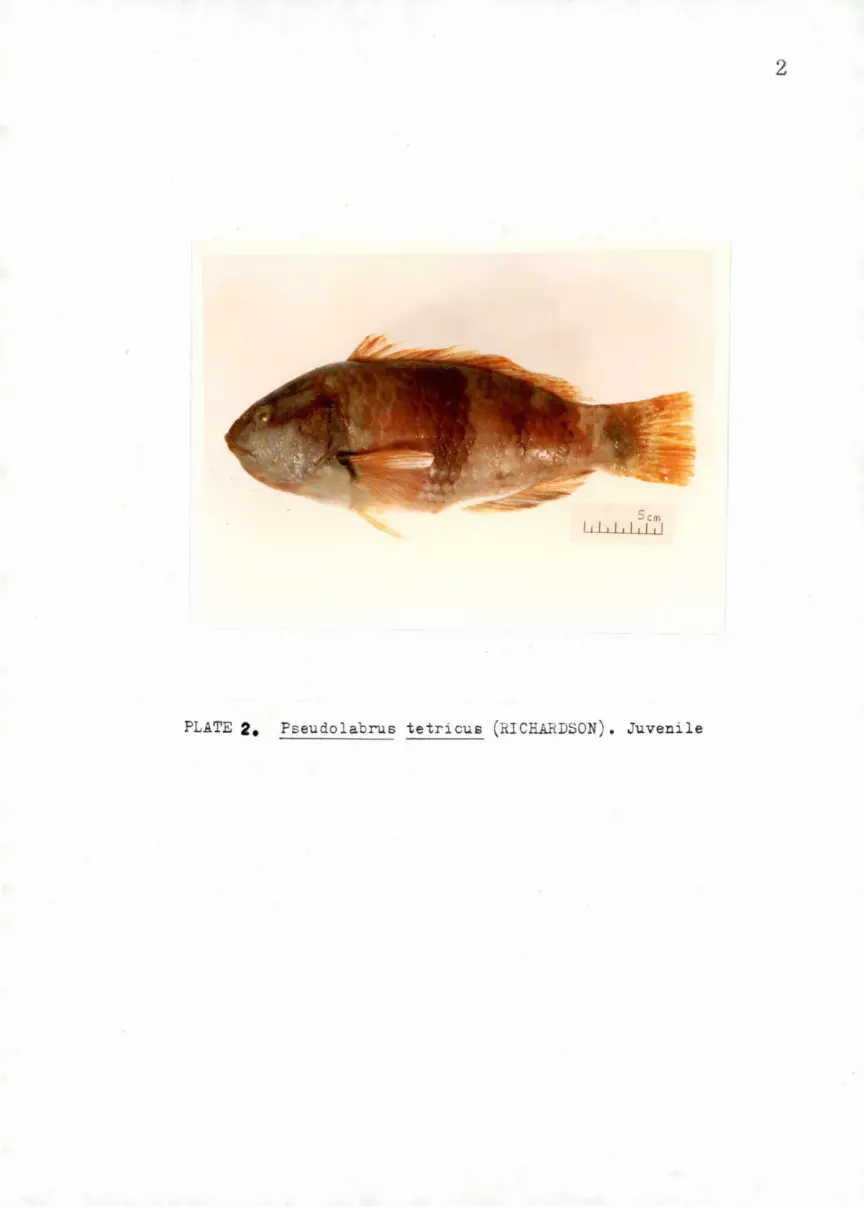
2
PLATE 2. Pseudolabrus tetricus (RICHARDSON). Juvenile

3
I
l 1lilililJ
PLATE ~• Pseudolabrus fucicola (RICHARDSON) .

4
PLATE •• Hybrid of P. tetricus & E._. fucicola

5
5 m
PLATE 5, Pseudolabrus mi le (BLOCH & SCHNEIDER)

Pmx
Dn
·:rig 6. Bone• (R) of the hea4 region ot p. ... tetrioua O')

Jig. 7. Skull
1
Mesethmoid
Postfrontal (Dermal sphenoti c
~>.-r.-t---Au tosphenotic
Sphenotic~-~-~__,
Foramen for N. Oticus
--'Hl'iol'--4'1r--P a r i et al
.,.J.>::~:.,.,..:..:+--Crecent formen
Exoccipital
Supraoccipital
(Dorsal view)
Pteroti c
Lateral pro cess
Postero-dorsal process
Posterior proc ess
Epioti c

Exoccip 1ta l Parasphenoid
Basioccipital
Pig. e. ,. Slrn l , (V e tra t view)
8

Fr onta l r
Sph enoti c r~ I. '~ dl Mf~~ •
Pre
Meset hmoi d
v omer---+--
, .. _ •• j;,.r,5' Suprao c cip i tal
~; __ .. , G(' ' .': "" : .!:'. ;_;· 1\ Ptero ti c
:~YPI~_; : ... :it .':>1) .:'Wl Short arm of the '-"'H>.:""" --·· ·- ·· """" post;tempora
attached here
Exoccipital
Foramen for N. vagus (X)
Basioccipital
'--~~~Foramen for N.
Parasphenoid
glossopharyngeal (IX )
~osterior opening of trigemino-facials chamber --~oramen for orbital artery
Foramen for internal carotid artery Anterior opening of trigemino-facials chamber
Prootic 1st hyomandibular socket
Yig. 9• Skull (Lateral view) c.o

~ supraoccipital
Epiotic >~~
~ Frontal
~.Er.~\ '-.:.O.::: · ".·I Mesethmoid
Pteroti c~~~~~"""'V'
Pre frontal~--~;;;
--.--~~~~~~~~~Vomer
Olfa c t ory foram e n ~
(For N. Olfactory) ~tt-~~~~~~~~~~~~-Parasphenoid
YiC• 10• Anterior view of the skull
~ 0

:i----Supraoccipital
:t..'\ Epiotic
.. .a Pterotic
Foramen '-------Exoccipi tal
Prootic ~ '-------Occipital c ondyle
Parasphenoid /!£Dr ~------Basioccipital
Pig. 11. Posterior view of the skull /
..... .....

PB 3.4.5.
/;Fig. 12.\ Visceral arches (Dorsal view)
12

PB 3 . 4 . 5 .
J-ig. 13. Visceral arches (Ventral view)
13
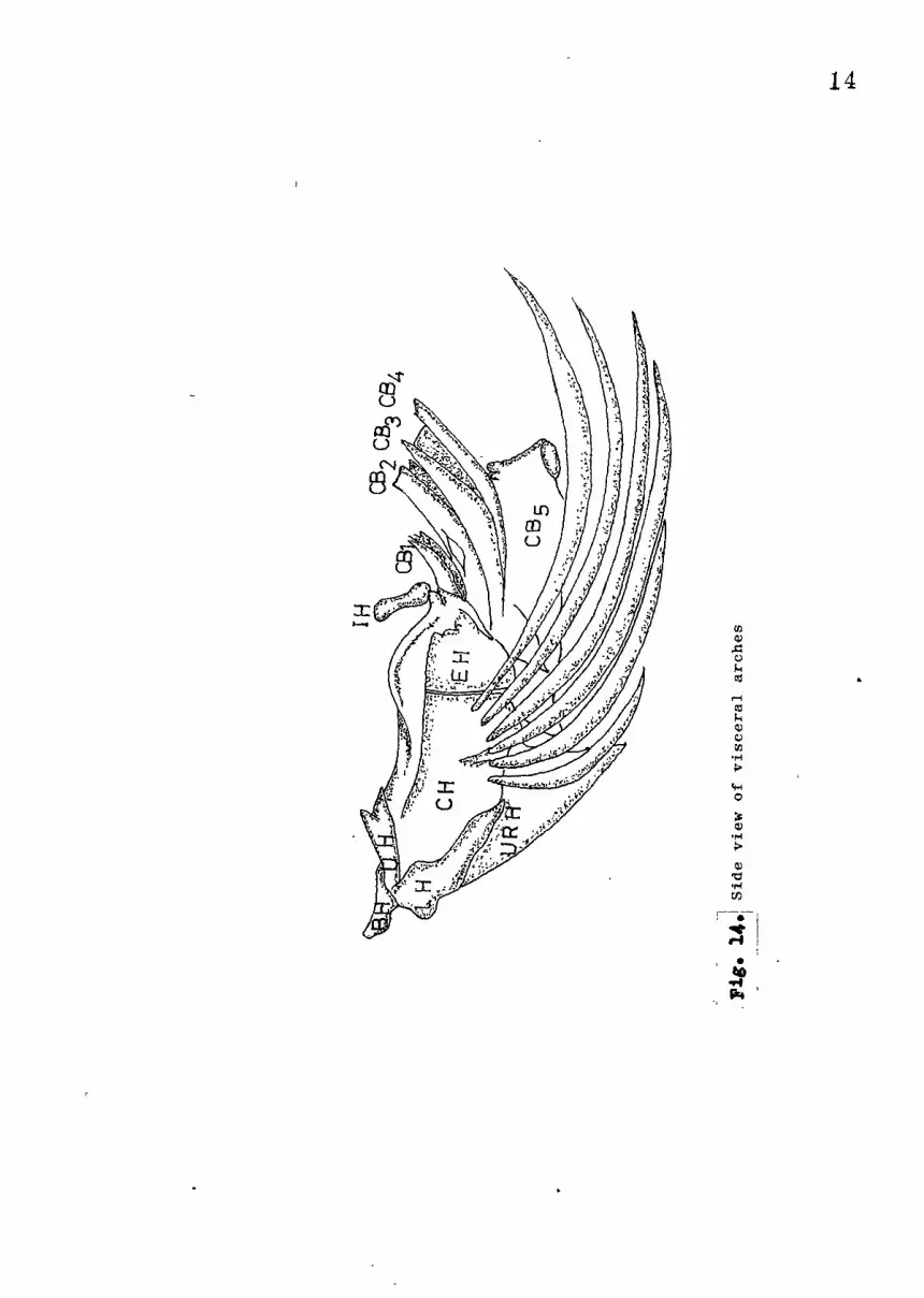
14
...... UJ ll.l .i::: 0
'"' c.ll
..... '° '"' ll.l C)
UJ •r-i
> 'H 0
~ ll.l
•l"i > ll.l "d .,..; rn
: ;fl :
• flO .... flrt

x HB1
EB1 CB
1 ~ T/ HB1
x PB2 EB2
1 CB2 HB2 Bf2/
EB3 ~ U P T.M.
CB3
• • PB ) (PB3 & 4
HB3 BB3
/ I EB4
connected 4( CB4 BB4
t ·x non-L P. T. M.
•(CB5
)
11g. i5J The connection of the elements of the branchial arches j
conected
1-c.n

1
3
4
5
Basibranehi•l
,Square bar .between mr ------ . ,_ extended anter~~
-"'-' ~ .. ---~--:_
ventrally; to·'BH-
Rectangular, a nonch
on each side fitt~d; •
... _J --..
HB1
Rhomboid
Hypobran<.?hia1 c·
Short enlarged on both Unco1
' ends conjoined to nextfand e·
'basibranchial through "W·ith vent
cartilage
same as above
·i~~!lqed vertical!]!· ~~~the ventra~·surfice ~ii'.' t~e b~~~JJjl:_:8,rcrui~
. on._ the._ conjuctmh :ii.:. - -- --'!Jo.:' - : --
Vestigial
C_artilage
There is a tendency,
~he BB4 and.BBS Become
ruminented
o:f' Bn ... - and CB -- -.. ) J
None
None
nar)I
Ol!e_ .
,!ogeil ~1arg
~ee.1·
The elements
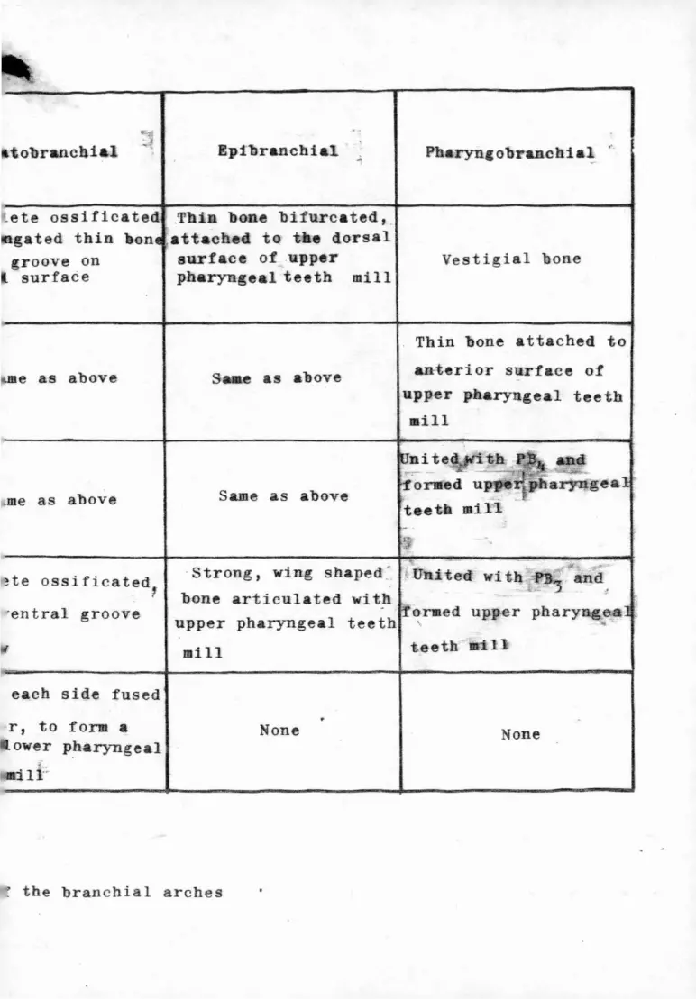
tobrancbia.1
_ete ossificated
gated thin &on
groove on surface
I e as above
.me as above
~te ossificated 1
'entral groove
each side fused
r, to form a ower P!1aryngeal
-111 ..
Epibranchial
bone bifurcated, _ attached to t e dorsal surf ce of upper pharyngeal teeth mill
S e as above
Same as above
Pharyngobranchia~ ~
Vestigial bone
Thin bone attached to
anterior surface of
upper pharyngeal teeth
•ill
· Strong, wing shaped ~ ~ United with =PB-3
and
bone articulated with ·ormed upp.er pharyng_e:a
upper pharyngeal teeth
mill teeth f'i l l
None None
• the branchial arches

Premaxillary (External surface)
~ig. 17• Premaxillary (Internal surface)
Articulated with the anterior surface of the inner arm of the maxillary
16

Inner arm Articulated with posterior surface of ascending process of' premaxillary ----_
Outer a rm -----·=
fi'acet for the anterior end of the palatine
~;:,;.;::;.;.:.:;.;.::~?'-----Depression for
Pig. 18.
the anterior arm of the pal a tine
Maxillary (External surface)
.-· . ;• . ·.
';. ~ ... · .. ------Articu lated with vomer
J'ig • . 19 •. Maxillary (Internal surface)
17

18
·~~~For laterosensory canal system
!Jentary (External surface)
Jig. 20.
Dentary (Internal surface)
11,. 21.
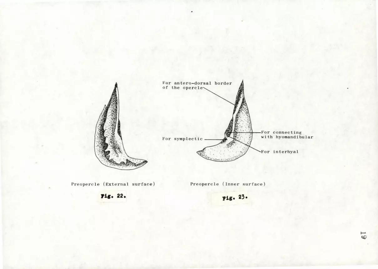
Preopercle (External surface)
rig. 22.
~nr antero-dorsal border of the opercle
, ~: . · · ·-~<I For connecting
For symplectic A/If with hyomandibular
For interhyal ._ .. ,: .
~'
Preopercle (Inner surface)
1'11• 23·
~
~

fit Anterior thickened margin overlaped by preopercle
Opercle (Exernal surface)
l'ig. 24.
For posterior condyle of hyomandibular
Fossa for M. lavator operculii
Opercle (Internal surfac e)
Pig. 25.
Nl 0

21
, .
/ . . . . ~ .. . .. \
\
Interopercle (External surface)
~:;;;~~~~~~~fftt~~~Arti culation conjoined
Interopercle (Internal surface)
Jlig. 27.
to the dorso-posterior condyle of the hyomandibular

22
Subopercle (External surface)
Suboperc l e (Internal surface)

r1a. 30.
l•' irst Postcleithrum (External surface)
Pia. 31.
F'irst Post c leithrum (Inte r nal surface)
N w

pig. ~2.
Sec ond Postcleithrum(External sur face)
Jlig. ~,.
Second Postc le ithrum(Internal surface)
t...:> IP--

Facet articulating the groove between the maxillary and its inner arm
Palatine (External surface)
Palatine (Internal surface)
:Pig. 35.
25
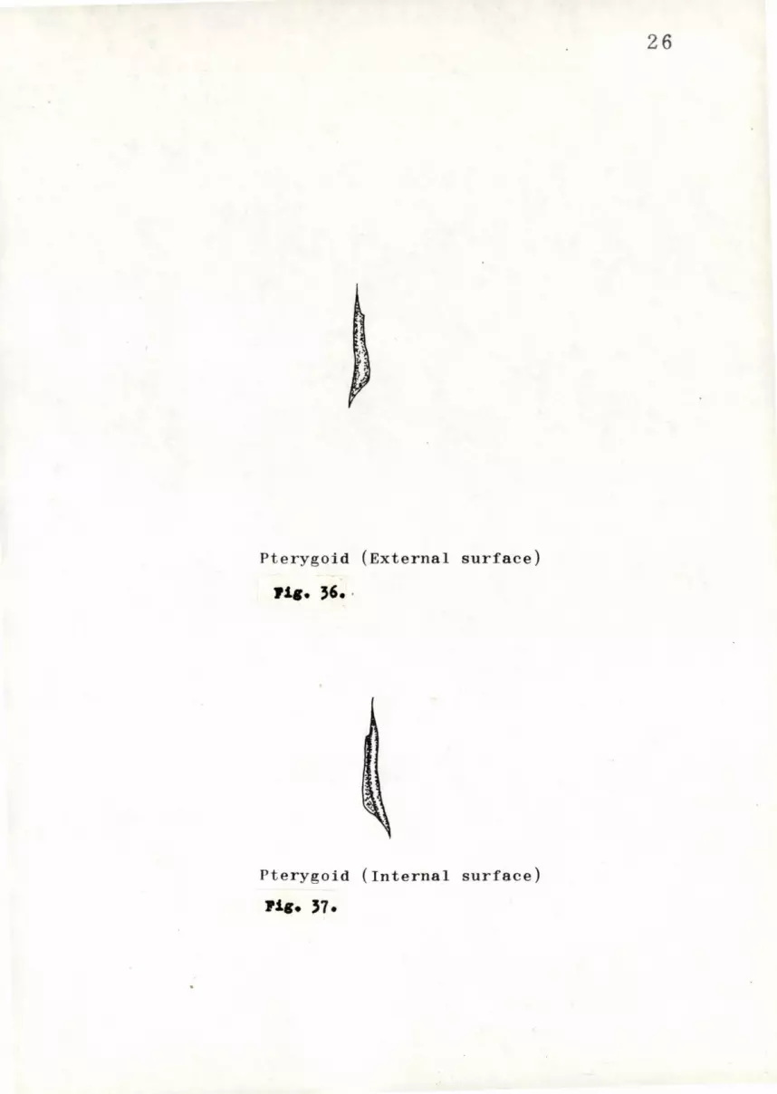
Pterygoid (External surface)
Jlia. ''· ·
Pterygoid (Internal surface)
Pia. 37•
26

Mesopterygoid(External surface)
Jig. 3a.
Mesopterygoid (Internal surface)
Jig. 39.
21

Metapterygoid (External surface)
fig. 40.
Metapterygoid (Internal surface)
fig. 41.
28

~ ~
Pig. 42. Quadrate (External surface)
Shov el -shaped process
Lower groove
Upper groove
Pig. 43. Quadrate (Internal surface)
29
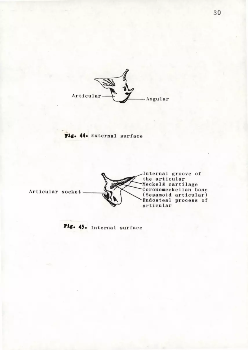
Articular----
71g. 44• External surf ace
7J.c. 45. Internal surface
Internal groove of the articular Meckels cartilage Coronomeckelian bone (Sesamoid articular) Endosteal process of articular
30

31
11•• 46. Symplectic (External surface)
· rig. 47• Symplectic (Internal surface)

Anterior facet------ Posterior f a cet
Condy le articulated with opercle
m&' Opening for N. hyomandibularis facial is
R::H Opening for N. hyomandibularis facial is
Anterior win g
Ventral process
External surfac e Internal surface
Pig. 48• Hyomandibular (Temporal)
w N

33
(External surface)
~ig. 50. Nasal (Internal surface)

Pig. 51. Lachrymal (External surf ace)
:Pig. 52.
Articu lated with the lateral process o f the prefron tal
Lachrymal (Internal surface)
w ~
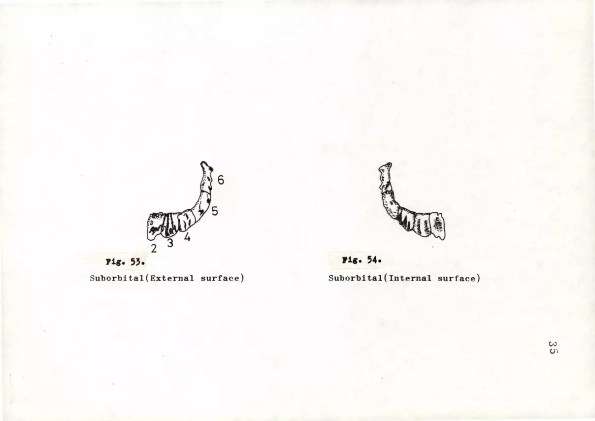
35
-a
)
0 "' ...., i.t
=
C1J
~
"' =
i.t G
l ~
=
H
-• .-4
• "'
"' ~
..... • ..
,Q
... i.t
... 0 ,Q
=
U)
-a
)
0
(.!) "' CH i.t
=
C1J
.-4
ell
= i.t a
)
~
N
~
r:z:J
-• .-4
"' "' "' ~
• .....
eo ,Q
...
i.t ....
0 ,Q
=
U)

G
Pi•• 55. La terosensory Canal Syu tei:I
v.,, CJ)

Extrascapular
Postt.emporal
lfY-. Supracleithrum
C 1 e i thrum ---------11
-+---Postc leithrum I
Scapula / ~,.•- -- . ~·"-?'7 >:;:::a~
Postcleithrum II
-· -Pre c oracoid process
Jlig. 56. Pectoral girdle (External surfa ce)
w ~

38
l::x. trar.capu 1 ar
::iupr•w~el. thrum '-------Cleithrum
rortclei tr.rum
r,intclPl thrum 11
Pectoral girdle (Internal surface)

39
•
Pig. 58. Pelvic girdle {Dorsal View)

40
~ig. 59. Pelvic girdle (Ventral view)

5
Epipleurals--'~~~~~~~~~ Ribs~-------rt\1,.---\I
Dorsal lateral depress ion
10 15 20
Ventral lateral depression
oramina for roots of spinal nerve
~iS• 60. Vertebral column and urostye
- --Epural
Hypural 3
~
~

Neural spin e
_ Neural arch
-~_....,..,... ___ Neural canal
Anterior view
Parapophy sis
Socket attached to
the occipital condy le
of the exoccipital
Cent rum
JliC• 61. First vertebra
-+-~-Haemal postzygapophy sis
Posterior view
,+::... N

,__parapophysis
Anterior vieW
fil• 6i· 2nd
vertebra
:-:·:
·~\ ~~· ; '
posterior view
~ e;..:l

RLll\\------Prezygapophysis
----- Parapophys is
Anterior view
Yig. 63. 5th vertebra
. :.~
Neural canal
J~ Neural postzygepophysis
~'"-•!h: Haemal postzygapophsis
\~ Epipleural
1;--- Pleural rib
Posterior view ~ ~

~\;
Anterior view
yig. 64· 9th vertebra
, ,: ~ .. ; \ ':· f1 n
~~~ Neural postzygapophysis
~Haemal postzygapophysis
Posterior view
~ CJl

. "O&"-- Prepophysis
Haema l arch ___ _.,..._ --4--...;.;..;.>,----Haemal canal
-'-~-- Parapophysis
:q1 .\ ~~·
,. ~ ;;+-------Haemal spine
' v {;
~
' ··' i;.
Anterior view
118• 65· 10th vertebra
t~ '•
-:. ..
Neural postzyg ap ophysis
~Haemal
~ •: ~ !: ·'
'J ·' '.I . t ·- ;
t f
J ~'?
Posterior view
p ostzygapophysis
~ ()')
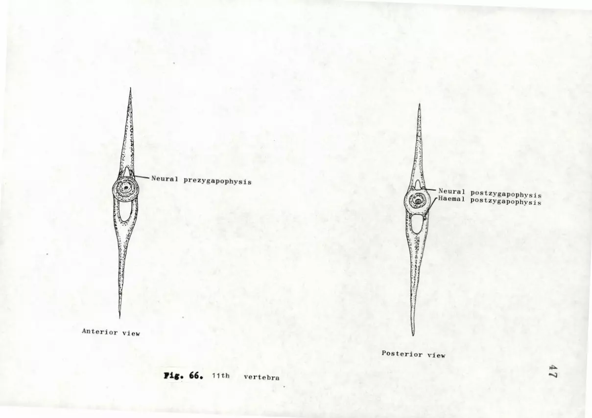
'·:I
~~Neural prezygapophys is
Anterior view
11,. ''· 11th vertebra
~~ l~
I~ .~ 1 ·1:
··> 1! ..... ;
Neural postzygapophysi s Haemal postzygapophysi s
Posterior view
~
-...:J

-; ... , ;
~ ~'
U~Neural prezygapophysis
W " .. 5' Haemal prezygapophysis
! ., '··· ~ ;,
:: ": \
..
Anterior view
J'i&• 6l• 21 st vertebra
~ !' ' • \,
:~ .~ . ~ ,,
fJ~ Neural postzygapophysj ~
...,.._..,__ Haemal postzygapophys 1 -
~ - i,'
: i ~ ~
·~ · I~ f 1;
posterior view
~
~

Cartilage ·~
-"'1r----- Uros t y l ·e
)ii:: · ~ Last vertebra
2 J
J'ig. 68. 3n nes c f the tail r e&ion
.;:... <.O

• $
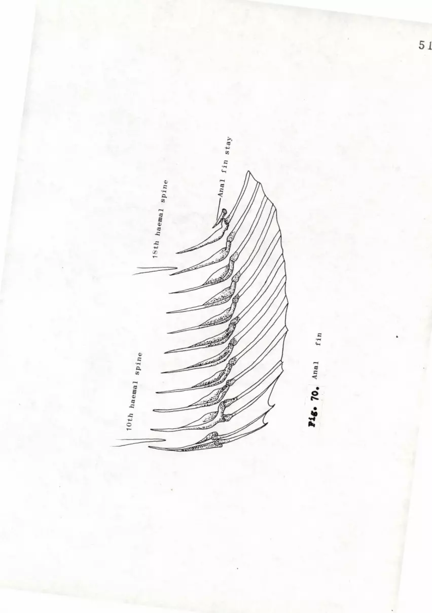
QJ
r::: .,., Q, (/)
...... q;J
s QJ (tJ
..c: ..c: ..., 0
--
QJ
r::: .,., Q, (/)
>
51
r::: .,., 'H
...... Ctl
" <t;
• 0 ,... .
• ~ ...
""

h 0
Q)
i::: ..... 0. rJJ
52

Fig 72• JIQ1cle1 et the head.
Al-A2-A3; kdductor mandibulae Adop~ Adduc tor operculi Dil. Op: Dilatator operculi Lap: Levator a.rcue palatini Lev. Op: Levator operouli

53

Fi g . 73.
/ Mu•ole• et the hea4.
Al-A2- A3; ~ddu cto r ro andibulae Adop: Addu ctor oper culi Di l. Op : Dilata tor operculi Lap: Levator ar cus pa latini Lev. Op: Levator operculi

54
~':
)> __.
/ /

Fi g 14• l!u•ole• •t the bead.
Al-A2- A3; ~dductor rnandibulae Ador : Adduc tor oper culi Di l. Op : Dilatator ope r cu li Lap: Leva.tor arcus pa.latini Lev. Op: Levator operculi

55

Fig 7,. Huaole• •t the pharJDX•
Abd8: Abductor superficialis Abdp: Abductor profundes Hy: Hyopectoralis Pee: Pharyngo-clavicularis externus Pei: Pha.ryngo-clavicula.ris internu~ Ph: Pha.ryngo-hyoideus Rabd: Retractores arcuum bra.nchialium
dorsales
I

56

Fig 76. Ku1ol•• ot the pectoral arch an4 tin.
Abd&: Abctuctor superficialis Abdp : Abductor profundes Hy: Hyopectoralia Pee: Pbaryngo-clavicularis externus Pcis Pharyngo-clavicularis internus Phi Pha.ryngo-hyoideue Rabd; Re t ractores arcuum bra.nchi a liwn
dorsales

Addp 2
: : i
' I'
,,
\ '
57
Adds

}ig 77. Muacle• ot the Tentral portioa, pelYio gir41• an4 tin.
l..bdp: Abducto::'.' ;:irofour.: ds pectoralis Abds: bductor superficialis pectorlis Abp: Abct:c tor i:·rofounds pelvi s kbs: Abductor surerficialis pelvis Ghi: Geniohyoideus inferior Gha: Geniohyoi deus superior Hhl. Hh2: Hyohyoideus Hyo: Hyopectoralis Im: Intermandibularis v~: Myomere of the body muscles

58
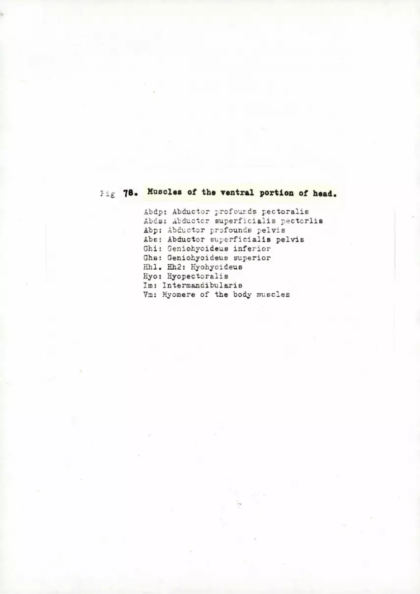
Fig 78. Muscle• ot the ventral portion ot head.
Ab dp: Abductor profourds i:-ectoralis Abds: Abductor superfici a l is pectorlis Abp: Abcucto r profounds pelvis kbs: Abductor supe rficialis pelvis Ghi: Geniohyoi deus inferior Ghs: Geniohyoideus superior Hhl. Hh2: Hyohyoideus Hyo: Ryopectorali s Im: Intermandibularis Vm: Myomere of the body mu scles

Hy. (ante-ior)
59·

.,9• Mu•ol•• ot the dor•al •urtace ot Phar7nx • Fig
1:.dl : Acductores a j·cuum t:-·ancf:ialium le~ : lavat~res brc.ncti a les externus
or the t hi rc arch Le4 : Lava:o_'es bre.r_ct iales exte r nu s
of t~e 4th er ch le5: Lavatores brar·cl". iales exte r nu s
of Le - . } . arct .; ,.. __
Lil: l av ato r es b ra!!C,,_. i a : es in ternus of ttc fi rst arch
Li2 : ~c.v3tores b r a.nc!1ialee internu s of t he s econd arch
Li 3 : !..,av a to res b ran chi a le s in ternus of the thir d arch
Li5: Lavatore s bra.n cb iales in t e rnu s of t h e )th arch
Ob: I nte r arcu al e s obli q_ui dors&.l es n ab d: Retract·:ire s ar cu'l;.m b r anch i a l i um
d.orsale s Tal : Tr ansversus dorsali s Al Ta2: Transvers~ s c~rsal i s A2 Tp : 'I'ransversus do r s alis posterio r

60
L__

Fi 80. Mu•ole• ot the ••ntral aurtaoe ot )har7Dx. Con . e so: Contr ictor eso~hacei Obl: Obliqu i ven tr-al es of t he
first ar cl: Ob 2 : Obliqui ventrales of t he
s econd arch Ob3: Obl i ui ven tral e s of the
~hi ·d a r ch Fee : Pharyngo-claviculari s e~t ernu s
Ph : Pharyngo-hyoideus Pei: Fharyngo-clavicularis internu• TV4: Tra.nsversus ventralie 4

61

Fig 81. Muaolea ot the e7eball.
Oi: Ob liq_uus inferior Os: Obl iquus superior Rif: Rectus inferior Rit: Rectu& internus Rex: Rectus externus lis: Hectus &uperior

sz

Fig 82. J(u•ol•• ot the e7eball.
Oi: Obliq_uus inferior Oe: Obliquus superi or Rif: Rectus i nfe r i or Rit: Rectus i n ternus Rex: Rectus externus lis: Rectus superior
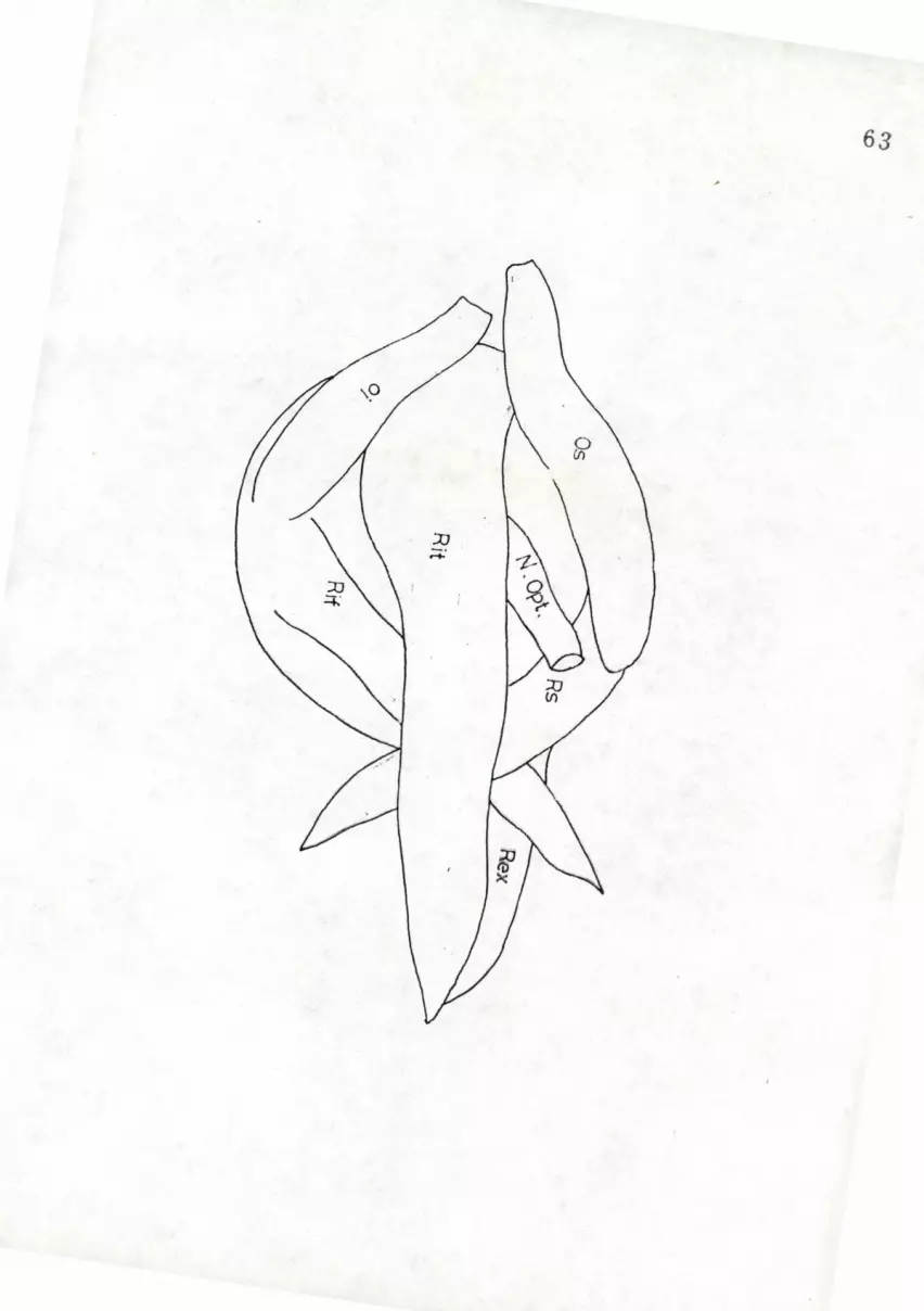
63
t •

Fig ''· xuaol•• ot ~h• caudal tin.
D: Flexor caudalis dorsalia V: Flexor caudalis ventralis 3rd: Adductor caudalis ventral is I: Intrinsic muscle

64
0

65
· · t · Er t r ·'",! \\~ ~. -.--- ec o ~ \ .,~, \ l \ \
. '\• " ' . " l l i l • • l" '\ ! • ~ \ . 't
Depressor -·------¥-
: Fig. 84.~ i I
Muscle of the dorsal fin ( 4th & 5th dorsal fin)

Fig 85.Section of the bucc~l cavity
BM: Ba~ement ~embra.ne 3V: Blood vee:sel SCm: St~ atum compactum SLM: Striated longitudinal
muscl e SM: Submu cosa SSqE: Stratified s~uL~ous epithelium
66

;__ ___ _ ____ c
k-------~- BM
~--~--~SSqZ
_. ______ cc
Fig 86. Section of the IIIUCOUs membra.ne of the buccal cavity
BM: Basement membrane C: Crypt CC: Columnar cell MC: Mucous-secreting cell SCm: Stratum compatum SSqE: Stratified s quar.1ous epithe l ium TB: Taste bud
67

~----SSqE
---SCM ---EF
llii----SM
~{aft'-!--- scm ~-·---BV .------MC
Fig 87. Section of the e'ophagus
BM: Basement membr2.ne BV: Blood vessel CC: Columnar cell EF: Ssopta.t;eal fold MC: Mucous-secreting cell SCm: ~tratum corr.pactum SCM: Striated circular muscle SM: Submucosa S$qE : Stratified sque.mous epitt elium TB: '!'aste bud ~uFil: Tunica propria
68

Fig 88. Section of the inte~tinal bulb
CoC: Columnar cell OoC: Goblet cell IntCa: Intestinal cavity IntPp: Inteatinal papilla Sm: Submucoaa StBo: Striated border Tu.Pr: ~unica propia

.=...- i---- I nNZ
~--------- GrL
~-----~----IntCa
---SpNZ
Fig 89. Section of tt e intestine
GrL: Granul&r layer InNZ: I nfra.nu c lear zone IntCa.: Intestin al cavity Nu Col: llucl eu s of columnar cell Srl\Z : Supra.nuclear zone StBo: Stri&ted borde r TuPr: 'I\lnica ~ropia
70
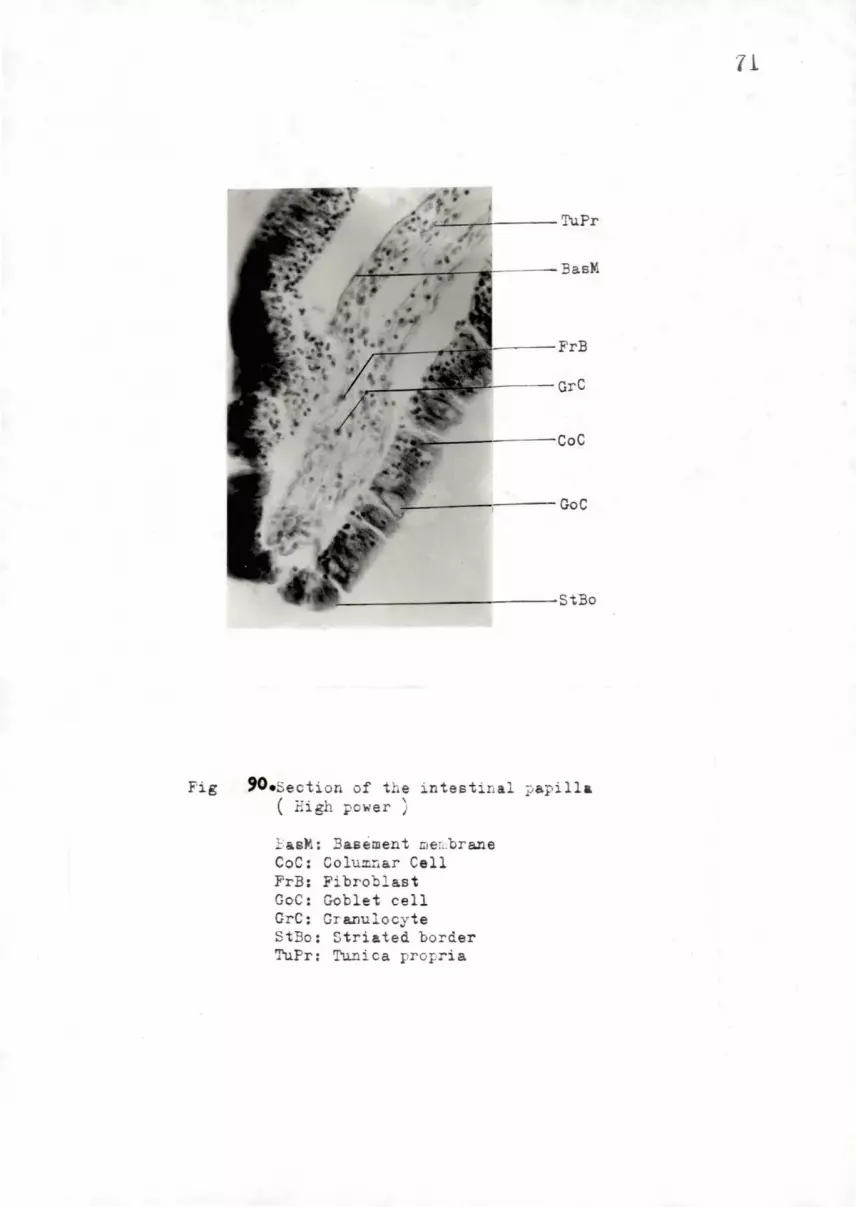
Fig 90.s ecti on of t e intestinal papill a ( High power )
};'.i;.sM: Basement mer..brane CoC: Columnar Cell FrB: Fibroblast Goe: Goblet cell GrC: Gra.nulocyte StBo: Striated border TuPr: Tunica propria
71

. .__ _ ____ 1 n tEp
.?\-.-..;.~!:-iii~~ ~.-::------ Bl Ve ~~~.k"lt:JJ~~~~·~~-CM
t;_J4~~~~-:itJ#!~~-~~Sr .:.........,+.::...-:--~~IE~77-::.~-~~-LM ~~....-_.__::.~~~~,_~~-~~sbS
Fig · .91. Section of the junction of the intestinal bulb and intestine
BlVe: Blood vessel CM: Circular muacle In: Intestine InB: Intestinal bulb IntEp: Intestinal epithelium LM: Longi tudinal muscle PyV: Pyloric valve SbS: Subserosa Sm: Submucosa Sr: Serosa
72

Fig 92. Section of t he intestine
BM: Basement membrane CE: Columna.r epithelium GoC: Goblet cell SM: Submu cosa StBo: Striated border TP: Tunica propi &
73

•---LuC ~---cE
Fig 93. Section of tJ-,e i r. testinal J: apillae ( Ei&h power )
CE: Columnar epithe l ium GoC: Goblet cell LuC: Lucocyte TP: '!'unica pro ~ ia TFl : Top plate
74

Fie 94• The structure of the lobule of tb.e liver
BD: Bile duct CV; Central vein HA: Hepatic artery EC: ~epatic cores ICT: Interlobular connective tissue PV: Fo::-tal vein RBC: Red blood corpuscle Si: Sinueoid.s
75

Fig 95. The structure of the skeletal mu s cle
Ar: Arteriole Em: Endomys i um NMF: Nucleus of muscle fiber Pe: Perimysium Ve: Vein
76

.._ _ _ __ Cup
Fie; 96. Se c ti on of tLe l a t e r a l sensory s~ster; 1
BV: Blood vessel Cup: C~;u la - one ~ar t of
neuro rn a s t :D ; rerrni s E: E;idermis FCT : Fit·:- ::us connective tiEsue ~ : Mu s cle
11!J:- : t.: .:,~o :: e; ti..:. ~n f: Fore PC: Pigment ce ll (chromatophore)
77