Embed Size (px)
Citation preview

The Antiepileptic Drug Levetiracetam Suppresses Non-Convulsive Seizure Activity and Reduces Ischemic BrainDamage in Rats Subjected to Permanent Middle CerebralArtery OcclusionOrnella Cuomo1, Vincenzo Rispoli2, Antonio Leo2, Giovanni Bosco Politi2, Antonio Vinciguerra1,Gianfranco di Renzo1, Mauro Cataldi1*
1 Division of Pharmacology, Department of Neuroscience, Reproductive and Odontostomatologic Sciences, Federico II University of Naples, Naples, Italy,2 Department of Health Sciences, University Magna Græcia of Catanzaro, Catanzaro, Italy
Abstract
The antiepileptic drug Levetiracetam (Lev) has neuroprotective properties in experimental stroke, cerebralhemorrhage and neurotrauma. In these conditions, non-convulsive seizures (NCSs) propagate from the core of thefocal lesion into perilesional tissue, enlarging the damaged area and promoting epileptogenesis. Here, we explorewhether Lev neuroprotective effect is accompanied by changes in NCS generation or propagation. In particular, weperformed continuous EEG recordings before and after the permanent occlusion of the middle cerebral artery(pMCAO) in rats that received Lev (100 mg/kg) or its vehicle immediately before surgery. Both in Lev-treated and incontrol rats, EEG activity was suppressed after pMCAO. In control but not in Lev-treated rats, EEG activityreappeared approximately 30-45 min after pMCAO. It initially consisted in single spikes and, then, evolved into spike-and-wave and polyspike-and-wave discharges. In Lev-treated rats, only rare spike events were observed and theEEG power was significantly smaller than in controls. Approximately 24 hours after pMCAO, EEG activity increasedin Lev-treated rats because of the appearance of polyspike events whose power was, however, significantly smallerthan in controls. In rats sacrificed 24 hours after pMCAO, the ischemic lesion was approximately 50% smaller in Lev-treated than in control rats. A similar neuroprotection was observed in rats sacrificed 72 hours after pMCAO. Inconclusion, in rats subjected to pMCAO, a single Lev injection suppresses NCS occurrence for at least 24 hours.This electrophysiological effect could explain the long lasting reduction of ischemic brain damage caused by thisdrug.
Citation: Cuomo O, Rispoli V, Leo A, Politi GB, Vinciguerra A, et al. (2013) The Antiepileptic Drug Levetiracetam Suppresses Non-Convulsive SeizureActivity and Reduces Ischemic Brain Damage in Rats Subjected to Permanent Middle Cerebral Artery Occlusion. PLoS ONE 8(11): e80852. doi:10.1371/journal.pone.0080852
Editor: Renping Zhou, Rutgers University, United States of America
Received August 2, 2013; Accepted October 11, 2013; Published November 13, 2013
Copyright: © 2013 Cuomo et al. This is an open-access article distributed under the terms of the Creative Commons Attribution License, which permitsunrestricted use, distribution, and reproduction in any medium, provided the original author and source are credited.
Funding: The present study was partially supported by a research grant by UCB Pharma. The funding company did not have any role in designing,performing or analyzing the data but approved both the initial protocol and the final manuscript. This economical support does not alter our adherence to allthe PLOS ONE policies on sharing data and materials. Additional support was received from the following grants: Ricerca Finalizzata 2009 [Grant RF-FSL-352059], and Ministero dell’Istruzione, dell’Università e della Ricerca, Programma Operativo Nazionale [Grants PON_01602 and 20089BARSR_002].A.L. is recipient of a Ph.D. fellowship grant from the European Community, the European Social Fund and the Regione Calabria.
Competing interests: The authors have the following interests: This work was partially supported by a research grant by UCB Pharma SPA (Italy). Thisdoes not alter the authors' adherence to all the PLOS ONE policies on sharing data and materials.
* E-mail: [email protected]
Introduction
Levetiracetam (Lev) is a second generation antiepileptic drugstructurally related to the nootropic and neuroprotectivepyrrolidone compound, piracetam [1]. Beside its potentantiepileptic activity, Lev also has antiepileptogenic effects inelectrical [2,3] and audiogenic kindling [4] and in several animalmodels of epilepsy such as WAG/Rij rats [5,6]. In addition, thisdrug protects neurons from different types of insults including
the intracerebroventricular injection of kainate [7] and brainischemia induced by middle cerebral artery occlusion (MCAO)[8] or neonatal hypoxia [9]. Lev also reduced brain damage inexperimental subarachnoid hemorrhage and closed headtrauma [10].
The mechanism responsible for Lev-induced neuroprotectionand antiepileptogenic effect is unknown. This drug differs fromall known antiepileptics for it targets SV2, a protein of thesynaptic vesicle fusion complex [11]. Through the interaction
PLOS ONE | www.plosone.org 1 November 2013 | Volume 8 | Issue 11 | e80852

with this protein, Lev acts as a general inhibitor ofneurotransmitter release [12]. Moreover, we reported that Levblocks Ca2+ release from intracellular IP3 stores [13] and asimilar effect was observed by others for ryanodine stores[14,15]. Lev also antagonizes the inhibitory effect of Zn2+ and β-carbolines on GABAA receptors [16] and has slight inhibitoryeffects on N-type Ca2+ channels [17]. Finally, its majormetabolite in humans, 2-pyrrolidinone-n-butyric acid, inhibitshystone deacetylases [18]. All these pharmacological effectscould, theoretically, contribute to neuroprotection by a directeffect on neurons [19]. Despite its efficacy in living animals,surprisingly, Lev was ineffective in models in vitro ofneurodegeneration. Specifically, this drug was unable toprotect hippocampal slices from the ischemic damage inducedby the combined deprivation of oxygen and glucose [20]. Thisfinding suggests that intact neuronal networks present in theliving brain and disrupted by the slicing procedure arenecessary for Lev-induced neuroprotection. This hypothesis isin keeping with the evidence that after a focal brain insult,depolarizing waves of spreading depolarization, called post-ischemic depolarizations (PIDs), enlarge the primary lesion bypropagating into the surrounding intact brain throughpreexisting synaptic networks (see 21 for review). At the EEG,PIDs have the electrophysiological features of seizure activity.Because they are not accompanied either by motor orbehavioral symptoms, these events are usually defined non-convulsive seizures (NCSs) [22]. Recently, the propagation ofNCSs has been directly visualized in the ischemic human brainand the demonstration of their relevance in the progression ofischemic lesions has also been obtained [23]. NCSs alsocontribute to epileptogenesis accounting for the frequentdevelopment of post-ischemic epilepsy in patients survivingstroke [24]. Therefore, a neuroprotective activity in stroke and aprotection from the development of postischemic epilepsycould be obtained by suppressing PIDs. In the present paper,we explored whether this electrophysiological effect could beobtained with Lev. To this aim, we examined Lev effect on thegeneration or propagation of NCSs occurring in rats after thepermanent occlusion of the middle cerebral artery (pMCAO), awell known experimental model of brain ischemia.
Materials and Methods
AnimalsAll the experiments were performed in 2-month old, male,
Sprague Dawley rats weighing 200-250 g (Charles River, Italy).Rats were group caged on a 12 h light/dark cycle and had freeaccess to food and water. The experimental protocol wasapproved by the Animal Care Committee of “Federico II”,University of Naples, Italy. Animal housing and experimentalprocedures were performed according to the recommendationsof the guidelines for care and use of experimental animals ofThe European Community Council directive (86/609/EEC). Allefforts were made to minimize animal suffering and to reducethe number of animals used in the experiments.
Electrode ImplantationTwo/three days before experimental brain ischemia or sham
operation, rats were deeply anesthetized by delivering a gasmixture containing 2% sevoflurane in a 70% nitrous oxide/30%oxygen through a nose cone. Then, two burr holes were drilledin each side of the skull, 2 mm laterally to the midline, and 2and 4 mm posteriorly to bregma to position the EEG recordingelectrodes onto the dural surface. According to the Paxinosand Watson coordinates, these positions correspond to theparietal region of the neocortex. In addition, a referenceelectrode was placed posterior to lambda. The electrodes weresecured to the skull with acrylic dental cement. Their free endswere soldered to a multipin connector (Plastic One, Roanoke,VA, USA) and the entire assembly was fixed to the skull withdental cement.
Basal EEG recordingsFor EEG recordings, we used a bipolar recording montage
with a ground electrode placed onto the nasal bone. On theday of the experiment, animals were placed in a recording cageequipped with a multichannel swivel commutator (Plastics One,Roanoke, VA, USA). The multipin connector on the rat skullwas connected to the swivel system via a flexible shieldedcable that allowed free movement of the animal during the EEGrecording [25]. The swivel commutator was interfaced throughshielded cables with an EEG recording and analysis system(Belight, Galileo NT, EB-Neuro, Florence, Italy). Signals werelow-pass filtered at 0.1 Hz before being band-passed between0.16 and 50 Hz. Data were digitized at 100 Hz, acquiredthrough the analysis software and stored on the hard disk ofthe computer for offline analysis.
Basal EEG activity was recorded for 30 minutes, then, theswivel cable was disconnected from the multipin connector onthe head of the animal. Lev or vehicle was injected asdescribed in the following section and the rat was deeplyanesthetized to be subjected to pMCAO as described below.After the surgical procedure, the animal was returned to itscage and was reconnected to the swivel cable. Twenty minutesafter the induction of ischemia, EEG recording was startedagain and continued for 48 hours.
Levetiracetam administrationLev was dissolved in saline solution and administered at the
doses of 50 or 100mg/Kg by a single intraperitoneal (ip)injection. Drug injection and the further experimentalprocedures were carried out in blind manner. Indeed, theresearchers who performed the experiments and analyzed datawere not aware of the drug treatment administered to theanimal.
Experimental brain ischemiapMCAO was performed using the method described by
Cuomo et al. with slight modifications [26]. Briefly, rats weredeeply anesthetized with a gas mixture (4% sevoflurane in a70% nitrous oxide/30% oxygen) delivered through a nose cone.Anaesthesia was then maintained with 2% sevoflurane duringthe entire surgical procedure. The skin of the neck and the
Levetiracetam Effect on Post-Ischemic Seizures
PLOS ONE | www.plosone.org 2 November 2013 | Volume 8 | Issue 11 | e80852

underneath fasciae were sectioned in the sternocleidomastoidregion and the neurovascular bundle of the neck was exposed.In sham-operated rats, the surgical section was, then, suturedwithout proceeding further with arterial occlusion whereas inrats undergoing pMCAO the right carotid bifurcation wasexposed and the external carotid artery coagulated distal to thebifurcation and cut. Afterwards, a 6-0 nylon filament wasinserted through the external carotid artery stump andadvanced into the left internal carotid artery until it blocked theorigin of the middle cerebral artery. During this surgicalprocedure, cortical cerebral blood flow (CBF) was continuouslymonitored with a laser-doppler flowmeter (Periflux system5000, Perimed, Milan Italy) [27]. Once a stable drop in bloodflow signal was obtained ipsilaterally to the occlusion, thefilament was secured to the vessel by ligation. The filamentwas left in place to permanently occlude the middle cerebralartery. Body temperature was monitored throughout the entireduration of the surgical procedure and maintained at 37.5 °Cwith a thermostatic blanket. Arterial blood gases weremeasured before and after ischemia through a catheterinserted into the femoral artery (Rapid lab 860; ChironDiagnostic, Emeryville, California, USA). Only the animalsshowing a reduction in CBF of at least 70% were consideredischemic and entered the further steps of the experimentalprotocol. No significant difference in CBF was found at theretrospective analysis of the laser-doppler flowmeter databetween among the different experimental groups (data notshown).
Evaluation of neurological deficits and of the ischemicvolume
Neurological deficits were evaluated 24 or 72 hours afterpMCAO according to Clark’s scale [28]. The statisticalsignificance of the difference in the scores obtained in thedifferent groups of animals was assessed by non-parametricanalysis using the Nemenyi test. The threshold for statisticalsignificance was set at p<0.05.
The extension of the ischemic lesion was assessed 24 or 72hours after pMCAO as described elsewhere [29]. Briefly, ratswere sacrificed by decapitation. Brains were quickly removed,placed in ice-cold saline solution for 5 minutes and then cut into500 µM coronal slices with a vibratome (Campden Instrument,752M; UK). Sections were incubated for 20 minutes in a salinesolution containing 2% Triphenyl tetrazolium chloride (TTC)and, then, fixed in 10% formalin overnight. The infarcted regionin each slice was identified as the white area after TTCstaining. The surface areas of the entire slice and of theischemic lesion were measured using the image analysissoftware Image-Pro Plus 4.1 (Media Cybernetic, Rockville, MD,USA) in all the slices between +4.7 and -4.9mm from bregmaaccording to the Paxinos atlas. These coordinates define thebrain region that usually includes the entire ischemic lesioncaused by pMCAO. The values obtained were used tocalculate the volumes of the hemisphere and of the infarctedarea in this specific region. The volume of the ischemic areawas, then, expressed, as percentage of the entire volume ofthe infarcted hemisphere. By this approach, the data werecorrected for brain edema. The statistical significance of the
difference between the experimental groups was evaluated byone-way ANOVA followed by the Newman-Keuls post hoc test.A p value <0.05 was considered statistically significant.
EEG analysisQualitative EEG analysis was performed off-line by visual
examination. By using this approach, we distinguished periodsof normal activity, of reduced electrical activity, in which thetraces became close to the isoelectric, and NCS events. All therhythmic discharges with spike components that persisted >10sec were classified as NCS [22,25]. NCSs were furthersubdivided according to their shape into single spikes, spike-wave, poly-spike or polyspike-waves discharges. The number,frequency and amplitude of all these events were computed ineach of the experimental groups.
Quantitative EEG analysis was performed using the FastFourier Transform (FFT) and a commercial software (Belight,Galileo NT;EB-Neuro, Florence, Italy software). Specifically,the bipolar signal from each neocortical area in both brainhemispheres was processed using Fast Fourier Transform andits main spectral components were separated. Specifically, fourmain band components were identified in the frequency rangebetween 0.25 and 32 Hz: delta (0.25–3.9 Hz), theta (4.0–7.9Hz), alpha (8.0–12.9 Hz) and beta (13.0–30 Hz). Both in controland in Lev-treated rats, the mean power (in μV2) of each ofthese components was computed by integration of the powerspectra in the respective frequency ranges.
All the EEG data are presented as mean±SEM. Statisticalanalysis of the results of qualitative and quantitative EEGanalysis was performed by one way ANOVA, followed by thepost-hoc Tukey-Kramer test for multiple comparisons. Thethreshold for significance was set at p<0.05. Computationalanalysis and signal processing were performed using theGraphPad Prism 5.0 software (GraphPad Inc., La Jolla, CA,USA).
Drugs and chemicalsLev was kindly provided by Dr. B. Ferrò (UCB, Italia).
Sevoflurane was from Abbott (Abbott Park, Illinois, USA)whereas oxygen and nitrous oxide were from Air Liquide Italia(Rome, Italy). All chemicals were of analytical grade and werepurchased from Sigma Aldrich Italia (Gallarate, Milan, Italy).
Results
Effect of Levetiracetam on ischemic brain damage inrats subjected to pMCAO
We evaluated the effect of two different Lev concentrations,50mg/kg and 100mg/kg on the development of ischemic braindamage and on the appearance of neurological symptoms afterpMCAO. A first group of Lev-treated and control animals weresacrificed 24 hours after pMCAO to assess the severity of theischemic lesion caused by vessel occlusion. In control rats, weobserved a large brain infarction that involved the majority ofthe temporo-parietal cortex in the brain hemisphere ipsilateralto pMCAO. This lesion extended from the cortex through thehemispheric white matter till the striatum. In the region between
Levetiracetam Effect on Post-Ischemic Seizures
PLOS ONE | www.plosone.org 3 November 2013 | Volume 8 | Issue 11 | e80852

+4.7 and -4.9mm from bregma, about 40% of the brainhemisphere was infarcted (Figure 1A). The normalized volumeof the ischemic brain was similar to that of controls in ratsreceiving 50mg/Kg Lev and significantly smaller in thoseinjected with 100mg/kg Lev (39.5 ± 3.1, 45.7 ± 4.2 and 21.3 ±6.3 % of the whole hemisphere in controls and in rats receiving50 or 100 mg/kg Lev, respectively, n=6 for each group, p<0.05100mg/Kg vs 50mg/kg Lev and vs controls) (Figure 1A).Relevant neurological symptoms appeared in control animalsafter brain ischemia, including abnormal posture and reducedspontaneous locomotor activity. These clinical manifestationswere markedly attenuated in rats treated with 100 mg/kg Lev
but not in those receiving the lower dose of 50mg/kg.Accordingly, Clark’s score was significantly lower in ratsreceiving 100mg/kg Lev respect to the other two groups ofanimals (median of deficit scores: 3.5 for vehicle treated rats vs3 for 100mg/kg Lev treated rats, p<0.05, n=6) (Figure 1B). Wenever observed motor or behavioral seizures neither in controlsnor in Lev-treated animals.
A second group of controls and of rats receiving 100mg/kgLev were sacrificed 72 hours after pMCAO to establish whetherthe neuroprotection elicited by a single injection of Lev stillpersisted at that time. Normalized volume of the ischemiclesion (43.9 ± 1.1 vs 24.1 ± 1.9 % of the whole hemisphere in
Figure 1. Levetiracetam reduces the ischemic brain damage evaluated 24 hours after pMCAO. A, percent ischemic damagein vehicle- and Lev-treated ischemic rats. The bar graph reports the mean+SEM of the volume of the ischemic lesion normalized tothe volume of the entire hemisphere ipsilateral to vessel occlusion. See the methods section for more details. The microphotographson the top of the bars show representative TTC-stained brain slices obtained 24 after pMCAO from rats belonging to the respectiveexperimental groups. Note that the ischemic area is smaller in the animal receiving the highest dose of Lev than in the other tworats.B, neurological deficits in vehicle and Lev-treated ischemic rats, evaluated 24 hours after pMCAO. The scatter graph shows theindividual data of the Clark’s score for generalized neurological deficits in rats receiving vehicle, 50mg/kg or 100 mg/kg Lev asindicated. *, p<0.05 vs vehicle (n=6 in all groups).doi: 10.1371/journal.pone.0080852.g001
Levetiracetam Effect on Post-Ischemic Seizures
PLOS ONE | www.plosone.org 4 November 2013 | Volume 8 | Issue 11 | e80852
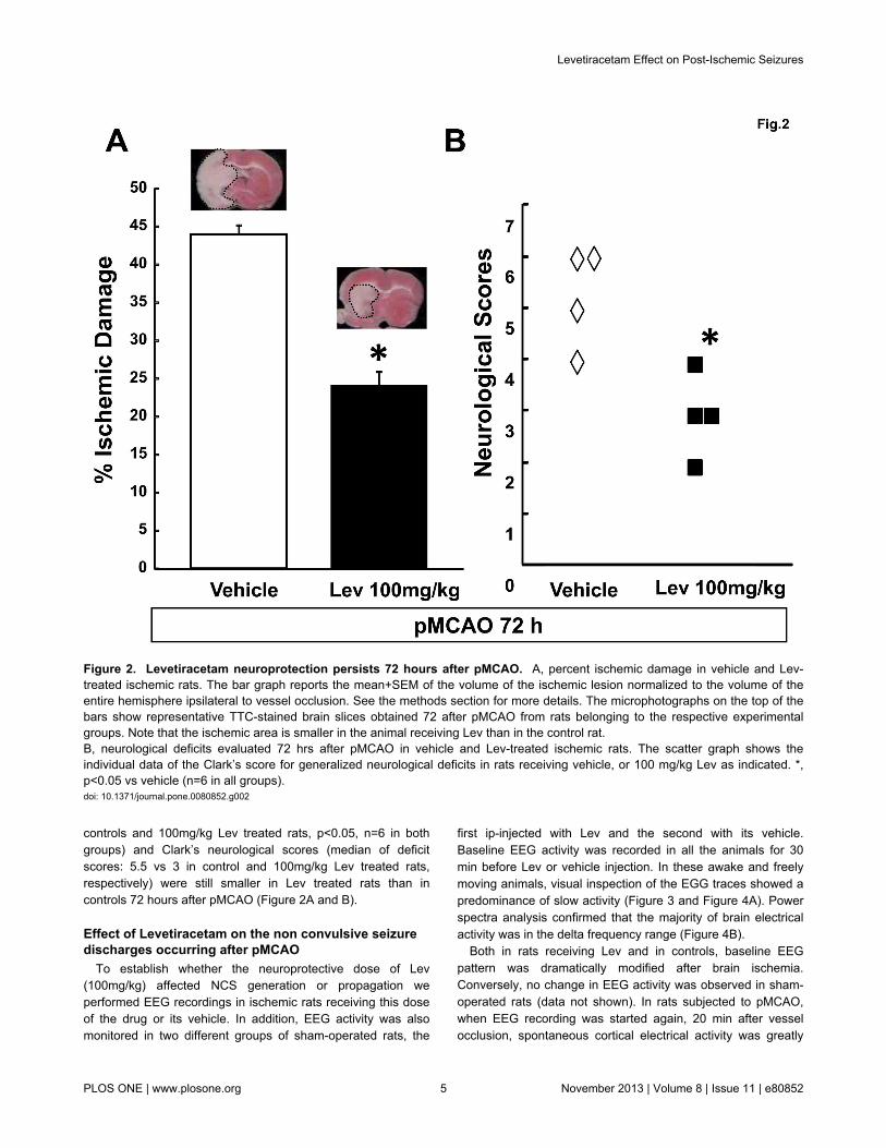
controls and 100mg/kg Lev treated rats, p<0.05, n=6 in bothgroups) and Clark’s neurological scores (median of deficitscores: 5.5 vs 3 in control and 100mg/kg Lev treated rats,respectively) were still smaller in Lev treated rats than incontrols 72 hours after pMCAO (Figure 2A and B).
Effect of Levetiracetam on the non convulsive seizuredischarges occurring after pMCAO
To establish whether the neuroprotective dose of Lev(100mg/kg) affected NCS generation or propagation weperformed EEG recordings in ischemic rats receiving this doseof the drug or its vehicle. In addition, EEG activity was alsomonitored in two different groups of sham-operated rats, the
first ip-injected with Lev and the second with its vehicle.Baseline EEG activity was recorded in all the animals for 30min before Lev or vehicle injection. In these awake and freelymoving animals, visual inspection of the EGG traces showed apredominance of slow activity (Figure 3 and Figure 4A). Powerspectra analysis confirmed that the majority of brain electricalactivity was in the delta frequency range (Figure 4B).
Both in rats receiving Lev and in controls, baseline EEGpattern was dramatically modified after brain ischemia.Conversely, no change in EEG activity was observed in sham-operated rats (data not shown). In rats subjected to pMCAO,when EEG recording was started again, 20 min after vesselocclusion, spontaneous cortical electrical activity was greatly
Figure 2. Levetiracetam neuroprotection persists 72 hours after pMCAO. A, percent ischemic damage in vehicle and Lev-treated ischemic rats. The bar graph reports the mean+SEM of the volume of the ischemic lesion normalized to the volume of theentire hemisphere ipsilateral to vessel occlusion. See the methods section for more details. The microphotographs on the top of thebars show representative TTC-stained brain slices obtained 72 after pMCAO from rats belonging to the respective experimentalgroups. Note that the ischemic area is smaller in the animal receiving Lev than in the control rat.B, neurological deficits evaluated 72 hrs after pMCAO in vehicle and Lev-treated ischemic rats. The scatter graph shows theindividual data of the Clark’s score for generalized neurological deficits in rats receiving vehicle, or 100 mg/kg Lev as indicated. *,p<0.05 vs vehicle (n=6 in all groups).doi: 10.1371/journal.pone.0080852.g002
Levetiracetam Effect on Post-Ischemic Seizures
PLOS ONE | www.plosone.org 5 November 2013 | Volume 8 | Issue 11 | e80852

reduced respect to baseline in the ischemic but not in thecontralateral brain hemisphere where it remained fairly stable
(Figure 3 and Figure 4A). Consequently, in both groups, thetotal power of the spectrum of the EEG recorded ipsilaterally to
Figure 3. Effect of brain ischemia and of Levetiracetam on EEG activity. The figure reports EEG recording samples of about40 sec in duration obtained in a representative control (on the left) and a Lev-treated rat (on the right) before and after pMCAO. Foreach animal, traces obtained immediately before, and 20 min, 120 min, 24 and 45 hours after pMCAO are reported. The tracesshown in the panels on the top of the figure were recorded ipsilaterally to MCAO whereas those in the bottom panels are from thecontralateral side. Note that EEG activity was markedly suppressed after pMCAO in the ipsi- but not in the contralateral brainhemisphere. Note also that a significant EEG activity reappeared, in the form of NCSs, 120 min after pMCAO in the control rat andonly 24 hours after vessel occlusion in the Lev-treated rat. In the traces from the contralateral hemisphere reported in the bottompanels, IRDAs can be easily identified as brief bursts occurring in isolation. Note that these events are already large and welldefined in traces obtained 120 min after pMCAO in control but not in Lev-treated rats in which they become clearly evident only after24 hours.doi: 10.1371/journal.pone.0080852.g003
Levetiracetam Effect on Post-Ischemic Seizures
PLOS ONE | www.plosone.org 6 November 2013 | Volume 8 | Issue 11 | e80852

MCAO decreased down to about 40% of basal values and ageneral decrease in EEG power occurred in the spectral trendgraph (Figure 5). No significant difference was observedbetween Lev-treated and control rats in the extent of thisischemia-induced suppression of the EEG activity. No changewas observed in the percent representation of the differentEEG spectral components neither in control nor in Lev-treatedrats. In the early phases after pMCAO, thus, the EEG spectrumstill showed a large prevalence of delta activity. On the
contrary, significant differences emerged, thereafter, betweenrats receiving 100 mg/kg Lev and controls. Indeed, in controlsbut not in rats treated with 100 mg/kg Lev, a paroxysmalelectrical activity appeared in the ischemic brain hemisphere by30-45 min after pMCAO. This resurgent electrical activitydiffered from that observed before the induction of ischemiaand was dominated by epileptiform discharges whose patternprogressively changed over the following 6-12 hours. The firstevents that we observed were rhythmic spikes (Figure 4A).
Figure 4. Time course and electrophysiological characteristics of the non-convulsive seizures in control rats and in ratstreated with Levetiracetam. A, examples of the different types of EEG activity recorded at different times after pMCAO in a control(top) and in a Lev-treated (bottom) rat. Note the different time course of NCSs in the control and in the Lev-treated rat. In the controlrat, spike-wave activity predominated 45 and 90 min after pMCAO, polyspike-waves after 12 hours and polyspikes after 24 hours. Inthe Lev-treated rat, NCSs appeared only 12 hour after pMCAO in the form of polyspike complexes. B, Spectral graph analysis of theEEG traces obtained at different times from pMCAO in a control rat (top) and in a rat injected with 100mg/kg Lev. The graphs showthe frequency distribution of the power of the EEG signal in the frequency range from 0.25 up to 32 Hz. The main band componentsof the EEG signal, delta, theta, alpha, beta 1, beta 2, are shown in different colors as detailed in the insets. Each spectral graph wasobtained by the analysis of 10 sec of artifact-free recordings. Note that, both in the control and in the Lev-treated rat, theappearance of NCSs corresponded to a strong increase of the power in the regions of high theta and low alpha bands.doi: 10.1371/journal.pone.0080852.g004
Levetiracetam Effect on Post-Ischemic Seizures
PLOS ONE | www.plosone.org 7 November 2013 | Volume 8 | Issue 11 | e80852

Mean amplitude, duration and frequency of these events were191.3 ± 11.9 μV, 41.9±2.7 sec, and 2.5 ± 0.9 Hz, respectively.Single spikes were gradually replaced by spike-wave andpolyspike complexes that represented the prevalent electricaldischarges 120 min after pMCAO (Figure 4A). Twenty fourhours after the induction of pMCAO, EEG activity was virtuallycomposed only by arrhythmic polyspikes (Figure 4A). Atspectral analysis, NCSs corresponded to the appearance of adistinct power peak in the regions of high theta and low alphaactivity with its maxima around 8 Hz (Figure 4B). In the spectraltrend graph NCSs appeared as brief high power episodessuperimposed on baseline (Figure 5A). These dischargeslasted approximately 45 hours. Afterwards, the EEG patternreturned to normal.
Lev (100mg/kg) had a dramatic effect on the EEG responseto pMCAO but it did not modify background EEG activity insham-operated rats (data not shown). Specifically, this drug
reduced NCS occurrence (Figure 3 and Figure 4A). While incontrol rats a mean of 6.4 + 0.3 NCSs occurred during the firsttwo hours of recordings after pMCAO, only 0.5 + 0.2 eventswere observed in Lev-treated animals (p<0.001). In this groupof rats, NCSs were also shorter (13.2+2.8, vs 41.9+2.7 sec,p<0.01, n=7) and their mean amplitude was lower than incontrols (74,3+5,9 vs 191,3+11,9 μV, p<0.01) (Figure 5B).Approximately 6 hours after Lev injection and pMCAOinduction, EEG activity begun to increase in Lev-treatedanimals (Figure 4A). The first EEG events observed in Lev-treated rats were sharp-waves. They were followed by singlespikes and, then, by spike and wave, polyspikes andpolyspikes and wave (Figure 4A). At power spectra analysis,the main spectral component peaked at approximately 8 Hz,encompassing both theta and alpha activity (Figure 4B).Therefore, this epileptiform activity recapitulated the electricalevents occurring at earlier stages in control animals. These
Figure 5. Levetiracetam prevents the occurrence of non convulsive seizures. A, representative spectral trend graphs showingthe changes in total EEG power after pMCAO in a control (left) and in a Lev-treated (right) rat. Each bar represents the total power(in μV2) calculated by integration of the power spectrum of the EEG recorded during about 120 min interval. The bars colored in reddenote the data obtained during the first 90 min of recordings after pMCAO. Note the dramatic drop in total EEG power afterpMCAO induction both in the presence and in the absence of Lev. The sudden and transient increases in EEG power correspond tothe NCS activity that appears in the control but not in the Lev-treated rat. B, Number, duration and amplitude of NCSs in Lev treatedand in control animals. The bar graphs show from the left to the right the mean+SEM of the number, duration in sec and amplitudein μV of the NCS observed during the first 2 hours after pMCAO in control (n=7) and Lev-treated (100mg/kg; n=7) rats. * p<0.05.doi: 10.1371/journal.pone.0080852.g005
Levetiracetam Effect on Post-Ischemic Seizures
PLOS ONE | www.plosone.org 8 November 2013 | Volume 8 | Issue 11 | e80852
events had, however, a power significantly smaller than thoseobserved in controls at the same time (23738.0+1306.0 vs7675.0+255.6 μV2, p<.0.01, n=7). In Lev-treated rats, as incontrols, epileptiform discharges disappeared approximately 45hours after pMCAO.
Both in control and in Lev-treated ischemic rats, EEG activitywas modified not only ipsilaterally but also contralaterally topMCAO. Specifically, an intermittent rhythmic delta activity(IRDA) appeared in control rats by 45 min from ischemiainduction (Figure 3). This activity consisted of bursts in thedelta range lasting less than 10 sec that occurred more often inisolation. IRDAs were still observed 24 hours after MCAOwhereas they completely disappeared by 45 hours. IRDAs alsooccurred in rats treated with 100 mg/kg Lev but, whencompared 120 min from pMCAO, to those recorded at thesame time in control animals, they had a smaller amplitude(97.3±8.3 vs 116.5±7.4 μV, p<0.01, n=7), lasted less (1.3±0.2vs 4.53±0.8 sec, p<0.01, n=7) and their frequency was lower(0.3±0.1 vs 1.24±0.1 Hz, p<0.05, n=7). As in control also inLev-treated rats IRDAs disappeared by 45 hours from MCAO(Figure 3).
Discussion
The main finding of the present study was that theantiepileptic drug Lev prevented the appearance of NCSs inthe parieto-temporal cortex of rats subjected to pMCAO.Parallel to this electrophysiological effect a significant reductionin the ischemic brain damage caused by this surgicalprocedure also occurred.
Since the seminal work of Leao [30] it has been establishedthat brain ischemia causes relevant changes in corticalelectrical activity both in men and in experimental animals (see21 for review). In accordance, in our control group, weobserved prominent changes in EEG activity after experimentalischemia. The alterations that we observed are similar to thosedescribed by Lu and coworkers [31] who reported a verydetailed topographical and spectral analysis of the electricalevents originating in the cerebral cortex of rats subjected toMCAO. In particular, in our experiments, a profoundsuppression of cortical electrical activity appeared in ratssubjected to pMCAO as early as 15-20 min after the initialinsult in the ischemic but not in the contralateral brainhemisphere. The evidence that no suppression of backgroundEEG activity did occur in sham-operated rats further supportsthe idea that it was actually caused by brain ischemia and itwas not a mere consequence of surgery and/or anesthesia.Ischemia-induced loss of EEG activity is currently consideredthe consequence of a non-spreading depression of thesynaptic activity caused either by the impairment of vesicularrelease of excitatory neurotransmitters [32] or by the massiverelease of adenosine in the lesioned brain [33]. Approximately45 min after the induction of ischemia, we observed aresurgent but abnormal EEG activity in the form of NCSs onthe side of middle cerebral artery occlusion. Interestingly, theelectrophysiological characteristics of these events graduallychanged over time. Specifically, whereas during the first 90 minthe EEG activity essentially consisted of single high voltage
and low frequency spikes, spike-wave discharges becameprevalent after 360 min. Brain ischemia also induced theappearance of an abnormal EEG activity in the brainhemisphere contralateral to pMCAO. This activity consisted ofbrief bursts in the delta frequency range and was similar toIRDAs described by Hartings et al. in experimental brainischemia in the rat [25]. Its genesis is still controversial but itcould be an indirect consequence of the damage occurring infar brain regions being caused, for instance, by subtledysfunction in midline cerebral structures due to the increase inintracranial pressure [25].
A single intraperitoneal injection of Lev at the dose of 100mg/kg virtually abolished both NCSs and IRDAs though thiseffect was only transient. In animals receiving the drug at thisconcentration EEG remained markedly suppressed till 24 hoursafter pMCAO when an epileptiform activity appeared in Lev-treated rats. This activity became, then, progressively moreintense though it was significantly lower than in controls. Thisgradual reappearance of activity can be easily explained bypharmacokinetics considerations. The half life of the drug in therat is, indeed, approximately 3 hours in blood and 5 hours incerebrospinal fluid [34].
The mechanism responsible for the suppression ofpostischemic electrical activity by Lev in both the cortex ipsiand contralateral to pMCAO remains to be determined.However, it could be dependent on the ability of this drug tosuppress neurotransmitter release [12] either by interactingwith the vesicular protein SV2 [11] that controls the priming ofsynaptic vesicles for fusion [35-37], or by decreasing Ca2+
release from the intracellular stores [38]. The integrity ofglutamatergic neurotransmission seems, indeed, to beessential for the propagation of spreading depolarization wavesin experimental brain ischemia in the rat [39]. These eventsthat can be measured as NCSs at EEG are generated at theborder between the core and the penumbra of the ischemiclesion when the anoxic depolarization invades the still alivebrain tissue of the latter region (see 21 for review). Thisdepolarization wave, presumably, activates local brainnetworks along which NCSs are then propagated. Inaccordance with this theory, NCSs are significantly increasedin transgenic mice overexpressing mutated, hyperfunctionalforms of the P/Q type channels, the predominant form ofvoltage-gated Ca2+ channels responsible for neurotransmitterrelease in the glutamatergic terminals [40]. Conversely, theycan be blocked by inducing a general inhibition ofneurotransmission with the simultaneous application ofblockers of AMPA/kainate and NMDA receptors and of voltagegated Na+ and Ca2+ channels [41]. The need of a markedsuppression of neurotransmitter release for a significant NCSsuppression could explain why not all the antiepileptic drugsare effective against these events [22] that are consideredespecially sensitive only to compounds acting on multipletargets [42].
In our experiments, the brain damage induced by ischemiawas significantly smaller in the animals that received Lev thanin controls. These data resemble those reported by Hanon andKlitgaard [8] in rats subjected to the transient occlusion of themiddle cerebral artery. Surprisingly, in brain slices undergoing
Levetiracetam Effect on Post-Ischemic Seizures
PLOS ONE | www.plosone.org 9 November 2013 | Volume 8 | Issue 11 | e80852

oxygen and glucose deprivation in vitro, Lev did not reduce theischemic damage evaluated either as propidium iodide staining[20] or as suppression of field electrical responses toextracellular stimulation [43]. Moreover, this drug did notsuppress the spreading depression elicited by oxygen andglucose deprivation in rat cortical brain slices in vitro [44]. Theinconsistency between the data obtained in vitro and in livinganimals suggests that Lev needs intact brain networks to exertits favourable effects in ischemia. This fits well with the ideathat this drug could suppress the synaptic propagation of NCS.Electrocorticographic and fMRI data show, indeed, that both inhumans and in rodents these depolarizing waves cycle aroundthe ischemic core for several days after the ischemic insult [23].We also have to remind that, recently, Meehan and co-workers[12,45] showed that Lev has to enter the synapse through therecycling vesicular route to exert its antiepileptic activity. Thisimplies that a certain degree of synaptic activity could beneeded to see Lev-induced neuroprotection. Therefore, we canspeculate that the lack of neuroprotection in the preparations invitro is the result of their low or absent spontaneous networkactivity. A significant neuroprotection in vivo despite the lack ofa neuroprotective effect in vitro has been reported also forgabapentin and ethosuximide [46]. Intriguingly, also thesecompounds significantly suppressed NCS occurrence [46].
In conclusion, we showed that Lev suppresses NCSs in ananimal model of stroke. Because NCSs contribute to enlargethe ischemic lesion [23] our findings could help explaining theneuroprotective activity of this drug. Considering that NCSsalso have a role in post-stroke epileptogenesis [24], the abilityof Lev to suppress these electrical events adds new argumentsto suggest that this drug could be useful for the prophylaxis oflate post-stroke seizures. This form of epilepsy represents amajor problem in clinical neurology as it occurs in 11.5% of thepatients surviving stroke [47] and accounts for the moreprevalent form of acquired epilepsy in adults [48,49]. Whileother antiepileptic drugs like ethosuximide, gabapentin,topiramate, phosphenytoin, and valproate also suppress NCSsafter MCAO [22,50], the better safety profile of Lev respect tothese older drugs warrants further studies to validate its earlyuse in stroke patients.
Author Contributions
Conceived and designed the experiments: MC VR GdR.Performed the experiments: OC AL GBP AV. Analyzed thedata: MC VR. Wrote the manuscript: MC VR.
References
1. Crepeau AZ, Treiman DM (2010) Levetiracetam: a comprehensivereview. Expert Rev Neurother 10: 159-171. doi:10.1586/ern.10.3.PubMed: 20136375.
2. Löscher W, Hönack D, Rundfeldt C (1998) Antiepileptogenic effects ofthe novel anticonvulsant levetiracetam (ucb L059) in the kindling modelof temporal lobe epilepsy. J Pharmacol Exp Ther 284: 474-479.PubMed: 9454787.
3. Stratton SC, Large CH, Cox B, Davies G, Hagan RM (2003) Effects oflamotrigine and levetiracetam on seizure development in a ratamygdala kindling model. Epilepsy Res 53: 95-106. doi:10.1016/S0920-1211(02)00254-1. PubMed: 12576171.
4. Vinogradova LV, van Rijn CM (2008) Anticonvulsive andantiepileptogenic effects of levetiracetam in the audiogenic kindlingmodel. Epilepsia 49: 1160-1168. doi:10.1111/j.1528-1167.2008.01594.x. PubMed: 18397292.
5. Russo E, Citraro R, Scicchitano F, De Fazio S, Di Paola ED et al.(2010) Comparison of the antiepileptogenic effects of an early long-term treatment with ethosuximide or levetiracetam in a genetic animalmodel of absence epilepsy. Epilepsia 51: 1560-1569. PubMed:19919665.
6. Russo E, Citraro R, Scicchitano F, De Fazio S, Perrotta I et al. (2011)Effects of early long-term treatment with antiepileptic drugs ondevelopment of seizures and depressive-like behavior in a rat geneticabsence epilepsy model. Epilepsia 52: 1341-1350. doi:10.1111/j.1528-1167.2011.03112.x. PubMed: 21635238.
7. Marini H, Costa C, Passaniti M, Esposito M, Campo GM et al. (2004)Levetiracetam protects against kainic acid-induced toxicity. Life Sci 74:1253-1264. doi:10.1016/j.lfs.2003.08.006. PubMed: 14697408.
8. Hanon E, Klitgaard H (2001) Neuroprotective properties of the novelantiepileptic drug levetiracetam in the rat middle cerebral arteryocclusion model of focal cerebral ischemia. Seizure 10: 287-293. doi:10.1053/seiz.2000.0511. PubMed: 11466025.
9. Kilicdag H, Daglıoglu K, Erdogan S, Guzel A, Sencar L et al. (2012)The effect of levetiracetam on neuronal apoptosis in neonatal rat modelof hypoxic ischemic brain injury. Early Hum Dev 89: 355-360. PubMed:23266150.
10. Wang H, Gao J, Lassiter TF, McDonagh DL, Sheng H et al. (2006)Levetiracetam is neuroprotective in murine models of closed headinjury and subarachnoid hemorrhage. Neurocrit Care 5: 71-78. doi:10.1385/NCC:5:1:71. PubMed: 16960300.
11. Lynch BA, Lambeng N, Nocka K, Kensel-Hammes P, Bajjalieh SM etal. (2003) The synaptic vesicle protein SV2A is the binding site for the
antiepileptic drug levetiracetam. Proc Natl Acad Sci U_S_A 26:9861-9866. PubMed: 15210974.
12. Meehan AL, Yang X, Yuan LL, Rothman SM (2012) Levetiracetam hasan activity-dependent effect on inhibitory transmission. Epilepsia 53:469-476. doi:10.1111/j.1528-1167.2011.03392.x. PubMed: 22292611.
13. Cataldi M, Lariccia V, Secondo A, di Renzo G, Annunziato L (2005)The antiepileptic drug levetiracetam decreases the inositol 1,4,5-trisphosphate-dependent [Ca2+]i increase induced by ATP andbradykinin in PC12 cells. J Pharmacol Exp Ther 313: 720-730.PubMed: 15644427.
14. Angehagen M, Margineanu DG, Ben-Menachem E, Rönnbäck L,Hansson E et al. (2003) Levetiracetam reduces caffeine-induced Ca2+
transients and epileptiform potentials in hippocampal neurons.Neuroreport 14: 471–475. PubMed: 12634506.
15. Nagarkatti N, Deshpande LS, DeLorenzo RJ (2008) Levetiracetaminhibits both ryanodine and IP3 receptor activated calcium inducedcalcium release in hippocampal neurons in culture. Neurosci Lett 436:289-293. doi:10.1016/j.neulet.2008.02.076. PubMed: 18406528.
16. Rigo JM, Hans G, Nguyen L, Rocher V, Belachew S et al. (2002) Theanti-epileptic drug levetiracetam reverses the inhibition by negativeallosteric modulators of neuronal GABA- and glycine-gated currents. BrJ Pharmacol 136: 659-672. doi:10.1038/sj.bjp.0704766. PubMed:12086975.
17. Lukyanetz EA, Shkryl VM, Kostyuk PG (2002) Selective blockade of N-type calcium channels by levetiracetam. Epilepsia 43: 9-18. doi:10.1046/j.1528-1157.43.s.9.4.x. PubMed: 11879381.
18. Eyal S, Yagen B, Sobol E, Altschuler Y, Shmuel M et al. (2004) Theactivity of antiepileptic drugs as histone deacetylase inhibitors.Epilepsia 45: 737-744. doi:10.1111/j.0013-9580.2004.00104.x.PubMed: 15230695.
19. Belcastro V, Pierguidi L, Tambasco N (2011) Levetiracetam in brainischemia: clinical implications in neuroprotection and prevention ofpost-stroke epilepsy. Brain Dev 33: 289-293. doi:10.1016/j.braindev.2010.06.008. PubMed: 20630672.
20. Rekling JC (2003) Neuroprotective effects of anticonvulsants in rathippocampal slice cultures exposed to oxygen/glucose deprivation.Neurosci Lett 335: 167-170. doi:10.1016/S0304-3940(02)01193-X.PubMed: 12531459.
21. Cataldi M (2013) The changing landscape of voltage-gated Ca2+
channels in neurovascular disorders and neurodegenerative diseases.Curr Neuropharmacol 11: 276-297. doi:10.2174/1570159X11311030004. PubMed: 24179464.
Levetiracetam Effect on Post-Ischemic Seizures
PLOS ONE | www.plosone.org 10 November 2013 | Volume 8 | Issue 11 | e80852

22. Williams AJ, Tortella FC, Lu XM, Moreton JE, Hartings JA (2004)Antiepileptic drug treatment of nonconvulsive seizures induced byexperimental focal brain ischemia. J Pharmacol Exp Ther 311:220-227. doi:10.1124/jpet.104.069146. PubMed: 15140918.
23. Nakamura H, Strong AJ, Dohmen C, Sakowitz OW, Vollmar S et al.(2010) Spreading depolarizations cycle around and enlarge focalischaemic brain lesions. Brain 133: 1994-2006. doi:10.1093/brain/awq117. PubMed: 20504874.
24. Dreier JP, Major S, Pannek HW, Woitzik J, Scheel M et al. (2012)Spreading convulsions, spreading depolarization and epileptogenesisin human cerebral cortex. Brain 135: 259-275. doi:10.1093/brain/awr303. PubMed: 22120143.
25. Hartings JA, Williams AJ, Tortella FC (2003) Occurrence ofnonconvulsive seizures, periodic epileptiform discharges, andintermittent rhythmic delta activity in rat focal ischemia. Exp Neurol 179:139-149. doi:10.1016/S0014-4886(02)00013-4. PubMed: 12618120.
26. Cuomo O, Gala R, Pignataro G, Boscia F, Secondo A et al. (2008) Acritical role for the potassium-dependent sodium/calcium exchangerNCKX2 in protection against focal ischemic brain damage. J Neurosci28: 2053-2063. doi:10.1111/j.1460-9568.2008.06502.x. PubMed:18305240.
27. Cuomo O, Pignataro G, Gala R, Scorziello A, Gravino E et al. (2007)Antithrombin reduces ischemic volume, ameliorates neurologic deficits,and prolongs animal survival in both transient and permanent focalischemia. Stroke 38: 3272-3279. doi:10.1161/STROKEAHA.107.488486. PubMed: 17975103.
28. Clark WM, Lessov NS, Dixon MP, Eckenstein F (1997) Monofilamentintraluminal middle cerebral artery occlusion in the mouse. Neurol Res19: 641-648. PubMed: 9427967.
29. Pignataro G, Boscia F, Esposito E, Sirabella R, Cuomo O et al. (2012)NCX1 and NCX3: two new effectors of delayed preconditioning in brainischemia. Neurobiol Dis 45: 616-623. doi:10.1016/j.nbd.2011.10.007.PubMed: 22036625.
30. Leaõ AAP (1947) Further observation on the spreading depression ofactivity in the cerebral cortex. J Neurophysiol 10: 409-414. PubMed:20268874.
31. Lu XC, Williams AJ, Tortella FC (2001) Quantitativeelectroencephalography spectral analysis and topographic mapping ina rat model of middle cerebral artery occlusion. Neuropathol ApplNeurobiol 27: 481-495. doi:10.1046/j.1365-2990.2001.00357.x.PubMed: 11903931.
32. Fleidervish IA, Gebhardt C, Astman N, Gutnick MJ, Heinemann U(2001) Enhanced spontaneous transmitter release is the earliestconsequence of neocortical hypoxia that can explain the disruption ofnormal circuit function. J Neurosci 21: 4600-4608. PubMed: 11425888.
33. Canals S, Larrosa B, Pintor J, Mena MA, Herreras O (2008) Metabolicchallenge to glia activates an adenosine-mediated safety mechanismthat promotes neuronal survival by delaying the onset of spreadingdepression waves. J Cereb Blood Flow Metab 28: 1835-1844. doi:10.1038/jcbfm.2008.71. PubMed: 18612316.
34. Doheny HC, Ratnaraj N, Whittington MA, Jefferys JG, Patsalos PN(1999) Blood and cerebrospinal fluid pharmacokinetics of the novelanticonvulsant levetiracetam (ucb L059) in the rat. Epilepsy Res 34:161-168. doi:10.1016/S0920-1211(98)00104-1. PubMed: 10210031.
35. Custer KL, Austin NS, Sullivan JM, Bajjalieh SM (2006) Synapticvesicle protein 2 enhances release probability at quiescent synapses. J
Neurosci 26: 1303-1313. doi:10.1523/JNEUROSCI.2699-05.2006.PubMed: 16436618.
36. Chang WP, Südhof TC (2009) SV2 renders primed synaptic vesiclescompetent for Ca2+-induced exocytosis. J Neurosci 29: 883-897. doi:10.1523/JNEUROSCI.4521-08.2009. PubMed: 19176798.
37. Yao J, Nowack A, Kensel-Hammes P, Gardner RG, Bajjalieh SM(2010) Cotrafficking of SV2 and synaptotagmin at the synapse. JNeurosci 30: 5569-5578. doi:10.1523/JNEUROSCI.4781-09.2010.PubMed: 20410110.
38. Fukuyama K, Tanahashi S, Nakagawa M, Yamamura S, Motomura E etal. (2012) Levetiracetam inhibits neurotransmitter release associatedwith CICR. Neurosci Lett 518: 69-74. doi:10.1016/j.neulet.2012.03.056.PubMed: 22484014.
39. Gill R, Andiné P, Hillered L, Persson L, Hagberg H (1992) The effect ofMK-801 on cortical spreading depression in the penumbral zonefollowing focal ischaemia in the rat. J Cereb Blood Flow Metab 12:371-319. doi:10.1038/jcbfm.1992.54. PubMed: 1314840.
40. Eikermann-Haerter K, Lee JH, Yuzawa I, Liu CH, Zhou Z et al. (2012)Migraine mutations increase stroke vulnerability by facilitating ischemicdepolarizations. Circulation 125: 335-345. doi:10.1161/CIRCULATIONAHA.111.045096. PubMed: 22144569.
41. Müller M, Somjen GG (1998) Inhibition of major cationic inwardcurrents prevents spreading depression-like depolarization in rathippocampal tissue slices. Brain Res 812: 1-13. doi:10.1016/S0006-8993(98)00812-9. PubMed: 9813218.
42. Leker RR, Neufeld MY (2003) Anti-epileptic drugs as possibleneuroprotectants in cerebral ischemia. Brain. Res Rev 42: 187-203.doi:10.1016/S0165-0173(03)00170-X.
43. Costa C, Martella G, Picconi B, Prosperetti C, Pisani A et al. (2006)Multiple mechanisms underlying the neuroprotective effects ofantiepileptic drugs against in vitro ischemia. Stroke 37: 1319-1326. doi:10.1161/01.STR.0000217303.22856.38. PubMed: 16574927.
44. Margineanu DG, Klitgaard H (2009) Brivaracetam inhibits spreadingdepression in rat neocortical slices in vitro. Seizure 18: 453-456. doi:10.1016/j.seizure.2009.01.002. PubMed: 19211275.
45. Meehan AL, Yang X, McAdams BD, Yuan L, Rothman SM (2011) Anew mechanism for antiepileptic drug action: vesicular entry maymediate the effects of levetiracetam. J Neurophysiol 106: 1227-1239.doi:10.1152/jn.00279.2011. PubMed: 21653714.
46. Williams AJ, Bautista CC, Chen RW, Dave JR, Lu XC et al. (2006)Evaluation of gabapentin and ethosuximide for treatment of acutenonconvulsive seizures following ischemic brain injury in rats. JPharmacol Exp Ther 318: 947-955. doi:10.1124/jpet.106.105999.PubMed: 16728590.
47. Burn J, Dennis M, Bamford J, Sandercock P, Wade D et al. (1997)Epileptic seizures after a first stroke: the Oxfordshire Community StrokeProject. BMJ 315: 1582-1587. doi:10.1136/bmj.315.7122.1582.PubMed: 9437276.
48. Camilo O, Goldstein LB (2004) Seizures and epilepsy after ischaemicstroke. Stroke 35: 1769-1775. doi:10.1161/01.STR.0000130989.17100.96. PubMed: 15166395.
49. Herman ST (2002) Epilepsy after brain insult: targeting epileptogenesis.Neurology 59 (Suppl 5): S21-S26. doi:10.1212/WNL.59.4.21A.PubMed: 12428028.
50. Williams AJ, Tortella FC, Gryder D, Hartings JA (2008) Topiramatereduces non-convulsive seizures after focal brain ischemia in the rat.Neurosci Lett 430: 7-12. doi:10.1016/j.neulet.2007.09.052. PubMed:18063309.
Levetiracetam Effect on Post-Ischemic Seizures
PLOS ONE | www.plosone.org 11 November 2013 | Volume 8 | Issue 11 | e80852


