Embed Size (px)
Citation preview

The Association of PARP1 Enzymatic Inhibition and Chromatin Complex Formation with Radiosensitization by PARP Inhibitors and Their Combination with ATR Inhibition in
Pancreatic Cancer
by
Carl G. Engelke
A dissertation submitted in partial fulfillment of the requirements of the degree of
Doctor of Philosophy (Molecular and Cellular Pathology)
in the University of Michigan 2020
Doctoral Committee: Professor Arul M. Chinnaiyan, Co-Chair Professor Theodore S. Lawrence, Co-Chair Professor Gary D. Hammer Associate Professor Meredith A. Morgan Assistant Professor Corey W. Speers Professor Scott A. Tomlins

ii
DEDICATION
This dissertation is dedicated to my family: to my wife and partner, Téa Prokes, and to
my parents, Alexandra Krikos and David Engelke. The love and support you have shown me has
meant more than I could ever express.

iii
ACKNOWLEDGEMENTS
First and foremost, I would like to acknowledge and thank my mentors. I have had the
very good fortune to be mentored not by one, but three wonderful scientists and advisors:
Meredith Morgan, Ted Lawrence, and Arul Chinnaiyan. They have provided me a scientific
home and contributed to my development as a scientist in ways that I appreciate and will
continue to appreciate throughout my life. From troubleshooting experiments and organizing
testable hypotheses, to managing projects and understanding the scientific field and how it
moves, they have contributed to my education. Each has provided me unique research
opportunities and scientific insight and I will always be grateful to them.
I would also like to thank the other three members of my thesis committee. Gary
Hammer, Corey Speers, and Scott Tomlins have provided me not only with scientific guidance
during our many formal committee meetings and informal discussions, but also wisdom about
what it means to be a physician scientist and how to navigate the path ahead of me.
The work presented in this dissertation would not have been possible without the effort
and assistance of many members of the Morgan lab, whom I would like to thank here. The
investigations into PARP trapping in combination with radiation and ATR inhibition is a project I
shared with our laboratory manager, Leslie Parsels. In addition to the enormous body of work
she contributed to this project, she has been a constant source of support and scientific
feedback. Josh Parsels is another member of the lab who contributed many experiments to the

iv
work presented in this dissertation; in addition, I appreciated his patience and tutelage in many
techniques where he demonstrates wizardry, including animal experiments and microscopy.
Finally, I would like to thank Qiang Zhang for his guidance in my earlier investigations into
ubiquitin ligases, which do not appear in this dissertation. It goes without saying that in
addition to the ways that I appreciate all members of the lab from the perspective of our
professional collaboration, I also consider them friends.
I would like to thank the various organizations that have supported me during my time
in graduate school. The MSTP has helped me navigate the transitions between medical school
and graduate school and has helped me stay on course during this long journey. Specifically, I
would like to thank director Ron Koenig and assistant directors Justine Hein and Ellen Elkin, in
addition to the associate directors and office staff. The Molecular and Cellular Pathology
training program, its director Zaneta Nikolovska-Coleska, and administrator Laura Labut have
provided me enormous help navigating the hurdles of graduate school. The Department of
Radiation Oncology and Michigan Center for Translational Pathology have provided me
scientific homes for the past five and a half years, for which I am grateful.
My family has been a constant source of support during graduate school and
throughout my life. My parents have always encouraged me to follow my passions. They
encouraged my curiosity and provided me every opportunity to explore and learn as I grew up.
They supported and encouraged me when I went to music school and in my brief career as a
professional musician, and again when I changed careers to pursue an MD and later a PhD.
When I began graduate school, I could not have imagined that I would meet and marry
the wonderful woman who is now my wife. Téa Prokes has been an incredible, loving, and

v
supportive partner in my life since we met. I love her and look forward to the adventures that
await us in the next chapter of our lives.

vi
TABLE OF CONTENTS
DEDICATION…….………………………………………………………………………………………..………………..………..ii
ACKNOWLEDGEMENTS……………………………………………………………………………………………..………….iii
LIST OF FIGURES……………………………………………………………………………………………………..……..……viii
LIST OF ILLUSTRATIONS…………………………………………………………………………………………..……..………x
ABSTRACT………………………………………..………………………………………………………..……………..………….xi
CHAPTER 1: Introduction…………..………………………………………………………………………………..…………1
1.1 Radiation and locoregional control in pancreatic cancer……………………………..…….…….1
1.2 The DNA Damage Response…………………………………………………………………………..…………3
1.3 Chemoradiation……………………………………………………………………………………………………….6
1.4 Checkpoint kinase 1 (Chk1) and Wee1 kinase (Wee1)………………………………………..…….8
1.5 Ataxia telagiectasia mutated and Rad3 related (ATR)…………………………………...………..10
1.6 Poly (ADP-ribose) polymerase 1 (PARP1)…………………………………………………………….….12
CHAPTER 2: The Association of PARP1 Enzymatic Inhibition and Chromatin Complex
Formation with Radiosensitization by PARP Inhibitors…….……………………………………….……….…19
2.1 Introduction…………………………………………………………………………………………………..……….19
2.2 Results……………………………………………………………………………………………………………………21
2.3 Conclusions and discussion.……………………………………………………………………………………29
2.4 Experimental procedures……………………………………………………………………………..………..32

vii
2.5 Data and Figures…………………………………………………………………………………………….………35
CHAPTER 3: Combined Inhibition of ATR and PARP As a Radiosensitizing Strategy in
Pancreatic Ductal Adenocarcinoma………………………………………………………………………………………48
3.1 Introduction……………………………………………………………………………………………………………48
3.2 Results……………………………………………………………………………………………………………………50
3.3 Conclusions and discussion………………………………………………………………………….…………55
3.4 Experimental procedures………………………………………………………………………………….……58
3.5 Data and Figures………………………………………………………………………………………….…………62
CHAPTER 4: Future Directions………………………………………………………………………………………..……74
4.1 Continued exploration of PARP trapping………………………………………………..………………74
4.2 PARP2 and PARP3………………………………………………………………………………………..…………80
4.3 PARP inhibitor resistance………………………………………………………………………………..………83
4.4 Immunomodulation and the DNA damage response..…………………………………….………86
REFERENCES……………………………………..…………………………………………………………………………………92

viii
LIST OF FIGURES
Figure 2.1 Figure 3.2 Sensitization to radiation by olaparib in pancreatic cancer cells…………..…36
Figure 2.2 Cellular PARP changes after treatment with olaparib and radiation……………………..…38
Figure 2.3 High conc. olaparib causes G2 arrest and delayed DNA damage resolution…………….49
Figure 2.4 High concentration olaparib prevents repair of DNA double strand breaks following
radiation……………………………………………………………………………………………………………………………….…40
Figure 2.5 Radiosensitizing potency of PARP inhibitors……………………………………………………………41
Figure 2.6 Enzymatic inhibition potency of PARP inhibitors…………………………………………………..…42
Figure 2.7 PARP1 deletion does not replicate the effects of high concetration olaparib………..…43
Figure 2.8 PARP inhibitor resistance by PARP1 deletion is reversed by exogenous expression of
PARP1………………………………………………………………………………………………………………………………..…….45
Figure 2.9 The proximity ligation assay as a measure of PARP trapping…………………………….…….46
Figure 3.1 The interaction of ATR and PARP inhibitors on cytotoxicity in HRP and HRD pancreatic
cancers…………………………………………………………………………………………………………………………….………63
Figure 3.2 Sensitization to radiation by olaparib and AZD6738 in pancreatic cancer cells…….….64
Figure 3.3 PARP1 null cells are not radiosensitized by olaparib and AZD6738…………………….……65
Figure 3.4 ATR and PARP inhibition delay the resolution of DNA following radiation……………….66
Figure 3.5 The effect of combined ATR and PARP1 inhibition on replication stress………………….68

ix
Figure 3.6 Radiosensitization of pancreatic tumor xenografts by AZD6738 and olaparib…………70

x
LIST OF ILLUSTRATIONS
Illustration 1.1 The DNA damage response and cell cycle checkpoints protect the cell from
ionizing radiation…………………………………………………………………………………………………………………..…17
Illustration 2.1 PARP1 response to DNA damage and consequences of inhibition……………………47
Illustration 3.1 ATR protects stalled replication forks………………………………………………………………73
Illustration 4.1 Proposed model of how PARP trapping leads to changes in replication fork
velocity…………………………………………………………………………………………………………………………………….91

xi
ABSTRACT
Pancreatic cancer has a five-year overall survival rate of less than 10%. Locoregional
progression is responsible for death in up to a third of all patients, highlighting the need for
local control. The use of chemotherapeutic agents in combination with radiation represents a
current standard of care for locally advanced PDAC, but new therapies are urgently needed.
PARP inhibitors have shown promise as radiosensitizing agents preclinically, both alone
and in combination with chemotherapeutic or molecularly targeted agents. Work has
demonstrated that their antitumor effect may be the product of their enzymatic inhibition or
ability to form PARP1-DNA complexes at DNA damage sites known as PARP trapping. These
complexes have been implicated as the cause for both therapeutic efficacy as well as many
dose-limiting toxicities seen clinically.
In our work, we investigated the radiosensitizing properties of PARP inhibitors in
pancreatic cancer cell line models. Using olaparib, which inhibits PARP at both low and high
concentrations but only traps at high concentrations, we demonstrated that pancreatic cancer
cells proficient in homologous recombination were only radiosensitized by high concentrations
of olaparib. While we demonstrated that olaparib possesses the ability to inhibit enzymatically
at both high and low concentrations, it is known to trap only at high. The ability to form PARP1-
DNA complexes correlated with increased DNA double strand breaks at higher olaparib
concentrations.

xii
We discovered that the radiosensitizing potency of three PARP inhibitors, veliparib,
olaparib, and talazoparib, corresponded to their described increasing potency as PARP trappers.
Deletion of PARP1 failed to phenocopy the radiosensitizing effects of PARP inhibitors and
protected cells from cytotoxicity from talazoparib. In order to more fully characterize PARP
trapping, we adapted the proximity ligation assay to measure trapped PARP1 in situ by
measuring the proximity of PARP1 and total histone H2AX in treated cells.
To further potentiate the radiosensitizing effects of PARP inhibition, we leveraged the
knowledge that PARP inhibition causes replication stress. ATR is known to regulate the cellular
response to replication stress and fork stability. Thus, we sought to combine PARP inhibition
with the ATR inhibitor AZD6738. The combinatorial effect of ATR and PARP inhibition in HR
proficient cell lines to radiosensitize and damage DNA was most pronounced at trapping
concentrations of olaparib. Further, this effect required presence of the PARP1 protein.
Combined treatment in mice bearing pancreatic cancer xenografts with olaparib, AZD6738, and
radiation substantially inhibited tumor growth relative to all other treatment groups while
causing minimal toxicity. These findings strongly recommend the merits of clinical investigation
into the efficacy of combined ATR and PARP inhibition with radiation for locally advanced
disease and suggest that the PARP inhibitors with greater PARP trapping potency may be most
efficacious.

1
CHAPTER 1:
Introduction
Our laboratory focuses on the use of pharmacologic agents to improve the efficacy
of therapeutic radiation for the treatment of pancreatic cancer. As such, our field has
broadly focused on the stages of pancreatic cancer where radiation holds the most
promise for treatment and the inherent makeup of pancreatic cancer that would render it
susceptible to specific types of intervention. Past work has found that the use of anti-
metabolite agents such as gemcitabine or 5-fluorouracil sensitizes pancreatic cancer to
ionizing radiation through an increase in DNA damage. Since then, we have undertaken
efforts to further increase the efficacy of these therapies through the introduction of
molecularly targeted agents directed at proteins involved in pathways that influence DNA
repair. My own work in the lab began with investigating inhibitors of cell cycle checkpoint
proteins checkpoint kinase 1 and Wee1 kinase, the latter of which has gone on to show
promise clinically in combination with chemoradiation. More recent work has focused on
the inhibition of two sensors and master regulators of the DNA damage response—poly
(ADP-ribose) polymerase 1 and ataxia telangiectasia mutated and Rad3-related—in
combination with radiation, which will be presented in this dissertation.
1.1 Radiation and locoregional control in pancreatic cancer

2
90% of cancers of the pancreas are pancreatic ductal adenocarcinoma (PDAC) [1].
Among the most lethal cancers, it has a 5 year survival less than 10% [2]. Most of those
in that 10% are among those able to undergo surgical resection [3]. Among the difficulties
leading to these poor outcomes is the relative paucity of symptoms, leading to late-stage
diagnosis in most patients [4]. To make matters worse, pancreatic cancer tends to be
inherently aggressive and prone to early metastasis. Its typical mutational landscape and
microenvironment render it highly resistant to conventional therapies. Finally, definitive
surgical resection is difficult due to the tendency for invasion of critical local structures,
leading to the famous “surgeon’s three rules” [5].
A relatively small number of PDACs (5-6%) contain germline mutations, the most
frequent of which tend to be genes involved in DNA repair [6], [7]. Overwhelmingly, the
most recurrently mutated gene in PDAC is KRAS, demonstrating an oncogenic activating
mutation in greater than 90% of patients. Other recurrently mutated genes include TP53,
SMAD4, and CDKN2A, which are reported at various frequencies but almost always
above 50% [8]–[13]. Another hallmark of pancreatic cancers is the thick desmoplastic
stroma surrounding tumor cells. It is estimated that up to 90% of pancreatic tumor volume
is comprised of cancer-associated fibroblasts [14]. For this reason, study of the tumor
microenvironment in PDAC represents a vital contribution to our understanding of the
disease, but is beyond the scope of the work presented here (reviewed in [15]–[17]).
The standard of treatment for local PDAC with well-defined margins is surgical
resection, but this only represents 10-20% patients at the time of diagnosis [18]. The
remainder fall into the category of locally advanced or metastatic cancer. For these, the
historical standard has been antimetabolite therapies such as Gem and 5-FluoroUracil

3
(5-FU) either alone or with radiation [19]. The recent introduction of a multidrug regimen
consisting of folinic acid, 5-FU, irinotecan, and oxplatin (FOLFIRINOX) has improved
outcomes [20] and despite increased toxicities associated with the more aggressive
regimen has still measurably improved quality of life for patients [21]. Together with the
addition of albumin-bound paclitaxel (nab-paclitaxel) to gemcitabine [22], [23], these
represent the current standard of care.
While most non-resectable disease is metastatic, necessitating the use of
aggressive systemic therapy, radiation represents an important therapeutic modality for
local disease. However, the role of radiation in the locally advanced setting was called
into question by a recent trial LAP07 [24], which showed no difference in median overall
survival (OS) between chemotherapy and chemoradiotherapy. However, other trials
suggest benefits of added radiation, with a study by the Eastern Cooperative Oncology
group demonstrating an improvement in median OS from 9.2 months with gemcitabine
alone to 11.1 months with gemcitabine plus radiation [25]. The importance of locoregional
control in the setting of pancreatic cancer is underlined by the fact that up to one third of
patients die from local disease progression [26] and that chemotherapy alone is unable
to cure gross disease. Taken together the data suggest that as our systemic therapies
continue to improve and reduce metastatic disease burden, the efficacy of radiation in the
local setting will become more readily appreciable. Above all, they underline the
inadequacy of our current approaches and the desperate need for advancement.
1.2 The DNA Damage Response

4
Therapeutic radiation has an upper limit of tolerability as a monotherapy due to
surrounding normal tissue toxicity. Therefore, it becomes important to search for ways to
enhance the tumor cell sensitivity to radiation while sparing normal tissues. One of the
basic hallmarks of cancer is genomic instability and mutation [27], [28]. While this property
is an advantage for growth and adaptation, it is also a liability as it renders the cancer cell
vulnerable to therapeutic interventions. Specifically, these genomic vulnerabilities render
the cell more susceptible to increased genomic stress and targeting of its remaining DNA
safeguards. Normal tissues, meanwhile, are able to tolerate greater amounts of damage
and removal of one or two redundant protective systems (reviewed in [29]). Much of the
work, therefore, has focused on targeting weaknesses in the DNA damage repair and cell
cycle checkpoint pathways, and the exploitation of replicative stress to preferentially
sensitize cancer cells to radiotherapy.
Cellular ionizing radiation most frequently causes damage to the DNA backbone,
creating a single-stranded break (SSB) or double-stranded break (DSB). SSBs are more
common and repaired through SSB repair where they are sensed by poly (ADP-ribose)
polymerase 1 (PARP1), which recruits repair factors including XRCC1 and Lig3. DSBs
are more lethal [30] and are repaired by two main pathways: non-homologous end joining
(NHEJ) or homologous recombination (HR) repair (Illustration 1.2a). NHEJ is the faster,
error-prone repair pathway available to the cell throughout the cell cycle. It begins with
the recognition and stabilization of DNA ends by the KU70 and KU80 heterodimers and
initial recruitment of DNA-dependent protein kinase (DNA-PK). The repair process
proceeds, recruiting other necessary factors, to ligate together blunt ends of the DNA and
repair the breach. While the process is fast and the integrity of the DNA backbone

5
preserved, deletions can be introduced in genes through loss of material to generate blunt
ends. For this reason, it is most frequently employed to repair simple DSBs. At complex
or single-ended DSBs the chances of generating a deletion or ligating to a distal location
in the genome is much greater.
During the S and G2 phases of the cell cycle, HR is available for the repair of DSBs.
Using sister chromatids as a template, this process is able to repair even complex DNA
lesions with high fidelity. After recognition of the insult by ataxia telangiectasia mutated
(ATM) and Mre11-Rad50-Nbs1(MRN) complex, 5’ ends of the DNA are resected and
bound with replication protein a (RPA). RPA is bound by ATRIP, which in turn attracts
ataxia telangiectasia mutated and RAD3-related (ATR) to sites of resolving DSBs. RAD51
recruitment to the site by BRCA2 allows sister chromatid strand invasion to provide a
template for homology, ensuring high fidelity resolution.
A third type of DSB repair exists, known as alternative end joining (Alt-EJ), which
may compensate in the absence of NHEJ or HR activity. Alt-EJ does not rely on the
components of these pathways, but rather repairs lesions with slow kinetics through the
actions of PARP1, XRCC1, Lig3, and other proteins that are still being elucidated. In some
cases, Alt-EJ may be facilitated by regions of DNA sequence microhomology [31].
Another type of DNA backbone break occurs physiologically during replication, when
the DNA strands are separated by helicase and newly synthesized DNA trails behind. At
replication forks, single strand breaks caused by unligated Okazaki fragments account
for a majority of cellular PARP1 activity. Replication forks that encounter an unpassable
genomic lesion can become stalled, generating large stretches of single-stranded DNA

6
and activating ATR. Unresolved stalled replication forks can collapse to generate complex
DSBs, which must then be repaired through HR.
In order to accommodate the repair of deleterious genomic lesions, the cell passes
through several cycle checkpoints where it can arrest and repair damage (Illustration
1.2b). These checkpoints are activated by ATM and ATR, making them master regulators
of the DNA damage response. The G1 checkpoint is crucial to prevent entry into S phase
with unresolved damage, and relies on the phosphorylation and activation of p53 by ATM.
The frequent mutation of p53 in pancreatic cancer leads the cell to rely heavily on its intra-
S and G2/M checkpoints to prevent mitotic progression with catastrophic damage. ATR
and ATM work in tandem through checkpoint kinase 1 (Chk1) and checkpoint kinase 2
(Chk2), respectively, to activate the intra-S and G2/M checkpoints. Wee1 kinase
additionally regulates the G2/M checkpoint through inhibitory phosphorylation of Cdk1.
1.3 Chemoradiation
Conventional chemotherapeutic agents were among the first to be found to sensitize
cancer cells in vitro to radiation [32]–[34] in the 1950’s. A decade later, promising results
were achieved in human trials by combining 5-FU with radiation in gastrointestinal
cancers [35], [36]. Shortly thereafter, the same group investigated adding a regimen of 5-
FU to radiotherapy for locally advanced unresectable pancreatic adenocarcinoma [37].
They found a significant survival benefit improving median survival from 23 weeks to
either 33 weeks or 39 weeks depending on the dose of radiation. The combination of 5-
FU and radiation was also investigated in the adjuvant setting for resected pancreatic
adenocarcinoma, improving median survival from 11 months to 20 months [38]. A pilot
7
study in the neoadjuvant setting [39] demonstrated improved rates of resectable disease
following radiation combined with 5-FU and mitomycin-C.
The increasing use of gemcitabine in pancreatic cancer led to its investigation in
preclinical studies, where it demonstrated powerful radiosensitization [40], [41]. A
retrospective analysis comparing radiation in combination with 5-FU or gemcitabine
demonstrated higher toxicities in the gemcitabine-radiation group but a small but
statistically insignificant median overall survival advantage [42]. These results
underscored the relatively narrow therapeutic index for optimal chemoradiation.
Cisplatin is the most widely used chemotherapeutic in combination with radiation.
Cisplatin is an inorganic platinum agent that forms intra-strand crosslinks on DNA, leading
to impaired replication and DNA damage, which accounts for most of its toxicity [43]. As
a single agent, these adducts are most frequently repaired by the nucleotide excision
repair (NER) pathway. However, it has been established that the lesions induced by the
radiosensitizing effects of cisplatin mostly involve NHEJ [44], and these adducts develop
into complex DSBs following radiation induced DNA damage [45]. Similarly,
temozolomide forms methyl adducts at specific positions on guanine and adenine which
are normally repaired by Mismatch Repair (MMR). In conjunction with radiation, it inhibits
DNA repair [46] and increases DSBs [47].
In pancreatic cancer, antimetabolites such as 5-FU and gemcitabine are the
standard agents used in combination with radiation. In their active forms, both of these
agents misincorporate into DNA, and inhibit enzymes involved in maintenance of the
cellular nucleotide pool, thymidylate synthase and ribonucleotide reductase, respectively.
The addition of these agents deplete available nucleotides and shift cells into incomplete

8
S-phase, where replication forks collide with radiation-induced SSBs to produce complex
DSB that are repaired slowly [48], [49].
Despite all of the improvements in radiation efficacy delivered by traditional
chemotherapeutics, their current delivery at maximally tolerated doses suggests that we
have already or nearly realized their maximum therapeutic potential. However, with the
development of molecularly targeted agents directed toward proteins involved in the DNA
damage response, opportunities exist to improve the DNA damaging efficacy of
chemoradiation without corresponding increases in toxicity.
1.4 Checkpoint kinase 1 (Chk1) and Wee1 kinase (Wee1)
Upon DNA damage, Chk1 is phosphorylated at S317 and S345 and hence activated
by ATR [50]. In turn, Chk1 phosphorylates CDC25A, leading to a decrease in CDK2
activity and activation of the S-phase cell cycle checkpoint [51]. Additional
phosphorylation events on CDC25B and CDC25C lead to their degradation and
decreased CDK1/cyclinB activity and G2/M arrest [52], [53]. In yeast, Chk1 also acts to
phophorylate Wee1 kinase to activate and further potentiate G2 arrest [54], [55], though
it is not clear that it performs this function in humans. Additionally, studies have reported
that it is involved in homologous recombination repair [56], [57] and the protection of
replication fork integrity [58].
Specific inhibitors of Chk1/2 (AZD7762) and Chk1 (MK8776) were developed as
anti-cancer therapeutics to exploit its cell cycle function by forcing cells through mitosis
with damage [59]. Chk1 inhibition is most effective in the setting of cancers that already
possess cell cycle defects. It is described to have synthetic lethality with the Fanconi

9
anemia pathway [60] and Rad17 [61] and Wee1 [62]. However, further investigation
demonstrated that increased DNA damage and inhibition of HR are major contributors to
the efficacy of Chk1 inhibitors [56].
Chk1 inhibition was first found to chemosensitize by the inhibitor UCN-01 [63], an
agent originally developed to inhibit members of the protein kinase C family, but with
many targets. More selective agents have been used in combination with gemcitabine
and irrinotecan ([64]) and radiation [57] preclinically. In agreement with its proposed
mechanism of forced mitotic entry, it preferentially sensitizes in p53 mutant cancers [65]–
[67], which lack a functional G1 checkpoint. It has also been found to chemoradiosensitize
in preclinical pancreatic cancer models [68]. Inhibitors have undergone clinical
investigation in combination with gemcitabine (NCT00937664 NCT00413686
NCT00839332 NCT01139775 NCT01341457 NCT00779584 NCT00437203) and
chemoradiation (NCT02555644).
Similar to Chk1, Wee1 functions to regulate Cdk1/CycB control of the G2/M
checkpoint (reviewed in [69]). Growth factor signals in active cells maintain this check on
mitotic entry through phosphorylation at S642 of CDK1 by Akt to activate [70]. In turn, it
phosphorylates CDK1 on Y15 to maintain an inactive state [71], [72]. In the absence of
unresolved DNA damage, Wee1 is targeted for degradation by phosphorylation from
PLK1 to release CDK1 inhibition [73]. Wee1 is another protein at the nexus of many
genomic integrity pathways besides cell cycle arrest, and has described roles in cell
division coordination [74] replication stability [75], [76] and HR [77].
AZD1775 (previously MK1775) was developed as a specific Wee1 inhibitor [78]. It
sensitizes with a variety of DNA damaging agents such as 5-FU [79] gemcitabine [80],

10
[81], and radiation [82]–[84], and increases the sensitivity to chemoradiation ([85]). Like
Chk1 inhibition, Wee1 inhibitors preferentially radiosensitize p53 mutant tumors [83], [84],
[86]. In an effort to move beyond the toxicities associated with traditional
chemotherapeutics, AZD1775 has also been combined with other DDR inhibitors as a
radiosensitizer including PARP1 [87] and Chk1 [88].
Many trials are currently ongoing combining AZD1775 with DNA damaging
chemotherapeutics or molecularly targeted agents. A dose escalation trial was recently
completed here at the University of Michigan in which 34 pancreatic cancer patients were
given four cycles of gemcitabine and AZD1775, adding radiation to cycles 2 and 3 [89].
Median overall survival was extended to 21.7 months from 13.6 months reported by the
LAP07 trial for gemcitabine alone [24].
1.5 Ataxia telagiectasia mutated and Rad3 related (ATR)
ATR is recruited to sites of RPA-bound single-stranded DNA—such as those found
at HR strand resection or stalled replication forks—with its partner ATRIP to begin its
regulation of the DNA damage response [90], [91]. Further work found that a strong
element of the recruitment signal is the junction between single-stranded DNA and
double-stranded DNA [92]. ATR’s sensing of these junctions suggests that it plays a much
more important role in the resolution of replication stress than simply DSB repair, as with
ATM and DNA-PK [93]. Once bound to target sites, ATR is further activated by cofactors
TOPB1 [94], [95] and ETAA1 [96], [97].
Downstream, ATR functions mainly through its phosphorylation and activation of
Chk1, activating cell cycle arrest and promoting DNA repair, as discussed earlier.

11
However, several additional functions of ATR contribute to the resolution of replication
stress outside of the ATR/CHK1 axis. Locally, ATR functions to maintain the stability of
stalled replication forks to prevent “collapse” [98]. While not fully understood, this
mechanism is thought to involve the dual actions of modulating the function of supportive
helicases at replication forks [99]–[101] and recruiting HR repair proteins to the site [102],
[103].
Additionally, ATR acts to support DNA replication by negatively regulating aberrant
initiation of dormant DNA replication origins through its phosphorylation of
minichromosome maintenance complex (MCM) proteins [104] and Fanconi anemia group
I protein (FANCI) [105]. It also acts to ensure a sufficient supply of deoxynucleotides
(dNTPs) for replication, in direct opposition to the stressful actions of anti-metabolites
described above. It accomplishes this by elevating cellular levels of ribonucleotide
reductase regulatory subunit M2 (RRM2) through increased transcription and decreased
proteasomal degradation [106], [107]. Additional evidence for these pathways of
protection is demonstrated by the cellular rescue of ATR-deficiency by increased dNTP
synthesis [108].
The cellular functions of ATR are much broader than its downstream effector Chk1,
and multiple studies have demonstrated the essentiality of the enzyme [109], [110].
Therefore, it was harder to envision the targeted specificity of an ATR inhibitor delivering
sufficient therapeutic window to be viable clinical candidates for the treatment of
malignancy. Nevertheless, recent years have seen the development of potent and
selective inhibitors of ATR and their rapid adoption into preclinical and clinical studies
(reviewed in [111]).

12
Among the applications of ATR inhibition as a single agent, use in the p53- or ATM-
deficient settings seem to be especially effective [112]–[117]. Encouragingly, for
pancreatic cancer, oncogenic KRAS also seems to lend sensitivity to ATR inhibition
through its contribution to replication stress [118]. This study found that oncogenic Ras
combined with ATR inhibition leads to an increase in genomic instability beyond what
could be explained by increased cell cycling rates. A possible explanation involves ATR’s
role in preventing the aberrant initiation of replication origins induced by oncogene
activity.
A number of studies have found that ATR inhibition in vitro radiosensitizes in
pancreatic cancer [119], [120]. However, the role of radiation in combination with ATR
loss is modest when compared with replication stress-inducing agents [121], suggesting
that the more plentiful single-stranded breaks induced by radiation at physiologically-
tolerated doses may not suffice to deliver optimal therapeutic efficacy. Attention has
quickly turned to how ATR inhibitors may be used in combination with other targeted
agents such inhibitors of Chk1 [122], Wee1 [123], IGF1R [124], BET family proteins [125],
[126], and PARP [127], [128].
Using a combination of molecularly targeted agent and radiation to generate a
background of replicative stress and DNA DSBs has the potential to “prime” cancer cells
for collapse with the addition of ATR inhibition, and is an area that warrants investigation.
1.6 Poly (ADP-ribose) polymerase 1 (PARP1)
PARP1 is the first member in a family of 17 proteins, which have diverse cellular
roles including DNA repair, transcriptional control, chromatin regulation, and many others

13
(reviewed in [129]–[131]). PARP1 is the most abundant and active member of the family
[132]. In response to genomic insult, PARP1 is recruited to breaks in the DNA backbone
through recognition by zinc finger motifs [133], inducing a conformational change that
activates its enzymatic function [134]. Once there, it polymerizes long branched chains of
poly(ADP-ribose) using NAD+ (known as PARylation), which it then attaches to Glu, Lys,
and Asp of target proteins. PARP1 also rapidly auto-PARylates, leading to dissociation
from chromatin.
These long, negatively-charged branched chains serve as scaffolds for the
recruitment of DNA repair factors such as XRCC1 [135], CHD4 [136], and APLF [137] to
sites of damage. Protein localization and docking is mediated by a variety of known PAR
binding motifs (reviewed in [138]) though our knowledge of these PAR binding proteins
continues to expand [139]. In addition to its well-described actions in SSB repair through
BER pathway [140], [141], PARP1 has been suggested to have roles in MMR [142] and
NER [143].
Investigation of PARP1 as a potential target in cancer has led to the elucidation of
varied, emerging roles in the resolution of DSBs (reviewed in [144]). PARylation at sites
of DNA damage facilitates the rapid recruitment of MRN [145] to promote HR, in which
strand resection is followed by BRCA1/2-mediated Rad51 complex formation [146].
PARP1 has become established as an essential component of alternative end joining
[147] and evidence has also suggested roles in classical NHEJ [148] and facilitating the
resolution of stalled replication forks [149], [150].
Despite intermittent interest in the development of PARP inhibitors clinically due to
their association with the DNA damage response, the field fully took off with the

14
publication of two seminal studies demonstrating the exquisite sensitivity of BRCA-mutant
cancers to PARP inhibition [151], [152] and subsequent single-agent clinical deployment
of olaparib [153]. The interaction between PARP inhibitors and BRCA mutations realized
the potential for synthetic lethality between a pharmacologic agent and a genetic
alteration, and remains the only such example to have made the successful transition
from laboratory to FDA-approved clinical usage [154]. The concept of synthetic lethality
was originally proposed nearly a century ago [155] wherein two individually survivable
genetic variations occurred concomitantly to result in a loss of viability. Increasingly
sophisticated genomic technology has allowed us to search for new genes that exhibit
synthetic lethality in combination with known alterations in cancer cells [156], [157]. These
candidate genes could then be exploited using specific, molecularly targeted
pharmacologic agents. Any novel and truly synthetic lethal combinations would represent
a perfect therapeutic window: a day-and-night difference between a drug’s effect on the
patient’s normal tissue and the cancer bearing that specific mutation.
Currently, the only labeled indications for use of PARP inhibitors are for breast,
ovarian, prostate, and pancreatic cancers with BRCA loss or existing platinum sensitivity.
However, because there is no standardized clinical assay for somatic BRCA status,
germline BRCA status is the only widespread method for the patient selection. These
limitations highlight the need to improve testing and expand the definition of “BRCAness”
to recommend other molecular defects synthetic lethal with PARP inhibition [158].
Subsequently, it was found that cancers with HR defects were broadly sensitive to PARP
inhibition [159]–[161]. This has led to efforts to define an assay for the functional status
of homologous recombination competency in patients outside of the status of single

15
individually defined genes, including sequencing signatures [162], [163], mutational
burden [164], and Rad51 focus formation [165], [166]. In time, one of these alternate
approaches may become a standard laboratory test for all patients with cancer in much
the same way that receptor status is assayed in breast cancer.
Most work on the efficacy of single-agent olaparib in ovarian [167], breast [168], and
prostate cancer [169], [170] has focused on the setting of well-defined BRCA mutations
or frequent alteration of HR genes. Classically, this has been attributed to the persistence
of SSB lesions in PARP inhibited cancer cells, which are then converted to DSBs by
collision with replication forks [171]. However, this may be an oversimplification that will
become more nuanced as we continue to learn about the biology of PARPs [172], [173].
Before the discovery of synthetic lethality in HR mutant cancers, PARP inhibitors
were evaluated as agents to sensitize to cytotoxic agents irrespective of HR status. These
studies were challenged by the high toxicity associated with PARP inhibitors at doses
required for efficacy, especially in combination with traditional chemotherapeutics [174].
Some evidence exists at the preclinical stage for the efficacy of this combination [175],
[176]. Clinical evaluation continues despite a temporary setback in which iniparib failed
to show benefit in combination with Gemcitabine/carboplatin [177]. It was later
demonstrated that iniparib does not inhibit PARP enzymes [178].
Given the immediate and essential functions of PARP1 in response to ionizing
radiation [179]–[181], PARP inhibitors have also been combined with radiation
preclinically [182], [183] and in numerous clinical trials, both as a single agent and as a
chemoradiosensitizer. The rationale for radiosensitization by PARP inhibitors is similar to
that in BRCA deficient cells; replication fork collision converts persistent SSB insults into

16
DSBs [184], [185]. But again, this may reflect a simplified understanding of PARP inhibitor
mechanistic action [186] and bears elucidation due to implications for clinical practice
discussed in this work.

17
Illustration 1.1
Illustration 1.1 – The DNA damage response and cell cycle checkpoints protect the cell from ionizing radiation. (A) Radiation induces single-strand breaks (SSBs) either directly or indirectly as intermediates of base excision repair. These breaks are sensed by PARP1, which recruits repair factors such as XRCC1 and Lig3 to sites of damage. Double-strand breaks (DSBs) are sensed by ATM and the MRE11-RAD50-NBS1 (MRN) complex. Simple DSBs are repaired with fast kinetics by non-homologous end-joining (NHEJ), where ends of DNA are bound by Ku70/80 heterodimers to recruit DNA-PK. Homologous recombination (HR) operates with high fidelity under slow kinetics and is partly responsible for repair of complex 2-ended DSBs and exclusively responsible for repair of 1-ended DSBs. MRN resects 5’ DNA strands to allow RAD51 mediated sister chromatid invasion. Stalled replication forks are stabilized by ATR, and if unresolved can be converted to complex DSBs. (B) Cell cycle checkpoints are activated in response to
ATM
NMR
PARP1
XRCC1
Lig3
PARP1
Ku70 Ku70
Ku80 Ku80
DNA-PK DNA-PK
RPARPA
RPARPA
ATR
ATRIP
BRCARad51
NMR
Rad51Rad51
Single Strand Break (SSB) Double Strand Break (DSB) Replication Fork
Non-Homologous End Joining (NHEJ) Homologous Recombination (HR)
Simple Complex
G1 S G2 M
ATM
p53
p21
CyclinD
Cdk4/6
CyclinE
Cdk2
ATM ATR
Chk1Chk2
Cdc25
CyclinB
Cdk1
Cdc25
CyclinE/A
Cdk2
Wee1
ATM ATR
Chk1Chk2
A
B

18
DNA damage to prevent propagation of cells with damaged DNA and to permit time for DNA repair. The major checkpoints include those occurring in G1, S and G2. While ATM activation is the initial response to radiation-induced DNA DSBs, ATR is subsequently activated and contributes to a sustained cell cycle checkpoint response. Adapted from Morgan and Lawrence [29].

19
CHAPTER 2:
The Association of PARP1 Enzymatic Inhibition and Chromatin Complex
Formation with Radiosensitization by PARP Inhibitors
2.1 Introduction
Inhibition of poly (ADP-ribose) polymerase 1 (PARP1) as a strategy for treating
cancer has emerged as a leading example of the use of molecularly targeted agents. It
is perhaps the most famous example of synthetic lethality between a therapeutic agent
and a genetic mutation for its activity in BRCA mutant cancers [151], [152].
Investigation into the function of PARP1 stretches back to 1963 following the
observation that addition of nicotinamide mononucletide to kidney nuclei induced the
inclusion of C14 adenine into the RNA fraction of the cell [187]. While initially
characterized as an RNA polymerase, the enzyme was shown to catalyze the formation
of polyA-like structures in a DNA-dependent manner. Later, it was discovered that
enzymatic function of PARP1 was triggered by DNA damage [188], [189]. Further early
characterization proposed a role for PARP1 in the DNA damage response and
demonstrated that pharmacologic inhibition could propagate the effects of radiation [190]–
[192].
PARP1 functions in the DNA damage response primarily through recruitment to
sites of DNA damage followed by the polymerization and deposition of long branched

20
chains of poly (ADP-ribose) (PAR) on chromatin associated proteins and itself; these
negatively-charged moieties serve as scaffolds for the recruitment of other DDR factors
(Illustration 2.1a-c). PARylation has been shown to play a role in numerous diverse DNA
repair pathways, including base excision repair, alternative end joining, non-homologous
end joining, and homologous recombination (reviewed in [193]).
A variety of specific PARP inhibitors have been developed. While they share
relatively similar properties as enzymatic inhibitors, they display large differences in
cytotoxicity and clinical tolerability. Variation in size and structure of the molecules are
thought to account for these distinctions by affecting their ability to modify the PARP1
NAD+ binding site and hence the ability of PARP1 to dissociate from chromatin following
recruitment. Veliparib, the smallest inhibitor, does not display an appreciable reduction in
its observed off-rate at clinically relevant concentrations while talazoparib, which is the
largest and possesses a rigid structure, increases retention the most. The formation of
these PARP1-DNA complexes has come to be called “PARP trapping.” Current
understanding proposes that more potent trappers such as talazoparib block the NAD+
binding site, preventing its use for auto-PARylation and dissociation from chromatin
([186], Illustration 2.1d-f). This secondary mechanism of action is also dependent on
concentration independently from its enzymatic inhibition. Olaparib, which displays an
intermediate trapping phenotype relative to veliparib and talazoparib, enzymatically
inhibits PARP1 at concentrations in the mid-nanomolar range but traps only at micromolar
concentrations [194].
PARP trapping has been proposed as a major mechanism of the observed
cytotoxicity of PARP inhibitors in preclinical studies [194]. In patients, a common major

21
adverse event seen is the development of hematologic toxicities [195]. Cytopenias have
been observed more frequently in patients treated with more potent trappers such as
talazoparib [196] and niraparib [197], as opposed to olaparib [198], [199]. Accordingly,
these dose-limiting-toxicities are thought to derive from the formation of cytotoxic DNA-
PARP1 complexes. Preclinical evidence exists to support the toxicity of PARP trapping in
bone marrow [200].
Several studies have been published combining PARP inhibition by veliparib with
radiation clinically to treat cancers of the brain, gastrointestinal system, and breast,
frequently in combination with another therapeutic agent (reviewed in [201]). One trial
combined olaparib with cetuximab and radiation for the treatment of squamous cell
carcinoma. Other trials of PARP inhibitors plus radiotherapy are ongoing. As we pursue
these inhibitors as radiosensitizing agents, it is important that we clarify whether their
efficacy with radiation is due to their inhibition of enzymatic activity or ability to form toxic
adducts. An answer to this question has implication for the selection and dosage of PARP
inhibitors used in combination with radiation.
2.2 Results
We began our study of the radiosensitizing properties of PARP inhibitors by
performing clonogenic survival assays in four cell line models of pancreatic cancer
(Figure 2.1). Given the close association of PARP inhibition with homologous
recombination (HR) and usage in HR-deficient (HRD) cancers, we first tested the
radiosensitizing properties of olaparib in two HRD models. One cell line, Capan-1.NEO
(Capan-1), is deficient in HR due to the deletion of one allele of BRCA2 and 6174delT

22
mutation in the other [202], [203]. The other model utilized a normally homologous
recombination-proficient cell line, MiaPaCa2, stably transfected with doxycycline-
inducible shRNA directed toward Rad51, a necessary component of HR, which, when
treated with doxycycline, renders the cell effectively HRD. This model is referred to in the
text as MPC2.HRP under normal conditions and MPC2.HRD under conditions of
doxycycline treatment. Treatment with 10nM olaparib—a concentration demonstrated to
enzymatically inhibit PARP1 in previous work in our laboratory—beginning one hour prior
to radiation and continuing through 24 hours post-radiation modestly sensitized Capan-1
and MPC2.HRD cells to radiation up to 8 Gy (Figure 2.1a-b).
Radiation enhancement is quantified as an enhancement ratio (ER) of the mean
inactivation dose, which is defined as the area under the survival curve, of the untreated
cells divided by the mean inactivation dose of the treatment group [204]. Radiation
survival curves used to generate ERs are normalized to the toxicity of the treatment group
in the absence of radiation, meaning that significant cytotoxicity may compromise a
treatment’s ability to demonstrate radiation enhancement. Ten nM olaparib produced an
ER of 1.26±0.06 in Capan-1 cells and 1.17±0.03 in MPC2.HRD cells, which represent
reasonable single-agent radiosensitization.
We shifted our focus to homologous recombination-proficient (HRP) models of
pancreatic cancer. MiaPaCa2 and Panc1, like ~85% of pancreatic cancers [10], [205], are
proficient in HR. Additionally, they carry mutations in KRAS and p53, as do most
pancreatic cancers. In these cell lines we saw little radiosensitization by 10nM olaparib,
generating enhancement ratios of 1.11±0.04 in MPC2.HRP and 1.03±0.07 in Panc1.
However, the increased tolerance of PARP inhibitors in HR proficient cell lines allowed

23
for investigation with higher concentrations of olaparib without the cytoxicity limitations of
HRD models. When treated on the same schedule with 3uM olaparib—concentrations
achieved in patients in clinical trials [206]—radiation sensitivity for both cell lines
deepened, with enhancement ratios of 1.43±0.08 for MPC2.HRP and 1.29±0.06 for
Panc1.
The difference in radiosensitizing potency of olaparib between the HRP and HRD
cell line models cannot be explained based solely on the extent of enzymatic inhibition.
The enzymatic activity of PARPs after treatment with olaparib can be measured using a
western blot for poly (ADP-ribose). We found that 10nM and 3uM olaparib create nearly
identical enzymatic inhibition in MiaPaCa2 and Panc1 cells (Figure 2.2a-b; this western
blot contains conditions of treatment with ATR inhibitor AZD3768 that will be discussed
in Chapter 3).
The discrepancy between radiosensitization and enzymatic inhibition is consistent
with early findings in the development of olaparib. Work in HRP cancer models
demonstrated that cytotoxicity EC50 values were much higher than enzymatic IC50
values for the drug leading to the discovery that high concentration olaparib decreases
the kinetic off-rate from chromatin of the PARP1 protein known as “PARP trapping.” We
next evaluated the effects of olaparib on PARP1-DNA binding in response to radiation-
induced DNA damage. Relative to untreated control cells, we found that radiation alone
caused an increase in chromatin associated PARP1, an effect which likely reflects the
transient association of PARP1 with DNA damage sites. Furthermore, olaparib caused a
modest increase in PARP1-DNA binding in response to radiation with minimal change in
the amount of PARP1 in whole cell lysates (Figure 2.2c-d). The subtle effect measured

24
by chromatin fractionation increased the difficulty of appreciating the lack of trapping by
lower concentrations of olaparib. However, using alkylating agents, other groups have
demonstrated that lower concentrations of olaparib display minimal trapping potential
even at concentrations as high as 100nM [194], [207], [208].
Profound differences between the effects of high and low concentration of olaparib
can also be observed in its effects on the cell cycle and resolution of DNA damage. In
response to 6 Gy, cells undergo G2 cell cycle arrest that is fully resolved 24 hours-post
RT (Figure 2.3 a-b). However, we found that cells treated with 3uM olaparib remained in
complete G2 arrest at this time in both MiaPaCa2 and Panc1 cells. Ten nM olaparib also
delayed resolution of cell cycle arrest, but to a lesser extent. We hypothesized that this
delayed resolution could be due to unresolved DNA damage at this timepoint. gH2AX flow
cytometry data supported this hypothesis (Figure 2.3 c-d). It demonstrates similar
induction of damage among all treatment groups, but elevated DNA damage associated
with 3uM olaparib and a mild elevation with 10nM olaparib at late time points.
We confirmed this finding through the use of a physical assay for DNA double
strand breaks. Following suspension in agarose and neutral lysis, nuclear DNA with
greater double-strand break damage will migrate further in an electrophoretic field,
generating “comets,” for which the assay is named. Comet assays in our pancreatic
cancer cell lines required optimization in handling due to their high background genomic
instability. The use of trypsin, vortexing, or lysis above 4˚C caused long comet tails in all
conditions, obscuring differences between the treatment groups. Quantification of comet
results measured the Olive Tail Moment (OTM), named for the original developer of the
assay [209], which combines length and intensity of the tail relative to the nucleus into a

25
numerical score. Comparison between replicates of treatment groups was accomplished
with the inclusion of a positive control of cells treated with 8 Gy on ice immediately prior
to processing to simulate maximum DNA damage at that dose. This internal control was
used to normalize different experiments before combining. Comet assay results
demonstrated significantly elevated DSB DNA damage in cells treated with 3uM olaparib
relative to 10nM olaparib and DMSO 24 hours after radiation (Figure 2.4).
We further sought to characterize the radiosensitizing potency of different PARP
inhibitors with respect to their enzymatic inhibition and PARP trapping properties.
Veliparib, olaparib, and talazoparib are all under clinical investigation and have been
described to have significantly different trapping potencies in increasing order as listed.
This potency mirrors the reported cytotoxicity of these agents in most cancers. The
availability of three clinical-grade inhibitors with low, middle, and high PARP trapping
potency was both a tool to help us explore PARP trapping biology and also gave our work
greater relevance to clinical decision-making about how and why to choose among these
options when crafting treatment plans for patients in the setting of radiation therapy.
We determined the radiosensitizing efficacy of each inhibitor by performing
clonogenic survival experiments in MiaPaCa2 and Panc1 cells at a range of
concentrations to determine the radiation enhancement of the inhibitor at 4 Gy radiation
(Figure 2.5). Olaparib and talazoparib demonstrated a concentration-dependent increase
in radiation enhancement, beginning at lower concentrations and reaching greater
enhancement ratios at high concentrations. Strikingly, veliparib produced negligible
radiation enhancement across the entire range of tested concentrations in both cell lines.

26
The absence of radiosensitization by veliparib in either cell line model stands in
contrast with its enzymatic inhibition. Concentration-response curves for each drug
demonstrate that all three inhibitors achieve complete enzymatic inhibition of PARP1 at
a concentration of 3uM or lower (Figure 2.6). While radiosensitization and enzymatic
inhibition trend together in olaparib and talazoparib, there are differences in the
concentration ranges where these changes occur. Taken together with the lack of
radiosensitization by veliparib, these results suggest a mechanism beyond catalytic
inhibition contributes to radiosensitization.
In order to study the effects of enzymatic inhibition independently from trapping,
we generated a genetic model in which the loss of the PARP1 protein would effectively
mimic complete enzymatic inhibition without the ability to trap. We deleted PARP1
homozygously from MiaPaCa2 cells using CRISPR-Cas9 technology to create two clones
that are PARP1-/-: MPC2 Null#1 and MPC2 Null#2. In tandem, we created a MiaPaCa2
clone stably transfected with the Cas9 enzyme without guide RNA as a control cell line.
PARP1-null cells do not express PARP1 nor do they exhibit cellular PARylation (Figure
2.7a; again, this western blot contains conditions of treatment with ATR inhibitor AZD3768
that will be discussed in Chapter 3). While the Null#1 and Null#2 clones display increased
radiation sensitivity relative to Cas9 at high radiation doses, the effect of PARP1 depletion
does not replicate the radiosensitizing effects of 3uM olaparib (Figure 2.7b). Further,
olaparib has little effect on radiation sensitivity in PARP null cells (Figure 2.7c). The
persistent DNA damage elevation at 16 and 24 hours after radiation seen in the Cas9
control cells treated with 3uM olaparib is not present in the MPC2 Null#1 cells (Figure
2.7d).

27
We also created a cell line using MPC2 Null#1 stably transfected with wild-type
PARP1 (MPC2.#110) to study the effects of exogenously reintroduced PARP1 protein in
the setting of a null background. We found that while PARP1 deletion conferred complete
resistance to the cytotoxic effects of 30nM talazoparib and partial resistance to 300nM
talazoparib, reintroduction of the protein partially restored sensitivity (Figure 2.8).
One challenge in studying the biology of PARP trapping has been the difficulty of
experimentally measuring trapped PARP on the chromatin following radiation. The gold
standard assay for the detection of PARP-DNA complexes is chromatin fractionation
followed by western blot, which has been both demonstrated and referenced earlier in
this chapter. This approach requires a large amount of starting cellular material that is
fresh and unfixed, which must be processed immediately with highly accurate and
reproducible pipetting and handling. Further, the amount of trapping observable by
chromatin fractionation combined with radiation—as opposed to an alkylating agent such
as methyl methanesulfonate (MMS) or temozolomide (TMZ)—was very subtle, even
under robust trapping conditions. This difficulty in the context of radiation was
corroborated in correspondence with peers in the field. Finally, the method only allows for
the processing of small batches of samples, limiting the number of productive
comparisons that can be made at one time between various conditions that can affect
trapping.
One other accepted method for the measurement of PARP trapping is the use of
laser microirradiation in combination with fluorescently-labeled PARP1. Briefly, a high
intensity laser is used either to pulse a spot in or draw a line across the nucleus of a single
cell, generating a DNA damage “scratch.” It is then possible to observe the recruitment of

28
fluorescently labeled proteins to that point or line and observe the kinetics of recruitment
of various factors involved in the DNA damage response. Measurement of PARP trapping
using this technique measures the length of retention at the site of DNA damage of
PARP1. While this assay delivers additional, valuable information about the kinetics of
PARP1-DNA complexes in response to DNA damage, the quality of damage is very
different from the ionizing radiation delivered in the therapeutic setting. We found that
PARP1 was retained at the site of DNA damage for over 30 minutes under conditions of
no drug treatment, well beyond what is seen in the literature [210]. We speculate that this
could be a result of the high intensity of radiation delivered to the site generating
insurmountable damage that altered the traditionally observed on/off kinetics of the
PARP1 enzyme. This was not a productive tool for us in the timeframe of this work but
could be helpful to shed light on the disassociation of PARP-DNA complexes in the future
if we are able to attenuate the radiation delivered to more relevant doses.
Ultimately, we utilized a new assay developed by Hopkins et al. [200], [208].
Detection of trapped PARP utilizes the proximity ligation assay (PLA) technique. In this
procedure, fixed cells or tissues are probed with primary antibodies against two proteins
of interest. Then they are incubated with secondary antibodies conjugated to
oligonucleotide linkers. If the two proteins reside within 400Å the linkers can be ligated to
form a circular template which is then polymerized and probed with fluorescently labeled
oligos, allowing for the mass amplification of signal. Using this technique, single protein-
protein interactions can be visualized with high sensitivity by light microscopy. Hopkins et
al. selected the pairing of antibodies against PARP1 and a panel of chromatin proteins,
mainly comprised of histones. They reported experimental success using high content

29
robotic handling and imaging for their samples, achieving the highest sensitivity and
specificity of trapped PARP detection by pairing anti-PARP1 with either anti-total H2AX
(tH2AX) or gH2AX.
We were interested to adapt this technique for more routine laboratory use in non-
high-throughput settings. We chose to focus wholly on the proximity detection of PARP1
and tH2AX. As demonstrated in figure 2.3c-d earlier and reported elsewhere, increasing
concentration of PARP inhibitors elevate levels of DNA damage and would present a
confounding effect on detection using the gH2AX antibody. In piloting this approach, we
also chose to focus on cells grown on coverslips in 12-well plates, which would allow the
greatest amount of experimental flexibility in vitro. Ultimately, we were able to
reproducibly demonstrate differences in PLA signal between robust non-trapping and
trapping conditions: 0.01% MMS and 0.01% MMS + 100nM talazoparib (Figure 2.9).
2.3 Conclusions
We have demonstrated that while low concentrations of olaparib sensitize
homologous recombination deficient cell line models of pancreatic cancer to ionizing
radiation, HR proficient cell lines require high concentration olaparib for sensitization.
These differences in sensitivity do not correspond to differences in enzymatic inhibition,
which is nearly complete at both concentrations. Instead, high concentration olaparib
leads to increased retention of PARP1 at chromatin, a phenomenon that has come to be
called “PARP trapping” in the literature. Trapping concentrations induce prolonged G2
cell cycle arrest in HRP cell lines up to 24 hours, while this effect is attenuated at low
concentrations of olaparib that only inhibit enzymatically. Prolonged arrest can be

30
explained by a delay in DNA damage resolution at trapping concentrations seen by
gH2AX flow. Increased DNA double strand break damage 24 hours following radiation
can be appreciated for cells treated with trapping concentrations of olaparib by comet
assay.
Concentration response curves in MiaPaCa2 and Panc1 cells for radiation
enhancement after treatment with a PARP inhibitor demonstrate great radiosensitizing
potency for talazoparib and nearly none for veliparib, with in intermediate potency for
olaparib. These potencies correspond to each agent’s described trapping potency in the
literature. However, differences in potency of enzymatic inhibition between the three
drugs is mild, and all three achieve total inhibition at submicromolar concentrations.
Genetic deletion of PARP1 from HR proficient cells does not replicate radiosensitizing
properties of high concentration olaparib, and the presence of PARP1 protein is required
for radiosensitization by olapaib. The delayed resolution of DNA damage in response to
PARP inhibition was not seen in PARP1-null cells.
The kinetics of trapped PARP detection using the chromatin fractionation assay
following radiation were surprisingly long. Other groups have found success in assaying
for trapped PARP 4-6 hours following treatment; their samples remain under constant
conditions of DNA damage due to the presence of an alkylating agent. A logical
hypothesis would be that following radiation, which is delivered in a single hit temporally,
an early timepoint would yield the greatest contrast between retained and released
PARP1. However, we found the opposite to be true. The greatest amount of signal under
trapping conditions relative to control occurred when samples were collected 24 hours
following radiation. It is unclear whether this represents an artifact of the collection and

31
assay process or whether substantial time must pass after radiation for trapped PARP to
accumulate. If the latter, this may suggest that some element of the repair process or the
progression through one or more specific phases of the cell cycle promote PARP
trapping.
Finally, we adapted a new assay for PARP trapping using the proximity ligation
assay. This new assay will be an improvement on current methods in its ability to handle
multiple samples, reduced reliance on handling skills of the researcher, ability to assess
trapping in cellular context, and ability to assess trapping in fixed animal and human
tissues. The potential advantages of using the PLA to measure levels of trapped PARP
over chromatin fractionation are various. First, while the handling requirements of
chromatin fractionation limit experimental batch size, two dozen PLA samples can easily
be processed together, if not more. Additionally, the treatment and fixation of cells for the
PLA assay can be performed without the necessity of immediately moving on to
processing samples, allowing for greater flexibility and convenience. In theory, sample
handling for the PLA should be less reliant on the handling of the technician as chromatin
fractionation. This property, once the assay is completely standardized, should lend it
greater repeatability between operators. Finally, and most importantly from a scientific
perspective, the ability to detect trapped PARP in the setting of fixed cells and tissues in
conjunction with visualization of the cellular context will broaden the experimental
opportunities for exploring PARP trapping. The obvious application for this advance in
technology is the application to in situ tissue samples from both experimental animals and
human patients. Beyond that, the capacity for evaluating trapped PARP in cellular context
will broaden our understanding of cellular heterogeneity in the PARP trapping response

32
and allow us to assay in mixed populations of cells such as we see in normal tissue,
delivering similar benefits as the use of single-cell sequencing technologies.
2.4 Experimental Procedures
Data shown in this work is the product of collaboration and represents the work of
many members of the laboratory, past and present. Leslie A. Parsels performed work
contributing to the clonogenic cell survival assays, immunoblotting, and chromatin
fractionation. Joshua D. Parsels contributed to clonogenic survival assays. Sheryl
Flanagan contributed to PAR immunoblots. CRISPR-Cas9 PARP1-null and control cell
lines were generated by Qiang Zhang.
Cell Culture and drug solutions
MiaPaCa2 and Panc1 cells were obtained from and authenticated by the American
Type Culture Collection. Capan1.NEO is a clonal cell line expressing the neomycin
resistance gene obtained from S. Powell (Memorial Sloan Kettering Cancer Center, New
York, NY) [203]. Cells were grown in either DMEM (MiaPaCa2 and Panc1; Invitrogen), or
IMDM medium (Capan1.NEO; Invitrogen) supplemented with 10% fetal bovine serum
(Premium Select; Atlanta Biologicals). Olaparib was dissolved in DMSO and stored in
aliquots at −20°C.
Clonogenic survival assays
Cells were treated with drugs/radiation and then replated at cloning densities. Cells
were grown for 9-14 days and then fixed and stained with methanol-acetic acid and trypan

33
blue and scored for colonies of >50 cells. Cell survival curves were fitted using the linear
quadratic equation and the mean inactivation dose was calculated through an integration
to infinity of an extrapolation of cell survival curves [204]. The mean inactivation dose,
therefore, represents the area under cell survival curves and more heavily weights low
rather than high doses of radiation. The radiation enhancement ratio was calculated as
the quotient of the mean inactivation dose under control conditions divided by that under
experimental conditions. An enhancement ratio greater than 1 indicated
radiosensitization.
Immunoblotting
Whole cell lysates were prepared in RIPA buffer (150 mM NaCl, 1% NP-40, 0.5%
sodium deoxycholate, 0.1% SDS, 25 mM Tris pH 7.4) supplemented with both PhosSTOP
phosphatase inhibitor and Complete protease inhibitor cocktails (Roche) as previously
described [211]. To assess PARP1 bound to chromatin, cells were fractionated using a
series of salt stringency buffers as previously described [207]. Briefly, nuclei from
approximately 3×10⁶ irradiated and drug-treated cells were isolated by gentle lysis in 100
μL ice-cold hypotonic buffer (50 mM HEPES, pH 7.9, 10 mM KCl, 1.5 mM MgCl2, 0.34 M
sucrose, 10% glycerol, 1 mM DTT, 0.1% Triton X-100, protease inhibitors), followed by
slow-speed centrifugation (1,300 × g at 4°C for 4 min). Washed nuclei were then lysed in
ice-cold buffer B (50 mM HEPES, pH 7.9, 100 mM KCl, 2.5 mM MgCl2, 0.05% Triton X-
100, protease inhibitors) and insoluble chromatin was collected by centrifugation (10 min,
15,000 × g, 4°C) and washed once in buffer C (50 mM HEPES, pH 7.9, 250 mM KCl, 2.5
mM MgCl2, 0.05% Triton X-100, protease inhibitors) and once in buffer D (50 mM HEPES,

34
pH 7.9, 500 mM KCl, 2.5 mM MgCl2, 0.1% Triton X-100, protease inhibitors). The
chromatin pellet was then resuspended with Buffer B supplemented with 5 mM CaCl2
and incubated at 37°C for 10 min with 3 units of micrococcal nuclease, centrifuged, and
processed for Western blot analysis as previously described [212].
Neutral Comet Assay
Cells were treated with AZD6738 and/or olaparib for one hour prior to 8 Gy
radiation and 24 hours post-radiation. Neutral comet assay was performed according to
the manufacturer's protocol (Trevigen, USA). Briefly, cells were scraped, mixed 1:10 with
1% molten LMAgarose, pipetted onto a CometSlide and submerged in neutral lysis buffer
overnight at 4˚C. Slides were rinsed 3x with TBE and subjected to neutral electrophoresis
for 35 minutes at 25V. Slides were incubated in 2.5 ug/ml propidium iodide for 20 minutes,
then rinsed in 70% ethanol and allowed to dry overnight. Slides were viewed by
epifluorescence microscopy. At least 50 cells were counted and imaged. Comet Assay IV
software (Instem) was used to quantify the average tail moment.
Flow Cytometry
Cells were trypsinized, washed with ice-cold PBS, and fixed at a concentration of
2 × 10⁶ cells/mL in ice-cold 70% ethanol. For γH2AX analysis, samples were incubated
with a mouse anti-γH2AX-specific antibody (clone JBW301; Millipore) overnight at 4°C
followed by incubation with a FITC-conjugated secondary antibody (Sigma) as previously
described (27). γH2AX positivity was quantified by setting a gate on the control, untreated
sample to define a region of positive staining for γH2AX of approximately 5%. This gate

35
was then overlaid on the drug/radiation-treated samples. Samples were stained with
propidium iodide to measure total DNA content and analyzed on a FACScan flow
cytometer (Becton Dickinson) with FlowJo software (Tree Star).
Proximity Ligation Assay
Cells were plated on round coverslips in a 12-well plate. Following treatment with
drug(s), samples were fixed for 10 minutes in 2% formaldehyde. Following fixation, cells
were blocked and permeablized for one hour in 2% bovine serum albumin (Sigma), 5%
normal donkey serum (), and 0.2% TritonX-100 in PBS. Slides were incubated overnight
at 4˚C in 1:250 total H2AX () and 1:250 PARP1 in Duolink antibody diluent (Sigma). The
following day, samples were incubated for one hour at 37˚C with Duolink secondary
probes according to the manufacturer’s protocol. Following incubation with secondary
probes, ligation and polymerization were performed according to Duolink protocol using
In Situ Detection Reagents Red according to manufacturer’s protocol; all washes were
performed seven times with 1x PBS. Following polymerization and final washes, Samples
were incubated with DAPI and mounted to slides for visualization by microscope.
2.5 Data and Figures
Figures begin next page

36
Figure 2.1
0 2 4 60.0001
0.001
0.01
0.1
1
0 2 4 6 8
0.01
0.1
1
0 2 4 6 80.0001
0.001
0.01
0.1
1
0 2 4 6 80.001
0.01
0.1
1
Surv
ivin
g Fr
actio
n
RT Dose (Gy)2 4 6 80
1
0.1
0.01
0.001
D
Control10nM olaparib3 µM olaparib
B
Surv
ivin
g Fr
actio
n
RT Dose (Gy)2 4 6 80
1
0.1
0.01
0.001
Control10nM olaparib
Panc1
Capan1
C
Surv
ivin
g Fr
actio
n
RT Dose (Gy)2 4 6 80
1
0.1
0.01
0.0001
0.001
Control10nM olaparib3 µM olaparib
MPC.HRP
ASu
rviv
ing
Frac
tion
RT Dose (Gy)2 4 60
1
0.1
0.01
0.0001
0.001
Control10nM olaparib
MPC.HRD
MiaPaCa-2 RAD51 shRNAHRP (no dox)
Olaparib (10 nmol/L)Olaparib (3 µmol/L)
HRD (+ dox)Olaparib (10 nmol/L)
RER
1.01.11 ± 0.041.43 ± 0.08a
1.01.17 ± 0.03
Cytotoxicity
1.00.88 ± 0.040.80 ± 0.04
1.00.80 ± 0.05
Condition
Panc-1Olaparib (10 nmol/L)Olaparib (3 µmol/L)
1.03 ± 0.071.29 ± 0.06a
0.83 ± 0.090.80 ± 0.12
Capan-1Olaparib (10 nmol/L) 1.26 ± 0.06 0.92 ± 0.12
E

37
Figure 2.1 Sensitization to radiation by olaparib in pancreatic cancer cells - MPC.HRD (A), Capan-1 (B), MPC.HRP (C), and Panc1 (D) cells were treated with 10nM or 3uM olaparib for one hour prior to radiation (RT; 0-8 Gy) and 24 hours after radiation, whereupon they were processed for evaluation of clonogenic survival. (E) Enhancement ratio and cytoxocicity for cell lines and treatments in A-D. Data are the mean ± SEM for n=3-6 independent experiments.

38
Figure 2.2
Figure 2.2 Cellular PARP changes after treatment with olaparib and radiation - Western blots showing whole-cell lysate of Panc1 (A) and MiaPaCa2 (B) cells treated with a combination of 6Gy radiation, 100nM AZD6738. and either 10nM or 3uM olaparib for one hour before radiation and 24 hours post-radiation. (C ) Cellular fractionation on Panc1 cells treated with a combination of 6Gy radiation and 3uM olaparib for one hour before radiation and 24 hours post-radiation. Immunoblotting was performed on these fractions for PARP1 and total histone H3 (tHH3). These are shown in comparison to whole cell lysates under the same conditions. (D) Chromatin-bound PARP1 in Panc1 and MiaPaCa2 cells were quantified by densitometry and are represented as fold change relative to untreated controls. Results are the mean of n=4 (Panc1) or n=2 (MiaPaCa2) ± SE.
1
3
5
C
tHH3
PARP1
Whole Cell Lysates
Chromatin-Bound
RT (6 Gy):Olaparib:
AZD6738:
---
+--
++-
D
RT (6 Gy):Olaparib (3uM):
++
+-
++
+-
Fold
Cha
nge
in C
hrom
atin
-Bou
nd
PA
RP
1 (r
elat
ive
to c
ontro
l) Panc1
tHH3
PARP1
MiaPaCa2
A
6Gy:100nM AZD6738:
10nM olaparib:3µM olaparib:
++--
+-+-
+--+
+---
+++-
++-+
-+--
--+-
---+
----
-++-
-+-+
PAR
PARP1
GAPDH
Panc1
PAR
PARP1
GAPDH
MiaPaCa2
6Gy:100nM AZD6738:
10nM olaparib:3µM olaparib:
++--
+-+-
+--+
+---
+++-
++-+
-+--
--+-
---+
----
-++-
-+-+
B

39
Figure 2.3
Figure 2.3 High concentration olaparib causes G2 arrest and delayed DNA damage resolution – MiaPaCa2 (A) and Panc1 (B) cells were treated with 10nM or 3uM olaparib prior to 6 Gy radiation. 24 hours after irradiation cells were fixed for flow cytometry and analyzed for DNA content by propidium iodide stain. MiaPaCa2 (C) and Panc1 (D) cells were treated with 10nM or 3uM olaparib prior to 6 Gy radiation. At the indicated times post-RT cells were fixed for flow cytometry and analyzed for gH2AX positivity. Data shown are the mean from n=2 independent experiments ± SE.
B
A
C D
0 4 8 12 16 20 240
20
40
60
80
100
Time post-RT (hours)
% γH
2AX
Posi
tive
DMSO10nM olaparib3uM olaparib
MiaPaCa2
0 4 8 12 16 20 240
20
40
60
80
100
Time post-RT (hours)
% γH
2AX
Posi
tive
Panc1
DMSO 10nM olaparib 3uM olaparib
DMSO 10nM olaparib 3uM olaparib
Mia
PaCa
2Pa
nc1

40
Figure 2.4
Figure 2.4 High concentration olaparib prevents repair of DNA double strand breaks following radiation - MiaPaCa2 and Panc1 cells were treated with 10nM or 3uM olaparib for one hour prior to 8 Gy radiation and 24 hours post-radiation. DNA double-strand breaks were evaluated by neutral comet assay. Representative comet images are shown for MiaPaCa2 (A) and Panc1 (B). (C) Comet Olive tail moment (OTM) was determined and normalized to DMSO control for MiaPaCa2 and Panc1. Results represent the aggregation of n=3 experiments normalized using an internal positive control irradiated on ice with mean ± SE.
MiaPaCa2
Panc1
A
B
DMSO
10nM olap
3uM olap
DMSO
10nM olap
3uM olap
0
1
2
3
4
5
Nor
mal
ized
OTM
****
MiaPaCa2
****
Panc1
DMSO 10nM olaparib 3uM olaparib
DMSO 10nM olaparib 3uM olaparib
C

41
Figure 2.5
Figure 2.5 Radiosensitizing potency of PARP inhibitors – MiaPaCa2 (A) and Panc1 (B) cells were treated with various concentrations of veliparib, olaparib, or talazoparib for one hour prior to 4 Gy radiation and 24 hours after radiation, whereupon they were processed for clonogenic survival. Data represent the mean ± SE of 3 independent experiments.
10-5 10-4 10-3 10-2 10-1 1000
5
10
15
Talazoparib (uM)10-3 10-2 10-1 100 101
0
2
4
6
8
Veliparib (uM)
4Gy
Enh
ance
men
t rat
io
10-3 10-2 10-1 100 1010
2
4
6
8
Veliparib (uM)
4Gy
Enh
ance
men
t rat
io
10-4 10-3 10-2 10-1 100 1010
2
4
6
8
Olaparib (uM)
10-5 10-4 10-3 10-2 10-1 1000
2
4
6
8
Talazoparib (uM)10-5 10-4 10-3 10-2 10-1 100 1010
2
4
6
8
Olaparib (uM)
Panc1
MiaPaCa2A
B

42
Figure 2.6
Figure 2.6 Enzymatic inhibition potency of PARP inhibitors - MiaPaCa2 cells were treated with increasing concentrations of veliparib (A), olaparib (C), and talazoparib (E) for six hours. Cells were collected for immunoblot and probed with antibodies to PAR and GAPDH. Quantitation of cellular PAR levels after 6-hour treatment with veliparib (A), olaparib (D), and talazoparib (F) by densitometry. PAR immunoblot intensity was individually against GAPDH and collectively against DMSO control. Data represent the mean ± SE of two experiments.
Veliparib
Olaparib
Talazoparib
PAR
GAPDH
PAR
GAPDH
PAR
GAPDH
10uM
100pMDM
SO
-4 -3 -2 -1 0 10
50
100
Concentration (log uM)PA
R L
evel
-4 -3 -2 -1 0 10
50
100
Concentration (log uM)
PAR
Lev
el
-4 -3 -2 -1 0 10
50
100
Concentration (log uM)
PAR
Lev
el
AB
C D
E F

43
Figure 2.7
Surv
ivin
g Fr
actio
n
RT Dose (Gy)
Null#1Null#1 + 3 µM olapNull#2Null#2 + 3 µM olap
MPC.PARP1-null
0 2 4 6 8
0.1
0.01
0.001
0.0001
0.00001
1B
cas9 controlcas9 3 µM olaparibPARP1 null#1PARP1 null#2
RT Dose (Gy)0 2 4 6 8
Surv
ivin
g Fr
actio
n
1
0.1
0.01
0.001
0.0001
A
0 6 12 18 240
20
40
60
80
100
Time post-RT (hours)
%g-
H2A
X-po
sitiv
e C
ells
cas9 Concas9 olap
PARP null#1 olaparibPARP1 null#1
C
PAR
PARP1
GAPDH
MPC.cas9 MPC.PARP1null#1
6Gy:3µM olaparib:
100nM AZD6738:
---
+--
++-
+-+
+++
---
+--
++-
+-+
+++
D

44
Figure 2.7 PARP trapping is required for radiosensitization and DNA damage – (A) Western blot showing whole cell lysate of MPC Cas9 and MPC PARP1 null#1 cells were treated with 3uM olaparib or 100nM AZD6738 for one hour prior to radiation and 24 hours following radiation. (B) Clonogenic survival of Cas9 control cell line ± 3uM olaparib and PARP1 null clones #1 and #2. (C) PARP1 null#1 and null#2 cells were treated with 3uM olaparib for one hour prior to radiation and 24 hours post-radiation, whereupon they were processed for clonogenic survival. (D) Cas9 control cells and PARP1 null#1 cells were treated with 3uM prior to 6 Gy radiation. At the indicated times post-RT cells were fixed for flow cytometry and analyzed for gH2AX positivity. Data shown are the mean from n=2-4 independent experiments ± SE.

45
Figure 2.8
Figure 2.8 PARP inhibitor resistance by PARP1 deletion is reversed by exogenous expression of PARP1 - Cas9, PARP null#1, PARP null#1 reconstituted with wild-type PARP1, PARP null#2, and PARP null#2 reconstituted with wild-type PARP1 cells were treated with 30 nM talazoparib for 72 hours. Following treatment, cell were collected for clonogenic survival analysis. Data represent the mean ± SE of 3-5 experiments except for PARP null#1 + wt PARP1 which represents a single experiment.
300 nM talazoparib
Surv
ivin
g Fr
actio
n
0.25
0.50
0.75
1.0
30 nM talazoparib
cas9
PARP null#1
PARP null#2
PARP null#1+ wtPARP1
PARP null#2+wtPARP1

46
Figure 2.9
Figure 2.9 The proximity ligation assay as a measure of PARP trapping in cells – Cells were treated with 0.01% MMS (A) or 0.01% MMS (B) plus 100nM talazoparib for five hours. Following treatment, cells were fixed and processed for the proximity ligation assay for total H2AX and PARP1. PLA signal is shown as red. Representative images from n=3 experiments are shown.
0.01% MMS
0.01% MMS + 100nM talazoparib
B
A

47
Illustration 2.1
Illustration 2 .1 PARP1 response to DNA damage and consequences of inhibition. In response to ionizing radiation, poly (ADP-ribose) polymerase 1 (PARP1) is recruited to sites of DNA damage (A). There, it catalyzes the formation of large, negatively-charged, branched-chain moieties known as poly (ADP-ribose) (PAR) on chromatin (B), which serves as a docking station for factors involved in the repair of DNA (C). PARP1 then auto-PARylates and dissociates from the DNA. In the presence of a PARP inhibitor, the formation of PAR is prevented (D). After inhibition by veliparib or nanomolar concentrations of olaparib, PARP1 auto-PARylates and dissociates from chromatin (E). However, after treatment with talazoparib or micromolar concentrations of olaparib, PARP1 is not able to auto-PARylate and becomes trapped on chromatin, leading to increased DNA damage (F).
PARP inhibition
VeliparibnM Olaparib
Dissociation
TalazoparibµM Olaparib
DNA Repair Factors
PARP1
PARP1
PARP1
PARP1
PARP1
PARP1
Trapped
A.
B.
C.
D.
E.
F.

48
Chapter 3:
Combined Inhibition of ATR and PARP As a Radiosensitizing Strategy in
Pancreatic Ductal Adenocarcinoma
3.1 Introduction
Local failure is responsible for up to 1/3 of pancreatic cancer related mortality [26]
and chemotherapy alone is insufficient for the treatment of gross local disease. While
radiation affords improved local disease control [24], the median survival of patients
receiving standard concurrent chemoradiation therapy is only approximately 1 year [24],
[25], [213]. Therefore, progress in the treatment of locally advanced pancreatic cancer
will need to address the gross primary tumor. Hence, radiation sensitizing strategies are
important for improved local control and will become increasingly important in light of
improvements in systemic therapy [20], [22].
Given that radiation kills tumor cells by the induction of DNA double strand breaks
(DSBs) [30], the DNA damage response (DDR) is a promising/logical target for
radiosensitization [29] . While extensive preclinical data support the efficacy of DDR
inhibitors as tumor radiosensitizers [85], [214], [215], recent clinical data also support this
concept in pancreatic cancer patients treated with the combination of a WEE1 inhibitor
with chemoradiation [89]. Subsequent laboratory studies have focused on strategic
combinations of DDR inhibitors, in lieu of cytotoxic chemotherapy, concurrent with

49
radiation, with the goal of reducing toxicity while improving tumor efficacy. Specifically,
our prior work has demonstrated the efficacy of the combination of WEE1 and PARP
inhibitors with radiation in pancreatic cancer [87]. Furthermore, mechanistic studies have
revealed that replication stress is the key mechanism of radiosensitization by combined
treatment with WEE1 and PARP inhibitors [212].
Based on the importance of replication stress to tumor radiosensitization [212],
[216], ATR (Ataxia telangiectasia and Rad3-related) and PARP1 (poly (ADP-ribose)
polymerase 1) are promising targets for radiosensitization. ATR is a central mediator of
the DNA replication stress response that is activated by RPA (replication protein A)-
coated ssDNA (single stranded DNA) that occurs at sites of stalled replication forks as
well as DNA damage sites [217], [218]. In cooperation with ATRIP and CHK1, ATR
promotes the stabilization and restart of stalled replication forks, inhibits the initiation of
aberrant replication forks, and pauses cell cycle progression until the completion of DNA
replication (Illustration 3.1). Similarly, PARP1 has several important functions in DNA
replication including stabilization, restart, and homologous recombination repair of stalled
DNA replication forks [149], [150], [219], as well as regulation of DNA replication fork
speed [220], [221]. Furthermore, the activity of several pharmacological inhibitors of
PARP1 has been attributed to their ability to prevent the release PARP1 from DNA
damage sites also known as PARP trapping a process thought to create an obstacle for
ongoing DNA replication forks.
While HR (homologous recombination) repair deficiencies are a predictor of PARP
inhibitor sensitivity, the mutations conferring these repair defects such as those occurring
in BRCA1/2 are relatively rare in pancreatic cancer [11]. Therefore, strategies to extend

50
the efficacy of PARP inhibitors to otherwise resistant HR proficient pancreatic cancers
are warranted.
In the study we investigated the activity of the combination of ATR and PARP
inhibitors with radiation in HR proficient (as well as HR deficient) pancreatic cancers.
When we found that catalytic inhibition of PARP1 was insufficient for the interaction of
ATR and PARP inhibitors on radiosensitization in HR proficient pancreatic cancers, we
went on to investigate the contribution of PARP1 protein and its DNA binding to
radiosensitization both in vitro and in pancreatic tumor models. In addition, we
investigated the contributions of replication stress and unrepaired DSBs to
radiosensitization by combined inhibition of ATR and PARP. Furthermore, the tolerability
and therapeutic efficacy of combined ATR and PARP inhibition with radiation were
confirmed in animal models. The results of this study form the foundation for future
clinical trials combining ATR and PARP inhibitors with radiation.
3.2 Results
To establish the efficacy of radiosensitization by combined ATR and PARP
inhibitors we began by defining similarly cytotoxic concentrations of ATR and PARP
inhibitors in both HR proficient and deficient pancreatic cancer cells. To this end, we
employed the previously described RAD51shRNA MiaPaCa2 cells that, following
doxycycline treatment, displayed reduced levels of RAD51 and sensitivity to both PARP
inhibition and radiation, consistent with functional HR deficiency. We found that the ATR
inhibitor AZD6738 similarly sensitized both the HR proficient (HRP) and deficient (HRD)
MiaPaCa2 cells to PARP inhibition by olaparib (Figure 3.1a-b). Much higher, albeit

51
clinically relevant [153], concentrations of olaparib (1-10uM) were required to achieve
similar sensitization in HR proficient cells to that observed in HR deficient cells (10-
100nM). This finding is in agreement with the concentrations of olaparib required for
sensitization by AZD6738 in HR proficient Panc1 cells and BRCA2 mutant, HR deficient
Capan1 cells (Figure 3.1c-d).
Using similarly cytotoxic concentrations of AZD6738 (100nM) in combination with
olaparib (10nM in HRD and 3uM in HRP), we next assessed radiosensitization following
a concurrent treatment (1h pre, 24h post-RT). While either agent alone produced modest
radiosensitization, combined treatment with ATR and PARP inhibitors was highly effective
in inducing radiosensitization with enhancement ratios ranging from 1.67±0.07 to
1.83±0.14 and 1.43±0.11 to 1.47±0.13 in HR proficient and deficient pancreatic cancer
cells, respectively (Figure 3.2a-d, Table 3.1a).Given that lower concentrations of olaparib
(10nM) in combination with ATR inhibitor induced significant radiosensitization in HR
deficient cells, we also tested the effects of a low olaparib concentration in HR proficient
cells. In MiaPaCa2 (HRP) cells, olaparib alone produced minimal radiosensitization that
was enhanced by the combination with AZD6738 (ER: 1.4±0.1) (Figure 3.2e, Table 3.1a)
although to a much lesser extent than in combination with a high olaparib concentration
(3uM; ER: 1.83±0.1). Similarly, in Panc1 cells a low concentration of olaparib given in
combination with AZD6738 did not radiosensitize (ER: 1.06±0.04; Figure 3.2f, Table
3.1a), whereas high concentration olaparib produced significant radiosensitization in
combination with AZD6738 (ER: 1.7±0.07). These data demonstrate marked differences
in the olaparib concentration required for maximal radiosensitization in HR proficient and

52
deficient cells in the context of combined therapy with ATR inhibitor. This may reflect
different, concentration-dependent mechanisms of action.
We next sought to evaluate the differential effects of low versus high olaparib
concentrations on PARP1 activity with a focus on HR proficient cells which represent the
majority of pancreatic cancer [153] and those most likely to benefit from the combination
of ATR and PARP inhibitors given their resistance to PARP inhibitor monotherapy. Our
work in Chapter 2 demonstrated that increased radiosensitization by 3uM olaparib relative
to 10nM olaparib is mainly attributable to the presence of persistent PARP1-DNA
complexes. We saw the same increased combinatorial efficacy of PARP and ATR
inhibition when olaparib was used in trapping ranges, despite a no significant difference
in enzymatic inhibition. Taken together, these data suggest that radiosensitization by the
combination of ATR and PARP inhibitors is not only a function of PARP catalytic inhibition
but also is associated with PARP1-DNA binding.
Previous work has established that PARP1 protein is required for the trapping
effect and thus cytotoxicity of PARP inhibitors [194]. Therefore, in order to test the
requirement for PARP1 protein in radiosensitization by olaparib and AZD6738, we deleted
PARP1 from MiaPaCa2 cells by CRISPR-Cas9. MiaPaCa2 Cas9 cells were sensitized by
olaparib alone (3uM; ER 1.59±0.09), an effect that was potentiated by AZD6738 (ER
2.1±0.17) (Figure 3.3a, Table 3.1b). In contrast, in PARP null MiaPaCa2 cells olaparib
failed to radiosensitize, either alone (ER 1.07±0.07) or in combination with AZD6738 (ER
1.20±0.03) (Figure 3.3b, Table 3.1b). Together, these demonstrate that PARP1 protein
is required for radiosensitization by olaparib and AZD6738, supporting the concept that

53
the combinatorial efficacy of ATR and PARP inhibitors is mediated by PARP1 DNA
binding rather than catalytic inhibition.
Given the requirement for PARP1 protein in radiosensitization by ATR and PARP
inhibitors, and its ability to form potentially cytotoxic PARP1-DNA complexes, we next
compared the consequences of PARP catalytic inhibition and PARP1-DNA binding by
using high or low olaparib concentrations in combination with AZD6738 and radiation. As
we have demonstrated earlier, high but not low concentrations of olaparib caused a delay
in the resolution of radiation-induced γH2AX (Figure 3.4a-b). However, while we
expected that ATR inhibition would further potentiate DNA damage, combination with
ATR inhibitor reduced the γH2AX signal. This finding is consistent with elevated
replication stress and subsequent ATR-dependent H2AX phosphorylation [222]. Given
that unrepaired DSBs are the lethal lesions induced by radiation, we next sought to
physically measure DSBs after radiation using neutral comet assays in both MiaPaCa2
and Panc1 cells. In contrast to low concentrations of olaparib which had no effect on the
resolution of radiation-induced DSBs in MiaPaCa2 or Panc1 cells, high concentrations of
olaparib caused a significant increase in persistent DSBs following radiation (Figure 3.4c-
d). In the absence of radiation, drug alone caused only mild elevation of DNA damage in
MiaPaCa2 and Panc1 (Figure 3.4e-f). More importantly, AZD6738 treatment caused a
significant increase in unrepaired DSBs following radiation but only when given in
combination with a high olaparib concentration. These data demonstrate that PARP
catalytic inhibition alone is insufficient to cause persistent DSBs in response to radiation
and suggest that the maximal interaction of ATR and PARP inhibitors leading to

54
unrepaired DSBs and radiosensitization requires not only PARP catalytic inhibition but
also PARP1 association with DNA.
To investigate the mechanism leading to unrepaired DSBs following ATR and
PARP inhibition, we assessed DNA replication stress using DNA fiber combing. Given
the requirement of PARP1 protein and concentrations of olaparib in excess of those
required for PARP catalytic inhibition to produce maximal DSBs and radiosensitization,
we hypothesized that PARP1 protein and its DNA binding create an obstacle for active
DNA replication forks that is further exacerbated by inhibition of ATR, ultimately resulting
in fork stalling/collapse and lethal DSBs. To test the effects of ATR and PARP inhibition
on DNA replication stress [219], MiaPaCa2 cells were treated under radiosensitizing
conditions with AZD6738 and olaparib and at the end of treatment active DNA replication
forks were labeled with IdU and CIdU (Figure 3.5a-b). Surprisingly and in contrast to our
initial hypothesis, treatment with AZD6738, olaparib, and radiation did not result in DNA
replication fork stalling or collapse (Figure 3.5c). Instead, high concentrations of olaparib
increased the speed of replication fork progression, suggesting, consistent with a prior
study, that PARP1-DNA complexes do not impede DNA replication [220] (Figure 3.5d).
This increase in replication velocity was associated with activation of the ATR-mediated
replication stress response marked by increased ATR (T1989) and CHK1 (S345)
phosphorylation (Figure 3.5e). Inhibition of ATR significantly inhibited DNA replication
speed, an effect that is consistent with the role of ATR in preventing excessive origin firing
which impedes DNA replication rates [223]. Furthermore, these effects of ATR inhibition
were dominant even in combination with high concentrations of olaparib that otherwise
stimulated DNA replication rates. Taken together, these data demonstrate increased

55
replication rates following a high olaparib concentration that are severely reduced by ATR
inhibition. In light of the persistent DSBs associated with combined ATR and PARP
inhibition, these data suggest that ATR is required for mitigating the replication stress
caused by faster DNA replication rates following PARP inhibition such that combined
inhibition of ATR and PARP has detrimental effects on DNA replication ultimately leading
to DSBs.
Based on the substantial radiosensitizing efficacy of ATR and PARP inhibitors in
vitro, we next tested the therapeutic efficacy and tolerability of combined AZD6738,
olaparib and radiation in animal tumor models. Nude mice bearing MiaPaCa2-derived
tumor xenografts were treated with daily AZD6738, olaparib, and radiation. While
radiation alone or in combination with AZD6738 or olaparib had a modest effect on tumor
growth, the combination of AZD6738, olaparib, and radiation had a profound effect on
tumor growth (Figure 3.6a). Tumors treated with this combination displayed a significantly
increased time to tumor volume tripling relative to all other groups including radiation
combined with either single agent (Figure 3.6b). Noteworthy, 28% of the tumors treated
with the combination of AZD6738, olaparib, and radiation had complete responses that
lasted for the duration of the study (80 days). These results were in the absence of any
significant toxicity as reflected by mouse weights, with the average weight loss being less
than 5% for any treatment group (Figure 3.6c). Taken together these data demonstrate
that the combination of ATR and PARP inhibitors is a highly active radiosensitizing
strategy with a favorable toxicity profile.
3.3 Conclusions

56
In this study we sought to investigate the efficacy of combined inhibition of ATR
and PARP1 in combination with radiation for the treatment of pancreatic cancer. We
began by assessing the combined cytotoxicity of AZD6738 and olaparib, inhibitors of ATR
and PARP1, respectively. We found that the combination was well tolerated even at high
concentrations of olaparib by cell lines proficient in HR, but HRD cell lines were rapidly
sensitized to olaparib by AZD6738. Similarly, we found that low concentration olaparib
was sufficient to sensitize HRD pancreatic cancer cell lines to radiation in combination
with AZD6738 but HRP cell lines required high concentrations to achieve this combined
radiosensitizing effect. Based on our previous work characterizing the effects of high
versus low concentration of olaparib on enzymatic inhibition and the formation of PARP1-
DNA complexes, we hypothesized that combinatorial efficacy in HR proficient cells was
based on the ability of olaparib to trap PARP1 to chromatin. In agreement with this
observation, a genetic model of PARP1 loss was not sensitized to radiation by AZD6738,
demonstrating that loss of enzymatic activity through deletion does not replicate the
combinatorial effects of olaparib. Further, the requirement for PARP1 protein was
confirmed by the failure of high concentration olaparib and AZD3768 to radiosensitize in
PARP1-null cells.
Work to uncover the mechanism of this combined sensitization showed an
increase in double-strand DNA damage at 24 hours following radiation in cells treated
with high concentration olaparib, but not low concentration. This effect was further
potentiated by the addition of AZD6738, which did not have an effect on its own or in
combination with non-trapping concentrations of olaparib. We hypothesized that this
increase in damage could be caused by the removal of ATR’s described functions in the

57
resolution of replication stress exhibited by olaparib-induced elevation in fork speed and
markers of stress. Finally, we demonstrated the combined efficacy of ATR and PARP
inhibition with radiation in mice bearing tumor xenografts. Tumors in the group receiving
the triple combination therapy demonstrated superior response to all other treatment
groups, including a 28% cure rate. Toxicity of the combination was minimal as measured
by animal weight.
At the time of writing, eight active and recruiting trials are investigating the
combination of olaparib and AZD6738 including one in pancreatic cancer. Many of these
trials are founded on work by Kim et al. [127] exploring the preclinical combination of ATR
and PARP inhibition in the setting of BRCA-mutant ovarian cancer. A strong emphasis on
HR status in the development of PARP inhibitors has meant that it has come into play
during the patient stratification (NCT03330847) and inclusion criteria (NCT02576444) for
some of these trials. Given the intimate involvement of both targets in the DNA damage
response, we hope that the remarkable response we have seen in our own preclinical
tumor study will encourage thought about combining these drugs with radiation as a
means for tackling HR proficient cancers, and that the mechanistic rationale will help
guide dosages and schedules into effective therapeutic ranges. Major dose limiting
toxicities in PARP inhibitor trials have been related to cytopenias [224], [225] a phenotype
that has been attributed to PARP trapping [200]. Some of these adverse effects may have
to be risked in order to harness the maximum efficacy of PARP inhibitors in their
combination with ATR inhibition.
It is not immediately clear which predictive biomarkers may best serve to select
patients for treatment with this combined therapy. Sensitivity to PARP inhibition,
58
discussed in Chapter 1, or ATR inhibition, which may be predicted through certain genetic
mutations including ATM [226], by themselves would enhance the anti-tumor activity of
the combination. However, based on the mechanistic rationale for the combination,
markers of inherent replication stress are likely to serve as biomarkers for tumors that will
respond to the triple combination. Currently, no clinically-deployed assay serves as a
reliable indicator of replication stress levels. Due to their direct activation in response to
replication stress, IHC for phosphorylated ATR (T1989) and Chk1 (S345). Our data
demonstrate the activation of these pathways after cellular treatment with radiation and
trapping levels of PARP inhibitor (Figure 3.5e). Other research has suggested that
replication stress could be measured through the expression level of a panel of five
proteins involved in the replication stress response in FFPE tissues [227]. Finally, levels
of chromosomal instability, one consequence of replication stress in cancers, measured
in circulating tumor DNA was reflective of therapeutic response in a study of gastric
cancer [228]. It is possible a similar assay could be used as a proxy measurement for
replication stress.
3.4 Experimental Procedures
Much of the data shown in this chapter is also displayed in Chapter 2 of this work.
The two projects were pursued concurrently and often experiments produced results
relevant to both. For convenience of comparison, those data are shown again in the
results section of this chapter.
Data shown in this work is the product of collaboration and represents the work of
many members of the laboratory, past and present. Leslie A. Parsels performed work

59
contributing to the clonogenic cell survival assays, immunoblotting, and DNA fiber
spreading. Joshua D. Parsels contributed to clonogenic survival assays and assisted with
tumor growth studies. Sheryl Flanagan contributed to PAR immunoblots. CRISPR-Cas9
PARP1-null and control cell lines were generated by Qiang Zhang.
Cell Culture and drug solutions
MiaPaCa2 and Panc1 cells were obtained from and authenticated by the American
Type Culture Collection. Capan1.NEO is a clonal cell line expressing the neomycin
resistance gene obtained from S. Powell (Memorial Sloan Kettering Cancer Center, New
York, NY) [203]. Cells were grown in either DMEM (MiaPaCa2 and Panc1; Invitrogen), or
IMDM medium (Capan1.NEO; Invitrogen) supplemented with 10% fetal bovine serum
(Premium Select; Atlanta Biologicals). Olaparib and AZD6738 (AstraZeneca) were each
dissolved in DMSO and stored in aliquots at −20°C. For in vivo studies, AZD6738 is
dissolved in 40% propylene glycol. Olaparib was diluted as needed in 2-hydroxypropyl-β-
cyclodextrin (Kleptose).
Clonogenic survival assays
Cells were treated with drugs/radiation and then replated at cloning densities. Cells
were grown for 9-14 days and then fixed and stained with methanol-acetic acid and trypan
blue and scored for colonies of >50 cells. Cell survival curves were fitted using the linear
quadratic equation, and the mean inactivation dose calculated [204]. The radiation
enhancement ratio was calculated as the quotient of the mean inactivation dose under

60
control conditions divided by that under experimental conditions. An enhancement ratio
greater than 1 indicated radiosensitization.
Immunoblotting
Whole cell lysates were prepared in RIPA buffer (150 mM NaCl, 1% NP-40, 0.5%
sodium deoxycholate, 0.1% SDS, 25 mM Tris pH 7.4) supplemented with both PhosSTOP
phosphatase inhibitor and Complete protease inhibitor cocktails (Roche) as previously
described [211]. Samples were processed for Western blot analysis as previously
described [212].
Neutral Comet Assay
Cells were treated with AZD6738 and/or olaparib for 1 h prior to and 24 h post-8
Gy RT. Neutral comet assay was performed according to the manufacturer's protocol
(Trevigen, USA). Briefly, cells were scraped, mixed 1:10 with 1% molten LMAgarose,
pipetted onto a CometSlide (Trevigen, USA) and submerged in neutral lysis buffer
(Trevigen, USA) overnight at 4˚C. Slides were rinsed 3 times with TBE (90 mM Tris base,
90 mM boric acid, 2 mM EDTA, pH 8.0) and subjected to neutral electrophoresis for 35
minutes at 25V. Slides were incubated in 2.5 ug/ml propidium iodide for 20 minutes, then
rinsed in 70% ethanol and allowed to dry overnight. Images were acquired using an
Olympus ix73 inverted microscope with a 10x objective. The tail moments from at least
50 cells were measured for each experimental condition. Comet Assay IV software
(Instem) was used to quantify the Olive tail moment. Tail moment for each replicate
experiment was normalized to an internal control condition of cells collected for neutral

61
comet assay immediately following irradiation with 8 Gy radiation. Tail moment values for
replicates of each condition were then pooled and all conditions were normalized to
DMSO control.
Flow Cytometry
Cells were trypsinized, washed with ice-cold PBS, and fixed at a concentration of
2 × 10⁶ cells/mL in ice-cold 70% ethanol. For γH2AX analysis, samples were incubated
with a mouse anti-γH2AX-specific antibody (clone JBW301; Millipore) overnight at 4°C
followed by incubation with a FITC-conjugated secondary antibody (Sigma) as previously
described (27). γH2AX positivity was quantified by setting a gate on the control, untreated
sample to define a region of positive staining for γH2AX of approximately 5%. This gate
was then overlaid on the drug/radiation-treated samples. Samples were stained with
propidium iodide to measure total DNA content and analyzed on a FACScan flow
cytometer (Becton Dickinson) with FlowJo software (Tree Star).
DNA Fiber Spreading
MiaPaCa2 cells treated as depicted in Fig. 4D were pulse-labeled with 25 uM 5-
iododeoxyrudine (IdU) for 30 min, followed by two gentle washes with pre-warmed PBS
and a second pulse with 250 uM 5-chloro-2’-deoxyuridine (CldU) for 30 min. Labelled
cells were collected and fibers spread on silane-coated slides (Lab Scientific 7801B) as
previously described [229] with modifications noted below. Two slides were spread and
stained for each experimental condition. IdU was detected with a mouse anti-BrdU
antibody (Becton Dickinson, 347580) and CldU with a rat anti-BrdU antibody (BIORAD,

62
OBT0030G). Secondary antibodies were Alexa Fluor 594 anti-mouse (Invitrogen,
A11062) used at a 1:1000 dilution and Alexa Fluor 488 anti-rat (Invitrogen, A21470) used
at a 1:200 dilution. Images of well-spread fibers were acquired using an Olympus ix73
inverted microscope with a 60x objective. For each experimental condition, double-
labelled replication forks from 3-8 fields/slide were analyzed manually using ImageJ
software (NIH). Fork measurements from two independent experiments were pooled and
the total number of DNA fibers from both experiments is presented.
Tumor Growth Studies
Animals were handled according to a protocol approved by the University of
Michigan Committee for Use and Care of animals. MiaPaCa-2 cells (5×10⁶) were
suspended in a 1:1 mixture of 10% FBS/DMEM:Matrigel (BD Biosciences) and injected
subcutaneously, bilaterally into the flanks of 3-5 week old, female, athymic nude mice.
Treatment was initiated when the average tumor volume reached 100mm3. Tumor size
was measured two times per week; tumor volume (TV) was calculated according to the
equation: TV = π / 6 (ab2), where a and b are the longer and shorter dimensions of the
tumor, respectively. Measurements were made until the tumor volume increased by
approximately a factor of 4.
3.5 Figures and Data
Figures begin next page

63
Figure 3.1
Figure 3.1 The interaction of ATR and PARP inhibitors on cytotoxicity in HRP and HRD pancreatic cancers - MiaPaCa2-HRP (A), MiaPaCa2-HRD (B), Panc1 (C), and Capan-1 (D) cells were treated with indicated concentrations of olaparib and AZD6738 for 72 hours, whereupon they were collected and processed for clonogenic survival. Results represent the mean ± standard error of 3-4 experiments.
0 1 2 3 4 5 6 7 8 9 10
0.01
0.1
1
Sur
vivi
ng F
ract
ion
Control100nM AZD6738300nM AZD67381uM AZD6738
MiaPaCa2-HRP
0.00 0.02 0.04 0.06 0.08 0.10
0.01
0.1
1
MiaPaCa2-HRD
A. B.
C. D.
0.0 0.2 0.4 0.6 0.8 1.0
0.01
0.1
1
Olaparib (uM)
Capan10 2 4 6 8 10
0.01
0.1
1
Olaparib (uM)
Surv
ivin
g fra
ctio
n
Panc1
56

64
Figure 3.2
Figure 3.2 Sensitization to radiation by olaparib and AZD6738 in pancreatic cancer cells - MPC.HRP (A,E), MPC.HRD (B), Panc1 (C,F), and Capan1 (D) cells were treated with 100nM AZD6738 and/or olaparib for one hour prior to radiation (RT; 0-8 Gy) and 24 hours after radiation, whereupon they were processed for evaluation of clonogenic survival. MPC.HRP and Panc1 cells were treated with 3uM olaparib while MPC.HRD and Capan1 cells were treated with 10nM olaparib.
0 2 4 6 80 .0 1
0 .1
1
0 2 4 6 80 .0 0 0 1
0 .0 0 1
0 .0 1
0 .1
1
0 2 4 60 .0 0 0 1
0 .0 0 1
0 .0 1
0 .1
1
0 2 4 6 80 .0 0 1
0 .0 1
0 .1
1
0 2 4 6 80 .0 0 1
0 .0 1
0 .1
1
Sur
vivi
ng F
ract
ion
RT Dose (Gy)2 4 6 80
1
0.1
0.01
0.001
C
ControlAZD67383 µM olaparibAZD6738 + olaparib
D
Sur
vivi
ng F
ract
ion
RT Dose (Gy)2 4 6 80
1
0.1
0.01
0.001
ControlAZD673810 nM olaparibAZD6738 + olaparib
Panc1 Capan1
0 2 4 6 80 .0 0 0 1
0 .0 0 1
0 .0 1
0 .1
1
AS
urvi
ving
Fra
ctio
n
RT Dose (Gy)2 4 6 80
1
0.1
0.01
0.0001
0.001
ControlAZD67383 µM olaparibAZD6738 + olaparib
MPC.HRP
B
Sur
vivi
ng F
ract
ion
RT Dose (Gy)2 4 60
1
0.1
0.01
0.0001
0.001
ControlAZD673810 nM olaparibAZD6738 + olaparib
MPC.HRD
Sur
vivi
ng F
ract
ion
RT Dose (Gy)2 4 6 80
1
0.1
0.01
0.001
F
ControlAZD673810 nM olaparibAZD6738 + olaparib
Panc1
E
Sur
vivi
ng F
ract
ion
RT Dose (Gy)2 4 6 80
1
0.1
0.01
0.0001
0.001
ControlAZD673810 nM olaparibAZD6738 + olaparib
MPC.HRP
57
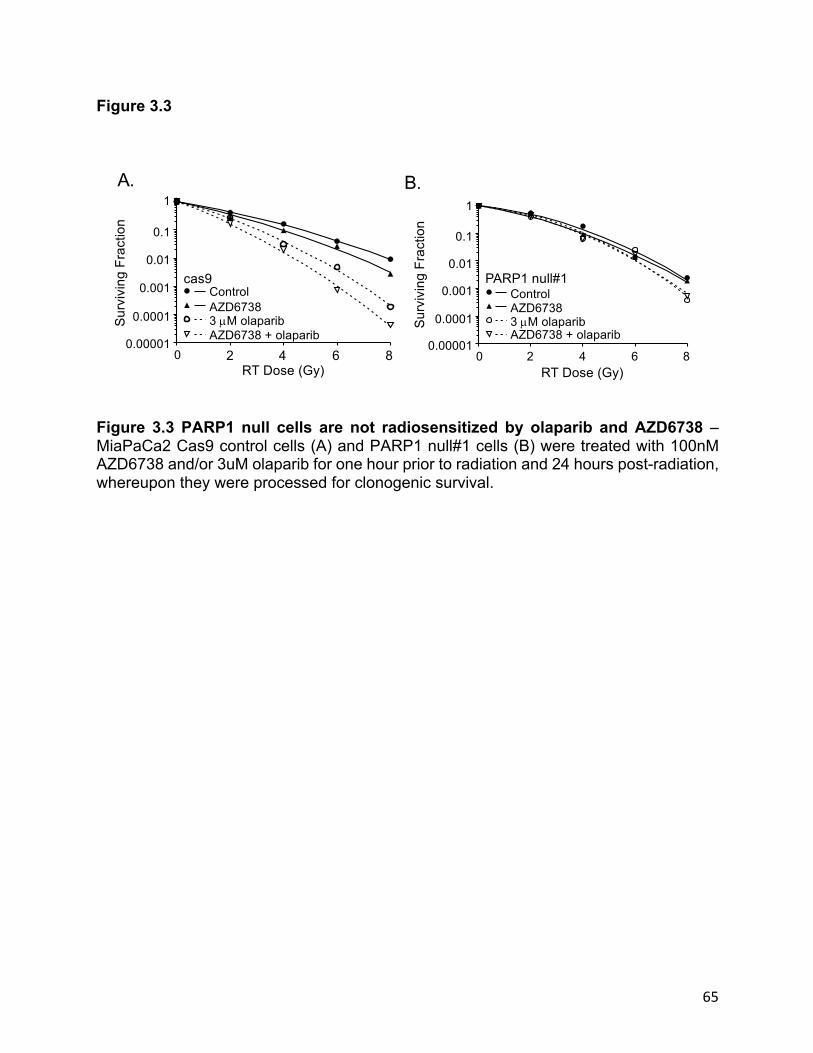
65
Figure 3.3
Figure 3.3 PARP1 null cells are not radiosensitized by olaparib and AZD6738 – MiaPaCa2 Cas9 control cells (A) and PARP1 null#1 cells (B) were treated with 100nM AZD6738 and/or 3uM olaparib for one hour prior to radiation and 24 hours post-radiation, whereupon they were processed for clonogenic survival.
Sur
vivi
ng F
ract
ion
RT Dose (Gy)
ControlAZD67383 µM olaparibAZD6738 + olaparib
cas9
0
1
0.1
0.01
0.001
0.0001
0.000012 4 6 8
B.
Sur
vivi
ng F
ract
ion
RT Dose (Gy)
ControlAZD67383 µM olaparibAZD6738 + olaparib
PARP1 null#1
0 2 4 6 8
0.1
0.01
0.001
0.0001
0.00001
1
A.
59

66
Figure 3.4
DMSO
10nM olap
3uM olap
AZD6738
AZD6738
+10n
M olap
AZD6738
+3uM olap
0
1
2
3
4
5
Norm
aliz
ed O
TM
**** ********Panc1
DMSO
10nM olap
3uM olap
AZD6738
AZD6738
+10n
M olap
AZD6738
+3uM olap
0
1
2
3
4
5
Norm
aliz
ed O
TM
**** ********
MiaPaCa2
C D
0 4 8 12 16 20 240
20
40
60
80
100
Time post-RT (hours)
%gH
2AX
Posi
tive
DMSO10nM olaparib3uM olaparib
AZD6738AZD6738+10nM olaparibAZD6738+3uM olaparib
MiaPaCa2
0 4 8 12 16 20 240
20
40
60
80
100
Time post-RT (hours)
%gH
2AX
Posi
tive
Panc1
A B
60

67
Figure 3.4 ATR and PARP inhibition delay the resolution of DNA following radiation – MiaPaCa2 (A) and Panc1 (B) cells were treated with 100nM AZD6738 and 10nM or 3uM olaparib one hour prior to 6 Gy radiation. At the indicated times post-RT cells were fixed for flow cytometry and analyzed for gH2AX positivity. Data shown are the mean from n=2 independent experiments ± SE. MiaPaCa2 (C) and Panc1 (D) cells were treated with 100nM AZD6738 and/or olaparib at the indicated concentrations for one hour prior to 8 Gy radiation and 24 hours post-radiation. DNA double-strand breaks were evaluated by neutral comet assay. Comets Olive tail moment (OTM) was determined and normalized to DMSO control. Results represent the aggregation of n=3 experiments normalized using an internal positive control irradiated on ice with mean ± SE. MiaPaCa2 (E) and Panc1 (F) cells were treated with 100nM AZD6738 and/or olaparib at the indicated concentrations without radiation for 24 hours. DNA double-strand breaks (DSBs) were evaluated by neutral comet assay. Comets Olive tail moment (OTM) was determined and normalized to DMSO control.
DMSO
10nM olap
3uM olap
AZD6738
10nM olap
+AZD6738
3uM olap
+AZD67
380
1
2
3
4
5No
rmal
ized
OTM
** **
*MiaPaCa2
DMSO
10nM olap
3uM olap
AZD6738
10nM olap
+AZD67
38
3uM olap
+AZD67
380
1
2
3
4
5
Nor
mal
ized
OTM
*******
nsPanc1
E F
61

68
Figure 3.5
Figure 3.5 The effect of combined ATR and PARP1 inhibition on replication stress – Treatment schedule (A) and representative images (B) of DNA fibers from MiaPaCa2 cells 24 h after treatment with 6 Gy radiation, 10 nM or 3 uM olaparib and 300 nM AZD6738. (C) Replication fork symmetry was calculated for individual forks labeled with an IdU track flanked on both sides by CldU tracks as the ratio of CldU track lengths. A track ratio of 1 indicates symmetrical fork elongation and greater than 2 indicates highly asymmetrical fork elongation. (D) Replication fork speed was calculated for individual forks labelled with an IdU track flanked by a single CldU track. Errors bars represent the
30’ - IdU 30’ - CldU
t 06Gy
t -1AZD6738+olap
t 24
Con
RT
RT+3µM olap
RT+AZD6738+3uM olap
A B
C D
E
Control
RT
RT+10n
M olap
RT+3uM olap
RT+AZD67
38
RT+AZD67
39+1
0nM olap
RT+AZD67
38+3
uM olap0
1
2
3
Rep
licat
ion
Rate
of
Ong
oing
For
ks (k
b/m
in)
********
pCHK1 (S345)
CHK1
pATR (T1989)
6Gy:100nM AZD6738:
10nM Olaparib:3µM Olaparib:
----
-+-+
+---
+-+-
+--+
++--
+++-
++-+
-++-
1
2
3
4
5
Rep
licat
ion
Fork
Sym
met
ry(G
reen
long
/ Gre
ensh
ort)
RT
RT
+ 3 µM
ola
p
RT
+ 10
nM
olap
RT
+AZ
D67
38
RT
+ 3 µM
ola
p+
AZD
6738
RT
+ 10
nM
olap
+ AZ
D67
38
62

69
median ± interquartile range from over 150 forks scored in two biological replicates. Statistical differences between distributions were assessed using the nonparametric Mann-Whitney rank sum test. (E) Western blot of whole cell extract from MiaPaCa2 cells treated with 100 nM AZD6738 and 10 nM or 3 uM olaprib for one hour prior to 6 Gy radiation and 24 hours after radiation.

70
Figure 3.6
Figure 3.6 Radiosensitization of pancreatic tumor xenografts by AZD6738 and olaparib - Athymic nude mice bearing bilateral, flank MiaPaCa-2 xenografts were treated with AZD6738 (25mg/kg) and olaparib (50mg/kg) one hour pre-RT, and radiation (RT; 1.8Gy/fraction) daily Monday through Friday for one cycle. (A) Tumor volumes were normalized to the first day of treatment (day 1) and are the mean ± SE of 13-18 tumors per treatment group. (B) The Kaplan-Meier plot illustrates the proportion of tumors tripled in volume within the full 80 day monitoring period. (C) Weights were normalized to the first day of treatment.
0 20 40 600
2
4
6
8
Days post-treatment initiation
Rel
ativ
e Tu
mor
Siz
e OlaparibAZD6738AZD6738+olaparibRTOlaparib+RTAZD6738+RTAZD6738+olaparib+RT
Control
0 5 10 150.90
0.95
1.00
1.05
Days post-treatment initiation
Rel
ativ
e W
eigh
t
A
B
0 20 40 60 800
20
40
60
80
100
Days post-treatment intiation
Perc
ent S
urvi
val
(Pro
porti
on o
f tum
ors
not t
riple
d)
**
C
64

71
Table 3.1
Table 3.1 Radiosensitization and cytoxocicity of combined ATR and PARP1 inhibition - (A) MiaPaCa2 HRP, MiaPaCa2 HRD, Panc-1, and Capan-1 cells were treated with AZD6738 and/or olaparib beginning one hour prior to radiation. Twenty-four post-radiation, cells were processed for clonogenic survival. Data are the mean radiation enhancement ratio (RER) ± SEM for n=3-6 independent experiments. Cytotoxicity in the absence of radiation treatment was calculated by normalizing the plating efficiencies of drug treated- to non-drug treated cells. (B) MiaPaCa2 Cas9 control cells and MiaPaCa2 PARP1 null cells were treated with AZD6738 and/or olaparib beginning one hour prior to radiation. Twenty-four post-radiation, cells were processed for clonogenic survival. Data
MiaPaCa-2 RAD51 shRNAHRP (no dox)
AZD6738 (100 nmol/L)Olaparib (10 nmol/L)Olaparib (3 µmol/L) AZD6738 + olaparib (10 nmol/L)AZD6738 + olaparib (3 µmol/L)
HRD (+ dox)AZD6738 (100 nmol/L)Olaparib (10 nmol/L)AZD6738 + olaparib
RER
1.01.14 ± 0.051.11 ± 0.041.43 ± 0.08a
1.40 ± 0.10a,b
1.83 ± 0.14a,b,c
1.01.16 ± 0.061.17 ± 0.031.43 ± 0.11a
Cytotoxicity
1.00.93 ± 0.070.88 ± 0.040.80 ± 0.040.84 ± 0.070.66 ± 0.03
1.00.98 ± 0.040.80 ± 0.050.63 ± 0.02
Condition
Panc-1AZD6738 (100 nmol/L)Olaparib (10 nmol/L)Olaparib (3 µmol/L)AZD6738 + olaparib (10 nmol/L)AZD6738 + olaparib (3 µmol/L)
1.02 ± 0.031.03 ± 0.071.29 ± 0.06a
1.06 ± 0.041.67 ± 0.07a,b,c
0.96 ± 0.080.83 ± 0.090.80 ± 0.120.87 ± 0.030.94 ± 0.08
Capan-1AZD6738 (100 nmol/L)Olaparib (10 nmol/L)AZD6738 + olaparib
1.14 ± 0.061.26 ± 0.061.47 ± 0.13a
0.92 ± 0.040.92 ± 0.120.87 ± 0.12
A
MiaPaCa2 – cas 9AZD6738 (100 nmol/L)Olaparib (3 µmol/L)AZD6738 + olaparib
1.01.24 ± 0.051.59 ± 0.092.10 ± 0.17d,e
1.00.88 ± 0.030.93 ± 0.110.60 ± 0.09
B
MiaPaCa2 – PARP1 nullAZD6738 (100 nmol/L)Olaparib (3 µmol/L)AZD6738 + olaparib
1.31 ± 0.141.55 ± 0.181.40 ± 0.191.49 ± 0.30 (n=2)
0.67 ± 0.030.69 ± 0.030.72 ± 0.040.64 ± 0.01 (n=2)
P < 0.05 vs. controla, AZD6738b, olaparibc, cas9 controld, cas9 AZD6738e.
65

72
are the mean radiation enhancement ratio (RER) ± SEM for n = 2-3 independent experiments. Cytotoxicity in the absence of radiation treatment was calculated by normalizing the plating efficiencies of drug treated- to non-drug treated cells.

73
Illustration 3.1
Illustration 3.1 - ATR protects stalled replication forks. When replication forks encounter trapped poly (ADP-ribose) polymerase 1 (PARP1) on chromatin, they stall and expose large sections of single stranded DNA. This is quickly coated by replication protein A (RPA), which recruits ataxia telangiectasia mutated and Rad3-related (ATR) through ATR interacting protein (ATRIP). ATR phosphorylates checkpoint kinase 1 (Chk1) which triggers intra-S phase arrest and inhibits aberrant replication origin initiation through the CDC25-CDK1/2 pathway. Separately, ATR protects the replication fork from degradation through SMARCAL1 and potentially through a second Chk1-mediated pathway.
RPARPA
RPARPA
RPA ATRIP
ATRIP
ATR Chk1P
SMARCAL1
Fork degradation
CDC25A
CDC25BCDC25C
CDK1
S-phase progression
CDK2
Replication origin firing
P
PARP1
Trapped

74
CHAPTER 4:
Future Directions
The concluding volume of Robert Jordan’s epic fantasy series, The Wheel of Time,
ends with this message: “This wind, it was not the ending. There are no endings, and
never will be, to the turning of the Wheel of Time. But it was an ending.” So too with any
graduation, publication, or presentation in the scientific world. While we may tie our
findings and advancements into neat stories for presentation to our peers and
adjudicators, the truth is that these conclusions are imposed on the tale of scientific
discovery, and more is left to be done than could be pursued in a lifetime. Personally, as
the time has come for me to defend my thesis and return to my clinical studies in the
medical school, I am filled with more questions than ever and excited to see how they will
be explored in the future.
4.1 Continued exploration of PARP trapping
In this work we have demonstrated that the radiosensitization achieved by PARP
inhibitors in HR competent pancreatic cancer models cannot be explained solely by their
enzymatic inhibition actions. The disconnection between enzymatic inhibition and
radiosensitization became very clear when looking at concentration response curves for
the three different PARP inhibitors. However, part of our inference relied on the trapping

75
potencies for these agents previously described in the literature by other groups. Olaparib
reaches an approximate trapping IC50 at 3uM and talazoparib at 50nM, while Veliparib
only begins to demonstrate trapping properties at 30uM [208]. Our work would benefit
from the establishment of these properties in our own hands and in our own cell lines.
One barrier has been the need to handle multiple samples in the same experiment in
order to generate these curves and we hope that we will be able to surmount this in the
future using the recently-adapted PLA.
In a similar vein, full characterization of two representative cell lines’ (MiaPaCa2 and
Panc1) concentration-dependent response to different PARP inhibitors was illuminating,
but similar work could be expanded to remaining cell lines in the pancreatic cancer toolkit.
Subtle molecular alterations exist among many of the commonly used PDAC cell line
models, as well as major PARP inhibition-relevant alterations as is the case with BRCA2
status in Capan1-neo [230]. Such an expansion would contribute toward affirming our
understanding of how PARP inhibitors radiosensitize in pancreatic cancer, and potentially
draw out some new and interesting information given the propensity for DDR inhibitors to
synergize with transformative mutations [231]. Among the cell lines interrogated for
concentration response to PARP inhibitors should be PARP-null and -reconstituted
models created by our group.
I feel that among the work done by our group to genetically tease out the difference
between enzymatic inhibition and trapping, we have created a clean and informative
system using CRISPR-Cas9 technology to delete PARP1 from cells. However, there are
several drawbacks to this system. One is the potential for adaptation to the permanent
loss of PARP1. By that same token, it affords an opportunity to study PARP1 loss as a

76
potential mechanism of resistance in HRP cancers, discussed later. Complete deletion
also limits the ability to interpret comparisons to drug treatment due to temporal concerns.
One recurrent theme of radiosensitization has always been the search for optimal
scheduling between administration of drug(s) and radiation - both in the relatively simple
system of cell culture, to animal models, and to the clinic [232]. How long does it take to
achieve target engagement and inhibition? Should the target be engaged during
radiation? How long after radiation should we continue therapy to maximize sensitization
and minimize toxicity? In the PARP1-null system, enzymatic activity is suppressed prior
to treatment all the way through the collection of growing colonies, whereas drug
treatments are discontinued upon processing and clonal plating 24 hours following
radiation. A system of shRNA under the control of doxycycline would allow for the
temporary and reversible knockdown of PARP1 to better replicate schedule-dependent
effects of PARP1 enzymatic inhibition.
The confounding effect of increasing concentrations of inhibitor leading both to
increased enzymatic inhibition and trapping, albeit with different potencies, has led our
comparisons of concentration response to rely heavily on induction. While the ability to
cleanly enzymatically inhibit without trapping through genetic deletion has represented an
improvement in mechanistic dissection, we have yet to demonstrate trapping in the
absence of enzymatic inhibition. Such deduction has so far been beyond the capabilities
of our current tools. A study by Steffen et al. discovered two mutations in PARP1, W318R
and E988A, that impaired catalytic activity and delayed release from chromatin in the
presence of NAD+ [233]. A recent study by Pettit et al. using CRISPR-Cas9 to screen for
mutations causing PARP inhibitor resistance reported finding mutants with similar

77
properties [210]. These mutations occurred outside the zinc finger binding domains and
did not affect PARP1 recruitment to chromatin. A focused effort to uncover more mutants
with decreased chromatin dissociation could yield a tool to genetically induce trapped
PARP at sites of DNA damage while still preserving PARP1 enzymatic function in the
background of PARP1wt cells.
The biology of trapping in the context of radiation warrants further investigation. The
phenotypic evidence for trapping with radiation is abundant, impacting radiosensitivity,
DNA damage, and cell cycle. However, biochemical assessment of PARP1-DNA
complexes remains elusive. Some have suggested that DNA lesions following radiation
are not suitable for trapping, yet the cellular effects can be readily observed. As noted in
Chapter 2, PARP trapping following radiation by chromatin fractionation was most readily
detected 24 hours after RT. This result is surprising given the understanding of PARP1
as a rapidly-recruited mediator of the DDR. Does the potentiation of damage late after
irradiation observed by gH2AX and comet assay recruit PARP1 to chromatin? The
expansion of our tools to observe trapped PARP may assist in clarifying these questions.
Real-time observation of PARP1 and PAR binding domain constructs to sites of damage
using a modified version the laser microirradiation assay would provide valuable
information about the timing of events in the formation and dissociation of PARP1-DNA
complexes after radiation that could significantly inform treatment schedules.
The development of the PLA assay to measure trapped PARP in situ has
applications beyond answering questions posed by our current study. We hope to further
optimize the assay to interrogate fixed tissue slices. In the laboratory, this will allow us to
consider the in vivo efficacy of different PARP inhibitor doses with respect to trapping in

78
tumors in mice. Further, it provides us with a model to study the contribution of PARP
trapping to clinically described dose limiting toxicities. We will be able to assay for trapping
in bone marrow, intestine, skin, and other organs in the context of animal models.
If, as we have suggested in this work, trapping is necessary for the productive
radiosensitization of HRP pancreatic cancers, the ability to assay for trapping in fixed
tissues will afford us the opportunity to monitor whether we are reaching therapeutic
concentrations and “hitting the target” in patients treated with PARP inhibitors. Information
provided from patient tissues treated with PARP inhibitors has the opportunity to inform
our understanding of the biology of PARP inhibition. Patients enrolled in trials combining
veliparib with radiation have experienced hematologic toxicity [234], radiation dermatitis
[235], and nausea and vomiting [235], [236]. Despite veliparib’s described inability to
stabilize DNA-PARP complexes, these events fit the profile of trapping-induced
cytotoxicity. Molecular interrogation in these cases may reveal unknown factors that
modulate the trapping potential of PARP inhibitors.
Perhaps the most intriguing outstanding question posed by the work presented here
remains the exact relationship between the treatments we have investigated and
replication fork progression. We began our study with the hypothesis that trapped PARP1
created an impediment to replication forks, which caused stalling and collapse. These
forks relied on ATR for stabilization and restart. Combined inhibition, therefore, would
lead to persistent DNA damage and increased radiosensitization. Several lines of
evidence supported this reasoning: DNA damage caused by PARP inhibitors is most
pronounced during the S-phase of the cell cycle [237], trapping concentrations induce
markers of replication stress (Figure 3.5e), and stabilization of replication forks leads to

79
PARP inhibitor resistance (discussed in section 4.3). However, this model was challenged
by a study by Bartek et al. in which they discovered that PARP inhibition seemed to speed,
not slow, replication forks, and that it did not lead to fork stalling or collapse [220]. They
proposed a model in which cellular PARylation leads to p53 transactivation of p21, which
acts to suppress replication forks. They show that PARP inhibition releases this inhibition,
leading to increased replication fork velocity, which in turn generates single stranded but
not double stranded DNA damage.
We have also observed that PARP inhibition increases replication fork velocity, but
several of our observations are not consistent with their model. We only observe the effect
on fork velocity at trapping concentrations of olaparib (Figure 3.5d). The data presented
by Bartek’s group also indicate that the effects on replication fork velocity are induced
under trapping conditions, suggesting that they are not due simply to changes in cellular
PARylation. Additionally, our observations were made in a p53 mutant system, leading
us to conclude that the effect must be p53-independent. Finally, we observed that the
addition of an ATR inhibitor slows replication fork speed while increasing DNA damage;
if replication fork speed were causative of olaparib-induced damage, AZD6738-induced
fork slowing should attenuate DNA damage.
We propose an alternative model in which increased replication fork velocity is a
downstream consequence of olaparib-induced replication stress and DNA damage
(Illustration 4.1). ATR is activated by the replication stress, which prevents origin firing
through Chk1. Origin number and velocity have a reciprocal relationship, acting to speed
up the origins that do initiate. This hypothesis is supported by the frequent observation
that PARP inhibition slows S-phase progression and the overall rate of DNA replication.

80
Combining this effect with ATR inhibition disinhibits aberrant replication origins, massively
slowing down fork progression, while still increasing DNA damage.
How does the PARP trapping lesion generate replication stress and DNA damage
to begin this cascade? Our data do not suggest a clear answer. Bartek et al. show that
PARP inhibition does not lead to fork stalling or collapse, measured by asymmetry of
symmetric origins with the DNA fiber combing technique. However, they also did not
induce concomitant DNA damage through radiation or genotoxins. It has been
demonstrated that different DNA damaging agents have strikingly different effects on fork
slowing and asymmetry [238]. It is possible that without the addition of ionizing radiation,
PARP trapping does not cause appreciable replication fork stalling and collapse, and
leads to milder DNA damage in the form of SSBs. Our experiments with radiation were
able to produce significant elevation in DSBs. While we also did not observe replication
fork asymmetry, we only measured 24 hours after radiation. At this time point, all cells in
the population have cycled through S-phase at least once. One plausible hypothesis is
that the deleterious effects of olaparib and radiation on replication forks are limited to the
first S-phase after radiation. Any cells still cycling 24 hours later will display the effects of
olaparib alone, which are milder. Therefore, the optimal window to assay for fork
asymmetry would be 2-4 hours following radiation. Further investigation will bring us
closer to understanding the mechanistic significance of trapped PARP versus enzymatic
inhibition.
4.2 PARP2 and PARP3

81
The primacy of PARP1 in the cellular environment and DNA damage response has
led to an overwhelming focus on it as the therapeutically relevant target of PARP
inhibitors. In reality, however, all clinically investigated PARP inhibitors also inhibit
PARP2, and olaparib, talazoparib, and rucaparib also inhibit PARP3 with varying
potencies [239], [240]. Indeed, observations in our own work not shown here demonstrate
that these other family members may play significant roles in the absence of PARP1. In
our PARP1-null model, after multiple passages in culture the level of cellular PARylation
returned to that seen in Cas9 control cells, with no indication of a reversion mutation or
presence of the protein. These levels of PAR were sensitive to treatment with olaparib,
suggesting that PARP2 or PARP3 could increase in abundance and/or activity in the
absence of PARP1 protein. However, due to limitations of time and availability of quality
detection reagents we were not able to pursue this line of inquiry.
Structurally, PARP2 and PARP3 share ~60% amino acid sequence homology with
PARP1 [241]. Both feature the C-terminal WRG and catalytic domains of PARP1, but
differ in their shorter N-termini, which lack DNA binding ZF and autoregulatory BRCT
domains. Rather they display preferential activation by 5’ phosphorylated DNA ends,
suggesting a more limited role in backbone repair [242], [243].
The importance of PARP2 in the DNA damage response is underscored by its
upregulation in response to DNA damaging agents [244], [245]. It has been shown to
cooperate with PARP1 in response to SSB lesions [246], [247] and genomic and
replication stability [149], [248]. PARP2 also assists in the recruitment of APLF to
chromatin through PARP1-dependent branch chain formation at damage sites [249].

82
On its own, there is evidence that PARP2 function contributes to the DSB repair
pathway choice [250]. The question of why cells choose to repair certain lesions by NHEJ
or HR is one for which many mediators and signaling events have unsatisfyingly benn
described but not answered. Does the cell know that a lesion has taken place in the
middle of nowhere and can be repaired “quick and dirty” simply to ensure the integrity of
the molecule, or if it has taken place among highly sensitive genetic material that merits
time and attention from HR machinery? The work by Fouquin et al. demonstrates that
PARP2 prevents the accumulation of 53BP1 at sites of DNA damage, allowing strand
resection by CtIP and progression of homology directed repair. As veliparib had no effect
on this process, they concluded that this function of PARP2 was independent of its
PARylation. Crucially, though, it has been demonstrated that PARP2 can be trapped on
chromatin like PARP1 [194], and it will be illuminating to see if this pathway choice can
be disrupted through the inhibition of PARP2 with a stronger trapper such as olaparib or
talazoparib.
A recent study also demonstrated that PARP2 facilitates androgen receptor (AR)
signaling in prostate cancer through interaction with FOXA1 [251]. PARP2 levels are
elevated in prostate cancer relative to normal tissue, and even further in castration
resistant prostate cancer (CRPC), where the AR signaling pathway frequently employs
mutations and truncations to evade inhibition by antiandrogens. If these results remain
robust in more prostate cancer models, this could represent an exciting new therapeutic
opportunity, especially considering the frequent activation of FOXA1 signaling [252].
PARP3 has been less extensively studied than PARP1 and PARP2. Nevertheless,
it has been ascribed some functions in the DNA damage response, and its loss promotes

83
sensitivity to chemotherapeutic drugs [253]. Interestingly, PARP3 seems to promote
NHEJ pathway choice by promoting the accumulation and activity of APLF [254], [255]
and ADP-ribosylation of Ku80 [256]. Taken together with the pathway choice actions of
PARP2, these studies suggest that we may be able to pharmacologically tip genomic
repair toward HR or NHEJ through our choice of PARP inhibitor, to say nothing of future
developments in pharmacology that target these other PARPs even more specifically.
4.3 PARP inhibitor resistance
The genomic and mutational instability hallmark of cancer [28] means that cancer
cells are quick to adapt to selective pressures. These advances can be difficult to
anticipate at the bench because the classic tools we use to model malignancy often fail
to capture the heterogeneity of mutational landscapes within patients’ tumors. In cancer
treatment, this fluidity frequently leads to treatment failure in the form of acquired
resistance. Broadly, there are four categories of PARP inhibitor resistance that have been
observed in the laboratory or the clinic: reversion to HR proficiency, alterations in the
target, replication fork stabilization, and increased drug efflux.
The upregulation of ATP-binding cassette transporters is a common and nonspecific
form of drug resistance for many therapies. Specifically for PARP inhibitors, the
upregulation of P-glycoprotein transporter was modeled as a mechanism of acquired
resistance in murine models of olaparib resistance [257], [258]. However, affinity for the
transporter varies among inhibitors [259], and is especially low for recently developed
agent AZD2461 [260]. Vigilance for this mechanism of resistance would allow for the
seamless transition between agents if the need should arise clinically.

84
Overwhelmingly, the cancers displaying acquired resistance to PARP inhibition in
the clinic have done so through reacquisition of proficient homologous recombination. As
discussed previously, tumors deficient in HR are exquisitely sensitive to PARP inhibition.
The major mutations conferring HR deficiency were first described as inactivating
mutations in the BRCA1 and BRCA2 genes. Correspondingly, secondary mutations
restoring functionality have been seen in these genes in breast, ovarian, prostate, and
even pancreatic cancers [261]–[266]. Reversion mutations have been observed in other
genes that confer “BRCA-ness” such as PALB2 [265], RAD51C and RAD51D [267].
PARPi sensitivity conferred by low expression of BRCA1 in patient derived xenograft
models has been overcome in the laboratory through demethylation of the promoter
region and re-expression of the protein [268].
One major and surprising method by which BRCA1 mutant cancers subvert their
PARPi sensitivity preclinically is through inactivation of the 53BP1 pathway [269]–[271].
While the exact downstream mediators of its effects are still being illuminated, studies
have shown that 53BP1 is involved in the cell’s choice of DSB repair pathway promoting
NHEJ in opposition to BRCA1 [272]. Thus, loss of 53BP1 resulted in rescue of DNA end
resection and partial restoration of HR [269], [270].
While restoration of homologous recombination in previously deficient cancers
clearly reduces sensitivity to PARP inhibition, one could argue with the characterization
of this state as entirely resistant. As we and others have demonstrated, opportunities
abound for treatment of HRP cancers with PARP inhibitors, albeit with a narrower
therapeutic window and rarely as a monotherapy. To harness the full potential of these
treatments, though, drug selection and dosage must be adjusted to harness trapping

85
potential in combination with radiation or other targeted agents. In fact, these cancers
may have provided us with one small advantage; in restoring HR they have improved
their genetic integrity and given up some of the very plasticity that lent them malignancy
in the first place.
Beside reversion mutations that confer HR, the only other mechanism of acquired
PARPi resistance observed so far clinically was a mutation in the WRG domain of PARP1
leading to decreased trapping capability [210]. The study by Pettit et al. used CRISPR-
Cas9 mutagenesis to further screen for other PARP1 mutations that would confer
resistance to talazoparib. They found that mutations in the zinc finger domain attenuated
the ability of PARP1 to localize to DNA and decreased sensitivity to talazoparib. Other
mutations discovered in their screen were mapped to residues observed to interact with
DNA binding domains. Their observation bolsters the hypothesis that PARP inhibitor
cytotoxicity is mediated by trapping in the setting of HR proficiency and that in these
cancers PARP1 mutation, downregulation, or genetic loss could be a major mechanism
of resistance [194], [210], [273], [274].
It has also been demonstrated that cellular PARylation can be rescued by the loss
of PAR glycohydrolase (PARG), the enzyme that depolymerizes chains of PAR [275].
Even in the absence of HR restoration and presence of PARP inhibition, the amplification
of residual PARP1 activity by PARG loss was sufficient to transform these cells to a
resistant phenotype. However, this very adaptation rendered cells sensitive to
temozolomide due to increased PARP1 retention on chromatin and the induction of
trapping. While PARG loss has been observed in treatment-naive breast and ovarian

86
cancers [275], it remains to be seen whether it will lead to PARP inhibitor resistance
clinically.
Recent studies have proposed several mechanisms by which PARP inhibitor
resistance derives from alterations in the response to replication stress. Loss of PTIP was
shown to rescue stalled replication forks from MRE11-mediated degradation following
PARP inhibition [276]. Similar protection of replication fork stability was promoted by
EZH2 loss or inhibition, disrupting its ability to recruit MUS81 nuclease to stalled forks.
Another study demonstrated that lethal replication fork block induced by PARPi is
mediated by SLFN11 [277]. Absence of SLFN11 promotes productive completion of
replication an entry into G2.
However, while these mutations protect cells from PARP inhibition, they allow
replication to proceed under stress, presenting a therapeutic opportunity. Work has
already shown that resistance arising from fork protection can be overcome with the
addition of an ATR inhibitor [278], [279], consistent with its role as a master regulator of
the response to replication stress [223]. These results are encouraging for the
combination of PARP and ATR inhibitors in other settings both as a demonstration of
induced sensitivity and in anticipation of treatment failure. However, the difference in how
ATR inhibition can sensitize HRD cells in the context of replication fork stabilization and
inherently resistant HRP cells to PARP inhibition offers a window into their biology. The
necessity for PARP trapping for combined sensitivity in HRP cells suggests that cellular
context may determine the type of DNA lesion for which ATR directs productive repair.
4.4 Immunomodulation and the DNA damage response

87
It does not receive a mention in this work until now, but the DDR including PARP
and ATR, have important immune modulatory functions that are further enhanced by
radiation as a therapy. Indeed, immunotherapy has become so ubiquitous that during the
preparation of this thesis, I heard an advertisement on NPR from the Dana Farber Cancer
Institute touting the availability of PD-L1 inhibition at their center. To provide a perspective
on all of immunotherapy would be its own manuscript and well beyond the scope of our
focus here (see review by Ribas et al. [280]). Instead, I will provide some context for the
use of immunotherapy with radiation and the ways it may be employed with other
inhibitors of the DNA damage response.
Even without pharmacologic manipulation, there are many ways in which radiation
has been described to dovetail with the immune system. The first of these involves several
effects that render the tumor more visible to immune surveillance by the host. It has been
demonstrated that radiation can facilitate the generation and release of neoantigens from
tumors ([281], [282]). Presentation of these antigens is also enhanced by an upregulation
of MHC class I molecules on the tumor surface after radiation[283], [284], counteracting
a common mechanism of immune evasion.
Radiation is also known to activate the innate immune response through its induction
of Type I interferon via the cGAS-STING pathway [285]. Radiation induces the release of
nuclear DNA in the cytosol and formation of micronuclei [286], [287], which in turn triggers
the viral pattern recognition receptor responses and the production of type I interferons.
Inside the cell, radiation has also been demonstrated to increase immune checkpoint
proteins [284] and has even been suggested to involve DNA DSB signaling from

88
ATM/ATR [288]. These effects can be observed in the clinical setting by the induction of
anti-tumor responses outside the treatment field of locoregional radiation [289]–[291].
These factors have combined to generate interest in whether immune modulation
could be used to sensitize cancers to radiation. The most common of these therapies is
immune checkpoint blockade (ICB). Proteins mediating immune checkpoints function
physiologically to dampen the immune response and protect the normal tissues of the
body from attack by T cells and NK cells. Among the earliest of these to be targeted was
cytotoxic T-lymphocyte associated protein 4 (CTLA-4), whose inhibitory antibody
ipilimumab was approved for use in metastatic melanoma in 2011 [292]. Two other major
targets major targets were quickly discovered: programmed cell death 1 (PD-1) and its
ligand PD-L1. Antibodies inhibiting PD-1 were first approved by the FDA in 2014
(Pembrolizumab and nivolumab) and PD-L1 in 2016 (Atezolizumab) and 2017 (Avelumab
and Durvalumab) (reviewed in [293]).
Anti-tumoral activity can be readily appreciated in the combination of immune
blockade and radiation. A study by Dewan et al. showed that the addition of CTLA-4
blockade synergized with radiation at the primary tumor site and induced an abscopal
effect outside the radiation field [294]. Similarly, anti-PD-1 and anti-PD-L1 therapy were
shown to combine with radiation for increased response [295], [296]. Promising results
have also been delivered by combining inhibitors of both major ICB pathways with
radiation [297], overcoming T-cell exhaustion by PD-L1 upregulation as a mechanism of
resistance to CTLA-4 therapy.
In addition to the immune stimulatory effects of radiation discussed briefly above,
there is evidence that sensitization by these modalities could operate in both directions.
89
ICB agents have been demonstrated to help modulate the tumor microenvironment by
increasing tumor vascularity and perfusion [298], [299], which in turn could combat the
radioprotective effects of hypoxia [300], [301].
A growing body of evidence suggests that improvement in the efficacy of immuno-
radiotherapy through the addition of targeted DNA damage response inhibitors may
represent the next frontier of therapy. Consistent with the findings described above
implicating DNA DSB pathway proteins in the regulation of immune modulatory proteins,
studies have found that ATR inhibition possesses inherent immunostimulatory effects.
Treatment with ATRi reduces PD-L1 expression and enhanced anti-tumor T cell activity
[302], [303]. In combination with radiation, it has also been found to increase the
immunostimulatory effects of radiation alone described above, including immune cell
infiltration, innate immune recognition of cytoplasmic DNA, and increased antigen
processing and presentation [304]. Dillon et al. attribute many of these findings to
increased DNA damage, though our own findings suggest that ATR inhibition alone may
only modestly increase damage. The addition of a PARP inhibitor may further potentiate
this effect. The addition of targeted checkpoint blockade to the intrinsic activity of ATRi
could deliver vast benefits due to the non-redundant checkpoint pathways [297].
The overlapping but separate functions of ATM make it an intriguing avenue for
further investigation into the DDR master regulator inhibition as a combination with
radiation. A study from our group recently demonstrated the efficacy of ATM inhibition to
induce the innate immune response and sensitize to anti-PD-L1 therapy [305].
On its own, PARP inhibition was demonstrated to upregulate PD-L1 expression in
cell culture models and sensitized murine tumors to PD-L1 inhibition in vivo [306]. A single

90
arm clinical trial assessing the combination of niraparib and pembrolizumab demonstrated
high response rates regardless of BRCA status [307].
Further investigation is warranted into the combination of targeting the DNA damage
response and immune checkpoint inhibition. On its own, elevated DNA damage by the
combination of ATR and PARP inhibition with radiation has the potential to potentiate the
immunostimulatory effects of ATR inhibition alone. Given the contrasting effects of ATRi
and PARPi on PD-L1 expression in previous studies it will be important to determine the
dominant phenotype of this effect when they are combined. Studies with radiation suggest
ATR inhibition will act to counteract the effect of PARPi-mediated PD-L1 upregulation.
Finally, we will determine whether the addition of immune checkpoint blockade will
potentiate the radiosensitizing effects of combined ATR/PARP inhibition with radiation.
Broadly, the rapid development of immunomodulating therapies has opened up new
and exciting avenues of investigation for those of us who have studied the DNA damage
response. As we learn more about the involvement of the DNA damage response in
modulating immune surveillance in cancer, our hard-won knowledge about these
pathways in the setting of radiation will be ready-made to apply for the benefit of our
patients.
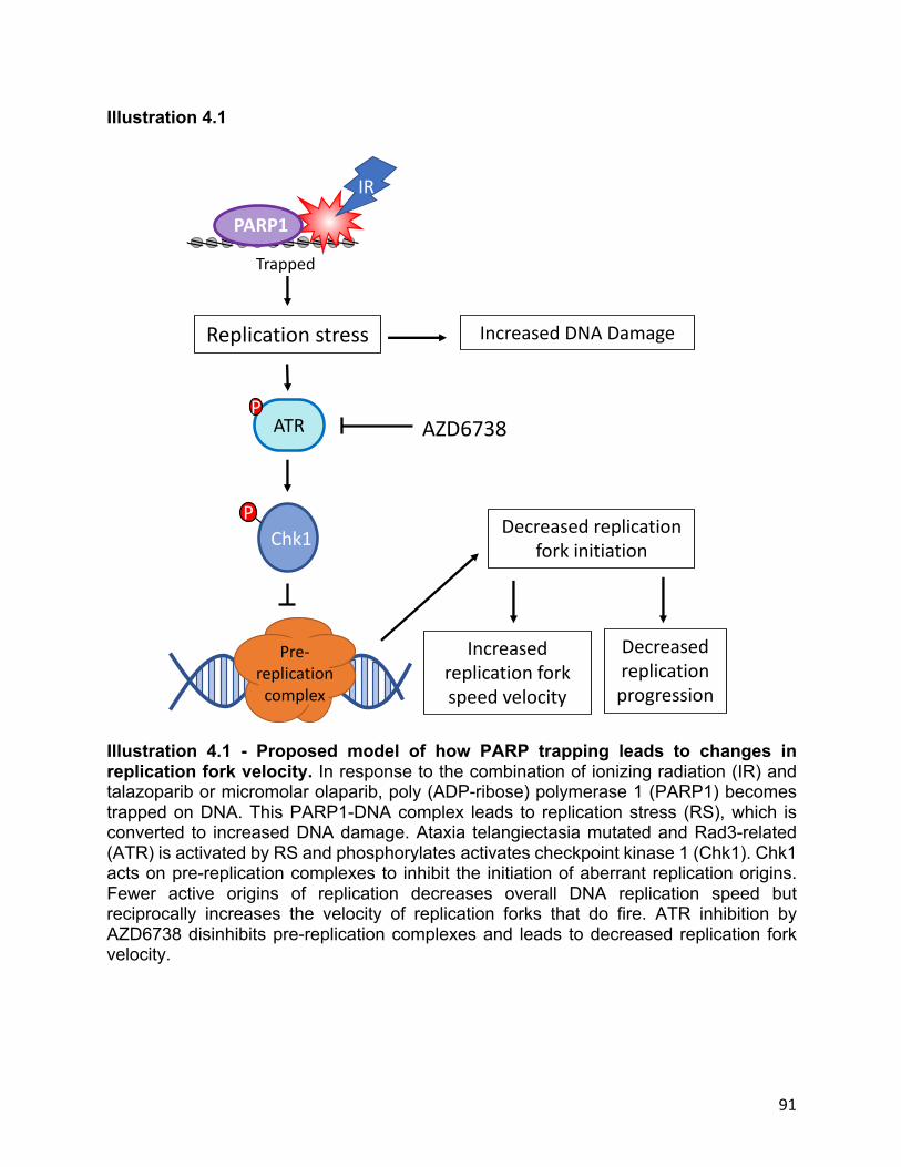
91
Illustration 4.1
Illustration 4.1 - Proposed model of how PARP trapping leads to changes in replication fork velocity. In response to the combination of ionizing radiation (IR) and talazoparib or micromolar olaparib, poly (ADP-ribose) polymerase 1 (PARP1) becomes trapped on DNA. This PARP1-DNA complex leads to replication stress (RS), which is converted to increased DNA damage. Ataxia telangiectasia mutated and Rad3-related (ATR) is activated by RS and phosphorylates activates checkpoint kinase 1 (Chk1). Chk1 acts on pre-replication complexes to inhibit the initiation of aberrant replication origins. Fewer active origins of replication decreases overall DNA replication speed but reciprocally increases the velocity of replication forks that do fire. ATR inhibition by AZD6738 disinhibits pre-replication complexes and leads to decreased replication fork velocity.
Chk1P
ATRP
PARP1
Trapped
Pre-replication
complex
Replication stress
Decreased replication fork initiation
Increased replication fork speed velocity
IR
Increased DNA Damage
AZD6738
Decreased replication progression

92
REFERENCES
[1] J. Kleeff et al., “Pancreatic cancer.,” Nat. Rev. Dis. Prim., vol. 2, p. 16022, Apr. 2016.
[2] R. L. Siegel, K. D. Miller, and A. Jemal, “Cancer statistics, 2018.,” CA. Cancer J. Clin., vol. 68, no. 1, pp. 7–30, Jan. 2018.
[3] J. He et al., “2564 resected periampullary adenocarcinomas at a single institution: trends over three decades.,” HPB (Oxford)., vol. 16, no. 1, pp. 83–90, Jan. 2014.
[4] B. Zhou et al., “Early detection of pancreatic cancer: Where are we now and where are we going?,” Int. J. cancer, vol. 141, no. 2, pp. 231–241, Jul. 2017.
[5] N. B. Merchant, “Eat When You Can, Sleep When You Can, and Don’t Mess with the Pancreas,” Surg. Oncol. Clin., vol. 25, no. 2, pp. xv–xvi, Apr. 2016.
[6] C. Hu et al., “Association Between Inherited Germline Mutations in Cancer Predisposition Genes and Risk of Pancreatic Cancer.,” JAMA, vol. 319, no. 23, pp. 2401–2409, Jun. 2018.
[7] B. J. Raphael et al., “Integrated Genomic Characterization of Pancreatic Ductal Adenocarcinoma,” Cancer Cell, vol. 32, no. 2, pp. 185-203.e13, Aug. 2017.
[8] S. Jones et al., “Core signaling pathways in human pancreatic cancers revealed by global genomic analyses.,” Science, vol. 321, no. 5897, pp. 1801–6, Sep. 2008.
[9] A. V Biankin et al., “Pancreatic cancer genomes reveal aberrations in axon guidance pathway genes.,” Nature, vol. 491, no. 7424, pp. 399–405, Nov. 2012.
[10] N. Waddell et al., “Whole genomes redefine the mutational landscape of pancreatic cancer.,” Nature, vol. 518, no. 7540, pp. 495–501, Feb. 2015.
[11] A. K. Witkiewicz et al., “Whole-exome sequencing of pancreatic cancer defines genetic diversity and therapeutic targets.,” Nat. Commun., vol. 6, p. 6744, Apr. 2015.
[12] P. Bailey et al., “Genomic analyses identify molecular subtypes of pancreatic cancer,” Nature, vol. 531, no. 7592, pp. 47–52, Mar. 2016.
[13] Cancer Genome Atlas Research Network. Electronic address: [email protected] and Cancer Genome Atlas Research Network, “Integrated Genomic Characterization of Pancreatic Ductal Adenocarcinoma.,”

93
Cancer Cell, vol. 32, no. 2, pp. 185-203.e13, 2017.
[14] S. K. Dougan, “The Pancreatic Cancer Microenvironment.,” Cancer J., vol. 23, no. 6, pp. 321–325.
[15] K. C. Valkenburg, A. E. de Groot, and K. J. Pienta, “Targeting the tumour stroma to improve cancer therapy,” Nat. Rev. Clin. Oncol., vol. 15, no. 6, pp. 366–381, Jun. 2018.
[16] M. Binnewies et al., “Understanding the tumor immune microenvironment (TIME) for effective therapy,” Nat. Med., vol. 24, no. 5, pp. 541–550, May 2018.
[17] X. Chen and E. Song, “Turning foes to friends: targeting cancer-associated fibroblasts,” Nat. Rev. DRUG Discov., vol. 18, no. 2, pp. 99–115, Feb. 2019.
[18] T. A. Sohn et al., “Resected adenocarcinoma of the pancreas-616 patients: results, outcomes, and prognostic indicators.,” J. Gastrointest. Surg., vol. 4, no. 6, pp. 567–579, 2000.
[19] A. Teague, K.-H. Lim, and A. Wang-Gillam, “Advanced pancreatic adenocarcinoma: a review of current treatment strategies and developing therapies.,” Ther. Adv. Med. Oncol., vol. 7, no. 2, pp. 68–84, Mar. 2015.
[20] T. Conroy et al., “FOLFIRINOX versus gemcitabine for metastatic pancreatic cancer,” N. Engl. J. Med., vol. 364, no. 19, pp. 1817–1825, May 2011.
[21] S. Gourgou-Bourgade et al., “Impact of FOLFIRINOX compared with gemcitabine on quality of life in patients with metastatic pancreatic cancer: Results from the PRODIGE 4/ACCORD 11 randomized trial,” J. Clin. Oncol., vol. 31, no. 1, pp. 23–29, Jan. 2013.
[22] D. D. Von Hoff et al., “Increased survival in pancreatic cancer with nab-paclitaxel plus gemcitabine,” N. Engl. J. Med., vol. 369, no. 18, pp. 1691–1703, 2013.
[23] P. Hammel et al., “Phase II LAPACT trial of nab-paclitaxel (nab-P) plus gemcitabine (G) for patients with locally advanced pancreatic cancer (LAPC).,” J. Clin. Oncol., vol. 36, no. 4_suppl, p. 204, 2018.
[24] P. Hammel et al., “Effect of chemoradiotherapy vs chemotherapy on survival in patients with locally advanced pancreatic cancer controlled after 4 months of gemcitabine with or without erlotinib the LAP07 randomized clinical trial,” JAMA - J. Am. Med. Assoc., vol. 315, no. 17, pp. 1844–1853, May 2016.
[25] P. J. Loehrer et al., “Gemcitabine alone versus gemcitabine plus radiotherapy in patients with locally advanced pancreatic cancer: An Eastern Cooperative Oncology Group trial,” J. Clin. Oncol., vol. 29, no. 31, pp. 4105–4112, Nov. 2011.
[26] C. A. Lacobuzio-Donahue et al., “DPC4 gene status of the primary carcinoma correlates with patterns of failure in patients with pancreatic cancer,” J. Clin. Oncol., vol. 27, no. 11, pp. 1806–1813, Apr. 2009.
[27] D. Hanahan and R. A. Weinberg, “The hallmarks of cancer,” Cell, vol. 100, no. 1.

94
pp. 57–70, 07-Jan-2000.
[28] D. Hanahan and R. A. Weinberg, “Hallmarks of cancer: The next generation,” Cell, vol. 144, no. 5. pp. 646–674, 04-Mar-2011.
[29] M. A. Morgan and T. S. Lawrence, “Molecular pathways: Overcoming radiation resistance by targeting DNA damage response pathways,” Clin. Cancer Res., vol. 21, no. 13, pp. 2898–2904, Jul. 2015.
[30] G. Iliakis, “The role of DNA double strand breaks in ionizing radiation-induced killing of eukaryotic cells.,” Bioessays, vol. 13, no. 12, pp. 641–8, Dec. 1991.
[31] R. Dueva and G. Iliakis, “Alternative pathways of non-homologous end joining (NHEJ) in genomic instability and cancer,” Transl. Cancer Res., vol. 2, no. 3, pp. 163–177, 2013.
[32] M. LEVINE, “The action of colchicine on cell division in human cancer, animal, and plant tissues.,” Ann. N. Y. Acad. Sci., vol. 51, no. 8, pp. 1365–408, Mar. 1951.
[33] H. E. Skipper, J. B. Chapman, and M. Bell, “The anti-leukemic action of combinations of certain known anti-leukemic agents.,” Cancer Res., vol. 11, no. 2, pp. 109–12, Feb. 1951.
[34] C. Heidelberger et al., “Studies on fluorinated pyrimidines. II. Effects on transplanted tumors.,” Cancer Res., vol. 18, no. 3, pp. 305–17, Apr. 1958.
[35] D. S. Childs, C. G. Moertel, M. A. Holbrook, R. J. Reitemeier, and M. Colby, “Treatment of unresectable adenocarcinomas of the stomach with a combination of 5-fluorouracil and radiation.,” Am. J. Roentgenol. Radium Ther. Nucl. Med., vol. 102, no. 3, pp. 541–4, Mar. 1968.
[36] C. G. Moertel, D. S. Childs, R. J. Reitemeier, M. Y. Colby, and M. A. Holbrook, “Combined 5-fluorouracil and supervoltage radiation therapy of locally unresectable gastrointestinal cancer.,” Lancet (London, England), vol. 2, no. 7626, pp. 865–7, Oct. 1969.
[37] C. G. Moertel et al., “Therapy of locally unresectable pancreatic carcinoma: A randomized comparison of high dose (6000 rads) radiation alone, moderate dose radiation (4000 rads + 5-fluorouracil), and high dose radiation + 5-fluorouracil. The gastrointestinal tumor study group,” Cancer, vol. 48, no. 8, pp. 1705–1710, 1981.
[38] M. H. Kalser and S. S. Ellenberg, “Pancreatic cancer. Adjuvant combined radiation and chemotherapy following curative resection.,” Arch. Surg., vol. 120, no. 8, pp. 899–903, Aug. 1985.
[39] J. P. Hoffman et al., “A pilot study of preoperative chemoradiation for patients with localized adenocarcinoma of the pancreas.,” Am. J. Surg., vol. 169, no. 1, pp. 71–7; discussion 77-8, Jan. 1995.
[40] T. S. Lawrence, E. Y. Chang, T. M. Hahn, L. W. Hertel, and D. S. Shewach, “Radiosensitization of pancreatic cancer cells by 2’,2’-difluoro-2’-deoxycytidine.,” Int. J. Radiat. Oncol. Biol. Phys., vol. 34, no. 4, pp. 867–72, Mar. 1996.

95
[41] D. S. Shewach and T. S. Lawrence, “Gemcitabine and radiosensitization in human tumor cells.,” Invest. New Drugs, vol. 14, no. 3, pp. 257–63, 1996.
[42] C. H. Crane et al., “Is the therapeutic index better with gemcitabine-based chemoradiation than with 5-fluorouracil-based chemoradiation in locally advanced pancreatic cancer?,” Int. J. Radiat. Oncol. Biol. Phys., vol. 52, no. 5, pp. 1293–302, Apr. 2002.
[43] Z. H. Siddik, “Cisplatin: mode of cytotoxic action and molecular basis of resistance.,” Oncogene, vol. 22, no. 47, pp. 7265–79, Oct. 2003.
[44] H. J. Boeckman, K. S. Trego, and J. J. Turchi, “Cisplatin sensitizes cancer cells to ionizing radiation via inhibition of nonhomologous end joining.,” Mol. Cancer Res., vol. 3, no. 5, pp. 277–85, May 2005.
[45] C. R. Sears and J. J. Turchi, “Complex cisplatin-double strand break (DSB) lesions directly impair cellular non-homologous end-joining (NHEJ) independent of downstream damage response (DDR) pathways,” J. Biol. Chem., vol. 287, no. 29, pp. 24263–24272, Jul. 2012.
[46] W. J. Kil et al., “In vitro and in vivo radiosensitization induced by the DNA methylating agent temozolomide.,” Clin. Cancer Res., vol. 14, no. 3, pp. 931–8, Feb. 2008.
[47] A. Chakravarti et al., “Temozolomide-mediated radiation enhancement in glioblastoma: a report on underlying mechanisms.,” Clin. Cancer Res., vol. 12, no. 15, pp. 4738–4746, Aug. 2006.
[48] C. J. McGinn, D. S. Shewach, and T. S. Lawrence, “Radiosensitizing nucleosides.,” J. Natl. Cancer Inst., vol. 88, no. 17, pp. 1193–203, Sep. 1996.
[49] P. Groth, M. L. Orta, I. Elvers, M. M. Majumder, A. Lagerqvist, and T. Helleday, “Homologous recombination repairs secondary replication induced DNA double-strand breaks after ionizing radiation.,” Nucleic Acids Res., vol. 40, no. 14, pp. 6585–94, Aug. 2012.
[50] Y. Liu, G. Vidanes, Y. C. Lin, S. Mori, and W. Siede, “Characterization of a Saccharomyces cerevisiae homologue of Schizosaccharomyces pombe Chk1 involved in DNA-damage-induced M-phase arrest.,” Mol. Gen. Genet., vol. 262, no. 6, pp. 1132–46, Jan. 2000.
[51] L. Busino, M. Chiesa, G. F. Draetta, and M. Donzelli, “Cdc25A phosphatase: combinatorial phosphorylation, ubiquitylation and proteolysis.,” Oncogene, vol. 23, no. 11, pp. 2050–6, Mar. 2004.
[52] C. Y. Peng, P. R. Graves, R. S. Thoma, Z. Wu, A. S. Shaw, and H. Piwnica-Worms, “Mitotic and G2 checkpoint control: Regulation of 14-3-3 protein binding by phosphorylation of Cdc25c on serine-216,” Science (80-. )., vol. 277, no. 5331, pp. 1501–1505, Sep. 1997.
[53] S. J. Elledge, “Cell cycle checkpoints: preventing an identity crisis.,” Science, vol. 274, no. 5293, pp. 1664–1672, Dec. 1996.
96
[54] M. J. O’Connell, J. M. Raleigh, H. M. Verkade, and P. Nurse, “Chk1 is a wee1 kinase in the G2 DNA damage checkpoint inhibiting cdc2 by Y15 phosphorylation.,” EMBO J., vol. 16, no. 3, pp. 545–54, Feb. 1997.
[55] J. Lee, A. Kumagai, and W. G. Dunphy, “Positive regulation of Wee1 by Chk1 and 14-3-3 proteins.,” Mol. Biol. Cell, vol. 12, no. 3, pp. 551–63, Mar. 2001.
[56] C. S. Sorensen et al., “The cell-cycle checkpoint kinase Chk1 is required for mammalian homologous recombination repair.,” Nat. Cell Biol., vol. 7, no. 2, pp. 195–201, Feb. 2005.
[57] M. A. Morgan et al., “Mechanism of radiosensitization by the Chk1/2 inhibitor AZD7762 involves abrogation of the G2 checkpoint and inhibition of homologous recombinational DNA repair.,” Cancer Res., vol. 70, no. 12, pp. 4972–4981, Jun. 2010.
[58] R. G. Syljuasen et al., “Inhibition of human Chk1 causes increased initiation of DNA replication, phosphorylation of ATR targets, and DNA breakage.,” Mol. Cell. Biol., vol. 25, no. 9, pp. 3553–3562, May 2005.
[59] M. A. Morgan, L. A. Parsels, J. D. Parsels, A. K. Mesiwala, J. Maybaum, and T. S. Lawrence, “Role of checkpoint kinase 1 in preventing premature mitosis in response to gemcitabine.,” Cancer Res., vol. 65, no. 15, pp. 6835–6842, Aug. 2005.
[60] C. C. Chen, R. D. Kennedy, S. Sidi, A. T. Look, and A. D’Andrea, “CHK1 inhibition as a strategy for targeting Fanconi Anemia (FA) DNA repair pathway deficient tumors.,” Mol. Cancer, vol. 8, p. 24, Apr. 2009.
[61] J. P. Shen et al., “Chemogenetic profiling identifies RAD17 as synthetically lethal with checkpoint kinase inhibition.,” Oncotarget, vol. 6, no. 34, pp. 35755–69, Nov. 2015.
[62] K. D. Davies et al., “Chk1 inhibition and Wee1 inhibition combine synergistically to impede cellular proliferation.,” Cancer Biol. Ther., vol. 12, no. 9, pp. 788–796, Nov. 2011.
[63] R. T. Bunch and A. Eastman, “Enhancement of cisplatin-induced cytotoxicity by 7-hydroxystaurosporine (UCN-01), a new G2-checkpoint inhibitor.,” Clin. Cancer Res., vol. 2, no. 5, pp. 791–797, May 1996.
[64] S. D. Zabludoff et al., “AZD7762, a novel checkpoint kinase inhibitor, drives checkpoint abrogation and potentiates DNA-targeted therapies.,” Mol. Cancer Ther., vol. 7, no. 9, pp. 2955–66, Sep. 2008.
[65] A. J. Massey, J. Borgognoni, C. Bentley, N. Foloppe, A. Fiumana, and L. Walmsley, “Context-dependent cell cycle checkpoint abrogation by a novel kinase inhibitor,” PLoS One, vol. 5, no. 10, 2010.
[66] S. M. Vance et al., “Selective radiosensitization of p53 mutant pancreatic cancer cells by combined inhibition of Chk1 and PARP1,” Cell Cycle, vol. 10, no. 24, pp. 4321–4329, Dec. 2011.

97
[67] Z. Ma, G. Yao, B. Zhou, Y. Fan, S. Gao, and X. Feng, “The Chk1 inhibitor AZD7762 sensitises p53 mutant breast cancer cells to radiation in vitro and in vivo.,” Mol. Med. Rep., vol. 6, no. 4, pp. 897–903, Oct. 2012.
[68] C. G. Engelke et al., “Sensitization of pancreatic cancer to chemoradiation by the Chk1 inhibitor MK8776.,” Clin. Cancer Res., vol. 19, no. 16, pp. 4412–21, Aug. 2013.
[69] M. Schmidt, A. Rohe, C. Platzer, A. Najjar, F. Erdmann, and W. Sippl, “Regulation of G2/M transition by inhibition of WEE1 and PKMYT1 Kinases,” Molecules, vol. 22, no. 12. MDPI AG, 01-Dec-2017.
[70] K. Katayama, N. Fujita, and T. Tsuruo, “Akt/protein kinase B-dependent phosphorylation and inactivation of WEE1Hu promote cell cycle progression at G2/M transition.,” Mol. Cell. Biol., vol. 25, no. 13, pp. 5725–37, Jul. 2005.
[71] R. Heald, M. McLoughlin, and F. McKeon, “Human wee1 maintains mitotic timing by protecting the nucleus from cytoplasmically activated cdc2 kinase,” Cell, vol. 74, no. 3, pp. 463–474, Aug. 1993.
[72] C. J. Squire, J. M. Dickson, I. Ivanovic, and E. N. Baker, “Structure and inhibition of the human cell cycle checkpoint kinase, Wee1A kinase: An atypical tyrosine kinase with a key role in CDK1 regulation,” Structure, vol. 13, no. 4, pp. 541–550, 2005.
[73] N. Watanabe et al., “M-phase kinases induce phospho-dependent ubiquitination of somatic Wee1 by SCFbeta-TrCP.,” Proc. Natl. Acad. Sci. U. S. A., vol. 101, no. 13, pp. 4419–24, Mar. 2004.
[74] D. R. Kellogg, “Wee1-dependent mechanisms required for coordination of cell growth and cell division.,” J. Cell Sci., vol. 116, no. Pt 24, pp. 4883–4890, Dec. 2003.
[75] R. Dominguez-Kelly et al., “Wee1 controls genomic stability during replication by regulating the Mus81-Eme1 endonuclease.,” J. Cell Biol., vol. 194, no. 4, pp. 567–579, Aug. 2011.
[76] H. Beck et al., “Cyclin-dependent kinase suppression by WEE1 kinase protects the genome through control of replication initiation and nucleotide consumption.,” Mol. Cell. Biol., vol. 32, no. 20, pp. 4226–36, Oct. 2012.
[77] M. Krajewska et al., “Forced activation of Cdk1 via wee1 inhibition impairs homologous recombination.,” Oncogene, vol. 32, no. 24, pp. 3001–8, Jun. 2013.
[78] H. Hirai et al., “Small-molecule inhibition of Wee1 kinase by MK-1775 selectively sensitizes p53-deficient tumor cells to DNA-damaging agents,” Mol. Cancer Ther., vol. 8, no. 11, pp. 2992–3000, Nov. 2009.
[79] H. Hirai et al., “MK-1775, a small molecule Wee1 inhibitor, enhances antitumor efficacy of various DNA-damaging agents, including 5-fluorouracil,” Cancer Biol. Ther., vol. 9, no. 7, pp. 514–522, Apr. 2010.

98
[80] N. V Rajeshkumar et al., “MK-1775, a potent Wee1 inhibitor, synergizes with gemcitabine to achieve tumor regressions, selectively in p53-deficient pancreatic cancer xenografts.,” Clin. Cancer Res., vol. 17, no. 9, pp. 2799–806, May 2011.
[81] J. M. Kreahling et al., “Wee1 inhibition by MK-1775 leads to tumor inhibition and enhances efficacy of gemcitabine in human sarcomas.,” PLoS One, vol. 8, no. 3, p. e57523, 2013.
[82] J. PosthumaDeBoer et al., “WEE1 inhibition sensitizes osteosarcoma to radiotherapy.,” BMC Cancer, vol. 11, p. 156, Apr. 2011.
[83] K. A. Bridges et al., “MK-1775, a novel Wee1 kinase inhibitor, radiosensitizes p53-defective human tumor cells.,” Clin. Cancer Res., vol. 17, no. 17, pp. 5638–48, Sep. 2011.
[84] B. Sarcar et al., “Targeting radiation-induced G(2) checkpoint activation with the Wee-1 inhibitor MK-1775 in glioblastoma cell lines.,” Mol. Cancer Ther., vol. 10, no. 12, pp. 2405–14, Dec. 2011.
[85] T. Kausar et al., “Sensitization of Pancreatic Cancers to Gemcitabine Chemoradiation by WEE1 Kinase Inhibition Depends on Homologous Recombination Repair.,” Neoplasia, vol. 17, no. 10, pp. 757–66, Oct. 2015.
[86] M. Aarts et al., “Forced mitotic entry of S-phase cells as a therapeutic strategy induced by inhibition of WEE1.,” Cancer Discov., vol. 2, no. 6, pp. 524–539, Jun. 2012.
[87] D. Karnak et al., “Combined inhibition of Wee1 and PARP1/2 for radiosensitization in pancreatic cancer.,” Clin. Cancer Res., vol. 20, no. 19, pp. 5085–96, Oct. 2014.
[88] C.-J. Busch et al., “G2-checkpoint targeting and radiosensitization of HPV/p16-positive HNSCC cells through the inhibition of Chk1 and Wee1.,” Radiother. Oncol., vol. 122, no. 2, pp. 260–266, Feb. 2017.
[89] K. C. Cuneo et al., “Dose Escalation Trial of the Wee1 Inhibitor Adavosertib (AZD1775) in Combination With Gemcitabine and Radiation for Patients With Locally Advanced Pancreatic Cancer.,” J. Clin. Oncol., vol. 37, no. 29, pp. 2643–2650, Oct. 2019.
[90] D. Cortez, S. Guntuku, J. Qin, and S. J. Elledge, “ATR and ATRIP: partners in checkpoint signaling.,” Science, vol. 294, no. 5547, pp. 1713–1716, Nov. 2001.
[91] L. Zou and S. J. Elledge, “Sensing DNA damage through ATRIP recognition of RPA-ssDNA complexes.,” Science, vol. 300, no. 5625, pp. 1542–1548, Jun. 2003.
[92] C. A. MacDougall, T. S. Byun, C. Van, M. Yee, and K. A. Cimprich, “The structural determinants of checkpoint activation.,” Genes Dev., vol. 21, no. 8, pp. 898–903, Apr. 2007.
[93] H. Gaillard, T. Garcia-Muse, and A. Aguilera, “Replication stress and cancer.,” Nat. Rev. Cancer, vol. 15, no. 5, pp. 276–289, May 2015.

99
[94] A. Kumagai, J. Lee, H. Y. Yoo, and W. G. Dunphy, “TopBP1 activates the ATR-ATRIP complex.,” Cell, vol. 124, no. 5, pp. 943–955, Mar. 2006.
[95] D. A. Mordes, G. G. Glick, R. Zhao, and D. Cortez, “TopBP1 activates ATR through ATRIP and a PIKK regulatory domain.,” Genes Dev., vol. 22, no. 11, pp. 1478–1489, Jun. 2008.
[96] T. E. Bass et al., “ETAA1 acts at stalled replication forks to maintain genome integrity.,” Nat. Cell Biol., vol. 18, no. 11, pp. 1185–1195, Nov. 2016.
[97] P. Haahr et al., “Activation of the ATR kinase by the RPA-binding protein ETAA1.,” Nat. Cell Biol., vol. 18, no. 11, pp. 1196–1207, Nov. 2016.
[98] J. A. Tercero and J. F. Diffley, “Regulation of DNA replication fork progression through damaged DNA by the Mec1/Rad53 checkpoint.,” Nature, vol. 412, no. 6846, pp. 553–557, Aug. 2001.
[99] P. Pichierri, F. Rosselli, and A. Franchitto, “Werner’s syndrome protein is phosphorylated in an ATR/ATM-dependent manner following replication arrest and DNA damage induced during the S phase of the cell cycle.,” Oncogene, vol. 22, no. 10, pp. 1491–1500, Mar. 2003.
[100] S. L. Davies, P. S. North, and I. D. Hickson, “Role for BLM in replication-fork restart and suppression of origin firing after replicative stress.,” Nat. Struct. Mol. Biol., vol. 14, no. 7, pp. 677–679, Jul. 2007.
[101] F. B. Couch et al., “ATR phosphorylates SMARCAL1 to prevent replication fork collapse.,” Genes Dev., vol. 27, no. 14, pp. 1610–1623, Jul. 2013.
[102] G. Lossaint et al., “FANCD2 binds MCM proteins and controls replisome function upon activation of s phase checkpoint signaling.,” Mol. Cell, vol. 51, no. 5, pp. 678–690, Sep. 2013.
[103] A. K. Murphy et al., “Phosphorylated RPA recruits PALB2 to stalled DNA replication forks to facilitate fork recovery.,” J. Cell Biol., vol. 206, no. 4, pp. 493–507, Aug. 2014.
[104] D. Cortez, G. Glick, and S. J. Elledge, “Minichromosome maintenance proteins are direct targets of the ATM and ATR checkpoint kinases.,” Proc. Natl. Acad. Sci. U. S. A., vol. 101, no. 27, pp. 10078–10083, Jul. 2004.
[105] Y.-H. Chen et al., “ATR-mediated phosphorylation of FANCI regulates dormant origin firing in response to replication stress.,” Mol. Cell, vol. 58, no. 2, pp. 323–338, Apr. 2015.
[106] Y.-W. Zhang, T. L. Jones, S. E. Martin, N. J. Caplen, and Y. Pommier, “Implication of checkpoint kinase-dependent up-regulation of ribonucleotide reductase R2 in DNA damage response.,” J. Biol. Chem., vol. 284, no. 27, pp. 18085–18095, Jul. 2009.
[107] V. D’Angiolella et al., “Cyclin F-mediated degradation of ribonucleotide reductase M2 controls genome integrity and DNA repair.,” Cell, vol. 149, no. 5, pp. 1023–

100
1034, May 2012.
[108] X. Zhao, E. G. Muller, and R. Rothstein, “A suppressor of two essential checkpoint genes identifies a novel protein that negatively affects dNTP pools.,” Mol. Cell, vol. 2, no. 3, pp. 329–340, Sep. 1998.
[109] E. J. Brown and D. Baltimore, “ATR disruption leads to chromosomal fragmentation and early embryonic lethality.,” Genes Dev., vol. 14, no. 4, pp. 397–402, Feb. 2000.
[110] A. de Klein et al., “Targeted disruption of the cell-cycle checkpoint gene ATR leads to early embryonic lethality in mice.,” Curr. Biol., vol. 10, no. 8, pp. 479–482, Apr. 2000.
[111] K. M. Foote, A. Lau, and J. W. M. Nissink, “Drugging ATR: progress in the development of specific inhibitors for the treatment of cancer.,” Future Med. Chem., vol. 7, no. 7, pp. 873–891, 2015.
[112] Y. Ruzankina, D. W. Schoppy, A. Asare, C. E. Clark, R. H. Vonderheide, and E. J. Brown, “Tissue regenerative delays and synthetic lethality in adult mice after combined deletion of Atr and Trp53.,” Nat. Genet., vol. 41, no. 10, pp. 1144–1149, Oct. 2009.
[113] P. M. Reaper et al., “Selective killing of ATM- or p53-deficient cancer cells through inhibition of ATR.,” Nat. Chem. Biol., vol. 7, no. 7, pp. 428–430, Apr. 2011.
[114] L. I. Toledo et al., “A cell-based screen identifies ATR inhibitors with synthetic lethal properties for cancer-associated mutations.,” Nat. Struct. Mol. Biol., vol. 18, no. 6, pp. 721–727, Jun. 2011.
[115] M. Kwok et al., “ATR inhibition induces synthetic lethality and overcomes chemoresistance in TP53- or ATM-defective chronic lymphocytic leukemia cells.,” Blood, vol. 127, no. 5, pp. 582–595, Feb. 2016.
[116] A. Schmitt et al., “ATM Deficiency Is Associated with Sensitivity to PARP1- and ATR Inhibitors in Lung Adenocarcinoma.,” Cancer Res., vol. 77, no. 11, pp. 3040–3056, Jun. 2017.
[117] A. Min et al., “AZD6738, A Novel Oral Inhibitor of ATR, Induces Synthetic Lethality with ATM Deficiency in Gastric Cancer Cells.,” Mol. Cancer Ther., vol. 16, no. 4, pp. 566–577, Apr. 2017.
[118] O. Gilad et al., “Combining ATR suppression with oncogenic Ras synergistically increases genomic instability, causing synthetic lethality or tumorigenesis in a dosage-dependent manner.,” Cancer Res., vol. 70, no. 23, pp. 9693–9702, Dec. 2010.
[119] E. Fokas et al., “Targeting ATR in vivo using the novel inhibitor VE-822 results in selective sensitization of pancreatic tumors to radiation.,” Cell Death Dis., vol. 3, p. e441, Dec. 2012.
[120] R. Prevo et al., “The novel ATR inhibitor VE-821 increases sensitivity of

101
pancreatic cancer cells to radiation and chemotherapy.,” Cancer Biol. Ther., vol. 13, no. 11, pp. 1072–1081, Sep. 2012.
[121] M. Murga et al., “A mouse model of ATR-Seckel shows embryonic replicative stress and accelerated aging.,” Nat. Genet., vol. 41, no. 8, pp. 891–898, Aug. 2009.
[122] K. Sanjiv et al., “Cancer-Specific Synthetic Lethality between ATR and CHK1 Kinase Activities.,” Cell Rep., vol. 14, no. 2, pp. 298–309, Jan. 2016.
[123] J. P. Y. Mak, W. Y. Man, H. T. Ma, and R. Y. C. Poon, “Pharmacological targeting the ATR-CHK1-WEE1 axis involves balancing cell growth stimulation and apoptosis.,” Oncotarget, vol. 5, no. 21, pp. 10546–10557, Nov. 2014.
[124] R. Josse et al., “ATR inhibitors VE-821 and VX-970 sensitize cancer cells to topoisomerase i inhibitors by disabling DNA replication initiation and fork elongation responses.,” Cancer Res., vol. 74, no. 23, pp. 6968–6979, Dec. 2014.
[125] S. V Muralidharan, J. Bhadury, L. M. Nilsson, L. C. Green, K. G. McLure, and J. A. Nilsson, “BET bromodomain inhibitors synergize with ATR inhibitors to induce DNA damage, apoptosis, senescence-associated secretory pathway and ER stress in Myc-induced lymphoma cells.,” Oncogene, vol. 35, no. 36, pp. 4689–4697, Sep. 2016.
[126] S. V. Muralidharan et al., “BET bromodomain inhibitors synergize with ATR inhibitors in melanoma.,” Cell Death Dis., vol. 8, no. 8, p. e2982, Aug. 2017.
[127] H. Kim et al., “Targeting the ATR/CHK1 Axis with PARP Inhibition Results in Tumor Regression in BRCA-Mutant Ovarian Cancer Models.,” Clin. Cancer Res., vol. 23, no. 12, pp. 3097–3108, Jun. 2017.
[128] N. R. Jette et al., “Combined poly-ADP ribose polymerase and ataxia-telangiectasia mutated/Rad3-related inhibition targets ataxia-telangiectasia mutated-deficient lung cancer cells.,” Br. J. Cancer, vol. 121, no. 7, pp. 600–610, Oct. 2019.
[129] B. A. Gibson and W. L. Kraus, “New insights into the molecular and cellular functions of poly(ADP-ribose) and PARPs.,” Nat. Rev. Mol. Cell Biol., vol. 13, no. 7, pp. 411–24, Jun. 2012.
[130] F. Y. Feng, J. S. de Bono, M. A. Rubin, and K. E. Knudsen, “Chromatin to Clinic: The Molecular Rationale for PARP1 Inhibitor Function.,” Mol. Cell, vol. 58, no. 6, pp. 925–934, Jun. 2015.
[131] R. Gupte, Z. Liu, and W. L. Kraus, “PARPs and ADP-ribosylation: recent advances linking molecular functions to biological outcomes.,” Genes Dev., vol. 31, no. 2, pp. 101–126, Jan. 2017.
[132] Z. Q. Wang et al., “Mice lacking ADPRT and poly(ADP-ribosyl)ation develop normally but are susceptible to skin disease,” Genes Dev., vol. 9, no. 5, pp. 509–520, Mar. 1995.

102
[133] M.-F. Langelier, J. L. Planck, S. Roy, and J. M. Pascal, “Crystal structures of poly(ADP-ribose) polymerase-1 (PARP-1) zinc fingers bound to DNA: structural and functional insights into DNA-dependent PARP-1 activity.,” J. Biol. Chem., vol. 286, no. 12, pp. 10690–701, Mar. 2011.
[134] M.-F. Langelier, J. L. Planck, S. Roy, and J. M. Pascal, “Structural basis for DNA damage-dependent poly(ADP-ribosyl)ation by human PARP-1.,” Science, vol. 336, no. 6082, pp. 728–32, May 2012.
[135] M. Masson, C. Niedergang, V. Schreiber, S. Muller, J. Menissier-de Murcia, and G. de Murcia, “XRCC1 Is Specifically Associated with Poly(ADP-Ribose) Polymerase and Negatively Regulates Its Activity following DNA Damage,” Mol. Cell. Biol., vol. 18, no. 6, pp. 3563–3571, Jun. 1998.
[136] D. M. Chou et al., “A chromatin localization screen reveals poly (ADP ribose)-regulated recruitment of the repressive polycomb and NuRD complexes to sites of DNA damage.,” Proc. Natl. Acad. Sci. U. S. A., vol. 107, no. 43, pp. 18475–80, Oct. 2010.
[137] G.-Y. Li et al., “Structure and identification of ADP-ribose recognition motifs of APLF and role in the DNA damage response.,” Proc. Natl. Acad. Sci. U. S. A., vol. 107, no. 20, pp. 9129–34, May 2010.
[138] F. Teloni and M. Altmeyer, “Survey and summary readers of poly(ADP-ribose): Designed to be fit for purpose,” Nucleic Acids Res., vol. 44, no. 3, pp. 993–1006, 2016.
[139] Q. Zhang et al., “The WD40 domain of FBXW7 is a poly(ADP-ribose)-binding domain that mediates the early DNA damage response,” Nucleic Acids Res., vol. 47, no. 8, pp. 4039–4053, May 2019.
[140] D. D’Amours, S. Desnoyers, I. D’Silva, and G. G. Poirier, “Poly(ADP-ribosyl)ation reactions in the regulation of nuclear functions,” Biochemical Journal, vol. 342, no. 2. pp. 249–268, 01-Sep-1999.
[141] M. De Vos, V. Schreiber, and F. Dantzer, “The diverse roles and clinical relevance of PARPs in DNA damage repair: current state of the art.,” Biochem. Pharmacol., vol. 84, no. 2, pp. 137–46, Jul. 2012.
[142] Y. Liu, F. A. Kadyrov, and P. Modrich, “PARP-1 enhances the mismatch-dependence of 5’-directed excision in human mismatch repair in vitro.,” DNA Repair (Amst)., vol. 10, no. 11, pp. 1145–53, Nov. 2011.
[143] M. Robu, R. G. Shah, N. Petitclerc, J. Brind’Amour, F. Kandan-Kulangara, and G. M. Shah, “Role of poly(ADP-ribose) polymerase-1 in the removal of UV-induced DNA lesions by nucleotide excision repair.,” Proc. Natl. Acad. Sci. U. S. A., vol. 110, no. 5, pp. 1658–63, Jan. 2013.
[144] C. Beck, I. Robert, B. Reina-San-Martin, V. Schreiber, and F. Dantzer, “Poly(ADP-ribose) polymerases in double-strand break repair: focus on PARP1, PARP2 and PARP3.,” Exp. Cell Res., vol. 329, no. 1, pp. 18–25, Nov. 2014.

103
[145] J.-F. Haince et al., “PARP1-dependent kinetics of recruitment of MRE11 and NBS1 proteins to multiple DNA damage sites.,” J. Biol. Chem., vol. 283, no. 2, pp. 1197–208, Jan. 2008.
[146] K. Gudmundsdottir and A. Ashworth, “The roles of BRCA1 and BRCA2 and associated proteins in the maintenance of genomic stability.,” Oncogene, vol. 25, no. 43, pp. 5864–74, Sep. 2006.
[147] I. Robert, F. Dantzer, and B. Reina-San-Martin, “Parp1 facilitates alternative NHEJ, whereas Parp2 suppresses IgH/c-myc translocations during immunoglobulin class switch recombination.,” J. Exp. Med., vol. 206, no. 5, pp. 1047–56, May 2009.
[148] M. S. Luijsterburg et al., “PARP1 Links CHD2-Mediated Chromatin Expansion and H3.3 Deposition to DNA Repair by Non-homologous End-Joining.,” Mol. Cell, vol. 61, no. 4, pp. 547–562, Feb. 2016.
[149] H. E. Bryant et al., “PARP is activated at stalled forks to mediate Mre11-dependent replication restart and recombination.,” EMBO J., vol. 28, no. 17, pp. 2601–15, Sep. 2009.
[150] S. Ying, F. C. Hamdy, and T. Helleday, “Mre11-dependent degradation of stalled DNA replication forks is prevented by BRCA2 and PARP1.,” Cancer Res., vol. 72, no. 11, pp. 2814–21, Jun. 2012.
[151] H. Farmer et al., “Targeting the DNA repair defect in BRCA mutant cells as a therapeutic strategy,” Nature, vol. 434, no. 7035, pp. 917–921, Apr. 2005.
[152] H. E. Bryant et al., “Specific killing of BRCA2-deficient tumours with inhibitors of poly(ADP-ribose) polymerase,” Nature, vol. 434, no. 7035, pp. 913–917, Apr. 2005.
[153] P. C. Fong et al., “Inhibition of poly(ADP-ribose) polymerase in tumors from BRCA mutation carriers,” N. Engl. J. Med., vol. 361, no. 2, pp. 123–134, Jul. 2009.
[154] C. J. Lord and A. Ashworth, “PARP inhibitors: Synthetic lethality in the clinic.,” Science, vol. 355, no. 6330, pp. 1152–1158, Mar. 2017.
[155] C. B. Bridges, “The origin of variations in sexual and sex-limited characters,” Am. Nat., vol. 56, no. 642, pp. 51–63, 1922.
[156] L. H. Hartwell, P. Szankasi, C. J. Roberts, A. W. Murray, and S. H. Friend, “Integrating genetic approaches into the discovery of anticancer drugs.,” Science, vol. 278, no. 5340, pp. 1064–1068, Nov. 1997.
[157] E. R. 3rd McDonald et al., “Project DRIVE: A Compendium of Cancer Dependencies and Synthetic Lethal Relationships Uncovered by Large-Scale, Deep RNAi Screening.,” Cell, vol. 170, no. 3, pp. 577-592.e10, Jul. 2017.
[158] N. Turner, A. Tutt, and A. Ashworth, “Hallmarks of ‘BRCAness’ in sporadic cancers,” Nature Reviews Cancer, vol. 4, no. 10. pp. 814–819, Oct-2004.

104
[159] A. M. Mendes-Pereira et al., “Synthetic lethal targeting of PTEN mutant cells with PARP inhibitors.,” EMBO Mol. Med., vol. 1, no. 6–7, pp. 315–22, Sep. 2009.
[160] T. Sourisseau et al., “Aurora-A expressing tumour cells are deficient for homology-directed DNA double strand-break repair and sensitive to PARP inhibition.,” EMBO Mol. Med., vol. 2, no. 4, pp. 130–42, Apr. 2010.
[161] R. Sultana et al., “Targeting XRCC1 deficiency in breast cancer for personalized therapy.,” Cancer Res., vol. 73, no. 5, pp. 1621–34, Mar. 2013.
[162] A. A. Rodriguez et al., “DNA repair signature is associated with anthracycline response in triple negative breast cancer patients.,” Breast Cancer Res. Treat., vol. 123, no. 1, pp. 189–96, Aug. 2010.
[163] L. B. Alexandrov et al., “Signatures of mutational processes in human cancer,” Nature, vol. 500, no. 7463, pp. 415–421, 2013.
[164] N. J. Birkbak et al., “Tumor mutation burden forecasts outcome in ovarian cancer with BRCA1 or BRCA2 mutations.,” PLoS One, vol. 8, no. 11, p. e80023, 2013.
[165] M. Graeser et al., “A marker of homologous recombination predicts pathologic complete response to neoadjuvant chemotherapy in primary breast cancer,” Clin. Cancer Res., vol. 16, no. 24, pp. 6159–6168, Dec. 2010.
[166] A. Mukhopadhyay et al., “Development of a functional assay for homologous recombination status in primary cultures of epithelial ovarian tumor and correlation with sensitivity to poly(ADP-ribose) polymerase inhibitors.,” Clin. Cancer Res., vol. 16, no. 8, pp. 2344–51, Apr. 2010.
[167] M. W. Audeh et al., “Oral poly(ADP-ribose) polymerase inhibitor olaparib in patients with BRCA1 or BRCA2 mutations and recurrent ovarian cancer: a proof-of-concept trial.,” Lancet (London, England), vol. 376, no. 9737, pp. 245–51, Jul. 2010.
[168] A. Tutt et al., “Oral poly(ADP-ribose) polymerase inhibitor olaparib in patients with BRCA1 or BRCA2 mutations and advanced breast cancer: a proof-of-concept trial.,” Lancet (London, England), vol. 376, no. 9737, pp. 235–44, Jul. 2010.
[169] J. Mateo et al., “DNA-Repair Defects and Olaparib in Metastatic Prostate Cancer.,” N. Engl. J. Med., vol. 373, no. 18, pp. 1697–708, Oct. 2015.
[170] E. S. Christenson and E. S. Antonarakis, “PARP inhibitors for homologous recombination-deficient prostate cancer,” Expert Opinion on Emerging Drugs, vol. 23, no. 2. Taylor and Francis Ltd, pp. 123–133, 03-Apr-2018.
[171] A. G. Patel, J. N. Sarkaria, and S. H. Kaufmann, “Nonhomologous end joining drives poly(ADP-ribose) polymerase (PARP) inhibitor lethality in homologous recombination-deficient cells.,” Proc. Natl. Acad. Sci. U. S. A., vol. 108, no. 8, pp. 3406–11, Feb. 2011.
[172] P. Gottipati et al., “Poly(ADP-ribose) polymerase is hyperactivated in homologous recombination-defective cells.,” Cancer Res., vol. 70, no. 13, pp. 5389–98, Jul.
105
2010.
[173] T. Helleday, “The underlying mechanism for the PARP and BRCA synthetic lethality: clearing up the misunderstandings.,” Mol. Oncol., vol. 5, no. 4, pp. 387–93, Aug. 2011.
[174] R. Plummer et al., “Phase I study of the poly(ADP-ribose) polymerase inhibitor, AG014699, in combination with temozolomide in patients with advanced solid tumors.,” Clin. Cancer Res., vol. 14, no. 23, pp. 7917–7923, Dec. 2008.
[175] C. K. Donawho et al., “ABT-888, an orally active poly(ADP-ribose) polymerase inhibitor that potentiates DNA-damaging agents in preclinical tumor models.,” Clin. Cancer Res., vol. 13, no. 9, pp. 2728–37, May 2007.
[176] M. Rouleau, A. Patel, M. J. Hendzel, S. H. Kaufmann, and G. G. Poirier, “PARP inhibition: PARP1 and beyond.,” Nat. Rev. Cancer, vol. 10, no. 4, pp. 293–301, Apr. 2010.
[177] J. O’Shaughnessy et al., “A randomized phase III study of iniparib (BSI-201) in combination with gemcitabine/carboplatin (G/C) in metastatic triple-negative breast cancer (TNBC).,” J. Clin. Oncol., vol. 29, no. 15_suppl, pp. 1007–1007, May 2011.
[178] A. G. Patel, S. B. De Lorenzo, K. S. Flatten, G. G. Poirier, and S. H. Kaufmann, “Failure of iniparib to inhibit poly(ADP-Ribose) polymerase in vitro.,” Clin. Cancer Res., vol. 18, no. 6, pp. 1655–62, Mar. 2012.
[179] M. S. Satoh and T. Lindahl, “Role of poly(ADP-ribose) formation in DNA repair.,” Nature, vol. 356, no. 6367, pp. 356–358, Mar. 1992.
[180] V. Schreiber et al., “A dominant-negative mutant of human poly(ADP-ribose) polymerase affects cell recovery, apoptosis, and sister chromatid exchange following DNA damage,” Proc. Natl. Acad. Sci. U. S. A., vol. 92, no. 11, pp. 4753–4757, May 1995.
[181] J. Ménissier De Murcia et al., “Requirement of poly(ADP-ribose) polymerase in recovery from DNA damage in mice and in cells,” Proc. Natl. Acad. Sci. U. S. A., vol. 94, no. 14, pp. 7303–7307, Jul. 1997.
[182] A. Chalmers, P. Johnston, M. Woodcock, M. Joiner, and B. Marples, “PARP-1, PARP-2, and the cellular response to low doses of ionizing radiation,” in International Journal of Radiation Oncology Biology Physics, 2004, vol. 58, no. 2, pp. 410–419.
[183] A. J. Chalmers, M. Lakshman, N. Chan, and R. G. Bristow, “Poly(ADP-ribose) polymerase inhibition as a model for synthetic lethality in developing radiation oncology targets.,” Semin. Radiat. Oncol., vol. 20, no. 4, pp. 274–81, Oct. 2010.
[184] D. A. Löser, A. Shibata, A. K. Shibata, L. J. Woodbine, P. A. Jeggo, and A. J. Chalmers, “Sensitization to radiation and alkylating agents by inhibitors of poly(ADP-ribose) polymerase is enhanced in cells deficient in DNA double-strand break repair.,” Mol. Cancer Ther., vol. 9, no. 6, pp. 1775–87, Jun. 2010.

106
[185] K. A. Bridges, C. Toniatti, C. A. Buser, H. Liu, T. A. Buchholz, and R. E. Meyn, “Niraparib (MK-4827), a novel poly(ADP-Ribose) polymerase inhibitor, radiosensitizes human lung and breast cancer cells.,” Oncotarget, vol. 5, no. 13, pp. 5076–86, Jul. 2014.
[186] Y. Pommier, M. J. O’Connor, and J. de Bono, “Laying a trap to kill cancer cells: PARP inhibitors and their mechanisms of action.,” Sci. Transl. Med., vol. 8, no. 362, p. 362ps17, 2016.
[187] P. CHAMBON, J. D. WEILL, and P. MANDEL, “Nicotinamide mononucleotide activation of new DNA-dependent polyadenylic acid synthesizing nuclear enzyme.,” Biochem. Biophys. Res. Commun., vol. 11, pp. 39–43, Apr. 1963.
[188] P. M. Goodwin, P. J. Lewis, M. I. Davies, C. J. Skidmore, and S. Shall, “The effect of gamma radiation and neocarzinostatin on NAD and ATP levels in mouse leukaemia cells.,” Biochim. Biophys. Acta, vol. 543, no. 4, pp. 576–582, Nov. 1978.
[189] C. J. Skidmore et al., “The involvement of poly(ADP-ribose) polymerase in the degradation of NAD caused by gamma-radiation and N-methyl-N-nitrosourea.,” Eur. J. Biochem., vol. 101, no. 1, pp. 135–142, Nov. 1979.
[190] B. W. Durkacz, O. Omidiji, D. A. Gray, and S. Shall, “(ADP-ribose)n participates in DNA excision repair.,” Nature, vol. 283, no. 5747, pp. 593–596, Feb. 1980.
[191] B. W. Durkacz, S. Shall, and J. Irwin, “The effect of inhibition of (ADP-ribose)n biosynthesis on DNA repair assayed by the nucleoid technique.,” Eur. J. Biochem., vol. 121, no. 1, pp. 65–69, Dec. 1981.
[192] B. W. Durkacz, J. Irwin, and S. Shall, “Inhibition of (ADP-ribose)n biosynthesis retards DNA repair but does not inhibit DNA repair synthesis.,” Biochem. Biophys. Res. Commun., vol. 101, no. 4, pp. 1433–1441, Aug. 1981.
[193] B. Golia, H. R. Singh, and G. Timinszky, “Poly-ADP-ribosylation signaling during DNA damage repair.,” Front. Biosci. (Landmark Ed., vol. 20, pp. 440–457, Jan. 2015.
[194] J. Murai et al., “Trapping of PARP1 and PARP2 by clinical PARP inhibitors,” Cancer Res., vol. 72, no. 21, pp. 5588–5599, Nov. 2012.
[195] C. J. LaFargue, G. Z. Dal Molin, A. K. Sood, and R. L. Coleman, “Exploring and comparing adverse events between PARP inhibitors.,” Lancet. Oncol., vol. 20, no. 1, pp. e15–e28, Jan. 2019.
[196] J. K. Litton et al., “Talazoparib in Patients with Advanced Breast Cancer and a Germline BRCA Mutation.,” N. Engl. J. Med., vol. 379, no. 8, pp. 753–763, Aug. 2018.
[197] M. R. Mirza et al., “Niraparib Maintenance Therapy in Platinum-Sensitive, Recurrent Ovarian Cancer.,” N. Engl. J. Med., vol. 375, no. 22, pp. 2154–2164, Dec. 2016.

107
[198] M. Robson et al., “Olaparib for Metastatic Breast Cancer in Patients with a Germline BRCA Mutation.,” N. Engl. J. Med., vol. 377, no. 6, pp. 523–533, Aug. 2017.
[199] E. Pujade-Lauraine et al., “Olaparib tablets as maintenance therapy in patients with platinum-sensitive, relapsed ovarian cancer and a BRCA1/2 mutation (SOLO2/ENGOT-Ov21): a double-blind, randomised, placebo-controlled, phase 3 trial.,” Lancet. Oncol., vol. 18, no. 9, pp. 1274–1284, Sep. 2017.
[200] T. A. Hopkins et al., “PARP1 Trapping by PARP Inhibitors Drives Cytotoxicity in Both Cancer Cells and Healthy Bone Marrow.,” Mol. Cancer Res., vol. 17, no. 2, pp. 409–419, Feb. 2019.
[201] P. Lesueur et al., “Poly-(ADP-ribose)-polymerase inhibitors as radiosensitizers: a systematic review of pre-clinical and clinical human studies.,” Oncotarget, vol. 8, no. 40, pp. 69105–69124, Sep. 2017.
[202] M. Goggins et al., “Germline BRCA2 gene mutations in patients with apparently sporadic pancreatic carcinomas.,” Cancer Res., vol. 56, no. 23, pp. 5360–5364, Dec. 1996.
[203] F. Xia et al., “Deficiency of human BRCA2 leads to impaired homologous recombination but maintains normal nonhomologous end joining,” Proc. Natl. Acad. Sci. U. S. A., vol. 98, no. 15, pp. 8644–8649, Jul. 2001.
[204] B. Fertil, H. Dertinger, A. Courdi, and E. P. Malaise, “Mean inactivation dose: a useful concept for intercomparison of human cell survival curves.,” Radiat. Res., vol. 99, no. 1, pp. 73–84, Jul. 1984.
[205] S. Shahda et al., “Homologous Recombination Deficiency in Patients With Pancreatic Ductal Adenocarcinoma and Response to Chemotherapy,” JCO Precis. Oncol., no. 2, pp. 1–11, 2018.
[206] A. Gupta et al., “Phase I study to determine the bioavailability and tolerability of a tablet formulation of the PARP inhibitor olaparib in patients with advanced solid tumors: Dose-escalation phase.,” J. Clin. Oncol., vol. 30, no. 15_suppl, p. 3051, 2012.
[207] J. Murai et al., “Stereospecific PARP trapping by BMN 673 and comparison with olaparib and rucaparib.,” Mol. Cancer Ther., vol. 13, no. 2, pp. 433–443, Feb. 2014.
[208] T. Hopkins, B. Ainsworth, V. Abraham, D. Maag, E. Johnson, and J. Wilsbacher, “Abstract B26: Development of a proximity ligation assay for the detection of PARP-1 trapped to chromatin,” 2017, pp. B26–B26.
[209] P. L. Olive, P. J. Johnston, J. P. Banath, and R. E. Durand, “The comet assay: a new method to examine heterogeneity associated with solid tumors.,” Nat. Med., vol. 4, no. 1, pp. 103–105, Jan. 1998.
[210] S. J. Pettitt et al., “Genome-wide and high-density CRISPR-Cas9 screens identify point mutations in PARP1 causing PARP inhibitor resistance.,” Nat. Commun.,

108
vol. 9, no. 1, p. 1849, May 2018.
[211] J. Mendez and B. Stillman, “Chromatin association of human origin recognition complex, cdc6, and minichromosome maintenance proteins during the cell cycle: assembly of prereplication complexes in late mitosis.,” Mol. Cell. Biol., vol. 20, no. 22, pp. 8602–8612, Nov. 2000.
[212] L. A. Parsels et al., “PARP1 Trapping and DNA Replication Stress Enhance Radiosensitization with Combined WEE1 and PARP Inhibitors,” Mol. Cancer Res., vol. 16, no. 2, pp. 222–232, Feb. 2018.
[213] E. Ben-Josef et al., “A phase I/II trial of intensity modulated radiation (IMRT) dose escalation with concurrent fixed-dose rate gemcitabine (FDR-G) in patients with unresectable pancreatic cancer,” Int. J. Radiat. Oncol. Biol. Phys., vol. 84, no. 5, pp. 1166–1171, Dec. 2012.
[214] M. A. Morgan, L. A. Parsels, J. Maybaum, and T. S. Lawrence, “Improving the efficacy of chemoradiation with targeted agents,” Cancer Discovery, vol. 4, no. 3. American Association for Cancer Research Inc., pp. 280–291, 2014.
[215] C. G. Engelke et al., “Sensitization of pancreatic cancer to chemoradiation by the Chk1 inhibitor MK8776,” Clin. Cancer Res., vol. 19, no. 16, pp. 4412–4421, Aug. 2013.
[216] R. D. Carruthers et al., “Replication stress drives constitutive activation of the DNA damage response and radioresistance in glioblastoma stem-like cells,” Cancer Res., vol. 78, no. 17, pp. 5060–5071, Sep. 2018.
[217] L. M. Karnitz and L. Zou, “Molecular pathways: Targeting ATR in cancer therapy,” Clin. Cancer Res., vol. 21, no. 21, pp. 4780–4785, Nov. 2015.
[218] M. K. Zeman and K. A. Cimprich, “Causes and consequences of replication stress,” Nature Cell Biology, vol. 16, no. 1. pp. 2–9, Jan-2014.
[219] E. Petermann, M. L. Orta, N. Issaeva, N. Schultz, and T. Helleday, “Hydroxyurea-stalled replication forks become progressively inactivated and require two different RAD51-mediated pathways for restart and repair.,” Mol. Cell, vol. 37, no. 4, pp. 492–502, Feb. 2010.
[220] A. Maya-Mendoza, P. Moudry, J. M. Merchut-Maya, M. Lee, R. Strauss, and J. Bartek, “High speed of fork progression induces DNA replication stress and genomic instability.,” Nature, vol. 559, no. 7713, pp. 279–284, 2018.
[221] J. M. Merchut-Maya, J. Bartek, and A. Maya-Mendoza, “Regulation of replication fork speed: Mechanisms and impact on genomic stability.,” DNA Repair (Amst)., vol. 81, p. 102654, Sep. 2019.
[222] I. M. Ward and J. Chen, “Histone H2AX is phosphorylated in an ATR-dependent manner in response to replicational stress.,” J. Biol. Chem., vol. 276, no. 51, pp. 47759–62, Dec. 2001.
[223] J. C. Saldivar, D. Cortez, and K. A. Cimprich, “The essential kinase ATR:

109
Ensuring faithful duplication of a challenging genome,” Nature Reviews Molecular Cell Biology, vol. 18, no. 10. Nature Publishing Group, pp. 622–636, 01-Oct-2017.
[224] J. X. Zhou, L. J. Feng, and X. Zhang, “Risk of severe hematologic toxicities in cancer patients treated with PARP inhibitors: a meta-analysis of randomized controlled trials.,” Drug Des. Devel. Ther., vol. 11, pp. 3009–3017, 2017.
[225] J. T. Ringley, D. C. Moore, J. Patel, and M. S. Rose, “Poly (ADP-ribose) Polymerase Inhibitors in the Management of Ovarian Cancer: A Drug Class Review.,” P T, vol. 43, no. 9, pp. 549–556, Sep. 2018.
[226] V. Serra et al., “Abstract 3503: Biomarkers for inhibitors of the replication stress response proteins WEE1 and ATR in triple negative breast cancer,” Cancer Res., vol. 79, no. 13 Supplement, p. 3503, 2019.
[227] L. Ren et al., “Potential biomarkers of DNA replication stress in cancer.,” Oncotarget, vol. 8, no. 23, pp. 36996–37008, Jun. 2017.
[228] Z. Chen et al., “Chromosomal instability of circulating tumor DNA reflect therapeutic responses in advanced gastric cancer,” Cell Death Dis., vol. 10, no. 10, Oct. 2019.
[229] I. P. Foskolou, D. Biasoli, M. M. Olcina, and E. M. Hammond, “Measuring DNA Replication in Hypoxic Conditions.,” Adv. Exp. Med. Biol., vol. 899, pp. 11–25, 2016.
[230] D. J. McConkey, W. Choi, K. Fournier, L. Marquis, V. Ramachandran, and T. Arumugam, “Molecular Characterization of Pancreatic Cancer Cell Lines,” in Pancreatic Cancer, New York, NY: Springer New York, 2010, pp. 457–469.
[231] P. G. Pilie, C. Tang, G. B. Mills, and T. A. Yap, “State-of-the-art strategies for targeting the DNA damage response in cancer.,” Nat. Rev. Clin. Oncol., vol. 16, no. 2, pp. 81–104, Feb. 2019.
[232] T. S. Lawrence, A. Eisbruch, and D. S. Shewach, “Gemcitabine-mediated radiosensitization.,” Semin. Oncol., vol. 24, no. 2 Suppl 7, pp. S7-24-S7-28, Apr. 1997.
[233] J. D. Steffen et al., “Targeting PARP-1 allosteric regulation offers therapeutic potential against cancer.,” Cancer Res., vol. 74, no. 1, pp. 31–37, Jan. 2014.
[234] L. Kleinberg et al., “Phase I adult brain tumor consortium (ABTC) trial of ABT-888 (veliparib), temozolomide (TMZ), and radiotherapy (RT) for newly diagnosed glioblastoma multiforme (GBM) including pharmacokinetic (PK) data.,” J. Clin. Oncol., vol. 31, no. 15_suppl, p. 2065, 2013.
[235] B. G. Czito et al., “Safety and tolerability of veliparib combined with capecitabine plus radiotherapy in patients with locally advanced rectal cancer: a phase 1b study.,” lancet. Gastroenterol. Hepatol., vol. 2, no. 6, pp. 418–426, Jun. 2017.
[236] M. P. Mehta et al., “Veliparib in combination with whole brain radiation therapy in patients with brain metastases: results of a phase 1 study.,” J. Neurooncol., vol.

110
122, no. 2, pp. 409–417, Apr. 2015.
[237] J. Michelena, A. Lezaja, F. Teloni, T. Schmid, R. Imhof, and M. Altmeyer, “Analysis of PARP inhibitor toxicity by multidimensional fluorescence microscopy reveals mechanisms of sensitivity and resistance.,” Nat. Commun., vol. 9, no. 1, p. 2678, Jul. 2018.
[238] D. R. Iyer and N. Rhind, “Replication fork slowing and stalling are distinct, checkpoint-independent consequences of replicating damaged DNA.,” PLoS Genet., vol. 13, no. 8, p. e1006958, Aug. 2017.
[239] C. L. Scott, E. M. Swisher, and S. H. Kaufmann, “Poly (ADP-ribose) polymerase inhibitors: recent advances and future development.,” J. Clin. Oncol., vol. 33, no. 12, pp. 1397–1406, Apr. 2015.
[240] A.-G. Thorsell et al., “Structural Basis for Potency and Promiscuity in Poly(ADP-ribose) Polymerase (PARP) and Tankyrase Inhibitors.,” J. Med. Chem., vol. 60, no. 4, pp. 1262–1271, Feb. 2017.
[241] L. Palazzo and I. Ahel, “PARPs in genome stability and signal transduction: implications for cancer therapy.,” Biochem. Soc. Trans., vol. 46, no. 6, pp. 1681–1695, Dec. 2018.
[242] M.-F. Langelier, A. A. Riccio, and J. M. Pascal, “PARP-2 and PARP-3 are selectively activated by 5’ phosphorylated DNA breaks through an allosteric regulatory mechanism shared with PARP-1.,” Nucleic Acids Res., vol. 42, no. 12, pp. 7762–7775, Jul. 2014.
[243] E. Obaji, T. Haikarainen, and L. Lehtio, “Structural basis for DNA break recognition by ARTD2/PARP2.,” Nucleic Acids Res., vol. 46, no. 22, pp. 12154–12165, Dec. 2018.
[244] L. Lin, Y. Zhang, Z.-Y. Chen, Y. Chen, and C.-P. Ren, “The clinicopathological significance of miR-149 and PARP-2 in hepatocellular carcinoma and their roles in chemo/radiotherapy.,” Tumour Biol., vol. 37, no. 9, pp. 12339–12346, Sep. 2016.
[245] Y. H. Kang, K.-A. Lee, J.-H. Kim, S.-G. Park, and D.-Y. Yoon, “Mitomycin C modulates DNA-double strand break repair genes in cervical carcinoma cells.,” Amino Acids, vol. 39, no. 5, pp. 1291–1298, Nov. 2010.
[246] V. Schreiber et al., “Poly(ADP-ribose) polymerase-2 (PARP-2) is required for efficient base excision DNA repair in association with PARP-1 and XRCC1.,” J. Biol. Chem., vol. 277, no. 25, pp. 23028–23036, Jun. 2002.
[247] H. Hanzlikova, W. Gittens, K. Krejcikova, Z. Zeng, and K. W. Caldecott, “Overlapping roles for PARP1 and PARP2 in the recruitment of endogenous XRCC1 and PNKP into oxidized chromatin.,” Nucleic Acids Res., vol. 45, no. 5, pp. 2546–2557, Mar. 2017.
[248] J. Menissier de Murcia et al., “Functional interaction between PARP-1 and PARP-2 in chromosome stability and embryonic development in mouse.,” EMBO J., vol.

111
22, no. 9, pp. 2255–2263, May 2003.
[249] Q. Chen, M. A. Kassab, F. Dantzer, and X. Yu, “PARP2 mediates branched poly ADP-ribosylation in response to DNA damage.,” Nat. Commun., vol. 9, no. 1, p. 3233, Aug. 2018.
[250] A. Fouquin, J. Guirouilh-Barbat, B. Lopez, J. Hall, M. Amor-Gueret, and V. Pennaneach, “PARP2 controls double-strand break repair pathway choice by limiting 53BP1 accumulation at DNA damage sites and promoting end-resection.,” Nucleic Acids Res., vol. 45, no. 21, pp. 12325–12339, Dec. 2017.
[251] B. Gui et al., “Selective targeting of PARP-2 inhibits androgen receptor signaling and prostate cancer growth through disruption of FOXA1 function,” Proc. Natl. Acad. Sci. U. S. A., vol. 116, no. 29, pp. 14573–14582, 2019.
[252] A. Parolia et al., “Distinct structural classes of activating FOXA1 alterations in advanced prostate cancer.,” Nature, vol. 571, no. 7765, pp. 413–418, Jul. 2019.
[253] C. Boehler et al., “Poly(ADP-ribose) polymerase 3 (PARP3), a newcomer in cellular response to DNA damage and mitotic progression.,” Proc. Natl. Acad. Sci. U. S. A., vol. 108, no. 7, pp. 2783–2788, Feb. 2011.
[254] S. L. Rulten et al., “PARP-3 and APLF function together to accelerate nonhomologous end-joining.,” Mol. Cell, vol. 41, no. 1, pp. 33–45, Jan. 2011.
[255] A. L. Fenton, P. Shirodkar, C. J. Macrae, L. Meng, and C. A. Koch, “The PARP3- and ATM-dependent phosphorylation of APLF facilitates DNA double-strand break repair.,” Nucleic Acids Res., vol. 41, no. 7, pp. 4080–4092, Apr. 2013.
[256] C. Beck et al., “PARP3 affects the relative contribution of homologous recombination and nonhomologous end-joining pathways.,” Nucleic Acids Res., vol. 42, no. 9, pp. 5616–5632, May 2014.
[257] S. Rottenberg et al., “High sensitivity of BRCA1-deficient mammary tumors to the PARP inhibitor AZD2281 alone and in combination with platinum drugs.,” Proc. Natl. Acad. Sci. U. S. A., vol. 105, no. 44, pp. 17079–17084, Nov. 2008.
[258] T. Hay et al., “Poly(ADP-ribose) polymerase-1 inhibitor treatment regresses autochthonous Brca2/p53-mutant mammary tumors in vivo and delays tumor relapse in combination with carboplatin.,” Cancer Res., vol. 69, no. 9, pp. 3850–3855, May 2009.
[259] R. Callaghan, F. Luk, and M. Bebawy, “Inhibition of the multidrug resistance P-glycoprotein: time for a change of strategy?,” Drug Metab. Dispos., vol. 42, no. 4, pp. 623–631, Apr. 2014.
[260] L. Oplustil O’Connor et al., “The PARP Inhibitor AZD2461 Provides Insights into the Role of PARP3 Inhibition for Both Synthetic Lethality and Tolerability with Chemotherapy in Preclinical Models.,” Cancer Res., vol. 76, no. 20, pp. 6084–6094, Oct. 2016.
[261] L. J. Barber et al., “Secondary mutations in BRCA2 associated with clinical

112
resistance to a PARP inhibitor.,” J. Pathol., vol. 229, no. 3, pp. 422–429, Feb. 2013.
[262] E. L. Gornstein et al., “BRCA2 Reversion Mutation Associated With Acquired Resistance to Olaparib in Estrogen Receptor-positive Breast Cancer Detected by Genomic Profiling of Tissue and Liquid Biopsy.,” Clin. Breast Cancer, vol. 18, no. 2, pp. 184–188, Apr. 2018.
[263] D. Quigley et al., “Analysis of Circulating Cell-Free DNA Identifies Multiclonal Heterogeneity of BRCA2 Reversion Mutations Associated with Resistance to PARP Inhibitors.,” Cancer Discov., vol. 7, no. 9, pp. 999–1005, Sep. 2017.
[264] B. Weigelt et al., “Diverse BRCA1 and BRCA2 Reversion Mutations in Circulating Cell-Free DNA of Therapy-Resistant Breast or Ovarian Cancer.,” Clin. Cancer Res., vol. 23, no. 21, pp. 6708–6720, Nov. 2017.
[265] J. Goodall et al., “Circulating Cell-Free DNA to Guide Prostate Cancer Treatment with PARP Inhibition.,” Cancer Discov., vol. 7, no. 9, pp. 1006–1017, Sep. 2017.
[266] M. J. Pishvaian et al., “BRCA2 secondary mutation-mediated resistance to platinum and PARP inhibitor-based therapy in pancreatic cancer.,” Br. J. Cancer, vol. 116, no. 8, pp. 1021–1026, Apr. 2017.
[267] O. Kondrashova et al., “Secondary Somatic Mutations Restoring RAD51C and RAD51D Associated with Acquired Resistance to the PARP Inhibitor Rucaparib in High-Grade Ovarian Carcinoma.,” Cancer Discov., vol. 7, no. 9, pp. 984–998, Sep. 2017.
[268] P. ter Brugge et al., “Mechanisms of Therapy Resistance in Patient-Derived Xenograft Models of BRCA1-Deficient Breast Cancer,” JNCI J. Natl. Cancer Inst., vol. 108, no. 11, 2016.
[269] S. F. Bunting et al., “53BP1 inhibits homologous recombination in Brca1-deficient cells by blocking resection of DNA breaks.,” Cell, vol. 141, no. 2, pp. 243–254, Apr. 2010.
[270] P. Bouwman et al., “53BP1 loss rescues BRCA1 deficiency and is associated with triple-negative and BRCA-mutated breast cancers.,” Nat. Struct. Mol. Biol., vol. 17, no. 6, pp. 688–695, Jun. 2010.
[271] L. Cao et al., “A selective requirement for 53BP1 in the biological response to genomic instability induced by Brca1 deficiency.,” Mol. Cell, vol. 35, no. 4, pp. 534–541, Aug. 2009.
[272] A. Shibata, “Regulation of repair pathway choice at two-ended DNA double-strand breaks.,” Mutat. Res., vol. 803–805, pp. 51–55, Oct. 2017.
[273] I. Bajrami et al., “Genome-wide profiling of genetic synthetic lethality identifies CDK12 as a novel determinant of PARP1/2 inhibitor sensitivity.,” Cancer Res., vol. 74, no. 1, pp. 287–297, Jan. 2014.
[274] S. J. Pettitt et al., “A genetic screen using the PiggyBac transposon in haploid

113
cells identifies Parp1 as a mediator of olaparib toxicity.,” PLoS One, vol. 8, no. 4, p. e61520, 2013.
[275] E. Gogola et al., “Selective Loss of PARG Restores PARylation and Counteracts PARP Inhibitor-Mediated Synthetic Lethality.,” Cancer Cell, vol. 33, no. 6, pp. 1078-1093.e12, Jun. 2018.
[276] A. Ray Chaudhuri et al., “Replication fork stability confers chemoresistance in BRCA-deficient cells.,” Nature, vol. 535, no. 7612, pp. 382–387, Jul. 2016.
[277] J. Murai et al., “SLFN11 Blocks Stressed Replication Forks Independently of ATR.,” Mol. Cell, vol. 69, no. 3, pp. 371-384.e6, Feb. 2018.
[278] S. A. Yazinski et al., “ATR inhibition disrupts rewired homologous recombination and fork protection pathways in PARP inhibitor-resistant BRCA-deficient cancer cells.,” Genes Dev., vol. 31, no. 3, pp. 318–332, Feb. 2017.
[279] J. Murai et al., “Resistance to PARP inhibitors by SLFN11 inactivation can be overcome by ATR inhibition.,” Oncotarget, vol. 7, no. 47, pp. 76534–76550, Nov. 2016.
[280] A. Ribas and J. D. Wolchok, “Cancer immunotherapy using checkpoint blockade,” Science (80-. )., vol. 359, no. 6382, SI, p. 1350+, Mar. 2018.
[281] K. W. Mouw, M. S. Goldberg, P. A. Konstantinopoulos, and A. D. D’Andrea, “DNA Damage and Repair Biomarkers of Immunotherapy Response.,” Cancer Discov., vol. 7, no. 7, pp. 675–693, Jul. 2017.
[282] M. E. Lomax, L. K. Folkes, and P. O’Neill, “Biological consequences of radiation-induced DNA damage: relevance to radiotherapy.,” Clin. Oncol. (R. Coll. Radiol)., vol. 25, no. 10, pp. 578–585, Oct. 2013.
[283] F. M. Marincola, E. M. Jaffee, D. J. Hicklin, and S. Ferrone, “Escape of human solid tumors from T-cell recognition: molecular mechanisms and functional significance.,” Adv. Immunol., vol. 74, pp. 181–273, 2000.
[284] E. A. Reits et al., “Radiation modulates the peptide repertoire, enhances MHC class I expression, and induces successful antitumor immunotherapy.,” J. Exp. Med., vol. 203, no. 5, pp. 1259–1271, May 2006.
[285] T. Li and Z. J. Chen, “The cGAS-cGAMP-STING pathway connects DNA damage to inflammation, senescence, and cancer.,” J. Exp. Med., vol. 215, no. 5, pp. 1287–1299, May 2018.
[286] C. Vanpouille-Box et al., “DNA exonuclease Trex1 regulates radiotherapy-induced tumour immunogenicity.,” Nat. Commun., vol. 8, p. 15618, Jun. 2017.
[287] S. M. Harding, J. L. Benci, J. Irianto, D. E. Discher, A. J. Minn, and R. A. Greenberg, “Mitotic progression following DNA damage enables pattern recognition within micronuclei.,” Nature, vol. 548, no. 7668, pp. 466–470, Aug. 2017.

114
[288] H. Sato et al., “DNA double-strand break repair pathway regulates PD-L1 expression in cancer cells.,” Nat. Commun., vol. 8, no. 1, p. 1751, Nov. 2017.
[289] M. A. Postow et al., “Immunologic correlates of the abscopal effect in a patient with melanoma.,” N. Engl. J. Med., vol. 366, no. 10, pp. 925–931, Mar. 2012.
[290] K. Reynders, T. Illidge, S. Siva, J. Y. Chang, and D. De Ruysscher, “The abscopal effect of local radiotherapy: using immunotherapy to make a rare event clinically relevant.,” Cancer Treat. Rev., vol. 41, no. 6, pp. 503–510, Jun. 2015.
[291] M. T. Yilmaz, A. Elmali, and G. Yazici, “Abscopal Effect, From Myth to Reality: From Radiation Oncologists’ Perspective.,” Cureus, vol. 11, no. 1, p. e3860, Jan. 2019.
[292] E. J. Lipson and C. G. Drake, “Ipilimumab: an anti-CTLA-4 antibody for metastatic melanoma.,” Clin. Cancer Res., vol. 17, no. 22, pp. 6958–6962, Nov. 2011.
[293] J. Gong, A. Chehrazi-Raffle, S. Reddi, and R. Salgia, “Development of PD-1 and PD-L1 inhibitors as a form of cancer immunotherapy: a comprehensive review of registration trials and future considerations.,” J. Immunother. cancer, vol. 6, no. 1, p. 8, Jan. 2018.
[294] M. Z. Dewan et al., “Fractionated but not single-dose radiotherapy induces an immune-mediated abscopal effect when combined with anti-CTLA-4 antibody.,” Clin. Cancer Res., vol. 15, no. 17, pp. 5379–5388, Sep. 2009.
[295] S. J. Dovedi et al., “Fractionated Radiation Therapy Stimulates Antitumor Immunity Mediated by Both Resident and Infiltrating Polyclonal T-cell Populations when Combined with PD-1 Blockade.,” Clin. Cancer Res., vol. 23, no. 18, pp. 5514–5526, Sep. 2017.
[296] L. Deng et al., “Irradiation and anti-PD-L1 treatment synergistically promote antitumor immunity in mice.,” J. Clin. Invest., vol. 124, no. 2, pp. 687–695, Feb. 2014.
[297] C. Twyman-Saint Victor et al., “Radiation and dual checkpoint blockade activate non-redundant immune mechanisms in cancer.,” Nature, vol. 520, no. 7547, pp. 373–377, Apr. 2015.
[298] L. Tian et al., “Mutual regulation of tumour vessel normalization and immunostimulatory reprogramming.,” Nature, vol. 544, no. 7649, pp. 250–254, Apr. 2017.
[299] X. Zheng et al., “Increased vessel perfusion predicts the efficacy of immune checkpoint blockade.,” J. Clin. Invest., vol. 128, no. 5, pp. 2104–2115, May 2018.
[300] S. Rockwell, I. T. Dobrucki, E. Y. Kim, S. T. Marrison, and V. T. Vu, “Hypoxia and radiation therapy: past history, ongoing research, and future promise.,” Curr. Mol. Med., vol. 9, no. 4, pp. 442–458, May 2009.
[301] T. W. H. Meijer, J. H. A. M. Kaanders, P. N. Span, and J. Bussink, “Targeting hypoxia, HIF-1, and tumor glucose metabolism to improve radiotherapy efficacy.,”

115
Clin. Cancer Res., vol. 18, no. 20, pp. 5585–5594, Oct. 2012.
[302] L.-L. Sun et al., “Inhibition of ATR downregulates PD-L1 and sensitizes tumor cells to T cell-mediated killing.,” Am. J. Cancer Res., vol. 8, no. 7, pp. 1307–1316, 2018.
[303] F. P. Vendetti et al., “ATR kinase inhibitor AZD6738 potentiates CD8+ T cell-dependent antitumor activity following radiation.,” J. Clin. Invest., vol. 128, no. 9, pp. 3926–3940, Aug. 2018.
[304] M. T. Dillon et al., “ATR Inhibition Potentiates the Radiation-induced Inflammatory Tumor Microenvironment.,” Clin. Cancer Res., vol. 25, no. 11, pp. 3392–3403, Jun. 2019.
[305] Q. Zhang et al., “Inhibition of ATM Increases Interferon Signaling and Sensitizes Pancreatic Cancer to Immune Checkpoint Blockade Therapy.,” Cancer Res., vol. 79, no. 15, pp. 3940–3951, Aug. 2019.
[306] S. Jiao et al., “PARP Inhibitor Upregulates PD-L1 Expression and Enhances Cancer-Associated Immunosuppression.,” Clin. Cancer Res., vol. 23, no. 14, pp. 3711–3720, Jul. 2017.
[307] P. A. Konstantinopoulos et al., “Single-Arm Phases 1 and 2 Trial of Niraparib in Combination With Pembrolizumab in Patients With Recurrent Platinum-Resistant Ovarian Carcinoma,” JAMA Oncol., vol. 5, no. 8, pp. 1141–1149, 2019.


