Embed Size (px)
Citation preview

The CD8 and CD4 T-Cell Response against Kaposi’sSarcoma-Associated Herpesvirus Is Skewed TowardsEarly and Late Lytic AntigensRebecca C. Robey1,2, Dimitrios Lagos2, Fiona Gratrix2, Stephen Henderson2, Nick C. Matthews1, Richard J.
Vart2, Mark Bower1, Chris Boshoff2, Frances M. Gotch1*
1 Department of Immunology, Imperial College London, London, United Kingdom, 2 UCL Cancer Institute, University College London, London, United Kingdom
Abstract
Kaposi’s sarcoma-associated herpesvirus (KSHV) is causally related to Kaposi’s sarcoma (KS), the most common malignancyin untreated individuals with HIV/AIDS. The adaptive T-cell immune response against KSHV has not been fully characterized.To achieve a better understanding of the antigenic repertoire of the CD8 and CD4 T-cell responses against KSHV, weconstructed a library of lentiviral expression vectors each coding for one of 31 individual KSHV open reading frames (ORFs).We used these to transduce monocyte-derived dendritic cells (moDCs) isolated from 14 KSHV-seropositive (12 HIV-positive)and 7 KSHV-seronegative (4 HIV-positive) individuals. moDCs were transduced with up to 3 KSHV ORFs simultaneously(ORFs grouped according to their expression during the viral life cycle). Transduced moDCs naturally process the KSHVgenes and present the resulting antigens in the context of MHC class I and II. Transduced moDCs were cultured withpurified autologous T cells and the CD8 and CD4 T-cell proliferative responses to each KSHV ORF (or group) was assessedusing a CFSE dye-based assay. Two pools of early lytic KSHV genes ([ORF8/ORF49/ORF61] and [ORF59/ORF65/K4.1]) werefrequently-recognized targets of both CD8 and CD4 T cells from KSHV seropositive individuals. One pool of late lytic KSHVgenes ([ORF28/ORF36/ORF37]) was a frequently-recognized CD8 target and another pool of late genes ([ORF33/K1/K8.1])was a frequently-recognized CD4 target. We report that both the CD8 and CD4 T-cell responses against KSHV are skewedtowards genes expressed in the early and late phases of the viral lytic cycle, and identify some previously unknown targetsof these responses. This knowledge will be important to future immunological investigations into KSHV and may eventuallylead to the development of better immunotherapies for KSHV-related diseases.
Citation: Robey RC, Lagos D, Gratrix F, Henderson S, Matthews NC, et al. (2009) The CD8 and CD4 T-Cell Response against Kaposi’s Sarcoma-AssociatedHerpesvirus Is Skewed Towards Early and Late Lytic Antigens. PLoS ONE 4(6): e5890. doi:10.1371/journal.pone.0005890
Editor: Douglas F. Nixon, University of California San Francisco, United States of America
Received April 24, 2009; Accepted April 30, 2009; Published June 17, 2009
Copyright: � 2009 Robey et al. This is an open-access article distributed under the terms of the Creative Commons Attribution License, which permitsunrestricted use, distribution, and reproduction in any medium, provided the original author and source are credited.
Funding: This work was supported by grant G0800168 from the Medical Research Council (http://www.mrc.ac.uk) and grant JRC 08/09 SG 006 from theWestminster Medical School Research Trust and the Chelsea and Westminster Health Charity (http://www.chelwestcharity.org.uk). The funders had no role instudy design, data collection and analysis, decision to publish, or preparation of the manuscript.
Competing Interests: The authors have declared that no competing interests exist.
* E-mail: [email protected]
Introduction
Kaposi’s sarcoma-associated herpesvirus (KSHV; also known as
human herpesvirus 8 [HHV-8]) is the etiological agent of Kaposi’s
sarcoma (KS), the most frequently-arising malignancy in untreated
individuals with HIV/AIDS [1] and consequently one of the most
common cancers in Sub-Saharan Africa [2]. KSHV is also
involved in the pathogenesis of at least two lymphoproliferative
disorders, primary effusion lymphoma (PEL) [3] and multicentric
Castleman’s disease (MCD) [4].
In immunocompetent individuals KSHV can establish life-long,
asymptomatic infection. However, when immune control declines
(for example, during AIDS) KSHV-related tumors may develop.
KS is over 100 times more common in HIV-infected individuals
than in immunocompetent individuals [1]. Moreover, spontaneous
tumor regression is seen in individuals with KS when immuno-
suppression is reversed through highly-active antiretroviral therapy
(HAART) [5], and this has been shown to correlate with a
quantitative increase in KSHV-specific CD8 T-cell responses [6–
8]. Likewise, KSHV-specific CD8 responses have been found to be
of higher frequency and with greater diversity in their antigenic
repertoire in asymptomatic carriers of KSHV compared to
individuals with KS [9,10]. Longitudinal studies of two individuals
with KS found a correlation between reduced levels of KSHV-
specific CD8 T cells and recurrence of active KS [10,11].
Together, these findings infer that KS oncogenesis is associated
with loss of T cell-mediated control of KSHV-infected cells.
T-cell responses have been detected against several lytic and
latent KSHV proteins [8–10,12–19]. Some of these responses have
been shown to be functionally cytotoxic in vitro [15,18] and there is
evidence that they exert evolutionary pressure on the virus in vivo
[17]. A few KSHV-specific T-cell epitopes have been identified
[8–10,12–14,16,17,20] but these are almost exclusively CD8
epitopes and they elicit weak responses compared to epitopes from
other viruses such as HIV-1 and EBV [12,13], indicating that
there may be immunodominant epitopes yet to be determined.
There has been a limited number of investigations into the CD4
T-cell response against KSHV: one group reported the identifi-
cation of two CD4 T-cell epitopes in K12 and K15 in one
individual with AIDS-KS [9]. Thus, neither the breadth of the
antigenic repertoire of the KSHV-specific T-cell immune
response, nor its immunodominant targets, are fully understood.
PLoS ONE | www.plosone.org 1 June 2009 | Volume 4 | Issue 6 | e5890

In previous studies, a necessary limiting factor has been the size
and complexity of the KSHV genome. Each study has been
confined to analysis of a handful of genes, selected according to
their homology with immunogenic genes from other c-herpesvi-
ruses [10]; their expression profile [14,16]; or evidence of sequence
variation arising from immunological pressure [17]. Epitope
identification has been performed using overlapping peptides for
smaller genes [8,9,12,17] or predictive algorithms for peptide
HLA-binding affinity for larger genes [9,13,14,16,20].
This study further investigates the immunogenic profile of
KSHV using monocyte-derived dendritic cells (moDCs) lentivi-
rally-transduced to express 31 different KSHV open reading
frames (ORFs) to perform a large-scale screen for immunogenicity.
Lentiviral vectors efficiently deliver foreign genetic material into
non-dividing cells such as moDCs [21] and integrate into the
cellular genome resulting in sustained transgene expression [21–
23]. Using overlapping peptides for this number of gene products
would be impractical, and even the use of epitope prediction
software (Immune Epitope Database; www.immuneepitope.org.
uk) yields over 1000 potential nine-mer epitopes (IC50 value less
than 5000 nM) from these 31 genes for HLA-A*0201 alone
(unpublished data). Lentiviral delivery of an antigenic gene into
moDCs allows the moDCs to present the naturally-occurring
optimal CD8 and CD4 epitopes, thereby avoiding the limitations
associated with the use of pre-determined peptides. No prior
knowledge of the optimal peptide or HLA-restriction is required
[24,25]. Lentiviral-transduced moDCs have been used to stimulate
both primary and recall antigen-specific T-cell responses in vitro
[23,26–29]. Such moDCs efficiently process and present both
MHC class I- and II-restricted epitopes and thereby prime both
CD8+ and CD4+ T cell responses [30,31]. One group has
reported the use of lentiviral-transduced moDCs expressing a
melanoma-specific antigenic protein to isolate an anti-melanoma
CD8+ T cell that recognized a previously unknown peptide [32].
Using this system, we report that both the CD8 and CD4 T cell
responses against KSHV are directed predominantly towards
genes expressed in the early and late lytic phases of the viral life
cycle, and we have identified novel immunogenic targets for future
investigations into host immune control of KSHV infection.
Results
moDCs can be lentivirally transduced to stably expressGFP or KSHV ORFs
The green fluorescence protein (GFP)-encoding lentivirus pCSGW
(the vector from which our pSIN vector was derived) was used to
demonstrate that moDCs can be successfully transduced using our
lentiviral vector and to investigate the kinetics of transgene expression
and the optimal multiplicity of infection (MOI) for our vector.
Transduction with pCSGW resulted in GFP expression by moDCs
(Figure 1a). A time-course experiment revealed that GFP transgene
expression increased steadily over several days (data not shown), as
has been reported by other groups [21,23]. A six-day period was
selected as a suitable length of time for culture of transduced moDCs,
representing a balance between obtaining good transgene expression
and optimal moDC viability. After six days, a transduction efficiency
of 12.262.5% (mean6s.d.) GFP-positive immature moDCs was
observed (n = 3) (Figure 1b). Interestingly, a downregulation of GFP
transgene expression was observed after maturation of moDCs
(7.561.7% GFP-positive cells). A titration experiment was performed
to assess the optimal multiplicity of infection (MOI) for the vector
(data not shown) and a target MOI of between 3 and 8 for each
transduction was selected. With our GFP construct this achieved
good transgene expression (between 11 and 15% GFP-positive
immature moDCs, data not shown), with no notable improvement if
the MOI was increased above 8. Furthermore, whilst there is a
consensus that lentiviral transduction of moDCs at MOIs of less than
10 does not affect moDC viability, immunophenotype or antigen-
presenting function [23,30,33,34], the evidence regarding transduc-
tion with higher MOIs is less clear [35].
We used RT-PCR to ensure that all KSHV ORFs were
expressed by moDCs after lentiviral transduction (Figure 2a) and
quantitative PCR to titre all KSHV-gene-encoding lentivirus
preparations (Figure 2b). The volume of each lentivirus used in all
subsequent experiments was then adjusted to achieve a uniform
MOI of between 3.4 and 7.2 lentiviral copies per cell for all
preparations (median 4.5; interquartile range 3.9 to 5.4; mean
4.75). This range was selected based on the results from
experiments with our GFP construct discussed above.
As the KSHV lentiviral library consists of 31 KSHV ORFs we
decided initially to perform our immunogenic screen with moDCs
transduced with up to three different KSHV ORFs simultaneous-
ly, in order to make the experiments more manageable and to
make the best use of clinical samples. KSHV ORFs were grouped
according to their expression profile to determine whether latent,
immediate-early, early or late lytic gene products elicit the
strongest T-cell responses. The classification of KSHV ORFs
was based on their expression in PEL cells [36,37]. We performed
a multiple transduction experiment and used RT-PCR to
demonstrate that moDCs can be transduced to express one, two
or three KSHV ORFs simultaneously (Figure 1c).
Lentiviral transduction does not affect moDC’s antigen-presenting surface phenotype or moDC maturation
We used GFP-transduced moDCs to examine MHC-I and
CD80 surface expression by non-transduced and lentiviral-
transduced moDCs. There was no difference between the
MHC-I or CD80 mean fluorescence intensities (MFIs) of non-
transduced or transduced immature moDCs, and these markers
were equally upregulated by non-transduced and transduced
moDCs after exposure to maturation stimuli (Figure 1d and 1e).
This indicates that lentiviral transduction does not affect the
antigen-presenting surface phenotype of moDCs or moDC
maturation.
T-cell proliferation responses to moDCs transduced toexpress KSHV genes
moDCs were isolated from 14 KSHV seropositive (12 HIV+)
individuals and 7 KSHV-seronegative (4 HIV+) individuals.
Characteristics of all study participants are shown in Table 1.
moDCs were transduced to express up to three KSHV ORFs
(grouped according to their expression profile) and then cultured
with autologous CFSE-stained T cells. KSHV ORFs known to
affect MHC-I expression (K3 and K5 [38]; and K9 and ORF 71
[39]) were used to singly transduce moDCs, since these genes’
function may affect T-cell priming by moDCs thus skewing the
results. After six days, T cells were harvested and flow cytometry
was used to assess the CD8-positive cytotoxic lymphocyte (CTL)
response and the CD8-negative (CD4) helper T-cell response to
each KSHV ORF or pool, as measured by the proportion of
CFSE-low proliferating cells (Figure 3a). An example of CD8 and
CD4 responses by one HIV-positive, KSHV-seropositive individ-
ual (P7) are shown in Figures 3b and 3c, respectively. We used
strict criteria to designate positive and borderline positive
responses (see Materials and Methods) as we wished to ensure
that no false positives were recorded as a result of the slight
variation we observed in individuals’ background response to
T-Cell Response against KSHV
PLoS ONE | www.plosone.org 2 June 2009 | Volume 4 | Issue 6 | e5890
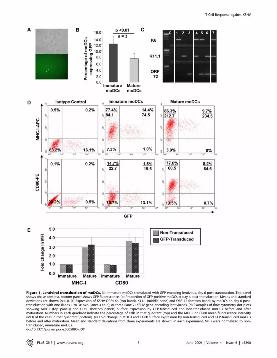
Figure 1. Lentiviral transduction of moDCs. (a) Immature moDCs transduced with GFP-encoding lentivirus, day 6 post-transduction. Top panelshows phase contrast, bottom panel shows GFP fluorescence. (b) Proportion of GFP-positive moDCs at day 6 post-transduction. Means and standarddeviations are shown (n = 3). (c) Expression of KSHV ORFs K6 (top band), K11.1 (middle band) and ORF 72 (bottom band) by moDCs on day 6 post-transduction with one (lanes 1 to 3); two (lanes 4 to 6); or three (lane 7) KSHV-gene-encoding lentiviruses. (d) Examples of flow cytometry dot plotsshowing MHC-I (top panels) and CD80 (bottom panels) surface expression by GFP-transduced and non-transduced moDCs before and aftermaturation. Numbers in each quadrant indicate the percentage of cells in that quadrant (top) and the MHC-I or CD80 mean fluorescence intensity(MFI) of the cells in that quadrant (bottom). (e) Fold change in MHC-I and CD80 surface expression by non-transduced and GFP-transduced moDCsbefore and after maturation. Mean and standard deviations from three experiments are shown. In each experiment, MFIs were normalized to non-transduced, immature moDCs.doi:10.1371/journal.pone.0005890.g001
T-Cell Response against KSHV
PLoS ONE | www.plosone.org 3 June 2009 | Volume 4 | Issue 6 | e5890
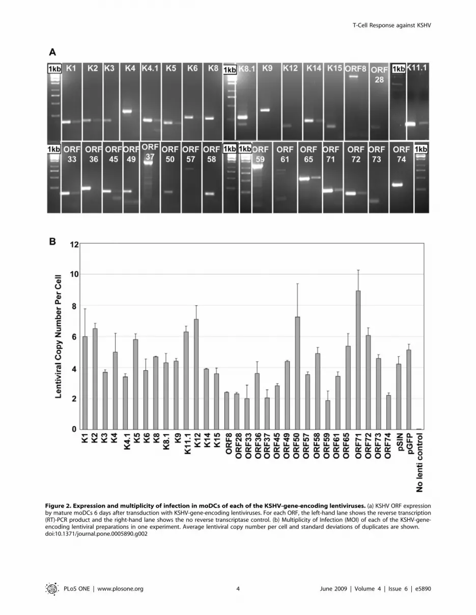
Figure 2. Expression and multiplicity of infection in moDCs of each of the KSHV-gene-encoding lentiviruses. (a) KSHV ORF expressionby mature moDCs 6 days after transduction with KSHV-gene-encoding lentiviruses. For each ORF, the left-hand lane shows the reverse transcription(RT)-PCR product and the right-hand lane shows the no reverse transcriptase control. (b) Multiplicity of Infection (MOI) of each of the KSHV-gene-encoding lentiviral preparations in one experiment. Average lentiviral copy number per cell and standard deviations of duplicates are shown.doi:10.1371/journal.pone.0005890.g002
T-Cell Response against KSHV
PLoS ONE | www.plosone.org 4 June 2009 | Volume 4 | Issue 6 | e5890

moDCs transduced with the empty vector alone. This may mean
that some weak T-cell responses were not identified in some or all
KSHV-seropositive individuals. However, it allows us to observe
patterns of immunodominant T-cell responses against KSHV.
CD8 and CD4 responses by all participants to all KSHV ORFs
or pools are summarized in Figures 4 and 5 respectively. A single
borderline CD8 response and two borderline CD4 responses were
observed in KSHV seronegative individuals compared to 32 CD8
responses and 21 CD4 responses by KSHV seropositive individuals.
All KSHV seronegative individuals gave strong positive responses to
moDCs transduced to express an adenovirus encoding the
immunodominant CMV gene phosphoprotein 65. This gives us
confidence that the responses measured are KSHV-specific.
One of the two KSHV+, HIV- participants who had active
classic KS at the time of venesection (P18) did not respond to any
of the KSHV ORFs (CD8 or CD4) and the other (P11) gave only
one borderline CD4 response. The KSHV+, HIV+ asymptomatic
carrier of KSHV (P10) gave six strong positive CD8 responses; and
one borderline and four strong CD4 responses to different KSHV
ORFs. Of the 11 KSHV+, HIV+ individuals with a history of
KSHV-related disease, six (P1, P2, P3, P5, P16 and P17) gave one
or no responses (total of CD8 and CD4), and were classified as
poor responders. There was no notable difference in the ages,
CD4 counts or years in remission from KSHV-related disease
between the poor responders and good responders (.1 response).
However, interestingly, one poor responder (P1) suffered a relapse
of KS within a year of venesection for this experiment. Three
other poor responders (P2, P5 and P17) had received no cancer-
specific chemotherapy treatment in addition to their antiretroviral
therapy, whereas all good responders received HAART in
combination with cancer chemotherapy. All poor responders
had a history of KS alone, except for P3, who had been treated for
PEL. Of the good responders, three (P6, P8 and P9) had a history
of KS alone and two (P4 and P7) had a history of KS and MCD.
These study participant characteristics are summarized in Table 2.
Targets of the KSHV-specific CD8 CTL T-cell responseCD8 responses in all responsive KSHV+ individuals showed a
bias towards early and late lytic gene products. Out of a total of 32
CD8 responses observed, 4 were directed against latent ORFs; 6
against immediate-early lytic ORFs; 11 against early lytic ORFs;
and 11 against late lytic ORFs. Two pools of early lytic ORFs were
frequently recognized CD8 targets: [ORF8/ORF49/ORF61] was
recognized by 6 individuals and [ORF59/ORF65/K4.1] was
recognized by 5 individuals. One pool of late lytic ORFs –
[ORF28/ORF36/ORF37] – was recognized by 7 individuals, and
was of particular interest to us as none of these gene products have
been previously investigated for immunogenicity. For the same
reason, a pool of immediate-early ORFs – [K6/K8/K14] – that
was recognized by 4 individuals is also of interest.
Targets of the KSHV-specific CD4+ T-cell responseCD4 responses across all individuals also showed a bias towards
early and late lytic gene products. Out of a total of 21 CD4
responses observed, 3 were directed against latent ORFs; 2 against
immediate-early lytic ORFs; 10 against early lytic ORFs; and 6
against late lytic ORFs. The most frequently recognized CD4
targets were the pool comprised of [ORF8/ORF49/ORF61],
which was recognized by 6 individuals; and the pools [ORF59/
ORF65/K4.1] and [ORF33/K1/K8.1], which were each recog-
nized by 4 individuals.
HLA Types of Good RespondersHLA typing was performed for all individuals classified as good
responders. As might be expected of an ethnically diverse cohort of
individuals from a central London clinic, the HLA types of these
individuals were mixed and are shown in Table 3.
Discussion
In this study, we have used lentiviral-transduced moDCs to
express a panel of KSHV ORFs to investigate the adaptive CD8
and CD4 T-cell responses against KSHV. This approach has
enabled us to perform a broad investigation into the KSHV-
specific T-cell response, and has enabled us to identify antigenic
hotspots within the KSHV genome.
Lentiviral transduction of moDCs with the conditions used did
not affect the moDCs immunophenotype or maturation, as
determined by expression of MHC-I and CD80. We showed that
moDCs can be transduced with up to three different KSHV-gene-
encoding lentiviruses simultaneously, resulting in expression of all
three transgenes. Interestingly, we found that after maturation of
moDCs, GFP expression was slightly down-regulated. Other
groups have reported that mature moDCs are harder to transduce
than immature moDCs [21,33], but to the best of our knowledge
there has been no previous documentation of decreased transgene
expression when maturation is induced after transduction. This
down-regulation may be the result of the terminal differentiation
of mature moDCs and the concurrent changes in cellular gene
expression and promoter activity. Importantly, even after
Table 1. Summary of study participant characteristics.
Number Male/FemaleMean Age(Range) KSHV status HIV status
Mean CD4 Count(Range)
HIV-/KSHV- Healthy Controls 3 3/0 37 (30 to 45) Seronegative Negative ND
HIV+/KSHV- Controls 4 3/1 48 (32 to 70) Seronegative Positive; On HAART; HIVviral load suppressed
345 (174 to 652)
HIV+/KSHV+; history of KSHVrelated neoplasia
11 11/0 47 (36 to 74) Seropositive*;KSHV-related diseasein remission
Positive; On HAART; HIVviral load suppressed
389 (171 to 785)
HIV+/KSHV+; asymptomaticcarriers of KSHV
1 1/0 56 Seropositive;Asymptomatic
Positive; On HAART; HIVviral load suppressed
373
HIV-/KSHV+; history of KSHVrelated neoplasia
2 2/0 37, 41 Seropositive; ActiveKS, regressing
Negative 550, ND
*Except for 2 patients (P3 and P5) for whom plasma were unavailable for serological testing; ND = not determined.doi:10.1371/journal.pone.0005890.t001
T-Cell Response against KSHV
PLoS ONE | www.plosone.org 5 June 2009 | Volume 4 | Issue 6 | e5890

T-Cell Response against KSHV
PLoS ONE | www.plosone.org 6 June 2009 | Volume 4 | Issue 6 | e5890

Figure 4. Summary of CD8 responses to KSHV ORFs by all study participants. Dark blue boxes represent positive responses; light blueboxes represent borderline responses; unfilled boxes represent no response; nd indicates not done due to insufficient numbers of available PBMCs.doi:10.1371/journal.pone.0005890.g004
Figure 5. Summary of CD4 responses to KSHV ORFs by all study participants. Dark green boxes represent positive responses; light greenboxes represent borderline responses; unfilled boxes represent no response; nd indicates not done due to insufficient numbers of available PBMCs.doi:10.1371/journal.pone.0005890.g005
Figure 3. T-cell proliferation responses to transduced moDCs. (a) Gating strategy for T-cell proliferation response assays. Gates are set on thelive T lymphocyte population (left panel) and CD8+ cells (middle panel), and the proportion of proliferating cells is taken as the percentage of CFSE-low cells in the bottom two quadrants (right panel). For CD4 responses, R2 is readjusted over the CD8-low cells. (b) Example of CD8 responses tomoDCs transduced with each KSHV gene or group of genes from one study participant (P7; HIV+, KSHV+, history of KSHV-related disease, inremission). Bar graphs show the proportion of CFSE-low cells in response to each gene or group of genes (with the background response to moDCstransduced with the empty pSIN vector subtracted). Mean of triplicates and standard deviations are shown. Two stars indicate a response that wasconsidered to be positive; one star indicates a response that was considered to be borderline positive according to the criteria described in Materialsand Methods. Flow cytometry histograms show examples of strong (top), medium (middle) and negative (bottom) CD8 responses to different KSHVantigens from the same study participant (P7). In all histograms, grey shading represents CD8 T cells cultured with moDCs transduced with the emptypSIN vector and black lines represent CD8 T cells cultured with moDCs transduced with different KSHV antigens. (c) Same as for (b), but showing CD4responses from the same study participant (P7).doi:10.1371/journal.pone.0005890.g003
T-Cell Response against KSHV
PLoS ONE | www.plosone.org 7 June 2009 | Volume 4 | Issue 6 | e5890

maturation we achieved an average of 7.5% GFP-positive moDCs,
a level comparable to those reported by other groups using a
similar protocol [21,23,26,28,33].
Although our study is too small to draw definite conclusions
regarding the differences in phenotypes of KSHV seropositive
individuals who made several responses to KSHV antigens and
those who did not, we show that individuals with active KS made
few responses and an asymptomatic KSHV carrier made several
responses, as has been previously described [9,10]. We found that all
individuals who had received HAART alone as treatment for their
KS were poor responders, whereas all good responders had received
HAART in combination with cytotoxic therapy. This concurs with
studies that have found better reconstitution of KSHV-specific
responses in individuals receiving HAART and cancer chemother-
apy compared with those receiving HAART alone [6]. We also
found that individuals with a history of MCD were good responders,
in line with a previous report of individuals with active MCD who
made responses to KSHV antigens comparable to those seen in
asymptomatic carriers of KSHV [40].
In a cohort of 14 KSHV seropositive individuals, we saw a distinct
skewing of both CD8 and CD4 responses towards early and late lytic
gene products. This was an unexpected result, as it appears to
contrast with observations of the T-cell response against Epstein-Barr
virus (EBV; also known as HHV-4, the most closely related human c-
herpesvirus to KSHV), which preferentially targets immediate-early
EBV gene products [41–44]. It has been suggested that immediate-
early gene products are frequent targets of herpesvirus-specific T-cell
responses as this enables activation of the host immune response
before the expression of viral genes involved in immune evasion
strategies. However, it has recently been reported that the CD8 T-cell
response against the murine c-herpesvirus 68 (MHV-68) is also
directed against early and late lytic gene products [45]. A broad
repertoire of epitopes derived from late lytic structural proteins and
early lytic proteins that are involved in DNA replication were
identified in MHV-68, but no epitopes derived from immediate-early
proteins were found. Likewise, 86% of CD8 and 90% of CD4 T-cell
responses against human cytomegalovirus (HCMV; a b-herpesvirus)
target early and late lytic gene products [46]. Thus, protein
abundance and, to a lesser degree, function may significantly affect
the immunogenicity of the gene product. We further speculate that
immunodominance of early and late lytic KSHV antigens may be an
important factor in establishing homeostasis between host and virus.
The activation of viral immune evasion strategies such as the down
regulation of MHC molecules before the expression of immunodo-
Table 2. Characteristics of HIV+ individuals who are in remission from KSHV-related disease.
Patient AgeCD4Count
KSHV-relateddisease history
Treatment for KSHV-relateddisease
Approximate yearsbetween active diseaseand venesection
Poor Responders P1 46 785 KS HAART+chemotherapy+radiotherapy ,1, since relapsed
P2 74 183 KS HAART only 2
P3 46 565 PEL HAART+chemotherapy 3.5
P5 39 406 KS HAART only ,1
P16 51 171 KS HAART+chemotherapy 5
P17 40 308 KS HAART only ,1
Mean 49 403 2
Good responders P4 36 645 KS and MCD HAART+chemotherapy ,1
P6 38 496 KS HAART+chemotherapy+radiotherapy 8.5
P7 53 281 KS and MCD HAART+chemotherapy 1.5
P8 49 226 KS HAART+chemotherapy 3.5
P9 49 209 KS HAART+chemotherapy ,1
Mean 45 371 3
doi:10.1371/journal.pone.0005890.t002
Table 3. HLA types of good responders.
Participant HLA Types
A B Cw DRB1DRB3 DRB4DRB5 DQB1
HIV+, KSHV-related neoplasia in remission P4 2, 29 7, 44 7, 16 7, 15 51, 53 2, 6
P6 24, 29 51, 65 8, 15 4, 13 52, 53 6, 8
P7 2, 3 7, 35 7, 4 1, 15 51 5, 6
P8 2, 24 13, 44 6, 5 4, 15 51,53 6, 7
P9 nd nd 6,16 7,11 nd nd
HIV+, Asymptomatic P10 3 7, 62 7, 10 4, 15 51,53 6, 8
nd = not determined.doi:10.1371/journal.pone.0005890.t003
T-Cell Response against KSHV
PLoS ONE | www.plosone.org 8 June 2009 | Volume 4 | Issue 6 | e5890

minant antigens may result in a blunting of the immune response,
enabling the virus to avoid elimination and to establish controlled
chronic infection.
We found that the three gene pools most frequently-recognized by
CD8 T cells from KSHV+ individuals were [ORF8/ORF49/
ORF61]; [ORF59/ORF65/K4.1]; and [ORF28/ORF36/ORF37].
ORF8 codes for glycoprotein B, which contains a well-described
CD8 epitope, aa492–500 (LMWYELSKI) [9,10,20]. ORF61 codes
for a large ribonucleotide reductase essential for DNA synthesis and
also contains a previously documented CD8 epitope, aa505–513
(GLADVFAEL) [10]. ORF65 codes for the minor capsid protein
which has also been identified as a target of the KSHV-specific CD8
T-cell response [19], and contains one identified CD8 epitope (aa35–
43; NMSQAEYLV). However, none of the gene products within the
most frequently-recognized CD8-target pool ([ORF28/ORF36/
ORF37], which was recognized by 7 out of 14 individuals) have
been previously investigated for immunogenicity. Of these three
proteins, ORF28 is a likely immunogenic candidate, as it has been
classified as an envelope glycoprotein based on positional and
structural similarities to EBV BDLF3 [47], which encodes the EBV
glycoprotein gp150. BDLF3 is a known target of the EBV-specific
CD4 response [41], and although KSHV ORF28 is its positional
equivalent, its gene product shares no amino acid sequence similarity
to EBV gp150. In the search for immunodominant virus-specific
epitopes it seems logical to focus on viral gene products with unique
sequences, as epitopes derived from such proteins are the most likely
to be biologically relevant in terms of effective host control of viral
infection. KSHV ORF36 encodes a serine protein kinase [48] and
ORF37 a modified DNA exonuclease involved in host mRNA shut
off [49]. Both these genes’ products share considerable amino acid
sequence similarity with the products of their respective EBV
homologues, BGLF4 and BGLF5. BGLF5 has not been identified as
a target of the EBV-specific T-cell response and although CD8
responses to BGLF4 have been detected in one individual [43], it is
not a major T-cell target. Thus these two KSHV gene products seem
less likely immunogenic candidates, although this remains to be
investigated.
Interestingly, [ORF8/ORF49/ORF61] and [ORF59/ORF65/
K4.1] were also common CD4 targets, recognized by 6 of 14 and
4 of 14 individuals, respectively. As previously mentioned, very
little is known of the CD4 response against KSHV. However it is
not surprising that it is directed against the same gene products
that elicit the strongest CD8 responses. In the T-cell response
against HCMV, 53% of the most recognized ORFs are common
to both the CD8 and CD4 response [46]. Similarly, the major
EBV CD8 targets BZLF1 and BMLF1 (immediate-early lytic) and
EBNA1, EBNA2, EBNA3A, EBNA3B, EBNA3C and LMP2
(latent) also elicit CD4 responses [41,42]. Further studies will
elucidate the most antigenic KSHV gene products from within
each pool, and will identify new CD8 and CD4 epitopes.
The present study has enabled a far broader investigation into the
targets of both the CD8 and CD4 KSHV-specific T-cell responses
than has been previously undertaken. We found, unexpectedly, that
these responses were skewed towards early and late lytic gene
products. This knowledge will be important to future immunolog-
ical investigations into KSHV and may eventually lead to the
development of better immunotherapies for KSHV-related diseases,
and potentially even a vaccine against KSHV.
Materials and Methods
Ethics StatementAll KSHV-infected and non-infected study participants were
recruited from Chelsea and Westminster Hospital, London, UK,
and provided written, informed consent. The study protocols were
approved by Riverside Research Ethics Committee.
Study ParticipantsFive groups of study participants were studied. 1) HIV-negative,
KSHV-seronegative, healthy individuals (n = 3); 2) HIV-positive,
KSHV-seronegative individuals (n = 4); 3) HIV-positive, KSHV-
seropositive individuals with a history of KSHV-related disease
(KS; KS and MCD; or PEL) but in remission at time of
venesection (n = 11); 4) HIV-positive, KSHV-seropositive individ-
uals with no history of KSHV-related disease (asymptomatic
carrier; n = 1); and 5) HIV-negative, KSHV-seropositive individ-
uals with active KSHV-related disease (KS), in regression at time
of venesection (n = 2).
All study participants were male apart from one HIV-positive,
KSHV-seronegative female. Study participants’ age on date of
venesection ranged from 30 to 74 years. All HIV-positive
individuals were on HAART and had an HIV viral load of ,50
copies per mL. CD4 counts ranged from 171 to 785 cells/mm3.
Characteristics of each group of study participants can be seen in
Table 1.
Construction of KSHV-gene-encoding lentiviral vectorsand lentivirus production
The lentiviral vector pSIN-MCS was derived from the green
fluorescence protein (GFP)-encoding vector pCSGW (a kind gift
from Professor Adrian Thrasher, Institute of Child Health,
University College London). Packaging plasmids p8.91 and pMD.G
were a kind gift from Professor Didier Trono, Ecole Polytechnique
Federale de Lausanne, Switzerland). KSHV ORFs were cloned
from cDNA or genomic DNA from the BC3 PEL cell line or from
previously constructed vectors into pSIN-MCS as previously
described [39,50]. Primers used in cloning of each gene are
available on request. Vesicular stomatitis virus-G envelope-
pseudotyped lentiviral virions were produced also as previously
described [39,50]. Briefly, 293T cells were cotransfected for 5 hours
with 2 mg of pSIN-MCS, 1.5 mg of p8.91 and 1.5 mg of pMD.G
using the standard FuGENE (Roche, East Sussex, UK) protocol.
Transfected 293Ts were then cultured for a further 48 hours in
fresh media (DMEM [Gibco, Invitrogen, Paisley, UK] supplement-
ed with 10% FCS and Penicillin-Streptomycin [both Sigma, Poole,
UK]), after which the virion-containing supernatant was harvested,
filtered (0.45 mm) and stored at 280uC for later use.
Generation of monocyte-derived dendritic cells (moDCs)from peripheral blood
Peripheral blood mononuclear cells (PBMCs) and plasma were
isolated from whole blood using Histopaque-1077 Hybri-Max
gradient separation (Sigma). 1 ml aliquots of plasma were stored at
280uC for later use in serological testing. PBMCs were separated
into CD14+ and CD14- fractions using a standard immunodeple-
tion protocol utilizing magnetic beads labeled with a monoclonal
antibody directed against CD14 (Miltenyi Biotech, Bergisch
Gladbach, Germany). CD14- monocytes were stored for later
use at 280uC in a freezer mix comprised of 50% RPMI 1640,
40% fetal calf serum (FCS) and 10% dimethyl sulfoxide (DMSO)
(all Sigma).
CD14+ monocytes were plated at 36105 cells per well on 24-
well plates (Cellstar, Greiner Bio-one, Gloucestershire, UK) and
cultured in RPMI with Hepes modification and supplemented
with L-Glutamine, Penicillin-streptomycin and 5% Human AB
Serum (Lot number 027K0432) (all Sigma). Differentiation to
moDCs was induced by addition of 70 ng/ml interleukin 4 (IL-4)
T-Cell Response against KSHV
PLoS ONE | www.plosone.org 9 June 2009 | Volume 4 | Issue 6 | e5890

and 70 ng/ml granulocyte/macrophage colony-stimulating factor
(GMCSF) (both R and D systems, Oxford, UK). Cells were fed
with fresh media and cytokines every second day until maturation
on day 8 (see below).
Lentiviral transduction of moDCsOn day 4 in culture, moDCs were transduced by incubation
with the appropriate volume of each lentiviral preparation
(typically between 150 ml and 600 ml, calculated according to the
multiplicity of infection [MOI] of each preparation). Lentiviral
preparations were thawed on ice for 30 minutes then warmed to
37uC in a water bath directly prior to use. Lentivirus was then
added directly to the cells in 24-well plates. On day 8 in culture
(day 4 post-transduction) moDCs were matured by stimulation
with a ‘‘cytokine cocktail’’ made up from 5 ng/ml tumor necrosis
factor a (TNFa), 5 ng/ml interleukin 1b (IL-1b), 150 ng/ml
interleukin 6 (IL-6) (all R and D systems) and 1 mg/ml
prostaglandin E2 (PGE2; Sigma). On day 10 in culture (day 6
post-transduction), moDCs were harvested for use in further
experiments. Expression of constructs was confirmed by reverse
transcriptase PCR (RT-PCR). GFP expression by pCSGW-
infected moDCs was assessed by fluorescent microscopy and flow
cytometry performed on a FACSCalibur (BD Biosciences, Oxford,
UK). To determine the MOI of each lentiviral preparation,
quantitative real time PCR (qPCR) was performed for the
lentiviral packaging signal using the glyceraldehyde-3-phosphate
dehydrogenase (GAPDH) gene as the reference signal, as
previously described [50].
Transduction of moDCs with an adenovirus encoding theCMV gene phosphoprotein 65
moDCs transduced to express the immunodominant CMV
gene phosphoprotein 65 (CMVpp65) were used as a positive
control. Unfortunately, despite extensive efforts, we were unable to
clone CMVpp65 into our lentiviral vector. We therefore used an
adenovirus encoding CMVpp65 (Adpp65) to transduce moDCs.
Adpp65 was a kind gift from Dr Magnus Essand of Uppsala
University and we used the previously published protocol for
transduction [51]. Briefly, on day 8 in culture, immature moDCs
were harvested by aspiration, spun at 1400 rpm for 8 minutes,
resuspended in 250 ml media and counted. moDCs were
transduced with Adpp65 (MOI = 300) for 2 hours at 37uC.
moDCs were than plated on 24-well plates and maturation
stimulus was added as above.
Immunophenotyping of moDCsmoDCs transduced with 300 ml pCSGW (MOI = 5.6) were
analyzed for expression of cell surface markers by multi-parameter
flow cytometry. moDCs were stained with monoclonal antibodies
against HLA-A,B,C conjugated to APC (mouse anti-human,
IgG1k; BD Biosciences) and CD80 conjugated to PE (mouse anti-
human, IgG1k; BD Biosciences) or their appropriate isotype
controls, at a final antibody dilution of 1 in 20, for 30 minutes on
ice, protected from light. Flow cytometry was performed on a
FACSCalibur (Becton Dickinson) and analyzed with CELLQuest
software (Becton Dickinson). 10 000 events were collected for each
sample. moDCs were gated on the live-cell population according to
the expected forward scatter and side scatter. Isotype controls were
used to set gates for positive staining for each of the antibodies.
Serology for KSHVAn in-house developed MIX-MAP ELISA against was used to
detect KSHV seropositivity as previously described [7] but with an
improved, modified peptide containing two copies each of a lytic and
a latent epitope (RSHLGFWQEGWSGQVYQDWLGRMNC-
SYENM derived from K8.1, and QPGPSREYRYVLRTSPP-
HRPGVRMRRV derived from ORF 73, respectively).
HLA TypingGenomic DNA was extracted from a minimum of 56106
PBMCs using the standard NucleonH BACC2 kit (Tepnel Life
Sciences, Manchester, UK) protocol. HLA typing was performed
by the Department of Histocompatibility and Immunogenetics,
Clinical Immunology Laboratory, Hammersmith Hospital, Lon-
don. Low resolution typing was performed using PCR-sequence
specific primers (PCR-SSP). High resolution typing was achieved
using reference strand conformational analysis (RSCA).
T cell PurificationT cells were isolated from thawed cryogenically-preserved
CD14- PBMCs using a standard protocol for immunodepletion
using magnetic beads labeled with monoclonal antibodies directed
against CD14, CD16, CD19, CD36, CD56, CD123 and
Glycophorin A (Miltenyi Biotech).
T-cell Proliferation AssaysCarboxy Fluorescein Succinimidyl Ester (CFSE; Molecular
Probes, Invitrogen, Paisley, UK) was reconstituted in DMSO to
make a 9 mM stock solution. CFSE stock solution was diluted to a
10 mM working solution in PBS. Purified T cells were resuspended
in 100 ml PBS and 100 ml 10 mM CFSE solution per 106 cells for 5
minutes at room temperature and then blocked with 200 ml FCS
per 106 cells for 15 minutes at room temperature.
CFSE-stained T cells were then plated for culture in proliferation
reactions with autologous transduced moDCs. Cell mixtures were
cultured in round-bottomed 96-well plates (Cellstar) in a total
volume of 200 ml T cell media (RPMI 1640 with Hepes
modification supplemented with L-Glutamine, Penicillin-Strepto-
mycin and 10% Human AB Serum, Lot number 027K0432). T
cells were cultured at 100, 000 cells per well with 2, 500 autologous
moDCs (transduced or non-transduced) per well. In addition, for
each experiment the following control wells were set up: 1) T cells
only 2) T cells+non-transduced moDCs+5 mg/ml phytohemagglu-
tinin (PHA; Sigma) 3) T cells+moDCs transduced with Adpp65.
After 6 days of culture, cells were harvested, stained with a
monoclonal antibody against CD8 conjugated to PeCy5 (mouse, anti-
human, IgG1k; BD Biosciences) or the appropriate isotype control at
a final antibody dilution of 1 in 20, for 30 minutes on ice, protected
from light. Flow cytometry was performed on a FACSCalibur
(Becton Dickinson) and analyzed with CELLQuest software (Becton
Dickinson). The maximum possible events (typically between 10 000
and 30 000) were collected for each sample. T cells were gated on the
live-lymphocyte population according to the expected forward scatter
and side scatter, and then on either CD8-high or CD8-low (CD4) T
cell populations (see Figure 3a).
Statistical AnalysisStatistics were performed on Microsoft Office Excel 2003
software. For T cell responses to transduced moDCs, a positive
response was designated as a response that fitted three criteria of
significance above the background response to moDCs transduced
with the empty lentiviral vector: 1) p,0.05; unpaired student T
test 2) response .3.5 standard deviations above background and
3) response .10% above background. A borderline response was
designated as p,0.05 and response .3 standard deviations and
.9 per cent above background.
T-Cell Response against KSHV
PLoS ONE | www.plosone.org 10 June 2009 | Volume 4 | Issue 6 | e5890

Acknowledgments
The authors thank study participants and staff at the Chelsea and
Westminster Hospital who contributed to this study and Dr Adriano
Boasso (Imperial College) for his help with the preparation of this
manuscript.
Author Contributions
Conceived and designed the experiments: RCR DL NCM MB CB FG.
Performed the experiments: RCR DL FG RJV. Analyzed the data: RCR
SRH. Contributed reagents/materials/analysis tools: MB. Wrote the
paper: RCR.
References
1. Boshoff C, Weiss R (2002) Aids-related malignancies. Nat Rev Cancer 2:
373–382.
2. Parkin DM, Sitas F, Chirenje M, Stein L, Abratt R, et al. (2008) Part I: Cancer
in Indigenous Africans–burden, distribution, and trends. The Lancet Oncology
9: 683–692.
3. Cesarman E, Chang Y, Moore PS, Said JW, Knowles DM (1995) Kaposi’s
Sarcoma-Associated Herpesvirus-Like DNA Sequences in AIDS-Related Body-
Cavity-Based Lymphomas. N Engl J Med 332: 1186–1191.
4. Soulier J, Grollet L, Oksenhendler E, Cacoub P, Cazals-Hatem D, et al. (1995)
Kaposi’s sarcoma-associated herpesvirus-like DNA sequences in multicentric
Castleman’s disease [see comments]. Blood 86: 1276–1280.
5. Cattelan AM, Calabro ML, Aversa SM, Zanchetta M, Meneghetti F, et al.
(1999) Regression of AIDS-related Kaposi’s sarcoma following antiretroviral
therapy with protease inhibitors: biological correlates of clinical outcome.
Eur J Cancer 35: 1809–1815.
6. Bihl F, Mosam A, Henry LN, Chisholm JV III, Dollard S, et al. (2007) Kaposi’s
sarcoma-associated herpesvirus-specific immune reconstitution and antiviral
effect of combined HAART/chemotherapy in HIV clade C-infected individuals
with Kaposi’s sarcoma. AIDS 21: 1245–1252.
7. Bourboulia D, Aldam D, Lagos D, Allen E, Williams I, et al. (2004) Short- and
long-term effects of highly active antiretroviral therapy on Kaposi sarcoma-
associated herpesvirus immune responses and viraemia. AIDS 18: 485–493.
8. Wilkinson J, Cope A, Gill J, Bourboulia D, Hayes P, et al. (2002) Identification of
Kaposi’s Sarcoma-Associated Herpesvirus (KSHV)-Specific Cytotoxic T-Lym-
phocyte Epitopes and Evaluation of Reconstitution of KSHV-Specific Responses
in Human Immunodeficiency Virus Type 1-Infected Patients Receiving Highly
Active Antiretroviral Therapy. J Virol 76: 2634–2640.
9. Guihot A, Dupin N, Marcelin A, Gorin I, Bedin A, et al. (2006) Low T Cell
Responses to Human Herpesvirus 8 in Patients with AIDSCCERelated and
Classic Kaposi Sarcoma. J Infect Dis 194: 1078–1088.
10. Lambert M, Gannage M, Karras A, Abel M, Legendre C, et al. (2006)
Differences in the frequency and function of HHV8-specific CD8 T cells
between asymptomatic HHV8 infection and Kaposi sarcoma. Blood 108:
3871–3880.
11. Barozzi P, Bonini C, Potenza L, Masetti M, Cappelli G, et al. (2008) Changes in
the Immune Responses Against Human Herpesvirus-8 in the Disease Course of
Posttransplant Kaposi Sarcoma. Transplantation 86: 738–744.
12. Bihl F, Narayan M, Chisholm JV III, Henry LM, Suscovich TJ, et al. (2007)
Lytic and Latent Antigens of the Human Gammaherpesviruses Kaposi’s
Sarcoma-Associated Herpesvirus and Epstein-Barr Virus Induce T-Cell
Responses with Similar Functional Properties and Memory Phenotypes. J Virol
81: 4904–4908.
13. Brander C, O’Connor P, Suscovich T, Jones N, Lee Y, et al. (2001) Definition of
an Optimal Cytotoxic T Lymphocyte Epitope in the Latently Expressed
Kaposi’s SarcomaCCoAssociated Herpesvirus Kaposin Protein. J Infect Dis 184:
119–126.
14. Micheletti F, Monini P, Fortini C, Rimessi P, Bazzaro M, et al. (2002)
Identification of cytotoxic T lymphocyte epitopes of human herpesvirus 8.
Immunology 106: 395–403.
15. Osman M, Kubo T, Gill J, Neipel F, Becker M, et al. (1999) Identification of
Human Herpesvirus 8-Specific Cytotoxic T-Cell Responses. J Virol 73:
6136–6140.
16. Ribechini E, Fortini C, Marastoni M, Traniello S, Spisani S, et al. (2006)
Identification of CD8+ T Cell Epitopes within Lytic Antigens of Human Herpes
Virus 8. J Immunol 176: 923–930.
17. Stebbing J, Bourboulia D, Johnson M, Henderson S, Williams I, et al. (2003)
Kaposi’s Sarcoma-Associated Herpesvirus Cytotoxic T Lymphocytes Recognize
and Target Darwinian Positively Selected Autologous K1 Epitopes. J Virol 77:
4306–4314.
18. Wang Q, Jenkins F, Jacobson L, Meng Y, Pellett P, et al. (2000) CD8+ Cytotoxic
T Lymphocyte Responses to Lytic Proteins of Human Herpes Virus 8 in Human
Immunodeficiency Virus Type 1CCoInfected and CCoUninfected Individuals.
J Infect Dis 182: 928–932.
19. Woodberry T, Suscovich TJ, Henry LM, Martin JN, Dollard S, et al. (2005)
Impact of Kaposi Sarcoma-Associated Herpesvirus (KSHV) Burden and HIV
Coinfection on the Detection of T Cell Responses to KSHV ORF73 and
ORF65 Proteins. J Infect Dis 192: 622–629.
20. Wang QJ, Huang XL, Rappocciolo G, Jenkins FJ, Hildebrand WH, et al. (2002)
Identification of an HLA A*0201-restricted CD8+ T-cell epitope for the
glycoprotein B homolog of human herpesvirus 8. Blood 99: 3360–3366.
21. Schroers R, Sinha I, Segall H, Schmidt-Wolf IGH, Rooney CM, et al. (2000)
Transduction of Human PBMC-Derived Dendritic Cells and Macrophages by
an HIV-1-Based Lentiviral Vector System. Mol Ther 1: 171–179.
22. Chinnasamy N, Chinnasamy D, Toso JF, Lapointe R, Candotti F, et al. (2000)
Efficient gene transfer to human peripheral blood monocyte-derived dendriticcells using human immunodeficiency virus type 1-based lentiviral vectors. Hum
Gene Ther 11: 1901–1909.
23. Dyall J, Latouche JB, Schnell S, Sadelain M (2001) Lentivirus-transducedhuman monocyte-derived dendritic cells efficiently stimulate antigen-specific
cytotoxic T lymphocytes. Blood 97: 114–121.
24. Collins MK, Cerundolo V (2004) Gene therapy meets vaccine development.
Trends in Biotechnology 22: 623–626.
25. Jenne L, Schuler G, Steinkasserer A (2001) Viral vectors for dendritic cell-basedimmunotherapy. Trends in Immunology 22: 102–107.
26. Esslinger C, Romero P, MacDonald HR (2002) Efficient transduction ofdendritic cells and induction of a T-cell response by third-generation
lentivectors. Hum Gene Ther 13: 1091–1100.
27. Firat H, Zennou V, Garcia-Pons F, Ginhoux F, Cochet M, et al. (2002) Use of alentiviral flap vector for induction of CTL immunity against melanoma.
Perspectives for immunotherapy. J Gene Med 4: 38–45.
28. Lizee G, Gonzales MI, Topalian SL (2004) Lentivirus vector-mediatedexpression of tumor-associated epitopes by human antigen presenting cells.
Hum Gene Ther 15: 393–404.
29. Lopes L, Fletcher K, Ikeda Y, Collins M (2006) Lentiviral vector expression of
tumour antigens in dendritic cells as an immunotherapeutic strategy. Cancer
Immunol Immunother 55: 1011–1016.
30. Dullaers M, Breckpot K, Van Meirvenne S, Bonehill A, Tuyaerts S, et al. (2004)
Side-by-Side Comparison of Lentivirally Transduced and mRNA-Electroporat-ed Dendritic Cells: Implications for Cancer Immunotherapy Protocols. Mol
Ther 10: 768–779.
31. He Y, Zhang J, Mi Z, Robbins P, Falo LD Jr (2005) Immunization withLentiviral Vector-Transduced Dendritic Cells Induces Strong and Long-Lasting
T Cell Responses and Therapeutic Immunity. J Immunol 174: 3808–3817.
32. Breckpot K, Heirman C, De Greef C, van der Bruggen P, Thielemans K (2004)Identification of New Antigenic Peptide Presented by HLA-Cw7 and Encoded
by Several MAGE Genes Using Dendritic Cells Transduced with Lentiviruses.J Immunol 172: 2232–2237.
33. Gruber A, Kan-Mitchell J, Kuhen KL, Mukai T, Wong-Staal F (2000) Dendritic
cells transduced by multiply deleted HIV-1 vectors exhibit normal phenotypesand functions and elicit an HIV-specific cytotoxic T-lymphocyte response in
vitro. Blood 96: 1327–1333.
34. Koya RC, Kasahara N, Favaro PM, Lau R, Ta HQ, et al. (2003) Potent
maturation of monocyte-derived dendritic cells after CD40L lentiviral gene
delivery. J Immunother 26: 451–460.
35. Chen X, He J, Chang LJ (2004) Alteration of T cell immunity by lentiviral
transduction of human monocyte-derived dendritic cells. Retrovirology 1: 37.
36. Jenner RG, Alba MM, Boshoff C, Kellam P (2001) Kaposi’s Sarcoma-Associated Herpesvirus Latent and Lytic Gene Expression as Revealed by DNA
Arrays. J Virol 75: 891–902.
37. Jenner RG, Boshoff C (2002) The molecular pathology of Kaposi’s sarcoma-
associated herpesvirus. Biochimica et Biophysica Acta (BBA) - Reviews on
Cancer 1602: 1–22.
38. Coscoy L, Ganem D (2000) Kaposi’s sarcoma-associated herpesvirus encodes
two proteins that block cell surface display of MHC class I chains by enhancingtheir endocytosis. Proc Natl Acad Sci U S A 97: 8051–8056.
39. Lagos D, Trotter MWB, Vart RJ, Wang HW, Matthews NC, et al. (2007)
Kaposi sarcoma herpesvirus-encoded vFLIP and vIRF1 regulate antigenpresentation in lymphatic endothelial cells. Blood 109: 1550–1558.
40. Guihot A, Oksenhendler E, Galicier L, Marcelin AG, Papagno L, et al. (2008)
Multicentric Castleman disease is associated with polyfunctional effectormemory HHV-8-specific CD8+ T cells. Blood 111: 1387–1395.
41. Hislop AD, Taylor GS, Sauce D, Rickinson AB (2007) Cellular responses to viralinfection in humans: lessons from Epstein-Barr virus. Annu Rev Immunol 25:
587–617.
42. Landais E, Saulquin X, Houssaint E (2005) The human T cell immune responseto Epstein-Barr virus. Int J Dev Biol 49: 285–292.
43. Pudney VA, Leese AM, Rickinson AB, Hislop AD (2005) CD8+ immunodo-minance among Epstein-Barr virus lytic cycle antigens directly reflects the
efficiency of antigen presentation in lytically infected cells. J Exp Med 201:
349–360.
44. Steven NM, Annels NE, Kumar A, Leese AM, Kurilla MG, et al. (1997)
Immediate Early and Early Lytic Cycle Proteins Are Frequent Targets of the
Epstein-Barr Virus-induced Cytotoxic T Cell Response. J Exp Med 185:1605–1618.
45. Gredmark-Russ S, Cheung EJ, Isaacson MK, Ploegh HL, Grotenbreg GM(2008) The CD8 T-Cell Response against Murine Gammaherpesvirus 68 Is
T-Cell Response against KSHV
PLoS ONE | www.plosone.org 11 June 2009 | Volume 4 | Issue 6 | e5890

Directed toward a Broad Repertoire of Epitopes from both Early and Late
Antigens. J Virol 82: 12205–12212.46. Sylwester AW, Mitchell BL, Edgar JB, Taormina C, Pelte C, et al. (2005)
Broadly targeted human cytomegalovirus-specific CD4+ and CD8+ T cells
dominate the memory compartments of exposed subjects. J Exp Med 202:673–685.
47. Zhu FX, Chong JM, Wu L, Yuan Y (2005) Virion Proteins of Kaposi’s Sarcoma-Associated Herpesvirus. J Virol 79: 800–811.
48. Hamza MS, Reyes RA, Izumiya Y, Wisdom R, Kung HJ, et al. (2004) ORF36
protein kinase of Kaposi’s sarcoma herpesvirus activates the c-Jun N-terminalkinase signaling pathway. J Biol Chem 279: 38325–38330.
49. Glaunsinger B, Ganem D (2004) Lytic KSHV infection inhibits host gene
expression by accelerating global mRNA turnover. Mol Cell 13: 713–723.
50. Vart RJ, Nikitenko LL, Lagos D, Trotter MWB, Cannon M, et al. (2007)
Kaposi’s Sarcoma-Associated Herpesvirus-Encoded Interleukin-6 and G-Pro-
tein-Coupled Receptor Regulate Angiopoietin-2 Expression in Lymphatic
Endothelial Cells. Cancer Res 67: 4042–4051.
51. Carlsson B, Cheng WS, Totterman TH, Essand M (2003) Ex vivo stimulation of
cytomegalovirus (CMV)-specific T cells using CMV pp65-modified dendritic
cells as stimulators. Br J Haematol 121: 428–438.
T-Cell Response against KSHV
PLoS ONE | www.plosone.org 12 June 2009 | Volume 4 | Issue 6 | e5890


