Embed Size (px)
Citation preview

For Peer Review
The changing face of hospitalization due to gastrointestinal bleeding and perforation
Journal: Alimentary Pharmacology & Therapeutics
Manuscript ID: APT-0637-2010.R2
Wiley - Manuscript type: Original Scientific Paper
Date Submitted by the Author:
11-Dec-2010
Complete List of Authors: Lanas, Angel; University Hospital Lozano Blesa, Gastroenterology; University of Zaragoza. School of Medicine, Medicine; CIBERehd Garcia Rodriguez, Luis Alberto; Centro Español de Investigacion Farmacoepidemiologica (CEIFE) Polo-Tomas, Monica; CIBERehd, Division of Gastroenterology Ponce, Marta; Hospital la Fe, Gastroenterology Unit Quintero, Enrique; Hospital Universitario de la Laguna, Gastroenterology Perez-Aisa, Maria; Hospital Costa del Sol, Gastroeneterology Gisbert, Javier; Hospital de la Princesa-CIBERehd, Division of Gastroenterology Bujanda, Luis; Hospital Donostia. CIBERehd, Gastroenterology Castro, Manuel; Hospital de Valme. CIBERehd, Gastroenterology Department Muñoz, Maria; University Hospital Lozano Blesa, Gastroenterology Del Pino, Maria Dolores; University Hospital Lozano Blesa, Gastroenterology Garcia, Santiago; Hospital Universitario Miguel Servet, Gastroenterology Calvet, Xavier; Parc Tauli, Gastroenterology
Keywords: Peptic ulcer disease < Disease-based, Diverticular disease < Disease-based, Stomach and duodenum < Organ-based, Epidemiology < Topics, NSAIDs < Topics
Alimentary Pharmacology & Therapeuticpe
er-0
0605
960,
ver
sion
1 -
5 Ju
l 201
1Author manuscript, published in "Alimentary Pharmacology & Therapeutics 33, 5 (2011) 585"
DOI : 10.1111/j.1365-2036.2010.04563.x

For Peer Review
1
The changing face of hospitalization due to gastrointestinal bleeding and
perforation
Authors:
Angel Lanas,1,2,3 Luis A. García-Rodríguez,4 Mónica Polo-Tomás2, Marta
Ponce5,2, Enrique Quintero,6 Maria Angeles Perez-Aisa7, Javier P. Gisbert8,2
Luis Bujanda9,2, Manuel Castro10,2, Maria Muñoz1, Dolores Del-Pino12, Santiago
Garcia13, Xavier Calvet14 ,2
Affiliations
1Servicio de Aparato Digestivo, Hospital Clínico. Universidad de Zaragoza. Spain
2 Centro de Investigación Biomédica en Red de enfermedades hepáticas y digestivas
(CIBERehd). Spain
3 Instituto Aragonés de Ciencias de la Salud (IACS)
4 Centro Español de Investigación Farmacoepidemiológica. Madrid. Spain
5Servicio de Aparato Digestivo. Hospital La Fe. Valencia
6Servicio de Aparato Digestivo. Hospital Universitario de Canarias. La Laguna. Tenerife.
Spain.
7. Servicio de Aparato Digestivo. Hospital Costa del Sol. Marbella. Spain.
8. Servicio de Aparato Digestivo. Hospital de la Princesa. Madrid. Spain.
9 Servicio de Aparato Digestivo. Hospital Donostia Universidad del País Vasco. San
Sebastián. Spain.
10 Servicio de Aparato Digestivo. Hospital de Valme.- Sevilla. Spain.
11. Servicio de Aparato Digestivo. Hospital de Asturias. Oviedo. Spain.
12. Servicio de Codificación. Hospital Clínico Universitario. Zaragoza. Spain
13 Servicio de Aparato Digestivo. Hospital Universitario Miguel Servet. Zaragoza. Spain
14 Servicio de Aparato Digestivo. Hospital de Sabadell. Barcelona. Spain
Short title: Trends in GI hospitalizations
To whom correspondence should be addressed:
Angel Lanas. Servicio de Aparato Digestivo. Hospital Clínico Universitario. C/ San Juan Bosco 15. 50009 Zaragoza. Spain. Phone: 34 976765786; Fax: 34 976765787). Email: [email protected]
Page 1 of 30 Alimentary Pharmacology & Therapeutic
123456789101112131415161718192021222324252627282930313233343536373839404142434445464748495051525354555657585960
peer
-006
0596
0, v
ersi
on 1
- 5
Jul 2
011

For Peer Review
2
Abstract:
Background: Temporal changes in the incidence of cause-specific
gastrointestinal (GI) complications may be one of the factors underlying
changing medical practice patterns. Aim: To report temporal changes in the
incidence of five major causes of specific gastrointestinal (GI) complication
events. Methodology: Population-based study of patients hospitalized due to
GI bleeding and perforation from 1996–2005 in Spain. We report crude rates,
and estimate regression coefficients of temporal trends, severity, and recorded
drug use for 5 frequent GI events. GI hospitalization charts were validated by
independent review of large random samples. Results: The incidence per
100,000 person-years of hospitalizations due to upper GI ulcer bleeding and
perforation decreased over time (from 54.6 and 3.9 in 1996 [R2=0.944] to 25.8
and 2.9 in 2005 [R2=0.410], respectively). On the contrary, the incidence per
100,000 person-years of colonic diverticular and angiodysplasia bleeding
increased over time (3.3 and 0.9 in 1996 [R2=0.443] and 8.0 and 2.6 in 2005
[R2=0.715], respectively). A small increasing trend was observed for the
incidence per 100,000 person-years of intestinal perforations (from 1.5 to 2.3
events). Based on data extracted from the validation process, recent recorded
drug intake showed an increased frequency of anticoagulants with colonic
diverticular and angiodysplasia bleeding, whereas NSAID and low-dose aspirin
use were more prevalent in peptic ulcer bleeding and colonic diverticular
bleeding respectively. Conclusions: From 1996–2005, hospitalizations due to
peptic ulcer bleeding and perforation have decreased significantly, whereas the
number of cases of colonic diverticular and angiodysplasia bleeding have
increased.
Key words: peptic ulcer, bleeding, perforation, angiodysplasia, diverticulum
Page 2 of 30Alimentary Pharmacology & Therapeutic
123456789101112131415161718192021222324252627282930313233343536373839404142434445464748495051525354555657585960
peer
-006
0596
0, v
ersi
on 1
- 5
Jul 2
011

For Peer Review
3
Background
Gastrointestinal (GI) complications are major causes of hospitalization.
Major therapeutic advances in the treatment and prevention of peptic ulcer
diseases have been implemented in the past decade, which should contribute
to a significant decrease in the incidence and mortality due to peptic ulcer
diseases. Opposing trends in peptic ulcer complications such as bleeding or
perforation have been reported in different countries, and no decrease or
increase in hospitalizations due to peptic ulcer bleeding complications have
been observed (1-6). More recently, two studies from different geographical
areas suggested that there has been a marked decrease in the incidence of
upper GI complications and a slight increase in the incidence of lower GI
complications (7,8); however, the specific lesions leading to these changes
have not been analyzed. Furthermore, the time trends for bleeding and
perforation may not be parallel, since the underlying pathogenic mechanisms
and risk factors could diverge (9,10). Additionally, the exact source of lower GI
complications are often more difficult to identify than upper GI complications
because of the anatomic complexity of the lower gut and available diagnostic
tests. Among the causes of lower GI bleeding, colonic diverticuli and
angiodysplasia are two lesions which could explain, at least in part, the recent
trends, since age was found to be one of the main risk factors for
hospitalizations (7). However, the time trends and clinical characteristics of
hospitalizations owing to these two lesions have not been reported.
Prevention strategies and optimization of hospital resources require a
clear understanding of the type of pathology causing hospitalization. In a
previous report, we presented the overall time trends of hospitalizations due to
GI complications, (7) which were obtained from a data-base including
information provided by 10 Spanish general hospitals representative of the
entire country (11). Now, as part of the pre-specified analysis plan, we aim to
characterize and analyze in detail the time trends for hospitalizations due to five
specific major causes of GI complications; namely, peptic ulcer bleeding, peptic
ulcer perforation, intestinal perforation, colonic diverticular bleeding, and
bleeding caused by angiodysplasia. We believe that these data are needed
since, as discussed above, the available literature for some of these causes and
Page 3 of 30 Alimentary Pharmacology & Therapeutic
123456789101112131415161718192021222324252627282930313233343536373839404142434445464748495051525354555657585960
peer
-006
0596
0, v
ersi
on 1
- 5
Jul 2
011

For Peer Review
4
the comparative trends among them are sparse or absent. Additionally, we also
describe the severity characteristics and recorded drug use for each of these
entities.
Methods:
Setting and data collection
The study (7) was approved by the Institutional Review Board of Aragón
and was carried out in 10 Spanish general hospitals distributed across the
entire country, serving a population of 3,281,973 people in 1996 and 3,681,822
in 2005. Based on previous reports (11), the population covered by these
hospitals was representative of the whole country, where the majority (80%) of
the population uses the Spanish NHS, which provides open access free-to-all
healthcare services including hospitals, drugs, and diagnostic and therapeutic
procedures.
The methodology of data collection was described in our first report of
this study (7). Each hospital provided data from January 1, 1996 to December
31, 2005 on patients identified with a primary discharge diagnosis, coded
according to the International Classification of Diseases (9th revision, Clinical
Modification [ICD9]), for the 5 specific diagnoses investigated in this study (1).
The codes used were as follows: (1) upper GI Bleeding, gastric ulcer with
bleeding, 531.00, 531.01, 531.20, 531.21, 531.40, 531.41, 531.60, and 531.61;
duodenal ulcer with bleeding, 532.00, 532.01, 532.20, 532.21, 532.40, 532.41,
532.60, and 532.61; peptic ulcer with bleeding, 533.00, 533.01, 533.21, 533.40,
533.41, 533.60, and 533.61; gastrojejunal ulcer with bleeding, 534.00, 534.01,
534.20, 534.21, 534.40, 534.41, 534.60, and 534.61; gastric ulcer with
perforation, 531.10, 531.11, 531.20, 531.21, 531.50, 531.51, 531.60, and
531.61. For (2) upper GI perforation, duodenal ulcer with perforation, codes
532.10, 532.11, 532.20, 532.21, 532.50, 532.51, 532.60, and 532.61; peptic
ulcer with perforation, 533.10, 533.11, 533.21, 533.50, 533.51, 533.60, and
533.61; gastrojejunal ulcer with perforation, 534.10, 534.11, 534.20, 534.21,
534.50, 534.51, 534.60, and 534.61; and for (3) GI perforation, intestinal
perforation, 569.83; and for (4) diverticuli, diverticulosis with bleeding, 562.02
Page 4 of 30Alimentary Pharmacology & Therapeutic
123456789101112131415161718192021222324252627282930313233343536373839404142434445464748495051525354555657585960
peer
-006
0596
0, v
ersi
on 1
- 5
Jul 2
011

For Peer Review
5
and 562.12; diverticulitis with bleeding, 562.03 and 562.13; and finally, for (5)
angiodysplasia, 569.85.
The primary discharge diagnosis was considered to be the cause leading
to hospitalization based on the clinical judgment of the physician who managed
the patient. In this way, each hospitalization event is unequivocally classified
according to the main diagnosis together with other variables (7). We only
included bleeding or perforation events that occurred in the community and
excluded those that developed after hospitalization, since they probably
represent a group different from our target population. The type and number of
variables provided by each hospital were the same and were introduced in a
common database specifically designed for this study.
Validation process
In brief and as described previously (7), we validated around 10% of events with
specific codes. However, since there was no experience or previous report on
the accuracy of some undefined events from the lower GI tract codes, we
undertook a more extensive validation process for intestinal perforation to study
the exact location of the perforation event (e.g. small vs. large bowel). The
selection of episodes available in each centre was carried out using the
“SAMPLE” procedure available in the SPSS program (SPSS, Chicago, IL USA).
This information was introduced into a second database along with other
variables which included the original diagnosis code (ICD9) undergoing
validation and the final diagnosis after the validation process. Data were coded
anonymously. The process of validating the codes and chart review was carried
out by gastroenterologists or trained GI residents with experience in these types
of studies (7,11). These investigators ensure the appropriate interpretation of
data and tests carried out during the hospitalization event. In addition to
validation of the diagnostic codes, this process allowed us to collect additional
information including death outcome, number of days of hospitalizations,
number of comorbidities, lowest Hb level detected, number of units of blood
transfusions, and recorded drug use. The severity and burden of the events for
this report were based on the following variables: (a) death rate, (b) days of
Page 5 of 30 Alimentary Pharmacology & Therapeutic
123456789101112131415161718192021222324252627282930313233343536373839404142434445464748495051525354555657585960
peer
-006
0596
0, v
ersi
on 1
- 5
Jul 2
011

For Peer Review
6
hospitalization, (c) number of diagnostic procedures, (d) weight of diagnosis-
related groups (DRG), (e) number of comorbidities, (f) lowest Hb level recorded
during hospitalization, and (g) number of blood units transfused as described
elsewhere (12).
We considered only recent use of drugs when they were taken by
patients within 7 days before the date of hospitalization. The data entry was
carried out by staff trained and experienced in managing databases, which was
designed to minimize the data entry errors. One in five questionnaires was
completely checked, and virtually no data entry errors were found.
Management and analysis of data
The data obtained from each hospital was entered in the two databases; one
included the information gathered from the Minimum Basic Data Set (MBDS)
and the other contained the information collected during the validation process.
Time trends were reported based on the first database (MBDS), whereas data
reported for severity of events and drug use was based on the database
obtained from the validation and chart review process. A data analysis plan
was pre-determined in advance for each database. Estimates of the actual
frequencies were based on the validation process. Outcome variables are
reported as rates, mean (SD), and 95% confidence interval (CI) depending on
the type of variable. Rates were calculated overall, by year, and by source of
the event. We estimated both crude and age- and sex-adjusted incidence rates
with 95% CI for the five GI events. However, since the number of events was
not large enough in 4 of the 5 codes to provide accurate age- and sex-adjusted
rates, we report only crude rates. Wherever it may apply, data from different
years (mean ± SD) were analyzed by one-way analysis of variance followed by
unpaired Student’s t-test. Additionally, we estimated regression coefficients of
the incidence trend line from 1996 to 2005 with the ordinary least squares
method. Categorical data were analyzed by Chi-square, and logistic regression
analysis was performed to estimate the effect of a number of risk factors
comparing upper versus lower GI events. Because of the multiple comparisons
made for some of the analyses, values were considered statistically significant
Page 6 of 30Alimentary Pharmacology & Therapeutic
123456789101112131415161718192021222324252627282930313233343536373839404142434445464748495051525354555657585960
peer
-006
0596
0, v
ersi
on 1
- 5
Jul 2
011

For Peer Review
7
when p-values were < 0.01. All statistics were carried out with Excel (Microsoft
Office 2000), SPSS (Chicago, IL USA), and STATA (StataCorp, 2005, TX USA).
Results
Time trends of events
Data obtained from the database collecting the MBDS information
provided by the participant hospitals showed a statistically significant decrease
in the incidence rate of peptic ulcer bleeding as well as ulcer perforation from
1996 to 2005 (Figures 1 and 2). These decreasing trends were seen for both
gastric and duodenal ulcer bleeding and perforations (data not shown). On the
contrary, the trends for both colonic diverticular and bleeding due to colonic
angiodysplasia showed a statistically significant increase over the same time
period (Figures 1 and 2). Intestinal (lower GI) perforations showed a non-
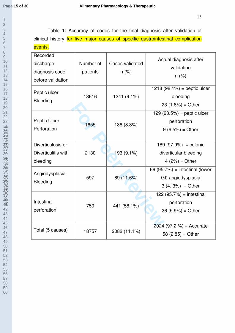
significant increase over the same time period. Validation of codes showed >
90% accuracy for the 5 GI complications (Table 1). Incidence rates were
adjusted according to the confirmation obtained with the manual chart review.
Based on the validation process of the 5 diagnostic codes identified in
2082 cases, 53.7% of intestinal perforations were located in the large bowel and
39% in the small bowel, whereas the remaining 7.2% were ascribed to the lower
GI tract without further site specification. Validation of codes for peptic ulcer
perforation showed that 6.5% of cases were indeed intestinal perforations.
Among cases coded as diverticular bleeding, the validation process showed
that 1% of cases were actually upper GI bleeding events, and another 1% were
unconfirmed events. Among those with angiodysplasia, 2.9% were upper GI
bleeding events and in 1.4% of cases the source could not be ascribed to any
cause. Finally, among peptic ulcer bleeds, only 1.5% were lower GI bleeding
events and in 0.3% of cases the source could not be identified.
The age and gender distribution was markedly different across the 5
causes of hospitalization (Table 2). Patients with bleeding from the colonic
diverticuli or angiodysplasia were older than those with other causes for
hospitalization, whereas males were predominant among those with peptic ulcer
perforation and bleeding. Very similar results were found in the validation
random sample (data not shown).
Page 7 of 30 Alimentary Pharmacology & Therapeutic
123456789101112131415161718192021222324252627282930313233343536373839404142434445464748495051525354555657585960
peer
-006
0596
0, v
ersi
on 1
- 5
Jul 2
011

For Peer Review
8
Overall, the case fatality rates were higher for perforation than for
bleeding events (intestinal perforation > peptic ulcer perforation > bleeding
angiodysplasia > peptic ulcer bleeding > diverticular bleeding). Overall mortality
trends did not change over time during the period studied for intestinal or peptic
ulcer perforation or angiodysplasia or diverticular bleeding (specific information
concerning these mortality trends can be seen on-line in “Supporting
Information Table 1”).
Severity of events and drug use
The review of charts provided an opportunity to collect a number of
variables that described the severity of the different types of events. The case
fatality rate was similar to that reported above in the overall sample. The length
of hospitalization was longer in patients with perforation than for those with
bleeding events, and the weight of DRG (an indirect measure of hospitalization
costs for each type of event) was higher for complications in the lower GI tract
when compared with those from the upper GI tract. The number of co-
morbidities was greater in patients with colonic diverticular bleeding and
angiodysplasia bleeding compared with the other type of lesions studied. As
expected, hemoglobin decline and the number of blood units transfused were
higher in patients with bleeding events than in those with perforations (specific
information concerning the severity of events based on chart validation can be
seen on-line in “Supporting Information Table 2”).
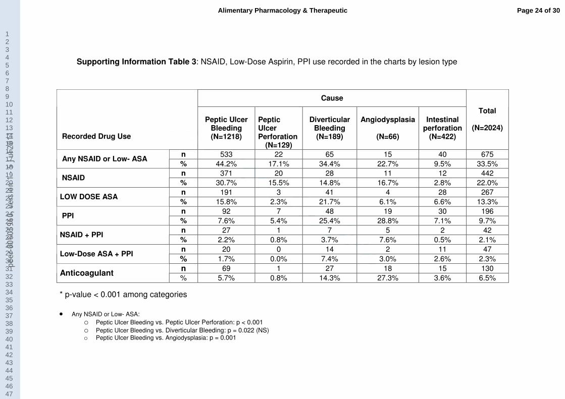
Detailed recorded drug use concerning nonsteroidal anti-inflammatory
drugs (NSAIDs), proton pump inhibitors (PPI), and anti-platelet agents can be
seen on-line in “Supporting Information Table 3”. Peptic ulcer and diverticular
colonic bleeding had the highest NSAID/aspirin drug use compared with cases
of perforation and even bleeding from GI angiodysplasia. PPI use was more
frequently recorded in patients with colonic and angiodysplasia bleeding events.
Recorded anticoagulant use was high in patients with diverticular bleeding and
in patients with angiodysplasia.
Page 8 of 30Alimentary Pharmacology & Therapeutic
123456789101112131415161718192021222324252627282930313233343536373839404142434445464748495051525354555657585960
peer
-006
0596
0, v
ersi
on 1
- 5
Jul 2
011

For Peer Review
9
Discussion
This study shows that hospitalizations due to both peptic ulcer bleeding
and perforations progressively and importantly decreased from 1996 to 2005.
These trends were not observed for other types of bleeding or perforation
events from the lower GI tract. In fact, we found that the incidences of colonic
diverticular bleeding or angiodysplasia are increasing, whereas the rate of
intestinal perforation remains virtually stable with a slight increase over the
study period.
There is wide agreement that hospitalizations due to uncomplicated
peptic ulcer are decreasing over time (1,2), but there were discrepancies
between hospitalizations on rates due to complicated peptic ulcers. Our results
agree with data from Sweden (14,15) and the USA (8) showing that
hospitalizations due to peptic ulcer bleeding are decreasing. However, these
data are not in agreement with reports from other European countries (3, 4, 5,
6,16), which show either no decrease or even an increase in hospitalizations for
this type of event. The reasons for these discrepancies are unclear, since a
decrease of H. pylori infection among the population, increasing H. pylori
eradication therapy, and increasing PPI use across Europe should be
accompanied by a progressive decrease in peptic ulcer complications (14, 15,
17). The variability in the use of low-dose aspirin (ASA) and gastro-protective
treatments between countries may partly explain these differences (18,19), but
our study and those reporting decreasing peptic ulcer bleeding rates collected
data from more recent years and were population-based (7, 8).
Our data also show very low rates of mortality due to peptic ulcer
bleeding, which is consistent with other studies (8, 14, 15). The lack of
improvement in case fatality rates is usually linked to a progressively aging
population with increasing numbers of co-morbidities, despite data that show
better bleeding management strategies associated with reduction in the risk of
rebleeding and a reduced need for surgery (20). In any case, our data agree
with a recent USA report (8) that shows an overall decrease (absolute numbers)
Page 9 of 30 Alimentary Pharmacology & Therapeutic
123456789101112131415161718192021222324252627282930313233343536373839404142434445464748495051525354555657585960
peer
-006
0596
0, v
ersi
on 1
- 5
Jul 2
011

For Peer Review
10
in in-hospital deaths linked to GI bleeding diagnoses between 1998 and 2006,
owing to a decrease in the number of hospitalizations.
Time trends on peptic ulcer perforation are rarely reported and those
available show no change or small changes overtime (9, 10, 21). Here we
report a clearly decreasing trend (∼ 50%) in peptic ulcer perforation from 1996
to 2005, which is consistent with a decrease in the overall incidence of peptic
ulcer and peptic ulcer complications. The decrease in incidence of
hospitalizations owing to peptic ulcer perforation was not associated with a
decrease in case fatality rates over this time.
Other studies have reported time trends for peptic ulcer bleeding or
perforation, but not other sources of GI bleeding or perforation. In our first
overall report of this study (7), we pointed out that the ratio of hospitalizations
for upper vs. lower GI complications has changed from a 4:1 to 1.4:1. Here we
report that two common reasons for hospitalizations, colonic diverticular and
angiodysplasia bleeding, are more frequent today than 10 years ago. A
progressively aging population and the increasing use of NSAIDs and low-dose
ASA may explain, at least in part, these results. (22). PPI use was more
frequently recorded in patients hospitalized with diverticular bleeding and
bleeding due to angiodysplasia than in those with peptic ulcer bleeding. This
could support the concept that PPI use is associated with the prevention of
upper GI but not lower GI complications, but age and its accompanying greater
number of co-morbidities could also explain the observed higher prevalence of
PPI use. Eventually, it must be noted that anticoagulant use, a growing clinical
practice, is especially associated with bleeding events and overall with
diverticular and angiodysplasia bleeding.
Trends on intestinal perforation are rarely reported. Here we report
overall intestinal (small and large bowel) perforation rates and found that these
rates remained stable over time, although with a numerical tendency to increase
in agreement with the data reported from the UK on diverticular perforation (23).
Page 10 of 30Alimentary Pharmacology & Therapeutic
123456789101112131415161718192021222324252627282930313233343536373839404142434445464748495051525354555657585960
peer
-006
0596
0, v
ersi
on 1
- 5
Jul 2
011

For Peer Review
11
Our study has strengths and limitations. A strength is that we carried out
an extensive validation of events. This is especially important for intestinal
perforation, and diverticular and bleeding from angiodysplasia lesions. Contrary
to peptic ulcer bleeding and perforation, these have rarely been reported in the
literature. Therefore, these ICD-9 codes have rarely been validated in
observational studies. This is of major importance since the diagnostic process
of diseases from the lower GI tract is more complex than those used in the
upper GI tract.
The study also has limitations. First, undefined codes such as “GI
bleeding” may include patients with both peptic ulcer bleeding and bleeding
from diverticular disease and angiodysplasia lesions. This means that the
reported incidence and trends reflect minimum rates, since it is possible that a
proportion of patients with the events studied here may not be counted because
of misclassification under undefined codes. In our previous report, we observed
that this proportion was constant over time (7). Another limitation refers to data
on drug use, which relies on data recorded in charts and are subject to reporting
bias in the clinical history. Ascertainment of NSAID, ASA, and PPI use may be
more frequently searched in patients with peptic ulcer complications than in
diverticular or angiodysplasia bleeding events. Finally, our mortality rates refer
to in-hospital mortality, as we could not provide 30-day mortality, since most
patients were discharged from hospital before this window of time.
In summary, our study shows that, over the past decade, there has been
a significant decrease in hospitalization rates for peptic ulcer bleeding and
perforation, but an increase for colonic diverticular and angiodysplasia bleeding,
with virtually no change in intestinal perforation. These data show a clear
change in the incidence of hospitalizations due to GI complications over time,
with upper GI events decreasing and lower GI events increasing. Since the
severity of these events are very different, these data should be of interest for
improving clinical practice in terms of preventive strategies and to better
address the increasing prevalence of lower GI events, specifically the increase
in colonic diverticular and angiodysplasia bleeding.
Page 11 of 30 Alimentary Pharmacology & Therapeutic
123456789101112131415161718192021222324252627282930313233343536373839404142434445464748495051525354555657585960
peer
-006
0596
0, v
ersi
on 1
- 5
Jul 2
011

For Peer Review
12
References:
1. Sung JJ, Kuipers EJ, El-serag HB, Systematic review. The global incidence and
prevalence of peptic ulcer disease. Aliment Pharmacol Ther 2009; 29: 938-46.
2. Sonnenberg A. Time trends of ulcer mortality in Europe. Gastroenterology.
2007 Jun;132(7):2320-7. Epub 2007 Apr 14.
3. Post PN, Kuipers EJ, Meijer GA. Declining incidence of peptic ulcer but not of
its complications: a nation-wide study in The Netherland. Aliment Pharmacol
Ther. 2006 Jun 1;23(11):1587-93.
4. Ohmann C, Imhof M, Ruppert C, et al. Time-trends in the epidemiology of
peptic ulcer bleeding. Scand J Gastroenterol 2005; 40: 914–20.
5. Kang JY, Elders A, Majeed A, Maxwell JD, Bardhan KD. Recent trends in
hospital admissions and mortality rates for peptic ulcer in Scotland 1982–2002.
Aliment Pharmacol Ther 2006; 24: 65–79.
6. Paimela H, Paimela L, Myllykangas-Luosujarvi R, Kivilaakso E. Current features
of peptic ulcer disease in Finland: incidenceof surgery, hospital admissions and
mortality for the disease during the past twenty-five years. Scand J
Gastroenterol 2002; 37: 399–403.
7. Lanas A, García-Rodríguez LA, Polo-Tomás M, Ponce M, Alonso-Abreu I,
Perez-Aisa MA, Perez-Gisbert J, Bujanda L, Castro M, Muñoz M, Rodrigo L,
alvet X, Del-Pino D, Garcia S. Time trends and impact of upper and lower
gastrointestinal bleeding and perforation in clinical practice. Am J Gastroenterol.
2009 Jul;104(7):1633-41.
8. Zhao, Y., and Encinosa, W. Hospitalizations for Gastrointestinal Bleeding in
1998 and 2006. HCUP Statistical Brief #65. December, 2008. Agency for
Healthcare Research and Quality, Rockville, MD. http://www.hcup-
us.ahrq.gov/reports/statbriefs/sb65.pdf
9. Bardhan KD, Royston C. Time, change and peptic ulcer disease in Rotherham,
UK. Dig Liver Dis. 2008 Jul;40(7):540-6.
10. Paimela H, Oksala NK, Kivilaakso E. Surgery for peptic ulcer today. A study on
the incidence, methods and mortality in surgery for peptic ulcer in Finland
between 1987 and 1999. Dig Surg. 2004;21(3):185-91.
11. Lanas A, Perez-Aisa MA, Feu F, Ponce J, Saperas E, Santolaria S, Rodrigo L,
Balanzo J, Bajador E, Almela P, Navarro JM, Carballo F, Castro M, Quintero E;
Investigators of the Asociación Española de Gastroenterología (AEG). A
nationwide study of mortality associated with hospital admission due to severe
gastrointestinal events and those associated with nonsteroidal antiinflammatory
drug use. Am J Gastroenterol. 2005 Aug;100(8):1685-93.
Page 12 of 30Alimentary Pharmacology & Therapeutic
123456789101112131415161718192021222324252627282930313233343536373839404142434445464748495051525354555657585960
peer
-006
0596
0, v
ersi
on 1
- 5
Jul 2
011

For Peer Review
13
12. Steven Harris. Medicare Severity-Refined DRGs: An Improved System.
www.irp.com.
13. Raiford DS, Pérez Gutthann S, García Rodríguez LA. Positive Predictive value
of ICD-9 codes in the identification of cases of complicated peptic ulcer disease
in the Saskatchewan hospital automated database. Epidemiology 1996; 7:101-
104.
14. Sadic J, Borgström A, Manjer J, Toth E, Lindell G. Bleeding peptic ulcer - time
trends in incidence, treatment and mortality in Sweden. Aliment Pharmacol
Ther. 2009 Aug 15;30(4):392-8.
15. Hermansson M, Ekedahl A, Ranstam J, Zilling T. Decreasing incidence of
peptic ulcer complications after the introduction of the proton pump inhibitors, a
study of the Swedish population from 1974-2002. BMC Gastroenterol. 2009 Apr
20;9:25.
16. Bak Andersen I, Bonnevie O, Jorgensen T, Sorensen T. Time Trends for peptic
ulcer disease in Danmark, 1981-1993. Analysis of hospitalization register and
mortalitydata. Scand J Gastroenterol 1998; 33: 260–6.
17. Pérez-Aisa MA, Del Pino D, Siles M, Lanas A. Clinical trends in ulcer diagnosis
in a population with high prevalence of Helicobacter pylori infection. Aliment
Pharmacol Ther. 2005 Jan 1;21(1):65-72.
18. Lanas A, Esplugues JV, Zapardiel J, Sobreviela E. Education-based approach
to addressing non-evidence-based practice in preventing NSAID-associated
gastrointestinal complications. World J Gastroenterol. 2009 Dec
21;15(47):5953-9.
19. Sturkenboom MC, Burke TA, Dieleman JP, Tangelder MJ, Lee F, Goldstein JL.
Underutilization of preventive strategies in patients receiving NSAIDs.
Rheumatology (Oxford). 2003 Nov;42 Suppl 3:iii23-31.
20. Barkun AN, Martel M, Toubouti Y, Rahme E, Bardou M. Endoscopic hemostasis
in peptic ulcer bleeding for patients with high-risk lesions: a series of meta-
analyses. Gastrointest Endosc. 2009 Apr;69(4):786-99.
21. Bardhan KD, Williamson M, Royston C, Lyon C. Admission rates for peptic
ulcer in the trent region, UK, 1972--2000. changing pattern, a changing
disease? Dig Liver Dis. 2004 Sep;36(9):577-88.
22. Lanas A, García-Rodríguez LA, Arroyo MT, Gomollón F, Feu F, González-
Pérez A, Zapata E, Bástida G, Rodrigo L, Santolaria S, Güell M, de Argila CM,
Quintero E, Borda F, Piqué JM; Asociación Española de Gastroenterología.
Risk of upper gastrointestinal ulcer bleeding associated with selective cyclo-
oxygenase-2 inhibitors, traditional non-aspirin non-steroidal anti-inflammatory
Page 13 of 30 Alimentary Pharmacology & Therapeutic
123456789101112131415161718192021222324252627282930313233343536373839404142434445464748495051525354555657585960
peer
-006
0596
0, v
ersi
on 1
- 5
Jul 2
011

For Peer Review
14
drugs, aspirin and combinations. Gut. 2006 Dec;55(12):1731-8. Epub 2006 May
10.
23. Humes DJ, Solaymani-Dodaran M, Fleming KM, Simpson J, Spiller RC, West J.
A population-based study of perforated diverticular disease incidence and
associated mortality. Gastroenterology. 2009 Apr;136(4):1198-205.
Page 14 of 30Alimentary Pharmacology & Therapeutic
123456789101112131415161718192021222324252627282930313233343536373839404142434445464748495051525354555657585960
peer
-006
0596
0, v
ersi
on 1
- 5
Jul 2
011
For Peer Review
15
Table 1: Accuracy of codes for the final diagnosis after validation of
clinical history for five major causes of specific gastrointestinal complication
events.
Recorded
discharge
diagnosis code
before validation
Number of
patients
Cases validated
n (%)
Actual diagnosis after
validation
n (%)
Peptic ulcer
Bleeding 13616 1241 (9.1%)
1218 (98.1%) = peptic ulcer
bleeding
23 (1.8%) = Other
Peptic Ulcer
Perforation 1655 138 (8.3%)
129 (93.5%) = peptic ulcer
perforation
9 (6.5%) = Other
Diverticulosis or
Diverticulitis with
bleeding
2130 193 (9.1%)
189 (97.9%) = colonic
diverticular bleeding
4 (2%) = Other
Angiodysplasia
Bleeding 597 69 (11.6%)
66 (95.7%) = intestinal (lower
GI) angiodysplasia
3 (4. 3%) = Other
Intestinal
perforation 759 441 (58.1%)
422 (95.7%) = intestinal
perforation
26 (5.9%) = Other
Total (5 causes) 18757 2082 (11.1%) 2024 (97.2 %) = Accurate
58 (2.85) = Other
Page 15 of 30 Alimentary Pharmacology & Therapeutic
123456789101112131415161718192021222324252627282930313233343536373839404142434445464748495051525354555657585960
peer
-006
0596
0, v
ersi
on 1
- 5
Jul 2
011

For Peer Review
16
Table 2: Age and gender distribution by lesion type.
a)
n Age
(Mean, SD)
Male Gender
(n, %)
Peptic ulcer bleeding 13616 63.50 (16.7) 9615 (70.6%)
Peptic ulcer perforation 1655 55.13 (19.1) 1050 (69.5%)
Diverticulosis or
Diverticulitis with bleeding 2130 75.91 (10.2) 950 (44.6%)
Angiodysplasia Bleeding 597 73.32 (11.6) 333 (55.9%)
Intestinal perforation 759 63.37 (18.6) 414 (54.5%)
Mean comparison among categories: p-value < 0.001
Page 16 of 30Alimentary Pharmacology & Therapeutic
123456789101112131415161718192021222324252627282930313233343536373839404142434445464748495051525354555657585960
peer
-006
0596
0, v
ersi
on 1
- 5
Jul 2
011

For Peer Review
17
Figure Legends Figure 1: Estimated number of peptic ulcer, colonic diverticular, and
angiodysplasia bleeding events per 100,000 person-years based on the
adjudication of events in the validation process. Regression coefficients of
temporal trends were: R2 = 0.944 (p<0.0001) for peptic ulcer bleeding rates; R2
= 0.443 (p = 0.03) for colonic diverticular bleeding rates; R2 = 0.715 (p = 0.002)
for angiodysplasia bleeding rates.
Figure 2: Estimated number of perforations per 100,000 person-years based on
the adjudication of events in the validation process. Regression coefficients of
temporal trends were: R2 = 0.410 (p = 0.04) for peptic ulcer perforation; R2 =
0.091 (p = 0.395) for intestinal perforation rates.
Page 17 of 30 Alimentary Pharmacology & Therapeutic
123456789101112131415161718192021222324252627282930313233343536373839404142434445464748495051525354555657585960
peer
-006
0596
0, v
ersi
on 1
- 5
Jul 2
011

For Peer Review
18
Figure 1:
GI BLEEDING RATES / 100,000 people
0
10
20
30
40
50
60
1996 1997 1998 1999 2000 2001 2002 2003 2004 2005
Peptic Ulcer Bleeding
Colonic Diverticular
Bleeding
Angiodysplasia
Page 18 of 30Alimentary Pharmacology & Therapeutic
123456789101112131415161718192021222324252627282930313233343536373839404142434445464748495051525354555657585960
peer
-006
0596
0, v
ersi
on 1
- 5
Jul 2
011

For Peer Review
19
Figure 2:
GI PERFORATIONS RATES / 100,000 people
0
1
2
3
4
5
6
7
1996 1997 1998 1999 2000 2001 2002 2003 2004 2005
Peptic Ulcer
Perforations
Intestinal
perforation
Page 19 of 30 Alimentary Pharmacology & Therapeutic
123456789101112131415161718192021222324252627282930313233343536373839404142434445464748495051525354555657585960
peer
-006
0596
0, v
ersi
on 1
- 5
Jul 2
011

For Peer Review
20
Statements of interest:
Acknowledgements: The study was supported by Pfizer Inc. Pfizer had no role
in the conduct of the study, or the analysis or interpretation of the data.
Information concerning the role of authors in the study
Dr. Lanas and Dr Garcia Rodriguez designed the study. Dr. Lanas drafted the
manuscript with major contributions from Mónica Polo-Tomás and Dr Luis
Garcia Rodriguez. All the authors contributed to the collection of data and had
full access to the raw data set from the study, the results, the manuscript, and
made their own comments and contributions. Dr. Dolores Del-Pino designed the
data extraction process for the ICD-9 codes. Mónica Polo-Tomás introduced the
data in the database and, with Luis Alberto Garcia Rodriguez and Dr Lanas,
checked the quality of the data base. Mónica Polo-Tomás produced the outputs
and ran the statistical analysis with Dr. Garcia Rodriguez. Dr. Lanas acted as
guarantor of the submission.
Conflict of interest:
Angel Lanas is member of the adjudication Committee of the international
multicenter CONDOR trial, sponsored by Pfizer, and has received honoraria for
lecturing at Pfizer- and AstraZeneca-sponsored symposiums.
Xavier Calvet has received honoraria for participating in advisory boards and for
lecturing at AstraZeneca-sponsored symposiums. Javier P. Gisbert has
received honoraria for lecturing at AstraZeneca- and Pfizer-sponsored
symposiums. Luis Bujanda has received small honoraria for lecturing at Pfizer-
sponsored symposiums.
The other study authors have no conflicts of interest to report.
Page 20 of 30Alimentary Pharmacology & Therapeutic
123456789101112131415161718192021222324252627282930313233343536373839404142434445464748495051525354555657585960
peer
-006
0596
0, v
ersi
on 1
- 5
Jul 2
011

For Peer Review
Supporting Information Table 1: Time trends of case fatality by lesion type (n =18757). No statistical differences were found for any of the 5 types of lesions over time.
Year Mortality
1996 1997 1998 1999 2000 2001 2002 2003 2004 2005 Total
n 46 59 57 53 41 29 45 40 39 18 427/13616 Peptic Ulcer Bleeding % 2.5 3.5 3.2 3.2 3.1 2.2 4.0 3.7 4.2 1.9 3.1
n 19 19 16 21 21 16 17 15 10 10 164/1655 Peptic ulcer perforation % 9.5 8.2 8.9 11.4 13.5 10.0 10.3 10.6 7.6 9.2 9.9
n 0 3 6 5 5 6 5 4 5 4 43/2130 Diverticular Bleeding % .0 1.8 2.5 2.5 2.8 2.7 2.1 1.6 2.3 1.3 2.0
n 2 2 1 4 2 3 9 1 2 4 30/597 Angiodysplasia Bleeding % 6.7 5.7 2.4 11.1 4.5 4.6 9.1 1.3 2.8 4.0 5.0
n 11 22 29 24 28 24 22 20 22 30 232/759 Intestinal perforation % 21.2 36.1 36.3 30.8 30.1 28.2 25.9 30.3 31.0 34.1 30.6
Page 21 of 30 Alimentary Pharmacology & Therapeutic
123456789101112131415161718192021222324252627282930313233343536373839404142434445464748495051525354555657585960
peer
-006
0596
0, v
ersi
on 1
- 5
Jul 2
011

For Peer Review
Supporting Information Table 2. Severity of events using several measures, by type of lesion based on the chart validation.
Lesion N Mortality Hospital
Stay (days)
Number of diagnostic
tests performed
DRG Weight
Number of Comorbidities
Lowest Hemoglobin
level
Blood units transfused
Mean (%) 1.69 6.93 1.04 1.13 1.55 9.18 1.51 Peptic Ulcer Bleeding 1218
95% CI [0.96, 2.43] [6.54, 7,32] [1,03, 1,06] [1.09, 1.18] [1.50, 1.60] [9.05, 9.30] [1.40, 1.63]
Mean (%) 4.40 11.56 1.20 3.06 1.40 12.25 1.16 Peptic Ulcer Perforation 129
95% CI [0.10, 8.69] [8.74, 14.38] [1.04, 1.36] [2.52, 3.61] [1.23, 1.56] [11.65, 12.85] [0.25, 2.08]
Mean (%) 2.84 9.34 1.31 1.28 2.07 10.14 1.28 Diverticular Bleeding 189
95% CI [0.36, 5.32] [8.32, 10.36] [1.20, 1.41] [1.15, 1.41] [1.92, 2.22] [9.76, 10.53] [0.95, 1.61]
Mean (%) 1.56 8.84 1.34 1.17 2.70 8.31 2.02 Angiodysplasia 66
95% CI [0, 4.68] [6.58, 11.10] [1.04, 1.64] [1.06, 1.29] [2.41, 3.00] [7.78, 8.85] [1.54, 2.50]
Mean (%) 28.66 21.78 0.99 4.04 1.85 10.96 0.91 Intestinal perforation 422
95% CI [23.58, 33.75]
[19.21, 24.35]
[0.92, 1.05] [3.64, 4.43] [1.73, 1.97] [10.66, 11.27] [0.59, 1.24]
Comparisons among categories:
• Mortality: o Intestinal perforation vs. Any other lesion: p < 0.001 o Peptic Ulcer Bleeding vs. Peptic Ulcer Perforation: p = 0.363 (NS)
• Hospital stay: Peptic Ulcer Bleeding vs. Peptic Ulcer Perforation: p < 0.001 • Number of diagnostic tests performed:
o Angiodysplasia vs. Peptic Ulcer Bleeding: p < 0.001
o Angiodysplasia vs. Peptic Ulcer Perforation: p = 0.019 (NS) o Angiodysplasia vs. Diverticular Bleeding: p = 0.905 (NS) o Angiodysplasia vs. Intestinal perforation: p < 0.001 o Diverticular Bleeding vs. Peptic Ulcer Bleeding: p < 0.001 o Diverticular Bleeding vs. Peptic Ulcer Perforation: p = 0.012 (NS) o Diverticular Bleeding vs. Intestinal perforation: p < 0.001
• Number of Comorbidities:
Page 22 of 30Alimentary Pharmacology & Therapeutic
123456789101112131415161718192021222324252627282930313233343536373839404142434445464748495051525354555657585960
peer
-006
0596
0, v
ersi
on 1
- 5
Jul 2
011

For Peer Review
o Angiodysplasia vs. Any other lesion: p < 0.001 o Diverticular Bleeding vs. Any other lesion: p < 0.001
• Lowest Hemoglobin level: o Angiodysplasia vs. Peptic Ulcer Bleeding: p = 0.012 (NS) o Angiodysplasia vs. Peptic Ulcer Perforation: p < 0.001 o Angiodysplasia vs. Diverticular Bleeding: p < 0.001 o Angiodysplasia vs. Intestinal perforation: p < 0.001 o Peptic Ulcer Bleeding vs. Peptic Ulcer Perforation: p < 0.001
• Blood units transfused: o Angiodysplasia vs. Peptic Ulcer Bleeding: p = 0.226 (NS) o Angiodysplasia vs. Peptic Ulcer Perforation: p = 0.035 o Angiodysplasia vs. Diverticular Bleeding: p = 0.087 (NS) o Angiodysplasia vs. Intestinal perforation: p = 0.002 o Peptic Ulcer Bleeding vs. Peptic Ulcer Perforation: p = 0.252 (NS)
Page 23 of 30 Alimentary Pharmacology & Therapeutic
123456789101112131415161718192021222324252627282930313233343536373839404142434445464748495051525354555657585960
peer
-006
0596
0, v
ersi
on 1
- 5
Jul 2
011
For Peer Review
Supporting Information Table 3: NSAID, Low-Dose Aspirin, PPI use recorded in the charts by lesion type
Cause
Recorded Drug Use
Peptic Ulcer Bleeding (N=1218)
Peptic Ulcer Perforation
(N=129)
Diverticular Bleeding (N=189)
Angiodysplasia
(N=66)
Intestinal perforation
(N=422)
Total
(N=2024)
n 533 22 65 15 40 675 Any NSAID or Low- ASA
% 44.2% 17.1% 34.4% 22.7% 9.5% 33.5%
n 371 20 28 11 12 442 NSAID
% 30.7% 15.5% 14.8% 16.7% 2.8% 22.0%
n 191 3 41 4 28 267 LOW DOSE ASA
% 15.8% 2.3% 21.7% 6.1% 6.6% 13.3%
n 92 7 48 19 30 196 PPI
% 7.6% 5.4% 25.4% 28.8% 7.1% 9.7%
n 27 1 7 5 2 42 NSAID + PPI
% 2.2% 0.8% 3.7% 7.6% 0.5% 2.1%
n 20 0 14 2 11 47 Low-Dose ASA + PPI
% 1.7% 0.0% 7.4% 3.0% 2.6% 2.3%
n 69 1 27 18 15 130 Anticoagulant
% 5.7% 0.8% 14.3% 27.3% 3.6% 6.5%
* p-value < 0.001 among categories
• Any NSAID or Low- ASA: o Peptic Ulcer Bleeding vs. Peptic Ulcer Perforation: p < 0.001 o Peptic Ulcer Bleeding vs. Diverticular Bleeding: p = 0.022 (NS) o Peptic Ulcer Bleeding vs. Angiodysplasia: p = 0.001
Page 24 of 30Alimentary Pharmacology & Therapeutic
123456789101112131415161718192021222324252627282930313233343536373839404142434445464748495051525354555657585960
peer
-006
0596
0, v
ersi
on 1
- 5
Jul 2
011

For Peer Review
o Peptic Ulcer Bleeding vs. Intestinal perforation: p < 0.001
• NSAID: o Peptic Ulcer Bleeding vs. Peptic Ulcer Perforation: p < 0.001 o Peptic Ulcer Bleeding vs. Diverticular Bleeding: p < 0.001 o Peptic Ulcer Bleeding vs. Angiodysplasia: p = 0.021 (NS)
o Peptic Ulcer Bleeding vs. Intestinal perforation: p < 0.001
• Low- ASA: o Diverticular Bleeding vs. Peptic Ulcer Bleeding: p = 0.080 (NS) o Diverticular Bleeding vs. Peptic Ulcer Perforation: p < 0.001 o Diverticular Bleeding vs. Angiodysplasia: p = 0.004
o Diverticular Bleeding vs. Intestinal perforation: p < 0.001
• PPI: o Diverticular Bleeding vs. Peptic Ulcer Bleeding: p < 0.001 o Diverticular Bleeding vs. Peptic Ulcer Perforation: p < 0.001 o Diverticular Bleeding vs. Angiodysplasia: p = 0.813 (NS)
o Diverticular Bleeding vs. Intestinal perforation: p < 0.001
• NSAID + PPI: o Angiodysplasia vs. Peptic Ulcer Bleeding: p = 0.008 o Angiodysplasia vs. Peptic Ulcer Perforation: p = 0.005 o Angiodysplasia vs. Diverticular Bleeding: p = 0.128 (NS)
o Angiodysplasia vs. Intestinal perforation: p = 0.001
• Low- ASA + PPI: o Diverticular Bleeding vs. Peptic Ulcer Bleeding: p < 0.001 o Diverticular Bleeding vs. Peptic Ulcer Perforation: p < 0.001 o Diverticular Bleeding vs. Angiodysplasia: p = 0.127 (NS)
o Diverticular Bleeding vs. Intestinal perforation: p = 0.001
• Anticoagulants
Page 25 of 30 Alimentary Pharmacology & Therapeutic
123456789101112131415161718192021222324252627282930313233343536373839404142434445464748495051525354555657585960
peer
-006
0596
0, v
ersi
on 1
- 5
Jul 2
011

For Peer Review
o Peptic Ulcer Bleeding vs. Peptic Ulcer Perforation: p = 0.103 (NS) o Peptic Ulcer Bleeding vs. Diverticular Bleeding: p < 0.001 o Peptic Ulcer Bleeding vs. Angiodysplasia: p < 0.001
o Peptic Ulcer Bleeding vs. Intestinal perforation: p = 0.376 (NS)
Page 26 of 30Alimentary Pharmacology & Therapeutic
123456789101112131415161718192021222324252627282930313233343536373839404142434445464748495051525354555657585960
peer
-006
0596
0, v
ersi
on 1
- 5
Jul 2
011

For Peer Review
Page 27 of 30 Alimentary Pharmacology & Therapeutic
123456789101112131415161718192021222324252627282930313233343536373839404142434445464748495051525354555657585960
peer
-006
0596
0, v
ersi
on 1
- 5
Jul 2
011

For Peer Review
Reply to comments from the Editor and Referees
1. EDITOR'S COMMENTS TO AUTHOR:
a) Please can you work on the standard of English - it is sub-optimal.
Reply: The manuscript was submitted to a professional English medical editing service for
correction and revision of the English language (www.sanfranciscoedit.com).
b) Please would you make Table 3 to be Supporting Information Table 1, and label the two
duplicate Tables 4 & 5 as Supporting Information Tables 2 and 3. [Supporting Information is
available to readers on-line. The important / significant results should be mentioned in the text,
and the supporting information referred to in the text. This is explained in the Author
Guidelines at www.APandT.org or at http://authorservices.wiley.com/bauthor/suppmat.asp ].
Reply: We made this change and incorporated the main data from Tables 3, 4, and 5 into the
text. These Tables have been converted to Supporting Information Tables 1, 2 and 3.
c) Can you really look at your paper, and make it clear what's new - in addition to your already
well-cited earlier paper?
Reply: We reworked the introduction and discussion to clarify what it is new in this manuscript.
Below is the list of the new information provided:
a) time trends for hospitalizations due to peptic (gastric and duodenal) ulcer bleeding and
its contrast with peptic ulcer perforation
b) time trends for intestinal perforation
c) time trends for colonic diverticular bleeding
d) time trends for bleeding due to angiodysplasia
e) mortality rates of these events
f) description of severity characteristics of these lesions
g) recorded drug use for each of these entities. We outlined the association between
anticoagulant use with both diverticular bleeding and angiodysplasia, which we believe
is of utmost importance.
Reviewer: 1
We thank referee 1 for saying that we adequately addressed the prior comments.
Concerning the new comments:
a) Abstract
The aim does not make sense: “To report temporal changes in the incidence of five major
cause-specific gastrointestinal (GI) bleeding and perforation.” Do the authors mean the
incidence of five major cause-specific gastrointestinal (GI) events?
Reply: We revised the sentence accordingly.
Introduction
Page 28 of 30Alimentary Pharmacology & Therapeutic
123456789101112131415161718192021222324252627282930313233343536373839404142434445464748495051525354555657585960
peer
-006
0596
0, v
ersi
on 1
- 5
Jul 2
011
For Peer Review
1. The first sentence is vague, and could mean anything: “Gastrointestinal complications are
major causes of GI hospitalization.” In any case, they need to make it clear that GI is an
abbreviation of gastrointestinal in this sentence.
Reply: We revised the sentence accordingly, and defined the abbreviation. We believe the
sentence specifies a clear and important fact, which is essential to our paper.
2. The statement on two occasions in the Introduction that “…no cause specific analyses were
performed” as part of their previous study (reference 7), and that “…trends for the specific
cause of lower GI complications were not reported” as part of their previous study again leaves
me with the feeling that this is a post hoc analysis of data in an attempt to obtain another
publication from the same piece of work. Why weren’t these analyses performed in the prior
publication if, as the authors presumably believe, this is important information?
Reply: We now make clear that this piece of information was not reported. As we responded to
the editor, the data presented are new and it well know that databases with extensive data can
provide important information that cannot always be included in a single publication.
Methods
1. Page 6, line 42/43. I think the authors mean that “…gastroenterologists or trained GI
residents with experience of these types of studies.” rather than “…on this type of studies.”
2. Page 6, line 56/57. There is now no mention whatsoever of what DRG is an abbreviation of.
This needs to be added.
Reply: We changed the text in accordance with these suggestions.
Results
1. Page 8, lines 7 to 10 don’t make sense: “…information provided by the participant hospitals
showed and statistically significant decrease…” The authors mean “…a statistically
significant…”
2. Page 9, lines 19/20: “…length of hospitalization were higher…” should be “…was higher…”
3. Page 9, lines 24/25: “…from the lower GI tract when compared those…” should be “…when
compared with those…”
Reply: The text was corrected by a professional editing service to improve the readability of the
manuscript.
Discussion
The authors state on page 11, lines 8 to 11 that: “Time trends on peptic ulcer perforation are rarely
reported and those available show no change or small changes overtime.” They reference their previous
study here. This makes it seem as though the results of the current study are at odds with those of the
previous study, yet how can this be the case, as they are derived from the same dataset?!
Reply: The referee is right. We included this reference because it reported overall trends, but
clearly it should not be there. We have removed reference 7.
Reviewer: 2
Comments for Transmission to the Authors
None
Page 29 of 30 Alimentary Pharmacology & Therapeutic
123456789101112131415161718192021222324252627282930313233343536373839404142434445464748495051525354555657585960
peer
-006
0596
0, v
ersi
on 1
- 5
Jul 2
011

For Peer Review
Reply: We thank reviewer 2 for being comfortable with our previous reply.
Page 30 of 30Alimentary Pharmacology & Therapeutic
123456789101112131415161718192021222324252627282930313233343536373839404142434445464748495051525354555657585960
peer
-006
0596
0, v
ersi
on 1
- 5
Jul 2
011


