Embed Size (px)
Citation preview
The Dorso-medial Visual Stream: from Neural
Activation to Sensorimotor Interaction
Eris Chinellatoa,∗, Beata J. Grzyba, Nicoletta Marzocchib, Annalisa Boscob,Patrizia Fattorib, Angel P. del Pobila,c
aRobotic Intelligence Lab
Universitat Jaume I, Castellon de la Plana, SpainbDipartimento di Fisiologia Umana e Generale
Universita di Bologna, ItalycDepartment of Interaction Science
Sungkyunkwan University, Seoul, South Korea
Abstract
The posterior parietal cortex of primates, and more exactly areas of thedorso-medial visual stream, are able to encode the peripersonal space of asubject in a way suitable for gathering visual information and contextuallyperform purposeful gazing and arm reaching movements. Such sensorimotorknowledge of the environment is not explicit, but rather emerges throughthe interaction of the subject with nearby objects. In this work, single-celldata regarding the activation of primate dorso-medial stream neurons duringgazing and reaching movements is studied, with the purpose of discoveringmeaningful pattern useful for modeling purposes. The outline of a model ofthe mechanisms which allow humans and other primates to build dynamicalrepresentations of their peripersonal space through active interaction withnearby objects is proposed, and a detailed description of how to employ theresults of the data analysis in the model is offered. The application of themodel to robotic systems will allow artificial agents to improve their skills intheir exploration of the nearby space, and will at the same time constitute away to validate modeling assumptions.
Keywords: single-cell experiments, visuomotor interaction, eye-handcoordination, basis function networks, bio-inspired systems
∗Corresponding authorEmail address: [email protected] (Eris Chinellato)
Preprint submitted to Neurocomputing July 2, 2010
1. Introduction
Humans and other primates possess a superior ability in dealing with ob-jects in their surrounding space. Neuroscience research showed that areas ofthe primate posterior parietal cortex are able to represent the surroundingenvironment in a way suitable for achieving complicate coordinated move-ments of eyes and arms. It appears that the reference frames required toencode for visual information and for planning and monitoring the move-ments of different effectors are maintained contextually by population ofneurons interacting through gain field effects. The way some areas of thebrain perform reference frame transformations for sensorimotor interactionsin the peripersonal space constitute the neuroscientific basis of this work.
The outline of a model toward the achievement of an integrated objectrepresentation is proposed in this paper, based on the active interaction of thesubject with its surrounding environment. Particular importance has beengiven to the use of binocular data and proprioceptive information regardingeye position, critical in the transformation of sensory data into appropriatemotor signals. The model is especially focused on the process of gazing andreaching toward nearby objects seen as visual targets. The second part ofthis work focuses on the study of the neuroscience data useful for the imple-mentation of different visuomotor functions, and the way the insights offeredby such data can be exploited in the model development. Data regardingexperiments with primates on gazing and reaching movements, and referredto the posterior parietal area of the visual cortex V6A, are analyzed and dis-cussed, with the goal of defining a detailed modeling of cortical mechanismsduring the interaction of a subject with his/her environment. Hypotheseson how sensorimotor interaction abilities are achieved by employing deter-minate neural populations are advanced, and ways to test them with furtherexperiments and with computational methods are proposed.
This work constitutes the first step toward a more complete attempt ofproviding a robot with advanced capabilities in its purposeful interactionwith the environment, through active exploration and multimodal integra-tion of the different stimuli it receives. Performing purposeful, flexible andreliable vision-based reaching toward nearby objects is a fundamental skill topursue in order to achieve such ambitious goal. On the other hand, exper-iments with the robot will constitute a novel, resourceful validation ground
2
for computational models and neuroscience hypotheses.
2. Reaching and Grasping in Primates
The visual cortex of the primate brain is organized in two parallel chan-nels, called “dorsal” and “ventral” streams. The former elaborates visualdata with the main purpose of endowing the subject with the ability of in-teracting with his/her environment, and its tasks are often synthesized as“vision for action”. The latter is dedicated to object recognition and concep-tual processing, and thus performs “vision for perception”. Although a tightinteraction between the two streams is necessary for most everyday tasks,dorsal stream areas are more strictly related to the planning and monitoringof reaching and grasping actions [1]. In fact, dorsal visual analysis is drivenby the absolute dimension and location of target objects, requiring continu-ous transformations from retinal data to an effector-based frame of reference.Such transformations are very likely executed by a contextual coding of theperipersonal space in different reference frames simultaneously, very likely byarea V6A and its neighbours.
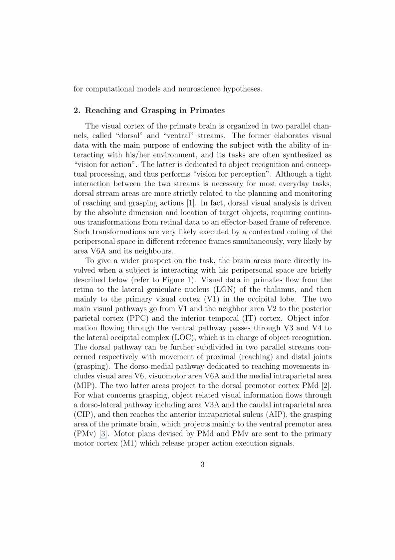
To give a wider prospect on the task, the brain areas more directly in-volved when a subject is interacting with his peripersonal space are brieflydescribed below (refer to Figure 1). Visual data in primates flow from theretina to the lateral geniculate nucleus (LGN) of the thalamus, and thenmainly to the primary visual cortex (V1) in the occipital lobe. The twomain visual pathways go from V1 and the neighbor area V2 to the posteriorparietal cortex (PPC) and the inferior temporal (IT) cortex. Object infor-mation flowing through the ventral pathway passes through V3 and V4 tothe lateral occipital complex (LOC), which is in charge of object recognition.The dorsal pathway can be further subdivided in two parallel streams con-cerned respectively with movement of proximal (reaching) and distal joints(grasping). The dorso-medial pathway dedicated to reaching movements in-cludes visual area V6, visuomotor area V6A and the medial intraparietal area(MIP). The two latter areas project to the dorsal premotor cortex PMd [2].For what concerns grasping, object related visual information flows througha dorso-lateral pathway including area V3A and the caudal intraparietal area(CIP), and then reaches the anterior intraparietal sulcus (AIP), the graspingarea of the primate brain, which projects mainly to the ventral premotor area(PMv) [3]. Motor plans devised by PMd and PMv are sent to the primarymotor cortex (M1) which release proper action execution signals.
3
V1
PMv AIP
LOC
CIP
V2 V3
V3A
V4
M1
PFC
V6
V6A
MIP
PMd
Figure 1: The 2 visual pathways in the human brain (top arrow: dorsal; bottom arrow:ventral) with the areas involved in reaching and grasping actions.
The hypothesis of parallel visuomotor channels for the transport and thepreshaping components of the reach-to-grasp action is well recognized [4].Anatomically, these two channels fall both inside the dorsal stream, and aresometimes named dorso-medial and dorso-lateral visuomotor channels [2].Cortical area nomenclature is still controversial, and the correspondence be-tween human and macaque studies not completely solved, but new studiesconfirm the duality of the reaching-grasping process [5]. According to moreestablished nomenclature, the most important reach-related cortical areas areV6A and MIP, both receiving their main input from V6 and projecting to thedorsal premotor cortex [2, 6, 7]. Neural response and functional mechanismsof the dorso-lateral stream were modeled in previous works [8, 9].
In order to elaborate a proper action on an external target, the dorsalstream, through its two parallel sub-streams, is able of contextually manageretinal information regarding the object with proprioceptive data referred toeyes, head and hand. Area V6A very likely represents a fundamental relaystation in this complex network. In fact, it employs information regarding eyeposition and gaze direction in order to estimate the position of surroundingobjects and guide reaching movements toward them. Two types of neuronshave been found in V6A which allow to sustain this hypothesis [10]. Thereceptive fields of neurons of the first type are organized in retinotopic co-ordinates, but they can encode spatial locations thanks to gaze modulation.The receptive fields of the second type of neurons are organized accordingto the real, absolute distribution of the subject peripersonal space. In addi-
4
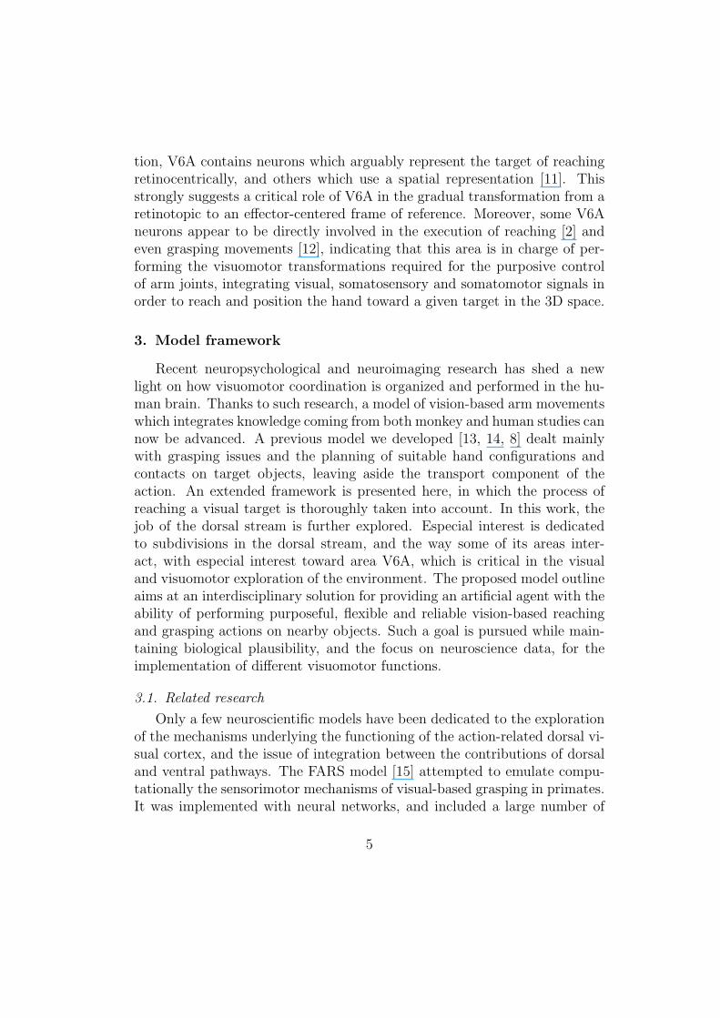
tion, V6A contains neurons which arguably represent the target of reachingretinocentrically, and others which use a spatial representation [11]. Thisstrongly suggests a critical role of V6A in the gradual transformation from aretinotopic to an effector-centered frame of reference. Moreover, some V6Aneurons appear to be directly involved in the execution of reaching [2] andeven grasping movements [12], indicating that this area is in charge of per-forming the visuomotor transformations required for the purposive controlof arm joints, integrating visual, somatosensory and somatomotor signals inorder to reach and position the hand toward a given target in the 3D space.
3. Model framework
Recent neuropsychological and neuroimaging research has shed a newlight on how visuomotor coordination is organized and performed in the hu-man brain. Thanks to such research, a model of vision-based arm movementswhich integrates knowledge coming from both monkey and human studies cannow be advanced. A previous model we developed [13, 14, 8] dealt mainlywith grasping issues and the planning of suitable hand configurations andcontacts on target objects, leaving aside the transport component of theaction. An extended framework is presented here, in which the process ofreaching a visual target is thoroughly taken into account. In this work, thejob of the dorsal stream is further explored. Especial interest is dedicatedto subdivisions in the dorsal stream, and the way some of its areas inter-act, with especial interest toward area V6A, which is critical in the visualand visuomotor exploration of the environment. The proposed model outlineaims at an interdisciplinary solution for providing an artificial agent with theability of performing purposeful, flexible and reliable vision-based reachingand grasping actions on nearby objects. Such a goal is pursued while main-taining biological plausibility, and the focus on neuroscience data, for theimplementation of different visuomotor functions.
3.1. Related research
Only a few neuroscientific models have been dedicated to the explorationof the mechanisms underlying the functioning of the action-related dorsal vi-sual cortex, and the issue of integration between the contributions of dorsaland ventral pathways. The FARS model [15] attempted to emulate compu-tationally the sensorimotor mechanisms of visual-based grasping in primates.It was implemented with neural networks, and included a large number of
5
different brain areas (mainly inspired on monkey physiology), but only areasAIP, F5 and the primary motor cortex F1 were modeled in detail. More-over, the FARS model was focused exclusively on grasping actions withoutconsidering the hand transport function. Herein, Jeannerod [4] proposedto extend the notion of parallel visuomotor channels about the mechanismswhich operate within the dorsal stream itself, and suggested the existence ofneural pathways for reaching and for grasping. These channels, although dis-tinct, must also share a common mechanism for achieving coordination witheach other. Murata and Ishida [16] also discussed parallel pathways for handreaching and grasping. Apart from network for the control of sensory-guidedhand and arm movements, their model included mechanisms of body aware-ness. Alternative models, such as the multiple finger reaching idea [17], arenot given much credit, due to the quantity and quality of evidence supportingthe mainstream hypothesis (see e.g. [18]).
Neuroscientific inspiration in artificial intelligence and robotics is basicallyconfined to the use of biologically plausible artificial neural networks. Visuo-motor transformations involving arm movements have been usually tackledwith the use of Self-Organizing Maps (SOM) [19, 20], feedforward and re-current neural networks [21] and less commonly with Radial Basis Functions(RBF) [22]. In the most common approach, the system firstly learns the map-ping between the image coordinates and the pan/tilt encoder coordinates ofthe eye motors, and then the transformation carrying from the visual inputto an appropriate arm posture, suitable for reaching and grasping a targetobject. Some reaching models, while using biologically-inspired neural net-works, disregard the necessity of reference frame transformations, computingthe difference vector between the target and the hand position in the eye-centered coordinate system without any additional stages [23, 24].
Summarizing, although a few attempts to model the functioning of theaction-related visual cortex exist, most of them do not provide any detailsfor possible computational, and especially robotic implementation of the pro-posed concepts. On the other hand, biological or neuroscientific inspirationin robotics is often too superficial and conditioned by pragmatic goals andtechnological constraints. Aside from the theoretical contribution, the re-search presented here is also the first step toward the goal of improving theskills of autonomous robotic systems in their exploration of the nearby spaceand interaction with surrounding objects, as described below.
6
3.2. A Subdivision Within the Dorsal Stream
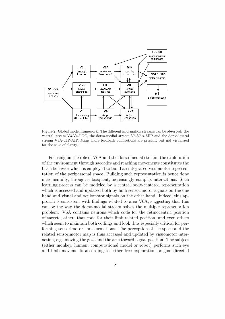
The whole framework of the proposed model is graphically represented inFigure 2. After the extraction of basic visual information in V1/V2, higherlevel features are generated in V3 and sent to the two streams. Along theventral stream, an increasingly invariant representation of object shape isgenerated in order to perform a gradual recognition of the object (areas V4and LOC [14]). In the dorsal stream, both object shape and location haveto be processed. For what concerns shape, area CIP integrates stereopticand perspective data in order to detect pose and proportion of the targetobject, using also information regarding object classification [9, 8]. Areas V6and V6A estimate object location and distance, integrating retinal data withproprioceptive information about eye position. Both V3A and CIP projectto AIP, which transforms object visual data in hand configurations suitablefor grasping. At the same time, areas V6A and MIP determine the reachingdirection and collaborate with AIP and PMd in order to execute the armmovement suitable for getting to the target object. Movement plans are de-vised in posterior-premotor loops, considering also the information on objectidentity coming from the ventral stream, and task requirements. Dorsal areasare supported by proprioceptive information coming from somatosensory ar-eas SI/SII. The signals for action execution are sent to the motor cortex M1,and a posterior parietal-premotor-cerebellum loop is in charge of monitoringaction execution in accordance to the plan.
3.3. Toward a visuomotor knowledge of the environment
It is often argued, and increasingly accepted by the neuroscientific com-munity, that the dorsal stream performs all the transformations required forsensorimotor interactions exploiting gain modulation between neural pop-ulations [25]. Basis function representations, which permit to simultane-ously represent stimuli in various reference frames, constitute a natural neu-ral structure which is especially suitable for implementing gain field effects.In fact, the basis function approach has the attractive feature that bothhead-centric representations for arm movements and retino-centric represen-tations for gaze movements can be encoded concurrently in the same neuralmap [26, 27]. In this way, explicit encoding of targets in retino-centric co-ordinates is enhanced via gain fields to hold in parallel an implicit encodingin other reference frames [28]. Such gain fields are found in retino-centricorganized eye movement areas and, most importantly, in posterior parietalarea V6A [29].
7
�� � ������� ����� ��� � �������� ���������� ��� �������
���� ���� ��������� �
����������� � ��� �
������������� ��� �� ! ��"#���� ������#
�������� $ ����� �%���� � �� � �������
&'���( ��� ���������
���� ������#� # ��
)� * )���������� �������� ������� �+�� � � �� ���# ������,
�+� �����������������
Figure 2: Global model framework. The different information streams can be observed: theventral stream V3-V4-LOC, the dorso-medial stream V6-V6A-MIP and the dorso-lateralstream V3A-CIP-AIP. Many more feedback connections are present, but not visualizedfor the sake of clarity.
Focusing on the role of V6A and the dorso-medial stream, the explorationof the environment through saccades and reaching movements constitutes thebasic behavior which is employed to build an integrated visuomotor represen-tation of the peripersonal space. Building such representation is hence doneincrementally, through subsequent, increasingly complex interactions. Suchlearning process can be modeled by a central body-centered representationwhich is accessed and updated both by limb sensorimotor signals on the onehand and visual and oculomotor signals on the other hand. Indeed, this ap-proach is consistent with findings related to area V6A, suggesting that thiscan be the way the dorso-medial stream solves the multiple representationproblem. V6A contains neurons which code for the retinocentric positionof targets, others that code for their limb-related position, and even otherswhich seem to maintain both codings and look thus especially critical for per-forming sensorimotor transformations. The perception of the space and therelated sensorimotor map is thus accessed and updated by visuomotor inter-action, e.g. moving the gaze and the arm toward a goal position. The subject(either monkey, human, computational model or robot) performs such eyeand limb movements according to either free exploration or goal directed
8
tasks. Eyes and arms can be considered as separate effectors which receivemotor control from a shared, implicit sensorimotor map of the peripersonalspace, and the outcome of their contextual movements is used to update themap itself. We propose to encode the integrated sensorimotor map of theperipersonal space implicitly in a basis function structure, which models thefunction of V6A and its connections with purely visual and premotor areas.In the next section, neuroscience data regarding area V6A suitable for biasingthe model definition and configuration are analyzed and discussed.
4. The Different Aspects of Neural Response During Reaching
In previous works, single-cell experiments performed on macaque monkeyswere described and analyzed [6, 10, 11]. This work is aimed at sheddingfurther light on the sort of transformations performed by V6A neurons andon the coding representations they use to this purpose. The analysis approachemployed here is different from the previous works, as it is performed withthe final goal of achieving a computational description of V6A neurons tobe used within a numerical model. In particular, the answers that need tobe asked are the following. How many types of neurons does V6A contain?What are their most relevant properties and toward what tasks are theyoriented? How do they perform the transformations required to coordinateand modulate retinal data, gaze direction and reaching movements?
4.1. Experiment Description
The experiments analyzed here were collected at the Universita di Bolognaon two trained macaque monkeys. Details on the experimental protocol andrelated data analysis are available in a previous work [11]. They were ap-proved by the Bioethical Committee of the University and carried out inaccordance with Italian national laws and European Directives on care anduse of laboratory animals. Data were collected while the monkeys were per-forming two possible reaching tasks toward given targets while gazing at acertain position (the fixation point).
Constant reaching task
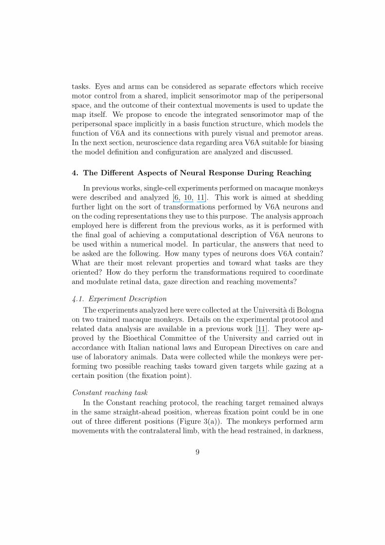
In the Constant reaching protocol, the reaching target remained alwaysin the same straight-ahead position, whereas fixation point could be in oneout of three different positions (Figure 3(a)). The monkeys performed armmovements with the contralateral limb, with the head restrained, in darkness,
9
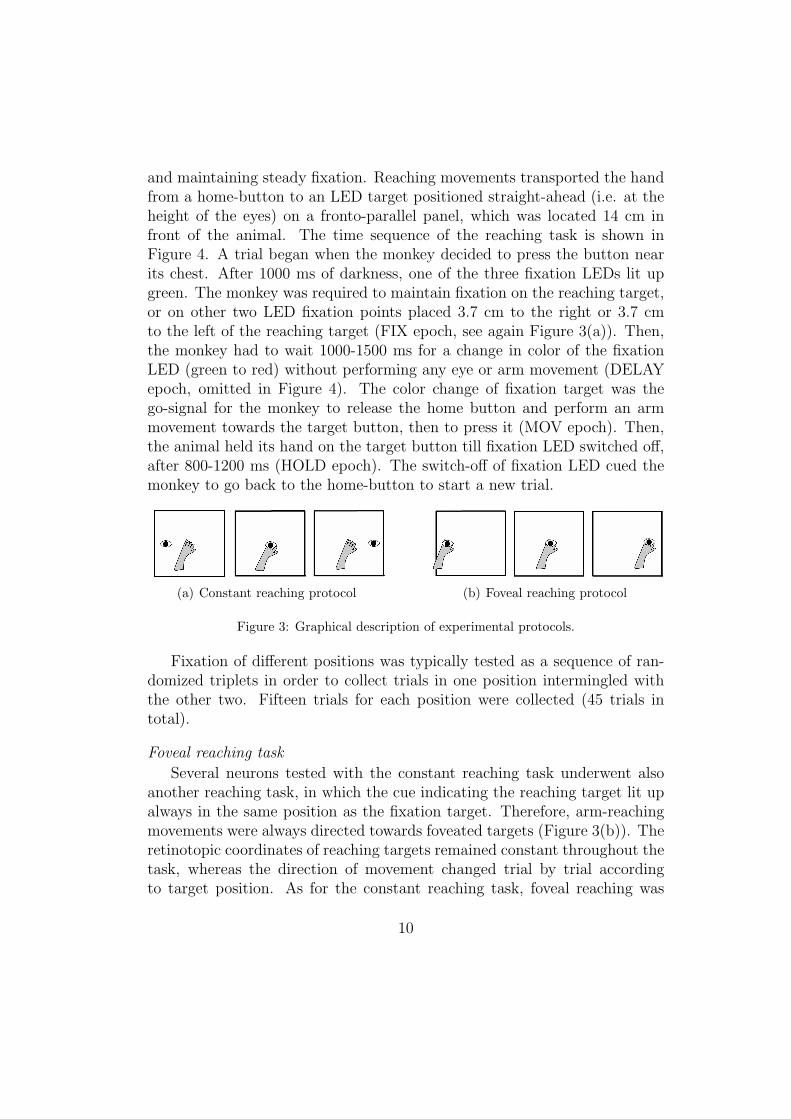
and maintaining steady fixation. Reaching movements transported the handfrom a home-button to an LED target positioned straight-ahead (i.e. at theheight of the eyes) on a fronto-parallel panel, which was located 14 cm infront of the animal. The time sequence of the reaching task is shown inFigure 4. A trial began when the monkey decided to press the button nearits chest. After 1000 ms of darkness, one of the three fixation LEDs lit upgreen. The monkey was required to maintain fixation on the reaching target,or on other two LED fixation points placed 3.7 cm to the right or 3.7 cmto the left of the reaching target (FIX epoch, see again Figure 3(a)). Then,the monkey had to wait 1000-1500 ms for a change in color of the fixationLED (green to red) without performing any eye or arm movement (DELAYepoch, omitted in Figure 4). The color change of fixation target was thego-signal for the monkey to release the home button and perform an armmovement towards the target button, then to press it (MOV epoch). Then,the animal held its hand on the target button till fixation LED switched off,after 800-1200 ms (HOLD epoch). The switch-off of fixation LED cued themonkey to go back to the home-button to start a new trial.
(a) Constant reaching protocol (b) Foveal reaching protocol
Figure 3: Graphical description of experimental protocols.
Fixation of different positions was typically tested as a sequence of ran-domized triplets in order to collect trials in one position intermingled withthe other two. Fifteen trials for each position were collected (45 trials intotal).
Foveal reaching task
Several neurons tested with the constant reaching task underwent alsoanother reaching task, in which the cue indicating the reaching target lit upalways in the same position as the fixation target. Therefore, arm-reachingmovements were always directed towards foveated targets (Figure 3(b)). Theretinotopic coordinates of reaching targets remained constant throughout thetask, whereas the direction of movement changed trial by trial accordingto target position. As for the constant reaching task, foveal reaching was
10
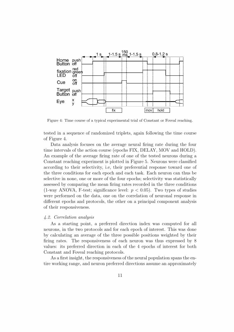
Figure 4: Time course of a typical experimental trial of Constant or Foveal reaching.
tested in a sequence of randomized triplets, again following the time courseof Figure 4.
Data analysis focuses on the average neural firing rate during the fourtime intervals of the action course (epochs FIX, DELAY, MOV and HOLD).An example of the average firing rate of one of the tested neurons during aConstant reaching experiment is plotted in Figure 5. Neurons were classifiedaccording to their selectivity, i.e, their preferential response toward one ofthe three conditions for each epoch and each task. Each neuron can thus beselective in none, one or more of the four epochs; selectivity was statisticallyassessed by comparing the mean firing rates recorded in the three conditions(1-way ANOVA, F-test; significance level: p < 0.05). Two types of studieswere performed on the data, one on the correlation of neuronal response indifferent epochs and protocols, the other on a principal component analysisof their responsiveness.
4.2. Correlation analysis
As a starting point, a preferred direction index was computed for allneurons, in the two protocols and for each epoch of interest. This was doneby calculating an average of the three possible positions weighted by theirfiring rates. The responsiveness of each neuron was thus expressed by 8values: its preferred direction in each of the 4 epochs of interest for bothConstant and Foveal reaching protocols.
As a first insight, the responsiveness of the neural population spans the en-tire working range, and neuron preferred directions assume an approximately
11
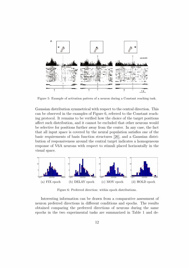
Figure 5: Example of activation pattern of a neuron during a Constant reaching task.
Gaussian distribution symmetrical with respect to the central direction. Thiscan be observed in the examples of Figure 6, referred to the Constant reach-ing protocol. It remains to be verified how the choice of the target positionsaffect such distribution, and it cannot be excluded that other neurons wouldbe selective for positions further away from the center. In any case, the factthat all input space is covered by the neural population satisfies one of thebasic requirements of basis function structures [26], and a Gaussian distri-bution of responsiveness around the central target indicates a homogeneousresponse of V6A neurons with respect to stimuli placed horizontally in thevisual space.
LEFT CENTER RIGHT0
5
10
15
(a) FIX epoch
LEFT CENTER RIGHT0
5
10
15
(b) DELAY epoch
LEFT CENTER RIGHT0
5
10
15
(c) MOV epoch
LEFT CENTER RIGHT0
5
10
15
(d) HOLD epoch
Figure 6: Preferred direction: within epoch distributions.
Interesting information can be drawn from a comparative assessment ofneuron preferred directions in different conditions and epochs. The resultsobtained comparing the preferred directions of neurons during the sameepochs in the two experimental tasks are summarized in Table 1 and de-
12
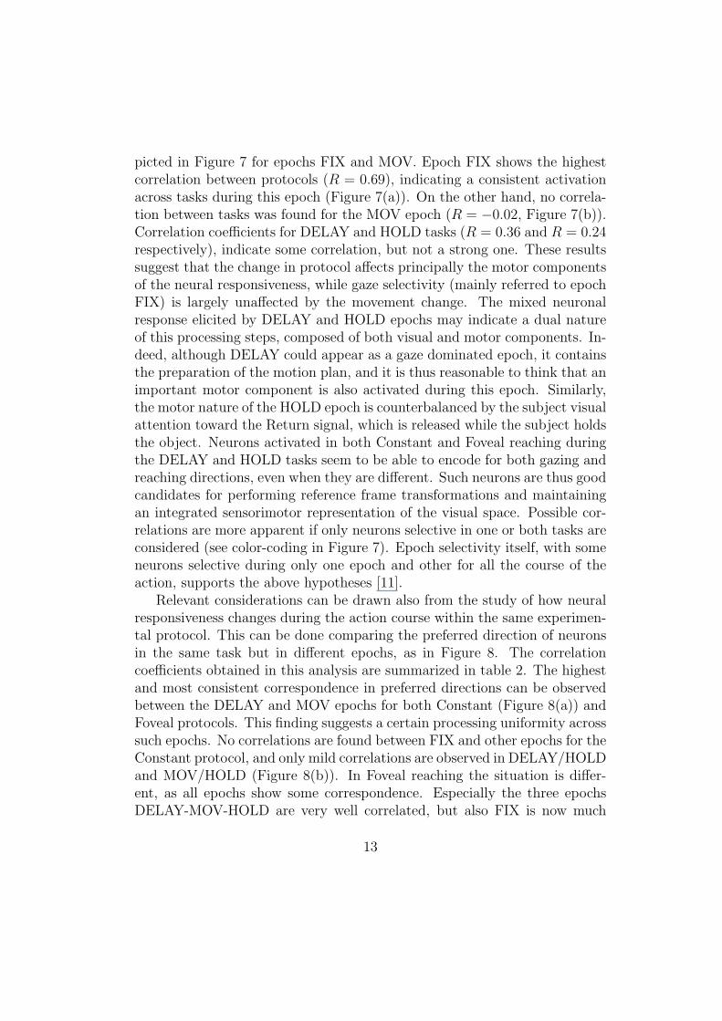
picted in Figure 7 for epochs FIX and MOV. Epoch FIX shows the highestcorrelation between protocols (R = 0.69), indicating a consistent activationacross tasks during this epoch (Figure 7(a)). On the other hand, no correla-tion between tasks was found for the MOV epoch (R = −0.02, Figure 7(b)).Correlation coefficients for DELAY and HOLD tasks (R = 0.36 and R = 0.24respectively), indicate some correlation, but not a strong one. These resultssuggest that the change in protocol affects principally the motor componentsof the neural responsiveness, while gaze selectivity (mainly referred to epochFIX) is largely unaffected by the movement change. The mixed neuronalresponse elicited by DELAY and HOLD epochs may indicate a dual natureof this processing steps, composed of both visual and motor components. In-deed, although DELAY could appear as a gaze dominated epoch, it containsthe preparation of the motion plan, and it is thus reasonable to think that animportant motor component is also activated during this epoch. Similarly,the motor nature of the HOLD epoch is counterbalanced by the subject visualattention toward the Return signal, which is released while the subject holdsthe object. Neurons activated in both Constant and Foveal reaching duringthe DELAY and HOLD tasks seem to be able to encode for both gazing andreaching directions, even when they are different. Such neurons are thus goodcandidates for performing reference frame transformations and maintainingan integrated sensorimotor representation of the visual space. Possible cor-relations are more apparent if only neurons selective in one or both tasks areconsidered (see color-coding in Figure 7). Epoch selectivity itself, with someneurons selective during only one epoch and other for all the course of theaction, supports the above hypotheses [11].
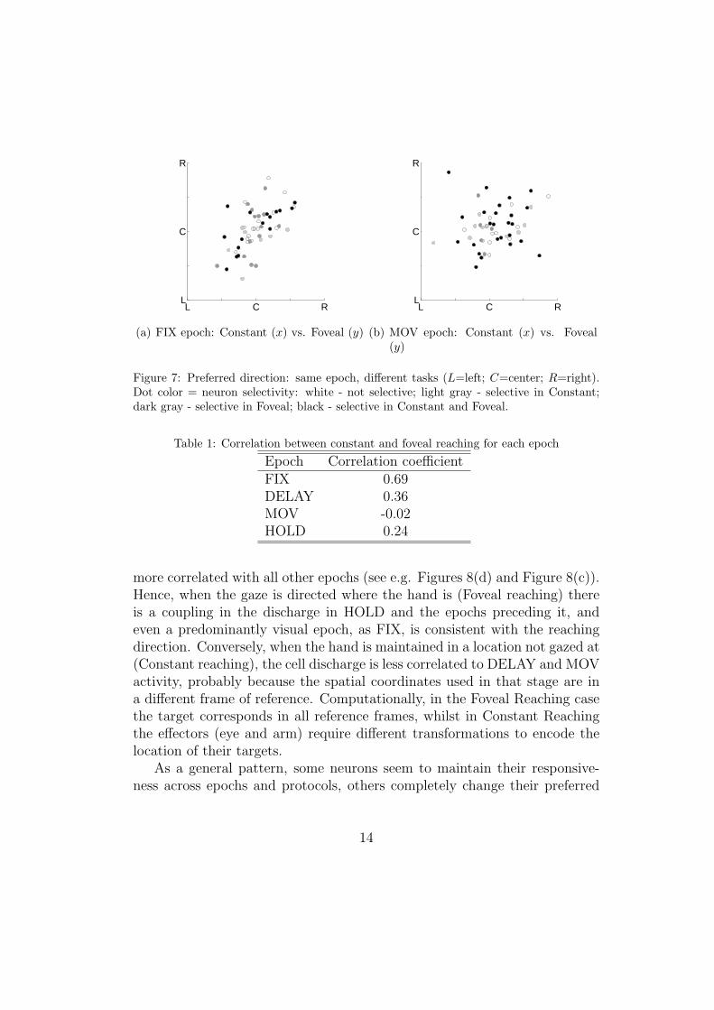
Relevant considerations can be drawn also from the study of how neuralresponsiveness changes during the action course within the same experimen-tal protocol. This can be done comparing the preferred direction of neuronsin the same task but in different epochs, as in Figure 8. The correlationcoefficients obtained in this analysis are summarized in table 2. The highestand most consistent correspondence in preferred directions can be observedbetween the DELAY and MOV epochs for both Constant (Figure 8(a)) andFoveal protocols. This finding suggests a certain processing uniformity acrosssuch epochs. No correlations are found between FIX and other epochs for theConstant protocol, and only mild correlations are observed in DELAY/HOLDand MOV/HOLD (Figure 8(b)). In Foveal reaching the situation is differ-ent, as all epochs show some correspondence. Especially the three epochsDELAY-MOV-HOLD are very well correlated, but also FIX is now much
13
L C RL
C
R
(a) FIX epoch: Constant (x) vs. Foveal (y)
L C RL
C
R
(b) MOV epoch: Constant (x) vs. Foveal(y)
Figure 7: Preferred direction: same epoch, different tasks (L=left; C=center; R=right).Dot color = neuron selectivity: white - not selective; light gray - selective in Constant;dark gray - selective in Foveal; black - selective in Constant and Foveal.
Table 1: Correlation between constant and foveal reaching for each epoch
Epoch Correlation coefficientFIX 0.69DELAY 0.36MOV -0.02HOLD 0.24
more correlated with all other epochs (see e.g. Figures 8(d) and Figure 8(c)).Hence, when the gaze is directed where the hand is (Foveal reaching) thereis a coupling in the discharge in HOLD and the epochs preceding it, andeven a predominantly visual epoch, as FIX, is consistent with the reachingdirection. Conversely, when the hand is maintained in a location not gazed at(Constant reaching), the cell discharge is less correlated to DELAY and MOVactivity, probably because the spatial coordinates used in that stage are ina different frame of reference. Computationally, in the Foveal Reaching casethe target corresponds in all reference frames, whilst in Constant Reachingthe effectors (eye and arm) require different transformations to encode thelocation of their targets.
As a general pattern, some neurons seem to maintain their responsive-ness across epochs and protocols, others completely change their preferred
14
L C RL
C
R
(a) Constant reaching: DELAY (x) vs.MOV (y)
L C RL
C
R
(b) Constant reaching: MOV (x) vs. HOLD(y)
L C RL
C
R
(c) Foveal reaching: FIX (x) vs. DELAY(y)
L C RL
C
R
(d) Foveal reaching: MOV (x) vs. HOLD(y)
Figure 8: Preferred direction: different epochs, same task (L=left; C=center; R=right).Dot color = neuron selectivity: white - not selective; light gray - selective in x epoch; darkgray - selective in y epoch; black - selective in both epochs.
Table 2: Correlation between different epochs for constant and foveal reaching
Epochs Constant reaching Foveal reachingFIX/ DELAY 0.15 0.90FIX/ MOV 0.02 0.76FIX/ HOLD 0.20 0.61DELAY/ MOV 0.86 0.83DELAY/ HOLD 0.56 0.81HOLD/ MOV 0.54 0.92
15
direction. These findings suggest the presence of important temporal issues,and a strong effect of action stage on neural responsiveness. A possibleinterpretation of this activity pattern is that some neurons sustain their ac-tivation, maybe for maintaining their coding of the target position in thehead-centered reference frame. Other neurons perform instead the transfor-mations between this frame and the retinocentric and arm-centered referenceswhen required. For modeling purposes, the first type of neurons would be incharge of maintaining the common visuomotor spatial representation, whilstthe second type of neurons would be in charge of accessing and modifyingsuch representation according to different sensorimotor events.
4.3. Principal Component Analysis
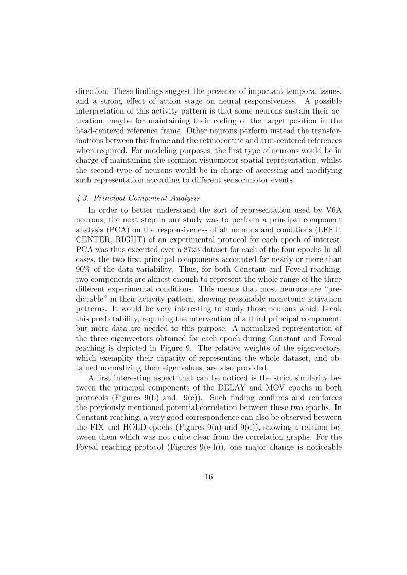
In order to better understand the sort of representation used by V6Aneurons, the next step in our study was to perform a principal componentanalysis (PCA) on the responsiveness of all neurons and conditions (LEFT,CENTER, RIGHT) of an experimental protocol for each epoch of interest.PCA was thus executed over a 87x3 dataset for each of the four epochs In allcases, the two first principal components accounted for nearly or more than90% of the data variability. Thus, for both Constant and Foveal reaching,two components are almost enough to represent the whole range of the threedifferent experimental conditions. This means that most neurons are “pre-dictable” in their activity pattern, showing reasonably monotonic activationpatterns. It would be very interesting to study those neurons which breakthis predictability, requiring the intervention of a third principal component,but more data are needed to this purpose. A normalized representation ofthe three eigenvectors obtained for each epoch during Constant and Fovealreaching is depicted in Figure 9. The relative weights of the eigenvectors,which exemplify their capacity of representing the whole dataset, and ob-tained normalizing their eigenvalues, are also provided.
A first interesting aspect that can be noticed is the strict similarity be-tween the principal components of the DELAY and MOV epochs in bothprotocols (Figures 9(b) and 9(c)). Such finding confirms and reinforcesthe previously mentioned potential correlation between these two epochs. InConstant reaching, a very good correspondence can also be observed betweenthe FIX and HOLD epochs (Figures 9(a) and 9(d)), showing a relation be-tween them which was not quite clear from the correlation graphs. For theFoveal reaching protocol (Figures 9(e-h)), one major change is noticeable
16
0 1 2 3 40
0.2
0.4
0.6
0.8
1
(a) FIX epoch:80.8; 15.2; 4.0
0 1 2 3 40
0.2
0.4
0.6
0.8
1
(b) DELAY epoch:76.0; 16.0; 8.0
0 1 2 3 40
0.2
0.4
0.6
0.8
1
(c) MOV epoch:79.1; 13.3; 7.6
0 1 2 3 40
0.2
0.4
0.6
0.8
1
(d) HOLD epoch:76.8; 16.4; 6.8
0 1 2 3 40
0.2
0.4
0.6
0.8
1
(e) FIX epoch:73.1; 15.8; 11.1
0 1 2 3 40
0.2
0.4
0.6
0.8
1
(f) DELAY epoch:73.6; 15.6; 10.8
0 1 2 3 40
0.2
0.4
0.6
0.8
1
(g) MOV epoch:84.2; 9.5; 6.3
0 1 2 3 40
0.2
0.4
0.6
0.8
1
(h) HOLD epoch:84.2; 10.7; 5.1
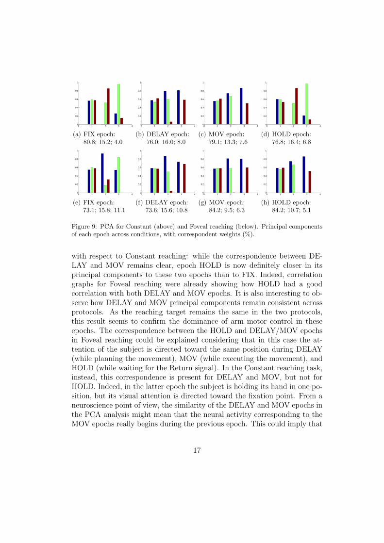
Figure 9: PCA for Constant (above) and Foveal reaching (below). Principal componentsof each epoch across conditions, with correspondent weights (%).
with respect to Constant reaching: while the correspondence between DE-LAY and MOV remains clear, epoch HOLD is now definitely closer in itsprincipal components to these two epochs than to FIX. Indeed, correlationgraphs for Foveal reaching were already showing how HOLD had a goodcorrelation with both DELAY and MOV epochs. It is also interesting to ob-serve how DELAY and MOV principal components remain consistent acrossprotocols. As the reaching target remains the same in the two protocols,this result seems to confirm the dominance of arm motor control in theseepochs. The correspondence between the HOLD and DELAY/MOV epochsin Foveal reaching could be explained considering that in this case the at-tention of the subject is directed toward the same position during DELAY(while planning the movement), MOV (while executing the movement), andHOLD (while waiting for the Return signal). In the Constant reaching task,instead, this correspondence is present for DELAY and MOV, but not forHOLD. Indeed, in the latter epoch the subject is holding its hand in one po-sition, but its visual attention is directed toward the fixation point. From aneuroscience point of view, the similarity of the DELAY and MOV epochs inthe PCA analysis might mean that the neural activity corresponding to theMOV epochs really begins during the previous epoch. This could imply that
17
V6A neurons are strongly involved in movement planning and preparation.Still, they maintain their activation during movement execution, very likelyfor performing a feed-forward control loop as part of a recurrent parietal-premotor circuit, as recent anatomical studies support [30]. At the sametime, this analysis suggests that a mixed basis function population with se-lectivity for different phases of the action across the visuomotor space canbe a good approximation for modeling the job of V6A neurons.
5. Discussion
The principal component analysis of the previous section offers insightson how to define a first approximation for modeling the job of V6A neurons.Starting from the above analysis, a population of artificial neurons can begenerated which is able to emulate the sort of transformations and modula-tions between visual data and gaze and arm movements performed by thedorso-medial stream. The different properties captured in this work can beused to tune the behavior of the neural population with various input setscorresponding to the different experimental conditions. As explained above,computational architectures based on basis functions are most suitable formodeling such behavior. A very important issue at this stage of the processis how to define the basis function neurons. Summarizing the above findings,it can be said that area V6A includes neurons having only visual response,neurons apparently involved mainly in motor actions and mixed neurons,activated in all phases of sensorimotor processes. These visuomotor neuronsare very likely dedicated to allow smooth transformations from one referenceframe to the other.
The model we are developing should assess what computational advan-tages could be obtained by such responsiveness pattern. The first modelingstep is thus to learn the transformations between visual, oculomotor and armmotor space using basis function networks inspired on V6A neurons. For sim-plicity at this stage, only two arm joints can be used, and no tilt movementsof the eyes, so that the accessible environment is a 2D space placed horizon-tally in front of the subject, as in most monkey experiments. The differenttypes of V6A neurons can be simulated with populations of radial basis func-tion neurons with receptive fields distributed according to different criteria.As a first approximation, predominantly visual neurons, such as those morestrongly activated during epochs FIX and HOLD, can be distributed uni-formly on the visual space; motor neurons, corresponding to epoch MOV,
18
are better organized according to arm joint space; intermediate neurons cansimulate a head-based reference frame, and can be placed according to differ-ent criteria at the same time, visual, joint and vergence/version oculomotorspace. Preliminary results indicate that this neural organization is especiallysuitable for both direct and inverse transformations between the differentreference frames required for visual data gathering, eye gazing movementsand arm reaching actions [31].
Data analysis and basis function modeling concord in giving V6A a crit-ical role in the whole model framework of Figure 2. In fact, V6A would actas a visuomotor relay station able to integrate and modulate between visualinformation (coming from earlier visual areas such as V2/V3), oculomotorsignals (in coordination with areas LIP and FEF, not shown in Figure 1),and arm motor signals (with other posterior parietal areas MIP and AIPand premotor areas, especially PMd). Once some more data are available,the analysis of V6A neuron activity presented in this paper could be ex-tended using singular value decomposition (SVD), successfully applied so farto premotor data recordings [32]. Such analysis would be helpful in testingwhether gaze direction and hand movement direction are separable variablesfor a given neuron. Computationally, the activations of the neurons fromeach class (separable, not separable) could be used for fitting the parametervalues for different classes of neurons which use direct, intermediate or gainfield encoding. Further experiments both on the neuroscience and the com-putational sides will help in assessing the above hypotheses, and better clarifywhat is the contribution of V6A in various different cortical mechanisms andthe corresponding data flows.
6. Conclusions
This work described research aimed at better understanding the role ofthe dorso-medial visual stream in the sensorimotor transformations requiredfor the planning and execution of gazing and reaching actions. The aboveanalysis helps in clarifying what sort of computation is performed by dorsalstream neurons, namely those pertaining to area V6A, in order to maintaina perfect coordination between retinal data, gaze direction and arm move-ments. The outline of a comprehensive model, detailed for what concernsthe changes in reference frames related to various sensorimotor conditions,was advanced in this paper. The model served as basis for a single-cell data
19
analysis performed with a computational stance, which contributes to sheda new light on the role and organization of V6A neural subpopulations.
Detailed model implementation and robotic experiments will help in fur-ther clarifying the mechanisms behind eye-arm coordination and recipro-cal guidance, and reference frame transformations in primates. This shouldcarry to a better understanding of the transformations performed betweenretinocentric, effector-based and distance/vergence-based representations invarious environments and working conditions. The predictions obtained bythe model and the robotic experiments could then be tested through the de-velopment of new neuroscience studies. From a pragmatic point of view, arobot emulating the above mechanisms should be able to purposefully andconsistently interact with its environment building its skills on the integra-tion of different stimuli. Such skills would be based on the building of aplastic representation of its nearby environment, representation which canbe exploited for more precise and complex interactions with the environmentcomponents. This research is thus expected to provide important advance-ments in both robotics and neuroscience.
Acknowledgment
Support for this research has been provided in part by the EuropeanCommission’s Seventh Framework Programme FP7/2007-2013 under grantagreement 217077 (EYESHOTS project), by the Spanish Ministerio de Cien-cia y Innovacion (DPI-2008-06636, FPU grant AP2007-02565), by the ItalianMinistry of University and Research, by Fundacio Caixa-Castello-Bancaixa(P1-1B2008-51) and by WCU (World Class University) program through theNational Research Foundation of Korea, funded by the Ministry of Educa-tion, Science and Technology (Grant No. R31-2008-000-10062-0).
References
[1] M. A. Goodale, A. D. Milner, Sight Unseen, Oxford University Press,2004.
[2] C. Galletti, D. F. Kutz, M. Gamberini, R. Breveglieri, P. Fattori, Roleof the medial parieto-occipital cortex in the control of reaching andgrasping movements., Experimental Brain Research 153 (2003) 158–170.
20
[3] J. C. Culham, C. Cavina-Pratesi, A. Singhal, The role of parietal cor-tex in visuomotor control: what have we learned from neuroimaging?,Neuropsychologia 44 (2006) 2668–2684.
[4] M. Jeannerod, Visuomotor channels: Their integration in goal-directedprehension, Human Movement Science 18 (1999) 201–218.
[5] J. C. Culham, J. P. Gallivan, C. Cavina-Pratesi, D. J. Quinlan, fMRIinvestigations of reaching and ego space in human superior parieto-occipital cortex, in: R. Klatzky, M. Behrmann, B. MacWhinney (Eds.),Embodiment, Ego-space and Action, Lawrence Erlbaum Associates,2008, pp. 247–274.
[6] P. Fattori, M. Gamberini, D. F. Kutz, C. Galletti, ’Arm-reaching’ neu-rons in the parietal area V6A of the macaque monkey., Eur J Neurosci13 (2001) 2309–2313.
[7] P. Dechent, J. Frahm, Characterization of the human visual V6 complexby functional magnetic resonance imaging., Eur J Neurosci 17 (2003)2201–2211.
[8] E. Chinellato, A. P. del Pobil, Distance and orientation estimation ofgraspable objects in natural and artificial systems, Neurocomputing 72(2008) 879–886.
[9] E. Chinellato, A. P. del Pobil, Neural coding in the dorsal visual stream,in: M. Asada, J. Hallam, J.-A. Meyer, J. Tanipages (Eds.), From Ani-mals to Animats, LNAI 5040, Springer, 2008, pp. 230–239.
[10] P. Fattori, D. F. Kutz, R. Breveglieri, N. Marzocchi, C. Galletti, Spatialtuning of reaching activity in the medial parieto-occipital cortex (areaV6A) of macaque monkey., Eur J Neurosci 22 (2005) 956–972.
[11] N. Marzocchi, R. Breveglieri, C. Galletti, P. Fattori, Reaching activ-ity in parietal area V6A of macaque: eye influence on arm activity orretinocentric coding of reaching movements?, Eur J Neurosci 27 (2008)775–789.
[12] P. Fattori, V. Raos, R. Breveglieri, A. Bosco, N. Marzocchi, C. Galletti,The dorsomedial pathway is not just for reaching: grasping neurons in
21
the medial parieto-occipital cortex of the macaque monkey., J Neurosci30 (2010) 342–349.
[13] E. Chinellato, Y. Demiris, A. P. del Pobil, Studying the human vi-sual cortex for achieving action-perception coordination with robots, in:A. P. del Pobil (Ed.), Artificial Intelligence and Soft Computing, ActaPress, Anaheim, CF, USA, 2006, pp. 184–189.
[14] E. Chinellato, B. J. Grzyb, A. P. del Pobil, Brain mechanisms for roboticobject pose estimation, in: IEEE International Joint Conference onNeural Networks, 2008, pp. 3268–3275.
[15] A. H. Fagg, M. A. Arbib, Modeling parietal-premotor interactions inprimate control of grasping., Neural Networks 11 (1998) 1277–1303.
[16] A. Murata, H. Ishida, Representaion of bodily self in the multimodalparieto-premotor network, in: F. S. (Ed.), Representaion and brain,Springer, 2007, pp. 151–176.
[17] J. B. J. Smeets, E. Brenner, M. Biegstraaten, Independent control of thedigits predicts an apparent hierarchy of visuomotor channels in grasp-ing., Behavioural Brain Research 136 (2002) 427–432.
[18] C. van de Kamp, F. T. J. M. Zaal, Prehension is really reaching andgrasping, Experimental Brain Research 182 (2007) 27–34.
[19] T. M. Martinetz, H. J. Ritter, K. J. Schulten, Three-dimensional neuralnet for learning visuomotor coordination of a robot arm, IEEE T NeuralNetwor 1 (1990) 131–136.
[20] M. Jones, D. Vernon, Using neural networks to learn hand-eye co-ordination, Neural Computing and Applications 2 (1994) 2–12.
[21] W. Schenck, H. Hoffmann, R. Moller, Learning internal models for eye-hand coordination in reaching and grasping, in: F. Schmalhofer, R. M.Young, G. Katz (Eds.), Proc. EuroCogSci 2003, pp. 289–294.
[22] M. Marjanovic, B. Scassellati, M. Williamson, Self-taught visually-guided pointing for a humanoid robot, in: International Conferenceon Simulation of Adaptive Behavior (SAB) 1996.
22
[23] G. Sun, B. Scassellati, A fast and efficient model for learning to reach,International Journal of Humanoid Robotics 2 (2005) 391–413.
[24] F. Nori, L. Natale, G. Sandini, G. Metta, Autonomous learning of 3Dreaching in a humanoid robot, in: IEEE International Conference onIntelligent Robots and Systems 2007.
[25] E. Salinas, P. Thier, Gain modulation: a major computational principleof the central nervous system., Neuron 27 (2000) 15–21.
[26] S. Pouget, A. Sejnowski, Spatial transformations in the parietal cortexusing basis functions, Journal of Cognitive Neuroscience 9 (1997) 222–237.
[27] A. Pouget, L. H. Snyder, Computational approaches to sensorimotortransformations., Nature Neuroscience 3 Suppl (2000) 1192–1198.
[28] S. Deneve, A. Pouget, Basis functions for object-centered representa-tions., Neuron 37 (2003) 347–359.
[29] C. Galletti, P. P. Battaglini, P. Fattori, Eye position influence on theparieto-occipital area PO (V6) of the macaque monkey., Eur J Neurosci7 (1995) 2486–2501.
[30] M. Gamberini, L. Passarelli, P. Fattori, M. Zucchelli, S. Bakola, G. Lup-pino, C. Galletti, Cortical connections of the visuomotor parietooccipitalarea V6Ad of the macaque monkey., J Comp Neurol 513 (2009) 622–642.
[31] E. Chinellato, M. Antonelli, B. Grzyb, A. del Pobil, Implicit sensorimo-tor mapping of the peripersonal space by gazing and reaching (submit-ted), IEEE Transactions on Autonomous Mental Development (2010).
[32] B. Pesaran, M. J. Nelson, R. A. Andersen, A relative position code forsaccades in dorsal premotor cortex., J Neurosci 30 (2010) 6527–6537.
23


