Embed Size (px)
Citation preview

Internationally Indexed Journal
Indexed in Chemical Abstract Services(USA),Index Coppernicus ,Ulrichs Directory of
Periodicals,Google scholar ,Cabi ,DOAJ ,PSOAR, EBSCO ,SCOPUS, EMBASE etc.
Rapid Publishing
��������������� ������������������������������ �������������������
������������������������������������� ����������������������� �!�����������
����� �!�������������������!������������������������ �����"���������
������������������"������������#���!��$��������������������������
���������������"�����������������������������������������������������
!����������������!����%�!��$�������&�����������������
������������������������'�����(�)'�����(�)'�����(�)'�����(�)*+*+*+*+����*Refer Instruction to authors available at
www.ijpbs.net
Journal Home page
www.ijpbs.net
������������������������� ����
www.ijpbs.net
������������������������

Int J Pharm Bio Sci 2013 Jan; 4(1): (B) 298 - 308
�
�������������� ������������������������ ������
��������
��������������������������������������������������������������������������������������������������������������������������������������������������� ������
��
��
�
�
������������!�"�������#��������������$������ $$%�
&�'(�)����
�
�*+�+,,+���-,�*-�.-%+���+��.+%��-%�/�0��
$1��� ��1,,�2-�.�..��0�32�%/�
��������������������� ��������������������������������
������������������������������������ ����������� ��� �����
����������������������������������� ��������������������
����������������� �������� ���� ��� ���������� ������ � ��������������� ����������
� � ��������� ��!�"�����# ���$�%�&�������������'�"(���!�"�����# ���$�
)�&����*$+$�*����# ��������# �������+$��$�� �,����& �������� � ��
-�.���� ����� �����,����& ���������������������� �������� ���/��$�$�
� � ��������������� ���������� � ��������� $��*&� ���01 23413234 3� $�
5� �������-�.���� ����,����& �������+� ������������ ���/�6��& �������!���� �� �����# �����
7!�� �8�$,69*'�:1�;9��<9� 933�93��:�������12�:23��4=�
�
����������
Lactation was induced in four non�pregnant, non�lactating buffaloes by subcutaneous injections of estradiol�17β and progesterone for 10 d @ 0.10 and 0.25 mg/kg b. w. /d respectively and Dexamethasone (@ 0.028 mg/kg b.w./d) treatment was given on 17th to 19th d. Milking was initiated on day 20. Biopsies of mammary glands were collected on 0, 7th, 14th, and 21st day from each animal. On 0th day of hormonal treatment on mammary tissues there was abundant connective and adipose tissues with very sparse lobuloalveolar structures. On 7th day, there was decrease in stroma, increase in epithelial cells area with increased lobulo�alveolar architecture. There was accumulation of intra�cellular and intra�luminal secretions with more lipid droplets. From 7th to 21st day, these changes were progressive although variable amongst buffaloes. The average size of the lobule, alveoli as well as number and volume of alveoli were significantly increased on the 21st day as compared to 0 day. ������������������������ ������������ �������������������������������
�
�
� �
�
��
��������������������������������������������������������������/������������������������������4����������5����������$�����6���*�4�������
����"�"���1��7������4������8�9���&&:���

Int J Pharm Bio Sci 2013 Jan; 4(1): (B) 298 - 308
�
�������������� ������������������������ ������
��������
��������������In Asia, buffalo has been an integral part of livestock agriculture for over the period of 5000 years producing animal draft power, milk, meat and hides. In global dairy scenario, India has the distinction of being the largest milk producing nation, which can be mainly credited to buffaloes for achieving and maintaining this unique status. Of the total milk production of 100.9 million tons in 2006�2007, about 55.6% has been contributed by buffalo. Buffalo is a more efficient milk producer than an indigenous cow. In India Parekh (2002) has worked on gross and microscopic studies of the udder of lactating and non�lactating, non�pregnant adult buffaloes. Patel ��� �� (2007) have reported comparative histology of mammary gland of heifer, pregnant, lactating and dry buffaloes. They have also reported effect of bovine Somatotropin (bST) administration on the histology of mammary gland in lactating buffaloes (Patel �������2007). Recent advances by Smith ��� ��., (1971 & 1973) have reported in the arena of artificial induction of lactation in dairy animals by exogenously administered progesterone and estrogen which have been more successful than earlier investigations (Meites, 1961 and Tucker, 1969, 1971, & 1974). Many researchers have reported a successful induced lactation in bovines by short treatments of steroid hormones (Chakriyarat ������1978, Croom �������1975, Delouis �������1978, Erb, �������1975, Howe �������1975, and Fleming �������1986). Alifakiotis ������ (1980) reported induced lactation in dairy ewes by various brief hormone treatments. Head ��� ��. (1975) had reported successful hormonal induction of lactation in sheep. Collier ��� ��� (1975) reported similar success in cattle with dexamethasone in addition to estrogen and progesterone. Fleming ��� ��� (1986) reported greater milk yield in the cows injected with Estradiol�17β and progesterone for 21 days instead of 7 days. Till date there is no report on the effect of estrogen and progesterone hormonal induction on the mammary gland
tissue architecture of buffaloes. The objective of present study was to study the morphological changes associated with induced lactation in buffalo mammary gland tissue.
�
������� �����������������������Four non�lactating, non�pregnant Surti buffaloes were reared in standard conditions of animal husbandry. They were stall�fed with optimum nutritional diet. ��������� �����������All animals were injected subcutaneously 1, 3, 5(10)� estratrien� 3, 17β diol (Estradiol 17 β) and 4�pregenen – 3, 20 dione (progesterone) (Sigma Chemical Co) dissolved in absolute alcohol at dosages of 0.10mg and 0.25mg / kg of body weight, respectively for ten days at 08.00 and 17.00 hrs �(Smith and Schanbacher F.L. 1973). From 17th to 19th day, the injection of Dexamethasone (0.28mg/kg of b.wt.) was given once in a day (Collier ��� ���� 1975). Initiation of lactogenesis was observed on 20th day. ������������������Buffaloes used for biopsy collection were given pre�operative feed withdrawal for 12 hrs. Bayrocin (enrofloxacin injection 10% 5mg/kg b.wt.) was injected S/C. 8 hrs prior to surgery as a prophylactic and Kosclot (ethamsylate injection 12.5%, 4mg/kg b.wt.) was injected intra�muscular 45min prior to surgery as a haemostat. Inj. Xylaxin (@0.05mg/kg b.wt,) I/V was used to obtain a mild sedation for the surgical procedure. The skin was prepared for aseptic surgery. The skin and subcutaneous tissue were desensitized by infiltrating local anesthesia using lignocaine hydrochloride injection 2% (Cadila Healthcare Ltd.) 9�10ml around the mammary branches of pudenal nerve in hind quarter and mammary branches

Int J Pharm Bio Sci 2013 Jan; 4(1): (B) 298 - 308
�
�������������� ������������������������ ������
����9&&�
of genito�femoral nerve and ilio�inguinal nerve in fore quarter. The biopsy was collected as per the surgical protocol described by Koringa ��� ��. (2008) from right hind quarter of animal on the zero day of hormonal induction and treatment of estrogen and progesterone was initiated after the surgery for 10 days. Biopsies from remaining quarters were collected on the 7th, 14th and 21st day of hormonal induction using same operative procedure from all the experimental animals. ������������������� ��The tissue biopsies were washed with PBS for removing the traces of blood and divided into 5�6 pieces devoid of adipose tissues. For histomorphological study, all collected mammary tissues were fixed in 10% neutral buffered formalin, followed by routine tissue processing for paraffin embedding technique. The tissue sections of 6�8 mm thickness were cut and stained by haematoxylin and eosin stain (H & E stain) for detailed histological studies as per Humanson, GL (1979), MeClung�Jones, R (1966) and Sanders, BJ (1972). Serial sections from each block of tissue were examined, for evaluating histological appearance. Size (length and width) of the well defined lobules was calculated and derived in micron under low power (10X) objective. Number of alveoli were counted per microscopic field (each of 0.0770078 sq. mm area) selected under low power (10X) objective. Diameter of alveoli was measured under high power (40X) objective by a graduated eyepiece. Finally, average diameter was calculated in micron. The volume of alveoli was obtained by the formula 4/3 πr3, where π = 3.14, r is the radius of the alveoli. ������������� ��� �������������Milk samples were collected from all buffaloes twice a day for 2 days with the first sample collected on the morning of 19th day of induction. Prior to milking the udder of was massaged twice a day from the 0th day of experimentation till the 21 day of
experimentation. This aids in onset of lactogenesis. �
��������������Blood was collected via tail vein�puncture on interval of every 7 days starting from 0th day through 21st day of hormonal induction (4 samples/buffalo). All samples were refrigerated for a minimum of 18 h and then centrifuged at 3000 rpm for 25min. Serum was separated and stored at �20°C until assayed. Later on the serum samples were analyzed as per the standard protocol to determine the concentration of estrogen and progesterone.�
�
���� ������������������
�The mammary gland is composed of stroma and parenchyma. The stroma is made up of connective tissue. The parenchyma is composed of secretory units and the ramifying ductular system. The secretory unit of the mammary gland consists of an alveoli and alveolar ducts arising from them. Several groups of secretory units form lobules separated by connective tissue septae. The secretory tissue in the mammary gland is organized into lobules and each lobule contains about 150�220 microscopic alveoli (Panchal and Vyas 2005). The alveoli are sack like structure where milk is synthesized and secreted. An alveolus is the discrete milk�producing unit. A single layer of secretory epithelial cells lines the lumen of the alveolus. The epithelial lining is surrounded by contractile myo�epithelial cells. The present study was undertaken on four different stages of estrogen�progesterone induction in buffalo which included 0 day, 7th day, 14th day and 21st day of induction. Difference in microstructure and micrometry of mammary gland among these stages of induction were studied and recorded. ������������������ ��The smaller and larger diameter of Lobule, volume of alveoli, length and width of alveoli

Int J Pharm Bio Sci 2013 Jan; 4(1): (B) 298 - 308
�
�������������� ������������������������ ������
����9&:�
and number of alveoli per square millimeter cross sectional area of the parenchyma were
observed and recorded for all four different stages of induced lactating buffalo (Table 1).
���������
��� ��!�"����!� ������������#������ �� �������� �������������������$������%������������������������������������� ������
��������������������
��������� ���������
����������!
���������
���������"�
���������
���������
No. of Alveoli 82 113 160 184
Volume of alveoli (Im3) x 10
5�m 99.54 305.68 904.72 2032.45
Diameter of Alveoli
(Im) x 103�m
Length 0.621 0.844 1.214 1.599
Width 0.530 0.827 1.188 1.546
Diameter of lobules
(Im) x 103�m
Length 0.463 0.768 1.047 1.133
Width 0.359 0.574 1.0292 1.110
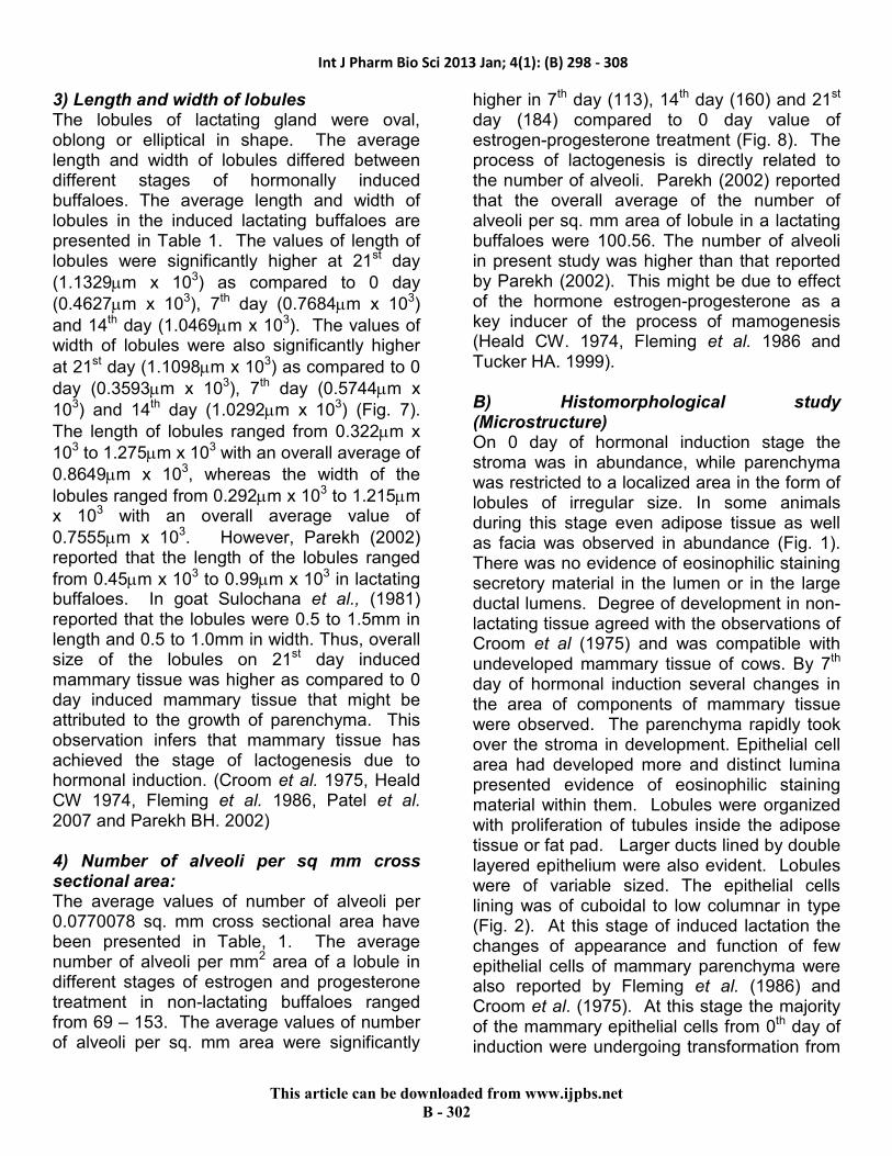
�&��'������������#����The alveoli were oval to elliptical in shape; hence, the smaller and larger diameters of alveoli were measured. The mean values of these measurements were worked out per lobule. The smaller diameter (width) of alveoli
ranged from 0.414 x 10³ to 1.490 x 10³ �m with
an overall mean of 1.003 x 10³�m in different
stages of hormonal induction. The overall average values of smaller diameter of alveoli
were 0.531 x 10³�m, 0.828 x 10³
�m, 1.188 x
103�m, 1.546 x 10³
�m, on 0 day (control), 7th day, 14th day and 21st day respectively of hormonal induction. The alveoli with smaller diameter were significantly more at 7th day, 14th day and 21st day of hormonal induction as compared to control (0 day) (Fig. 5). The larger diameter (length) of alveoli was ranging from
0.568 x 10³ to 1.725 x 10³�m with an average
of 1.089 x 10³�m in different stages of
hormonal induction. The overall average values of larger diameter of alveoli were
0.621�m x 103, 0.845 x 10³�m, 1.214 x 10³
�m
and 1.599 x 10³�m on 0th day (control), 7th day,
14th day and 21st day respectively of hormonal induction. The alveoli with larger diameter were significantly more on 21st day of hormonal induction as compared to 0 day of hormonal induction (Fig. 5). Similar study was carried out by Parekh (2002) in lactating and non lactating buffaloes. She reported that the average of
smaller diameter of alveoli in all lactating
animals rang from 15.90 to 145.75�m and the average of larger diameter of alveoli in all
lactating buffaloes ranged from 40 to 212�m. However, Sulochana ��� ��� (1981) reported in
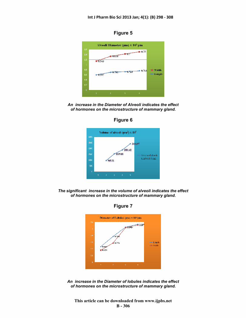
goat that alveoli were ranged from 18 to 80 �m in diameter. Patel ��� ��� (2007) reported that the smaller and larger diameter of alveoli increased when animal developed to lactating stage. However, these parameters were decreased when animal reached to dry stage (i.e. non�lactating stage). The same phenomena occurred in the various stages of hormonal induction. (��"���������#����The volume of the alveoli was derived from those which were measured for their diameter. The volume of alveoli was 99.547 x 10⁵, 304.98
x 105, 904.72 X 10⁵, and 2032.45 x 105cu. �m (Fig. 6). The volume of alveoli was significantly higher in 21st day of hormonal treated animal as compared to that of control. Patel (2007) observed that the volume of alveoli was
significantly higher (1.015 ± 0.362 x 10⁵ �m) in lactating buffalo as compared to that of dry
animal (0.135± 0.029 x 10⁵ �m). Our finding also suggests that the same phenomena occurred in the various stages of hormonal induction.
Int J Pharm Bio Sci 2013 Jan; 4(1): (B) 298 - 308
�
�������������� ������������������������ ������
����9&��
)��*�������� �+� ������ ������The lobules of lactating gland were oval, oblong or elliptical in shape. The average length and width of lobules differed between different stages of hormonally induced buffaloes. The average length and width of lobules in the induced lactating buffaloes are presented in Table 1. The values of length of lobules were significantly higher at 21st day
(1.1329�m x 103) as compared to 0 day
(0.4627�m x 103), 7th day (0.7684�m x 103)
and 14th day (1.0469�m x 103). The values of width of lobules were also significantly higher
at 21st day (1.1098�m x 103) as compared to 0
day (0.3593�m x 103), 7th day (0.5744�m x
103) and 14th day (1.0292�m x 103) (Fig. 7).
The length of lobules ranged from 0.322�m x
103 to 1.275�m x 103 with an overall average of
0.8649�m x 103, whereas the width of the
lobules ranged from 0.292�m x 103 to 1.215�m x 103 with an overall average value of
0.7555�m x 103. However, Parekh (2002) reported that the length of the lobules ranged
from 0.45�m x 103 to 0.99�m x 103 in lactating buffaloes. In goat Sulochana ��� ���� (1981) reported that the lobules were 0.5 to 1.5mm in length and 0.5 to 1.0mm in width. Thus, overall size of the lobules on 21st day induced mammary tissue was higher as compared to 0 day induced mammary tissue that might be attributed to the growth of parenchyma. This observation infers that mammary tissue has achieved the stage of lactogenesis due to hormonal induction. (Croom �������1975, Heald CW 1974, Fleming ��� ���� 1986, Patel ��� ����2007 and Parekh BH. 2002) �,�� ��� ��� �� ��#���� ���� �-� ��� ������������������.��The average values of number of alveoli per 0.0770078 sq. mm cross sectional area have been presented in Table, 1. The average number of alveoli per mm2 area of a lobule in different stages of estrogen and progesterone treatment in non�lactating buffaloes ranged from 69 – 153. The average values of number of alveoli per sq. mm area were significantly
higher in 7th day (113), 14th day (160) and 21st day (184) compared to 0 day value of estrogen�progesterone treatment (Fig. 8). The process of lactogenesis is directly related to the number of alveoli. Parekh (2002) reported that the overall average of the number of alveoli per sq. mm area of lobule in a lactating buffaloes were 100.56. The number of alveoli in present study was higher than that reported by Parekh (2002). This might be due to effect of the hormone estrogen�progesterone as a key inducer of the process of mamogenesis (Heald CW. 1974, Fleming ��� ���� 1986 and Tucker HA. 1999). ���� �������������� ��� ��/���������������On 0 day of hormonal induction stage the stroma was in abundance, while parenchyma was restricted to a localized area in the form of lobules of irregular size. In some animals during this stage even adipose tissue as well as facia was observed in abundance (Fig. 1). There was no evidence of eosinophilic staining secretory material in the lumen or in the large ductal lumens. Degree of development in non�lactating tissue agreed with the observations of Croom ��� �� (1975) and was compatible with undeveloped mammary tissue of cows. By 7th day of hormonal induction several changes in the area of components of mammary tissue were observed. The parenchyma rapidly took over the stroma in development. Epithelial cell area had developed more and distinct lumina presented evidence of eosinophilic staining material within them. Lobules were organized with proliferation of tubules inside the adipose tissue or fat pad. Larger ducts lined by double layered epithelium were also evident. Lobules were of variable sized. The epithelial cells lining was of cuboidal to low columnar in type (Fig. 2). At this stage of induced lactation the changes of appearance and function of few epithelial cells of mammary parenchyma were also reported by Fleming ��� ���� (1986) and Croom �����. (1975). At this stage the majority of the mammary epithelial cells from 0th day of induction were undergoing transformation from

Int J Pharm Bio Sci 2013 Jan; 4(1): (B) 298 - 308
�
�������������� ������������������������ ������
����9&9�
resting, non�differentiated cells into active, differentiated cells. Visualization of well developed epithelial cell area with distinct lumina and evidence of eosinophilic staining material within them agrees with the observation of Fleming �����.(1986) and Croom �����. (1975) Between day seven and fourteen, there was concomitant increase in the epithelial and luminal area and decrease of stroma. Parenchyma was highly proliferated whereas lobules were relatively more organized with complete development of lumen inside. A large number of infiltrating cells (lymphocytes) were found in the intertubular space. The tubules were lined by cuboidal type epithelium where as, at some places even flat epithelium was found. Tubules were well lumenized and some were found with the eosinophilic secretory material inside (Fig. 3). All these observations indicate that a major change had occurred in mammary cells between 7th day and 14th day. Croom �������(1975) and Fleming �����. (1986) observed that in cattle on 16th day of hormonal induction differentiation of mammary epithelial cells were extensive. Alveolar lumina were expanded with secretions and ultrastructurally the parenchyma cells were similar to a differentiated lactating cell. Thus in present experiment the changes in the structure of hormonally induced mammary epithelial cells of buffaloes showed relevance with the experimental findings of hormonally induced mammary gland of cattle. By 14th day the estrogen and progesterone treatment was completed and both had played a major role in the development of labulo�alveolar cells, decrease in stroma, highly proliferated parenchyma with well organized tubules. In mammary tissue of 21st day of induction epithelial area was almost doubled as compared to day 0 and corresponding increase of luminal area and decrease in stroma. Lobules were highly developed and tightly packed with less interalveolar connective tissue. The stroma was comparatively less. In all the lobules prominent secretory cells were present. They were showing eosinophilic
secretory material in their lumen. The linings of epithelium cells were squamous to cuboidal or low columnal in type. In one of the animal, even secretory material was also found in the secretory duct (Fig. 4). The degree of development of 21st day hormonal induced mammary gland was compatible with the developed mammary gland of lactating buffaloes (Patel ��� ���� 2007). As per Tucker (1999), Dexamethasone (glucocorticoid) plays a key role in differentiation of lobule�alveolar system of the mammary gland. He reported that estrogen and progesterone are also playing a key role in the development of mammary gland from non�lactating phase to lactating phase. He stated that estrogen acts in at least two ways for initiation of lactation: 1) in several species it caused release of prolactin from the anterior pituitary gland into blood which in turn, would initiate lactation; and 2) estrogen also increases the number of prolactin receptors in mammary cells which is another lactogenic event. His findings also infer that exogenous progesterone synergized with estrogen induces lobule�alveolar growth. This was also supported by the observation that mammogenesis during pregnancy in cattle coincided with increased secretion of both estrogen and progesterone. At the different stages of hormonal induction mammary gland of buffaloes started developing in to lactating stage due to the effect of estrodiol�17β and progesterone treatment along with dexamethasone (Glucocorticoid). ��������������������������All animals started lactating in the range of about 50�200ml of milk on morning of 19th day of hormonal treatment. This milk was collected from the unoperated quarter of udder. The quantity increased to about 150ml to 350ml from the evening of 19th day to morning of 21st day of hormonal treatment. In the evening of 21st day of hormonal treatment the mammary tissue was collected from the milking quarter. Therefore, it was not possible to collect milk from the evening of 21st day. It is observed that a healthy Surti buffalo can give about 2.5 to 4

Int J Pharm Bio Sci 2013 Jan; 4(1): (B) 298 - 308
�
�������������� ������������������������ ������
����9&;�
liter of milk at a time from all four quarters. In our experimental animals out of four three quarters were operated for the collection of mammary tissue at different stages of hormonal treatment. The operated quarters have inflammation because of surgery and suture. Thus lactation/milk collection from those quarters was not possible. In such case the amount of milk secreted for two days from a single quarter is significant. �������������The level of estrogen and progesterone was less on the 0 day of hormonal induction. It shooted up on 7th day of hormonal induction due to hormonal treatment. The level of hormones in the blood serum on 14th day was greater than the 0th day and lesser than the 7th day of hormonal treatment. As the course of hormonal treatment got completed on 10th day of induction the level of estrogen and progesterone was high as compared to 0th day
of induction. On the 21st day of induction the amount of both the hormones in serum was lesser than the 7th and 14th day of hormonal treatment. This indicates that the hormonal level was high during the course of treatment and it slowly decreases upon completion of the treatment, indicating the utilization of the estrogen and progesterone in the development of microstructure of mammary gland and initiation of lactogenesis. The study of induced lactogenesis was performed on mice, cattle, ewe, sheep etc. (Chakriyarat ������1978, Croom ��� ���� 1975, Delouis ��� ���� 1978, Erb, ��� ����1975, Howe ��� ���� 1975, Fleming ��� ����1986, Alifakiotis ������ 1980, Head et al. 1975). It was performed on buffalo too but the results were unfruitful (Unpublished data). In our case it broke the record of unproductive result of hormonal induction in buffalo by giving the milk yield as well as increase in the size of lobules, alveoli, number of alveoli and volume of alveoli of buffalo’s mammary gland.
#� ������
0���'������� ������
������������� �� ����!�1���������������������� ����������2� ��������������������� �������������������2�3�������������� ���������������� 3�
����������������������� �����+����+���������� � ����������3�
�������

Int J Pharm Bio Sci 2013 Jan; 4(1): (B) 298 - 308
�
�������������� ������������������������ ������
����9&(�
#� ����"�4���'������� ������
��
5�����2� �� �����+�������������������� ��������� ���������� ����������������������3��� ���������#���� ��������2��+������+�����������3��6�������������+���� ���#� �
�#� ����$�
&,���'������� ������
��
�� ���������������#�������������2� �+�������������� �#���� ����������� �3�������� ��������������� ���� � ��������������������3����������������������������������3��1��������������� ���������������#����
���3���� ���������+����������2� ��� ���� ������ ����+�������������������������������� ��
�#� ����!�
(&���'������� ������
���� ���������������� �#���� �+���������������� ����������3�������������� � ������+����������������3������������
���������������������� �������� ���#� ������������������+��� ����3�
Int J Pharm Bio Sci 2013 Jan; 4(1): (B) 298 - 308
�
�������������� ������������������������ ������
����9&)�
�#� ����%�
�
��
��������������������'������������#������ ������������������������������������������������������������� 3�
�#� ����&�
�
�
���������������������������������#���������#������ ������������������������������������������������������������� 3�
�#� ���� �
�
��������������������'����������� ������� ������������������������������������������������������������� 3�

Int J Pharm Bio Sci 2013 Jan; 4(1): (B) 298 - 308
�
�������������� ������������������������ ������
����9&'�
�#� ����'�
�
�
������� ��������#����������������7(������-���/��0� ������&7,�/��(&� ������������������������� ���������������������������
���������������������������������� 3
�
������ ���������
�This study was funded by Department of Biotechnology, Ministry of Science & Technology, Government of India (Grant No.BT/PR 5984/AAQ/01/228/2005, dated 9�05�2006). Authors would also like to acknowledge Dr Javed Khan, Professor & Head, Department
of English, SP University, Vallabh Vidyanagar and Dr RB Subramanian, Professor, BRD School of Biosciences, Vallabh Vidyanagar for their esteemed guidance in preparation of manuscript.
����������
�1. Alifakiotis TA, Katanos K, Hatjiminaoglou I,
Zervas N, Zirfiridis G. (1980) Induced lactation in dairy ewes by various brief hormone treatments.������ � 63, 750.
2. Chakriyarat S, Head HH, Thatcher WW, Neal FC, Wilcox CJ (1978) Induction of lactation: Lactational, physiological, and hormonal responses in the bovine. ����� � 61, 1715–1724.
3. Collier, RJ, Bauman, DE, Hays. RL (1975) Milk Production and Reproductive Performance of Cows Hormonally Induced into Lactation. ����� 58, 1524�1527.
4. Croom WJ, Collier RJ, Bauman DE, Hays RL (1975) Cellular studies of Mammary Tissue from Cows Hormonally Induced into Lactation: Histology and uItrastructure. ����� � 59 (7), 1232�1246.
5. Delouis CD, Djiane J, Kann G, Terqui M, Head HH (1978) Induced lactation in cows and heifers by short�term treatment with steroid hormones. ����� ����� ���� �� ������������18 721.
6. Erb RE, Monk EL, Callahan CJ (1975) Salvaging infertile dairy cows by inducing lactation with hormones. ���� � ���������� 8, 8
7. Head HH, Delouis C, Terqui M, Kann G, Djiane J (1975) Hormonal induction of lactation in sheep. ���� � � 58 140. (Abstract)
8. Heald CW (1974) Hormonal effects on mammary cytology.������ 57, 917.
9. Humanson GL (1979) Animal Tissue Techniques. 4th Edn. W.H. Freeman & Co., San Francisco.

Int J Pharm Bio Sci 2013 Jan; 4(1): (B) 298 - 308
�
�������������� ������������������������ ������
����9&��
10. Howe JE, Heald CW, Bib TL (1975) Histology of Induced Bovine Lactogenesis. ����� � 58 (6), 853�860
11. Fleming, JR, Head HH, Bachman, KC, Becker HN, Wilcox CJ (1986) Induction of Lactation: Histological and Biochemical Development of Mammary Tissue and Milk Yields of Cows injected with Estradiol�173 and Progesterone for 21 Days. ���� � 69, 3008�3021
12. Koringa PG, Patel AK, Nandasana, KN, ����� (2008) Mammary gland biopsy in pregnant and lactating buffaloes in standing position ������ ���� ����� 29 (1), 50�51
13. Meites J (1961) Hormonal induction of lactation and galactopoiesis. Milk: The mammary gland and its secretions. Vol. I. Page 321 (S. K. Kon and A. T. Cowie, ed. Academic Press, New York and London).
14. MeClung�Jones R (1966) Basic microscopic techniques ���� ��� Univ. Chicago Press, Chicago, IL , Page 242
15. Panchal KM, Vyas YL (2005) The anatomy of udder of Buffalo: A complete monologue. Department of Anatomy and Histology, Anand Agricultural University, Anand.
16. Patel AK, Koringa PG, Nandasana KN, ������� � !!"# Comparative histology of mammary gland in heifer, pregnant, lactating and dry buffaloes. ������ ������������ 19(1), 71�78.
17. Patel AK, Koringa PG, Nandasana KN, ������� (2007) Effect of bovine somatotropin (bST) administration on the histology of mammary gland in lactating buffaloes. ������������������ 19(2), 22�28.
18. Parekh BH (2002) Gross and microscopic studies on the udder of lactating and non�lactating, non�pregnant adult buffaloes (Bubalus bubalis). M.VSc. thesis submitted to Gujarat Agricultural University, Anand.
19. Sanders BJ ($%" #� Animal Histology Procedures of the Pathological Technology Section of the National Cancer Institute. US Govt. Printing Office Stock #1742�0043, Page 34.
20. Smith KL, Schanbacher FL (1973) Hormone induced lactation in the bovine. I. Lactational performance following injections of 17β�estradiol and progesterone. ����� 56, 738.
21. Smith KL, Redman DR, Schanbacher FL (1973) Efficiency of 17β�estradiol and progesterone treatment to initiate lactation in infertile cows. ���� � 56, 657(Abstract).
22. Smith KL, Muir LA, Ferguson LC, Conrad HR (1971) Selective transport of IgG1 into the mammary gland: Role of estrogen and progesterone. ����� 54, 1886.
23. Sulochana S, Haffezuddin M, Singh UB (1981). Histological and histochemical studies on the mammary gland of the Indian goat (&���� � ��). ��������� �������'�(�����)�������*�+���� � XI (2), 287�291.
24. Topper YJ, Freeman CS (1980) Multiple hormone interactions in the developmental biology of the mammary gland. ,������+�( 60, 1049�1106
25. Tucker HA (1969) Factors affecting mammary gland cell numbers.� ���� � . 52, 720
26. Tucker HA (1971) Hormonal response to milking ����� � �������� $#32, 137(Abstract)
27. Tucker HA (1974) General endocrinological control of lactation. �Lactation: A comprehensive treatise. Vol. I. Page 277 (B. L. Larson and V. R. Smith, ed. Academic Press, New York and London).
28. Tucker HA (1999) Hormonal regulation of milk synthesis Hormones, Mammary Growth, and Lactation: a 41 year perspective������ 83, 874�884.
.


