Embed Size (px)
Citation preview

ORIGINAL ARTICLE
The evolutionarily conserved EBR module of RALT/MIG6 mediates
suppression of the EGFR catalytic activity
S Anastasi1, MF Baietti1,3, Y Frosi1,3, S Alema2 and O Segatto1
1Department of Experimental Oncology, Laboratory of Immunology, Regina Elena Cancer Institute, Rome, Italy and 2Institute of CellBiology, CNR, Monterotondo, Italy
Physiological signalling by the epidermal growth factorreceptor (EGFR) controls developmental processes andtissue homeostasis, whereas aberrant EGFR activitydrives oncogenic cell transformation. Under normalconditions, the EGFR must therefore generate outputsof defined strength and duration. To this aim, cells balanceEGFR activity via different modalities of negativesignalling. Increasing attention is being drawn ontranscriptionally controlled feedback inhibitors of EGFR,namely RALT/MIG6, LRIG1, SOCS4 and SOCS5.Genetic studies in mice have revealed the essential role ofRalt/Mig6 in regulating Egfr-driven skin morphogenesisand tumour formation, yet the mechanisms through whichRALT abrogates EGFR activity are still undefined. Wereport that RALT suppresses EGFR function by inhibit-ing its catalytic activity. The evolutionarily conservedErbB-binding region (EBR) is necessary and sufficient tocarry out RALT-dependent suppression of EGFR kinaseactivity in vitro and in intact cells. The mechanisminvolves binding of the EBR to the 953RYLVIQ958sequence, which is located in the aI helix of the EGFRkinase and has been shown to participate in allostericcontrol of EGFR catalytic activity. Our results uncover anovel mechanism of temporal regulation of EGFR activityin vertebrate organisms.Oncogene (2007) 26, 7833–7846; doi:10.1038/sj.onc.1210590;published online 18 June 2007
Keywords: EGFR; RALT; MIG6; feedback inhibition;negative signalling; tumour suppression
Introduction
Signal output by receptor tyrosine kinases (RTKs)is controlled via a dynamic equilibrium betweenmechanisms of signal generation and signal extinction.Significantly, unrestrained RTK activity may cause
developmental abnormalities and post-natal pathologiessuch as cancer (Fiorini et al., 2001).
The epidermal growth factor (EGF) receptor (EGFR)has served as a paradigm model system to studymechanisms of negative signalling to RTKs. Onceengaged by EGF, and concomitantly to the initiationof its signalling activity, the EGFR is rapidly removedfrom the cell surface. Internalized EGF:EGFR com-plexes are destined to be recycled to the cell surfaceunless sustained EGFR signalling leads to receptorubiquitylation (Marmor and Yarden, 2004). The role ofubiquitin tags is to divert EGFR molecules to lateendosomes/multivescicular bodies, thus promotingtheir eventual degradation in lysosomes (Marmor andYarden, 2004; Sigismund et al., 2004). While recyclingallows for recursive EGFR signalling (Wiley et al.,2003), trafficking to lysosomes results in the depletionof both ligands and receptors, ultimately leading tosignal attenuation (Marmor and Yarden, 2004; Poloet al., 2004).
Recent studies have indicated that negative signallingto the EGFR is reinforced over time by the activity offeedback inhibitors, namely LRIG1 (Gur et al., 2004;Laederich et al., 2004), SOCS4, SOCS5 (Kario et al.,2005; Nicholson et al., 2005) and RALT (also known asMIG6 or Gene 33) (Hackel et al., 2001; Anastasi et al.,2003; Xu et al., 2005), which are expressed in the contextof transcriptional responses triggered by EGFR itself.RALT was the first feedback inhibitor of EGFR to bediscovered in mammalian cells. Its overexpression incultured cells causes potent suppression of mitogenicand transforming signals downstream to EGFR andother ErbB RTKs (Fiorentino et al., 2000; Hackel et al.,2001; Anastasi et al., 2003; Xu et al., 2005). Conversely,loss of RALT signalling mediated by RNAi enhancesthe mitogenic potency of suboptimal doses of EGF. Thisis paralleled by amplified ERK and AKT activation aswell as increased accumulation of cyclins crucial for theG1/S transition (Anastasi et al., 2005; Xu et al., 2005).Recent genetic studies in mice have yielded results fullyconsistent with the notion that RALT is a physiologicalsuppressor of EGFR signalling. Thus, Errfi1�/� (ErbBreceptor feedback inhibitor 1 is the HUGO designationfor the gene encoding Ralt/Mig6) mice display a highlypenetrant skin phenotype characterized by abnormalproliferation of keratinocytes and enhanced carcinogen-esis in skin (Ferby et al., 2006). This phenotype is to be
Received 23 January 2007; revised 3 May 2007; accepted 15 May 2007;published online 18 June 2007
Correspondence: Dr O Segatto and S Anastasi, Laboratory ofImmunology, Regina Elena Cancer Institute, via Delle Messi d’Oro,156/158, 00158 Rome, Italy.E-mails: [email protected] and [email protected] authors contributed equally to this work.
Oncogene (2007) 26, 7833–7846& 2007 Nature Publishing Group All rights reserved 0950-9232/07 $30.00
www.nature.com/onc

ascribed to excess Egfr signalling, since it is rescued inthe wa2/wa2 background (the waved 2 allele encodes ahypomorphic Egf receptor, see Luetteke et al., 1994) andmay be prevented by the administration of gefitinib,a clinically used EGFR inhibitor (Ferby et al., 2006).In remarkable consonance, targeted expression of aK14-Errfi1 transgene in mouse skin (Ballaro et al., 2005)produced a phenotype similar to that observed in wavedmice (Luetteke et al., 1993, 1994).
An outstanding issue is the clarification of themechanistic basis of RALT signalling to EGFR. RALTis able to form a physical complex with ligand-activatedEGFR via its EBR (ErbB binding region, Anastasiet al., 2003). This datum suggests that RALT may act ata receptor proximal level. A clue to a role of RALT indirect regulation of EGFR signalling comes from therecent observation that loss of RALT expression isassociated with increased tyrosine phosphorylation ofEGFR in both primary keratinocytes obtainedfrom Errfi1�/� mice (Ferby et al., 2006) and immortalcell lines subjected to RNAi (Ballaro et al., 2005; Xuet al., 2005).
Here, we report that RALT inhibits the catalyticfunction of the EGFR. Binding of the EBR module tothe EGFR kinase domain is necessary and sufficient tocarry out RALT-mediated suppression of EGFR kinaseactivity in vitro and in intact cells. We also demonstratestructural and functional conservation of the EBRthroughout the evolution of vertebrates.
Results
The EBR module of RALT inhibits the catalyticfunction of EGFRUnlike LRIG1 (Gur et al., 2004; Laederich et al., 2004),SOCS4 and SOCS5 (Kario et al., 2005), which suppressEGFR signalling by promoting its ubiquitin-drivendegradation, ectopic RALT was able to reduce CBL-dependent ubiquitylation of EGFR (data not shown). Theability of RALT to antagonize CBL function correlatedwith reduced autophosphorylation of the EGFR. RALT-driven suppression of EGFR autophosphorylation wasdosage dependent, as also reported by Kyriakis andcoworkers (Xu et al., 2005), and required the integrity ofthe EBR module (Supplementary Figure 1).
Ectopically expressed RALT inhibited to a similarextent the phosphorylation of Tyr 845, 992, 1045, 1068and 1173 of EGFR (Figure 1a). Global inhibition ofEGFR phosphorylation was sustained (Figure 1a) and
could be observed over a wide range of EGF concentra-tions (Figure 2a). Ectopic RALT inhibited also theautophosphorylation of ERBB2 and ERBB4, whereastyrosine phosphorylation of PDGFR was similar incontrol and RALT overexpressing NIH-EGFR cells(Supplementary Figure 2). The minimal EBR wasmapped to aa. 323–372 (Anastasi et al., 2003). Ectopi-cally expressed RALT282–396, that is the shortest EBRcontaining peptide which could be expressed at satisfac-tory levels in mammalian cells, was sufficient torecapitulate the suppression of EGFR signallingmediated by RALT (Figure 1a), in agreement with itsability to form a ligand-dependent complex with EGFRin living cells (Figure 1b). Importantly, RALT andRALT282–396 were capable of inhibiting EGFR activa-tion by TGFa (Figure 2b), a ligand that renders theEGFR refractory to the suppressive activity mediatedby the endocytosis/degradation pathway.
Suppression of EGFR phosphorylation by RALTand RALT282–396 led also to robust attenuation ofdownstream signalling, as assessed by probing celllysates with phospho-antibodies against activated ERKsand AKT (Figure 1a). In general, inhibition of ERK andAKT activation was stronger in cells expressingRALT282–396, which we interpret as a reflection of thelower residual EGFR activity observed in cells expres-sing RALT282–396, in comparison to those expressingfull-length RALT. Some subtle differences could also beobserved when comparing the kinetics of ERK andAKT activation in control and RALT/RALT282–396
expressing cells. While AKT activation was reduced in arobust and proportional fashion at all time points,inhibition of ERK activation was less pronounced at theearliest time point of EGF stimulation (5min). Underinitial conditions, it is conceivable that the occupancy ofligand-activated EGFR molecules by either RALT orRALT282–396 lags behind the catalytic amplificationintrinsic to the RAS-ERK signalling module, thusallowing for an initial, albeit short-lived, burst ofEGF-induced ERK activity.
In principle, RALT could impact EGFR autopho-sphorylation by either inhibiting EGFR catalyticactivity or enhancing phosphate removal by phospho-tyrosine phosphatases (PTPs). Inhibition of PTP activityby pervanadate, while able to potentiate EGFRautophosphorylation in control cells, did not rescueEGFR autophosphorylation in RALT overexpressingcells (data not shown). In contrast, purified recombinantglutathione S-transferase (GST)-RALT proteins con-taining an intact EBR (Figure 3a) were able to suppress
Figure 1 RALT suppresses global EGFR autophosphorylation in intact cells via its EBR module. (a) MYC-tagged RALT andRALT282–396 were expressed in NIH-EGFR cells via retrovirus infection followed by puromycin selection. Control cells (PIE) wereinfected with empty virus. Quiescent cells were lysed either before or after stimulation at 371C with 20 ng/ml EGF for the indicated time(minutes). Equal amounts of cell lysate were analysed by immunoblot with the indicated antibodies. Detection was by ECL coupled toautoradiography. (b) MYC-tagged versions of RALT and RALT282–396 were expressed in HeLa cells via retrovirus infection followedby puromycin selection. Cells infected with empty PIE retrovirus were used as control. Cells were rendered quiescent by serumdeprivation and lysed either before or after stimulation with 20 ng/ml EGF for 5min at 371C. Lysates were either subjected toimmunoprecipitation with anti-EGFR MoAb 108 followed by immunoblot analysis with anti-EGFR and anti-MYC antibodies (leftpanel) or analysed directly by immunoblot (right panel). Detection was by ECL coupled to autoradiography.
RALT inhibits the kinase activity of EGFRS Anastasi et al
7834
Oncogene

the catalytic activity of purified recombinant humanEGFR (aa 672–1186, henceforth referred to as EGFR)in a number of in vitro kinase assays (see below).
Preincubation of EGFR with a 20-fold molar excessof GST had no consequences on EGFR autophos-phorylation, whereas anti-p-Tyr immunoreactivity was
RALT inhibits the kinase activity of EGFRS Anastasi et al
7835
Oncogene

abolished in reactions containing the EGFR chemicalinhibitor AG1478 (Figure 3b). Preincubation of EGFRwith GST-RALT282–396 caused a dose-dependent
reduction of anti-pTyr immunoreactivity (Figure 3b),with IC50 values similar to those determined for theinsulin receptor kinase inhibitor GRB-14 (Bereziat et al.,2002), and was associated to loss of reactivity with site-specific p-Tyr antibodies as well (Figure 3c). The in vitroautophosphorylation of EGFR was blunted also by theGST-RALT262–459 peptide, which nevertheless lost itssuppressive activity upon deletion of aa 315–361 (DEBRmutant) (Figures 3a and c). Recombinant peptidesspanning positions 284–399 and 325–375 of humanRALT (whether used as GST fusions or purified afterGST tag removal) were likewise able to inhibit EGFRautophosphorylation and they did so independently ofbeing preincubated with EGFR (not shown) or mixedwith EGFR concomitantly to the start of the kinasereaction (Figure 3d). Densitometric scan of the auto-radiogram shown in Figure 3d indicated that recombi-nant HsRALT fragments suppressed EGFRautophosphorylation by about 30 and 90% when addedat 4- and 16-fold molar excess over EGFR, respectively.
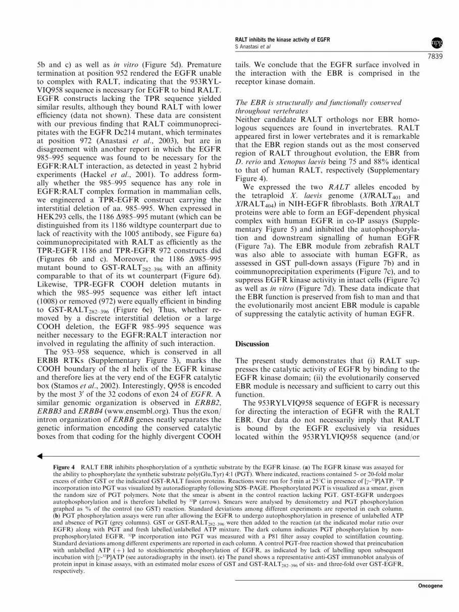
Next, we assayed the influence of RALT on EGFR-driven phosphorylation of the synthetic substrate poly(Glu,Tyr) 4:1 (PGT). As shown in Figure 4a, preincuba-tion of GST-RALT282–396 and GST-RALT262–459
caused a dose-dependent reduction of phosphateincorporation into PGT, whereas neither GST northe DEBR derivative of GST-RALT262–459 were ableto suppress PGT phosphorylation by EGFR. Substratephosphorylation was suppressed also when theEGFR was allowed to undergo maximal autophos-phorylation with cold ATP before the addition of GST-RALT282–396, PGT and [g-32P]ATP to the reaction(Figure 4b). The latter experiment ruled out thatinhibition of substrate phosphorylation in reactionscontaining GST-RALT282–396 could be due to reducedEGFR autophosphorylation, as reported for somepeptide substrates (Hsu et al., 1991), rather than todirect inhibition of EGFR catalytic activity.
RALT binds to the EGFR kinase domainTyrosine kinases show a great deal of plasticity, beingsubjected to tight allosteric constraints exerted bystructural determinants residing in the kinase domainitself or in adjacent regulatory modules (Huse and
Figure 2 RALT suppresses EGFR autophosphorylation inducedby TGFa and independently of EGF concentration. (a) QuiescentPIE, RALT and RALT282–396 derivatives of NIH-EGFR cells werestimulated with the indicated concentrations of EGF for 5min at371C. Lysates were immunoblotted with the indicated antibodies.Detection was by ECL coupled to autoradiography. (b) QuiescentPIE, RALT and RALT282–396 derivatives of NIH-EGFR cells werestimulated at 371C with either EGF (10 ng/ml for 5min) or TGFa(50 ng/ml for 5 and 15min). Lysates were immunoblotted with theindicated antibodies. Detection was by ECL coupled to autoradio-graphy.
Figure 3 RALT EBR is necessary and sufficient to suppress EGFR autophosphorylation in vitro. (a) Schematic representation ofGST fusion proteins spanning aa 282–396, 262–459 and 262–459 DEBR of rat (Rn) RALT. Full-length RALT is shown as reference.(b) Purified recombinant EGFR (aa 672–1186) was assayed for autophosphorylation in 1 and 5min reactions at 251C after 30minpreincubation with either GST or GST-RALT282–396 at the indicated molar excess over EGFR. Controls included reactions run in theabsence of GST �/þ the EGFR chemical inhibitor AG1478. Reaction products were analysed by sequential immunoblotting withanti-p-Tyr and anti-EGFR antiserum. Detection was by ECL followed by autoradiography. The top anti-p-Tyr panels were exposedfor the same time, the middle panel shows a shorter exposure of the 5min reaction. For this representative experiment thedensitometric analysis of anti-p-Tyr reactivity normalized for kinase input is reported in the graph. Data are plotted as % of GSTcontrol. Variability among different experiments did not exceed 15%. (c) The autophosphorylation of GST-EGFR was assayed in vitroafter a 30min preincubation with a 20-fold molar excess of the indicated GST proteins. Reactions were run at 251C for 5min andproducts analysed by sequential immunoblotting with the indicated antibodies. Detection was by ECL coupled to autoradiography.(d) EGFR autophosphorylation was assayed in a 7min reaction at 41C. The indicated recombinant human (Hs) RALT proteins weremixed with EGFR simultaneously with the ATP-containing kinase reaction mix at the indicated molar ratio over EGFR. Reactionproducts were analysed by sequential immunoblot with the indicated antibodies. The lower panel shows a Coomassie stain of purifiedHsRALT proteins (�/þ GST tag) used in the kinase reaction. To facilitate detection, the input of each protein was increased by25-fold in comparison to the actual input in the kinase reaction. Molecular-weight markers were loaded in the rightmost lane.
RALT inhibits the kinase activity of EGFRS Anastasi et al
7836
Oncogene

Kuriyan, 2002). Thus, the identification of the EGFRsurface necessary for RALT binding may provideinsights into the mechanism underlying the EBR
suppressive activity. We engineered stepwise COOHdeletions of the EGFR intracellular domain and assayedthese mutants for their ability to recruit RALT in co-IP
RALT inhibits the kinase activity of EGFRS Anastasi et al
7837
Oncogene

and GST pull-down assays. Cytoplasmic sequences ofEGFR (Figure 5a) were fused to the dimerizationdomain of TPR present in the TPR-MET oncogene
(Ponzetto et al., 1994), to mimic ligand-driven EGFRdimers. EGFR constructs extending to Q958, or beyond,were fully capable to bind RALT in intact cells (Figures
RALT inhibits the kinase activity of EGFRS Anastasi et al
7838
Oncogene
5b and c) as well as in vitro (Figure 5d). Prematuretermination at position 952 rendered the EGFR unableto complex with RALT, indicating that the 953RYL-VIQ958 sequence is necessary for EGFR to bind RALT.EGFR constructs lacking the TPR sequence yieldedsimilar results, although they bound RALT with lowerefficiency (data not shown). These data are consistentwith our previous finding that RALT coimmunopreci-pitates with the EGFR Dc214 mutant, which terminatesat position 972 (Anastasi et al., 2003), but are indisagreement with another report in which the EGFR985–995 sequence was found to be necessary for theEGFR:RALT interaction, as detected in yeast 2 hybridexperiments (Hackel et al., 2001). To address form-ally whether the 985–995 sequence has any role inEGFR:RALT complex formation in mammalian cells,we engineered a TPR-EGFR construct carrying theinterstitial deletion of aa. 985–995. When expressed inHEK293 cells, the 1186 D985–995 mutant (which can bedistinguished from its 1186 wildtype counterpart due tolack of reactivity with the 1005 antibody, see Figure 6a)coimmunoprecipitated with RALT as efficiently as theTPR-EGFR 1186 and TPR-EGFR 972 constructs did(Figures 6b and c). Moreover, the 1186 D985–995mutant bound to GST-RALT282–396 with an affinitycomparable to that of its wt counterpart (Figure 6d).Likewise, TPR-EGFR COOH deletion mutants inwhich the 985–995 sequence was either left intact(1008) or removed (972) were equally efficient in bindingto GST-RALT282–396 (Figure 6e). Thus, whether re-moved by a discrete interstitial deletion or a largeCOOH deletion, the EGFR 985–995 sequence wasneither necessary to the EGFR:RALT interaction norinvolved in regulating the affinity of such interaction.
The 953–958 sequence, which is conserved in allERBB RTKs (Supplementary Figure 3), marks theCOOH boundary of the aI helix of the EGFR kinaseand therefore lies at the very end of the EGFR catalyticbox (Stamos et al., 2002). Interestingly, Q958 is encodedby the most 30 of the 32 codons of exon 24 of EGFR. Asimilar genomic organization is observed in ERBB2,ERBB3 and ERBB4 (www.ensembl.org). Thus the exon/intron organization of ERBB genes neatly separates thegenetic information encoding the conserved catalyticboxes from that coding for the highly divergent COOH
tails. We conclude that the EGFR surface involved inthe interaction with the EBR is comprised in thereceptor kinase domain.
The EBR is structurally and functionally conservedthroughout vertebratesNeither candidate RALT orthologs nor EBR homo-logous sequences are found in invertebrates. RALTappeared first in lower vertebrates and it is remarkablethat the EBR region stands out as the most conservedregion of RALT throughout evolution, the EBR fromD. rerio and Xenopus laevis being 75 and 88% identicalto that of human RALT, respectively (SupplementaryFigure 4).
We expressed the two RALT alleles encoded bythe tetraploid X. laevis genome (XlRALT401 andXlRALT404) in NIH-EGFR fibroblasts. Both XlRALTproteins were able to form an EGF-dependent physicalcomplex with human EGFR in co-IP assays (Supple-mentary Figure 5) and inhibited the autophosphoryla-tion and downstream signalling of human EGFR(Figure 7a). The EBR module from zebrafish RALTwas also able to associate with human EGFR, asassessed in GST pull-down assays (Figure 7b) and incoimmunoprecipitation experiments (Figure 7c), and tosuppress EGFR kinase activity in intact cells (Figure 7c)as well as in vitro (Figure 7d). These data indicate thatthe EBR function is preserved from fish to man and thatthe evolutionarily most ancient EBR module is capableof suppressing the catalytic activity of human EGFR.
Discussion
The present study demonstrates that (i) RALT sup-presses the catalytic activity of EGFR by binding to theEGFR kinase domain; (ii) the evolutionarily conservedEBR module is necessary and sufficient to carry out thisfunction.
The 953RYLVIQ958 sequence of EGFR is necessaryfor directing the interaction of EGFR with the RALTEBR. Our data do not necessarily imply that RALTis bound by the EGFR exclusively via residueslocated within the 953RYLVIQ958 sequence (and/or
Figure 4 RALT EBR inhibits phosphorylation of a synthetic substrate by the EGFR kinase. (a) The EGFR kinase was assayed forthe ability to phosphorylate the synthetic substrate poly(Glu,Tyr) 4:1 (PGT). Where indicated, reactions contained 5- or 20-fold molarexcess of either GST or the indicated GST-RALT fusion proteins. Reactions were run for 5min at 251C in presence of [g-32P]ATP. 32Pincorporation into PGT was visualized by autoradiography following SDS–PAGE. Phosphorylated PGT is visualized as a smear, giventhe random size of PGT polymers. Note that the smear is absent in the control reaction lacking PGT. GST-EGFR undergoesautophosphorylation and is therefore labelled by 32P (arrow). Smears were analysed by densitometry and PGT phosphorylationgraphed as % of the control (no GST) reaction. Standard deviations among different experiments are reported in each column.(b) PGT phosphorylation assays were run after allowing the EGFR to undergo autophosphorylation in presence of unlabelled ATPand absence of PGT (grey columns). GST or GST-RALT282–396 were then added to the reaction (at the indicated molar ratio overEGFR) along with PGT and fresh labelled/unlabelled ATP mixture. The dark column indicates PGT phosphorylation by non-prephosphorylated EGFR. 32P incorporation into PGT was measured with a P81 filter assay coupled to scintillation counting.Standard deviations among different experiments are reported in each column. A control PGT-free reaction showed that preincubationwith unlabelled ATP (þ ) led to stoichiometric phosphorylation of EGFR, as indicated by lack of labelling upon subsequentincubation with [g-32P]ATP (see autoradiography in the inset). (c) The panel shows a representative anti-GST immunoblot analysis ofprotein input in kinase assays, with an estimated molar excess of GST and GST-RALT282–396 of six- and three-fold over GST-EGFR,respectively.
RALT inhibits the kinase activity of EGFRS Anastasi et al
7839
Oncogene

immediately NH2 to it). With this caution, we note thatthere are several interesting features in the 953RYL-VIQ958 sequence worth to be discussed here. This motifis conserved in all ErbB family members from fish toman (data not shown), cannot be retrieved from TK
sequences other than those of ErbB receptors (consis-tently with the notion that ErbB receptors are so far theonly RTKs able to bind RALT (Fiorentino et al., 2000;Hackel et al., 2001)) and is necessary to direct also theinteraction between ERBB2 and RALT in co-IP assays
Figure 5 The EGFR surface involved in RALT recognition is comprised in the tyrosine kinase domain. (a) Schematic representationof TPR-EGFR constructs. EGFR sequences fused to the TPR dimerization domain comprise the juxtamembrane region (JM), thetyrosine kinase box (TK) and the COOH-terminal tail (C-tail). Numbering corresponds to residues in wt EGFR (1186 residues,accession P00533). The ability of each recombinant protein to complex with RALT in co-IP assays is reported on the right (RALTbinding). (b and c) MYC-TPR or the indicated MYC-tagged TPR-EGFR chimeras were coexpressed with HA-RALT in HEK 293cells. Lysates were subjected to immunoprecipitation with anti-HA antibody followed by anti-MYC immunoblot (b; upper left panel)or the reciprocal IP/WB combination (c, lower left panel). Filters were stripped and reprobed with the immunoprecipitating antibody(lower left panel in b; upper left panel in c). Lysates correspond to 6% of the input in immunoprecipitations (b and c, right panels).Detection was by ECL coupled to autoradiography. (d) TPR and the indicated TPR-EGFR fusion proteins were expressed in HEK293cells. Lysates were incubated with purified GST or GST-RALT282–396 immobilized onto glutathione–agarose beads. Precipitatedproteins (upper left panel) and lysates corresponding to 5% of the input in binding assays (right panel) were analysed by immunoblotwith anti-MYC antibodies. Loading of GST proteins in binding assays was verified by Ponceau red staining of filter (lower left panel).
RALT inhibits the kinase activity of EGFRS Anastasi et al
7840
Oncogene

(Supplementary Figure 3). Mutations of the LVI motifof ERBB3 and ERBB4, colinear to that of the953RYLVIQ958 EGFR sequence, were found toabolish trans-activation of the ERBB2 kinase (Schaeferet al., 1999). This led to the suggestion that the LVImotif plays a crucial role in the catalytic activation ofERBB dimers (Stamos et al., 2002). Indeed, recentstructural data support a model in which activation ofthe EGFR catalytic function takes place in the contextof asymmetric dimers in which the COOH lobe of thecatalytic domain of one receptor contacts the NH2 lobeof the partner kinase domain, imposing to the latter acatalytically productive conformation (Zhang et al.,2006). Arg953 and Val956 participate in the creation ofthis asymmetric dimer interface (Zhang et al., 2006),likely explaining why mutations of the LVI motifprevent ERBB trans-activation (Schaefer et al., 1999).Within this framework, it may be proposed that, bycontacting the aI helix of the EGFR kinase domain, theEBR prevents the COOH lobe from exerting its trans-activating function within receptor dimers. This will beformally addressed by solving the structure of theEGFR TK:EBR complex.
From the cell biology standpoint, the most relevantissue is how the EBR suppressive activity may accountfor the essential role of RALT in negative signalling toEGFR (Ferby et al., 2006) and, possibly, the entireERBB network. In this regard, it is productive tocontrast the catalytic suppression mediated by the EBRwith other modalities of negative signalling to EGFR.We note that, despite its being regarded by a prevailingopinion as the major element of negative regulationof EGFR, the internalization/degradation pathway isnevertheless ineffective in several physiological scenar-ios. Low (o2 ng/ml), and yet biologically relevant, dosesof EGF do not trigger ubiquitylation and ensuingdegradation of EGFR (Sigismund et al., 2005). Like-wise, EGFR ligands such as epiregulin (Shelly et al.,1998), epigen (Kochupurakkal et al., 2005) and TGFa(Lenferink et al., 1998; Longva et al., 2002) do nottrigger receptor degradation, even at high doses. ERBB2is refractory to endocytosis/degradation (Baulida et al.,1996) and may act trans-dominantly to inhibit thedegradation of EGFR molecules recruited in EGFR.ERBB2 hetero-dimers (Lenferink et al., 1998; Hendrikset al., 2003; Haslekas et al., 2005). It is thereforeremarkable that degradation–refractory ERBB signal-ling units, which are endowed with robust signallingactivity (Citri and Yarden, 2006), are liable to inhibitionby RALT, as exemplified by the ability of RALT tosuppress (a) the autophosphorylation of EGFR acti-vated by TGFa and low doses of EGF and (b) theautophosphorylation of ERBB2.
The added value of RALT is also evident when weplace its function in the context of global feedbackregulation of EGFR signalling. Here, the EBR-suppres-sive function emerges as an essential control element ofEGFR activity in time and space. LRIG1, SOCS4 andSOCS5 inhibit EGFR by enhancing its ubiquitylationand ensuing degradation (Gur et al., 2004; Laederichet al., 2004; Kario et al., 2005). EGFR molecules
targeted by LRIG1 and SOCS4/5 are therefore expectedto retain full signalling activity until they are segregatedinto the degradative compartment (Miaczynska et al.,2004). In contrast, binding of RALT to the EGFR leadsto the immediate blockade of EGFR catalytic activity.As a consequence, we envision that signalling by EGFRmolecules that form a complex with RALT immediatelyafter ligand binding is aborted at the plasmamembrane(or within its immediate proximity). Most likely, RALTis also able to bind trafficking EGFR molecules. This isrelevant, since RALT becomes expressed at a time inwhich cells contain a sizeable population of activatedEGFRs that are being routed through the endosomalcompartment. The blockade of kinase activity imposedby RALT to trafficking EGFR molecules is expected toshift the kinase/PTPs equilibrium in favour of PTPs,therefore leading to a precipitous decline of pY-EGFRlevels and concomitant loss of signalling competence.Consistently with this scenario, a dramatic abbreviationof the kinetics of EGFR dephosphorylation is observedin EGF stimulated cells when they are washed free ofEGF and chased in presence of a pharmacologicalinhibitor of the EGFR kinase (Supplementary Figure 6).
Finally, it must be noted that the RALT negativefeedback loop operates in the same window of time inwhich ErbB signalling is reinforced by positive feedbackloops established by ErbB ligands, such as TGFa, whoseexpression is regulated at the transcriptional level by theRAS-ERK pathway (Schulze et al., 2001). Given thatTGFa uncouples internalized EGFR molecules fromdegradation by driving their rapid recycling to the cellsurface (Lenferink et al., 1998; Longva et al., 2002),RALT may represent the major, if not the only, means ofrestraining the recursive positive feedback loop generatedby autocrine stimulation of the EGFR via TGFa.
Concluding remarksThe feedback inhibitors Aos and Kek1 restrain Droso-phila EGF receptor (DER) activity by preventing thebinding of ligands to DER (Shilo, 2003). We note thatorthologs of Aos and Kek1 have not been identified invertebrates and hypothesize that RALT may haveevolved to preserve in vertebrates a transcriptionallycontrolled feedback loop dedicated to a fast terminationof ErbB signalling. This function is essential, as geneticablation of Errfi1 leads to widespread tumourigenesis inthe mouse (Ferby et al., 2006; Zhang et al., 2007). Theability of RALT to suppress the catalytic activation ofErbB RTKs may prevent cell transformation by (a)blocking ErbB signalling regardless of the type andsubcellular localization of the ligand–receptor combina-tion at work; (b) counteracting autocrine positivefeedback loops that provide for robust cell activation,but have an intrinsic oncogenic potential.
Materials and methods
MaterialsEGF and PDGF were from Upstate Biotechnology (LakePlacid, NY, USA), TGFa was from R&D Systems
RALT inhibits the kinase activity of EGFRS Anastasi et al
7841
Oncogene

(Minneapolis, MN, USA), tissue culture media were purchasedfrom Cambrex (North Brunswick, NJ, USA) and sera fromHyClone (Logan, UT, USA). Protein A sepharose and
glutathione–agarose were from Amersham Biosciences (Piscat-away, NJ, USA), DNA restriction and modifying enzymeswere from New England BioLabs (Ipswich, MA, USA).
RALT inhibits the kinase activity of EGFRS Anastasi et al
7842
Oncogene

Cell cultureHeLa, HEK293, ecotropic and amphotropic Phoenix packa-ging cell lines were grown in Dulbecco’s modified minimalessential medium (DMEM) containing 10% (v/v) foetal calfserum (FCS). To induce quiescence, cell monolayers wererinsed with phosphate-buffered solution and incubated inDMEM 0.2% FCS. NIH-EGFR, NIH-ERBB-4 (Anastasiet al., 2003) and NIH-EGFR/ERBB-2 cells (Di Fiore et al.,1990) have been described. Recombinant retrovirus stockswere generated in Phoenix cells and infections carried out asdescribed (Fiorentino et al., 2000).
Immunochemical proceduresFor immunoprecipitation and western blot analysis cells werelysed in ice-cold HNTG buffer (50mM HEPES, pH 7.4,150mM NaCl, 10% glycerol, 5mM EDTA, 1% Triton X-100)containing 1mM Na3VO4 and a cocktail of protease inhibitors.For western blot analysis lysates were electrophoresed insodium dodecyl sulphate–polyacrylamide gel electrophoresis(SDS–PAGE) and transferred onto nitrocellulose filters asdescribed (Fiorentino et al., 2000). For immunoprecipitationsmonoclonal antibodies 9E10 (anti-MYC), 12CA5 (anti-HA)and 108 (anti-EGFR) were covalently coupled to protein Asepharose and used at 10–20mg Ab/IP. Immunoprecipitationswere carried out at 41C for 3–4 h. For western blot detection,purified rabbit antibodies anti-ERK (Cell Signaling Technol-ogy, Danvers, MA, USA), anti-p-AKT (p-Ser 473, CellSignaling), anti-EGFR C terminus (Ab 1005, Santa CruzBiotechnology, Santa Cruz, CA, USA), anti-EGFR phos-phorylated Y845, Y992, Y1045, Y1068, Y1173 (Cell SignalingTechnology) were used at 2mg/ml, anti-p-Tyr MoAb 4G10(Upstate Biotechnology) was used at 0.5 mg/ml, monoclonalantibodies anti-MYC (9E10), anti-HA (12CA5), anti p-ERK(p-Thr 202, p-Tyr 204, Cell Signaling Technology) and anti-RALT (19C5/4) were used at 2 mg/ml. Rabbit polyclonalantiserum D09 raised against the most COOH 12 residues ofhuman EGFR was a gift from S Polo and was used at 1:6000dilution. Secondary HRP-conjugated antibodies were fromBio-Rad (Hercules, CA, USA). ECL detection (Amersham)was as described (Fiorentino et al., 2000).
In vitro kinase assaysRecombinant GST-EGFR (aa 672–1186) was obtained fromUpstate Biotechnology. GST and GST-RALT fusion proteinswere expressed and purified as described (Fiorentino et al.,2000). HsRALT 284–399 and 325–375 were purified free ofGST by digestion with Prescission protease (Amersham)according to manufacturer’s recommendations. After
incubating the EGFR kinase (30–50 ng/assay) with GST orGST-RALT fusions for 30min at 41C, kinase reactions wereinitiated at 251C by adding 60 mM ATP and terminated byadding ethylenediaminetetraacetic acid (EDTA) to a finalconcentration of 0.1M. Where appropriate, GST proteins wereadded concomitantly to the ATP mix. Reactions were run in50 ml volume containing 8mM MOPS, pH 7.0, 0.5mM HEPES,pH 7.5, 0.2mM EDTA, 200mM (NH4)2SO4, 10mM MnCl2,7mM MgCl2. Poly(Glu, Tyr) 4:1 (Sigma, St Louis, MO, USA)was used at 10–30mM. PGT phosphorylation assays containedalso 10 mCi/reaction [g-32P]ATP (3000Ci/mM, Amersham).PGT assays were analysed by either autoradiography (follow-ing SDS–PAGE) or counting c.p.m. retained on P81 filters.Background was determined by parallel processing of reac-tions containing all reagents, but GST-EGFR.
GST pull-down assaysGST fusion proteins were immobilized onto glutathione–agarose beads (Amersham) and incubated with cell lysatesfor 16 h at 41C to detect binding at equilibrium conditions. Forassaying the apparent affinities of TPR-EGFR proteins forGST-RALT282–396, binding assays were carried out for 2 h at41C in a 500ml volume. GST-RALT282–396 was used in a 440–8.8 nM range. The input of TPR-EGFR (present in HEK293cell lysates) was kept constant and normalized to obtainhomogeneous concentrations of the different TPR-EGFRrecombinant proteins.
Recombinant DNApcDNA3-RALT and pcDNA3-RALT DEBR were described(Anastasi et al., 2003). To obtain MYC-tagged constructs,cDNAs encoding RALT and RALT282–396 were generated byPCR amplification and cloned downstream a 6�MYC tag inthe EcoRI-XhoI sites of the pCS2 MT vector. MYC-taggedBamHI-XhoI inserts from pCS2 MT constructs were trans-ferred into the retroviral pMX PIE vector. The sequenceencoding the dimerization motif of TPR, corresponding to aa.1–142 of the TPR-MET oncoprotein (accession U19348), wasPCR amplified from the pMT2-TPR-MET vector (Ponzettoet al., 1994) and cloned into the EcoRI and XhoI sites of pCS2MT to generate the pCS2 MT-TPR vector. Human EGFRfragments were generated by PCR amplification of a cDNAtemplate with primers containing 50 XhoI and 30 XbaI tags. AllPCR primer sequences are available upon request. PCRproducts were cloned in the pCS2 MT-TPR vector to generate6�MYC-tagged TPR-EGFR fusions. The EGFR D985–995mutant was generated by cloning an XhoI-XbaI PCR fragment
Figure 6 The EGFR 985–995 sequence is dispensable for RALT binding. (a) Lysates from HEK293 cells expressing the indicatedMYC-tagged TPR-EGFR chimeras were analysed by immunoblot with the indicated antibodies. Note that rabbit antiserum D09(generated against a peptide corresponding to the C-terminal 12 residues of human EGFR) recognizes both TPR-EGFR 1186 andTPR-EGFR 1186 D985–995, whereas Santa Cruz Biotechnology antibody 1005 (raised against a peptide spanning positions 981–992)lacks reactivity against the D985–995 mutant, as expected. Lysates from HeLa cells were used as control for immunoreactivity againstfull length EGFR. (b and c). The indicated Myc-tagged TPR-EGFR chimeras were coexpressed with HA-RALT in HEK293 cells.Lysates were subjected to immunoprecipitation with anti-HA antibody followed by anti-MYC immunoblot (b, right panels) or thereciprocal IP/WB procedure (c, right panels). Filters were stripped and reprobed with the immunoprecipitating antibody. Lysatescorrespond to 5% of the input in immunoprecipitations (b and c, left panels). Detection was by ECL coupled to autoradiography.(d and e) Different amounts of GST-RnRALT282–396 were immobilized onto glutathione–agarose beads (10, 5, 1 and 0.2mg,concentration range 440–8.8 nM) and incubated with extracts derived from HEK 293 cells transiently transfected to express theindicated MYC-tagged TPR-EGFR chimeras. Precipitated proteins and lysates corresponding to 2 and 10% of the input in pull-downassay were analysed by immunoblot with anti-MYC antibody. The input of GST-RALT282–396 in pull-down assays was visualized byanti-GST antibody (GST). Detection was by ECL coupled to autoradiography. In (d), shorter (s.e.) and longer (l.e.) exposures of theanti-MYC autoradiography are shown.
RALT inhibits the kinase activity of EGFRS Anastasi et al
7843
Oncogene

corresponding to aa 996–1186 into pCS2 MT-TPR. An XhoI-XhoI PCR product (aa 645–984) was then joined to the aboveXhoI-XbaI fragment to generate the D985–995 construct. Inthis construct the LE sequence generated by the novel XhoI siteused to engineer the mutation replaces the 985DVVDADEY-LIP995 sequence of wt EGFR.
GST-RALT262–459 and GST-RALT282–396 have been described(Fiorentino et al., 2000). GST-RALT262–459 DEBR was generatedby cloning an EcoRV-XhoI fragment from pcDNA3-RALTDEBRinto pGEX 4T1. GST-HsRALT 284–399 and 325–375 weregenerated by cloning PCR amplified fragments into the BamHIand SalI sites of pGEX6p-2RBS (obtained from A Musacchio).
Figure 7 The RALT EBR function is conserved throughout vertebrates. (a) MYC-tagged RnRALT and XlRALT401 (Xenopus laevis)were expressed in NIH-EGFR cells via retrovirus infection followed by puromycin selection. PIE cells were used as control. Quiescentmonolayers were lysed before or after stimulation with 20 ng/ml EGF at 371C for the indicated time (minutes). Equal amounts of celllysate were immunoblotted with the indicated antibodies. Note that XlRALT401 is 58 aa smaller than RnRALT and therefore migratesfaster in SDS–PAGE. (b) GST, GST-RnRALT EBR and GST-DrRALT EBR (DrRALT EBR corresponds to the Danio rerio RALTsequence aligning with aa. 282–396 of RnRALT, see Supplementary Figure 4) were purified onto glutathione–agarose beads andincubated with extracts obtained from quiescent HeLa cells stimulated for 5min at 371C with either carrier or 20 ng/ml EGF.Precipitated proteins and lysates corresponding to 5% of the input in binding assays were analysed by immunoblot with anti-EGFRantibodies. Detection was by ECL followed by autoradiography. Loading of GST proteins in binding assays was verified by stainingthe filter with Ponceau red (lower panel). (c) The EBR module of DrRALT was expressed in NIH-EGFR cells as 6�MYC fusion usingretrovirus infection followed by puromycin selection. PIE and RnRALT cells were used as controls. Lysates (left panel) and anti-EGFR immunoprecipitates (right panel) were probed with the indicated antibodies. Detection was by ECL followed byautoradiography. (d) The indicated purified GST fusion proteins were mixed with purified recombinant EGFR at the indicatedmolar excess. Kinase reactions were run for 5min at 251C and sequentially immunoblotted with the indicated antibodies. Detectionwas by ECL coupled to autoradiography.
RALT inhibits the kinase activity of EGFRS Anastasi et al
7844
Oncogene

Acknowledgements
We thank V Federici for ERBB2/RALT co-IP studies,M Fanciulli,C Gaetano, A Musacchio, S Giordano, P Pisu, S Polo, Y Yarden
and A Zingoni for reagents, PG Natali for support, L Sibilio forhelp with PDB analysis and R Fraioli for antibody purification. OSis supported by MIUR-FIRB, the Italian Ministry of Health andAIRC. S Alema is supported by MIUR-FIRB and AIRC.
References
Anastasi S, Fiorentino L, Fiorini M, Fraioli R, Sala G,Castellani L et al. (2003). Feedback inhibition by RALTcontrols signal output by the ErbB network. Oncogene 22:4221–4234.
Anastasi S, Sala G, Huiping C, Caprini E, Russo G, Iacovelli Set al. (2005). Loss of RALT/MIG-6 expression in ERBB2-amplified breast carcinomas enhances ErbB-2 oncogenicpotency and favors resistance to Herceptin. Oncogene 28:4540–4548.
Ballaro C, Ceccarelli S, Tiveron C, Tatangelo L, SalvatoreAM, Segatto O et al. (2005). Targeted expression of RALTin mouse skin inhibits epidermal growth factor receptorsignalling and generates a waved-like phenotype. EMBORep 6: 755–761.
Baulida J, Kraus MH, Alimandi M, Di Fiore PP, Carpenter G.(1996). All ErbB receptors other than the epidermal growthfactor receptor are endocytosis impaired. J Biol Chem 271:5251–5257.
Bereziat V, Kasus-Jacobi A, Perdereau D, Cariou B, Girard J,Burnol AF. (2002). Inhibition of insulin receptor catalyticactivity by the molecular adapter Grb14. J Biol Chem 277:4845–4852.
Citri A, Yarden Y. (2006). EGF-ERBB signalling: towards thesystems level. Nat Rev Mol Cell Biol 7: 505–516.
Di Fiore PP, Segatto O, Taylor WG, Aaronson SA, Pierce JH.(1990). EGF receptor and erbB-2 tyrosine kinase domainsconfer cell specificity for mitogenic signaling. Science 248:79–83.
Ferby I, Reschke M, Kudlacek O, Knyazev P, Pante G,Amann K et al. (2006). Mig6 is a negative regulator of EGFreceptor-mediated skin morphogenesis and tumor forma-tion. Nat Med 12: 568–573.
Fiorentino L, Pertica C, Fiorini M, Talora C, Crescenzi M,Castellani L et al. (2000). Inhibition of ErbB-2 mitogenicand transforming activity by RALT, a mitogen-inducedsignal transducer which binds to the ErbB-2 kinase domain.Mol Cell Biol 20: 7735–7750.
Fiorini M, Alimandi M, Fiorentino L, Sala G, Segatto O.(2001). Negative regulation of receptor tyrosine kinasesignals. FEBS Lett 490: 132–141.
Gur G, Rubin C, Katz M, Amit I, Citri A, Nilsson J et al.(2004). LRIG1 restricts growth factor signaling by enhan-cing receptor ubiquitylation and degradation. EMBO J 23:3270–3281.
Hackel PO, Gishizky M, Ullrich A. (2001). Mig-6 is a negativeregulator of the epidermal growth factor receptor signal.Biol Chem 382: 1649–1662.
Haslekas C, Breen K, Pedersen KW, Johannessen LE,Stang E, Madshus IH. (2005). The inhibitory effect ofErbB2 on epidermal growth factor-induced formationof clathrin-coated pits correlates with retention ofepidermal growth factor receptor-ErbB2 oligomericcomplexes at the plasma membrane. Mol Biol Cell 16:5832–5842.
Hendriks BS, Opresko LK, Wiley HS, Lauffenburger D.(2003). Coregulation of epidermal growth factor receptor/human epidermal growth factor receptor 2 (HER2) levelsand locations: quantitative analysis of HER2 overexpressioneffects. Cancer Res 63: 1130–1137.
Hsu CY, Hurwitz DR, Mervic M, Zilberstein A. (1991).Autophosphorylation of the intracellular domain of theepidermal growth factor receptor results in different effectson its tyrosine kinase activity with various peptide sub-strates. Phosphorylation of peptides representing Tyr(P)sites of phospholipase C-gamma. J Biol Chem 266: 603–608.
Huse M, Kuriyan J. (2002). The conformational plasticity ofprotein kinases. Cell 109: 275–282.
Kario E, Marmor MD, Adamsky K, Citri A, Amit I,Amariglio N et al. (2005). Suppressors of cytokine signaling4 and 5 regulate epidermal growth factor receptor signaling.J Biol Chem 280: 7038–7048.
Kochupurakkal BS, Harari D, Di Segni A, Maik-Rachline G,Lyass L, Gur G et al. (2005). Epigen, the last ligand of ErbBreceptors, reveals intricate relationships between affinity andmitogenicity. J Biol Chem 280: 8503–8512.
Laederich MB, Funes-Duran M, Yen L, Ingalla E, Wu X,Carraway III KL et al. (2004). The leucine-rich repeatprotein LRIG1 is a negative regulator of ErbB familyreceptor tyrosine kinases. J Biol Chem 279: 47050–47056.
Lenferink AE, Pinkas-Kramarski R, van de Poll ML, vanVugt MJ, Klapper LN, Tzahar E et al. (1998). Differentialendocytic routing of homo- and hetero-dimeric ErbBtyrosine kinases confers signaling superiority to receptorheterodimers. EMBO J 17: 3385–3397.
Longva KE, Blystad FD, Stang E, Larsen AM, JohannessenLE, Madshus IH. (2002). Ubiquitination and proteasomalactivity is required for transport of the EGF receptor toinner membranes of multivesicular bodies. J Cell Biol 156:843–854.
Luetteke NC, Phillips HK, Qiu TH, Copeland NG, Earp HS,Jenkins NA et al. (1994). The mouse waved-2 phenotyperesults from a point mutation in the EGF receptor tyrosinekinase. Genes Dev 8: 399–413.
Luetteke NC, Qiu TH, Peiffer RL, Oliver P, Smithies O,Lee DC. (1993). TGF alpha deficiency results in hair follicleand eye abnormalities in targeted and waved-1 mice. Cell73: 263–278.
Marmor MD, Yarden Y. (2004). Role of protein ubiquityla-tion in regulating endocytosis of receptor tyrosine kinases.Oncogene 23: 2057–2070.
Miaczynska M, Pelkmans L, Zerial M. (2004). Not just a sink:endosomes in control of signal transduction. Curr Opin CellBiol 16: 400–406.
Nicholson SE, Metcalf D, Sprigg NS, Columbus R, Walker F,Silva A et al. (2005). Suppressor of cytokine signaling(SOCS)-5 is a potential negative regulator of epidermalgrowth factor signaling. Proc Natl Acad Sci USA 102:2328–2333.
Polo S, Pece S, Di Fiore PP. (2004). Endocytosis and cancer.Curr Opin Cell Biol 16: 156–161.
Ponzetto C, Bardelli A, Zhen Z, Maina F, dalla ZP, GiordanoS et al. (1994). A multifunctional docking site mediatessignaling and transformation by the hepatocyte growthfactor/scatter factor receptor family. Cell 77: 261–271.
Schaefer G, Akita RW, Sliwkowski MX. (1999). A discretethree-amino acid segment (LVI) at the C-terminal end ofkinase-impaired ErbB3 is required for transactivation ofErbB2. J Biol Chem 274: 859–866.
RALT inhibits the kinase activity of EGFRS Anastasi et al
7845
Oncogene

Schulze A, Lehmann K, Jefferies HB, McMahon M, Down-ward J. (2001). Analysis of the transcriptional programinduced by Raf in epithelial cells. Genes Dev 15: 981–994.
Shelly M, Pinkas-Kramarski R, Guarino BC, Waterman H,Wang LM, Lyass L et al. (1998). Epiregulin is a potent pan-ErbB ligand that preferentially activates heterodimericreceptor complexes. J Biol Chem 273: 10496–10505.
Shilo BZ. (2003). Signaling by the Drosophila epidermalgrowth factor receptor pathway during development. ExpCell Res 284: 140–149.
Sigismund S, Polo S, Di Fiore PP. (2004). Signaling throughmonoubiquitination. Curr Top Microbiol Immunol 286:149–185.
Sigismund S, Woelk T, Puri C, Maspero E, Tacchetti C,Transidico P et al. (2005). Clathrin-independent endocytosisof ubiquitinated cargos. Proc Natl Acad Sci USA 102:2760–2765.
Stamos J, Sliwkowski MX, Eigenbrot C. (2002). Structure ofthe epidermal growth factor receptor kinase domain aloneand in complex with a 4-anilinoquinazoline inhibitor. J BiolChem 277: 46265–46272.
Wiley HS, Shvartsman SY, Lauffenburger DA. (2003).Computational modeling of the EGF-receptor system: aparadigm for systems biology. Trends Cell Biol 13: 43–50.
Xu D, Makkinje A, Kyriakis JM. (2005). Gene 33 is anendogenous inhibitor of epidermal growth factor (EGF)receptor signaling and mediates dexamethasone-inducedsuppression of EGF function. J Biol Chem 280: 2924–2933.
Zhang X, Gureasko J, Shen K, Cole PA, Kuriyan J. (2006). Anallosteric mechanism for activation of the kinase domain ofepidermal growth factor receptor. Cell 125: 1137–1149.
Zhang YW, Staal B, Su Y, Swiatek P, Zhao P, Cao B et al.(2007). Evidence that MIG-6 is a tumor-suppressor gene.Oncogene 26: 269–276.
Supplementary Information accompanies the paper on the Oncogene website (http://www.nature.com/onc).
RALT inhibits the kinase activity of EGFRS Anastasi et al
7846
Oncogene


