Embed Size (px)
Citation preview
Neuropsychologia 44 (2006) 594–609
The fusiform face area is not sufficient for face recognition: Evidencefrom a patient with dense prosopagnosia and no occipital face area
Jennifer K.E. Steevesa,∗, Jody C. Culhama, Bradley C. Duchaineb,Cristiana Cavina Pratesia, Kenneth F. Valyeara, Igor Schindlerc,G. Keith Humphreya, A. David Milnerc, Melvyn A. Goodalea
a Department of Psychology, The University of Western Ontario, London, Ont., Canada N6A 5C2b Vision Sciences Laboratory, Department of Psychology, Harvard University, Cambridge, MA, USA
c Cognitive Neuroscience Research Unit, University of Durham, Durham, UK
Received 24 February 2005; received in revised form 17 June 2005; accepted 30 June 2005Available online 25 August 2005
Abstract
cognitionb that like ourc is consistentw lesionsa recognizef r emotionale faces givens . can usec at she alsoc hich she canc data supportt ular theO©
K
1
inbi
ATf
thevere
curas
,
osiadata
0d
We tested functional activation for faces in patient D.F., who following acquired brain damage has a profound deficit in object reased on form (visual form agnosia) and also prosopagnosia that is undocumented to date. Functional imaging demonstratedontrol observers, D.F. shows significantly more activation when passively viewing face compared to scene images in an area thatith the fusiform face area (FFA) (p < 0.01). Control observers also show occipital face area (OFA) activation; however, whereas D.F.’sppear to overlap the OFA bilaterally. We asked, given that D.F. shows FFA activation for faces, to what extent is she able to
aces? D.F. demonstrated a severe impairment in higher level face processing—she could not recognize face identity, gender oxpression. In contrast, she performed relatively normally on many face categorization tasks. D.F. can differentiate faces from non-ufficient texture information and processing time, and she can do this is independent of color and illumination information. D.Fonfigural information for categorizing faces when they are presented in an upright but not a sideways orientation and given thannot discriminate half-faces she may rely on a spatially symmetric feature arrangement. Faces appear to be a unique category, wlassify even when she has no advance knowledge that she will be shown face images. Together, these imaging and behavioralhe importance of the integrity of a complex network of regions for face identification, including more than just the FFA—in particFA, a region believed to be associated with low-level processing.2005 Elsevier Ltd. All rights reserved.
eywords: fMRI; FFA; OFA; Face recognition; Prosopagnosia
. Introduction
Prosopagnosia is a neurological deficit characterized by annability to recognize faces despite intact intellectual and cog-itive function and spared early visual processing. Cases haveeen reported where this dissociation occurs with little or no
mpairment in visual recognition of other types of stimuli
∗ Corresponding author. Present address: Department of Psychology,tkinson Faculty of Liberal & Professional Studies, York University,oronto, Ont., Canada M3J 1P3. Tel.: +1 416 736 2100x20452;ax: +1 416 736 5662.
E-mail address: [email protected] (J.K.E. Steeves).
(e.g.Duchaine & Nakayama, 2005; McNeil & Warrington,1993; Nunn, Postma, & Pearson, 2001; Whiteley & War-rington, 1977). Complementary cases have shown thatconverse dissociation, normal face recognition with seobject agnosia, is also possible (Humphreys & Rumiati, 1998;McMullen, Fisk, & Phillips, 2000; Moscovitch, Winocur, &Behrmann, 1997) Prosopagnosia, however, may also ocin combination with other visual recognition deficits suchan inability to recognize objects and/or words (e.g.DamasioDamasio, & Van Hoesen, 1982) or landmarks (e.g.Pallis,1955). The nature of lesions associated with prosopagnhas long been documented and anatomical and imaging
028-3932/$ – see front matter © 2005 Elsevier Ltd. All rights reserved.oi:10.1016/j.neuropsychologia.2005.06.013
J.K.E. Steeves et al. / Neuropsychologia 44 (2006) 594–609 595
(CT, MRI and SPECT) from patients with prosopagnosia con-verge on bilateral inferior occipito-temporal cortical damage(seeFarah, 1990for a summary). This suggests that a rel-atively localized cortical area is involved in the inability toperceive faces. Whether prosopagnosia occurs in isolation oris accompanied by other agnosias presumably depends on theextent of the cortical damage.
Paralleling neuropsychological evidence, functionalimaging in neurologically intact individuals shows dis-crete cortical areas that are significantly more active whenpassively viewing faces than other non-face stimuli suchas objects (Kanwisher, McDermott, & Chun, 1997), letterstrings (Puce, Allison, Asgari, Gore, & McCarthy, 1996) orhouses (Tong et al., 2000). This area within the fusiform gyrushas been termed the fusiform face area (FFA) (Kanwisher etal., 1997). FFA activation correlates well with successful faceprocessing but not with successful object processing (Grill-Spector, Knouf, & Kanwisher, 2004). Similarly, functionalmagnetic resonance imaging (fMRI) has shown other corticalareas to be selectively more active when viewing other classesof stimuli. This includes objects—the lateral occipital com-plex (LOC—an area comprising the lateral surface near thelateral occipital sulcus (LO), the ventral occipito-temporalregions (LOa/pFs) extending into the posterior and midfusiform gyrus and occipito-temporal sulcus) (Grill-Spector,Kourtzi, & Kanwisher, 2001; Malach et al., 1995), scenes orpK rala nb rtex( n,& a-t herc callyi riora eta ,C es y activ rf .T ess-i heser areai rforml singsf rgerL
pro-c ceneb ntss s arei e ando ,& ce-
specific effects of visual processing that do not affect otherimage categories. For instance, rotating a face image upside-down disturbs face recognition ability more than objectrecognition (Yin, 1969). In contrast, the ability to classify ascene image correctly is not significantly affected by invert-ing it (Steeves et al., 2004). Further, face recognition appearsto involve more holistic processing than object recognition,which can often operate using more part-based mechanisms.For example, individual parts of a face are more accuratelyrecognized when presented within the whole face rather thanin isolation. This is not the case for other types of stimulisuch as scrambled faces, inverted faces or houses (Tanaka &Farah, 1993).
It seems intuitive to expect then that damage to these brainareas would result in domain-specific agnosias. To a cer-tain degree, this does appear to be the case. Topographicalagnosia patients, who have damage localized to the regionof the PPA, are impaired in scene recognition but not objectrecognition and do not show functional activation for sceneimages in this brain region (Epstein, DeYoe, Press, Rosen, &Kanwisher, 2001). Consistent with this notion our researchgroup recently performed magnetic resonance imaging(MRI) and fMRI scans on a patient, D.F. who suffers fromprofound visual form agnosia (a deficit in object recogni-tion based on form). It was revealed that her area of damageoverlaps with the object-selective lateral occipital area oft ,Cr tientD ogni-t andP oo func-t FA.H s ani jectsa intactP func-t ineh ratesF cat-e . Wes theO theO sing.
2
2
raind oninga forsb ive
laces—the parahippocampal place area (PPA) (Epstein &anwisher, 1998), letter strings—the left occipito-tempond inferior occipital sulci (Puce et al., 1996) and the humaody—a region in the right lateral occipito-temporal coextrastriate body area or EBA) (Downing, Jiang, Shuma
Kanwisher, 2001). Early studies of face-selective activion in the cortex saw that, in addition to the FFA, otortical areas were selectively active for faces, specifin the superior temporal sulcus (STS) and in the infend mid occipital gyri (e.g.Halgren et al., 1999; Haxbyl., 1999; Kanwisher et al., 1997; Vaina, Solomonhowdhury, Sinha, & Belliveau, 2002), although in somtudies these areas appeared to be less systematicallated (e.g.Kanwisher et al., 1997) or showed a weakeace-selective response (Gauthier et al., 2000) than the FFAhe importance of the inferior occipital area in face proc
ng has been, until recently, somewhat overlooked for teasons and also because it is a relatively “early” visualn the ventral stream—earlier areas are assumed to peower level processing rather than higher level procesuch as face recognition.Gauthier et al. (2000)termed theace-selective inferior occipital area that falls within the laOC region, the occipital face area (OFA).
Consistent with the notion of discrete brain areas foressing such image classes as faces, objects and sehavioral evidence in neurologically intact participauggests that qualitatively different cognitive processenvolved. For example, the attentional demands of scenbject processing appear to be different (Li, VanRullen, Koch
Perona, 2002). Other behavioral measures show fa
-
s,
he LOC in normal participants in both hemispheres (Jamesulham, Humphrey, Milner, & Goodale, 2003). We also
ecently examined functional activation for scenes in pa.F. and observed that despite an absence of object rec
ion she had relatively normal scene recognition abilityPA activation (Steeves et al., 2004). In that paper, we alsbserved that D.F. showed what appeared to be normal
ional activation for faces in an area consistent with the Fowever, it has been informally noted that patient D.F. ha
nability to recognize faces. If she cannot recognize obnd has no LO but can recognize scenes and has anPA, why can she not recognize faces when she shows
ional activation in the FFA? Here, we extensively examer inability to recognize faces given that she demonstFA activation for faces and find that D.F. has spared facegorization but no higher level face processing abilitiespeculate that since her bilateral LO lesions overlap withFA bilaterally, an intact network between the FFA andFA may be necessary to drive higher level face proces
. Methods
.1. Patient history
D.F. is a female patient, age 47 years, who suffered bamage as a result of accidental carbon monoxide poist age 34 years. D.F. shows relatively normal perimetrytatic targets in the central visual field up to 30◦ eccentricityut with some lower visual field loss. Details of extens
596 J.K.E. Steeves et al. / Neuropsychologia 44 (2006) 594–609
neuropsychological and sensory testing of D.F. are describedin Milner et al. (1991). She has profound visual form agnosia(a deficit in object recognition based on form) which hasalso been detailed elsewhere (Milner et al., 1991). D.F. hasgreat difficulty perceiving the shape, size and orientation ofobjects, as well as in recognizing or copying line drawingsof objects (Servos et al., 1993). She can discriminate, how-ever, amongst hues and name colors appropriately (Milner& Heywood, 1989). As a result, D.F. can recognizerealobjects, particularly natural objects such as fruit and vegeta-bles, based on surface information such as color and visualtexture (Humphrey, Goodale, Jakobson, & Servos, 1994). Ithas been noted that D.F. is unable to recognize the faces ofpeople familiar to her during previous neuropsychologicalexamination (Milner et al., 1991) although this prosopag-nosia has not been extensively quantified to date. Patient D.F.behaves like a prosopagnosic in that she recognizes peoplethat are familiar to her on the basis of non-face cues such asclothing, hair, stature, gait and voice, for example. In addition,D.F. does not respond to facial expression in her interactionwith others.
Recent brain imaging data suggest that D.F.’s deficits inform vision are largely a consequence of localized damageto occipito-temporal regions involved in object recognition.Magnetic resonance imaging carried out 1 year after the acci-dent revealed a distributed pattern of brain damage consistentw lat-e ion( or-t .F.i -t ainl herea ieto-o& so inn rds,D ito-t sualpa ugh-o n inv sub-s sping( enp beena ven-t ction,r
2
lthyc ioralt uates
and 2 female age-matched controls (ages 46 and 57 years)served as control participants for most tests. For some tests,we did not include a control group.
3. fMRI investigation of activation for face images
3.1. Stimuli
This dataset was originally used to investigate functionalactivation for scenes, and therefore full details of fMRI meth-ods can be found elsewhere (Steeves et al., 2004). Also, ourfunctional imaging of D.F.’s brain included several differentscene image conditions in addition to face images. DuringfMRI, D.F. viewed visual images of faces, normally coloredscenes, grayscale scenes, black and white scenes or a fixationstimulus alone. The face stimuli consisted of color images offamous faces, eight males and eight females, seen from afrontal viewpoint on a black background. Scene images weretaken from a CD photo image library. Faces and scenes sub-tended approximately the same retinal image size (12◦). Eachstimulus epoch lasted 16 s, during which 16 different stim-uli were presented for 1 s each. Each stimulus condition wasrepeated four times within each run (with a fixation periodevery fifth epoch) in pseudo-random order. Two runs wereobtained on D.F. For the control participants, the functionalr eenb loreds ain-t antsw rest”s
3
ole-b singb anda T1-w uous,5 ion,s achf cho-c lanaria flipa of1 ume.W ion-p uired( ls par-t tureb gesw icali to
ith anoxia, but the damage was most evident in theral occipital cortex and the medial occipito-parietal regMilner et al., 1991). Our research group had the oppunity to perform additional MRI and fMRI scans of Dn 2001 (Culham, 2004; James et al., 2003). An examinaion of the anatomical MRI images suggested three mesions, one in the lateral occipital cortex of each hemispnd one in the left hemisphere near the top of the parccipital sulcus. The location in stereotaxic space (TalairachTournoux, 1988) of D.F.’s bilateral lateral occipital lesion
verlap almost completely with fMRI activation of LOormal observers viewing images of objects. In other wo.F.’s lesions are localized in the very regions of the occip
emporal cortex that have been implicated in the virocessing of objects (James et al., 2003). Although D.F.’snatomical scans reveal a widening of the sulci throut the cerebral cortex, fMRI showed normal activatioisual cortex and dorsal stream regions that appear toerve her preserved visuomotor abilities such as graGoodale & Milner, 1992). This clear dissociation betweerception and action in a brain-damaged patient hasgreat contribution to the current distinction between
ral and dorsal streams for processing perception and aespectively.
.2. Control participants
For the functional imaging, we tested three normal heaontrol participants (mean age = 30 years). For the behavests of face perception, 16 male and female undergrad
un presented blocks of 16 s of fixation alternating betwlocks of either sixteen 1 s face images or sixteen 1 s cocene images, repeated for four cycles. In order to main attention, both D.F. and neurologically intact participere asked to press a button when they perceived a “focene.
.2. Data acquisition
Scans were conducted with a 4 T Siemens-Varian whody MRI system at the Robarts Research Institute ulood oxygenation level dependent (BOLD) imaginghead coil for functional images. A series of sagittaleighted scout images were acquired to select 17 contigmm thick functional slices in a quasi-coronal orientatampling the occipital and posterior temporal cortex. Eunctional volume was acquired using a navigator-eorrected, slice-interleaved multishot (two shots) echo pmaging (EPI) pulse sequence with a 64× 64 matrix sizend a total volume acquisition time of 2 s [TE = 15 ms,ngle = 45◦, FOV = 19.2 cm]. Each imaging run consisted40 consecutive acquisitions of the selected brain volithin the same imaging session, high-resolution invers
repared 3-D T1-weighted anatomical images were acq64 slices, 256× 256, 0.75 mm× 0.75 mm× 3 mm voxeize, TR = 9.8 ms TE = 5.2 ms). In another session,icipants were scanned using a cylindrical quadrairdcage-style radio frequency (rf) coil. Functional imaere manually realigned to high-resolution anatom
mages (1× 1× 1) that sampled the whole brain in order
J.K.E. Steeves et al. / Neuropsychologia 44 (2006) 594–609 597
obtain full-brain anatomical images to allow computation ofstereotaxic co-ordinates (Talairach & Tournoux, 1988).
3.3. Image analysis
Analyses were carried out using Brain Voyager 4.6 soft-ware and functional images underwent linear trend removal.General linear model analyses were performed with separatepredictors for each stimulus condition. Contrasts betweenpredictors were used to identify regions of interest (e.g.+faces,−scenes). Areas were defined as all of the contiguousactivated voxels in the vicinity of the appropriate anatomi-cal area that met a minimum threshold ofp = 0.0001 for theFFA andp = 1.7× 10⁻⁸ for the PPA. Because the functionalrun for D.F. included several scene conditions, for her wedefined the FFA and PPA using the contrast between all threescene stimuli and the face stimuli. For control participants,the FFA and PPA were defined as a contrast between faces andscenes.
4. Results
Anatomically, D.F. shows a pattern of diffuse brain dam-age, which is common with hypoxia, but the concentrationof damage is in bilateral ventral lateral-occipital cortex. Thel herei lefth ut theb ce ofr thesea .,2
cedg thandi iew-i acti-v andc theO LOl ort s anda n-t (A)a ) iss ren-d POl n iss her s thez rs inDti FAi of
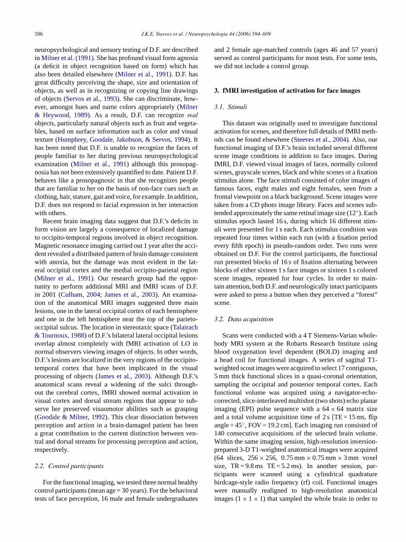
Table 1Talairach coordinates (x, y, z) of brain regions with stronger responses tofaces than places in each subject
Subject FFA OFA STS
Patient D.F.LH −37,−56,−21 −46,−56, 5RH 40,−54,−20 52,−55, 5
Control 1LH −39,−53,−19 −41,−81,−6 −45,−70, 8RH 37,−54,−16 43,−76,−3
Control 2LH −33,−49,−16 −38,−76,−12RH 39,−49,−17 29,−80,−11 56,−48, 29
Control 3LH −42,−47,−11 −43,−81,−11 −54,−58, 6RH 33,−42,−12 38,−68,−15
activation in each hemisphere of D.F.’s brain that did not over-lap with the OFA of our controls but were relatively nearby.In the left hemisphere, there were two small clusters on theborder of her lesion that measured 0.08 and 0.05 cm3 butwere more anterior and inferior than the OFA of our controlsubjects [Talairach coordinates—cluster 1:−41,−67,−14;cluster 2:−38,−72,−14; mean OFA controls:−40.7 (2.5),−79.3 (2.9),−9.7 (3.2)]. In the right hemisphere, there was alarger region of face-selective activation measuring 0.9 cm3
that was more lateral than the OFA of our controls [Talairachcoordinates—50,−70,−8; mean OFA controls: 36.7 (7.1),−74.7 (6.1),−9.7 (6.1)].
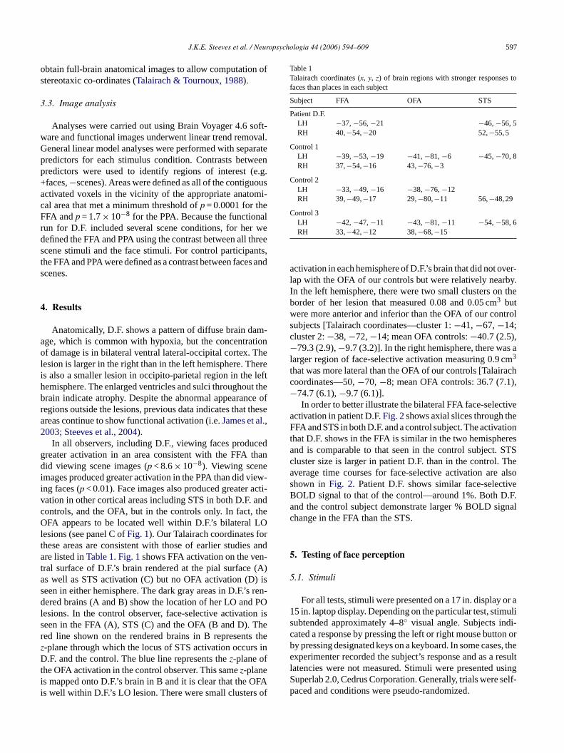
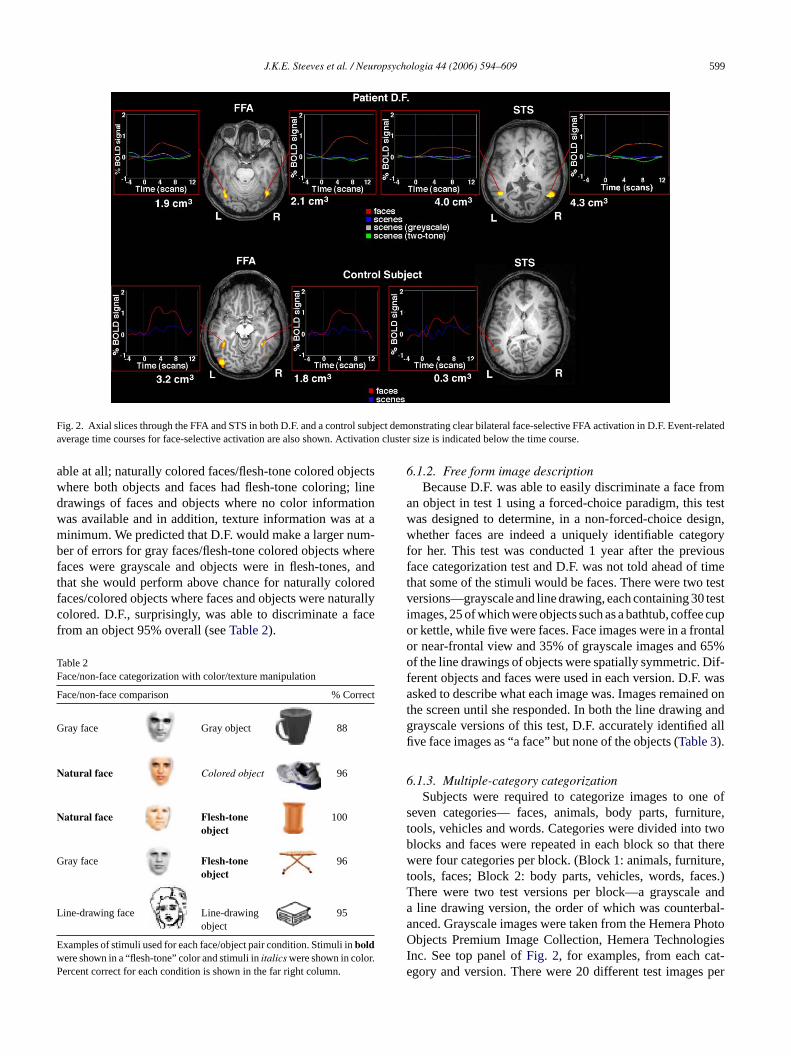
In order to better illustrate the bilateral FFA face-selectiveactivation in patient D.F.Fig. 2shows axial slices through theFFA and STS in both D.F. and a control subject. The activationthat D.F. shows in the FFA is similar in the two hemispheresand is comparable to that seen in the control subject. STScluster size is larger in patient D.F. than in the control. Theaverage time courses for face-selective activation are alsoshown inFig. 2. Patient D.F. shows similar face-selectiveBOLD signal to that of the control—around 1%. Both D.F.and the control subject demonstrate larger % BOLD signalchange in the FFA than the STS.
5
5
or a1 mulis i-c ton orb es, thee resultl usingS self-p
esion is larger in the right than in the left hemisphere. Ts also a smaller lesion in occipito-parietal region in theemisphere. The enlarged ventricles and sulci throughorain indicate atrophy. Despite the abnormal appearanegions outside the lesions, previous data indicates thatreas continue to show functional activation (i.e.James et al003; Steeves et al., 2004).
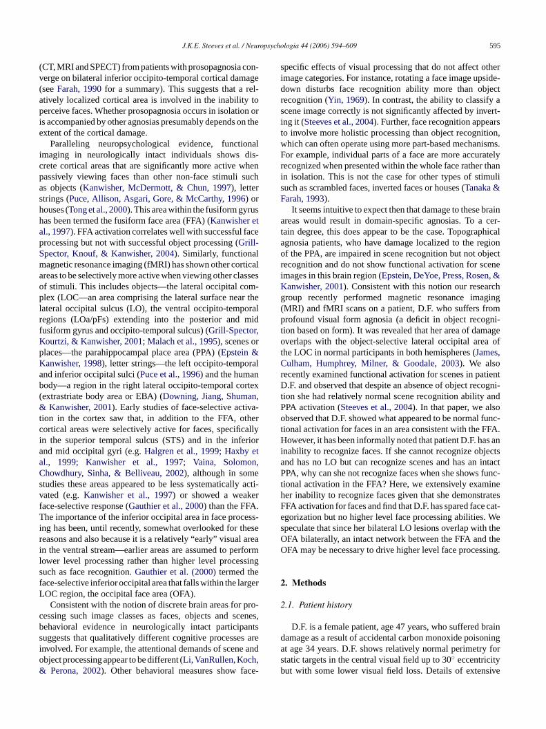
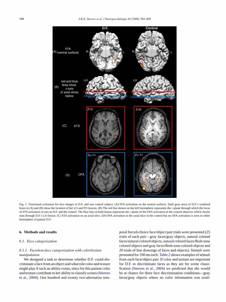
In all observers, including D.F., viewing faces produreater activation in an area consistent with the FFAid viewing scene images (p < 8.6× 10⁻⁸). Viewing scene
mages produced greater activation in the PPA than did vng faces (p < 0.01). Face images also produced greateration in other cortical areas including STS in both D.F.ontrols, and the OFA, but in the controls only. In fact,FA appears to be located well within D.F.’s bilateral
esions (see panel C ofFig. 1). Our Talairach coordinates fhese areas are consistent with those of earlier studiere listed inTable 1. Fig. 1shows FFA activation on the ve
ral surface of D.F.’s brain rendered at the pial surfaces well as STS activation (C) but no OFA activation (Deen in either hemisphere. The dark gray areas in D.F.’sered brains (A and B) show the location of her LO and
esions. In the control observer, face-selective activatioeen in the FFA (A), STS (C) and the OFA (B and D). Ted line shown on the rendered brains in B represent-plane through which the locus of STS activation occu.F. and the control. The blue line represents thez-plane of
he OFA activation in the control observer. This samez-planes mapped onto D.F.’s brain in B and it is clear that the Os well within D.F.’s LO lesion. There were small clusters
. Testing of face perception
.1. Stimuli
For all tests, stimuli were presented on a 17 in. display5 in. laptop display. Depending on the particular test, stiubtended approximately 4–8◦ visual angle. Subjects indated a response by pressing the left or right mouse buty pressing designated keys on a keyboard. In some casxperimenter recorded the subject’s response and as a
atencies were not measured. Stimuli were presenteduperlab 2.0, Cedrus Corporation. Generally, trials wereaced and conditions were pseudo-randomized.
598 J.K.E. Steeves et al. / Neuropsychologia 44 (2006) 594–609
Fig. 1. Functional activation for face images in D.F. and one control subject. (A) FFA activation on the ventral surfaces. Dark gray areas of D.F.’s renderedbrain in (A) and (B) show the location of her LO and PO lesions. (B) The red line shown on the left hemisphere represents thez-plane through which the locusof STS activation occurs in D.F. and the control. The blue line on both brains represents thez-plane of the OFA activation in the control observer which clearlyruns through D.F.’s LO lesion. (C) STS activation on an axial slice. (D) OFA activation in the axial slice in the control but no OFA activation is seen in eitherhemisphere of patient D.F.
6. Methods and results
6.1. Face categorization
6.1.1. Face/non-face categorization with color/texturemanipulation
We designed a task to determine whether D.F. could dis-criminate a face from an object and what role color and texturemight play if such an ability exists, since for this patient colorand texture contribute to her ability to classify scenes (Steeveset al., 2004). One hundred and twenty two-alternative tem-
poral forced-choice face/object pair trials were presented (25trials of each pair—gray faces/gray objects, natural coloredfaces/natural colored objects, natural colored faces/flesh-tonecolored objects and gray faces/flesh-tone colored objects and20 trials of line drawings of faces and objects). Stimuli werepresented for 100 ms each.Table 2shows examples of stimulifrom each face/object pair. If color and texture are importantfor D.F. to discriminate faces as they are for scene classi-fication (Steeves et al., 2004) we predicted that she wouldbe at chance for three face discrimination conditions—grayfaces/gray objects where no color information was avail-
J.K.E. Steeves et al. / Neuropsychologia 44 (2006) 594–609 599
Fig. 2. Axial slices through the FFA and STS in both D.F. and a control subject demonstrating clear bilateral face-selective FFA activation in D.F. Event-relatedaverage time courses for face-selective activation are also shown. Activation cluster size is indicated below the time course.
able at all; naturally colored faces/flesh-tone colored objectswhere both objects and faces had flesh-tone coloring; linedrawings of faces and objects where no color informationwas available and in addition, texture information was at aminimum. We predicted that D.F. would make a larger num-ber of errors for gray faces/flesh-tone colored objects wherefaces were grayscale and objects were in flesh-tones, andthat she would perform above chance for naturally coloredfaces/colored objects where faces and objects were naturallycolored. D.F., surprisingly, was able to discriminate a facefrom an object 95% overall (seeTable 2).
Table 2Face/non-face categorization with color/texture manipulation
Face/non-face comparison % Correct
Gray face Gray object 88
Natural face Colored object 96
Natural face Flesh-toneobject
100
Gray face Flesh-tone 96
L
Ew r.P
6.1.2. Free form image descriptionBecause D.F. was able to easily discriminate a face from
an object in test 1 using a forced-choice paradigm, this testwas designed to determine, in a non-forced-choice design,whether faces are indeed a uniquely identifiable categoryfor her. This test was conducted 1 year after the previousface categorization test and D.F. was not told ahead of timethat some of the stimuli would be faces. There were two testversions—grayscale and line drawing, each containing 30 testimages, 25 of which were objects such as a bathtub, coffee cupor kettle, while five were faces. Face images were in a frontalor near-frontal view and 35% of grayscale images and 65%of the line drawings of objects were spatially symmetric. Dif-ferent objects and faces were used in each version. D.F. wasasked to describe what each image was. Images remained onthe screen until she responded. In both the line drawing andgrayscale versions of this test, D.F. accurately identified allfive face images as “a face” but none of the objects (Table 3).
6.1.3. Multiple-category categorizationSubjects were required to categorize images to one of
seven categories— faces, animals, body parts, furniture,tools, vehicles and words. Categories were divided into twoblocks and faces were repeated in each block so that therewere four categories per block. (Block 1: animals, furniture,t es.)T anda bal-a PhotoO giesI t-e s per
object
ine-drawing face Line-drawingobject
95
xamples of stimuli used for each face/object pair condition. Stimuli inboldere shown in a “flesh-tone” color and stimuli initalics were shown in coloercent correct for each condition is shown in the far right column.
ools, faces; Block 2: body parts, vehicles, words, fachere were two test versions per block—a grayscaleline drawing version, the order of which was counter
nced. Grayscale images were taken from the Hemerabjects Premium Image Collection, Hemera Technolo
nc. See top panel ofFig. 2, for examples, from each cagory and version. There were 20 different test image
600 J.K.E. Steeves et al. / Neuropsychologia 44 (2006) 594–609
Table 3Free form image description
Test version % Correct
Grayscale
Object 0
Face 100
Line drawing
Object 0
Face 100
Example images from each version.
category in each block and test version, giving 80 images perrun.
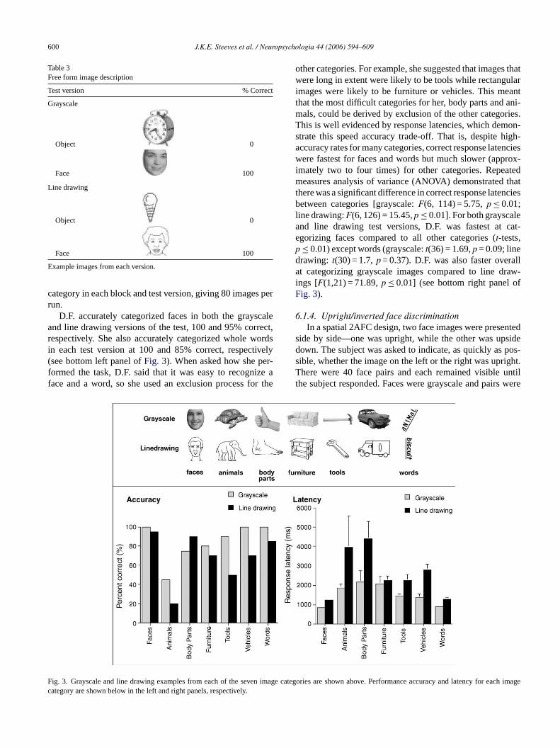
D.F. accurately categorized faces in both the grayscaleand line drawing versions of the test, 100 and 95% correct,respectively. She also accurately categorized whole wordsin each test version at 100 and 85% correct, respectively(see bottom left panel ofFig. 3). When asked how she per-formed the task, D.F. said that it was easy to recognize aface and a word, so she used an exclusion process for the
other categories. For example, she suggested that images thatwere long in extent were likely to be tools while rectangularimages were likely to be furniture or vehicles. This meantthat the most difficult categories for her, body parts and ani-mals, could be derived by exclusion of the other categories.This is well evidenced by response latencies, which demon-strate this speed accuracy trade-off. That is, despite high-accuracy rates for many categories, correct response latencieswere fastest for faces and words but much slower (approx-imately two to four times) for other categories. Repeatedmeasures analysis of variance (ANOVA) demonstrated thatthere was a significant difference in correct response latenciesbetween categories [grayscale:F(6, 114) = 5.75,p ≤ 0.01;line drawing:F(6, 126) = 15.45,p ≤ 0.01]. For both grayscaleand line drawing test versions, D.F. was fastest at cat-egorizing faces compared to all other categories (t-tests,p ≤ 0.01) except words (grayscale:t(36) = 1.69,p = 0.09; linedrawing: t(30) = 1.7,p = 0.37). D.F. was also faster overallat categorizing grayscale images compared to line draw-ings [F(1,21) = 71.89,p ≤ 0.01] (see bottom right panel ofFig. 3).
6.1.4. Upright/inverted face discriminationIn a spatial 2AFC design, two face images were presented
side by side—one was upright, while the other was upsidedown. The subject was asked to indicate, as quickly as pos-s ight.T untilt s were
Fc
ig. 3. Grayscale and line drawing examples from each of the seven imagategory are shown below in the left and right panels, respectively.
ible, whether the image on the left or the right was uprhere were 40 face pairs and each remained visible
he subject responded. Faces were grayscale and pair
e categories are shown above. Performance accuracy and latency for each image
J.K.E. Steeves et al. / Neuropsychologia 44 (2006) 594–609 601
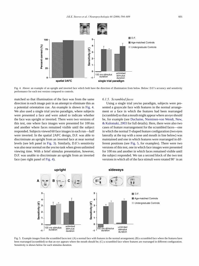
Fig. 4. Above: an example of an upright and inverted face which both have the direction of illumination from below. Below: D.F.’s accuracy and sensitivityperformance for each test version compared to controls.
matched so that illumination of the face was from the samedirection in each image pair in an attempt to eliminate this asa potential orientation cue. An example is shown inFig. 4.We also used a single trial yes/no paradigm, where subjectswere presented a face and were asked to indicate whetherthe face was upright or inverted. There were two versions ofthis test, one where face images were presented for 100 msand another where faces remained visible until the subjectresponded. Subjects viewed 60 face images in each run—halfwere inverted. In the spatial 2AFC design, D.F. was able todiscriminate an upright from an inverted face at near normallevels (see left panel inFig. 3). Similarly, D.F.’s sensitivitywas also near normal on the yes/no task when given unlimitedviewing time. With a brief stimulus presentation, however,D.F. was unable to discriminate an upright from an invertedface (see right panel ofFig. 4).
6.1.5. Scrambled facesUsing a single trial yes/no paradigm, subjects were pre-
sented a grayscale face with features in the normal arrange-ment or a face in which the features had been rearranged(scrambled) so that a mouth might appear where an eye shouldbe, for example (seeDuchaine, Nieminen-von Wendt, New,& Kulomaki, 2003for full details). Here, there were also twocases of feature rearrangement for the scrambled faces—onein which the normal T-shaped feature configuration (two eyeslaterally at the top with a nose and mouth in line below) wasmaintained and one in which features were rearranged in dif-ferent positions (seeFig. 5, for examples). There were twoversions of this test, one in which face images were presentedfor 100 ms and another in which faces remained visible untilthe subject responded. We ran a second block of the two testversions in which all of the face stimuli were rotated 90◦ in an
F ce with featuresb h shou t configuraS
ig. 5. Example images from the scrambled faces test: (A) a normal faeen rearranged (scrambled) so that an eye appears where the moutensitivity is shown below for each stimulus duration.
features in the normal arrangement; (B) a scrambled face where thehaveld be; (C) a scrambled face where features are rearranged in differention.
602 J.K.E. Steeves et al. / Neuropsychologia 44 (2006) 594–609
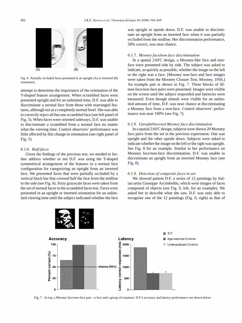
Fig. 6. Partially occluded faces presented in an upright (A) or inverted (B)orientation.
attempt to determine the importance of the orientation of theT-shaped feature arrangement. When scrambled faces werepresented upright and for an unlimited time, D.F. was able todiscriminate a normal face from those with rearranged fea-tures, although not at a completely normal level. She was ableto correctly reject all but one scrambled face (see left panel ofFig. 5). When faces were oriented sideways, D.F. was unableto discriminate a scrambled from a normal face no matterwhat the viewing time. Control observers’ performance waslittle affected by this change in orientation (see right panel ofFig. 5).
6.1.6. Half-facesGiven the findings of the previous test, we needed to fur-
ther address whether or not D.F. was using the T-shapedsymmetrical arrangement of the features in a normal faceconfiguration for categorizing an upright from an invertedface. We presented faces that were partially occluded by avertical black bar that covered half the face from the midlineto the side (seeFig. 6). Sixty grayscale faces were taken fromthe set of normal faces in the scrambled faces test. Faces werepresented in an upright or inverted orientation for an unlim-ited viewing time until the subject indicated whether the face
was upright or upside down. D.F. was unable to discrimi-nate an upright from an inverted face when it was partiallyoccluded from the midline. Her discrimination performance,58% correct, was near chance.
6.1.7. Mooney face/non-face discriminationIn a spatial 2AFC design, a Mooney-like face and non-
face were presented side by side. The subject was asked toindicate, as quickly as possible, whether the image on the leftor the right was a face. [Mooney non-face and face imageswere taken from the Mooney Closure Test;Mooney, 1956.]An example pair is shown inFig. 7. Three blocks of fif-teen face/non-face pairs were presented. Images were visibleon the screen until the subject responded and latencies weremeasured. Even though stimuli were visible for an unlim-ited amount of time, D.F. was near chance at discriminatinga Mooney face from a non-face. Control observers’ perfor-mance was near 100% (seeFig. 7).
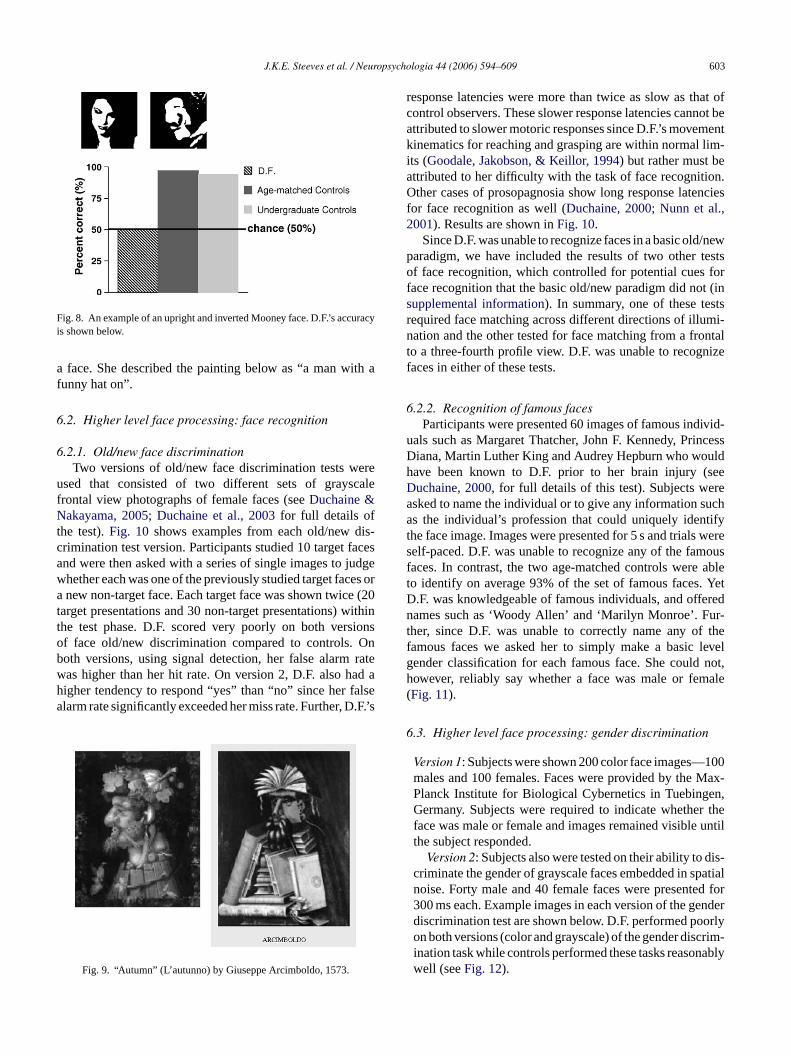
6.1.8. Upright/inverted Mooney face discriminationIn a spatial 2AFC design, subjects were shown 20 Mooney
face pairs from the set in the previous experiment. One wasupright and the other upside down. Subjects were asked toindicate whether the image on the left or the right was upright.SeeFig. 8 for an example. Similar to her performance onM e tod (seeF
6Ital-
i cesc ea le tor f
roup o .
Fig. 7. At top, a Mooney face/non-face pair—a face and a gooney face/non-face discrimination, D.F. was unabliscriminate an upright from an inverted Mooney faceig. 8).
.1.9. Detection of composite faces in artWe showed patient D.F. a series of 12 paintings by
an artist Giuseppe Arcimboldo, which were images of faomposed of objects (seeFig. 9, left, for an example). Wsked her to describe what she saw. D.F. was only abecognize one of the 12 paintings (Fig. 9, right) as that o
f tomatoes. D.F.’s accuracy and latency performance are shown below
J.K.E. Steeves et al. / Neuropsychologia 44 (2006) 594–609 603
Fig. 8. An example of an upright and inverted Mooney face. D.F.’s accuracyis shown below.
a face. She described the painting below as “a man with afunny hat on”.
6.2. Higher level face processing: face recognition
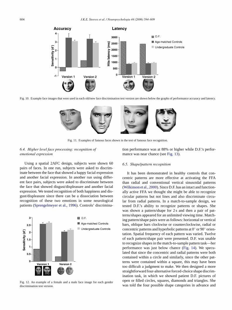
6.2.1. Old/new face discriminationTwo versions of old/new face discrimination tests were
used that consisted of two different sets of grayscalefrontal view photographs of female faces (seeDuchaine &Nakayama, 2005; Duchaine et al., 2003for full details ofthe test).Fig. 10 shows examples from each old/new dis-crimination test version. Participants studied 10 target facesand were then asked with a series of single images to judgewhether each was one of the previously studied target faces ora new non-target face. Each target face was shown twice (20target presentations and 30 non-target presentations) withinthe test phase. D.F. scored very poorly on both versionsof face old/new discrimination compared to controls. Onboth versions, using signal detection, her false alarm ratewas higher than her hit rate. On version 2, D.F. also had ahigher tendency to respond “yes” than “no” since her falsealarm rate significantly exceeded her miss rate. Further, D.F.’s
response latencies were more than twice as slow as that ofcontrol observers. These slower response latencies cannot beattributed to slower motoric responses since D.F.’s movementkinematics for reaching and grasping are within normal lim-its (Goodale, Jakobson, & Keillor, 1994) but rather must beattributed to her difficulty with the task of face recognition.Other cases of prosopagnosia show long response latenciesfor face recognition as well (Duchaine, 2000; Nunn et al.,2001). Results are shown inFig. 10.
Since D.F. was unable to recognize faces in a basic old/newparadigm, we have included the results of two other testsof face recognition, which controlled for potential cues forface recognition that the basic old/new paradigm did not (insupplemental information). In summary, one of these testsrequired face matching across different directions of illumi-nation and the other tested for face matching from a frontalto a three-fourth profile view. D.F. was unable to recognizefaces in either of these tests.
6.2.2. Recognition of famous facesParticipants were presented 60 images of famous individ-
uals such as Margaret Thatcher, John F. Kennedy, PrincessDiana, Martin Luther King and Audrey Hepburn who wouldhave been known to D.F. prior to her brain injury (seeDuchaine, 2000, for full details of this test). Subjects werea ucha tifyt weres ousf ablet . YetD redn ur-t thef levelg not,h ale(
6
100Max-en,r theuntil
dis-patiald for
enderorlyrim-ably
Fig. 9. “Autumn” (L’autunno) by Giuseppe Arcimboldo, 1573.
sked to name the individual or to give any information ss the individual’s profession that could uniquely iden
he face image. Images were presented for 5 s and trialself-paced. D.F. was unable to recognize any of the famaces. In contrast, the two age-matched controls wereo identify on average 93% of the set of famous faces.F. was knowledgeable of famous individuals, and offeames such as ‘Woody Allen’ and ‘Marilyn Monroe’. F
her, since D.F. was unable to correctly name any ofamous faces we asked her to simply make a basicender classification for each famous face. She couldowever, reliably say whether a face was male or femFig. 11).
.3. Higher level face processing: gender discrimination
Version 1: Subjects were shown 200 color face images—males and 100 females. Faces were provided by thePlanck Institute for Biological Cybernetics in TuebingGermany. Subjects were required to indicate whetheface was male or female and images remained visiblethe subject responded.
Version 2: Subjects also were tested on their ability tocriminate the gender of grayscale faces embedded in snoise. Forty male and 40 female faces were presente300 ms each. Example images in each version of the gdiscrimination test are shown below. D.F. performed poon both versions (color and grayscale) of the gender discination task while controls performed these tasks reasonwell (seeFig. 12).
604 J.K.E. Steeves et al. / Neuropsychologia 44 (2006) 594–609
Fig. 10. Example face images that were used in each old/new face discrimination test version are shown below the graphs of performance accuracy and latency.
Fig. 11. Examples of famous faces shown in the test of famous face recognition.
6.4. Higher level face processing: recognition ofemotional expression
Using a spatial 2AFC design, subjects were shown 60pairs of faces. In one run, subjects were asked to discrim-inate between the face that showed a happy facial expressionand another facial expression. In another run using differ-ent face pairs, subjects were asked to discriminate betweenthe face that showed disgust/displeasure and another facialexpression. We tested recognition of both happiness and dis-gust/displeasure since there can be a dissociation betweenrecognition of these two emotions in some neurologicalpatients (Sprengelmeyer et al., 1996). Controls’ discrimina-
F enderd
tion performance was at 88% or higher while D.F.’s perfor-mance was near chance (seeFig. 13).
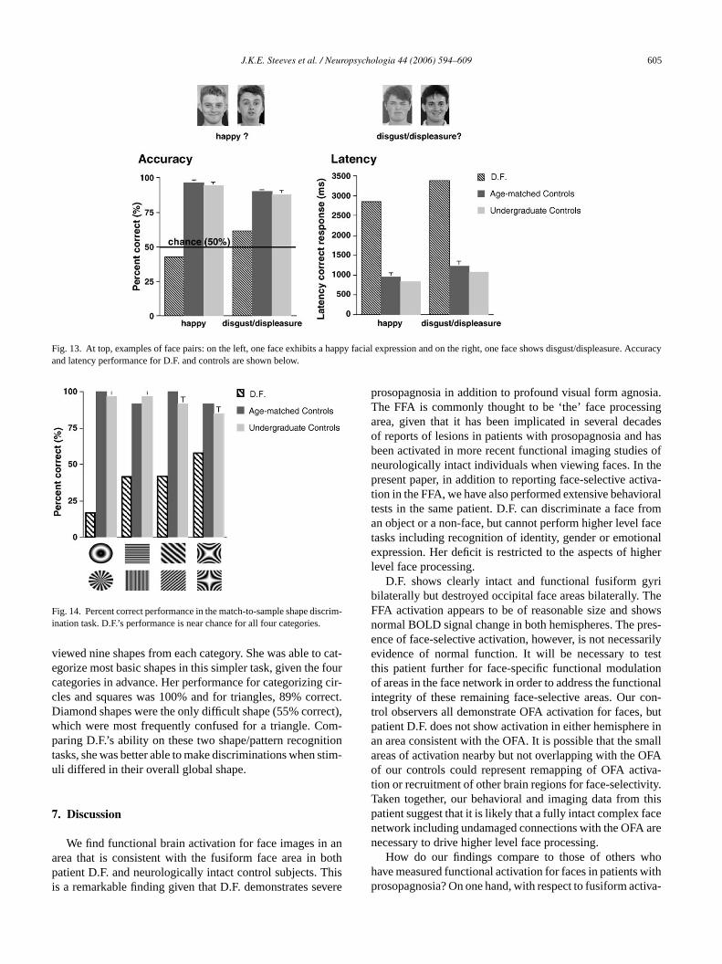
6.5. Shape/pattern recognition
It has been demonstrated in healthy controls that con-centric patterns are more effective at activating the FFAthan radial and conventional vertical sinusoidal patterns(Wilkinson et al., 2000). Since D.F. has an intact and function-ally active FFA we thought she might be able to recognizecircular patterns but not lines and also discriminate circu-lar from radial patterns. In a match-to-sample design, wetested D.F.’s ability to recognize patterns or shapes. Shewas shown a pattern/shape for 2 s and then a pair of pat-terns/shapes appeared for an unlimited viewing time. Match-ing pattern/shape pairs were as follows: horizontal or verticalbars, oblique bars clockwise or counterclockwise, radial orconcentric patterns and hyperbolic patterns at 0◦ or 90◦ orien-tation. Spatial frequency of each pattern was varied. Twelveof each pattern/shape pair were presented. D.F. was unableto recognize shapes in the match-to-sample pattern task—herperformance was just below chance (Fig. 14). We specu-lated that since the concentric and radial patterns were bothcontained within a circle and similarly, since the other pat-terns were contained within a square, this may have beentoo difficult a judgment to make. We then designed a mores rim-i s ofo . Shew e and
ig. 12. An example of a female and a male face image for each giscrimination test version.
traightforward four-alternative forced-choice shape discnation task, in which we showed patient D.F. picturepen or filled circles, squares, diamonds and trianglesas told the four possible shape categories in advanc
J.K.E. Steeves et al. / Neuropsychologia 44 (2006) 594–609 605
Fig. 13. At top, examples of face pairs: on the left, one face exhibits a happy facial expression and on the right, one face shows disgust/displeasure. Accuracyand latency performance for D.F. and controls are shown below.
Fig. 14. Percent correct performance in the match-to-sample shape discrim-ination task. D.F.’s performance is near chance for all four categories.
viewed nine shapes from each category. She was able to cat-egorize most basic shapes in this simpler task, given the fourcategories in advance. Her performance for categorizing cir-cles and squares was 100% and for triangles, 89% correct.Diamond shapes were the only difficult shape (55% correct),which were most frequently confused for a triangle. Com-paring D.F.’s ability on these two shape/pattern recognitiontasks, she was better able to make discriminations when stim-uli differed in their overall global shape.
7. Discussion
We find functional brain activation for face images in anarea that is consistent with the fusiform face area in bothpatient D.F. and neurologically intact control subjects. Thisis a remarkable finding given that D.F. demonstrates severe
prosopagnosia in addition to profound visual form agnosia.The FFA is commonly thought to be ‘the’ face processingarea, given that it has been implicated in several decadesof reports of lesions in patients with prosopagnosia and hasbeen activated in more recent functional imaging studies ofneurologically intact individuals when viewing faces. In thepresent paper, in addition to reporting face-selective activa-tion in the FFA, we have also performed extensive behavioraltests in the same patient. D.F. can discriminate a face froman object or a non-face, but cannot perform higher level facetasks including recognition of identity, gender or emotionalexpression. Her deficit is restricted to the aspects of higherlevel face processing.
D.F. shows clearly intact and functional fusiform gyribilaterally but destroyed occipital face areas bilaterally. TheFFA activation appears to be of reasonable size and showsnormal BOLD signal change in both hemispheres. The pres-ence of face-selective activation, however, is not necessarilyevidence of normal function. It will be necessary to testthis patient further for face-specific functional modulationof areas in the face network in order to address the functionalintegrity of these remaining face-selective areas. Our con-trol observers all demonstrate OFA activation for faces, butpatient D.F. does not show activation in either hemisphere inan area consistent with the OFA. It is possible that the smallareas of activation nearby but not overlapping with the OFAo iva-t ity.T thisp acen aren
hoh withp ctiva-
f our controls could represent remapping of OFA action or recruitment of other brain regions for face-selectivaken together, our behavioral and imaging data fromatient suggest that it is likely that a fully intact complex fetwork including undamaged connections with the OFAecessary to drive higher level face processing.
How do our findings compare to those of others wave measured functional activation for faces in patientsrosopagnosia? On one hand, with respect to fusiform a
606 J.K.E. Steeves et al. / Neuropsychologia 44 (2006) 594–609
tion, this finding is relatively compatible with two recent stud-ies, which have also shown FFA activation in patients withprosopagnosia.Rossion et al. (2003)show face-selective acti-vation in the right fusiform gyrus in a patient with acquiredprosopagnosia.Hasson, Avidan, Deouell, Bentin, and Malach(2003)report that the activation for faces in their congenitalprosopagnosia patient is normal with respect to the anatomi-cal location, activation profiles and hemispheric laterality ofthe FFA in controls. On the other hand, two earlier studies byHadjikhani and de Gelder (2002)and Marotta, Genovese,and Behrmann (2001)report that the activation for facesin the fusiform gyrus in their patients with acquired andearly prosopagnosia, respectively, is not normal compared tocontrols.
The cortical damage in one ofMarotta et al. (2001)patientswas right anterior and posterior temporal while in the otherthe damage was to the right temporal and medial occipitallobes and the right fusiform gyrus. These patients, however,exhibited more functional activation for faces in the ante-rior portion of the fusiform gyrus than did controls and onepatient showed more left than right hemisphere activation.The authors did not test for OFA activity in their study, how-ever. The stereotaxic coordinates of the locus of face-selectivefusiform activation in one of their patients in particular, werealtogether more anterior than those of their controls, whichmay account for the overall more anterior activation in thef 2)d nort el-o omer ringo or-m s bute
loca-t sis-t hesec rs byo ,1 on-t (e.g.K t al.,1 FAa earst hereso l.( ablet gen-d withc led onM stentw ea.T thoseo l.( ivityf en
our findings, these differences could account for this patient’sinability to recognize known faces despite preserved process-ing of other higher level face attributes including recognitionof gender, age and emotional expression. This is consistentwith our supposition that a functionally intact complex facenetwork including undamaged connections with the OFA isindeed more important than suspected for such higher levelface processing.
Rossion et al. (2003)tested a patient with left middlefusiform damage and right inferior occipital damage (pre-sumably including the right OFA) but an intact right middlefusiform gyrus and left OFA. Behaviorally, although impairedcompared to their control subjects on tests of face recogni-tion, their patient still performed well above chance on tests ofgender decision and recognition of emotional expression andwas normal in her ability to assess the age of faces. Theirpatient showed left inferior occipital cortex responses forfaces, which likely corresponds to the OFA, in an area poste-rior to the damaged area in one of the two scanning sessions.Given that bothHasson et al. (2003)andRossion et al. (2003)found some face-selective activation, although abnormal, inthe OFA and that their two patients have some residual higherlevel face processing abilities including recognition of gen-der, age and emotional expression it seems likely that theOFA or its interconnections are partially damaged in thesepatients but those that remain are adequate to help drivet PETsc sing.S ras-t erala sk.T ationi herea hlyl et-w forf gain,f face-s izeri shes f ourc
ma-r fromn singt for-m ntedi t shea tiallys r to bea e hasn ges.D iringr D.F.i in all
usiform gyrus. Similarly,Hadjikhani and de Gelder (200id not find normal face-selective activation in the FFA
he inferior occipital gyrus (IOG) in their patients with devpmental or childhood prosopagnosia but they did find selatively normal activation in object-selective areas dubject viewing. Their subjects performed just below nal on the Benton and Warrington face recognition test
xhibited no evident lesions on the MR scan.In the present study, however, we find the anatomical
ion of face-selective fusiform activation in D.F. to be conent with those of our control observers and further, that toordinates are similar to those seen in normal observethers (e.g.Epstein & Kanwisher, 1998; Kanwisher et al.997). We also find OFA and STS activation in our c
rols, as do earlier studies of face-selective activationanwisher et al., 1997; McCarthy et al., 1997; Puce e995) Patient D.F. also shows STS activation, but no Octivation consistent with that of our controls. This app
o be because her lateral occipital lesions in both hemispverlap with the anatomical locus of the OFA.Hasson et a2003)studied a congenital prosopagnosic who was uno recognize famous faces but who could recognize faceer, age and emotional expression. As is often the caseongenital cases, no evident structural lesion was reveaR scan. Their patient showed activation in areas consiith the FFA and the OFA within the lateral occipital aralairach coordinates for these areas are comparable tof our controls. But, the interesting result in theHasson et a2003)study is that there were subtle differences in selector faces in the left from the right OFA of their patient. Giv
hese residual higher level face processes. An earliertudy bySergent, Ohta, and MacDonald (1992)also impli-ates the OFA in aspects of higher level face procespecifically, they found activation changes in right ext
riate cortex for gender recognition and additional bilatctivation of the fusiform gyrus for a face recognition tahe data from patient D.F. show that she has no activ
n an area consistent with the OFA in either hemispnd no higher level face processing abilities. It is higikely that face recognition involves a complex face nork requiring intact connections with the OFA area
ace processing beyond basic face categorization. Aurther research is needed to determine whether D.F.’selective activation in the fusiform revealed by a locals indicative of normal functionality and also whetherhows remapping of the OFA in areas outside of that oontrols.
How capable is D.F. at face categorization? To sumize the behavioral data: D.F. can differentiate faceson-faces given sufficient texture information and proces
ime, and this is independent of color and illumination ination. She can use configural information when prese
n an upright but not sideways orientation and given thalso cannot discriminate half-faces she may rely on a spaymmetric feature arrangement. Moreover, faces appeaunique category, which she can classify even when sho advance knowledge that she will be shown face ima.F. cannot make any higher level discriminations requ
ecognition of known faces, emotion or gender. In short,s a unique patient demonstrating a severe impairment
J.K.E. Steeves et al. / Neuropsychologia 44 (2006) 594–609 607
aspects of higher level face processing but relatively sparedface categorization.
It is possible that D.F. is able to categorize internallysymmetrical stimuli as faces without actuallyseeing themas faces. However, a large number of object images fromwhich she discriminated faces were also spatially symmetric.Given her good performance on basic shape discriminationbut poor performance on pattern discrimination when con-tained within similar global shapes, it is possible that patientD.F. initially uses differences in global shape to help herdistinguish faces from other objects. It would be useful infuture research to test D.F.’s ability to discriminate faces fromother non-face stimuli with similar internal symmetry andalso faces from other non-face stimuli with similar globalshape, such as flowers or round fruit. In addition, it would beworthwhile to test functional activation in the face-processingnetwork with symmetrical versus non-symmetrical shapes aswell as images with similar and different global shape in orderto address the role of different components of this networkin basic configural processing in D.F.
Several imaging studies in neurologically intact humanshave made the case that the FFA is involved in face cate-gorization but not necessarily higher level face recognition.For instance,Kanwisher, Tong, and Nakayama (1998)alsodemonstrated that face discrimination is better for uprightthan inverted grayscale faces but activation in the FFA is onlys )d elec-t ions.T ellt ss tos thatc incep its inc or,2 le inc g thel
den-t ns ntity,a ofd tedly.T al-l oesi y fori tiona o uses go-r ourd rk isn d forh res-s hef FA,c the
OFA. The FFA may process higher level face-sensitive infor-mation that is ultimately used for fine-grained visual analysisof faces at the individual level through feedback connectionsto the OFA.
As a final point, one should certainly exercise cautionwhen interpreting data from single-patient studies given indi-vidual variability with respect to lesions and behavioral per-formance. Further, the site of a lesion does not necessarilycorrespond to the locus of the area responsible for percep-tual/cognitive processing which is disrupted but could insteadcorrespond to an interruption in the pathways to other areaswhere such processing is accomplished. When interpretingdata from the present case of patient D.F., one must bear inmind that she does have profound agnosia of the apperceptivetype, affecting more than just higher level face processing.Nonetheless, these data in conjunction with those from otherneuropsychological patients (i.e.Hasson et al., 2003; Rossionet al., 2003) contribute to a clearer picture of necessity of anintact complex face network in face processing. Patient D.F.has undamaged and functionally active FFAs but destroyedOFAs in both hemispheres and she also demonstrates a clearbehavioral deficit inall aspects of face processing beyondcategorization. These data lend strong support to the impor-tance of the integrity of a complex network of regions for faceidentification, including more than just the FFA—in particu-lar the OFA, a region believed to be associated with low-levelp
A
con-t ankm ningg ongk pro-v uslyg acesf eref S),P nkJ
A
n bef ia.2
R
B 002).nfig-
lightly lower for the latter condition.Haxby et al. (1999emonstrated similar findings—inverted faces do not s
ively diminish the response to faces in face-selective regong et al. (2000)demonstrated that the FFA responds wo human, animal and cartoon faces but responds lechematic faces or facial features alone. It seems likelyonfigural face information is processed in the FFA satients with right fusiform face area damage show deficonfigural processing (Barton, Press, Keenan, & O’Conn002). These findings suggest that the FFA plays a roonscious detection of a face possibly by representinocal features and global configuration of a face.
Configural information may nonetheless be used to iify individual faces.Gauthier et al. (2000)showed that wheubjects attend to the location of faces rather than idectivity in the FFA and OFA is higher for presentationsifferent faces than presentation of the same face repeahey argue that the FFA is involved in specific individu
evel face processing. It is likely the case that the FFA dndeed process information that is ultimately necessarndividual-level face recognition such as face configurand feature arrangement. Patient D.F. does appear tpatially symmetric configural information for face cateization and shows FFA activation for faces. However,ata suggest that it is likely that an intact complex netwoecessary for this same configural information to be useigher level recognition tasks, such as identity and expion.Rossion et al. (2003)make a similar argument that tace-selective activation in the earlier visual area, the Oould result from feedback connections from the FFA to
rocessing.
cknowledgements
Foremost, we thank patient D.F. for her patience andinued willingness to participate in our experiments. We thany people who helped provide test images: Paul Dowenerously gave us line drawings of body parts, Frank Tindly sent us Mooney face images, some images wereided courtesy of Mike Tarr, and Tzvika Ganel generoave us faces for the colored face categorization test. F
or the face-matching and emotion recognition tasks wrom the Psychological Image Collection at Stirling (PICsychology Department, University of Stirling. We thaennifer Rycroft for collecting some of the control data.
ppendix A. Supplementary data
Supplementary data associated with this article caound, in the online version, at10.1016/j.neuropsycholog005.06.013.
eferences
arton, J. J. S., Press, D. Z., Keenan, J. P., & O’Connor, M. (2Lesions of the fusiform face area impair perception of facial couration in prosopagnosia.Neurology, 58, 71–78.
608 J.K.E. Steeves et al. / Neuropsychologia 44 (2006) 594–609
Culham, J. (2004).Neuroimaging investigations of visually-guided grasp-ing. Attention and performance XX: Functional brain imaging ofhuman cognition. Oxford: Oxford University Press, pp. 415–436.
Damasio, A. R., Damasio, H., & Van Hoesen, G. W. (1982). Prosopag-nosia: Anatomic basis and behavioral mechanisms.Neurology, 32,331–341.
Downing, P. E., Jiang, Y., Shuman, M., & Kanwisher, N. (2001). A cor-tical area selective for visual processing of the human body.Science,293, 2470–2473.
Duchaine, B. C. (2000). Developmental prosopagnosia with normal con-figural processing.Neuroreport, 11(1), 79–83.
Duchaine, B. C., & Nakayama, K. (2005). Dissociations of face andobject recognition in developmental prosopagnosic.Journal of Cog-nitive Neuroscience, 17, 249–261.
Duchaine, B., Nieminen-von Wendt, T., New, J., & Kulomaki, T. (2003).Dissociations of visual recognition in a developmental prosopag-nosic: Evidence for separate developmental processes.Neurocase, 9,380–389.
Epstein, R., & Kanwisher, N. (1998). A cortical representation of thelocal visual environment.Nature, 392, 598–601.
Epstein, R., DeYoe, E. A., Press, D. Z., Rosen, A. C., & Kanwisher,N. (2001). Neuropsychological evidence for a topographical learningmechanism in parahippocampal cortex.Cognitive Neuropsychology,18(6), 481–508.
Farah, M. H. (1990).Visual agnosia: Disorders of object recognition andwhat they tell us about normal vision. Cambridge, MA: MIT Press.
Gauthier, I., Tarr, M. J., Moylan, J., Skudlarski, P., Gore, J. C., & Ander-son, W. A. (2000). The fusiform “face area” is part of a network thatprocesses faces at the individual level.Journal of Cognitive Neuro-science, 12(3), 495–504.
Goodale, M. A., & Milner, A. D. (1992). Separate visual pathways for
G cesents.
G ip-
G ceentifi-
H osia:
H ic,rtex
H 03).
H an,ity
H 94).of a.
H osiaects.
J ale,but.
K rmface
K face
Li, F. F., VanRullen, R., Koch, C., & Perona, P. (2002). Rapid naturalscene categorization in the near absence of attention.Proceedings ofthe National Academy of Sciences of the United States of America,99(14), 9596–9601.
Malach, R., Reppas, J. B., Benson, R. R., Kwong, K. K., Jiang, H.,Kennedy, W. A., et al. (1995). Object-related activity revealed byfunctional magnetic resonance imaging in human occipital cortex.Proceedings of the National Academy of Sciences of the United Statesof America, 92, 8135–8139.
Marotta, J. J., Genovese, C. R., & Behrmann, M. (2001). A functionalMRI study of face recognition in patients with prosopagnosia.Neu-roreport, 12, 1581–1587.
McCarthy, G., Puce, A., Luby, M., Belger, A., & Allison, T. (1997).Magnetic resonance imaging studies of functional brain activation:analysis and interpretation.Electroencephalography Clininical Neuro-physiology Supplement, 47, 15–31.
McMullen, P., Fisk, J. D., & Phillips, S. (2000). Apperceptive agnosiaand face recognition.Neurocase, 6, 403–414.
McNeil, J. E., & Warrington, E. K. (1993). Prosopagnosia: A face-specificdisorder.The Quarterly Journal of Experimental Psychology A, 46,1–10.
Milner, A. D., & Heywood, C. A. (1989). A disorder of lightnessdiscrimination in a case of visual form agnosia.Cortex, 25, 489–494.
Milner, A. D., Perrett, D. I., Johnston, R. S., Benson, P. J., Jordan, T. R.,Heeley, D. W., et al. (1991). Perception and action in ‘visual formagnosia’.Brain, 114, 405–428.
Mooney, C. M. (1956). Closure with negative after images under filteringlight. Canadian Journal of Psychology, 10, 191–199.
Moscovitch, M., Winocur, G., & Behrmann, M. (1997). What is specialabout face recognition? Nineteen experiments on a person with visual
N opag-
P with
P itive
P 6).ngs,
R ., &reasface
S tomytudy.
S aw-ace
S nge,of
S Mil-ing
cene
T
perception and action.Trends in Neuroscience, 15, 20–25.oodale, M. A., Jakobson, L. S., & Keillor, J. M. (1994). Differen
in the visual control of pantomimed and natural grasping movemNeuropsychologia, 32, 1159–1178.
rill-Spector, K., Kourtzi, Z., & Kanwisher, N. (2001). The lateral occital complex and its role in object recognition.Vision Research, 41,1409–1422.
rill-Spector, K., Knouf, N., & Kanwisher, N. (2004). The fusiform faarea subserves face perception, not generic within-category idcation.Nature Neuroscience, 7(5), 555–562.
adjikhani, N., & de Gelder, B. (2002). Neural basis of prosopagnAn fMRI study. Human Brain Mapping, 16, 176–182.
algren, E., Dale, A. M., Sereno, M. I., Tootell, R. B. H., MarinkovK., & Rosen, B. R. (1999). Location of human face-selective cowith respect to retinotopic areas.Human Brain Mapping, 7, 29–37.
asson, U., Avidan, G., Deouell, L. Y., Bentin, S., & Malach, R. (20Face-selective activation in a congenital prosopagnosic subject.Jour-nal of Cognitive Neuroscience, 15(3), 419–431.
axby, J. V., Ungerleider, L. G., Clark, V. P., Schouten, J. L., HoffmE. A., & Martin, A. (1999). The effect of face inversion on activin human neural systems for face and object perception.Neuron, 22,189–199.
umphrey, G. K., Goodale, M. A., Jakobson, L. S., & Servos, P. (19The role of surface information in object recognition: Studiesvisual form agnosic and normal subjects.Perception, 23, 1457–1481
umphreys, G., & Rumiati, R. I. (1998). Agnosia without prosopagnor alexia: Evidence for stored visual memories specific to objCognitive Neuropsychology, 15, 243–277.
ames, T. W., Culham, J. C., Humphrey, G. K., Milner, A. D., & GoodM. A. (2003). Ventral occipital lesions impair object recognitionnot object-directed grasping: A fMRI study.Brain, 126, 2463–2475
anwisher, N., McDermott, J., & Chun, M. M. (1997). The fusifoface area: A module in human extrastriate cortex specialized forperception.Journal of Neuroscience, 17, 4302–4311.
anwisher, N., Tong, F., & Nakayama, K. (1998). The effect ofinversion on the human fusiform face area.Cognition, 68(1), 1–11.
object agnosia and dyslexia but normal face recognition.Journal ofCognitive Neuroscience, 9, 555–604.
unn, J. A., Postma, P., & Pearson, R. (2001). Developmental prosnosia: Should it be taken at face value?Neurocase, 7, 15–27.
allis, C. A. (1955). Impaired identification of faces and placesagnosia for colours; report of a case due to cerebral embolism.Journalof Neurochemistry, 18(3), 218–224.
uce, A., Allison, T., Gore, J. C., & McCarthy, G. (1995). Face-sensregions in human extrastriate cortex studied by functional MRI.Jour-nal of Neurophysiology, 74, 1192–1199.
uce, A., Allison, T., Asgari, M., Gore, J. C., & McCarthy, G. (199Differential sensitivity of human visual cortex to faces, letterstriand textures: A functional magnetic resonance imaging study.Journalof Neuroscience, 16(6), 5205–5215.
ossion, B., Caldara, R., Seghier, M., Schuller, A.-M., Lazeyras, FMayer, E. (2003). A network of occipito-temporal face-sensitive abesides the right middle fusiform gyrus is necessary for normalprocessing.Brain, 126, 2381–2395.
ergent, J., Ohta, S., & MacDonald, B. (1992). Functional neuroanaof face and object processing. A positron emission tomography sBrain, 115, 15–36.
ervos, P., Goodale, M. A., & Humphrey, G. K. (1993). The dring of objects by a visual form agnosic: contribution of surfproperties and memorial representations.Neuropsychologia, 31, 251–259.
prengelmeyer, R., Young, A. W., Calder, A. J., Karnat, A., LaH., Homberg, V., et al. (1996). Loss of disgust. Perceptionfaces and emotions in Huntington’s disease.Brain, 119, 1647–1665.
teeves, J. K. E., Humphrey, G. K., Culham, J. C., Menon, R. S.,ner, A. D., & Goodale, M. A. (2004). Behavioral and neuroimagevidence for a contribution of color and texture information to sclassification in a patient with visual form agnosia.Journal of Cog-nitive Neuroscience, 16, 6.
alairach, J., & Tournoux, P. (1988).Co-planar stereotaxic atlas of thehuman brain. New York: Thieme Medical Publishers.

J.K.E. Steeves et al. / Neuropsychologia 44 (2006) 594–609 609
Tanaka, J. W., & Farah, M. J. (1993). Parts and wholes in face recognition.Quarterly Journal of Experimental Psychology A, 46(2), 225–245.
Tong, F., Nakayama, K., Moscovitch, M., Weinrib, O., & Kanwisher, N.(2000). Response properties of the human fusiform face area.Cogni-tive Neuropsychology, 17, 257–279.
Vaina, L. M., Solomon, J., Chowdhury, S., Sinha, P., & Belliveau. (2002).Functional neuroanatomy of biological motion perception in humans.Proceedings of the National Academy of Sciences of the United Statesof America, 98, 11656–11661.
Whiteley, A. M., & Warrington, E. K. (1977). Prosopagnosia: A clini-cal, psychological, and anatomical study of three patients.Journal ofNeurology, Neurosurgery & Psychology, 40(4), 395–403.
Wilkinson, F., James, T. W., Wilson, H. R., Gati, J. S., Menon, R. S., &Goodale, M. A. (2000). An fMRI study of the selective activation ofhuman extrastriate form vision areas by radial and concentric gratings.Current Biology, 16, 1455–1458.
Yin, R. K. (1969). Looking at upside-down faces.Journal of ExperimentalPsychology, 81, 141–145.