Embed Size (px)
Citation preview
MINIREVIEW
The hairpin conformation of the amyloid b peptide is an importantstructural motif along the aggregation pathway
Axel Abelein • Jan Pieter Abrahams • Jens Danielsson •
Astrid Graslund • Juri Jarvet • Jinghui Luo • Ann Tiiman •
Sebastian K. T. S. Warmlander
Received: 19 September 2013 / Accepted: 2 April 2014 / Published online: 16 April 2014
� SBIC 2014
Abstract The amyloid b (Ab) peptides are 39–42 residue-
long peptides found in the senile plaques in the brains of
Alzheimer’s disease (AD) patients. These peptides self-
aggregate in aqueous solution, going from soluble and
mainly unstructured monomers to insoluble ordered fibrils.
The aggregation process(es) are strongly influenced by
environmental conditions. Several lines of evidence indicate
that the neurotoxic species are the intermediate oligomeric
states appearing along the aggregation pathways. This
minireview summarizes recent findings, mainly based on
solution and solid-state NMR experiments and electron
microscopy, which investigate the molecular structures and
characteristics of the Ab peptides at different stages along
the aggregation pathways. We conclude that a hairpin-like
conformation constitutes a common motif for the Ab pep-
tides in most of the described structures. There are certain
variations in different hairpin conformations, for example
regarding H-bonding partners, which could be one reason for
the molecular heterogeneity observed in the aggregated
systems. Interacting hairpins are the building blocks of the
insoluble fibrils, again with variations in how hairpins are
organized in the cross-section of the fibril, perpendicular to
the fibril axis. The secondary structure propensities can be
seen already in peptide monomers in solution. Unfortu-
nately, detailed structural information about the intermediate
oligomeric states is presently not available. In the review,
special attention is given to metal ion interactions, particu-
larly the binding constants and ligand structures of Abcomplexes with Cu(II) and Zn(II), since these ions affect the
aggregation process(es) and are considered to be involved in
the molecular mechanisms underlying AD pathology.
Keywords Alzheimer’s disease � Amyloid b peptide �Hairpin � Protein aggregation � Neurotoxicity
Introduction
Amyloid is an old term describing the solid starch-like
deposits observed in the senile plaques in the brains of
Alzheimer’s disease (AD) patients. More recent biochem-
ical work (reviewed in [1, 2]) has identified the major
plaque component to be Amyloid b (Ab) peptides. These
peptides are relatively short, typically 39–42 residues long,
and are formed by enzymatic processing from the amyloid
precursor protein (APP). The amyloid deposits contain
relatively regular fibrillar structures, which can be visual-
ized by e.g., electron microscopy (EM).
From a biophysical/structural perspective, amyloid
material may be defined by a characteristic pattern in X-ray
fiber diffraction measurements. There is a common struc-
tural motif in these fibers, which is named ‘‘cross-b struc-
ture’’, that gives rise to 4.7 A meridional and 10 A equatorial
reflexions in the diffraction pattern [3, 4]. The structure
Responsible Editors: Lucia Banci and Claudio Luchinat.
A. Abelein � J. Danielsson (&) � A. Graslund (&) � J. Jarvet �A. Tiiman � S. K. T. S. Warmlander
Department of Biochemistry and Biophysics, Arrhenius
Laboratories, Stockholm University, 106 91 Stockholm, Sweden
e-mail: [email protected]
A. Graslund
e-mail: [email protected]
J. P. Abrahams � J. Luo
Gorlaeus Laboratory, Leiden Institute of Chemistry, Leiden
University, 2300 RA Leiden, The Netherlands
J. Jarvet
The National Institute of Chemical Physics and Biophysics,
Akadeemia tee 23, 12618 Tallinn, Estonia
123
J Biol Inorg Chem (2014) 19:623–634
DOI 10.1007/s00775-014-1131-8
consists of repeating units of peptide (protein) segments
which are positioned perpendicular to the fiber (fibril,
describing a very thin fiber) axis. The ‘‘cross-b structure’’
arises when b-sheets are formed with intermolecular
H-bonds aligned along the fibril axis. In mature fibrils, the b-
sheets are generally in register and parallel to each other. The
Ab peptides are one of several kinds of amyloid-forming
peptides/proteins associated with human neurodegenerative
disorders. Other examples are a-synuclein, associated with
Parkinson’s disease, superoxide dismutase 1 (SOD1), asso-
ciated with amyotrophic lateral sclerosis, and the prion
protein associated with Creutzfeldt-Jacob’s disease (the
‘‘mad cow’’ disease in cattle) (reviewed in [5]).
The forming of the amyloid fibrils has certain similari-
ties to a crystallization process, and may be considered an
analogous phenomenon. At the beginning a nucleus is
formed, which then grows and gives rise to soluble inter-
mediates, which finally aggregate into insoluble fibrils. In
the nomenclature used here, we will refer to the observable
participants in the amyloid formation process as ‘‘mono-
mers’’, ‘‘oligomers’’, and ‘‘protofibrils’’ (all soluble) toge-
ther with ‘‘fibrils’’ (insoluble)—c.f. Ref. [6]. ‘‘Aggregates’’
is a more general term that will be used for both amyloid
and non-amyloid forms of molecular assemblies. Whereas
it was originally believed that the amyloid fibrils were the
culprits causing neuronal damage in AD, the oligomeric
states are now generally considered the most neurotoxic
ones [1, 6], although maybe not the only toxic species. The
amyloid cascade hypothesis formulated more than two
decades ago [7, 8] is still valid, albeit with some modifi-
cations. A suggested mechanism of toxicity is membrane
integrity disruption (e.g., transient pore formation) evoked
by oligomeric and/or aggregating Ab, causing ion dysho-
meostasis and subsequent cell death [9, 10].
Amyloid material, understood in a broader sense, is on
the borderline between being soluble and insoluble in
aqueous solution. The monomers and the intermediate
states along the fibrillation pathways leading to amyloid
fibrils are water soluble and can often be visualized by
various spectroscopic methods. Labeling of oligomeric
amyloid intermediates or fibrillar states may be performed
by small molecules such as Congo Red, which exhibits
birefringence when bound to amyloid material, or Thio-
flavin T (ThT), which becomes strongly fluorescent upon
binding [11, 12]. The specific binding of these dyes, or by
specific antibodies, may be considered an alternative
operational definition of amyloid, in vitro. Antibodies that
specifically bind to oligomeric and fibrillar states have been
developed [13]. The more detailed structural definitions of
oligomeric and protofibrillar amyloid states are more elu-
sive. A protofibril can be considered a short and soluble
fibril with mostly b-structure, whereas an oligomeric state
would appear earlier along the aggregation pathway and
displays a loosely defined weak b-structure with no dom-
inating fibril elongation. However, the detailed molecular
and structural characteristics of a ‘‘toxic Ab oligomer’’ still
remain to be defined and determined [14].
Among structure determination methods, electron
microscopy (EM) can be applied to the oligomeric, pro-
tofibrillar and fibrillar states but does not provide atomic-
resolution data. X-ray crystallography is generally not
possible with fibrillar material, since suitable crystals
cannot be formed except from rather short peptides [15,
16]. Solid-state NMR (ssNMR) has proven a successful
method for obtaining information on fibrillar structures
with atomic resolution [17, 18], while solution NMR gives
detailed information about molecular interactions and
dynamics [19], mainly for the monomeric state. Indirect
information about oligomeric states can however be
obtained via advanced solution NMR techniques [20–22].
This minireview describes some recent NMR and EM
results regarding the Ab peptides. A special focus is placed
on metal ion interactions with Ab, as metal ions such as
Cu(II) and Zn(II) are considered important for the outcome
of the Ab aggregation processes. The review is dedicated to
the memory of Professor Ivano Bertini, who was a pioneer
in the field of paramagnetic metal ion effects in NMR [23,
24], but who also recently contributed new ssNMR insights
on Ab fibril structures [25].
Structures of the monomeric Ab peptide
Prior to proteolytic release from the membrane-bound
APP, Ab is mainly a-helical
The Ab peptides are cleavage products created by proteo-
lytic degradation of the membrane-associated APP [26].
Prior to proteolysis, the C-terminus of the Ab region K28
to V40/A42 (Ab numbering, Fig. 1a) is located in the
hydrophobic membrane interior. To avoid free hydrogen
bond partners, this region has to adopt a secondary struc-
ture or form supramolecular complexes. No high-resolution
structure of the full-length APP is yet available, but the
transmembrane region has been modeled to adopt an a-
helical conformation. The central part of the Ab peptides,
i.e., Q15 to N27, resides in the interface between the apolar
interior of the membrane and the polar head-group region.
The N-terminus of the Ab region in APP points out from
the membrane and into the extracellular space [27]. A
recent study by the Sanders group has shown that before
cleavage of APP by c-secretase inside the membrane, the
transmembrane domain of APP is a flexibly curved a-helix
[28]. Upon cleavage of APP, the Ab peptides are released
as mainly unstructured monomers. From the hydropho-
bicity of the primary structure Ab can be divided into four
624 J Biol Inorg Chem (2014) 19:623–634
123
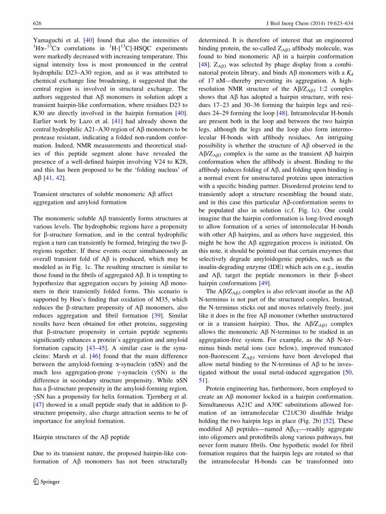
regions: two hydrophobic and two hydrophilic (Fig. 1a).
The 16 first N-terminal residues constitute a hydrophilic
tail. The two hydrophobic regions are the central L17–A21
part and the C-terminus A30–V40/A42, which are sepa-
rated by the hydrophilic central region E22–G29.
Secondary structure propensity of monomeric Abin solution
Although the Ab monomers are intrinsically disordered
[29], they adopt mainly helical conformations when
introduced to an apolar environment. When Ab is added to
the micellar form of the simple membrane-mimetic sodium
dodecyl sulphate (SDS) or its lithium salt (LiDS), the
hydrophobic regions fold into helices [30, 31]. Addition of
paramagnetic Mn(II) to the Ab–SDS mixture has revealed
that the C-terminal helix is buried into the interior of the
micelle, while the central helix is positioned on the surface
of the micelle, only partially buried [31]. This may reflect
the positioning of the Ab region prior to cleavage from
APP, and it underlines the ability for monomeric Ab to
adopt secondary structures.
In general, intrinsically disordered proteins (IDPs) and
peptides possess, despite the lack of overall structure,
transient structures or secondary structure propensities. Abmonomers are no exception, as they display a number of
properties indicating a secondary structure propensity [32,
33]. As a coarse-grained assessment of Ab-structure, the
overall apparent size of both full-length and truncated Abhas been determined from the hydrodynamic radii, RH, by
PFG NMR. The relation between RH and the monomer
length, i.e., the scaling exponent m, was found to be sub-
stantially smaller (0.44) than what would be expected for a
corresponding excluded-volume statistical coil (0.58) [34].
This was interpreted as Ab transiently forming more
compact structures, either as global collapses or as sec-
ondary structure formations. Indeed, a more recent study
showed that IDPs generally are more compact than dena-
tured globular proteins, and an average m for IDPs was
determined to be 0.45, in good agreement with findings for
Ab. This shows that Ab may behave similarly to other
IDPs that adopt transient structures [35].
The secondary structure of Ab monomers has been
studied by circular dichroism (CD) in numerous studies,
and the CD spectrum of Ab in room temperature shows all
characteristics of a random coil spectrum. However,
reducing the temperature induces gradual changes in the
spectrum that correspond to an increased population of left-
handed polyproline type 2 helix (PPII) [36, 37]. Similar
structural changes have been found in other IDPs upon
lowered temperature. PPII is an extended conformation,
possibly facilitated by steric interactions between bulky
side chains. The dihedral angles in PPII are very close to
that of an aggregation-prone b-conformation. Although CD
gives no information on a residual level, peptide truncation
studies indicate that the Ab PPII helix mainly involves the
N-terminal region [37].
In addition, the hydrophobic regions appear to undergo
temperature-induced structural changes. Determination of3JHNHa couplings indicates that the hydrophobic regions
tend to have a b propensity in aqueous solution [37]. NMR
relaxation measurements have shown that the transient
secondary structures in these regions give rise to reduced
motional freedom and restricted dynamics relative to the
more flexible N-terminal region [38]. The Ab regions that
form helices in membrane-mimetic environments tran-
siently form extended conformations rather than helical
conformations in solution (Fig. 1b). The temperature
dependence is typical for such transient structures: the
extended PPII structural propensity shows a cooperative
transition to random coil already at relatively low tem-
peratures, i.e., the melting temperature is significantly
lower than physiologically relevant temperatures [38].
Using detailed chemical shift analysis, Hou et al. [39]
has showed that the propensity to form b conformations is
reduced when M35 is oxidized, indicating transient long-
range cross talk in monomeric Ab.
Transiently folded states of Ab
In NMR studies of Ab not only chemical shifts, J-cou-
plings, and relaxation parameters have been found to be
temperature dependent, but a temperature increase also
induces a general loss in 1H-[15N]-HSQC signal intensity.
This was initially thought to be caused by increased
exchange rates of the unprotected amide protons. However,
Fig. 1 The two hydrophobic regions L17–A21 and A30–V40 exhibit
a secondary structure propensity for b-structure and are highlighted as
arrows in the primary structure (a). These regions transiently adopt b-
conformation (b) and may in turn transiently fold into a hairpin (c)
J Biol Inorg Chem (2014) 19:623–634 625
123
Yamaguchi et al. [40] found that also the intensities of1Ha–13Ca correlations in 1H-[13C]-HSQC experiments
were markedly decreased with increasing temperature. This
signal intensity loss is most pronounced in the central
hydrophilic D23–A30 region, and as it was attributed to
chemical exchange line broadening, it suggested that the
central region is involved in structural exchange. The
authors suggested that Ab monomers in solution adopt a
transient hairpin-like conformation, where residues D23 to
K30 are directly involved in the hairpin formation [40].
Earlier work by Lazo et al. [41] had already shown the
central hydrophilic A21–A30 region of Ab monomers to be
protease resistant, indicating a folded non-random confor-
mation. Indeed, NMR measurements and theoretical stud-
ies of this peptide segment alone have revealed the
presence of a well-defined hairpin involving V24 to K28,
and this has been proposed to be the ‘folding nucleus’ of
Ab [41, 42].
Transient structures of soluble monomeric Ab affect
aggregation and amyloid formation
The monomeric soluble Ab transiently forms structures at
various levels. The hydrophobic regions have a propensity
for b-structure formation, and in the central hydrophilic
region a turn can transiently be formed, bringing the two b-
regions together. If these events occur simultaneously an
overall transient fold of Ab is produced, which may be
modeled as in Fig. 1c. The resulting structure is similar to
those found in the fibrils of aggregated Ab. It is tempting to
hypothesize that aggregation occurs by joining Ab mono-
mers in their transiently folded forms. This scenario is
supported by Hou’s finding that oxidation of M35, which
reduces the b-structure propensity of Ab monomers, also
reduces aggregation and fibril formation [39]. Similar
results have been obtained for other proteins, suggesting
that b-structure propensity in certain peptide segments
significantly enhances a protein’s aggregation and amyloid
formation capacity [43–45]. A similar case is the synu-
cleins: Marsh et al. [46] found that the main difference
between the amyloid-forming a-synuclein (aSN) and the
much less aggregation-prone c-synuclein (cSN) is the
difference in secondary structure propensity. While aSN
has a b-structure propensity in the amyloid-forming region,
cSN has a propensity for helix formation. Tjernberg et al.
[47] showed in a small peptide study that in addition to b-
structure propensity, also charge attraction seems to be of
importance for amyloid formation.
Hairpin structures of the Ab peptide
Due to its transient nature, the proposed hairpin-like con-
formation of Ab monomers has not been structurally
determined. It is therefore of interest that an engineered
binding protein, the so-called ZAb3 affibody molecule, was
found to bind monomeric Ab in a hairpin conformation
[48]. ZAb3 was selected by phage display from a combi-
natorial protein library, and binds Ab monomers with a Kd
of 17 nM—thereby preventing its aggregation. A high-
resolution NMR structure of the Ab/ZAb3 1:2 complex
shows that Ab has adopted a hairpin structure, with resi-
dues 17–23 and 30–36 forming the hairpin legs and resi-
dues 24–29 forming the loop [48]. Intramolecular H-bonds
are present both in the loop and between the two hairpin
legs, although the legs and the loop also form intermo-
lecular H-bonds with affibody residues. An intriguing
possibility is whether the structure of Ab observed in the
Ab/ZAb3 complex is the same as the transient Ab hairpin
conformation when the affibody is absent. Binding to the
affibody induces folding of Ab, and folding upon binding is
a normal event for unstructured proteins upon interaction
with a specific binding partner. Disordered proteins tend to
transiently adopt a structure resembling the bound state,
and in this case this particular Ab-conformation seems to
be populated also in solution (c.f. Fig. 1c). One could
imagine that the hairpin conformation is long-lived enough
to allow formation of a series of intermolecular H-bonds
with other Ab hairpins, and as others have suggested, this
might be how the Ab aggregation process is initiated. On
this note, it should be pointed out that certain enzymes that
selectively degrade amyloidogenic peptides, such as the
insulin-degrading enzyme (IDE) which acts on e.g., insulin
and Ab, target the peptide monomers in their b-sheet
hairpin conformations [49].
The Ab/ZAb3 complex is also relevant insofar as the AbN-terminus is not part of the structured complex. Instead,
the N-terminus sticks out and moves relatively freely, just
like it does in the free Ab monomer (whether unstructured
or in a transient hairpin). Thus, the Ab/ZAb3 complex
allows the monomeric Ab N-terminus to be studied in an
aggregation-free system. For example, as the Ab N-ter-
minus binds metal ions (see below), improved truncated
non-fluorescent ZAb3 versions have been developed that
allow metal binding to the N-terminus of Ab to be inves-
tigated without the usual metal-induced aggregation [50,
51].
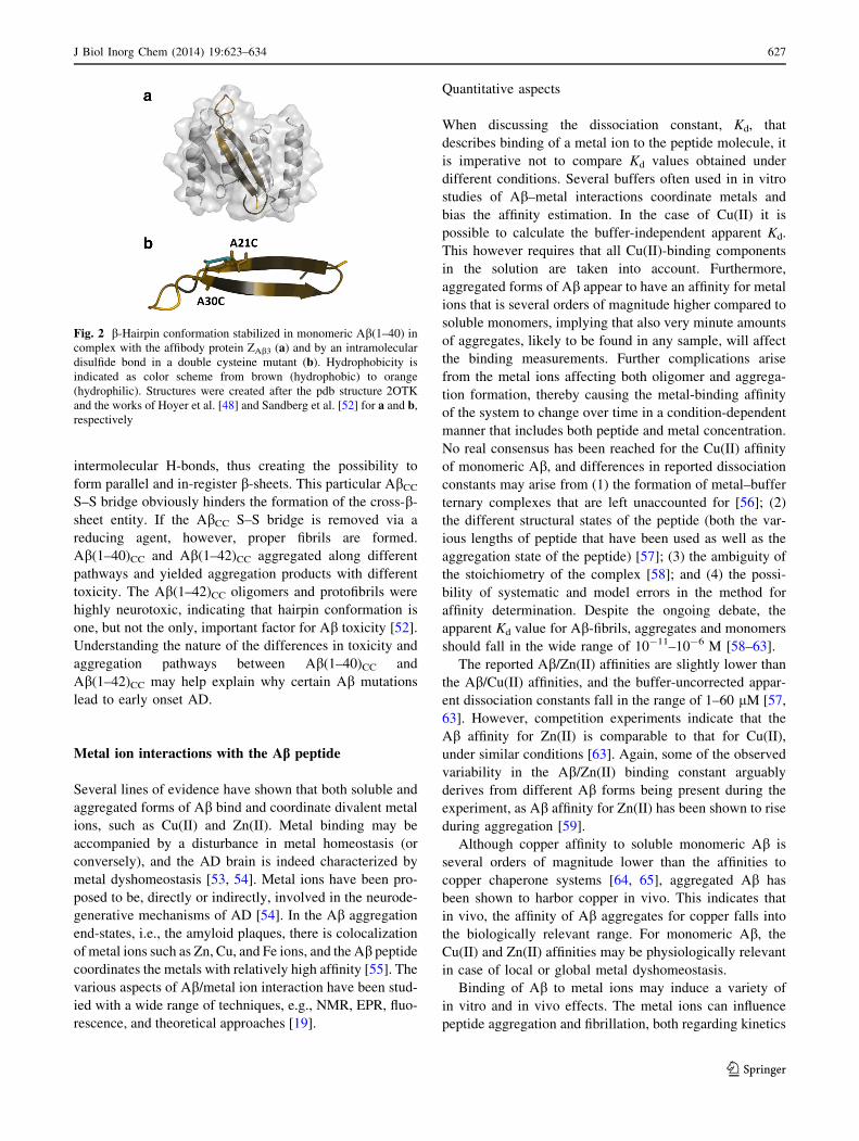
Protein engineering has, furthermore, been employed to
create an Ab monomer locked in a hairpin conformation.
Simultaneous A21C and A30C substitutions allowed for-
mation of an intramolecular C21/C30 disulfide bridge
holding the two hairpin legs in place (Fig. 2b) [52]. These
modified Ab peptides—named AbCC—readily aggregate
into oligomers and protofibrils along various pathways, but
never form mature fibrils. One hypothetic model for fibril
formation requires that the hairpin legs are rotated so that
the intramolecular H-bonds can be transformed into
626 J Biol Inorg Chem (2014) 19:623–634
123
intermolecular H-bonds, thus creating the possibility to
form parallel and in-register b-sheets. This particular AbCC
S–S bridge obviously hinders the formation of the cross-b-
sheet entity. If the AbCC S–S bridge is removed via a
reducing agent, however, proper fibrils are formed.
Ab(1–40)CC and Ab(1–42)CC aggregated along different
pathways and yielded aggregation products with different
toxicity. The Ab(1–42)CC oligomers and protofibrils were
highly neurotoxic, indicating that hairpin conformation is
one, but not the only, important factor for Ab toxicity [52].
Understanding the nature of the differences in toxicity and
aggregation pathways between Ab(1–40)CC and
Ab(1–42)CC may help explain why certain Ab mutations
lead to early onset AD.
Metal ion interactions with the Ab peptide
Several lines of evidence have shown that both soluble and
aggregated forms of Ab bind and coordinate divalent metal
ions, such as Cu(II) and Zn(II). Metal binding may be
accompanied by a disturbance in metal homeostasis (or
conversely), and the AD brain is indeed characterized by
metal dyshomeostasis [53, 54]. Metal ions have been pro-
posed to be, directly or indirectly, involved in the neurode-
generative mechanisms of AD [54]. In the Ab aggregation
end-states, i.e., the amyloid plaques, there is colocalization
of metal ions such as Zn, Cu, and Fe ions, and the Ab peptide
coordinates the metals with relatively high affinity [55]. The
various aspects of Ab/metal ion interaction have been stud-
ied with a wide range of techniques, e.g., NMR, EPR, fluo-
rescence, and theoretical approaches [19].
Quantitative aspects
When discussing the dissociation constant, Kd, that
describes binding of a metal ion to the peptide molecule, it
is imperative not to compare Kd values obtained under
different conditions. Several buffers often used in in vitro
studies of Ab–metal interactions coordinate metals and
bias the affinity estimation. In the case of Cu(II) it is
possible to calculate the buffer-independent apparent Kd.
This however requires that all Cu(II)-binding components
in the solution are taken into account. Furthermore,
aggregated forms of Ab appear to have an affinity for metal
ions that is several orders of magnitude higher compared to
soluble monomers, implying that also very minute amounts
of aggregates, likely to be found in any sample, will affect
the binding measurements. Further complications arise
from the metal ions affecting both oligomer and aggrega-
tion formation, thereby causing the metal-binding affinity
of the system to change over time in a condition-dependent
manner that includes both peptide and metal concentration.
No real consensus has been reached for the Cu(II) affinity
of monomeric Ab, and differences in reported dissociation
constants may arise from (1) the formation of metal–buffer
ternary complexes that are left unaccounted for [56]; (2)
the different structural states of the peptide (both the var-
ious lengths of peptide that have been used as well as the
aggregation state of the peptide) [57]; (3) the ambiguity of
the stoichiometry of the complex [58]; and (4) the possi-
bility of systematic and model errors in the method for
affinity determination. Despite the ongoing debate, the
apparent Kd value for Ab-fibrils, aggregates and monomers
should fall in the wide range of 10-11–10-6 M [58–63].
The reported Ab/Zn(II) affinities are slightly lower than
the Ab/Cu(II) affinities, and the buffer-uncorrected appar-
ent dissociation constants fall in the range of 1–60 lM [57,
63]. However, competition experiments indicate that the
Ab affinity for Zn(II) is comparable to that for Cu(II),
under similar conditions [63]. Again, some of the observed
variability in the Ab/Zn(II) binding constant arguably
derives from different Ab forms being present during the
experiment, as Ab affinity for Zn(II) has been shown to rise
during aggregation [59].
Although copper affinity to soluble monomeric Ab is
several orders of magnitude lower than the affinities to
copper chaperone systems [64, 65], aggregated Ab has
been shown to harbor copper in vivo. This indicates that
in vivo, the affinity of Ab aggregates for copper falls into
the biologically relevant range. For monomeric Ab, the
Cu(II) and Zn(II) affinities may be physiologically relevant
in case of local or global metal dyshomeostasis.
Binding of Ab to metal ions may induce a variety of
in vitro and in vivo effects. The metal ions can influence
peptide aggregation and fibrillation, both regarding kinetics
Fig. 2 b-Hairpin conformation stabilized in monomeric Ab(1–40) in
complex with the affibody protein ZAb3 (a) and by an intramolecular
disulfide bond in a double cysteine mutant (b). Hydrophobicity is
indicated as color scheme from brown (hydrophobic) to orange
(hydrophilic). Structures were created after the pdb structure 2OTK
and the works of Hoyer et al. [48] and Sandberg et al. [52] for a and b,
respectively
J Biol Inorg Chem (2014) 19:623–634 627
123
and aggregation pathways [66]. Interestingly, the effect on
Ab aggregation is dose-dependent, i.e., low sub-stoichi-
ometric concentrations of divalent metals are suggested to
reduce oligomeric stability and protect from aggregation
[67], while higher metal concentrations produce amor-
phous aggregation [68]. Such formation of amorphous
aggregates may protect from oligomer and amyloid for-
mation. However, these amorphous aggregates are not
dead-end products of the aggregation pathway. It has been
demonstrated that they may evolve into fibrils [69] or
spherical aggregates [70]. Inhibition of amyloid fibril for-
mation can be monitored by ThT fluorescence kinetics
experiments, and Yoshiike et al. [71] has demonstrated that
Zn(II) and Cu(II) prevent the formation of ThT-active
fibrillar structures at a stoichiometric ratio of 1:2 Ab:metal
ion. Instead, the amount of amorphous aggregates increa-
ses, as revealed by significant signal losses in CD experi-
ments on metal-induced Ab aggregates [72].
In addition to the aggregation propensity effects, bind-
ing of redox-active Cu(II) to Ab may prompt the formation
of reactive oxygen species, which in turn may cause oxi-
dative damage to neurons [73]. Such damage may be
involved in AD pathology, which is why Cu(II) has been
suggested as a possible causative agent behind AD. On the
other hand, chelation of Zn(II) by amyloid aggregates may
induce cognitive loss due to local depletion of extracellular
Zn [53]. In fact, in vitro cell toxicity studies of metal
depleted SOD1 have shown that local zinc chelation by
apoSOD1 is an effective neurotoxic mechanism, and that
this toxicity can be abolished by zinc addition. In line with
this observation, addition of zinc to a cell culture also
reduces the Ab toxicity [74, 75].
Structural aspects of metal binding
Despite the consensus regarding the importance of Ab–
metal interactions, no high-resolution structure of low
molecular weight Ab in complex with metal ions is
available, and the ligands involved are still debated. The
reason for this lack of structural information might be that
the metal ions do not induce a specific structure, but rather
an ensemble of structures. The conformation and coordi-
nation of the peptide–metal ion complex seem to depend on
the experimental conditions. However, the minimal metal-
binding site of the Ab peptide has been identified to consist
of the first 16 N-terminal amino acids [76], and the struc-
ture of a 16-mer peptide model in complex with zinc has
been solved by NMR [77]. The four metal ligands were
found to consist of the side chains of H6, E11, H13, and
H14 (Fig. 3a). In contrast to this structure, several other
metal-binding modes for the N-terminal peptide Ab(1–16)
have been suggested from EPR/NMR experiments, and the
Ab(1–16)/Cu(II) complex can adopt at least two additional
different structures, depending on the pH [78]. In this
truncated variant and in longer truncated peptides, i.e.,
Ab(1–28), the ligands involved in metal binding have been
suggested to be the histidines and/or D1 [79], A2 [78], E3
[80], R5 [81], or E11 [79]. This variety of proposed ligands
underlines the difficulties in determining a single structural
state of the complex, and arguably reflect the diversity of
metal-binding conformations in the truncated Ab variants,
and possibly also in the native peptide.
For the full-length peptide, a number of biophysical
studies show that the three His residues are involved in
metal coordination. The N-terminus [63, 82, 83], D1 [84],
Y10 [85], or coordinated H2O [86] are the proposed can-
didates for the fourth ligand. However, in NMR experi-
ments on Ab(1–40) at physiological pH, R5, Y10 and E11
showed no chemical shift changes or line broadening upon
addition of metal ions, indicating that they are not involved
in metal ligation [63]. The cross peak of D1 in a 1H-[13C]-
HSQC spectrum on the other hand, is shifted and line
broadened when zinc is added. This indicates that D1 is
involved in Zn binding, either through the N-terminus or
the D1 side chain (c.f. structural models in Fig. 3b, c).
Several studies have suggested that metals may serve as
bridges between Ab molecules, thereby forming higher-
order complexes, which may serve as a mechanistic
explanation for metal-induced amorphous aggregation
[87].
Recent NMR studies of zinc binding to Ab(1–40) sug-
gest that N-terminal metal binding induces a turn in the
central hydrophilic region, indicating long-range cross talk
in the peptide and again highlight the propensity for turn-
formation in full-length Ab [83].
The apparent contradictions between the metal-binding
results for Ab(1–16/28) and Ab(1–40) may reflect an
involvement of the C-terminus in the overall full-length
Ab/Zn(II) structure. In fact, the C-terminus has been pro-
posed to be involved in metal coordination in the aggre-
gated state of Ab, c.f. next chapter.
Solid-state NMR studies of Ab fibril structures
Solid-state NMR (ssNMR) has made major contributions to
our understanding of the molecular structures of amyloid
fibrils. Different aspects of amyloid fibril structures, stud-
ied by ssNMR, have been covered in recent review articles
[18, 88].
Ab(1–40) fibrils are typically straight and unbranched,
have diameters on the order of 10 nm, and contain ribbon-
like b-sheets where the b-strands run approximately per-
pendicular to the long fibril axis, while the inter-strand
hydrogen bonds run approximately parallel to the long
fibril axis. Amyloid fibrils are intrinsically insoluble and
628 J Biol Inorg Chem (2014) 19:623–634
123
non-crystalline, and are therefore not amenable to high-
resolution structure determination techniques such as X-ray
crystallography or high-resolution solution NMR spec-
troscopy. ssNMR techniques have been particularly valu-
able as ssNMR measurements on isotope-labeled samples
have the unique capability of providing detailed structural
constraints in non-crystalline, fibrillar structures. With
complementary long-range constraints obtained from EM
or small-angle neutron scattering studies, and in combi-
nation with site-directed mutagenesis, it has proven pos-
sible to develop full structural models for amyloid fibrils
from solid-state NMR data. Such models reveal both the
molecular conformation and the supramolecular organiza-
tion (Fig. 4).
Intrinsic sample polymorphism of Ab fibrillar structures
is a major obstacle for ssNMR studies, as it results in
samples with low structural homogeneity. Nevertheless, a
consensus structure for Ab peptides has been obtained,
describing a b-hairpin with a turn in the 25–29 region.
ssNMR data show that the cross-b motifs in Ab(1–40)
fibrils are comprised of parallel b-sheets, while those in
Ab(11–25) fibrils are comprised of antiparallel b-sheets.
Studies with ssNMR on amyloid fibrils prepared in vitro
from synthetic Ab peptides have shown that the molecular
structure of Ab(1–40) fibrils is not uniquely determined by
the amino acid sequence. Instead, the fibril structure also
depends on the precise details of the growth conditions
[89]. The earliest ssNMR studies on Ab peptides were
performed by Lansbury et al. [90–92] on the nine amino
acid Ab(34–42) peptide. Multiple-quantum ssNMR mea-
surements indicate a parallel organization of b-sheets in Abfibrils [93]. NMR dipolar recoupling methods for the
measurement of inter-peptide distances have established
that the central core of Ab(10–35) consists of a parallel b-
sheet structure where identical residues in adjacent chains
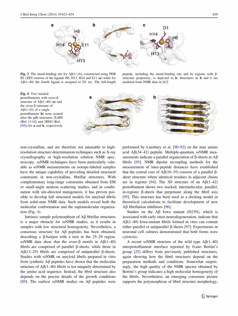
are in register [94]. The 3D structure of an Ab(1–42)
protofilament shows two stacked, intermolecular, parallel,
in-register b-sheets that perpetuate along the fibril axis
[95]. This structure has been used as a docking model in
theoretical calculations to facilitate development of new
Ab fibrillation inhibitors [96].
Studies on the Ab Iowa mutant (D23N), which is
associated with early onset neurodegeneration, indicate that
Ab(1–40) Iowa-mutant fibrils formed in vitro can contain
either parallel or antiparallel b-sheets [97]. Experiments in
neuronal cell cultures demonstrated that both forms were
cytotoxic.
A recent ssNMR structure of the wild type Ab(1–40)
interprotofilament interface reported by Ivano Bertini’s
group [25] differs from previously published structures,
again showing how the fibril structures depend on the
preparation methods and conditions. Somewhat surpris-
ingly, the high quality of the NMR spectra obtained by
Bertini’s group indicates a high molecular homogeneity of
the fibrils. Nevertheless, an emerging consensus picture
supports the polymorphism of fibril structure morphology,
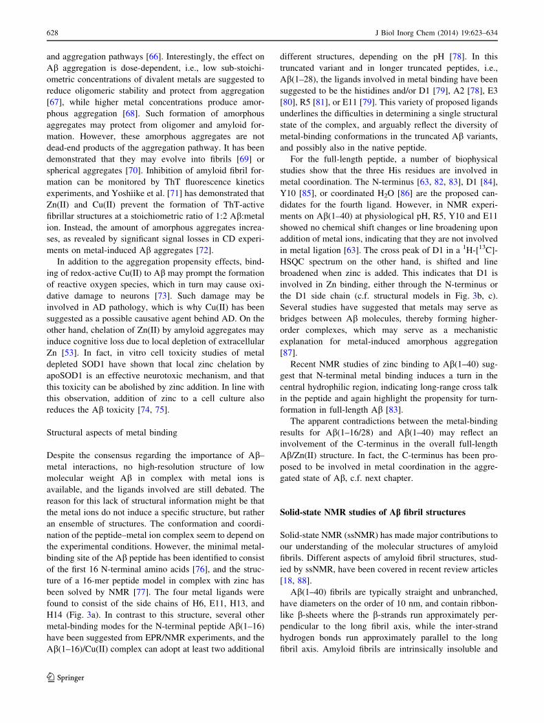
Fig. 3 The metal-binding site for Ab(1–16), constructed using PDB
ID 1ZE9 consists of the ligands H6, H13, H14 and E11 (a) while for
Ab(1–40) the fourth ligand is assigned to D1 (c). The full-length
peptide, including the metal-binding site and its regions with b-
structure propensity, is depicted in b. Structures in b and c are
modeled from NMR data in [63]
Fig. 4 Two stacked
protofilaments with cross-bstructure of Ab(1–40) (a) and
the cross-b structure of
Ab(1–42) of a single
protofilament (b) were created
after the pdb structures 2LMN
(Ref. [114]) and 2BEG (Ref.
[95]) for a and b, respectively
J Biol Inorg Chem (2014) 19:623–634 629
123
possibly with multiple structures co-existing, even though
all tertiary structures are based on parallel in-register b-
sheet secondary structures, generally showing hairpin
variants. The fibril cross-sections display either twofold or
threefold symmetry regarding the arrangement of the
ordered hairpins.
A very recent study by the Tycko group [98] shows the
molecular structure of a Ab(1–40) fibril grown from a seed
of a human brain extract. ssNMR spectra recorded for
fibrils seeded from two AD patients were significantly
different. From one of the seeded fibrils it was possible to
determine a detailed and unique structure. The fibril cross-
section showed a threefold symmetry unit based on three
hairpin structures. The observation that the seeded fibrils
contained two clearly different Ab conformations makes
the Ab fibrillation process reminiscent of the propagation
of different prion strains [99]. Indeed, it has been proposed
that also Ab assemblies are capable of self-propagation
inside the human brain [100].
Bertini’s group has also developed a method known as
sedimented solutes NMR (SedNMR), in which ssNMR
experiments are used to observe proteins that are sedi-
mented from solution via an ultracentrifugal field [101].
ssNMR studies on Ab fibrils have also been used to
investigate binding sites and binding structures of Cu(II) to
Ab [102]. The molecular details of Cu(II) binding to
amyloid Ab(1–40) fibrils were probed using paramagnetic
signal quenching in 1D and 2D high-resolution 13C
ssNMR. Selective quenching observed in 13C spectra of
Cu(II)-bound Ab(1–40) suggested that primary Cu(II)
binding sites in Ab(1–40) fibrils include Ne in H13 and
H14, together with carboxyl groups inV40 as well as in
glutamic acid side chains (i.e., E3, E11, and/or E22). 13C
chemical shift analysis demonstrated no major structural
changes upon Cu(II) binding in the hydrophobic core
regions (residues 18–25 and 30–36) of Ab. Although, the
occurrence of ROS production via oxidization of M35 in
the presence of Cu(II) has long been suspected, the 13CeH3-
S signal in M35 showed little change after Cu(II) binding,
demonstrating that M35 cannot be oxidized by Cu(II)
alone.
In summary, ssNMR spectroscopy is a powerful tech-
nique for structural studies in amyloid fibrils that deepened
our understanding of the structure of the fibril and the
process of its formation.
Cryo-electron microscopy studies of Ab fibrils
Biological structures can be visualized in the electron
microscope (EM), but the combined effect of poor contrast
and radiation damage makes it difficult to achieve high-
resolution images. Negative staining with salts of heavy
atoms followed by dehydration can enhance contrast.
However, this compromises resolution, as the staining
procedure induces random structural distortions at the
molecular level. Alternatively, the sample can be main-
tained in a hydrated state and—to a limited degree—be
protected against radiation damage by rapid freezing and
imaging at liquid N2 temperatures. This technique pre-
serves resolution, but suffers from poor contrast. By
combining advanced microscopes, the latest electron
detectors, and dedicated image processing algorithms,
contrast can be enhanced by averaging 2D images of many
identical molecular structures into a 3D structure. This
approach is especially attractive for structures with internal
symmetry: icosahedral viruses have been visualized to
almost 3 A resolution [103], and 3D images of protein
nanocrystals suggest that atomic resolution might be
around the corner [104]. The 1D crystalline nature of the
Ab peptide helical fibrils makes them an attractive target
for cryo-EM. Cross-sections of fibrils (Fig. 5a, b) as well as
details of fibril structures (Fig. 5c) may be determined.
Platinum shadowing (not a cryo-EM technique) has shown
that Ab(1–40) fibrils exhibit periodic cross-b structures
with a left-handed supertwist. In a 3D cryo-EM recon-
struction (2.6 nm resolution), these fibrils were shown to
have a ribbon-like shape and had a clear polarity [105].
Extension of the resolution to 8 A revealed that the fibril
consists of two protofilaments, each containing *5 nm
long regions of b-sheet structure (Fig. 5b). Cross-sections
showed two Ab(1–40) peptides folded symmetrically into a
paired b-sheet structure [106]. This is in agreement with
the super structure formed by a smaller Ab peptide, solved
by X-ray crystallography [16]. The fibrils are homogeneous
in width, but significantly vary in their helical pitch. Two
subgroups of Ab(1–40) fibrils were distinguished: F120
and F140 fibrils, with a helical repeat (cross-over distance)
of (120 ± 10) nm and (140 ± 10) nm, respectively [107].
In cross-section, F120 and F140 are highly similar and their
difference may be arbitrary [107].
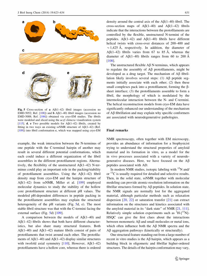
The Ab(1–42) fibrils are markedly different from cryo-
EM models of Ab(1–40) fibrils. Proteolysis analysis and
iterative real-space helical reconstruction has revealed that
two protofilaments form an interprotofilament with a hol-
low core that holds the Ab(1–42) C-terminus (Fig. 5a)
[108]. Thus, the large hydrophobic C-terminal residues are
buried inside the fibril, something that presumably also
increases fibril stability (Fig. 5d). In cross-section, the
protofilament measures around 40 A 9 20 A [108], a
density into which the ssNMR hairpin structure of the
peptide [95] could be fitted. It has been proposed that the
flexible N-terminus of Ab(1–42) might stack on the hairpin
loop of another Ab(1–42) unit [108]. Even within a single
preparation morphological diversity of Ab(1–42) fibrils
was observed, which may have several causes. For
630 J Biol Inorg Chem (2014) 19:623–634
123
example, the weak interaction between the N-terminus of
one peptide with the C-terminal hairpin of another may
result in several different potential conformations, which
each could induce a different organization of the fibril
assemblies in the different protofilament regions. Alterna-
tively, the flexibility of the unstructured Ab(1–42) N-ter-
minus could play an important role in the packing/stability
of protofilament assemblies. Using the Ab(1–42) fibril
density map from cryo-EM and the hairpin structure of
Ab(1–42) from ssNMR, Miller et al. [109] employed
molecular dynamics to study the stability of the hollow
core protofilament structure at different pH values. The
modeled pH-dependent differences in peptide packing in
the protofilament assemblies may explain the structural
heterogeneity of the pH variants (Fig. 5d, e). The most
stable fibril structure was that with the C-termini facing the
external surface (Fig. 5d) [109].
A comparison between the models of Ab(1–40) and
Ab(1–42) fibrils shows that both have different character-
istics, but also share many structural features. Both
Ab(1–40) and Ab(1–42) mature fibrils consist of pairs of
protofilaments that twist around each other. The protofila-
ments of Ab(1–40) and Ab(1–42) display similar structures
with twofold axial symmetry [110]. However, Ab(1–42)
protofilaments have a hollow core, whereas there is ordered
density around the central axis of the Ab(1–40) fibril. The
cross-section maps of Ab(1–40) and Ab(1–42) fibrils
indicate that the interactions between the protofilaments are
controlled by the flexible, unstructured N-termini of the
peptides. Ab(1–42) and Ab(1–40) fibrils have different
helical twists with cross-over distances of 200–400 and
*1,425 A, respectively. In addition, the diameter of
Ab(1–42) fibrils varies from 67 to 85 A, whereas the
diameter of Ab(1–40) fibrils ranges from 60 to 200 A
[108].
The unstructured flexible Ab N-terminus, which appears
to regulate the assembly of Ab protofilaments, might be
developed as a drug target. The mechanism of Ab fibril-
lation likely involves several steps: (1) Ab peptide seg-
ments initially associate with each other; (2) then these
small complexes pack into a protofilament, forming the b-
sheet interface; (3) the protofilaments assemble to form a
fibril, the morphology of which is modulated by the
intermolecular interaction between the N- and C-termini.
The helical reconstruction models from cryo-EM data have
significantly enhanced our understanding of the mechanism
of Ab fibrillation and may explain why specific conformers
are associated with neurodegenerative pathologies.
Final remarks
NMR spectroscopy, often together with EM microscopy,
provides an abundance of information for a biophysicist
trying to understand the structural properties of amyloid
material and its formation in vitro, as a model for the
in vivo processes associated with a variety of neurode-
generative diseases. Here, we have focused on the Abpeptides associated with AD.
In modern NMR studies, isotopic labeling with 15N and/
or 13C is usually required for detailed and selective results.
Then, in the solid state, ssNMR together with molecular
modeling can provide atomic-resolution information on the
fibrillar structures formed by Ab peptides. In solution state,
the NMR signals are normally lost for the aggregated
material, although particular methods such as relaxation
dispersion [20, 22] or saturation transfer [21] can extract
information on the structures and kinetics associated with
the amyloid material in its ‘‘dark state’’ of NMR [21, 111].
Relatively simple solution experiments such as 1H-[15N]-
HSQC can give the first clues about the interactions
between monomeric Ab and small molecules or metal ions,
which often influence both the Ab NMR spectra and the
Ab aggregation pathways (kinetically or structurally).
One structural feature standing out from a large number of
recent in vitro studies is the Ab hairpin, which appears as a
building block in oligomeric and fibrillar higher-ordered
structures. The details of the hairpin conformation may vary,
Fig. 5 Cross-section of a Ab(1–42) fibril images (accession no
EMD-5052; Ref. [108]) and b Ab(1–40) fibril images (accession no
EMD-5008; Ref. [106]) obtained via cryo-EM studies. The fibrils
were modeled and sliced using the ucsf chimera visualization system
[115]. d, e Two possible models for Ab(1–42) fibrils, created by
fitting in two ways an existing ssNMR structure of Ab(1–42) (Ref.
[109]) into fibril conformation c, which was mapped using cryo-EM
J Biol Inorg Chem (2014) 19:623–634 631
123
depending on the exact nature of the peptide sample and its
environmental conditions, but the overall properties seem to
converge. This more or less ordered hairpin, accompanied by
a less-structured N-terminus, is probably the most important
Ab form that interacts with the environment and determines
the continued process of aggregation. The observation that a
fibril seeded from a human brain extract also shows this basic
structural unit indicates that it is probably also relevant for
human disease.
There are still many unresolved questions regarding the
Ab aggregation and fibrillation processes. Major questions
concern the connection between the observations of the
aggregation process in vitro and the disease aspects in vivo.
Some examples of unresolved topics are:
• the structure(s) of the neurotoxic Ab assemblies and
what mechanism(s) causes neurotoxicity,
• the spreading of the neurotoxic assemblies between
neurons and the potential similarities to prion disease
mechanisms [98, 100, 112],
• the roles of metal ions, redox active and inactive, in the
neurotoxic processes involving the Ab peptide [113],
and
• the possibility to base drug development on the in vitro
observations of the Ab peptide aggregation process.
The gap of knowledge between in vitro studies and
understanding of in vivo pathology in humans is at present
visible in the disturbing lack of reliable and relevant model
systems as well as absence of structure–toxicity relation-
ship, as the in vitro observations alone may not be suffi-
cient to develop a detailed mechanistic understanding of
the in vivo disease manifestations.
Despite the encouraging progress made in molecular
amyloid research during the last decade, much more
knowledge is needed before we can formulate therapeutic
strategies for the associated devastating diseases.
Acknowledgments This study was supported by grants from the
Swedish Research Council to A.G., from NOW (TOP.08.B3.014) to
J.P.A., Estonian Ministry of Education and Research (Targeting
Financing Theme SF 9690034s09) to J.J. and from the Magnus
Bergvall foundation to S.W. Funding for J.D. was from Swedish
Foundation for Strategic Research (MDB10-0030). We thank Dr.
Goran Eriksson for fruitful discussions.
References
1. Haass C, Selkoe DJ (2007) Nat Rev Mol Cell Biol 8:101–112
2. Masters CL, Selkoe DJ (2012) Cold Spring Harb Perspect Med
2:a006262
3. Sunde M, Blake CC (1998) Q Rev Biophys 31:1–39
4. Eisenberg D, Jucker M (2012) Cell 148:1188–1203
5. Chiti F, Dobson CM (2006) Annu Rev Biochem 75:333–366
6. Fandrich M (2012) J Mol Biol 421:427–440
7. Selkoe DJ (1991) Neuron 6:487–498
8. Hardy JA, Higgins GA (1992) Science 256:184–185
9. Butterfield SM, Lashuel HA (2010) Angew Chem Int Ed Engl
49:5628–5654
10. Lashuel HA, Hartley D, Petre BM, Walz T, Lansbury PT Jr
(2002) Nature 418:291
11. Biancalana M, Koide S (2010) Biochim Biophys Acta
1804:1405–1412
12. Skeby KK, Sørensen J, Schiøtt B (2013) J Am Chem Soc
135:15114–15128
13. Necula M, Kayed R, Milton S, Glabe CG (2007) J Biol Chem
282:10311–10324
14. Benilova I, Karran E, De Strooper B (2012) Nat Neurosci
15:349–357
15. Nelson R, Sawaya MR, Balbirnie M, Madsen AØ, Riekel C,
Grothe R, Eisenberg D (2005) Nature 435:773–778
16. Sawaya MR, Sambashivan S, Nelson R, Ivanova MI, Sievers
SA, Apostol MI, Thompson MJ, Balbirnie M, Wiltzius JJ,
McFarlane HT, Madsen AØ, Riekel C, Eisenberg D (2007)
Nature 447:453–457
17. Fitzpatrick AW, Debelouchina GT, Bayro MJ, Clare DK, Ca-
porini MA, Bajaj VS, Jaroniec CP, Wang L, Ladizhansky V,
Muller SA, MacPhee CE, Waudby CA, Mott HR, De Simone A,
Knowles TP, Saibil HR, Vendruscolo M, Orlova EV, Griffin RG,
Dobson CM (2013) Proc Natl Acad Sci USA 110:5468–5473
18. Tycko R (2011) Annu Rev Phys Chem 62:279–299
19. Warmlander S, Tiiman A, Abelein A, Luo J, Jarvet J, Soderberg
KL, Danielsson J, Graslund A (2013) ChemBioChem
14:1692–1704
20. Abelein A, Lang L, Lendel C, Graslund A, Danielsson J (2012)
FEBS Lett 586:3991–3995
21. Fawzi NL, Ying J, Torchia DA, Clore GM (2010) J Am Chem
Soc 132:9948–9951
22. Abelein A, Kaspersen JD, Nielsen SB, Jensen GV, Christiansen
G, Pedersen JS, Danielsson J, Otzen DE, Graslund A (2013) J
Biol Chem 288:23518–23528
23. Bertini I, Luchinat C, Parigi G, Pierattelli R (2005) ChemBio-
Chem 6:1536–1549
24. Bertini I, Luchinat C, Parigi G, Pierattelli R (2008) Dalton
Trans, 3782–3790
25. Bertini I, Gonnelli L, Luchinat C, Mao J, Nesi A (2011) J Am
Chem Soc 133:16013–16022
26. De Strooper B, Saftig P, Craessaerts K, Vanderstichele H, Gu-
hde G, Annaert W, Von Figura K, Van Leuven F (1998) Nature
391:387–390
27. Gralle M, Ferreira ST (2007) Prog Neurobiol 82:11–32
28. Barrett PJ, Song Y, Van Horn WD, Hustedt EJ, Schafer JM,
Hadziselimovic A, Beel AJ, Sanders CR (2012) Science
336:1168–1171
29. Riek R, Guntert P, Dobeli H, Wipf B, Wuthrich K (2001) Eur J
Biochem 268:5930–5936
30. Shao H, Jao S, Ma K, Zagorski MG (1999) J Mol Biol
285:755–773
31. Jarvet J, Danielsson J, Damberg P, Oleszczuk M, Graslund A
(2007) J Biomol NMR 39:63–72
32. Dyson HJ, Wright PE (2005) Nat Rev Mol Cell Biol 6:197–208
33. Lacy ER, Filippov I, Lewis WS, Otieno S, Xiao L, Weiss S,
Hengst L, Kriwacki RW (2004) Nat Struct Mol Biol 11:358–364
34. Danielsson J, Jarvet J, Damberg P, Graslund A (2002) Magn
Reson Chem 40:S89–S97
35. Bernado P, Blackledge M (2009) Biophys J 97:2839–2845
36. Jarvet J, Damberg P, Danielsson J, Johansson I, Eriksson LE,
Graslund A (2003) FEBS Lett 555:371–374
37. Danielsson J, Jarvet J, Damberg P, Graslund A (2005) FEBS J
272:3938–3949
38. Danielsson J, Andersson A, Jarvet J, Graslund A (2006) Magn
Reson Chem 44 Spec No:S114–S121
632 J Biol Inorg Chem (2014) 19:623–634
123
39. Hou L, Shao H, Zhang Y, Li H, Menon NK, Neuhaus EB,
Brewer JM, Byeon IJ, Ray DG, Vitek MP, Iwashita T, Makula
RA, Przybyla AB, Zagorski MG (2004) J Am Chem Soc
126:1992–2005
40. Yamaguchi T, Matsuzaki K, Hoshino M (2011) FEBS Lett
585:1097–1102
41. Lazo ND, Grant MA, Condron MC, Rigby AC, Teplow DB
(2005) Protein Sci 14:1581–1596
42. Roychaudhuri R, Yang M, Condron MM, Teplow DB (2012)
Biochemistry 51:3957–3959
43. Moriarty DF, Raleigh DP (1999) Biochemistry 38:1811–1818
44. Chiti F, Taddei N, Baroni F, Capanni C, Stefani M, Ramponi G,
Dobson CM (2002) Nat Struct Biol 9:137–143
45. Simmons LK, May PC, Tomaselli KJ, Rydel RE, Fuson KS,
Brigham EF, Wright S, Lieberburg I, Becker GW, Brems DN
et al (1994) Mol Pharmacol 45:373–379
46. Marsh JA, Singh VK, Jia Z, Forman-Kay JD (2006) Protein Sci
15:2795–2804
47. Tjernberg L, Hosia W, Bark N, Thyberg J, Johansson J (2002) J
Biol Chem 277:43243–43246
48. Hoyer W, Gronwall C, Jonsson A, Stahl S, Hard T (2008) Proc
Natl Acad Sci USA 105:5099–5104
49. Shen Y, Joachimiak A, Rosner MR, Tang WJ (2006) Nature
443:870–874
50. Lindgren J, Wahlstrom A, Danielsson J, Markova N, Ekblad C,
Graslund A, Abrahmsen L, Eriksson Karlstrom A, Warmlander
SKTS (2010) Protein Sci 19:2319–2329
51. Lindgren J, Segerfeldt P, Sholts SB, Graslund A, Karlstrom AE,
Warmlander SK (2013) J Inorg Biochem 120:18–23
52. Sandberg A, Luheshi LM, Sollvander S, Pereira de Barros T,
Macao B, Knowles TP, Biverstal H, Lendel C, Ekholm-Pett-
erson F, Dubnovitsky A, Lannfelt L, Dobson CM, Hard T
(2010) Proc Natl Acad Sci USA 107:15595–15600
53. Adlard PA, Bush AI (2006) J Alzheimers Dis 10:145–163
54. Ayton S, Lei P, Bush AI (2013) Free Radic Biol Med 62:76–89
55. Lovell MA, Robertson JD, Teesdale WJ, Campbell JL, Mar-
kesbery WR (1998) J Neurol Sci 158:47–52
56. Tougu V, Tiiman A, Palumaa P (2011) Metallomics 3:250–261
57. Tiiman A, Palumaa P, Tougu V (2013) Neurochem Int
62:367–378
58. Alies B, Renaglia E, Rozga M, Bal W, Faller P, Hureau C
(2013) Anal Chem 85:1501–1508
59. Tougu V, Karafin A, Palumaa P (2008) J Neurochem
104:1249–1259
60. Sacco C, Skowronsky RA, Gade S, Kenney JM, Spuches AM
(2012) J Biol Inorg Chem 17:531–541
61. Rozga M, Kloniecki M, Dadlez M, Bal W (2010) Chem Res
Toxicol 23:336–340
62. Sarell CJ, Syme CD, Rigby SE, Viles JH (2009) Biochemistry
48:4388–4402
63. Danielsson J, Pierattelli R, Banci L, Graslund A (2007) FEBS J
274:46–59
64. Badarau A, Dennison C (2011) J Am Chem Soc 133:2983–2988
65. Jeney V, Itoh S, Wendt M, Gradek Q, Ushio-Fukai M, Harrison
DG, Fukai T (2005) Circ Res 96:723–729
66. Suzuki K, Miura T, Takeuchi H (2001) Biochem Biophys Res
Commun 285:991–996
67. Garai K, Sengupta P, Sahoo B, Maiti S (2006) Biochem Biophys
Res Commun 345:210–215
68. Garai K, Sahoo B, Kaushalya SK, Desai R, Maiti S (2007)
Biochemistry 46:10655–10663
69. Tougu V, Karafin A, Zovo K, Chung RS, Howells C, West AK,
Palumaa P (2009) J Neurochem 110:1784–1795
70. Pedersen JT, Ostergaard J, Rozlosnik N, Gammelgaard B, He-
egaard NH (2011) J Biol Chem 286:26952–26963
71. Yoshiike Y, Tanemura K, Murayama O, Akagi T, Murayama M,
Sato S, Sun X, Tanaka N, Takashima A (2001) J Biol Chem
276:32293–32299
72. Ghalebani L, Wahlstrom A, Danielsson J, Warmlander SK,
Graslund A (2012) Biochem Biophys Res Commun 421:554–560
73. Huang X, Atwood CS, Hartshorn MA, Multhaup G, Goldstein
LE, Scarpa RC, Cuajungco MP, Gray DN, Lim J, Moir RD,
Tanzi RE, Bush AI (1999) Biochemistry 38:7609–7616
74. Cardoso SM, Rego AC, Pereira C, Oliveira CR (2005) Neurotox
Res 7:273–281
75. Johansson AS, Vestling M, Zetterstrom P, Lang L, Leinartaite L,
Karlstrom M, Danielsson J, Marklund SL, Oliveberg M (2012)
PLoS ONE 7:e36104
76. Minicozzi V, Stellato F, Comai M, Dalla Serra M, Potrich C,
Meyer-Klaucke W, Morante S (2008) J Biol Chem
283:10784–10792
77. Zirah S, Kozin SA, Mazur AK, Blond A, Cheminant M, Segalas-
Milazzo I, Debey P, Rebuffat S (2006) J Biol Chem
281:2151–2161
78. Drew SC, Noble CJ, Masters CL, Hanson GR, Barnham KJ
(2009) J Am Chem Soc 131:1195–1207
79. Gaggelli E, Janicka-Klos A, Jankowska E, Kozlowski H, Mi-
gliorini C, Molteni E, Valensin D, Valensin G, Wieczerzak E
(2008) J Phys Chem B 112:100–109
80. Hureau C, Coppel Y, Dorlet P, Solari PL, Sayen S, Guillon E,
Sabater L, Faller P (2009) Angew Chem 48:9522–9525
81. Zirah S, Rebuffat S, Kozin SA, Debey P, Fournier F, Lesage D,
Tabet JC (2003) Int J Mass Spectrom 228:999–1016
82. Valiente-Gabioud AA, Torres-Monserrat V, Molina-Rubino L,
Binolfi A, Griesinger C, Fernandez CO (2012) J Inorg Biochem
117:334–341
83. Rezaei-Ghaleh N, Giller K, Becker S, Zweckstetter M (2011)
Biophys J 101:1202–1211
84. Yang DS, McLaurin J, Qin K, Westaway D, Fraser PE (2000)
Eur J Biochem 267:6692–6698
85. Miura T, Suzuki K, Takeuchi H (2001) J Mol Struct 598:79–84
86. Curtain CC, Ali F, Volitakis I, Cherny RA, Norton RS, Bey-
reuther K, Barrow CJ, Masters CL, Bush AI, Barnham KJ
(2001) J Biol Chem 276:20466–20473
87. Miura T, Suzuki K, Kohata N, Takeuchi H (2000) Biochemistry
39:7024–7031
88. Heise H (2008) ChemBioChem 9:179–189
89. Paravastu AK, Qahwash I, Leapman RD, Meredith SC, Tycko R
(2009) Proc Natl Acad Sci USA 106:7443–7448
90. Spencer RG, Halverson KJ, Auger M, McDermott AE, Griffin
RG, Lansbury PT Jr (1991) Biochemistry 30:10382–10387
91. Lansbury PT Jr, Costa PR, Griffiths JM, Simon EJ, Auger M,
Halverson KJ, Kocisko DA, Hendsch ZS, Ashburn TT, Spencer
RG et al (1995) Nat Struct Biol 2:990–998
92. Costa PR, Kocisko DA, Sun BQ, Lansbury PT Jr, Griffin RG
(1997) J Am Chem Soc 119:10487–10493
93. Antzutkin ON, Balbach JJ, Leapman RD, Rizzo NW, Reed J,
Tycko R (2000) Proc Natl Acad Sci USA 97:13045–13050
94. Benzinger TL, Gregory DM, Burkoth TS, Miller-Auer H, Lynn
DG, Botto RE, Meredith SC (1998) Proc Natl Acad Sci USA
95:13407–13412
95. Luhrs T, Ritter C, Adrian M, Riek-Loher D, Bohrmann B,
Dobeli H, Schubert D, Riek R (2005) Proc Natl Acad Sci USA
102:17342–17347
96. Luo J, Otero JM, Yu CH, Warmlander SK, Graslund A, Over-
hand M, Abrahams JP (2013) Chemistry 19:17338–17348
97. Qiang W, Yau WM, Luo Y, Mattson MP, Tycko R (2012) Proc
Natl Acad Sci USA 109:4443–4448
98. Lu JX, Qiang W, Yau WM, Schwieters CD, Meredith SC, Tycko
R (2013) Cell 154:1257–1268
J Biol Inorg Chem (2014) 19:623–634 633
123
99. Safar J, Wille H, Itri V, Groth D, Serban H, Torchia M, Cohen
FE, Prusiner SB (1998) Nat Med 4:1157–1165
100. Stohr J, Watts JC, Mensinger ZL, Oehler A, Grillo SK, DeAr-
mond SJ, Prusiner SB, Giles K (2012) Proc Natl Acad Sci USA
109:11025–11030
101. Bertini I, Gallo G, Korsak M, Luchinat C, Mao J, Ravera E
(2013) ChemBioChem 14:1891–1897
102. Parthasarathy S, Long F, Miller Y, Xiao Y, McElheny D,
Thurber K, Ma B, Nussinov R, Ishii Y (2011) J Am Chem Soc
133:3390–3400
103. Grigorieff N, Harrison SC (2011) Curr Opin Struct Biol
21:265–273
104. Nederlof I, Li YW, van Heel M, Abrahams JP (2013) Acta
Crystallogr Sect D Biol Crystallogr 69:852–859
105. Sachse C, Xu C, Wieligmann K, Diekmann S, Grigorieff N,
Fandrich M (2006) J Mol Biol 362:347–354
106. Sachse C, Fandrich M, Grigorieff N (2008) Proc Natl Acad Sci
USA 105:7462–7466
107. Sachse C, Grigorieff N, Fandrich M (2010) Angew Chem Int Ed
Engl 49:1321–1323
108. Zhang R, Hu X, Khant H, Ludtke SJ, Chiu W, Schmid MF,
Frieden C, Lee JM (2009) Proc Natl Acad Sci USA
106:4653–4658
109. Miller Y, Ma B, Tsai CJ, Nussinov R (2010) Proc Natl Acad Sci
USA 107:14128–14133
110. Schmidt M, Sachse C, Richter W, Xu C, Fandrich M, Grigorieff
N (2009) Proc Natl Acad Sci USA 106:19813–19818
111. Bodner CR, Dobson CM, Bax A (2009) J Mol Biol 390:775–790
112. Hallbeck M, Nath S, Marcusson J (2013) Neuroscientist
19:560–566
113. Matlack KE, Tardiff DF, Narayan P, Hamamichi S, Caldwell
KA, Caldwell GA, Lindquist S (2014) Proc Natl Acad Sci USA
111:4013–4018
114. Petkova AT, Yau WM, Tycko R (2006) Biochemistry
45:498–512
115. Pettersen EF, Goddard TD, Huang CC, Couch GS, Greenblatt
DM, Meng EC, Ferrin TE (2004) J Comput Chem 25:1605–1612
634 J Biol Inorg Chem (2014) 19:623–634
123


