Embed Size (px)
Citation preview

Available online at www.sciencedirect.com
www.elsevier.com/locate/foodchemtox
Food and Chemical Toxicology 46 (2008) 1548–1554
The identification of potential alternative biomarkersof nitrofurazone abuse in animal derived food products
J.V. Samsonova a,*, A.J. Douglas b, K.M. Cooper a, D.G. Kennedy b, C.T. Elliott a
a Queen’s University Belfast, University Road Belfast, BT7 1NN, Northern Ireland, UKb Agri-Food and Biosciences Institute, Stoney Road, Belfast, UK
Received 15 April 2006; accepted 10 December 2007
Abstract
Semicarbazide (SEM) was considered to be a characteristic protein-bound side-chain metabolite of the banned veterinary drug nitro-furazone and used as a marker of nitrofurazone abuse. It was recently discovered that SEM can arise in food from sources other thannitrofurazone. This uncertainty over the source of SEM may be overcome if alternative markers specific to tissue-bound nitrofurazoneresidues can be determined. The structure of nitrofurazone metabolites in vivo and particular proteins to which they are bound are notknown. These proteins with altered structure due to the presence of the drug metabolites can be considered as potential alternative bio-markers of nitrofurazone abuse. The proteins implicated in the in vivo binding of nitrofurazone were separated and identified. A crudemixture of proteins extracted from the liver of a rat treated with the drug was separated using a series of different techniques such as pre-parative isoelectric focusing and size exclusion HPLC. Multiple fractions were assayed by LC–MS/MS to detect the presence of SEM. Theproteins containing SEM residues were identified by peptide mass mapping using trypsin digestion and MALDI-TOF. The first proteinidentified as containing high concentration of SEM was albumin. It was also shown that low molecular weight species within a proteinmixture whose main constituent was glutathione S-transferase contained a high concentration of SEM. The chemical composition of thesecomponents is under investigation. Preliminary data suggest the SEM forms part of a nitrofurazone metabolite conjugated to glutathione.� 2007 Elsevier Ltd. All rights reserved.
Keywords: Nitrofurazone; Semicarbazide; Alternative biomarker; Rat liver; Albumin
1. Introduction
Four drugs – furazolidone, furaltadone, nitrofurazoneand nitrofurantoin – belong to a group of nitrofuran anti-bacterial agents which were widely used for treatment of gas-trointestinal infections of cattle, pigs and poultry. Due to thepotential carcinogenicity and mutagenicity of nitrofuransand theirs metabolites since mid 1990s using of any nitrofu-
0278-6915/$ - see front matter � 2007 Elsevier Ltd. All rights reserved.doi:10.1016/j.fct.2007.12.019
Abbreviations: DMFA, dimethylformamide; GSH, glutathione; GST,glutathione S-transferase; IEF, isoelectric focusing; SDS, sodium dodecylsulfate; SEM, semicarbazide.
* Corresponding author. Present address: Department of ChemicalEnzymology, Faculty of Chemistry, M.V. Lomonosov Moscow StateUniversity, 119992 Moscow, Russia. Tel.: +7 495 939 3407; fax: +7 495939 2742.
E-mail address: [email protected] (J.V. Samsonova).
ran in any food-producing animal within the EU, or in anyanimal destined for export into the EU has been banned(Commission Regulation, 1995). EU Member States arerequired to ensure compliance with this ban through theirannual residue monitoring plans (Council Directive, 1996).
The nitrofuran antibiotics are characterised by quickmetabolism, half-life time in vivo is a few hours (McCrackenet al., 1995). Therefore determination of the parent com-pounds is unsuitable for monitoring purposes. Recently itwas found that high concentrations of nitrofuran metabo-lites accumulate in the retina of pigs (Cooper and Kennedy,2005) and that parent nitrofurazone accumulates in avianeyes (Cooper et al., 2005a). It was suggested that discoveryof these residue depots may allow monitoring of parentdrugs instead of theirs metabolites for the detection of drugabuse. However this will only be the case where samples ofeyes are available for analytical work.
ON
NH
NH2
O
O2NNH2 N
HNH2
O
nitrofurazone semicarbazide
H+
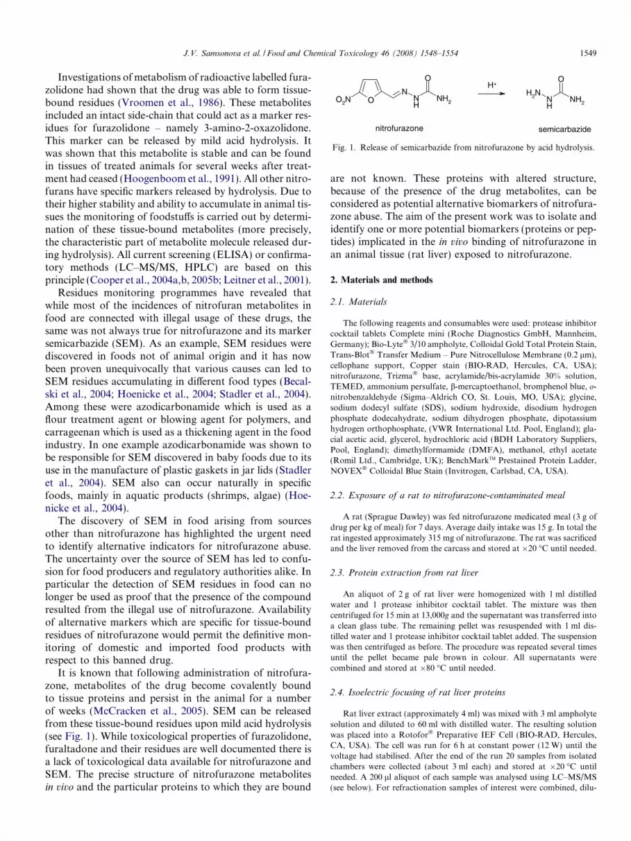
Fig. 1. Release of semicarbazide from nitrofurazone by acid hydrolysis.
J.V. Samsonova et al. / Food and Chemical Toxicology 46 (2008) 1548–1554 1549
Investigations of metabolism of radioactive labelled fura-zolidone had shown that the drug was able to form tissue-bound residues (Vroomen et al., 1986). These metabolitesincluded an intact side-chain that could act as a marker res-idues for furazolidone – namely 3-amino-2-oxazolidone.This marker can be released by mild acid hydrolysis. Itwas shown that this metabolite is stable and can be foundin tissues of treated animals for several weeks after treat-ment had ceased (Hoogenboom et al., 1991). All other nitro-furans have specific markers released by hydrolysis. Due totheir higher stability and ability to accumulate in animal tis-sues the monitoring of foodstuffs is carried out by determi-nation of these tissue-bound metabolites (more precisely,the characteristic part of metabolite molecule released dur-ing hydrolysis). All current screening (ELISA) or confirma-tory methods (LC–MS/MS, HPLC) are based on thisprinciple (Cooper et al., 2004a,b, 2005b; Leitner et al., 2001).
Residues monitoring programmes have revealed thatwhile most of the incidences of nitrofuran metabolites infood are connected with illegal usage of these drugs, thesame was not always true for nitrofurazone and its markersemicarbazide (SEM). As an example, SEM residues werediscovered in foods not of animal origin and it has nowbeen proven unequivocally that various causes can led toSEM residues accumulating in different food types (Becal-ski et al., 2004; Hoenicke et al., 2004; Stadler et al., 2004).Among these were azodicarbonamide which is used as aflour treatment agent or blowing agent for polymers, andcarrageenan which is used as a thickening agent in the foodindustry. In one example azodicarbonamide was shown tobe responsible for SEM discovered in baby foods due to itsuse in the manufacture of plastic gaskets in jar lids (Stadleret al., 2004). SEM also can occur naturally in specificfoods, mainly in aquatic products (shrimps, algae) (Hoe-nicke et al., 2004).
The discovery of SEM in food arising from sourcesother than nitrofurazone has highlighted the urgent needto identify alternative indicators for nitrofurazone abuse.The uncertainty over the source of SEM has led to confu-sion for food producers and regulatory authorities alike. Inparticular the detection of SEM residues in food can nolonger be used as proof that the presence of the compoundresulted from the illegal use of nitrofurazone. Availabilityof alternative markers which are specific for tissue-boundresidues of nitrofurazone would permit the definitive mon-itoring of domestic and imported food products withrespect to this banned drug.
It is known that following administration of nitrofura-zone, metabolites of the drug become covalently boundto tissue proteins and persist in the animal for a numberof weeks (McCracken et al., 2005). SEM can be releasedfrom these tissue-bound residues upon mild acid hydrolysis(see Fig. 1). While toxicological properties of furazolidone,furaltadone and their residues are well documented there isa lack of toxicological data available for nitrofurazone andSEM. The precise structure of nitrofurazone metabolitesin vivo and the particular proteins to which they are bound
are not known. These proteins with altered structure,because of the presence of the drug metabolites, can beconsidered as potential alternative biomarkers of nitrofura-zone abuse. The aim of the present work was to isolate andidentify one or more potential biomarkers (proteins or pep-tides) implicated in the in vivo binding of nitrofurazone inan animal tissue (rat liver) exposed to nitrofurazone.
2. Materials and methods
2.1. Materials
The following reagents and consumables were used: protease inhibitorcocktail tablets Complete mini (Roche Diagnostics GmbH, Mannheim,Germany); Bio-Lyte� 3/10 ampholyte, Colloidal Gold Total Protein Stain,Trans-Blot� Transfer Medium – Pure Nitrocellulose Membrane (0.2 lm),cellophane support, Copper stain (BIO-RAD, Hercules, CA, USA);nitrofurazone, Trizma� base, acrylamide/bis-acrylamide 30% solution,TEMED, ammonium persulfate, b-mercaptoethanol, bromphenol blue, o-nitrobenzaldehyde (Sigma–Aldrich CO, St. Louis, MO, USA); glycine,sodium dodecyl sulfate (SDS), sodium hydroxide, disodium hydrogenphosphate dodecahydrate, sodium dihydrogen phosphate, dipotassiumhydrogen orthophosphate, (VWR International Ltd. Pool, England); gla-cial acetic acid, glycerol, hydrochloric acid (BDH Laboratory Suppliers,Pool, England); dimethylformamide (DMFA), methanol, ethyl acetate(Romil Ltd., Cambridge, UK); BenchMarkTM Prestained Protein Ladder,NOVEX� Colloidal Blue Stain (Invitrogen, Carlsbad, CA, USA).
2.2. Exposure of a rat to nitrofurazone-contaminated meal
A rat (Sprague Dawley) was fed nitrofurazone medicated meal (3 g ofdrug per kg of meal) for 7 days. Average daily intake was 15 g. In total therat ingested approximately 315 mg of nitrofurazone. The rat was sacrificedand the liver removed from the carcass and stored at �20 �C until needed.
2.3. Protein extraction from rat liver
An aliquot of 2 g of rat liver were homogenized with 1 ml distilledwater and 1 protease inhibitor cocktail tablet. The mixture was thencentrifuged for 15 min at 13,000g and the supernatant was transferred intoa clean glass tube. The remaining pellet was resuspended with 1 ml dis-tilled water and 1 protease inhibitor cocktail tablet added. The suspensionwas then centrifuged as before. The procedure was repeated several timesuntil the pellet became pale brown in colour. All supernatants werecombined and stored at �80 �C until needed.
2.4. Isoelectric focusing of rat liver proteins
Rat liver extract (approximately 4 ml) was mixed with 3 ml ampholytesolution and diluted to 60 ml with distilled water. The resulting solutionwas placed into a Rotofor� Preparative IEF Cell (BIO-RAD, Hercules,CA, USA). The cell was run for 6 h at constant power (12 W) until thevoltage had stabilised. After the end of the run 20 samples from isolatedchambers were collected (about 3 ml each) and stored at �20 �C untilneeded. A 200 ll aliquot of each sample was analysed using LC–MS/MS(see below). For refractionation samples of interest were combined, dilu-

1550 J.V. Samsonova et al. / Food and Chemical Toxicology 46 (2008) 1548–1554
ted to 60 ml with distilled water and run on the IEF cell for 5 hours. Afterharvesting a 200 ll aliquot of each sample was analysed by LC–MS/MS,the remainder of the samples were stored at �20 �C.
2.5. Gel electrophoresis of proteins under denaturating conditions
(SDS-PAGE)
Gels were cast and run on the Mini PROTEAN� 3 ElectrophoresisCell and Power Pac 300 (BIO-RAD, Hercules, CA, USA) under denatu-rating conditions with SDS and b-mercaptoethanol. The gel dimensionswere 8.0 cm width � 7.5 cm length, the thickness of gels was 0.75 mm. Thestacking and resolving gel were 0.5 and 7 cm long, respectively.
The following buffers were used: resolving gel buffer–1.5 M Tris–HCl,14 mM SDS, pH 8.9; stacking gel buffer–0.5 M Tris–HCl, 14 mM SDS,pH 6.7; running buffer–52 mM Tris–HCl, 53 mM glycine, 0.1% (w/v) SDS.
Composition of the gels were as follows: resolving gel (10% (w/v)acrylamide)-acrylamide/bis-acrylamide 30% solution (6.0 ml), resolvinggel buffer (4.5 ml), distilled water (7.5 ml), TEMED (7.5 ll), 10% (w/v)ammonium persulfate (125 ll); stacking gel-acrylamide/bis-acrylamide30% solution (1.0 ml), stacking gel buffer (1.5 ml), distilled water (3.5 ml),TEMED (3.5 ll), 10% w/v ammonium persulfate (125 ll). Ammoniumpersulfate was made fresh daily.
Samples were mixed with sample loading buffer in the ratio 4:1, boiledat 90–100 �C for 10 min and then applied into the gel wells (15 ll per well)together with prestained molecular weight marker (7.5 ll per well). Sampleloading buffer consisted of distilled water (2.0 ml), 0.5 M Tris–HCl pH 6.8(625 ll), glycerol (1 ml), 10% (w/v) SDS (1 ml), b-mercaptoethanol(250 ll), 0.1% (w/v) bromphenol blue (125 ll).
Gels were run at a constant voltage of 200 V until the dye front hadreached 1 cm from the bottom of the gel. Proteins were stained withCoomassie blue stain and then dried between two cellophane sheets(GelAir Dryer, BIO-RAD, Hercules, CA, USA). When necessary gelswere stained with copper. Then protein bands of interest were excised,incubated in 20 mM phosphate buffer (pH 7.2) for 24 h and the resultingbuffer solution was analysed by LC–MS/MS.
2.6. Analysis of protein fractions by LC–MS/MS
Protein fractions were analysed for total SEM concentration by liquidchromatography tandem mass spectroscopy (LC–MS/MS) based on themethod of Cooper et al. (2005b). Briefly, samples (not pre-washed withsolvents) were derivatised with o-nitrobenzaldehyde in 7 ml 0.1 M HCl,neutralised with 7 ml 0.1 M dipotassium hydrogen orthophosphate and0.4 ml 1 M sodium hydroxide, then extracted into ethyl acetate (7 + 7 ml).All other conditions were as described (Cooper et al., 2005b).
2.7. Size exclusion HPLC
A SYSTEM GOLD� HPLC system with diode array detector (Beck-man Coulter, Fullerton, CA, USA) operating under 32 KaratTM 7.2 soft-ware was used for protein separation. The system was equipped withBioSep-SEC-S 2000 PEEK, 300 � 7.50 mm column (Phenomenex�, Mac-clesfield, Cheshire, UK). Before separation IEF fractions of interest wereconcentrated 10 fold. To achieve this they were combined, freeze dried andredissolved in smaller volume of distilled water. Undissolved sediment wasremoved by centrifugation. An aliquot (100 ll) of the combined materialwas injected onto the column and the material was separated at a flow rateof 1.0 ml/min using 20 mM phosphate buffer (pH 7.2) as eluant. The totalrun time was 25 min and fractions were collected every 15 s.
2.8. Gel elution
Samples of interest were run in a preparative cell on SDS-PAGE underthe conditions described above. Without staining, a gel then was placedwithin a Mini Whole Gel Eluter (BIO-RAD, Hercules, CA, USA) and runfor 30 min at a constant current of 100 mA. The elution buffer used wasthe same as for SDS-PAGE. Fourteen fractions (0.5–0.6 ml each) corre-
sponding to different sections of a gel were collected, then analysed bySDS-PAGE and LC–MS/MS.
2.9. Electroblotting
Samples of interest (3–4 wells) were run on SDS-PAGE under theconditions described above. Proteins were transferred from the gel onto anitrocellulose membrane using a Trans-Blot� Electrophoretic TransferCell (BIO-RAD, Hercules, CA, USA) for 1 h at a constant voltage of100 V. Transfer buffer was 25 mM Tris–HCl, 192 mM glycine and 20% (v/v) methanol, pH 8.3. Then membrane was then stained with colloidal gold.Membrane pieces with transferred proteins were excised, dissolved in200 ll DMFA and analysed by LC–MS/MS.
3. Results and discussion
3.1. Fractionation of rat liver proteins: preparative
isoelectric focusing (IEF)
To identify proteins implicated in the in vivo binding ofnitrofurazone, a rat was fed with meal contaminated withthe drug. A crude mixture of extracted liver proteins wasseparated by preparative IEF over pH range 2–10 (seeFig. 2a and b) to yield 20 different fractions. The fractionsobtained were assayed by LC–MS/MS to detect the pres-ence of the characteristic nitrofurazone side-chain (SEM).LC–MS/MS analysis of the fractions for the presence ofSEM showed the existence of two substantial peaks (seeFig. 2c). The major protein concentrated in fractions ##5–8 with molecular weight in the region of 65 kDa whichwas hypothesised to be albumin (see Fig. 2a). In fractions##11–16 within pH range 6.25–7.26 a number of proteinswas observed. Fractions with the highest SEM concentra-tion (##5–8 and ##11–16) were combined again and refrac-tionated by preparative IEF (see Fig. 3). Thus from the poolof fractions ##5–8 a major protein (tentatively albumin)was separated from the heavier protein containing SEM res-idues (mixtures #1 and #2, respectively). Refractionation ofthe fractions ##11–16 still yielded a mixture of SEM-con-taining proteins (mixture #3). Fractions with the highestSEM concentration were combined again to give three mix-tures of proteins (see Fig. 3). The same procedure wasrepeated twice starting from crude extract of liver. Finallythe fractions of interest were pooled from the repeated sep-arations to yield three protein mixtures. Fractionation andrefractionation of proteins extracted from negative rat (notfed with nitrofurazone) liver by preparative IEF followedby size exclusion HPLC (see later) resulted in a very similardistribution of proteins (data not shown).
3.2. Separation of proteins with bound SEM: size exclusion
HPLC
The above three mixture of proteins were separated fur-ther by size exclusion HPLC (see Fig. 4). LC–MS/MS anal-ysis of the fractions revealed that the highest SEMconcentrations were observed in protein mixtures ##1and 3 (see Fig. 5). The small peaks observed in all three

Fraction No
1 2 3 4 5 6 7 8 9 10 11 12 13 14 15 16 17 18 19 20
pH
0
2
4
6
8
10
12
Fraction No
1 2 3 4 5 6 7 8 9 10 11 12 13 14 15 16 17 18 19 20
[SE
M],
ng/m
L
0
50
100
150
200
250
300
Fraction No 1 2 3 4 5 6 7 8 9 10 11 12 13 14 15 16 17 18 19 20
181.8115.582.264.248.8
37.1
25.919.4
14.8
6.0 kDa
Fig. 2. SDS-PAGE, pH profile and concentration of SEM analysed by LC–MS/MS of IEF fractions ##1–20.For SDS-PAGE each fraction was diluted 2times with distilled water. IEF fractions which were combined for the following refractionation are circled.
J.V. Samsonova et al. / Food and Chemical Toxicology 46 (2008) 1548–1554 1551
mixtures in the non-protein region (above fraction #40)may be due to the presence of a range of low molecularweight substances such as ampholytes and protease inhibi-tors. SDS-PAGE of the fractions with the highest SEMconcentrations showed the presence of one major proteinin each mixture (see Fig. 4b). These major proteins were
identified by peptide mass mapping using trypsin digestionand MALDI-TOF.
The peptide mass mapping results obtained for mixture#1 confirmed the hypothesis that the major protein was ratalbumin (MW 70670 Da) (see Fig. 4b). The presenceof SEM residues bound to albumin was confirmed by

IEF fraction No
5 10 15 20
[SE
M],
ng/m
L
0
5
10
15
20
25
fractions ##5-8fractions ##11-16
Mixture #1 Mixture #2
Mixture #3
Fig. 3. Concentration of SEM in combined and refractioned IEFfractions ##5–8 and ## 11–16. Pooled IEF fractions (mixtures ##1–3)are circled.
Mix
ture
1
Mix
ture
2
Mix
ture
3
Mix
ture
1
Mix
ture
2
Mix
ture
3
181.8115.582.264.2
48.8
37.1
25.919.4
14.8
6.0kDa
Alb
GST
LRRG03
Fig. 4. SDS-PAGE for mixtures ##1–3 before (a) and after (b) sizeexclusion HPLC. Gel (b) illustrates the separation of the HPLC fractionswith the highest SEM concentration in each mixture (see Fig. 5).
HPLC fraction No
25 30 35 40 45 50 55
[SE
M],
ng/m
L
0
5
10
15
20
25
30
35
40
Mixture 1Mixture 2Mixture 3
Fig. 5. Concentration of SEM in HPLC fractions for mixtures ##1–3.
1552 J.V. Samsonova et al. / Food and Chemical Toxicology 46 (2008) 1548–1554
LC–MS/MS analysis of electroblotting and gel elutionsamples. SEM was found only in a band (electroblotting)
corresponding to albumin. Furthermore elution of proteinsfrom horizontal sections of SDS-PAGE gel confirmed thepresence of SEM only in fractions containing albumin.
The major protein in mixture #2 was identified as LiverRegeneration-Related Protein LRRG03 with MW 78512Da (see Fig. 4b). However, a very low concentration ofSEM, just above background levels, were found in bothelectroblotting and SDS-PAGE gel elution fractions(results not shown). Based on the low concentration ofSEM detected in mixture #2 (see Fig. 5) LRRG03 proteinwas not considered to be a good candidate to act as analternative biomarker for nitrofurazone.
Mixture #3 consisted of at least three proteins (seeFig. 4b), the major protein being identified as class mu chi-meric glutathione S-transferase (GST, chain A, isoenzymesM1-2 and M2-1, MW 25,858 Da). Glutathione S-transfer-ases are a group of enzymes that play an important role inthe detoxication of endogenous and xenobiotic electro-philic substances by catalysing the reaction between the tri-peptide glutathione (GSH) and xenobiotic compoundsbearing electrophilic functional groups (Ji et al., 1992).The enzymes are ubiquitous, for example, they compriseabout 5% of the total cytosolic protein in hepatic tissue.Glutathione (glutamyl-L-cysteinyl-glycine) is also animportant antioxidant, antitoxin and enzyme cofactor.Glutathione often attains millimolar concentrations insidecells, and is most concentrated in the liver (10 mM).
It could be clearly seen that considerable amounts of lowmolecular weight substances probably associated with theproteins in mixture #3 was released under the denaturatingconditions of SDS-PAGE (see Fig. 4b). LC–MS/MS analy-sis of the fractions eluted from different sections of SDS-PAGE gel showed that the majority of SEM was found inthis low molecular weight component. To determine theamount of SEM associated with low molecular weight com-ponents after SDS-PAGE separation of HPLC fraction #32(mixture #3, see Fig. 4b) the front (lower) section of theSDS-PAGE gel was excised, cut into pieces, placed into atube with phosphate buffer and incubated for a few hoursto extract the gel contents. The buffer was then analysedby LC–MS/MS. It was found that approximately 80% ofthe total SEM in HPLC fraction #32 was associated withthe low molecular weigh components. Total SEM concen-tration was found by analysing an equal amount of fraction#32 before SDS-PAGE separation. Similar results wereobtained by analysing fractions eluted from horizontal sec-tions of SDS-PAGE gel and SDS-PAGE gel sectionsstained by copper and excised from a gel. The ratio ofSEM concentration found in the enzyme sections (GST)and low molecular weight sections by these methods were35%:65% and 15%:85%, respectively.
The chemical composition of these low molecular weightcomponents containing high concentrations of SEMrequires careful investigation. Preliminary studies to iden-tify these components suggest that the SEM may be inthe form of nitrofurazone metabolites conjugated to gluta-thione. Previous studies of furazolidone metabolism in vitro

J.V. Samsonova et al. / Food and Chemical Toxicology 46 (2008) 1548–1554 1553
using swine liver microsomes showed that one possiblepathway could be a reversible conjugation of glutathionewith nitrofuran metabolites (Vroomen et al., 1988). Theauthors suggested that in vivo glutathione conjugates areformed in the liver, excreted in the blood and release reac-tive intermediates at other sites of the organism. So gluta-thione may act as a carrier of reactive derivatives offurazolidone. Further investigations in vitro demonstratedthat glutathione plays an important protective role againstthe toxicity of furazolidone but drug metabolism may varyamong different cell types with different metabolic capabil-ities (De Angelis et al., 1999). Sorrentino et al. (1987) andHoener (1988) investigated nitrofurazone disposition byperfused rat liver and also detected a new conjugatedmetabolite of nitrofurazone and glutathione.
Further studies are required to prove conclusively thepresence of GSH–nitrofurazone metabolite conjugates innitrofurazone treated animals. If such a conjugate wereshown to be widely distributed in nitrofurazone treatedanimals, it may be a candidate target for confirmatoryLC–MS/MS analysis. GSH is a widely distributed mole-cule, with a consistent structure, across many species andtissues, including blood, which raises the desirable prospectof a test for nitrofurazone abuse is live animals. However,given that GSH acts as part of the body’s detoxificationprocess, GSH–nitrofurazone metabolite conjugates mayhave a relatively short elimination half-life and may be suit-able as a marker residue for nitrofurazone abuse in animalsfoe a restricted period following cessation of medication.
4. Conclusions
Ideally, a biomarker for a drug such as nitrofurazoneshould be distributed in a wide range of animal tissues.Albumin appears to be a highly promising candidate forsuch a biomarker for nitrofurazone exposure because itcontains high concentrations of nitrofurazone metabolites(indicated by high SEM concentrations) and can be foundin many animal tissues. Albumin also has a relatively longelimination half-life (2–3 weeks in humans) and may,therefore, be sufficiently persistent in vivo to be a viablebiomarker for the monitoring of nitrofurazone abuse.To our knowledge this is the first report where anin vivo protein containing nitrofurazone metabolite resi-dues (rat liver albumin) has been separated and identified.This albumin fraction containing nitrofurazone metabo-lites has been used recently in this laboratory as an immu-nogen to raise antibodies to the modified protein. Theresults of this work will be presented elsewhere. Studiesare underway to produce monoclonal antibodies that bindto albumin only after exposure to nitrofurazone. Thiswork raises the prospect of a screening test for nitrofura-zone that does not rely on the detection of SEM. Conju-gates of glutathione and nitrofurazone metabolites arealso suggested as potential low molecular weight markerresidues for the monitoring of this banned nitrofuranantibiotic. Further elucidation of these potential biomark-
ers and the development of tests for their determination infoods of animal origin are required urgently to enable EUMember States to fully implement Council Directive 96/23/EC and to protect the consumer in Europe andworld-wide. The current study has been a proof of princi-ple on the identification of nitrofuran biomarkers. Theability to use the biomarkers identified will be reliant onthem being present in species other than rat, i.e. thefood-producing species where nitrofurans are known tobe misused in, e.g. poultry and pigs. Further studies willbe required to ensure this is the case.
Conflict of Interest Statement
There is no conflict of interest relating to the publicationon this manuscript with any of the named authors.
Acknowledgements
The financial support of the European Union, DGHealth and Consumer Protection, Joint Research Centre(Service contract for markers for nitrofurazone and onsemicarbazide analysis – LOT 3 – 2004-08-ic) is gratefullyacknowledged. The authors thank Ian Davidson (Prote-ome Facility, University of Aberdeen) for identificationof proteins by peptide mass mapping using trypsin diges-tion and MALDI-TOF.
References
Becalski, A., Lau, B.P.-Y., Lewis, D., Seaman, S.W., 2004. Semicarbazideformation in azodicarbonamide-treated flour: a model study. Journalof Agricultural and Food Chemistry 52, 5730–5734.
Commission Regulation (EC) 1442/95, 1995, of 26 June 1995 amendingannexes I, II, III and IV to regulation (EEC) No. 2377/90 laying downa community procedure for the establishment of maximum residuelimits of veterinary medicinal products in foodstuffs of animal origin.Official Journal of the European Communities L143, 26–30.
Cooper, K.M., Kennedy, D.G., 2005. Nitrofuran antibiotic metabolitesdetected at parts per million concentrations in retina of pigs – a newmatrix for enhanced monitoring of nitrofuran abuse. Analyst 130, 466–468.
Cooper, K.M., Caddell, A., Elliott, C.T., Kennedy, D.G., 2004a.Production and characterisation of polyclonal antibodies to a deriv-ative of 3-amino-2-oxazolidone, a metabolite of the nitrofuranfurazolidone. Analytica Chimica Acta 520, 79–86.
Cooper, K.M., Elliott, C.T., Kennedy, D.G., 2004b. Detection of 3-amino-2-oxazolidone (AOZ), a tissue-bound metabolite of the nitro-furan furazolidone, in prawn tissue by enzyme immunoassay. FoodAdditives and Contaminants 21, 841–848.
Cooper, K.M., McCracken, R.J., Kennedy, D.G., 2005a. Nitrofurazoneaccumulates in avian eyes – a replacement for semicarbazide as amarker of abuse. Analyst 130, 824–827.
Cooper, K.M., Mulder, P.P.J., van Rhijn, J.A., Kovacsics, L., McCrac-ken, R.J., Young, P.B., Kennedy, D.G., 2005b. Depletion of fournitrofuran antibiotics and their tissue-bound metabolites in porcinetissues and determination using LC–MS/MS and HPLC-UV. FoodAdditives and Contaminants 22, 406–414.
Council Directive 96/23/EC, 1996, of 29 April 1996 on measures tomonitor certain substances and residues thereof in live animals andanimal products. Official Journal of the European Communities L125,10–32.

1554 J.V. Samsonova et al. / Food and Chemical Toxicology 46 (2008) 1548–1554
De Angelis, I., Rossi, L., Pedersen, J.Z., Vignoli, A.L., Vincentini, O.,Hoogenboom, L.A.P., Polman, T.H.G., Stammati, A., Zucco, F.,1999. Metabolism of furazolidone: alternative pathways and modes oftoxicity in different cell lines. Xenobiotica 29, 1157–1169.
Hoener, B.-A., 1988. Nitrofurazone: kinetics and oxidative stress in thesinglepass isolated perfused rat liver. Biochemical Pharmacology 37,1629–1636.
Hoenicke, K., Gatermann, R., Hartig, L., Mandix, M., Otte, S., 2004.Formation of semicarbazide (SEM) in food by hypochlorite treatment:is SEM a specific marker for nitrofurazone abuse. Food Additives andContaminants 21, 526–537.
Hoogenboom, L.A.P., Van Kammen, M., Berghmans, M.C.J., Koeman,J.H., Kuiper, H.A., 1991. The use of pig hepatocytes to study thenature of protein-bound metabolites of furazolidone: a new analyticalmethod for their detection. Food and Chemical Toxicology 29, 321–328.
Ji, X., Zhang, P., Armstrong, R.N., Gilliland, G.L., 1992. The three-dimensional structure of a glutathione S-transferase from the Mugene class. Structural analysis of the binary complex of isoenzyme3-3 and glutathione at 2.2-A resolution. Biochemistry 31, 10169–10184.
Leitner, A., Zollner, P., Lindner, W., 2001. Determination of themetabolites of nitrofuran antibiotics ion animal tissue by high-performance liquid chromatography–tandem mass spectrometry.Journal of Chromatography A 939, 49–58.
McCracken, R.J., Blanchflower, W.J., Rowan, C., McCoy, M.A., Ken-nedy, D.G., 1995. Determination of furazolidone in porcine tissueusing thermospray liquid chromatography–mass spectrometry and astudy of the pharmacokinetics and stability of its residues. Analyst 120,2347–2351.
McCracken, R.J., Van Rhijn, J.A., Kennedy, D.G., 2005. The occurrenceof nitrofuran metabolites in the tissues of chickens exposed to very lowdietary concentrations of the nitrofurans. Food Additives and Con-taminants 22, 567–572.
Sorrentino, D., Bode, W., Hoener, B.-A., 1987. Nitrofurazone dispositionby perfused rat liver. Effect of dose size and glutathione depletion.Biochemical Pharmacology 36, 915–919.
Stadler, R.H., Mottier, P., Guy, P., Gremaud, E., Varga, N., Lalljie, S.,Whitaker, R., Kinscher, J., Dudler, V., Read, W.A., Castle, L., 2004.Semicarbazide is a minor thermal decomposition product of azodicar-bonamide used in the gaskets of certain food jars. Analyst 129, 276–281.
Vroomen, L.H.M., Berghmans, M.C.J., van Leeuwen, P., van der Struijs,T.D.B., de Vries, P.H.U., Kuiper, H.A., 1986. Kinetics of 14C-furazolidone in piglets upon oral administration during 10 days and itsinteraction with tissue macro-molecules. Food Additives and Con-taminants 3, 331–346.
Vroomen, L.H.M., Berghmans, M.C.J., Groten, J.P., Koeman, J.H., vanBladerent, P.J., 1988. Reversible interaction of a reactive intermediatederived from furazolidone with glutathione and protein. Toxicologyand Applied Pharmacology 95, 53–60.


![[Biomarkers of alcohol abuse. Part I. Traditional biomarkers and their interpretation]](https://img.pdfslide.net/doc/110x75/6338c558e5d84507a20c3ac6/biomarkers-of-alcohol-abuse-part-i-traditional-biomarkers-and-their-interpretation.jpg)


