Embed Size (px)
Citation preview
The Importance of Pattern Recognition Receptors in
Mycobacterial Immunity and Pathogenesis
Jean-Yves Dubé
Department of Microbiology & Immunology
McGill University, Montreal
Submitted September 2021.
A thesis submitted to McGill University in partial fulfillment of
the requirements of the degree of Doctor of Philosophy
© Jean-Yves Dubé. 2021.
J.-Y. Dubé ii Ph.D. Thesis
ABSTRACT
Mycobacterium tuberculosis (Mtb) has afflicted humankind over centuries and continues
to be one of the most lethal infectious agents globally. Although a clear role for host genetics
during Mtb infection has defied decades of research efforts, some genes have been linked to
altered immunity and to other mycobacterial infections, like the pattern recognition receptor
(PRR) gene NOD2. Previous studies by our group demonstrated that NOD2 is important for
innate and adaptive immune responses to mycobacteria. Mycobacteria have a unique cell wall
modification, N-glycolylation of muramic acid, that renders the corresponding NOD2 ligand,
muramyl dipeptide (MDP), more potent. Other groups have pointed to the unique mycobacterial
molecule trehalose dimycolate (TDM) and its host PRR Mincle as key drivers of the
mycobacterial immune response.
In this thesis, I aimed to measure the sufficiency and essentiality of NOD2 and Mincle
signalling in innate and adaptive immune responses to mycobacteria, plus determine the
importance of these pattern recognition receptors, alone and together, during mycobacterial
infection. I hypothesize that NOD2 and Mincle are major pattern recognition receptors of
mycobacteria and thus contribute to immunity and the outcome of infection.
We studied mycobacterial immunity in the context of Complete Freund’s adjuvant (CFA,
essentially dead Mtb in mineral oil). By using mycobacterial and murine knockouts plus
chemical complementation, we demonstrated that the NOD2/MDP and Mincle/TDM pathways
were essential for the mycobacterial adjuvant effect. Dendritic cell effector functions were
synergistically augmented with a combination of synthetic N-glycolyl MDP and TDM. This
J.-Y. Dubé iii Ph.D. Thesis
wholly synthetic adjuvant also induced experimental autoimmunity qualitatively similar to but
quantitatively less than CFA, demonstrating that much of innate mycobacterial signalling comes
from the NOD2 and Mincle pathways and that the reactogenic CFA can be substituted with a
molecularly-defined adjuvant for future pharmacological optimization.
We next sought to define the importance of NOD2 and Mincle singly and in combination
during infection with live Mtb. Aerosol infection of mice deficient in NOD2, Mincle or both
receptors showed minor or insignificant differences in bacterial burden compared to wildtype
controls. Mice without NOD2 (regardless of Mincle status) survived around 290 days post
infection while wildtype counterparts survived longer than 363 days; Mincle deficiency alone
was intermediate. Hastened mortality in NOD2 deficient mice was accompanied by weight loss,
increased bacterial burden plus distinct pulmonary necrotic cell death. Using an established
model of murine intraperitoneal Mycobacterium avium paratuberculosis infection, we similarly
showed maintained bacterial control in PRR deficient mice with the trend of altered immunity,
compared to wildtype controls.
We have demonstrated that PRRs are essential for the immune response elicited by
mycobacteria in different contexts, yet their benefit to the host during infection is subtle. NOD2
was more important than Mincle in terms of survival, indicating that an immune response
elicited by a PRR per se does not correlate with control or tolerance of infection. Acting as
immune modifiers, the contribution of genetic variation in PRRs will need to be investigated
with a nuanced approach.
J.-Y. Dubé iv Ph.D. Thesis
RESUMÉ
Mycobacterium tuberculosis (Mtb) a affligé l'humanité au cours des siècles et continue
d'être l'un des agents infectieux les plus mortels au monde. Bien que le rôle clair de la génétique
de l'hôte au cours de l'infection par Mtb ait défié des décennies d'efforts de recherche, certains
gènes ont été liés à une immunité altérée et à d'autres infections mycobactériennes, comme le
gène du récepteur de reconnaissance de motifs moléculaires (PRR) NOD2. Des études
antérieures de notre groupe ont démontré que NOD2 est important pour les réponses
immunitaires innées et adaptatives aux mycobactéries. Les mycobactéries ont une modification
de la paroi cellulaire unique, la N-glycolylation de l'acide muramique, qui rend le ligand de
NOD2 correspondant, muramyl dipeptide (MDP), plus puissant. D'autres groupes ont postulé
que la molécule mycobactérienne unique tréhalose dimycolate (TDM) et son PRR Mincle sont
les principaux moteurs de la réponse immunitaire mycobactérienne.
Dans cette thèse, j'avais pour objectif de mesurer la suffisance et l'essentialité de la
signalisation NOD2 et Mincle dans les réponses immunitaires innées et adaptatives aux
mycobactéries, ainsi que de déterminer l'importance de ces récepteurs de reconnaissance de
formes, seuls et ensembles, lors d'une infection mycobactérienne. J'ai émis l'hypothèse que
NOD2 et Mincle sont des récepteurs majeurs de reconnaissance de formes de mycobactéries et
contribuent ainsi à l'immunité et au résultat de l'infection.
Nous avons étudié l'immunité mycobactérienne dans le contexte de l'adjuvant complet de
Freund (CFA, Mtb essentiellement mort dans l'huile minérale). En utilisant des mycobactéries et
souris transgéniques ainsi que deux substitutions chimiques, nous avons démontré que les voies
J.-Y. Dubé v Ph.D. Thesis
NOD2/MDP et Mincle/TDM étaient essentielles pour l'effet adjuvant mycobactérien. Les
fonctions effectrices des cellules dendritiques ont été augmentées de manière synergique avec
une combinaison de N-glycolyl MDP et de TDM synthétiques. Cet adjuvant entièrement
synthétique a également induit une auto-immunité expérimentale qualitativement similaire mais
quantitativement inférieure à celle observée avec CFA, démontrant qu'une grande partie de la
signalisation mycobactérienne innée provient des voies NOD2 et Mincle et que le CFA
réactogène peut être remplacé par un adjuvant moléculairement défini pour une future
optimisation pharmacologique.
Nous avons ensuite cherché à définir l'importance de NOD2 et de Mincle seuls et en
combinaison lors d'une infection par Mtb vivant. L'infection par aérosol de souris déficientes en
NOD2, Mincle ou les deux récepteurs a montré des différences mineures ou insignifiantes dans
la charge bactérienne par rapport aux témoins de type sauvage. Les souris sans NOD2
(indépendamment du statut Mincle) ont survécu environ 290 jours après l'infection, tandis que
leurs homologues de type sauvage ont survécu plus de 363 jours ; le déficit en Mincle seul était
intermédiaire. La mortalité accélérée chez les souris déficientes en NOD2 était accompagnée
d'une perte de poids, d'une augmentation de la charge bactérienne et d'une mort cellulaire
nécrotique pulmonaire distincte. En utilisant un modèle établi d'infection intrapéritonéale murine
à Mycobacterium avium paratuberculosis, nous avons également montré un contrôle bactérien
maintenu chez des souris déficientes en PRR avec une tendance à une immunité altérée, par
rapport aux témoins de type sauvage.
Nous avons démontré que les PRR sont essentiels pour la réponse immunitaire
déclenchée par les mycobactéries dans différents contextes, mais leur bénéfice pour l'hôte
pendant l'infection est subtile. NOD2 était plus important que Mincle en termes de survie,
J.-Y. Dubé vi Ph.D. Thesis
indiquant qu'une réponse immunitaire déclenchée par un PRR en soi n'est pas corrélée avec le
contrôle ou la tolérance de l'infection. Agissant comme des modificateurs immunitaires, la
contribution de la variation génétique dans les PRR devra être étudiée avec une approche
nuancée.

J.-Y. Dubé vii Ph.D. Thesis
ACKNOWLEDGEMENTS
I thank my supervisor, Marcel Behr, for allowing me to mess around in his laboratory for
several years. Marcel’s has thoroughly encouraged me to pursue whichever direction,
methodology or hypothesis was of interested to me, while ensuring that my activities were
producing interpretable, quality data and were framed in the larger context. Generating useless
knowledge with your guidance has been wonderful, especially when the accumulated results
eventually moved the field forward. You are a rare teacher of an excellent level of science. I
hope you find the adjectives are acceptable; I think they are warranted in this paragraph.
I have been lucky to also have many other remarkable teachers during my studies.
Entering among such good company was admittedly intimidating, but I was firmly supported by
Damien Montamat-Sicotte, a mentor of immunology and life for me in my first two years, and a
remarkable individual. Once established, I was only able to continue being productive because
of Fiona McIntosh, lab manager but also much more, without whom little of this research would
be possible. Damien and Fiona, together with Marwan Ghanem, Joyce Wang, and many other
Behr lab alumni: the environment created in our team was amazing! Thank you for these years.
I am very grateful to Sam David and the members of his lab for very generously
instructing and assisting me in the methods of neuroscience. I thank Jérôme Nigou for making
Chapter II possible. For the useful feedback on my research, I also express my gratitude to my
advisory committee members Samantha Gruenheid, Connie Krawczyk and Joyce Rauch. To
members of the Schurr, Sheppard and Divangahi labs who have been vital, I am appreciative. I
thank my parents and brothers for their continued support and outside perspective.
J.-Y. Dubé viii Ph.D. Thesis
TABLE OF CONTENTS
ABSTRACT ................................................................................................................................... ii
RESUMÉ ...................................................................................................................................... iv
ACKNOWLEDGEMENTS ....................................................................................................... vii
TABLE OF CONTENTS .......................................................................................................... viii
LIST OF FIGURES ................................................................................................................... xiii
LIST OF TABLES ..................................................................................................................... xvi
LIST OF ABBREVIATIONS .................................................................................................. xvii
CONTRIBUTIONS TO ORIGINAL KNOWLEDGE ......................................................... xxiii
CONTRIBUTIONS OF AUTHORS ....................................................................................... xxv
CHAPTER I – General Introduction .......................................................................................... 1
1 – The Mycobacteria-Human Interaction ............................................................................. 1
1.1 – The mycobacteria ........................................................................................................... 2
1.1.1 – Mycobacterium tuberculosis complex ...................................................................... 3
1.1.2 – M. leprae and M. lepromatosis ................................................................................ 4
1.1.3 – Non-tuberculous mycobacteria (NTM) .................................................................... 5
1.2 – Tuberculosis (TB) ........................................................................................................... 8
1.2.1 – Modern TB in humans .............................................................................................. 9
1.2.2 – Early stages of Mtb infection ................................................................................. 10
J.-Y. Dubé ix Ph.D. Thesis
1.2.3 – The Granuloma: Containment and Transmission.................................................. 10
1.2.4 – Diagnosis and treatment of Mtb infection ............................................................. 12
1.3 – Molecular Mechanisms of Mycobacterial Disease ....................................................... 14
2 – Underwhelming or misunderstood? Genetic Variability of Pattern Recognition
Receptors in Immune Responses and Resistance to Mycobacterium Tuberculosis ........... 16
2.1 – Abstract ......................................................................................................................... 17
2.2 – Introduction .................................................................................................................. 18
2.3 – PRRs and their functions against Mtb at the cellular level ........................................... 20
2.3.1 – Toll-like receptors .................................................................................................. 20
2.3.2 – C-type lectin receptors ........................................................................................... 22
2.3.3 – Soluble CLRs .......................................................................................................... 24
2.3.4 – NOD-like receptors ................................................................................................ 25
2.3.5 – Nucleic acid cytosolic surveillance receptors........................................................ 26
2.3.6 – Scavenger receptors and complement ................................................................... 26
2.4 – Consequences of PRR KOs in mice ............................................................................. 27
2.4.1 – Systematic literature search of Mtb and PRRs ...................................................... 28
2.4.2 – TLR KOs resulted in small and inconsistent effects on survival and Mtb burden . 30
2.4.3 – Few CLR KOs resulted in small reductions in survival and bacterial control ...... 31
2.4.4 – KOs of certain cytosolic PRRs worsened Mtb infection outcome.......................... 32
2.4.5 – No other PRR KOs were detrimental to the host during Mtb infection ................. 34
J.-Y. Dubé x Ph.D. Thesis
2.5 – PRR diversity in humans and outcomes of Mtb infection ............................................ 35
2.6 – How some non-PRR KOs compare .............................................................................. 37
2.7 – Why have genetic studies of TB in humans been underwhelming? ............................. 39
2.8 – Final thoughts and conclusion ...................................................................................... 41
3 – Immunity & Mycobacterial Microbe-Associated Molecular Patterns ......................... 44
3.1 – Mycobacteria as Immunological Agents ...................................................................... 44
3.1.1 – Bacille Calmette-Guérin (BCG) ............................................................................ 44
3.1.2 – Mycobacterium indicus pranii (Mip) ..................................................................... 46
3.1.3 – Complete Freund’s Adjuvant (CFA) ...................................................................... 47
3.1.4 – Mycobacteria-induced autoimmunity .................................................................... 49
3.2 – MDP and NOD2 ........................................................................................................... 50
3.2.1 – Muramyl Dipeptide (MDP) .................................................................................... 50
3.2.2 – Nucleotide-binding Oligomerization Domain-containing 2 (NOD2) .................... 53
3.3 – TDM and Mincle .......................................................................................................... 56
3.3.1 – Trehalose-6,6’-dimycolate (TDM) ......................................................................... 56
3.3.2 – Macrophage-inducible C-type lectin (Mincle) ....................................................... 58
4 – Hypothesis and Objectives of the Research .................................................................... 60
5 – Figures of Chapter I .......................................................................................................... 61
6 – Tables of Chapter I ........................................................................................................... 63
PREFACE TO CHAPTER II .................................................................................................... 66
J.-Y. Dubé xi Ph.D. Thesis
CHAPTER II – Synthetic mycobacterial molecular patterns partially complete Freund’s
adjuvant ....................................................................................................................................... 67
1 – Abstract .............................................................................................................................. 68
2 – Introduction ....................................................................................................................... 68
3 – Results ................................................................................................................................ 70
3.1 – N-glycolylation of PGN by mycobacteria is required for complete adjuvancy ........... 70
3.2 – Host Mincle and Nod2 are necessary for complete mycobacterial adjuvancy, but do not
mediated the entire CFA effect. ............................................................................................. 71
3.3 – Synthetic minimal structures of mycobacterial PGN and TDM work synergistically to
stimulate dendritic cell effector functions. ............................................................................ 73
3.4 – Synthetic mycobacterial NOD2 and Mincle ligands can complement IFA to increase
antigen-specific T-cell responses. .......................................................................................... 75
3.5 – Synthetic mycobacterial MAMPs increase DC numbers and effectors in lymph nodes.
............................................................................................................................................... 76
3.6 – Synthetic mycobacterial MAMPs can induce EAE similar to CFA. ............................ 77
4 – Discussion ........................................................................................................................... 79
5 – Methods .............................................................................................................................. 85
6 – Acknowledgements ............................................................................................................ 89
7 – Figures ................................................................................................................................ 91
8 – Supplemental Figures ..................................................................................................... 105
9 – Supplemental Tables ....................................................................................................... 121
J.-Y. Dubé xii Ph.D. Thesis
PREFACE TO CHAPTER III ................................................................................................. 124
CHAPTER III – Respective and combined disruption of Mincle and Nod2 distinctly alter
mycobacterial immunity and resistance ................................................................................. 125
1 – Abstract ............................................................................................................................ 126
2 – Introduction ..................................................................................................................... 127
3 – Materials and Methods ................................................................................................... 129
4 – Results .............................................................................................................................. 136
4.1 – Mincle-Nod2 DKO resulted in altered immunity and a small difference in bacterial
control during Mtb infection. ............................................................................................... 136
4.2 – Impact of Mincle and Nod2 DKO during Mtb infection was similar to SKOs in terms
of bacterial burden and adaptive immunity. ........................................................................ 137
4.3 – NOD2 was more important to survival than Mincle in Mtb-infected mice. ............... 138
4.4 – Absence of Nod2 was associated with altered immunity near Mtb-related mortality. 139
4.5 – Mincle and NOD2 disruption caused early altered immunity during Map infection
while bacterial control was maintained. .............................................................................. 141
5 – Discussion ......................................................................................................................... 143
6 – Acknowledgements .......................................................................................................... 146
7 – Figures .............................................................................................................................. 148
8 – Supplemental Figures ..................................................................................................... 160
CHAPTER IV – DISCUSSION ............................................................................................... 168
BIBLIOGRAPHY ..................................................................................................................... 175
J.-Y. Dubé xiii Ph.D. Thesis
LIST OF FIGURES
Chapter I, Figure 1. The mycobacterial cell envelope. ........................................................... 61
Chapter I, Figure 2. The various host PRR and Mtb MAMP interactions........................... 62
Chapter II, Figure 1. CFA-dependent cell-mediated immune responses as a function of
mycobacterial namH. .................................................................................................................. 91
Chapter II, Figure 2. CFA-dependent cell-mediated immune responses as a function of
host Nod2 and Mincle.................................................................................................................. 93
Chapter II, Figure 3. MHC-II expression, costimulatory molecule upregulation and TNF
production and by BMDCs stimulated with GlcC14C18 and MDPs. .................................... 95
Chapter II, Figure 4. Complementation of IFA with synthetic mycobacterial MAMPs. ... 97
Chapter II, Figure 5. Dynamics of cDCs in lymph nodes after immunization with synthetic
mycobacterial MAMPs. .............................................................................................................. 99
Chapter II, Figure 6. RR-EAE induced by IFA+GlcC14C18+MDP. ................................. 101
Chapter II, Figure 7. Spinal cord pathology in RR-EAE mice induced by
IFA+GlcC14C18+MDP. ........................................................................................................... 103
Chapter II, Figure S1. Flow cytometry gating strategies for lymph node cells. ................ 105
J.-Y. Dubé xiv Ph.D. Thesis
Chapter II, Figure S2. Addition data on CFA-dependent cell-mediated immune responses
as a function of mycobacterial namH. ..................................................................................... 107
Chapter II, Figure S3. Additional data on CFA-dependent immune responses as a function
of Nod2 and Mincle. .................................................................................................................. 109
Chapter II, Figure S4. CFA-dependent cell-mediated immune responses as a function of
host Nod2 and Mincle expressed in total cell numbers .......................................................... 111
Chapter II, Figure S5. Additional data on MHC-II expression and costimulatory molecule
upregulation by BMDCs stimulated with GlcC14C18 and MDPs. ...................................... 113
Chapter II, Figure S6. Synthetic adjuvant-dependent IFN-γ responses compared to CFA
by ELISpot. ................................................................................................................................ 115
Chapter II, Figure S7. Lymph node cell subset numbers after immunization with synthetic
adjuvants. ................................................................................................................................... 117
Chapter II, Figure S8. Additional statistics for RR-EAE. ................................................... 119
Chapter III, Figure 1. Effect of Mincle and Nod2 disruptions on the macrophage and
pulmonary immune response to Mtb. ...................................................................................... 148
Chapter III, Figure 2. Mtb control and lymphocyte recruitment in mice lacking Mincle and
Nod2 singly and in combination. ............................................................................................. 150
Chapter III, Figure 3. Survival of Mincle and Nod2 deficient mice infected with Mtb. .... 152
Chapter III, Figure 4. Pulmonary pathology at death in Mincle and Nod2 deficient mice
infected with Mtb. ...................................................................................................................... 154
J.-Y. Dubé xv Ph.D. Thesis
Chapter III, Figure 5. Control of Map infection in Mincle and Nod2 deficient mice. ....... 156
Chapter III, Figure 6. Map-specific CD4+ T cell response elicited during Map infection in
Mincle and Nod2 deficient mice. .............................................................................................. 158
Chapter III, Figure S1. Flow cytometry of lung cells of Mtb-infected Mincle and Nod2
deficient mice. ............................................................................................................................ 160
Chapter III, Figure S2. Survival and weight of Mincle and Nod2 deficient mice infected
with Mtb. .................................................................................................................................... 162
Chapter III, Figure S3. Quantitation of extrapulmonary bacterial burdens and pulmonary
pathology associated with death in Mtb-infected Mincle and Nod2 deficient mice. ............ 164
Chapter III, Figure S4. Bacterial control and immunity during Map infection in Mincle
and Nod2 deficient mice............................................................................................................ 166
J.-Y. Dubé xvi Ph.D. Thesis
LIST OF TABLES
Chapter I, Table 1. Results of KO mouse studies of Mtb infection. ...................................... 63
Chapter II, Table S1. Flow cytometry antibodies for lymph node cell cytokines .............. 121
Chapter II, Table S2. Flow cytometry antibodies for lymph node cell DC and subset
analysis ....................................................................................................................................... 122
Chapter II, Table S3. Flow cytometry antibodies for BMDCs ............................................ 123
J.-Y. Dubé xvii Ph.D. Thesis
LIST OF ABBREVIATIONS
ADP adenosine diphosphate
Ag antigen
AIDS acquired immunodeficiency syndrome
AIM2 absent in melanoma 2
APC allophycocyanin
ATP adenosine triphosphate
BCG bacille Calmette-Guérin
BMDC bone marrow-derived dendritic cell
BMDM bone marrow-derived macrophage
BUV brilliant ultraviolet
BV brilliant violet
CARD caspase activation and recruitment domain
CDC Centers for Disease Control and Prevention
cDC conventional dendritic cell
c-di-AMP cyclic-di-adenosine monophosphate
CFA complete Freund’s adjuvant
J.-Y. Dubé xviii Ph.D. Thesis
CFU colony-forming unit
cGAS cyclic GMP–AMP synthase
CIA collagen-induced arthritis
CLR C-type lectin receptor
CMI cell-mediated immunity
CNS central nervous system
COVID-19 coronavirus disease 2019
Cy cyanine
DC dendritic cell
DC-SIGN DC-specific intercellular adhesion molecule-3-grabbing non-integrin
Dectin dendritic cell-associated C-type lectin
DKO double knockout
DNA deoxyribonucleic acid
DTH delayed-type hypersensitivity
EAE experimental autoimmune encephalomyelitis
ELISA enzyme-linked immunosorbent assay
ELISpot enzyme-linked immunosorbent spot assay
ESAT-6 6-kDa early secretory antigenic target
J.-Y. Dubé xix Ph.D. Thesis
ESX-1 ESAT-6 secretion system 1
FcRγ Fc receptor γ chain
FITC fluorescein isothiocyanate
FSC forward scatter
HIV human immunodeficiency virus
H&E hematoxylin and eosin
IFA incomplete Freund’s adjuvant
IFN interferon
IGRA interferon gamma release assay
IL interleukin
ITAM immunoreceptor tyrosine-based activation motif
KO knockout
LAM lipoarabinomannan
LPS lipopolysaccharide
LRR leucine-rich repeat
MAC Mycobacterium avium complex
MAMP microbe-associated molecular pattern
ManLAM mannose-capped lipoarabinomannan
J.-Y. Dubé xx Ph.D. Thesis
Map Mycobacterium avium subspecies paratuberculosis
MAPK mitogen-activated protein kinase
MARCO macrophage receptor with collagenous structure
MCL macrophage C-type lectin
MDP muramyl dipeptide
MFI median fluorescence intensity
MHC major histocompatibility complex
Mincle macrophage-inducible C-type lectin
Mip Mycobacterium indicus pranii
MOG myelin oligodendrocyte glycoprotein
Mtb Mycobacterium tuberculosis
MTBC Mycobacterium tuberculosis complex
NamH N-acetyl muramic acid hydroxylase
NBD nucleotide-binding domain
NF-κB nuclear factor κ-light-chain-enhancer of activated B cells
NIH National Institutes of Health
NLR NOD-like receptor
NLRP NLR family pyrin domain-containing
J.-Y. Dubé xxi Ph.D. Thesis
NOD nucleotide-binding oligomerization domain-containing
NTM non-tuberculous mycobacteria
OVA ovalbumin
PBS phosphate buffered saline
PBMCs peripheral blood mononuclear cells
pDC plasmacytoid dendritic cell
PE phycoerythrin
PGN peptidoglycan
PIM phosphatidylinositol mannoside
PPD purified protein derivative
PRR pattern recognition receptor
RD region of difference
RIPK2 receptor-interacting serine/threonine-protein kinase 2
RNA ribonucleic acid
RPMI Roswell Park Memorial Institute
RR-EAE relapsing-remitting EAE
SARS-CoV-2 severe acute respiratory syndrome coronavirus 2
SNP single nucleotide polymorphism
J.-Y. Dubé xxii Ph.D. Thesis
spp. species (plural)
SSC side scatter
STING stimulator of interferon genes
SYK spleen tyrosine kinase
TB tuberculosis
TBVI TuBerculosis Vaccine Initiative
TDB trehalose-6,6’-dibehenate
TDM trehalose-6,6’-dimycolate
Th T-helper
TLR Toll-like receptor
TNF tumor necrosis factor
TST tuberculin skin test
UDP uridine diphosphate
UNAIDS Joint United Nations Programme on HIV/AIDS
USDA United States Department of Agriculture
UTP uridine triphosphate
WHO World Health Organisation
WT wildtype
J.-Y. Dubé xxiii Ph.D. Thesis
CONTRIBUTIONS TO ORIGINAL KNOWLEDGE
The major original findings presented in this thesis are summarized below:
1. First to systematically review and present the literature results on PRR KOs during Mtb
infection of mice:
a. PRRs are largely dispensable for bacteriologic control and survival
b. PRR KOs result in immunologic changes that should be sought in human studies
2. First to demonstrate synergy between NOD2 and Mincle pathways in DCs stimulated
with N-glycolyl MDP and GlcC14C18:
a. In DC expression of MHC (signal 1), costimulatory molecules (signal 2) and
cytokine (signal 3)
b. N-glycolyl MDP was more potent and efficacious than N-acetyl MDP in DCs
3. Rationally deconstructed CFA, identifying essential MAMPs, and formulated a wholly
synthetic adjuvant with corresponding MAMPs, N-glycolyl MDP and GlcC14C18:
a. namH, Nod2 and Mincle were essential for CFA adjuvancy
b. N-glycolyl MDP plus GlcC14C18 upregulated DC effector functions in vivo
c. N-glycolyl MDP plus GlcC14C18 generated T-cell immunity in vivo
d. N-glycolyl MDP plus GlcC14C18 produced EAE comparable to CFA
4. First to cross C57BL/6 Mincle-/- mice with C57BL/6 Nod2-/- mice to generate Mincle-/-
Nod2-/- double knockout (DKO) mice:
a. DKO mice did not have severe immune deficiency compared to single knockouts
b. DKO mice controlled mycobacterial infection similar to single knockouts
J.-Y. Dubé xxiv Ph.D. Thesis
5. First to quantify survival of Mincle-/- and DKO mice during Mtb infection:
a. Mincle-/- survival was intermediate between Nod2-/- and WT
b. DKO survival was indistinguishable from Nod2-/-
6. First to show pathology associated with Nod2 deficiency at Mtb-caused mortality in mice
a. Altered pulmonary immunity manifested as increased volume of cell death
b. Distinct necrotic foci in the lungs
J.-Y. Dubé xxv Ph.D. Thesis
CONTRIBUTIONS OF AUTHORS
This thesis was written by Jean-Yves Dubé with assistance and editing from Dr. Marcel
A. Behr. It was prepared according to the guidelines provided by McGill University’s Graduate
and Postdoctoral Studies unit. The format of the thesis is “manuscript-based”. The work
presented herein was conducted entirely under the supervision of Dr. Marcel A. Behr. Below is
a chapter-by-chapter breakdown of contributions.
Chapter I – General Introduction
Jean-Yves Dubé created the entirety of this chapter with the assistance of Dr. Marcel A.
Behr save for the inset adapted manuscript:
Dubé J-Y, Fava VM, Schurr E and Behr MA. Underwhelming or Misunderstood?
Genetic Variability of Pattern Recognition Receptors in Immune Responses and Resistance
to Mycobacterium tuberculosis. Front. Immunol. 2021 June 30; 12:714808.
In this manuscript, Drs. Vinicius Medeiros Fava and Erwin Schurr wrote the original
draft for the sections concerning human genetic associations with TB (Chapter I, sections 2.5 and
2.7). Jean-Yves Dubé jointly created the figure with Dr. Vinicius Medeiros Fava. Jean-Yves
Dubé drafted the remainder of the manuscript, including the table. All four authors were
involved in editing the manuscript for publication. All authors contributed to its
conceptualization.
J.-Y. Dubé xxvi Ph.D. Thesis
Chapter II – Dubé J-Y, McIntosh F, Zarruk JG, David S, Nigou J, Behr MA. Synthetic
mycobacterial molecular patterns partially complete Freund's adjuvant. Sci Rep. 2020
April 3;10(1):5874.
Jean-Yves Dubé wrote the original manuscript and adapted it to this thesis. With input
from Dr. Marcel A. Behr, all experiments and assays were designed, performed and analyzed by
Jean-Yves Dubé with the following exceptions: in harvesting of lymph nodes and extracting their
cells, Jean-Yves Dubé was assisted by one or two others per experiment including Fiona
McIntosh, Damien Montamat-Sicotte, Daniel Houle, Sarah Danchuck and Andréanne Lupien; in
EAE work, Juan G. Zarruk, performed animal injections and spinal cord extractions, and Orania
Tsatas plus Laura Curan assisted Jean-Yves Dubé with spinal cord sectioning and staining.
GlcC14C18 was prepared by Alexiane Decout with Jérôme Nigou. Writing of the manuscript
was mostly accomplished by Jean-Yves Dubé, with assistance from Dr. Marcel A. Behr and
suggestions on revision from all co-authors.
Chapter III – Dubé J-Y, McIntosh F, Duffy SC, Behr MA. Respective and combined
disruption of Mincle and Nod2 distinctly alter mycobacterial immunity and resistance.
(Manuscript in preparation).
Jean-Yves Dubé wrote the original manuscript and adapted it to this thesis. With input
from Dr. Marcel A. Behr, all experiments and assays were designed, performed and analyzed by
Jean-Yves Dubé with the following exceptions: in various aspects of the work with Mtb-infected
mice, JYD was assisted by Fiona McIntosh, Andréanne Lupien, Helena Strand Clemmensen and
Sarah Danchuck. Shannon Duffy and Fiona McIntosh assisted with methodology and executing
experiments with Map-infected mice. Writing of the manuscript was mostly accomplished by
J.-Y. Dubé xxvii Ph.D. Thesis
Jean-Yves Dubé, with assistance from Dr. Marcel A. Behr and suggestions on revision from all
co-authors.
Chapter IV – Discussion
Jean-Yves Dubé wrote the entirety of this chapter with input from Dr. Marcel A. Behr.
J.-Y. Dubé 1 Ph.D. Thesis
CHAPTER I – General Introduction
1 – The Mycobacteria-Human Interaction
Throughout history, humans and microorganisms have had a tumultuous relationship.
The benefits provided by microbes to our lifestyles are innumerable, from food and digestion to
making entire ecosystems possible. However, a minority of microorganisms are pathogenic and
the resulting negative impacts on our species are substantial. The legacy of infectious diseases
on the development and trajectory of humankind is hard to grasp, but we can vaguely sense its
significance in considering some major events: plagues of Yersinia pestis claimed an estimated
200 million lives in the middle ages alone and continues to cause occasional but dangerous
outbreaks 1. Endemic since at least the Middle Ages, smallpox killed approximately 30% of
those infected and left a similar proportion of survivors blind, up until eradication was declared
in 1980 2,3. In two years, the pandemic of the 1918 strain of Influenza claimed 17 million lives
globally, possibly more 4. Many infectious diseases continue to flourish despite advances in
medicine: Malaria kills 400,000 people annually (WHO); AIDS has killed around 36 million
since the beginning of the HIV pandemic and just under one million per year in the last few years
(UNAIDS); COVID-19 has killed over 4 million since SARS-CoV-2 was discovered at the end
of 2019 (WHO). One disease in particular has both a bloodied past and present: tuberculosis
(TB), caused by Mycobacterium tuberculosis (Mtb), is estimated to have killed over 1 billion
people in the last 130 years alone and continues to kill over 1 million annually (NIH, WHO).
This thesis addresses the immunological interactions between Mtb and humankind which
influence our relationship with this deadly pathogen.
J.-Y. Dubé 2 Ph.D. Thesis
1.1 – The mycobacteria
The genus Mycobacterium belongs to the actinobacteria phylum. Other notable genera of
actinobacteria are Streptomyces, Nocardia, Corynebacterium, and Rhodococcus. Mycobacteria
are bacilli (rod-shaped), typically under 5 μm in length. Most mycobacteria species are soil or
water dwelling, and their unique structural features evolved in such environments 5. However, a
few mycobacterial species have used their unique attributes for pathogenesis and a subset of
these have evolved to be obligate pathogens. In a non-host environment, many mycobacteria are
saprophytic and resemble fungal hyphae in that they grow in fibres, hence the prefix ‘myco’ in
the name Mycobacterium, dubbed by Lehmann and Neumann in the 19th century 6. Some
mycobacteria, like Mtb, form distinct rough colonies when grown on solid media 7, while others
are smooth 8. Many of the mycobacteria are considered slow-growing, requiring hours to days to
complete one replication. In contrast, Escherichia coli replicates in as fast as 20 minutes.
While they do not properly stain Gram-positive like certain other actinobacteria,
mycobacteria stain acid-fast, and this criterium is used in the diagnosis of infection. The
rationale of acid-fast-staining is based on the distinct cell envelope of Mycobacterium species
(fig. 1). Like other bacteria, a cell wall of peptidoglycan (PGN) is located around the plasma
membrane of the cell. However, beyond this, mycobacteria possess a unique second membrane
called a ‘mycomembrane’. Unlike the outer membrane of Gram-negative species (e.g. E. coli),
the mycomembrane is anchored with arabinogalactan to the cell wall 5,9. The mycomembrane is
a highly hydrophobic barrier around the cell, making mycobacteria impermeable to Gram stains,
but also retain the carbol fuchsin dye during acid alcohol washing in the Ziehl-Neelsen technique
which renders the cells ‘acid-fast’ 10. Mycobacteria also have a carbohydrate capsule forming
the outermost surface of the cell which varies in composition across the genus 11,12.
J.-Y. Dubé 3 Ph.D. Thesis
1.1.1 – Mycobacterium tuberculosis complex
Mtb was established as the causative agent of TB by Robert Koch in 1882 by transferring
material with the ‘tubercule bacillus’ from infected animals, to in vitro culture medium, and back
into naïve animals to reproduce the disease. He announced his discovery to the Berlin
Physiological Society on March 24th, 1882, and then in writing shortly after 7,13,14. In the many
years since Koch’s momentous discovery, different subspecies and lineages of Mtb have been
defined. The Mycobacterium tuberculosis complex (MTBC) comprises multiple different
species that cause TB in humans and various animal hosts. They are highly genetically similar at
the nucleotide level; over 99.9 % between lineages 15 and 97 % between subspecies 8. However,
genomic differences potentially contain or regulate virulence factors. It is not yet clear whether
macroscopic (environmental/human-animal proximity) barriers limit zoonosis or whether
interspecies transmission is limited by pathogen-host specialization. Nonetheless infection of
non-traditional hosts occurs occasionally.
Hitherto, nine different lineages of Mtb have been described that infect humans, based on
genetic phylogeny, where lineages 2, 3 and 4 cause most human TB. Along with 2, 3 and 4,
linage 1 and 7 comprise “Mtb sensu stricto”. Linages 5 and 6 are restricted geographically to
west Africa, are referred to as Mtb subspecies africanum, and are less capable of causing disease
16,17. Lineage 8 was recently defined from isolates in the African Great Lakes region as a ‘sister
lineage’ of the MTBC, having certain genetic regions more similar to the smooth, divergent
Mycobacterium canettii 18. A lineage 9 was yet more recently described and is also restricted to
Africa 19. The global distribution of lineages 2, 3 and 4 was partly explained by greater virulence
in animal models from deletion of the TbD1 region of the genome, which is retained in the other
‘ancestral’ (i.e. less human-adapted) linages 20. In the laboratory, one of the most common
J.-Y. Dubé 4 Ph.D. Thesis
strains used is the H37Rv strain, which was the first Mtb strain to have its genome sequenced and
assembled 21. H37Rv is from linage 4 and was first isolated at the Trudeau Institute at Saranac
Lake in upstate New York 22.
TB can be caused by other MTBC species for which the reservoirs are in animals. A
notable example is M. bovis, which causes bovine TB in cattle, zoonotic TB in humans, and is
the source species for the Bacille Calmette-Guérin (BCG) vaccine developed by French
microbiologists Calmette and Guérin a century ago 23. Interestingly, M. orygis (MTBC isolated
from the oryx 24) was recently detected in multiple TB cases in India, while zero M. bovis
isolates were detected from the same sampling, suggesting that bovine TB and/or zoonotic TB in
the region may be caused by M. orygis than M. bovis 25. There are a handful of other MTBC
species, including M. caprae (the goat bacillus), M. microti (infects rodents and shrews) and M.
pinnipedii (seal bacillus), which occasionally also infect humans.
1.1.2 – M. leprae and M. lepromatosis
Mycobacterium leprae is another important mycobacterium in terms of human disease.
Leprosy has afflicted the human body and conscience for millennia and continues to newly infect
about 200,000 people per year, causing physical disability in the untreated. By infecting the
skin, peripheral nerves, mucosa and eyes, M. leprae causes immunopathology, neuropathy and
eventually deformity and disability. Leprosy occurs along a spectrum from tuberculoid
(paucibacillary, small bacterial burden) to lepromatous (multibacillary, large bacterial burden) 26.
Additionally, the host can react differently to the infection in what are well defined as lepra
reactions: type 1 involves increased cell-mediated inflammation of existing lesions; type 2
J.-Y. Dubé 5 Ph.D. Thesis
involves sudden antibody and immune complex formation with widespread inflammation 27.
These differences in disease presentation have been defined as endophenotypes of leprosy, which
is the exophenotype 28. Treating leprosy requires a combination of drugs (dapsone, rifampicin
and clofazimine) for 6 to 12 months (WHO).
M. leprae DNA has been detected and sequenced with impressive coverage from human
remains approximately 1,000 years old around Europe 29,30. The M. leprae genome is degenerate
in comparison to Mtb 31, indicative of an obligate pathogen that has acquired a specific, restricted
lifestyle. M. leprae spreads between people, but transmission between the nine-banded
armadillo (a natural M. leprae reservoir) and humans is also probable 32. A new similar species,
M. lepromatosis, was recently described in from two people with lepromatous leprosy 33.
Recently, Red squirrels in the British Isles were shown to uniquely carry both M. leprae and M.
lepromatosis and hence two animal reservoirs are known 34,35. M. leprae is uncultivable in
standard laboratory animals or culture medium, making the study of its virulence difficult.
1.1.3 – Non-tuberculous mycobacteria (NTM)
Mycobacterium spp. which are neither part of the MTBC nor M. leprae nor M.
lepromatosis are called non-tuberculous mycobacteria (NTM). NTM include bona fide
pathogens and opportunists. The difference in ability for the various NTM and MTBC to cause
pathology has aided research into the mechanisms of mycobacterial disease, using comparative
genomics combined with molecular biology and immunology.
Mycobacterium kansasii is an opportunistic NTM that is relatively genetically similar to
the MTBC 36. It was first described in the 1950s as the “yellow bacillus” because of its
J.-Y. Dubé 6 Ph.D. Thesis
pigmentation before being officially renamed “the mycobacterium of Kansas” 37,38. It can be
found in tap water and cause TB-like lung disease in the immunocompromised or those who
have prior lung dysfunction. Because of its relatedness to the MTBC, it has been used to model
the pathoevolution of Mtb by heterologous expression of Mtb-specific-genes in M. kansasii 39. A
“M. kansasii complex” (MKC) was recently described by sequencing hundreds of globally
isolates from water and clinical sources, along with the suggestion that the MKC’s success in
infecting humans can be explained by its relative genetic homology to Mtb 40.
Mycobacterium marinum was first isolated from saltwater fish of a Philadelphia fish tank
by Aronson in the 1920s 41. Human M. marinum infection is often acquired from exposure to a
contaminated fish tank 42, and hence the term “fish tank granuloma” is used to describe the
ensuing pathology that forms in the infected skin. M. marinum is a useful model organism for
studying macrophage granulomas in the zebrafish (Danio rerio), where mechanisms of
mycobacterium-macrophage biology can be directly visualized 43,44.
Mycobacterium ulcerans is relatively closely related to M. marinum. M. ulcerans causes
Buruli ulcer, a lesion afflicting the skin, and sometimes bone, which can grow to debilitating
size. The disease was first described by Cook in Uganda at the end of the 19th century, and the
organism was first isolated by MacCallum with samples from Bairnsdale, Australia, in the 1930s
(WHO). It is a very slow-growing mycobacterium (replication time of 1-3 days when cultured in
vitro 45) and therefore difficult to study. Possibly its most unique feature is the production of
mycolactone, which is responsible for the distinct pathology M. ulcerans causes 46.
The Mycobacterium avium complex (MAC) may include the (sub)species avium,
intracellulaire and chimaera, or others depending on the field 47. These opportunistic pathogens
are found in the environment or animal hosts (e.g. livestock), but also cause disease in
J.-Y. Dubé 7 Ph.D. Thesis
immunocompromised humans. Persons with bronchiectasis, chronic obstructive pulmonary
disease or cystic fibrosis are vulnerable to pulmonary MAC. The spread of HIV resulted in a
MAC epidemic: people with low CD4+ T cell counts as a result of AIDS are particularly
vulnerable but restoring T-cell numbers with antiretroviral therapy helps 48. MAC can be treated
with clarithromycin, azithromycin, ethambutol or rifabutin. However, the five-year all-cause
mortality rate is approximately 27 % in developed countries 49.
Mycobacterium avium paratuberculosis (Map) is a subspecies of M. avium that infects
ruminants. As established by Twort and Ingram in 1912, it is the cause of Johne’s disease in
cattle, often presenting as an infection of the small intestine that causes diarrhoea, emaciation,
and death 50. Most herds in the USA contain at least one infected animal (USDA). Map has
been detected by polymerase chain reaction and culture from raw milk as well as pasteurized or
processed dairy around the world, including developed countries, indicating that human Map
infection is possible through food products 51. Because of similarities in intestinal pathology and
details of mycobacterial genetic susceptibility that are discussed later, Map has and continues to
be hypothesized to play a role in Crohn’s disease 52: Crohn’s disease most often afflicts the
terminal ileum of the small intestine, but can manifest anywhere along the digestive tract,
typically as chronic granulomatous inflammation 53,54. It is commonly experienced as abdominal
pain, diarrhea, fatigue, and malnutrition.
Phylogenetically close to MAC species, Mycobacterium indicus pranii (Mip, formerly
known as Mycobacterium w) was selected from a collection of atypical mycobacteria in the
1970s for its useful immunomodulatory effects against M. leprae 55,56. This non-pathogenic
mycobacterium is being studied as an immunotherapeutic agent for various conditions (discussed
later in section 3 of this chapter).
J.-Y. Dubé 8 Ph.D. Thesis
The mycobacteria described hitherto are slow-growing species, with replication times
over 12 hours. The Mycobacterium abscessus complex (MABC) comprises three species of fast-
growing mycobacteria: abscessus, bolletii and massiliense 57. MABC species most often cause
lung and skin disease (CDC). They are common infections in people with cystic fibrosis due to
compromised function of the respiratory tract (~15% of patients) and are naturally resistant to
known-antimycobacterial drugs, making MABC treatment difficult 58.
Another well-studied fast-growing mycobacterium is Mycobacterium smegmatis. It is a
completely non-pathogenic species, making it a convenient model organism for mycobacteria. It
was first isolated and characterized by Alvarez and Tavel in normal smegma while looking to
confirm the reports from Lustgarten of an acid-fast bacillus in syphilitic secretions 59. M.
smegmatis is useful for developing mycobacterial molecular biology methods because of its
safety, speed of growth and thorough characterization.
1.2 – Tuberculosis (TB)
TB is an ancient disease, known to have afflicted humans for millennia. Scientific efforts
have aimed to describe the pathogenesis of TB for centuries. Over this time TB has gone by
many names, including ‘phthisis,’ ‘consumption’ and the ‘white plague’. Descriptions of illness
consistent with TB were reported by Hippocrates 60, and the Greek historian Herodotus nearly
2,500 years ago 61. Laënnec suspected the “tubercule” was the common feature of the disease,
and the name “tuberculosis” was subsequently coined by Schönlein in 1839 14. Recently, traces
of mycolic acids and MTBC-specific DNA sequences were found in a now-submerged 9,000
year-old settlement on the coast of Israel in the skeletal remains of a woman and ill infant 62.
Mtb-specific DNA was also found on a 3,000 year-old Egyptian mummy 63 and a 1,000 year-old
J.-Y. Dubé 9 Ph.D. Thesis
Peruvian mummy 64 with pathological features of TB. Based on genomic sequencing, the most
recent common ancestor of the MTBC is estimated to have existed between 2,000 and 6,000
years ago 65,66.
1.2.1 – Modern TB in humans
Most people (>90%) who inhale Mtb to become infected never experience TB. Of those
that do, the majority (~90%) progress to TB within 2-3 years of the primary infection 67.
Progression to TB more than three years after infection is exceptional, an extreme example being
33 years after primary infection 68,69. TB is best known as a lung disease, but Mtb can afflict any
part of the body. The most common symptoms of human pulmonary TB are persistent coughing,
fever, night sweats and weight loss. Coughing up sputum and/or blood is also common (CDC
and WHO). Occasionally, Mtb disseminates to different organs. Mtb infection in the spine
(tuberculous spondylitis), especially the thoracic vertebrae, is called Pott’s disease and can result
in mechanical and/or neurological impairment 70. Tuberculous cervical adenitis, also called
scrofula, is another possibility 71, as are tuberculous meningitis or miliary (widely disseminated)
TB. A surge of TB was observed concurrent with the spread of HIV at the end of the 20th
century: HIV+ people are at a higher risk of developing TB. AIDS-associated TB is more often
extrapulmonary and pulmonary cavitation is less frequent on radiological examination.
Detection of AFB positive sputum is less likely and immune anergy is also possible,
complicating testing 72. While HIV+ people can have TB with normal CD4+ T cell counts, the
risk of TB increases as CD4 counts decline with AIDS 73.
J.-Y. Dubé 10 Ph.D. Thesis
1.2.2 – Early stages of Mtb infection
Mtb infection involves numerous tissues over extended amounts of time. Additionally,
the host’s response can be varied by genetic and environmental factors. There are however
defining elements of the host response to Mtb. After Mtb is inhaled, the first host cell to
meaningfully interact with the bacillus is the alveolar macrophage, along with possibly a few
epithelial cells 74. Infection can be established with a single bacterium in both mouse 75 and
primate models 76. Within the first two weeks of infection, most Mtb in the lungs resides in
alveolar macrophages, and their migration from the alveolar space into the lung interstitium
provides a mechanism for Mtb to spread 74. By three weeks post infection, with the recruitment
of myeloid cells, monocyte-derived cells and PMNs become the predominant cell types to
harbour Mtb in the lungs 74,77. A few weeks pass before the adaptive immune response becomes
detectable. Immune-based testing for infection is not recommended until 8-10 weeks post
probable infection due to this delay (CDC). Dendritic cells (DCs) carry bacilli to the lymph
nodes where the adaptive immunity can be instructed, but this also establishes a second site of
infection 78-80. The Ghon complex, composed of a primary, pulmonary lesion (Ghon focus) plus
lymphadenopathy of the infected regional lymph node, demonstrates that Mtb is present in the
lymphatic system. The Ranke complex is the healed, calcified Ghon complex, visible with
radiology and indicative of a past primary infection.
1.2.3 – The Granuloma: Containment and Transmission
The characteristic pathology caused by mycobacteria is the granuloma. It is formed of
macrophages that surround the bacteria in an attempt to destroy, or at least contain the invader.
J.-Y. Dubé 11 Ph.D. Thesis
In humans, these macrophages become activated and ‘epithelioid-like’: enlarged with elongated
nuclei. A layer of lymphocytes surrounds the macrophages where adaptive immunity is present
and the granuloma can form a fibrotic shell. Other cells in the area include a few neutrophils,
DCs and giant multinucleated cells 43. Over time, macrophages become ‘foamy’ in appearance
(by H&E) owing to the accumulation of lipid drops within. Necrosis of the granuloma can also
occur, and a granuloma can become ‘caseous’. The caseum is a lipid-rich environment formed
largely of necrotized foamy macrophages and supports extracellular Mtb 81. The granuloma is a
proinflammatory environment while the living tissue buttressing caseum is relatively anti-
inflammatory 82. Interestingly, the formation of a granuloma and subsequently a necrotic cavity
is essential for efficient transmission of Mtb via aerosolization 83, and thus the immune response
to Mtb is also necessary for its lifecycle.
While animals are useful to model human TB, mice for example do not produce ‘true’
granulomas with activated macrophages. Common mouse strains like C56BL/6 and BALB/C
have little pulmonary necrosis, while the C3HeB/FeJ (Kramnik) mouse develops ‘necrotic
microfoci’ 84,85. Guinea pigs, rabbits and non-human primates produce necrotic granulomas. As
lesions mature, they may calcify and sterilize, or they may become caseous and cavitary.
Comparing FDG-PET-CT scans over time, human pulmonary TB lesions were shown to be
highly dynamic, diverse and greatly fluctuated in size and inflammatory activity over just two
months 84. More of these features of human granulomas were recapitulated in the mouse when
the inoculum of infection was representative of the human infection (one to three founding
bacilli, as opposed to the common 50 to 100 dose) 75.
J.-Y. Dubé 12 Ph.D. Thesis
1.2.4 – Diagnosis and treatment of Mtb infection
Mtb infection is easily diagnosed with the tuberculin skin test (TST, also called Mantoux
or PPD test). Purified protein derivative (PPD) derived from Mtb is injected intradermally and
the diameter of induration is read 48-72 hours later to permit the development of a delayed-type
hypersensitivity (DTH) immune reaction. Induration equates to pre-existing DTH to Mtb
antigens, indicating a mycobacterial infection 86. However, caveats with this test include: 1) the
immune response indicates a past infection which may or may not be ongoing (i.e. risk of false
positive for live infection); 2) past NTM infection or BCG vaccination can sometimes cause an
immune response to the mixture of proteins in PPD (false positive for Mtb infection) 87 ; 3) a
severely immunocompromised individual may not mount a strong enough immune response
(false negative). The interferon-gamma release assay (IGRA) utilizes pure Mtb antigens: 6-kDa
early secretory antigenic target (ESAT-6), 10-kDa culture filtrate protein (CFP-10) and TB-7.7
(Rv2654). These antigens stimulate IFN-γ release from the patient’s antigen-specific T cells ex
vivo, giving a more specific result than PPD. IGRAs might also contain a mitogen positive
control, to indicate that sufficient IFN-γ can be made from the individual’s cells (e.g.
QuantiFERON-TB Gold Plus). Thus the IGRA improves upon caveats 2 and 3 of the TST.
The diagnosis of TB often includes a chest roentgenogram to see if the lungs have
abnormal densities indicative of infection (e.g. granulomas, scarring, cavitation, etc.). Definitive
criteria for a TB diagnosis are acquired from the sputum. Observation of acid-fast bacilli under
the microscope, growth of Mtb in vitro, or detection of Mtb-specific nucleic acids (e.g. via
GeneXpert® (Cepheid)) from sputum demonstrates an individual is infected and contagious, and
therefore should be treated.
J.-Y. Dubé 13 Ph.D. Thesis
Drug susceptible Mtb is typically cured with combinations of drugs: isoniazid, rifampin
pyrazinamide and ethambutol are first-line antibiotics. However, drug resistant TB requires the
use of second-line antibiotics, which generally have worse side-effects 88. The course of
antibiotic therapy is typically 6 to 9 months in duration (CDC). Over this extended period of
time, liver toxicity, neuropathy, discoloration of skin and secretions, and other adverse effects
can occur.
Because most people who are TST positive do not develop TB in their lifetime, a
diagnostic that accurately predicts risk of developing TB is of immense interest. Currently,
blood transcriptional signatures are being investigated for their predictive value, with some
promising leads, but as yet are deemed only predictive enough for triage, not confirmation 89.
Signatures are limited to usefully predicting imminent conversion of incipient TB to active TB
(i.e. within a 3-6 month range) 90. Ideally, antimycobacterials could be given only to those who
are highly likely develop TB or already have TB, which would be more efficient and ethical than
treating all TST positive individuals (vis-à-vis costs and drug toxicity).
A vaccine that reliably prevents or treats adult pulmonary TB would be also ideal.
However, TB vaccine development is hampered by the lack of knowledge of correlates of
protection 91. Although possessing a strong immunological profile in terms of antigen-specific T
cell development, the experimental MVA85A vaccine failed to demonstrate protection in
humans in a phase 2b study 92. Current promising candidates include the M72/AS01E (which
showed 50% protection against TB over three years 93), BCG modified to express listeriolysin
(which uniquely elicited IL-17+ CD8+ T cells compared to BCG) 94 and Mip (discussed in
section 3 of introduction) (TBVI). Host-directed therapies to treat TB are also under
development. Such treatments will need to appropriately promote or supress inflammatory
J.-Y. Dubé 14 Ph.D. Thesis
processes at the right time to increase bacterial clearance while limiting collateral lung damage
from host effectors 95.
1.3 – Molecular Mechanisms of Mycobacterial Disease
Not all mycobacteria cause disease. By comparing genomes of pathogenic mycobacteria
with harmless ones, genes encoding virulence factors have been discovered over the last few
decades. Comparisons of extracts of different mycobacteria have also revealed unique molecules
in pathogenic species. These studies have revealed that Mtb manipulates the phagosomal
environment of the macrophage to avoid phagosomal maturation and the associated microbial
killing mechanisms like low pH, reactive oxygen species and antimicrobial peptides. It is also
becoming apparent that Mtb evades and manipulates aspects of the immune response by
controlling its recognition by immune sensors.
Possibly the most well-studied virulence factor in Mtb is the type VII secretion system
called ESX-1 (ESAT-6 secretion system 1). ESX-1 is composed of genes named esxA to esxW
encoding various proteins, where esxA (encoding ESAT-6) and esxB (encoding CFP10) are
relatively well studied. ESAT-6 participates in disruption of host cell membranes, creating
admixture of phagosomal and cytosolic content, and is essential for a Mtb-host-membrane
contact-dependent mechanism of phagosomal rupture 44. ESAT-6 is a major secreted antigen of
Mtb to which immunity is readily relatively rapidly generated 96, and thus it is used in the IGRA.
ESX-1-dependent phagosomal permeabilization has a distinct effect on the recognition of the
invading mycobacterium and ensuing cellular response, favouring type I IFN production 97.
J.-Y. Dubé 15 Ph.D. Thesis
Mycobacteria contain many lipid virulence factors. Phthiocerol dimycoserates (PDIM)
are lipids found in the mycomembrane and were associated with virulence in the mouse model
98,99. PDIM may mask other elements (like microbe-associated molecular patters, MAMPs) on
the surface of Mtb to evade immunity 100, and aid in phagosomal and cellular escape 101. A
screen for Mtb mutants unable to prevent phagosome acidification identified loci for lipid
synthesis, one of which encodes genes for the synthesis of the terpene-nucleoside 1-
tuberculosinyladenosine (1-TbAd) 102,103, which aids in phagosomal disruption through a pH
mechanism 39,104. Mtb also produces a sulfolipid that stimulates nociceptive neurons in the
airways to induce cough: this is technically a case of a transmission factor, not a virulence factor
per se 105.
Mtb produces products that alert the immune system. This might seem surprising;
however, the mammalian innate immune system has evolved to sense microbe-associated
molecular patterns (MAMPs) which are common between large groups of microbes. At some
point in the past, the ancestor of Mtb was not an obligate pathogen, and therefore its MAMPs
evolved without host selective pressure. What is interesting however, are MAMPs or MAMP
modifications which are seemingly unnecessary for Mtb survival but are retained in all Mtb
isolates. For example, mycobacterial PGN contains a distinct, N-glycolyl group, rather than the
common N-acetyl group, on muramic acid,106,107 and this makes the bacteria more immunogenic
108,109. This modification is non-essential for survival, since it can be deleted from M. smegmatis
and Mtb 108,109, and is degenerate in the M. leprae genome 110. It is seemingly retained by
purifying selection. In the following section of the General Introduction, I discuss the role of
MAMPs and their host receptors in Mtb infection from an evolutionary standpoint.
J.-Y. Dubé 16 Ph.D. Thesis
2 – Underwhelming or misunderstood? Genetic Variability of Pattern Recognition
Receptors in Immune Responses and Resistance to Mycobacterium Tuberculosis
(This section is adapted from a review article published in Frontiers in Immunology)
Jean-Yves Dubé 1, 2, 3, Vinicius M. Fava 2, 3, Erwin Schurr 1, 2, 3, 4, 5, Marcel A. Behr 1, 2, 3, 5
1. Department of Microbiology and Immunology, McGill University, Montreal, Quebec, Canada
2. Program in Infectious Diseases and Immunity in Global Health, The Research Institute of the
McGill University Health Centre, Montreal, Quebec, Canada
3. McGill International TB Centre, Montreal, Quebec, Canada
4. Department of Human Genetics, Faculty of Medicine, McGill University, Montreal, Quebec,
Canada
5. Department of Medicine, Faculty of Medicine, McGill University, Montreal, Quebec, Canada
J.-Y. Dubé 17 Ph.D. Thesis
2.1 – Abstract
Human genetic control is thought to affect a considerable part of the outcome of infection
with Mycobacterium tuberculosis (Mtb). Most of us deal with the pathogen by containment
(associated with clinical “latency”) or sterilization, but tragically millions each year do not.
After decades of studies on host genetic susceptibility to Mtb infection, genetic variation has
been discovered to play a role in tuberculous immunoreactivity and tuberculosis (TB) disease.
Genes encoding pattern recognition receptors (PRRs) enable a consistent, molecularly direct
interaction between humans and Mtb which suggests the potential for co-evolution. In this
review, we explore the roles ascribed to PRRs during Mtb infection and ask whether such a
longstanding and intimate interface between our immune system and this pathogen plays a
critical role in determining the outcome of Mtb infection. The scientific evidence to date
suggests that PRR variation is clearly implicated in altered immunity to Mtb but has a more
subtle role in limiting the pathogen and pathogenesis. In contrast to ‘effectors’ like IFN-γ, IL-12,
Nitric Oxide and TNF that are critical for Mtb control, ‘sensors’ like PRRs are less critical for the
outcome of Mtb infection. This is potentially due to redundancy of the numerous PRRs in the
innate arsenal, such that Mtb rarely goes unnoticed. Genetic association studies investigating
PRRs during Mtb infection should therefore be designed to investigate endophenotypes of
infection – such as immunological or clinical variation – rather than just TB disease, if we hope
to understand the molecular interface between innate immunity and Mtb.
J.-Y. Dubé 18 Ph.D. Thesis
2.2 – Introduction
Tuberculosis (TB) was the number one cause of death due to a single infectious agent,
Mycobacterium tuberculosis (Mtb), in the year 2019 according to the WHO. SARS-CoV-2 has
surpassed Mtb in the last year; however, deployment of vaccines and experience with
containment measures should blunt the death rate from COVID-19 in the years to come, such
that TB may reprise its role as the most important cause of infectious mortality. Near 40 million
people have died from TB in the last 20 years while treatment has saved 60 million (WHO). Yet,
in the same interval, an estimated 10- to 20-fold more people were infected but did not progress
to disease 67,111. Together, this suggests broad host control or tolerance of this pathogen, despite
the important minority who progress to disease each year.
Our time together with Mtb has potentially spurred human adaptation to allow us as a
population to subsist with this obligate pathogen. Mtb has been evolving to parasitize humans
for millennia and within that time the relationship has possibly changed us too, when and where
Mtb was endemic 65,66,112. Current and past abundance of human genetic diversity allows
researchers to test the importance of genetic variation in Mtb infection outcomes and infer an
evolutionary response by our species to survive the Mtb pandemic. One example where Mtb has
potentially exerted a purifying selection on humans is that of the TYK2 P1104A variant, which
was calculated to have decreased in western Europeans concomitant with endemic TB over the
last two millennia 113,114. The TYK2 P1104A variant is known to disrupt IL-23-dependent IFN-γ
production 113 and was associated with a 5-fold increased risk for developing TB in the
contemporary UK biobank 115. We are not aware of any evidence of positive selection of a TB
resistance gene to date.
J.-Y. Dubé 19 Ph.D. Thesis
Is every case of TB a situation where the host genetic combination is vulnerable to Mtb?
We can hypothesize a genetic combination impervious to Mtb. We may not have to extend our
imagination very far, as there are documented cases of people who remain TST negative in high-
burden settings, such that it is statistically unlikely that they have never inhaled Mtb (recently
reviewed in 116). Therefore, developing TB is, in part, a result of genetics, and not just being a
human exposed to Mtb, a postulate supported by the 21% heritability estimate for household
contacts in Peru progressing from TST positivity to TB 117. Environmental parameters can also
have an effect (e.g. level of exposure, lung damage, HIV co-infection) and thus in theory
identical twins could have different outcomes with Mtb infection. Mtb also has variation which
might contribute to a different outcome for the bacterium and the host: there are 9 lineages
described to date 18,19,118 with some being deemed more virulent in experimental models 20.
Genetic variation creates differences that can fine tune a host-pathogen interaction, or
abrogate it completely, resulting in altered immunity. One modality where there is a direct
opportunity for co-evolution is in physical interactions between host molecules and Mtb
molecules. These interactions can be placed into a few camps including: 1) between classical T-
cell receptors and MHC molecules presenting microbial epitopes 119; 2) between antibodies and
cognate microbial ligands 120; 3) between donor-unrestricted T cells and their respective
mycobacterial epitopes presented on invariant host molecules operating analogously to MHC
121,122; 4) between inborn sensors of microbial products, otherwise known as pattern recognition
receptors (PRRs), and their cognate microbe-associated molecular patterns (MAMPs). By their
nature as structural molecules, MAMPs are subjected to a stronger purifying selection than many
proteins. Unlike T-cell receptors, PRRs cannot generate diversity within an individual, yet there
is variability amongst human population PRR gene pools as discussed further below. Most of
J.-Y. Dubé 20 Ph.D. Thesis
all, should we even expect strong selective pressure on host PRRs to recognize Mtb MAMPs? In
this paper, we sought to review what is known about the relative importance of the MAMP-PRR
interaction for the mammalian host during Mtb infection primarily through two sources of data:
1) controlled animal experiments using engineered genetic knockouts (KOs) of PRRs; 2) natural
experiments in humans where genetic diversity permits us to seek associations between
polymorphisms and the course of Mtb infection. We later place this in perspective with genes
known to have strong effects on animal outcomes and lastly discuss how to approach human
genetic studies of PRRs in the years to come.
2.3 – PRRs and their functions against Mtb at the cellular level
The interactions between many PRRs, Mtb and Mtb MAMPs have been described over
the last few decades and are summarized in figure 2. Various mechanisms have been uncovered
by which PRR recognition of Mtb leads to a cellular effect. Immediately below, we briefly
review the molecular functionality of the PRRs which have been demonstrated to mediate an
immune response to mycobacteria. Whether these molecular and/or cellular effects translate to
protection or pathology in the whole animal is examined in the subsequent section.
2.3.1 – Toll-like receptors
Toll-like receptors (TLRs) were the prototypical PRR fitting the hypothesis proposed
earlier by Janeway Jr. 123 that there existed inborn sensors in animals for products common
among groups of microbes but absent from the host, allowing host recognition of non-self
invading microbes – a form of antibody or T-cell receptor for innate immunity. The discoveries
J.-Y. Dubé 21 Ph.D. Thesis
in the 1990s on the Toll gene of Drosophila, followed by work exploiting mutant forms of TLR4
in human cells and mice demonstrated that mammalian TLR4 was a sensor of Gram-negative
endotoxin (a.k.a. lipopolysaccharide, LPS) 124-127. In total there are 10 TLRs in humans (13 in
mice), each with different microbial ligands and slightly varying effects. TLR2 cooperates with
TLR1 or TLR6, as well as other PRRs like CD14, to sense mycobacterial lipoproteins and
lipoglycans. Identified mycobacterial TLR2 ligands include LAM (non-capped araLAM and not
ManLAM) 128,129, 19 kDa lipoprotein (LpqH), 38 kDa lipoprotein (PstS1) 130, PIMs (with
differing activities) 131,132, 27 kDa lipoprotein (LprG) 133, and LprA 134 to name a few 135.
Mycobacteria including Mtb shed membrane vesicles containing TLR2 ligands that are sufficient
to generate a TLR2-dependent immune response 136. More recently Mtb sulfoglycolipids have
been shown to be competitive TLR2 antagonists 137. TLR4’s mycobacterial ligands are less
clear, but Mtb extracts have TLR4-dependent stimulatory activity; many proteins have been
proposed as TLR4 agonists, with GroEL1 and 2 being examples 138. TLR5, which recognizes
flagellin, does not have a known mycobacterial ligand (mycobacteria do not swim – they float).
TLR3, TLR7 and TLR8 recognize RNA, and recent reports revealed that they may respond to
host and/or mycobacterial RNA during infection 139-141. TLR9 recognizes CG-rich DNA (i.e.
CpG motifs) and has been shown to contribute to the cellular response to Mtb’s CG-rich genomic
DNA 142,143. TLR10 has no known ligands, mycobacterial or otherwise. Murine TLR11, 12, and
13 are not reviewed here because they have no direct relevance to human health.
A complete review of TLR signalling, not specific to mycobacteria, has been recently
published elsewhere 144. Briefly, when TLRs are engaged and oligomerize on the membrane,
adaptor proteins MyD88 or TRIF are recruited to the cytoplasmic side to form ‘myddosomes’ or
‘triffosomes’, respectively. These supramolecular platforms direct signalling events that lead to
J.-Y. Dubé 22 Ph.D. Thesis
activation of MAPK and NF-κB pathways, for example. Such signalling begins an inflammatory
response by the cell which includes upregulation of costimulatory molecules and antigen
presentation by MHC molecules, plus secretion of soluble factors like cytokines, in the cases of
macrophages and dendritic cells (DCs).
2.3.2 – C-type lectin receptors
C-type lectin receptors (CLRs) are a large, diverse category of receptors of which some
members function as PRRs by binding to MAMPs; other CLRs bind endogenous ligands or non-
microbial exogenous ligands. The etymology of the name originates from some members
requiring calcium (Ca++, hence “C”) to bind their respective carbohydrate ligands (hence
“lectin”). There are both membrane-bound and soluble forms of CLRs. A full review of this
complex category of PRRs has recently been published and provides more mechanistic detail
than is presented here 145.
The mannose receptor (CD206) expressed on macrophages was shown to assist these
phagocytes in uptake of the tubercule bacillus 146 with ManLAM being the mycobacterial ligand
for CD206 147. The ManLAM-CD206 interaction was later demonstrated to uniquely induce IL-
8 and cyclooxengenase expression via PPARγ, while PPARγ knockdown was associated with
reduced bacterial growth and increased TNF production during monocyte-derived macrophage
infection 148. Pparg-KO mice had about half the pulmonary bacterial burden and reduced lung
pathology than WT counterparts when aerosol infected with Mtb 149.
The CLR called DC-SIGN (Dendritic Cell-Specific Intercellular adhesion molecule-3-
Grabbing Non-integrin, a.k.a. CD209) is a main receptor on DCs for binding to Mtb 150. DC-
J.-Y. Dubé 23 Ph.D. Thesis
SIGN expressed on DCs has been show to interact with ManLAM 151, PIM6 152 and capsular
alpha-glucan 153. The DC-SIGN homologues L-SIGN (human) and SIGNR1 (mouse, one of five
homologues), have been shown to interact with ManLAM too 154. DC-SIGN ManLAM ligation
modulated TLR-induced signalling (e.g. NF-κB pathway) via Raf-1 155,156.
MINCLE (Macrophage inducible Ca++-dependent lectin receptor, encoded by CLEC4E)
associates with FcRγ to bind mycobacterial cord factor trehalose-6,6’-dimycolate (TDM) and
TDM is sufficient to induce granuloma formation in murine lungs if functional MINCLE and
FcRγ are present 157. MINCLE signals via the SYK-CARD9 pathway to lead to the production
of proinflammatory cytokines 158. Card9-KO mice succumb more quickly to Mtb than WT,
associated with defective anti-inflammatory signalling presumably leading to lethal
immunopathology 159. MINCLE expression is low in resting macrophages, and first requires
induction via signalling through MCL (encoded by Clec4d and not to be confused with MCL-1).
MCL is also a FcRγ-coupled and TDM CLR but cannot mediate the same pro-inflammatory
response on its own 160. MINCLE and MCL expression are co-dependent 161,162.
Dectin-1 was shown to mediate part of the immune response of splenic DCs to Mtb 163.
Mif-KO mice have impaired survival and immunity compared to WT during aerosol infection
with Mtb HN878 strain, while bacterial killing and cytokine production were nearly restored
when Mif-KO cells were complemented with Dectin-1 (overexpressed). These results suggest the
MIF defect mostly manifests in defective Dectin-1 signalling 164. The mycobacterial ligand for
Dectin-1 remains unknown. Dectin-2 was reported to recognize ManLAM 165,166, but
pathogenesis studies have yet to be done with this CLR.
Recently, DCAR (dendritic cell immunoactivating receptor; encoded by Clec4b1), also a
FcRγ-coupled CLR, was demonstrated to be a receptor of PIMs. DCAR is expressed on
J.-Y. Dubé 24 Ph.D. Thesis
monocytes and macrophages. Clec4b1-KO mice had partially defective immune responses and
bacterial control during BCG infection 167.
2.3.3 – Soluble CLRs
Collectins are soluble, non-cell-bound proteins; they are CLRs in that they specifically
associate with sugars on the surface of microbes to mediate an effect. Surfactant proteins (SP)
are collectins that exist in pulmonary surfactant. SP-A promoted attachment and phagocytosis of
Mtb by alveolar macrophages by a mechanism that required mannose receptor but not SP-A
contacting Mtb 168,169. SP-A supressed nitrite production from alveolar macrophages preventing
Mtb killing and controlling bacterial growth 170. SP-A was shown to bind to ManLAM 171 and
APA, the alanine- and proline-rich antigenic glycoprotein 172. SP-D binds to ManLAM and
agglutinates Mtb, but in contrast to SP-A, SP-D reduced Mtb binding to macrophages 173.
Reduced uptake occurred without agglutination using a modified SP-D 174. However, SP-D
increased phagosome-lysosome fusion, but did not alter the respiratory burst 175.
The collectin mannan-binding lectin (MBL) and ficolins are serum-borne receptors that
bind to microbes to initiate the complement cascade. MBL was first demonstrated to interact
with Mtb and M. leprae sonicate 176. Ficolins, of which there are at least three in humans and
two in mice, are also part of the lectin-complement system. Ficolin-2 was shown to bind to Mtb
to play a protective role involving opsonization and inflammatory signalling in macrophages 177.
Another group suggested ficolin-3 was important for agglutination and phagocytosis of Mtb 178.
MBL and ficolins were suggested to bind to ManLAM and/or Ag85 179,180.
J.-Y. Dubé 25 Ph.D. Thesis
2.3.4 – NOD-like receptors
Nucleotide-binding oligomerization domain-containing (NOD)-like receptors (NLRs) are
a group of cytoplasmic sensors. Reviews with more detail on their mechanisms of action than
presented here have been published, for example 181. Its members NOD1 and NOD2 are
essential in the detection of peptidoglycan fragments D-glutamyl-meso-diaminopimelic acid (iE-
DAP) and muramyl dipeptide (MDP), respectively 182-185. Both NOD1 and NOD2 signal through
the adaptor protein RIPK2 to reach NF-κB and MAPK pathways. Few reports have been
published on an important role for NOD1 during Mtb infection, one showing NOD1 plays a role
in cytokine production only in the absence of NOD2 or after LPS-pretreatment of BMDMs 186.
Mycobacteria do posses the iE-DAP moiety in their peptidoglycan 110. In contrast, NOD2 has
been well-studied in Mtb infection (Pubmed searches of “Mycobacterium tuberculosis AND
NOD1” or “…NOD2” yielded 7 and 57 hits, respectively, at the time of writing (15-2-2021)).
Mycobacteria produce a distinct NOD2 ligand, N-glycolyl MDP, while most other
bacteria produce N-acetyl MDP 106,107. N-glycolylated peptidoglycan and MDP were shown to
be better inducers of immune responses compared to the N-acetylated forms by comparing with
mycobacterial KOs and synthetic MDPs 108,109,187. The absence of NOD2 during Mtb infection
was accompanied by reduced nitric oxide and cytokine production from mouse macrophages
188,189 and reduced iNOS from human macrophages 190. NOD2 signalling has been called “non-
redundant” in that although there are shared pathways with TLR and CLR signalling (e.g. NF-
κB), NOD2 signalling appears to work synergistically with other MAMPs and little immune
response is produced with MDP stimulation alone 108,187,191.
Only one other NLR has been significantly studied in the context of Mtb infection:
NLRP3 (NLR family pyrin domain-containing 3). There is no known mycobacterial ligand for
J.-Y. Dubé 26 Ph.D. Thesis
this NLR – it has been suggested that NLRP3 can sense specific host products in the context of
infection 181. Recently, ESX-1-mediated membrane damage has been tied to caspase-1
activation, NLRP3 oligomerization, inflammasome formation and finally IL-1β release 192.
2.3.5 – Nucleic acid cytosolic surveillance receptors
AIM2 (absent in melanoma 2) appears to play a role in Mtb infection. This cytosolic
DNA receptor was necessary for full caspase-1 cleavage/activation and IL-1β release during Mtb
infection and Mtb DNA transfection 193. STING (Stimulator of interferon genes), part of a
cytosolic DNA sensing system, was essential for autophagy targeting of ESAT-6-producing
mycobacteria in mouse bone marrow-derived macrophages 194 and zebrafish embryos 195. cGAS
(cyclic guanosine monophosphate–adenosine monophosphate (cGAMP) synthase), a DNA
sensor that works with STING, was required for Mtb autophagy in addition to STING 196. Type I
IFN production during Mtb infection elicited by cGAS-produced, STING-sensed cGAMP was
dependent on RD-1197,198. Mtb-produced c-di-AMP was also shown to contribute to type I IFN
production through STING 199. It has been suggested that Mtb DNA engagement of the AIM2
inflammasome leads to the inhibition of host cell-protective STING functions 200.
2.3.6 – Scavenger receptors and complement
Scavenger receptors (SR) are a diverse and poorly defined group of cell surface receptors
that interact with endogenous and microbial ligands. Details of these receptors have been
recently reviewed elsewhere 201,202. Inhibitors of scavenger receptors reduced Mtb binding to
macrophages 203.
J.-Y. Dubé 27 Ph.D. Thesis
MARCO is a scavenger receptor that was suggested to bind and “tether” Mtb to a
macrophage’s surface by interacting with TDM 204, and zebrafish lacking MARCO expression
had reduced macrophage uptake of M. marinum 205. Similarly, blocking MARCO on human
mesenchymal stems cells reduced Mtb uptake 206.
KO of the gene encoding scavenger receptor A (SR-A) did not affect inflammatory gene
transcription during Mtb infection 207. The KO increased TNF and MIP-1α production from
alveolar macrophages treated with TDM 208. Overexpression of Scavenger receptor B1 (SR-B1)
in immortal cells was associated with increased BCG and Mtb binding to the cells, but the
corresponding KO in murine macrophages had no effect on BCG binding 209. SR-B1 was
essential for EsxA-mediated transcytosis of Mtb across M cells 210. A related SR-B family
member, CD36, was identified with a Drosophila RNAi screen to be essential for uptake of M.
fortuitum 211. CD36 knockdown in human monocyte-derived macrophages reduced surfactant
lipid uptake as well as intracellular growth of Mtb, suggesting CD36 normally promotes
intracellular Mtb growth or survival 212. Another group showed Cd36 KO macrophages control
Mtb, BCG and M. marinum infections better, independent of phagocytosis rate, nitric oxide and
ROS production. Similarly, mice receiving BCG i.p. had lower bacterial loads with Cd36 KO vs
WT 213. Homologues of SR-BII and CD36 in Dictyostelium discoideum (a social amoeba) are
similarly involved in phagocytosis of M. marinum 214.
2.4 – Consequences of PRR KOs in mice
The overall importance of individual genes during Mtb infection is best addressed in two
ways: observing what happens to individuals with diverse expression or functionality of the gene
J.-Y. Dubé 28 Ph.D. Thesis
of interest who perchance become infected with Mtb (the natural experiment); alternatively,
individuals of known or controlled gene status can be intentionally infected with Mtb – unethical
in humans and therefore animal models are necessary. As mentioned above, many PRRs are
important for specific cellular processes relevant to Mtb infection. It is therefore hypothesized
that in the absence of a PRR, certain aspects of the host-Mtb interaction are lost, which should
result in a phenotype in the whole animal. We have also assumed that the animal would suffer
most from the aberrant immune response. This assumption is perhaps too simplistic: Mtb is a
professional, obligate pathogen, and although it is perceived as hard-to-kill, it might also suffer
from a host environment that does not behave as Mtb has evolved to ‘expect’. Additionally, we
have suggested that the potential coevolution of humans and Mtb has shaped the PRR-MAMP
interaction, which clearly would not apply in infections of animals like mice, which are not
natural hosts for Mtb. However, one can still use mice to generate testable hypotheses for human
studies and validate genetic effects observed first in humans. Numerous KO mouse studies of
Mtb infection have been performed over the years with hypotheses of defective immunity in the
animal that should manifest as decreased survival, increased bacterial burden and/or detectable
differences in the immune response (e.g. bronchoalveolar lavage cytokines or T-cell defects).
2.4.1 – Systematic literature search of Mtb and PRRs
To non-biasedly form a conclusion as to the importance of PRRs during Mtb infection in
animal models, we used the Medline database via Pubmed to repeatedly search every known
PRR and its role in Mtb infection in a living animal with the term below (where “[PRR]” was
changed in each search):
J.-Y. Dubé 29 Ph.D. Thesis
“Mycobacterium tuberculosis AND [PRR]”
Where PRR names were ambiguous, we searched multiple times using the different names.
After removing duplicates, this search produced over 1100 papers, which were screened for data
using KO animals during Mtb infection. The results of this in silico exercise are summarized in
table 1. We have added a few studies of which we were aware but that were missed by the
screen (noted in table 1). It is possible that other appropriate data are absent; however the non-
biased approach reinforces the validity of our subsequent conclusions.
In table 1, we summarized the results of individual experiments presented in the literature
on murine Mtb infections comparing a PRR KO to the ‘WT’ control animal. All data found were
exclusive to the mouse. We have included the dose, Mtb strain and route of infection per
experiment. Mtb can establish an infection via the lungs with just a few bacilli 75,215, and
therefore models using large doses and atypical routes may represent different aspects of Mtb
disease but not necessarily follow the normal mode of infection. The magnitude of disease in
mice also changes with the strain of Mtb, where for example the H37Rv strain is expected to be
less virulent than the related Erdman strain and the HN878 strain. It is possible that some of the
different outcomes across different studies addressing the same PRR knockout were due to
differences in the infection model. However, our review of the data did not reveal an obvious
effect of dose, strain nor route (table 1).
Where survival data were present, it was clear that PRRs can have an effect on survival,
although in most cases there was either no significant difference in survival from WT to KO, or
it was quantitatively small. There were two instances where KO mice survived longer than WT
(CD14 216 and SR-A 217), demonstrating that some host systems are detrimental to Mtb tolerance.
J.-Y. Dubé 30 Ph.D. Thesis
2.4.2 – TLR KOs resulted in small and inconsistent effects on survival and Mtb burden
For TLR2, two of four experiments showed reduced survival in KOs. A single Tlr6-KO
study did not show a difference in bacterial burden nor immune response 218. For TLR4, two of
seven experiments showed reduced survival in KOs. Note that many TLR4 studies took
advantage of the C3H/HeJ mouse (having a spontaneous Tlr4 loss-of-function mutation)
employing other only somewhat related C3H strains as wildtype control. The maximum
difference in pulmonary bacterial burden observed in most of these papers was approximately
one log more in Tlr2 or Tlr4 KOs vs WT. Defects in immune responses were observed in a
majority of Tlr2 KO experiments and a minority of Tlr4 KO experiments. Two experiments with
Tlr9 KO from one study showed more rapid death with high dose infection compared to low
dose, and only the high-dose resulted in a statistically significant increase in pulmonary bacterial
burden 219. No survival data for other TLRs have been published. Interestingly, most
experiments with combination KOs of Tlr2, 4 and/or 9 resulted in no differences in bacterial
burden nor immunologic responses. Two of seven experiments (Tlr2/9 double KOs) resulted in
shortened survival times, but with small or unreported differences in bacterial burden. Together,
mutations in TLRs, even multiple, had only modest or negligible effects on the host’s survival
and bacterial control but were frequently associated with altered immune responses. In
particular, TLR2 and 9 stood out.
Of note, Myd88-KO mice succumbed rapidly (all dead within 1-2 months) to Mtb
infection, despite TLRs seeming to be largely dispensable. This was attributed to the necessity
of MyD88 for IL-1R signalling (Il1r1 KO mice are equally susceptible) and intrinsic
macrophage function requiring MyD88 220-222. An earlier report with Myd88-KO mice showed a
J.-Y. Dubé 31 Ph.D. Thesis
nearly 2-log increase in pulmonary colony-forming units (CFU) compared to WT but mice
survived at least 12 weeks with limited immunological changes; no survival was presented 223.
2.4.3 – Few CLR KOs resulted in small reductions in survival and bacterial control
For CLRs, only MCL and Dectin-1 were found by us to have been disrupted in Mtb
survival challenges. In one report, MCL (Clec4d)-KO caused a significant difference in survival,
but specifically this was 20% mortality by 6 weeks, after which no Clec4d-KO mice died to
week 10 (when the experiment was ended) 224. However, in the same study, pulmonary bacterial
burden was less than half a log higher in the KO at four months (no significant difference at 2
months). Proinflammatory immunologic responses were elevated in the KO. Thus, MCL might
play a role early in infection to control the immune response, but not so much for bacterial
control.
Another lone report showed Dectin-1 (Clec7a)-KO mice did not have changed mortality
after infection with Mtb, and in fact had slightly lower bacterial burdens compared to WT at 2
and 4 months post infection 225. Therefore, Dectin-1 is likely not required by the host during Mtb
infection. No survival data was found for murine DC-SIGN homologues, mannose receptor, nor
MINCLE. Only one of three studies reported a difference in bacterial burden with SIGNR1
(Cd209b)-KO at one- and nine-months post infection, but by scoring Ziehl-Neelsen-stained lung
sections rather than directly counting CFUs 226. When assessed, altered immunity was
consistently observed with this KO. A single study found a difference in bacterial burden and
immunological response with a SIGNR3 (CD209d)-KO but not a SIGNR5 (Cd209a)-KO 227.
Another lone study addressing the mannose receptor showed no bacterial or immunologic effect
J.-Y. Dubé 32 Ph.D. Thesis
with KO 226. Two studies on MINCLE presented opposing data on pulmonary bacterial burden
(more or less a half log compared to WT) and only one identified significant immunological
changes with KO. Double KO of the genes encoding mannose receptor plus SIGNR1 showed no
bacterial or immunologic differences. KOs of other membrane-bound CLRs have not been
tested during in vivo Mtb infection.
KOs of genes encoding collectins SP-A and SP-D had no long-term effect on bacterial
burden during Mtb infection – survival was not tested/presented. Immunologic responses were
similar to WT but with decreased neutrophil numbers in the lung. Double KO for SP-A and SP-
D genes was similar to the SP-A single KO 228. KO of the gene encoding ficolin-A (homologue
of human ficolin-2 and/or 3) decreased the survival of mice given one million CFU H37Rv strain
i.v. compared to WT, but survival was enhanced compared to WT when KO mice were given a
plasmid containing ficolin-A or ficolin-2 by i.m. electroporation on the day of infection 177. This
suggests ficolins might help control systemic or bloodborne Mtb. Thus, as with TLRs, CLRs are
generally dispensable for Mtb immunity. The few exceptions seem to suggest an early, minor
role in Mtb infection for CLRs like MCL and SP-A/D.
2.4.4 – KOs of certain cytosolic PRRs worsened Mtb infection outcome
NOD2 disruption produced a late survival phenotype: KO mice died faster than WT near
6 months post Mtb infection. Bacterial burden was slightly higher and immunological responses
were also reduced in KO mice in this study 189. A separate study did not find significant
differences in bacterial burden nor immunological responses with Nod2-KO nor Nod2-Tlr2-
double KO, but survival was not evaluated 188. In contrast, Nlrp3-KO had no effect on bacterial
J.-Y. Dubé 33 Ph.D. Thesis
burden in three studies. Immunological responses with Nlrp3-KO can be altered, but survival
did not change.
Aim2-KO mice succumbed rapidly to infection with one million CFU H37Rv strain
delivered i.t. compared to WT. The KO had greater bacterial burden and pathology and altered
immunity 193. However, no other independent studies were found besides this one, and the high
dose delivery makes the result difficult to compare to other PRR-KO survival studies with the
more physiological low-dose aerosol infection. The importance of AIM2 during mycobacterial
infection is supported by data with BCG, where repeated infection of WT and Aim2-KO mice
vial the tail vein showed KO mice were defective in controlling bacterial burden which was
associated with altered immunity (enhance type I IFN, reduced type II IFN) 200. Additionally,
the adaptor protein ASC (a.k.a. PYCARD) was also shown to be important for survival in at least
two separate studies 229,230. This supports the importance of AIM2 and/or another inflammasome
sensor for Mtb infection, with NLRP3 seemingly dispensable.
Pulmonary burden of Mtb in Cgas-KO and Sting1gt/gt mice was unchanged from WT at 3
and 6 weeks post aerosol infection of 200 CFU of the Erdman strain, although Cgas-KO mice
had late reduced survival (deaths between 100 and 200 days p.i.) while the STING mutant did
not differ from WT 196. In another study, an Erdman-strain aerosol experiment running 100 days
did not reveal a difference between WT and Cgas-KO mice in terms of survival and bacterial
burden, but less type I IFN was present in the lungs and serum 198. In a third study with i.n.
infection with 1000 CFU H37Rv strain, cGAS and STING mutations did not affect survival past
250 days (no mice died as with WT), although Cgas-KO mice did not maintain weight as well.
Bacterial burden and immunology were the same as WT too 231. Together, these studies suggest
cGAS plays a minor role during Mtb infection (that emerged as a death phenotype late in one
J.-Y. Dubé 34 Ph.D. Thesis
study), while STING is dispensable. These findings are difficult to reconcile with the proposed
model where cGAS functions upstream of STING as the mycobacterial DNA sensor; accordingly
a STING mutant should be defective for cGAS functions. Furthermore, Mtb CDC1551 mutants
that either lack their own c-di-AMP production, or overexpress it, significantly decreased and
increased survival relative to WT, respectively 199, contributing further confusion regarding the
importance of STING. It is possible that the importance of cGAS during mouse survival of Mtb
infection is related to a STING-independent function of cGAS, and that the STING phenotype
with mutant Mtb is more valuable as a mechanistic lesson than a biologically relevant one.
2.4.5 – No other PRR KOs were detrimental to the host during Mtb infection
No data was found showing scavenger receptor KOs were detrimental to Mtb control. A
difference in bacterial burden during Mtb infection of Marco-KO mice was only detected by
scoring Ziehl-Neelsen-stained lungs, not by CFU enumeration, at 6 and 9 months post infection
226. KOs for SR-A and SR-B genes did not result in increased bacterial burdens nor were they
consistently associated with immunological changes. Two independent studies examining the
complement receptor CR3/Cd11b KO during Mtb infection found no evidence that they play a
role in Mtb control nor survival 232,233.
In summary, most PRR KO experiments presented did not show reduced survival
compared to WT. Control of bacterial burden was either unaffected or just slightly increased by
PRR KOs in most experiments. We suspect that publication bias against negative data would
also mean that PRR KO effects are, if anything, over-represented in the literature. In contrast,
altered immunity was found often in PRR KOs during Mtb infection. It is possible that the
J.-Y. Dubé 35 Ph.D. Thesis
effects on immunity with some PRR KOs are not large or relevant enough to result in changes in
survival and bacterial burden that are sufficiently robust to be statistically detectable with a
practical number of animals.
2.5 – PRR diversity in humans and outcomes of Mtb infection
Selective pressure caused by human-microbe interactions coupled with population
admixture has helped shape the response of modern humans to pathogens 234. A recent example
is a locus controlling COVID-19 severity in modern humans that can be traced to Neanderthal
introgression 235. Mtb and humans have coexisted for an estimated 2,000 – 6,000 years 65,66 and
purifying selection of human genes by Mtb was traced to the bronze age for the TYK2 P1104A
mutation. Similarly, as members of the first line of host innate immune defense PRRs have been
subjected to purifying selection 236. PRR diversity in humans may explain, at least in part, the
variable susceptibility to Mtb across populations.
For example, humans express 10 functional TLRs which are subdivided in two
categories: cell surface (TLR1, 2, 4 – 6 and 10) and intracellular endolysosomal (TLR3, 7 – 9).
The intracellular TLRs underwent strong purifying selection and have poor tolerance to loss of
function mutations 237. Conversely, cell surface TLRs are more permissive to genetic variation
across human populations 238. This difference may be attributed to the nature of ligands.
Bacterial antigens detected by cell surface TLRs are clearly distinct from host molecules while
nucleic acids detected by intracellular TLRs (RNA or DNA with CpGs) can resemble host
endogenous factors. It has been proposed that mutations in intracellular TLRs are less tolerated
to prevent “autoimmunity” 239-241. Mtb is detected by heterodimers of TLR1, 2 and 6, therefore
J.-Y. Dubé 36 Ph.D. Thesis
presenting redundancy in the host response. Interestingly, mutations in the TLR1 (S248N,
I602S), TLR6 (P249S) and TLR10 (I775V) genes clustered on chromosome 4p14 have shown
signs of recent positive selection in Europeans 237. It has been suggested, although not confirmed,
that TB and leprosy epidemics in Europe have played a role in this selective pressure 242. Of
particular interest is the TLR1 I602S mutation which has been associated with both TB and
leprosy 243-247. The TLR1 602S amino acid was shown to impair NF-κB activity in response to
Mtb and decrease IL-6 production 246. Studies evaluating TLR2 mutations in TB have provided
inconsistent results, which limited the interpretation of its role in TB pathogenesis 248,249.
Moreover, TLR4 and TLR9 have also been suggested to contribute to TB susceptibility 250-252
NLR is another group of PRRs that shows signs of diversity between populations. NLRs
encompass three families of cytosolic PRRs (NOD receptors, NLRPs and IPAFs) involved in
viral and intracellular bacterial pathogen recognition. An excess of rare NOD1 non-synonymous
variants segregating in the human population provided evidence for weak negative selection
against these variants 253. In contrast, there was evidence among Asians and Europeans of
positive selection for rare variants in NOD2 253. In a meta-analysis, the NOD2 R702W amino
acid change was associated with protection from TB 254,255. Curiously, the same NOD2 R702W
mutation is one of the strongest known genetic risk factors for Crohn’s disease, suggesting a
pivotal role for NOD2 in balancing host inflammatory responses 256. Most NLRPs shows signs of
strong selective constrains emphasizing their essential function in the human innate immune
response 253. Macrophages challenged with Mtb or M. marinum in vitro showed a NLRP3-
dependent increase in IL1β production 248. In a small population of cases with HIV/Mtb co-
infection a non-coding variant in NLRP3 had a weak association with early mortality 257.
J.-Y. Dubé 37 Ph.D. Thesis
DC-SIGN (CD209), a member of CLR family, is a major DC receptor of Mtb 150. In
ancient humans a duplication of CD209 gave origin to the CD209L gene. Interestingly, natural
selection has prevented accumulation of amino acid changes in CD209 while the closely related
CD209L gene was permissive 258. This discrepancy in selective pressures highlights the
importance of function for CD209 while diversity in CD209L might have benefitted human
adaptation to pathogens. Two promoter variants in CD209 are associated with TB in multiple
African populations 259-261, South Asians 262 and Brazilians 263. Other PRRs, such as ficolins,
have been evaluated for association with TB 264,265, while studies with genes encoding proteins of
the complement and PRRs for the RIG-1 family have not yet been reported.
2.6 – How some non-PRR KOs compare
Through animal experimentation, certain genes and associated pathways have been
shown to be major determinants of the host outcome upon Mtb infection. Here, we define how
some of these pathways compare to PRR pathways at the molecular level, and the level of
importance to Mtb infection, as internal positive controls to our review.
For the sake of controls, similar systems to the PRR-MAMP interaction would include
endogenous receptor-ligand systems. Receptor-cytokine interactions are an example which
includes mechanisms that are even functionally related to PRR signalling pathways (e.g. the IL-
1R/IL-1 system, which uses MyD88 like the TLRs as mentioned above).
IL-1R deficient mice were more susceptible to Mtb after intranasal infection with 105
CFU H37Rv strain, with a median survival of around 110 days, while no WT had died by 140
days; the remaining KO mice had 4 logs more pulmonary CFU than WT at 140 days post
J.-Y. Dubé 38 Ph.D. Thesis
infection 266. Another study by a different group showed that with 100 CFU Kurono strain
aerosol infection Il1r1 KO mice had died after 45 days (KO mice had 3 logs more pulmonary
CFU than WT at 35 days) 267. During another H37Rv strain infection (200 CFU i.n.), Il1r1 KO
mice phenocopied Myd88 KO mice (died around 4 weeks post infection) 220. There have been
variable phenotypes with IL-1α and IL-1β deficiency: in one study Il1b KO was sufficient to
phenocopy Il1r1 KO 229; in another study the double cytokine KO only reduced pulmonary Mtb
CFU control 268; in a third study only double cytokine KO, not single, shortened survival like
Il1r1 KO 269. Lastly, heterozygous deficiency of IL-1R antagonist, overexpressed in mice
carrying the Sst1 (super susceptibility to tuberculosis 1) locus, almost completely rescued these
mice from their type-I IFN driven early mortality and excessive pulmonary CFU burden during
Mtb Erdman strain infection, again highlighting the protective effect of IL-1R signalling 270.
Thus, MyD88-dependent cytokine-receptor systems can be critical for Mtb control in mice.
In mice lacking TNF receptor, or treated with anti-TNF antibodies, mice succumbed to
uncontrolled Mtb Erdman strain i.v. infection in about a month while WT controls all survived
past 125 days 271. This result has been replicated in Tnf KO mice in numerous studies over the
years 222,269,272. TNF receptor deficient mice died approximately as rapidly as Tnf KO, even if
the receptor KO was only on myeloid cells; lymphoid cell receptor KO did not differ from WT
273. Thus, the TNF pathway is critical for Mtb control in mice to prevent rapid death. The
importance of TNF with Mtb infection in humans was demonstrated when anti-TNF treatment
was associated with the emergence of TB in patients receiving this treatment for other reasons
274.
Similarly, IFN-γ signalling has been known to be critical for control of Mtb in animal
models for decades 275,276. IFN-γ from CD4+ T cells in particular is necessary for survival, and
J.-Y. Dubé 39 Ph.D. Thesis
animals lacking IFN-γ from just CD4+ T cells succumb after two months post aerosol infection;
however IFN-γ’s role was mostly extrapulmonary with a limited role in the lungs 277. IL-12p40,
upon which IFN-γ is partly dependent, has also been knocked-out in mice and resulted in
uncontrolled replication of Mtb (Erdman strain, administered i.v.) and mortality within 1.5
months compared to WT mice which lived “to old age” 278. Human mutations in IFN-γ or IL-12
pathway genes causing impaired IFN-γ-mediated immunity result in Mendelian Susceptibility to
Mycobacterial Disease, which manifests as childhood BCG dissemination or non-tuberculous
mycobacterial infection, and occasionally Mtb infection later in those who live 279.
Cytokine and PRR signalling on their own do not have direct bactericidal effects – they
are thought to modulate innate defense mechanisms and instruct adaptive immunity. The
endgame of bacteriologic control is the host’s killing mechanisms, which in macrophages
include low phagosomal pH, digestive enzymes like lysozymes, and reactive oxygen species. As
an example, the well-studied nitric oxide is produced by NOS2 to attack Mtb. Mice lacking
NOS2 all died within 50 days of i.v. infection with 105 CFU Erdman strain while WT median
survival was about 150 days 280. In a separate study, aerosol infection with 100 CFU H37Rv
strain similarly resulted in death before day 50 associated with increased Mtb burden 281. Thus,
effectors like nitric oxide are irreplaceable for control of Mtb and host survival.
2.7 – Why have genetic studies of TB in humans been underwhelming?
Genetic epidemiology studies have provided only a handful of PRR and non-PRR genes
as global risk factors for TB. This lack of success is in striking contrast to leprosy, the second
most common mycobacterial disease in humans 282. Strain diversity of Mtb compared to M.
J.-Y. Dubé 40 Ph.D. Thesis
leprae might have played a role; however, the most likely cause for the lack of consistent results
is phenotypic heterogeneity among TB cases. Most studies define TB as a single entity
combining cases regardless of their clinical and biological characteristics. While this approach
has worked for leprosy 283,284, in other instances combining all leprosy cases has proven
troublesome due to the presence of well-defined endophenotypes 285,286. Common
endophenotypes in leprosy are excessive host inflammatory responses, so-called lepra reactions,
that sub-divide the overall group of patients. Endophenotypes can result in misclassification of
genetic effects 286,287. Indeed, the genetic associations can be in opposite direction between
endophenotype and disease per se 285,288.
Genetic modulators with opposing effects on unrecognized endophenotypes and
clinically defined TB might be difficult to detect even in studies with very large sample sizes.
This raises the question if similar, perhaps more complex endophenotypes, underlie the
disappointing results from TB genetic studies. Specifically, considering the impact of PPR genes
on intermediary immune phenotypes in the mouse, it is conceivable that PPR polymorphism may
yet have a role to play in the genetics of TB pathogenesis. Heterogeneity among cases appears to
be predominant in large scale genetic studies in TB and the existence of TB endotypes has been
proposed 289. Recent advances in molecular and analytical techniques have allowed the
identification of at least two TB endotypes through unbiased clustering of transcriptional changes
in distinct molecular pathways 290. One endophenotype presented immune exhaustion resulting in
poor prognosis compared to the second endophenotype.
What remains unclear is to what extent TB endophenotypes represent the continued
progression of TB pathogenesis or if they are distinct forms of the same disease. More studies
will be necessary to settle this question. Such future studies need to focus on defining
J.-Y. Dubé 41 Ph.D. Thesis
endophenotypes with the full weight of omics approaches, keeping in mind that these better-
resolution phenotypes may represent kinetic entities. Such a ‘systems-medicine’ definition of
TB, in excess of clinical and microbiological data, is expected to improve power for efficient
mapping of endophenotypes. Molecular (RNA, proteins and metabolites) and immune (cellular)
phenotyping using blood can provide information for dissociating TB cases into endophenotypes.
This is a two-step approach, where first identification of interindividual molecular/cellular
similarities is done prior to the genetic study. How to deal with the genetic study in the second
step would depend on the groups, but could be either a continuous phenotype or stratified by
endophenotype. Using an omics signature would overcome the limitations where patients are
clinically similar but the genetic cause of TB is not the same. Clinical heterogeneity with Mtb
infection that is ambiguous (e.g. placement on a spectrum from TST positive to active TB) can
be better-defined or bypassed with non-biased omics data. However, independently of the nature
of TB endotypes, it is now clear that heterogeneity may impact on genetic studies of TB and
perhaps shed new light on the role of PPR polymorphisms.
2.8 – Final thoughts and conclusion
PRRs appear to be important for immunologic responses but have a more subtle role in
control of Mtb and the course of TB. We hypothesize that this is partly due to the redundancy of
many PRRs sensing different Mtb MAMPs. Amongst this redundancy, however, there may be
unique immunological adjustments performed by specific PRRs. In contrast, genes that produce
products mediating distinct effects, like IFN-γ, IL-12, nitric oxide and TNF are clearly essential
to the host’s survival. Although we can consider PRRs ‘less important’ than effectors, this
prompts an interesting question: Is this a situation of reduced selective pressure, which explains
J.-Y. Dubé 42 Ph.D. Thesis
human PRR diversity? It is imaginable that the immunological outcome performed by an
orchestra of PRRs can be quite varied as individual PRR activities are tuned differently by
genetics. By contrast, altering the potency of an effector like IFN-γ would directly correlate with
Mtb control, and therefore selection would be purifying.
Human genetic association studies of TB have yielded but a few promising leads.
Animal and cellular human data clearly demonstrate that PRRs affect immunity during Mtb
infection, despite small and/or delayed survival and bacteriologic phenotypes in PRR KO mice.
Thus, PRR mutation in humans might manifest in endophenotypes of Mtb infection – states of
altered immunity wherein the progression of TB may possess subtly different parameters.
Defining such endophenotypes of Mtb infection through molecular and immunological profiling
of patients may provide a roadmap on which to trace the effects of PRR variation on the course
of TB.
Acknowledgements
J.Y.D. is supported by a Canadian Institutes of Health Research (CIHR) Canada Graduate
Scholarship – Master’s Program, Fonds de Recherche du Québec – Santé (FRQ-S) Doctoral
Training Award, RI-MUHC studentships and scholarships from the McGill Department of
Microbiology and Immunology. M.B.’s research is funded by a CIHR foundation grant (FDN-
148362) and a Tier 1 Canada Research Chair. E.S. is funded by a CIHR foundation grant (FDN-
143332) and a grant by NIH (1R01AI124349).
J.-Y. Dubé 43 Ph.D. Thesis
Data collection, processing and presentation
Literature searches were performed on the Medline database with Pubmed and results
were collected and curated using Endnote X9 (Clarivate Analytics, USA). The text, table and
figure were created with Microsoft Word, Excel and PowerPoint, respectively.
Author contributions
All authors listed have made a substantial, direct, and intellectual contribution to the
work, and approved it for publication.
J.-Y. Dubé 44 Ph.D. Thesis
3 – Immunity & Mycobacterial Microbe-Associated Molecular Patterns
Many MAMPs of mycobacteria are distinct. Some of the exotic lipids produced by
mycobacteria are MAMPs, including TDM, PIMs, lipoarabinomannanas and lipoproteins. As
mentioned previously, common MAMPs like PGN and DNA are also unusual in mycobacteria.
3.1 – Mycobacteria as Immunological Agents
Despite their role in disease, mycobacteria and their component are increasingly
recognised for having potential as therapeutic and/or immunologic agents. Beginning with
Koch’s tuberculin, which he had first mistakenly proposed as a treatment for TB, this or a similar
extract of Mtb is used in one of the most common medical tests globally: the TST, thanks to
Clemens von Pirquet and Charles Mantoux. BCG is also one of the most common vaccines
distributed globally and, along with Mip, are being studied for non-mycobacteria-specific effects.
In the research setting, mycobacteria-based adjuvants have distinct properties that made them
invaluable reagents. Their unique capacity to cause disease may be related to potentially useful
biological effects of mycobacterial products which communicate with our immune system
through mechanisms such as PRRs.
3.1.1 – Bacille Calmette-Guérin (BCG)
Albert Calmette and Camille Guérin extensively passaged M. bovis on glycerin-potato
medium to make their attenuated, eponymous vaccine strain Bacille Calmette-Guérin (BCG).
BCG was first administered to children in Paris with great acclaim, while controlled clinical
trials followed decades later 23,91,291. A century later, BCG is administered to millions of infants
J.-Y. Dubé 45 Ph.D. Thesis
each year across the world. The utility of BCG is nuanced: although it is accepted that BCG
provides protection from TB meningitis and miliary TB in children, its protective effect against
Mtb infection or pulmonary TB in adults is less clear, where studies in some populations had
shown protection but others not 91. Various strains have evolved from the BCG of Calmette and
Guérin and it is not clear if they differ significantly in protective effect. What separates BCG
strains from virulent MTBC and each other are “regions of difference” (RD) and point mutations
accumulated from repeated in vitro passaging, which have resulted in absent or altered
expression of major MTBC antigens, such as ESAT-6 and MPT70/83, respectively 292-294.
Calmette had thought of BCG as an innocuous vehicle for the delivery of antigens for the
purpose of generating immunity to Mtb 23. Today, we know BCG produces an immune response
in the form of antigen-specific T-cells. Although we know that T cells are essential to
controlling human Mtb infection (as evidenced by persons with AIDS and TB), no data has
shown that T-cell mediated immunity is sufficient for sterilization. People with TB are TST
positive, indicating T-cell immunity in the presence of sickness. T-cell immunity is also critical
in the murine model 295, where interestingly T-cell immunity (specifically IFN-γ) is of greater
importance for extrapulmonary than pulmonary control of Mtb 277. BCG was estimated to offer
10-fold protection against miliary TB and TB meningitis in infants, while protection was harder
to detect for other forms of TB or demographics 296. The role of BCG-induced T-cell mediated
immunity in Mtb infection may be limited to non-sterilizing bacteriologic control.
In recent years, attention has been given to the possibility that trained innate immunity
elicited by BCG may be key to the utility of the bacillus. Trained innate immunity is a form of
immunological memory maintained in leukocytes and their progeny through epigenetic changes
in their chromosomes. This form of genomic rewiring can result in more robust production of
J.-Y. Dubé 46 Ph.D. Thesis
immune mediators upon a future, unrelated microbial insult 297. Some studies pointed to BCG
being useful for the prevention of various childhood infectious diseases through non-specific
immune activation, a potential serendipitous effect of BCG immunization in children 298,299.
However, after more than 10 years of negative or inconclusive clinical studies the effect is small
at best 300,301. BCG is also used to treat bladder cancer by repeated intravesical instillation of the
bacilli. The mechanism of this treatment is hypothesized to include trained innate immunity
because BCG does not contain cancerous antigens itself but augments the immune response
against the cancer 302. The role of trained innate immunity in natural human TB is unclear. BCG
appeared to result in elevated immune responses from human monocytes and lymphocytes to
Mtb and heterologous pathogens for months to a year after BCG administration, which may be
depended on PRRs, notably NOD2 297,303. In the mouse, BCG that has access to the bone
marrow instils epigenetic changes in myeloid progenitors, making differentiated myeloid cells
better at killing Mtb 304, while Mtb has the opposite effect on myeloid precursors 305.
3.1.2 – Mycobacterium indicus pranii (Mip)
Mip was hypothesized to assist leprosy treatment by eliciting cross-reactive
mycobacterial immunity to similar M. leprae antigens (an inactivated M. leprae would be
impossible to mass produce for a vaccine and M. leprae is thought to induce immunological
tolerance to its antigens) 306. Currently, Mip has been approved as an adjunct therapy to treat
leprosy in India, along with the usual antibiotics. Mip is currently being tested in the treatment
of TB. A randomized trial found that adjunct Mip accelerated sputum conversion rate in
category II TB patients (those who failed a first attempt of treatment, for any reason) 307.
However, Mip with or without prednisolone corticosteroid as adjunctive therapy did not
J.-Y. Dubé 47 Ph.D. Thesis
demonstrate any benefit to treating tuberculous pericarditis in a large randomized trial 308. A
clear use for Mip in adjunctive TB therapy is yet unclear. Mip is also being studied as an
immune potentiator in cancer therapy 309,310, based on utility of mycobacteria for non-specific
immune system activation.
3.1.3 – Complete Freund’s Adjuvant (CFA)
Jules Freund’s eponymous adjuvant is composed of liquid paraffin (a mineral oil), aracel
A (a surfactant, also called mannide monooleate) and heat-killed desiccated Mtb (or on occasion
a different mycobacterium). This oily part is mixed with a saline solution containing antigen(s)
of interest to produce a water-in-oil emulsion to be injected for the purpose of immunization.
While researching in New York, by 1937 Freund had begun combining heat-killed, dried Mtb
with mineral oil, based on work from Abelardo Saenz 311, a collaborator of Calmette, Guérin and
Canetti of the Pasteur Institute in Paris. Freund had begun experimenting with the vaccination of
animals with heat-killed tubercule bacilli to prevent TB, where it became apparent through
survival and antigen-sensitization experiments that tubercule bacilli drive a strong immune
response 312,313. Later, he developed the method of producing water-in-oil emulsions 314 and
began testing mycobacteria-adjuvanted immunization against various antigens and other
infectious agents 315-319. The function of the water-in-oil emulsion results in a slow release of the
aqueous materials, as noted too by Freund 314.
At the same time at the Rockefeller Institute in Manhattan, Merrill Chase and Karl
Landsteiner were utilizing the adjuvant of Freund to immunize guinea pigs against picryl
chloride, and then transferring the peritoneal exudates to naïve guinea pigs and conducting skin
J.-Y. Dubé 48 Ph.D. Thesis
tests with antigen to measure the immune response in the recipient animal. First with picryl
chloride and later with tuberculin, they observed DTH in the guinea pigs receiving cells from the
immunized animals. DTH was only transferable through the cellular fraction of peritoneal
exudates and was sensitive to heating at 48oC for 15 minutes as well as freezing or overnight
refrigeration (conditions that would harm cells but not proteins) 320,321. These experiments
demonstrated for the first time the cellular nature of DTH and marked the discovery of cell-
mediated immunity (CMI), as opposed to humoral (antibody) immunity. These experiments are
key evidence that Mtb distinctively elicits CMI, in the form of CFA.
Currently, CFA has numerous uses in experimental animal models but is too reactogenic
for human use 322. CFA is often used in animal models to induce autoimmunity. In this context,
an autoantigen relevant to a specific tissue is emulsified with CFA for immunization. There are
CFA-driven mouse models for encephalomyelitis, arthritis and uveitis as examples 323-325. CFA
can also be used as a model adjuvant for immunization against an infectious agent, and to
generate an immune response against a purified antigen solely to harvest antibodies afterwards
for other applications. Without mycobacteria, the oil fraction by itself is called incomplete
Freund’s adjuvant (IFA), and is sufficient to boost antibody titers while lacking the ability to
generate CMI 326. The ability of the mycobacteria in CFA to elicit robust immune responses to
even autoantigens has drawn attention to uncovering what properties of mycobacteria make them
good adjuvants. In Chapter II, I present data wherein we addressed this point of interest by
deconstructing CFA into some of its essential MAMPs.
J.-Y. Dubé 49 Ph.D. Thesis
3.1.4 – Mycobacteria-induced autoimmunity
Autoimmune-mediated neuropathy has been described in various natural and laboratory
contexts over decades. Early versions of Pasteur’s rabies vaccine, made from dried rabbit spinal
cords, would occasionally result in paralysis of variable severity in the recipients. Just a few
decades later, scientists investigating the causes of paralysis showed that it would sometimes
occur in rabbits or monkeys repeatedly injected with central nervous system (CNS) tissue of
different animals 327. More recently in 2006-2008, 24 workers from two pork processing plant in
Minnesota and Indiana developed autoimmune polyradiculoneuropathy where a machine using
highly pressurized air was being used to remove pigs’ brains from the skull, creating aerosols 328.
Thus, exposure to exceptional amounts of CNS antigens can cause autoimmune neuropathy. In
the 1940s, Kabat and Morgan first demonstrated that adjuvating injections of CNS tissue with
CFA made the induction of experimental autoimmune encephalomyelitis (EAE) in monkeys far
quicker and more efficient 329,330. Thus, EAE was born.
EAE can be induced in mice by immunization with myelin oligodendrocyte glycoprotein
(MOG) mixed with CFA. Different degrees of intensity can be achieved based on the
concentration of mycobacteria and MOG at immunization: a relapsing-remitting (RR)-EAE is
achieved with lower-dose injection while a progressive EAE ensues with high-dose 331. Pertussis
toxin is also administered at MOG/CFA immunization intravenously. The inclusion of pertussis
toxin was classically thought increase vascular permeability in the central nervous system to
leukocytes, but direct activation of leukocytes is also possible, although this rationale is
confounded by the chance of endotoxin contamination 332-336. The inclusion of pertussis toxin
increases the efficiency of inducing EAE 325.
J.-Y. Dubé 50 Ph.D. Thesis
EAE is characterized by inflammation and demyelination in the central nervous system.
Myelin-reactive T cells infiltrate the CNS and attack the myelin sheath around the axons of
neurons resulting in demyelination, reduced conductivity of electrical signals and axonal death.
The ensuing paralysis is ascending, affecting the lower body/limbs of the animal before the
upper ones. Because of the many similarities to multiple sclerosis, EAE has been used
extensively to model this disease although it is not a perfect surrogate: an obvious difference
being that EAE is achieved by adjuvants and a known antigen, resulting in a rapid disease, while
multiple sclerosis occurs without a known adjuvant nor autoantigen and proceeds more slowly
331,337.
3.2 – MDP and NOD2
The adjuvant effect of the mycobacteria in CFA has been attributed to the mycobacterial
cell wall for decades, and specifically MDP since the 1970s. It was not until the mid-2000s that
NOD2 was discovered to be essential for the recognition of MDP by immune cells and NOD2
was deemed the PRR for MDP. NOD2 and MDP are well-studied components of immunity and
both have a distinct relationship with mycobacteria. These aspects are further described in the
following sections.
3.2.1 – Muramyl Dipeptide (MDP)
In the 1970s, Lederer and colleagues showed that the minimal adjuvant-active motif of
the mycobacterial cell wall was “N-acetyl-muramyl-L-alanyl-D-isoglutamine” (i.e. N-acetyl
MDP) 338. Since then, various MDP derivatives have been synthesized for experimental and
J.-Y. Dubé 51 Ph.D. Thesis
clinical purposes. Many MDP derivatives are approved in certain countries as an adjuvant or to
treat infections, cancer or inflammatory conditions 339. The most widely authorized MDP
derivative is mifamurtide, which is approved in the European Union in combination with
chemotherapy for the treatment of osteosarcoma.
PGN is highly studied in part because of its ability to be targeted by many antibiotics;
thus, the steps in its biological synthesis are well-known. PGN monomers originate in the
cytoplasm. Firstly, N-acetyl glucosamine (GlcNAc) is synthesized and then reacted with UTP to
form UDP-GlcNAc. The muramic acid (Mur) moiety is generated by reacting phosphoenoyl
pyruvate with UDP-GlcNAc to form UDP-MurNAc. In mycobacteria, some of this intermediate
becomes N-glycolylated by the enzyme N-acetyl muramic acid hydroxylase (NamH) using
molecular oxygen to form UDP-MurNGlyc 106,107,110. Of the actinobacteria, Nocardia spp. and
Rhodococcus spp. also have namH and N-glycolylated PGN, while Streptococcus spp. and
Corynebacterium spp. have neither 106. Next, the amino acids L-alanine, D-iso-glutamine and
meso-diaminopimelic acid are added one-by-one, and lastly the D-alanine dimer is added to form
the pentapeptide attached to muramic acid. UDP is released from MurNAc (or MurNGlyc) as it
is reacted with decaprenyl phosphate to anchor the muropeptide to the inner leaflet of the plasma
membrane, forming a molecule called lipid I. GlcNAc is reacted with lipid I to form the
complete basic PGN subunit (GlcNAc-MurN(Ac/Glyc)-pentapeptide, called lipid II). A flippase
orients the PGN subunit to the periplasmic side of the plasma membrane. Transglycosylase and
transpeptidase (3-3 and 3-4 linkages of pentapeptides) activity forms the PGN polymer from the
monomers and the decaprenyl bond is broken (reviewed in 340).
Different processes have been used to chemically synthesize synthetic muramyl
dipeptides with modifications at different parts of the molecule 341-344. N-glycolyl MDP is better
J.-Y. Dubé 52 Ph.D. Thesis
than N-acetyl MDP in stimulating the NOD2 pathway 108,341-343. Substitutions at different parts
of the molecule modestly increase or completely ablate activity 341-343, suggesting the natural
versions, particularly N-glycolyl, are the evolutionary preference for the host receptor. Large
additions such as lipids have been attached to the end of the peptide, and the molecule remains
active (e.g. mifamurtide) 345. Others have functionalized carbon 6 of the muramic acid ring
346,347. Most immunological studies on MDP are conducted with chemically synthesized MDP,
rather than purified PGN fragments, ruling out the possibility of contamination by other MAMPs
and ruling in that MDP is a bona fide MAMP. MDP alone is often insufficient to provoke a
measurable immune response and requires the presence of another MAMP with which to
synergize 108,348. This was explained with a block to translation of MDP-induced TNF transcripts
which needed to be alleviated by MAMP (e.g. LPS) costimulation 349.
When bacteria are taken up by a phagocyte, PGN must be digested in the phagosome by
lysozyme to release muropeptides 350. MDP is then thought to traverse a transporter (such as the
small peptide transporters of the SLC15A family 351-353) to gain access to the cytosol, where
NOD2 resides. Soluble synthetic MDP requires clathrin-, dynamin- and pH-dependent
endocytosis for NOD2 sensing 354,355. NOD2 requires localization to the cellular membrane for
MDP recognition, presumably to be in proximity to MDP upon cytoplasmic ingress 356-358.
The physical binding of MDP to NOD2 has not been irrefutably demonstrated. Surface
plasmon resonance was used by the same researchers in two studies to show an interaction
between NOD2 and MDP, although true negative controls were lacking (e.g. the biologically
inactive stereoisomer of MDP also interacted with NOD2, albeit less than the active MDP) 347,359.
Another study showed detection of tagged NOD2 from a pull-down with biotin-MDP, enhanced
when NOD2 also bound ATP rather that ADP (NOD2 is an ATPase) 360. The increased
J.-Y. Dubé 53 Ph.D. Thesis
biological activity of N-glycolyl MDP was explained by it stabilizing NOD2 from proteolysis for
a longer period of time than N-acetyl MDP 343. The physiological muropeptide that interacts
with NOD2 has not been demonstrated. MDP is the minimal biologically active PGN fragment,
but the part of PGN that naturally interacts with NOD2 is unknown, whether it is a larger
fragment than MDP or modified by host factors first.
Besides NOD2, muropeptides are thought to interact with other host proteins. Muramyl
di-, tri- and penta-peptide were shown to interact with PGN recognition proteins (PGLYRPs) 361-
363 and the amidase activity of PGLYRP-2 can cleave the peptide from PGN, removing the MDP
motif and NOD2-mediated recognition 364. In contrast, lysozyme cleaves PGN polymers
between muramic acid and glucosamine residues, leaving muropeptides intact. NLRP1 was also
suggested to interact with MDP for inflammasome formation 365, but its crystal structure did not
contain an obvious MDP binding site in the expected domain 366. The metabolism of
muropeptides in mammalian cells is not well studied, but presumably humans and other
mammals have evolved to be able to utilize or excrete muropeptides given that we house many
bacteria in our guts. MDP is detectable in the circulation too 367. Exactly how these elements
contribute to the immune response to muropeptides is not well understood.
3.2.2 – Nucleotide-binding Oligomerization Domain-containing 2 (NOD2)
NOD2 first received major attention in 2001 when single nucleotide polymorphisms
(SNPs) in the gene were associated with Crohn’s disease 368-370 and Blau syndrome 371. By 2003,
it was shown that NOD2 was essential for the innate immune response to the common bacterial
PGN motif MDP 182,183. Soon after, Map had been reported in multiple patients with NOD2
J.-Y. Dubé 54 Ph.D. Thesis
polymorphisms and/or Crohn’s disease 372,373 and NOD2 was shown to be necessary for full
recognition of Mtb 191 and Map 374 by PBMCs. NOD2 is clearly expressed in myeloid cells but
has also been reported in many other cell types, such as epithelial cells 375-378.
NOD2 has four domains: two N-terminal caspase activation and recruitment domains
(CARDs), a nucleotide-binding domain (NBD) and a C-terminal leucine rich repeat (LRR)
domain. The CARDs mediate interactions with other proteins (e.g. through CARD-CARD
interactions). The NBD binds ATP, which was proposed to provide a conformational change
that enhances the MDP interaction and oligomerization, while ATP hydrolysis to ADP has the
opposite effect 360. The LRRs are thought to compose the ligand (MDP) binding domain 359,379.
Mutations in NOD2 have been associated with various diseases, but most strongly with
Crohn’s disease: especially the common LRR domain mutations 2104CT (R702W), 2722GC
(G908R) and 3020insC (1007FS) 368-370,380,381. For these three mutations, the increased risk of
Crohn’s disease is 2.0, 3.1 and 4.6 in heterozygotes, respectively, and 3.3, 12.1 and 34.7 in
homozygotes, respectively 382. These are all recessive loss-of-function mutations (i.e. loss of
MDP responsiveness) 182,183. A 1001GA (R334Q) mutation in the NBD was associated with
Blau syndrome 371, a chronic and systemic inflammatory disease with granulomatous pathology.
Contrary to initial hypothesis that NOD2 polymorphisms associated with Blau syndrome are
gain-of-function mutations (i.e. greater NOD2 activation) 379, data have shown neutral or
defective MDP signalling 383-386. However, one study using overexpression of NOD2 with
various Blau-associated NBD polymorphisms unanimously showed higher, MDP-independent,
basal levels of NF-κB activation compared to unmutated NOD2, complicating the NOD2
mechanism in Blau 387 (this phenotype was not seen in patient-derived cells 384,385). NOD2 and
RIPK2 SNPs (synonymous or non-coding) have been associated with leprosy 284,388-391. In one
J.-Y. Dubé 55 Ph.D. Thesis
study, the R702W mutation was linked with a decreased risk of active TB while the A725G
mutation was associated with increased risk 255. Intronic SNPs in NOD2 were associated with a
Mtb resistor phenotype (perpetually negative TST) but not TB per se in another study 392.
NOD2 signalling begins when NOD2 molecules are recruited to the membrane where
bacterial sensing takes place and NOD2 molecules oligomerize. Signalling continues with the
recruitment of the RIPK2 (aka RICK) to the membrane with NOD2 through CARD-CARD
interactions 393,394. RIPK2 is an adaptor kinase specific for NOD1 and NOD2 signalling 395.
RIPK2 forms filaments that recruit IAPs and TRAFs, which in turn leads to the ubiquitination of
RIPK2 and recruitment of the linear ubiquitin assembly complex (LUBAC). TAK1 and TAK1-
binding proteins are assembled at RIPK2, forming the complete “nodosome”. The IKK complex
is ubiquitinated, phosphorylated, and in turn phosphorylates Inhibitor of κB (IκB) molecules,
leading to their degradation and release of NF-κB molecules which become free to translocate
into the nucleus and activate transcription of various genes of immunity. Additionally, the
nodosome can activate MAPK pathways (p38, c-Jun, JNK) to phosphorylate and activate AP-1
transcription factor components (reviewed in 376,396).
NOD2 has been linked to other cellular processes. One study showed an essential
interaction between NOD2 and MAVS for the IRF3 response to viral RNA 397. This may be
related to a NOD2 interaction with the viral RNA sensor RIG-1 398. NOD2 expression in T cells
was reported in 2009 to be essential to the immune response to Toxoplasma gondii 399, but this
phenotype was subsequently contradicted 400 and never replicated. Autophagy is enhanced by
MDP and functional NOD2 is required for proper recruitment of autophagy machinery (e.g.
ATG16L1) as well as autophagy-dependent bacterial control and antigen processing/presentation
in DCs 401,402. Interestingly, ATG16L1 polymorphisms are linked to Crohn’s disease like NOD2
J.-Y. Dubé 56 Ph.D. Thesis
403,404. Reports have also linked NOD2 to LRRK2-dependent intestinal homeostasis and RIPK2
phosphorylation 405-407 (LRRK2 polymorphisms are also associated with Crohn’s disease and
leprosy susceptibility 256,284,408-411). A few other reports have linked NOD1/2 and/or RIPK2 to
the endoplasmic reticulum stress response 412-415. These studies suggest a broader role for NOD2
than simply a sensor of MDP, but which requires further study.
3.3 – TDM and Mincle
TDM, the molecule associated with the “cording” phenotype of mycobacteria, was
isolated and described in the 1950s. It was sought after to explain mycobacterial virulence but
was later recognized to also possess adjuvant properties. In 2009, Mincle was discovered to be a
receptor for TDM. Thus, TDM and by extension Mincle continue to be of interest in
understanding the mycobacteria-host interaction.
3.3.1 – Trehalose-6,6’-dimycolate (TDM)
The “cord factor” was hypothesized by Dubos and Middlebrook in the 1940s as a
molecular entity driving the cord-like fashion in which virulent mycobacteria grow in culture
media 416,417. The structure of a lipid deemed responsible for cording was first isolated by Bloch
in 1950 and reported by Bloch and Lederer as trehalose-6,6’-dimycolate by 1956 417,418. The
toxic effects associated with cording and cord factor (cytolytic effects and animal weight-
loss/mortality 416,417) labeled this mycomembrane lipid as a virulence factor 419. However, its
adjuvant properties have resulted in the use of TDM analogues in research and vaccines. For
example, trehalose-6,6’-dibehenate (TDB) is a synthetic analogue of TDM and is the adjuvant
J.-Y. Dubé 57 Ph.D. Thesis
active part of the CAF01 adjuvant which has been tested in multiple phase I vaccine trials against
different infectious agents with a good safety and immunogenicity profile 420-423.
TDM synthesis in mycobacteria begins with mycolic acid synthesis. Fatty acids are
assembled de novo from acetyl-CoA and energy from NADH and NADPH, using the enzyme
FAS-I and the enzyme complex FAS-II. FAS-I and II products are linked and combined to
trehalose with polyketide synthase 13 (Pks13) to form trehalose monomycolate (TMM).
MmpL3 (mycobacterial membrane proteins large 3) with TtfA (TMM transport factor A 424)
transport TMM into the periplasmic space. Two TMM molecules combine to form TDM in the
mycomembrane (reviewed in 425,426). Trehalose lipids are also produced by a few other
actinobacteria, including Corynebacterium spp., Nocardia spp. and Rhodococcus spp. 427
The natural structure of TDM is difficult to reproduce synthetically, thus synthetic TDM
analogues with simplified structures have been created for experimentation and adjuvants. TDB
has simplified acyl chains attached to trehalose, instead of complex mycolic acids. A rationally
designed minimal Mincle-active motif was described by reverse-engineering TDM down to a
single glucose and simplified acyl group, called GlcC14C18, which also lacks the cytotoxicity
associated with purified TDM 428.
In 2009, Ishikawa et al. demonstrated that Mincle was an essential receptor for cellular
recognition of TDM and TDM-induced granuloma formation in mice 157. Prior to this, Marco
had been proposed to assist in TDM adherence to immune cells in one study 204. Mincle was
also confirmed to be the receptor for TDB 429. The similar C-type lectin MCL also interacts with
TDM and is thought to be the first TDM sensor that, while not driving the immune response
itself, upregulates Mincle expression for immune activation 160.
J.-Y. Dubé 58 Ph.D. Thesis
3.3.2 – Macrophage-inducible C-type lectin (Mincle)
Mincle was first described in 1999 as a NF-IL6-dependent C-type lectin having inducible
expression from LPS, TNF, IL-6 or IFN-γ stimulation 430, and next shown as a sensor of dead
cells via the endogenous small nuclear riboprotein SAP130 431. In the same study, the
dependence of Mincle on FcRγ for signalling was demonstrated. Besides mycobacteria, Mincle
was described as a sensor of the fungi Candida albicans 432,433 and Malassezia spp. 434.
Polymorphisms in MINCLE have only recently been associated with TB in a single study 435.
Mincle expression is low on resting myeloid cells, requiring induction as its name suggests (e.g.
through MCL, TLR4 or cytokine signalling as previously mentioned) 160,430.
Mincle has a small N-terminal cytoplasmic tail, a transmembrane domain that spans the
plasma membrane, and a C-terminal carbohydrate recognition domain outside of the cytoplasm.
Mincle interacts with TDM trough its carbohydrate recognition domain, binding each glucose of
trehalose in distinct pockets, one with an adjacent hydrophobic groove to accommodate the
attached mycolic acid 428,436. Mincle does not have its own cytoplasmic signalling domain but is
coupled to FcRγ which contains an immunoreceptor tyrosine-based activation motif (ITAM) 431.
When Mincle molecules bind ligand, the cytoplasmic ITAM tyrosine residues of FcRγ molecules
are phosphorylated by SRC kinases. This recruits and activates Spleen Tyrosine Kinase (SYK),
leading to activation of phospholipase Cγ which in turn leads to calcineurin and Protein Kinase
Cδ (PKCδ) activation. Calcineurin dephosphorylates the transcription factor NFAT. PKCδ
phosphorylates CARD9 for the formation of a CARD9-BCL10-MALT1 signalling complex,
which activates NF-κB and MAPK pathways. MALT1 also has “paracaspase activity” which
cleaves various regulators of immune signalling, including mRNA binding proteins (reviewed in
437,438).
J.-Y. Dubé 59 Ph.D. Thesis
That Card9 was critical to Mtb control in a knockout mouse study 159 reveals little about
Mincle’s importance during TB, since CARD9 is an adaptor molecule shared by many pathways
438. However, TDM-Mincle signalling accounted for most of the Card9-dependent adjuvant
effect of the mycobacteria in CFA, which was further enhanced with purified PGN 439. Notably,
NOD2 also interacted with overexpressed CARD9 and the murine macrophage immune response
to MDP was Card9-dependent 440. In Chapter III, I present data which specifically addressed
the importance of both Mincle and NOD2, singly and in combination, during infection with Mtb
and Map.
J.-Y. Dubé 60 Ph.D. Thesis
4 – Hypothesis and Objectives of the Research
Mycobacteria are important agents of disease. In addition, they are historically,
genetically and biochemically linked to our understanding of immunity. Healthcare and research
are major consumers of BCG and CFA, respectively; however, we do not fully understand how
these mycobacterial entities work to stimulate innate and adaptive immune responses. Genetics
have pointed to a pivotal role for NOD2 in inflammatory diseases, as well as a relationship to
mycobacterial infection. The NOD2-mycobacteria connection is also supported by the fact that
mycobacteria produce a distinct NOD2 ligand, namely N-glycolyl MDP. Mycobacteria produce
many unique molecules, including the glycolipid of many properties, TDM. Given that N-
glycolyl MDP and TDM are unique MAMPs recognized by human PRRs, I hypothesize that
MDP/NOD2 and TDM/Mincle play important roles in mycobacterial immunity affecting
the host outcome. This hypothesis was investigated through two distinct research aims:
1. To measure the essentiality and sufficiency of NOD2 and Mincle ligands in the
adjuvancy of CFA. This was done with mycobacterial plus murine KOs and chemical
complementation using two mouse models: one of antigen-specific T-cell immunity after
immunization; another of adjuvant-driven autoimmunity (EAE).
2. To determine the importance of Mincle and NOD2 during infection with Mtb. This was
accomplished using KO mice infected with virulent Mtb with measures of bacterial
burden, immunity and survival.
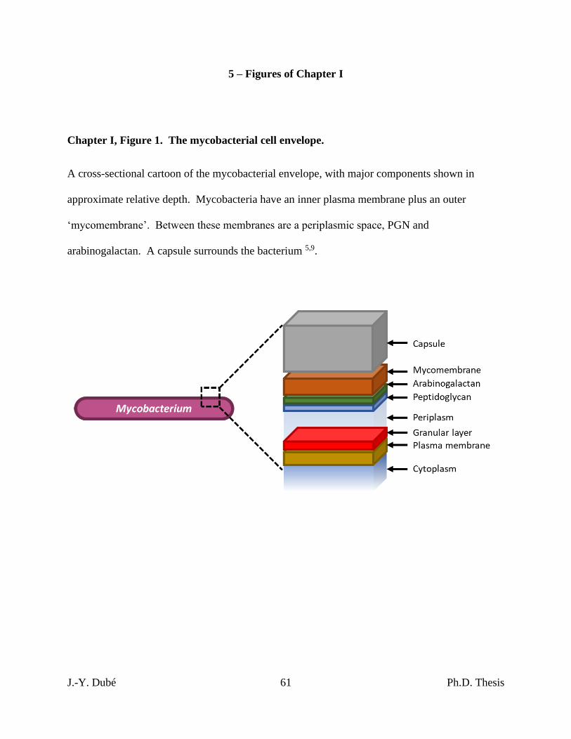
J.-Y. Dubé 61 Ph.D. Thesis
5 – Figures of Chapter I
Chapter I, Figure 1. The mycobacterial cell envelope.
A cross-sectional cartoon of the mycobacterial envelope, with major components shown in
approximate relative depth. Mycobacteria have an inner plasma membrane plus an outer
‘mycomembrane’. Between these membranes are a periplasmic space, PGN and
arabinogalactan. A capsule surrounds the bacterium 5,9.
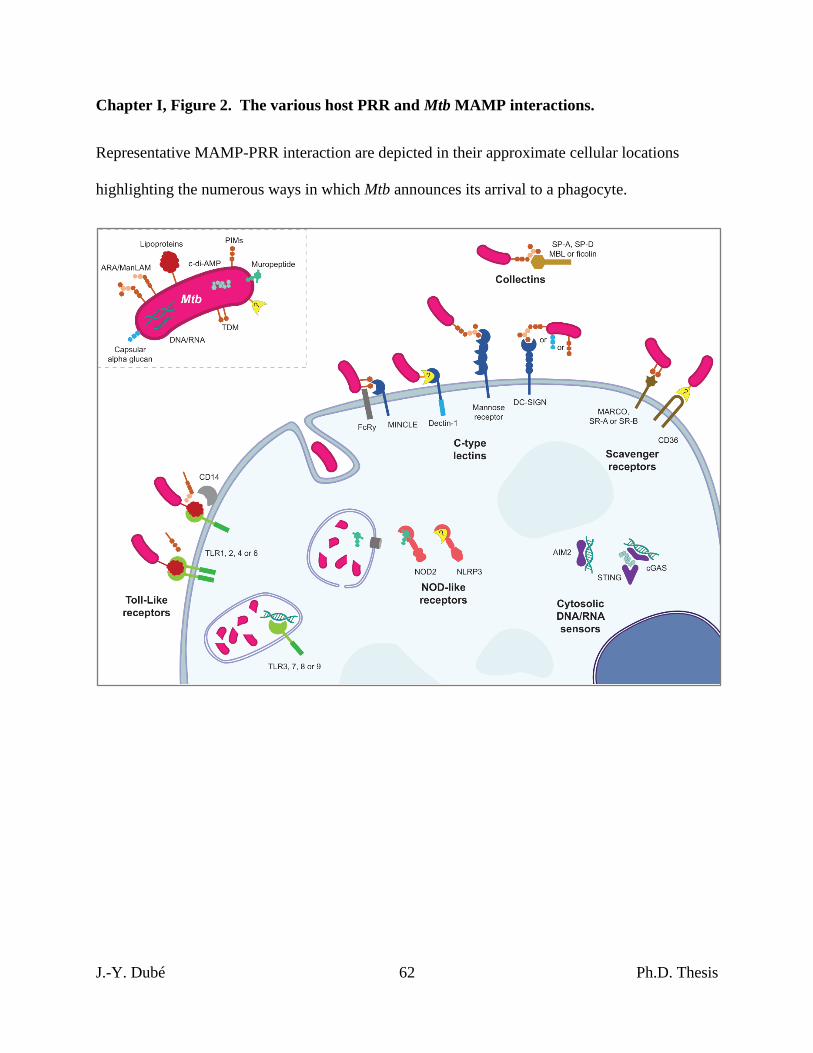
J.-Y. Dubé 62 Ph.D. Thesis
Chapter I, Figure 2. The various host PRR and Mtb MAMP interactions.
Representative MAMP-PRR interaction are depicted in their approximate cellular locations
highlighting the numerous ways in which Mtb announces its arrival to a phagocyte.
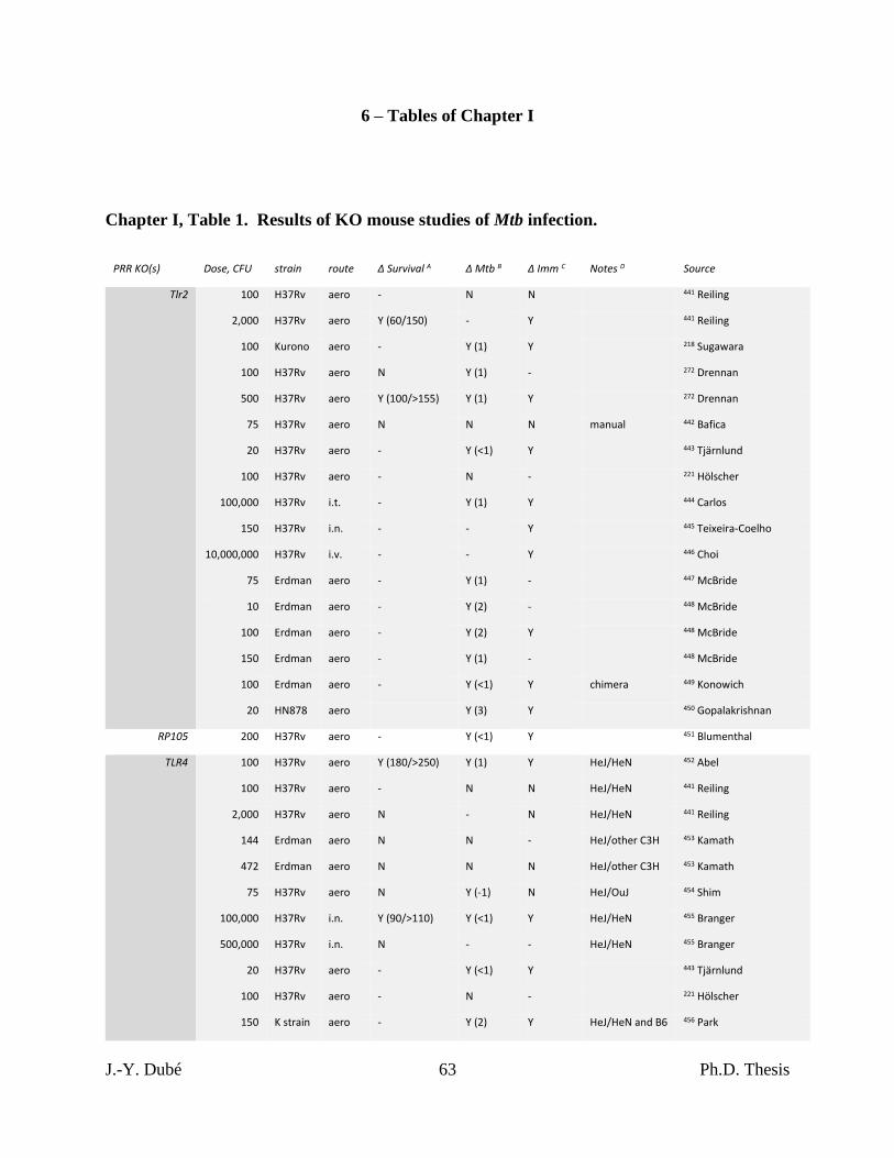
J.-Y. Dubé 63 Ph.D. Thesis
6 – Tables of Chapter I
Chapter I, Table 1. Results of KO mouse studies of Mtb infection.
PRR KO(s) Dose, CFU strain route Δ Survival A Δ Mtb B Δ Imm C Notes D Source
Tlr2
100 H37Rv aero - N N
441 Reiling
2,000 H37Rv aero Y (60/150) - Y
441 Reiling
100 Kurono aero - Y (1) Y
218 Sugawara
100 H37Rv aero N Y (1) -
272 Drennan
500 H37Rv aero Y (100/>155) Y (1) Y
272 Drennan
75 H37Rv aero N N N manual 442 Bafica
20 H37Rv aero - Y (<1) Y
443 Tjärnlund
100 H37Rv aero - N -
221 Hölscher
100,000 H37Rv i.t. - Y (1) Y
444 Carlos
150 H37Rv i.n. - - Y
445 Teixeira-Coelho
10,000,000 H37Rv i.v. - - Y
446 Choi
75 Erdman aero - Y (1) -
447 McBride
10 Erdman aero - Y (2) -
448 McBride
100 Erdman aero - Y (2) Y
448 McBride
150 Erdman aero - Y (1) -
448 McBride
100 Erdman aero - Y (<1) Y chimera 449 Konowich
20 HN878 aero
Y (3) Y
450 Gopalakrishnan
RP105 200 H37Rv aero - Y (<1) Y
451 Blumenthal
TLR4
100 H37Rv aero Y (180/>250) Y (1) Y HeJ/HeN 452 Abel
100 H37Rv aero - N N HeJ/HeN 441 Reiling
2,000 H37Rv aero N - N HeJ/HeN 441 Reiling
144 Erdman aero N N - HeJ/other C3H 453 Kamath
472 Erdman aero N N N HeJ/other C3H 453 Kamath
75 H37Rv aero N Y (-1) N HeJ/OuJ 454 Shim
100,000 H37Rv i.n. Y (90/>110) Y (<1) Y HeJ/HeN 455 Branger
500,000 H37Rv i.n. N - - HeJ/HeN 455 Branger
20 H37Rv aero - Y (<1) Y
443 Tjärnlund
100 H37Rv aero - N -
221 Hölscher
150 K strain aero - Y (2) Y HeJ/HeN and B6 456 Park
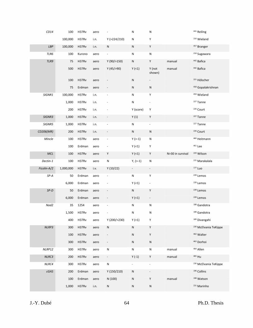
J.-Y. Dubé 64 Ph.D. Thesis
CD14
100 H37Rv aero - N N
441 Reiling
100,000 H37Rv i.n. Y (>224/210) N Y
216 Wieland
LBP 100,000 H37Rv i.n. N N Y
457 Branger
TLR6 100 Kurono aero - N N
218 Sugawara
TLR9
75 H37Rv aero Y (90/>150) N Y manual 442 Bafica
500 H37Rv aero Y (45/>90) Y (<1) Y (not shown)
manual 442 Bafica
100 H37Rv aero - N -
221 Hölscher
75 Erdman aero - N N
458 Gopalakrishnan
SIGNR1
100,000 H37Rv i.n. - N Y
459 Wieland
1,000 H37Rv i.n. - N -
227 Tanne
200 H37Rv i.n. - Y (score) Y
226 Court
SIGNR3 1,000 H37Rv i.n. - Y (1) Y
227 Tanne
SIGNR5 1,000 H37Rv i.n. - N -
227 Tanne
CD206(MR) 200 H37Rv i.n. - N N
226 Court
Mincle
100 H37Rv aero - Y (>-1) N
460 Heitmann
100 Erdman aero - Y (<1) Y
461 Lee
MCL 100 H37Rv aero Y Y (<1) Y N>30 in survival 224 Wilson
Dectin-1 100 H37Rv aero N Y, (>-1) N
225 Marakalala
Ficolin-A/2 1,000,000 H37Rv i.v. Y (10/22) - -
177 Luo
SP-A
50 Erdman aero - N Y
228 Lemos
6,000 Erdman aero - Y (<1) -
228 Lemos
SP-D
50 Erdman aero - N Y
228 Lemos
6,000 Erdman aero - Y (<1) -
228 Lemos
Nod2
35 1254 aero - N N
188 Gandotra
1,500 H37Rv aero - N N
188 Gandotra
400 H37Rv aero Y (200/>230) Y (<1) Y
189 Divangahi
NLRP3
300 H37Rv aero N N Y
230 McElvania TeKippe
100 H37Rv aero - N Y
462 Walter
300 H37Rv aero - N N
463 Dorhoi
NLRP12 300 H37Rv aero N N N manual 464 Allen
NLRC3 200 H37Rv aero - Y (-1) Y manual 465 Hu
NLRC4 300 H37Rv aero N - -
230 McElvania TeKippe
cGAS
200 Erdman aero Y (150/210) N -
196 Collins
100 Erdman aero N (100) N Y manual 198 Watson
1,000 H37Rv i.n. N N N
231 Marinho
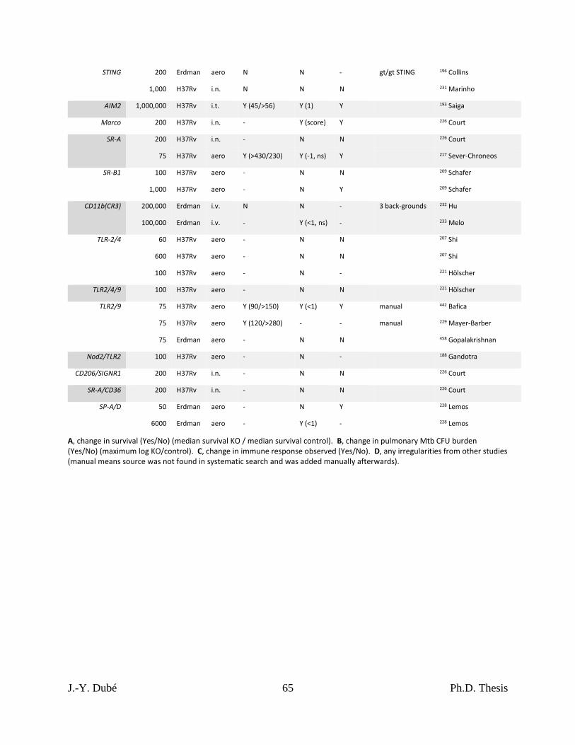
J.-Y. Dubé 65 Ph.D. Thesis
STING
200 Erdman aero N N - gt/gt STING 196 Collins
1,000 H37Rv i.n. N N N
231 Marinho
AIM2 1,000,000 H37Rv i.t. Y (45/>56) Y (1) Y
193 Saiga
Marco 200 H37Rv i.n. - Y (score) Y
226 Court
SR-A
200 H37Rv i.n. - N N
226 Court
75 H37Rv aero Y (>430/230) Y (-1, ns) Y
217 Sever-Chroneos
SR-B1
100 H37Rv aero - N N
209 Schafer
1,000 H37Rv aero - N Y
209 Schafer
CD11b(CR3)
200,000 Erdman i.v. N N - 3 back-grounds 232 Hu
100,000 Erdman i.v. - Y (<1, ns) -
233 Melo
TLR-2/4
60 H37Rv aero - N N
207 Shi
600 H37Rv aero - N N
207 Shi
100 H37Rv aero - N -
221 Hölscher
TLR2/4/9 100 H37Rv aero - N N
221 Hölscher
TLR2/9
75 H37Rv aero Y (90/>150) Y (<1) Y manual 442 Bafica
75 H37Rv aero Y (120/>280) - - manual 229 Mayer-Barber
75 Erdman aero - N N
458 Gopalakrishnan
Nod2/TLR2 100 H37Rv aero - N -
188 Gandotra
CD206/SIGNR1 200 H37Rv i.n. - N N
226 Court
SR-A/CD36 200 H37Rv i.n. - N N
226 Court
SP-A/D
50 Erdman aero - N Y
228 Lemos
6000 Erdman aero - Y (<1) -
228 Lemos
A, change in survival (Yes/No) (median survival KO / median survival control). B, change in pulmonary Mtb CFU burden (Yes/No) (maximum log KO/control). C, change in immune response observed (Yes/No). D, any irregularities from other studies (manual means source was not found in systematic search and was added manually afterwards).
J.-Y. Dubé 66 Ph.D. Thesis
PREFACE TO CHAPTER II
In Chapter I, I have highlighted the history shared between mycobacteria and cell-
mediated immunity through CFA, and how PRRs are known to contribute to the immune
response to mycobacteria. Through the work presented in Chapter II, we sought to determine the
precise contributions of NOD2 and Mincle pathways to the immune response elicited by CFA.
With this information, we approached the question of whether an adjuvant composed purely of
synthetic mycobacterial ligands could generate similar immunity as the whole mycobacteria in
CFA. We show that this is indeed the case with a synthetic adjuvant composed of just two
MAMPs, N-glycolyl MDP and GlcC14C18.
J.-Y. Dubé 67 Ph.D. Thesis
CHAPTER II – Synthetic mycobacterial molecular patterns partially
complete Freund’s adjuvant
(This Chapter is adapted from an article published in Scientific Reports)
Jean-Yves Dubé 1,2,3, Fiona McIntosh 2,3, Juan G. Zarruk 4, Samuel David 4, Jérôme Nigou 5,
Marcel A. Behr 1,2,3,6
1. Department of Microbiology and Immunology, McGill University, Montreal, Quebec, Canada
2. Infectious diseases and Immunity in Global Health Program, Research Institute of the McGill
University Health Centre, Montreal, Quebec, Canada
3. McGill International TB Centre, Montreal, Quebec, Canada
4. Centre for Research in Neuroscience, Research Institute of the McGill University Health
Centre, Montreal, Quebec, Canada
5. Institut de Pharmacologie et de Biologie Structurale, Université de Toulouse, CNRS,
Université Paul Sabatier, Toulouse, France.
6. Department of Medicine, McGill University Health Centre, Montreal, Quebec, Canada
J.-Y. Dubé 68 Ph.D. Thesis
1 – Abstract
Complete Freund’s adjuvant (CFA) has historically been one of the most useful tools of
immunologists. Essentially comprised of dead mycobacteria and mineral oil, we asked ourselves
what is special about the mycobacterial part of this adjuvant, and could it be recapitulated
synthetically? Here, we demonstrate the essentiality of N-glycolylated peptidoglycan plus
trehalose dimycolate (both unique in mycobacteria) for the complete adjuvant effect using
knockouts and chemical complementation. A combination of synthetic N-glycolyl muramyl
dipeptide and minimal trehalose dimycolate motif GlcC14C18 was able to upregulate dendritic
cell effectors, plus induce experimental autoimmunity qualitatively similar but quantitatively
milder compared to CFA. This research outlines how to substitute CFA with a consistent,
molecularly-defined adjuvant which may inform the design of immunotherapeutic agents and
vaccines benefitting from cell-mediated immunity. We also anticipate using synthetic microbe-
associated molecular patterns (MAMPs) to study mycobacterial immunity and
immunopathogenesis.
2 – Introduction
Infection with Mycobacterium tuberculosis, or administration of bacille Calmette-Guérin
(BCG), normally leads to cell-meditated immunity (CMI) to the corresponding bacterial
antigens. The tuberculin skin test is positive when the immune response, a type IV
hypersensitivity reaction or delayed-type hypersensitivity (DTH), occurs to tuberculin (a protein
extract of M. tuberculosis). This “cutaneous sensitivity” was closely examined in the 1940s
using complete Freund’s adjuvant (CFA, heat-killed M. tuberculosis in mineral oil plus
J.-Y. Dubé 69 Ph.D. Thesis
surfactant) 320,321. These studies provided the first direct evidence for the cellular nature of DTH
by transfer from a CFA-immunized guinea pig to a naïve one only through the washed, heat-
liable cellular fraction of peritoneal exudates 320,321. It is now known that DTH is mediated
specifically by antigen-sensitive T cells.
Today, CFA is a ‘gold standard’ adjuvant for eliciting CMI in research models of
autoimmune disease. Notable is the experimental autoimmune encephalomyelitis (EAE) model
of T-cell meditated destruction of myelin causing ascending paralysis, used most often to model
multiple sclerosis 327,337. CFA is not used in humans because of high reactogenicity 322. We
have asked ourselves: what was the impetus for Jules Freund to develop his eponymous adjuvant
with M. tuberculosis? For those conducting TB research, it is well appreciated that handling M.
tuberculosis (a slow growing, clumping, fastidious, and lethally pathogenic organism) is a
significant task per se. So, why did Freund choose to incorporate these bacteria?
Effort has been made to describe the microbe-associated molecular patterns (MAMPs) in
mycobacteria that drive the adjuvant effect. Evidence has pointed to mycobacterial
peptidoglycan (PGN), specifically down to the molecular motif muramyl dipeptide (MDP) 108,338.
Mycobacteria are distinct in that they produce the N-glycolyl MDP motif in their cell wall
106,107,110 and this PGN modification has been shown to be more potent compared to the common
N-acetyl MDP motif possessed by other bacteria 108,109. MDP is thought to be recognized
through the host molecule NOD2 182, and mutations in NOD2 predispose humans to increased
risk of mycobacterial and inflammatory diseases 255,284,370,371. Alternatively, others have pointed
to the mycobacterial cell wall lipid trehalose-6,6’-dimycolate (TDM) alone or synergistically
with purified PGN 439. TDM is recognized by the host with the C-type lectin Mincle in concert
J.-Y. Dubé 70 Ph.D. Thesis
with FcRγ and MCL 157,160. TDM has been demonstrated in animal models to alone be sufficient
for granuloma formation and immunopathological responses 157,466.
Both N-glycolyl MDP and TDM are MAMPs unique to mycobacteria. Their role in
mycobacterial immune responses is supported by the literature and their host receptors are well-
known. Additionally, N-glycolyl MDP is producible synthetically 108,344,467. Recently a minimal
motif of TDM called GlcC14C18 was produced synthetically and shown to retain adjuvancy but
with minimal toxicity 428. As a complex biologic, CFA is subjected to batch inconsistency. We
hypothesized that it is possible to create an entirely synthetic CFA using a rational approach to
identify essential MAMPs to replace the whole mycobacteria in the adjuvant. In this work, we
establish the necessity for N-glycolylation of PGN, NOD2 and Mincle for full CFA adjuvancy.
We also demonstrate that the mycobacteria in CFA can be replaced with synthetic N-glycolyl
MDP and GlcC14C18 in combination to partially restore the adjuvant effect in both a murine
model of antigen-specific T-cell immunity as well as the EAE model of autoimmune ascending
paralysis.
3 – Results
3.1 – N-glycolylation of PGN by mycobacteria is required for complete adjuvancy
Previous work indicated mycobacteria required N-glycolylated PGN to elicit a maximal
immune response during live infection 108,109. To determine if CMI elicited from dead
mycobacteria in the context of Freund’s adjuvant similarly required N-glycolylated PGN, we
prepared in-house complete Freund’s adjuvant with heat-killed M. tuberculosis strain H37Rv,
J.-Y. Dubé 71 Ph.D. Thesis
and H37Rv ΔnamH. NamH is the enzyme responsible for N-glycolylation of PGN units as they
are being synthesized in the cytoplasm, and the ΔnamH mutant was previously shown to be
devoid of N-glycolylation 109 (fig. 1A). We next immunized mice against ovalbumin (OVA, an
exemplary antigen) with our CFAs or incomplete Freund’s adjuvant (IFA, lacks mycobacteria),
and examined OVA-specific cytokine production by CD4+ T-cells from the draining lymph
nodes seven days post-immunization (fig. 1B; gating in fig. S1A). Both mycobacteria and namH
were required to generate the highest proportion of OVA-specific IFN-γ-producing CD4+ T cells
(fig. 1C). OVA-specific IL-17A-producing cells required mycobacteria but did not significantly
depend on namH (fig. 1D). Similar results were seen when examining total numbers of
cytokine-producing CD4+ T cells in the lymph nodes (fig. S2A-B), and a closer analysis of the
pooled data showed that namH contributes to about one third of mycobacteria-induced antigen-
specific IFN-γ-producing Th cells (fig. S2C-D). The individual results of experiments are shown
separately in fig. S2E; in all four cases we saw the same trends with IFN-γ. These results are
consistent with the hypothesized role of mycobacteria being a key ingredient in CFA for eliciting
CMI, and that mycobacterial PGN modification by NamH makes a more potent adjuvant.
3.2 – Host Mincle and Nod2 are necessary for complete mycobacterial adjuvancy, but do not
mediated the entire CFA effect.
Recognition of mycobacteria during live infection requires the host molecule NOD2 189.
Because we hypothesize mycobacterial PGN plays a role in CFA adjuvancy, specifically the
MDP motif where N-glycolylation occurs, we addressed whether the PGN/MDP sensor NOD2 is
important for CFA adjuvancy in our OVA model. Nod2+/+ and Nod2-/- mice were immunized
against OVA in the context of CFA or IFA of commercial provenance. A greater proportion of
J.-Y. Dubé 72 Ph.D. Thesis
OVA-specific IFN-γ-producing CD4+ cells was generated in CFA-immunized mice with Nod2
than without (fig. 2A). An independently produced IFN-γ ELISpot of total lymph node cells
supported the same conclusion (fig. S3A), as did another independent experiment using flow
cytometry (fig. S3C). In the absence of Nod2, the proportion of OVA-specific IL-17A-
producing cells was also impaired in CFA-immunized mice (fig. 2B and fig. S3C). With IFA as
adjuvant, IFN-γ and IL-17A responses were Nod2-independent as expected (fig. 2A-B and S3A-
C). Similar trends were observed with total cell numbers (fig. S3B-C and S4A-B). Together,
these results corroborate the role for NOD2 and accordingly mycobacterial PGN in the ability of
CFA to elicit CMI.
The adjuvant effect of mycobacteria declined partially with Nod2 knock-out, but not
completely. Therefore, there is clearly other mycobacterial MAMP recognition besides that
through NOD2 which is important for the remainder CFA adjuvancy. Others have indicated that
Mincle-mediated recognition of TDM contributes to mycobacterial adjuvancy 439. We tested the
dependence of our OVA-immunization model with Mincle-KO mice 433. Mincle+/+ and Mincle-
/- mice were immunized against OVA in the presence of CFA and IFA. Generation of CFA-
elicited OVA-specific IFN-γ- and IL-17A-producing CD4+ T cells was partially dependent on
Mincle (fig. 2C-D), while IFA-immunized mice showed Mincle-independent responses, as
expected (fig. 2C-D). Similar trends were again observed with total cell numbers but did not
reach statistical significance (fig. S4C-D). Un-pooled data showed the same trends in both
experiments performed (fig. S3D). These results implicate Mincle and thus its main
mycobacterial ligand, TDM, in CFA adjuvancy.
Both Nod2 and Mincle were required for CFA adjuvancy in our model, but neither were
responsible for the entire adjuvant effect. We asked what the sum of the immune responses
J.-Y. Dubé 73 Ph.D. Thesis
elicited by these PRRs is – if it appears these PRRs signal synergistically. We generated Mincle-
Nod2 double knock-out mice (DKO) and immunized them against OVA with CFA and IFA. The
proportion of OVA-specific IFN-γ- and IL-17A-producing CD4+ T cells in CFA-immunized
mice declined with DKO relative to WT mice, with no change in IFA-immunized mice (fig. 2E-
F). However, the decrease when measuring IFN-γ did not appear obviously larger with DKO
than what was observed with single-KO mice. Likewise, the decrease with IL-17A was
intermediate, and similar trends were seen with total cell numbers (fig. S4E-F). The residual
effect of CFA when both Nod2 and Mincle are disrupted suggests that at least two mycobacterial
MAMPs, N-glycolylated PGN as well as TDM, are essential, but insufficient, for the full
adjuvant effect.
3.3 – Synthetic minimal structures of mycobacterial PGN and TDM work synergistically to
stimulate dendritic cell effector functions.
To explore the effect of Mincle and NOD2 signalling on antigen presenting cells
(necessary to generate antigen-specific T-cell immunity), we stimulated bone-marrow derived
dendritic cells (BMDCs) from WT, Mincle-/-, Nod2-/- and DKO mice with synthetic versions of
the respective PRR ligands. Recently, a rationally determined minimal chemical structure of
TDM called GlcC14C18 was shown to retain activity as a Mincle agonist without the ‘toxicity’
associated with purified TDM 428. Both synthetic N-glycolyl and N-acetyl MDP are
commercially available. To elicit T-cell immunity, we would expect that mycobacterial MAMPs
promote antigen presentation, co-stimulation and cytokine production by antigen presenting
cells, the classical signals 1, 2 and 3, respectively.
J.-Y. Dubé 74 Ph.D. Thesis
After 24 hours of MAMP stimulation in vitro, we measured the proportion of cells in
BMDC culture highly expressing MHC-II (fig. 3A-B). With WT cells, MDPs alone had little
effect; GlcC14C18 alone increased the fraction of MHC-IIhi cells in WT BMDC cultures (fig.
3A). Combinations of GlcC14C18 with N-glycolyl MDP or N-acetyl MDP produced the largest
responses. Knock-out of Mincle greatly reduced the effect of GlcC14C18 in Mincle-/- and DKO
BMDCs, and knock-out of Nod2 abrogated the effect of MDPs in Nod2-/- and DKO BMDCs.
These results suggest these mycobacterial MAMPs can direct myeloid cells towards MHC-IIhi
expression as expected for professional antigen presenting cells. Similar results were obtained at
48 hours of stimulation (fig. S5A).
On MHC-IIhi cells, we looked at the level of expression of MHC-II, CD40, CD80 and
CD86 by median fluorescence intensity (MFI) after 24 hours of stimulation in vitro (fig. S5F-I).
With WT cells and for all markers examined, MDPs alone were unable to substantially enhance
expression over the control (fig. 3C-F). GlcC1418 alone increased marker expression, but
GlcC14C18 together with N-glycolyl MDP synergistically produced the greatest responses. N-
acetyl MDP was generally less efficacious than N-glycolyl MDP. Knockouts behaved as
expected: cells without Mincle responded poorly to GlcC14C18; cells without Nod2 failed to
respond to MDPs (fig. 3C-F). I addition to 24-hour stimulation, cells were also collected after 48
hours of stimulation, yielding similar results (fig. S5B-I).
BMDCs from WT mice produced TNF in a dose-dependent manner in response to
GlcC14C18, but not to MDPs alone (fig. 3G). When combined with limiting or saturating doses
of GlcC14C18, N-glycolyl MDP synergistically elevated TNF production in a dose-dependent
manner. N-acetyl MDP only significantly elevated TNF production at higher doses of
GlcC14C18 plus MDP and worked less efficaciously than N-glycolyl MDP. In the absence of
J.-Y. Dubé 75 Ph.D. Thesis
functional Mincle, BMDCs did not respond to GlcC14C18 as expected. Mincle-/- BMDCs
stimulated with GlcC14C18 and MDPs behaved like BMDCs exposed to MDP alone (little to no
detectable TNF production). Nod2-/- BMDCs responded to GlcC14C18 but not to MDPs as
expected. DKO BMDCs did not respond to either synthetic MAMP as expected. Overall,
GlcC14C18 and N-glycolyl MDP combined, through Mincle and Nod2 respectively, are
sufficient to upregulate BMDC antigen presentation, co-stimulation and cytokine production.
3.4 – Synthetic mycobacterial NOD2 and Mincle ligands can complement IFA to increase
antigen-specific T-cell responses.
As NOD2 and Mincle ligands worked synergistically in BMDCs in vitro to elicit effector
functions in these antigen presenting cells, we hypothesized that IFA can be complemented with
NOD2 and/or Mincle ligands to recapitulate at least a portion of the adjuvant effect of whole
mycobacteria. Using minimal synthetic MAMPs for PGN (i.e. N-glycolyl MDP) and TDM (i.e.
GlcC14C18), we complemented IFA to test if CMI could be achieved with a completely
synthetic adjuvant containing these essential mycobacterial MAMPs. First, using 10 µg
GlcC14C18 per mouse with varying doses of N-glycolyl MDP, we observed a dose dependent
increase in the proportion and numbers of OVA-specific cytokine-producing CD4+ cells which
was significantly greater than IFA alone at the highest dose (fig. 4A). Notably, previous
attempts complementing IFA with only N-glycolyl MDP did not induce significant numbers of
IFN-γ producing cells (fig. S6A-B).
Next, using 30 µg of N-glycolyl MDP per mouse and varying the dose of GlcC14C18, we
similarly observed a dose dependent increase in the immune response; however this response did
not surpass more than half of the level elicited with CFA by IFN-γ and IL-17A (fig. 4B). An
J.-Y. Dubé 76 Ph.D. Thesis
IFN-γ ELISpot performed in parallel provided a similar conclusion (fig. S6C-D). Together,
these results show that mycobacterial adjuvancy in the context of CFA can be partially
phenocopied synthetically with two mycobacterial MAMPs: namely N-glycolyl MDP and
GlcC14C18.
3.5 – Synthetic mycobacterial MAMPs increase DC numbers and effectors in lymph nodes.
GlcC14C18 and N-glycolyl MDP together were sufficient to elicit antigen-specific T-cell
responses. We next looked to see if this MAMP combination was associated with augmentation
of DC effector functions in vivo as we had seen in vitro with BMDCs. Gating for DC subsets as
well as other leukocytes in the lymph nodes is depicted in fig. S1B (guided by 468). We looked in
the lymph nodes at 4- and 7-days post-immunization. Both CD11b+ and CD11b- conventional
DC (cDC) numbers were increased in the presence of IFA + 10 μg GlcC14C18 + 30 μg N-
glycolyl MDP compared to IFA alone within 7 days, to levels similar to CFA (fig 5A-B). Trends
suggest that early accumulation of cDCs could be driven by MDP alone, but GlcC14C18
together with N-glycolyl MDP were required to sustain numbers to day 7.
By MFI, expression levels of MHC-I and -II on both CD11b+ and CD11b- cDCs were
not largely altered by MDP nor GlcC14C18 (fig. 5A-B). On CD11b+ cDCs, CD40 expression
was elevated the highest and significantly with IFA + GlcC14C18 MDP at both 4 and 7 days.
CD80 and CD86 expression were also highest with IFA + GlcC14C18 MDP, with statistical
significance at day 4 but not day 7 when expression was possibly waning. On CD11b- cDCs,
CD80 and CD86 expression behaved similarly to that on CD11b+ cDCs. CD40 expression of
CD11b- cDCs was not statistically significantly greater with IFA + GlcC14C18 MDP compared
J.-Y. Dubé 77 Ph.D. Thesis
to IFA alone at both timepoints. Notably, IFA + GlcC14C18 MDP produced equal or greater
costimulatory molecule expression compared to CFA in all cases.
In addition to increased numbers of cDCs in the lymph nodes of mice immunized with
GlcC14C18 + N-glycolyl MDP, we observed that other leukocytes were substantially elevated in
numbers after immunization with this adjuvant (fig. S7A-F). B cells, CD4+ and CD8+ T cells,
plasmacytoid DCs, monocytes and PMNs were all increased by 7 days post-immunization
relative to IFA, to levels similar to or greater than CFA. By day 7, while MDP alone was
insufficient to elevate cell numbers over the IFA background, MDP in combination with
GlcC14C18 enhanced numbers beyond the levels attained with GlcC14C18 alone. This provides
evidence for synergy between these MAMPs in vivo.
3.6 – Synthetic mycobacterial MAMPs can induce EAE similar to CFA.
One of the common uses of CFA is in animal models of autoantigen-specific
autoimmunity. To determine if our synthetic formulation can phenocopy the effect of whole
mycobacteria to produce a more complex biological outcome such as autoimmunity (and thereby
provide a further measure of the ‘completeness’ of the synthetic formula), we tested IFA +
GlcC14C18 + N-glycolyl MDP against CFA in ability to induce relapsing-remitting EAE (RR-
EAE). Briefly, mice were randomly immunized against myelin oligodendrocyte glycoprotein
(MOG) synthetic peptide with CFA or IFA + 10 μg GlcC14C18 + 30 μg N-glycolyl MDP, and
onset of RR-EAE was determined by clinically scoring ascending paralysis daily in a blinded
manner (fig. 6A). Both CFA and IFA + GlcC14C18 + MDP produced RR-EAE that was
indistinguishable except quantitatively: over the course of the experiment, the average EAE
score was lower with the synthetic adjuvant compared to the CFA control (fig. 6B), with a
J.-Y. Dubé 78 Ph.D. Thesis
cumulative score suggesting about half the disease burden (58% of CFA cumulative score) (fig.
6C). Expectedly, lower EAE scores also corresponded with less weight-loss (fig. S8A). Of note,
there were mice in the synthetic adjuvant group that reached the same scores as in the CFA
group, but fewer (fig. S8B). Therefore, GlcC14C18 and N-glycolyl MDP were sufficient to
recapitulate the adjuvant effect of the whole mycobacterial cell in EAE, albeit quantitatively less
at the tested doses of MAMPs. Additionally, we had attempted RR-EAE with IFA + TDM +
MDP previously, which produced a far less compelling phenocopy of CFA (fig. S8D-G). The
IFA + GlcC14C18 + N-glycolyl MDP adjuvant has the advantage of being completely synthetic
and more efficacious than the TDM-containing adjuvant.
To verify if there were overt qualitative histopathological differences in EAE between
CFA and the synthetic adjuvant, we examined spinal cords from three mice from each adjuvant
group, having EAE scores 2, 3 and 3.5 at harvest (disease profile of these mice in fig. S8C).
Both cellular infiltrates and demyelination of the white matter looked equivalent between
adjuvants upon visual inspection (fig. 7A). Mice with different EAE scores had correspondingly
different areas of diseased tissue of the spinal cord (fig. 7B), but in comparing adjuvants there
was no detectable difference in the area of diseased spinal cord tissue (with statistical power to
detect as low as +/- 15% CFA levels) (fig. 7C). The main determining variable was EAE score,
not adjuvant. Overall, we observed qualitatively similar autoimmune pathology using IFA +
GlcC14C18 + N-glycolyl MDP compared to the gold standard CFA.
J.-Y. Dubé 79 Ph.D. Thesis
4 – Discussion
The ability of an adjuvant to elicit CMI can depend on ligand-receptor interactions,
specifically MAMP-PRR interactions. It is well appreciated since the hypothesis of Charles
Janeway Jr. that the host decision to mount an adaptive immune response requires genetically
inborn sensors to detect the presence of microbial products, or microbes by association, and that
this is the foundation of classical adjuvants including CFA 123. These interactions can be thought
of as an “arms race” between host and microbe, originating through antagonistic evolution in the
case of immune evasion 469. Additionally, we can imagine the case where a microbe might
evolve to increase a specific PRR interaction that favours a specific active immunological
environment necessary for its lifecycle. When mycobacteria interact with their host, either in the
form of M. tuberculosis infection, BCG vaccination or immunization using CFA, CMI normally
occurs. The immune response to mycobacteria has been attributed to its unique cell envelope,
especially the MDP motif of PGN 108,109,189,338 and TDM 160,439. We have shown in the context of
both CFA-induced immunization and autoimmunity with wholly synthetic N-glycolyl MDP and
TDM (GlcC14C18) that these MAMP-PRR pathways together contribute partially but
significantly to the mycobacterial adjuvant effect.
There are other mycobacterial MAMPs previously identified (and likely more yet
unidentified), plus a greater number of PRRs linked to these microbial products. However, most
are not yet producible synthetically. Purified mannose-capped lipoarabinomannan (ManLAM)
was recently shown to elicit EAE in mice, dependent on the host C-type lectin Dectin-2
(Clec4n), at a dose of 500 µg per animal 165. Of note, we were not able yet to produce synthetic
mimics of ManLAM that induce Dectin-2 signaling 166. Similarly, purified TDM has been used
to elicit EAE at 500 µg per mouse, mostly dependent on MCL (Clec4d) and partially on Mincle
J.-Y. Dubé 80 Ph.D. Thesis
(Clec4e) 160. These findings support a role for mycobacterial MAMPs driving the CFA effect,
but require purification of biologically-sourced MAMPs in high doses. When in combination,
our synthetically pure MAMPs have shown clear activity in vivo beginning at 1-10 µg. Of note,
with CFA we inject 50 µg of mycobacterial mass into mice.
TLR-2 has been documented to interact with multiple mycobacterial lipids 470. The
literature is inconsistent on whether or not TLR-2 promotes CFA-induced EAE 471-474.
Mycobacterial DNA was recently shown to signal through the cGAS/STING pathway after
phagosomal disruption via ESX-1 196,198. In the context of Freund’s adjuvant, it is not clear if
heat-killed mycobacteria could have cytosolic access to activate cGAS/STING. The IFA fraction
perhaps could deliver MAMPs across membranes. TLR-9 also senses DNA, and appears to play
a role during M. tuberculosis infection in mice 219, and in humans 475. TLR-9 was also shown to
be necessary for full induction of EAE 471,473. Although DNA is not unique to mycobacteria like
TDM and N-glycolylated PGN, synthetic DNA is available and pursuing the role of DNA in
mycobacterial adjuvancy interests us, perhaps as a third synthetic MAMP to work with MDP and
GlcC14C18.
Mycobacteria are not completely unique in possessing MAMPs that elicit CMI. As an
example, the TB vaccine candidate M72/AS01E utilizes monophosphoryl lipid A (MPLA), a
TLR-4 agonist based on lipopolysaccharide (LPS), to elicit CMI. LPS is made in Gram-negative
bacteria, not mycobacteria. We concede that many microbes likely contain MAMPs able to elicit
CMI; here we have concerned ourselves primarily with the history of mycobacterial adjuvancy,
CMI and Jules Freund. It is conceivable that a combination of MAMPs from different bacteria
could elicit ‘unnatural’ immunity that may be beneficial to control certain infectious agents that
otherwise evade natural immune responses.
J.-Y. Dubé 81 Ph.D. Thesis
We were able to show that N-glycolylation of M. tuberculosis PGN altered the adjuvancy
of the mycobacterial cell in the context of CFA. This is consistent with our previous work on the
increased potency of mycobacterial PGN in other contexts 108,109. Interestingly, the benefit of N-
glycolylation of PGN seemed primarily skewed toward IFN-γ production rather than IL-17A,
while Nod2-KO showed a greater effect with IL-17A. This may indicate that mycobacteria
retain N-glycolylation of PGN specifically to shift immune responses in a Th1 bias and should be
further investigated during live infection. Nod2 was necessary for full CFA adjuvancy by
measures of IFN-γ and IL-17A, supporting the role for mycobacterial PGN. We were somewhat
surprised that MDP emulsified by itself with IFA and antigen was insufficient to recapitulate the
adjuvant effect of CFA. In a 1975 paper, N-acetyl MDP appeared sufficient in a guinea pig
model to elicit DTH using OVA as antigen 338. In 2009 using a very similar model to our current
work, N-glycolyl MDP alone was sufficient to increase ELISpot IFN-γ to near CFA levels, while
N-acetyl MDP failed 108. One explanation is the possibility of contaminating MAMPs in certain
preparations of OVA. OVA is known to often come with a dose of endotoxin which affects the
immunological outcome of experiments 476. In the current investigations we have used
endotoxin-free/ultra-pure OVA. Much literature of in vitro experiments shows MDP requires
another MAMP to produce measurable outputs like cytokine 108,348,349,477, although in few cases
MDP was shown to elicit cellular responses by itself 348,477. Responses measured from MDP
alone may be unrepresentative of a cell’s maximum capacity.
Other groups have supported a role for TDM in mycobacterial adjuvancy 160,439. TDM
together with purified PGN was shown to synergistically promote IL-17 production from OVA-
specific (OT-II) CD4+ T cells adoptively transferred into congenic mice 439. This and other work
directed us to test Mincle-dependence of CFA and MDP synergy with the minimal TDM motif
J.-Y. Dubé 82 Ph.D. Thesis
GlcC14C18. Mincle-/- mice allowed us to infer that CFA adjuvancy required TDM for maximal
induction of IFN-γ- and IL-17A-producing Th cells. TDM is thought to be the main Mincle
ligand in mycobacteria; however, there are other ligands. Purified from H37Rv, trehalose-6,6’-
monomycolate, glucose monomycolate, diacyl-trehalose and triacyl-trehalose were shown to
stimulate mouse and human Mincle (plus glycerol monomycolate stimulates human Mincle only)
428. It is possible that the phenotype of reduced immunity was because the sensing of these other
MAMPs was decreased. Nevertheless, Mincle signalling was essential for full CFA adjuvancy.
Interestingly, simultaneous knock-out of both Mincle and Nod2 resulted in incomplete
abrogation of CFA adjuvancy. DKO mice more closely resembled single-KO from separate
experiments, compared to controls, than IFA. This indicates two things: 1) that much of the
adjuvancy of mycobacteria is not mediated through Mincle and NOD2, and therefore other
mycobacterial MAMPs like those mentioned above may play a similarly important role as PGN
and TDM; 2) that Mincle and NOD2 signalling might work synergistically, where in the absence
of Mincle, NOD2 sensing of PGN is blunted as we have seen clearly in vitro with GlcC14C18
and N-glycolyl MDP, and therefore DKO resembles single knock-out of either receptor.
We demonstrated that GlcC14C18 plus N-glycolyl MDP synergistically were sufficient to
promote DC effector functions in vitro. The GlcC14C18 plus N-glycolyl MDP adjuvant was
also the most efficacious adjuvant to drive costimulatory molecule upregulation on cDCs in vivo,
more than either MAMP alone. While either of these MAMPs by themselves failed to come
close to the whole mycobacteria of CFA in our OVA model of CMI, together we showed they
could produce about half the Th cell IFN-γ/IL-17A response and EAE disease burden as CFA.
Resultingly, we are interested in further understanding the implications for MAMP signalling
(which MAMPs synergize together; what redundancy exists; do different combinations alter the
J.-Y. Dubé 83 Ph.D. Thesis
Th type of CMI?). As the DKO data suggest, other mycobacterial MAMPs may be necessary to
fully recapitulate the adjuvant effect of whole mycobacteria. An optimal dose of the two
MAMPs may not have been reached, although additional dosing experiments (not shown) failed
to find a clearly superior formulation than those presented here.
We chose to proceed primarily with the 10 µg GlcC14C18 + 30 µg N-glycolyl MDP
doses because these quantities were sufficient for some biological activity on their own and/or in
combination (fig. 4A-B, S6D and not shown). Detailed subset data also showed other biological
synergy at the 10 µg / 30 µg dose (fig. S7). Mycobacteria are an estimated 30% PGN by mass,
of which the MDP motif comprises about half, or 15% total mycobacterial mass 478. Therefore,
when injecting 50 ug of mycobacteria as CFA, we inject about 8 µg of MDP. The 10 µg / 30 µg
dose did not quantitatively phenocopy the level of CMI from CFA in the OVA model, but we did
not want to achieve this by using grossly unrepresentative quantities of MAMPs. Considering
the DKO data showing Mincle and Nod2 stimulation do not drive all of the CFA effect, we
proceeded with tempered expectations for EAE. Nonetheless, we have demonstrated that
synthetic TDM and N-glycolyl MDP signalling, unique to mycobacteria, enhance the adjuvancy
of CFA. This partly corroborates the natural history of CFA in why it is a uniquely potent
adjuvant. Remaining MAMPs, not necessarily unique to mycobacteria, might fill the difference.
With the wholly synthetic formulation of GlcC14C18 N-glycolyl MDP, we were able to
induce RR-EAE in mice that was qualitatively indistinguishable from that induced with CFA, but
milder overall (lower EAE scores on average). We selected spinal cords from mice having
matched EAE scores between CFA and synthetic adjuvant groups; however, extrapolating the
histopathological data to all the mice in our experiment, we would expect that the synthetic
adjuvant produced less pathology in the spinal cord on average since EAE scores were lower on
J.-Y. Dubé 84 Ph.D. Thesis
average. The EAE result correlated with the incomplete complementation in the OVA model.
EAE that failed with TDM plus MDP earlier was also less potent in the OVA model than the
synthetic formulation (not shown), further indicating correlation between these readouts. CFA
often needs to be ‘titrated’ to account for batch-dependent efficacy in our experience (not
shown), and CFA with higher concentrations of mycobacteria (i.e. 4 mg/ml) is also used when a
more severe, chronic progressive EAE is necessary for studies 331. These facts point to dosing as
a means of controlling the degree of EAE, which may work with the synthetic adjuvant. Other
experimental uses of CFA include the collagen-induced arthritis (CIA) model and for producing
specific antibodies. CIA takes longer and has a greater role for humoral immunity than EAE
which is T-cell driven, (our interest was in CMI for this study) 479. Wherever mycobacterial
adjuvancy is useful, we suspect a synthetic adjuvant could be beneficial. Indeed, intravesical
injection of BCG is used to treat bladder cancer (i.e. BCG can provide MAMP-driven protection,
not simply antigen-specific protection). Furthermore, BCG administration to millions of babies
each year can protect not just against childhood TB, but non-specifically against other diseases
297.
Our work demonstrates that mycobacterial NOD2 and Mincle ligands contribute to the
adjuvant effect of the mycobacterial cell, and that a necessarily combinatorial formulation of
synthetic MAMPs, N-glycolyl MDP and GlcC14C18, can partly recapitulate the adjuvant effect
of whole mycobacteria to induce T-cell immunity and EAE. In addition to demonstrating the
first entirely synthetic multi-MAMP mycobacterial adjuvant, we have outlined an approach to
investigate the contribution of other MAMPs to adjuvant design. Moreover, the synthetic
approach may be useful to probe mycobacterial immunity and immunopathogenesis.
J.-Y. Dubé 85 Ph.D. Thesis
5 – Methods
Mice
C57BL/6J (‘wild-type’) as well as Nod2-/- mice were obtained from Jackson laboratories and
were bred or used immediately for experiments. Mincle-/- mice breeders were provided courtesy
of the laboratory of Christine Wells 433. Mice were genotyped to confirm absence of the Dock2
mutation recently reported in Nod2-/- mice 480. We crossed Nod2-/- and Mincle-/- mice to obtain
double knock-out mice. All experiments used mice from 6 to 20 weeks of age. All protocols
involving mice followed the guidelines of the Canadian Council on Animal Care (CCAC) and
were approved by the ethics committees of the RI-MUHC.
Preparation of adjuvants and ovalbumin immunization
Complete and incomplete Freund’s adjuvant were purchased from Sigma or InvivoGen. In some
cases, IFA was made by the experimenter from purified mineral oil and mannide monooleate
(Sigma). Adjuvants were prepared on the day of immunization by emulsifying CFA (1 mg/ml
M. tuberculosis) or IFA with a PBS solution containing 1 mg/ml ovalbumin (Endofit brand,
InvivoGen) in a 1:1 ratio. Emulsification was accomplished using all-plastic syringes and
repeated passage through an 18-G blunt-end needle. Where IFA was complemented with
mycobacterial components: MDP (InvivoGen) was diluted in the PBS fraction of the adjuvant
before emulsification; TDM (Sigma) or GlcC14C18 428 were dissolved in the IFA fraction before
emulsification as described previously 439; heat-killed mycobacteria in saline were diluted in PBS
and added to the PBS fraction of the adjuvant before emulsification for mice to receive an
J.-Y. Dubé 86 Ph.D. Thesis
equivalent of 108 CFU. To make heat-killed mycobacteria, cultures were grown to equivalent
600-nm absorbance at mid-log phase, were pelleted and washed three times with saline, then
heat-killed at 100oC for 30 minutes and frozen at -80oC until use. Mice were injected
subcutaneously with 100 μl adjuvant-antigen emulsions in the tail 1-2 cm from the body, towards
the body, for sufficient and consistent drainage to the inguinal lymph nodes. Four or seven days
after injection mice were euthanized and organs were harvested for analysis.
ELISpot and flow cytometry of lymph node cells
Lymph nodes extracted from immunized mice were gently crushed over 70-μm cell strainers to
release the cells therein. Cell concentrations were determined by counting with a
haemocytometer or with a BD Accuri C6 flow cytometer. Lymph node cells (LNCs) were
washed in culture medium, counted and cultured in 100 μl RPMI + 10% FBS (R10) at 250,000
or 500,000 cells per well in IFNγ ELISpot plates (R&D Systems) with or without 100 μg OVA
for ~40 hours at 37oC 5% CO2 before developing the ELISpot plates. For flow cytometry
analysis of OVA-specific cytokine production, washed LNCs were cultured at 6 million cells per
ml in 200 μl R10 with or without 200 μg OVA for ~40 hours at 37oC 5% CO2, Brefeldin A
(GolgiPlug, BD) was added for an additional 5 hours, and then cells were stained and fixed (BD
fixation/permeabilization solution) for flow cytometry on ice. Intracellular staining was
performed on the same or the next day using BD Perm/Wash buffer. See Table S1 for antibody
probes used. For flow cytometry of leukocyte subsets, 3 million LNCs were taken immediately
after harvest for extracellular staining and fixing. See Table S2 for antibody probes used. A BD
LSRFortessa X-20 was used for cellular phenotyping.
J.-Y. Dubé 87 Ph.D. Thesis
Bone marrow-derived dendritic cells (BMDCs)
Bone marrow was extracted from mice by flushing femora and tibiae with PBS + 2% BSA + 2%
glucose using a 25-G syringe. Red blood cells were lysed and remaining bone marrow cells
were filtered through 70 µm cell strainer. Cells were cultured at 500,000 cells / ml R10 with 20
ng/ml murine rGM-CSF (PeproTech); cells were fed on day 3 with R10 + rGM-CSF, and on day
6 with R10 alone. On day 7, loosely adherent cells were harvested by gentle pipetting and used
in assays. These cells were ~60% CD11c+ MHC-II+ as we obtain routinely. To assess cytokine
production, 100,000 BMDCs were transferred per well to 96-well plates precoated with
GlcC14C18 (by dissolving in isopropanol and drying) and containing MDP in a final volume of
200 µl. BMDCs were incubated with stimulants for 6 hours at 37oC, 5% CO2 before supernatant
was removed and stored at -80oC for later ELISA analysis (mouse TNF ELISA from
ThermoScientific). To assess surface marker expression, 300,000 BMDCs were transferred per
FACS tube precoated with 50 ng GlcC14C18 (by dissolving in isopropanol and drying) and
containing 100 ng/ml MDP in a final volume of 200 µl. BMDCs were incubated with stimulants
for 24 or 48 hours at 37oC, 5% CO2 before staining and fixing for flow cytometry analysis. See
Table S3 for antibodies used in flow cytometry.
Relapsing-remitting experimental autoimmune encephalomyelitis (RR-EAE)
Adjuvants were prepared similarly as above, but with myelin oligodendrocyte glycoprotein
(MOG) used as antigen. Briefly, PBS solution containing 1 mg/ml MOG +/- 600 μg/ml N-
glycolyl MDP was emulsified with CFA (1 mg/ml M. tuberculosis) / IFA plus 200 μg/ml
J.-Y. Dubé 88 Ph.D. Thesis
GlcC14C18 (or 20 μg/ml TDM) by back-and-forth extrusion through an 18 G two-way needle
with two all-plastic syringes. 8-12 week-old female C57BL/6 mice (Charles River) were
induced for RR-EAE by a standard protocol: briefly, on day 0, pertussis toxin (PT) (200 ng) was
administered i.v. in the tail vein, and mice were immunized with 50 μg MOG by bilateral s.c.
injection in the back towards the tail with 100 μl total of an emulsification of CFA or IFA plus
mycobacterial MAMPs (adjuvant group was randomized within and across cages). On day 2,
mice received a second equivalent dose of PT i.v.. After about one week, EAE was scored
blinded (i.e. the scorer did not know the adjuvant received by the mouse) every day according to
these cumulative criteria: 0, no paralysis (normal); 0.5, partial tail weakness observed as < 50%
of tail dragging when mouse walks; 1, tail paralysis observed as >50% of tail dragging as mouse
walks; 2, slow righting reflex (delay < 5 seconds) when mouse is flipped; 3, very slow or absent
righting reflex (> 5 seconds) or inability to bear weight with back legs observed as dragging
hindquarters when walking; 3.5, partial paralysis of one or both hind limbs; 4, complete paralysis
of one or both hindlimbs; 4.5, complete paralysis of one or both hind limbs plus trunk weakness;
5, weakness or paralysis of forelimbs; 6, found dead. Mice were weighed every other day during
scoring. Mice reaching a score of 5 were euthanized within 24 hours. Blinding was
accomplished by having one person inject the mice and another person score/weigh the mice,
unaware of experimental group.
Histopathology
At the end of EAE scoring, the experiment was un-blinded and three mice from each group with
matching scores were selected. These six mice were anesthetized with peritoneal ketamine
injection, were perfused with PBS and formalin, and then spinal cords were extracted,
J.-Y. Dubé 89 Ph.D. Thesis
equilibrated in sucrose and then frozen in OTC compound (VWR). Frozen tissue was sectioned
14-nm thick onto slides and then stained (Nissl or Luxol fast blue). Sections of good quality (20
per mouse, 5 cervical, 5 upper thoracic, 5 lower thoracic and 5 lumbar) were randomly selected
and photographed with a Nikon Eclipse NI microscope. Randomized (blinded) Nissl stain
photographs were used to quantify the area of disease in the spinal cord by subtracting the total
2D area of each tissue section with the area without cellular infiltration in white matter (thus, the
difference of areas is the area with cellular infiltration). This was divided by the total area to
determine the fraction or percent of diseased area.
Software, data analysis and statistics
Flow cytometry data was acquired using FACSDiva software (BD). FCS files were analyzed
using FlowJo V10 (BD). Digital microscopy images were analyzed with ImageJ (NIH). Graphs
were generated and routine statistical testing was accomplished with GraphPad Prism 8.1.1
(GraphPad Software Inc.; https://www.graphpad.com/). Assessment for normal data, then
parametric or non-parametric analyses were applied, as indicated. Sample-size and power
calculations were performed manually using Microsoft Excel. Manuscript figures were
assembled with Microsoft PowerPoint.
6 – Acknowledgements
We would like to thank the Containment Level 3 platform of the Research Institute of the
McGill University Health Centre (RI-MUHC) for providing the needed facilities and intellectual
support. We thank the Immunophenotyping platform of the RI-MUHC for cytometers and
J.-Y. Dubé 90 Ph.D. Thesis
advice. We are very grateful to Christine Wells (University of Melbourne, Australia) for
providing Mincle-KO mice and to Alexiane Decout for preparing GlcC14C18. For tutelage and
technical assistance, J.Y.D. is thankful to Damien Montamat-Sicotte, Ourania Tsatas, Sarah
Danchuk, Iain Roe, Andréanne Lupien, Nimara Asbah, and Daniel Houle. J.Y.D. was personally
supported by the Canadian Institutes of Health Research (CIHR) Canada Graduate Scholarship –
Master’s Program, Fonds de Recherche du Québec – Santé (FRQ-S) Doctoral Training Award,
RI-MUHC studentships (Master’s and Ph.D.) and scholarships from the McGill Department of
Microbiology and Immunology. Research activities were funded by CIHR foundation grant held
by M.B. and a CIHR grant held by S.D.
Additional Information
Competing interests: the authors have none.
Author Contributions
Conceptualization, J.Y.D. and M.B.; Methodology, J.Y.D. and J.G.Z.; Investigation, J.Y.D.,
F.M. and J.G.Z.; Resources, S.D., J.N. and M.B.; Writing – Original Draft, J.Y.D. and M.B.;
Writing – Review & Editing, J.Y.D., F.M., J.G.Z., S.D., J.N. and M.B.; Visualization, J.Y.D.;
Supervision, S.D. and M.B.; Funding acquisition, J.Y.D., S.D. and M.B.
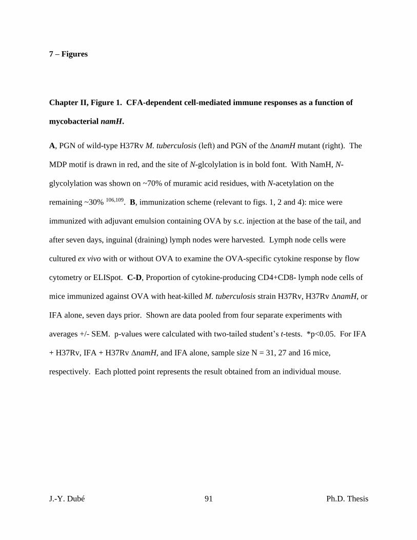
J.-Y. Dubé 91 Ph.D. Thesis
7 – Figures
Chapter II, Figure 1. CFA-dependent cell-mediated immune responses as a function of
mycobacterial namH.
A, PGN of wild-type H37Rv M. tuberculosis (left) and PGN of the ΔnamH mutant (right). The
MDP motif is drawn in red, and the site of N-glcolylation is in bold font. With NamH, N-
glycolylation was shown on ~70% of muramic acid residues, with N-acetylation on the
remaining ~30% 106,109. B, immunization scheme (relevant to figs. 1, 2 and 4): mice were
immunized with adjuvant emulsion containing OVA by s.c. injection at the base of the tail, and
after seven days, inguinal (draining) lymph nodes were harvested. Lymph node cells were
cultured ex vivo with or without OVA to examine the OVA-specific cytokine response by flow
cytometry or ELISpot. C-D, Proportion of cytokine-producing CD4+CD8- lymph node cells of
mice immunized against OVA with heat-killed M. tuberculosis strain H37Rv, H37Rv ΔnamH, or
IFA alone, seven days prior. Shown are data pooled from four separate experiments with
averages +/- SEM. p-values were calculated with two-tailed student’s t-tests. *p<0.05. For IFA
+ H37Rv, IFA + H37Rv ΔnamH, and IFA alone, sample size N = 31, 27 and 16 mice,
respectively. Each plotted point represents the result obtained from an individual mouse.
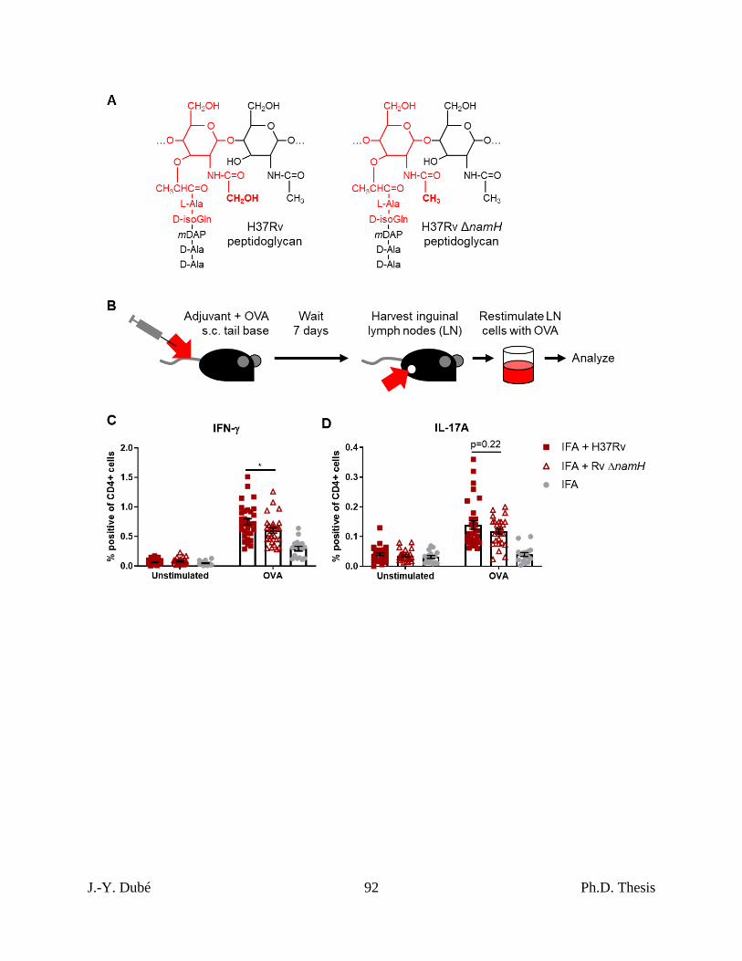
J.-Y. Dubé 92 Ph.D. Thesis
J.-Y. Dubé 93 Ph.D. Thesis
Chapter II, Figure 2. CFA-dependent cell-mediated immune responses as a function of
host Nod2 and Mincle.
Proportion of cytokine-producing CD4+CD8- lymph node cells of mice immunized against OVA
with CFA or IFA seven days prior. A-B, Nod2+/+ vs. Nod2-/- mice, data representative of two
independent experiments with averages +/- SEM. p-values were calculated with two-tailed
student’s t-tests. *p<0.05; **p<0.01. For CFA Nod2+/+, CFA Nod2-/-, IFA Nod2+/+ and IFA
Nod2-/-, sample size N = 12, 13, 9 and 7 mice, respectively. C-D, Mincle+/+ vs. Mincle-/- mice,
data pooled from two independent experiments with averages +/- SEM. p-values were calculated
with two-tailed student’s t-tests. *p<0.05; **p<0.01. For CFA Mincle+/+, CFA Mincle-/-, IFA
Mincle+/+ and IFA Mincle-/-, sample size N = 14, 16, 15 and 11 mice, respectively. E-F, WT
(Mincle+/+Nod2+/+) vs. DKO (Mincle-/-Nod2-/-) mice with averages +/- SEM. p-values were
calculated with two-tailed student’s t-tests. *p<0.05; **p<0.01. For CFA WT, CFA DKO, IFA
WT and IFA DKO, sample size N = 10, 7, 7 and 7 mice, respectively.
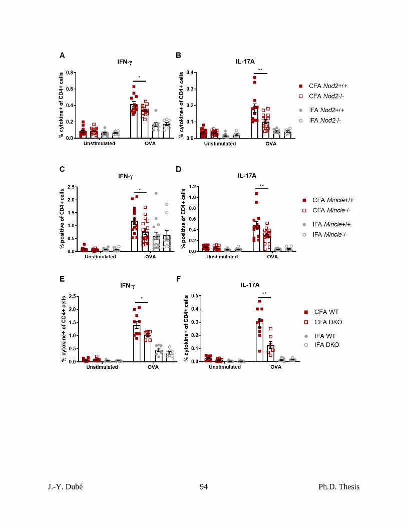
J.-Y. Dubé 94 Ph.D. Thesis
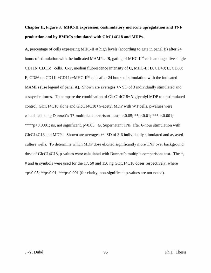
J.-Y. Dubé 95 Ph.D. Thesis
Chapter II, Figure 3. MHC-II expression, costimulatory molecule upregulation and TNF
production and by BMDCs stimulated with GlcC14C18 and MDPs.
A, percentage of cells expressing MHC-II at high levels (according to gate in panel B) after 24
hours of stimulation with the indicated MAMPs. B, gating of MHC-IIhi cells amongst live single
CD11b+CD11c+ cells. C-F, median fluorescence intensity of C, MHC-II; D, CD40; E, CD80;
F, CD86 on CD11b+CD11c+MHC-IIhi cells after 24 hours of stimulation with the indicated
MAMPs (use legend of panel A). Shown are averages +/- SD of 3 individually stimulated and
assayed cultures. To compare the combination of GlcC14C18+N-glycolyl MDP to unstimulated
control, GlcC14C18 alone and GlcC14C18+N-acetyl MDP with WT cells, p-values were
calculated using Dunnett’s T3 multiple comparisons test; p<0.05; **p<0.01; ***p<0.001;
****p<0.0001; ns, not significant, p>0.05. G, Supernatant TNF after 6-hour stimulation with
GlcC14C18 and MDPs. Shown are averages +/- SD of 3-6 individually stimulated and assayed
culture wells. To determine which MDP dose elicited significantly more TNF over background
dose of GlcC14C18, p-values were calculated with Dunnett’s multiple comparisons test. The *,
# and & symbols were used for the 17, 50 and 150 ng GlcC14C18 doses respectively, where
*p<0.05; **p<0.01; ***p<0.001 (for clarity, non-significant p-values are not noted).
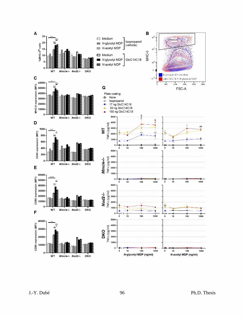
J.-Y. Dubé 96 Ph.D. Thesis
J.-Y. Dubé 97 Ph.D. Thesis
Chapter II, Figure 4. Complementation of IFA with synthetic mycobacterial MAMPs.
Proportion of cytokine-producing CD4+CD8- lymph node cells of WT mice immunized against
OVA seven days prior. A, N-glycolyl MDP dose-dependent response with 10 µg GlcC14C18:
IFA (N=4), IFA + 10 µg GlcC14C18 (N=7), IFA + 10 µg GlcC14C18 + 1 µg N-glycolyl MDP
(N=6), and IFA + 10 µg GlcC14C18 + 3 µg N-glycolyl MDP (N=7). Shown are data from
individual mice, with averages +/- SEM. p-values were calculated with Welch’s two-tailed
student’s t-tests. *p<0.05; **p<0.01. B, GlcC14C18 dose-dependent response with 30 µg N-
glycolyl MDP: IFA + 30 µg N-glycolyl MDP (N=6), IFA + 30 µg N-glycolyl MDP + 10 µg
GlcC14C18 (N=7), IFA + 30 µg N-glycolyl MDP + 30 µg GlcC14C18 (N=7), and CFA (N=7).
Shown are data from individual mice, with averages +/- SEM. In comparing IFA + 30 µg N-
glycolyl MDP to IFA + N-glycolyl MDP + 10 or 30 µg GlcC14C18, p-values were calculated
using Dunnett’s T3 multiple comparisons test. *p<0.05; **p<0.01; ns, not significant, p>0.05.
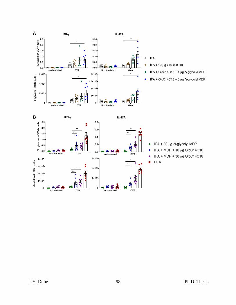
J.-Y. Dubé 98 Ph.D. Thesis
J.-Y. Dubé 99 Ph.D. Thesis
Chapter II, Figure 5. Dynamics of cDCs in lymph nodes after immunization with synthetic
mycobacterial MAMPs.
A, CD11b+ cDC quantities and median fluorescence intensity of MHC-I, MHC-II, CD40, CD80
and CD86 in mice immunized with the indicated adjuvants after 4 or 7 days. B, CD11b- cDC
quantities and median fluorescence intensity of MHC-I, MHC-II, CD40, CD80 and CD86 in
mice immunized with the indicated adjuvants after 4 or 7 days. For all adjuvant groups, N=8
mice (each plotted point represents data from an individual mouse). Shown are averages +/-
SEM. To determine if IFA + GlcC14C18 + N-glycolyl MDP altered cDC parameters compared
to IFA alone, p-values were calculated using Dunnett’s T3 multiple comparisons test correcting
for multiple timepoints and cDC type (CD11b+/-). *p<0.05; **p<0.01; ns, not significant,
p>0.05.
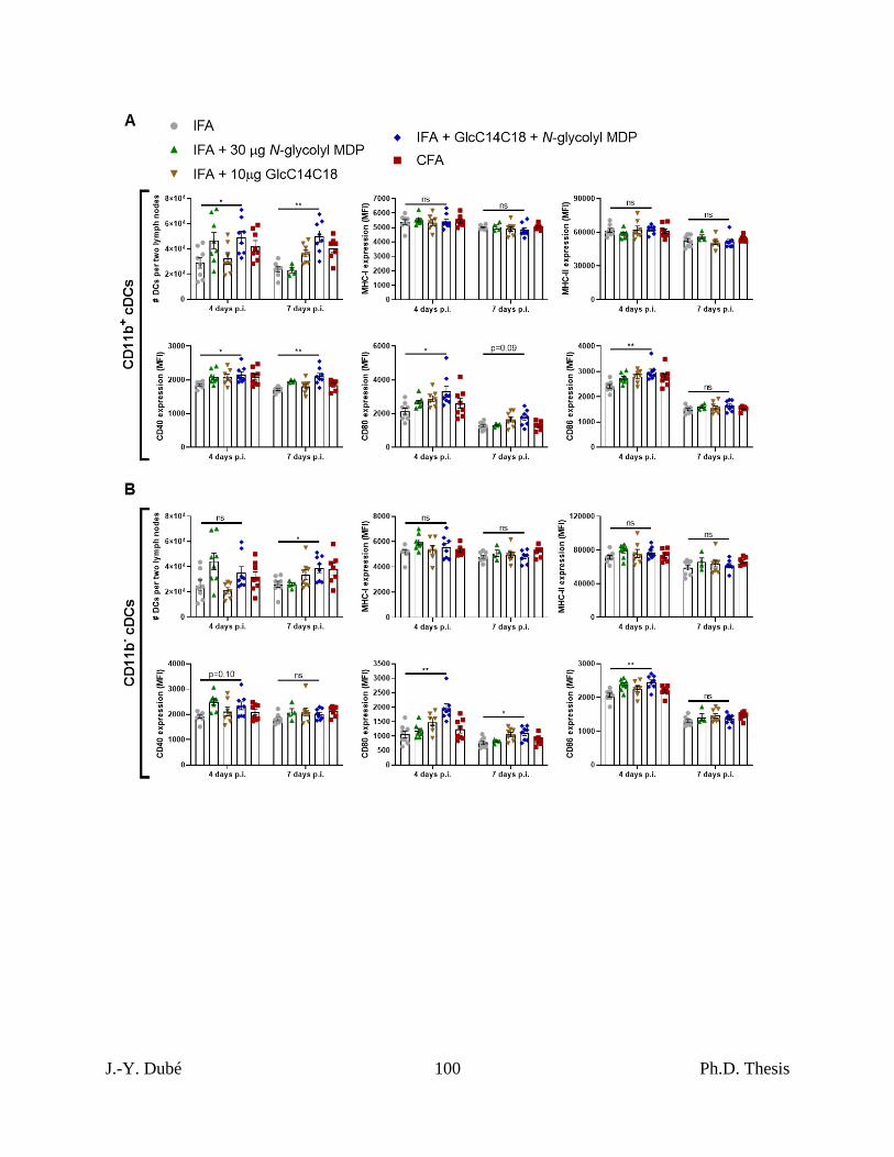
J.-Y. Dubé 100 Ph.D. Thesis
J.-Y. Dubé 101 Ph.D. Thesis
Chapter II, Figure 6. RR-EAE induced by IFA+GlcC14C18+MDP.
A, experimental timeline. B, average EAE score +/- SEM over time of mice induced with CFA
(N=15) or IFA + 10 µg GlcC14C18 + 30 µg N-glycolyl MDP (N=15). Mice were euthanized on
day 28 post injection. C, Cumulative EAE score, obtained by adding the EAE score of each
mouse over each of the 28 days. Lines represent averages +/- SEM. p-value was calculated with
two-tailed student’s t-test. **p = 0.0022.
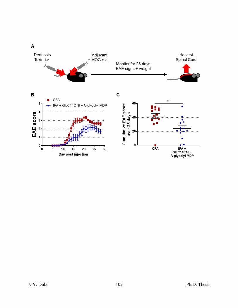
J.-Y. Dubé 102 Ph.D. Thesis
J.-Y. Dubé 103 Ph.D. Thesis
Chapter II, Figure 7. Spinal cord pathology in RR-EAE mice induced by
IFA+GlcC14C18+MDP.
A, Nissl and Luxol fast blue (LFB) stains of spinal cord sections from RR-EAE-induced mice at
day 28 post injection, having an EAE score of 3.5 upon euthanasia. Red boxes highlight cellular
infiltration and spatially associated demyelination of the white matter seen by Nissl and LFB
staining, respectively. B, Quantitative spinal cord pathology per EAE score upon euthanasia.
Statistical significance was determined by Tukey’s multiple comparisons test. *p<0.05 and
****p<0.0001. N=40 for each group (20 from CFA and 20 from synthetic adjuvant of
equivalent EAE scores). Lines indicate averages +/- SEM C, Quantitative spinal cord
pathology per adjuvant. Statistical significance was tested with two-tailed unpaired Welch’s t-
test; a power calculation for given variances and N=60 per group indicated an ability to discern
+/- 15% difference vs. CFA control. Lines indicate averages +/- SEM.
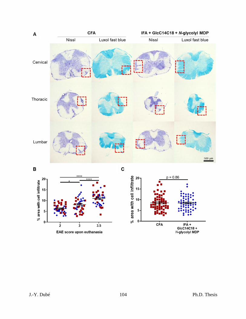
J.-Y. Dubé 104 Ph.D. Thesis
J.-Y. Dubé 105 Ph.D. Thesis
8 – Supplemental Figures
Chapter II, Figure S1. Flow cytometry gating strategies for lymph node cells.
A, gating to quantify cytokine-producing CD4+CD8- T cells. Shown are representative gating
and data from an OVA-stimulated sample (CFA-immunized WT mouse). For comparison,
cytokine production for the corresponding unstimulated sample is included. B, gating to identify
lymph node cell subsets. Shown is gating on a representative sample (CFA-immunized WT
mouse). pDCs are B220+Ly6C+MHC-II+CD11b-CD11c+ cells. B cells are B220+Ly6C-
CD11b-CD11c-CD4-CD8a- cells. cDCs are B220-MHC-IIhiLy6C-CD11c+ cells (analyzed
subsets are CD11b+/-). CD4+ T cells are B220-CD11b-CD4+CD8a- cells. CD8+ T cells are
B220-CD11b-CD4-CD8a+ cells. Monocytes are B220-CD11b+Ly6C+SSClo cells. PMNs are
B220-CD11b+Ly6CmedSSChi CD11c- cells.
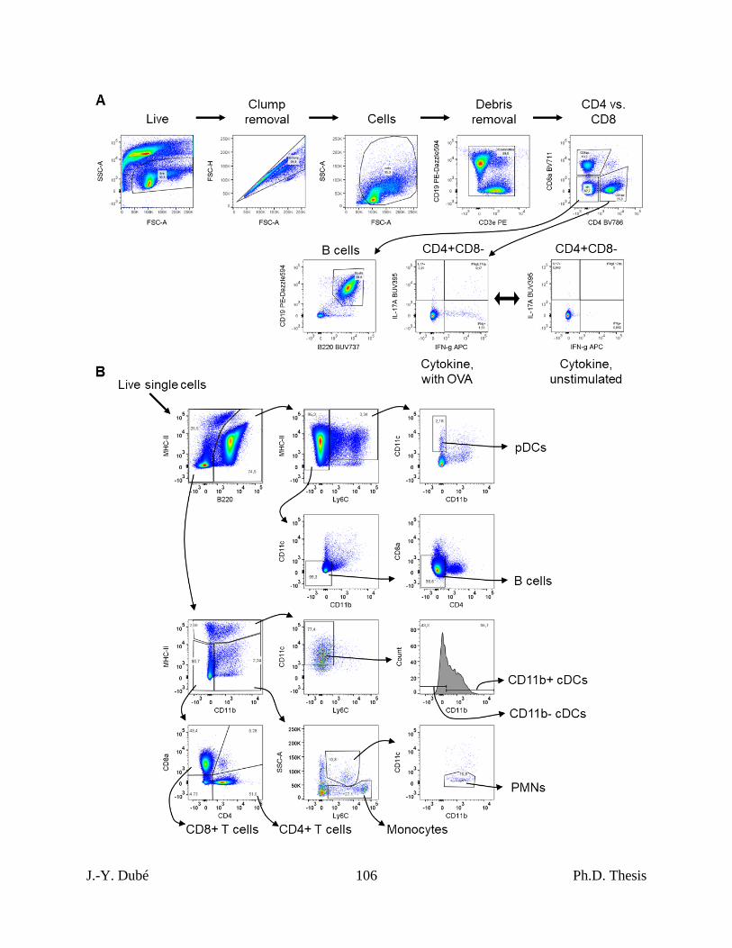
J.-Y. Dubé 106 Ph.D. Thesis
J.-Y. Dubé 107 Ph.D. Thesis
Chapter II, Figure S2. Addition data on CFA-dependent cell-mediated immune responses
as a function of mycobacterial namH.
These data are from the same experiments shown in fig. 1C (refer to fig. 1C legend for details).
A-B, Total numbers (from two lymph nodes) of cytokine-producing CD4+CD8- lymph node
cells of mice immunized against OVA with H37Rv, H37Rv ΔnamH, or IFA alone. Shown are
averages +/- SEM. p-values were calculated with two-tailed student’s t-tests. **p<0.01. C-D,
Contribution of namH to the mycobacterial portion of OVA-specific IFN-γ elicited by CFA. C,
result was obtained from %IFN-γ+ data by subtracting the average IFA background from all
CFA data, and plotting the results as % of IFA+H37Rv ‘wild-type’. D, result was obtained from
# IFN-γ+ data by subtracting the average IFA background from all CFA data, and plotting the
results as % of IFA+H37Rv ‘wild-type’. Shown are averages +/- SEM. p-values were
calculated with two-tailed student’s t-tests. *p<0.05. E, results separated by experimental run
for the data presented in fig. 1C and fig. S2A-B. Shown are averages +/- SEM.
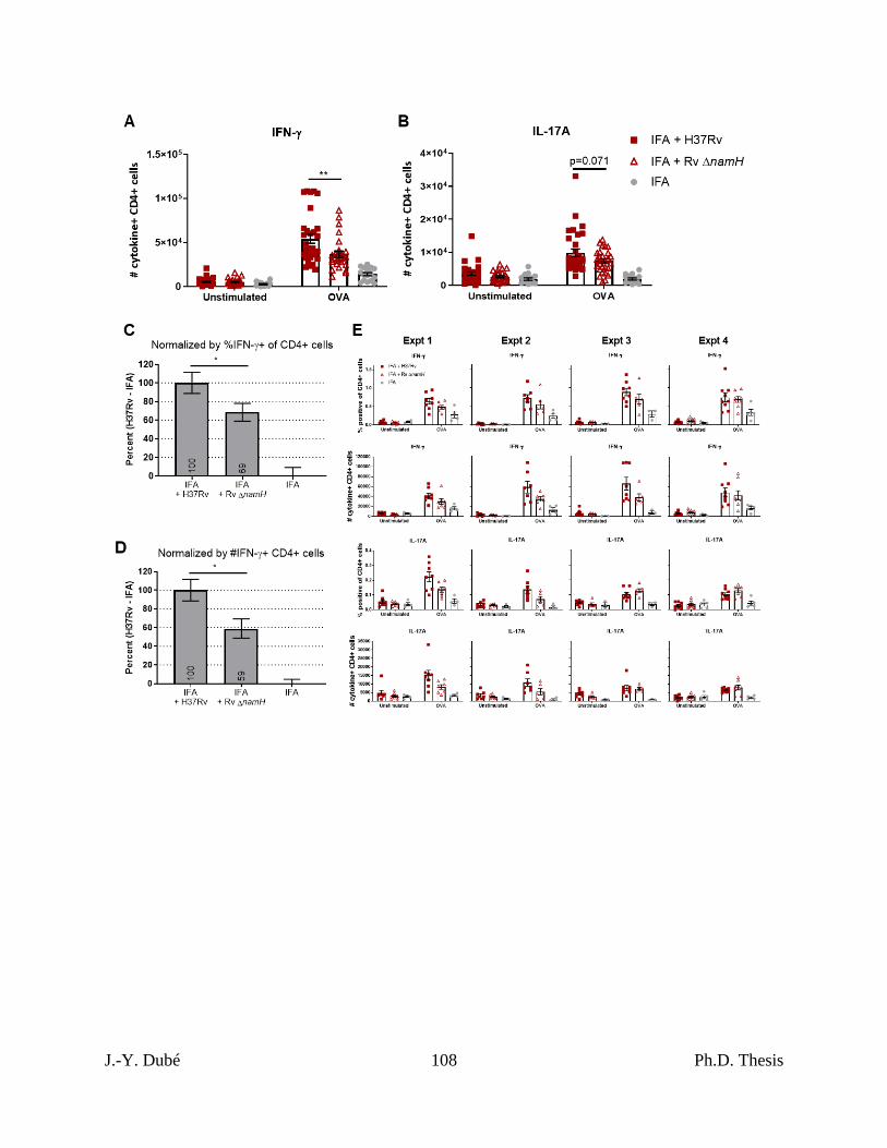
J.-Y. Dubé 108 Ph.D. Thesis
J.-Y. Dubé 109 Ph.D. Thesis
Chapter II, Figure S3. Additional data on CFA-dependent immune responses as a function
of Nod2 and Mincle.
A-B, IFN-γ ELISpot of inguinal lymph node cells mice immunized against OVA seven days
prior, produced in an independent experiment. A, number of IFN-γ spot-forming cells per one
million cells. B, total number of IFN-γ spot-forming cells per two inguinal lymph nodes. Shown
are averages +/- SEM. p-values were calculated with two-tailed student’s t-tests. *p<0.05. For
CFA Nod2+/+, CFA Nod2-/-, IFA Nod2+/+ and IFA Nod2-/-, N = 6, 8, 6 and 6 mice,
respectively. C, flow cytometry of inguinal lymph node cells mice immunized against OVA
seven days prior, produced in an independent experiment similar to that in fig. 2A-B. D, results
separated by experimental run for the data presented in fig. 2C-D and fig. S4C-D. Shown are
averages +/- SEM.
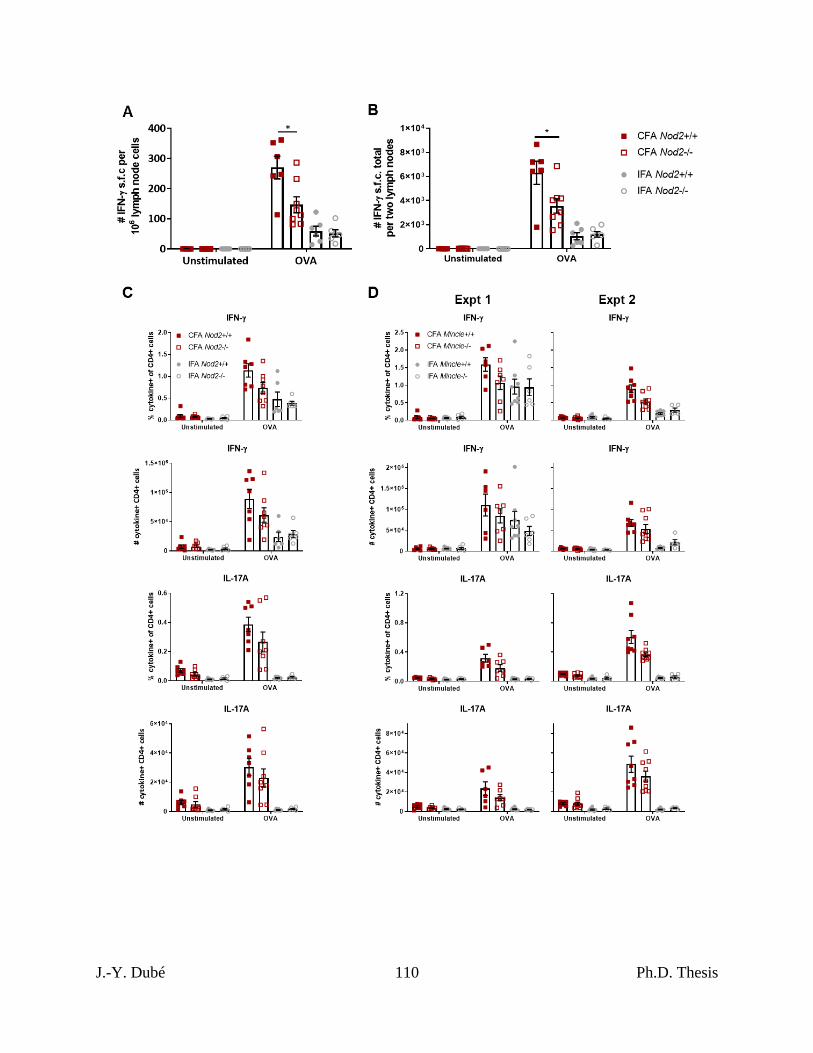
J.-Y. Dubé 110 Ph.D. Thesis
J.-Y. Dubé 111 Ph.D. Thesis
Chapter II, Figure S4. CFA-dependent cell-mediated immune responses as a function of
host Nod2 and Mincle expressed in total cell numbers
These data are from the same experiments shown in fig. 2 (refer to fig. 2 legend for details). A-
F, Total numbers (from two lymph nodes) of cytokine-producing CD4+CD8- lymph node cells
of mice immunized against OVA with the indicated adjuvant as a function of: A-B, Nod2; C-D,
Mincle; E-F, Mincle and Nod2 together. Shown are averages +/- SEM. p-values were calculated
with two-tailed student’s t-tests. *p<0.05; ns, not significant, p>0.05.
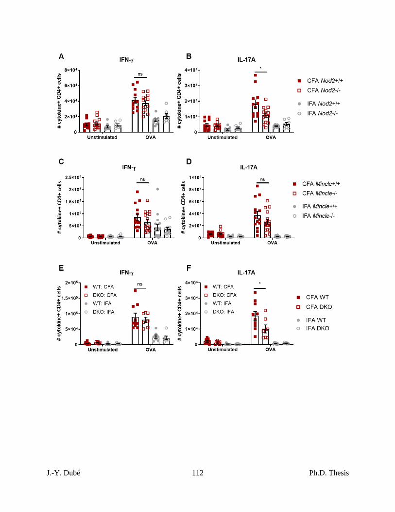
J.-Y. Dubé 112 Ph.D. Thesis
J.-Y. Dubé 113 Ph.D. Thesis
Chapter II, Figure S5. Additional data on MHC-II expression and costimulatory molecule
upregulation by BMDCs stimulated with GlcC14C18 and MDPs.
A, percentage of cells expressing MHC-II at high levels (according to gate in fig. 3B) after 48
hours of stimulation with the indicated MAMPs. B-E, median fluorescence intensity of B,
MHC-II; C, CD40; D, CD80; E, CD86 on CD11b+CD11c+MHC-IIhi cells after 48 hours of
stimulation with the indicated MAMPs (use legend of panel A). Shown are averages +/- SD of 3
individually stimulated and assayed cultures. F-I, histograms of CD11b+CD11c+MHC-IIhi cells
demonstrating expression levels of F, MHC-II; G, CD40; H, CD80; I, CD86, for both timepoints
(FMOC is not shown for MHC-II because the analysis required MHC-II gating).
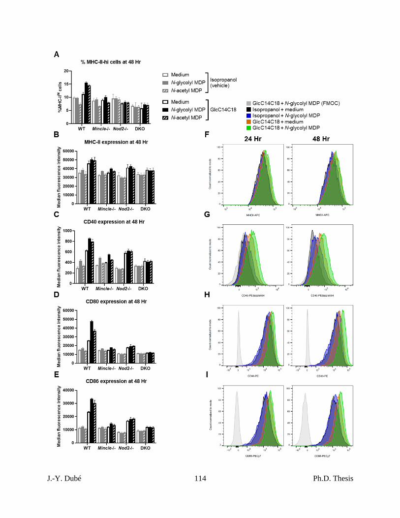
J.-Y. Dubé 114 Ph.D. Thesis
J.-Y. Dubé 115 Ph.D. Thesis
Chapter II, Figure S6. Synthetic adjuvant-dependent IFN-γ responses compared to CFA
by ELISpot.
A-B, IFN-γ ELISpot of inguinal lymph node cells of mice immunized against OVA with the
indicated adjuvant seven days prior. A, number of IFN-γ spot-forming cells per one million
cells. B, total number of IFN-γ spot-forming cells per two inguinal lymph nodes. Data was
pooled from two independent experiments. Shown are averages +/- SEM, where for IFA, IFA +
3 µg N-glycolyl MDP, IFA + 10 µg N-glycolyl MDP, IFA + 30 µg N-glycolyl MDP, IFA + 100
µg N-glycolyl MDP and CFA, N = 11, 6, 12, 11, 6 and 11 mice, respectively. C-D, These data
are from the same mice used in fig. 4B (refer to fig. 4B legend for details). C, number of IFN-γ
spot-forming cells per one million cells. D, total number of IFN-γ spot-forming cells per two
inguinal lymph nodes. Shown are averages +/- SEM. In comparing IFA + 30 µg MDP to IFA +
MDP + 10 or 30 µg GlcC14C18, p-values were calculated using Dunnett’s T3 multiple
comparisons test. *p<0.05; **p<0.01; ns, not significant, p>0.05.
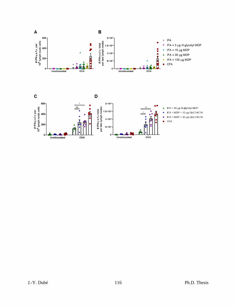
J.-Y. Dubé 116 Ph.D. Thesis
J.-Y. Dubé 117 Ph.D. Thesis
Chapter II, Figure S7. Lymph node cell subset numbers after immunization with synthetic
adjuvants.
These data are from the same mice used in fig. 5 (refer to fig. 5 legend for details of the
experiment). A-F, From two inguinal lymph nodes at 4 or 7 days post-immunization, shown are
total numbers of extracted A, B cells; B, CD4+ T cells; C, CD8+ T cells; D, plasmacytoid
dendritic cells (pDCs); E, monocytes; F, polymorphonuclear cells (PMNs). Data are graphed as
averages +/- SEM.
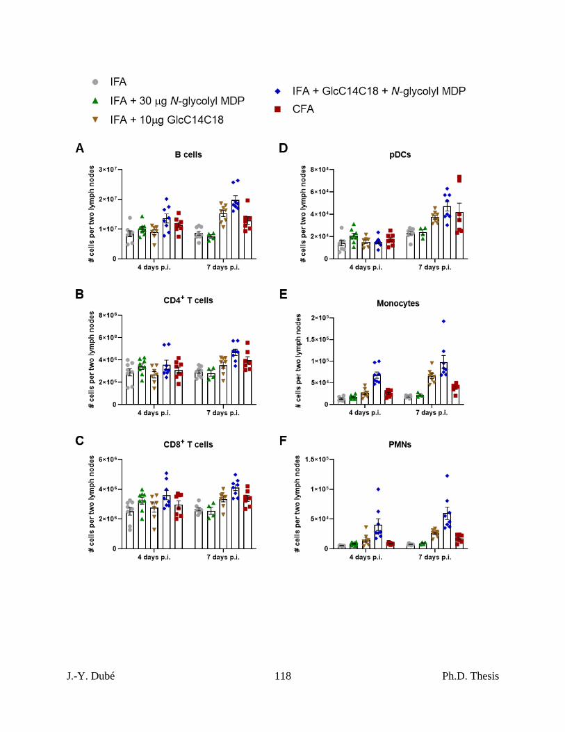
J.-Y. Dubé 118 Ph.D. Thesis
J.-Y. Dubé 119 Ph.D. Thesis
Chapter II, Figure S8. Additional statistics for RR-EAE.
These data are from the same set shown in fig. 5 (refer to fig. 5 legend for details). A, average
weight of mice over time +/- SEM. B, maximum EAE score reached on any day by mice, as of
day 28. C, disease course of mice selected for spinal cord histopathology. D-G, Statistics for
RR-EAE induced by IFA+TDM+MDP. D, average EAE score +/- SEM over time of mice
induced with CFA (N=13) or IFA + 1 µg TDM + 30 µg N-glycolyl MDP (N=13). Mice were
euthanized on day 27 post injection. E, cumulative EAE score, obtained by adding the EAE
score of each mouse over each of the 27 days. Lines represent averages +/- SEM. F, average
weight of mice over time +/- SEM. G, maximum EAE score reached on any day by mice, as of
day 27.
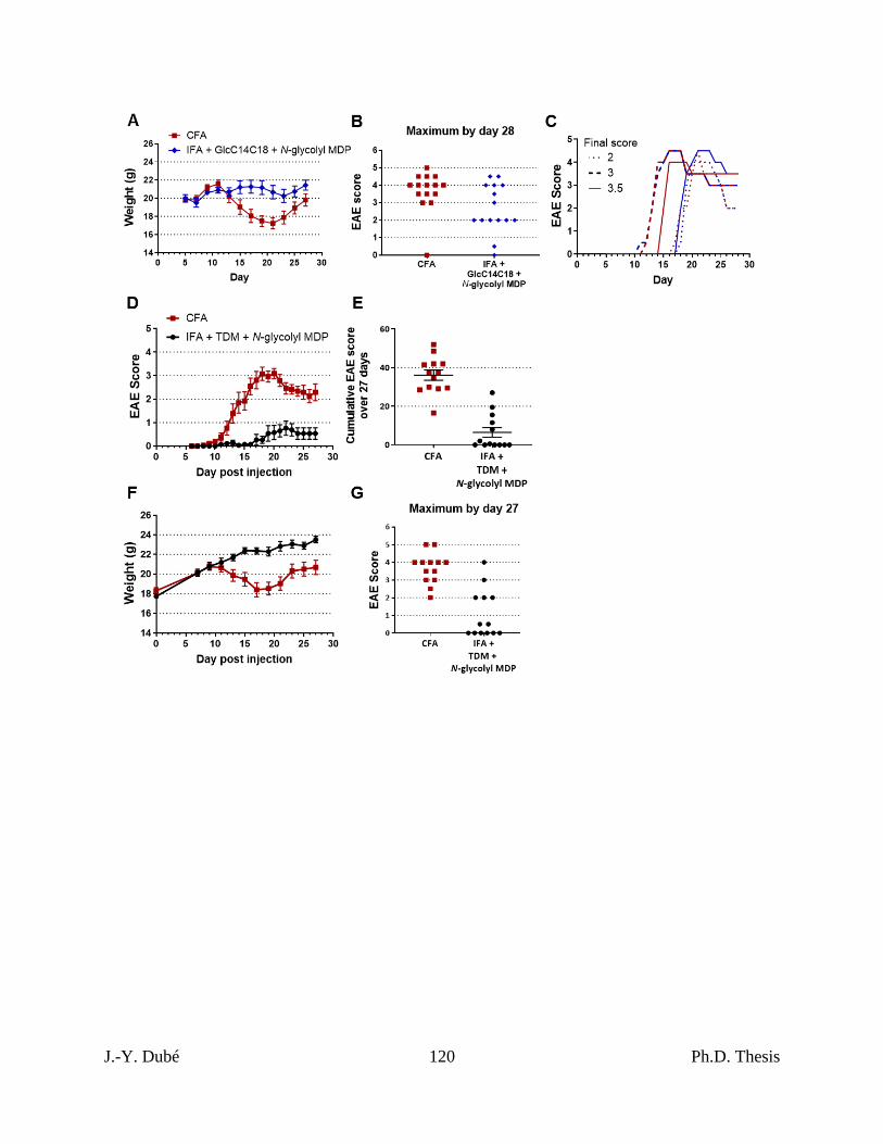
J.-Y. Dubé 120 Ph.D. Thesis
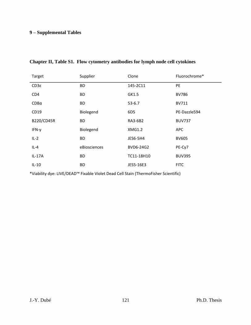
J.-Y. Dubé 121 Ph.D. Thesis
9 – Supplemental Tables
Chapter II, Table S1. Flow cytometry antibodies for lymph node cell cytokines
Target Supplier Clone Fluorochrome*
CD3ε BD 145-2C11 PE
CD4 BD GK1.5 BV786
CD8α BD 53-6.7 BV711
CD19 Biolegend 6D5 PE-Dazzle594
B220/CD45R BD RA3-6B2 BUV737
IFN-γ Biolegend XMG1.2 APC
IL-2 BD JES6-5H4 BV605
IL-4 eBiosciences BVD6-24G2 PE-Cy7
IL-17A BD TC11-18H10 BUV395
IL-10 BD JES5-16E3 FITC
*Viability dye: LIVE/DEAD™ Fixable Violet Dead Cell Stain (ThermoFisher Scientific)
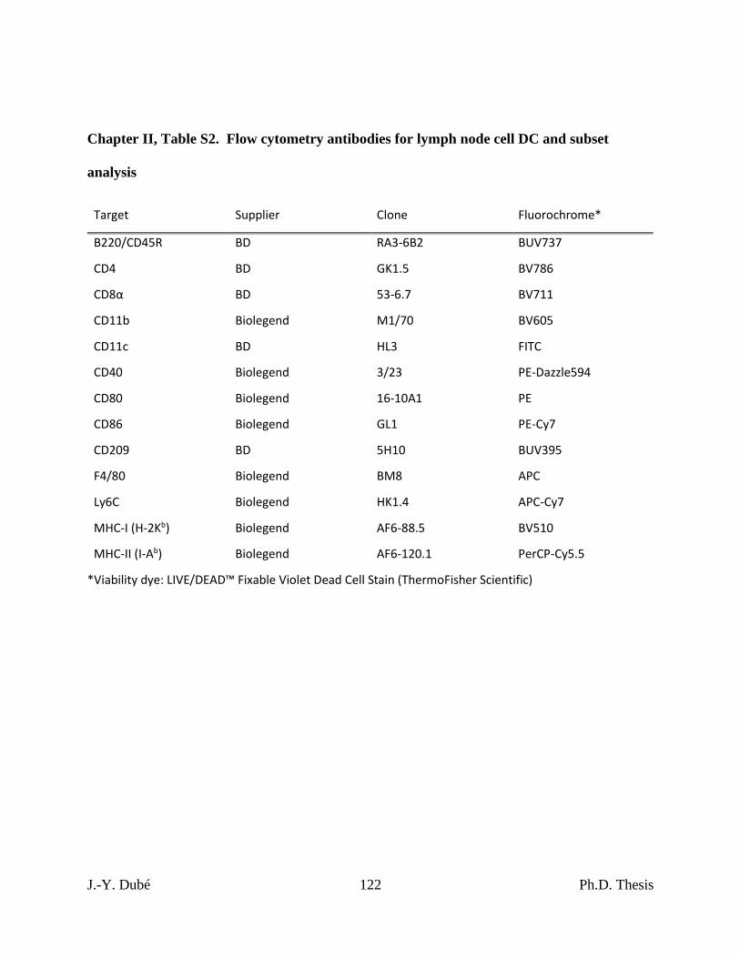
J.-Y. Dubé 122 Ph.D. Thesis
Chapter II, Table S2. Flow cytometry antibodies for lymph node cell DC and subset
analysis
Target Supplier Clone Fluorochrome*
B220/CD45R BD RA3-6B2 BUV737
CD4 BD GK1.5 BV786
CD8α BD 53-6.7 BV711
CD11b Biolegend M1/70 BV605
CD11c BD HL3 FITC
CD40 Biolegend 3/23 PE-Dazzle594
CD80 Biolegend 16-10A1 PE
CD86 Biolegend GL1 PE-Cy7
CD209 BD 5H10 BUV395
F4/80 Biolegend BM8 APC
Ly6C Biolegend HK1.4 APC-Cy7
MHC-I (H-2Kb) Biolegend AF6-88.5 BV510
MHC-II (I-Ab) Biolegend AF6-120.1 PerCP-Cy5.5
*Viability dye: LIVE/DEAD™ Fixable Violet Dead Cell Stain (ThermoFisher Scientific)
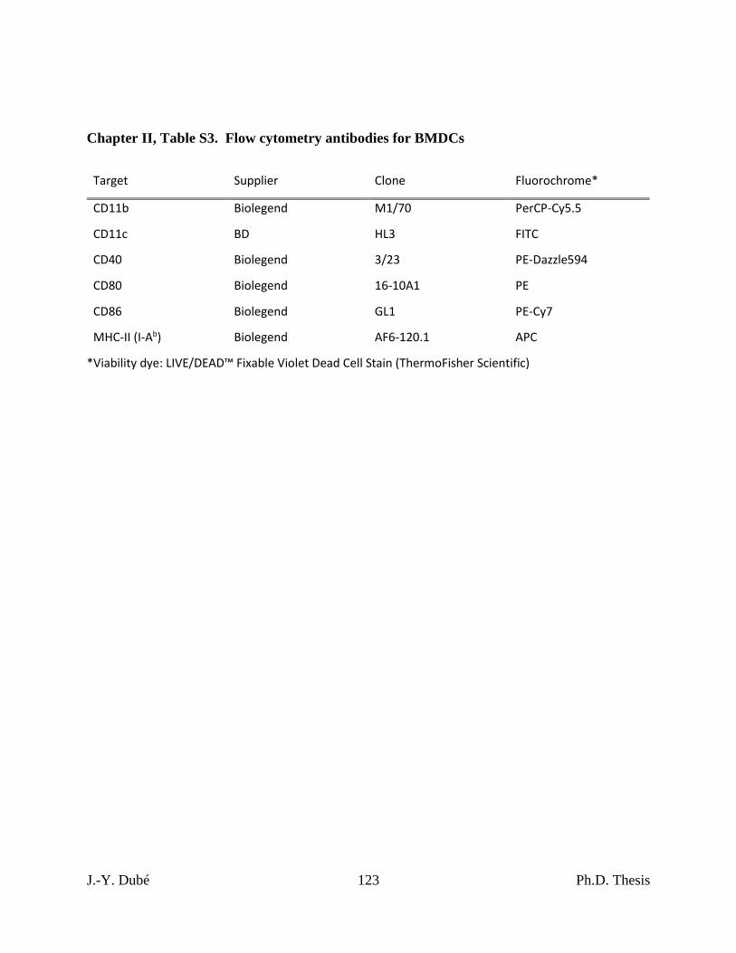
J.-Y. Dubé 123 Ph.D. Thesis
Chapter II, Table S3. Flow cytometry antibodies for BMDCs
Target Supplier Clone Fluorochrome*
CD11b Biolegend M1/70 PerCP-Cy5.5
CD11c BD HL3 FITC
CD40 Biolegend 3/23 PE-Dazzle594
CD80 Biolegend 16-10A1 PE
CD86 Biolegend GL1 PE-Cy7
MHC-II (I-Ab) Biolegend AF6-120.1 APC
*Viability dye: LIVE/DEAD™ Fixable Violet Dead Cell Stain (ThermoFisher Scientific)
J.-Y. Dubé 124 Ph.D. Thesis
PREFACE TO CHAPTER III
In Chapter I, the importance of PRRs during Mtb infection was demonstrated to be
generally limited to altered immunity with few reports of PRR KOs in mice resulting in
significant loss of bacterial control resulting in premature death. In Chapter II, we showed that
NOD2 and Mincle were essential for a complete immune response to mycobacteria, that these
PRR pathways synergize and that together they can recapitulate about half of the mycobacterial
adjuvant effect. Therefore, with two PRRs known to be important, and cooperative, for the
mycobacterial immune response, in Chapter III we test whether double knockout (DKO) of
Mincle and Nod2 during live mycobacterial infection results in significant loss of bacterial
control due to doubly defective immunity. The absence of NOD2 was associated with altered
immunity during disparate mycobacterial infections and reduced survival particularly during Mtb
infection, regardless of Mincle status, demonstrating a distinct role for Nod2 in the host response
to mycobacteria that was more important than Mincle.
J.-Y. Dubé 125 Ph.D. Thesis
CHAPTER III – Respective and combined disruption of Mincle and Nod2
distinctly alter mycobacterial immunity and resistance
(This Chapter is adapted from a manuscript in preparation)
Jean-Yves Dubé*, †, ‡, Fiona McIntosh †, ‡, Shannon C. Duffy *, †, ‡, Marcel A. Behr *, †, ‡, §
*, Department of Microbiology and Immunology, McGill University.
†, Infectious diseases and Immunity in Global Health Program, Research Institute of the McGill
University Health Centre.
‡, McGill International TB Centre.
§, Department of Medicine, McGill University Health Centre, Montréal, Canada.
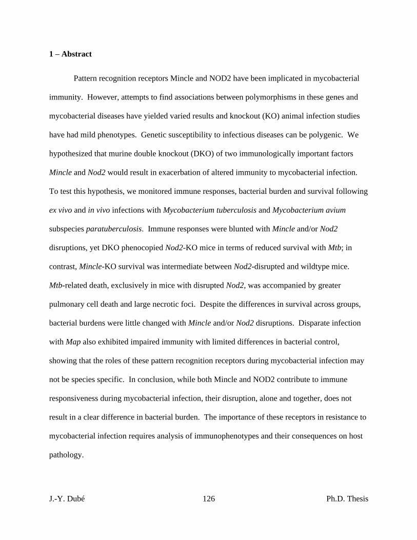
J.-Y. Dubé 126 Ph.D. Thesis
1 – Abstract
Pattern recognition receptors Mincle and NOD2 have been implicated in mycobacterial
immunity. However, attempts to find associations between polymorphisms in these genes and
mycobacterial diseases have yielded varied results and knockout (KO) animal infection studies
have had mild phenotypes. Genetic susceptibility to infectious diseases can be polygenic. We
hypothesized that murine double knockout (DKO) of two immunologically important factors
Mincle and Nod2 would result in exacerbation of altered immunity to mycobacterial infection.
To test this hypothesis, we monitored immune responses, bacterial burden and survival following
ex vivo and in vivo infections with Mycobacterium tuberculosis and Mycobacterium avium
subspecies paratuberculosis. Immune responses were blunted with Mincle and/or Nod2
disruptions, yet DKO phenocopied Nod2-KO mice in terms of reduced survival with Mtb; in
contrast, Mincle-KO survival was intermediate between Nod2-disrupted and wildtype mice.
Mtb-related death, exclusively in mice with disrupted Nod2, was accompanied by greater
pulmonary cell death and large necrotic foci. Despite the differences in survival across groups,
bacterial burdens were little changed with Mincle and/or Nod2 disruptions. Disparate infection
with Map also exhibited impaired immunity with limited differences in bacterial control,
showing that the roles of these pattern recognition receptors during mycobacterial infection may
not be species specific. In conclusion, while both Mincle and NOD2 contribute to immune
responsiveness during mycobacterial infection, their disruption, alone and together, does not
result in a clear difference in bacterial burden. The importance of these receptors in resistance to
mycobacterial infection requires analysis of immunophenotypes and their consequences on host
pathology.
J.-Y. Dubé 127 Ph.D. Thesis
2 – Introduction
Each year, Mycobacterium tuberculosis (Mtb) causes tuberculosis (TB) in about 10
million people and results in the death of over a million (WHO). Other species in the Mtb
complex as well as non-tuberculous mycobacteria cause disease in livestock and occasionally in
humans too. For example, Mycobacterium avium subspecies paratuberculosis (Map) is the
etiologic agent of Johne’s disease, a lethal wasting illness affecting ruminants including cattle.
Many aspects of immunity have been linked to susceptibility to mycobacterial infection (e.g.
HIV status, host genetics). Therefore, understanding which immune mechanisms are important
for host control or tolerance of mycobacteria is needed to guide the development of
immunotherapeutic agents and vaccines.
Pattern recognition receptors (PRRs) help to initiate innate immunity and instruct the
development of adaptive immunity to pathogens. They are a collection of genetically inborn
sensors that detect microbe-associated molecular patterns (MAMPs) to initiate the inflammatory
response at the cellular level (e.g. through NF-κB and MAPK pathways). One such PRR is
Macrophage-Inducible C-type Lectin (Mincle), which binds to the mycobacterial outer
membrane lipid trehalose 6,6’-dimycolate (TDM, aka cord factor) 157. TDM is an abundant
glycolipid in the outer membrane of mycobacteria and has been posited as both a host-beneficial
MAMP as well as a virulence factor of pathogenic mycobacteria 481. Mincle-/- mice have altered
immunity to mycobacteria 157,460,461,482, although whether Mincle polymorphisms are associated
with human mycobacterial infection is only starting to emerge: at least two such studies reported
statistically significant associations 435,483.
Another PRR of interest is Nucleotide-binding Oligomerization Domain-containing 2
(NOD2), which is a cytoplasmic molecule essential for the recognition of the bacterial
J.-Y. Dubé 128 Ph.D. Thesis
peptidoglycan through the muropeptide moiety 182,183,347. NOD2 has been implicated in
mycobacterial immunity through chemical, immunologic and epidemiologic evidence: 1)
mycobacteria contain a distinct NOD2 agonist in that they possess N-glycolylated muropeptides
while other bacteria are N-acetylated 106,107; 2) N-glycolylation of the muropeptide was shown to
be a more potent activator of innate immune cells 108,187 and Nod2-/- mice have altered
(decreased) immune responses to mycobacteria 188,189; 3) polymorphisms in NOD2 have been
associated with mycobacterial infection and disease 255,388,392. NOD2 variation is also associated
with predisposition to Crohn’s disease 368-370, for which Map has long been hypothesized to
contribute 52.
We have recently shown that both Mincle and Nod2 are essential to the immune response
elicited by Freund’s adjuvant, a mycobacterial adjuvant, and that Mincle and NOD2 ligands
together account for about 30-50% of the cellular immune response to dead Mtb 187. However,
Nod2-/- mouse studies have shown only modestly reduced Mtb control188,189 and survival 189.
Mincle-/- mice were shown to have little difference from WT in terms of Mtb burden and
survival analysis has not been reported 460,461. The importance of PRRs during Mtb infection
may indeed be restricted to specific phenotypes due their nature as redundant sensors of
infection, in contrast to the essentiality of unique immune effectors 484. Therefore, we
hypothesized that simultaneous loss of Mincle and Nod2 could result in a greater degree of
altered immunity leading to a larger impairment in bacterial control and survival. The ‘double-
knockout’ simulates a genetic ‘two-hit’ scenario which could explain why some loss-of-function
polymorphisms do not appear to have complete disease phenotype penetrance.
To address this, we used Mincle-/-Nod2-/- (double knockout, DKO) mice previously
generated 187 (with the corresponding single knockouts, SKO) to model genetic susceptibility to
J.-Y. Dubé 129 Ph.D. Thesis
mycobacterial disease during infection with Mtb and Map. Our data show that DKO mice had
defective mycobacterial immunity similar to Nod2-/- mice, but that these immunological defects
appeared limited in terms of bacterial control. DKO and Nod2-/- mice had similarly shortened
survival during Mtb infection, and although mortality occurred late it was occasionally
accompanied by necrotic foci in the lungs not seen in NOD2-competent animals. In contrast, for
Mincle-KO the immunologic phenotypes and survival data were intermediate between WT and
Nod2-KO. Our data indicate that PRRs like Mincle and NOD2 are largely dispensable for
control of mycobacterial burden and that the absence of NOD2 is more important than Mincle
for dictating the ultimate host response to mycobacterial infection.
3 – Materials and Methods
Mice
Nod2-/- mice were initially obtained from Jackson laboratories. Mincle-/- mice breeders were
generously provided by the laboratory of Christine Wells 433. Imported mice were genotyped to
confirm that the Dock2 mutation reported in certain Nod2-/- mice was not present 480. All mice
were obtained by crossing Nod2-/- and Mincle-/- mice (both C57BL/6 background) to generate
double heterozygotes which were mated to regenerate single Nod2 and Mincle knockouts as well
as ‘wild-type’ (WT, Mincle+/+Nod2+/+) and ‘double knockout’ (DKO, Mincle-/-Nod2-/-).
Homozygotic mice were then used as breeders to sustain the colony. For Mtb experiments, all
mice were from 2 to 4 months of age at the start of the experiment. For intraperitoneal Map
infection, mice were from 2 to 8 months of age at the start of the experiment. Mice were age and
sex matched where applicable across groups in all experiments All protocols requiring mice
J.-Y. Dubé 130 Ph.D. Thesis
adhered to the guidelines of the Canadian Council on Animal Care (CCAC) and were authorized
by the RI-MUHC animal resource division.
Bacteria
Mtb H37Rv and Map K10 were grown at 37oC in complete 7H9 medium (Middlebrook 7H9
(BD), ADC (BD), 0.2% glycerol (Sigma), 0.05% Tween-80 (Sigma)). Map was also
supplemented with 1 μg/ml ferric mycobactin J (Allied Monitor, Missouri USA). Cultures in
mid-log phase were used for experiments. Mtb culture used in aerosol experiments was
aliquoted into 7H9 complete medium with 20% glycerol at OD-600 ~ 0.5, frozen at -80oC and
thawed immediately before use. For plating on solid media, we used complete 7H10
(Middlebrook 7H10 (BD), OADC (BD), 0.5% glycerol (Sigma)) including PANTA (BD) and,
for Map experiments, 1 μg/ml ferric mycobactin J.
Bone marrow-derived macrophage (BMDM) infection
Bone marrow was extracted from murine tibiae and femora and differentiated into macrophages
with recombinant M-CSF (100 U/ml) (Peprotech) over 7 days as previously described 485. After
differentiation, BMDMs were lifted using CellStripper Solution (Corning) and replated into 96-
well plates at 200,000 cells per well for Mtb infection. BMDMs were cultured in R10 (RPMI
1640 media supplemented with non-essential amino acids, 10 mM HEPES, 10% FBS and, while
not being infected, Penicillin/Streptomycin (Wisent)). Mtb was resuspended in R10 at an OD-
600 of 0.2 and 100 μl of this (approximately 2 million CFU) was added to BMDMs (t = 0).
After four hours, extracellular bacteria were removed by washing wells three times with R10
after which R10 was restored. Supernatant was harvested and frozen at -80oC at the indicated
J.-Y. Dubé 131 Ph.D. Thesis
timepoints, and was later thawed, filter sterilized and subjected to ELISA analysis
(ThermoFisher). In the case of heat-killed bacteria, Mtb was heated at 80oC for 30 minutes
before being added to BMDMs, no washing was performed and supernatant was harvested the
following day only.
Mtb aerosol infections
Mice were infected by aerosol using an ONARES device (New Jersey, USA) over 15 minutes
with an Mtb suspension in PBS + 0.05% Tween-80 at OD-600 = 0.04. This method routinely
resulted in a low dose Mtb infection (< 60 CFU total in both lungs one day after infection).
Actual day 1 pulmonary loads are indicated in figures and/or legends.
Mtb survival
Mice infected with Mtb were weighed and periodically examined every 1-4 weeks with a body
score system (4 = normal; 3 = slight hunch; 2 = hunch plus less active or reduced grooming; 2- =
2 with slow gait and very bad posture; 1 = hardly moving, dehydrated, thin and ungroomed).
Endpoints were set to determine whether mice were imminently dying of Mtb infection and/or
required compassionate euthanasia: body score under 2; body score of 2 with relatively low and
rapidly dropping weight (such that death is likely within a week); body score of 3 with extremely
low (<20 g) and steadily dropping weight (losing >1g/week for last 2 weeks); non-TB ailment
normally requiring euthanasia (e.g. serious wound).
J.-Y. Dubé 132 Ph.D. Thesis
Map infection
Mice were infected with Map as described previously in an established model 486-488. Briefly,
Map culture was grown to OD-600 ~ 0.5 (equivalent to 108 CFU/ml), centrifuged and washed
with PBS, and then resuspended in PBS + 0.1% Tween-80 (Sigma) at 1/5 the original volume.
Mice were injected intraperitoneally with 200 μl of this bacterial suspension to receive an
equivalent of ~ 108 CFU total. At the indicated timepoints, mice were euthanized and organs
were harvested for CFU enumeration or immunophenotyping.
Enumeration of organ CFUs
Lungs, spleens and livers were removed from mice after euthanasia and processed immediately,
except in the case of survival studies with Mtb, in which case organs were frozen at -80oC for
later processing together once all mice were euthanized. Organs were placed in 1 ml complete
7H9 and were homogenized with an Omni Tissue Homogenizer TH (Omni International) for 30
seconds. Homogenate was serially diluted and plated. After approximately 3 and 5 weeks for
Mtb and Map, respectively, CFUs were enumerated from culture plates.
Lung cell preparation for flow cytometry
Three minutes before euthanasia by cervical dislocation, mice were injected intravenously with 5
μg of anti-CD45-FITC (BD) antibody to stain circulatory leukocytes for later differentiation
from parenchymal leukocytes. Right lungs were harvested, sliced into small pieces in RPMI-
1640 medium and digested in 150 U/ml collagenase (purified from Clostridium histolyticum,
Sigma) for 1 hour at 37oC. Digested lung tissue was pressed through a 100 μM cell strainer,
J.-Y. Dubé 133 Ph.D. Thesis
treated with red blood cell lysis buffer (Roche), and resuspended in a small volume of RPMI-
1640 medium. 20% of this suspension was taken for flow cytometry staining.
Splenocyte preparation for flow cytometry
Spleens harvested from Map infected mice were immediately weighed and placed in RPMI-1640
medium. Spleens were next pressed through a 100 μM cell strainer, treated with red blood cell
lysis buffer (Roche), and resuspended in 5 ml of R10. Cell concentrations were estimated by
counting with a hemocytometer and set to 20 million cells per ml. Approximately 2 million cells
were immediately stained and fixed for flow cytometry analysis. Another 2 million cells were
cultured ex vivo in 142 μl R10 in the presence of 5 μg soluble Map lysate (or 8 μl equivalent PBS
volume control) for 21 hours at 37oC 5% CO2, after which 2 μg of Brefeldin A (Sigma) in 50 μl
R10 were added and cells were incubated for another 5 hours before staining and fixing for flow
cytometry. Soluble Map lysate was prepared from a logarithmically growing culture of Map,
which was centrifuged and washed three times with PBS, subjected to lysis by bead beating with
silica beads (MP Biomedicals, FastPrep-24), and the supernatant passed through a 0.2 μm filter
and stored at -80oC until needed.
Flow cytometry analysis
Cells were washed in PBS then stained with LIVE/DEAD™ fixable violet dead cell stain
(ThermoFisher Scientific). Next, cells were washed with FACS buffer (PBS, 5% BSA, 2mM
EDTA), Fc receptors were blocked with TruStain FcX (Biolegend), and then cells were
incubated with antibody cocktail prepared in a 1:1 mix of FACS buffer and BD Horizon™
Brilliant Stain Buffer. Cells were washed in FACS buffer, fixed in BD fixation/permeabilization
J.-Y. Dubé 134 Ph.D. Thesis
solution, washed in FACS buffer and stored at 4oC until analysis. Where intracellular staining
was performed, after fixation, cells were washed twice with BD Perm/Wash™ buffer, incubated
with antibody cocktail for 30 minutes, then washed twice again with BD Perm/Wash™ buffer
and once more with FACS buffer and stored at 4oC until analysis. Equal numbers of Precision
Count Beads™ (Biolegend) were added to samples just before analysis to determine total cell
numbers per organ. A BD Fortessa X-20 cytometer was used for all experiments. Flow
cytometry data were collected with FACSDivaTM software (BD) and FCS files were analyzed
using FlowJo V10 (BD). Antibody probes from BD included (target, clone, fluorochrome):
B220, RA3-6B2, BUV737; CD3ε, 145-2C11, PE; CD8α, 53-6.7, BV711; CD11c, HL3,
BUV395; CD44, IM7, BUV395; CD45, 30-F11, FITC; Siglec-F, E50-2440, BV786; IL-2, JES6-
5H4, BV605; IL-10, JES5-16E3, FITC; IL-17A, TC11-18H10, BV786. Antibody probes from
Biolegend included (target, clone, fluorochrome): CD4, GK1.5, BV510 or BV786; CD11b,
M1/70, PE-Cy7 or PerCP-Cy5.5; CD19, 6D5, PE-Dazzle594; CD44, IM7, BV510; F4/80, BM8,
APC; Ly6C, HK1.4, APC-Cy7; Ly6G, 1A8, PerCP-Cy5.5; MHC-II (I-Ab), AF6-120.1, APC or
AlexaFluor488; IFN-γ, XMG1.2, APC; IL-4, 11B11, PE-Cy7; TNF, MP6-XT22, APC-Cy7.
Histology
For short-term (3-6 week) timepoints, the accessory (post-caval) lobe of the right lung was
harvested and placed immediately in 10% formalin for storage until H&E processing. For
survival experiments, after CO2 asphyxiation lungs were injected with 10% formalin through an
intratracheal catheter in situ as described elsewhere 489. Briefly, a reservoir of formalin was
maintained 25 cm above the mouse to provide a standardized pressure to the injection. A fluid
line from this reservoir was fitted with an intravenous catheter which was inserted into an
J.-Y. Dubé 135 Ph.D. Thesis
incision made in the trachea. Silk thread was tied around the trachea to seal the catheter in place,
after which the stopcock was opened to allow formalin into the lungs at constant pressure which
was maintained for about 20 minutes. After in situ fixation, lungs were stored in 10% formalin
until H&E processing. All tissue processing post fixation (embedding, cutting and staining) was
performed by the Histology Core of McGill University.
Digital images of H&E- and Ziehl–Neelsen-stained sections were generated with a Nikon
Eclipse microscope or a Leica Aperio AT Turbo digital pathology scanner at 40X. For 3-6 week
timepoints, one representative image of each accessory lobe was obtained for blinded ranking.
For survival experiments, three representative images per mouse’s lungs were obtained for pixel
counting. To quantify the relative two-dimensional alveolar airspace and nuclei-occupied area, a
Python code was written. Briefly, reading the H&E section in RGB colour code per pixel, the
program called a pixel tissue if it was predominantly red, nuclei if it was predominantly blue, or
elsewise it was airspace, and then the % of the airspace and nuclei were calculated over the total
area denominator (equal to the sum of tissue, nuclei and airspace pixels; irrelevant areas were cut
out). Calculations, image processing, and the GUI utilized the NumPy (https://numpy.org/),
scikit-image (https://scikit-image.org/) and Pygame (https://www.pygame.org/news) Python
modules, respectively. Digitally scanned slides containing whole lung sections were examined
and analyzed with QuPath 490 to quantify cell death by manually annotating lesions with cell
death to calculate its area relative to the total lung area analyzed.
Data management and statistical analyses
Numerical data were collected with Microsoft Excel. All graphs were generated with GraphPad
Prism v9 (GraphPad Software Inc.). Determinations of data normalcy and statistical significance
J.-Y. Dubé 136 Ph.D. Thesis
were performed with the assistance of GraphPad. Figures were completed in Microsoft
PowerPoint. The manuscript was written with Microsoft Word.
4 – Results
4.1 – Mincle-Nod2 DKO resulted in altered immunity and a small difference in bacterial control
during Mtb infection.
Given the roles ascribed to Mincle and NOD2 individually in the immune response to
Mtb 188,189,460,461, we first sought to demonstrate whether knockout of these PRRs individually
and together alter the immune response in macrophages, which play a major role in Mtb’s
lifecycle and are known to express both these receptors. Bone-marrow derived macrophages
responded to heat-killed Mtb by releasing TNFα, with cytokine production dependent on Mincle
and Nod2 (fig. 1A). DKO of both PRRs resulted in similar or lower TNFα compared to SKOs.
Heat-killed bacteria retain MAMPs but may lack virulence activities of live bacteria, thus we
infected WT, SKOs and DKO BMDMs with live Mtb and monitored the immune response by
TNFα production over 3 days (fig. 1B). TNFα was produced most abundantly in WT BMDMs,
and reduced in Mincle-KO; Nod2-KO and DKO cells produced the least and appeared to
phenocopy each other. Thus, the macrophage response to Mtb (live or dead) is dependent on
Mincle and Nod2, with NOD2 exerting the larger effect in terms of TNFα.
We next sought to determine if the immune defect observed in DKO cells translates into
reduced Mtb control during infection in a whole animal. WT and DKO mice were infected with
Mtb by aerosol and lungs were harvested at 3- and 6-weeks post-infection to examine pulmonary
bacterial burden and pathology. We observed a small (less than one log) increase in bacterial
J.-Y. Dubé 137 Ph.D. Thesis
burden in DKO mice compared to WT reaching significance at 6 weeks (fig. 1C). Additionally,
blinded ranking of H&E-stained sections of lungs resulted in DKO mice ranking with higher
pathology at 6 weeks post-infection (fig. 1D). DKO mice tended to have less alveolar airspace
than WT, but inflammation was not accompanied by large numbers of nuclei suggestive of
lymphocyte infiltration (fig. 1E). Previous studies had shown Mincle and Nod2 SKOs had
reduced lymphocyte immunity and/or similarly small increases in bacterial burden compared to
WT during mycobacterial infection 189,461,482. These results with DKO mice showed a
comparable reduction of Mtb control and adaptive immunity.
4.2 – Impact of Mincle and Nod2 DKO during Mtb infection was similar to SKOs in terms of
bacterial burden and adaptive immunity.
To address whether DKO of Mincle and Nod2 results in a greater impairment in Mtb
control and adaptive immunity than WT and SKOs, we infected all four genotypes with Mtb by
aerosol and, at 3- and 6-weeks post-infection, examined lung, spleen and liver bacterial burden,
plus conducted flow cytometry on lung homogenates to determine the state of leukocytes in the
lung parenchyma and associated vasculature (using intravenous CD45 staining just prior to
euthanasia; fig. S1A for gating). Bacterial burden tended to be greater in all three organs in
Nod2-KO and DKO mice at 3 and/or 6 weeks, compared to WT and Mincle-KO, but did not
attain statistical significance given the multiple comparisons and small effect (less than one log
difference as in fig. 1C) (fig. 2A).
At 3 weeks, when the bacterial burden was comparable or slightly higher in the DKO
than WT, there was a trend to reduced lymphocyte (CD4+ T cells, CD8+ T cells and B cells)
numbers in DKO mice, in both the lung parenchyma and vasculature (fig. 2B). This difference
J.-Y. Dubé 138 Ph.D. Thesis
was not apparent by 6 weeks. SKOs appeared to have slightly reduced T cell numbers at 3
weeks too, but similarly converged to WT at 6 weeks. None of these trends were statistically
significant. With other markers included in the flow cytometry panel, we were able to provide
descriptive data on other cell types in the lung. Alveolar macrophage numbers remained similar
across the different genotypes (fig. S1B). At 3 weeks post infection, there were slight trends of
decreased neutrophils and Ly6Clo monocytes in the lung parenchyma of DKO compared to WT
(fig. S1C). Again, these were small, less than one log differences. Additionally, like
lymphocytes, any trends observed in these cells at 3 weeks were unseen by 6 weeks post
infection. Thus, it appears that Nod2-KO and DKO mice might have an impairment in the
timeliness of mounting an adaptive immune response, associated with increased bacterial burden
compared to WT, but comparable numbers of lymphocytes are eventually elicited. Importantly,
DKO mice did not respond differently from Nod2-KO mice. We had also collected accessory
lobes at these timepoints for H&E staining, but no differences were observed here at six weeks
post infection (not shown).
4.3 – NOD2 was more important to survival than Mincle in Mtb-infected mice.
Nod2-KO mice had been shown to have reduced survival during Mtb infection compared
to WT mice in one previous study 189. We could not find any published work presenting a
survival curve for Mtb infection in Mincle-KO mice. Thus, as an unbiased readout of the
importance of these PRRs during Mtb infection, we tested both SKO and DKO mice for
comparison to WT. We infected WT, Mincle-KO, Nod2-KO and DKO mice with Mtb by aerosol
and monitored these mice over the course of a year. Mice were euthanized when they reached a
predefined endpoint (see methods). Median survival times of Mincle-KO, Nod2-KO and DKO
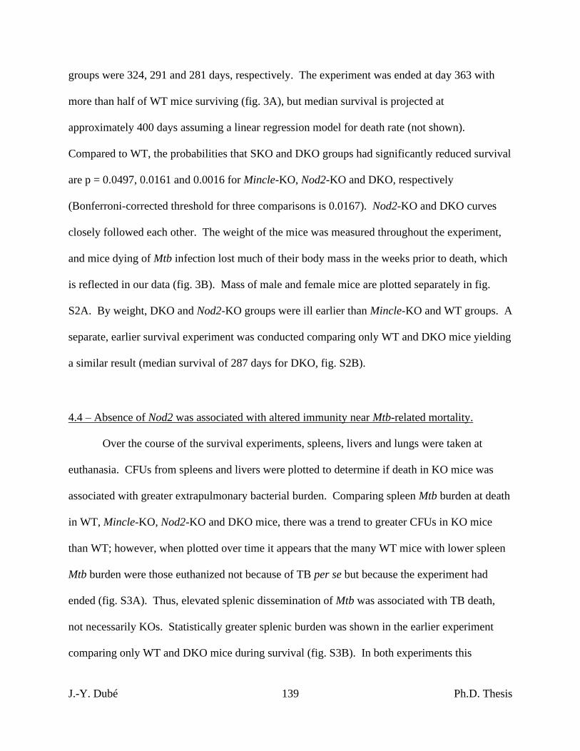
J.-Y. Dubé 139 Ph.D. Thesis
groups were 324, 291 and 281 days, respectively. The experiment was ended at day 363 with
more than half of WT mice surviving (fig. 3A), but median survival is projected at
approximately 400 days assuming a linear regression model for death rate (not shown).
Compared to WT, the probabilities that SKO and DKO groups had significantly reduced survival
are p = 0.0497, 0.0161 and 0.0016 for Mincle-KO, Nod2-KO and DKO, respectively
(Bonferroni-corrected threshold for three comparisons is 0.0167). Nod2-KO and DKO curves
closely followed each other. The weight of the mice was measured throughout the experiment,
and mice dying of Mtb infection lost much of their body mass in the weeks prior to death, which
is reflected in our data (fig. 3B). Mass of male and female mice are plotted separately in fig.
S2A. By weight, DKO and Nod2-KO groups were ill earlier than Mincle-KO and WT groups. A
separate, earlier survival experiment was conducted comparing only WT and DKO mice yielding
a similar result (median survival of 287 days for DKO, fig. S2B).
4.4 – Absence of Nod2 was associated with altered immunity near Mtb-related mortality.
Over the course of the survival experiments, spleens, livers and lungs were taken at
euthanasia. CFUs from spleens and livers were plotted to determine if death in KO mice was
associated with greater extrapulmonary bacterial burden. Comparing spleen Mtb burden at death
in WT, Mincle-KO, Nod2-KO and DKO mice, there was a trend to greater CFUs in KO mice
than WT; however, when plotted over time it appears that the many WT mice with lower spleen
Mtb burden were those euthanized not because of TB per se but because the experiment had
ended (fig. S3A). Thus, elevated splenic dissemination of Mtb was associated with TB death,
not necessarily KOs. Statistically greater splenic burden was shown in the earlier experiment
comparing only WT and DKO mice during survival (fig. S3B). In both experiments this
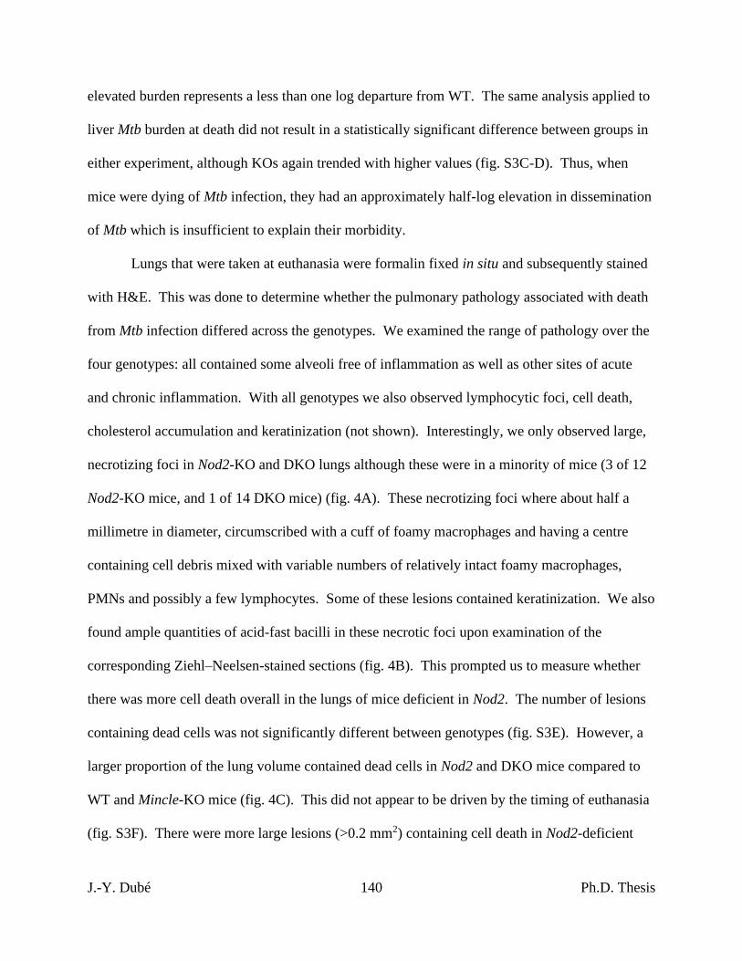
J.-Y. Dubé 140 Ph.D. Thesis
elevated burden represents a less than one log departure from WT. The same analysis applied to
liver Mtb burden at death did not result in a statistically significant difference between groups in
either experiment, although KOs again trended with higher values (fig. S3C-D). Thus, when
mice were dying of Mtb infection, they had an approximately half-log elevation in dissemination
of Mtb which is insufficient to explain their morbidity.
Lungs that were taken at euthanasia were formalin fixed in situ and subsequently stained
with H&E. This was done to determine whether the pulmonary pathology associated with death
from Mtb infection differed across the genotypes. We examined the range of pathology over the
four genotypes: all contained some alveoli free of inflammation as well as other sites of acute
and chronic inflammation. With all genotypes we also observed lymphocytic foci, cell death,
cholesterol accumulation and keratinization (not shown). Interestingly, we only observed large,
necrotizing foci in Nod2-KO and DKO lungs although these were in a minority of mice (3 of 12
Nod2-KO mice, and 1 of 14 DKO mice) (fig. 4A). These necrotizing foci where about half a
millimetre in diameter, circumscribed with a cuff of foamy macrophages and having a centre
containing cell debris mixed with variable numbers of relatively intact foamy macrophages,
PMNs and possibly a few lymphocytes. Some of these lesions contained keratinization. We also
found ample quantities of acid-fast bacilli in these necrotic foci upon examination of the
corresponding Ziehl–Neelsen-stained sections (fig. 4B). This prompted us to measure whether
there was more cell death overall in the lungs of mice deficient in Nod2. The number of lesions
containing dead cells was not significantly different between genotypes (fig. S3E). However, a
larger proportion of the lung volume contained dead cells in Nod2 and DKO mice compared to
WT and Mincle-KO mice (fig. 4C). This did not appear to be driven by the timing of euthanasia
(fig. S3F). There were more large lesions (>0.2 mm2) containing cell death in Nod2-deficient
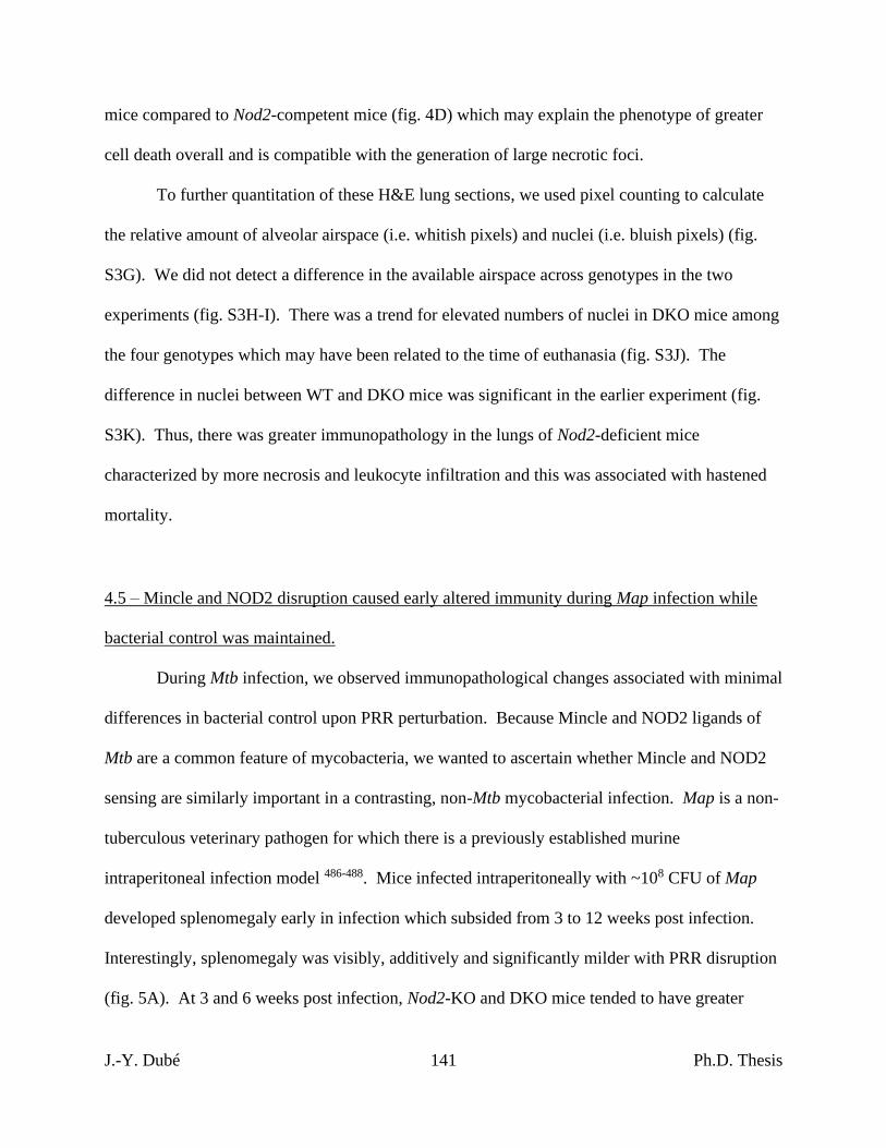
J.-Y. Dubé 141 Ph.D. Thesis
mice compared to Nod2-competent mice (fig. 4D) which may explain the phenotype of greater
cell death overall and is compatible with the generation of large necrotic foci.
To further quantitation of these H&E lung sections, we used pixel counting to calculate
the relative amount of alveolar airspace (i.e. whitish pixels) and nuclei (i.e. bluish pixels) (fig.
S3G). We did not detect a difference in the available airspace across genotypes in the two
experiments (fig. S3H-I). There was a trend for elevated numbers of nuclei in DKO mice among
the four genotypes which may have been related to the time of euthanasia (fig. S3J). The
difference in nuclei between WT and DKO mice was significant in the earlier experiment (fig.
S3K). Thus, there was greater immunopathology in the lungs of Nod2-deficient mice
characterized by more necrosis and leukocyte infiltration and this was associated with hastened
mortality.
4.5 – Mincle and NOD2 disruption caused early altered immunity during Map infection while
bacterial control was maintained.
During Mtb infection, we observed immunopathological changes associated with minimal
differences in bacterial control upon PRR perturbation. Because Mincle and NOD2 ligands of
Mtb are a common feature of mycobacteria, we wanted to ascertain whether Mincle and NOD2
sensing are similarly important in a contrasting, non-Mtb mycobacterial infection. Map is a non-
tuberculous veterinary pathogen for which there is a previously established murine
intraperitoneal infection model 486-488. Mice infected intraperitoneally with ~108 CFU of Map
developed splenomegaly early in infection which subsided from 3 to 12 weeks post infection.
Interestingly, splenomegaly was visibly, additively and significantly milder with PRR disruption
(fig. 5A). At 3 and 6 weeks post infection, Nod2-KO and DKO mice tended to have greater
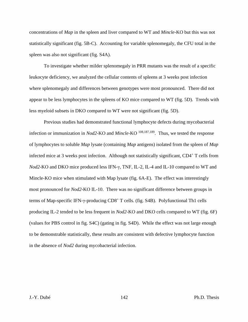
J.-Y. Dubé 142 Ph.D. Thesis
concentrations of Map in the spleen and liver compared to WT and Mincle-KO but this was not
statistically significant (fig. 5B-C). Accounting for variable splenomegaly, the CFU total in the
spleen was also not significant (fig. S4A).
To investigate whether milder splenomegaly in PRR mutants was the result of a specific
leukocyte deficiency, we analyzed the cellular contents of spleens at 3 weeks post infection
where splenomegaly and differences between genotypes were most pronounced. There did not
appear to be less lymphocytes in the spleens of KO mice compared to WT (fig. 5D). Trends with
less myeloid subsets in DKO compared to WT were not significant (fig. 5D).
Previous studies had demonstrated functional lymphocyte defects during mycobacterial
infection or immunization in Nod2-KO and Mincle-KO 108,187,189. Thus, we tested the response
of lymphocytes to soluble Map lysate (containing Map antigens) isolated from the spleen of Map
infected mice at 3 weeks post infection. Although not statistically significant, CD4+ T cells from
Nod2-KO and DKO mice produced less IFN-γ, TNF, IL-2, IL-4 and IL-10 compared to WT and
Mincle-KO mice when stimulated with Map lysate (fig. 6A-E). The effect was interestingly
most pronounced for Nod2-KO IL-10. There was no significant difference between groups in
terms of Map-specific IFN-γ-producing CD8+ T cells. (fig. S4B). Polyfunctional Th1 cells
producing IL-2 tended to be less frequent in Nod2-KO and DKO cells compared to WT (fig. 6F)
(values for PBS control in fig. S4C) (gating in fig. S4D). While the effect was not large enough
to be demonstrable statistically, these results are consistent with defective lymphocyte function
in the absence of Nod2 during mycobacterial infection.
J.-Y. Dubé 143 Ph.D. Thesis
5 – Discussion
The PRRs Mincle and NOD2 uniquely interact with mycobacteria and have been shown
to synergistically drive immunological functions 108,157,187. In this study, we used measures of the
immune response, bacterial control and survival to examine the course of Mtb infection, and
infection with the disparate mycobacterium Map, in mice defective for Mincle and/or NOD2.
Despite their established immunological roles, these PRRs contributed little to mycobacterial
control. Both PRRs were important to prevent premature death in Mtb infected mice. Because
Nod2-KO and DKO mice had very similar reduced survival times, while Mincle-KO mice
survived longer, it appears that absence of Nod2 was the ‘dominant’ driver of hastened mortality
in DKO mice, and the added Mincle defect was unimportant. The Nod2-deficient death
phenotype was associated with increased cell death in the lungs with distinct necrotic foci.
In humans, one study found a significant association between NOD2 loss-of-function
mutations and an increased risk of necrotizing enterocolitis or focal intestinal perforation in very
low-birth-weight infants 491, suggesting NOD2 deficiency is related to necrotic pathology in
humans. Murine Nod2 and MDP treatment promoted the formation of necrotic zones in a murine
model of atherosclerosis however 492, so the relationship between NOD2 and necrosis seems
complex. The necrotic foci seen in Nod2 deficient mice were reminiscent of similar pathology in
the Kramnik mouse 85,493, the susceptibility of which has been attributed to the Sst1 locus and
excess type I IFN production 270. However, NOD2 signaling contributed to type I IFN
production 97, so how the absence of Nod2 lead to the formation of necrotic foci is not so easily
explainable. Perhaps the amount of type I IFN in space and time is a critical element for these
outcomes, or that excess type I IFN precipitates a common secondary effect that leads to
runaway necrosis. Heterozygous deficiency in IL-1R antagonist or anti-IL-1R antagonist
J.-Y. Dubé 144 Ph.D. Thesis
treatment ameliorated morbidity and lung pathology in mice with the susceptible Sst1 allele 270.
IL-1 signaling contributed to PGE2 production, and PGE2 was protective against Mtb in IL-1
signaling-deficient mice 494. Thus, roles for NOD2 in IL-1 and PGE2 pathways should be further
explored in late Mtb infection. When infected with Mtb, human PBMCs with functional NOD2
produced more IL-1 than human PBMCs containing the NOD2 3020insC mutation 495. MDP
also promoted PGE2 production by monocytes and macrophages 496,497. The balance of immune
mediators like type I IFN, IL-1 and PGE2 may play a key role in pulmonary cell death during
Mtb infection.
Extensive necrosis was not seen in Nod2-deficient mouse lungs at early timepoints (3 and
6 weeks post infection) (fig. 1E and not shown). The phenotype of large necrotic foci was late,
appearing in mice euthanized at 223, 275, 307 (Nod2-KO) and 254 (DKO) days post infection,
and occurred in a minority of the animals. It will be challenging to directly study the
mechanisms of this phenomenon with this model. A 10X higher-dose aerosol may increase the
rate of death in Nod2-KO mice 189, but could overlook the phenotype that occurred with our
more epidemiologically relevant dose of Mtb if time is needed for the pathology to develop. At 6
weeks post infection, DKO mouse lungs were more congested than WT controls but without
lymphocytic clusters (fig. 1E), consistent with what has been demonstrated before with Nod2-
KO mice 189. Our trend of decreased lymphocytes in Nod2-KO and DKO mouse lungs at three
weeks post infection with Mtb is similar to results with Nod2-KO mouse lungs at 4 weeks post
infection with BCG Russia 189. We did not observe a difference in splenic lymphocyte numbers
during high-dose Map infection across genotypes, but the trend of reduced Map-specific
cytokine production and polyfunctionality in Nod2 deficient mice still suggests a defective
lymphocyte response. The defect in innate immunity caused by Nod2 mutation results in altered
J.-Y. Dubé 145 Ph.D. Thesis
instruction of the adaptive immune response 187,189, but while the innate defect is undoubtedly
present throughout the course of the infection (and therefore well deserves attention), we are
only certain the adaptive defect is present early. NOD2 deficiency may also impair macrophage
production of nitric oxide as well as secreted immune mediators 188.
Throughout this study, what had not shone through is the importance of Mincle. Mincle-
KO mice demonstrated a null or intermediate phenotype between WT and Nod2-deficient
animals. Such intermediate trends were occasionally observed for measures of immunity, but
bacterial loads were similar to WT except at Mtb-related death. Most noteworthy was the slight
reduction in survival with Mtb for Mincle-KO mice compared to WT, which to our knowledge
has not been published before. Previous Mincle-KO studies of Mtb infection did not present
survival data but had only reported bacteriologic and immunologic changes which were minor,
not long-lasting or inconsistent between studies 460,461. Administration of TDM or synthetic
TDM analogues has been shown to produce distinct, robust biologic effects 157,187 but this is
dependent on an arbitrarily selected dose which may not be representative of the amount of
immune-available material from the millions of bacilli in a mouse. Interestingly, as Nod2-KO
and DKO mice phenocopied each other in terms of survival with Mtb, NOD2 appears to be
dominant over Mincle in this system. This result suggests Mincle is dispensable in the absence
of NOD2 (i.e. Mincle-NOD2 synergy) or NOD2 deficiency simply leads to death before Mincle
deficiency has an effect.
NOD2 polymorphisms are strongly associated with Crohn’s disease yet a long-
hypothesized role for Map in Crohn’s disease remains unproven. Unlike Mtb, Map lacks a
functioning Esx-1 secretion system that enables the bacteria to disrupt the phagosomal
membrane and access the host cytosolic compartment. However, Map is still pathogenic in cattle
J.-Y. Dubé 146 Ph.D. Thesis
and a long list of other hosts. Map burden remained within one log from 3 to 12 weeks in mice,
even as the appearance of the internal organs returned to normal. This persistence might permit
the development of an altered pathologic response in a genetically susceptible host, however, the
intraperitoneal infection route that we used is unable to produce detectable pathology in the
gastrointestinal tract. Future work can test whether Map can precipitate inflammatory pathology
in the intestines of Nod2-KO mouse using an experimental model that is more relevant to
Crohn’s disease. The trend we observed of reduced IL-10-producing Map-specific CD4+ T cells
in Nod2-KO mice should also be sought in such a model 498. Associations of NOD2
polymorphisms and TB have not been as strong, but future studies exploring how altered
immunity in NOD2-deficient hosts leads to distinct disease states will inform the definitions of
TB endophenotypes which can be tested for associations with NOD2 polymorphisms at large.
Our work has asserted that NOD2 is indeed important in Mtb infection through reduced survival
associated with distinct pathology. Defining the mechanisms of these NOD2 phenotypes will
inform our understanding of human mycobacterial diseases.
6 – Acknowledgements
We are grateful for the Containment Level 3 platform of the Research Institute of the
McGill University Health Centre (RI-MUHC). We thank the Immunophenotyping and
Histology platforms of the RI-MUHC, and the Histology Core of McGill University for tissue
processing. We greatly appreciate the generosity of Christine Wells (University of Melbourne,
Australia) for providing Mincle-KO mice. We thank Richard Fraser of the McGill department of
Pathology for guiding us with histopathological analyses. For advice and assistance in
J.-Y. Dubé 147 Ph.D. Thesis
experiments, J.Y.D. would like to thank Andréanne Lupien, Helena Strand Clemmensen and
Sarah Danchuck.
J.Y.D. was personally supported by the Canadian Institutes of Health Research (CIHR)
Canada Graduate Scholarship – Master’s Program, Fonds de Recherche du Québec – Santé
(FRQ-S) Doctoral Training Award, RI-MUHC studentships (Master’s and Ph.D.) and
scholarships from the McGill Department of Microbiology and Immunology. Research activities
were funded by a CIHR foundation grant held by M.A.B. (FDN-148362).
Additional Information
The authors have no competing interests.
Author Contributions
Conceptualization, J.Y.D. and M.A.B.; Methodology, J.Y.D., F.M. and S.C.D.; Investigation,
J.Y.D., F.M. and S.C.D.; Resources, M.A.B.; Writing – Original Draft, J.Y.D. and M.A.B.;
Writing – Review & Editing, J.Y.D., F.M., S.C.D. and M.A.B.; Visualization, J.Y.D.;
Supervision, M.A.B.; Funding acquisition, J.Y.D. and M.A.B.
J.-Y. Dubé 148 Ph.D. Thesis
7 – Figures
Chapter III, Figure 1. Effect of Mincle and Nod2 disruptions on the macrophage and
pulmonary immune response to Mtb.
A, TNFα in supernatant of BMDM culture after incubation with heat-killed Mtb (HK H37Rv) or
medium control for 20 hours. Mean +/- standard deviation of four individually treated culture
wells. **p < 0.01 with one-way ANOVA on HK H37Rv treated BMDMs. B, TNFα in
supernatant of BMDM culture after infection with live Mtb. Mean +/- standard deviation of
eight (t = 4 hours) or four (all other timepoints) individually infected culture wells. C, total Mtb
burden in both lungs of WT and DKO mice. N = 2 mice per group on day 1, while N = 4 to 6
mice per group for days 26 and 42 post infection. *p < 0.05 with Dunnett's T3 multiple
comparisons test over days 26 and 42; ns, not significant, p > 0.05. D, rank of pathology on
H&E lung sections at 42 days post infection. Ranking was obtained from the median rank of
seven blinded individuals. *p < 0.05 with one-tailed Mann Whitney test. E, representative H&E
sections from WT (rank 5) and DKO (rank 10) mice.
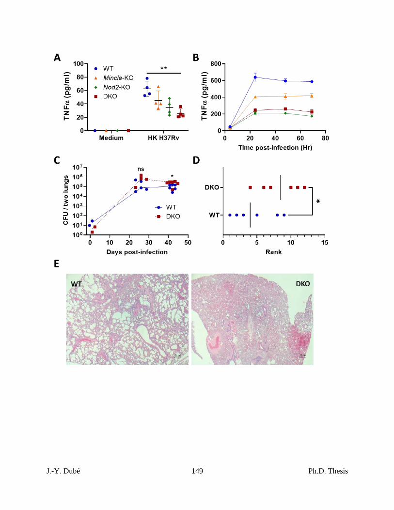
J.-Y. Dubé 149 Ph.D. Thesis
J.-Y. Dubé 150 Ph.D. Thesis
Chapter III, Figure 2. Mtb control and lymphocyte recruitment in mice lacking Mincle and
Nod2 singly and in combination.
A, Mtb CFU burden per gram of left lung (left), spleen (middle) and liver (right) for the indicated
timepoint. Median day 1 pulmonary Mtb load was 15 CFU. B, Absolute numbers of pulmonary
lymphocytes at the indicated timepoints post Mtb infection. Using intravenous anti-CD45-FITC
staining just prior to euthanasia, parenchymal (CD45-FITC-) or vascular (CD45-FITC+) location
was determined. Shown are medians for N = 5 to 7 mice per group per timepoint; mice are
represented by individual points on the graphs. No statistically significant differences were
found between genotypes for the given variables and timepoints.
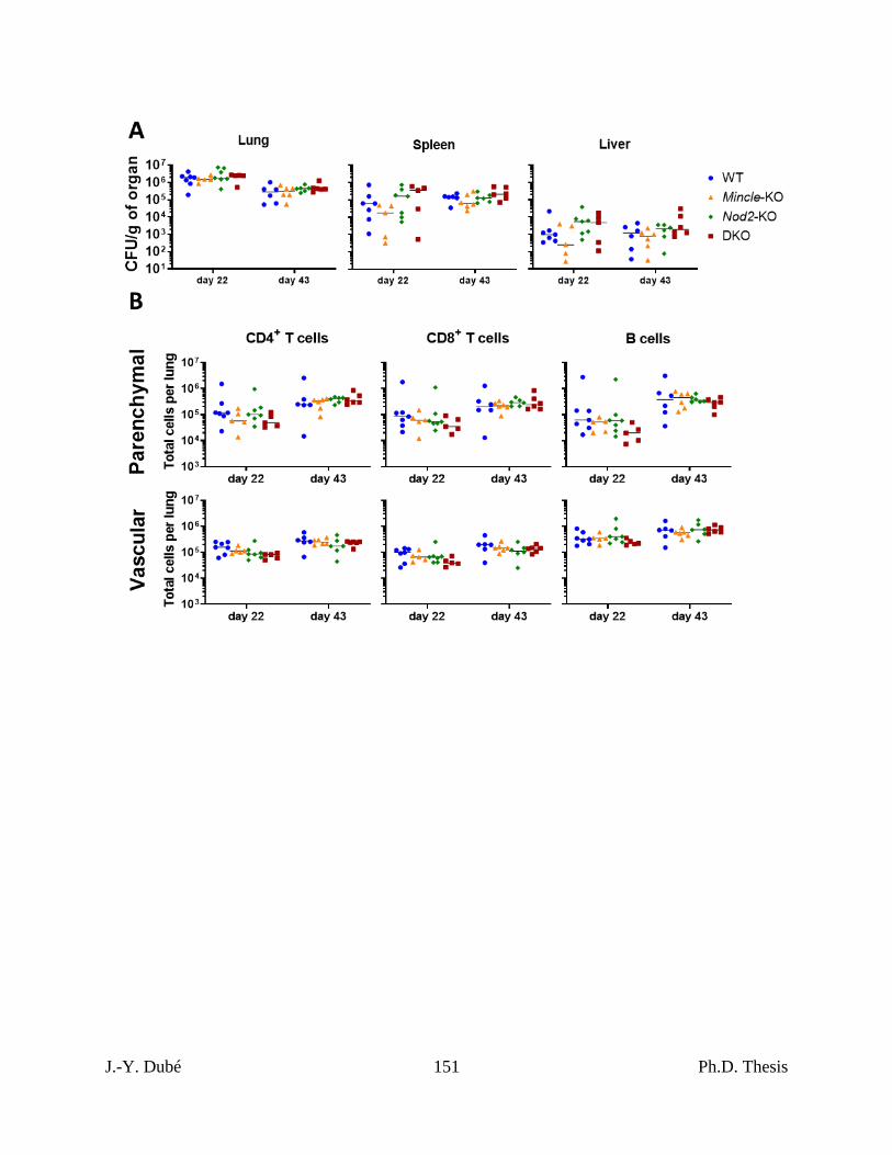
J.-Y. Dubé 151 Ph.D. Thesis
J.-Y. Dubé 152 Ph.D. Thesis
Chapter III, Figure 3. Survival of Mincle and Nod2 deficient mice infected with Mtb.
A, percent survival of Mtb-infected mice of the indicated genotype over time. Log-rank (Mantel-
Cox) test p values generated from comparing to the WT group are reported next to legend. The
Bonferrroni-corrected threshold for three such comparisons is 0.0167. B, percent weight from 6
days post infection over time for the indicated genotype (male and female mice combined);
median (lines) +/- interquartile range (shading) are shown. Euthanized mice received a weight of
0. Weight in grams is plotted in fig. S2A. Median day 1 pulmonary Mtb load was 31 CFU. N =
12, 8, 12 and 14 for WT, Mincle-KO, Nod2-KO and DKO, respectively.
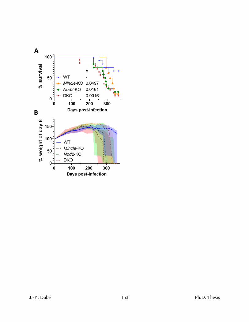
J.-Y. Dubé 153 Ph.D. Thesis
J.-Y. Dubé 154 Ph.D. Thesis
Chapter III, Figure 4. Pulmonary pathology at death in Mincle and Nod2 deficient mice
infected with Mtb.
A, images of largest lesions containing cell death, each from a different mouse: one WT (largest
lesion in all WT mice), two Mincle-KO, three Nod2-KO (all representative necrotic foci) and
three DKO (top, necrotic focus, middle, diffuse necrosis, bottom, airway filled with
inflammatory infiltrate containing dead cell debris). Scale bars, 100 μm. B, Ziehl–Neelsen
stained lung section containing large necrotic focus, from Nod2-KO mouse shown in A (center).
C, percentage of lung space containing dead cells with medians. Kruskal-Wallis test was used
determine statistical significance; *p < 0.05. D, sizes of individual lesions containing dead
cells, separated by mouse according to genotype (colour/shape coded) and in order of survival
time (left to right, shortest to longest). Dotted line, to delineate lesions larger than 0.2 mm2. N =
11, 7, 12 and 14 for WT, Mincle-KO, Nod2-KO and DKO, respectively.
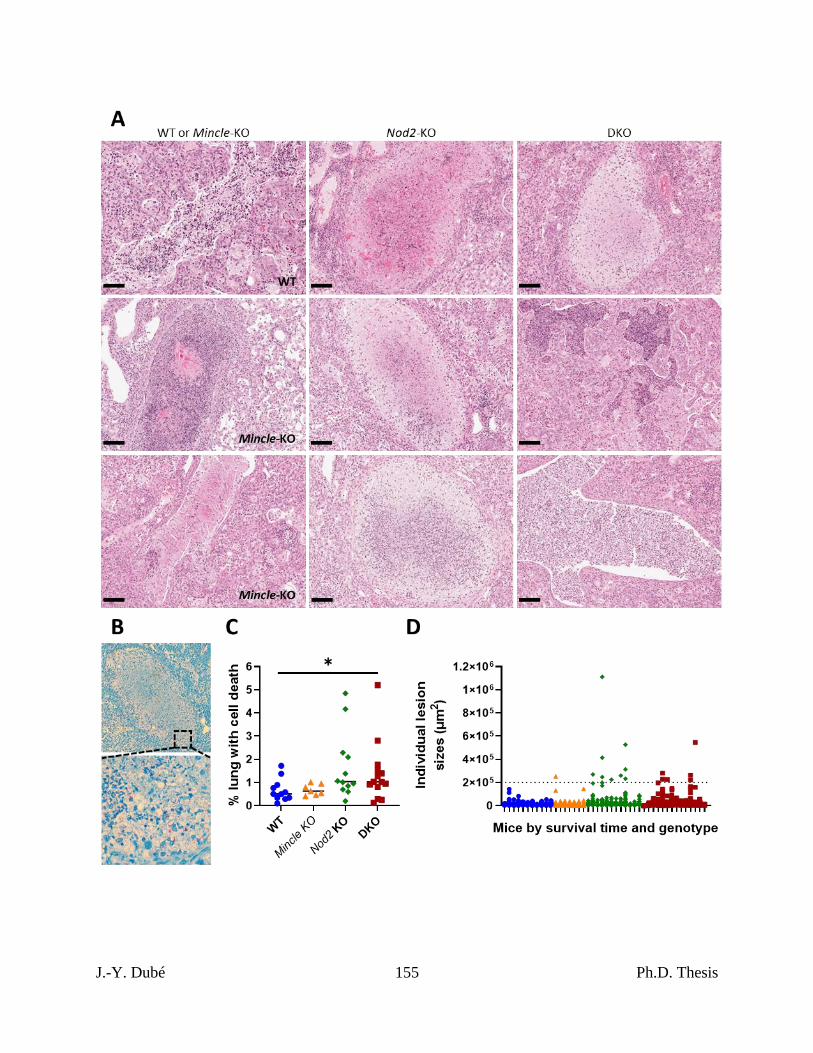
J.-Y. Dubé 155 Ph.D. Thesis
J.-Y. Dubé 156 Ph.D. Thesis
Chapter III, Figure 5. Control of Map infection in Mincle and Nod2 deficient mice.
A, mass of spleens upon euthanasia at the indicated timepoint. Shown are medians of N = 5 to
13 mice per group per timepoint (data for day 21 were combined from two independent
experiments). Welch’s ANOVA was performed at each timepoint; *p < 0.05. B-C, Map CFU
burden per gram of spleen (B) and liver (C) at the indicated timepoint. Shown are medians of N
= 5 to 7 mice per group. Kruskal-Wallis test was performed at each timepoint for each organ; ns,
not significant (p > 0.05). D, flow cytometry enumeration of leukocyte subsets in the spleens of
Map infected mice at 3 weeks post infection. Populations were identified with t-SNE similarly
to fig. S1. Shown are medians of N = 6 to 8 mice per group; mice are represented by individual
points on the graphs. No statistically significant differences were found.
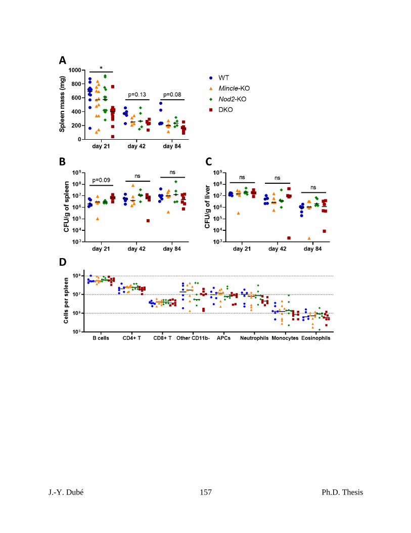
J.-Y. Dubé 157 Ph.D. Thesis
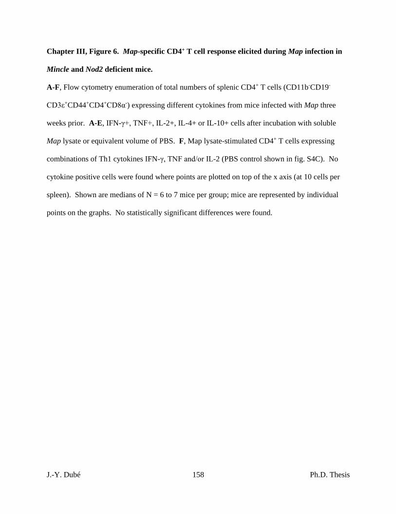
J.-Y. Dubé 158 Ph.D. Thesis
Chapter III, Figure 6. Map-specific CD4+ T cell response elicited during Map infection in
Mincle and Nod2 deficient mice.
A-F, Flow cytometry enumeration of total numbers of splenic CD4+ T cells (CD11b-CD19-
CD3ε+CD44+CD4+CD8α-) expressing different cytokines from mice infected with Map three
weeks prior. A-E, IFN-γ+, TNF+, IL-2+, IL-4+ or IL-10+ cells after incubation with soluble
Map lysate or equivalent volume of PBS. F, Map lysate-stimulated CD4+ T cells expressing
combinations of Th1 cytokines IFN-γ, TNF and/or IL-2 (PBS control shown in fig. S4C). No
cytokine positive cells were found where points are plotted on top of the x axis (at 10 cells per
spleen). Shown are medians of N = 6 to 7 mice per group; mice are represented by individual
points on the graphs. No statistically significant differences were found.
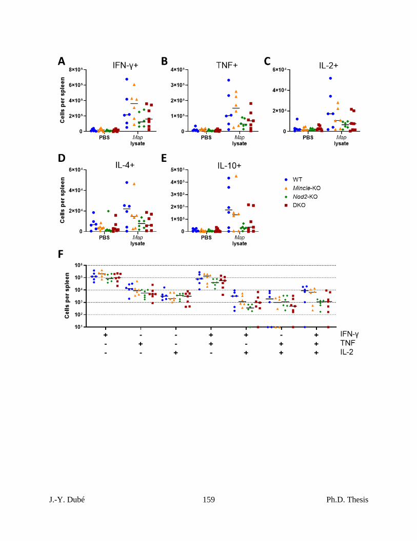
J.-Y. Dubé 159 Ph.D. Thesis
J.-Y. Dubé 160 Ph.D. Thesis
8 – Supplemental Figures
Chapter III, Figure S1. Flow cytometry of lung cells of Mtb-infected Mincle and Nod2
deficient mice.
A, gating strategy used to enumerate lung cell types. After gating on single, live cells,
downsampling and concatenation were performed to put all samples through t-SNE. Populations
in the two t-SNE generated dimensions were identified and gated by checking expression of
characteristic markers. Per population, gating of CD45-FITC positive and negative cells was
performed separately to account for autofluorescence of difference leukocyte types. Total
numbers of events in each final gate were exported and proportioned based on the number of
counting beads passed with the cells to determine total cell type numbers per lung. B, total
alveolar macrophage numbers per lung (none were considered vascular). C, total numbers of
parenchymal and vascular cells of the indicated phenotype. Shown are medians for N = 5 to 7
mice per group per timepoint; mice are represented by individual points on the graphs. No
statistically significant differences were found between genotypes for the given variables and
timepoints.
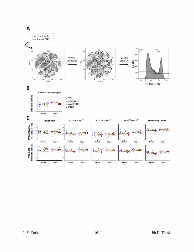
J.-Y. Dubé 161 Ph.D. Thesis
J.-Y. Dubé 162 Ph.D. Thesis
Chapter III, Figure S2. Survival and weight of Mincle and Nod2 deficient mice infected
with Mtb.
A, weight in grams of mice subjected to survival challenge shown in fig. 3. B, independent
experiment comparing just WT and DKO mice: median day 1 pulmonary Mtb load was 29 CFU.
Shown are percent survival (top), weight in grams (middle) and percent weight from two days
post infection (bottom) over time. **p < 0.01 by log-rank (Mantel-Cox) test. All mice were
female. Median (lines) +/- interquartile range (shading) are shown for weight and percent
weight. N = 14 and 16 for WT and DKO, respectively.
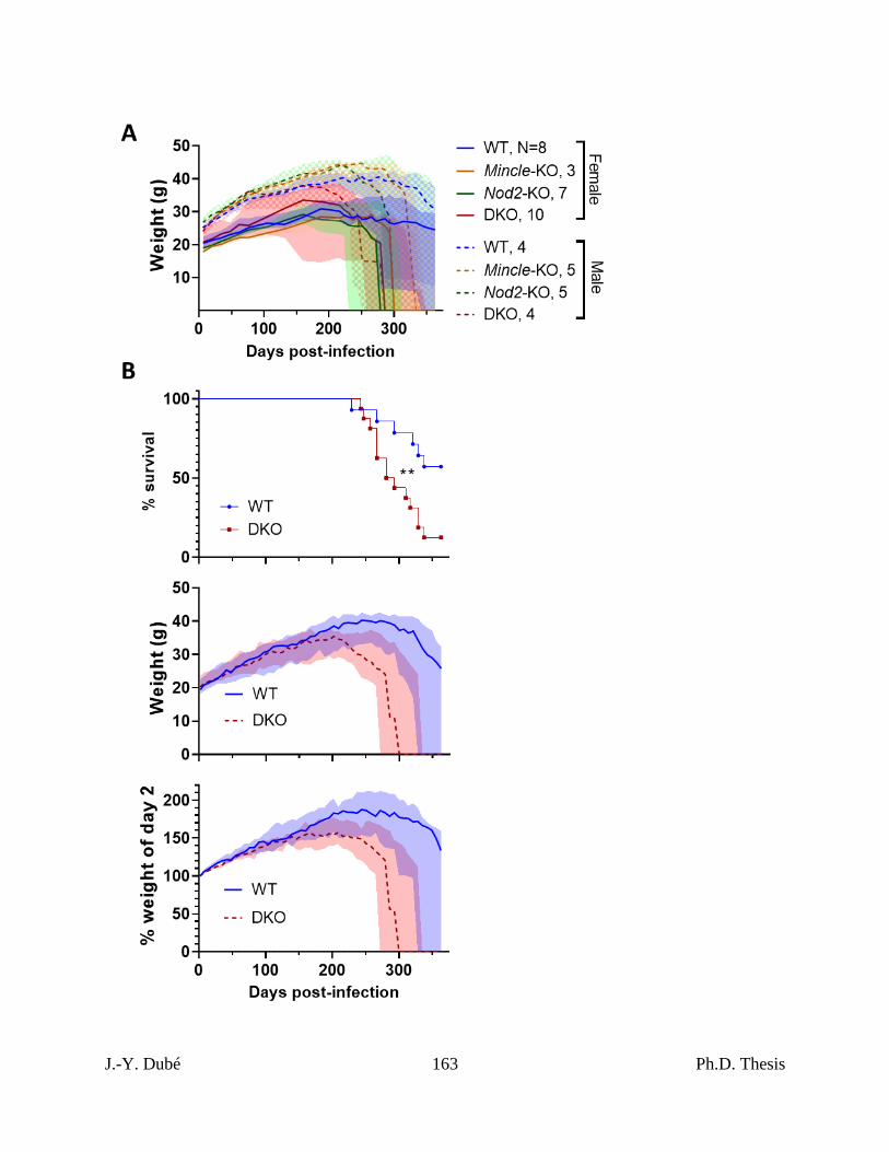
J.-Y. Dubé 163 Ph.D. Thesis
J.-Y. Dubé 164 Ph.D. Thesis
Chapter III, Figure S3. Quantitation of extrapulmonary bacterial burdens and pulmonary
pathology associated with death in Mtb-infected Mincle and Nod2 deficient mice.
A, C, spleen and liver Mtb CFUs per gram of organ harvested upon endpoint euthanasia from
survival challenge in fig. 3. B, D, spleen and liver Mtb CFUs per gram of organ harvested upon
endpoint euthanasia from survival challenge in fig. S2B. Linear regressions of bacterial burden
over time of euthanasia were calculated per group to highlight slope lines’ deviation from zero
(left graph of panels). Assuming the date of euthanasia is not confounding, groups were
compared by Welch’s ANOVA (A and C) or Welch’s t-test (B and D); ns, p > 0.05; *p < 0.05
(right graph of panels). Shown are averages of N = 7 to 14 mice per group; mice are represented
by individual points on the graphs. E, Number of lesions containing dead cells per area of lung
plotted with medians. Kruskal-Wallis test was used determine statistical significance; ns, p >
0.05. F, percentage of lung space containing dead cells plotted over time per group with linear
regressions. No slopes were significantly non-zero. G, Example of Python code processing used
to quantify tissue (red), nuclei (blue) and alveolar airspace (white); details in Methods. H, J,
alveolar airspace and nuclei in lung sections harvested upon endpoint euthanasia from survival
challenge in fig. 3. I, K, alveolar airspace and nuclei in lung sections harvested upon endpoint
euthanasia from survival challenge in fig. S2B. Linear regressions of indicated variable over
time of euthanasia were calculated per group to highlight slope lines’ deviation from zero (left
graph of panels). Assuming the date of euthanasia is not confounding, groups were compared by
Welch’s ANOVA (A and C) or Welch’s t-test (B and D); ns, p > 0.05; *p < 0.05 (right graph of
panels). Shown are averages of N = 7 to 14 mice per group; mice are represented by individual
points on the graphs.
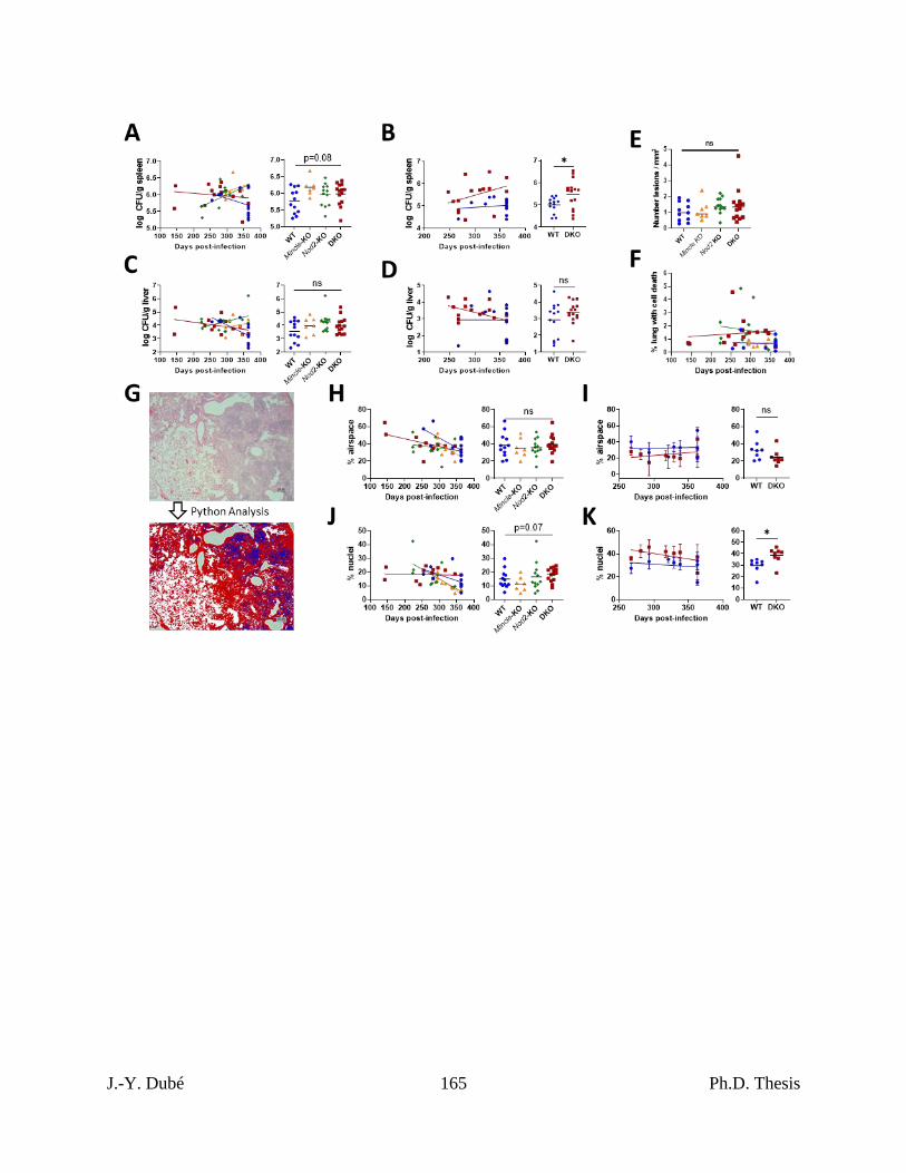
J.-Y. Dubé 165 Ph.D. Thesis
J.-Y. Dubé 166 Ph.D. Thesis
Chapter III, Figure S4. Bacterial control and immunity during Map infection in Mincle
and Nod2 deficient mice.
A, Total Map burden per spleen of infected mice. Complementary data to that in fig, 5B.
Kruskal-Wallis test was performed at each timepoint; ns, not significant (p > 0.05). B, Flow
cytometry enumeration of total numbers of splenic CD8+ T cells (CD11b-CD19-CD3ε+ CD4-
CD8α+) expressing IFN-γ after incubation with soluble Map lysate or equivalent volume of PBS,
from mice infected with Map three weeks prior. C, PBS controls for data in fig. 6F. No
cytokine positive cells were found where points are plotted on top of the x axis (at 10 cells per
spleen). Shown are medians of N = 5 to 7 mice per group; mice are represented by individual
points on the graphs. D, gating strategy to identify cytokine positive CD4+ T cells (CD11b-CD19-
CD3ε+CD44+CD4+CD8α-), generated with representative samples. Map lysate treatment is
shown with red histograms and PBS control is shown with blue histograms.
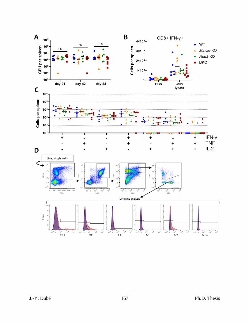
J.-Y. Dubé 167 Ph.D. Thesis
J.-Y. Dubé 168 Ph.D. Thesis
CHAPTER IV – DISCUSSION
We studied the importance of the NOD2 and Mincle pathways to the mycobacteria-host
interaction using models of immunity and infection. In my first study, NOD2 and Mincle were
essential for the immune response elicited by the mycobacteria in CFA, and ligands of these
PRRs, when combined, were sufficient to partially recreate the adjuvant effect by direct
measures of T-cell activity as well as EAE. With this insight into the importance of these PRR
pathways and their cooperation in mycobacterial immunity, we sought to examine their roles
more closely during live mycobacterial infection in the second study. There, we observed that
deficiency in NOD2 and Mincle, even together, had but a modest effect on bacterial control in
the presence of altered immunity. Nevertheless, NOD2 deficiency resulted in premature death
from Mtb associated with distinct pulmonary necrosis.
As reviewed in Chapter I, many PRR KOs have produced significant immunophenotypes
with little or no change in survival and bacterial burden 484. This conclusion is consistent with
the results of our infections of NOD2- and Mincle-deficient animals. As studies on PRRs
continue, the immunological phenotypes specific to different PRR pathways during
mycobacterial infection should be defined and compared to assist our understanding of the
mycobacterial immune response and correlates of protection. As exists for M. leprae,
endophenotypes based on the inflammatory state of the host should be defined for Mtb, tested for
associations with PRR polymorphisms and mechanistically explored in animal models.
That NOD2 SNPs have currently been strongly associated with M. leprae, of all
mycobacteria, is a curious development: namH is a non-functional pseudogene and there is no N-
J.-Y. Dubé 169 Ph.D. Thesis
glycolylation of PGN in M. leprae 106,110. Although it has not yet been tested, M. leprae would
be expected to signal less strongly through NOD2 as has been shown with namH-mutant M.
smegmatis and Mtb 108,109. All known M. leprae-associated NOD2 SNPs are non-coding or
synonymous 284,388-391, so their function is unclear but possibly related to the expression level of
NOD2, in which case the system may be more vulnerable to a weaker (N-acetyl) MDP ligand. In
our adjuvant studies, we did not see a role for NOD2 outside of MDP sensing: the responses to
IFA and GlcC14C18 were Nod2-independent, while MDP responses were fully Nod2-dependent.
Other groups have proposed NOD2 possesses functions beyond MDP sensing (e.g. cellular stress
499), which may contribute to a SNP-associated phenotype. We cannot determine whether MDP-
independent functions of NOD2 contributed to the phenotypes presented in our infection studies
in Chapter III. We sought to simulate NOD2 loss-of-function during Mtb infection using murine
Nod2-KO alleles, which we accomplished.
We demonstrated that NOD2 signalling promotes CMI in Chapter II. Favouring humoral
immunity (i.e. a change in Th1/Th2 balance) is associated with multibacillary leprosy, which is
more contagious 27. NOD2 polymorphisms were more strongly associated with multibacillary
than paucibacillary infection, and with type II (antibody-mediated) than type I (DTH) lepra
reactions 284,388. Therefore, weakened NOD2 signalling, and thus weakened CMI, seems to
favour M. leprae transmission. This contrasts with Mtb, which is more transmissible in those
with cavitary disease which requires CMI 72,83. This differential requirement of CMI for
transmission may explain the evolutionary decay of namH in M. leprae (and M. lepromatosis) in
contrast to Mtb.
The importance of NOD2 during NTM infection in humans has not been established. A
few case reports have been published on NOD2 mutations in patients with M. abscessus or MAC
J.-Y. Dubé 170 Ph.D. Thesis
infection 500, but controlled genetic studies associating NOD2 with these bacteria have not been
performed. In mice, absence of Nod2 during M. abscessus infection resulted in higher bacterial
burden during clearance (up to 10-fold more in KO), worse pulmonary histopathology and
reduced cytokine production compared to WT 501. These results are comparable with both Mtb
and Map data in Chapter III. Macrophage experiments have shown that NOD2 is important for
the recognition of a range of bacteria with NamH, from Map and M. smegmatis to Nocardia
asteroides and Rhodococcus equi, where NOD2 signalling was almost nil with M. smegmatis
namH knockout in the same study 108. Supporting the role for the NamH-NOD2 axis in immune
responses, Jules Freund himself tested N. asteroides in place of Mtb during the development of
CFA to successfully sensitize animals to foreign and self antigens 502.
NamH is a non-essential modifier of PGN. Its modification of the MDP motif may be
regulated, and this regulation may be connected to pathogenesis in Mtb. The requirement for
molecular oxygen for NamH-mediated N-glycolylation 106,107 is interesting , as Mtb experiences
different concentrations of O2 in different zones of pulmonary lesions 81. Indeed, Mtb cultured in
vitro under hypoxic conditions for weeks upregulates namH (Rv3818) expression upon returning
to an O2-replete growth environment 503. Mtb performs immune invasion but also elicits CMI in
the course of its lifecycle, and therefore understanding the regulation and timing of namH
expression my be informative about the role of NOD2 sensing during Mtb infection.
Like N-glycolylated PGN, TDM is present in many mycobacteria, while various trehalose
lipids (which are also Mincle agonists) are present in Corynebacterium, Nocardia and
Rhodococcus spp. 427,428. Therefore, Mincle is likely involved in the immune response to these
various actinobacteria. However, Mincle offered little resistance to infection in our studies of
disparate mycobacteria, despite its importance for adjuvancy. Thus, Mincle sensing of trehalose
J.-Y. Dubé 171 Ph.D. Thesis
lipids may not be a critical factor in host resistance during actinobacterial infections. This
difference in importance is perhaps reflected in the current paucity of literature on Mincle in
comparison to NOD2 at the level of human genetic studies, where a larger genetic effect is easier
to detect statistically. It is rather paradoxical that the receptor for this abundant glycolipid famed
for its distinct immunologic effects has failed to demonstrate a significant role in survival or
bacterial control. A recent study using single-cell RNA seq demonstrated that Mincle expression
is uneven across subsets of murine pulmonary interstitial and alveolar macrophages during Mtb
infection. Expression was greater in cells with a proinflammatory phenotype, expected to be
competent killers of Mtb, which suggests Mtb killing would occur without Mincle if Mincle is
unnecessary for their existence and effector functions. Indeed, in the same study analogous
proinflammatory AMs were already present in uninfected humans. NOD2 expression was not
reported in the study 504.
We have studied Mtb infection as a function of Mincle, but did not explore Mtb infection
as a function of TDM per se. Delipidation of Mtb with petroleum ether, which primarily extracts
TDM without significantly reducing viability, promotes macrophage migration, lysosome
targeting and killing of the bacteria, while simultaneously reducing cytokine responses 417,505,506.
A TDM-poor Mtb mutant (lacking mycolyltransferase Ag85a) also had impaired survival in
macrophages, associated with increased phagosomal maturation and killing, and was more
immunogenic 507. How much these phenotypes are dependent on Mincle is unknown but could
be investigated. Lastly, the extent to which MCL sensing of TDM can promote immunity in the
absence of Mincle is not clear but does not appear to be nil 160. In one study, MCL KO during
Mtb infection with 100 CFU resulted in 20% mortality in the first few weeks following infection
while WT all survived, but no further mortality occurred until the end of the experiment at 10
J.-Y. Dubé 172 Ph.D. Thesis
weeks 224. This may be a result of delayed adaptive immunity failing to catch up to and control a
high bacterial inoculum.
Mtb infection as a function of MDP has been previously explored: KO of namH did not
result in a significant difference in survival in immunocompetent mice, but Rag1-KO mice
survived 40% (2 weeks) longer with the namH mutant than with WT Mtb 109. Therefore, in the
absence of CMI (T cells), increased NOD2 sensing of Mtb was harmful, possibly through
immunopathology not examined in the study. The namH mutant was not attenuated for growth
in vitro nor in vivo and was not associated in altered immunity at three weeks post infection of
immunocompetent mice 109. Incorporated into CFA, we showed that the same namH mutant was
defective in producing antigen-specific IFN-γ-producing T cells at one week post immunization.
A namH mutant of M. smegmatis produced reduced amounts of Th1 cytokines 14 days after
infection upon restimulation with autologous bacteria 108. In our Mtb infections, trends of
lymphocyte defects in Nod2-KO mice at 3 weeks post infection disappeared by 6 weeks. A
previous report showed a reduction in pulmonary lymphocytes at 4 weeks post BCG infection in
Nod2-KO mice compared to WT 189. The enhancement of CMI through Mtb NamH may only be
short-lived since N-acetyl MDP (and thus NOD2 signalling) is still present.
In Chapter II, we alluded to the idea that a defined cocktail of synthetic MAMPs could
copy or improve upon mycobacterial reagents. Our work focused on CFA, while BCG is the
most common mycobacterial product used in medicine. We demonstrated the sufficiency of a
multi-MAMP adjuvant to generate antigen-specific CMI, and thus expect that a similar synthetic
adjuvant could recapitulate this activity of BCG (after careful antigen selection 96). Recent work
has suggested that part of the utility of BCG is in its capacity to generate trained innate immunity
297,304. NOD2 has been identified to promote this response with BCG 297, but no similar reports
J.-Y. Dubé 173 Ph.D. Thesis
exist for Mincle. However, fungal cell wall β-glucan is a well-defined inducer of trained innate
immunity. The CLR Dectin-1 senses β-glucan 508,509, and thus by analogy a CLR like Mincle
may be able to generate similar immune responses with TDM 510. Mincle and Dectin-1
signalling differ at least in that Dectin-1 has its own cytoplasmic ITAM while Mincle requires
the ITAM of FcRγ, but both CLRs subsequently signal to the SYK-CARD9 pathway 438.
BCG is a safe, well-studied, century-old vaccine. Any BCG replacement would have to
exceed the protective effect of BCG, not just demonstrate non-inferiority. A novel genetically
engineered BCG vaccine for TB is attempting to do that 94. While NOD2 and Mincle offered
modest resistance to Mtb infection in our experiments, the immunotherapeutic capacity through
targeting these receptors during Mtb infection was not tested. Unlike BCG, Mtb antagonizes
trained innate immunity using RD-1305, and thus experiments targeting NOD2 and Mincle
positively in Mtb infection should be done. Besides TB, a mycobacterial MAMP cocktail may
apply to the treatment of bladder cancer analogous to BCG. Assuming the utility of
mycobacterial therapies are largely determined by MAMPs, Mip can also be compared against a
MAMP adjuvant where applicable (e.g. leprosy adjunctive therapy). Future studies should
investigate the potential of NOD2 plus CLR/Mincle signalling together in the induction of
trained innate immunity, especially as we have shown the potential for synergy.
J.-Y. Dubé 174 Ph.D. Thesis
Final Conclusions
Through my investigations, we know that mycobacterial adjuvancy can be recapitulated
synthetically using combinations of MAMPs inspired by PRR pathways essential to the immune
mycobacterial response. Interestingly, while NOD2 and Mincle are important in the process of
mycobacteria-stimulated CMI, these PRRs serve subtle, unequal purposes during Mtb infection.
I have demonstrated that NOD2 is necessary to prevent premature TB death associated with
increased pulmonary necrosis, the precise mechanism of which will need further study. The
results of my work indicate that the NOD2 and Mincle pathways can be exploited together in
immunotherapeutic reagents, while uncovering the precise roles of these PRRs in disease will
require a greater exploration of the pathological phenomena associated with PRR dysfunction.
J.-Y. Dubé 175 Ph.D. Thesis
BIBLIOGRAPHY
1 DeLeo, F. R. & Hinnebusch, B. J. A plague upon the phagocytes. Nat. Med. 11, 927-928,
doi:10.1038/nm0905-927 (2005).
2 Henderson, D. A. The eradication of smallpox--an overview of the past, present, and
future. Vaccine 29 Suppl 4, D7-9, doi:10.1016/j.vaccine.2011.06.080 (2011).
3 Riedel, S. Edward Jenner and the history of smallpox and vaccination. Proc. (Bayl. Univ.
Med. Cent.) 18, 21-25, doi:10.1080/08998280.2005.11928028 (2005).
4 Spreeuwenberg, P., Kroneman, M. & Paget, J. Reassessing the Global Mortality Burden
of the 1918 Influenza Pandemic. Am. J. Epidemiol. 187, 2561-2567,
doi:10.1093/aje/kwy191 (2018).
5 Vincent, A. T. et al. The Mycobacterial Cell Envelope: A Relict From the Past or the
Result of Recent Evolution? Frontiers in Microbiology 9, doi:10.3389/fmicb.2018.02341
(2018).
6 Lehmann, K. B. & Neumann, R. Atlas und Grundriss der Bakteriologie und Lehrbuch der
speziellen bakteriologischen Diagnostik. (1896).
7 Koch, R. Die Aetiologie der Tuberculose. Berliner Klinische Wochenschrift 15, 287-296,
doi:10.1016/s0174-3031(82)80099-1 (1882).
8 Supply, P. et al. Genomic analysis of smooth tubercle bacilli provides insights into
ancestry and pathoadaptation of Mycobacterium tuberculosis. Nat. Genet. 45, 172-179,
doi:10.1038/ng.2517 (2013).
J.-Y. Dubé 176 Ph.D. Thesis
9 Dulberger, C. L., Rubin, E. J. & Boutte, C. C. The mycobacterial cell envelope — a
moving target. Nature Reviews Microbiology 18, 47-59, doi:10.1038/s41579-019-0273-7
(2020).
10 Willey, J. M., Sherwood, L., Woolverton, C. J. & Prescott, L. M. Prescott's
microbiology. 8th ed. edn, 37 (McGraw-Hill, 2011).
11 Kalscheuer, R. et al. The Mycobacterium tuberculosis capsule: a cell structure with key
implications in pathogenesis. Biochem. J. 476, 1995-2016, doi:10.1042/bcj20190324
(2019).
12 Lemassu, A. et al. Extracellular and surface-exposed polysaccharides of non-tuberculous
mycobacteria. Microbiology (Reading) 142 ( Pt 6), 1513-1520, doi:10.1099/13500872-
142-6-1513 (1996).
13 Fraenkel, A. R. Koch, Ueber Tuberculose. Dtsch. Med. Wochenschr. 15, 210-211 (1882).
14 Sakula, A. Robert Koch: centenary of the discovery of the tubercle bacillus, 1882. Thorax
37, 246-251, doi:10.1136/thx.37.4.246 (1982).
15 Garnier, T. et al. The complete genome sequence of Mycobacterium bovis. Proc. Natl.
Acad. Sci. U. S. A. 100, 7877-7882, doi:10.1073/pnas.1130426100 (2003).
16 Cá, B. et al. Experimental Evidence for Limited in vivo Virulence of Mycobacterium
africanum. Front Microbiol 10, 2102, doi:10.3389/fmicb.2019.02102 (2019).
17 de Jong, B. C. et al. Differences between tuberculosis cases infected with Mycobacterium
africanum, West African type 2, relative to Euro-American Mycobacterium tuberculosis:
an update. FEMS Immunol. Med. Microbiol. 58, 102-105, doi:10.1111/j.1574-
695X.2009.00628.x (2010).
J.-Y. Dubé 177 Ph.D. Thesis
18 Ngabonziza, J. C. S. et al. A sister lineage of the Mycobacterium tuberculosis complex
discovered in the African Great Lakes region. Nature Communications 11, 2917,
doi:10.1038/s41467-020-16626-6 (2020).
19 Coscolla, M. et al. Phylogenomics of Mycobacterium africanum reveals a new lineage
and a complex evolutionary history. Microb Genom 7, doi:10.1099/mgen.0.000477
(2021).
20 Bottai, D. et al. TbD1 deletion as a driver of the evolutionary success of modern
epidemic Mycobacterium tuberculosis lineages. Nature Communications 11, 684,
doi:10.1038/s41467-020-14508-5 (2020).
21 Cole, S. T. et al. Deciphering the biology of Mycobacterium tuberculosis from the
complete genome sequence. Nature 393, 537-544, doi:10.1038/31159 (1998).
22 Steenken, W., Oatway, W. H. & Petroff, S. A. BIOLOGICAL STUDIES OF THE
TUBERCLE BACILLUS : III. DISSOCIATION AND PATHOGENICITY OF THE R
AND S VARIANTS OF THE HUMAN TUBERCLE BACILLUS (H(37)). J. Exp. Med.
60, 515-540, doi:10.1084/jem.60.4.515 (1934).
23 Calmette, A. Preventive Vaccination Against Tuberculosis with BCG. Proc. R. Soc. Med.
24, 1481-1490 (1931).
24 van Ingen, J. et al. Characterization of Mycobacterium orygis as M. tuberculosis complex
subspecies. Emerg. Infect. Dis. 18, 653-655, doi:10.3201/eid1804.110888 (2012).
25 Duffy, S. C. et al. Reconsidering Mycobacterium bovis as a proxy for zoonotic
tuberculosis: a molecular epidemiological surveillance study. Lancet Microbe 1, e66-e73,
doi:10.1016/s2666-5247(20)30038-0 (2020).
J.-Y. Dubé 178 Ph.D. Thesis
26 Franco-Paredes, C. et al. Cutaneous Mycobacterial Infections. Clin. Microbiol. Rev. 32,
doi:10.1128/cmr.00069-18 (2018).
27 Khadilkar, S. V., Patil, S. B. & Shetty, V. P. Neuropathies of leprosy. J. Neurol. Sci. 420,
117288, doi:10.1016/j.jns.2020.117288 (2021).
28 Fava, V. M. et al. A genome wide association study identifies a lncRna as risk factor for
pathological inflammatory responses in leprosy. PLoS Genet 13, e1006637,
doi:10.1371/journal.pgen.1006637 (2017).
29 Schuenemann, V. J. et al. Genome-wide comparison of medieval and modern
Mycobacterium leprae. Science 341, 179-183, doi:10.1126/science.1238286 (2013).
30 Krause-Kyora, B. et al. Ancient DNA study reveals HLA susceptibility locus for leprosy
in medieval Europeans. Nature Communications 9, 1569, doi:10.1038/s41467-018-
03857-x (2018).
31 Cole, S. T. et al. Massive gene decay in the leprosy bacillus. Nature 409, 1007-1011,
doi:10.1038/35059006 (2001).
32 Truman, R. W. et al. Probable Zoonotic Leprosy in the Southern United States. N. Engl.
J. Med. 364, 1626-1633, doi:10.1056/NEJMoa1010536 (2011).
33 Han, X. Y. et al. A new Mycobacterium species causing diffuse lepromatous leprosy. Am.
J. Clin. Pathol. 130, 856-864, doi:10.1309/ajcpp72fjzzrrvmm (2008).
34 Avanzi, C. et al. Red squirrels in the British Isles are infected with leprosy bacilli.
Science 354, 744-747, doi:10.1126/science.aah3783 (2016).
35 Schilling, A.-K. et al. British Red Squirrels Remain the Only Known Wild Rodent Host
for Leprosy Bacilli. Frontiers in Veterinary Science 6, doi:10.3389/fvets.2019.00008
(2019).
J.-Y. Dubé 179 Ph.D. Thesis
36 Veyrier, F., Pletzer, D., Turenne, C. & Behr, M. A. Phylogenetic detection of horizontal
gene transfer during the step-wise genesis of Mycobacterium tuberculosis. BMC Evol.
Biol. 9, 196, doi:10.1186/1471-2148-9-196 (2009).
37 Buhler, V. B. & Pollak, A. Human infection with atypical acid-fast organisms; report of
two cases with pathologic findings. Am. J. Clin. Pathol. 23, 363-374,
doi:10.1093/ajcp/23.4.363 (1953).
38 Hanks, J. H. et al. Mycobacterium Kansasii Hauduroy Subcommittee on Mycobacteria,
American Society for Microbiology. J. Bacteriol. 83, 931-932, doi:10.1128/jb.83.4.931-
932.1962 (1962).
39 Ghanem, M. et al. Heterologous Production of 1-Tuberculosinyladenosine in
Mycobacterium kansasii Models Pathoevolution towards the Transcellular Lifestyle of
Mycobacterium tuberculosis. mBio 11, e02645-02620, doi:10.1128/mBio.02645-20
(2020).
40 Luo, T. et al. Population genomics provides insights into the evolution and adaptation to
humans of the waterborne pathogen Mycobacterium kansasii. Nat Commun 12, 2491,
doi:10.1038/s41467-021-22760-6 (2021).
41 Aronson, J. D. Spontaneous Tuberculosis in Salt Water Fish. The Journal of Infectious
Diseases 39, 315-320 (1926).
42 Aubry, A., Chosidow, O., Caumes, E., Robert, J. & Cambau, E. Sixty-three Cases of
Mycobacterium marinum Infection: Clinical Features, Treatment, and Antibiotic
Susceptibility of Causative Isolates. Arch. Intern. Med. 162, 1746-1752,
doi:10.1001/archinte.162.15.1746 (2002).
J.-Y. Dubé 180 Ph.D. Thesis
43 Ramakrishnan, L. Revisiting the role of the granuloma in tuberculosis. Nature Reviews
Immunology 12, 352-366, doi:10.1038/nri3211 (2012).
44 Conrad, W. H. et al. Mycobacterial ESX-1 secretion system mediates host cell lysis
through bacterium contact-dependent gross membrane disruptions. Proceedings of the
National Academy of Sciences 114, 1371-1376, doi:10.1073/pnas.1620133114 (2017).
45 Zingue, D., Panda, A. & Drancourt, M. A protocol for culturing environmental strains of
the Buruli ulcer agent, Mycobacterium ulcerans. Sci. Rep. 8, 6778, doi:10.1038/s41598-
018-25278-y (2018).
46 van der Werf, T. S., Stinear, T., Stienstra, Y., van der Graaf, W. T. & Small, P. L.
Mycolactones and Mycobacterium ulcerans disease. Lancet 362, 1062-1064,
doi:10.1016/s0140-6736(03)14417-0 (2003).
47 Turenne, C. Y., Wallace, R., Jr. & Behr, M. A. Mycobacterium avium in the postgenomic
era. Clin. Microbiol. Rev. 20, 205-229, doi:10.1128/cmr.00036-06 (2007).
48 Marochi-Telles, J. P., Muniz, R., Jr., Sztajnbok, J. & Cosme-de Oliveira, A.
Disseminated Mycobacterium avium on HIV/AIDS: Historical and Current Literature
Review. AIDS Rev 22, 9-15, doi:10.24875/AIDSRev.20000104 (2020).
49 Diel, R., Lipman, M. & Hoefsloot, W. High mortality in patients with Mycobacterium
avium complex lung disease: a systematic review. BMC Infect. Dis. 18, 206,
doi:10.1186/s12879-018-3113-x (2018).
50 Twort, F. W. & Ingram, G. L. Y. A Method for Isolating and Cultivating the
Mycobacterium enteritidis chronicae pseudotuberculosae bovis, Johne, and some
Experiments on the Preparation of a Diagnostic Vaccine for Pseudo-tuberculous Enteritis
J.-Y. Dubé 181 Ph.D. Thesis
of Bovines. Proceedings of the Royal Society of London. Series B, Containing Papers of
a Biological Character 84, 517-542 (1912).
51 Gill, C. O., Saucier, L. & Meadus, W. J. Mycobacterium avium subsp. paratuberculosis
in dairy products, meat, and drinking water. J. Food Prot. 74, 480-499,
doi:10.4315/0362-028x.Jfp-10-301 (2011).
52 Dalziel, T. K. Chronic Interstitial Enteritis. The British Medical Journal 2, 1068-1070
(1913).
53 Crohn, B. B., Ginzburg, L. & Oppenheimer, G. D. REGIONAL ILEITIS: A
PATHOLOGIC AND CLINICAL ENTITY. J. Am. Med. Assoc. 99, 1323-1329,
doi:10.1001/jama.1932.02740680019005 (1932).
54 Vinh, D. C. & Behr, M. A. Crohn's as an immune deficiency: from apparent paradox to
evolving paradigm. Expert Rev. Clin. Immunol. 9, 17-30, doi:10.1586/eci.12.87 (2013).
55 Talwar, G. P. Towards development of a vaccine against leprosy. Introduction. Lepr.
India 50, 492-497 (1978).
56 Saini, V. et al. Polyphasic Taxonomic Analysis Establishes Mycobacterium indicus
pranii as a Distinct Species. PLoS One 4, e6263, doi:10.1371/journal.pone.0006263
(2009).
57 Tortoli, E. et al. Emended description of Mycobacterium abscessus, Mycobacterium
abscessus subsp. abscessus and Mycobacterium abscessus subsp. bolletii and designation
of Mycobacterium abscessus subsp. massiliense comb. nov. Int J Syst Evol Microbiol 66,
4471-4479, doi:10.1099/ijsem.0.001376 (2016).
J.-Y. Dubé 182 Ph.D. Thesis
58 Victoria, L., Gupta, A., Gómez, J. L. & Robledo, J. Mycobacterium abscessus complex:
A Review of Recent Developments in an Emerging Pathogen. Frontiers in Cellular and
Infection Microbiology 11, doi:10.3389/fcimb.2021.659997 (2021).
59 Alvarez & Tavel. Recherches sur le bacille de Lustgarten. Archives de physiologie
normale et pathologique 6, 303-321 (1885).
60 Hippocrates & Adams, F. e. The genuine works of Hippocrates. London: The Sydenham
Society, 353-355 (1849).
61 Herodotus, De Sélincourt, A. & Marincola, J. The Histories. Further revised edn, 444
(Penguin Books, 2003).
62 Hershkovitz, I. et al. Detection and molecular characterization of 9,000-year-old
Mycobacterium tuberculosis from a Neolithic settlement in the Eastern Mediterranean.
PLoS One 3, e3426, doi:10.1371/journal.pone.0003426 (2008).
63 Nerlich, A. G., Haas, C. J., Zink, A., Szeimies, U. & Hagedorn, H. G. Molecular
evidence for tuberculosis in an ancient Egyptian mummy. The Lancet 350, 1404,
doi:10.1016/S0140-6736(05)65185-9 (1997).
64 Salo, W. L., Aufderheide, A. C., Buikstra, J. & Holcomb, T. A. Identification of
Mycobacterium tuberculosis DNA in a pre-Columbian Peruvian mummy. Proc. Natl.
Acad. Sci. U. S. A. 91, 2091-2094, doi:10.1073/pnas.91.6.2091 (1994).
65 Menardo, F., Duchêne, S., Brites, D. & Gagneux, S. The molecular clock of
Mycobacterium tuberculosis. PLoS Pathog. 15, e1008067,
doi:10.1371/journal.ppat.1008067 (2019).
66 Bos, K. I. et al. Pre-Columbian mycobacterial genomes reveal seals as a source of New
World human tuberculosis. Nature 514, 494-497, doi:10.1038/nature13591 (2014).
J.-Y. Dubé 183 Ph.D. Thesis
67 Behr, M. A., Edelstein, P. H. & Ramakrishnan, L. Revisiting the timetable of
tuberculosis. BMJ 362, k2738, doi:10.1136/bmj.k2738 (2018).
68 Lillebaek, T. et al. Molecular evidence of endogenous reactivation of Mycobacterium
tuberculosis after 33 years of latent infection. J. Infect. Dis. 185, 401-404,
doi:10.1086/338342 (2002).
69 Lillebaek, T. et al. Substantial molecular evolution and mutation rates in prolonged latent
Mycobacterium tuberculosis infection in humans. Int. J. Med. Microbiol. 306, 580-585,
doi:10.1016/j.ijmm.2016.05.017 (2016).
70 Khanna, K. & Sabharwal, S. Spinal tuberculosis: a comprehensive review for the modern
spine surgeon. Spine J 19, 1858-1870, doi:10.1016/j.spinee.2019.05.002 (2019).
71 Grzybowski, S. & Allen, E. A. History and importance of scrofula. The Lancet 346,
1472-1474, doi:https://doi.org/10.1016/S0140-6736(95)92478-7 (1995).
72 Drobniewski, F. A., Pozniak, A. L. & Uttley, A. H. Tuberculosis and AIDS. J. Med.
Microbiol. 43, 85-91, doi:10.1099/00222615-43-2-85 (1995).
73 Geremew, D. et al. Tuberculosis and its association with CD4+ T cell count among adult
HIV positive patients in Ethiopian settings: a systematic review and meta-analysis. BMC
Infect. Dis. 20, 325, doi:10.1186/s12879-020-05040-4 (2020).
74 Cohen, S. B. et al. Alveolar Macrophages Provide an Early Mycobacterium tuberculosis
Niche and Initiate Dissemination. Cell Host Microbe 24, 439-446.e434,
doi:10.1016/j.chom.2018.08.001 (2018).
75 Plumlee, C. R. et al. Ultra-low Dose Aerosol Infection of Mice with Mycobacterium
tuberculosis More Closely Models Human Tuberculosis. Cell Host Microbe 29, 68-
82.e65, doi:10.1016/j.chom.2020.10.003 (2021).
J.-Y. Dubé 184 Ph.D. Thesis
76 Lin, P. L. et al. Sterilization of granulomas is common in active and latent tuberculosis
despite within-host variability in bacterial killing. Nat. Med. 20, 75-79,
doi:10.1038/nm.3412 (2014).
77 Wolf, A. J. et al. Mycobacterium tuberculosis infects dendritic cells with high frequency
and impairs their function in vivo. J. Immunol. 179, 2509-2519,
doi:10.4049/jimmunol.179.4.2509 (2007).
78 Behr, M. A. & Waters, W. R. Is tuberculosis a lymphatic disease with a pulmonary
portal? The Lancet Infectious Diseases 14, 250-255, doi:10.1016/s1473-3099(13)70253-6
(2014).
79 Lozza, L. et al. Communication between Human Dendritic Cell Subsets in Tuberculosis:
Requirements for Naive CD4+ T Cell Stimulation. Front. Immunol. 5,
doi:10.3389/fimmu.2014.00324 (2014).
80 Wolf, A. J. et al. Initiation of the adaptive immune response to Mycobacterium
tuberculosis depends on antigen production in the local lymph node, not the lungs. J.
Exp. Med. 205, 105-115, doi:10.1084/jem.20071367 (2008).
81 Sarathy, J. P. & Dartois, V. Caseum: a Niche for Mycobacterium tuberculosis Drug-
Tolerant Persisters. Clin. Microbiol. Rev. 33, doi:10.1128/cmr.00159-19 (2020).
82 Marakalala, M. J. et al. Inflammatory signaling in human tuberculosis granulomas is
spatially organized. Nat. Med. 22, 531-538, doi:10.1038/nm.4073 (2016).
83 Ehlers, S. & Schaible, U. E. The granuloma in tuberculosis: dynamics of a host-pathogen
collusion. Front. Immunol. 3, 411-411, doi:10.3389/fimmu.2012.00411 (2013).
J.-Y. Dubé 185 Ph.D. Thesis
84 Lenaerts, A., Barry III, C. E. & Dartois, V. Heterogeneity in tuberculosis pathology,
microenvironments and therapeutic responses. Immunol. Rev. 264, 288-307,
doi:https://doi.org/10.1111/imr.12252 (2015).
85 Driver, E. R. et al. Evaluation of a mouse model of necrotic granuloma formation using
C3HeB/FeJ mice for testing of drugs against Mycobacterium tuberculosis. Antimicrob.
Agents Chemother. 56, 3181-3195, doi:10.1128/aac.00217-12 (2012).
86 Yang, H., Kruh-Garcia, N. A. & Dobos, K. M. Purified protein derivatives of tuberculin -
past, present, and future. FEMS Immunol. Med. Microbiol. 66, 273-280,
doi:10.1111/j.1574-695X.2012.01002.x (2012).
87 Farhat, M., Greenaway, C., Pai, M. & Menzies, D. False-positive tuberculin skin tests:
what is the absolute effect of BCG and non-tuberculous mycobacteria? Int. J. Tuberc.
Lung Dis. 10, 1192-1204 (2006).
88 Rendon, A. et al. Classification of drugs to treat multidrug-resistant tuberculosis (MDR-
TB): evidence and perspectives. J. Thorac. Dis. 8, 2666-2671,
doi:10.21037/jtd.2016.10.14 (2016).
89 Turner, C. T. et al. Blood transcriptional biomarkers for active pulmonary tuberculosis in
a high-burden setting: a prospective, observational, diagnostic accuracy study. Lancet
Respir Med 8, 407-419, doi:10.1016/s2213-2600(19)30469-2 (2020).
90 Gupta, R. K. et al. Concise whole blood transcriptional signatures for incipient
tuberculosis: a systematic review and patient-level pooled meta-analysis. Lancet Respir
Med 8, 395-406, doi:10.1016/s2213-2600(19)30282-6 (2020).
91 McShane, H. et al. BCG: myths, realities, and the need for alternative vaccine strategies.
Tuberculosis (Edinb) 92, 283-288, doi:10.1016/j.tube.2011.12.003 (2012).
J.-Y. Dubé 186 Ph.D. Thesis
92 Tameris, M. D. et al. Safety and efficacy of MVA85A, a new tuberculosis vaccine, in
infants previously vaccinated with BCG: a randomised, placebo-controlled phase 2b trial.
The Lancet 381, 1021-1028, doi:https://doi.org/10.1016/S0140-6736(13)60177-4 (2013).
93 Tait, D. R. et al. Final Analysis of a Trial of M72/AS01E Vaccine to Prevent
Tuberculosis. N. Engl. J. Med. 381, 2429-2439, doi:10.1056/NEJMoa1909953 (2019).
94 Loxton, A. G. et al. Safety and Immunogenicity of the Recombinant Mycobacterium
bovis BCG Vaccine VPM1002 in HIV-Unexposed Newborn Infants in South Africa.
Clin. Vaccine Immunol. 24, doi:10.1128/cvi.00439-16 (2017).
95 Krug, S., Parveen, S. & Bishai, W. R. Host-Directed Therapies: Modulating
Inflammation to Treat Tuberculosis. Front. Immunol. 12, 660916,
doi:10.3389/fimmu.2021.660916 (2021).
96 Clemmensen, H. S. et al. In Vivo Antigen Expression Regulates CD4 T Cell
Differentiation and Vaccine Efficacy against Mycobacterium tuberculosis Infection.
mBio 12, e00226-00221, doi:10.1128/mBio.00226-21 (2021).
97 Pandey, A. K. et al. NOD2, RIP2 and IRF5 play a critical role in the type I interferon
response to Mycobacterium tuberculosis. PLoS Pathog. 5, e1000500,
doi:10.1371/journal.ppat.1000500 (2009).
98 Camacho, L. R., Ensergueix, D., Perez, E., Gicquel, B. & Guilhot, C. Identification of a
virulence gene cluster of Mycobacterium tuberculosis by signature-tagged transposon
mutagenesis. Mol. Microbiol. 34, 257-267, doi:10.1046/j.1365-2958.1999.01593.x
(1999).
J.-Y. Dubé 187 Ph.D. Thesis
99 Cox, J. S., Chen, B., McNeil, M. & Jacobs, W. R., Jr. Complex lipid determines tissue-
specific replication of Mycobacterium tuberculosis in mice. Nature 402, 79-83,
doi:10.1038/47042 (1999).
100 Cambier, C. J. et al. Mycobacteria manipulate macrophage recruitment through
coordinated use of membrane lipids. Nature 505, 218-222, doi:10.1038/nature12799
(2014).
101 Quigley, J. et al. The Cell Wall Lipid PDIM Contributes to Phagosomal Escape and Host
Cell Exit of Mycobacterium tuberculosis. mBio 8, e00148-00117,
doi:10.1128/mBio.00148-17 (2017).
102 Pethe, K. et al. Isolation of Mycobacterium tuberculosis mutants defective in the arrest of
phagosome maturation. Proc. Natl. Acad. Sci. U. S. A. 101, 13642-13647,
doi:10.1073/pnas.0401657101 (2004).
103 Layre, E. et al. Molecular profiling of Mycobacterium tuberculosis identifies
tuberculosinyl nucleoside products of the virulence-associated enzyme Rv3378c. Proc.
Natl. Acad. Sci. U. S. A. 111, 2978-2983, doi:10.1073/pnas.1315883111 (2014).
104 Buter, J. et al. Mycobacterium tuberculosis releases an antacid that remodels
phagosomes. Nat. Chem. Biol. 15, 889-899, doi:10.1038/s41589-019-0336-0 (2019).
105 Ruhl, C. R. et al. Mycobacterium tuberculosis Sulfolipid-1 Activates Nociceptive
Neurons and Induces Cough. Cell 181, 293-305 e211, doi:10.1016/j.cell.2020.02.026
(2020).
106 Raymond, J. B., Mahapatra, S., Crick, D. C. & Pavelka, M. S., Jr. Identification of the
namH gene, encoding the hydroxylase responsible for the N-glycolylation of the
J.-Y. Dubé 188 Ph.D. Thesis
mycobacterial peptidoglycan. J. Biol. Chem. 280, 326-333, doi:10.1074/jbc.M411006200
(2005).
107 Essers, L. & Schoop, H. J. Evidence for the incorporation of molecular oxygen, a
pathway in biosynthesis of N-glycolylmuramic acid in Mycobacterium phlei. Biochim.
Biophys. Acta 544, 180-184 (1978).
108 Coulombe, F. et al. Increased NOD2-mediated recognition of N-glycolyl muramyl
dipeptide. J. Exp. Med. 206, 1709-1716, doi:10.1084/jem.20081779 (2009).
109 Hansen, J. M. et al. N-glycolylated peptidoglycan contributes to the immunogenicity but
not pathogenicity of Mycobacterium tuberculosis. J. Infect. Dis. 209, 1045-1054,
doi:10.1093/infdis/jit622 (2014).
110 Mahapatra, S., Crick, D. C., McNeil, M. R. & Brennan, P. J. Unique structural features of
the peptidoglycan of Mycobacterium leprae. J. Bacteriol. 190, 655-661,
doi:10.1128/JB.00982-07 (2008).
111 Houben, R. M. & Dodd, P. J. The Global Burden of Latent Tuberculosis Infection: A Re-
estimation Using Mathematical Modelling. PLoS Med. 13, e1002152,
doi:10.1371/journal.pmed.1002152 (2016).
112 Gagneux, S. Host-pathogen coevolution in human tuberculosis. Philos. Trans. R. Soc.
Lond. B Biol. Sci. 367, 850-859, doi:10.1098/rstb.2011.0316 (2012).
113 Boisson-Dupuis, S. et al. Tuberculosis and impaired IL-23-dependent IFN-γ immunity in
humans homozygous for a common TYK2 missense variant. Sci Immunol 3,
doi:10.1126/sciimmunol.aau8714 (2018).
J.-Y. Dubé 189 Ph.D. Thesis
114 Kerner, G. et al. Human ancient DNA analyses reveal the high burden of tuberculosis in
Europeans over the last 2,000 years. Am. J. Hum. Genet. 108, 517-524,
doi:10.1016/j.ajhg.2021.02.009 (2021).
115 Kerner, G. et al. Homozygosity for TYK2 P1104A underlies tuberculosis in about 1% of
patients in a cohort of European ancestry. Proc. Natl. Acad. Sci. U. S. A. 116, 10430-
10434, doi:10.1073/pnas.1903561116 (2019).
116 Möller, M. et al. Genetic Resistance to Mycobacterium tuberculosis Infection and
Disease. Front. Immunol. 9, doi:10.3389/fimmu.2018.02219 (2018).
117 Luo, Y. et al. Early progression to active tuberculosis is a highly heritable trait driven by
3q23 in Peruvians. Nat Commun 10, 3765, doi:10.1038/s41467-019-11664-1 (2019).
118 Napier, G. et al. Robust barcoding and identification of Mycobacterium tuberculosis
lineages for epidemiological and clinical studies. Genome Med. 12, 114,
doi:10.1186/s13073-020-00817-3 (2020).
119 Ernst, J. D. Antigenic Variation and Immune Escape in the MTBC. Adv. Exp. Med. Biol.
1019, 171-190, doi:10.1007/978-3-319-64371-7_9 (2017).
120 Kroon, E. E. et al. An observational study identifying highly tuberculosis-exposed, HIV-
1-positive but persistently TB, tuberculin and IGRA negative persons with M.
tuberculosis specific antibodies in Cape Town, South Africa. EBioMedicine 61, 103053,
doi:10.1016/j.ebiom.2020.103053 (2020).
121 Joosten, S. A. et al. Harnessing donor unrestricted T-cells for new vaccines against
tuberculosis. Vaccine 37, 3022-3030, doi:10.1016/j.vaccine.2019.04.050 (2019).
J.-Y. Dubé 190 Ph.D. Thesis
122 Ruibal, P., Voogd, L., Joosten, S. A. & Ottenhoff, T. H. M. The role of donor-
unrestricted T-cells, innate lymphoid cells, and NK cells in anti-mycobacterial immunity.
Immunol. Rev., doi:10.1111/imr.12948 (2021).
123 Janeway, C. A. Approaching the Asymptote? Evolution and Revolution in Immunology.
Cold Spring Harb. Symp. Quant. Biol. 54, 1-13 (1989).
124 Lemaitre, B., Nicolas, E., Michaut, L., Reichhart, J. M. & Hoffmann, J. A. The
dorsoventral regulatory gene cassette spätzle/Toll/cactus controls the potent antifungal
response in Drosophila adults. Cell 86, 973-983, doi:10.1016/s0092-8674(00)80172-5
(1996).
125 Medzhitov, R., Preston-Hurlburt, P. & Janeway, C. A., Jr. A human homologue of the
Drosophila Toll protein signals activation of adaptive immunity. Nature 388, 394-397,
doi:10.1038/41131 (1997).
126 Poltorak, A. et al. Defective LPS signaling in C3H/HeJ and C57BL/10ScCr mice:
mutations in Tlr4 gene. Science 282, 2085-2088, doi:10.1126/science.282.5396.2085
(1998).
127 Qureshi, S. T. et al. Endotoxin-tolerant mice have mutations in Toll-like receptor 4
(Tlr4). J. Exp. Med. 189, 615-625, doi:10.1084/jem.189.4.615 (1999).
128 Means, T. K. et al. The CD14 ligands lipoarabinomannan and lipopolysaccharide differ
in their requirement for Toll-like receptors. J. Immunol. 163, 6748-6755 (1999).
129 Means, T. K. et al. Human toll-like receptors mediate cellular activation by
Mycobacterium tuberculosis. J. Immunol. 163, 3920-3927 (1999).
J.-Y. Dubé 191 Ph.D. Thesis
130 Brightbill, H. D. et al. Host defense mechanisms triggered by microbial lipoproteins
through toll-like receptors. Science 285, 732-736, doi:10.1126/science.285.5428.732
(1999).
131 Jones, B. W. et al. Different Toll-like receptor agonists induce distinct macrophage
responses. J. Leukoc. Biol. 69, 1036-1044 (2001).
132 Gilleron, M., Quesniaux, V. F. & Puzo, G. Acylation state of the phosphatidylinositol
hexamannosides from Mycobacterium bovis bacillus Calmette Guerin and
Mycobacterium tuberculosis H37Rv and its implication in Toll-like receptor response. J.
Biol. Chem. 278, 29880-29889, doi:10.1074/jbc.M303446200 (2003).
133 Gehring, A. J., Dobos, K. M., Belisle, J. T., Harding, C. V. & Boom, W. H.
Mycobacterium tuberculosis LprG (Rv1411c): a novel TLR-2 ligand that inhibits human
macrophage class II MHC antigen processing. J. Immunol. 173, 2660-2668,
doi:10.4049/jimmunol.173.4.2660 (2004).
134 Pecora, N. D., Gehring, A. J., Canaday, D. H., Boom, W. H. & Harding, C. V.
Mycobacterium tuberculosis LprA is a lipoprotein agonist of TLR2 that regulates innate
immunity and APC function. J. Immunol. 177, 422-429, doi:10.4049/jimmunol.177.1.422
(2006).
135 Drage, M. G. et al. TLR2 and its co-receptors determine responses of macrophages and
dendritic cells to lipoproteins of Mycobacterium tuberculosis. Cell. Immunol. 258, 29-37,
doi:10.1016/j.cellimm.2009.03.008 (2009).
136 Prados-Rosales, R. et al. Mycobacteria release active membrane vesicles that modulate
immune responses in a TLR2-dependent manner in mice. J. Clin. Invest. 121, 1471-1483,
doi:10.1172/jci44261 (2011).
J.-Y. Dubé 192 Ph.D. Thesis
137 Blanc, L. et al. Mycobacterium tuberculosis inhibits human innate immune responses via
the production of TLR2 antagonist glycolipids. Proc. Natl. Acad. Sci. U. S. A. 114,
11205-11210, doi:10.1073/pnas.1707840114 (2017).
138 Cehovin, A. et al. Comparison of the moonlighting actions of the two highly homologous
chaperonin 60 proteins of Mycobacterium tuberculosis. Infect. Immun. 78, 3196-3206,
doi:10.1128/iai.01379-09 (2010).
139 Bai, W. et al. TLR3 regulates mycobacterial RNA-induced IL-10 production through the
PI3K/AKT signaling pathway. Cell. Signal. 26, 942-950,
doi:10.1016/j.cellsig.2014.01.015 (2014).
140 Keegan, C. et al. Mycobacterium tuberculosis Transfer RNA Induces IL-12p70 via
Synergistic Activation of Pattern Recognition Receptors within a Cell Network. J.
Immunol. 200, 3244-3258, doi:10.4049/jimmunol.1701733 (2018).
141 Pawar, K., Shigematsu, M., Sharbati, S. & Kirino, Y. Infection-induced 5'-half molecules
of tRNAHisGUG activate Toll-like receptor 7. PLoS Biol. 18, e3000982,
doi:10.1371/journal.pbio.3000982 (2020).
142 Matsumoto, S. et al. DNA augments antigenicity of mycobacterial DNA-binding protein
1 and confers protection against Mycobacterium tuberculosis infection in mice. J.
Immunol. 175, 441-449, doi:10.4049/jimmunol.175.1.441 (2005).
143 Ruiz, A., Guzmán-Beltrán, S., Carreto-Binaghi, L. E., Gonzalez, Y. & Juárez, E. DNA
from virulent M. tuberculosis induces TNF-α production and autophagy in M1 polarized
macrophages. Microb. Pathog. 132, 166-177, doi:10.1016/j.micpath.2019.04.041 (2019).
144 Fitzgerald, K. A. & Kagan, J. C. Toll-like Receptors and the Control of Immunity. Cell
180, 1044-1066, doi:10.1016/j.cell.2020.02.041 (2020).
J.-Y. Dubé 193 Ph.D. Thesis
145 Brown, G. D., Willment, J. A. & Whitehead, L. C-type lectins in immunity and
homeostasis. Nature Reviews Immunology 18, 374-389, doi:10.1038/s41577-018-0004-8
(2018).
146 Schlesinger, L. S. Macrophage phagocytosis of virulent but not attenuated strains of
Mycobacterium tuberculosis is mediated by mannose receptors in addition to complement
receptors. J. Immunol. 150, 2920-2930 (1993).
147 Schlesinger, L. S., Hull, S. R. & Kaufman, T. M. Binding of the terminal mannosyl units
of lipoarabinomannan from a virulent strain of Mycobacterium tuberculosis to human
macrophages. J. Immunol. 152, 4070-4079 (1994).
148 Rajaram, M. V. et al. Mycobacterium tuberculosis activates human macrophage
peroxisome proliferator-activated receptor gamma linking mannose receptor recognition
to regulation of immune responses. J. Immunol. 185, 929-942,
doi:10.4049/jimmunol.1000866 (2010).
149 Guirado, E. et al. Deletion of PPARγ in lung macrophages provides an immunoprotective
response against M. tuberculosis infection in mice. Tuberculosis (Edinb) 111, 170-177,
doi:10.1016/j.tube.2018.06.012 (2018).
150 Tailleux, L. et al. DC-SIGN is the major Mycobacterium tuberculosis receptor on human
dendritic cells. J. Exp. Med. 197, 121-127, doi:10.1084/jem.20021468 (2003).
151 Maeda, N. et al. The cell surface receptor DC-SIGN discriminates between
Mycobacterium species through selective recognition of the mannose caps on
lipoarabinomannan. J. Biol. Chem. 278, 5513-5516, doi:10.1074/jbc.C200586200 (2003).
J.-Y. Dubé 194 Ph.D. Thesis
152 Driessen, N. N. et al. Role of phosphatidylinositol mannosides in the interaction between
mycobacteria and DC-SIGN. Infect. Immun. 77, 4538-4547, doi:10.1128/iai.01256-08
(2009).
153 Geurtsen, J. et al. Identification of mycobacterial alpha-glucan as a novel ligand for DC-
SIGN: involvement of mycobacterial capsular polysaccharides in host immune
modulation. J. Immunol. 183, 5221-5231, doi:10.4049/jimmunol.0900768 (2009).
154 Koppel, E. A. et al. Identification of the mycobacterial carbohydrate structure that binds
the C-type lectins DC-SIGN, L-SIGN and SIGNR1. Immunobiology 209, 117-127,
doi:10.1016/j.imbio.2004.03.003 (2004).
155 Gringhuis, S. I. et al. C-type lectin DC-SIGN modulates Toll-like receptor signaling via
Raf-1 kinase-dependent acetylation of transcription factor NF-kappaB. Immunity 26, 605-
616, doi:10.1016/j.immuni.2007.03.012 (2007).
156 Gringhuis, S. I., den Dunnen, J., Litjens, M., van der Vlist, M. & Geijtenbeek, T. B.
Carbohydrate-specific signaling through the DC-SIGN signalosome tailors immunity to
Mycobacterium tuberculosis, HIV-1 and Helicobacter pylori. Nat. Immunol. 10, 1081-
1088, doi:10.1038/ni.1778 (2009).
157 Ishikawa, E. et al. Direct recognition of the mycobacterial glycolipid, trehalose
dimycolate, by C-type lectin Mincle. J. Exp. Med. 206, 2879-2888,
doi:10.1084/jem.20091750 (2009).
158 Ostrop, J. et al. Contribution of MINCLE-SYK Signaling to Activation of Primary
Human APCs by Mycobacterial Cord Factor and the Novel Adjuvant TDB. J. Immunol.
195, 2417-2428, doi:10.4049/jimmunol.1500102 (2015).
J.-Y. Dubé 195 Ph.D. Thesis
159 Dorhoi, A. et al. The adaptor molecule CARD9 is essential for tuberculosis control. J.
Exp. Med. 207, 777-792, doi:10.1084/jem.20090067 (2010).
160 Miyake, Y. et al. C-type lectin MCL is an FcRγ-coupled receptor that mediates the
adjuvanticity of mycobacterial cord factor. Immunity 38, 1050-1062,
doi:10.1016/j.immuni.2013.03.010 (2013).
161 Miyake, Y., Oh-hora, M. & Yamasaki, S. C-Type Lectin Receptor MCL Facilitates
Mincle Expression and Signaling through Complex Formation. The Journal of
Immunology 194, 5366-5374, doi:10.4049/jimmunol.1402429 (2015).
162 Kerscher, B. et al. Mycobacterial receptor, Clec4d (CLECSF8, MCL), is coregulated
with Mincle and upregulated on mouse myeloid cells following microbial challenge. Eur.
J. Immunol. 46, 381-389, doi:10.1002/eji.201545858 (2016).
163 Rothfuchs, A. G. et al. Dectin-1 interaction with Mycobacterium tuberculosis leads to
enhanced IL-12p40 production by splenic dendritic cells. J. Immunol. 179, 3463-3471,
doi:10.4049/jimmunol.179.6.3463 (2007).
164 Das, R. et al. Macrophage migration inhibitory factor (MIF) is a critical mediator of the
innate immune response to Mycobacterium tuberculosis. Proc. Natl. Acad. Sci. U. S. A.
110, E2997-3006, doi:10.1073/pnas.1301128110 (2013).
165 Yonekawa, A. et al. Dectin-2 is a direct receptor for mannose-capped lipoarabinomannan
of mycobacteria. Immunity 41, 402-413, doi:10.1016/j.immuni.2014.08.005 (2014).
166 Decout, A. et al. Deciphering the molecular basis of mycobacteria and lipoglycan
recognition by the C-type lectin Dectin-2. Sci. Rep. 8, 16840, doi:10.1038/s41598-018-
35393-5 (2018).
J.-Y. Dubé 196 Ph.D. Thesis
167 Toyonaga, K. et al. C-Type Lectin Receptor DCAR Recognizes Mycobacterial
Phosphatidyl-Inositol Mannosides to Promote a Th1 Response during Infection. Immunity
45, 1245-1257, doi:10.1016/j.immuni.2016.10.012 (2016).
168 Downing, J. F., Pasula, R., Wright, J. R., Twigg, H. L., 3rd & Martin, W. J., 2nd.
Surfactant protein a promotes attachment of Mycobacterium tuberculosis to alveolar
macrophages during infection with human immunodeficiency virus. Proc. Natl. Acad.
Sci. U. S. A. 92, 4848-4852, doi:10.1073/pnas.92.11.4848 (1995).
169 Gaynor, C. D., McCormack, F. X., Voelker, D. R., McGowan, S. E. & Schlesinger, L. S.
Pulmonary surfactant protein A mediates enhanced phagocytosis of Mycobacterium
tuberculosis by a direct interaction with human macrophages. J. Immunol. 155, 5343-
5351 (1995).
170 Pasula, R., Wright, J. R., Kachel, D. L. & Martin, W. J., 2nd. Surfactant protein A
suppresses reactive nitrogen intermediates by alveolar macrophages in response to
Mycobacterium tuberculosis. J. Clin. Invest. 103, 483-490, doi:10.1172/jci2991 (1999).
171 Sidobre, S., Nigou, J., Puzo, G. & Rivière, M. Lipoglycans are putative ligands for the
human pulmonary surfactant protein A attachment to mycobacteria. Critical role of the
lipids for lectin-carbohydrate recognition. J. Biol. Chem. 275, 2415-2422,
doi:10.1074/jbc.275.4.2415 (2000).
172 Ragas, A., Roussel, L., Puzo, G. & Rivière, M. The Mycobacterium tuberculosis cell-
surface glycoprotein apa as a potential adhesin to colonize target cells via the innate
immune system pulmonary C-type lectin surfactant protein A. J. Biol. Chem. 282, 5133-
5142, doi:10.1074/jbc.M610183200 (2007).
J.-Y. Dubé 197 Ph.D. Thesis
173 Ferguson, J. S., Voelker, D. R., McCormack, F. X. & Schlesinger, L. S. Surfactant
protein D binds to Mycobacterium tuberculosis bacilli and lipoarabinomannan via
carbohydrate-lectin interactions resulting in reduced phagocytosis of the bacteria by
macrophages. J. Immunol. 163, 312-321 (1999).
174 Ferguson, J. S., Voelker, D. R., Ufnar, J. A., Dawson, A. J. & Schlesinger, L. S.
Surfactant protein D inhibition of human macrophage uptake of Mycobacterium
tuberculosis is independent of bacterial agglutination. J. Immunol. 168, 1309-1314,
doi:10.4049/jimmunol.168.3.1309 (2002).
175 Ferguson, J. S. et al. Surfactant protein D increases fusion of Mycobacterium
tuberculosis-containing phagosomes with lysosomes in human macrophages. Infect.
Immun. 74, 7005-7009, doi:10.1128/iai.01402-06 (2006).
176 Garred, P., Harboe, M., Oettinger, T., Koch, C. & Svejgaard, A. Dual role of mannan-
binding protein in infections: another case of heterosis? Eur. J. Immunogenet. 21, 125-
131, doi:10.1111/j.1744-313x.1994.tb00183.x (1994).
177 Luo, F. et al. Ficolin-2 defends against virulent Mycobacteria tuberculosis infection in
vivo, and its insufficiency is associated with infection in humans. PLoS One 8, e73859,
doi:10.1371/journal.pone.0073859 (2013).
178 Bartlomiejczyk, M. A., Swierzko, A. S., Brzostek, A., Dziadek, J. & Cedzynski, M.
Interaction of lectin pathway of complement-activating pattern recognition molecules
with mycobacteria. Clin. Exp. Immunol. 178, 310-319, doi:10.1111/cei.12416 (2014).
179 Polotsky, V. Y., Belisle, J. T., Mikusova, K., Ezekowitz, R. A. & Joiner, K. A.
Interaction of human mannose-binding protein with Mycobacterium avium. J. Infect. Dis.
175, 1159-1168, doi:10.1086/520354 (1997).
J.-Y. Dubé 198 Ph.D. Thesis
180 Świerzko, A. S. et al. Mycobacterial antigen 85 complex (Ag85) as a target for ficolins
and mannose-binding lectin. Int. J. Med. Microbiol. 306, 212-221,
doi:10.1016/j.ijmm.2016.04.004 (2016).
181 Zhong, Y., Kinio, A. & Saleh, M. Functions of NOD-Like Receptors in Human Diseases.
Front. Immunol. 4, 333, doi:10.3389/fimmu.2013.00333 (2013).
182 Girardin, S. E. et al. Nod2 is a general sensor of peptidoglycan through muramyl
dipeptide (MDP) detection. J. Biol. Chem. 278, 8869-8872, doi:10.1074/jbc.C200651200
(2003).
183 Inohara, N. et al. Host recognition of bacterial muramyl dipeptide mediated through
NOD2. Implications for Crohn's disease. J. Biol. Chem. 278, 5509-5512,
doi:10.1074/jbc.C200673200 (2003).
184 Chamaillard, M. et al. An essential role for NOD1 in host recognition of bacterial
peptidoglycan containing diaminopimelic acid. Nat. Immunol. 4, 702-707,
doi:10.1038/ni945 (2003).
185 Girardin, S. E. et al. Nod1 detects a unique muropeptide from gram-negative bacterial
peptidoglycan. Science 300, 1584-1587, doi:10.1126/science.1084677 (2003).
186 Lee, J. Y. et al. The role of nucleotide-binding oligomerization domain 1 during cytokine
production by macrophages in response to Mycobacterium tuberculosis infection.
Immunobiology 221, 70-75, doi:10.1016/j.imbio.2015.07.020 (2016).
187 Dubé, J.-Y. et al. Synthetic mycobacterial molecular patterns partially complete Freund’s
adjuvant. Sci. Rep. 10, 5874, doi:10.1038/s41598-020-62543-5 (2020).
J.-Y. Dubé 199 Ph.D. Thesis
188 Gandotra, S., Jang, S., Murray, P. J., Salgame, P. & Ehrt, S. Nucleotide-binding
oligomerization domain protein 2-deficient mice control infection with Mycobacterium
tuberculosis. Infect. Immun. 75, 5127-5134, doi:10.1128/IAI.00458-07 (2007).
189 Divangahi, M. et al. NOD2-Deficient Mice Have Impaired Resistance to Mycobacterium
tuberculosis Infection through Defective Innate and Adaptive Immunity. J. Immunol.
181, 7157-7165, doi:10.4049/jimmunol.181.10.7157 (2008).
190 Landes, M. B., Rajaram, M. V., Nguyen, H. & Schlesinger, L. S. Role for NOD2 in
Mycobacterium tuberculosis-induced iNOS expression and NO production in human
macrophages. J. Leukoc. Biol. 97, 1111-1119, doi:10.1189/jlb.3A1114-557R (2015).
191 Ferwerda, G. et al. NOD2 and toll-like receptors are nonredundant recognition systems of
Mycobacterium tuberculosis. PLoS Pathog. 1, 279-285,
doi:10.1371/journal.ppat.0010034 (2005).
192 Beckwith, K. S. et al. Plasma membrane damage causes NLRP3 activation and
pyroptosis during Mycobacterium tuberculosis infection. Nat Commun 11, 2270,
doi:10.1038/s41467-020-16143-6 (2020).
193 Saiga, H. et al. Critical role of AIM2 in Mycobacterium tuberculosis infection. Int.
Immunol. 24, 637-644, doi:10.1093/intimm/dxs062 (2012).
194 Watson, R. O., Manzanillo, P. S. & Cox, J. S. Extracellular M. tuberculosis DNA targets
bacteria for autophagy by activating the host DNA-sensing pathway. Cell 150, 803-815,
doi:10.1016/j.cell.2012.06.040 (2012).
195 van der Vaart, M. et al. The DNA damage-regulated autophagy modulator DRAM1 links
mycobacterial recognition via TLR-MYD88 to autophagic defense [corrected]. Cell Host
Microbe 15, 753-767, doi:10.1016/j.chom.2014.05.005 (2014).
J.-Y. Dubé 200 Ph.D. Thesis
196 Collins, A. C. et al. Cyclic GMP-AMP Synthase Is an Innate Immune DNA Sensor for
Mycobacterium tuberculosis. Cell Host Microbe 17, 820-828,
doi:10.1016/j.chom.2015.05.005 (2015).
197 Wassermann, R. et al. Mycobacterium tuberculosis Differentially Activates cGAS- and
Inflammasome-Dependent Intracellular Immune Responses through ESX-1. Cell Host
Microbe 17, 799-810, doi:10.1016/j.chom.2015.05.003 (2015).
198 Watson, Robert O. et al. The Cytosolic Sensor cGAS Detects Mycobacterium
tuberculosis DNA to Induce Type I Interferons and Activate Autophagy. Cell Host &
Microbe 17, 811-819, doi:10.1016/j.chom.2015.05.004 (2015).
199 Dey, B. et al. A bacterial cyclic dinucleotide activates the cytosolic surveillance pathway
and mediates innate resistance to tuberculosis. Nat. Med. 21, 401-406,
doi:10.1038/nm.3813 (2015).
200 Yan, S. et al. Deficiency of the AIM2-ASC Signal Uncovers the STING-Driven
Overreactive Response of Type I IFN and Reciprocal Depression of Protective IFN-γ
Immunity in Mycobacterial Infection. J. Immunol. 200, 1016-1026,
doi:10.4049/jimmunol.1701177 (2018).
201 Patten, D. A. & Shetty, S. More Than Just a Removal Service: Scavenger Receptors in
Leukocyte Trafficking. Front. Immunol. 9, 2904, doi:10.3389/fimmu.2018.02904 (2018).
202 Pombinho, R., Sousa, S. & Cabanes, D. Scavenger Receptors: Promiscuous Players
during Microbial Pathogenesis. Crit. Rev. Microbiol. 44, 685-700,
doi:10.1080/1040841X.2018.1493716 (2018).
203 Zimmerli, S., Edwards, S. & Ernst, J. D. Selective receptor blockade during phagocytosis
does not alter the survival and growth of Mycobacterium tuberculosis in human
J.-Y. Dubé 201 Ph.D. Thesis
macrophages. Am. J. Respir. Cell Mol. Biol. 15, 760-770,
doi:10.1165/ajrcmb.15.6.8969271 (1996).
204 Bowdish, D. M. et al. MARCO, TLR2, and CD14 are required for macrophage cytokine
responses to mycobacterial trehalose dimycolate and Mycobacterium tuberculosis. PLoS
Pathog. 5, e1000474, doi:10.1371/journal.ppat.1000474 (2009).
205 Benard, E. L., Roobol, S. J., Spaink, H. P. & Meijer, A. H. Phagocytosis of mycobacteria
by zebrafish macrophages is dependent on the scavenger receptor Marco, a key control
factor of pro-inflammatory signalling. Dev. Comp. Immunol. 47, 223-233,
doi:10.1016/j.dci.2014.07.022 (2014).
206 Khan, A. et al. Mesenchymal stem cells internalize Mycobacterium tuberculosis through
scavenger receptors and restrict bacterial growth through autophagy. Sci. Rep. 7, 15010,
doi:10.1038/s41598-017-15290-z (2017).
207 Shi, S. et al. Expression of many immunologically important genes in Mycobacterium
tuberculosis-infected macrophages is independent of both TLR2 and TLR4 but dependent
on IFN-alphabeta receptor and STAT1. J. Immunol. 175, 3318-3328,
doi:10.4049/jimmunol.175.5.3318 (2005).
208 Ozeki, Y. et al. Macrophage scavenger receptor down-regulates mycobacterial cord
factor-induced proinflammatory cytokine production by alveolar and hepatic
macrophages. Microb. Pathog. 40, 171-176, doi:10.1016/j.micpath.2005.12.006 (2006).
209 Schäfer, G., Guler, R., Murray, G., Brombacher, F. & Brown, G. D. The role of
scavenger receptor B1 in infection with Mycobacterium tuberculosis in a murine model.
PLoS One 4, e8448, doi:10.1371/journal.pone.0008448 (2009).
J.-Y. Dubé 202 Ph.D. Thesis
210 Khan, H. S. et al. Identification of scavenger receptor B1 as the airway microfold cell
receptor for Mycobacterium tuberculosis. Elife 9, doi:10.7554/eLife.52551 (2020).
211 Philips, J. A., Rubin, E. J. & Perrimon, N. Drosophila RNAi screen reveals CD36 family
member required for mycobacterial infection. Science 309, 1251-1253,
doi:10.1126/science.1116006 (2005).
212 Dodd, C. E., Pyle, C. J., Glowinski, R., Rajaram, M. V. & Schlesinger, L. S. CD36-
Mediated Uptake of Surfactant Lipids by Human Macrophages Promotes Intracellular
Growth of Mycobacterium tuberculosis. J. Immunol. 197, 4727-4735,
doi:10.4049/jimmunol.1600856 (2016).
213 Hawkes, M. et al. CD36 deficiency attenuates experimental mycobacterial infection.
BMC Infect. Dis. 10, 299, doi:10.1186/1471-2334-10-299 (2010).
214 Sattler, N. et al. Functions of the Dictyostelium LIMP-2 and CD36 homologues in
bacteria uptake, phagolysosome biogenesis and host cell defence. J. Cell Sci. 131,
doi:10.1242/jcs.218040 (2018).
215 Flynn, J. L., Gideon, H. P., Mattila, J. T. & Lin, P. L. Immunology studies in non-human
primate models of tuberculosis. Immunol. Rev. 264, 60-73, doi:10.1111/imr.12258
(2015).
216 Wieland, C. W., van der Windt, G. J., Wiersinga, W. J., Florquin, S. & van der Poll, T.
CD14 contributes to pulmonary inflammation and mortality during murine tuberculosis.
Immunology 125, 272-279, doi:10.1111/j.1365-2567.2008.02840.x (2008).
217 Sever-Chroneos, Z., Tvinnereim, A., Hunter, R. L. & Chroneos, Z. C. Prolonged survival
of scavenger receptor class A-deficient mice from pulmonary Mycobacterium
J.-Y. Dubé 203 Ph.D. Thesis
tuberculosis infection. Tuberculosis (Edinb) 91 Suppl 1, S69-74,
doi:10.1016/j.tube.2011.10.014 (2011).
218 Sugawara, I. et al. Mycobacterial infection in TLR2 and TLR6 knockout mice.
Microbiol. Immunol. 47, 327-336, doi:10.1111/j.1348-0421.2003.tb03404.x (2003).
219 Bafica, A. et al. TLR9 regulates Th1 responses and cooperates with TLR2 in mediating
optimal resistance to Mycobacterium tuberculosis. J. Exp. Med. 202, 1715-1724,
doi:10.1084/jem.20051782 (2005).
220 Fremond, C. M. et al. IL-1 receptor-mediated signal is an essential component of
MyD88-dependent innate response to Mycobacterium tuberculosis infection. J. Immunol.
179, 1178-1189, doi:10.4049/jimmunol.179.2.1178 (2007).
221 Hölscher, C. et al. Containment of aerogenic Mycobacterium tuberculosis infection in
mice does not require MyD88 adaptor function for TLR2, -4 and -9. Eur. J. Immunol. 38,
680-694, doi:10.1002/eji.200736458 (2008).
222 Fremond, C. M. et al. Fatal Mycobacterium tuberculosis infection despite adaptive
immune response in the absence of MyD88. J. Clin. Invest. 114, 1790-1799,
doi:10.1172/jci21027 (2004).
223 Sugawara, I., Yamada, H., Mizuno, S., Takeda, K. & Akira, S. Mycobacterial infection in
MyD88-deficient mice. Microbiol. Immunol. 47, 841-847, doi:10.1111/j.1348-
0421.2003.tb03450.x (2003).
224 Wilson, G. J. et al. The C-type lectin receptor CLECSF8/CLEC4D is a key component of
anti-mycobacterial immunity. Cell Host Microbe 17, 252-259,
doi:10.1016/j.chom.2015.01.004 (2015).
J.-Y. Dubé 204 Ph.D. Thesis
225 Marakalala, M. J. et al. The Syk/CARD9-coupled receptor Dectin-1 is not required for
host resistance to Mycobacterium tuberculosis in mice. Microbes Infect 13, 198-201,
doi:10.1016/j.micinf.2010.10.013 (2011).
226 Court, N. et al. Partial redundancy of the pattern recognition receptors, scavenger
receptors, and C-type lectins for the long-term control of Mycobacterium tuberculosis
infection. J. Immunol. 184, 7057-7070, doi:10.4049/jimmunol.1000164 (2010).
227 Tanne, A. et al. A murine DC-SIGN homologue contributes to early host defense against
Mycobacterium tuberculosis. J. Exp. Med. 206, 2205-2220, doi:10.1084/jem.20090188
(2009).
228 Lemos, M. P., McKinney, J. & Rhee, K. Y. Dispensability of surfactant proteins A and D
in immune control of Mycobacterium tuberculosis infection following aerosol challenge
of mice. Infect. Immun. 79, 1077-1085, doi:10.1128/iai.00286-10 (2011).
229 Mayer-Barber, K. D. et al. Cutting Edge: Caspase-1 Independent IL-1β Production Is
Critical for Host Resistance to Mycobacterium tuberculosis and Does Not Require TLR
Signaling In Vivo. The Journal of Immunology 184, 3326-3330,
doi:10.4049/jimmunol.0904189 (2010).
230 McElvania Tekippe, E. et al. Granuloma formation and host defense in chronic
Mycobacterium tuberculosis infection requires PYCARD/ASC but not NLRP3 or
caspase-1. PLoS One 5, e12320, doi:10.1371/journal.pone.0012320 (2010).
231 Marinho, F. V. et al. The cGAS/STING Pathway Is Important for Dendritic Cell
Activation but Is Not Essential to Induce Protective Immunity against Mycobacterium
tuberculosis Infection. J. Innate Immun. 10, 239-252, doi:10.1159/000488952 (2018).
J.-Y. Dubé 205 Ph.D. Thesis
232 Hu, C., Mayadas-Norton, T., Tanaka, K., Chan, J. & Salgame, P. Mycobacterium
tuberculosis infection in complement receptor 3-deficient mice. J. Immunol. 165, 2596-
2602, doi:10.4049/jimmunol.165.5.2596 (2000).
233 Melo, M. D., Catchpole, I. R., Haggar, G. & Stokes, R. W. Utilization of CD11b
knockout mice to characterize the role of complement receptor 3 (CR3, CD11b/CD18) in
the growth of Mycobacterium tuberculosis in macrophages. Cell. Immunol. 205, 13-23,
doi:10.1006/cimm.2000.1710 (2000).
234 Quintana-Murci, L. Human Immunology through the Lens of Evolutionary Genetics. Cell
177, 184-199, doi:10.1016/j.cell.2019.02.033 (2019).
235 Zeberg, H. & Paabo, S. The major genetic risk factor for severe COVID-19 is inherited
from Neanderthals. Nature 587, 610-612, doi:10.1038/s41586-020-2818-3 (2020).
236 Barreiro, L. B. & Quintana-Murci, L. From evolutionary genetics to human immunology:
how selection shapes host defence genes. Nat Rev Genet 11, 17-30, doi:10.1038/nrg2698
(2010).
237 Barreiro, L. B. et al. Evolutionary dynamics of human Toll-like receptors and their
different contributions to host defense. PLoS Genet 5, e1000562,
doi:10.1371/journal.pgen.1000562 (2009).
238 Ferwerda, B. et al. TLR4 polymorphisms, infectious diseases, and evolutionary pressure
during migration of modern humans. Proc Natl Acad Sci U S A 104, 16645-16650,
doi:10.1073/pnas.0704828104 (2007).
239 Pisitkun, P. et al. Autoreactive B cell responses to RNA-related antigens due to TLR7
gene duplication. Science 312, 1669-1672, doi:10.1126/science.1124978 (2006).
J.-Y. Dubé 206 Ph.D. Thesis
240 Krieg, A. M. & Vollmer, J. Toll-like receptors 7, 8, and 9: linking innate immunity to
autoimmunity. Immunol Rev 220, 251-269, doi:10.1111/j.1600-065X.2007.00572.x
(2007).
241 Celhar, T., Magalhaes, R. & Fairhurst, A. M. TLR7 and TLR9 in SLE: when sensing self
goes wrong. Immunol Res 53, 58-77, doi:10.1007/s12026-012-8270-1 (2012).
242 Mathieson, I. et al. Genome-wide patterns of selection in 230 ancient Eurasians. Nature
528, 499-503, doi:10.1038/nature16152 (2015).
243 Wong, S. H. et al. Leprosy and the adaptation of human toll-like receptor 1. PLoS Pathog
6, e1000979, doi:10.1371/journal.ppat.1000979 (2010).
244 Ma, X. et al. Full-exon resequencing reveals toll-like receptor variants contribute to
human susceptibility to tuberculosis disease. PLoS One 2, e1318,
doi:10.1371/journal.pone.0001318 (2007).
245 Uciechowski, P. et al. Susceptibility to tuberculosis is associated with TLR1
polymorphisms resulting in a lack of TLR1 cell surface expression. J. Leukoc. Biol. 90,
377-388, doi:10.1189/jlb.0409233 (2011).
246 Hawn, T. R. et al. A common human TLR1 polymorphism regulates the innate immune
response to lipopeptides. Eur. J. Immunol. 37, 2280-2289, doi:10.1002/eji.200737034
(2007).
247 Marques Cde, S. et al. Toll-like receptor 1 N248S single-nucleotide polymorphism is
associated with leprosy risk and regulates immune activation during mycobacterial
infection. J Infect Dis 208, 120-129, doi:10.1093/infdis/jit133 (2013).
248 Mortaz, E. et al. Interaction of Pattern Recognition Receptors with Mycobacterium
Tuberculosis. J. Clin. Immunol. 35, 1-10, doi:10.1007/s10875-014-0103-7 (2015).
J.-Y. Dubé 207 Ph.D. Thesis
249 Jin, X., Yin, S., Zhang, Y. & Chen, X. Association between TLR2 Arg677Trp
polymorphism and tuberculosis susceptibility: A meta-analysis. Microb Pathog 144,
104173, doi:10.1016/j.micpath.2020.104173 (2020).
250 Cubillos-Angulo, J. M. et al. Polymorphisms in TLR4 and TNFA and Risk of
Mycobacterium tuberculosis Infection and Development of Active Disease in Contacts of
Tuberculosis Cases in Brazil: A Prospective Cohort Study. Clin. Infect. Dis. 69, 1027-
1035, doi:10.1093/cid/ciy1001 (2019).
251 Graustein, A. D. et al. TLR9 gene region polymorphisms and susceptibility to
tuberculosis in Vietnam. Tuberculosis (Edinb) 95, 190-196,
doi:10.1016/j.tube.2014.12.009 (2015).
252 Velez, D. R. et al. Variants in toll-like receptors 2 and 9 influence susceptibility to
pulmonary tuberculosis in Caucasians, African-Americans, and West Africans. Hum
Genet 127, 65-73, doi:10.1007/s00439-009-0741-7 (2010).
253 Vasseur, E. et al. The evolutionary landscape of cytosolic microbial sensors in humans.
Am J Hum Genet 91, 27-37, doi:10.1016/j.ajhg.2012.05.008 (2012).
254 Wang, C. et al. NOD2 polymorphisms and pulmonary tuberculosis susceptibility: a
systematic review and meta-analysis. Int J Biol Sci 10, 103-108, doi:10.7150/ijbs.7585
(2013).
255 Austin, C. M., Ma, X. & Graviss, E. A. Common nonsynonymous polymorphisms in the
NOD2 gene are associated with resistance or susceptibility to tuberculosis disease in
African Americans. J. Infect. Dis. 197, 1713-1716, doi:10.1086/588384 (2008).
J.-Y. Dubé 208 Ph.D. Thesis
256 Hui, K. Y. et al. Functional variants in the LRRK2 gene confer shared effects on risk for
Crohn's disease and Parkinson's disease. Sci. Transl. Med. 10,
doi:10.1126/scitranslmed.aai7795 (2018).
257 Ravimohan, S. et al. Common Variation in NLRP3 Is Associated With Early Death and
Elevated Inflammasome Biomarkers Among Advanced HIV/TB Co-infected Patients in
Botswana. Open Forum Infect Dis 5, ofy075, doi:10.1093/ofid/ofy075 (2018).
258 Barreiro, L. B. et al. The heritage of pathogen pressures and ancient demography in the
human innate-immunity CD209/CD209L region. Am J Hum Genet 77, 869-886,
doi:10.1086/497613 (2005).
259 Barreiro, L. B. et al. Promoter variation in the DC-SIGN-encoding gene CD209 is
associated with tuberculosis. PLoS Med. 3, e20, doi:10.1371/journal.pmed.0030020
(2006).
260 Vannberg, F. O. et al. CD209 genetic polymorphism and tuberculosis disease. PLoS One
3, e1388, doi:10.1371/journal.pone.0001388 (2008).
261 Ben-Ali, M. et al. Promoter and neck region length variation of DC-SIGN is not
associated with susceptibility to tuberculosis in Tunisian patients. Hum. Immunol. 68,
908-912, doi:10.1016/j.humimm.2007.09.003 (2007).
262 Kobayashi, K. et al. Association of CD209 polymorphisms with tuberculosis in an
Indonesian population. Hum. Immunol. 72, 741-745, doi:10.1016/j.humimm.2011.04.004
(2011).
263 da Silva, R. C. et al. Association of CD209 and CD209L polymorphisms with
tuberculosis infection in a Northeastern Brazilian population. Mol. Biol. Rep. 41, 5449-
5457, doi:10.1007/s11033-014-3416-y (2014).
J.-Y. Dubé 209 Ph.D. Thesis
264 Xu, D. D. et al. Association of the FCN2 Gene Single Nucleotide Polymorphisms with
Susceptibility to Pulmonary Tuberculosis. PLoS One 10, e0138356,
doi:10.1371/journal.pone.0138356 (2015).
265 Li, Y. et al. Association of ficolin-1 and ficolin-3 gene variation and pulmonary
tuberculosis susceptibility in a Chinese population. J Clin Lab Anal 35, e23732,
doi:10.1002/jcla.23732 (2021).
266 Juffermans, N. P. et al. Interleukin-1 signaling is essential for host defense during murine
pulmonary tuberculosis. J. Infect. Dis. 182, 902-908, doi:10.1086/315771 (2000).
267 Sugawara, I., Yamada, H., Hua, S. & Mizuno, S. Role of interleukin (IL)-1 type 1
receptor in mycobacterial infection. Microbiol. Immunol. 45, 743-750,
doi:10.1111/j.1348-0421.2001.tb01310.x (2001).
268 Yamada, H., Mizumo, S., Horai, R., Iwakura, Y. & Sugawara, I. Protective Role of
Interleukin-1 in Mycobacterial Infection in IL-1 α/β Double-Knockout Mice. Lab. Invest.
80, 759-767, doi:10.1038/labinvest.3780079 (2000).
269 Bourigault, M. L. et al. Relative contribution of IL-1α, IL-1β and TNF to the host
response to Mycobacterium tuberculosis and attenuated M. bovis BCG. Immun Inflamm
Dis 1, 47-62, doi:10.1002/iid3.9 (2013).
270 Ji, D. X. et al. Type I interferon-driven susceptibility to Mycobacterium tuberculosis is
mediated by IL-1Ra. Nat Microbiol 4, 2128-2135, doi:10.1038/s41564-019-0578-3
(2019).
271 Flynn, J. L. et al. Tumor necrosis factor-alpha is required in the protective immune
response against Mycobacterium tuberculosis in mice. Immunity 2, 561-572,
doi:10.1016/1074-7613(95)90001-2 (1995).
J.-Y. Dubé 210 Ph.D. Thesis
272 Drennan, M. B. et al. Toll-like receptor 2-deficient mice succumb to Mycobacterium
tuberculosis infection. Am. J. Pathol. 164, 49-57, doi:10.1016/s0002-9440(10)63095-7
(2004).
273 Segueni, N. et al. Innate myeloid cell TNFR1 mediates first line defence against primary
Mycobacterium tuberculosis infection. Sci. Rep. 6, 22454, doi:10.1038/srep22454 (2016).
274 Keane, J. et al. Tuberculosis associated with infliximab, a tumor necrosis factor alpha-
neutralizing agent. N. Engl. J. Med. 345, 1098-1104, doi:10.1056/NEJMoa011110
(2001).
275 Cooper, A. M. et al. Disseminated tuberculosis in interferon gamma gene-disrupted mice.
J. Exp. Med. 178, 2243-2247, doi:10.1084/jem.178.6.2243 (1993).
276 Flynn, J. L. et al. An essential role for interferon gamma in resistance to Mycobacterium
tuberculosis infection. J. Exp. Med. 178, 2249-2254, doi:10.1084/jem.178.6.2249 (1993).
277 Sakai, S. et al. CD4 T Cell-Derived IFN-gamma Plays a Minimal Role in Control of
Pulmonary Mycobacterium tuberculosis Infection and Must Be Actively Repressed by
PD-1 to Prevent Lethal Disease. PLoS Pathog. 12, e1005667,
doi:10.1371/journal.ppat.1005667 (2016).
278 Cooper, A. M., Magram, J., Ferrante, J. & Orme, I. M. Interleukin 12 (IL-12) is crucial to
the development of protective immunity in mice intravenously infected with
Mycobacterium tuberculosis. J. Exp. Med. 186, 39-45, doi:10.1084/jem.186.1.39 (1997).
279 Döffinger, R. et al. Inherited disorders of IL-12- and IFNgamma-mediated immunity: a
molecular genetics update. Mol. Immunol. 38, 903-909, doi:10.1016/s0161-
5890(02)00017-2 (2002).
J.-Y. Dubé 211 Ph.D. Thesis
280 MacMicking, J. D. et al. Identification of nitric oxide synthase as a protective locus
against tuberculosis. Proceedings of the National Academy of Sciences 94, 5243-5248,
doi:10.1073/pnas.94.10.5243 (1997).
281 Jung, Y. J., LaCourse, R., Ryan, L. & North, R. J. Virulent but not avirulent
Mycobacterium tuberculosis can evade the growth inhibitory action of a T helper 1-
dependent, nitric oxide Synthase 2-independent defense in mice. J. Exp. Med. 196, 991-
998, doi:10.1084/jem.20021186 (2002).
282 Fava, V. M., Dallmann-Sauer, M. & Schurr, E. Genetics of leprosy: today and beyond.
Hum Genet 139, 835-846, doi:10.1007/s00439-019-02087-5 (2020).
283 Zhang, F. et al. Identification of two new loci at IL23R and RAB32 that influence
susceptibility to leprosy. Nat Genet 43, 1247-1251, doi:10.1038/ng.973 (2011).
284 Zhang, F. R. et al. Genomewide association study of leprosy. N. Engl. J. Med. 361, 2609-
2618, doi:10.1056/NEJMoa0903753 (2009).
285 Fava, V. M. et al. Reply to Zhang et al.: The differential role of LRRK2 variants in
nested leprosy phenotypes. Proc Natl Acad Sci U S A 117, 10124-10125,
doi:10.1073/pnas.2002654117 (2020).
286 Fava, V. M. et al. Association of TNFSF8 regulatory variants with excessive
inflammatory responses but not leprosy per se. J. Infect. Dis. 211, 968-977,
doi:10.1093/infdis/jiu566 (2015).
287 Fava, V. M., Sales-Marques, C., Alcais, A., Moraes, M. O. & Schurr, E. Age-Dependent
Association of TNFSF15/TNFSF8 Variants and Leprosy Type 1 Reaction. Front
Immunol 8, 155, doi:10.3389/fimmu.2017.00155 (2017).
J.-Y. Dubé 212 Ph.D. Thesis
288 Fava, V. M. et al. Pleiotropic effects for Parkin and LRRK2 in leprosy type-1 reactions
and Parkinson's disease. Proc. Natl. Acad. Sci. U. S. A. 116, 15616-15624,
doi:10.1073/pnas.1901805116 (2019).
289 Lange, C. et al. Perspective for Precision Medicine for Tuberculosis. Front. Immunol. 11,
566608, doi:10.3389/fimmu.2020.566608 (2020).
290 DiNardo, A. R. et al. Gene expression signatures identify biologically and clinically
distinct tuberculosis endotypes. medRxiv, 2020.2005.2013.20100776,
doi:10.1101/2020.05.13.20100776 (2021).
291 Singh, A. K., Netea, M. G. & Bishai, W. R. BCG turns 100: its nontraditional uses
against viruses, cancer, and immunologic diseases. J. Clin. Invest. 131,
doi:10.1172/jci148291 (2021).
292 Mahairas, G. G., Sabo, P. J., Hickey, M. J., Singh, D. C. & Stover, C. K. Molecular
analysis of genetic differences between Mycobacterium bovis BCG and virulent M. bovis.
J. Bacteriol. 178, 1274-1282, doi:doi:10.1128/jb.178.5.1274-1282.1996 (1996).
293 Behr, M. A. et al. Comparative Genomics of BCG Vaccines by Whole-Genome DNA
Microarray. Science 284, 1520-1523, doi:10.1126/science.284.5419.1520 (1999).
294 Charlet, D. et al. Reduced expression of antigenic proteins MPB70 and MPB83 in
Mycobacterium bovis BCG strains due to a start codon mutation in sigK. Mol. Microbiol.
56, 1302-1313, doi:10.1111/j.1365-2958.2005.04618.x (2005).
295 Ladel, C. H., Blum, C., Dreher, A., Reifenberg, K. & Kaufmann, S. H. Protective role of
gamma/delta T cells and alpha/beta T cells in tuberculosis. Eur. J. Immunol. 25, 2877-
2881, doi:10.1002/eji.1830251025 (1995).
J.-Y. Dubé 213 Ph.D. Thesis
296 Mangtani, P. et al. Protection by BCG Vaccine Against Tuberculosis: A Systematic
Review of Randomized Controlled Trials. Clin. Infect. Dis. 58, 470-480,
doi:10.1093/cid/cit790 (2013).
297 Kleinnijenhuis, J. et al. Bacille Calmette-Guerin induces NOD2-dependent nonspecific
protection from reinfection via epigenetic reprogramming of monocytes. Proc. Natl.
Acad. Sci. U. S. A. 109, 17537-17542, doi:10.1073/pnas.1202870109 (2012).
298 Aaby, P. et al. Randomized Trial of BCG Vaccination at Birth to Low-Birth-Weight
Children: Beneficial Nonspecific Effects in the Neonatal Period? The Journal of
Infectious Diseases 204, 245-252, doi:10.1093/infdis/jir240 (2011).
299 Biering-Sørensen, S. et al. Small Randomized Trial Among Low–birth-weight Children
Receiving Bacillus Calmette-Guérin Vaccination at First Health Center Contact. The
Pediatric Infectious Disease Journal 31, 306-308, doi:10.1097/INF.0b013e3182458289
(2012).
300 Aaby, P. & Benn, C. S. BCG: new life for a centenarian vaccine. The Lancet Infectious
Diseases 21, 897-898, doi:10.1016/S1473-3099(20)30714-3 (2021).
301 Prentice, S. et al. BCG-induced non-specific effects on heterologous infectious disease in
Ugandan neonates: an investigator-blind randomised controlled trial. The Lancet
Infectious Diseases 21, 993-1003, doi:10.1016/S1473-3099(20)30653-8 (2021).
302 van Puffelen, J. H. et al. Trained immunity as a molecular mechanism for BCG
immunotherapy in bladder cancer. Nat Rev Urol 17, 513-525, doi:10.1038/s41585-020-
0346-4 (2020).
J.-Y. Dubé 214 Ph.D. Thesis
303 Kleinnijenhuis, J. et al. Long-lasting effects of BCG vaccination on both heterologous
Th1/Th17 responses and innate trained immunity. J. Innate Immun. 6, 152-158,
doi:10.1159/000355628 (2014).
304 Kaufmann, E. et al. BCG Educates Hematopoietic Stem Cells to Generate Protective
Innate Immunity against Tuberculosis. Cell 172, 176-190.e119,
doi:10.1016/j.cell.2017.12.031 (2018).
305 Khan, N. et al. M. tuberculosis Reprograms Hematopoietic Stem Cells to Limit
Myelopoiesis and Impair Trained Immunity. Cell 183, 752-770.e722,
doi:10.1016/j.cell.2020.09.062 (2020).
306 Nath, I. A vaccine for leprosy. Nat. Med. 4, 548-550, doi:10.1038/nm0598-548 (1998).
307 Sharma, S. K. et al. Efficacy and Safety of Mycobacterium indicus pranii as an adjunct
therapy in Category II pulmonary tuberculosis in a randomized trial. Sci. Rep. 7, 3354,
doi:10.1038/s41598-017-03514-1 (2017).
308 Mayosi, B. M. et al. Prednisolone and Mycobacterium indicus pranii in tuberculous
pericarditis. N. Engl. J. Med. 371, 1121-1130, doi:10.1056/NEJMoa1407380 (2014).
309 Sur, P. K. & Dastidar, A. G. Role of mycobacterium w as adjuvant treatment of lung
cancer (non-small cell lung cancer). J. Indian Med. Assoc. 101, 118, 120 (2003).
310 Subramaniam, M. et al. Anti-Cancer Effects of Synergistic Drug–Bacterium
Combinations on Induced Breast Cancer in BALB/c Mice. Biomolecules 9, 626 (2019).
311 Freund, J., Casals, J. & Hosmer, E. P. Sensitization and Antibody Formation after
Injection of Tubercle Bacilli and Paraffin Oil. Proc. Soc. Exp. Biol. Med. 37, 509-513,
doi:10.3181/00379727-37-9625 (1937).
J.-Y. Dubé 215 Ph.D. Thesis
312 Opie, E. L. & Freund, J. AN EXPERIMENTAL STUDY OF PROTECTIVE
INOCULATION WITH HEAT KILLED TUBERCLE BACILLI. J. Exp. Med. 66, 761-
788, doi:10.1084/jem.66.6.761 (1937).
313 Freund, J. & Opie, E. L. SENSITIZATION AND ANTIBODY FORMATION WITH
INCREASED RESISTANCE TO TUBERCULOUS INFECTION INDUCED BY HEAT
KILLED TUBERCLE BACILLI. J. Exp. Med. 68, 273-298, doi:10.1084/jem.68.2.273
(1938).
314 Freund, J. & Thomson, K. J. A SIMPLE, RAPID TECHNIC OF PREPARING WATER-
IN-OIL EMULSIONS OF PENICILLIN, DRUGS AND BIOLOGICS. Science 101, 468-
469, doi:10.1126/science.101.2627.468-a (1945).
315 Freund, J., Sommer, H. E. & Walter, A. W. IMMUNIZATION AGAINST MALARIA:
VACCINATION OF DUCKS WITH KILLED PARASITES INCORPORATED WITH
ADJUVANTS. Science 102, 200-202, doi:10.1126/science.102.2643.200 (1945).
316 Freund, J., Thomson, K. J., Sommer, H. E., Walter, A. W. & Schenkein, E. L.
IMMUNIZATION OF RHESUS MONKEYS AGAINST MALARIAL INFECTION (P.
KNOWLESI) WITH KILLED PARASITES AND ADJUVANTS. Science 102, 202-204,
doi:10.1126/science.102.2643.202 (1945).
317 Freund, J., Stern, E. R. & Pisani, T. M. Isoallergic encephalomyelitis and radiculitis in
guinea pigs after one injection of brain and Mycobacteria in water-in-oil emulsion. J.
Immunol. 57, 179-194 (1947).
318 Freund, J. & McDermott, K. Sensitization to Horse Serum by Means of Adjuvants. Proc.
Soc. Exp. Biol. Med. 49, 548-553, doi:10.3181/00379727-49-13625 (1942).
J.-Y. Dubé 216 Ph.D. Thesis
319 Freund, J., Lipton, M. M. & Thompson, G. E. Aspermatogenesis in the guinea pig
induced by testicular tissue and adjuvants. J. Exp. Med. 97, 711-726,
doi:10.1084/jem.97.5.711 (1953).
320 Chase, M. W. & Landsteiner, K. Experiments on Transfer of Cutaneous Sensitivity to
Simple Compounds. Proc. Soc. Exp. Biol. Med. 49, 688-690 (1942).
321 Chase, M. W. The Cellular Transfer of Cutaneous Hypersensitivity to Tuberculin. Proc.
Soc. Exp. Biol. Med. 59, 134-135 (1945).
322 Vogel, F. R. Improving Vaccine Performance with Adjuvants. Clin. Infect. Dis. 30,
S266–270 (2000).
323 Courtenay, J. S., Dallman, M. J., Dayan, A. D., Martin, A. & Mosedale, B. Immunisation
against heterologous type II collagen induces arthritis in mice. Nature 283, 666-668,
doi:10.1038/283666a0 (1980).
324 Caspi, R. R. et al. A new model of autoimmune disease. Experimental autoimmune
uveoretinitis induced in mice with two different retinal antigens. J. Immunol. 140, 1490-
1495 (1988).
325 Lee, J. M. & Olitsky, P. K. Simple method for enhancing development of acute
disseminated encephalomyelitis in mice. Proc. Soc. Exp. Biol. Med. 89, 263-266,
doi:10.3181/00379727-89-21778 (1955).
326 Freund, J. The mode of action of immunologic adjuvants. Bibl. Tuberc., 130-148 (1956).
327 Baxter, A. G. The origin and application of experimental autoimmune encephalomyelitis.
Nat. Rev. Immunol. 7, 904-912 (2007).
J.-Y. Dubé 217 Ph.D. Thesis
328 Lachance, D. H. et al. An outbreak of neurological autoimmunity with
polyradiculoneuropathy in workers exposed to aerosolised porcine neural tissue: a
descriptive study. Lancet Neurol. 9, 55-66, doi:10.1016/s1474-4422(09)70296-0 (2010).
329 Kabat, E. A., Wolf, A. & Bezer, A. E. Rapid Production of Acute Disseminated
Encephalomyelitis in Rhesus Monkeys by Injection of Brain Tissue With Adjuvants.
Science 104, 362-363, doi:10.1126/science.104.2703.362 (1946).
330 Morgan, I. M. ALLERGIC ENCEPHALOMYELITIS IN MONKEYS IN RESPONSE
TO INJECTION OF NORMAL MONKEY NERVOUS TISSUE. J. Exp. Med. 85, 131-
140, doi:10.1084/jem.85.1.131 (1947).
331 Berard, J. L., Wolak, K., Fournier, S. & David, S. Characterization of relapsing-remitting
and chronic forms of experimental autoimmune encephalomyelitis in C57BL/6 mice.
Glia 58, 434-445, doi:10.1002/glia.20935 (2010).
332 Linthicum, D. S., Munoz, J. J. & Blaskett, A. Acute experimental autoimmune
encephalomyelitis in mice. I. Adjuvant action of Bordetella pertussis is due to vasoactive
amine sensitization and increased vascular permeability of the central nervous system.
Cell. Immunol. 73, 299-310, doi:10.1016/0008-8749(82)90457-9 (1982).
333 Shive, C. L., Hofstetter, H., Arredondo, L., Shaw, C. & Forsthuber, T. G. The enhanced
antigen-specific production of cytokines induced by pertussis toxin is due to clonal
expansion of T cells and not to altered effector functions of long-term memory cells. Eur.
J. Immunol. 30, 2422-2431, doi:10.1002/1521-4141(2000)30:8<2422::Aid-
immu2422>3.0.Co;2-h (2000).
334 Hofstetter, H. H., Shive, C. L. & Forsthuber, T. G. Pertussis toxin modulates the immune
response to neuroantigens injected in incomplete Freund's adjuvant: induction of Th1
J.-Y. Dubé 218 Ph.D. Thesis
cells and experimental autoimmune encephalomyelitis in the presence of high frequencies
of Th2 cells. J. Immunol. 169, 117-125, doi:10.4049/jimmunol.169.1.117 (2002).
335 Kerfoot, S. M. et al. TLR4 contributes to disease-inducing mechanisms resulting in
central nervous system autoimmune disease. J. Immunol. 173, 7070-7077,
doi:10.4049/jimmunol.173.11.7070 (2004).
336 Waldner, H., Collins, M. & Kuchroo, V. K. Activation of antigen-presenting cells by
microbial products breaks self tolerance and induces autoimmune disease. J. Clin. Invest.
113, 990-997, doi:10.1172/jci19388 (2004).
337 Constantinescu, C. S., Farooqi, N., O’Brien, K. & Gran, B. Experimental autoimmune
encephalomyelitis (EAE) as a model for multiple sclerosis (MS). Br. J. Pharmacol. 164,
1079-1106, doi:10.1111/bph.2011.164.issue-4 (2011).
338 Adam, A., Ellouz, F., Ciorbaru, R., Petit, J. F. & Lederer, E. Peptidoglycan adjuvants:
minimal structure required for activity. Z. Immunitatsforsch. Exp. Klin. Immunol. 149,
341-348 (1975).
339 Guryanova, S. V. & Khaitov, R. M. Strategies for Using Muramyl Peptides - Modulators
of Innate Immunity of Bacterial Origin - in Medicine. Front. Immunol. 12, 607178,
doi:10.3389/fimmu.2021.607178 (2021).
340 Abrahams, K. A. & Besra, G. S. Mycobacterial cell wall biosynthesis: a multifaceted
antibiotic target. Parasitology 145, 116-133, doi:10.1017/s0031182016002377 (2018).
341 Rubino, S. J. et al. Identification of a synthetic muramyl peptide derivative with
enhanced Nod2 stimulatory capacity. Innate Immun. 19, 493-503,
doi:10.1177/1753425912471691 (2013).
J.-Y. Dubé 219 Ph.D. Thesis
342 Chen, K. T. et al. Synthesis of Diverse N-Substituted Muramyl Dipeptide Derivatives and
Their Use in a Study of Human NOD2 Stimulation Activity. Chemistry (Easton) 21,
11984-11988, doi:10.1002/chem.201501557 (2015).
343 Melnyk, J. E., Mohanan, V., Schaefer, A. K., Hou, C. W. & Grimes, C. L. Peptidoglycan
Modifications Tune the Stability and Function of the Innate Immune Receptor Nod2. J.
Am. Chem. Soc. 137, 6987-6990, doi:10.1021/jacs.5b01607 (2015).
344 Xing, S. & Gleason, J. L. A robust synthesis of N-glycolyl muramyl dipeptide via
azidonitration/reduction. Org. Biomol. Chem. 13, 1515-1520, doi:10.1039/c4ob02147a
(2015).
345 Ogawa, C., Liu, Y. J. & Kobayashi, K. S. Muramyl dipeptide and its derivatives: peptide
adjuvant in immunological disorders and cancer therapy. Curr Bioact Compd 7, 180-197,
doi:10.2174/157340711796817913 (2011).
346 Grimes, C. L., Podolsky, D. K. & O'Shea, E. K. Synthesis of biologically active
biotinylated muramyl dipeptides. Bioorg. Med. Chem. Lett. 20, 6061-6063,
doi:10.1016/j.bmcl.2010.08.056 (2010).
347 Grimes, C. L., Ariyananda Lde, Z., Melnyk, J. E. & O'Shea, E. K. The innate immune
protein Nod2 binds directly to MDP, a bacterial cell wall fragment. J. Am. Chem. Soc.
134, 13535-13537, doi:10.1021/ja303883c (2012).
348 Kobayashi, K. S. et al. Nod2-dependent regulation of innate and adaptive immunity in
the intestinal tract. Science 307, 731-734, doi:10.1126/science.1104911 (2005).
349 Wolfert, M. A., Murray, T. F., Boons, G. J. & Moore, J. N. The origin of the synergistic
effect of muramyl dipeptide with endotoxin and peptidoglycan. J. Biol. Chem. 277,
39179-39186, doi:10.1074/jbc.M204885200 (2002).
J.-Y. Dubé 220 Ph.D. Thesis
350 Davis, K. M., Nakamura, S. & Weiser, J. N. Nod2 sensing of lysozyme-digested
peptidoglycan promotes macrophage recruitment and clearance of S. pneumoniae
colonization in mice. J. Clin. Invest. 121, 3666-3676, doi:10.1172/jci57761 (2011).
351 Nakamura, N. et al. Endosomes are specialized platforms for bacterial sensing and NOD2
signalling. Nature 509, 240-244, doi:10.1038/nature13133 (2014).
352 Ismair, M. G. et al. hPepT1 selectively transports muramyl dipeptide but not Nod1-
activating muramyl peptides. Can. J. Physiol. Pharmacol. 84, 1313-1319,
doi:10.1139/y06-076 (2006).
353 Charrière, G. M. et al. Identification of Drosophila Yin and PEPT2 as evolutionarily
conserved phagosome-associated muramyl dipeptide transporters. J. Biol. Chem. 285,
20147-20154, doi:10.1074/jbc.M110.115584 (2010).
354 Marina-García, N. et al. Clathrin- and dynamin-dependent endocytic pathway regulates
muramyl dipeptide internalization and NOD2 activation. J. Immunol. 182, 4321-4327,
doi:10.4049/jimmunol.0802197 (2009).
355 Lee, J. et al. pH-dependent internalization of muramyl peptides from early endosomes
enables Nod1 and Nod2 signaling. J. Biol. Chem. 284, 23818-23829,
doi:10.1074/jbc.M109.033670 (2009).
356 Schaefer, A. K. et al. Membrane Association Dictates Ligand Specificity for the Innate
Immune Receptor NOD2. ACS Chem. Biol. 12, 2216-2224,
doi:10.1021/acschembio.7b00469 (2017).
357 Dixon, C. L. & Fairn, G. D. S-palmitoylation of NOD2 controls its localization to the
plasma membrane. J. Lipid Res. 62, doi:10.1016/j.jlr.2021.100097 (2021).
J.-Y. Dubé 221 Ph.D. Thesis
358 Lu, Y. et al. Palmitoylation of NOD1 and NOD2 is required for bacterial sensing. Science
366, 460-467, doi:10.1126/science.aau6391 (2019).
359 Lauro, M. L., D'Ambrosio, E. A., Bahnson, B. J. & Grimes, C. L. Molecular Recognition
of Muramyl Dipeptide Occurs in the Leucine-rich Repeat Domain of Nod2. ACS Infect
Dis 3, 264-270, doi:10.1021/acsinfecdis.6b00154 (2017).
360 Mo, J. et al. Pathogen sensing by nucleotide-binding oligomerization domain-containing
protein 2 (NOD2) is mediated by direct binding to muramyl dipeptide and ATP. J. Biol.
Chem. 287, 23057-23067, doi:10.1074/jbc.M112.344283 (2012).
361 Guan, R. et al. Structural basis for peptidoglycan binding by peptidoglycan recognition
proteins. Proc. Natl. Acad. Sci. U. S. A. 101, 17168-17173, doi:10.1073/pnas.0407856101
(2004).
362 Guan, R. et al. Crystal structure of human peptidoglycan recognition protein I alpha
bound to a muramyl pentapeptide from Gram-positive bacteria. Protein Sci. 15, 1199-
1206, doi:10.1110/ps.062077606 (2006).
363 Sharma, P. et al. Multiligand specificity of pathogen-associated molecular pattern-
binding site in peptidoglycan recognition protein. J. Biol. Chem. 286, 31723-31730,
doi:10.1074/jbc.M111.264374 (2011).
364 Duerr, C. U. et al. Control of intestinal Nod2-mediated peptidoglycan recognition by
epithelium-associated lymphocytes. Mucosal Immunol. 4, 325-334,
doi:10.1038/mi.2010.71 (2011).
365 Faustin, B. et al. Reconstituted NALP1 Inflammasome Reveals Two-Step Mechanism of
Caspase-1 Activation. Mol. Cell 25, 713-724,
doi:https://doi.org/10.1016/j.molcel.2007.01.032 (2007).
J.-Y. Dubé 222 Ph.D. Thesis
366 Reubold, T. F., Hahne, G., Wohlgemuth, S. & Eschenburg, S. Crystal structure of the
leucine-rich repeat domain of the NOD-like receptor NLRP1: implications for binding of
muramyl dipeptide. FEBS Lett. 588, 3327-3332, doi:10.1016/j.febslet.2014.07.017
(2014).
367 Verschoor, C. P. et al. Circulating muramyl dipeptide is negatively associated with
interleukin-10 in the frail elderly. Inflammation 38, 272-277, doi:10.1007/s10753-014-
0030-z (2015).
368 Hampe, J. et al. Association between insertion mutation in NOD2 gene and Crohn's
disease in German and British populations. Lancet 357, 1925-1928, doi:10.1016/s0140-
6736(00)05063-7 (2001).
369 Hugot, J. P. et al. Association of NOD2 leucine-rich repeat variants with susceptibility to
Crohn's disease. Nature 411, 599-603, doi:10.1038/35079107 (2001).
370 Ogura, Y. et al. A frameshift mutation in NOD2 associated with susceptibility to Crohn's
disease. Nature 411, 603-606, doi:10.1038/35079114 (2001).
371 Miceli-Richard, C. et al. CARD15 mutations in Blau syndrome. Nat. Genet. 29, 19-20,
doi:10.1038/ng720 (2001).
372 Behr, M. A., Semret, M., Poon, A. & Schurr, E. Crohn's disease, mycobacteria, and
NOD2. Lancet Infect. Dis. 4, 136-137, doi:10.1016/s1473-3099(04)00935-1 (2004).
373 Sechi, L. A. et al. Mycobacterium avium sub. paratuberculosis in tissue samples of
Crohn's disease patients. New Microbiol. 27, 75-77 (2004).
374 Ferwerda, G. et al. Mycobacterium paratuberculosis is recognized by Toll-like receptors
and NOD2. J. Leukoc. Biol. 82, 1011-1018, doi:10.1189/jlb.0307147 (2007).
J.-Y. Dubé 223 Ph.D. Thesis
375 Zanello, G. et al. The Cytosolic Microbial Receptor Nod2 Regulates Small Intestinal
Crypt Damage and Epithelial Regeneration following T Cell–Induced Enteropathy. The
Journal of Immunology 197, 345-355, doi:10.4049/jimmunol.1600185 (2016).
376 Mukherjee, T. et al. NOD1 and NOD2 in inflammation, immunity and disease. Arch.
Biochem. Biophys. 670, 69-81, doi:10.1016/j.abb.2018.12.022 (2019).
377 Berrebi, D. et al. Card15 gene overexpression in mononuclear and epithelial cells of the
inflamed Crohn's disease colon. Gut 52, 840-846, doi:10.1136/gut.52.6.840 (2003).
378 Ogura, Y. et al. Expression of NOD2 in Paneth cells: a possible link to Crohn’s ileitis.
Gut 52, 1591-1597, doi:10.1136/gut.52.11.1591 (2003).
379 Tanabe, T. et al. Regulatory regions and critical residues of NOD2 involved in muramyl
dipeptide recognition. EMBO J. 23, 1587-1597, doi:10.1038/sj.emboj.7600175 (2004).
380 Cuthbert, A. P. et al. The contribution of NOD2 gene mutations to the risk and site of
disease in inflammatory bowel disease. Gastroenterology 122, 867-874,
doi:10.1053/gast.2002.32415 (2002).
381 Lesage, S. et al. CARD15/NOD2 mutational analysis and genotype-phenotype
correlation in 612 patients with inflammatory bowel disease. Am. J. Hum. Genet. 70, 845-
857, doi:10.1086/339432 (2002).
382 Pascoe, L., Zouali, H., Sahbatou, M. & Hugot, J.-P. Estimating the odds ratios of Crohn
disease for the main CARD15/NOD2 mutations using a conditional maximum likelihood
method in pedigrees collected via affected family members. Eur. J. Hum. Genet. 15, 864-
871, doi:10.1038/sj.ejhg.5201839 (2007).
J.-Y. Dubé 224 Ph.D. Thesis
383 Dugan, J. et al. Blau syndrome-associated Nod2 mutation alters expression of full-length
NOD2 and limits responses to muramyl dipeptide in knock-in mice. J. Immunol. 194,
349-357, doi:10.4049/jimmunol.1402330 (2015).
384 Martin, T. M. et al. The NOD2 defect in Blau syndrome does not result in excess
interleukin-1 activity. Arthritis Rheum. 60, 611-618, doi:10.1002/art.24222 (2009).
385 Son, S. et al. Altered cytokine profiles of mononuclear cells after stimulation in a patient
with Blau syndrome. Rheumatol. Int. 30, 1121-1124, doi:10.1007/s00296-009-1342-4
(2010).
386 Galozzi, P. et al. Ex vivo and in vitro production of pro-inflammatory cytokines in Blau
syndrome. Reumatismo 66, 277-284, doi:10.4081/reumatismo.2014.772 (2015).
387 Okafuji, I. et al. Role of the NOD2 genotype in the clinical phenotype of Blau syndrome
and early-onset sarcoidosis. Arthritis Rheum. 60, 242-250, doi:10.1002/art.24134 (2009).
388 Berrington, W. R. et al. Common polymorphisms in the NOD2 gene region are
associated with leprosy and its reactive states. J. Infect. Dis. 201, 1422-1435,
doi:10.1086/651559 (2010).
389 Leturiondo, A. L. et al. Association of NOD2 and IFNG single nucleotide
polymorphisms with leprosy in the Amazon ethnic admixed population. PLoS Negl. Trop.
Dis. 14, e0008247, doi:10.1371/journal.pntd.0008247 (2020).
390 Sales-Marques, C. et al. NOD2 and CCDC122-LACC1 genes are associated with leprosy
susceptibility in Brazilians. Hum. Genet. 133, 1525-1532, doi:10.1007/s00439-014-1502-
9 (2014).
J.-Y. Dubé 225 Ph.D. Thesis
391 Sales-Marques, C. et al. Genetic polymorphisms of the IL6 and NOD2 genes are risk
factors for inflammatory reactions in leprosy. PLoS Negl. Trop. Dis. 11, e0005754,
doi:10.1371/journal.pntd.0005754 (2017).
392 Hall, N. B. et al. Polymorphisms in TICAM2 and IL1B are associated with TB. Genes
Immun. 16, 127-133, doi:10.1038/gene.2014.77 (2015).
393 Lécine, P. et al. The NOD2-RICK complex signals from the plasma membrane. J. Biol.
Chem. 282, 15197-15207, doi:10.1074/jbc.M606242200 (2007).
394 Ogura, Y. et al. Nod2, a Nod1/Apaf-1 family member that is restricted to monocytes and
activates NF-kappaB. J. Biol. Chem. 276, 4812-4818, doi:10.1074/jbc.M008072200
(2001).
395 Park, J. H. et al. RICK/RIP2 mediates innate immune responses induced through Nod1
and Nod2 but not TLRs. J. Immunol. 178, 2380-2386, doi:10.4049/jimmunol.178.4.2380
(2007).
396 Trindade, B. C. & Chen, G. Y. NOD1 and NOD2 in inflammatory and infectious
diseases. Immunol. Rev. 297, 139-161, doi:10.1111/imr.12902 (2020).
397 Sabbah, A. et al. Activation of innate immune antiviral responses by NOD2. Nat.
Immunol. 10, 1073-1080, doi:10.1038/ni.1782 (2009).
398 Morosky, S. A., Zhu, J., Mukherjee, A., Sarkar, S. N. & Coyne, C. B. Retinoic acid-
induced gene-I (RIG-I) associates with nucleotide-binding oligomerization domain-2
(NOD2) to negatively regulate inflammatory signaling. J. Biol. Chem. 286, 28574-28583,
doi:10.1074/jbc.M111.227942 (2011).
399 Shaw, M. H. et al. T cell-intrinsic role of Nod2 in promoting type 1 immunity to
Toxoplasma gondii. Nat. Immunol. 10, 1267-1274, doi:10.1038/ni.1816 (2009).
J.-Y. Dubé 226 Ph.D. Thesis
400 Caetano, B. C. et al. Intrinsic expression of Nod2 in CD4+ T lymphocytes is not
necessary for the development of cell-mediated immunity and host resistance to
Toxoplasma gondii. Eur. J. Immunol. 41, 3627-3631, doi:10.1002/eji.201141876 (2011).
401 Cooney, R. et al. NOD2 stimulation induces autophagy in dendritic cells influencing
bacterial handling and antigen presentation. Nat. Med. 16, 90-97, doi:10.1038/nm.2069
(2010).
402 Travassos, L. H. et al. Nod1 and Nod2 direct autophagy by recruiting ATG16L1 to the
plasma membrane at the site of bacterial entry. Nat. Immunol. 11, 55-62,
doi:10.1038/ni.1823 (2010).
403 Hampe, J. et al. A genome-wide association scan of nonsynonymous SNPs identifies a
susceptibility variant for Crohn disease in ATG16L1. Nat. Genet. 39, 207-211,
doi:10.1038/ng1954 (2007).
404 Rioux, J. D. et al. Genome-wide association study identifies new susceptibility loci for
Crohn disease and implicates autophagy in disease pathogenesis. Nat. Genet. 39, 596-
604, doi:10.1038/ng2032 (2007).
405 Zhang, Q. et al. Commensal bacteria direct selective cargo sorting to promote symbiosis.
Nat. Immunol. 16, 918-926, doi:10.1038/ni.3233 (2015).
406 Yan, R. & Liu, Z. LRRK2 enhances Nod1/2-mediated inflammatory cytokine production
by promoting Rip2 phosphorylation. Protein Cell 8, 55-66, doi:10.1007/s13238-016-
0326-x (2017).
407 Wang, H. et al. Rip2 Is Required for Nod2-Mediated Lysozyme Sorting in Paneth Cells.
J. Immunol. 198, 3729-3736, doi:10.4049/jimmunol.1601583 (2017).
J.-Y. Dubé 227 Ph.D. Thesis
408 Barrett, J. C. et al. Genome-wide association defines more than 30 distinct susceptibility
loci for Crohn's disease. Nat. Genet. 40, 955-962, doi:10.1038/ng.175 (2008).
409 Fava, V. M. et al. A Missense LRRK2 Variant Is a Risk Factor for Excessive
Inflammatory Responses in Leprosy. PLoS Negl. Trop. Dis. 10, e0004412,
doi:10.1371/journal.pntd.0004412 (2016).
410 Liu, T. C. et al. LRRK2 but not ATG16L1 is associated with Paneth cell defect in
Japanese Crohn's disease patients. JCI Insight 2, e91917, doi:10.1172/jci.insight.91917
(2017).
411 Rivas, M. A. et al. Insights into the genetic epidemiology of Crohn's and rare diseases in
the Ashkenazi Jewish population. PLoS Genet 14, e1007329,
doi:10.1371/journal.pgen.1007329 (2018).
412 Keestra-Gounder, A. M. et al. NOD1 and NOD2 signalling links ER stress with
inflammation. Nature 532, 394-397, doi:10.1038/nature17631 (2016).
413 Singh, K. et al. Parkin targets NOD2 to regulate astrocyte endoplasmic reticulum stress
and inflammation. Glia 66, 2427-2437, doi:10.1002/glia.23482 (2018).
414 Pham, O. H. et al. NOD1/NOD2 and RIP2 Regulate Endoplasmic Reticulum Stress-
Induced Inflammation during Chlamydia Infection. mBio 11, doi:10.1128/mBio.00979-
20 (2020).
415 Fritz, T., Niederreiter, L., Adolph, T., Blumberg, R. S. & Kaser, A. Crohn's disease:
NOD2, autophagy and ER stress converge. Gut 60, 1580-1588,
doi:10.1136/gut.2009.206466 (2011).
416 Dubos, R. J. CELLULAR STRUCTURES AND FUNCTIONS CONCERNED IN
PARASITISM. Bacteriol. Rev. 12, 173-194, doi:10.1128/br.12.3.173-194.1948 (1948).
J.-Y. Dubé 228 Ph.D. Thesis
417 Bloch, H. Studies on the virulence of tubercle bacilli; isolation and biological properties
of a constituent of virulent organisms. J. Exp. Med. 91, 197-218, pl,
doi:10.1084/jem.91.2.197 (1950).
418 Noll, H., Bloch, H., Asselineau, J. & Lederer, E. The chemical structure of the cord factor
of Mycobacterium tuberculosis. Biochim. Biophys. Acta 20, 299-309, doi:10.1016/0006-
3002(56)90289-x (1956).
419 Behling, C. A., Bennett, B., Takayama, K. & Hunter, R. L. Development of a trehalose
6,6'-dimycolate model which explains cord formation by Mycobacterium tuberculosis.
Infect. Immun. 61, 2296-2303, doi:doi:10.1128/iai.61.6.2296-2303.1993 (1993).
420 van Dissel, J. T. et al. A novel liposomal adjuvant system, CAF01, promotes long-lived
Mycobacterium tuberculosis-specific T-cell responses in human. Vaccine 32, 7098-7107,
doi:10.1016/j.vaccine.2014.10.036 (2014).
421 Abraham, S. et al. Safety and immunogenicity of the chlamydia vaccine candidate
CTH522 adjuvanted with CAF01 liposomes or aluminium hydroxide: a first-in-human,
randomised, double-blind, placebo-controlled, phase 1 trial. Lancet Infect. Dis. 19, 1091-
1100, doi:10.1016/s1473-3099(19)30279-8 (2019).
422 Dejon-Agobe, J. C. et al. Controlled Human Malaria Infection of Healthy Adults With
Lifelong Malaria Exposure to Assess Safety, Immunogenicity, and Efficacy of the
Asexual Blood Stage Malaria Vaccine Candidate GMZ2. Clin. Infect. Dis. 69, 1377-
1384, doi:10.1093/cid/ciy1087 (2019).
423 Nouatin, O. et al. Effect of immune regulatory pathways after immunization with GMZ2
malaria vaccine candidate in healthy lifelong malaria-exposed adults. Vaccine 38, 4263-
4272, doi:10.1016/j.vaccine.2020.04.046 (2020).
J.-Y. Dubé 229 Ph.D. Thesis
424 Fay, A. et al. Two Accessory Proteins Govern MmpL3 Mycolic Acid Transport in
Mycobacteria. mBio 10, doi:10.1128/mBio.00850-19 (2019).
425 Batt, S. M., Minnikin, D. E. & Besra, G. S. The thick waxy coat of mycobacteria, a
protective layer against antibiotics and the host's immune system. Biochem. J. 477, 1983-
2006, doi:10.1042/bcj20200194 (2020).
426 Abrahams, K. A. & Besra, G. S. Synthesis and recycling of the mycobacterial cell
envelope. Curr. Opin. Microbiol. 60, 58-65, doi:10.1016/j.mib.2021.01.012 (2021).
427 Kugler, J. H., Le Roes-Hill, M., Syldatk, C. & Hausmann, R. Surfactants tailored by the
class Actinobacteria. Front Microbiol 6, 212, doi:10.3389/fmicb.2015.00212 (2015).
428 Decout, A. et al. Rational design of adjuvants targeting the C-type lectin Mincle. Proc.
Natl. Acad. Sci. U. S. A. 114, 2675-2680, doi:10.1073/pnas.1612421114 (2017).
429 Schoenen, H. et al. Cutting edge: Mincle is essential for recognition and adjuvanticity of
the mycobacterial cord factor and its synthetic analog trehalose-dibehenate. J. Immunol.
184, 2756-2760, doi:10.4049/jimmunol.0904013 (2010).
430 Matsumoto, M. et al. A novel LPS-inducible C-type lectin is a transcriptional target of
NF-IL6 in macrophages. J. Immunol. 163, 5039-5048 (1999).
431 Yamasaki, S. et al. Mincle is an ITAM-coupled activating receptor that senses damaged
cells. Nat. Immunol. 9, 1179-1188, doi:10.1038/ni.1651 (2008).
432 Bugarcic, A. et al. Human and mouse macrophage-inducible C-type lectin (Mincle) bind
Candida albicans. Glycobiology 18, 679-685, doi:10.1093/glycob/cwn046 (2008).
433 Wells, C. A. et al. The macrophage-inducible C-type lectin, mincle, is an essential
component of the innate immune response to Candida albicans. J. Immunol. 180, 7404-
7413, doi:10.4049/jimmunol.180.11.7404 (2008).
J.-Y. Dubé 230 Ph.D. Thesis
434 Yamasaki, S. et al. C-type lectin Mincle is an activating receptor for pathogenic fungus,
Malassezia. Proc. Natl. Acad. Sci. U. S. A. 106, 1897-1902,
doi:10.1073/pnas.0805177106 (2009).
435 Kabuye, D., Chu, Y., Lao, W., Jin, G. & Kang, H. Association between CLEC4E gene
polymorphism of mincle and pulmonary tuberculosis infection in a northern Chinese
population. Gene 710, 24-29, doi:10.1016/j.gene.2019.05.011 (2019).
436 Feinberg, H. et al. Mechanism for recognition of an unusual mycobacterial glycolipid by
the macrophage receptor mincle. J. Biol. Chem. 288, 28457-28465,
doi:10.1074/jbc.M113.497149 (2013).
437 Ivashkiv, L. B. Cross-regulation of signaling by ITAM-associated receptors. Nat.
Immunol. 10, 340-347, doi:10.1038/ni.1706 (2009).
438 Ruland, J. & Hartjes, L. CARD–BCL-10–MALT1 signalling in protective and
pathological immunity. Nature Reviews Immunology 19, 118-134, doi:10.1038/s41577-
018-0087-2 (2019).
439 Shenderov, K. et al. Cord factor and peptidoglycan recapitulate the Th17-promoting
adjuvant activity of mycobacteria through Mincle/CARD9 signaling and the
inflammasome. J. Immunol. 190, 5722-5730, doi:10.4049/jimmunol.1203343 (2013).
440 Hsu, Y. M. et al. The adaptor protein CARD9 is required for innate immune responses to
intracellular pathogens. Nat. Immunol. 8, 198-205, doi:10.1038/ni1426 (2007).
441 Reiling, N. et al. Cutting edge: Toll-like receptor (TLR)2- and TLR4-mediated pathogen
recognition in resistance to airborne infection with Mycobacterium tuberculosis. J.
Immunol. 169, 3480-3484, doi:10.4049/jimmunol.169.7.3480 (2002).
J.-Y. Dubé 231 Ph.D. Thesis
442 Báfica, A., Scanga, C. A., Schito, M. L., Hieny, S. & Sher, A. Cutting edge: in vivo
induction of integrated HIV-1 expression by mycobacteria is critically dependent on Toll-
like receptor 2. J. Immunol. 171, 1123-1127, doi:10.4049/jimmunol.171.3.1123 (2003).
443 Tjärnlund, A., Guirado, E., Julián, E., Cardona, P. J. & Fernández, C. Determinant role
for Toll-like receptor signalling in acute mycobacterial infection in the respiratory tract.
Microbes Infect 8, 1790-1800, doi:10.1016/j.micinf.2006.02.017 (2006).
444 Carlos, D. et al. TLR2-dependent mast cell activation contributes to the control of
Mycobacterium tuberculosis infection. Microbes Infect 11, 770-778,
doi:10.1016/j.micinf.2009.04.025 (2009).
445 Teixeira-Coelho, M. et al. TLR2 deficiency by compromising p19 (IL-23) expression
limits Th 17 cell responses to Mycobacterium tuberculosis. Int. Immunol. 23, 89-96,
doi:10.1093/intimm/dxq459 (2011).
446 Choi, H. H. et al. Effects of mycobacterial infection on proliferation of hematopoietic
precursor cells. Microbes Infect 13, 1252-1260, doi:10.1016/j.micinf.2011.08.001 (2011).
447 McBride, A., Bhatt, K. & Salgame, P. Development of a secondary immune response to
Mycobacterium tuberculosis is independent of Toll-like receptor 2. Infect. Immun. 79,
1118-1123, doi:10.1128/iai.01076-10 (2011).
448 McBride, A., Konowich, J. & Salgame, P. Host defense and recruitment of Foxp3⁺ T
regulatory cells to the lungs in chronic Mycobacterium tuberculosis infection requires
Toll-like receptor 2. PLoS Pathog. 9, e1003397, doi:10.1371/journal.ppat.1003397
(2013).
J.-Y. Dubé 232 Ph.D. Thesis
449 Konowich, J. et al. Divergent Functions of TLR2 on Hematopoietic and
Nonhematopoietic Cells during Chronic Mycobacterium tuberculosis Infection. J.
Immunol. 198, 741-748, doi:10.4049/jimmunol.1601651 (2017).
450 Gopalakrishnan, A., Dietzold, J., Verma, S., Bhagavathula, M. & Salgame, P. Toll-like
Receptor 2 Prevents Neutrophil-Driven Immunopathology during Infection with
Mycobacterium tuberculosis by Curtailing CXCL5 Production. Infect. Immun. 87,
doi:10.1128/iai.00760-18 (2019).
451 Blumenthal, A. et al. RP105 facilitates macrophage activation by Mycobacterium
tuberculosis lipoproteins. Cell Host Microbe 5, 35-46, doi:10.1016/j.chom.2008.12.002
(2009).
452 Abel, B. et al. Toll-like receptor 4 expression is required to control chronic
Mycobacterium tuberculosis infection in mice. J. Immunol. 169, 3155-3162,
doi:10.4049/jimmunol.169.6.3155 (2002).
453 Kamath, A. B., Alt, J., Debbabi, H. & Behar, S. M. Toll-like receptor 4-defective
C3H/HeJ mice are not more susceptible than other C3H substrains to infection with
Mycobacterium tuberculosis. Infect. Immun. 71, 4112-4118, doi:10.1128/iai.71.7.4112-
4118.2003 (2003).
454 Shim, T. S., Turner, O. C. & Orme, I. M. Toll-like receptor 4 plays no role in
susceptibility of mice to Mycobacterium tuberculosis infection. Tuberculosis (Edinb) 83,
367-371, doi:10.1016/s1472-9792(03)00071-4 (2003).
455 Branger, J. et al. Toll-like receptor 4 plays a protective role in pulmonary tuberculosis in
mice. Int. Immunol. 16, 509-516, doi:10.1093/intimm/dxh052 (2004).
J.-Y. Dubé 233 Ph.D. Thesis
456 Park, J. et al. Toll-like receptor 4 signaling-mediated responses are critically engaged in
optimal host protection against highly virulent Mycobacterium tuberculosis K infection.
Virulence 11, 430-445, doi:10.1080/21505594.2020.1766401 (2020).
457 Branger, J. et al. Lipopolysaccharide binding protein-deficient mice have a normal
defense against pulmonary mycobacterial infection. Clin. Immunol. 116, 174-181,
doi:10.1016/j.clim.2005.03.014 (2005).
458 Gopalakrishnan, A., Dietzold, J. & Salgame, P. Vaccine-mediated immunity to
experimental Mycobacterium tuberculosis is not impaired in the absence of Toll-like
receptor 9. Cell. Immunol. 302, 11-18, doi:10.1016/j.cellimm.2015.12.009 (2016).
459 Wieland, C. W. et al. Mice lacking SIGNR1 have stronger T helper 1 responses to
Mycobacterium tuberculosis. Microbes Infect 9, 134-141,
doi:10.1016/j.micinf.2006.10.018 (2007).
460 Heitmann, L., Schoenen, H., Ehlers, S., Lang, R. & Holscher, C. Mincle is not essential
for controlling Mycobacterium tuberculosis infection. Immunobiology 218, 506-516,
doi:10.1016/j.imbio.2012.06.005 (2013).
461 Lee, W. B. et al. Neutrophils Promote Mycobacterial Trehalose Dimycolate-Induced
Lung Inflammation via the Mincle Pathway. PLoS Pathog. 8, e1002614,
doi:10.1371/journal.ppat.1002614 (2012).
462 Walter, K., Hölscher, C., Tschopp, J. & Ehlers, S. NALP3 is not necessary for early
protection against experimental tuberculosis. Immunobiology 215, 804-811,
doi:10.1016/j.imbio.2010.05.015 (2010).
J.-Y. Dubé 234 Ph.D. Thesis
463 Dorhoi, A. et al. Activation of the NLRP3 inflammasome by Mycobacterium tuberculosis
is uncoupled from susceptibility to active tuberculosis. Eur. J. Immunol. 42, 374-384,
doi:10.1002/eji.201141548 (2012).
464 Allen, I. C. et al. Characterization of NLRP12 during the In Vivo Host Immune Response
to Klebsiella pneumoniae and Mycobacterium tuberculosis. PLoS One 8, e60842,
doi:10.1371/journal.pone.0060842 (2013).
465 Hu, S. et al. NLRC3 negatively regulates CD4+ T cells and impacts protective immunity
during Mycobacterium tuberculosis infection. PLoS Pathog. 14, e1007266,
doi:10.1371/journal.ppat.1007266 (2018).
466 Bekierkunst, A. Acute Granulomatous Response Produced in Mice by Trehalose-6,6-
Dimycolate. J. Bacteriol. 96, 958-961 (1968).
467 Shigeru, K. et al. Synthesis of Muramyl Dipeptide Analogs with Enhanced Adjuvant
Activity. Bull. Chem. Soc. Japan 53, 2570-2577 (1980).
468 Merad, M., Sathe, P., Helft, J., Miller, J. & Mortha, A. The dendritic cell lineage:
ontogeny and function of dendritic cells and their subsets in the steady state and the
inflamed setting. Annu. Rev. Immunol. 31, 563-604, doi:10.1146/annurev-immunol-
020711-074950 (2013).
469 Odendall, C. & Kagan, J. C. Host-Encoded Sensors of Bacteria: Our Windows into the
Microbial World. MicrobiolSpectrum 7 (2019).
470 Stamm, C. E., Collins, A. C. & Shiloh, M. U. Sensing of Mycobacterium tuberculosis and
consequences to both host and bacillus. Immunol. Rev. 264, 204-219,
doi:10.1111/imr.12263 (2015).
J.-Y. Dubé 235 Ph.D. Thesis
471 Prinz, M. et al. Innate immunity mediated by TLR9 modulates pathogenicity in an animal
model of multiple sclerosis. J. Clin. Invest. 116, 456-464, doi:10.1172/JCI26078 (2006).
472 Reynolds, J. M. et al. Toll-like receptor 2 signaling in CD4(+) T lymphocytes promotes T
helper 17 responses and regulates the pathogenesis of autoimmune disease. Immunity 32,
692-702, doi:10.1016/j.immuni.2010.04.010 (2010).
473 Miranda-Hernandez, S. et al. Role for MyD88, TLR2 and TLR9 but not TLR1, TLR4 or
TLR6 in experimental autoimmune encephalomyelitis. J. Immunol. 187, 791-804,
doi:10.4049/jimmunol.1001992 (2011).
474 Shaw, P. J. et al. Signaling via the RIP2 adaptor protein in central nervous system-
infiltrating dendritic cells promotes inflammation and autoimmunity. Immunity 34, 75-84,
doi:10.1016/j.immuni.2010.12.015 (2011).
475 Chen, Z. et al. Association between toll-like receptors 9 (TLR9) gene polymorphism and
risk of pulmonary tuberculosis: meta-analysis. BMC Pulm. Med. 15, 57,
doi:10.1186/s12890-015-0049-4 (2015).
476 Eisenbarth, S. C. et al. Lipopolysaccharide-enhanced, toll-like receptor 4-dependent T
helper cell type 2 responses to inhaled antigen. J. Exp. Med. 196, 1645-1651,
doi:10.1084/jem.20021340 (2002).
477 Khan, N. et al. Signaling through NOD-2 and TLR-4 Bolsters the T cell Priming
Capability of Dendritic cells by Inducing Autophagy. Sci. Rep. 6, 19084,
doi:10.1038/srep19084 (2016).
478 Allison, D. G. & Lambert, P. A. in Molecular Medical Microbiology Ch. 32, 583-598
(2015).
J.-Y. Dubé 236 Ph.D. Thesis
479 Brand, D. D., Latham, K. A. & Rosloniec, E. F. Collagen-induced arthritis. Nat. Protoc.
2, 1269-1275, doi:10.1038/nprot.2007.173 (2007).
480 Wong, S. Y. et al. B Cell Defects Observed in Nod2 Knockout Mice Are a Consequence
of a Dock2 Mutation Frequently Found in Inbred Strains. J. Immunol. 201, 1442-1451,
doi:10.4049/jimmunol.1800014 (2018).
481 Lang, R. Recognition of the mycobacterial cord factor by Mincle: relevance for
granuloma formation and resistance to tuberculosis. Front. Immunol. 4, 5,
doi:10.3389/fimmu.2013.00005 (2013).
482 Behler, F. et al. Role of Mincle in alveolar macrophage-dependent innate immunity
against mycobacterial infections in mice. J. Immunol. 189, 3121-3129,
doi:10.4049/jimmunol.1201399 (2012).
483 Olvany, J. M. et al. CLEC4E (Mincle) genetic variation associates with pulmonary
tuberculosis in Guinea-Bissau (West Africa). Infect. Genet. Evol. 85, 104560,
doi:10.1016/j.meegid.2020.104560 (2020).
484 Dubé, J.-Y., Fava, V. M., Schurr, E. & Behr, M. A. Underwhelming or Misunderstood?
Genetic Variability of Pattern Recognition Receptors in Immune Responses and
Resistance to Mycobacterium tuberculosis. Front. Immunol. 12,
doi:10.3389/fimmu.2021.714808 (2021).
485 Zhang, X., Goncalves, R. & Mosser, D. M. The isolation and characterization of murine
macrophages. Curr. Protoc. Immunol. Chapter 14, Unit 14.11,
doi:10.1002/0471142735.im1401s83 (2008).
J.-Y. Dubé 237 Ph.D. Thesis
486 Scandurra, G. M. et al. Assessment of live candidate vaccines for paratuberculosis in
animal models and macrophages. Infect. Immun. 78, 1383-1389, doi:10.1128/iai.01020-
09 (2010).
487 Hines, M. E. et al. Experimental challenge models for Johne's disease: A review and
proposed international guidelines. Vet. Microbiol. 122, 197-222,
doi:https://doi.org/10.1016/j.vetmic.2007.03.009 (2007).
488 Stabel, J. R. & Ackermann, M. R. Temporal Mycobacterium paratuberculosis infection
in T-cell receptor (TCR)-alpha and TCR-delta-deficient mice. Vet. Immunol.
Immunopathol. 89, 127-132, doi:10.1016/s0165-2427(02)00167-8 (2002).
489 Limjunyawong, N., Mock, J. & Mitzner, W. Instillation and Fixation Methods Useful in
Mouse Lung Cancer Research. JoVE, e52964, doi:doi:10.3791/52964 (2015).
490 Bankhead, P. et al. QuPath: Open source software for digital pathology image analysis.
Sci. Rep. 7, 16878, doi:10.1038/s41598-017-17204-5 (2017).
491 Härtel, C. et al. NOD2 Loss-of-Function Mutations and Risks of Necrotizing
Enterocolitis or Focal Intestinal Perforation in Very Low-birth-weight Infants. Inflamm.
Bowel Dis. 22, 249-256, doi:10.1097/mib.0000000000000658 (2016).
492 Johansson, M. E. et al. Innate immune receptor NOD2 promotes vascular inflammation
and formation of lipid-rich necrotic cores in hypercholesterolemic mice. Eur. J. Immunol.
44, 3081-3092, doi:10.1002/eji.201444755 (2014).
493 Kramnik, I., Dietrich, W. F., Demant, P. & Bloom, B. R. Genetic control of resistance to
experimental infection with virulent Mycobacterium tuberculosis. Proc. Natl. Acad. Sci.
U. S. A. 97, 8560-8565, doi:10.1073/pnas.150227197 (2000).
J.-Y. Dubé 238 Ph.D. Thesis
494 Mayer-Barber, K. D. et al. Host-directed therapy of tuberculosis based on interleukin-1
and type I interferon crosstalk. Nature 511, 99-103, doi:10.1038/nature13489 (2014).
495 Kleinnijenhuis, J. et al. Transcriptional and inflammasome-mediated pathways for the
induction of IL-1beta production by Mycobacterium tuberculosis. Eur. J. Immunol. 39,
1914-1922, doi:10.1002/eji.200839115 (2009).
496 Liu, F. et al. T cell-derived soluble glycoprotein GPIbα mediates PGE(2) production in
human monocytes activated with the vaccine adjuvant MDP. Sci Signal 12,
doi:10.1126/scisignal.aat6023 (2019).
497 Bansal, K. & Balaji, K. N. Intracellular pathogen sensor NOD2 programs macrophages to
trigger Notch1 activation. J. Biol. Chem. 286, 5823-5835, doi:10.1074/jbc.M110.192393
(2011).
498 Ip, W. K. E., Hoshi, N., Shouval, D. S., Snapper, S. & Medzhitov, R. Anti-inflammatory
effect of IL-10 mediated by metabolic reprogramming of macrophages. Science 356, 513-
519, doi:10.1126/science.aal3535 (2017).
499 Pei, G. et al. Cellular stress promotes NOD1/2-dependent inflammation via the
endogenous metabolite sphingosine-1-phosphate. The EMBO Journal 40, e106272,
doi:https://doi.org/10.15252/embj.2020106272 (2021).
500 Gonzalez-Mancera, M. S., Forghani, I. & Mirsaeidi, M. Missense (p.Glu778Lys) and
(p.Gly908Arg) variants of NOD2 gene are associated with recurrent pulmonary non-
tuberculous mycobacterial infections. Scand. J. Immunol. 92, e12935,
doi:https://doi.org/10.1111/sji.12935 (2020).
501 Lee, J. Y. et al. Nucleotide-Binding Oligomerization Domain 2 Contributes to Limiting
Growth of Mycobacterium abscessus in the Lung of Mice by Regulating Cytokines and
J.-Y. Dubé 239 Ph.D. Thesis
Nitric Oxide Production. Front. Immunol. 8, 1477, doi:10.3389/fimmu.2017.01477
(2017).
502 Freund, J. & Lipton, M. M. Potentiating effect of Nocardia asteroides on sensitization to
picryl chloride and on production of isoallergic encephalomyelitis. Proc. Soc. Exp. Biol.
Med. 68, 373-377, doi:10.3181/00379727-68-16490 (1948).
503 Du, P., Sohaskey, C. D. & Shi, L. Transcriptional and Physiological Changes during
Mycobacterium tuberculosis Reactivation from Non-replicating Persistence. Front
Microbiol 7, 1346, doi:10.3389/fmicb.2016.01346 (2016).
504 Pisu, D. et al. Single cell analysis of M. tuberculosis phenotype and macrophage lineages
in the infected lung. J. Exp. Med. 218, doi:10.1084/jem.20210615 (2021).
505 Indrigo, J., Hunter, R. L. & Actor, J. K. Influence of trehalose 6,6'-dimycolate (TDM)
during mycobacterial infection of bone marrow macrophages. Microbiology (Reading)
148, 1991-1998, doi:10.1099/00221287-148-7-1991 (2002).
506 Indrigo, J., Hunter, R. L. & Actor, J. K. Cord factor trehalose 6,6'-dimycolate (TDM)
mediates trafficking events during mycobacterial infection of murine macrophages.
Microbiology (Reading) 149, 2049-2059, doi:10.1099/mic.0.26226-0 (2003).
507 Katti, M. K. et al. The Delta fbpA mutant derived from Mycobacterium tuberculosis
H37Rv has an enhanced susceptibility to intracellular antimicrobial oxidative
mechanisms, undergoes limited phagosome maturation and activates macrophages and
dendritic cells. Cell. Microbiol. 10, 1286-1303, doi:10.1111/j.1462-5822.2008.01126.x
(2008).
508 Brown, G. D. & Gordon, S. Immune recognition. A new receptor for beta-glucans.
Nature 413, 36-37, doi:10.1038/35092620 (2001).
J.-Y. Dubé 240 Ph.D. Thesis
509 Quintin, J. et al. Candida albicans infection affords protection against reinfection via
functional reprogramming of monocytes. Cell Host Microbe 12, 223-232,
doi:10.1016/j.chom.2012.06.006 (2012).
510 Del Fresno, C., Iborra, S., Saz-Leal, P., Martínez-López, M. & Sancho, D. Flexible
Signaling of Myeloid C-Type Lectin Receptors in Immunity and Inflammation. Front.
Immunol. 9, 804, doi:10.3389/fimmu.2018.00804 (2018).


![Conformationally-constrained indeno[2,1-c]quinolines – a new class of anti-mycobacterial agents](https://img.pdfslide.net/doc/110x75/6332c8cb83bb92fe980491ac/conformationally-constrained-indeno21-cquinolines-a-new-class-of-anti-mycobacterial.jpg)


