Embed Size (px)
Citation preview
Current Concepts Review
The orthopaedic management of long bone deformities in genetically and acquired generalized bone weakening conditions
T. Wirth
Abstract
Purpose Diseases such as osteogenesis imperfecta, fibrous dysplasia, hypophosphataemic rickets and others lead to soft and weak bones and long bone deformity in affected patients. As a consequence, these patients lose their walking capacity and functional abilities of the upper extremities as well.
Methods In combination with bisphosphonate treatment and physical rehabilitation programmes surgical interven-tions are being applied to correct and stabilize the deformed and less mechanically resistant long bones. Intramedullary devices, ideally with an elongating telescopic mechanism, have proven to be the most suitable implants.
Results The surgical correction and stabilization of deformed bones in weak bone diseases is very beneficial to the patients. Pain restriction, reduction of fracture events, minimization of consequences of traumatic events and falls have resulted in a significant functional improvement. The patients live on a better activity level with a much-improved individual inde-pendence. Despite a high complication and revision rate of the intramedullary rods the gain of quality of life outweighs these negative aspects by far.
Conclusion Orthopaedic surgical treatment for deformed bones in patients with weak bone diseases has changed the life of the patients and plays a very important role in the dis-ease management protocols.
This paper was written under the guidance of the Study Group Genetics and Metabolic Diseases of the European Pae-diatric Orthopaedic Society.
Cite this article: Wirth T. The orthopaedic management of long bone deformities in genetically and acquired generalized bone weakening conditions. J Child Orthop 2019;13:12-21. DOI: 10.1302/1863-2548.13.180184
Keywords: osteogenesis imperfecta; fibrous dysplasia; hypophosphataemic rickets; bone deformity
IntroductionA variety of congenital and acquired conditions lead to a localized or general bone weakness which may result in more or less severe long bone deformities. The most important and most frequent diseases are hypophos-phataemic rickets, fibrous dysplasia and osteogenesis imperfecta (OI) representing examples of genetically determined entities and rickets and osteopenia due to malabsorption syndromes as typical causes of acquired diseases (Table 1).1,2
All these bone weakening conditions are subject to a higher fracture rate, mainly from low energy and inad-equate injuries, and to gradual development of bone deformities. The muscular forces generated along the upper and lower limbs’ long bones are stronger than the biomechanically transmitted tensile strengths of the bones themselves. Among typical resulting deformities are a varus deviation of the distal humerus or a varus and flexion deformity of the femur. The clinical implications are severe axial deformities with considerable functional impairment. Severe forearm deformity, for example, may lead to a complete restriction of pro- and supination. Seri-ous deformations of the lower extremity may be associ-ated with the inability to stand or walk, or trigger the loss of pre-existing walking capacities.3,4 As a consequence of bone deformation fractures may develop at the site of maximum curvature.
The treatment of weak bones diseases has made signif-icant progress in the past years both through improved medical and surgical strategies. The introduction of bis-phosphonate treatment for patients suffering from mod-erate and severe forms of OI has reduced the fracture rate and improved bone stability.5-8 Increasingly enzyme and antibody treatment will be available for these conditions in order to reduce their clinical symptoms, as it is currently already available for hypophosphatasia9 and hypophos-phataemic rickets.10 In the orthopaedic field, improved surgical techniques such as minimally invasive procedures and better and more sophisticated implants, have facili-tated correction and stabilization of long bone deformities and furthermore even extended indications for surgical
Department of Orthopaedics, Klinikum Stuttgart, Olgahospital, Stuttgart, Germany
Correspondence should be sent to T. Wirth, MD, PhD, Department of Orthopaedics, Klinikum Stuttgart, Olgahospital, Kriegsbergstraße 62, D-70176 Stuttgart, Germany. E-mail: [email protected]
ORTHOPAEDIC MANAGEMENT OF LONG BONE DEFORMITIES
J Child Orthop 2019;13:12-21 13
interventions.11,12 This work on current orthopaedic treat-ment in weak long bones was inspired by the symposium on key aspects of OI at the 37th Annual Meeting of the European Paediatric Orthopaedic Society in Oslo/Norway (13 April 2018).
Upper extremityDeformities of the upper extremities seem to occur less frequently and to play a less significant role than lower limb deformities. Why do we then need to pay attention to them, if their functional impairment does not seem to be such an obvious issue? With increasing degrees of sever-ity, the extent of deformity progresses and the subsequent fractures risk increases. The larger number of fractures and the development of more pronounced deformities do inflict on the functionality of the elbow joint and forearm
movements.4 Mainly patients with fibrous dysplasia and, in particular, moderately and severely affected children with OI require surgical interventions for upper extremity deformities.13,14
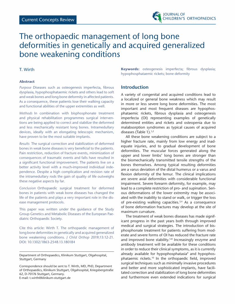
In general, the well-known principles of deformity correction apply. The osteotomies are being placed at the apex of the deformity, and the number of osteotomies are chosen according to the number of apices (Fig. 1). The choice of the right implant may pose another dif-ficulty. In growing patients, telescoping implants are mostly used, if the diameter of the bones allows them to be fitted. In the upper extremity, however, telescoping nails are only usable for humeral rodding.15 The radius and ulna do not generally accept telescoping nails due to the small diameters of the intramedullary canal and the difficult accessibility of the radius. Hence, flexible intramedullary nails, Rush rods or Kirschner (K)-wires
Table 1 Genetically determined diseases leading to soft bones and its consequences
Disease Genetics Main deformity Conservative treatment Surgical treatment
Fibrous dysplasia GNAS gene All bones affected Bisphosphonates Intramedullary stabilizationHypophosphatasia TNSALP gene Long bones affected Asfotase Alfa Intramedullary stabilization,
growth modulationHypophosphataemic rickets
PHEX gene Lower limbs, bow legs Burosumab Intramedullary stabilization, growth modulation
Osteogenesis imperfecta, type I to V(1, 2)
Col 1A1, Col 1A2, dominant
All long bones affected Bisphosphonates, physiotherapy
Intramedullary stabilization
Osteogenesis imperfecta, type IV to XVII(2)
Many gene locations, autosomal recessive
All long bones affected Bisphosphonates, physiotherapy
Intramedullary stabilization
GNAS, Guanine Nucleotide Binding Protein (G Protein), alpha stimulating activity; TNSALP, tissue-nonspecific alkaline phosphatase; PHEX, Phosphate regulating endopeptidase homolog X-linked; Col 1A1, collagen 1A1; Col 1A2, collagen 1A2
Fig. 1 Severely affected osteogenesis imperfecta patient with significant deformity of the right humerus (a) and right forearm (b). A simultaneous correction of both the right humerus and forearm was performed using multiple osteotomies; telescopic nail for humerus; and Kirschner-wires in the radius and ulna (c).
ORTHOPAEDIC MANAGEMENT OF LONG BONE DEFORMITIES
14 J Child Orthop 2019;13:12-21
of appropriate sizes are the implant of choice for fore-arm correction and stabilization (Fig. 1). In patients with fibrous dysplasia, it is usually larger bones that are affected, and several suitable intramedullary implants can be utilized. Plates are not chosen routinely but they may be helpful to fix rotationally unstable osteotomies. Small tubular plates or small locking plates with use of single cortex screw fixation are very useful in this con-text. If plates are chosen for fixing osteotomies or frac-tures, they should be removed soon after completion of bony healing. Because of the increased stability of the plated bone area, stress risers are created above and below with predictable deformity occurrence, if they are kept in place as a single implant.
While humerus corrections in OI patients are mostly possible from the age of three years,15 forearm correction requires a minimum size of both bones and the intramed-ullary canal. The indication for upper extremity correction is currently worked out with the patients themselves and their parents. They can identify the difficulties of daily liv-ing best and judge the impairment and disability caused by the present deformity. It is on the orthopaedic surgeon to outline treatment and offer strategies to overcome the patient’s problems, and to warn them from subsequent surgeries, if difficulties arise.
There are several strategies to address the upper extremity problem of OI patients. If the humerus is the only bone affected, unilateral or simultaneous bilateral corrective osteotomies are possible. If the upper arm and forearm need to be corrected, a side-by-side intervention is preferable. Forearm correction can be very tricky, requir-ing many osteotomies and meticulous surgical procedure. It is best to start with the humerus first, followed by radius and ulnar correction (Fig. 1). In severe deformities both bones must be corrected simultaneously through sepa-rate incisions. If you try to correct them one-by-one the intact bone may prevent adequate alignment of the oste-otomized bone. Postoperatively a split above the elbow cast may be necessary for four weeks, if some instability persists.
Lower extremityThe functionality of the lower limbs in OI patients focuses on all aspects of verticalization and implicates, therefore, different goals than described for the upper extremity. In mildly and moderately affected patients walking in an as normal a way possible must be the main goal of treatment, with the aim to prevent fractures particularly in the latter group. In the severely affected patients the avoidance of multiple fracture episodes and the stabili-zation of the weak bones to allow safe standing, transfer at home and maybe some walking with use of aids for
short distances are most important. A whole variety of implants have been used and developed with the third generation of telescopic nails being the most up-to-date implant.11,16-19. Accordingly, surgical intervention for defor-mity correction and long bone stabilization starts earlier than upper extremity surgery.
In other weak bone diseases, the development of lower limb deformities occurs at a slower rate and later in life. Consequently, the indication for surgical interventions is very closely linked to the clinical effect of the developing deformity and is timed to the needs of the patients.13,20
The primary goal in most of these patients, namely early verticalization and weight bearing, requires bones which can be biomechanically highly loaded from the beginning. Due to soft bone quality and microfracturing transmitted by the muscular force alone the long bones show some deformity at walking age already. With increasing load, the deformation continues to worsen, ultimately result-ing in fatigue or acute fractures at or near the apex of the deformity. This process is highly dependent on the sever-ity of the disease and the associated bone quality. Ideal timing of any corrective procedure in the lower extremity includes the assessment of deformity, bone size and qual-ity to fit the best suitable implant, in order to schedule it prior to any expected acute incident.21
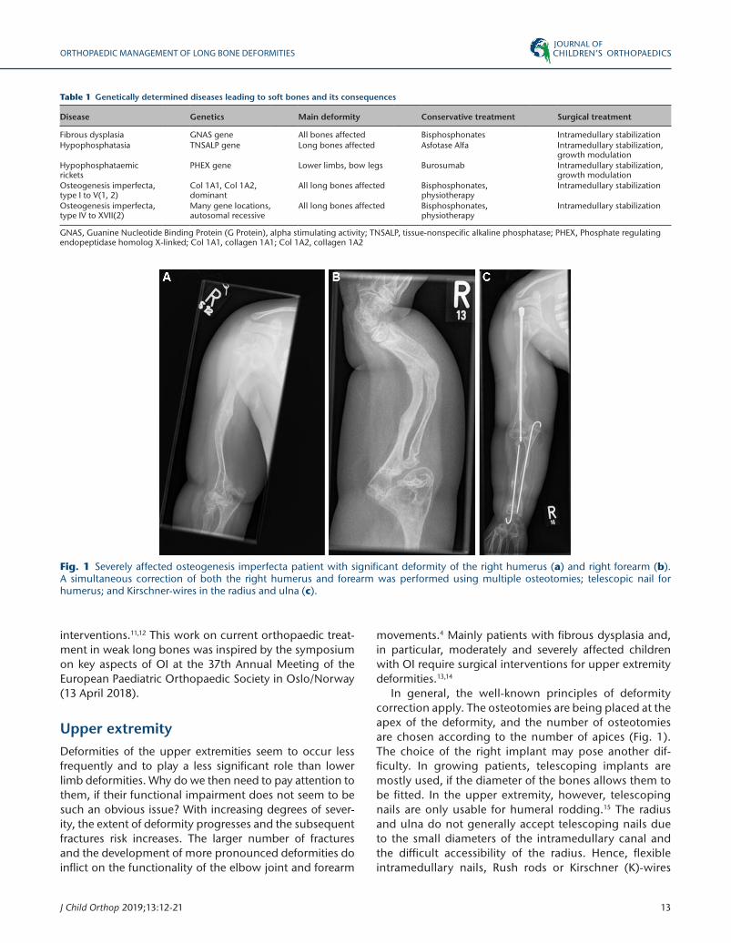
Any conceivable deformity is possible in both the femur and tibia in all patients with OI. However, femur varum and antecurvatum and tibia vara and antecurvata are the predominant deformities with the varus angulation of the tibia being rather distal. The strongest muscles of every long bone segment act as the most significant deforming forces bending it away from the largest muscle mass. The femoral neck area is another weak point with a trend to coxa vara development. In addition, there is a tendency to femoral retroversion which has a clear trend to recur after correction.22 In fibrous dysplasia coxa vara, femur antecurvatum and varum are the typical deformity con-figurations. In the tibia, increased antecurvation is the predominant finding. In hypophosphataemic rickets we mostly find long C-shaped varus deformities of both the femur and the tibia20 (Fig. 2).
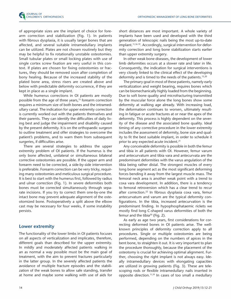
As early as age two years, first considerations for cor-recting deformed bones in OI patients arise. The well-known principles of deformity correction apply to all procedures. Single or multiple osteotomies are being performed, depending on the numbers of apices in the bent bone, to straighten it out. It is very important to plan the procedure thoroughly, because the placement of the osteotomy is crucial for achieving optimal alignment. Fur-ther, choosing the right implant is not always easy. Ide-ally intramedullary devices with elongating capacities are utilized in growing patients (Fig. 3). These are tele-scoping rods or flexible intramedullary nails inserted in opposite direction.11,19 In cases of too small a medullary
ORTHOPAEDIC MANAGEMENT OF LONG BONE DEFORMITIES
J Child Orthop 2019;13:12-21 15
canal for insertion of such implants simple K-wires or Rush rods can also be used without exhibiting the telescop-ing effect.18 Despite similar clinical outcomes compared with telescopic nails, their protecting effect is limited to the length of the implant causing secondary deformities above and below in the growth period.23 In adult patients,
custom-made or readily available small intramedullary locking nails are very suitable implants. In all of these cor-rective surgeries special attention has to be paid to the correct rotational alignment, particularly in the femur. All patients tend to present with femoral retroversion, which should be corrected during the procedure. If the osteot-omy or the fixation do not provide rotational stability the application of a small tubular or locking plate with unicor-tical screw fixation solves this problem.24
A specific issue of the femur applies to the area of the proximal femur and femoral neck. There is a great ten-dency of the femoral neck to gradually deviate into varus and retroversion while supporting the development of acetabular protrusion and high riding trochanter. When should it be protected and when should coxa vara be cor-rected? There are no general guidelines available. In cases with a neck-shaft angle of below 120° and the need for a very proximal femoral osteotomy, a support of the fem-oral neck by two K-wires which are attached to the lat-eral femoral cortex by circular wires22 is recommended. In severe coxa vara (neck-shaft angle below 100°) and an obvious proximal femoral deformity, surgical correction should also be strongly considered, using the same fixa-tion. In older children with larger bones special implants that allow screw fixation of the femoral neck are available and very useful. In all other patients the clinical symptoms and the chronological changes over time should be used as guidance for indicating operative correction.
Fig. 2 Typical long bone deformity in a patient with hypophosphataemic rickets, both tibias more affected than both femurs (a). Simultaneous bilateral correction by guided growth (femur) and double osteotomies of the tibias (b).
Fig. 3 Two stage surgical interventions in a one leg at a time technique for deformed lower limbs in a severely affected osteogenesis imperfecta patient. Surgery was performed one week apart. Pre- (a) and postoperative results (b and c) with a one-year follow-up (d).
ORTHOPAEDIC MANAGEMENT OF LONG BONE DEFORMITIES
16 J Child Orthop 2019;13:12-21
There are two techniques of performing the opera-tion: minimal-invasive and open approaches. In straight forward cases with moderate deformities the osteotomies can be done percutaneously through a small incision by drilling the bone and osteotomizing it with a chisel. Patients with severe deformities and patients without an intramedullary canal may not be suitable for the tech-nique and need open approaches. The medullary cavity has to be recreated to accept a nail. Sometimes shortening of the bones is inevitable for proper realignment, and this requires longer incisions. To use or mix several percutane-ous and formal incisions of appropriate length may be the best pragmatic principle.11
Most patients, regardless of which underlying pathol-ogy causes the deformity, are in need of having several bones corrected: both femurs, both tibias or all four long
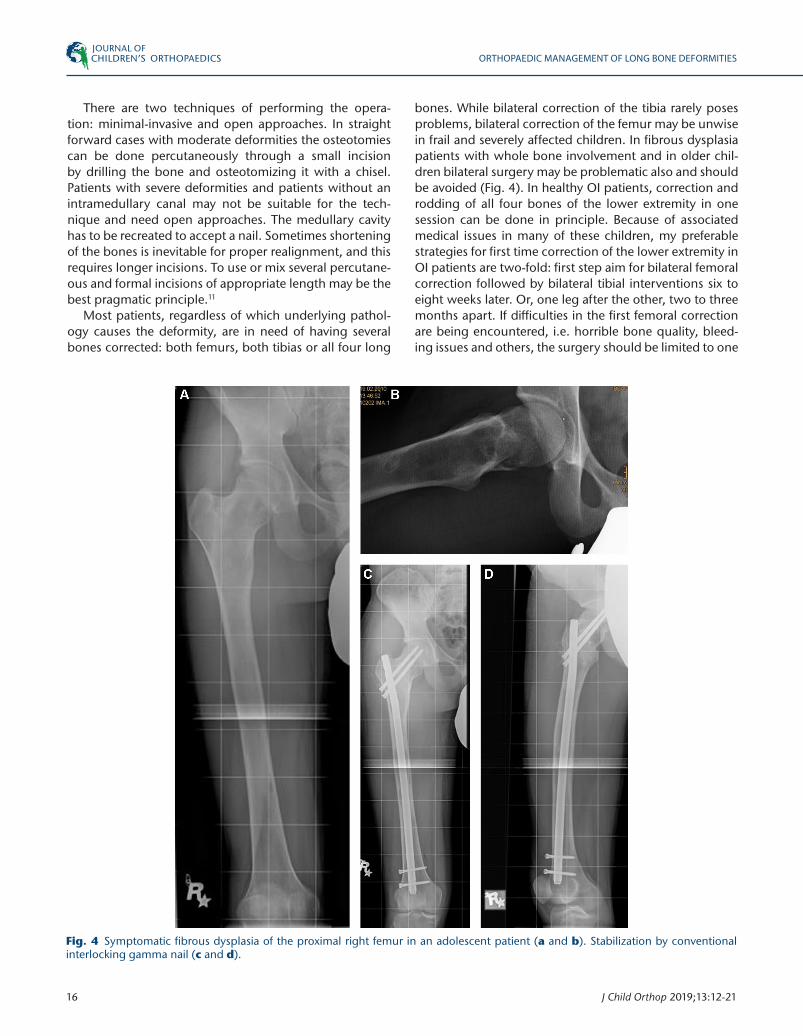
bones. While bilateral correction of the tibia rarely poses problems, bilateral correction of the femur may be unwise in frail and severely affected children. In fibrous dysplasia patients with whole bone involvement and in older chil-dren bilateral surgery may be problematic also and should be avoided (Fig. 4). In healthy OI patients, correction and rodding of all four bones of the lower extremity in one session can be done in principle. Because of associated medical issues in many of these children, my preferable strategies for first time correction of the lower extremity in OI patients are two-fold: first step aim for bilateral femoral correction followed by bilateral tibial interventions six to eight weeks later. Or, one leg after the other, two to three months apart. If difficulties in the first femoral correction are being encountered, i.e. horrible bone quality, bleed-ing issues and others, the surgery should be limited to one
Fig. 4 Symptomatic fibrous dysplasia of the proximal right femur in an adolescent patient (a and b). Stabilization by conventional interlocking gamma nail (c and d).
ORTHOPAEDIC MANAGEMENT OF LONG BONE DEFORMITIES
J Child Orthop 2019;13:12-21 17
side only and a three-step approach should be taken with the second femur to be done in a separate intervention (see Fig. 2).
Complications and causes of revision surgeryComplications and the need for revisions are connected to disease-related factors such as poor bone quality and softness, which impedes anchorage of the implants, to technical issues in conjunction with the operation and to implant-related issues (Fig. 5).
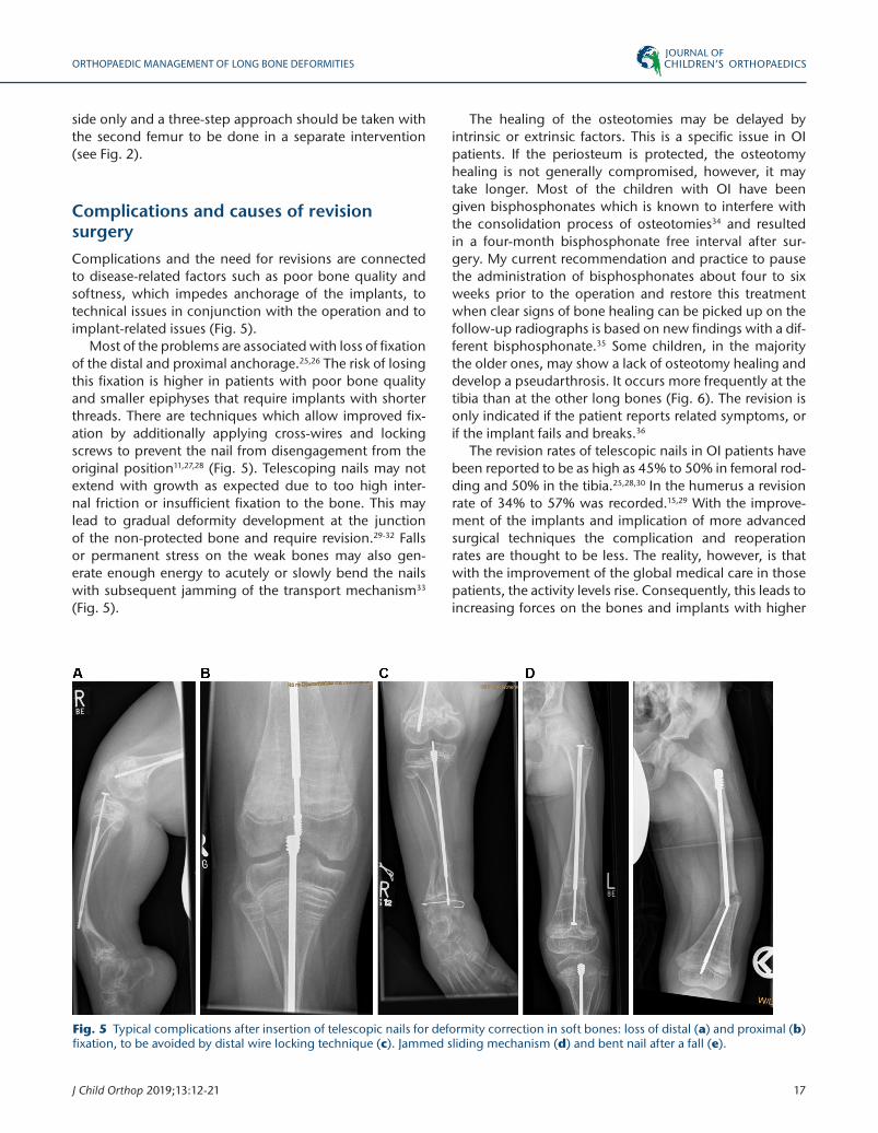
Most of the problems are associated with loss of fixation of the distal and proximal anchorage.25,26 The risk of losing this fixation is higher in patients with poor bone quality and smaller epiphyses that require implants with shorter threads. There are techniques which allow improved fix-ation by additionally applying cross-wires and locking screws to prevent the nail from disengagement from the original position11,27,28 (Fig. 5). Telescoping nails may not extend with growth as expected due to too high inter-nal friction or insufficient fixation to the bone. This may lead to gradual deformity development at the junction of the non-protected bone and require revision.29-32 Falls or permanent stress on the weak bones may also gen-erate enough energy to acutely or slowly bend the nails with subsequent jamming of the transport mechanism33 (Fig. 5).
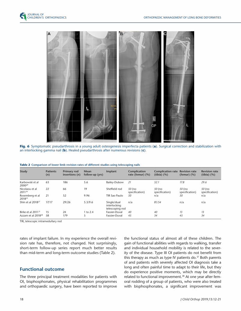
The healing of the osteotomies may be delayed by intrinsic or extrinsic factors. This is a specific issue in OI patients. If the periosteum is protected, the osteotomy healing is not generally compromised, however, it may take longer. Most of the children with OI have been given bisphosphonates which is known to interfere with the consolidation process of osteotomies34 and resulted in a four-month bisphosphonate free interval after sur-gery. My current recommendation and practice to pause the administration of bisphosphonates about four to six weeks prior to the operation and restore this treatment when clear signs of bone healing can be picked up on the follow-up radiographs is based on new findings with a dif-ferent bisphosphonate.35 Some children, in the majority the older ones, may show a lack of osteotomy healing and develop a pseudarthrosis. It occurs more frequently at the tibia than at the other long bones (Fig. 6). The revision is only indicated if the patient reports related symptoms, or if the implant fails and breaks.36
The revision rates of telescopic nails in OI patients have been reported to be as high as 45% to 50% in femoral rod-ding and 50% in the tibia.25,28,30 In the humerus a revision rate of 34% to 57% was recorded.15,29 With the improve-ment of the implants and implication of more advanced surgical techniques the complication and reoperation rates are thought to be less. The reality, however, is that with the improvement of the global medical care in those patients, the activity levels rise. Consequently, this leads to increasing forces on the bones and implants with higher
Fig. 5 Typical complications after insertion of telescopic nails for deformity correction in soft bones: loss of distal (a) and proximal (b) fixation, to be avoided by distal wire locking technique (c). Jammed sliding mechanism (d) and bent nail after a fall (e).
ORTHOPAEDIC MANAGEMENT OF LONG BONE DEFORMITIES
18 J Child Orthop 2019;13:12-21
Table 2 Comparison of lower limb revision rates of different studies using telescoping nails
Study Patients (n)
Primary rod insertions (n)
Mean follow-up (yrs)
Implant Complication rate (femur) (%)
Complication rate (tibia) (%)
Revision rate (femur) (%)
Revision rate (tibia) (%)
Karbowski et al 200029
63 186 5.6 Bailey-Dubow 21 52.1 17.8 29.6
Nicolaou et al 201130
22 66 19 Sheffield rod 50 (no specification)
50 (no specification)
50 (no specification)
50 (no specification)
Rosemberg et al 201825
21 52 9.96 TIR Sao Paulo 50 n/a 50 n/a
Shin et al 201827 17/17 29/26 5.3/9.6 Single/dual interlocking telescoping rod
n/a 81/54 n/a n/a
Birke et al 201131 15 24 1 to 2.4 Fassier-Duval 40 40 13 13Azzam et al 201828 58 179 5 Fassier-Duval 45 34 45 34
TIR, telescopic intramedullary rod
rates of implant failure. In my experience the overall revi-sion rate has, therefore, not changed. Not surprisingly, short-term follow-up series report much better results than mid-term and long-term outcome studies (Table 2).
Functional outcomeThe three principal treatment modalities for patients with OI, bisphosphonates, physical rehabilitation programmes and orthopaedic surgery, have been reported to improve
the functional status of almost all of these children. The gain of functional abilities with regards to walking, transfer and individual household mobility is related to the sever-ity of the disease. Type III OI patients do not benefit from this therapy as much as type IV patients do.37 Both parents of and patients with severely affected OI diagnosis take a long and often painful time to adapt to their life, but they do experience positive moments, which may be directly related to functional improvement.38 At one year after fem-oral rodding of a group of patients, who were also treated with bisphosphonates, a significant improvement was
Fig. 6 Symptomatic pseudarthrosis in a young adult osteogenesis imperfecta patients (a). Surgical correction and stabilization with an interlocking gamma nail (b). Healed pseudarthrosis after numerous revisions (c).
ORTHOPAEDIC MANAGEMENT OF LONG BONE DEFORMITIES
J Child Orthop 2019;13:12-21 19
noticed in range of movement of hip and knee joints and in the Functional Ability Questionnaire Score.32 Crawling, standing, walking and running as well as all domains of the Gross Motor Function Measure and the Pediatric Evaluation of Disability Inventory mobility and self-care scores changed considerably in favour of functionality. Greater severity of the disease and body weight were negative predictors for a more favourable outcome of this type of surgery.32 The lesser improvement of function in OI type III patients has prompted the idea of focusing on the upper extremity being vital for mastering daily activities and self-care in the severely affected individuals. Upper extremity surgery was found to be very beneficial to them.39 The greater the defor-mity the more disabling and functionally impairing the clinical implication was.4 The great benefit of upper limb surgery could be proven to be significant as well as the sus-tainable improvement of the self-care and mobility scores tested by the Pediatric Evaluation of Disability Inventory.40,41
DiscussionSurgical stabilization and deformity correction of weak bones, regardless of the underlying disease has become the main stake in all recently published consensus state-ments on their treatment, particularly true for OI.42,43 The lower limb has been the focus of surgical interven-tions since the beginning in order to allow the patients to stand and walk on stable lower extremities. The choice of implant was always in favour of intramedullary devices. Although double Rush rods in the femur and single Rush rods in the tibia were identified to yield comparable results with telescoping rods,23 telescopic nails became the stan-dard implant. The Bailey-Dubow, Sheffield, Fassier-Duval and other rods have clearly set aside other intramedul-lary devices.11,17,27,30 Only in very small bones that cannot accommodate telescoping rods or specific anatomic loca-tions like the forearm are K-wires and small Rush rods needed. Larger long bones, mostly found in adolescent patients with mild OI or fibrous dysplasia and hypophos-phataemic rickets, are predisposed to small interlocking nails.13,20
The timing of the procedures should be individually planned and discussed with the patients. When it is evi-dent that corrective surgery for both the lower and upper limb is inevitable, a time plan should be set up with the parents. A one leg at a time approach may be a very use-ful tactic to deal with the problem in a reasonable time period. This includes a detailed and individually tailored postoperative rehabilitation programme, to minimize the immobilization period.43 Depending on the size of the bone and the overall condition of the child, surgery can be scheduled as early as the second year of life. Upper limb surgery has come into focus more and more in recent
years15 on the same principal approach as in the lower extremities and a considerably older age at surgery. The outcomes are very promising.15,39-41
In all the years, despite improved surgical techniques and more sophisticated implants, the overall complication and reoperation rate has not changed a lot.25,27-29,31 The causes of failure have not changed either: distal and prox-imal rod migration, jamming of the telescoping mech-anism, bending rods due to the operating mechanical and muscular forces and technical issues with the oper-ation.25,27-29,31 Most of these problems are caused by the weak bones; the disease itself. Despite of all these neg-ative points the overall activity level of our patients with weak bones has clearly improved. Mildly affected patients can lead an almost normal life, including participation in sports activities. Severely affected patients can man-age transfer and short distance walks in their homes by themselves and have reached a greater degree of indepen-dence. They accept the high revision rate, because they know that they gain mobility and quality of life.
Received 12 November 2018; accepted 30 November 2018.
COMPLIANCE WITH ETHICAL STANDARDS
FUNDING STATEMENTNo benefits in any form have been received or will be received from a commercial party related directly or indirectly to the subject of this article.
OA LICENCE TEXTThis article is distributed under the terms of the Creative Commons Attribution-Non Commercial 4.0 International (CC BY-NC 4.0) licence (https://creativecommons.org/licenses/by-nc/4.0/) which permits non-commercial use, reproduction and distribution of the work without further permission provided the original work is attributed.
ETHICAL STATEMENTEthical approval: This article does not contain any studies with human participants or animals performed by any of the authors.Informed consent: Not required for this work.
ICMJE CONFLICT OF INTEREST STATEMENTThe author declares no conflict of interest.
REFERENCES
1. Sillence DO, Senn A, Danks DM. Genetic heterogeneity in osteogenesis imperfecta. J Med Genet 1979;16:101-116.
2. Van Dijk FS, Sillence DO. Osteogenesis imperfecta: clinical diagnosis, nomenclature and severity assessment. Am J Med Genet A 2014;164A:1470-1481.
3. Engelbert RH, Uiterwaal CS, Gerver WJ, et al. Osteogenesis imperfecta in childhood: impairment and disability. A prospective study with 4-year follow-up. Arch Phys Med Rehabil 2004;85:772-778.
4. Amako M, Fassier F, Hamdy RC, et al. Functional analysis of upper limb deformities in osteogenesis imperfecta. J Pediatr Orthop 2004;24:689-694.
ORTHOPAEDIC MANAGEMENT OF LONG BONE DEFORMITIES
20 J Child Orthop 2019;13:12-21
5. Glorieux FH. Experience with bisphosphonates in osteogenesis imperfecta. Pediatrics 2007;119:S163-S165.
6. Antoniazzi F, Zamboni G, Lauriola S, et al. Early bisphosphonate treatment in infants with severe osteogenesis imperfecta. J Pediatr 2006;149:174-179.
7. Land C, Rauch F, Glorieux FH. Cyclical intravenous pamidronate treatment affects metaphyseal modeling in growing patients with osteogenesis imperfecta. J Bone Miner Res 2006;21:374-379.
8. Vuorimies I, Toiviainen-Salo S, Hero M, Mäkitie O. Zoledronic acid treatment in children with osteogenesis imperfecta. Horm Res Paediatr 2011;75:346-353.
9. Kitaoka T, Tajima T, Nagasaki K, et al. Safety and efficacy of treatment with asfotase alfa in patients with hypophosphatasia: results from a Japanese clinical trial. Clin Endocrinol (Oxf) 2017;87:10-19.
10. Carpenter TO, Whyte MP, Imel EA, et al. Burosumab therapy in children with x-linked hypophosphatemia. N Engl J Med 2018;378:1987-1998.
11. Fassier F. Fassier-Duval telescopic system: how I do it? J Pediatr Orthop 2017;37:S48-S51.
12. Hefti F, Donnan L, Krieg AH. Treatment of shepherd’s crook deformity in patients with polyostotic fibrous dysplasia using a new type of custom made retrograde intramedullary nail: a technical note. J Child Orthop 2017;11:64-70.
13. Ippolito E, Bray EW, Corsi A, et al. Natural history and treatment of fibrous dysplasia of bone: a multicenter clinicopathologic study promoted by the European Pediatric Orthopaedic Society. J Pediatr Orthop B 2003;12:155-177.
14. Franzone JM, Bober MB, Rogers KJ, McGreal CM, Kruse RW. Re-alignment and intramedullary rodding of the humerus and forearm in children with osteogenesis imperfecta: revision rate and effect on fracture rate. J Child Orthop 2017;11:185-190.
15. Grossman LS, Price AL, Rush ET, et al. Initial experience with percutaneous IM rodding of the humeri in children with osteogenesis imperfecta. J Pediatr Orthop 2018;38:484-489.
16. Sofield HA, Millar EA. Fragmentation, realignment, and intramedullary rod fixation of deformities of the long bones in children. J Bone Joint Surg [Am] 1959;41-A:1371-1391.
17. Bailey RW, Dubow HI. Studies of longitudinal bone growth resulting in an extensible nail. Surg Forum 1963;14:455-458.
18. Mulpuri K, Joseph B. Intramedullary rodding in osteogenesis imperfecta. J Pediatr Orthop 2000;20:267-273.
19. Boutaud B, Laville JM. Elastic sliding central medullary nailing with osteogenesis imperfecta. Fourteen cases at eight years follow-up. Rev Chir Orthop Repar Appar Mot 2004;90:304-311.
20. Pavone V, Testa G, Gioitta Iachino S, et al. Hypophosphatemic rickets: etiology, clinical features and treatment. Eur J Orthop Surg Traumatol 2015;25:221-226.
21. Wirth T. Osteogenesis imperfecta. Orthopade 2012;41:773-782.
22. Fassier F, Sardar Z, Aarabi M, et al. Results and complications of a surgical technique for correction of coxa vara in children with osteopenic bones. J Pediatr Orthop 2008;28:799-805.
23. Joseph B, Rebello G, B CK. The choice of intramedullary devices for the femur and the tibia in osteogenesis imperfecta. J Pediatr Orthop B 2005;14:311-319.
24. Franzone JM, Kruse RW. Intramedullary nailing with supplemental plate and screw fixation of long bones of patients with osteogenesis imperfecta: operative technique and preliminary results. J Pediatr Orthop B 2018;27:344-349.
25. Rosemberg DL, Goiano EO, Akkari M, Santili C. Effects of a telescopic intramedullary rod for treating patients with osteogenesis imperfecta of the femur. J Child Orthop 2018;12:97-103.
26. Lee K, Park MS, Yoo WJ, et al. Proximal migration of femoral telescopic rod in children with osteogenesis imperfecta. J Pediatr Orthop 2015;35:178-184.
27. Shin CH, Lee DJ, Yoo WJ, Choi IH, Cho TJ. Dual interlocking telescopic rod provides effective tibial stabilization in children with osteogenesis imperfecta. Clin Orthop Relat Res 2018;476:2238-2246.
28. Azzam KA, Rush ET, Burke BR, Nabower AM, Esposito PW. Mid-term results of femoral and tibial osteotomies and Fassier-Duval nailing in children with osteogenesis imperfecta. J Pediatr Orthop 2018;38:331-336.
29. Karbowski A, Schwitalle M, Brenner R, et al. Experience with Bailey-Dubow rodding in children with osteogenesis imperfecta. Eur J Pediatr Surg 2000;10:119-124.
30. Nicolaou N, Bowe JD, Wilkinson JM, Fernandes JA, Bell MJ. Use of the Sheffield telescopic intramedullary rod system for the management of osteogenesis imperfecta: clinical outcomes at an average follow-up of nineteen years. J Bone Joint Surg [Am] 2011;93-A:1994-2000.
31. Birke O, Davies N, Latimer M, Little DG, Bellemore M. Experience with the Fassier-Duval telescopic rod: first 24 consecutive cases with a minimum of 1-year follow-up. J Pediatr Orthop 2011;31:458-464.
32. Ruck J, Dahan-Oliel N, Montpetit K, Rauch F, Fassier F. Fassier-Duval femoral rodding in children with osteogenesis imperfecta receiving bisphosphonates: functional outcomes at one year. J Child Orthop 2011;5:217-224.
33. Cho TJ, Kim JB, Lee JW, et al. Fracture in long bones stabilised by telescopic intramedullary rods in patients with osteogenesis imperfecta. J Bone Joint Surg [Br] 2011;93-B:634-638.
34. Munns CF, Rauch F, Zeitlin L, Fassier F, Glorieux FH. Delayed osteotomy but not fracture healing in pediatric osteogenesis imperfecta patients receiving pamidronate. J Bone Miner Res 2004;19:1779-1786.
35. Anam EA, Rauch F, Glorieux FH, Fassier F, Hamdy R. Osteotomy healing in children with osteogenesis imperfecta receiving bisphosphonate treatment. J Bone Miner Res 2015;30:1362-1368.
36. To M, Gupta V, Chow W. Surgical management of long bone pseudarthrosis with severe limb length discrepancy in osteogenesis imperfecta. J Pediatr Orthop B 2013;22:63-69.
37. Montpetit K, Dahan-Oliel N, Ruck-Gibis J, et al. Activities and participation in young adults with osteogenesis imperfecta. J Pediatr Rehabil Med 2011;4:13-22.
38. Dogba MJ, Bedos C, Durigova M, et al. The impact of severe osteogenesis imperfecta on the lives of young patients and their parents - a qualitative analysis. BMC Pediatr 2013;13:153.
39. Khoshhal KI, Ellis RD. Functional outcome of Sofield procedure in the upper limb in osteogenesis imperfecta. J Pediatr Orthop 2001;21:236-237.
40. Ashby E, Montpetit K, Hamdy RC, Fassier F. Functional outcome of humeral rodding in children with osteogenesis imperfecta. J Pediatr Orthop 2018;38:49-53.
ORTHOPAEDIC MANAGEMENT OF LONG BONE DEFORMITIES
J Child Orthop 2019;13:12-21 21
41. Ashby E, Montpetit K, Hamdy RC, Fassier F. Functional outcome of forearm rodding in children with osteogenesis imperfecta. J Pediatr Orthop 2018;38:54-59.
42. Hoyer-Kuhn H, Bartz-Seel J, Blickheuser R et al. Diagnostik und therapie der osteogenesis imperfecta. Konsensus-statement der 30. Jahrestagung
2014 der Deutschen Gesellschaft für Osteogenesis imperfecta Betroffene e.V. Monatsschr Kinderheilkd 2017;165:333-346.
43. Mueller B, Engelbert R, Baratta-Ziska F, et al. Consensus statement on physical rehabilitation in children and adolescents with osteogenesis imperfecta. Orphanet J Rare Dis 2018;13:158.


