Embed Size (px)
Citation preview

The polymodal ion channel TRPV4 modulates calcium flux,spiking rate and apoptosis of mouse retinal ganglion cells
Daniel A. Ryskamp1,2,*, Paul Witkovsky3,*, Peter Barabas1,*, Wei Huang1, ChristopherKoehler4, Nikolay P. Akimov4, Suk Hee Lee5, Shiwani Chauhan1, Wei Xing1, René C.Rentería4, Wolfgang Liedtke5, and David Krizaj1,2,6
1Department of Ophthalmology & Visual Sciences, John A. Moran Eye Center, University of UtahSchool of Medicine, Salt Lake City, UT 841322Interdepartmental Program in Neuroscience, University of Utah School of Medicine, Salt LakeCity, UT 841323Department of Ophthalmology, New York University School of Medicine, New York, NY100104Department of Physiology and Center for Biomedical Neuroscience, University of Texas HealthScience Center at San Antonio, San Antonio, TX5Department of Medicine and Neurobiology, and Center for Translational Neuroscience, DukeUniversity Medical Center, Durham, NC 277106Department of Physiology, University of Utah School of Medicine, Salt Lake City, UT 84132
AbstractSustained increase in intraocular pressure represents a major risk factor for eye disease yet thecellular mechanisms of pressure transduction in the posterior eye are essentially unknown. Herewe show that the mouse retina expresses mRNA and protein for the polymodal TRPV4 cationchannel known to mediate osmo- and mechanotransduction. TRPV4 antibodies labeled perikarya,axons and dendrites of retinal ganglion cells (RGCs) and intensely immunostained the optic nervehead. Müller glial cells, but not retinal astrocytes or microglia, also expressed TRPV4immunoreactivity. The selective TRPV4 agonists 4α-PDD and GSK1016790A elevated [Ca2+]i indissociated RGCs in a dose-dependent manner whereas the TRPV1 agonist capsaicin had no effecton [Ca2+]RGC. Exposure to hypotonic stimulation evoked robust increases in [Ca2+]RGC. RGCresponses to TRPV4-selective agonists and hypotonic stimulation were absent in Ca2+-free salineand were antagonized by the nonselective TRP channel antagonists Ruthenium Red andgadolinium, but were unaffected by the TRPV1 antagonist capsazepine. TRPV4-selective agonistsincreased the spiking frequency recorded from intact retinas recorded with multielectrode arrays.Sustained exposure to TRPV4 agonists evoked dose-dependent apoptosis of RGCs. Our resultsdemonstrate functional TRPV4 expression in RGCs and suggest that its activation mediatesresponse to membrane stretch leading to elevated [Ca2+]i and augmented excitability. ExcessiveCa2+ influx through TRPV4 predisposes RGCs to activation of Ca2+-dependent pro-apoptoticsignaling pathways, indicating that TRPV4 is a component of the response mechanism topathological elevations of intraocular pressure.
Correspondence to: David Krizaj, Dept. of Ophthalmology & Visual Sciences, Moran Eye Center, Univ. of Utah School ofMedicine, Salt Lake City, UT 84132, [email protected].*equal contribution
NIH Public AccessAuthor ManuscriptJ Neurosci. Author manuscript; available in PMC 2011 November 11.
Published in final edited form as:J Neurosci. 2011 May 11; 31(19): 7089–7101. doi:10.1523/JNEUROSCI.0359-11.2011.
NIH
-PA Author Manuscript
NIH
-PA Author Manuscript
NIH
-PA Author Manuscript

Keywordsretina; TRP channels; osmoregulation; pressure-sensitivity; glaucoma; ganglion cell
IntroductionCells of multicellular organisms experience mechanical stimuli that range from the directmechanical impact of pulling and stretching to changes in osmotic and hydrostatic pressure(Wang and Thampatty, 2006; Bourque, 2008). Mechanical stretch or pressure activates ionchannels in the plasma membrane (Loukin et al., 2010a, b), resulting in depolarization,increased intracellular Ca2+ concentration ([Ca2+]i) (Zabel et al., 1996; Wu and Davis, 2001)and changes in gene expression, cell shape and cytoskeletal organization (Naruse et al.,1998; Thodeti et al., 2009).
Recent studies have established that members of the transient receptor potential (TRP)superfamily transduce visual, chemical, thermal, mechanical, painful and osmotic stimuliinto Ca2+ fluxes (reviewed in Liedtke and Kim, 2005; Kung, 2005; Christensen and Corey,2007; Sharif-Naeini et al., 2008). We are particularly interested in the possibility that thevertebrate retina, which is exposed to systemic blood pressure, hydrostatic pressure from thecerebrospinal fluid and to intrinsic intraocular pressure (IOP), contains one or morepressure-sensitive TRP channel. Pathological elevations in IOP or systemic pressurerepresent primary risk factors for glaucoma, a group of inherited optic neuropathiescharacterized by apoptotic loss of RGCs, degeneration of the optic nerve and progressiveloss of visual fields (Quigley, 2005; Whitmore et al., 2005). The cellular pathophysiology ofglaucoma is not well understood, in part because the mechanisms that couple the mechanicalstimulus (ΔIOP) to cellular signal transduction remain to be characterized.
In eukaryotic cells, the TRPV4 channel (transient receptor potential vanilloid-4; GenBankaccession number NM_022017) represents a polymodal mechanism that transduces osmoticpressure, shear force stimuli, mechanical stretch and moderate warmth (~27–37°C) intocation influx with a preference for Ca2+ (PCa/PNa ~6; Liedtke et al., 2000; Güler et al., 2002;O’Neil and Heller, 2005). The channel may be activated by intracellular mediators such asarachidonic acid and cytochrome P450-dependent formation of 5, 6-epoxyeicosatrienoicacid (Vriens et al., 2004, but see Loukin et al., 2010). In neural tissues, TRPV4 expressionhas been localized to sensory neurons in dorsal root and trigeminal ganglia, inner ear haircells, Merkel cells, hippocampal and hypothalamic neurons, and astrocytes (Liedtke et al.,2000; Reiter et al., 2006; Shibasaki et al., 2007; Benfenati et al., 2007; Alessandri-Haber etal. 2009). Mice lacking TRPV4 have defects in noxious mechano- and pressure sensation(Suzuki et al., 2003; Liedtke and Friedman, 2003) whereas the functional ortholog ofTRPV4 in the worm Caenorhabditis elegans, OSM-9, mediates osmotic, mechanical, andchemical avoidance (Liedtke et al., 2003). Together, these findings suggest that TRPV4represents an evolutionarily conserved element of the neural response to mechanicalstimulation.
We report here that TRPV4 is expressed in retinal ganglion cell (RGC) somata and opticaxon fibers. Transient activation of TRPV4 induced Ca2+ influx and increased theexcitability of ganglion cells, whereas sustained activation resulted in RGC apoptosis. Theprominent expression of TRPV4-ir at the optic nerve head (ONH), and the role of TRPV4 ingating Ca2+ entry and RGC firing, implicate this channel in the retinal remodeling thatoccurs during chronic increases in IOP.
Ryskamp et al. Page 2
J Neurosci. Author manuscript; available in PMC 2011 November 11.
NIH
-PA Author Manuscript
NIH
-PA Author Manuscript
NIH
-PA Author Manuscript

Materials and MethodsA. Animals
C57BL/6J mice of either sex were obtained from commercial suppliers whereas B6.Cg-Tg(Thy1-CFP)23Jrs/J (hereafter, Thy1:CFP) animals or retinal sections were a kind giftfrom Dr. Ning Tian (University of Utah). The animals were maintained in the Universityanimal quarters on a 12h: 12h light dark cycle. Mice were fed lab chow and water adlibitum. Mice were euthanized prior to isolation and dissociation of retinas (Duncan et al.,2006). Animal handling and anesthetic procedures were approved by University InstitutionalAnimal Care committees and conform to National Institutes of Health guidelines.
B. HistologyEyes were enucleated and their corneas and lenses dissected away. The remaining posteriorpole of the eye was fixed by immersion for 1 hour at room temperature in freshly prepared4% paraformaldehyde in 0.1M phosphate buffer, pH 7.2, then washed 3 × 10 min. in pH 7.2phosphate buffered saline (PBS). Fixed eye tissue was then immersed for 12–16h in 30%sucrose at 4° C, embedded in OCT (Ted Pella, Redding, PA), cryostat sectioned at 16 µm,and mounted on Superfrost slides (Fisher Scientific, Pittsburgh, PA). After drying for 1h at37° C, slides were stored frozen (−80° C) until used. Dissociated cells were isolated withpapain (7U/ml; Worthington, Lakewood, NJ), plated on concanavalin A-coated coverslips,fixed in 4% paraformaldehyde in 0.1M phosphate buffer, pH 7.2 for 30 min, washed 3 × 10min. in pH 7.2 PBS and stored at 4° C.
Immunohistochemistry—For fluorescence immunocytochemistry, cryostat sectionswere thawed, washed in PBS, then placed for 30 min-1h in blocking solution (10 ml PBS, 30µL Triton-X 100, 100 mg bovine serum albumin, 100 µL 10% w/v Na azide solution).Primary and secondary antibodies were diluted in blocking solution and applied for 2 hoursand 1 hour, respectively at room temperature, with 3 intervening washes in PBS. After afinal set of washes in PBS, the sections were covered in VectaShield (Vector Labs,Burlingame, CA).
Terminal deoxynucleotidyl transferase-mediated dUTP-biotin nick endlabeling (TUNEL) assay—Retinas were embedded, cryosectioned, and processed forterminal deoxynucleotidyl transferase-mediated dUTP-biotin nick end labeling (TUNEL)following the protocol by Gavrieli et al. (1992). After 30 min rehydration in 70% ethanol,2×5 min rinses in PBS and 1% Triton-X in 1% citrate buffer (pH 7.3), the slides weresubjected to a final rinse in PBS. Slides were then incubated for 30 min in the reactionbuffer (30 mM Trizma-HCl, 140 mM Na+ cacodylate, 1.0 mM CaCl2, 0.2% Triton X-100,pH 7.2). Positive controls were treated with DNAse I (Roche Diagnostics; Branford, CT; 10U/ml) in the reaction buffer. Next, slides were treated for 1 hour with 0.03 units/µl terminaltransferase and 4 µM biotin-16-dUTP (Roche Diagnostics). The reaction was terminated in30 mM Na+ citrate, 300 mM NaCl and 0.2% Triton X-100 in PBS (5 min) and rinsed in PBS(2×5 min), exposed to 1% bovine serum albumin in PBS for 20 min and rinsed. As anegative control, the retinal sections were processed omitting the incubation step withterminal deoxynucleotidyl transferase during DNA labeling. Apoptotic cells were visualizedwith a confocal microscope (Zeiss LSM510) and categorized by size. We found that ourfixation protocol resulted in ~1µm shrinkage in the diameter of dissociated Brn3a-ir cells inretinal sections. Therefore, the TUNEL analysis compared cells with diameter > 6 µm(presumed RGCs) to photoreceptor cells with 3–5 µm diameters (the identity ofphotoreceptors was confirmed by immunostaining for mouse cone arrestin (a gift from Dr.Wolfgang Baehr, University of Utah) and rhodopsin (Santa Cruz Technologies, Santa Cruz,
Ryskamp et al. Page 3
J Neurosci. Author manuscript; available in PMC 2011 November 11.
NIH
-PA Author Manuscript
NIH
-PA Author Manuscript
NIH
-PA Author Manuscript

CA). A cell was deemed to be TUNEL positive if the fluorescent signal completely filled thecell body.
C. Image Acquisition and ProcessingSections were examined by confocal microscopy (Zeiss LSM 510 or Nikon PM 800).Digital images were acquired separately from each laser channel, then recombined. Fileswere further processed with deconvolution software (AutoQuant Imaging, Watervliet, NY).Adjustments of contrast and intensity were made in Photoshop (Adobe, San Jose, CA); anysuch adjustments were made uniformly to the entire image.
D. Western BlotWe essentially followed the procedure outlined in previous publications (Liedtke andFriedman, 2003; Phan et al., 2009). In brief, homogenized retinal tissue was subjected toprotein preparation and denaturation, and proteins (10µg per lane) were separated on a SDS-gel, then immunoblotted to PVDF membranes. After blocking with milk-powder, TRPV4was immunodetected using the above antibody (Phan et al., 2009) and peroxidase-basedchemoluminescence.
E. Primary and Secondary AntibodiesTable 1 shows the primary and secondary antibodies used in this study. Multiplecommercially available anti-TRPV4 antibodies (ACC-034, Alomone Labs, Jerusalem,Israel; LS-A8583, MBL International Corporation, Woburn, MA; OSR00136W, AffinityBioreagents, Golden, CO and ab63003, Abcam, Cambridge, MA) were tested byimmunostaining and Western blots in wild type and TRPV4 null mice. However, the onlyantibody that proved completely satisfactory was LS-C94498 (Lifespan Biosciences, Seattle,WA), based on the following criteria: (a) The antibody stained Western blot bands of 85 and106 kD from retinal tissue harvested from wild type mice but not from TRPV4 null mice(Fig. 1), (b) it colocalized with green fluorescent protein (GFP) in RGCs from transgenicanimals in which GFP was driven by the Trpv4 gene promoter (Fig. 2C) and (c) it detectedprotein bands of appropriate molecular mass from HEK293 cells transfected with Trpv4cDNA, whereas the bands were missing in immunoblots from non-expressing cells (Fig. 1).All TRPV4-ir data reported in the present study were obtained with the anti-TRPV4antibody, LS-C94498. No immunostaining was observed when the primary antibody wasomitted.
F. Semiquantitative RT-PCRTotal RNA from retina was extracted with Trizol and total RNA (2 µg total RNA used forfirst strand synthesis with oligo (dT) and primers) was converted to cDNA using theSuperScript III First-Strand Synthesis kit from Invitrogen. PCR products were amplified in athermocycler (Veriti; ABI, Foster City, CA) with nucleic acid stain (SYBR Green, ABI)reagents according to the manufacturer’s instructions. Amplification of PCR products wasmeasured by fluorescence associated with binding of double-stranded DNA to SYBR Greenin the reaction mixture. After an initial denaturation step of 50° C for 2 min and 95° C for 10min, PCR was repeated for 40 cycles at 95° C for 15 sec, 58° C for 30 sec, and 72° C for 30sec. After amplification, the ratio of gene-of-interest mRNA to a housekeeping gene,glyceraldehyde-3-phosphate dehydrogenase (Gapdh), was calculated for each sample. Fiveto ten independent biological replicates (retinas) were used at each age.
Ryskamp et al. Page 4
J Neurosci. Author manuscript; available in PMC 2011 November 11.
NIH
-PA Author Manuscript
NIH
-PA Author Manuscript
NIH
-PA Author Manuscript

G. Generation of a bacterial artificial chromosome (BAC) transgenic mouse line with afluorescent reporter driven by the Trpv4 promoter
A mouse BAC harboring the Trpv4 gene was modeled by “recombineering” (Copeland etal., 2003) so that copepod-GFP coding region was placed directly after the ATG start codonof mouse Trpv4. In addition, this BAC was engineered to not harbor exons 10–14, so that afunctional ion channel could not arise from the transgene. Engineered DNA served as atransgenesis template, which was constructed following standard procedures (Zhao et al.,2008). Of 4 resulting transgenic lines, the one used revealed robust fluorescence intrigeminal ganglion sensory neurons (not illustrated), both in acute sections and acutedissociations, co-localizing with TRPV4 immunolabeling. This line was outcrossed ≥5generations before analysis of retinas.
H. Calcium ImagingCalcium imaging was performed on acutely isolated retinal cells, as described previously(Szikra et al. 2009). In brief, dissociated retinal cells were plated on concanavalin A-coated(0.2 mg/ml; Sigma, ST. Louis, MO) coverslips, loaded with fura-2 AM (1–5 µM;Invitrogen, Carlsbad, CA) for 15–30 min and washed for 10 min in dye-free L-15 medium.Cells were viewed with Nikon Ti inverted or 600EF upright microscopes using 20× 0.95N.A., 40× 0.85 N.A. or 40× 1.25 N.A. objective lenses. Excitation for 340 nm and 380 nmfilters (Chroma, Brattleboro, VT and Semrock, Rochester, NY) was provided by a 150WXenon arc lamp (DG4, Sutter Instruments, Novato, CA). Fluorescence emission was highpass-filtered at 510 nm and captured with cooled digital CCD cameras (HQ2, Photometrics,Tucson, AZ). Data acquisition and F340/F380 ratio calculations were performed by NISElements software (Melville, NY). Fluorescence imaging was performed on Regions ofInterest (ROI) encompassing the RGC perikaryon, typically at 3×3 binning. Backgroundfluorescence was measured in similarly sized ROIs in neighboring areas devoid of cells.After sequential image acquisition (0.167 – 0.5 Hz) of cell fluorescence at 340/380 nm, thebackground was subtracted. Calibration of free [Ca2+]i was carried out in vivo using 10 µMionomycin and 10 mM Ca2+ or 0 Ca2+/3 mM EGTA. The apparent free [Ca2+]i wasdetermined from the equation [Ca2+]i = ((R-Rmin)/(Rmax-R)) × (F380
max/F380min) × Kd,
where R is the ratio of emission intensity at 510 nm evoked by 340 nm excitation vs.emission intensity at 510 nm evoked by 380 nm excitation; Rmin is the ratio at zero freeCa2+; Rmax is the ratio at saturating Ca2+, and the dissociation constant Kd for Ca2+-fura 2 atroom temperature was taken to be 224 nM (Neher, 1995). Glutamate (100 µM) was added atthe beginning of each experiment to control for neuronal health, type and responsiveness.DMSO, the solvent for the indicator dye, did not induce any responses in RGCs (data notshown). Previous studies using Mn2+ quenching showed that 95% of the fura-2 fluorescenceemanates from the cells’ cytosol (Szikra et al., 2009). Experiments were conducted at roomtemperature and encompassed stimulation with glutamate, TRPV4 and TRPV1 agonists andantagonists.
I. Cell identificationRGCs in short-term culture were identified initially by morphology and perikaryal size (7–15 µm). In a subset of experiments, RGCs were isolated from Thy1:CFP retinas were usedand identified by intrinsic fluorescence (Raymond et al., 2009). Alternatively, test neuronsisolated from wild type retinas were confirmed as RGCs by immunocytochemistry. In eachexperiment, a 100 µM glutamate stimulus was used to confirm that the visually identifiedputative RGCs expressed ionotropic glutamate receptors. Presumed RGCs responded to 30mM KCl with rapid high-amplitude increases in [Ca2+]i, indicating that the cells werehealthy and maintained their excitability. In a subset of experiments, cells recorded duringstimulation with hypotonic saline or TRPV4 agonists were fixed and immunostained withTRPV4 and Brn3a antibodies.
Ryskamp et al. Page 5
J Neurosci. Author manuscript; available in PMC 2011 November 11.
NIH
-PA Author Manuscript
NIH
-PA Author Manuscript
NIH
-PA Author Manuscript

J. Solutions and ReagentsThe isotonic superfusing saline contained, in mM, 133 NaCl, 2.5 KCl, 2 CaCl2, 1.5 MgCl2,1.25 NaH2PO4, 10 HEPES hemisodium salt, 10 glucose, 1 pyruvic acid, 1 lactic acid and 0.5glutathione. In Ca2+-free solutions, no external Ca2+ was added and the saline wassupplemented with 1 mM EGTA. The osmolarity and pH of each external solution wasmeasured before each experiment. pH was adjusted to 7.4 with NaOH. Osmolarity wasmeasured with a vapor-pressure osmometer (VAPRO, Logan, UT); for control saline,osmolarity was 280 mOsm. For experiments involving hypotonic stimulation, the isotonicringer contained 132 mM mannitol and the NaCl concentration was reduced to 57.5 mM.Hypoosmotic solutions were prepared by reducing the final concentration of mannitol to44.5 mM without changing the ionic composition. In a subset of experiments usinghypotonic stimulation, cells were co-loaded with fura-2 AM + calcein AM (1 µM;Invitrogen). Calcein fluorescence was elicited using 490 nm excitation filters. Becausecalcein fluorescence is Ca2+-independent and volume-dependent, it was used as a measureof changes in the cell volume.
Unless otherwise indicated, the salts and reagents were purchased from Sigma. RutheniumRed and capsaicin were from Ascent Scientific (Princeton, NJ). Capsaicin and capsazepinewere also purchased from Tocris (Ellisville, MO). 4α-PDD was obtained from LCLaboratories (Woburn, MA).
K. Multielectrode array (MEA) recordings and data analysisMEA recordings were performed using multielectrode, extracellular recording chambers(MEA1060 system; MultiChannel Systems, Reutlingen, Germany) consisting of an array of60 planar electrodes, each 10 µm in diameter, spaced 100 µm apart for a total array size of700 µm2, as described previously (Rentería et al., 2006). Briefly, young adult C57Bl6J micewere sacrificed after 1 hr of dark adaptation, and an eye was removed under dim redillumination. The retina was dissected and placed with the ganglion cell side against therecording electrodes using a piece of nitrocellulose paper as support. The retina wastypically placed on the array within a millimeter of the optic nerve head and a manipulator(Cell Micro Controls, Norfolk, VA) was used to hold the tissue down with slight pressure.Retinas were perfused at room temperature for 30 min and at 30°C for another 30 minbefore recordings were started, the temperature was maintained at 30°C thereafter, andspontaneous spiking in the dark recorded. The perfusion saline was either Ames’ medium orRinger modified as follows (in mM): 124 NaCl, 2.5 KCl, 2 CaCl2, 2 MgCl2, 1.25 NaH2PO4,26 NaHCO3, and 22.2 glucose; the pH was maintained at 7.3–7.4 by bubbling with 95%O2/5% CO2 mixed gas. Voltage signals sampled at 50 kHz were bandpass filtered at 100 Hzto 3 kHz, and waveforms that crossed a negative voltage threshold (set at −5.5 standarddeviations of the mean noise) were recorded to disk for offline analysis. Under theseconditions, nearly all recorded cells are likely to be RGCs (see Rentería et al., 2006).
Spike trains for each RGC were determined by spike sorting based on clustering in principalcomponent space using software (Offline Sorter; Plexon, Inc., Dallas TX). Not everydetected waveform was assigned to a unit; obvious automatic sorting errors were correctedfor each cluster manually (Rentería et al., 2006). Time stamps of the action potentials ofeach sorted unit were used to generate peristimulus time histograms (30 sec bins). Theaverage spike rates during the 3 min period before and after drug application weredetermined for every recorded cell and expressed as the percent change in firing in thepresence of drug. This normalization allowed for comparison of cells with varied initialspike rates. The very few cells that had zero rates before application and spiking during drugapplication were considered to have increased by 100%.
Ryskamp et al. Page 6
J Neurosci. Author manuscript; available in PMC 2011 November 11.
NIH
-PA Author Manuscript
NIH
-PA Author Manuscript
NIH
-PA Author Manuscript

L. Statistical analysisData are expressed as mean ± SEM with the number of cells, slides or animals indicated byN. Cell diameter data are expressed as mean ± SD. Statistical comparisons between twotreatments in the same cell were determined using the t-test; unless indicated otherwise,comparisons between different groups were evaluated by the Mann-Whitney test or theWilcoxon signed rank test. Data obtained in multielectrode array studies were analyzed withthe Wilcoxon signed rank test. A value of P<0.05 was considered statistically significant.
ResultsTRPV4 mRNA and protein in the mouse retina
RT-PCR was performed using total RNA extracted from adult (postnatal day 90) mouseretinas. Gel analysis revealed PCR products at the appropriate size of 174 bp. The sametranscript was amplified in kidney tissue, known to express Trpv4 (Strotmann et al., 2000,Liedtke et al., 2000; Güler et al., 2002) (Fig. 1A).
We next compared TRPV4 protein expression in tissues derived from wild type and TRPV4null mice (cf. Methods). Consistent with RT-PCR, immunoblots from wild type mouseretinas showed a primary band of 85 kD and a secondary band at 105 kD which probablycorrespond to unglycosylated and N-glycosylated forms of TRPV4, respectively, in keepingwith previous reports (Fig. 1B) (Liedtke and Friedman, 2003; Benfenati et al., 2007;Hartmannsgruber et al., 2007). Both bands were absent in retinal tissue derived from TRPV4null mice (Fig. 1B). The TRPV4 antibody was validated in Western blots from HEK293cells transfected with Trpv4 cDNA (Fig. 1C) and in trigeminal ganglion tissue known torobustly express Trpv4 (Liedtke et al., 2000; Liedtke and Friedman, 2003). As an additionaltest of the TRPV4 antibody, we immunostained the kidney, choroid plexus and nodoseganglion tissues in which TRPV4 is expressed (Liedtke et al., 2000; Strotmann et al., 2000;Brierley et al., 2008). The patterns of TRPV4 expression in these tissues were identical tothose identified in previous reports (data not shown). For example, the somata of neuronswithin the nodose ganglion (Fig. 1Di) were strongly TRPV4-immunoreactive whereas thenodose tissue from a TRPV4 null mouse was unlabeled by the TRPV4 antibody (Fig. 1Dii).
TRPV4 immunoreactivity in the mouse retinaa. TRPV4-ir in retinal ganglion cells—In wild type mouse retinas, TRPV4-ir waspunctate and distributed throughout the inner retina (Fig. 2A). A dense array of TRPV4-irpuncta was seen within cell bodies located in the ganglion cell layer (gcl). TRPV4-ir wasdistributed at a lower density throughout the inner plexiform layer (ipl) but only very sparseTRPV4-ir was located within the inner nuclear layer (inl). This immunostaining pattern wasabsent in retinas of TRPV4 null mice (Fig. 2B). The apparent immunostaining of the outerplexiform layer (opl) is non-specific since it was identical in wild type and TRPV4 nullretinas (Fig. 2A, B). Within the outer nuclear layer (opl) consisting primarily ofphotoreceptor nuclei, TRPV4-ir was seen in vertical processes of Müller glial cells thattraversed the opl (cf. also Fig. 2M below).
We also analyzed a transgenic mouse in which green fluorescent protein (GFP) was drivenby the entire promoter of the mouse Trpv4 gene. The 50 kDa Trpv4 gene was centeredwithin the 200 kDa GFP insert present in a BAC (cf. Methods). Using an antibody againstGFP, retinal immunostaining was confined to cell bodies within the ganglion cell layer,although additional staining was noted in retinal blood vessels, in keeping with wellestablished vascular-endothelial TRPV4 expression (Hartmannsgruber et al., 2007; Mendozaet al., 2010). When the retina was stained with anti-GFP, the marker was detected in cellbodies of the ganglion cell layer (Fig. 2C). Confirming that reporter gene expression was
Ryskamp et al. Page 7
J Neurosci. Author manuscript; available in PMC 2011 November 11.
NIH
-PA Author Manuscript
NIH
-PA Author Manuscript
NIH
-PA Author Manuscript
faithful to the targeted gene, Trpv4, GFP-ir was absent in wild type mouse retina (notillustrated).
To probe whether TRPV4-ir is associated primarily or exclusively with ganglion cells, weimmunostained the retina for Brn3a (also called POU4f1), a transcription factor foundalmost exclusively in developing (Liu et al., 2000) and mature ganglion cells (Nadal-Nicolaset al., 2009). We found that TRPV4-ir colocalized with that of Brn3a in ganglion cellperikarya and dendrites (Fig. 2D, arrow). Prominent additional TRPV4-ir was found withinganglion cell axon bundles in the optic fiber layer (ofl, Fig. 2D) and also was abundantwithin the optic nerve head (Fig. 2H) and the laminar region of the optic nerve (Fig. 2I). Thenearly universal distribution of Brn3a-ir in mouse retinal ganglion cells, together with thefinding that Brn3a-ir and TRPV4-ir almost invariably colocalize, indicates that TRPV4protein is distributed among a large fraction of mouse retinal ganglion cells.
A second marker for ganglion cells was provided by a construct in which GFP was linked tothe promoter of Thy1, a ganglion cell-specific protein (cf Methods). GFP-ir and TRPV4-ircolocalize in perikarya and dendrites of RGCs (Fig. 2E) and in displaced ganglion cellperikarya (Fig. 2L). Finally, immunostaining with a SMI-32 antibody which labelsintermediate filaments in somata and axonal processes of wide field alpha RGCs,colocalized with TRPV4-ir processes in the OFL entering the optic nerve head (Fig. 2K).
b. TRPV4-ir is weak or absent in retinal amacrine cells—About 50% of the cellswithin the ganglion cell layer in rodent retinas are displaced amacrine neurons rather thanganglion cells whereas a subset of cells in the proximal INL belong to displaced RGCs(Perry and Walker, 1980; Jeon et al., 1998). To probe possible colocalization of TRPV4-irwith amacrine cells, we immunostained the retina with an antibody against cholineacetyltransferase, a marker for starburst amacrine cells (Fig. 2G), and with glutamic aciddecarboxylase 65 (GAD-65) a marker for GABAergic amacrine cells (Fig. 2F and Fig. 3A)that constitute a substantial fraction of displaced amacrine cells in the RGC layer (May etal., 2007). These tests revealed no colocalization of TRPV4-ir with ChAT-ir cells.Moreover, no colocalization with the dopaminergic amacrine cell marker tyrosinehydroxylase was observed (data not shown). 4.6 percent of cells that labeled with theGABAergic marker GAD-65 stained with the TRPV4 antibody, however the intensity ofTRPV4-ir signals in this subset of cells was always significantly weaker compared to Brn3a-ir cells. Occasionally, a Brn3a-ir or SMI-32-ir cell body was detected in the INL. Theseperikarya were always TRPV4-imunopositive (Fig. 2L). Collectively these data indicate thatTRPV4-ir is associated with mouse retinal ganglion cells and may be localized to a smallsubset of GABAergic amacrine cells. Within the mouse ganglion cell, TRPV4-ir isassociated principally with the perikaryon, and the axon, but at least some ganglion celldendrites also express TRPV4 (Fig. 2D & F, arrow).
c. TRPV4-ir in retinal glial cells—Detailed inspection of TRPV4-ir in the inner andouter retina revealed radial immunopositive processes that correspond to Müller macroglia(arrowheads in Fig. 2M). To assess TRPV4 expression in astrocytes, sections from the retinaand at the glial lamina at the optic nerve head were colabeled with the GFAP antibody. Nocolocalization was observed, as TRPV4 and GFAP signals in the retina and the glial laminaappeared to label two distinct populations (Fig. 2I & J). Likewise, costaining with themicroglia-specific Mac-1/CD11b antibody showed no overlap with TRPV4 (Fig. 2M) butshowed staining in the radial processes emanating from the inner limiting membrane thatmay correspond to Müller macroglia. This data indicates that in the wild type mouse retinaTRPV4 is excluded from astrocytes and microglia but is expressed in Müller cells.
Ryskamp et al. Page 8
J Neurosci. Author manuscript; available in PMC 2011 November 11.
NIH
-PA Author Manuscript
NIH
-PA Author Manuscript
NIH
-PA Author Manuscript

Physiological tests of TRPV4 function in mouse retinaa. Identification of RGCs in short-term culture—In retinal neurons isolated intoshort-term culture (cf. Methods) GABAergic neurons, identified by GAD65-ir, wereinvariably distinct from cells showing TRPV4-ir (Fig. 3Ai-iii). It was also apparent thatTRPV4-ir cells were larger than those immunostained by GAD-65 (Fig. 3Aiii & B). Anothertest of ganglion cell identity in cultured neurons was immunoreactivity to an anti-Brn3aantibody (Fig. 4Ai). Brn3a-ir cells were invariably immunopositive for TRPV4 whereasphotoreceptor and presumed amacrine perikarya were not (Figs. 2 – 4).
We extended measures of cell diameters to Brna3a-ir and Thy1:CFP neurons in culture andcompared their dimensions to those of Brna3a-ir neurons in retinal slices; the data aresummarized in Fig. 3B. Diameters of dissociated Brn3a-ir cells were 9.1 ± 1.5 µm (N=74;mean ± SD) not significantly different from 10.2 ± 1.8 µm (N=58) of dissociated Thy1:CFPcells and 10.3 ± 2.0 µm in Brn3a-ir cells in retinal sections (N=201) (p>0.05; Dunn’smultiple comparisons test) (Fig. 3B). The distribution of cell diameters confirms that thevast majority of ganglion cells have diameters ≥ 8 µm, whereas GABAergic amacrines arealmost invariably smaller than 8 µm. Moreover the size distributions of ganglion celldiameters based on the different criteria illustrated in Fig. 3B are co-extensive. Thus ourselection of a neuron for physiological study initially was made on the basis of cell diameter.In a few cases, we recorded Ca2+ signals from CFP-positive RGCs isolated from Thy1:CFPretinas and in other experiments we confirmed the identity of TRPV4 agonist-respondingcells by immunostaining the test cells for TRPV4-ir and Brn3a-ir following drug exposure.
b. TRPV4 agonists induce an increase in [Ca2+]RGC—Once a putative RGC wasselected for experimentation we first tested the ability of glutamate to evoke an increase in[Ca2+]i. RGCs have ionotropic glutamate receptors that result in ganglion cell depolarizationand calcium entry, so a vigorous response to glutamate is a good initial test of RGCviability. To evaluate functional expression of TRPV4 channels in the retina, we measuredcalcium concentrations in dissociated RGCs before and after TRPV4 agonist application,using ratiometric Ca2+ imaging of RGCs loaded with Fura-2 AM.
On average, glutamate (100 µM) evoked a [Ca2+]RGC increase of 490 ± 25 nM (mean ± SE)over a baseline of 45 ± 2 nM (N=216; P<0.0001; Wilcoxon matched-pairs signed-rankstest). Endogenous TRPV4 channels in dissociated neurons were activated with two selectiveTRPV4 activators; the synthetic phorbol ester 4α -phorbol 12-myristate13-acetate(didecanoate; 4α -PDD), which directly binds to the transmembrane region of the protein(Watanabe et al., 2002; Vriens et al., 2007; Loukin et al., 2010a), and the recentlycharacterized, high-affinity agonist GSK1016790A (hereafter GSK; EC50 ~1–10 nM;Thorneloe et al., 2008; Willette et al., 2008). Fig. 4B illustrates a simultaneous recordingfrom two putative RGCs that responded to glutamate and the two TRPV4 agonists (GSK, 25nM; 4α -PDD, 30 µM). At 25 and 100 nM, GSK elicited robust [Ca2+]i increases in 40.5 and47.2% of glutamate-responding cells (N=10 and 4 experiments), respectively (Figs. 4 – 6).In the continued presence of TRPV4 activators, [Ca2+]i levels typically exhibited a decline,consistent with desensitization of the channel (Fig. 4B).
Mean [Ca2+]RGC elevation induced by 25 nM GSK was 381 ± 41 nM over the [Ca2+]RGCbaseline (Fig. 3G) (N=23; P<0.0001; Wilcoxon matched-pairs signed-ranks test) whereas100 nM GSK elevated [Ca2+]i by 600 ± 210 nM (N=9; P<0.004; Wilcoxon matched-pairssigned-ranks test). In 15 putative RGCs, 4α-PDD (30 µM), increased the 340/380 ratio offrom 0.464 ± 0.029 to 1.430 ± 0.097 (P<0.0001; paired t-test) (Fig. 4B & E). When cells thathad been stimulated with TRPV4 activators were subsequently immunostained for TRPV4and Brn3a, the two markers labeled responding cells (Fig. 4C; arrowhead) but not GSK-insensitive cells. The average diameter of cells that responded to TRPV4 agonists was 8.9 ±
Ryskamp et al. Page 9
J Neurosci. Author manuscript; available in PMC 2011 November 11.
NIH
-PA Author Manuscript
NIH
-PA Author Manuscript
NIH
-PA Author Manuscript

1.0 µm, not significantly different from diameters of Thy1:CFP and Brn3a-ir dissociatedcells (P>0.05) (Figs. 3B & 4G). The very substantial overlap of these two populations,responders and immunoreactive cells, provides more evidence that at least the great majorityof cells selected for physiological study were retinal ganglion cells. Our data would alsosuggest that a subset of large-diameter RGCs may not express TRPV4 (Fig. 3B) or respondto TRPV4 agonists (Fig. 4G).
Ruthenium Red and lanthanides are non-selective TRP channel blockers that also antagonizeactivation of TRPV4 channels (Strotmann et al., 2000; Liedtke et al., 2000; Watanabe et al.,2002). [Ca2+]i responses to GSK and 4α-PDD were suppressed by Ruthenium Red (10 µM;N=85) (Fig. 4D, E & G). The antagonist decreased the amplitude of GSK-evoked [Ca2+]iresponses from 341 ± 41 nM (N=23) to 139 ± 22 nM (Fig. 4G) (N=16; P=0.0001; Mann-Whitney test) and reduced the percentage of cells responding to GSK from 40.5 ± 6.7(N=10) to 16.9 ± 4.2 % (N=4; P=0.026; unpaired t- test). In the phorbol ester-insensitive celldepicted in Fig. 4E, Ruthenium Red by itself lowered baseline [Ca2+]i (arrowhead),indicating suppression of tonic Ca2+ influx through TRP-like channels. On average,Ruthenium Red decreased baseline [Ca2+]i from 50 ± 3nM to 47 ± 4nM (N=133; P=0.0003;Wilcoxon matched-pairs signed-ranks test).
The rise in intracellular Ca2+ induced by exposure to GSK depends on extracellular Ca2+
Fig. 4F illustrates that a test cell shows normal increases in [Ca2+]i in responses to GSK in 2mM [Ca2+]o, but this increase was abolished in zero extracellular [Ca2+]o (N=8; P=0.78;paired t-test). The mean fluorescence changes induced by glutamate or GSK in normal vs.zero extracellular Ca2+ are summarized in the inset to Fig. 4F.
Ligand-evoked [Ca2+]i increases in mouse RGCs can be augmented by secondarycontributions from voltage-operated Ca2+ channels (Hartwick et al., 2008). We thereforeinvestigated whether the response to 25 nM GSK was affected by suppression of voltage-operated Ca2+ entry. In the presence of the non-selective voltage-gated calcium channelantagonist cadmium (Cd2+; 100 µM), high KCl (30 mM)-evoked [Ca2+]i increases wereblocked (N=5; data not shown). Nonetheless, GSK elevated [Ca2+]i to 355 ± 110 nM inCd2+-containing saline (N = 6), a value not significantly different from responses to theagonist alone (P > 0.89; Mann-Whitney test). Cd2+ had no significant effect on the percentof GSK-responding cells (31.3 ± 5.2%; P>0.36; unpaired t-test)
A representation of glutamate- and GSK-evoked [Ca2+]i elevations in a presumed RGC isrepresented graphically in Figure 5. This cell was proximal to a perikaryon from a presumedrod photoreceptor (asterisk in Fig. 5A). A 60 sec exposure to saturating concentration of theneurotransmitter (100 µM) and sustained (5 min) exposure to GSK (25 nM) evoked anincrease in global free [Ca2+]i across the RGC cytosol, with superimposed local [Ca2+]ielevations. The response to the TRPV4 agonist desensitized during continued exposure (Fig.5J and K) in contrast to responses to sustained [Ca2+]i elevations that were observed during3–5 min exposures to glutamate. No changes in [Ca2+]i were observed in the simultaneouslyrecorded rod perikaryon.
Hypotonic stimuli evoke changes in cytosolic [Ca2+]iTRPV4 was originally identified as a plasma membrane channel activated by hypotonic cellswelling (Liedtke et al., 2000; Strotmann et al., 2000; Wissenbach et al., 2000; Vriens et al.,2004; Loukin et al., 2010a). A decrease in extracellular osmolarity induces Ca2+ influxthrough TRPV4 channels (Güler et al., 2002; Liedtke and Friedman, 2003; Raoux et al.,2007; Phan et al., 2009). To determine if hypotonicity modulates [Ca2+]RGCs cells wereexposed to saline solutions with osmolarity reduced from 280 mOsm to 190 mOsm. Asillustrated in Fig. 6A for a RGC loaded with the Ca2+–insensitive cell-volume indicator dye
Ryskamp et al. Page 10
J Neurosci. Author manuscript; available in PMC 2011 November 11.
NIH
-PA Author Manuscript
NIH
-PA Author Manuscript
NIH
-PA Author Manuscript

calcein AM, a reduction in osmolarity of the superfusing saline from 280 mOsm to 192mOsm saline evoked sustained swelling of the cell. The resulting increase in cell volumewas detected as a decrease in the intensity of calcein fluorescence (Fig. 6A, green trace). Incontrast to hypotonic stimuli, no change in intracellular volume was observed duringexposure to 100 µM glutamate.
Cell swelling was accompanied by tonicity-dependent elevations in [Ca2+]i. 190–195 mOsmsaline evoked an average [Ca2+]i increase of 439 ± 59 nM (N=48; P<0.0001) (Fig. 6) in 72 ±7% of putative RGCs. During continued stretch, [Ca2+]i levels gradually returned to controllevels, indicating desensitization of the mechanosensing mechanism (Fig. 6). The diametersof cells that responded to hypotonic stimulation with [Ca2+]i increases (8.7 ± 1 µm; N=126)were not significantly different from diameters of dissociated Brn3a -immunopositive cells(9.1 ± 1.5 um; P>0.05; N=74) (Fig. 6E). Swelling-mediated [Ca2+]i signals also weremeasured in the presence of gadolinium (Gd3+), an universal antagonist of Ca2+-permeableTRP channels (including TRPV4; Gustin et al., 1988; Becker et al., 2005). Gd3+ (100 µM)reduced, from 72.1 ± 6.7 % (N=10) to 26.0 ± 9.5% (N=3 slides), the percent of RGCs thatresponded to hypotonic stimulation with a [Ca2+]i increase (P<0.02; unpaired t-test, Welchcorrected) but had no effect on cell swelling, as indicated by unchanged calcein responses(Fig. 6A). Overall, the amplitude of hypotonic stretch-induced [Ca2+]i increases was reducedby Gd3+ to 261 ± 42 nM (N=17; P<0.05; Mann-Whitney test). Hypotonicity-induced [Ca2+]iresponses were suppressed by Ruthenium Red to 206 ± 21 nM (N=22; P<0.0001; Mann-Whitney test) (Fig. 6C & D) and the percentage of RGCs responding to hypotonicstimulation in the presence of the antagonist decreased from 72.1 ± 6.7% (N=10) to 16.9 ±4.2% (N=3 slides; P<0.005; unpaired t-test, Welch corrected).
We asked whether hypotonically induced [Ca2+]i elevations are mediated by TRPV4.[Ca2+]i increases induced by TRPV4 agonists and membrane stretch typically desensitizedwith a time constant of several tens of seconds. Stimulation with GSK at the asymptotic endof the desensitized response elicited little or no change in [Ca2+]i (169.9 ± 66.2 nM) whereassignificant [Ca2+]i increases (524.0 ± 63.8 nM) were measured following washout withisotonic saline (Fig. 6G) (N=14, P<0.002, Wilcoxon matched-pairs signed-ranks test). Of84.4% ± 4.6% putative RGCs that responded to hypotonic stimuli in this experiment, 2.0 ±0.7% exhibited a response to GSK during membrane stretch, significantly less than the 51.7± 7% that responded to GSK alone (N=3 slides, 14 cells; P<0.05; paired t test). To confirmthat hypotonic cell swelling evoked Ca2+ influx from the extracellular space, cells werechallenged with Ca2+-free saline. RGCs responding to hypotonic stimulation in 2 mM Ca2+-containing saline with [Ca2+]i elevations displayed no change in [Ca2+]i in Ca2+-freehypotonic saline (N=0/14; Fig. 6D). Thus, stretch-induced [Ca2+]i elevations in RGCsprimarily depend on entry of extracellular Ca2+ rather than on release from intracellularstores. Taken together, these data indicate that RGCs express an osmosensitive channel thatexhibits the pharmacological profile of TRPV4.
GSK-responding RGCs constituted a subset of the total hypotonicity-sensitive cell cohort,possibly indicating the expression of other volume-increase sensitive channels within RGCs.We therefore tested for functional expression of TRPV1, another vanilloid TRP channel thathas been associated with pressure-sensitive [Ca2+]i responses in RGCs (Sappington et al.,2009). Under our experimental conditions, the TRPV1 agonist capsaicin (5–100 µM) did notinduce a rise in [Ca2+]i in acutely isolated mouse RGCs (N>110). Moreover, the selectiveTRPV1 antagonist capsazepine (5 µM) had no effect on the amplitude of hypotonic stretch-evoked increases (487 ± 59 nM in controls, N=48 vs. 429 ± 48 in the presence ofcapsazepine; N=29; P=0.39, Mann-Whitney test) (Fig. 6C) or the percent cells responding tohypotonic stimulation (N=2; 62.0 ± 14.9%; P=0.62; unpaired t-test). These results argue
Ryskamp et al. Page 11
J Neurosci. Author manuscript; available in PMC 2011 November 11.
NIH
-PA Author Manuscript
NIH
-PA Author Manuscript
NIH
-PA Author Manuscript

against a significant role for TRPV1 signals in stretch-evoked Ca2+ signaling in mouseRGCs.
TRPV4 agonists modulate RGC firingGiven that TRPV4 activation elevates [Ca2+] in RGCs, we asked whether the depolarizingcation influx through the TRPV4 channel modulates RGC excitability. Intact retinas wereisolated from the eye and positioned on microelectrode arrays (MEAs), which allowrecording of spontaneous spikes from many RGCs simultaneously (Rentería et al., 2006).Perfusion with the TRPV4 agonists 4α-PDD (5 µM) or GSK (100 or 300 nM) evoked atransient increase in the frequency of spontaneous firing in a subset of recorded RGCs. Thespike rates of three RGCs, in which typical increases in spiking after GSK applicationoccurred, are shown in Figure 7A. For all recorded cells, the average firing rate during thefirst 3 minutes after drug application was compared to the pre-application frequency (4α-PDD: N = 160 RGCs from three retinas; GSK: N = 115 RGCs from 4 retinas). Figure 7Band C show the changes in cell firing evoked by each TRPV4 agonist as a percent of the pre-application firing frequency. Each agonist evoked increased spike rates of twofold or morein a large percentage of the recorded RGCs (arrows in Fig. 7B & C) (P<0.00001 for 4α-PDD; P<0.02 for GSK; Wilcoxon signed-rank test). The increase in spike rate during drugapplication was transient, exhibiting significant adaptation in the continued presence of drug(e.g., Fig. 7A). Displaced amacrine cells either do not spike (Zhou and Fain, 1996) orexpress few action potentials in response to depolarizing current injection (Ozaita et al.,2004). This suggests that the majority of cells responding to TRPV4 agonists were RGCs,although some non-responders (5–10%/retina) may have been displaced amacrine cells.These experiments indicate that TRPV4 activation transiently increased RGC excitabilityand augmented RGC output from the retina.
Sustained activation of TRPV4 channels is cytotoxic for RGCsWe next considered whether extended stimulation with a TRPV4 agonist impacts thesurvival of acutely isolated RGCs. As shown previously (Otori et al., 1998; Hartwick et al.,2008), 1 hour incubation with the AMPA/KA receptor agonist kainate (10 µM) increased thenumber of TUNEL-positive cells by 68 ± 7 % (N=305; P<0.0001; Mann-Whitney) (Fig. 8Band D) whereas RGCs exposed only to control saline supplemented with L15 showed littleTUNEL signal (Fig. 8A and D). Exposure to GSK1016790A induced significant cell death,which was confined to cells with large somata (>6 µm, arrowheads). 25 nM GSK inducedTUNEL-positive signals in 33 ± 7% (N=1523; P=0.0002) and 100 nM GSK inducedTUNEL labeling in 67 ± 13% of presumed RGCs (N=917; P<0.0005). GSK had relativelylittle effect on signals in presumed photoreceptor perikarya (cell diameters 3–5 µm; arrows).
DiscussionThis study provides anatomical, molecular and physiological evidence that the polymodalpressure-sensitive TRPV4 ion channel participates in the transduction of hypotonic stimuliand contributes to Ca2+-dependent intracellular signaling and membrane excitability ofmammalian RGCs. First, we have shown that the wild type mouse retina expresses TRPV4mRNA and protein. Second, we show that the TRPV4 protein is localized primarily in RGCsomata and axons. Third, using Ca2+-imaging and MEA recording we directly demonstratethat TRPV4 channels contribute to transmembrane Ca2+ flux and spike firing rates of RGCs.Finally, we show that pharmacological characteristics of hypotonicity-induced [Ca2+]iincreases in RGCs match the known pharmacology of TRPV4 and that sustained activationof this subtype of TRPV channels initiates cell death pathways in RGCs.
Ryskamp et al. Page 12
J Neurosci. Author manuscript; available in PMC 2011 November 11.
NIH
-PA Author Manuscript
NIH
-PA Author Manuscript
NIH
-PA Author Manuscript

In the mouse retina, almost every cell identified with RGC-specific markers (Thy1:CFP;Thy1:GFP; Brn3a; SMI-32) was immunopositive for TRPV4, indicating that all identifiedRGC perikarya express TRPV4. TRPV4-ir was particularly prominent within RGC axons inthe nerve fiber layer and the optic nerve head, both of which represent initial targets fordeleterious effects of increased IOP. TRPV4 staining was excluded from amacrine, astrocyteand microglial processes in the retina but was apparent in Müller cell processes. Ourdemonstration of the channels’ localization to RGCs suggests that retinal output neurons arecapable of transducing mechanical, thermal and/or osmotic stimuli.
Functional Role for TRPV4 in the retinaCell stretch induced by hypotonic swelling caused a marked increase in [Ca2+]i of RGCs.Because the resting membrane potential of RGCs is within the operational range forinactivating and persistent Na+ conductances (~ −60 mV; Kim and Rieke, 2003; Hayashidaand Ishida, 2004; Margolis et al., 2010), we postulate that sustained depolarization throughstretch-sensitive cation-permeable channels will contribute to the excitability and dynamicrange of RGC responses (Shibasaki et al., 2007) by countering, in a significant subset ofRGC, the tonic inhibition mediated by GABAergic and glycinergic amacrines. Consistentwith this hypothesis, TRPV4 activators 4α-PDD and GSK1016790A elevated [Ca2+]i andcaused a >100-fold increase in the frequency of spontaneous RGC spiking. While TRPV4activation may also have influenced the increase in spiking through reciprocal neuroglialinteractions (e.g., Newman, 2001), the comparable time courses of agonist-evoked [Ca2+]isignals and spike firing argues for a predominant action on RGCs themselves. This findingmay be relevant to entoptic physiology as it implicates TRPV4 channels in perception of“pressure phosphenes” believed to originate in RGCs (Grüsser et al., 1989).
The effects of pharmacological and hypotonic stimulation observed in RGCs are consistentwith properties of heterologously expressed and endogenous TRPV4 channels (Liedtke etal., 2000; Strotmann et al., 2000; Becker et al., 2005; Hartmannsgruber et al., 2007). Plasmamembrane ion channels activated by an increase in cell volume were reported to be identicalto TRPV4 agonist-sensitive channels with respect to Ca2+ permeability, rectificationbehavior and permeability sequence of monovalent cations (Watanabe et al., 2002; but seePhan et al., 2009). While all Brn3a-ir and Thy1:CFP-positive cells labeled with TRPV4antibodies, only a subset of dissociated putative RGCs was activated by hypotonicstimulation (~72%) or TRPV4 agonists (~16.7–95.8%). We attribute the variability instretch- and agonist-induced responses to the mechanical trauma associated with thedissociation protocol, which may have compromised a necessary step in TRPV4 activation.It is possible that, as suggested for other tissues (Raoux et al., 2007; Sharif-Naeini et al.,2008; Alessandri-Haber et al. 2009), RGC plasma membranes have other volume-increasesensitive channels such as TRPC1, TRPV1, TRPV2, TREK-1, 2 or TRAAK (e.g., Maingretet al., 1999; Reyes et al., 2000; Krizaj, 2005; Leonelli et al., 2009). In particular, TRPV1was recently implicated in mediating [Ca2+]i increases and pressure-induced apoptotic celldeath in cultured rat RGCs (Sappington et al. 2009). While no isoforms of TRPV1 areknown to be affected by hypotonic stimulation (Strotmann et al., 2000; Liedtke et al., 2000;Loukin et al., 2009) and stimulation with specific TRPV1 agonists/antagonists failed toinduce changes in [Ca2+]i in acutely dissociated mouse RGCs under isotonic or hypotonicconditions, the difference between our study and that of Sappington et al. (2009) can beattributed in part to the dramatic upregulation of Trpv1 gene expression in cultured RGCs.We propose that TRPV4 is a necessary sensing/transducer protein that participates in theRGC response to volume increase, whereas TRPV1 may contribute to RGC excitability aswell, once up-regulated under certain culture conditions.
Consistent with a role in sensory transduction, TRPV4 is expressed in sensory neurons ofdorsal root ganglia, the trigeminal ganglion and in hair cells (Liedtke et al., 2000;
Ryskamp et al. Page 13
J Neurosci. Author manuscript; available in PMC 2011 November 11.
NIH
-PA Author Manuscript
NIH
-PA Author Manuscript
NIH
-PA Author Manuscript
Alessandri-Haber et al., 2009). A major TRPV4 function in healthy RGCs may be tofunction as an osmoreceptor (Liedtke et al., 2003; Liedtke and Kim, 2005). The RGCcytosolic volume should be susceptible to local changes in tonicity that occur during thelight response or retinal waves (Huang and Karwoski, 1992; Dmitriev et al., 1999).Swelling-induced increases in [Ca2+]i, but not swelling itself, were suppressed by Gd3+ andRuthenium Red, two compounds that inhibit osmosensory transduction (Liedtke et al., 2003;Bourque, 2008). Activation of endogenous TRPV4 channels has been implicated in othermechanotransduction events including shear-stress in vascular endothelia (Hartmannsgruberet al 2007), stretch-induced integrin signaling (Thodeti et al., 2009), shear stress-mediatedrelaxation of endothelial cells (Mendoza et al., 2010) and viscous load -coupled ciliaryactivity in epithelial cells (Andrade et al., 2005).
Implications for glaucomaTRPV4 localization to the NFL, ONH and the proximal optic nerve together withproapoptotic effects of sustained TRPV4 channel stimulation appear to implicate thischannel in the initiation and progression of glaucomatous remodeling. The pressure-inducedactivation range of TRPV4 (10–30 mm Hg; Loukin et al., 2010) matches the sustained IOPelevations in chronic glaucoma that can span 10 to several tens of mm Hg over the controlvalue of 10–15 mm Hg (Bonomi et al., 2001; Quigley, 2005; Whitmore et al., 2005).Increases in hydrostatic or intraocular pressure are correlated with RGC death in cellcultures (Tezel and Wax, 2000; Agar et al., 2006), isolated retinas (Resta et al., 2007), theacute rat glaucoma model (Morrison et al., 1997), the chronic DBA/2J mouse model (John etal., 1998; Libby et al., 2005b) and in human glaucoma patients (Bonomi et al., 2001; Gordonet al., 2002) whereas lowering of the IOP slows the progression of axonal loss at all stagesof glaucomatous degeneration (Quigley, 2005). Rapid and severe IOP rises (such as occur inacute angle closure glaucoma; Saw et al., 2003) may cause RGC loss within hours (Naskaret al., 2002) by compromising RGC function at their axons (Quigley et al., 1983) and theircell bodies (Libby et al., 2005a; Agar et al., 2006; Liu et al., 2007).
Our calcium imaging and immunolocalization experiments suggest that RGC perikarya andaxons could be targeted by changes in mechanical and osmotic pressure. At the level of theperikaryon, [Ca2+]RGC increases induced by membrane stretch and TRPV4 agonists hadmaximal amplitudes comparable to levels evoked by intense stimulation of ionotropicglutamate receptors. Consistent with the hypothesis that RGCs represent the first responderto an acute increase in IOP is the finding that the cornea-positive arm of the scotopicthreshold response (pSTR), the component of the visual ERG response that is the mostsensitive to increased IOP, has a significant contribution from RGCs (Bui and Fortune,2004; Kong et al., 2009). While retinal astrocytes and microglia were not immunolabeled bythe TRPV4 antibody, it remains to be determined whether TRPV4 expression is affected bysustained IOP increases, a possibility based on TRPV4 expression in cortical astrocytes(Benfenati et al., 2007) and the sensitivity of astrocyte Ca2+ homeostasis to increases inhydrostatic pressure (Mandal et al., 2010).
While the Trpv4 gene plays a critical function in regulation of systemic tonicity in mammals(Liedtke et al., 2000; Bourque, 2008; McHugh et al., 2010), inappropriate activation ofTRPV4 in rodents and canines produces an acute circulatory collapse associated withedema, pulmonary hypertension, endothelial injury, ischemia and/or cell death (Willette etal., 2008). We report that sustained exposure to TRPV4 agonists compromises the viabilityof mouse RGCs by triggering the apoptotic process, consistent with the observation thateven low levels of elevated [Ca2+]i are toxic for RGCs if sustained over an extended periodof time (Hartwick et al., 2008). RGCs express the PAR-2 receptor that has been implicatedin sensitization of TRPV4 to mechanical stimuli (Luo et al., 2005; Grant et al., 2007).Moreover, gain-of-function TRPV4 mutations cause a range of cellular problems that
Ryskamp et al. Page 14
J Neurosci. Author manuscript; available in PMC 2011 November 11.
NIH
-PA Author Manuscript
NIH
-PA Author Manuscript
NIH
-PA Author Manuscript

include axonal neuropathy and suppression of growth (Zimon et al., 2010; Loukin et al.,2010). Thus, antagonizing excessive TRPV4 activation may be protective against apoptosisin RGCs stressed by sustained mechanical and/or osmotic stimulation.
Taken together, our data indicate the presence, in mammalian RGCs, of a novel backgroundcation-permeable channel that confers mechanical/pressure sensitivity to cells that conveylight-evoked signals to visual centers in the brain. The TRPV4 mechanism in RGCsrepresents a prime molecular target for severe blinding diseases such as diabetic retinopathyand glaucoma.
AcknowledgmentsThe work was supported by the National Institutes of Health (T32DC008553, RO1EY13870, P30EY014800), theInternational Retina Research Foundation, the Richard H. Chartrand Foundation, The Foundation FightingBlindness and the Moran TIGER award. The research was also supported by unrestricted grants from Research toPrevent Blindness to the Moran Eye Institute at the University of Utah. We thank Dr. Ning Tian (University ofUtah) for the gift of Thy1:CFP mice and Thy1:GFP sections, and Dr. Tünde Molnar and Ms. Carolyn Groves forhelp with data analysis.
ReferencesAgar A, Li S, Agarwal N, Coroneo MT, Hill MA. Retinal ganglion cell line apoptosis induced by
hydrostatic pressure. Brain Res. 2006; 1086:191–200. [PubMed: 16638612]Alessandri-Haber N, Dina OA, Chen X, Levine JD. TRPC1 and TRPC6 channels cooperate with
TRPV4 to mediate mechanical hyperalgesia and nociceptor sensitization. J Neurosci. 2009;29:6217–6228. [PubMed: 19439599]
Andrade YN, Fernandes J, Vázquez E, Fernández-Fernández JM, Arniges M, Sánchez TM, VillalónM, Valverde MA. TRPV4 channel is involved in the coupling of fluid viscosity changes toepithelial ciliary activity. J Cell Biol. 2005; 168:869–874. [PubMed: 15753126]
Becker D, Blase C, Bereiter-Hahn J, Jendrach M. TRPV4 exhibits a functional role in cell-volumeregulation. J Cell Sci. 2005; 118:2435–2440. [PubMed: 15923656]
Benfenati V, Amiry-Moghaddam M, Caprini M, Mylonakoü MN, Rapisarda C, Ottersen OP, FerroniS. Expression and functional characterization of transient receptor potential vanilloid-relatedchannel 4 (TRPV4) in rat cortical astrocytes. Neuroscience. 2007; 148:876–892. [PubMed:17719182]
Bonomi L, Marchini G, Marraffa M, Morbio R. The relationship between intraocular pressure andglaucoma in a defined population: data from the Egna-Neumark glaucoma study. Ophthalmologica.2001; 215:34–38. [PubMed: 11125267]
Bourque CW. Central mechanisms of osmosenstation and systemic osmoregulation. Nat Rev Neurosci.2008; 9:519–531. [PubMed: 18509340]
Brierley SM, Page AJ, Hughes PA, Adam B, Liebregts T, Cooper NJ, Holtmann G, Liedtke W,Blackshaw LA. Selective role for TRPV4 ion channels in visceral sensory pathways.Gastroenterology. 2008; 134:2059–2069. [PubMed: 18343379]
Bui BV, Fortune B. Ganglion cell contributions to the rat full-field electroretinogram. J Physiol. 2004;555:153–173. [PubMed: 14578484]
Camacho N, Krakow D, Johnykutty S, Katzman PJ, Pepkowitz S, Vriens J, Nilius B, Boyce BF, CohnDH. Dominant TRPV4 mutations in nonlethal and lethal metatropic dysplasia. Am J Med GenetA. 152A:1169–1177. [PubMed: 20425821]
Christensen AP, Corey DP. TRP channels in mechanosensation: direct of indirect activation? Nat RevNeurosci. 2007; 8:510–521. [PubMed: 17585304]
Copeland NG, Jenkins NA, Court DL. Recombineering: a powerful new tool for mouse functionalgenomics. Genome Res. 2003; 13:476–484. [PubMed: 12618378]
Dmitriev AV, Govardovskii VI, Schwahn HN, Steinberg RH. Light-induced changes of extracellularions and volume in the isolated chick retina-pigment epithelium preparation. Vis Neurosci. 1999;16:1157–1167. [PubMed: 10614595]
Ryskamp et al. Page 15
J Neurosci. Author manuscript; available in PMC 2011 November 11.
NIH
-PA Author Manuscript
NIH
-PA Author Manuscript
NIH
-PA Author Manuscript

Duncan JL, Haidong Y, Doan T, Silverstein RS, Murphy GJ, Nune G, Liu X, Copenhagen D, TempelBL, Rieke F, Krizaj D. Scotopic visual signaling in the mouse retina is modulated by high-affinityplasma membrane calcium extrusion. J Neurosci. 2006; 26:7201–7211. [PubMed: 16822977]
Gao X, Wu L, O’Neil RG. Temperature-modulated diversity of TRPV4 channel gating: activation byphysical stresses and phorbol ester derivatives through protein kinase C-dependent and -independent pathways. J Biol Chem. 2003; 278:27129–27137. [PubMed: 12738791]
Gavrieli Y, Sherman Y, Ben-Sasson SA. Identification of programmed cell death in situ via specificlabeling of nuclear DNA fragmentation. J Cell Biol. 1992; 119:493–501. [PubMed: 1400587]
Gordon MO, Beiser JA, Brandt JD, Heuer DK, Higginbotham EJ, Johnson CA, Keltner JL, Miller JP,Parrish RK, Wilson MR, Kass MA. The ocular hypertension treatment study. Arch Ophthalmol.2002; 120:714–720. [PubMed: 12049575]
Grant AD, Cottrell GS, Amadesi S, Trevisani M, Nicoletti P, Materazzi S, Altier C, Cenac N, ZamponiGW, Bautista-Cruz F, Lopez CB, Joseph EK, Levine JD, Liedtke W, Vanner S, Vergnolle N,Geppetti P, Bunnett NW. Protease-activated receptor 2 sensitizes the transient receptor potentialvanilloid 4 ion channel to cause mechanical hyperalgesia in mice. J Physiol. 2007; 578(Pt 3):715–733. [PubMed: 17124270]
Güler AD, Lee H, Iida T, Shimizu I, Tominaga M, Caterina M. Heat-evoked activation of the ionchannel, TRPV4. J Neurosci. 2002; 22:6408–6414. [PubMed: 12151520]
Gustin MC, Zhou XL, Martinac B, Kung C. A mechanosensitive ion channel in the yeast plasmamembrane. Science. 1988; 242:762–765. [PubMed: 2460920]
Grüsser OJ, Grüsser-Cornehls U, Kusel R, Przybyszewski AW. Responses of retinal ganglion cells toeyeball deformation: a neurophysiological basis for "pressure phosphenes". Vision Res. 1989;29:181–194. [PubMed: 2800346]
Hartmannsgruber V, Heyken WT, Kacik M, Kaistha A, Grgic I, Harteneck C, Liedke W, Hoyer J,Köhler R. Arterial response to shear stress critically depends on endothelial TRPV4 expression.PLoS ONE. 2007; 2:e827. [PubMed: 17786199]
Hartwick ATE, Hamilton CM, Baldridge WH. Glutamatergic calcium dynamics and deregulation ofrat retinal ganglion cells. J Physiol. 2008; 586:3425–3446. [PubMed: 18483069]
Hayashida Y, Ishida AT. Dopamine receptor activation can reduce voltage-gated Na+ current bymodulating both entry into and recovery from inactivation. J Neurophysiol. 2004; 92:3134–3141.[PubMed: 15486428]
Huang B, Karwoski CJ. Light-evoked expansion of subretinal space volume in the retina of the frog. JNeurosci. 1992; 12:4243–4252. [PubMed: 1331360]
Jeon CJ, Strettoi E, Masland RH. The major cell populations of the mouse retina. J Neurosci. 1998;18:8936–8946. [PubMed: 9786999]
John SWM, Smith RS, Savinova OV, Hawes NL, Chang B, Turnbull D, Davisson M, Roderick RH,Heckenlively JR. Essential iris atrophy, pigment dispersion, and glaucoma in DBA/2j mice. InvestOphthalmol Vis Sci. 1998; 39:951–962. [PubMed: 9579474]
Johnson EC, Deppmeier LMH, Wentzien SKF, Hsu I, Morrison JC. Chronology of optic nerve headand retinal responses to elevated intraocular pressure. Invest Ophthamol Vis Sci. 2000; 41:431–442.
Kauer JA, Gibson HE. Hot flash: TRPV channels in the brain. Trends in Neurosci. 2009; 32:215–224.Kim KJ, Rieke F. Slow Na+ inactivation and variance adaptation in salamander retinal ganglion cells. J
Neurosci. 2003; 23:1506–1516. [PubMed: 12598639]Kong YX, Crowston JG, Vingrys AJ, Trounce IA, Bui BV. Functional changes in the retina during and
after acute intraocular pressure elevation in mice. Invest Ophthalmol and Vis Sci. 2009; 50:5732–5740. [PubMed: 19643960]
Krizaj D. Compartmentalization of calcium entry pathways in mouse rods. Eur J Neurosci. 2005;22:3292–3296. [PubMed: 16367794]
Kung C. A possible unifying principle for mechanosensation. Nature. 2005; 436:647–654. [PubMed:16079835]
Leonelli M, Martins DO, Kihara AH, Britto LRG. Ontogenetic expression of the vanilloid receptorsTRPV1 and TRPV2 in the rat retina. Int J Dev Neurosci. 2009; 27:709–718. [PubMed: 19619635]
Ryskamp et al. Page 16
J Neurosci. Author manuscript; available in PMC 2011 November 11.
NIH
-PA Author Manuscript
NIH
-PA Author Manuscript
NIH
-PA Author Manuscript

Libby RT, Li Y, Savinova OV, Barter J, Smith RS, Nickells RW, John SW. Susceptibility toneurodegeneration in a glaucoma is modified by Bax gene dosage. PLoS Genet. 2005a; 1:17–26.[PubMed: 16103918]
Libby RT, Gould DB, Anderson MG, John SWM. Complex genetics of glaucoma susceptibility. AnnuRev Genomics Hum Genet. 2005b; 6:15–44. [PubMed: 16124852]
Liedtke W, Choe Y, Marti-Renom MA, Bell AM, Denis CS, Sali A, Hudspeth AJ, Friedman JM,Heller S. Vanilloid receptor-related osmotically activated channel (VR-OAC), a candidatevertebrate osmoreceptor. Cell. 2000; 103:525–535. [PubMed: 11081638]
Liedtke W, Friedmann JM. Abnormal osmotic regulation in trpv4−/− mice. Proc Natl Acad Sci U S A.2003; 100:13698–13703. [PubMed: 14581612]
Liedtke W, Tobin DM, Bargmann CI, Friedmann JM. Mammalian TRPV4 (VR-OAC) directsbehavioral responses to osmotic and mechanical stimuli in Caenorhabditis elegans. Proc Natl AcadSci USA. 2003; 100:14531–14536. [PubMed: 14581619]
Liedtke W, Kim C. Functionality of the TRPV subfamily of TRP ion channels: add mechano-TRP andosmo-TRP to the lexicon! Cell Mol Life Sci. 2005; 62:2985–3001. [PubMed: 16314934]
Liu W, Khare SL, Liang X, Peters MA, Liu X, Cepko CL, Xiang M. All Brn3 genes can promoteretinal ganglion cell differentiation in the chick. Development. 2000; 127:3237–3247. [PubMed:10887080]
Liu Q, Ju WK, Croston JG, Xie F, Perry G, Smith MA, Lindsey JD, Weinreb R. Oxidative stress is anearly event in hydrostatic pressure-induced retinal ganglion cell damage. Invest Ophthalmol VisSci. 2007; 48:4580–4589. [PubMed: 17898281]
Loukin SH, Su Z, Kung C. Hypotonic shocks activate rat TRPV4 in yeast in the absence ofpolyunsaturated fatty acids. FEBS Lett. 2009; 583:754–758. [PubMed: 19174160]
Loukin S, Su Z, Zhou X, Kung C. Forward genetic analysis reveals multiple gating mechanisms ofTRPV4. J Biol Chem. 2010a; 285:19884–19890. [PubMed: 20424166]
Loukin S, Zhou X, Su Z, Saimi Y, Kung C. Wild-type and brachyolmia-causing mutant TRPV4channels respond directly to stretch force. J Biol Chem. 2010b; 285:27176–27181. [PubMed:20605796]
Lumpkin EA, Caterina MJ. Mechanisms of sensory transduction in the skin. Nature. 2007; 445:858–865. [PubMed: 17314972]
Luo W, Wang Y, Reiser G. Two types of protease-activated receptors (PAR-1 and PAR-2) mediatecalcium signaling in rat retinal ganglion cells RGC-5. Brain Res. 2005; 1047:159–167. [PubMed:15907810]
Maingret F, Fosset M, Lesage F, Lazdunski M, Honoré E. TRAAK is a mammalian neuronalmechano-gated K+ channel. J Biol Chem. 1999; 274:1381–1387. [PubMed: 9880510]
Mandal A, Shahidullah M, Delamere NA. Hydrostatic pressure-induced release of stored calcium incultured rat optic nerve head astrocytes. Physiol and Phamacol. 2010; 51:3129–3138.
Margolis DJ, Gartland AJ, Euler T, Detwiler PB. Dendritic calcium signaling in ON and OFF mouseretinal ganglion cells. J Neurosci. 2010; 30:7127–7138. [PubMed: 20505081]
May CA, Nakamura K, Fujiyama F, Komatsu Y, Yanagawa Y. Homozygous GAD65 andheterozygous GAD67 knock-out mice reveal normal retinal development and maintenance despitereduced amounts of GABA. Acta Neuropathol. 2007; 113:101–103. [PubMed: 17089133]
McHugh J, Keller NR, Appalsamy M, Thomas SA, Raj SR, Diedrich A, Biaggioni I, Jordan J,Robertson. Portal osmopressor mechanism linked to transient receptor potential vanilloid 4 andblood pressure control. Hypertension. 2010; 55:1438–1443. [PubMed: 20385965]
Mendoza SA, Fang J, Gutterman DD, Wilcox DA, Bubolz AH, Li R, Suzuki M, Zhang DX. TRPV4-mediated endothelial Ca2+ influx and vasodilation in response to shear stress. Am J Physiol HeartCirc Physiol. 2010; 298:H466–H476. [PubMed: 19966050]
Morrison JC, Moore CG, Deppmeier LMH, Gold BG, Meshul CK, Johnson EC. A rat model ofchronic pressure-induced optic nerve damage. Exp Eye Res. 1997; 64:85–96. [PubMed: 9093024]
Nadal-Nicolás FM, Jiménez-López M, Sobrado-Calvo P, Nieto-López L, Cánovas-Martínez I, Salinas-Navorro M, Vidal-Sanz M, Agudo M. Brn3a as a marker of retinal ganglion cells: qualitative andquantitative time course studies in naïve and optic nerve-injured retinas. Invest Ophthalmol VisSci. 2009; 50:3860–3868. [PubMed: 19264888]
Ryskamp et al. Page 17
J Neurosci. Author manuscript; available in PMC 2011 November 11.
NIH
-PA Author Manuscript
NIH
-PA Author Manuscript
NIH
-PA Author Manuscript
Naruse K, Yamada T, Sokabe M. Involvement of SA channels in orienting response of culturedendothelial cells to cyclic stretch. Am J Physiol. 1998; 274:H1532–H1538. [PubMed: 9612360]
Naskar R, Wissing M, Thanos S. Detection of early neuron degeneration and accompanying microglialresponses in the retina of a rat model of glaucoma. Invest Ophthalmol Vis Sci. 2002; 43(9):2962–2968. [PubMed: 12202516]
Neher E. The use of fura-2 for estimating Ca buffers and Ca fluxes. Neuropharmacology. 1995;34:1423–1442. [PubMed: 8606791]
Newman EA. Calcium signaling in retinal glial cells and its effect on neuronal activity. Prog BrainRes. 2001; 132:241–254. [PubMed: 11544993]
O’Neil RG, Heller S. The mechanosensitive nature of TRPV channels. Eur J Physiol. 2005; 451:193–203.
Otori Y, Wei JY, Barnstable CJ. Neurotoxic effects of low doses of glutamate on purified rat retinalganglion cells. Invest Ophthalmol Vis Sci. 1998; 39:972–981. [PubMed: 9579476]
Ozaita A, Petit-Jacques J, Völgyi B, Ho CS, Joho RH, Bloomfield SA, Rudy B. A unique role for Kv3voltage-gated potassium channels in starburst amacrine cell. J Neurosci. 2004; 24:7335–7343.[PubMed: 15317859]
Perry VH, Walker M. Amacrine cells, displaced amacrine cells and interplexiform cells in the retina ofthe rat. Proc R Soc Lond B Biol Sci. 1980; 208:415–431. [PubMed: 6158054]
Phan MN, Leddy HA, Votta BJ, Kumar S, Levy DS, Lipshutz DB, Lee SH, Liedtke W, Guilak F.Functional characterization of TRPV4 as an osmotically sensitive ion channel in porcine articularchondrocytes. Arthritis Rheum. 2009; 60:3028–3037. [PubMed: 19790068]
Quigley HA. Experimental glaucoma damage mechanism. Arch Ophthalmol. 1983; 101:1301–1302.[PubMed: 6882262]
Quigley HA. Glaucoma: macrocosm to microcosm the Friedenwald Lecture. Invest Ophthalmol VisSci. 2005; 26:2663–2670.
Raoux M, Rodat-Despoix L, Azorin N, Giamarchi A, Hao J, Maingret F, Crest M, Coste B, Delmas P.Mechanosensor channels in mammalian somatosensory neurons. Sensors. 2007; 7:1667–1682.
Raymond ID, Pool AL, Vila A, Brecha NC. A thy1-CFP DBA/2J mouse line with cyan fluorescentprotein expression in retinal ganglion cells. Vis Neurosci. 2009; 26:453–465. [PubMed: 19930759]
Reiter B, Kraft R, Günzel D, Zeissig S, Schulzke JD, Fromm M, Harteneck. TRPV4-mediatedregulation of epithelial permeability. FASEB J. 2006; 20:1802–1812. [PubMed: 16940152]
Rentería RC, Tian N, Cang J, Nakanishi S, Stryker MP, Copenhagen DR. Intrinsic ON responses ofthe retinal OFF pathway are suppressed by the ON pathway. J Neurosci. 2006; 26:11857–11869.[PubMed: 17108159]
Resta V, Novelli E, Vozzi G, Scarpa C, Caleo M, Ahluwalia A, Solini A, Santini E, Parisi V, DiVirgilio F, Galli-Resta L. Acute retinal ganglion cell injury caused by intraocular pressure spikesis mediated by endogenous extracellular ATP. Eur J Neurosci. 2007; 25:2741–2754. [PubMed:17459106]
Reyes R, Lauritzen I, Lesage F, Ettaiche M, Fosset M, Lazdunski M. Immunolocalization of thearachidonic acid and mechanosensitive baseline traak potassium channel in the nervous system.Neuroscience. 2000; 95:893–901. [PubMed: 10670456]
Sappington RM, Sidorova T, Long DJ, Calkins DJ. TRPV1: contribution to retinal ganglion cellapoptosis and increased intracellular Ca2+ with exposure to hydrostatic pressure. InvestOphthalmol Vis Sci. 2009; 50:717–728. [PubMed: 18952924]
Saw SM, Gazzard G, Friedman DS. Interventions for angle-closure glaucoma: an evidence-basedupdate. Ophthalmology. 2003; 110:1869–1878. [PubMed: 14522756]
Sharif-Naeini R, Ciura S, Zhang Z, Bourque CW. Contribution of TRPV channels to osmosensorytransduction, thirst, and vasopressin release. Kidney Int. 2008; 73:811–815. [PubMed: 18200003]
Shibasaki K, Suzuki M, Mizuno A, Tominaga M. Effects of body temperature on neural activity in thehippocampus: regulation of resting membrane potentials by transient receptor potential vanilloid 4.J Neurosci. 2007; 27:1566–1575. [PubMed: 17301165]
Strotmann R, Harteneck C, Nunnenmacher K, Schultz G, Plant TD. OTRPC4, a nonselective cationchannel that confers sensitivity to extracellular osmolarity. Nat Cell Biol. 2000; 2:695–702.[PubMed: 11025659]
Ryskamp et al. Page 18
J Neurosci. Author manuscript; available in PMC 2011 November 11.
NIH
-PA Author Manuscript
NIH
-PA Author Manuscript
NIH
-PA Author Manuscript

Suzuki M, Mizuno A, Kodaira K, Imai M. Impaired pressure sensation with mice lacking TRPV4. JBiol Chem. 2003; 278:22664–22668. [PubMed: 12692122]
Szikra T, Barabas P, Bartoletti TM, Huang W, Akopian A, Thoreson WB, Krizaj D. Calciumhomeostasis and cone signaling are regulated by interactions between calcium stores and plasmamembrane ion channels. PLoS One. 2009; 4:e6723. [PubMed: 19696927]
Tezel G, Wax MB. Increased production of tumor necrosis factor-alpha by glial cells exposed tosimulated ischemia or elevated hydrostatic pressure induces apoptosis in cocultured retinalganglion cells. J Neurosci. 2000; 20:8693–8700. [PubMed: 11102475]
Thodeti CK, Matthews B, Ravi A, Mammoto A, Ghosh K, Bracha AL, Ingber DE. TRPV4 channelsmediate cyclic strain-induced endothelial cell reorientation through integrin-to-integrin signaling.Circ Res. 2009; 104:1123–1130. [PubMed: 19359599]
Thorneloe KS, et al. N-((1S)-1-{[4-((2S)-2-{[(2,4-Dichlorophenyl)sulfonyl]amino }-3-hydroxypropanoyl)-1-piperazinyl]carbonyl}-3-methylbutyl)-1-benzothiophene-2-carboxamide(GSK1016790A), a novel and potent transient receptor potential vanilloid 4 channel agonistinduces urinary bladder contraction and hyperactivity: Part I. J Pharmacol Exp Ther. 2008;326:432–442. [PubMed: 18499743]
Vriens J, Watanabe H, Janssens A, Droogmans G, Voets T, Nilius B. Cell swelling, heat, and chemicalagonists use distinct pathways for the activation of the cation channel TRPV4. Proc Natl Acad SciU S A. 2004; 101:396–401. [PubMed: 14691263]
Vriens J, Owsianik G, Janssens A, Voets T, Nilius B. Determinants of 4 alpha-phorbol sensitivity intransmembrane domains 3 and 4 of the cation channel TRPV4. J Biol Chem. 2007; 282:12796–12803. [PubMed: 17341586]
Wang JH, Thampatty BP. An introductory review of cell mechanobiology. Biomech ModelMechanobiol. 2006; 5:1–16. [PubMed: 16489478]
Watanabe H, Davis JB, Smart D, Jerman JC, Smith GD, Hayes P, Vriens J, Cairns W, Whitmore AV,Libby RT, John SW. Glaucoma: thinking in new ways-a rôle for autonomous axonal self-destruction and other compartmentalised processes? Prog Retin Eye Res. 2005; 24:639–662.[PubMed: 15953750]
Willette RN, et al. Systemic activation of the transient receptor potential V4 channel causes endothelialfailure and circulatory collapse. J Pharmacol Exp Ther. 2008; 326:443–452. [PubMed: 18499744]
Wissenbach U, Prenen J, Flockerzi V, Droogmans G, Benham CD, Nilius B. Activation of TRPV4channels (hVRL-2/mTRP12) by phorbol derivatives. J Biol Chem. 2002; 277 13569-1357.
Wu X, Davis MJ. Characterization of stretch-activated cation current in coronary smooth muscle cells.Am J Physiol Heart Circ Physiol. 2001; 280:H1751–H1761. [PubMed: 11247789]
Zabel M, Koller BS, Sachs F, Franz MR. Stretch-induced voltage changes in the isolated beating heart:importance of the timing of stretch and implications for stretch-activated ion channels. CardiovascRes. 1996; 32:120–130. [PubMed: 8776409]
Zhao S, Cunha C, Zhang F, Liu Q, Gloss B, Deisseroth K, Augustine GJ, Feng G. Improvedexpression of halorhodopsin for light-induced silencing of neuronal activity. Brain Cell Biol. 2008;36:141–154. [PubMed: 18931914]
Zimoń M, Baets J, Auer-Grumbach M, Berciano J, Garcia A, Lopez-Laso E, Merlini L, Hilton-JonesD, McEntagart M, Crosby AH, Barisic N, Boltshauser E, Shaw CE, Landouré G, Ludlow CL,Gaudet R, Houlden H, Reilly MM, Fischbeck KH, Sumner CJ, Timmerman V, Jordanova A,Jonghe PD. Dominant mutations in the cation channel gene transient receptor potential vanilloid 4cause an unusual spectrum of neuropathies. Brain. 2010; 133:1798–1809. [PubMed: 20460441]
Zhou ZJ, Fain GL. Starburst amacrine cells change from spiking to nonspiking neurons during retinaldevelopment. PNAS. 1996; 93:8057–8062. [PubMed: 8755602]
Ryskamp et al. Page 19
J Neurosci. Author manuscript; available in PMC 2011 November 11.
NIH
-PA Author Manuscript
NIH
-PA Author Manuscript
NIH
-PA Author Manuscript

Figure 1.Expression of TRPV4 at the mRNA and protein levels in mouse retina. (A) PCR ampliconsfor Trpv4 and glyceraldehyde-3-phosphate dehydrogenase (Gapdh) isolated from totalretinal RNA pool and separated on a 2% agarose gel. (B) TRPV4 antibody recognizes aprimary band of 85 kD and a secondary band at 105 kD in wild type adult mouse retina. Nosignal is detected in TRPV4−/− retinas. (C) TRPV4 antibody recognized a major 105 kDaband in TRPV4-expressing HEK293 cell cultures and trigeminal ganglion tissue; no signalwas apparent in a TRPV4 non-expressor line. (D) Nodose ganglion. TRPV4-ir is observed inwild type (Fi) but not TRPV4−/− (Fii) tissue.
Ryskamp et al. Page 20
J Neurosci. Author manuscript; available in PMC 2011 November 11.
NIH
-PA Author Manuscript
NIH
-PA Author Manuscript
NIH
-PA Author Manuscript

Figure 2. TRPV4 immunoreactivity in the mouse retinaA. Vertical section of mouse retina. onl, outer nuclear layer; opl, outer plexiform layer; inl,inner nuclear layer; ipl, inner plexiform layer; gcl, ganglion cell layer. TRPV4-ir isexpressed in ganglion cell somata and throughout the ipl. TRPV-ir in the onl is expressed invertical fibers of Müller glial cells which run through this layer. Scale bar is 20 µm in A–C,I–M, 15 µm in D–G and 40 µm in H.B. Absence of specific TRPV4 immunostaining in the retina of a TRPV4 null mouse.C. Transgenic retina produced by ‘recombineering’ with copGFP sequence inserted cis- tothe TRPV4 promoter (see Methods). GFP (red) is localized to cytosol of retinal ganglioncells
Ryskamp et al. Page 21
J Neurosci. Author manuscript; available in PMC 2011 November 11.
NIH
-PA Author Manuscript
NIH
-PA Author Manuscript
NIH
-PA Author Manuscript

D. Colocalization of TRPV4-ir (green) and Brn3a-ir (red), a ganglion cell specific protein inganglion cell somata. Additional TRPV4-ir is seen in a ganglion cell dendrite (arrow) and inthe optic fiber layer (ofl) which contains ganglion cell axons en route to the optic nerve.E. Colocalization of TRPV4-ir (green) and GFP-ir (red) in a mouse line in which CFP wascoupled to the promoter of Thy-1, a ganglion cell specific protein. Colocalization is seenboth in the ganglion cell bodies (yellow) and in a ganglion cell dendrite (arrow).F. TRPV4-ir in ganglion cell bodies (green) does not colocalize with GAD-65-ir (red).G. TRPV4-ir (green) does not colocalize with ChAT-ir (red)seen in cell bodies of starburstamacrine cells (red) and in two discrete horizontal bands in the ipl. A labeled ganglion cellbody is indicated by an asterisk.H. Montage of the optic nerve (on) showing immunolabeling of optic nerve fibers withTRPV4 (green). Cell nuclei are stained with Sytox Orange. bv, blood vessel; ret, retinaI. Higher magnification view of the optic nerve, illustrating that TRPV4-ir (green) isconfined to optic nerve axons, whereas astrocytes labeled with a glial fibrillar acidic protein(GFAP) antibody (red) do not colocalize TRPV4-ir.J. Vertical section of retina illustrating ganglion cell (asterisk) and optic fiber layers (arrow)labeled with a TRPV4 antibody. GFAP-ir labeling astrocytes in the ofl does not colocalizewith TRPV4-ir.K. Vertical section of retina illustrating boundary between optic fiber layer (ofl) and opticnerve (on). TRPV4-ir (green) is seen within these layers and it colocalizes with intermediatefilament immunoreactivity (SMI-32; red). The merged portions appear yellow.L. Vertical section of retina illustrating a displaced ganglion cell (asterisk) at the inl/iplborder that colocalizes TRPV4-ir (green) and GFP-ir in mice in which GFP is coupled to theThy-1 promoter. Amacrine cell bodies at this same interface do not label with eitherantibody. A labeled ganglion cell (yellow profile) in the gcl is seen at bottom.M. Vertical section of retina labeled with antibodies against TRPV4 (red) and Alexa fluor-conjugated rat anti-mouse CD11B (green), a microglial specific antibody. TRPV4-ir is seenin ganglion cell bodies (one illustrated by asterisk) and Müller glial cell processes (arrows).Two labeled microglial cells do not colocalize TRPV4-ir.
Ryskamp et al. Page 22
J Neurosci. Author manuscript; available in PMC 2011 November 11.
NIH
-PA Author Manuscript
NIH
-PA Author Manuscript
NIH
-PA Author Manuscript

Figure 3.TRPV4 is not expressed in amacrine cells. (A) Double labeling for GAD-65-ir (i; blue),TRPV-ir (ii; green) and superimposed transmitted image (iii) shows that TRPV4 signals areexpressed in a population of cells that is distinct from GABAergic amacrine cells. Scale bar= 10 µm (B) The distribution of cell diameters from dissociated RGCs that were (Brn3a) orwere not (Thy1:CFP+) fixed, overlaps with Brn3a-ir somata from retinal sections. Littleoverlap in diameter distributions is seen between the RGC cohorts and GABAergic cells.
Ryskamp et al. Page 23
J Neurosci. Author manuscript; available in PMC 2011 November 11.
NIH
-PA Author Manuscript
NIH
-PA Author Manuscript
NIH
-PA Author Manuscript
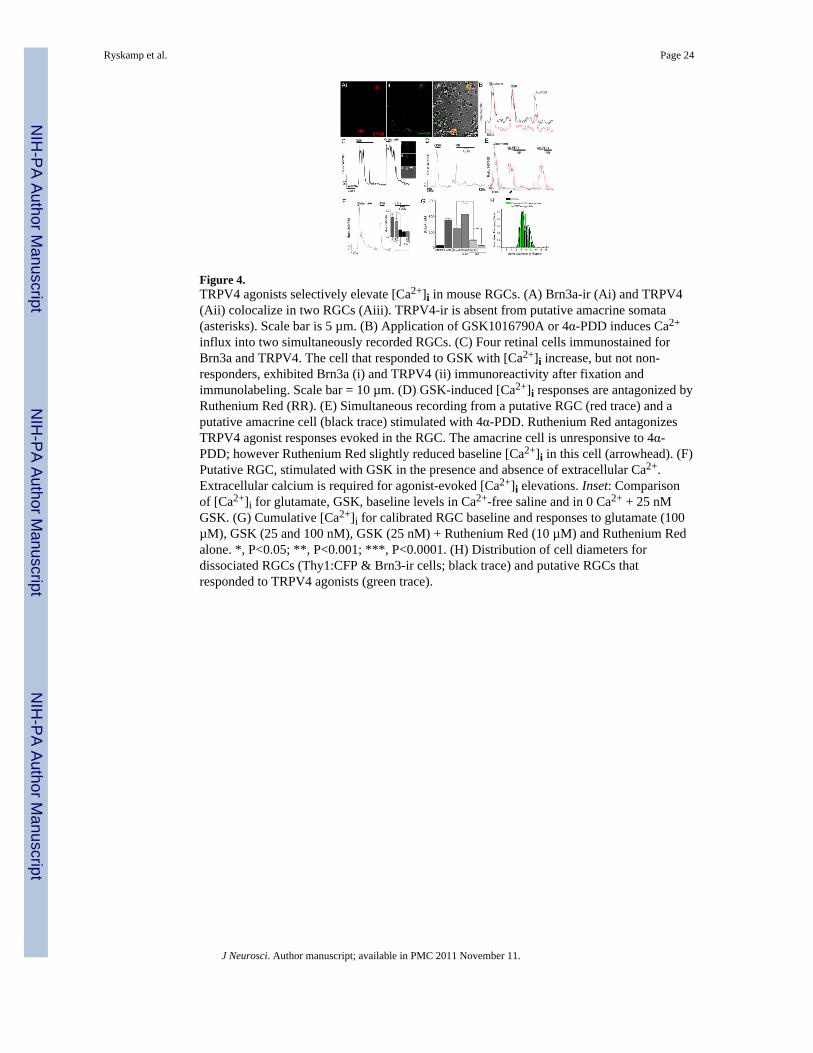
Figure 4.TRPV4 agonists selectively elevate [Ca2+]i in mouse RGCs. (A) Brn3a-ir (Ai) and TRPV4(Aii) colocalize in two RGCs (Aiii). TRPV4-ir is absent from putative amacrine somata(asterisks). Scale bar is 5 µm. (B) Application of GSK1016790A or 4α-PDD induces Ca2+
influx into two simultaneously recorded RGCs. (C) Four retinal cells immunostained forBrn3a and TRPV4. The cell that responded to GSK with [Ca2+]i increase, but not non-responders, exhibited Brn3a (i) and TRPV4 (ii) immunoreactivity after fixation andimmunolabeling. Scale bar = 10 µm. (D) GSK-induced [Ca2+]i responses are antagonized byRuthenium Red (RR). (E) Simultaneous recording from a putative RGC (red trace) and aputative amacrine cell (black trace) stimulated with 4α-PDD. Ruthenium Red antagonizesTRPV4 agonist responses evoked in the RGC. The amacrine cell is unresponsive to 4α-PDD; however Ruthenium Red slightly reduced baseline [Ca2+]i in this cell (arrowhead). (F)Putative RGC, stimulated with GSK in the presence and absence of extracellular Ca2+.Extracellular calcium is required for agonist-evoked [Ca2+]i elevations. Inset: Comparisonof [Ca2+]i for glutamate, GSK, baseline levels in Ca2+-free saline and in 0 Ca2+ + 25 nMGSK. (G) Cumulative [Ca2+]i for calibrated RGC baseline and responses to glutamate (100µM), GSK (25 and 100 nM), GSK (25 nM) + Ruthenium Red (10 µM) and Ruthenium Redalone. *, P<0.05; **, P<0.001; ***, P<0.0001. (H) Distribution of cell diameters fordissociated RGCs (Thy1:CFP & Brn3-ir cells; black trace) and putative RGCs thatresponded to TRPV4 agonists (green trace).
Ryskamp et al. Page 24
J Neurosci. Author manuscript; available in PMC 2011 November 11.
NIH
-PA Author Manuscript
NIH
-PA Author Manuscript
NIH
-PA Author Manuscript

Figure 5.GSK-induced [Ca2+]i dynamics in a RGC. (A) Transmitted image of a putative RGC androd photoreceptor. Scale bar = 5 µm. The temporal progression of upper and lower imagesequences is illustrated by the adjacent traces. (C–D) The RGC responds to the bathapplication of glutamate with a large increase in [Ca2+]i. (E–F) Washout. Scale bar in Fdenotes the 340/380 ratio level. (H–K) Application of GSK transiently elevates [Ca2+]i inthe RGC but not the rod. (J–K) [Ca2+]i levels decline in the continued presence of GSK. (L)Washout.
Ryskamp et al. Page 25
J Neurosci. Author manuscript; available in PMC 2011 November 11.
NIH
-PA Author Manuscript
NIH
-PA Author Manuscript
NIH
-PA Author Manuscript

Figure 6.Cell stretch elevates [Ca2+]RGC. (A) Simultaneous recording of RGC volume (calcein, greentrace), 340 nm (red trace), 380 nm (blue trace) intensities and [Ca2+]i (fura-2 ratio, blacktrace). An increase in RGC volume was indicated by a drop in calcein fluorescence intensityduring hypotonic stimulation (decreased osmolarity in superfusing saline from 280 mOsm to192 mOsm). Glutamate stimulation increased [Ca2+]i but evoked no change in the calceinsignal. The increase in [Ca2+]i induced by hypotonic stimulation was antagonized by Gd3+.(B) Hypotonically induced [Ca2+]i elevations are antagonized by Ruthenium Red. (C)Cumulative total for calibrated RGC [Ca2+]i responses to glutamate (100 µM), hypotonicstimulation (192 mOsm), and hypotonic stimulation in the presence of capsazepine,Ruthenium Red (10 µM) or Gd3+ (100 µM). NS, P>0.05; *, P<0.05; **, P<0.001; ***,P<0.0001. (D) [Ca2+]i trace from a GSK-sensitive RGC. Hypotonically evoked [Ca2+]iincrease requires Ca2+ influx from the extracellular space. Inset: Relative [Ca2+]i totals forcontrol and hypotonically stimulated RGCs with and without the presence of extracellularcalcium. (E) Distribution of cell diameters from dissociated RGCs (Thy1:CFP & Brn3-ircells; black trace) and the hypotonicity-responding cohort (green trace). (F) Hypotonicity-responding cell, labeled with TRPV4 and Brn3a antibodies. Scale bar is 10 µm. (G)Hypotonicity-induced membrane stretch occludes the response to GSK. The volumedecrease following the hypotonic step was associated with a transient increase in [Ca2+]i.(H) Cumulative calibrated responses to glutamate (100 µM), hypotonic stimulation (190 to195 mOsm), GSK (25 nM) and GSK in the presence of hypotonic stretch shows a markedreduction in the amplitude of the TRPV4 agonist-evoked response during mechanicalstimulation.
Ryskamp et al. Page 26
J Neurosci. Author manuscript; available in PMC 2011 November 11.
NIH
-PA Author Manuscript
NIH
-PA Author Manuscript
NIH
-PA Author Manuscript

Figure 7.TRPV4 agonists transiently increase spontaneous firing rate of RGCs. A. The spontaneousfiring rates of three RGCs recorded on a multielectrode array are shown during theapplication of GSK at time zero (300 nM; black bar). The rate increased for 2–3 min beforereturning to the initial baseline level during the continued presence of the drug for nearly allresponsive cells. (B) A histogram of the change in firing rates of individual RGCs during the3 min after 4α-PDD (5 µM) application compared to the pre-treatment firing rate is shownfor all cells recorded from 3 treated retinas. Cells with a rate double or higher in drug arecombined in the rightmost bin (“>100”; arrow); nearly half of the cells were in this category,and most cells increased their firing. As with GSK, the increase was transient (notillustrated). (C) Histogram as in B, but here the applied drug was GSK (100 or 300 nM; N =4 retinas). As in B, cells with a spike rate double or higher during the initial period of drugexposure are combined in the rightmost bin (“>100”; arrow), constituting over 12% of therecorded cells. The bias in the distribution to positive values reveals that most cellsincreased firing in response to GSK application.
Ryskamp et al. Page 27
J Neurosci. Author manuscript; available in PMC 2011 November 11.
NIH
-PA Author Manuscript
NIH
-PA Author Manuscript
NIH
-PA Author Manuscript

Figure 8.Exposure to TRPV4 agonist induces RGC apoptosis. (A) TUNEL (FITC) + transmittedimage in control dissociated mouse retinal cells. No TUNEL staining is observed undercontrol conditions. (B) TUNEL staining + transmitted image. Cells were exposed to kainate(KA; 10 µM) for 1 hour. (C & D) Transmitted image and TUNEL staining in cells exposedfor 1 hour to 25 nM GSK. Arrowheads point at putative RGCs; arrows point at putativephotoreceptors. (D) Cumulative data for putative photoreceptors (cell diameters 3–5 µm)and putative RGCs (cell diameters >6 µm) in L15 medium, 25 nM GSK, 100 nM GSK andKA in all experiments.
Ryskamp et al. Page 28
J Neurosci. Author manuscript; available in PMC 2011 November 11.
NIH
-PA Author Manuscript
NIH
-PA Author Manuscript
NIH
-PA Author Manuscript

NIH
-PA Author Manuscript
NIH
-PA Author Manuscript
NIH
-PA Author Manuscript
Ryskamp et al. Page 29
Table 1
Target Company Catalog Number
TRPV4 Alomone Labs ACC-034 rabbit 1:500–1:1000
TRPV4 MBL International Corporation LS-A8583 rabbit 1:200–1:1000
TRPV4Affinity Bioreagents/ThermoScientific Pierce/OSenses OSR00136W rabbit 1:200–1:1000
TRPV4 Abcam Ab63003 rabbit 1:200–1:1000
TRPV4 Lifespan Biosciences LS-C94498 rabbit 1:200–1:1000
Brn3a Santa Cruz Biotechnology sc-8429 mouse 1:100
Conearrestin
Gift from Dr. Wolfgang Baehr, Univ.of Utah mouse 1:100–1:500
Rhodopsin Santa Cruz Biotechnology sc-57433 mouse 1:100
ChAT Chemicon AB144-P goat 1:200
GAD-65Developmental Studies HybridomaBank GAD-6 mouse 1:1000
GFAP Sigma G3893 mouse 1:500
GFP Santa Cruz Biotechnology sc-9996 mouse 1:100–1:500
J Neurosci. Author manuscript; available in PMC 2011 November 11.


