Embed Size (px)
Citation preview
© 2013 Copyright by Arup
The pressure response in the brain during short duration impacts
C. Pearce1, P.G. Young
1, L. Cowlam
2, and B. Walker
2
1College of Engineering, Mathematics and Physical Sciences, University of Exeter, Harrison Building,
North Park Road, Exeter, EX4 4QF, UK. Contact: [email protected] 2Arup, UK
1 Abstract
The mechanisms which lead to brain injury in response to blunt head impacts are investigated using three finite-element models of the human head, which range from low to high biofidelity. The models were developed directly from MRI image data using a technique adapted from the marching cubes approach which automates the generation of meshes and allows for a number of different structures (e.g. skull, scalp, brain) to be meshed simultaneously. Experiments were carried out on the finite-element models to validate an analytical representation of head impact based on full 3D elasticity equations developed by one of the authors, and good agreement was observed. The analytical and numerical models were used in parallel to explore the phenomenon of large transient pressure magnification in the brain. This behaviour, proposed by one of the authors, occurs as a result of low duration low velocity impacts. The implications of these high pressure transients are also discussed. Finally individual case studies demonstrate the relevance of this research to realistic head injury scenarios.
2 Introduction
2.1 Impact induced head injury
Every year in the United States roughly 1.7 million people sustain traumatic brain injury (TBI), and of these 52,000 die. The US National Institute of Health estimated that, for the year 2000 alone, the direct medical costs of TBI and indirect costs through loss of productivity totalled $60 billion. [1] When biological structures are subjected to impact, injury will occur if the tissues deform beyond their recoverable limit. [2] TBI is commonly caused by impact to the head, which can be broadly classified as being either blunt or penetrating depending on whether or not the cranial vault is breached. As opposed to penetrating injuries, the mechanisms of blunt impact injury remain poorly understood and cannot be well predicted. [3] The current work investigates blunt head impact and is concerned mainly with the intracranial pressure response. The efficacy of pressure as a direct quantitative measure of tissue damage has been debated, particularly when predicting the onset of diffuse axonal injury (DAI) which is a function of strain not pressure. [4] However, the aim of the current investigation is not to provide an absolute measure of injury severity, but to qualitatively describe an injury mechanism that is convenient to express in terms of intracranial pressure. Furthermore, intracranial pressure is still regarded as a good indicator of injury severity and has been linked to cerebral contusion. [3], [5]
2.2 Previous work: numerical study
In the majority of blunt head impact experiments (whether analytical, numerical, or cadaveric) the range of contact durations investigated typically lies within a narrow window of 3 to 10 ms. In comparatively long duration, non-rotational impacts such as these, it has previously been shown that the resulting intracranial pressures are primarily caused by linear rigid-body acceleration of the skull. [4], [6] Therefore, if the contribution of the viscoelastic properties of brain tissue can be negated (as supported by the literature [7], [8]), the pressure response will be essentially quasi-static: a linear pressure gradient will exist throughout the cranial contents, with a maximum of positive pressure Pquasi under the site of impact (the “coup”) varying to a minimum of negative pressure of equal magnitude –Pquasi at the opposite pole (the “contrecoup”).
9th European LS-DYNA Conference 2013 _________________________________________________________________________________
© 2013 Copyright by Arup
This quasi-static intracranial pressure response can be straightforwardly described by the expression:
(1)
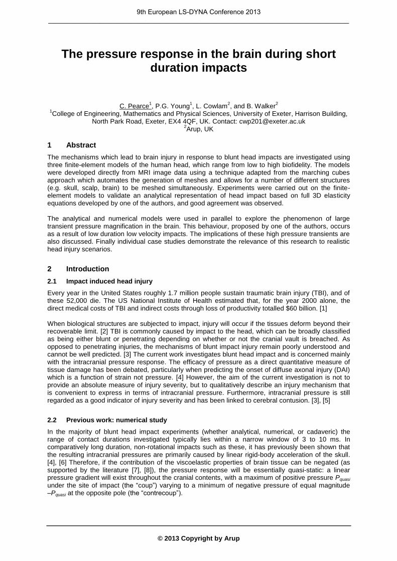
where rc is the radius of the brain from its centre of gravity to its exterior at the coup or contrecoup, ρ represents the density of the brain, Fmax is the peak force transferred by the impactor, and m is the total mass of the head. [6] There is a surprisingly limited amount of research which explores impacts below 3 ms, and hence investigates the intracranial response outside of this quasi-static domain. In their paper [6], Young and Morfey performed a range of simulated impacts on a simple 2-phase finite-element model of the human head, approximating it as a fluid-filled sphere. An outer spherical shell which was based on representative geometric values of the human skull (radius, thickness) was filled with an inviscid fluid representing the brain. Appropriate linear elastic material properties were assigned to these structures. The simplified model was employed in order to reduce the number of variables of the system, such that it was easier to identify critical parameters which have a large effect. The finite-element model used was in the form of a 30º wedge, with symmetry conditions applied such that it behaved as a full sphere. Impact loading was replicated indirectly through the application of a pressure-time history over a circular cap on the exterior of the shell, defined by a sector half angle of 15º. The pressure-time history followed a Hanning squared function (governed by force F(t) in Equation 2), which is a reasonable approximation to actual impact loading.
(2)
where t is time, and Tp is the impact duration. [6] Parametric studies were undertaken in order to explore the sensitivity of the brain’s pressure response to changes in loading parameters. A range of impacts were performed with varying impact duration Tp, while all other parameters, including the peak impact force Fmax, were held constant.
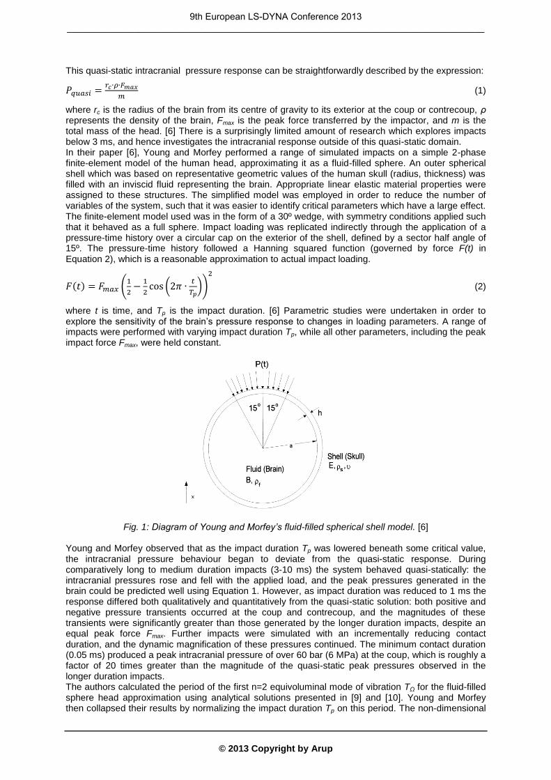
Fig. 1: Diagram of Young and Morfey’s fluid-filled spherical shell model. [6] Young and Morfey observed that as the impact duration Tp was lowered beneath some critical value, the intracranial pressure behaviour began to deviate from the quasi-static response. During comparatively long to medium duration impacts (3-10 ms) the system behaved quasi-statically: the intracranial pressures rose and fell with the applied load, and the peak pressures generated in the brain could be predicted well using Equation 1. However, as impact duration was reduced to 1 ms the response differed both qualitatively and quantitatively from the quasi-static solution: both positive and negative pressure transients occurred at the coup and contrecoup, and the magnitudes of these transients were significantly greater than those generated by the longer duration impacts, despite an equal peak force Fmax. Further impacts were simulated with an incrementally reducing contact duration, and the dynamic magnification of these pressures continued. The minimum contact duration (0.05 ms) produced a peak intracranial pressure of over 60 bar (6 MPa) at the coup, which is roughly a factor of 20 times greater than the magnitude of the quasi-static peak pressures observed in the longer duration impacts. The authors calculated the period of the first n=2 equivoluminal mode of vibration TΩ for the fluid-filled sphere head approximation using analytical solutions presented in [9] and [10]. Young and Morfey then collapsed their results by normalizing the impact duration Tp on this period. The non-dimensional
9th European LS-DYNA Conference 2013 _________________________________________________________________________________
© 2013 Copyright by Arup
ratio Tp / TΩ was found to be an excellent predictor of the system’s response, and the authors were able to discern a critical value of approximately Tp / TΩ = 2, above which quasi-static behaviour is observed, and below which dynamic pressure magnification occurs. [6] This intracranial pressure behaviour that develops as a result of short duration impacts was termed the “dynamic pressure magnification” response. This response is characterised by pressure transients at the coup and contrecoup in the brain, which are much larger than those expected based on the quasi-static solution, and which fluctuate, alternating sign between positive and negative pressure.
Fig. 2: Pressure response in the brain at the coup and contrecoup during Young and Morfey’s experiments, for Tp = 1, 3, and 10 ms. [6]
2.3 Previous work: analytical modelling and experimental validation
In previous work [11] one of the authors developed an analytical model describing the head (approximated again by a fluid-filled sphere) subject to impact by a spherical mass. The model considers Hertzian contact stiffness and local bending and membrane stiffness in a decoupled manner, which allowed the derivation of explicit expressions for global impact characteristics such as: impact duration, peak impact force, peak acceleration, and the non-dimensional ratio Tp / TΩ. The dynamic pressure magnification response was validated and explored experimentally by Johnson and Young in [12] and [13] through a series of impact studies on physical head models (a polymer spherical shell, a rapid prototyped model of a skull based on in vivo MRI scan data, and a cadaveric skull) filled with fluid. The results agreed well with the observed behaviour in Young and Morfey’s original paper [6] and the behaviour predicted by the analytical model [11].
3 Methodology
Recent advances in image processing techniques allow the conversion of volume-capture scan data directly into finite-element meshes. [14] “Image-based meshing” is a semi-automated method, making it feasible to perform in depth numerical analysis on geometrically accurate representations of complex structures. This method is therefore particularly suited to biomechanical investigations. The current research aims to explore the mechanics of the dynamic pressure magnification effect observed in previous work, the causative mechanism of this intracranial response, and whether or not it is applicable to real world head impacts, i.e. whether it is simply an artefact of the large assumptions (in particular, the spherical geometry) made in Young and Morfey’s original model. The current research employs three finite-element models of the human head, from low to high biofidelity. Parametric studies were carried out on these models to investigate the intracranial pressure response to varying impact parameters. All model generation and meshing was performed in the ScanIP, +FE, and +CAD software packages (Simpleware Ltd.), while impacts were simulated using the LS-DYNA® explicit finite-element code (LSTC Inc.). Model pre- and post-processing utilised the Oasys LS-DYNA Environment (Oasys Ltd.).
0 2 4 6 8 10 12
Time (ms)
-8
-6
-4
-2
0
2
4
6
8P
ressu
re (
ba
rs)
T p = 1 ms
T p = 3 ms
T p = 10 ms
Quasi-static for 10 ms
1 ms
3 ms
10 ms
10 ms
3 ms
1 ms
TIME (ms)0 Tp
FO
RC
E (
kN
)
0
10
TIME HISTORY OF IMPACT
9th European LS-DYNA Conference 2013 _________________________________________________________________________________
© 2013 Copyright by Arup
3.1 Fluid-filled sphere: Stage 1
The preliminary model approximates the human head as a fluid-filled sphere with the same dimensions and material properties as Young and Morfey’s original work [6]. The external radius of the spherical skull was 80.01 mm and it had a thickness of 3.81 mm. The skull bone was represented by a simple linear elastic material with modulus E of 13.79 GPa, Poisson’s ratio ν of 0.25, and density ρ of 2140 kg/m
3. The interior volume of the shell was filled with an “elastic fluid” material
(MAT_001_FLUID) representing the brain, with bulk modulus B of 2.18 GPa, and density ρ of 1002 kg/m
3. These values were originally taken from [15] and represent reasonable approximate
dimensions and properties of the average human skull. Rather than the application of a predefined pressure-time history, impacts were simulated directly through the collinear collision of the initially stationary head with a spherical impactor mass of radius 40 mm. The impactor was linear elastic with a modulus of 0.8 GPa, and Poisson’s ratio of 0.25. The parametric study took the form of a series of 10 impact simulations, in which the initial velocity of this impactor was varied incrementally from 0.2 to 3.8 m/s. The analytical expressions developed in [11] were employed to estimate the peak impact force resulting from each impact case, and the mass of the impactor was changed accordingly such that the predicted peak impact force remained constant across all cases. In this way the mass of the impactor was incrementally reduced from 8.0 to 0.022 kg. This resulted in a range of impacts measuring from 0.34 to 3.32 ms in duration, which had approximately equal peak force (roughly 800 N).
3.2 Skull and brain: Stage 2
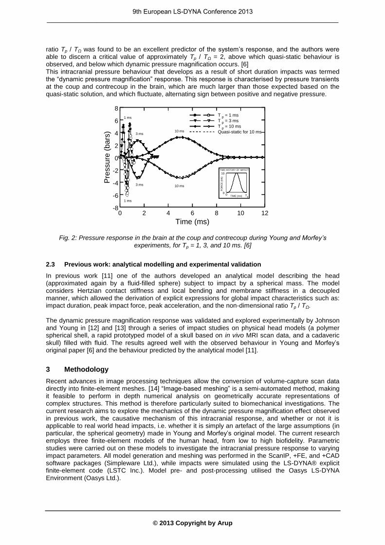
The second model was similar to the first, in that it was a simple 2-phase model comprising of only the skull and brain, with identical material properties applied to these. However, the geometry was modified by replacing the spherical skull with realistic skull geometry extracted from in vivo high resolution T1-weighted MRI scan data of a male volunteer (between 25 and 30 years of age, and of average height and build). As before, the cranial cavity was occupied by elastic fluid representing the brain. Impacts were performed using the same impactor as Stage 1, in the posterior-anterior direction, and were aligned such that the axis of impact passes through the head model’s centre of gravity. A parametric study was carried out in the same manner as above: 12 impacts of approximately equal peak force (970 N) were simulated producing impacts measuring from 0.17 to 3.28 ms in duration.
Fig. 3: a) Stage 2 model, isometric view. b) Stage 2 model and spherical impactor, mid-sagittal section view.
3.3 Full biofidelic head: Stage 3
The final biofidelic head model was constructed from the full set of MRI data from which the skull had been extracted in Stage 2. The skull, vertebrae, intervertebral discs, cerebrum, cerebellum, brain stem and spinal cord, cerebrospinal fluid (CSF), scalp and surrounding flesh were all segmented as separate regions and meshed simultaneously in ScanIP. Representative linear material properties were applied to these structures based on a comprehensive review of the literature. Structures deemed of particular importance to the impact response were assigned more complex material
9th European LS-DYNA Conference 2013 _________________________________________________________________________________
© 2013 Copyright by Arup
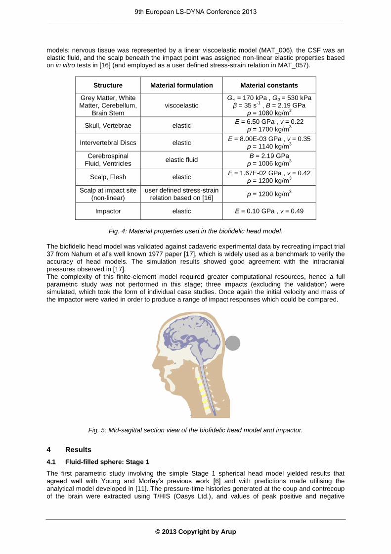
models: nervous tissue was represented by a linear viscoelastic model (MAT_006), the CSF was an elastic fluid, and the scalp beneath the impact point was assigned non-linear elastic properties based on in vitro tests in [16] (and employed as a user defined stress-strain relation in MAT_057).
Structure Material formulation Material constants
Grey Matter, White Matter, Cerebellum,
Brain Stem viscoelastic
G∞ = 170 kPa , G0 = 530 kPa β = 35 s
-1 , B = 2.19 GPa
ρ = 1080 kg/m3
Skull, Vertebrae elastic E = 6.50 GPa , ν = 0.22
ρ = 1700 kg/m3
Intervertebral Discs elastic E = 8.00E-03 GPa , ν = 0.35
ρ = 1140 kg/m3
Cerebrospinal Fluid, Ventricles
elastic fluid B = 2.19 GPa
ρ = 1006 kg/m3
Scalp, Flesh elastic E = 1.67E-02 GPa , ν = 0.42
ρ = 1200 kg/m3
Scalp at impact site (non-linear)
user defined stress-strain relation based on [16]
ρ = 1200 kg/m3
Impactor elastic E = 0.10 GPa , ν = 0.49
Fig. 4: Material properties used in the biofidelic head model.
The biofidelic head model was validated against cadaveric experimental data by recreating impact trial 37 from Nahum et al’s well known 1977 paper [17], which is widely used as a benchmark to verify the accuracy of head models. The simulation results showed good agreement with the intracranial pressures observed in [17]. The complexity of this finite-element model required greater computational resources, hence a full parametric study was not performed in this stage; three impacts (excluding the validation) were simulated, which took the form of individual case studies. Once again the initial velocity and mass of the impactor were varied in order to produce a range of impact responses which could be compared.
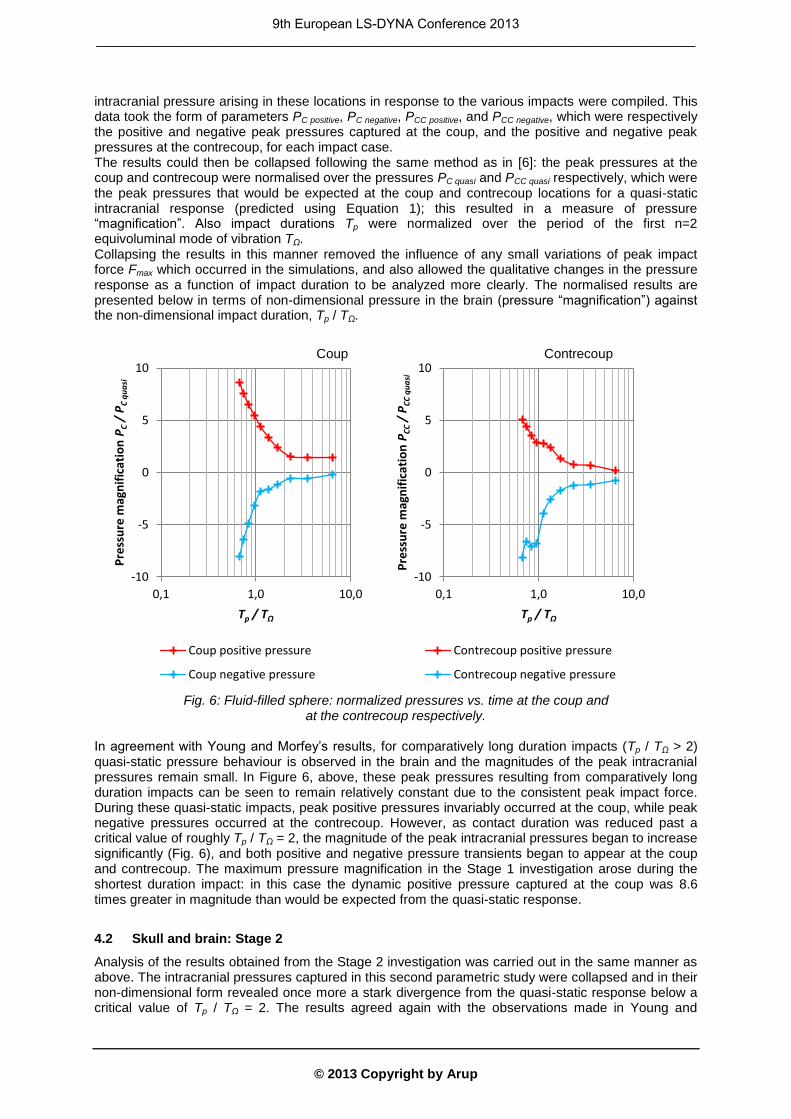
Fig. 5: Mid-sagittal section view of the biofidelic head model and impactor.
4 Results
4.1 Fluid-filled sphere: Stage 1
The first parametric study involving the simple Stage 1 spherical head model yielded results that agreed well with Young and Morfey’s previous work [6] and with predictions made utilising the analytical model developed in [11]. The pressure-time histories generated at the coup and contrecoup of the brain were extracted using T/HIS (Oasys Ltd.), and values of peak positive and negative
9th European LS-DYNA Conference 2013 _________________________________________________________________________________
© 2013 Copyright by Arup
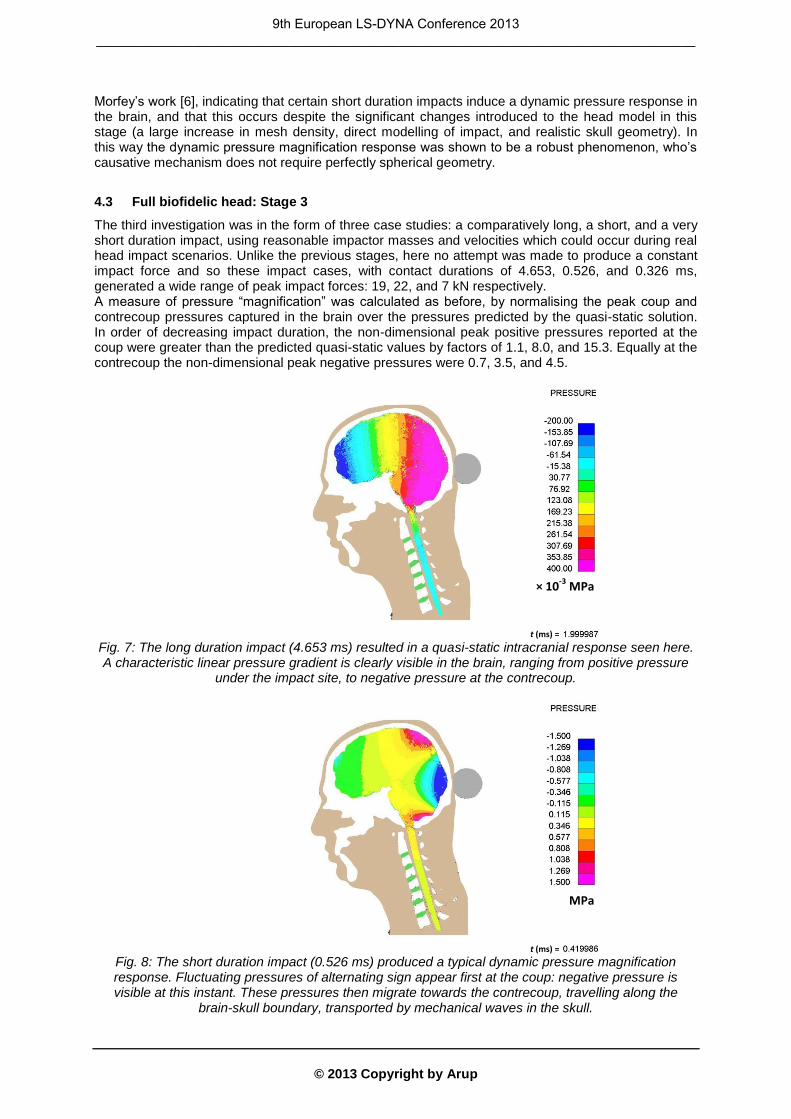
intracranial pressure arising in these locations in response to the various impacts were compiled. This data took the form of parameters PC positive, PC negative, PCC positive, and PCC negative, which were respectively the positive and negative peak pressures captured at the coup, and the positive and negative peak pressures at the contrecoup, for each impact case. The results could then be collapsed following the same method as in [6]: the peak pressures at the coup and contrecoup were normalised over the pressures PC quasi and PCC quasi respectively, which were the peak pressures that would be expected at the coup and contrecoup locations for a quasi-static intracranial response (predicted using Equation 1); this resulted in a measure of pressure “magnification”. Also impact durations Tp were normalized over the period of the first n=2 equivoluminal mode of vibration TΩ. Collapsing the results in this manner removed the influence of any small variations of peak impact force Fmax which occurred in the simulations, and also allowed the qualitative changes in the pressure response as a function of impact duration to be analyzed more clearly. The normalised results are presented below in terms of non-dimensional pressure in the brain (pressure “magnification”) against the non-dimensional impact duration, Tp / TΩ.
Fig. 6: Fluid-filled sphere: normalized pressures vs. time at the coup and
at the contrecoup respectively. In agreement with Young and Morfey’s results, for comparatively long duration impacts (Tp / TΩ > 2) quasi-static pressure behaviour is observed in the brain and the magnitudes of the peak intracranial pressures remain small. In Figure 6, above, these peak pressures resulting from comparatively long duration impacts can be seen to remain relatively constant due to the consistent peak impact force. During these quasi-static impacts, peak positive pressures invariably occurred at the coup, while peak negative pressures occurred at the contrecoup. However, as contact duration was reduced past a critical value of roughly Tp / TΩ = 2, the magnitude of the peak intracranial pressures began to increase significantly (Fig. 6), and both positive and negative pressure transients began to appear at the coup and contrecoup. The maximum pressure magnification in the Stage 1 investigation arose during the shortest duration impact: in this case the dynamic positive pressure captured at the coup was 8.6 times greater in magnitude than would be expected from the quasi-static response.
4.2 Skull and brain: Stage 2
Analysis of the results obtained from the Stage 2 investigation was carried out in the same manner as above. The intracranial pressures captured in this second parametric study were collapsed and in their non-dimensional form revealed once more a stark divergence from the quasi-static response below a critical value of Tp / TΩ = 2. The results agreed again with the observations made in Young and
-10
-5
0
5
10
0,1 1,0 10,0
Pre
ssu
re m
agn
ific
atio
n P
C /
PC
qu
asi
Tp / TΩ
Coup positive pressure
Coup negative pressure
-10
-5
0
5
10
0,1 1,0 10,0
Pre
ssu
re m
agn
ific
atio
n P
CC /
PC
C q
ua
si
Tp / TΩ
Contrecoup positive pressure
Contrecoup negative pressure
Coup Contrecoup
9th European LS-DYNA Conference 2013 _________________________________________________________________________________
© 2013 Copyright by Arup
Morfey’s work [6], indicating that certain short duration impacts induce a dynamic pressure response in the brain, and that this occurs despite the significant changes introduced to the head model in this stage (a large increase in mesh density, direct modelling of impact, and realistic skull geometry). In this way the dynamic pressure magnification response was shown to be a robust phenomenon, who’s causative mechanism does not require perfectly spherical geometry.
4.3 Full biofidelic head: Stage 3
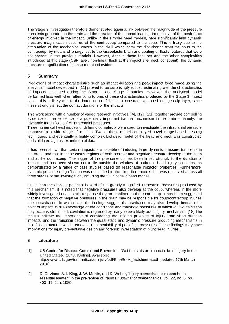
The third investigation was in the form of three case studies: a comparatively long, a short, and a very short duration impact, using reasonable impactor masses and velocities which could occur during real head impact scenarios. Unlike the previous stages, here no attempt was made to produce a constant impact force and so these impact cases, with contact durations of 4.653, 0.526, and 0.326 ms, generated a wide range of peak impact forces: 19, 22, and 7 kN respectively. A measure of pressure “magnification” was calculated as before, by normalising the peak coup and contrecoup pressures captured in the brain over the pressures predicted by the quasi-static solution. In order of decreasing impact duration, the non-dimensional peak positive pressures reported at the coup were greater than the predicted quasi-static values by factors of 1.1, 8.0, and 15.3. Equally at the contrecoup the non-dimensional peak negative pressures were 0.7, 3.5, and 4.5.
Fig. 7: The long duration impact (4.653 ms) resulted in a quasi-static intracranial response seen here. A characteristic linear pressure gradient is clearly visible in the brain, ranging from positive pressure
under the impact site, to negative pressure at the contrecoup.
Fig. 8: The short duration impact (0.526 ms) produced a typical dynamic pressure magnification response. Fluctuating pressures of alternating sign appear first at the coup: negative pressure is visible at this instant. These pressures then migrate towards the contrecoup, travelling along the
brain-skull boundary, transported by mechanical waves in the skull.
× 10-3
MPa
× 10-3
MPa
t (ms) =
t (ms) =
9th European LS-DYNA Conference 2013 _________________________________________________________________________________
© 2013 Copyright by Arup
The Stage 3 investigation therefore demonstrated again a link between the magnitude of the pressure transients generated in the brain and the duration of the impact loading, irrespective of the peak force or energy involved in the impact. Unlike in the simpler head models, here significantly less dynamic pressure magnification occurred at the contrecoup compared to the coup. This is likely due to the attenuation of the mechanical waves in the skull which carry the disturbance from the coup to the contrecoup, by means of energy lost to the viscoelastic brain and coating of flesh, features that were not present in the previous models. However, despite these features and the other complexities introduced at this stage (CSF layer, non-linear flesh at the impact site, neck constraint), the dynamic pressure magnification response remained evident.
5 Summary
Predictions of impact characteristics such as impact duration and peak impact force made using the analytical model developed in [11] proved to be surprisingly robust, estimating well the characteristics of impacts simulated during the Stage 1 and Stage 2 studies. However, the analytical model performed less well when attempting to predict these characteristics produced by the Stage 3 impact cases: this is likely due to the introduction of the neck constraint and cushioning scalp layer, since these strongly affect the contact durations of the impacts. This work along with a number of varied research initiatives ([6], [12], [13]) together provide compelling evidence for the existence of a potentially important trauma mechanism in the brain – namely, the “dynamic magnification” of intracranial pressures. Three numerical head models of differing complexity were used to investigate the intracranial pressure response to a wide range of impacts. Two of these models employed novel image-based meshing techniques, and eventually a highly complex biofidelic model of the head and neck was constructed and validated against experimental data. It has been shown that certain impacts are capable of inducing large dynamic pressure transients in the brain, and that in these cases regions of both positive and negative pressure develop at the coup and at the contrecoup. The trigger of this phenomenon has been linked strongly to the duration of impact, and has been shown not to lie outside the window of authentic head injury scenarios, as demonstrated by a range of case studies based on reasonable impactor properties. Furthermore, dynamic pressure magnification was not limited to the simplified models, but was observed across all three stages of the investigation, including the full biofidelic head model. Other than the obvious potential hazard of the greatly magnified intracranial pressures produced by this mechanism, it is noted that negative pressures also develop at the coup, whereas in the more widely investigated quasi-static response they are confined to the contrecoup. It has been suggested that the formation of negative pressures in the brain may be responsible for coup/contrecoup injuries due to cavitation: in which case the findings suggest that cavitation may also develop beneath the point of impact. While knowledge of the conditions and threshold pressures at which in vivo cavitation may occur is still limited, cavitation is regarded by many to be a likely brain injury mechanism. [18] The results indicate the importance of considering the inflated prospect of injury from short duration impacts, and the transition between the quasi-static and dynamic pressure producing mechanisms in fluid-filled structures which removes linear scalability of peak fluid pressures. These findings may have implications for injury preventative design and forensic investigation of blunt head injuries.
6 Literature
[1] US Centre for Disease Control and Prevention, “Get the stats on traumatic brain injury in the United States,” 2010. [Online]. Available: http://www.cdc.gov/traumaticbraininjury/pdf/BlueBook_factsheet-a.pdf (updated 17th March 2010).
[2] D. C. Viano, A. I. King, J. W. Melvin, and K. Weber, “Injury biomechanics research: an essential element in the prevention of trauma,” Journal of biomechanics, vol. 22, no. 5, pp. 403–17, Jan. 1989.
9th European LS-DYNA Conference 2013 _________________________________________________________________________________
© 2013 Copyright by Arup
[3] W. N. Hardy, T. B. Khalil, and A. I. King, “Literature review of head injury biomechanics,” International Journal of Impact Engineering, pp. 561–586, 1994.
[4] D. Bradshaw and C. L. Morfey, “Pressure and shear responses in brain injury models,” in Proceedings of the 17th International Technical Conference on the Enhanced Safety of Vehicles, 2001, pp. 1–10.
[5] F. A. Bandak, “Shaken baby syndrome: a biomechanics analysis of injury mechanisms,” Forensic science international, vol. 151, no. 1, pp. 71–9, Jun. 2005.
[6] P. G. Young and C. L. Morfey, “Intracranial pressure transients caused by head impacts,” in International Research Council on the Biomechanics of Impact (IRCOBI) Conference Proceedings, 1998, pp. 391–403.
[7] A. Kuijpers, M. H. A. Claessens, and A. Sauren, “A two-dimensional FEM analysis of the response of the human head to impact: the importance of boundary conditions,” Computer Methods in Biomechanics and Biomedical Engineering, pp. 207–216, 1996.
[8] R. Willinger, H. S. Kang, and B. Diaw, “Three-dimensional human head finite-element model validation against two experimental impacts,” Annals of biomedical engineering, vol. 27, no. 3, pp. 403–10, 1999.
[9] H. Jiang, P. G. Young, and S. Dickinson, “Natural frequencies of vibration of layered hollow spheres using exact three-dimensional elasticity equations,” Journal of Sound and Vibration, vol. 195, no. 1, pp. 155–162, 1996.
[10] P. G. Young, “A parametric study on the axisymmetric modes of vibration of multi-layered spherical shells with liquid cores of relevance to head impact modelling,” Journal of Sound and Vibration, vol. 256, no. 4, pp. 665–680, 2002.
[11] P. G. Young, “An analytical model to predict the response of fluid-filled shells to impact - a model for blunt head impacts,” Journal of Sound and Vibration, vol. 267, no. 5, pp. 1107–1126, Nov. 2003.
[12] E. Johnson and P. G. Young, “On the use of a patient-specific rapid-prototyped model to simulate the response of the human head to impact and comparison with analytical and finite element models,” Journal of biomechanics, vol. 38, no. 1, pp. 39–45, Jan. 2005.
[13] E. Johnson, “The response of the human head to blunt impact: experimental validation of an analytical model,” University of Exeter, 2005.
[14] P. G. Young, T. Beresford-West, S. Coward, B. Notarberardino, B. Walker, and A. Abdul-Aziz, “An efficient approach to converting three-dimensional image data into highly accurate computational models,” Philosophical Transactions of the Royal Society A: Mathematical, Physical and Engineering Sciences, vol. 366, no. 1878, p. 3155, 2008.
[15] A. E. Engin, “The axisymmetric response of a fluid-filled spherical shell to a local radial impulse – a model for head injury,” Journal of Biomechanics, vol. 2, no. 3, pp. 325–341, 1969.
[16] C. W. Gadd, A. M. Nahum, D. C. Schneider, and R. G. Madeira, “Tolerance and properties of superficial soft tissues in situ,” in Proceedings of the 14th STAPP Car Crash Conference, 1970, pp. 356–368.
[17] A. M. Nahum, R. Smith, and C. C. Ward, “Intracranial pressure dynamics during head impact,” New Orleans, LA, USA, 1977.
9th European LS-DYNA Conference 2013 _________________________________________________________________________________

© 2013 Copyright by Arup
[18] C. Brennen, “Cavitation in biological and bioengineering contexts,” in Proceedings of the 5th International Symposium on Cavitation, 2003.
9th European LS-DYNA Conference 2013 _________________________________________________________________________________


