Embed Size (px)
Citation preview

Biophysical Journal Volume 106 May 2014 2085–2095 2085
The Proapoptotic Protein tBid Forms Both Superficially Bound andMembrane-Inserted Oligomers
Sanjeevan Shivakumar,†Martin Kurylowicz,‡Nehad Hirmiz,‡Yaseen Manan,†Ouided Friaa,‡Aisha Shamas-Din,†
Pourya Masoudian,† Brian Leber,†§ David W. Andrews,† and Cecile Fradin†‡*†Department of Biochemistry and Biomedical Sciences, ‡Department of Physics and Astronomy, and §Department of Medicine, McMasterUniversity, Hamilton, Ontario, Canada
ABSTRACT Bid is a proapopotic activator protein of the Bcl-2 family that plays a pivotal role in controlling mitochondrial outermembrane permeabilization during apoptosis. Here, we characterized the interaction of fluorescently labeled truncated Bid(tBid) with a mitochondria-like supported lipid bilayer at the single-molecule level. The proteins observed at the membrane ex-hibited a very wide range of mobility. Confocal images of the membrane displayed both diffraction-limited Gaussian spots andhorizontal streaks, corresponding to immobile and mobile tBid species, respectively. We observed 1), fast-diffusing proteins cor-responding to a loosely, probably electrostatically bound state; 2), slowly diffusing proteins, likely corresponding to a superficiallyinserted state; and 3), fully immobilized proteins, suggesting a fully inserted state. The stoichiometry of these proteins was deter-mined by normalizing their fluorescence intensity by the brightness of a tBid monomer, measured separately using fluorescencefluctuation techniques. Strikingly, the immobile species were found to be mainly tetramers and higher, whereas the mobile spe-cies had on average a significantly lower stoichiometry. Taken together, these results show that as soluble Bid progressestoward a membrane-inserted state, it undergoes an oligomerization process similar to that observed for Bax.
INTRODUCTION
Apoptosis is a form of programmed cell death vital fordevelopment and homeostasis in multicellular organisms.In mammals, the initiation of apoptosis is tightly regulatedthrough a network of protein-protein and protein-membraneinteractions involving the Bcl-2 family of proteins (1,2).Bcl-2 proteins are functionally divided into two groups,anti- and proapoptotic. Antiapoptotic family members,such as Bcl-XL, share four conserved Bcl-2 homology(BH) regions, and they inhibit the onset of apoptosis. Proap-optotic members are further subdivided into two categories.Multiregion proapoptotic proteins, such as Bax, contain BH1–3 regions and permeabilize organelle membranes by poreformation. Proapoptotic BH3 proteins contain only the BH3region and function as inhibitors of the antiapoptotic pro-teins (3,4) or as activators that also promote pore formationby activating the multiregion proapoptotic proteins (5,6).
The BH3-interacting domain death agonist (Bid) is a22 kDa protein that has been classified as a proapoptoticBH3 activator (5–8). Bid function is regulated by a numberof posttranslational modifications. Of particular importancein apoptosis is the cleavage of cytosolic Bid by caspase-8in response to apoptotic stimuli (9). The two fragments ofcleaved Bid (cBid) remain bound together through hydro-phobic interactions in solution, but in the presence of mito-chondrial membranes they rapidly separate, and the larger,
Submitted September 25, 2013, and accepted for publication March 31,
2014.
*Correspondence: [email protected]
David W. Andrews’s present address is Sunnybrook Research Institute,
Toronto, ON, M4N 3M5, Canada.
Editor: Paul Wiseman.
� 2014 by the Biophysical Society
0006-3495/14/05/2085/11 $2.00
15 kDa fragment, known as truncated Bid (tBid), insertsinto the lipid bilayer (10,11). Membrane-bound tBid recruitscytosolic Bax to mitochondria, resulting in mitochondrialouter-membrane permeabilization followed by release ofapoptotic factors in the cytoplasm, activation of cellular cas-pases, and, ultimately, cell death (12). Interestingly, tBidalso recruits antiapoptotic Bcl-XL to bind membranes (13).
Evidence suggests that despite its classification as a BH3protein, Bid resembles the multiregion Bcl-2 proteins Baxand Bcl-XL in many respects (14). Unlike other BH3 pro-teins, which are unstructured, Bid maintains a helical struc-ture in solution when it is not membrane-associated, with afold similar to that of Bax and Bcl-XL (15). The conforma-tional changes that occur upon activation also exhibit simi-larities among the three proteins: hydrophobic helixes thatare buried in the interior of the protein when in solution(helices 6 and 7 for Bid) become exposed and promotemembrane binding (14). Furthermore, as we have recentlyshown, just like Bax, tBid may adopt multiple conforma-tions at the membrane, and these conformations are notfunctionally equivalent (11). Finally, Bid contains a struc-turally conserved hydrophobic pocket in the core of the pro-tein that resembles that of multiregion Bcl-2 proteins (16).These pockets play a central role in regulating the interac-tions between Bcl-2 family proteins. It is believed that theBH3 region of BH3 proteins can insert into the hydrophobicpocket of multiregion proteins (17). The presence of ahomologous hydrophobic pocket in Bid suggests the possi-bility for interactions with other BH3 proteins. In particular,it raises the question of whether tBid might be able to formhomooligomers when interacting with mitochondrial mem-branes, a hypothesis supported by previous cross-linking
http://dx.doi.org/10.1016/j.bpj.2014.03.049

2086 Shivakumar et al.
and fluorescence resonance energy transfer (FRET) experi-ments in apoptotic cells (18).
Investigations of the molecular mechanisms underlyingBid function have primarily used ensemble techniques,e.g., to characterize the interaction of Bid with other Bcl-2family proteins and with membranes (10,11,18,19). Yetfor proteins exhibiting different conformations and formingoligomers with multiple stoichiometries, the average dataobtained from ensemble methods may not provide a fulldescription of the system. We therefore used single-particlemethods to observe the behavior of individual Bid com-plexes when bound to supported lipid bilayers (SLBs)with complex mitochondria-like lipid composition. Molecu-lar complexes formed by tBid at the lipid bilayer wereimaged with both confocal and total internal reflection fluo-rescence (TIRF) microscopy and additionally characterizedby fluorescence correlation spectroscopy (FCS). We devel-oped what we believe to be a new way of analyzing confocalimages that allows simultaneous assessment of both theoligomeric state and the mobility of detected fluorescentobjects. The first is accomplished by performing a calibra-tion of the monomer specific brightness using fluorescenceintensity distribution analysis (FIDA), the latter by recog-nizing that immobile and mobile objects have different sig-natures in confocal images, with mobile objects appearingas horizontal streaks (20). We find that membrane-associ-ated tBid is present predominantly as monomers and dimersthat have a low to high mobility, but that higher order olig-omers with zero to low mobility are also present. We discussthe implications of these findings for the role of Bid inapoptosis.
MATERIALS AND METHODS
Protein purification and labeling
Recombinant full-length murine Bid with an N-terminal His-tag was puri-
fied, fluorescently labeled at position 126, and cleaved as previously
described (10,11,21). Labeling efficiency between 50 and 70% were
achieved for Alexa Fluor 488 (Invitrogen Molecular Probes, now Life
Technologies, Carlsbad, CA) and between 70 and 90% for the other fluoro-
phores used: Atto 647 from ATTO-TEC (Siegen, Germany) and 7-diethyla-
minocoumarin-3-carbonyl (DAC) and nitrobenzoxadiazole (NBD) from
Invitrogen. The activity of the purified labeled proteins was assessed by
measuring their capacity to induce Bax-mediated pore formation using a
well-established ANTS release assay (22).
Preparation of liposomes and SLBs
The reconstituted membranes used in this study had a lipid composition re-
flecting that of mitochondrial membranes: 48 mol % phosphatidylcholine
(PC), 28 mol % phosphatidylethanolamine (PE), 10 mol % phosphatidyli-
nositol (PI), 10 mol % phosphatidylserine (PS), and 4 mol % cardiolipin
(CL). This mainly represents an average between the composition of the
inner and outer mitochondrial membranes, as determined in yeast and
several vertebrates (23,24). The exception is cardiolipin, which is found
in large amounts (~25%) in the inner membrane, but which we used here
at the lower concentration (~4%) at which it is usually found in the outer
Biophysical Journal 106(10) 2085–2095
membrane. This mitochondria-like lipid composition has been shown to
support Bid- and Bax-mediated pore formation in liposomes (7,22,25).
All lipids were purchased from Avanti Polar Lipids (Alabaster, AL). Phos-
phatidylcholine and phosphatidylethanolamine were egg extracts (Avanti
840051 and 841118, both with 16:0/18:1 as the predominant acyl chain
composition). Phosphatidylinositol was liver extract (Avanti 840042,
predominant form 18:0/20:4). PS was synthesized as 18:1/18:1 PS
(DOPS, Avanti 840035). CL was synthesized as 18:1/18:1/18:1/18:1 CL
(TOCL, Avanti 710335). When needed, the membranes were fluorescently
labeled by adding either 0.0025 mol % (for single-particle tracking) or
0.008 mol % (for membrane position determination or liposome detection)
of lipophilic dye (DiO-C18 or DiD-C18, both from Invitrogen). Liposomes
were formed as previously described (10,11). SLBs were formed on mica
substrate by direct liposome fusion, following a protocol adapted from
Chiantia et al. (26) and Oreopoulos and Yip (27). Mica-coated coverslips
were mounted in an ~400 mL FCS2 perfusion chamber (Bioptechs, Butler,
PA). An assay buffer solution with 0.5 mg/mL liposome was injected into
the chamber, and the samples were then incubated at 37�C for 1 h to
encourage liposome fusion on the solid surface. Excess liposomes were
then removed by washing the membrane with ~5 mL assay buffer pumped
through the chamber at a rate of ~1.5 mL/min, after which the bilayer was
allowed to recover at 37�C for 15 min. When required, a 1 mL solution of
cBid diluted in assay buffer (with concentration 100 nM for ensemble bind-
ing measurements or 100–400 pM for single-particle measurements) was
injected into the chamber at a rate of ~1.5 mL/min, and incubated for
15 min at 37�C.
Steady-state fluorescence spectroscopy
The homooligomerization and membrane insertion of cBid were detected
concurrently using FRET and environment-sensitive fluorescence, mainly
as described before (11). To prevent protein adhesion, cuvettes were surfac-
tant-coated with DOPC. Assay buffer containing up to 8 nM mitochondria-
like liposomes (that is up to 0.5 mg/mL or 650 mM lipids) was then placed
in the cuvette, followed by 20 nM cBid-DAC (donor) and finally 100 nM
cBid-NBD (acceptor). Control reactions in the absence of acceptor, i.e.,
where cBid-NBD was replaced by unlabeled cBid, were carried out in par-
allel. The reaction was stirred and kept at 37�C. The NBD fluorescence
(lex ¼ 475 nm, lem ¼ 530 nm) and the DAC fluorescence (lex ¼380 nm, lem ¼ 460 nm) were monitored simultaneously using a Quanta-
master fluorometer (Photon Technology International, Birmingham, NJ).
FRET efficiency was calculated from the donor fluorescence by accounting
for background, progressive loss of protein to the walls of the cuvette, and
environment sensitivity of DAC, as explained in detail in the Supporting
Material.
Confocal microscopy
Instrument
All confocal measurements (fluorescence fluctuation and imaging) were
performed on an Insight fluorescence fluctuation microscope (Evotec Tech-
nologies, Hamburg, Germany, now Perkin-Elmer, Waltham, USA),
described in detail elsewhere (28). Fluorescence excitation was achieved
using either a Sapphire 488-20 CDRH solid-state laser (Coherent, Santa
Clara, CA), when detecting cBid-Alexa488 or DiO-labeled lipid mem-
branes, or a Radius 635-25 solid-state laser (Coherent), when detecting
cBid-Atto647 or DiD-labeled membranes.
Fluorescence fluctuation measurements
Unless otherwise stated, a ~20 mW excitation power was used, and at
least three repeats of 5–60 s each were collected for each condition. Anal-
ysis of the photon-counting histograms by FIDA was performed using the
Acapella software (Evotec Technologies) and returned both the specific

Stoichiometry of tBid Membrane Oligomers 2087
brightness, B, and the concentration of the different fluorescent species in
solution (29). Analysis of the autocorrelation functions was done assuming
diffusion in two or three dimensions (see Supporting Material for details),
using either Acapella or Kaleidagraph (Synergy Software, Reading, PA),
and returning the diffusion coefficient, D, of the different species present.
In almost all cases, the analysis was done assuming the presence of a
single fluorescent species. The exception was the analysis of the data
collected for cBid-Alexa488 incubated with DiD-labeled liposomes. In
that case, the photon-counting histograms and autocorrelation functions
collected in the protein channel were analyzed assuming the coexistence
of two species, corresponding to the bound and unbound fraction of the
protein, as explained in more detail in Shamas-Din et al. (11) and Satsoura
et al. (25).
Confocal image acquisition
To establish the membrane position at the beginning of each experiment and
to quantify protein binding, images were acquired perpendicular to the
plane of the membrane. Those were typically 100 � 100 pixel images
with a pixel-to-pixel distance of d ¼ 1 mm and a pixel dwell time of
d ¼ 1 ms. These images were also used for membrane-binding quantifica-
tion, as explained in the Supporting Material. For the imaging of single par-
ticles at the membrane, stacks of 10 images were acquired parallel to the
plane of the membrane, starting below and finishing above the plane of
the membrane, in increments of 1 mm. The imaging conditions used in
this case (10 � 10 mm, d ¼ 0.1 mm, d ¼ 1 ms, resulting in a line time of
tL ¼ 150 ms) offered the best compromise between obtaining sufficient
signal to resolve single spots and reducing photobleaching.
Single-particle detection of mobile and immobileobjects in confocal images
An ImageJ plugin was written for automated particle detection in stacks of
images acquired parallel to the plane of the membrane, inspired both by the
single-particle localization method described in Henriques et al. (30) and by
the work on mobile-particle detection done previously by our group (20).
The developed algorithm is described in detail in the Supporting Material.
In brief, the brightest stack in the image was first duplicated. One copy was
used for event detection and the other for event characterization. The posi-
tion of the pixel with the highest intensity in the detection image was deter-
mined, and the corresponding region of the characterization image was
submitted to a least-squared 2D-Gaussian fit to obtain the maximum fluo-
rescence intenstiy, imax, noise level, iB, widths along and perpendicular to
the scanning direction, wx and wy, respectively, and exact coordinates, x0and y0, of this particular event. That event was then deleted from the detec-
tion image and the process was repeated until all events above the threshold
value p¼<iB>þ B/2 had been detected, where B is the specific brightness
of a protein monomer as measured in solution by FIDA, and <iB> is
the running average value of the noise level in the image. Each fitted
event with a normalized c-squared >1.4 was tested for classification as a
diffraction-limited spot (the expected morphology for immobile particles)
or horizontal streak (the expected morphology for mobile particles), as
explained in the Results section. The distribution of oligomer stoichiometry
was obtained from the distribution of maximum fluorescence intensity
by normalizing it by the monomer specific brightness and correcting for
fluorophore labeling efficiency, as explained in detail in the Supporting
Material.
TIRF microscopy
SLBs either labeled with DiD or interacting with cBid-Atto647 were
imaged at an excitation wavelength of 640 nm using a Nikon Eclipse Ti
TIRF microscope equipped with a 100� oil immersion objective (NA
1.49). Series of 250 � 250 pixel images were recorded using an iXon3
897 EMCCD camera (Andor Technology, Belfast, United Kingdom) with
a frame interval of 35 ms and a pixel size of 16 mm (corresponding to a
pixel-to-pixel distance in the lens focal plane of 160 nm). Single-particle
tracking was subsequently performed using a plugin written for ImageJ.
The position of clearly identified objects was determined in successive
frames using the same 2D Gaussian fitting procedure as for single-particle
localization in confocal images. This procedure was applied directly on
image series without any prior background subtraction, and it returned
the position, dimension, intensity, and local background for the particle
for each image. From this information, the average mean-squared dis-
placement, <r2(t)>, was calculated for each particle and fitted with the
function <r2(t)> ¼ 4Dt to obtain the particle diffusion coefficient.
RESULTS
tBid oligomerizes after inserting into themembrane
To confirm and extend the previous observation that tBidcan homooligomerize in the presence of mitochondrialmembranes (18), we used a reconstituted system composedof purified cBid labeled with either DAC (cBid-DAC,labeled at residue 126, i.e., on the tBid fragment of theprotein) or NBD (cBid-NBD, also labeled at residue 126)and liposomes with mitochondria-like lipid composition.When 100 nM cBid-NBD was added to 20 nM cBid-DACpreincubated with liposomes, an interaction between thetBid fragments of the two types of protein occurred, asevidenced by the observed FRET between the donor fluoro-phore, DAC, and the acceptor fluorophore, NBD (Fig. 1 A).This interaction got noticeably stronger in the presence ofliposomes. The FRET efficiency curves were fit using twoexponential terms. The first term corresponded to the inter-action between tBid fragments in solution, with a half-timefixed to the average half-time observed in the absence oflipids, 16 s. The second term corresponded to the interactionbetween membrane-bound tBid fragments, with a half-timeon the order of 100–200 s, depending on lipid concentration.In addition, a third exponential term had to be added forsamples at low lipid concentration, to account for proteinloss to binding on sample surfaces. The tBid oligomeriza-tion process was compared to the insertion of the proteininto the membrane, followed by the increase in fluorescenceof the environment-sensitive dye NBD (Fig. 1 B). As wereported previously, the average hydrophobicity aroundresidue 126 of cBid increases in the presence of lipidmembranes with single-exponential kinetics, due to inser-tion of the protein into the membrane (11). Comparing thehalf-time for oligomerization of membrane tBid species(~100–200 s) with that of insertion of different residuesinto the membrane (~10–50 s), we see that tBid oligomeri-zation proceeds or continues to proceed long after theprotein is fully inserted into the membrane (Fig. 1 C).This is consistent with a model for the interaction of cleavedBid with membranes, where after very rapid fragment sep-aration and loose binding to the lipid bilayer, the proteinundergoes a complex conformational change resulting in
Biophysical Journal 106(10) 2085–2095

FIGURE 1 tBid forms oligomers in the mem-
brane of mitochondria-like liposomes. (A and B)
FRET efficiency calculated from the DAC fluores-
cence and corrected for environmental effects
using the signal obtained in the absence of acceptor
(A) and normalized NBD fluorescence (B) mea-
sured after addition of 100 nM acceptor cBid-
NBD to 20 nM donor cBid-DAC premixed with
liposomes. Each curve corresponds to the average
of three repeats. Lines are single (B) or multiple
(A) exponential fits of the data. (C) Comparison be-
tween the half-times associated with the insertion
of Bid residue 126 (solid triangles and squares)
or 163 (solid inverted triangles) in the membrane
and those associated with tBid oligomerization
(open circles), as obtained from the exponential
fit of either NBD-fluorescence or DAC-NBD
FRET data as shown in A and B (mean 5 SD,
n ¼ 3). Data shown as squares and circles are
from this study, whereas data shown as triangles
and inverted triangles was taken from Shamas-
Din et al. (11). (D) Simple model for the interac-
tion of cBid with a lipid membrane. To see this
figure in color, go online.
2088 Shivakumar et al.
partial insertion into the membrane (as we recently proposed(11)), only then followed by oligomerization (Fig. 1 D, andsee the Supporting Material). This model predicts that aslipid concentration increases, the fraction of tBid bound toliposomes increases, whereas the number of bound tBidper liposome decreases. Due to the interplay between thesetwo opposing factors, the fraction of oligomerized protein atthe membrane thus first increases and then decreases, as weindeed observed (Fig. 1 A).
Mitochondria-like SLBs are a tenable modelsystem for the study of the interaction of tBid withmembranes
To further test the validity of the model for Bid interactionwith membranes shown in Fig. 1 D, we turned to single-molecule experiments to resolve the different conformationsand oligomeric states of tBid when bound to a lipid bilayer.We first tested the validity of using single SLBs with mito-chondria-like composition as a model system to study theinteraction of Bid with membranes. SLBs were formed onfreshly cleaved mica substrates by liposome fusion. Themembrane structure was probed by atomic force microscopy(see text and Fig. S1 in the Supporting Material). The mem-brane thickness was always found to correspond to that of asingle lipid bilayer (~4 nm), as expected when usingthe liposome fusion method (31,32). In addition, no observ-able lateral features were detected, consistent with theformation of a homogeneous fluid phase. The diffusion ofa lipophilic dye placed in the membrane, DiD, was assessedby single-particle tracking, and found to be D ¼ 2.2 mm2/s.Although this value is lower than that of dyes in free-standing lipid bilayers (33,34), it falls in the range mea-sured for both dyes and fluorescently labeled lipids in
Biophysical Journal 106(10) 2085–2095
SLBs in the fluid liquid-disordered phase, found to be be-tween 1 and 6 mm2/s, depending on experimental conditions(33,35,36). This shows that the mitochondria-like SLBsretain fluid properties.
A further validation of the SLB system was the ability ofBid to bind the membrane. This was tested by acquiringconfocal images in the presence and absence of a DiO-labeled SLB incubated with solutions of cBid-Atto647(Fig. 2 A). In the absence of an SLB, only slight nonspecificbinding of cBid-Atto647 to the mica surface was observed.In contrast, in the presence of an SLB, the tBid fragment ofcBid (observed by monitoring the fluorescence of the boundAtto647 dye) bound rapidly (within ~2 min) and with highspecificity to the SLB, similar to what has been reportedfor cBid binding to liposomes (10,11). The interactionbetween cBid and the membrane was further quantified bycomparing the fluorescence coming from the protein at themembrane, <IM>, with that coming from the protein insolution, <IS>. Both were obtained from the average inten-sity profile (along the optical axis) of confocal images of themembrane (Fig. 2 B). As expected for a simple partitionbetween the fluid phase and the membrane phase (see Sup-porting Material for details), we observed a linear relation-ship between <IM> and <IS> (Fig. 2 C). This means thatthere is no saturation of the binding in the considered con-centration range. We find that <IM>/<IS> ¼ 9.4, leadingto an estimate for the value of the partition coefficient ofPM/S ¼ (7.0 5 1.5) � 103. This corresponds to an apparentdissociation constant between cBid and the lipids in theSLB, KD ¼ 1605 40 mM. This value is only slightly higherthan that obtained for the binding of cBid to 100 nm lipo-somes with the same lipid composition, KD ¼ 76 mM(11). Thus, tBid still efficiently binds the lipids in theSLB. Finally, incubations of fluorescently labeled Bax

FIGURE 2 tBid binds specifically to mitochondria-like SLBs. (A) Repre-
sentative confocal images in the absence (left) or presence (right) of a mito-
chondria-like SLB, incubated for 12 min with 25 nM cBid-Atto647. Images
acquired at 635 nm excitation (upper) were used to localize cBid-Atto647,
whereas those acquired at 488 nm excitation (lower) were used to confirm
the formation of the DiO-labeled SLB. (B) Average intensity profiles along
the optical axis of the images shown in A. (C) Average membrane signal,
<IM>, as a function of average solution signal, <IS>, measured at
635 nm excitation, for SLBs incubated with different concentration of
cBid-Atto647 (1–30 nM). Each point represents the mean 5 SD of three
different images acquired at different locations in the same sample. The
continuous line is a linear fit to the data, with a slope of 9.4.
Stoichiometry of tBid Membrane Oligomers 2089
proteins with SLBs showed that Bax was significantly re-cruited to the membrane only when preincubated withcBid (data not shown), showing that tBid bound to mito-chondria-like SLBs retains Bax activating capacity.
Stoichiometry of Bid membrane species
Wenext evaluated the stoichiometry of single tBidmolecularassemblies bound to SLBs by acquiring and quantita-tively analyzing confocal images. Solutions with a subnano-molar concentration (100–400 pM) of cBid-Atto647 wereintroduced into a perfusion chamber containing a mito-chondria-like SLB. At these concentrations, the chance forcoincidental detection of two different membrane-boundparticles in the same pixel is very low. Stacks of confocalimages were acquired with a spacing of 1 mm, such thatone of the images in the stack could almost always be foundwith the plane of the membrane in focus (Fig. 3 A). Reliably,two types of fluorescent events could be observed. Thefirst was diffraction-limited spots, such as the one indicatedby a blue arrow in Fig. 3 A. Such a feature indicates the pres-ence of fluorescent particles smaller than the instrumentresolution (~300 nm) and immobile on the timescale of theacquisition of the 10 lines necessary to image them, i.e.,withD< w0
2/(40tL) ~ 0.02 mm2/s. The second type was hor-izontal streaks, such as the one indicated by a red arrow inFig. 3 A. This is the feature expected for particles that areimmobile on the timescale of the pixel dwell time (D <w0
2/(4d) ~ 40 mm2/s) but mobile on the timescale of a linescan (D > w0
2/(4tL) ~ 0.2 mm2/s), meaning that they areobserved only for one or two lines before moving away.
To properly characterize both types of detected events, wedeveloped a dedicated single-particle detection algorithm,as described in the Materials and Methods. Each detectedevent was fitted with a 2D-Gaussian function (Fig. 3 B), re-turning the peak fluorescence intensity, imax, and the 1/e2
radii of the particle along (wx) and perpendicular to (wy)the scanning direction. The distribution of events in the(wx,wy) plane (Fig. 3 C, corresponding to a total of 723events detected in 15 images acquired for the same sample)renders obvious their separation into two distinct categories.tBid spots closely clustered around wx z wy z w0,PSF ¼300 nm, between the expected value w0 ¼ 0.4l/NA ¼235 nm, and the experimental value w0 ¼ 380 nm obtainedfrom FCS calibration measurements. We thus classifiedevents as well-resolved diffraction-limited spots if they laywithin w0,PSF/2 of the point (w0,PSF, w0,PSF) in the (wx,wy)plane. tBid streaks, on the other hand, clustered in the regiondefined by wx < 200 nm, as expected for mobile particlesvisible for only one or two lines in the confocal image.Thus, we classified events as well-resolved streaks if wy <w0,PSF/2. Examples of events corresponding to spots (immo-bile particles) and streaks (mobile particles), together withtheir Gaussian fit, are presented in Fig. 3 D (more particlesare shown in Fig. S3).
The distributions of peak fluorescence intensity formobile and immobile particles, obtained for the same setof 15 images as in Fig. 3 C, are shown in Fig. 3 E. The fluo-rescence intensity was normalized by the specific brightnessof a cBid-Atto647 monomer in solution (as justified below).
Biophysical Journal 106(10) 2085–2095
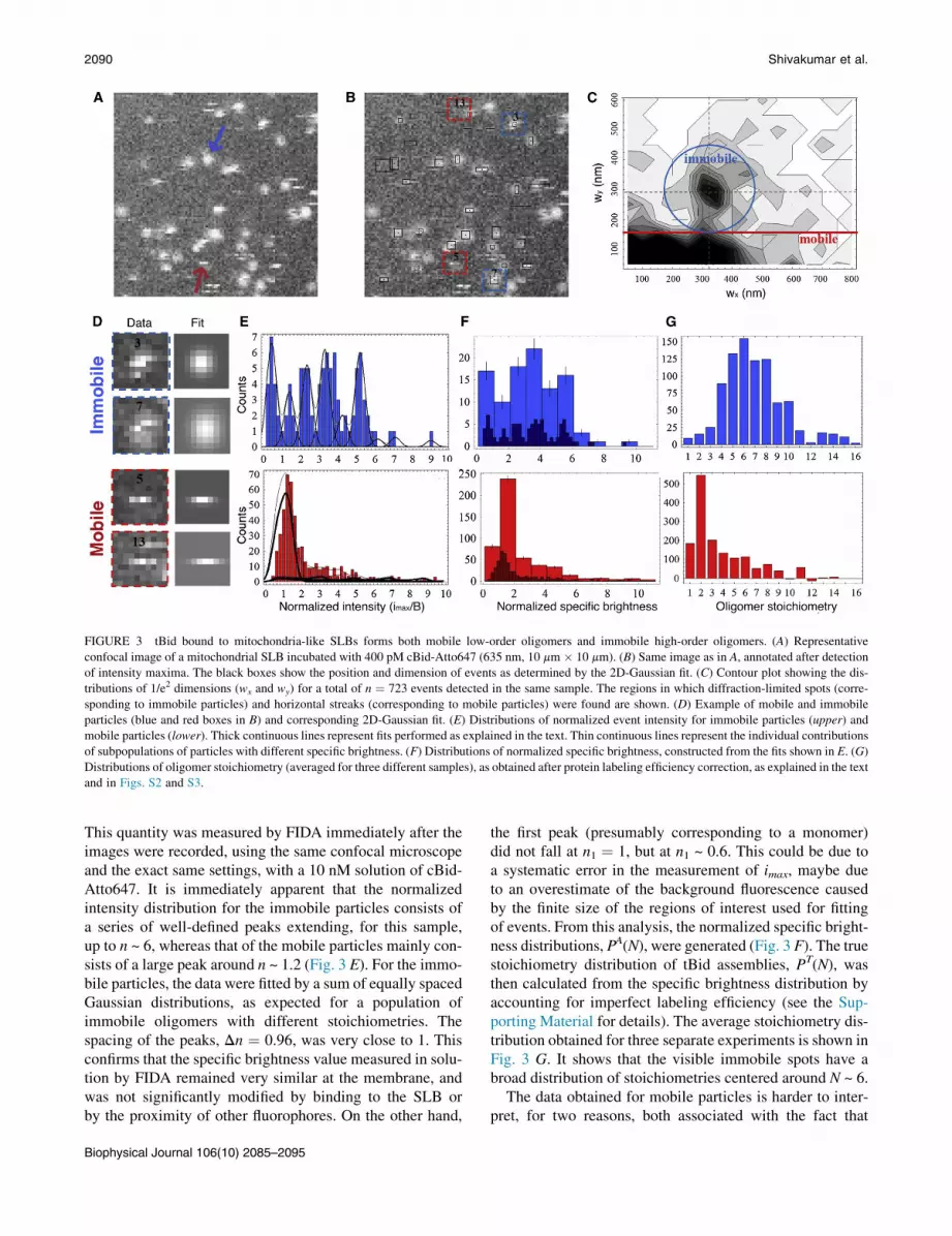
FIGURE 3 tBid bound to mitochondria-like SLBs forms both mobile low-order oligomers and immobile high-order oligomers. (A) Representative
confocal image of a mitochondrial SLB incubated with 400 pM cBid-Atto647 (635 nm, 10 mm � 10 mm). (B) Same image as in A, annotated after detection
of intensity maxima. The black boxes show the position and dimension of events as determined by the 2D-Gaussian fit. (C) Contour plot showing the dis-
tributions of 1/e2 dimensions (wx and wy) for a total of n ¼ 723 events detected in the same sample. The regions in which diffraction-limited spots (corre-
sponding to immobile particles) and horizontal streaks (corresponding to mobile particles) were found are shown. (D) Example of mobile and immobile
particles (blue and red boxes in B) and corresponding 2D-Gaussian fit. (E) Distributions of normalized event intensity for immobile particles (upper) and
mobile particles (lower). Thick continuous lines represent fits performed as explained in the text. Thin continuous lines represent the individual contributions
of subpopulations of particles with different specific brightness. (F) Distributions of normalized specific brightness, constructed from the fits shown in E. (G)
Distributions of oligomer stoichiometry (averaged for three different samples), as obtained after protein labeling efficiency correction, as explained in the text
and in Figs. S2 and S3.
2090 Shivakumar et al.
This quantity was measured by FIDA immediately after theimages were recorded, using the same confocal microscopeand the exact same settings, with a 10 nM solution of cBid-Atto647. It is immediately apparent that the normalizedintensity distribution for the immobile particles consists ofa series of well-defined peaks extending, for this sample,up to n ~ 6, whereas that of the mobile particles mainly con-sists of a large peak around n ~ 1.2 (Fig. 3 E). For the immo-bile particles, the data were fitted by a sum of equally spacedGaussian distributions, as expected for a population ofimmobile oligomers with different stoichiometries. Thespacing of the peaks, Dn ¼ 0.96, was very close to 1. Thisconfirms that the specific brightness value measured in solu-tion by FIDA remained very similar at the membrane, andwas not significantly modified by binding to the SLB orby the proximity of other fluorophores. On the other hand,
Biophysical Journal 106(10) 2085–2095
the first peak (presumably corresponding to a monomer)did not fall at n1 ¼ 1, but at n1 ~ 0.6. This could be due toa systematic error in the measurement of imax, maybe dueto an overestimate of the background fluorescence causedby the finite size of the regions of interest used for fittingof events. From this analysis, the normalized specific bright-ness distributions, PA(N), were generated (Fig. 3 F). The truestoichiometry distribution of tBid assemblies, PT(N), wasthen calculated from the specific brightness distribution byaccounting for imperfect labeling efficiency (see the Sup-porting Material for details). The average stoichiometry dis-tribution obtained for three separate experiments is shown inFig. 3 G. It shows that the visible immobile spots have abroad distribution of stoichiometries centered around N ~ 6.
The data obtained for mobile particles is harder to inter-pret, for two reasons, both associated with the fact that

Stoichiometry of tBid Membrane Oligomers 2091
most mobile particles will have less-than-ideal trajectoriespassing away from the center of the confocal detectionvolume. A first consequence of this is that instead of a sym-metric distribution of intensity values around the particlespecific brightness, we expect an asymmetric distributionskewed toward values lower than the specific brightness(see Materials and Methods for details on the shape of theexpected distribution). A second consequence is that mobileparticles carrying a single fluorophore (e.g., monomers)might not always be detected due to a low signal/noise(S/N) ratio. The main contribution to noise in single-particleexperiments comes from the photon shot noise of the signal(the photon noise of the background is negligible). The spe-cific brightness of tBid-Atto647 was reproducibly found tobe B ~ 20 kHz in our experimental conditions, translatinginto an average signal S ~ 20 photons for an immobiletBid monomer placed in the center of the detection volume(given a pixel dwell time of 1 ms). Thus, in our experiments,a signal/noise ratio of S/N ~ S1/2 ~ 4.5, just above the lowestacceptable value of S/N ¼ 3, was associated with immobilemonomeric tBid species. However, for mobile particles notpassing through the center of the detection volume or notresiding at its center for the full duration of the 1 ms pixeldwell time, the S/N is even lower. As a result, many of themobile tBid monomers will escape detection. Since theFRET data presented in Fig. 1 suggest that the dissociationcoefficient associated with dimer formation in the mem-brane is ~100 mm�1 or more (the surface concentration ofthe protein in the FRET experiments), at the very low sur-face concentration used for our single-particle trackingexperiments we expect monomeric species to dominate.Thus, many mobile particles escaped detection. We can stillsafely conclude that the stoichiometry distribution formobile particles is dominated by low stoichiometry species,N ¼ 1–3, and, of more importance, that mobile particleshave low stoichiometry compared to immobile ones. Thus,tBid species in mitochondria-like SLBs mostly fall intotwo broad categories, mobile complexes with low stoichi-ometry, and immobile complexes with higher stoichiometry.
FIGURE 4 Mobility of membrane-bound tBid as observed by single-
particle tracking and FCS. (A) Representative trajectories for three different
kinds of particles (mobile (particle 1), immobile (particle 2), alternatively
mobile, and immobile (particle 3)) tracked in TIRF movies of cBid associ-
ated with an SLB. All trajectories are 10.2 s long, and each color represents
a 1.7 s time interval. (B) Mean-squared displacement (MSD) calculated for
the three trajectories shown in A. Lines are linear fits of the data. (C) His-
togram of diffusion coefficients measured at the membrane by single-parti-
cle tracking and MSD analysis for tBid (green, n ¼ 122) and DiD (black,
n ¼ 98). (D) Average autocorrelation function obtained for cBid in solution
(blue symbols, mean 5 SD, n ¼ 15) and when associated with an SLB
(green symbols, n ¼ 24). Lines correspond to fits of the data assuming
one fluorescent species, yielding D ¼ 118 mm2/s for the protein in solution
and D ¼ 22 mm2/s for the protein at the membrane. Residuals are shown in
the lower panels.
Mobility of Bid membrane species
To get an insight into their degree of insertion into themembrane, we next investigated the diffusion of mobiletBid species. We used two separate techniques—FCS andsingle-particle tracking in TIRF movies—to cover a largerange of mobilities. TIRF movies were acquired for sam-ples prepared in a manner similar to those used for theconfocal experiments discussed in the previous section.These movies confirmed the presence of two different cat-egories of tBid species, mobile and immobile. The detectedparticles were first identified using Gaussian filtering of theimage, which was followed by thresholding and thentracked using the same 2D-Gaussian fit that was used forevent detection in confocal images. Fig. 4 A shows
Biophysical Journal 106(10) 2085–2095

2092 Shivakumar et al.
examples of trajectories obtained for three different repre-sentative particles. Usually, mobile particles remained mo-bile throughout the 1.7 s tracking sequence (e.g., particle1), whereas immobile particles remained immobile (e.g.,particle 2). Occasionally, however, particles were observedto transition from mobile to immobile, sometimes severaltimes, as was the case for particle 3. The motion of mobileparticles was characterized through mean-squared displace-ment analysis. The observed mobile Bid species displayeda mainly diffusive behavior, characterized by a roughlylinear mean-squared displacement (Fig. 4 B), allowing themeasurement of a diffusion coefficient for each detectedmobile particle. The distribution of diffusion coefficientsobtained for the visible mobile Bid species is shown inFig. 4 C and compared to the distribution of diffusioncoefficients measured separately for the lipophilic dyeDiD inserted into mitochondria-like SLBs. The averagediffusion coefficient of the tBid membrane species fol-lowed by single-particle tracking, D ¼ (0.8 5 0.5) mm2/s(mean 5 SD, n ¼ 123), was about a third of that measuredfor the lipophilic dye, D ¼ (2.2 5 2.7) mm2/s (n ¼ 100),suggesting that these proteins were partially inserted intothe membrane.
Because TIRF movies were acquired here with a frametime of t ¼ 35 ms, particles with a diffusion coefficient ofD > ~L2/(4t) (where L ¼ 5 pxl ¼ 0.8 mm is the maximumdisplacement allowed for a particle between frames tobe considered the same particle), i.e., particles withD > ~5 mm2/s, were not detected in the TIRF experiments.We therefore also performed FCS experiments. The averageautocorrelation functions measured for cBid in solution andtBid membrane species are shown in Fig. 4 D. The cBidmonomer diffuses in solution with D ¼ (118 5 6) mm2/s,corresponding to a hydrodynamic radius of 3.9 nm.Compared to the solution species, the membrane specieshave a noticeably reduced diffusion coefficient, D ¼(22 5 2) mm2/s. However, remarkably, this value is stillone order of magnitude higher than what is detected byTIRF, emphasizing that both methods detect distinct tBidmembrane species, with very different mobility. In compar-ison, when measuring the diffusion coefficient of DiD byfluorescence fluctuation methods (FCS or image correlationspectroscopy, data not shown), we obtained values that wereeither the same or only slightly higher than that obtainedfrom single-particle tracking, showing that the discrepancyobserved for the protein is meaningful. We note that thisfast-diffusing membrane-bound tBid species was not de-tected in a previous line-scanning FCS study of tBid inter-acting with giant unilamellar liposomes (19). The reasonis that the temporal resolution of line-scanning FCS (justas that of TIRF) is not high enough to resolve fast membranespecies. The fact that the diffusion coefficient of this fast-diffusing species is larger than that of the lipids suggeststhat these proteins were only superficially bound to themembrane.
Biophysical Journal 106(10) 2085–2095
DISCUSSION
Early models for the activity of Bcl-2 family proteinsconsidered the system as a simple rheostat regulated bythe balance between the total concentrations of pro- andantiapoptotic proteins (37). However, it is now clear that avery complex network of interactions controls the cellularresponse to the life-or-death question at the heart ofapoptosis (2,38). This is manifest in the number of possiblebinding partners, conformations, and stoichiometries foreach Bcl-2 family protein. Characterizing these differentspecies represents an experimental challenge, made harderby the fact that most are membrane species. Here, we metthis challenge by employing a range of complementary fluo-rescence techniques. A new type of analysis in confocalimages allowed the observation of membrane-bound tBidmolecular complexes with distinct specific brightness andmobility. Techniques with complementary time resolution(such as FCS and TIRF) allowed characterizing the mobilityof these species. Based on our results, we propose a modelfor the sequence of interactions between cBid and mito-chondria-like membranes, illustrated in Fig. 5. By includingoligomer formation, it refines and extends a model we hadrecently proposed based on ensemble fluorescence measure-ments that had already demonstrated the coexistence ofdifferent membrane tBid species (11). The tenets of thisrefined model are that cBid first loosely associates withthe membrane as a monomer from solution, then partiallyinserts into the membrane before forming dimers, some ofwhich later oligomerize into larger structures.
One important, to our knowledge novel, finding of ourstudy is the existence of a loosely membrane-bound tBidspecies. In the absence of a lipid membrane, the two frag-ments of cBid remain associated via noncovalent hydropho-bic interactions (11) and cBid adopts a globular a-helicalconformation similar to that of full-length Bid (39). Accord-ingly, we find that soluble cBid has a diffusion coefficient(D ¼ 118 mm2/s) corresponding to an object with a hydro-dynamic radius r ¼ 3.9 nm, just slightly larger than whathas been reported for tBid (D ¼ 143 mm2/s, r ¼ 3.2 nm(19)). However, as soon as a membrane is present, the twofragments of cBid separate (11). Electrostatics must play amajor role in fragment separation, since the negativelycharged mitochondrial membrane must attract and retainthe positively charged tBid fragment while driving backthe negatively charged p7 fragment (16,40). The fast-diffusing species that we detected by FCS then almostcertainly corresponds to tBid proteins loosely electrostati-cally bound to the membrane (Fig. 5 B). The diffusioncoefficient of this species (D ¼ 22 mm2/s) is too low tocorrespond to a solution species, but too high to correspondto a membrane-inserted one (even partially inserted proteinscannot diffuse faster than the lipids in the bilayer (41,42)).Once inserted into the membrane (Fig. 4 C), the mobilityof the protein is significantly reduced, with D ~ 0.8 mm2/s,

FIGURE 5 Proposed sequence for the interaction of cBid with lipid bilayers. (A) In solution, the two fragments of cBid remain noncovalently bound. The
depicted tertiary structure was drawn according to the known soluble cBid structure (16). The red region on helix 3 represents the BH3 region. (B) Upon
approaching the membrane, the fragments separate and tBid becomes electrostatically trapped to the lipid bilayer. (C) A progressive conformational change
leads to superficial insertion in the lipid bilayer, as described in Shamas-Din et al. (11). A structure with shallow insertion of all helices as pictured here is
supported by the NMR data from Wang and Tjandra (45). (D) Two inserted tBid monomers associate into a dimer. For illustration purposes, the tBid dimer
structure was depicted with a symmetric BH3-in-the-groove structure inspired by that proposed for Bax dimers in Czabotar et al. (58). (E) Further oligo-
merization takes place leading to the formation of higher order oligomers. (F) Large oligomers can adopt a transmembrane configuration. To see this figure
in color, go online.
Stoichiometry of tBid Membrane Oligomers 2093
i.e., significantly less than that of the surrounding lipids. Itmust therefore involve the (maybe partial) insertion ofseveral helices into the membrane (41,42). Likely candi-dates are helices 4, 6, 7, and 8, which all have been impli-cated in membrane binding (11,40,43,44). Alternatively,as suggested by a recent NMR study, all of tBid helicesmight in fact be shallowly inserted into the membrane inthat conformation (45).
The concept of electrostatic scanning was put forwardwhen advancing the notion that a reactant adsorbed on a sur-face would take less time to find its target than if it werediffusing in the bulk, since its diffusion would be confinedto two dimensions (46). However, for this strategy to be effi-cient (for an organelle the size of mitochondria) the diffu-sion coefficient of the protein on the surface should be atleast a tenth of its diffusion coefficient in the bulk (47).The loosely bound tBid species uncovered by our FCS ex-periments (D ¼ 22 mm2/s) fulfills this condition, but thepartially inserted one (D ~ 0.8 mm2/s) does not, suggestingthat in its loosely membrane-bound form tBid can moreefficiently search for binding partners (other proteins,e.g., Mtch2 or Bax, or lipid domains). Why has thisloosely bound state proven so elusive? Our experimentsshow that single-particle techniques are too slow to capturefast-diffusing membrane species. In general, diffusionexperiments with a characteristic length scale d and acharacteristic timescale T will only detect and characterizewell particles with D < d2/T. For microscopy, d ~ 0.5 mmis the pixel size and T is either the frame rate (TIRF,T ~ 1–30 ms) or the line time (confocal microscopy andline-scanning FCS, T ~ 1–100 ms). Thus, particles withD > 5 mm2/s will easily be overlooked when using these
techniques. Signs of their presence in single-particle exper-iments may include a larger-than-expected fluorescentbackground and step-size distributions deviating from theexpected c distribution (48).
A second believed novel finding reported here is that tBidreadily forms dimers in membranes. We previously reportedthat fragment separation is closely followed by a complexand progressive conformational change of tBid, leading tosuperficial insertion of helices 6 and 7, and to the coexis-tence of two main membrane-inserted conformationsdistinguished by a change in environment for the N- andC-terminal regions (11). Based on the results presentedhere, we now propose that these two membrane-insertedconformations represent a monomeric state and a dimericstate of the protein (Fig. 5, C and D). Indeed, helix 1 atthe N-terminal of tBid (helix 3 of Bid) contains the BH3 re-gion that mediates the interaction of tBid with other familymembers (49). The detection of tBid-tBid FRET localized atthe mitochondria in apoptotic cells demonstrates that tBidhomooligomers form in vivo (18). Although it had initiallybeen suggested that the dominant homooligomer specieswas trimers (18), the same group later showed that the~45 kDa complex identified as a tBid homotrimer was infact a complex between tBid and the 33 kDa mitochondrialprotein Mtch2 (50). Thus, it seems reasonable that mem-brane tBid would preferentially form dimers in our reconsti-tuted system, even though in cells, numerous interactions(e.g., with Mtch2, Bax, Bak, and Bcl-XL) may competewith the formation of tBid homooligomers. It is importantto note that the forced homooligomerization of tBid hasbeen shown to promote apoptosis (18). What then is thefunction of the tBid oligomers? A first possibility is that
Biophysical Journal 106(10) 2085–2095
2094 Shivakumar et al.
tBid oligomerization changes its affinity for some bindingpartners, e.g. Bax. A second possibility is that the tBiddimer acts as a sink state, which, if favored, e.g., by thecorrect lipid composition, could enhance the binding oftBid to specific membranes. The tBid dimer would thenhave a function similar to that of the tBid-Mtch2 hetero-dimer, whose formation renders the interaction with themembrane quasi-irreversible (11). A third possibility isthat the tBid dimer simply represents the default stable stateof the protein in the membrane while waiting to interact withother proteins for which it may have a higher affinity, e.g.,Bax or Bcl-XL.
Assemblies of tBid molecules beyond the dimers werealso clearly detected, and were often found to be immobile.None of the helices of tBid is thought to completely span themembrane bilayer (43,45). However, the presence of immo-bile tBid complexes, and of tBid complexes alternatingbetween a mobile and an immobile state, could be explainedby a (transiently or permanently) transmembrane helix in-teracting with the mica substrate. Another possibility isthat immobilization occurs because of the interaction be-tween the substrate and the lipids. We note that whereasthe FRET signal we observe in the presence of membraneproves that the interaction of the two tBid molecules in ahomodimer is a close physical interaction, the interactionbetween two dimers could instead be an indirect interactionmediated, for example, by lipid domains (i.e., it could repre-sent colocalization into diffraction-limited lipid domains).In any case, the observation of tBid assemblies larger thandimers is interesting, because so far, among Bcl-2 familyproteins, only Bax and Bak are believed to associate in largecomplexes.
Classically, Bid has been characterized as a proapopoticBH3 protein and a direct activator of Bax and Bak pore for-mation, meaning that it physically interacts with Bax andBak, even if only transiently (5,7,8). However, Bid also pre-sents similarities with multidomain Bcl-2 family proteins,and our work reinforces this idea in several ways. First, itshows that, like Bax and Bcl-XL, tBid is able to oligomerizein mitochondria-like membranes. Second, the loosely boundtBid conformation reported here is reminiscent of theloosely associated Bax species characterized in previousworks (22,25,51). Third, we showed that the sequence ofinteraction of tBid with the membrane is similar to that re-ported for Bax (10,52): Like Bax, tBid first binds the mem-brane as a monomer, then inserts into the membrane, andonly subsequently dimerizes. Obviously, the functionalitiesof Bid and Bax are ultimately different. In contrast to Bax,tBid does not cause membrane thinning (25), and although itcan cause ion release from liposomes and form ion channelsin planar membranes (53), it cannot by itself form poreslarge enough for cytochrome c release. However, the stepsshown in Fig. 5 could constitute a universal sequence forother Bcl-2 family proteins, such as Bcl-XL or Bad, thatswitch from a soluble to a membrane-bound state.
Biophysical Journal 106(10) 2085–2095
SUPPORTING MATERIAL
Three figures, Supporting Methods, and references (54–57) are available at
http://www.biophysj.org/biophysj/supplemental/S0006-3495(14)00388-9.
This work was supported by grants FRN86657 to C.F. and FRN12517 to
D.W.A. and B.L. from the Canadian Institutes of Health Research (CIHR).
REFERENCES
1. Antonsson, B., and J. C. Martinou. 2000. The Bcl-2 protein family.Exp. Cell Res. 256:50–57.
2. Leber, B., J. Lin, and D. W. Andrews. 2007. Embedded together: thelife and death consequences of interaction of the Bcl-2 family withmembranes. Apoptosis. 12:897–911.
3. Certo, M., V. Del Gaizo Moore, ., A. Letai. 2006. Mitochondriaprimed by death signals determine cellular addiction to antiapoptoticBCL-2 family members. Cancer Cell. 9:351–365.
4. Chipuk, J. E., J. C. Fisher, ., D. R. Green. 2008. Mechanism ofapoptosis induction by inhibition of the anti-apoptotic BCL-2 proteins.Proc. Natl. Acad. Sci. USA. 105:20327–20332.
5. Eskes, R., S. Desagher, ., J. C. Martinou. 2000. Bid induces the olig-omerization and insertion of Bax into the outer mitochondrial mem-brane. Mol. Cell. Biol. 20:929–935.
6. Letai, A., M. C. Bassik, ., S. J. Korsmeyer. 2002. Distinct BH3 do-mains either sensitize or activate mitochondrial apoptosis, serving asprototype cancer therapeutics. Cancer Cell. 2:183–192.
7. Kuwana, T., M. R. Mackey, ., D. D. Newmeyer. 2002. Bid, Bax, andlipids cooperate to form supramolecular openings in the outer mito-chondrial membrane. Cell. 111:331–342.
8. Terrones, O., B. Antonsson,., G. Basanez. 2004. Lipidic pore forma-tion by the concerted action of proapoptotic BAX and tBID. J. Biol.Chem. 279:30081–30091.
9. Li, H., H. Zhu,., J. Yuan. 1998. Cleavage of BID by caspase 8 medi-ates the mitochondrial damage in the Fas pathway of apoptosis. Cell.94:491–501.
10. Lovell, J. F., L. P. Billen,., D. W. Andrews. 2008. Membrane bindingby tBid initiates an ordered series of events culminating in membranepermeabilization by Bax. Cell. 135:1074–1084.
11. Shamas-Din, A., S. Bindner, ., C. Fradin. 2013. tBid undergoes mul-tiple conformational changes at the membrane required for Bax activa-tion. J. Biol. Chem. 288:22111–22127.
12. Tait, S. W., and D. R. Green. 2010. Mitochondria and cell death: outermembrane permeabilization and beyond. Nat. Rev. Mol. Cell Biol.11:621–632.
13. Billen, L. P., C. L. Kokoski,., D. W. Andrews. 2008. Bcl-XL inhibitsmembrane permeabilization by competing with Bax. PLoS Biol.6:e147.
14. Billen, L. P., A. Shamas-Din, and D. W. Andrews. 2008. Bid: a Bax-like BH3 protein. Oncogene. 27 (Suppl 1):S93–S104.
15. Petros, A. M., E. T. Olejniczak, and S. W. Fesik. 2004. Structuralbiology of the Bcl-2 family of proteins. Biochim. Biophys. Acta.1644:83–94.
16. McDonnell, J. M., D. Fushman,., D. Cowburn. 1999. Solution struc-ture of the proapoptotic molecule BID: a structural basis for apoptoticagonists and antagonists. Cell. 96:625–634.
17. Shamas-Din, A., H. Brahmbhatt, ., D. W. Andrews. 2011. BH3-onlyproteins: orchestrators of apoptosis. Biochim. Biophys. Acta. 1813:508–520.
18. Grinberg, M., R. Sarig,., A. Gross. 2002. tBID homooligomerizes inthe mitochondrial membrane to induce apoptosis. J. Biol. Chem.277:12237–12245.
19. Garcıa-Saez, A. J., J. Ries, ., P. Schwille. 2009. Membrane promotestBID interaction with BCL(XL). Nat. Struct. Mol. Biol. 16:1178–1185.

Stoichiometry of tBid Membrane Oligomers 2095
20. Friaa, O., M. Furukawa, A. Shamas-Din, B. Leber, D. W. Andrews, andC. Fradin. 2013. Optimizing the acquisition and analysis of confocalimages for quantitative single-mobile-particle detection. Chemphy-schem. 14:2476–2490.
21. Dlugosz, P. J., L. P. Billen, ., D. W. Andrews. 2006. Bcl-2 changesconformation to inhibit Bax oligomerization. EMBO J. 25:2287–2296.
22. Yethon, J. A., R. F. Epand,., D. W. Andrews. 2003. Interaction with amembrane surface triggers a reversible conformational change in Baxnormally associated with induction of apoptosis. J. Biol. Chem.278:48935–48941.
23. Horvath, S. E., and G. Daum. 2013. Lipids of mitochondria. Prog. LipidRes. 52:590–614.
24. van Meer, G., D. R. Voelker, and G. W. Feigenson. 2008. Membranelipids: where they are and how they behave. Nat. Rev. Mol. Cell Biol.9:112–124.
25. Satsoura, D., N. Ku�cerka, ., C. Fradin. 2012. Interaction of the full-length Bax protein with biomimetic mitochondrial liposomes: asmall-angle neutron scattering and fluorescence study. Biochim. Bio-phys. Acta. 1818:384–401.
26. Chiantia, S., N. Kahya, and P. Schwille. 2005. Dehydration damage ofdomain-exhibiting supported bilayers: an AFM study on the protectiveeffects of disaccharides and other stabilizing substances. Langmuir.21:6317–6323.
27. Oreopoulos, J., and C. M. Yip. 2008. Combined scanning probe andtotal internal reflection fluorescence microscopy. Methods. 46:2–10.
28. Satsoura, D., B. Leber, ., C. Fradin. 2007. Circumvention of fluoro-phore photobleaching in fluorescence fluctuation experiments: abeam scanning approach. ChemPhysChem. 8:834–848.
29. Kask, P., K. Palo,., K. Gall. 1999. Fluorescence-intensity distributionanalysis and its application in biomolecular detection technology. Proc.Natl. Acad. Sci. USA. 96:13756–13761.
30. Henriques, R., M. Lelek, ., M. M. Mhlanga. 2010. QuickPALM: 3Dreal-time photoactivation nanoscopy image processing in ImageJ. Nat.Methods. 7:339–340.
31. Reviakine, I., and A. Brisson. 2000. Formation of supported phospho-lipid bilayers from unilamellar vesicles investigated by atomic forcemicroscopy. Langmuir. 16:1806–1815.
32. Richter, R. P., and A. R. Brisson. 2005. Following the formation of sup-ported lipid bilayers on mica: a study combining AFM, QCM-D, andellipsometry. Biophys. J. 88:3422–3433.
33. Sonnleitner, A., G. J. Schutz, and T. Schmidt. 1999. Free Brownianmotion of individual lipid molecules in biomembranes. Biophys. J.77:2638–2642.
34. Przybylo, M., J. Sykora,., M. Hof. 2006. Lipid diffusion in giant uni-lamellar vesicles is more than 2 times faster than in supported phospho-lipid bilayers under identical conditions. Langmuir. 22:9096–9099.
35. Chiantia, S., J. Ries, N. Kahya, and P. Schwille. 2006. Combined AFMand two-focus SFCS study of raft-exhibiting model membranes. Chem-physchem. 7:2409–2418.
36. Guo, L., J. Y. Har, J. Sankaran, Y. Hong, B. Kannan, and T. Wohland.2008. Molecular diffusion measurement in lipid bilayers overwide concentration ranges: a comparative study. Chemphyschem.9:721–728.
37. Korsmeyer, S. J., J. R. Shutter, ., Z. N. Oltvai. 1993. Bcl-2/Bax: arheostat that regulates an anti-oxidant pathway and cell death. Semin.Cancer Biol. 4:327–332.
38. Chipuk, J. E., and D. R. Green. 2008. How do BCL-2 proteins inducemitochondrial outer membrane permeabilization? Trends Cell Biol.18:157–164.
39. Chou, J. J., H. Li, ., G. Wagner. 1999. Solution structure of BID, anintracellular amplifier of apoptotic signaling. Cell. 96:615–624.
40. Veresov, V. G., and A. I. Davidovskii. 2007. Monte Carlo simulationsof tBid association with the mitochondrial outer membrane. Eur.Biophys. J. 37:19–33.
41. Gambin, Y., R. Lopez-Esparza, ., W. Urbach. 2006. Lateral mobilityof proteins in liquid membranes revisited. Proc. Natl. Acad. Sci. USA.103:2098–2102.
42. Gambin, Y., M. Reffay, ., W. Urbach. 2010. Variation of the lateralmobility of transmembrane peptides with hydrophobic mismatch.J. Phys. Chem. B. 114:3559–3566.
43. Oh, K. J., S. Barbuto, ., S. J. Korsmeyer. 2005. Conformationalchanges in BID, a pro-apoptotic BCL-2 family member, upon mem-brane binding. A site-directed spin labeling study. J. Biol. Chem.280:753–767.
44. Lutter, M., M. Fang, ., X. Wang. 2000. Cardiolipin provides speci-ficity for targeting of tBid to mitochondria. Nat. Cell Biol. 2:754–761.
45. Wang, Y., and N. Tjandra. 2013. Structural insights of tBid, thecaspase-8-activated Bid, and its BH3 domain. J. Biol. Chem. 288:35840–35851.
46. Adam, G., and M. Delbruck. 1968. Reduction of dimensionality in bio-logical diffusion processes. In Structural Chemistry and MolecularBiology. A. Rich and N. Davidson, editors. W. H. Freeman, NewYork, pp. 198–215.
47. McCloskey, M. A., and M. M. Poo. 1986. Rates of membrane-associ-ated reactions: reduction of dimensionality revisited. J. Cell Biol.102:88–96.
48. Rozovsky, S., M. B. Forstner, ., J. T. Groves. 2012. Single moleculekinetics of ENTH binding to lipid membranes. J. Phys. Chem. B.116:5122–5131.
49. Wang, K., X. M. Yin, ., S. J. Korsmeyer. 1996. BID: a novel BH3domain-only death agonist. Genes Dev. 10:2859–2869.
50. Grinberg, M., M. Schwarz, ., A. Gross. 2005. Mitochondrial carrierhomolog 2 is a target of tBID in cells signaled to die by tumor necrosisfactor a. Mol. Cell. Biol. 25:4579–4590.
51. Antonsson, B., S. Montessuit, ., J. C. Martinou. 2001. Bax is presentas a high molecular weight oligomer/complex in the mitochondrialmembrane of apoptotic cells. J. Biol. Chem. 276:11615–11623.
52. Annis, M. G., E. L. Soucie,., D. W. Andrews. 2005. Bax forms multi-spanning monomers that oligomerize to permeabilize membranes dur-ing apoptosis. EMBO J. 24:2096–2103.
53. Schendel, S. L., R. Azimov, ., J. C. Reed. 1999. Ion channel activityof the BH3 only Bcl-2 family member, BID. J. Biol. Chem. 274:21932–21936.
54. Petrasek, Z., and P. Schwille. 2008. Precise measurement of diffusioncoefficients using scanning fluorescence correlation spectroscopy.Biophys. J. 94:1437–1448.
55. Loman, A., C. B. Muller, ., J. Enderlein. 2008. Absolute and precisemeasurements of the diffusion of small fluorescent dye moleculesacross the visible spectrum. Poster. Int. Workshop Single-Mol.Spectrosc. Ultrasens. Anal. Life Sci.,14th, Berlin.
56. Abu-Arish, A., P. Kalab, ., C. Fradin. 2009. Spatial distribution andmobility of the Ran GTPase in live interphase cells. Biophys. J.97:2164–2178.
57. Kasai, R. S., K. G. Suzuki, ., A. Kusumi. 2011. Full characterizationof GPCR monomer-dimer dynamic equilibrium by single moleculeimaging. J. Cell Biol. 192:463–480.
58. Czabotar, P. E., D.Westphal,., P. M. Colman. 2013. Bax crystal struc-tures reveal how BH3 domains activate Bax and nucleate its oligomer-ization to induce apoptosis. Cell. 152:519–531.
Biophysical Journal 106(10) 2085–2095


