Embed Size (px)
Citation preview

The Proteasome System in Infection: Impact of b5 andLMP7 on Composition, Maturation and Quantity ofActive Proteasome ComplexesThorsten Joeris1, Nicole Schmidt1, David Ermert2, Petra Krienke1, Alexander Visekruna3,
Ulrike Kuckelkorn4, Stefan H. E. Kaufmann1, Ulrich Steinhoff1,3*
1 Department of Immunology, Max Planck Institute for Infection Biology, Berlin, Germany, 2 Department of Cellular Microbiology, Max Planck Institute for Infection
Biology, Berlin, Germany, 3 Institute for Microbiology and Hygiene, Philipps University Marburg, Hessen, Marburg, Germany, 4 Institute for Biochemistry, Charite Berlin,
Berlin, Germany
Abstract
Proteasomes are the major enzyme complexes for non-lysosomal protein degradation in eukaryotic cells. Mammals expresstwo sets of catalytic subunits: the constitutive subunits b1, b2 and b5 and the immunosubunits LMP2 (b1i), MECL-1 (b2i) andLMP7 (b5i). The LMP7-propeptide (proLMP7) is required for optimal maturation of LMP2/MECL-1-containing precursors tomature immunoproteasomes, but can also mediate efficient integration into mixed proteasomes containing b1 and b2. Incontrast, the b5-propeptide (prob5) has been suggested to promote preferential integration into b1/b2-containingprecursors, consequently favouring the formation of constitutive proteasomes. Here, we show that prob5 predominantlypromotes integration into LMP2/MECL-1-containing precursors in IFNc-stimulated, LMP7-deficient cells and infected LMP7-deficient mice. This demonstrates that prob5 does not direct preferential integration into b1/b2-containing precursors, butinstead promotes the formation of mixed LMP2/MECL-1/b5 proteasomes under inflammatory conditions. Moreover, thepropeptides substantially differ in their capacity to promote proteasome maturation, with proLMP7 showing a significantlyhigher chaperone activity as compared to prob5. Increased efficiency of proteasome maturation mediated by proLMP7 isrequired for optimal MHC class I cell surface expression and is equally important as the catalytic activity ofimmunoproteasomes. Intriguingly, induction of LMP7 by infection not only results in rapid exchange of constitutive byimmunosubunits, as previously suggested, but also increases the total proteasome abundance within the infected tissue.Hence our data identify a novel LMP7-dependend mechanism to enhance the activity of the proteasome system ininfection, which is based on the high chaperone activity of proLMP7 and relies on accelerated maturation of activeproteasome complexes.
Citation: Joeris T, Schmidt N, Ermert D, Krienke P, Visekruna A, et al. (2012) The Proteasome System in Infection: Impact of b5 and LMP7 on Composition,Maturation and Quantity of Active Proteasome Complexes. PLoS ONE 7(6): e39827. doi:10.1371/journal.pone.0039827
Editor: Rachel Louise Allen, University of London, St George’s, United Kingdom
Received March 17, 2012; Accepted May 27, 2012; Published June 29, 2012
Copyright: � 2012 Joeris et al. This is an open-access article distributed under the terms of the Creative Commons Attribution License, which permitsunrestricted use, distribution, and reproduction in any medium, provided the original author and source are credited.
Funding: Funding was provided by the following sources: German Research Foundation, projects KU1261/1 and SFB650 TP18 (htt://www.dfg.de), and a PhDgrant of the Charite Berlin (http://www.charite.de); a PhD grant of the Sonnenfeld-Stiftung (http://www.sonnenfeld-stiftung.de); and a PhD grant of the IMPRS-IDI(http://www.zibi-graduateschool-berlin.de). The funders had no role in study design, data collection and analysis, decision to publish, or preparation of themanuscript.
Competing Interests: The authors have declared that no competing interests exist.
* E-mail: [email protected]
Introduction
Proteasomes execute the majority of non-lysosomal protein
degradation in eukaryotic cells [1]. The central 20S complex of the
proteasome is a barrel-like structure composed of a- and b-
subunits, which form heptameric rings arranged in an a1–7b1–7b1–
7a1–7 stoichiometry [2]. The assembly of 20S proteasomes is a
well-ordered process. At first the proteasome assembling chaper-
ones (PAC1-4) form a scaffold for the organisation of an a-ring,
which subsequently serves as a matrix for binding of the b-
subunits, resulting in the formation of 13-15S precursor protea-
somes [3,4,5]. Finally, the proteasome maturation protein (POMP)
assists the assembly of two 15S half proteasomes to mature 20S
complexes [6,7]. During the assembly process the propeptides of
b-subunits exert a chaperone-like function, which is required for
the effective maturation of precursor proteasomes [6,8]. Comple-
tion of 20S proteasome assembly is accompanied by autocatalytic
removal of the propeptides, which activates the catalytic sites of
the mature b-subunits [9].
The 20S proteasome has three major proteolytic activities
defined as caspase-, trypsin- and chymotrypsin-like [2], with the
corresponding catalytic sites being assigned to the constitutive
subunits b1 (Y), b2 (Z) and b5 (X), respectively [10]. In
mammals, the proteasome system displays further plasticity as
three interferon-gamma (IFNc-inducible b-subunits, the immu-
nosubunits LMP2 (b1i), MECL-1 (b2i) and LMP7 (b5i), can
replace their constitutive counterparts. Integration of either
constitutive or immunosubunits gives rise to the major 20S
proteasome subsets, named constitutive and immunoprotea-
somes respectively [11].
In the immune system proteasomes are the major source of
antigenic peptides presented on major histocompatibility com-
plex (MHC) class I molecules [1] and induction of immunopro-
teasomes is known to optimize this process [11]. Deletion of
PLoS ONE | www.plosone.org 1 June 2012 | Volume 7 | Issue 6 | e39827

LMP7, but not LMP2 or MECL-1, reduces MHC class I cell
surface expression by about 25–50% [11,12], demonstrating the
unique importance of this subunit for optimal antigen presen-
tation.
In addition, LMP7 containing proteasomes have been
described to drive inflammatory responses, which can in part
be attributed to enhanced activation of the transcription factor
NF-kB [13,14,15] and expression of LMP7 was shown to be
crucial for resistance against oxidative stress [16]. At present,
these diverse functions of LMP7 are solely attributed to its
specific proteolytic activity [11,15,16]. However, besides its
catalytic activity, expression of LMP7 has a substantial impact
on the assembly and composition of 20S proteasomes. In this
context, it has been shown that expression of LMP7 accelerates
the rate of proteasome assembly, supporting the rapid formation
of immunoproteasomes in infection and inflammation [17].
Further, it has been described that efficient maturation of
LMP2/MECL-1-containing precursor proteasomes requires the
propeptide of LMP7 (proLMP7), a concept known as cooper-
ative assembly of immunoproteasomes [18,19]. However,
integration of LMP7 is not restricted to immunoproteasomes,
since proLMP7 also mediates integration into proteasomes
containing b1 and b2 [19]. In line with this, various combina-
tions of mixed proteasomes were recently identified in human
tissues and cell lines [20,21]. In contrast to proLMP7, the
propeptide of b5 (prob5) has been suggested to favour
integration into b1/b2-containing precursors and thus formation
of mixed proteasomes with LMP2/MECL-1/b5 stoichiometry is
supposed to be a rare event [18,19]. Despite this extensive
knowledge on the impact of LMP7 and b5 on proteasome
assembly and composition, it is still not clear how the structural
functions of these two subunits influence the proteasome system
in infection.
Here, we analyzed the function of proLMP7 and prob5 in
reconstituted LMP7-deficient murine embryonic fibroblasts
(lmp72/2 Mefs) and mimicked inflammatory conditions by IFNcstimulation. We found that not only proLMP7, but also prob5
mediates predominant integration into LMP2/MECL-1-contain-
ing precursor proteasomes following IFNcstimulation, leading to
considerable formation of mixed proteasomes with LMP2/
MECL-1/b5 stoichiometry. High abundance of such mixed
proteasomes was also detected following infection of lmp72/2
mice, confirming that prob5 does not have a preference for b1/
b2-containing precursors, but can also generate mixed protea-
somes. The propeptides however differed significantly in their
capacity to promote proteasome maturation under inflammatory
conditions, with proLMP7 showing a significantly higher
efficiency to promote this process as compared to prob5. In
infection, accelerated proteasome maturation driven by LMP7
did not only result in a rapid exchange of constitutive by
immunoproteasomes as previously suggested [17,22], but also in
increased total proteasome quantity within the infected tissue.
The specific proteolytic activity of LMP7 was however not crucial
for enhanced proteasome maturation, since the chimeric
proLMP7mb5 protein had a similar capacity to promote this
process as compared to full-length LMP7. This identifies
proLMP7 as the critical pacemaker, which accelerates the
maturation of proteasomes under inflammatory conditions. Thus,
we delineate a novel mechanism of LMP7-dependent regulation
of the proteasome system in infection, which increases the
proteasomal activity by enhanced generation of mature protea-
some complexes.
Results
proLMP7 and prob5 mediate incorporation into LMP2/MECL-1-containing precursor proteasomes uponIFNcstimulation
To study the function of prob5 and proLMP7 under
homeostatic and inflammatory conditions, we generated LMP7-
deficient murine embryonic fibroblasts (lmp72/2 Mefs), which
were reconstituted with Flag-tagged constructs of either full-length
LMP7 (LMP7-Flag), proLMP7 fused to mature b5
(proLMP7mb5-Flag), full-length b5 (b5-Flag) or prob5 fused to
mature LMP7 (prob5mLMP7-Flag) (Fig. 1A). The reconstituted
lmp72/2 Mefs displayed high expression of b1 and b2 during
homeostasis and high amounts of LMP2 and MECL-1 after
IFNcstimulation, which mimicked inflammatory conditions
(Fig. 1B–E). The anti-Flag antibody did not precipitate protea-
somes in lmp72/2 Mefs transduced with the empty vector
construct (Fig. S1A), verifying that the precipitation was specific
for flag-tagged complexes. Immunoprecipitation of the proLMP7-
containing subunits, LMP7-Flag and proLMP7mb5-Flag, cleared
the supernatants of all catalytic proteasome subunits in unstimu-
lated and IFNc treated cells, demonstrating that proLMP7
mediates effective integration into all types of proteasomes, present
under both conditions (Fig. 1B–C). Complete co-precipitation of
b1 and b2 was also observed in unstimulated lmp72/2 Mefs
expressing the prob5-containing subunits, b5-Flag or
prob5mLMP7-Flag (Fig. 1D–E), demonstrating effective matura-
tion of b1/b2-containing precursors as expected. However,
following IFNc stimulation, LMP2 and MECL-1 were efficiently
co-precipitated with prob5-containing subunits (Fig. 1D–E),
revealing that prob5 can also mediate substantial maturation of
LMP2/MECL-1-containing precursors. Simultaneously, the
abundance of b1 and b2 was reduced upon IFNc stimulation
(Fig. 1B–E, Fig. S1A), suggesting that the prob5-containing
subunits did not favour integration into proteasomes containing
b1 and b2, but integrated into LMP2/MECL-1-containing
precursors instead.
To assess, whether b5 is capable to integrate into LMP2/
MECL-1-containing precursors in the presence of LMP7, b5-Flag
was over-expressed in wild type Mefs (WT-Mefs). In this setting,
b5-Flag also co-precipitated with LMP2 and MECL-1 following
IFNc stimulation, while the amount of co-precipitated b1 and b2
was reduced (Fig. S1B). This confirms that b5 can integrate into
LMP2/MECL-1-containing precursors, even in competition with
LMP7.
Still, low levels of unprocessed pLMP2 and pMECL-1 were only
detected in supernatants of lmp72/2 Mefs reconstituted with the
prob5-containing subunits (Fig. 1D–E), revealing that prob5 is a
limiting factor for maturation of proteasomes under inflammatory
conditions. However, our data indicate that this is not due to a
preference of prob5 for b1/b2-containing precursors as suggested
previously [19]. Instead, it appears that prob5 displays a generally
lower capacity to promote proteasome maturation, which
subsequently becomes a limiting factor for the maturation of
proteasomes under inflammatory conditions.
proLMP7 mediates higher efficiency of proteasomematuration compared to prob5
It has been suggested that accelerated proteasome maturation
by LMP7 is a function of its propeptide, since proLMP7 shows
high affinity to the maturation factor POMP [17]. However, direct
experimental evidence that proLMP7 mediates accelerated
proteasome maturation is missing. Thus it remains unclear,
whether only the propeptide or also the specific proteolytic activity
LMP7 Regulates Proteasome Quantity in Infection
PLoS ONE | www.plosone.org 2 June 2012 | Volume 7 | Issue 6 | e39827

and/or the carboxy-terminus of LMP7 are involved in this
process. To address this issue, we analysed proteasome maturation
in the reconstituted lmp72/2 Mefs.
The maturation factor POMP was used as an indicator for the
presence of precursor proteasomes, since it is found in 13-15S
precursors, but not in mature complexes [6,7]. POMP was
exclusively detected in IFNc-treated lmp72/2 Mefs reconstituted
with the prob5-containing subunits, b5-Flag or prob5mLMP7-
Flag (Fig. 2A), confirming that prob5 limits proteasome-matura-
tion specifically under inflammatory conditions. When IFNc-
stimulated, reconstituted lmp72/2 Mefs were analysed by immu-
noprecipitation, POMP was exclusively detected in the superna-
Figure 1. Co-immunoprecipitation analysis using prob5- and LMP7-containing proteasome subunits over-expressed inlmp72/2Mefs. Flag-tagged full-length LMP7 (LMP7-Flag), proLMP7 fused to mature b5 (proLMP7mb5-Flag), full-length b5 (b5-Flag) and prob5fused to mature LMP7 (prob5mLMP7-Flag) were over-expressed in lmp72/2Mefs by retroviral transduction (A). The lmp72/2 Mef lines expressing thefour different constructs were either left unstimulated or cultured in the presence of 50 U/ml IFNc for 4 days. Following cell lysis, the Flag-taggedsubunits were precipitated with anti-Flag-M2H agarose. Co-precipitation of the catalytic proteasome subunits b1, b2, LMP2 and MECL-1 with theproLMP7-containing subunits LMP7-Flag (B) and proLMP7mb5-Flag (C) or the prob5-containing subunits b5-Flag (D) and prob5mLMP7-Flag (E), wasanalysed by Two-colour fluorescent immunoblot analysis. The abundance of each subunit was determined in the input material (i), the supernatant ofthe immunoprecipitation (SN) and the precipitate (P) for both conditions tested.doi:10.1371/journal.pone.0039827.g001
LMP7 Regulates Proteasome Quantity in Infection
PLoS ONE | www.plosone.org 3 June 2012 | Volume 7 | Issue 6 | e39827

tants, but not the precipitates (Fig. 2B), demonstrating separation
of mature proteasomes and the precursor fraction (Fig. 2B). This
enabled us to determine the relative abundance of mature vs.
precursor proteasomes in the reconstituted lmp72/2 Mefs. The
abundance of a4 was used as correlate for the quantity of mature
and precursor proteasomes, since this structural subunit is
integrated in both fractions. The efficiency of proteasome
maturation was expressed as ratio of a4-abundance in mature
vs. precursor proteasomes. Accordingly, high ratios indicate
effective maturation, while low ratios reflect accumulation of
precursors and thus low maturation efficiency (Fig. 2C). LMP7-
Flag and proLMP7mb5-Flag showed no significant difference in
the ratios of mature vs. precursor proteasomes, revealing that the
proLMP7-containing subunits are equally efficient in driving
proteasome maturation (Fig. 2C). Similarly, b5-Flag or
prob5mLMP7-Flag did not show a significant difference in their
ratios; demonstrating that the prob5-containing subunits also have
a similar capacity to mediate proteasome maturation (Fig. 2C).
These results were confirmed by staining with a polyclonal
antiserum recognizing various a- and bsubunits (pan-20S-subunits,
Fig. 2B, D).
It is well known that proteasome maturation requires the
catalytic activity of the b-subunits for autocatalytic removal of the
propeptides and for degradation of POMP [6,9]. However, the
efficiency of proteasome maturation observed for full-length
LMP7 and the chimeric proLMP7mb5 protein did not differ
and the same holds true for full-length b5 and the chimeric
prob5mLMP7 protein (Fig. 2C, D). Hence, the specific proteolytic
activity of the fused catalytic subunit is of minor importance for the
efficiency of proteasome maturation, since it is not impaired as
long as the proteolytic activity of either LMP7 or b5 is present.
Accordingly, the propeptides are the crucial protein domains,
which regulate the efficiency of proteasome maturation, whereas
the proteolytic activities of the respective subunits or their carboxy-
termini have no significant impact on this process.
Intriguingly, the ratios of mature vs. precursor proteasomes for
the proLMP7-containing subunits were 3 to 4-fold higher as
compared to the prob5-containing subunits (Fig. 2C–D), demon-
strating that proLMP7 has a substantially higher capacity to
promote proteasome maturation as compared to prob5. This
confirms that accelerated proteasome maturation mediated by
LMP7 is an exclusive function of its propeptide, as previously
proposed [17]. Moreover, this finding underlines that the
chaperone-function of prob5 might become a limiting factor for
proteasome maturation under inflammatory conditions.
Optimal MHC class I expression requires both, catalyticactivity of LMP7 and efficient proteasome maturation byproLMP7
Deletion of LMP7 results in a significant decrease in MHC class
I cell surface expression [12], but it is not known whether this
phenotype is entirely caused by a lack of proteolytic activity of
LMP7 or also affected by impaired proteasome maturation. Since
proteasome maturation was restored in lmp72/2 Mefs expressing
proLMP7mb5-Flag (Fig. 2A–D), we wondered whether the
formation of mixed LMP2/MECL-1/b5 proteasomes, was also
capable of rescuing MHC class I cell surface expression. However,
in contrast to reconstitution with LMP7-Flag, expression of
proLMP7mb5-Flag did not rescue surface expression of the
MHC I molecules H2Kb and H2Db (Fig. 3A–B). Accordingly,
even strong formation of mixed LMP2/MECL-1/b5 proteasomes
in lmp72/2 Mefs expressing proLMP7mb5-Flag (Fig. 1C) could
not substitute for the formation of immunoproteasomes, demon-
strating that the proteolytic activity of LMP7 is indispensable for
optimal MHC class I cell surface expression. Nevertheless, surface
expression of H2Kb and H2Db was also impaired in lmp72/2 Mefs
reconstituted with prob5mLMP7-Flag (Fig. 3A–B), but since
considerable amounts of immunoproteasomes were detected in
these cells after IFNc stimulation (Fig. 1E), an increase in MHC
class I surface expression would have been expected. Accordingly,
the fusion of prob5 to LMP7 also limits the MHC class I cell
surface expression. This reveals that optimal MHC class I cell
surface expression does not only require the proteolytic activity of
LMP7, but furthermore requires the chaperone activity of
proLMP7 to drive optimal proteasome maturation.
Infection of lmp72/2 mice with listeriae inducesformation of mixed proteasomes
The formation of mixed proteasomes containing LMP2,
MECL-1 and b5 has been suggested to be inefficient due to
preferential integration of b5 into constitutive proteasomes
[18,19]. Since we predominantly found LMP2/MECL-1/b5
proteasomes in IFNc-stimulated lmp72/2Mefs in vitro, we won-
dered to which extent these mixed proteasomes were formed
following infection of lmp72/2 mice in vivo. To this end, wild type
control mice (WT mice) and lmp72/2 mice were infected with
Listeria monocytogenes, which is known to trigger IFNc-dependent
expression of immunosubunits [22,23]. 2D Blue Native/SDS-
PAGE was used to identify various mature and precursor
proteasome complexes (Fig. S2), which allowed to track the
integration of catalytic proteasome subunits into diverse protea-
some complexes.
As expected listeria-infection of WT mice induced increased
formation of immunoproteasomes in the liver (Fig. S3A), while
their abundance was constitutively high in spleen (Fig. S3B).
Noteworthy, high abundance of LMP2 and LMP7 but not
MECL-1 was detected in mature proteasomes of naıve WT liver
(Fig. S3A), indicating the formation of LMP2/b2/LMP7 or b1/
b2/LMP7 proteasomes, which were recently also identified in
various human tissues [20].
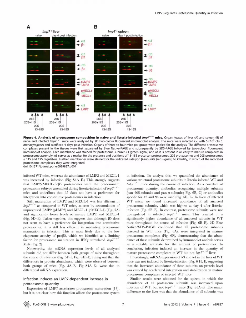
Interestingly, LMP2 but not MECL-1 was detected in mature
proteasomes of naıve lmp72/2 liver, revealing the formation of
LMP2/b2/b5 proteasomes, which have not been described yet
(Fig. 4A). Infection induced the integration of both, LMP2 and
MECL-1, into mature proteasomes in lmp72/2 liver, confirming
the formation of LMP2/MECL-1/b5 proteasomes in vivo (Fig. 4A).
LMP2/MECL-1/b5 proteasomes were also detected in spleens of
naıve and infected lmp72/2 mice, demonstrating constitutive
formation of mixed proteasomes in the spleen. This is in line with
the constitutively high expression of LMP2 and MECL-1
commonly found in lymphoid tissues [23]. Taken together this
underlines that prob5 does not only promote the formation of
mixed LMP2/MECL-1/b5 in vitro, but also in infected or
lymphoid tissue of lmp72/2 mice in vivo.
Mixed LMP2/MECL-1/b5 proteasomes are thepredominant proteasome type assembled followinglisteria-infection of lmp72/2 mice
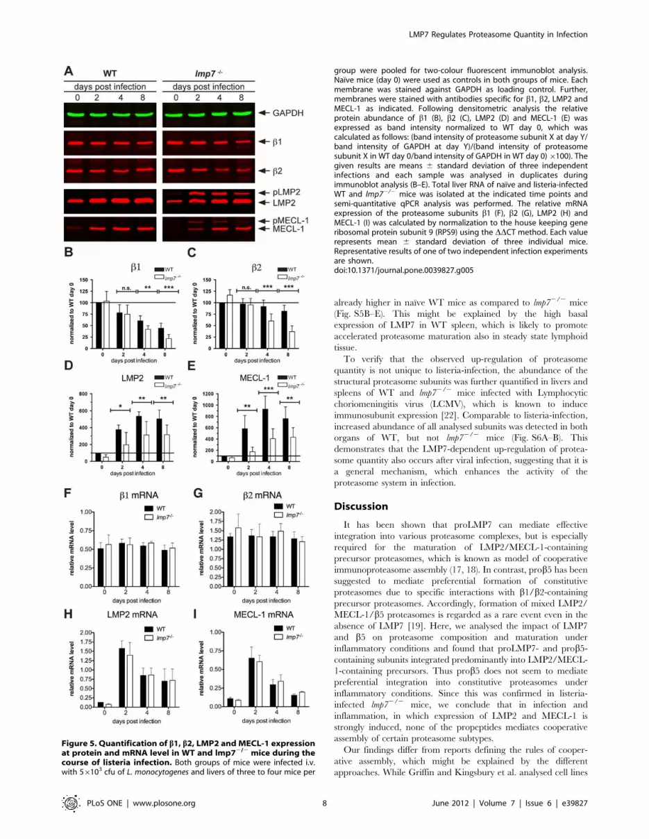
Next, we aimed to quantify the abundance of constitutive and
mixed LMP2/MECL-1/b5 proteasomes during the course of
infection in lmp72/2 mice. In addition, we wanted to assess the
maturation efficiency of LMP2/MECL-1-containing precursors in
infected lmp72/2 compared to WT mice. For this purpose, the
abundance of mature b1-b2-, LMP2- and MECL-1- subunits was
determined by immunoblot analysis in mutant- and WT-mice
(Fig. 5A). Following normalization we found that the abundance of
b1andb2 was substantially decreased at days 4 and 8 after
LMP7 Regulates Proteasome Quantity in Infection
PLoS ONE | www.plosone.org 4 June 2012 | Volume 7 | Issue 6 | e39827

LMP7 Regulates Proteasome Quantity in Infection
PLoS ONE | www.plosone.org 5 June 2012 | Volume 7 | Issue 6 | e39827

infection in both groups of mice (Fig. 5 B–C). However, this drop
in constitutive subunits was more pronounced in lmp72/2 as
compared to WT mice (Fig. 5 B–C), indicating stronger
replacement of constitutive proteasomes in infected lmp72/2 mice.
Simultaneously, the abundance of mature LMP2 and MECL-1
was increased following listeria infection of lmp72/2 mice (Fig. 5
D–E), which is in line with the formation of LMP2/MECL-1/b5
proteasomes in infected lmp72/2 liver as shown in Fig. 4A. Similar
results were obtained for the spleen of infected lmp72/2 mice, in
which the abundance of b1andb2 was also lower compared to
Figure 2. Relative quantification of proteasome maturation in lmp72/2 Mefs expressing proLMP7- or prob5-containing subunits. (A)lmp72/2 Mefs expressing the proLMP7-containing subunits LMP7-Flag and proLMP7mb5-Flag or the prob5-containing subunits b5-Flag andprob5mLMP7-Flag were either left unstimulated or cultured in the presence of 50 U/ml IFNc for 4 days. The abundance of POMP in the cell lysates ofthe four cell lines was determined by immunoblot analysis. (B) Immunoprecipitation was performed with anti-Flag M2H agarose using cell lysates ofthe four different lmp72/2 Mef lines, which were grown in the presence of 50 U/ml IFNc for 4 days. The abundance of GAPDH, POMP, a4 and pan-20Sproteasome subunits (pan 20S subunits) was determined by immunoblot analysis of the input material (i), the supernatants (SN) and precipitates (P)following immunoprecipitation with anti-Flag M2H agarose. (C, D) The ratio mature/precursor resembles the abundance of a4 subunits (C) or pan 20Ssubunits (D) integrated into mature proteasomes divided by their abundance in precursor proteasomes and was calculated as follows: band intensityof subunit X in the precipitate (P) divided by the band intensity of subunit X in the supernatant (SN). Given values for each cell line are mean values 6
standard deviations of at least three independent experiments.doi:10.1371/journal.pone.0039827.g002
Figure 3. Analysis of MHC class I cell surface expression in lmp72/2 Mefs expressing proLMP7- or prob5-containing subunits.lmp72/2 Mefs expressing LMP7-Flag, proLMP7mb5-Flag, b5-Flag, prob5mLMP7-Flag or cells transduced with the empty vector construct werecultured in the presence of 50 U/ml IFNc for 4 days. WT-Mefs stimulated with IFNc for 4 days were used as a positive control. The MHC class I cellsurface expression of the haplotypes H-2Kb (E) and H-2Db (F) on the various cell lines was determined by flow cytometry. The relative MHC class Idensity of the different cell lines was calculated by normalization of the median fluorescence intensity (MFI) of the indicated cell line to the MFI oflmp72/2 Mefs transduced with the empty vector construct. Given values are means 6 standard deviations of five independent experiments.doi:10.1371/journal.pone.0039827.g003
LMP7 Regulates Proteasome Quantity in Infection
PLoS ONE | www.plosone.org 6 June 2012 | Volume 7 | Issue 6 | e39827
infected WT mice, whereas the abundance of LMP2 and MECL-1
was increased by infection (Fig. S4A–E.) This strongly suggests
that LMP2/MECL-1/b5 proteasomes were the predominant
proteasome subtype assembled during listeria-infection of lmp72/2
mice and underlines that b5 does not have a preference for
integration into constitutive proteasomes in infection.
Still, maturation of LMP2 and MECL-1 was less efficient in
lmp72/2 as compared to WT mice, as seen by accumulation of
unprocessed LMP2 (pLMP2) and MECL-1 (pMECL-1) (Fig. 5A)
and significantly lower levels of mature LMP2 and MECL-1
(Fig. 5D–E). Taken together, this suggests that although b5 does
not seem to have a preference for integration into constitutive
proteasomes, it is still less efficient in mediating proteasome
maturation in infection. This is most likely due to the low
chaperone activity of prob5, which we identified as a limiting
factor for proteasome maturation in IFNc stimulated lmp72/2
Mefs (Fig. 2).
Noteworthy, the mRNA expression levels of all analysed
subunits did not differ between both groups of mice throughout
the course of infection (Fig. 5F–I; Fig. S4F–I), ruling out that the
differences in protein abundance, which were observed between
both groups of mice (Fig. 5A–E; Fig. S4A–E), were due to
differential mRNA expression.
Infection induces an LMP7-dependent increase inproteasome quantity
Expression of LMP7 accelerates proteasome maturation [17],
but it is not clear how this function affects the proteasome system
in infection. To analyse this, we quantified the abundance of
various structural proteasome subunits in listeria-infected WT and
lmp72/2 mice during the course of infection. As a correlate of
proteasome quantity, antibodies recognizing multiple subunits
(pan 20S-subunits and pan a-subunits; Fig. 6B, C) or antibodies
specific for a3 and a4 were used (Fig. 6D, E). In livers of infected
WT mice, we found increased abundance of all analysed
proteasome subunits, which was highest at day 4 after listeria-
infection (Fig. 6B–E). In contrast, proteasome subunits were not
up-regulated in infected lmp72/2 mice. This resulted in a
significantly higher abundance of all analysed subunits in WT
mice throughout the course of infection (Fig. 6B–E). 2D Blue
Native/SDS-PAGE confirmed that all proteasome subunits
detected in WT mice (Fig. 6A), were integrated in mature
proteasome complexes (Fig. 6F), demonstrating that the abun-
dance of these subunits determined by immunoblot analysis serves
as a suitable correlate for the amount of proteasomes. In
conclusion, infection induced an increase in the quantity of
mature proteasome complexes in WT but not lmp72/2 liver.
Interestingly, mRNA expression of a3 and a4 in the liver of WT
mice was not induced by listeria-infection (Fig. 6 H, I), suggesting
that the increased abundance of these subunits on protein level
was caused by accelerated integration and stabilization in mature
proteasome complexes of infected WT mice.
Similar results were obtained for the spleen, in which the
abundance of all proteasome subunits was increased upon
infection of WT, but not lmp72/2 mice (Fig. S5A–I). The major
difference to the liver was that the abundance of all subunits was
Figure 4. Analysis of proteasome composition in naive and listeria-infected lmp72/2 mice. Organ lysates of liver (A) and spleen (B) ofnaıve and infected lmp72/2 mice were analysed by 2D two-colour fluorescent immunoblot analysis. The mice were infected i.v. with 56103 cfu L.monocytogenes and sacrificed 4 days post infection. Organs of three to four mice per group were pooled for the analysis. The different proteasomecomplexes present in the tissues were first separated by Blue Native-PAGE and subsequently by SDS-PAGE followed by two-colour fluorescentimmunoblot analysis. Each membrane was stained for proteasome subunit a3 (green signal) and as it is present in all early to mature complexes inproteasome assembly, a3 serves as a marker for the presence and positions of 13-15S precursor proteasomes, 20S proteasomes and 20S proteasomes+ 11S and 19S regulators. Further, membranes were stained for the indicated catalytic b-subunits (red signals) to identify, in which of the indicatedproteasome complexes they were integrated.doi:10.1371/journal.pone.0039827.g004
LMP7 Regulates Proteasome Quantity in Infection
PLoS ONE | www.plosone.org 7 June 2012 | Volume 7 | Issue 6 | e39827
already higher in naıve WT mice as compared to lmp72/2 mice
(Fig. S5B–E). This might be explained by the high basal
expression of LMP7 in WT spleen, which is likely to promote
accelerated proteasome maturation also in steady state lymphoid
tissue.
To verify that the observed up-regulation of proteasome
quantity is not unique to listeria-infection, the abundance of the
structural proteasome subunits was further quantified in livers and
spleens of WT and lmp72/2 mice infected with Lymphocytic
choriomeningitis virus (LCMV), which is known to induce
immunosubunit expression [22]. Comparable to listeria-infection,
increased abundance of all analysed subunits was detected in both
organs of WT, but not lmp72/2 mice (Fig. S6A–B). This
demonstrates that the LMP7-dependent up-regulation of protea-
some quantity also occurs after viral infection, suggesting that it is
a general mechanism, which enhances the activity of the
proteasome system in infection.
Discussion
It has been shown that proLMP7 can mediate effective
integration into various proteasome complexes, but is especially
required for the maturation of LMP2/MECL-1-containing
precursor proteasomes, which is known as model of cooperative
immunoproteasome assembly (17, 18). In contrast, prob5 has been
suggested to mediate preferential formation of constitutive
proteasomes due to specific interactions with b1/b2-containing
precursor proteasomes. Accordingly, formation of mixed LMP2/
MECL-1/b5 proteasomes is regarded as a rare event even in the
absence of LMP7 [19]. Here, we analysed the impact of LMP7
and b5 on proteasome composition and maturation under
inflammatory conditions and found that proLMP7- and prob5-
containing subunits integrated predominantly into LMP2/MECL-
1-containing precursors. Thus prob5 does not seem to mediate
preferential integration into constitutive proteasomes under
inflammatory conditions. Since this was confirmed in listeria-
infected lmp72/2 mice, we conclude that in infection and
inflammation, in which expression of LMP2 and MECL-1 is
strongly induced, none of the propeptides mediates cooperative
assembly of certain proteasome subtypes.
Our findings differ from reports defining the rules of cooper-
ative assembly, which might be explained by the different
approaches. While Griffin and Kingsbury et al. analysed cell lines
Figure 5. Quantification of b1, b2, LMP2 and MECL-1 expressionat protein and mRNA level in WT and lmp72/2 mice during thecourse of listeria infection. Both groups of mice were infected i.v.with 56103 cfu of L. monocytogenes and livers of three to four mice per
group were pooled for two-colour fluorescent immunoblot analysis.Naıve mice (day 0) were used as controls in both groups of mice. Eachmembrane was stained against GAPDH as loading control. Further,membranes were stained with antibodies specific for b1, b2, LMP2 andMECL-1 as indicated. Following densitometric analysis the relativeprotein abundance of b1 (B), b2 (C), LMP2 (D) and MECL-1 (E) wasexpressed as band intensity normalized to WT day 0, which wascalculated as follows: (band intensity of proteasome subunit X at day Y/band intensity of GAPDH at day Y)/(band intensity of proteasomesubunit X in WT day 0/band intensity of GAPDH in WT day 0)6100). Thegiven results are means 6 standard deviation of three independentinfections and each sample was analysed in duplicates duringimmunoblot analysis (B–E). Total liver RNA of naıve and listeria-infectedWT and lmp72/2 mice was isolated at the indicated time points andsemi-quantitative qPCR analysis was performed. The relative mRNAexpression of the proteasome subunits b1 (F), b2 (G), LMP2 (H) andMECL-1 (I) was calculated by normalization to the house keeping generibosomal protein subunit 9 (RPS9) using the DDCT method. Each valuerepresents mean 6 standard deviation of three individual mice.Representative results of one of two independent infection experimentsare shown.doi:10.1371/journal.pone.0039827.g005
LMP7 Regulates Proteasome Quantity in Infection
PLoS ONE | www.plosone.org 8 June 2012 | Volume 7 | Issue 6 | e39827

in the steady state with fixed expression of constitutive and
immunosubunits [18,19], we used inducible systems, in which
expression of LMP2 and MECL1 is strongly up-regulated by IFNcstimulation or infection. The strong induction of immunosubunits
might have shifted the balance considerably towards formation of
mixed LMP2/ MECL1/b5 proteasomes as compared to studies
using fixed subunit expression. This is in line with work by Fruh et
al, who similar to our study employed inducible systems for the
expression of immunosubunits and demonstrated that the expres-
sion level substantially influences the integration of immunosubu-
nits. Hence, they suggested that integration of constitutive or
immunosubunits is regulated by competition at the protein level
[24]. In addition, they describe that integration of LMP2 and
LMP7 can occur independent of each other, consequently
resulting in any combination of b1 or LMP2 with b5 or LMP7,
which is also in conflict with the rules of cooperative assembly
[24]. In accordance, we found that b5 has a high capacity to
integrate into mixed proteasomes with LMP2 and MECL-1, if
competition with LMP7 is abrogated in mutant cell lines or mice,
supporting that integration of constitutive or immunosubunits is
rather regulated by competition at protein level than by
cooperative assembly.
A further explanation for the conflicting data might be the
different origins of the cell lines used to study the impact of
Figure 6. Relative quantification of mature proteasomes in WT and lmp72/2 mice during the course of listeria infection. WT andlmp72/2 mice were infected i.v. with 56103 cfu of L. monocytogenes and livers of three to four mice per group were pooled for two-colour fluorescentimmunoblot analysis. Naıve mice (day 0) were used as controls in both groups of mice. Each membrane was stained against GAPDH as loadingcontrol. Further, membranes were stained with antibodies recognizing pan-20S subunits, pan-a-subunits, a3 and a4as indicated. Representative blotsof three independent experiments are shown (A). Following densitometric analysis the relative protein abundance of pan-20S subunits (B), pan-a-subunits (C), a3 (D) and a4E was expressed as band intensities normalized to WT day 0, which was calculated as follows: (band intensity ofproteasome subunit X at day Y/band intensity of GAPDH at day Y)/(band intensity of proteasome subunit X in WT day 0/band intensity of GAPDH inWT day 0) 6100). The given results are means 6 standard deviation of three independent infection experiments and each sample was analysed induplicates during immunoblot analysis (B–E). To analyse in which proteasome complexes the analysed structural subunits are integrated, 2D two-colour fluorescent immunoblot analysis was performed on liver lysates of naıve and infected WT (F) and lmp72/2 mice (G). Brackets mark the areas, inwhich the different proteasome fractions are found. qPCR analysis on total liver cDNA was performed to assess the mRNA expression of a3 (H) anda4I during the course of listeria infection in WT and lmp72/2 mice. The relative mRNA expression of the proteasome subunits was calculated bynormalization to the housekeeping gene RPS9 using the DDCT method. Each value represents mean 6 standard deviation of three individual mice.doi:10.1371/journal.pone.0039827.g006
LMP7 Regulates Proteasome Quantity in Infection
PLoS ONE | www.plosone.org 9 June 2012 | Volume 7 | Issue 6 | e39827

immunosubunit on proteasome composition. We and Fruh et al.
used fibroblasts [24], which display high expression of constitutive
subunits in the steady state, but strongly induced expression of
immunosubunits upon IFNc stimulation. In contrast, the data
suggesting cooperative assembly were derived from T2 cells, which
are of lymphoid origin and presumably express low levels of
constitutive subunits [18,19]. However, the expression level of b5
has not been defined in T2 cells and thus low formation of mixed
LMP2/MECL-1/b5 proteasomes might be a result of restricted
availability of b5 in this lymphoid cell line. In line with this, we
found that formation of mixed LMP2/MECL-1/b5 proteasomes
is less efficient in the spleen of lmp72/2 mice, which mostly
contains lymphoid cell types, as compared to the liver, in which
the tissue largely consists of non-lymphoid cells.
It has been proposed that proLMP7 is required for accelerated
proteasome maturation mediated byLMP7, due to its high affinity
to the proteasome maturation factor POMP [17], but direct
experimental evidence was missing. Moreover it has been
described that the catalytic activity of b-subunits is crucial to
complete proteasome assembly [6,9] and that carboxy-terminal
domains of some b-subunits can have stabilizing effects, which
facilitate proteasome maturation [25,26]. Accordingly, each of
these factors could contribute to accelerated proteasome matura-
tion mediated by LMP7. Here we found that the fusion protein
proLMP7mb5 is equally efficient in mediating proteasome
maturation as compared to full-length LMP7. Hence the
proteolytic activity of b5 is not limiting for proteasome maturation
and we can rule out that the specific catalytic activity of LMP7 is
required to accelerate proteasome assembly. Moreover the
proLMP7mb5 fusion protein reveals that the carboxy-terminus
of LMP7 is not required for accelerated proteasome maturation.
These findings clearly identify proLMP7 as critical pacemaker,
which drives accelerated proteasome maturation following induc-
tion of LMP7.
In yeast it has been shown that the propeptide of the b5
homologue Doa3 can support proteasome assembly, if it is
expressed in trans, suggesting that it serves as an intra-molecular
chaperone in the full-length protein [9]. In mammalian cells, we
found that subunits containing proLMP7 have a 3 to 4-fold higher
capacity to promote proteasome maturation in comparison to
subunits containing prob5, revealing that proLMP7 has a
substantially higher chaperone activity as compared to prob5.
With regard to the regulation of proteasome composition, the high
chaperone activity of proLMP7 constitutes a major advantage and
is consistent with the fact that b5 is rapidly replaced, as soon as
LMP7 is expressed [22,23,24]. This also explains, why mixed
proteasomes with LMP2/b2/LMP7 or b1/b2/LMP7 stoichiom-
etry can even be found during low-level expression of LMP7 in the
steady state. Such mixed proteasomes were recently identified in
various human tissues and cancer-derived cell lines and have been
shown to be crucial for the MHC class I restricted presentation of
specific tumour-derived peptides [20]. This clearly shows that the
formation of these mixed proteasomes is not simply a bystander
product of ‘‘unbalanced’’ expression of constitutive or immuno-
subunits, but that the chaperone function of proLMP7 is a relevant
biological function.
In contrast, the low chaperone activity of prob5 is a limiting
factor for proteasome assembly under inflammatory conditions,
resulting in accumulation of precursor proteasomes, if LMP7 is
deleted. Moreover, the low chaperone activity of prob5 is in line
with the low abundance of LMP2/MECL-1/b5 proteasomes,
when LMP7 is present. Still, we were able to detect LMP2/
MECL-1/b5 proteasomes in WT-Mefs, however only, if the
capacity of b5 to compete with LMP7 was increased by exogenous
over-expression. Although this situation might be artificial, this
finding is still mechanistically relevant, since it further underlines
that integration of LMP7 and b5 is substantially regulated by their
expression level and competition at the protein level. However, in
this competition LMP7 has a substantial advantage due to its
propeptide, which displays a higher affinity to POMP [17] and a
superior chaperone activity as compared to prob5. This does not
only explain the rapid formation of immunoproteasomes in
infection and inflammation, but is also consistent with the fact
that LMP2/MECL-1/b5 proteasomes are rarely found in a
normal wild-type situation.
Noteworthy, the proteasome content in lmp72/2 mice remained
constant throughout the course of infection. This is in line with the
low chaperone activity of prob5 shown here and the approxi-
mately 4-fold lower rate of proteasome assembly mediated by b5
described by others [17]. It suggests that the physiological role of
b5 is to provide a constant low-level turnover of proteasomes in
the steady state. In this respect, the low chaperone activity of
prob5 is reasonable, as it appears to be adjusted to the long half-
life, which has been reported for constitutive proteasomes [17].
Infection with L. monocytogenes or Lymphocytic choriomeningitis virus
induces rapid replacement of constitutive by immunoproteasomes
[22,23] and accelerated proteasome assembly mediated by LMP7
supports this process [17]. However, beyond this mere shift to
immunoproteasome formation, we found that induction of LMP7
leads to increased total proteasome quantity within the infected
tissue. Hence, we identified a novel role of LMP7 in regulating the
proteasome system by enhancing the output of active proteasome
complexes. Still, the possibility remains that increased proteasome
quantity of infected tissues in WT mice is a consequence of higher
influx of immune cells as compared to lmp72/2 mice. This
however is unlikely, since lmp72/2 mice develop normal, have
unaltered numbers of T and B cells [12] and generate similar
frequencies of listeria-specific CD8+ T cells as compared to WT
mice [23].
Expression of LMP7 has been shown to promote inflammatory
responses, which is in part due to improved NF-kB activation by
LMP7-containing proteasomes [13,14,15]. Further, expression of
LMP7 has been shown to facilitate degradation of oxidatively
damaged proteins [16] and MHC class I antigen presentation
[12]. So far, these functions of LMP7 have been exclusively
associated with its proteolytic activity. Here, we show that optimal
MHC class I antigen presentation was equally dependent on
efficient maturation of proteasomes mediated by proLMP7. Thus
it is tempting to speculate that the increase in proteasome quantity
mediated by proLMP7, does not only improve MHC class I
presentation, but might also be relevant for NF-kB activation and
resistance to oxidative stress. Indeed, an increase in proteasome
quantity mediated by the NFR2/KEAP-1 signalling pathway or
the transcription factor NCF11 is known to enhance the resistance
towards oxidative stress [27,28]. At the same time, oxidative stress
has been shown to induce LMP7 expression [29]. Thus it is likely
that the LMP7-mediated increase in proteasome quantity protects
against oxidative stress, either by itself or synergistically with other
factors like e.g. NRF2/KEAP-1 and NCF11.
In summary, we describe a novel pathway of LMP7-mediated
proteasome regulation, which relies on increased output of active
proteasome complexes during infection and probably other
inflammatory conditions. Moreover, we could identify proLMP7
as the critical pacemaker driving accelerated maturation of
proteasomes in this context. Although it is likely that this
mechanism contributes to the defence against infections, its
precise impact on distinct biological functions such as MHC class
LMP7 Regulates Proteasome Quantity in Infection
PLoS ONE | www.plosone.org 10 June 2012 | Volume 7 | Issue 6 | e39827

I presentation, NF-kB activation and oxidative stress responses,
remains a matter of future studies.
Materials and Methods
Experimental animalsAll mice were kept under specific pathogen-free conditions
(SPF). C57Bl/6N (WT) mice were obtained from Charles River
(Berlin, Germany) and lmp72/2 mice were bred at the Max Planck
Institute for Infection Biology (Berlin, Germany). Mice were
infected intravenously (i.v.) with 56103 colony forming units (cfu)
of Listeria monocytogenes strain EGD or 106 pfu of Lymphocytic
choriomeningitis virus strain WE (LCMV WE). All experiments were
performed in strict accordance with German Animal Protection
Law and granted by the ethical committee of the ‘‘Landesamt fur
Gesundheit und Soziales Berlin’’ (permission numbers G0165/04
and T0144/05) in Berlin, Germany.
Cell culturesPhoenix E cells, for ecotropic packaging of retroviral vectors
(ATCC product # SD 3444), were kept in D10 Medium
[Dulbeccos Modified Eagle Medium (Gibco) with 10% fetal calf
serum (FCS), 1 mM L-glutamine, 1 mM sodium-pyruvate, 16pencillin/streptomycin solution (Gibco), 50 mM b-mercaptoetha-
nol]. Primary murine embryonic fibroblasts (Mefs) were isolated
from 13- to 14-day-old embryos of WT or lmp72/2 mice.
Spontaneous immortalization of Mefs was achieved by frequent
passaging.
Preparation of protein lysatesFrozen organs or cells were homogenized with a pestle. The
homogenates were resuspended in 1 volume of 16NativePAGETM
Sample Buffer (Invitrogen) complemented with 0.5% Igepal,
0.2 mM sodium vanadate, 5 mM sodium fluoride, 1 mM PMSF,
1 mM PefablocH SC (Roche Applied Science), 16 complete
protease inhibitor cocktail (Roche Applied Science) and subjected
to three freeze-thaw cycles. The debris was sedimented at
13,000 rpm for 30 min at 4uC. Protein concentrations were
determined with Protein-Assay solution (Bio-Rad) according to
manufacturer’s instructions.
Two-colour fluorescent immunoblot analysisTwenty-five to fifty mg total protein diluted in 16 Laemmli
buffer (50 mM TrisHCl pH6.8, 100 mM dithiothreitol, 2% (w/v)
sodium dodecyl sulfate (SDS), 10% (v/v) glycerol, 0,1% (w/v)
Bromophenol Blue) per lane were loaded on tris-glycine buffered
15% (w/v) SDS-PAGE gels and run in tris-glycine buffer (25 mM
Tris, 250 mM Glycine, 0.1% (w/v) SDS). Following SDS-PAGE,
proteins were transferred to Immobilon-FL PVDF membrane.
Membranes were blocked in Odyssey Blocking Reagent (Licor
Bioscience) and stained with rabbit or chicken polyclonal
antibodies against proteasome subunits or POMP as well as
mouse monoclonal antibodies against GAPDH. This was followed
by staining with the respective secondary antibodies goat anti-
rabbit IgG AlexaFluor680 (Molecular Probes), goat anti-chicken
IgG IrDye700 (Rockland) or goat-anti-mouse IgG IrDye800
(Rockland). For evaluation, membranes were scanned with the
OdysseyH Infrared Imaging system (Licor Biosciences). Densito-
metric analysis was performed with the OdysseyH Image Analyser
Software Version 1.2 (Licor Biosciences). Band intensities were
first normalized to the signal of the loading control GAPDH and
subsequently to the signal of naıve WT mice (WT day 0), which
was used as an internal standard on each gel.
Polyclonal rabbit antisera against b5, LMP2 as well as
polyclonal chicken antibodies against b2 and POMP were
obtained from Abcam. Rabbit polyclonal antibodies specific for
MECL-1 were obtained from Biomol. The mouse monoclonal
antibodies against a-subunits (MCP231), a3 (clone MCP257) and
GAPDH (clone 6C5) were obtained from Calbiochem. Rabbit
polyclonal antibodies against b1, LMP7, 20S proteasome subunits
(MP3), a4, PA28aand 19S subunit S4 were kindly provided by the
Institute for Biochemistry, Charite, Berlin.
2D Two-colour fluorescent immunoblot analysisOrgan lysates (50 mg per lane) supplemented with 0.125% (v/v)
NativePAGETM G-250 Sample Additive (Invitrogen) were loaded
on NativePAGETM Novex 4–16% Bis-Tris Gels (Invitrogen) and
gels were run according to manufacturers instructions. Gels were
sliced into single lanes and equilibrated in 26Laemmli buffer for
30 min. Slices were placed in preparative slots of tris-glycine-
buffered 15% (w/v) SDS-PAGE gels and run in tris-glycine buffer,
which was followed by two-colour fluorescent immunoblot analysis
(see above).
Semiquantitative real-time RT-PCR (qPCR)Organs were homogenized in TRIZOLH Reagent (Invitrogen)
and purification of RNA was performed according to manufac-
turer’s instructions. The concentration and quality of the RNA
was determined using a 2100 Bioanalyzer (Agilent Technologies).
For cDNA synthesis, 2–4 mg total RNA were transcribed using
random hexamer primers and SuperScriptTM II Reverse Tran-
scriptase (Invitrogen) according to manufacturer’s instructions.
qPCR reactions contained 16 SYBR Green mix (Applied
Biosystems), 10 pmol forward-primer, 10 pmol reverse-primer
and 5 ml cDNA template diluted 1:20 in UltraPure Water
(Millipore). The amplification was performed with an ABI Prism
7900H detection system (Applied Biosystems) and data were
evaluated with the SDS2.2.2 Software (Applied Biosystems). The
expression of ribosomal protein subunit 9 (RPS9) was used as
internal standard. The relative expression was calculated as fold
difference to RSP9 using the DDCT method. Primer sequences are
listed in Table S1.
Generation of retrovirally transduced MEFsThe coding sequences of prob5, proLMP7, b5, mb5, LMP7 and
mLMP7 with a carboxy-terminal Flag-tag fusion were amplified
from a murine liver cDNA by nested PCR. The b5- and LMP7-
Flag inserts were amplified using the primer pairs b5-start-for/b5-
Flag-rev or LMP7-start-for/LMP7-Flag-rev. The prob5mLMP7
or proLMP7mb5 were generated by blunt-end ligation of the
respective fragments using T4 Ligase (Fermentas) at 16uCovernight. The fragments were amplified using the following
primer pairs: b5-start-for/prob5-rev for prob5, LMP7-start-for/
proLMP7-rev for proLMP7, mb5-for/b5-Flag-rev for mb5 and
LMP7-start-for/mLMP7-rev for mLMP7. All inserts were then
amplified with the attB1- and attB2-adapter primers introducing
the full attachment sites for Gateway recombination. The IRES-
eGFP coding sequence was amplified with the primers IRES-
eGFP-for/IRES-eGFP-rev containing the attB2/attB3 attachment
sites and the EF1a-Promotor was amplified with primers EF1a-
for-B4/EF1a-rev-B1 containing the attB4/attB1 attachment sites.
Primer sequences are listed in Table S1. The proteasome subunit
inserts were first subcloned into the entry vector pDONRTM221
(Invitrogen), the IRES-eGFP insert into pDONRTMP2R-P3
(Invitrogen) and the EF1a-promotor into pDONRTMP4-P1R
using the BP-recombination reaction according to the MultiSite
GatewayH Three-Fragment Vector Construction Kit Manual.
LMP7 Regulates Proteasome Quantity in Infection
PLoS ONE | www.plosone.org 11 June 2012 | Volume 7 | Issue 6 | e39827

Final expression vectors were generated by a LR-recombination
reaction between pDest-Super, pDONRTMP4-P1R-EF-1a, the
pDONRTM221 entry vectors containing the proteasome-subunit
inserts and pDONRTMP2R-P3-IRES-eGFP according to manu-
facturer’s instructions resulting in the expression vectors pEX-EF-
1a-b5-Flag-IRES-eGFP, pEX-EF-1a-LMP7-Flag-IRES-eGFP,
pEX-EF-1a-proLMP7mb5-Flag-IRES-eGFP and pEX-EF-1a-
prob5mLMP7-Flag-IRES-eGFP, respectively. The expression
vectors were packed into ecotropic retroviral particles by transient
transfection of Phoenix E cells using CaPO4 precipitation.
Twenty-four hours after transfection the medium was exchanged
and the supernatants containing retroviral particles were harvested
after 24 and 48 h. For retroviral transduction 2 ml of the
supernatants were added per well to Mefs seeded in 6-well plates
with 50% confluency. The supernatants were replaced by fresh
supernatant 24 h later and then exchanged against D10 medium
supplemented with 10 mg/ml puromycin (Sigma-Aldrich) another
24 h later for selection. After 2–4 weeks, eGFP-expressing cells
were repeatedly sorted with a DIVA cell sorter (BD Biosciences)
until a purity of .98% of eGFP high-expressing Mefs was
achieved.
Co-immunoprecipitationRetrovirally transduced lmp72/2 Mefs expressing b5-Flag,
LMP7-Flag, proLMP7mb5-Flag and prob5mLMP7-Flag or con-
taining an empty eGFP-expression vector were either left
untreated or stimulated with 100 U/ml IFNc(Strathmann Biotec).
After 4 days the cells were harvested and cell-lysates were
prepared according to the instruction described above. Cell-lysates
(600 mg total protein) were diluted in 300 ml lysis buffer (50 mM
TrisHCl pH 7.4, 150 mM NaCl, 1mM EDTA, 0,5% Igepal) and
mixed with 80 ml Anti-FlagH M2 Affinity Gel (Sigma Aldrich)
equilibrated in TBS (25 mM TrisHCl pH 7.4, 50 mM NaCl).
Samples were shaken head over tail at 4uC over night and then the
gel matrix was sedimented at 8,000 6 g for 1 min. The
supernatant was saved and mixed with 1 volume 26 Laemmli
buffer. The gel matrix was washed thrice with 0.5 ml TBS and
subsequently resuspended in 300 ml lysis buffer plus 300 ml 26Laemmli buffer. The supernatants and the precipitated proteins
(pull down) were denaturized at 95uC for 5 min and 25 ml/lane,
resembling 25 mg of the initial total protein input, were loaded on
15% SDS-PAGE gels.
Measurement of MHC class I cell surface expressionlmp72/2 Mefs expressing b5-Flag, LMP7-Flag, proLMP7mb5-
Flag and prob5mLMP7-Flag or the empty eGFP-expression
vector were grown in the presence of 50 U/ml IFNc for 4 days.
Cells (16106) were stained in FACS-buffer (PBS, 0.1% BSA,
2 mM NaN3) containing the haplotype-specific antibodies H-2Kb-
APC (clone AF88.5.5.3, BD Biosciences) or H-2Db-Cy5 (clone
HB27) at 4uC for 30 min. Cells were analysed on a FACS-Canto
(BD Biosciences). The relative MHC class I surface density of H-
2Kb and H-2Db was determined as median fluorescence intensity
(MFI).
StatisticsAll values presented in figures throughout the manuscript are
means 6 standard deviation. The number of replicates for each
experiment is given in the figure legend. Significance was
determined by unpaired, two-tailed t test and the indicated
significance levels are n.s. – not significant, * P,0.05, ** P,0.01,
*** P,0.001.
Supporting Information
Figure S1 Co-immunoprecipitation analysis in lmp7 2/2
Mefs transduced with the empty vector construct and inWT-MEFs overexpressing b5.
(PDF)
Figure S2 Identification of mature proteasome andprecursor complexes by 2D two-colour fluorescentimmunoblot analysis.
(PDF)
Figure S3 Analysis of proteasome composition in naiveand listeria-infected WT mice.
(PDF)
Figure S4 Quantifying the expression of b1, b2, LMP2and MECL-1 at protein and mRNA level in WT andlmp72/2 mice during the course of listeria infection.
(PDF)
Figure S5 Relative Quantification of mature protea-somes in WT and lmp72/2 mice during the course oflisteria infection.
(PDF)
Figure S6 Relative quantification of proteasome abun-dance in WT and lmp72/2 mice during the course ofLCMV infection.
(PDF)
Table S1 Primer sequences used for qPCR and molec-ular cloning.
(PDF)
Acknowledgments
We thank Dagmar Oberbeck-Muller for excellent technical assistance and
Prof. Hansjorg Schild (University of Mainz, Germany) for kindly providing
us lmp72/2 mice and Marie Louise Grossmann for editing of the
manuscript. Further, we thank Dr. Constantin Urban, MPIIB (Berlin) for
support with molecular cloning strategies.
Author Contributions
Conceived and designed the experiments: TJ AV UK SHEK US.
Performed the experiments: TJ NS DE PK. Analyzed the data: TJ US.
Contributed reagents/materials/analysis tools: DE AV UK SHEK US.
Wrote the paper: TJ SHEK US.
References
1. Rock KL, Gramm C, Rothstein L, Clark K, Stein R, et al. (1994) Inhibitors of
the proteasome block the degradation of most cell proteins and the generation of
peptides presented on MHC class I molecules. Cell 78: 761–771.
2. Groll M, Ditzel L, Lowe J, Stock D, Bochtler M, et al. (1997) Structure of 20S
proteasome from yeast at 2.4 A resolution. Nature 386: 463–471.
3. Hirano Y, Hayashi H, Iemura S, Hendil KB, Niwa S, et al. (2006) Cooperation
of Multiple Chaperones Required for the Assembly of Mammalian 20S
Proteasomes. MolCell 24: 977–984.
4. Hirano Y, Hendil KB, Yashiroda H, Iemura S, Nagane R, et al. (2005) A
heterodimeric complex that promotes the assembly of mammalian 20S
proteasomes. Nature 437: 1381–1385.
5. Nandi D, Woodward E, Ginsburg DB, Monaco JJ (1997) Intermediates in the
formation of mouse 20S proteasomes: implications for the assembly of precursor
beta subunits. EMBO J 16: 5363–5375.
6. Ramos PC, Hockendorff J, Johnson ES, Varshavsky A, Dohmen RJ (1998)
Ump1p is required for proper maturation of the 20S proteasome and becomes
its substrate upon completion of the assembly. Cell 92: 489–499.
LMP7 Regulates Proteasome Quantity in Infection
PLoS ONE | www.plosone.org 12 June 2012 | Volume 7 | Issue 6 | e39827

7. Witt E, Zantopf D, Schmidt M, Kraft R, Kloetzel PM, et al. (2000)
Characterisation of the newly identified human Ump1 homologue POMP andanalysis of LMP7(beta 5i) incorporation into 20 S proteasomes. JMolBiol 301: 1–
9.
8. Schmidt M, Zantopf D, Kraft R, Kostka S, Preissner R, et al. (1999) Sequenceinformation within proteasomal prosequences mediates efficient integration of
beta-subunits into the 20 S proteasome complex. JMolBiol 288: 117–128.9. Chen P, Hochstrasser M (1996) Autocatalytic subunit processing couples active
site formation in the 20S proteasome to completion of assembly. Cell 86: 961–
972.10. Dick TP, Nussbaum AK, Deeg M, Heinemeyer W, Groll M, et al. (1998)
Contribution of proteasomal beta-subunits to the cleavage of peptide substratesanalyzed with yeast mutants. J Biol Chem 273: 25637–25646.
11. Groettrup M, Kirk CJ, Basler M (2010) Proteasomes in immune cells: more thanpeptide producers? Nat Rev Immunol 10: 73–78.
12. Fehling HJ, Swat W, Laplace C, Kuhn R, Rajewsky K, et al. (1994) MHC class I
expression in mice lacking the proteasome subunit LMP-7. Science 265: 1234–1237.
13. Muchamuel T, Basler M, Aujay MA, Suzuki E, Kalim KW, et al. (2009) Aselective inhibitor of the immunoproteasome subunit LMP7 blocks cytokine
production and attenuates progression of experimental arthritis. Nat Med.
14. Schmidt N, Gonzalez E, Visekruna A, Kuhl AA, Loddenkemper C, et al. (2010)Targeting the proteasome: partial inhibition of the proteasome by bortezomib or
deletion of the immunosubunit LMP7 attenuates experimental colitis. Gut 59:896–906.
15. Visekruna A, Joeris T, Seidel D, Kroesen A, Loddenkemper C, et al. (2006)Proteasome-mediated degradation of IkappaBalpha and processing of p105 in
Crohn disease and ulcerative colitis. JClinInvest 116: 3195–3203.
16. Seifert U, Bialy LP, Ebstein F, Bech-Otschir D, Voigt A, et al. (2010)Immunoproteasomes preserve protein homeostasis upon interferon-induced
oxidative stress. Cell 142: 613–624.17. Heink S, Ludwig D, Kloetzel PM, Kruger E (2005) IFN-gamma-induced
immune adaptation of the proteasome system is an accelerated and transient
response. ProcNatlAcadSciUSA 102: 9241–9246.18. Griffin TA, Nandi D, Cruz M, Fehling HJ, Kaer LV, et al. (1998)
Immunoproteasome assembly: cooperative incorporation of interferon gamma(IFN-gamma)-inducible subunits. JExpMed 187: 97–104.
19. Kingsbury DJ, Griffin TA, Colbert RA (2000) Novel propeptide function in 20 S
proteasome assembly influences beta subunit composition. JBiolChem 275:
24156–24162.
20. Guillaume B, Chapiro J, Stroobant V, Colau D, Van Holle B, et al. (2010) Two
abundant proteasome subtypes that uniquely process some antigens presented by
HLA class I molecules. Proc Natl Acad Sci U S A.
21. Visekruna A, Joeris T, Schmidt N, Lawrenz M, Ritz JP, et al. (2009)
Comparative expression analysis and characterization of 20S proteasomes in
human intestinal tissues: The proteasome pattern as diagnostic tool for IBD
patients. Inflamm Bowel Dis 15: 526–533.
22. Khan S, van den BM, Schwarz K, de Giuli R, Diener PA, et al. (2001)
Immunoproteasomes largely replace constitutive proteasomes during an antiviral
and antibacterial immune response in the liver. JImmunol 167: 6859–6868.
23. Strehl B, Joeris T, Rieger M, Visekruna A, Textoris-Taube K, et al. (2006)
Immunoproteasomes Are Essential for Clearance of Listeria monocytogenes in
Nonlymphoid Tissues but Not for Induction of Bacteria-Specific CD8+ T Cells.
JImmunol 177: 6238–6244.
24. Fruh K, Gossen M, Wang K, Bujard H, Peterson PA, et al. (1994) Displacement
of housekeeping proteasome subunits by MHC-encoded LMPs: a newly
discovered mechanism for modulating the multicatalytic proteinase complex.
EMBO J 13: 3236–3244.
25. Li X, Kusmierczyk AR, Wong P, Emili A, Hochstrasser M (2007) beta-Subunit
appendages promote 20S proteasome assembly by overcoming an Ump1-
dependent checkpoint. EMBO J.
26. Ramos PC, Marques AJ, London MK, Dohmen RJ (2004) Role of C-terminal
extensions of subunits beta2 and beta7 in assembly and activity of eukaryotic
proteasomes. JBiolChem 279: 14323–14330.
27. Kwak MK, Wakabayashi N, Greenlaw JL, Yamamoto M, Kensler TW (2003)
Antioxidants enhance mammalian proteasome expression through the Keap1-
Nrf2 signaling pathway. Mol Cell Biol 23: 8786–8794.
28. Steffen J, Seeger M, Koch A, Kruger E (2010) Proteasomal degradation is
transcriptionally controlled by TCF11 via an ERAD-dependent feedback loop.
Mol Cell 40: 147–158.
29. Kotamraju S, Matalon S, Matsunaga T, Shang T, Hickman-Davis JM, et al.
(2006) Upregulation of immunoproteasomes by nitric oxide: potential antiox-
idative mechanism in endothelial cells. Free RadicBiolMed 40: 1034–1044.
LMP7 Regulates Proteasome Quantity in Infection
PLoS ONE | www.plosone.org 13 June 2012 | Volume 7 | Issue 6 | e39827


