Embed Size (px)
Citation preview
The receptor potential in type I and type II vestibular system hair cells :a model analysis
Enrique Soto a;*, Rosario Vega a, Ruben Budelli b
a Instituto de Fisiolog|¤a, Beneme¤rita Universidad Auto¤noma de Puebla, Apartado Postal 406, Puebla Pue. 72000, Mexicob Instituto de Biolog|¤a, Facultad de Ciencias, Universidad de la Repu¤blica, Montevideo, Uruguay
Received 22 June 2001; accepted 18 October 2001
Abstract
Several studies have shown that type I hair cells present a large outward rectifying potassium current (gK;L) that is substantiallyactivated at the resting potential, greatly reducing cell input resistance and voltage gain. In fact, mechanoelectrical transducercurrents seem not to be large enough to depolarize type I hair cells to produce neurotransmitter release. Also, the strongly nonlineartransducer currents and the limited voltage oscillations found in some hair cells did not account for the bidirectionality of response inhair cell systems. We developed a model based in the analysis of nonlinear Goldman^Hodgkin^Katz equations to calculate the haircell receptor potential and ionic movements produced by transducer current activation. Type I hair cells displaying the large gK;L
current were found to produce small receptor potentials (3^13.8 mV) in response to mechanoelectrical transducer current input. Incontrast, type II cells that lack gK;L produced receptor potentials of about 30 mV. Properties of basolateral ionic conductances intype II hair cells will linearize hair bundle displacement to receptor potential relationship. The voltage to obtain the half maximalactivation of gK;L significantly affects the resting membrane potential, the amplitude, and the linearity of the receptor potential.Electrodiffusion equations were also used to analyze ionic changes in the intercellular space between type I hair cell and calyxendings. Significant Kþ accumulation could take place at the intercellular space depending on calyx structure. 7 2002 ElsevierScience B.V. All rights reserved.
Key words: Transduction; Vestibular; Sensory coding; Membrane potential ; A¡erent synapse
1. Introduction
Hair cells from the inner ear form part of an epithe-lium separating two £uids with di¡erent ionic composi-tion. Their basolateral membrane faces the perilymph(with low potassium and high sodium concentration),and the apical side of the cell is in contact with the
endolymph (with high potassium and low sodium com-position). This condition implies that in vivo membranepotential generation should be complex. Non-homoge-neous currents would arise in di¡erent regions of thecell membrane, depending on local permeabilities, andon the ionic concentrations of the surrounding £uid.
Ionic conductance of vestibular hair cells varies de-
0378-5955 / 02 / $ ^ see front matter 7 2002 Elsevier Science B.V. All rights reserved.PII: S 0 3 7 8 - 5 9 5 5 ( 0 1 ) 0 0 4 1 8 - X
* Corresponding author. Tel. : +52 (222) 2441 657; Fax: +52 (222) 2334 511. E-mail address: [email protected] (E. Soto).
Abbreviations: CR, coe⁄cient of recti¢cation; DV, hair bundle position; e, endolymphatic or apical; EK and ENa, equilibrium potentials for Kþ
and Naþ respectively; F, Faraday constant; GHK, Goldman^Hodgkin^Katz; gh, slow inward recti¢er; gK;A, transient Kþ conductance; gK;DR,delayed recti¢er Kþ conductance; gK;IR, rapid voltage-dependent inward conductance; gK, lumped Kþ conductance; gK;L, low voltage-activatedKþ conductance; gl, leakage permeability; i, interior of the cell ; Ix, speci¢c ionic current; INa and IK, lumped Naþ and Kþ currents; IMET,mechanoreceptor current; IK;MET, mechanoreceptor current carried by Kþ ; INa;MET, mechanoreceptor current carried by Naþ ; MET,mechanoelectrical transducer; p, perilymphatic or basolateral; Pe, permeability of the transducer to Kþ ions; PK;h and PNa;h, permeabilities toKþ and Naþ of gh ; PK, lumped Kþ permeability; PK;L, PK;DR, PK;A, potassium permeability of gK;L, gK;DR and gK;A ; PNa, Naþ permeability; Pp,total perilymphatic permeability; Px, speci¢c channel permeability; Psc
k , PscNa, P
scCl, subsynaptic conductor permeability to Kþ, Naþ and Cl3 ; RMET,
membrane resistance of apical portion of the cell; R, gas constant; T, absolute temperature; V, membrane voltage; Vs, subsynaptic voltage; Vi,intracellular voltage; V1=2, half voltage activation
HEARES 3806 16-5-02
Hearing Research 165 (2002) 35^47
www.elsevier.com/locate/heares
pending on the animal species, the region where the cellsoriginate, and the developmental stage of the animal(Masetto et al., 1994; Eatock and Ru«sch, 1997). How-ever, hair cells share some characteristic ionic currentsand, in all cells studied to date, potassium currents dom-inate the basolateral membrane response (Hudspeth,1986; Hudspeth and Lewis, 1988). Therefore, modelsconsidering the cellular location and voltage dependenceof ionic currents may be helpful in establishing therange under which di¡erent hair cells may operate.
The most striking di¡erence between vestibular haircell types is the expression of the low voltage-activated,non-inactivating outward Kþ current in type I hair cells(Correia and Lang, 1990; Rennie and Ashmore, 1991;Correia, 1992; Eatock and Hutzler, 1992). This currenthas been extensively characterized in type I hair cellsfrom the rat utricle, referred to as gK;L (Ru«sch andEatock, 1996a,b), and in pigeon inner ear, where ithas been referred to as gKI (Ricci et al., 1996). Inmany type I hair cells, gK;L is substantially activatedat the resting potential, greatly reducing cell input re-sistance (Rennie et al., 1996; Ru«sch and Eatock,1996b). In fact, mechanoelectrical transducer (MET)currents seem not to be large enough to depolarizetype I hair cells to produce neurotransmitter release.Although the real value of the half activation voltage(V1=2) of gK;L has been questioned (Lennan et al., 1999),there seem to be substantial data supporting that V1=2
value of gK;L can be as negative as 390 mV (range 340to 390 mV) in physiological conditions (Hurley andEatock, 1999; Chen and Eatock, 2000).
Even though there is substantial information on volt-age-dependent conductances in vestibular hair cells,only a few authors have registered them and maintainedthe ionic composition similar to the endo- and the peri-lymphatic £uids (Bracho and Budelli, 1978; Corey andHudspeth, 1979; Ricci and Fettiplace, 1997, 1998). Inmost of the works published so far, hair cells have beenrecorded in vitro while being bathed by a homogeneouslow-potassium high-sodium saline solution. Therefore,the receptor potential and ionic currents in vivo cannotbe straightforwardly deduced from these data.
Some of the problems to be solved concerning theresponse of the vestibular system hair cells are relatedto the fact that gK;L is signi¢cantly activated at the zerocurrent potential, thus considerably in£uencing the gainof the receptor potential. Also, in view of the rectifyingnonlinear transducer currents and the limited voltageoscillations found in type I hair cells, it is importantto de¢ne the degree of recti¢cation of the receptor po-tential and its participation in determining the bidirec-tional sensitivity of the system. Finally, the low gain ofthe receptor potential of type I hair cells poses the ques-tion about the mechanism of a¡erent transmitter releasein this cell type. Type I hair cells have a very peculiar
a¡erent synapse which forms a calyceal structure sur-rounding the basolateral surface of the cell ; many au-thors have wondered what the possible function of thiscalyx is (Scarfone et al., 1988; Yamashita and Ohmori,1990; Schessel et al., 1991; Guth et al., 1998). Thecomplex calyx structure suggests that synaptic mecha-nisms in this cell type are peculiar. The calyx forms amicrodomain space between the basolateral region ofthe hair cell and the a¡erent neuron. One possibilitythat has been put forward by Goldberg (1996) is thatthe basolateral ionic £uxes from type I hair cells maylead to important ionic concentration changes in thesynaptic cleft, contributing to determine the hair cellmembrane potential and the subsequent neurotransmit-ter release.
We report a model to determine, from experimentalmeasurements made in a homogeneous environment,the in vivo receptor potential of hair cells. This modelmade it possible to analyze the in£uence of basolateralionic conductances on the hair cell response to thetransducer current input. Analysis of basolateral ionicconcentration changes in type I hair cells (surroundedby a calyx) that are produced as a consequence oftransducer activation was also performed. Some of theresults presented here have appeared in abstract form(Soto et al., 1999).
2. Methods
2.1. The model
2.1.1. The apical membraneFor the measurement of membrane conductance
changes during mechanical stimulation in isolated haircells, Ru«sch and Eatock (1996a) voltage-clamped thecells with a holding potential of 384 mV. The equilib-rium potentials for potassium (EK) and sodium (ENa)ions according to the intra- and extracellular solutionsused by the authors in those experiments were 385 mVfor Kþ and 108 mV for Naþ. Consequently, since EK isalmost equal to the holding potential used in voltageclamp experiments, the MET current must be carriedmainly by Naþ : hence from the Goldman^Hodgkin^Katz (GHK) equation (Goldman, 1943; Hodgkin andKatz, 1949):
IMETwINa;MET ¼ PNaVF 2
RT½NA�i3½Na�oeðFV=RTÞ
13eðFV=RTÞð1Þ
where V is the membrane potential ; F, R and T havetheir usual meaning as the Faraday constant, gas con-stant and absolute temperature respectively; PNa is thepermeability to Naþ and INa;MET denotes the currentthrough the mechanoreceptor channels.
HEARES 3806 16-5-02
E. Soto et al. /Hearing Research 165 (2002) 35^4736
Although in vivo transducer current (IMET) is mainlycarried by Kþ and only a fraction by Ca2þ, in isolatedcells (surrounded by a uniform extracellular perilym-phatic-like medium), transducer current is carriedmainly by Naþ.
Substituting for the Ru«sch and Eatock (1996a)experimental conditions (V=384 mV, [Na]o = 144,[Na]i = 2.5 mM) and maximal transducer current(INa;MET = 250 pA), we found that the Naþ permeabilityof the transducer channels, when they are fully acti-vated, is :
PNaRT INa;MET
VF 2
13eðFV=RTÞ
½Na�i3½Na�oeðFV=RTÞ¼ 50U10315 m3 s31
ð2Þ
Since (according to Ohmori, 1985) the permeabilityratio of the apical membrane for Naþ and Kþ is PK/PNaw1.04, then, 06PK 6 52U10315 m3 s31. Other au-thors have reported similar permeability ratios of theMET channels for Naþ and Kþ in the hair cells of theturtle basilar papilla (PNa :PK :PCs = 1:0.99:0.79, Craw-ford et al., 1991). Although in Eq. 2 the permeability isexpressed as a function of the voltage, it has been dem-onstrated that transducer current exhibits no voltagedependence (Ohmori, 1987). This means that INa;MET
must vary as a linear function of the holding potential,for a given hair bundle position, so the resulting PNa
does not depend on the voltage.Ionic concentrations in vivo are completely di¡erent
from those used in isolated cell experiments. Accordingto the values measured for the rat in vivo: [Na]iI[K]i,[Na]eI[K]e (i, intracellular ; e, endolymph); [K]iw[K]ew140 mM and Vw380 mV (Bosher and Warren,1968; Sterkers et al., 1988; Sauer et al., 1999).
From here, it follows that, in vivo, the MET currentwill vary between 0 and
IMETwIK;MET ¼ P VF2
RT½K�w230U10312 A ð3Þ
when the channel open probability is maximal. Mem-brane resistance of the apical portion of the cell, assum-ing linearity, can then be calculated from:
RMET ¼ VIMET
w350 M6
In the apical membrane, only the MET current wasconsidered and it was used in the model as a sourceof current. The current versus hair bundle position(DV) relationship was obtained by using a second-orderBoltzmann equation (Corey and Hudspeth, 1983), andis used in the model just as a current source.
IðDVÞ ¼ Imax
1þ ek1ðDV13DVÞði þ ek2ðDV 23DVÞÞ
where I(DV) is the transducer current, DV is the hairbundle de£ection, and k1, k2, DV1 and DV2 are con-stants that set the steepness and x-axis position of thefunction.
2.1.2. The basolateral membraneAt the basolateral surface of the cell the following
voltage-dependent permeabilities were considered:(a) the low voltage-activated Kþ conductance (gK;L);(b) the slow inward recti¢er (gh); (c) the delayed recti-¢er (gK;DR); (d) the transient potassium permeability(gK;A). Both gK;L and gK;DR are Kþ-selective outwarddelayed conductances. They di¡er in their V1=2, gK;L
having a very negative value (negative to 360 mV).Since the model is based on thermodynamic equilibriumequations, the relevant parameters of the currents aretheir voltage dependence and their ionic selectivity.Also a voltage-independent leakage permeability (gl)was included. We de¢ned lumped conductances as:
PK ¼ PK;L þ PK;h þ PK;DR þ PK;A þ PL
PNa ¼ PNa;h
where PK and PNa are the permeabilities to Kþ andNaþ of each of the conductances considered.
2.2. GHK model
In this model, hair cell conductances are nonlinear,and the main simpli¢cation resides in accepting the con-stant ¢eld hypothesis (Goldman, 1943). Membrane per-meabilities (corresponding to ion channels considered)were calculated as a function of the membrane potential(Ru«sch and Eatock, 1996a,b). Reciprocally, using thesepermeabilities, it was possible to calculate the in vivomembrane potential, taking advantage of the fact thatin the vestibular system there is practically no potentialdi¡erence between the endo- and the perilymphaticcompartments (Bracho and Budelli, 1978).
The following assumptions were made: (1) that thereis no potential di¡erence between the endolymphaticand perilymphatic regions; (2) transducer permeabilityfollows a second-order Boltzmann dynamics (Corey etal., 1983; Ru«sch and Eatock, 1996; Ge¤le¤oc at al.,1997); (3) the basolateral membrane of the hair cell ispredominantly permeable to potassium; (4) the ioniccompositions of the cell, the perilymph and the endo-lymph are constant (for the intracellular space, this is aconsequence of the other assumptions. For the endo-lymph and the perilymph, their volumes far outweighionic changes due to hair cell activation.); (5) since noexplicit equations about Cl3 currents in hair cells exists,no Cl3 current was taken in consideration; (6) we as-
HEARES 3806 16-5-02
E. Soto et al. /Hearing Research 165 (2002) 35^47 37
sume a low Cl3 membrane permeability that contrib-utes to the leakage current.
The transmembrane currents for a given ionic speciescan be calculated by using the following equation(Hodgkin and Katz, 1949):
IX ¼ PX FVFRT
½X �e3½X �i eð3FV=RTÞ13eð3FV=RTÞ
ð4aÞ
where X=Kþ or Naþ, V is the test potential used torecord the voltage-dependent currents (the holding po-tential for the case of MET current recording), PX isthe speci¢c channel permeability and IX is the ioniccurrent.
De¢ning:
f ðVÞ ¼ F2
RTV
13eð3FV=RTÞ;
we may express Eq. 4a as:
IX ¼ PX ½X �e3½X �ieð3FV=RTÞ� �
f ðVÞ ð4bÞ
Consequently, permeabilities are proportional to tailor receptor currents, where the permeability constantcan be calculated by the expression multiplying PX inEq. 4b. For the gK;L and gK channels (which are notpermeable to Naþ) the permeability constants at thetesting voltage (84 mV) are 3.5U1034 and 2.4U1034
m3 s31, respectively. In contrast, both MET and ghchannels are permeable to Naþ and Kþ with permeabil-ity constants for Kþ of 2.2U1035 and 1.46U1034 m3
s31, respectively. The permeability ratio (PK/PNa) isabout 1.04 for the MET channels (Ohmori, 1985) and6.95 for the gh channels. The latter value was estimatedfrom the reversal potential (345 mV) reported for ghchannels in hair cells (Ru«sch and Eatock, 1996a).
The membrane potential of the hair cells can be ob-tained from the ionic composition of the intracellular,the endo- and the perilymphatic £uids, and from themembrane permeabilities to di¡erent ions. The ioniccurrents due to Naþ and Kþ from either the endo- orthe perilymphatic side of the cell are:
I eNa ¼ PeNa ð½Na�e3½Na�ieVF=RT Þ f ðVÞ ð5Þ
IpNa ¼ PpNa ð½Na�p3½Na�ieVF=RT Þ f ðVÞ ð6Þ
I eK ¼ PeK ð½K�e3½K�ieVF=RT Þ f ðVÞ ð7Þ
IpK ¼ PpK ð½K�p3½K�ieVF=RT Þ f ðVÞ ð8Þ
where INa and IK are the lumped Kþ and Naþ currents.PNa and PK are the permeabilities to Naþ and to Kþ
which are a voltage function f(V) as previously de¢ned.
Superscripts in permeabilities and currents indicate thecorresponding portion of the membrane (e: endolym-phatic or apical ; p: perilymphatic or basolateral).Subscripts in permeabilities and currents indicate thecorresponding ion. Subscripts in ionic concentrationsindicate the compartment (e: endolymph; p: peri-lymph; i : the interior of the cell). Since I eNa þ I
pNaþ
I eK þ IpK ¼ 0, and considering that the potential di¡er-ence between the endolymph and the perilymph is neg-ligible, then:
eVF=RT ½Na�i ðPeNa þ P
pNaÞ þ ½K�i ðPe
K þ PpKÞ
� �¼
PeNa½Na�e þ Pp
Na½Na�p þ PeK½K�e þ Pp
K½K�p ð9Þ
By assuming that the voltage-dependent permeabil-ities are all located in the basolateral membrane, andthe MET permeability is at the apical side of the cellmembrane, we obtain:
eVF=RT ¼ Peð½K�e þ ½Na�e=1:04Þ þ Pp½K�p þ PNa;h½Na�p½Na�iðPe=1:04þ PNa;hÞ þ ½K�iðPe þ PpÞ
ð10Þ
where Pp is the total perilymphatic potassium perme-ability, and Pe is the permeability of the transducer topotassium ions, obtained from Eq. 2.
Considering that [Na]iI[K]i and that Kþ permeabil-ity (Pp) is much larger than that for Naþ then Eq. 10can be simpli¢ed to:
V ¼ RTF
lnPeð½K�e þ ½Na�e=1:04Þ þ Pp½K�p þ PNa;h½Na�p
½K�i ðPe þ PpÞ¼
RTF
lnPeð140þ 5=1:04Þ þ 5 Pp þ 140 PNa;h
140 ðPe þ PpÞð11Þ
At equilibrium, the voltage calculated by Eq. 11 mustbe the same membrane potential (V) as that used tocalculate the permeability Pp =PK;L+PK+PK;h andPNa;h. This equation has as variable V and the perme-abilities, which in turn are functions of V and the posi-tion of the hair bundle. Thus, it turns out to be arelation between V and the position of the hair bundle.We developed an iterative Matlab1 program to calcu-late this function; the program runs iteratively, initiallythe input current produced by a small hair bundle dis-placement is calculated; then, the consequent ionicmovements and the new membrane potential valueswere calculated using Eq. 11. With the new voltage,the membrane permeabilities are calculated again andconsequently using Eq. 11 a new voltage is obtained.The process continues until iterated voltages becomealmost constant. In this form, the program allowedfor de¢ning the voltage change produced by hair bundle
HEARES 3806 16-5-02
E. Soto et al. /Hearing Research 165 (2002) 35^4738
movements and the consequent changes in permeabil-ities.
2.3. Subsynaptic compartment ionic concentrationanalysis
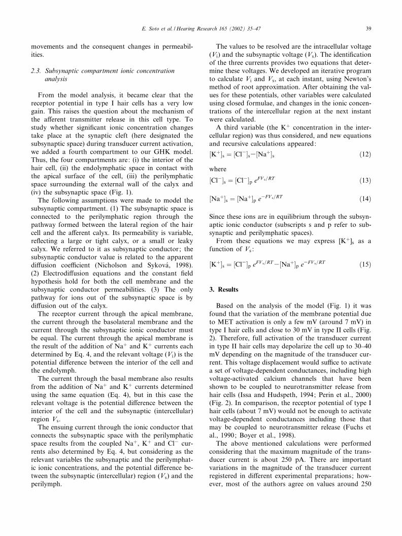
From the model analysis, it became clear that thereceptor potential in type I hair cells has a very lowgain. This raises the question about the mechanism ofthe a¡erent transmitter release in this cell type. Tostudy whether signi¢cant ionic concentration changestake place at the synaptic cleft (here designated thesubsynaptic space) during transducer current activation,we added a fourth compartment to our GHK model.Thus, the four compartments are: (i) the interior of thehair cell, (ii) the endolymphatic space in contact withthe apical surface of the cell, (iii) the perilymphaticspace surrounding the external wall of the calyx and(iv) the subsynaptic space (Fig. 1).
The following assumptions were made to model thesubsynaptic compartment. (1) The subsynaptic space isconnected to the perilymphatic region through thepathway formed between the lateral region of the haircell and the a¡erent calyx. Its permeability is variable,re£ecting a large or tight calyx, or a small or leakycalyx. We referred to it as subsynaptic conductor; thesubsynaptic conductor value is related to the apparentdi¡usion coe⁄cient (Nicholson and Sykova¤, 1998).(2) Electrodi¡usion equations and the constant ¢eldhypothesis hold for both the cell membrane and thesubsynaptic conductor permeabilities. (3) The onlypathway for ions out of the subsynaptic space is bydi¡usion out of the calyx.
The receptor current through the apical membrane,the current through the basolateral membrane and thecurrent through the subsynaptic ionic conductor mustbe equal. The current through the apical membrane isthe result of the addition of Naþ and Kþ currents eachdetermined by Eq. 4, and the relevant voltage (Vi) is thepotential di¡erence between the interior of the cell andthe endolymph.
The current through the basal membrane also resultsfrom the addition of Naþ and Kþ currents determinedusing the same equation (Eq. 4), but in this case therelevant voltage is the potential di¡erence between theinterior of the cell and the subsynaptic (intercellular)region Vs.
The ensuing current through the ionic conductor thatconnects the subsynaptic space with the perilymphaticspace results from the coupled Naþ, Kþ and Cl3 cur-rents also determined by Eq. 4, but considering as therelevant variables the subsynaptic and the perilymphat-ic ionic concentrations, and the potential di¡erence be-tween the subsynaptic (intercellular) region (Vs) and theperilymph.
The values to be resolved are the intracellular voltage(Vi) and the subsynaptic voltage (Vs). The identi¢cationof the three currents provides two equations that deter-mine these voltages. We developed an iterative programto calculate Vi and Vs, at each instant, using Newton’smethod of root approximation. After obtaining the val-ues for these potentials, other variables were calculatedusing closed formulae, and changes in the ionic concen-trations of the intercellular region at the next instantwere calculated.
A third variable (the Kþ concentration in the inter-cellular region) was thus considered, and new equationsand recursive calculations appeared:
½Kþ�s ¼ ½Cl3�s3½Naþ�s ð12Þ
where
½Cl3�s ¼ ½Cl3�p eFV s=RT ð13Þ
½Naþ�s ¼ ½Naþ�p e3FV s=RT ð14Þ
Since these ions are in equilibrium through the subsyn-aptic ionic conductor (subscripts s and p refer to sub-synaptic and perilymphatic spaces).
From these equations we may express [Kþ]s as afunction of Vs :
½Kþ�s ¼ ½Cl3�p eFV s=RT3½Naþ�p e3FV s=RT ð15Þ
3. Results
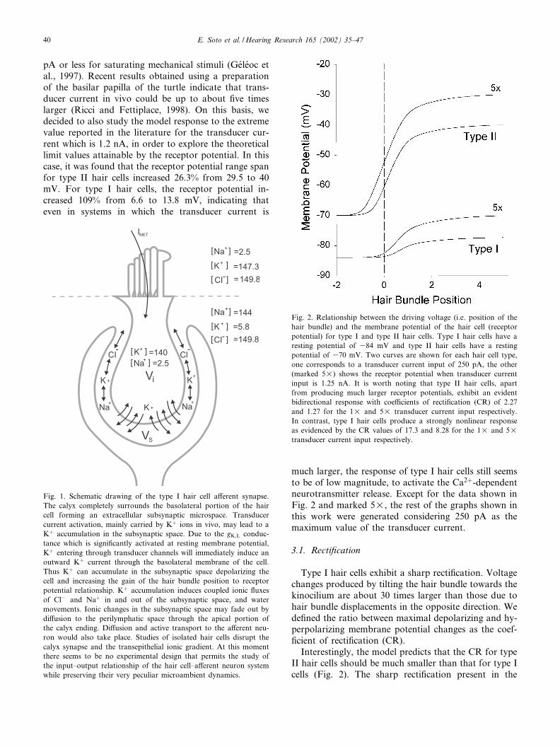
Based on the analysis of the model (Fig. 1) it wasfound that the variation of the membrane potential dueto MET activation is only a few mV (around 7 mV) intype I hair cells and close to 30 mV in type II cells (Fig.2). Therefore, full activation of the transducer currentin type II hair cells may depolarize the cell up to 30^40mV depending on the magnitude of the transducer cur-rent. This voltage displacement would su⁄ce to activatea set of voltage-dependent conductances, including highvoltage-activated calcium channels that have beenshown to be coupled to neurotransmitter release fromhair cells (Issa and Hudspeth, 1994; Perin et al., 2000)(Fig. 2). In comparison, the receptor potential of type Ihair cells (about 7 mV) would not be enough to activatevoltage-dependent conductances including those thatmay be coupled to neurotransmitter release (Fuchs etal., 1990; Boyer et al., 1998).
The above mentioned calculations were performedconsidering that the maximum magnitude of the trans-ducer current is about 250 pA. There are importantvariations in the magnitude of the transducer currentregistered in di¡erent experimental preparations; how-ever, most of the authors agree on values around 250
HEARES 3806 16-5-02
E. Soto et al. /Hearing Research 165 (2002) 35^47 39
pA or less for saturating mechanical stimuli (Ge¤le¤oc etal., 1997). Recent results obtained using a preparationof the basilar papilla of the turtle indicate that trans-ducer current in vivo could be up to about ¢ve timeslarger (Ricci and Fettiplace, 1998). On this basis, wedecided to also study the model response to the extremevalue reported in the literature for the transducer cur-rent which is 1.2 nA, in order to explore the theoreticallimit values attainable by the receptor potential. In thiscase, it was found that the receptor potential range spanfor type II hair cells increased 26.3% from 29.5 to 40mV. For type I hair cells, the receptor potential in-creased 109% from 6.6 to 13.8 mV, indicating thateven in systems in which the transducer current is
much larger, the response of type I hair cells still seemsto be of low magnitude, to activate the Ca2þ-dependentneurotransmitter release. Except for the data shown inFig. 2 and marked 5U, the rest of the graphs shown inthis work were generated considering 250 pA as themaximum value of the transducer current.
3.1. Recti¢cation
Type I hair cells exhibit a sharp recti¢cation. Voltagechanges produced by tilting the hair bundle towards thekinocilium are about 30 times larger than those due tohair bundle displacements in the opposite direction. Wede¢ned the ratio between maximal depolarizing and hy-perpolarizing membrane potential changes as the coef-¢cient of recti¢cation (CR).
Interestingly, the model predicts that the CR for typeII hair cells should be much smaller than that for type Icells (Fig. 2). The sharp recti¢cation present in the
Fig. 1. Schematic drawing of the type I hair cell a¡erent synapse.The calyx completely surrounds the basolateral portion of the haircell forming an extracellular subsynaptic microspace. Transducercurrent activation, mainly carried by Kþ ions in vivo, may lead to aKþ accumulation in the subsynaptic space. Due to the gK;L conduc-tance which is signi¢cantly activated at resting membrane potential,Kþ entering through transducer channels will immediately induce anoutward Kþ current through the basolateral membrane of the cell.Thus Kþ can accumulate in the subsynaptic space depolarizing thecell and increasing the gain of the hair bundle position to receptorpotential relationship. Kþ accumulation induces coupled ionic £uxesof Cl3 and Naþ in and out of the subsynaptic space, and watermovements. Ionic changes in the subsynaptic space may fade out bydi¡usion to the perilymphatic space through the apical portion ofthe calyx ending. Di¡usion and active transport to the a¡erent neu-ron would also take place. Studies of isolated hair cells disrupt thecalyx synapse and the transepithelial ionic gradient. At this momentthere seems to be no experimental design that permits the study ofthe input^output relationship of the hair cell^a¡erent neuron systemwhile preserving their very peculiar microambient dynamics.
Fig. 2. Relationship between the driving voltage (i.e. position of thehair bundle) and the membrane potential of the hair cell (receptorpotential) for type I and type II hair cells. Type I hair cells have aresting potential of 384 mV and type II hair cells have a restingpotential of 370 mV. Two curves are shown for each hair cell type,one corresponds to a transducer current input of 250 pA, the other(marked 5U) shows the receptor potential when transducer currentinput is 1.25 nA. It is worth noting that type II hair cells, apartfrom producing much larger receptor potentials, exhibit an evidentbidirectional response with coe⁄cients of recti¢cation (CR) of 2.27and 1.27 for the 1U and 5U transducer current input respectively.In contrast, type I hair cells produce a strongly nonlinear responseas evidenced by the CR values of 17.3 and 8.28 for the 1U and 5Utransducer current input respectively.
HEARES 3806 16-5-02
E. Soto et al. /Hearing Research 165 (2002) 35^4740
transducer current versus mechanical displacementcurve raises an important physiological question: howdo the a¡erent neurons exhibit a bidirectional responsewith small or no recti¢cation?, given that the transducercurrent has an almost absolute recti¢cation.
For parameters corresponding to a type II hair cell,an almost completely rectifying transducer permeability(with a CR of 48.7) was transformed into a weaklyrectifying receptor potential (CR=2.22), thus withgood bidirectional sensitivity (CRW2 means that theamplitude of the response in the depolarizing direction,when the hair bundle is displaced towards the kinoci-lium, is two times larger than in the hyperpolarizingdirection). In comparison, parameters for type I haircells produced a receptor potential with much largerrecti¢cation (CR=29.3), thus with poor bidirectionalsensitivity.
The di¡erence in the response of types I and II haircells suggests that the receptor potential CR decreasedwhen the basolateral resting permeability is low (andpossibly a nonlinear function of voltage). To examinethis possibility, we calculated the receptor potential as a
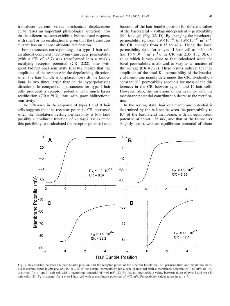
function of the hair bundle position for di¡erent valuesof the basolateral ^ voltage-independent ^ permeability(Kþ leakage) (Fig. 3A^D). By changing the basolateralpermeability Pp from 1.9U10316 to 1.9U10313 m3 s31,the CR changes from 0.37 to 42.4. Using the basalpermeability data for a type II hair cell at 360 mV(i.e. 1.9U10315 m3 s31), the CR was 2.35 (Fig. 3B), avalue which is very close to that calculated when thebasal permeability is allowed to vary as a function ofthe voltage (CR=2.22). These results indicate that theamplitude of the total Kþ permeability of the basolat-eral membrane mainly determines the CR. Evidently, aconstant Kþ permeability accounts for most of the dif-ferences in the CR between type I and II hair cells.However, also, the variations of permeability with themembrane potential contribute to decrease the recti¢ca-tion.
In the resting state, hair cell membrane potential isdetermined by the balance between the permeability toKþ of the basolateral membrane, with an equilibriumpotential of about 385 mV, and that of the transducer(slightly open), with an equilibrium potential of about
Fig. 3. Relationship between the hair bundle position and the receptor potential for di¡erent basolateral Kþ permeabilities and maximum trans-ducer current input is 250 pA. (A) PK is 1/10 of the normal permeability for a type II hair cell with a membrane potential of 360 mV. (B) PK
is normal for a type II hair cell with a membrane potential of 360 mV. (C) PK has an intermediate value, between those of type I and type IIhair cells. (D) PK is normal for a type I hair cell with a membrane potential of 375 mV. Permeability values given in m3 s31.
HEARES 3806 16-5-02
E. Soto et al. /Hearing Research 165 (2002) 35^47 41
0 mV. Consequently, the resting potential is about 362mV in cells with a basolateral permeability close to thenormal permeability of a type II hair cell (Fig. 3B).With hair bundle movements in the hyperpolarizing di-rection, the transducer channel open probability de-creases, and the potential approaches the equilibriumpotential of the basolateral membrane (almost 385mV). When hair bundle displacement is in the oppositedirection and the transducer channel open probabilityincreases, the membrane potential becomes V37 mVnear the apical equilibrium potential (0 mV). As a con-sequence, CR is 2.2. However, when the basolateralpermeability is 10-fold smaller (Fig. 3A), the restingmembrane potential is V325 mV, and recti¢cation isinverted (CR=0.37). In general, increasing transducerchannel open probability drives the membrane potentialclose to 0 mV, and closing the transducer channelsdrives the membrane potential close to the membranepotential for the basolateral membrane (which has alimit value determined by the Kþ equilibrium potential,in this case 385 mV).
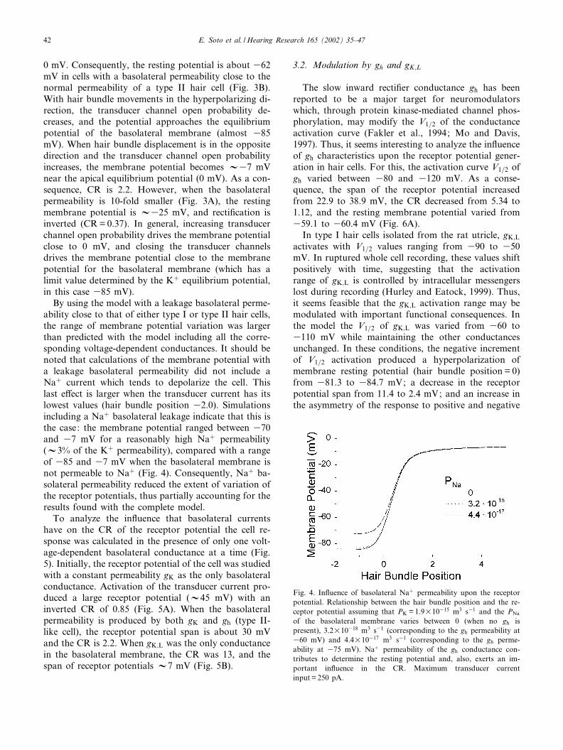
By using the model with a leakage basolateral perme-ability close to that of either type I or type II hair cells,the range of membrane potential variation was largerthan predicted with the model including all the corre-sponding voltage-dependent conductances. It should benoted that calculations of the membrane potential witha leakage basolateral permeability did not include aNaþ current which tends to depolarize the cell. Thislast e¡ect is larger when the transducer current has itslowest values (hair bundle position 32.0). Simulationsincluding a Naþ basolateral leakage indicate that this isthe case: the membrane potential ranged between 370and 37 mV for a reasonably high Naþ permeability(V3% of the Kþ permeability), compared with a rangeof 385 and 37 mV when the basolateral membrane isnot permeable to Naþ (Fig. 4). Consequently, Naþ ba-solateral permeability reduced the extent of variation ofthe receptor potentials, thus partially accounting for theresults found with the complete model.
To analyze the in£uence that basolateral currentshave on the CR of the receptor potential the cell re-sponse was calculated in the presence of only one volt-age-dependent basolateral conductance at a time (Fig.5). Initially, the receptor potential of the cell was studiedwith a constant permeability gK as the only basolateralconductance. Activation of the transducer current pro-duced a large receptor potential (V45 mV) with aninverted CR of 0.85 (Fig. 5A). When the basolateralpermeability is produced by both gK and gh (type II-like cell), the receptor potential span is about 30 mVand the CR is 2.2. When gK;L was the only conductancein the basolateral membrane, the CR was 13, and thespan of receptor potentials V7 mV (Fig. 5B).
3.2. Modulation by gh and gK;L
The slow inward recti¢er conductance gh has beenreported to be a major target for neuromodulatorswhich, through protein kinase-mediated channel phos-phorylation, may modify the V1=2 of the conductanceactivation curve (Fakler et al., 1994; Mo and Davis,1997). Thus, it seems interesting to analyze the in£uenceof gh characteristics upon the receptor potential gener-ation in hair cells. For this, the activation curve V1=2 ofgh varied between 380 and 3120 mV. As a conse-quence, the span of the receptor potential increasedfrom 22.9 to 38.9 mV, the CR decreased from 5.34 to1.12, and the resting membrane potential varied from359.1 to 360.4 mV (Fig. 6A).
In type I hair cells isolated from the rat utricle, gK;L
activates with V1=2 values ranging from 390 to 350mV. In ruptured whole cell recording, these values shiftpositively with time, suggesting that the activationrange of gK;L is controlled by intracellular messengerslost during recording (Hurley and Eatock, 1999). Thus,it seems feasible that the gK;L activation range may bemodulated with important functional consequences. Inthe model the V1=2 of gK;L was varied from 360 to3110 mV while maintaining the other conductancesunchanged. In these conditions, the negative incrementof V1=2 activation produced a hyperpolarization ofmembrane resting potential (hair bundle position = 0)from 381.3 to 384.7 mV; a decrease in the receptorpotential span from 11.4 to 2.4 mV; and an increase inthe asymmetry of the response to positive and negative
Fig. 4. In£uence of basolateral Naþ permeability upon the receptorpotential. Relationship between the hair bundle position and the re-ceptor potential assuming that PK =1.9U10315 m3 s31 and the PNa
of the basolateral membrane varies between 0 (when no gh ispresent), 3.2U10318 m3 s31 (corresponding to the gh permeability at360 mV) and 4.4U10317 m3 s31 (corresponding to the gh perme-ability at 375 mV). Naþ permeability of the gh conductance con-tributes to determine the resting potential and, also, exerts an im-portant in£uence in the CR. Maximum transducer currentinput= 250 pA.
HEARES 3806 16-5-02
E. Soto et al. /Hearing Research 165 (2002) 35^4742
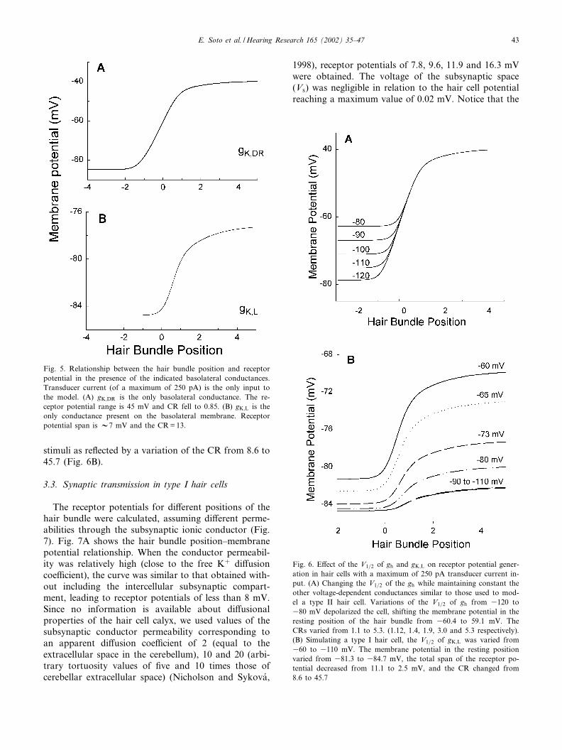
stimuli as re£ected by a variation of the CR from 8.6 to45.7 (Fig. 6B).
3.3. Synaptic transmission in type I hair cells
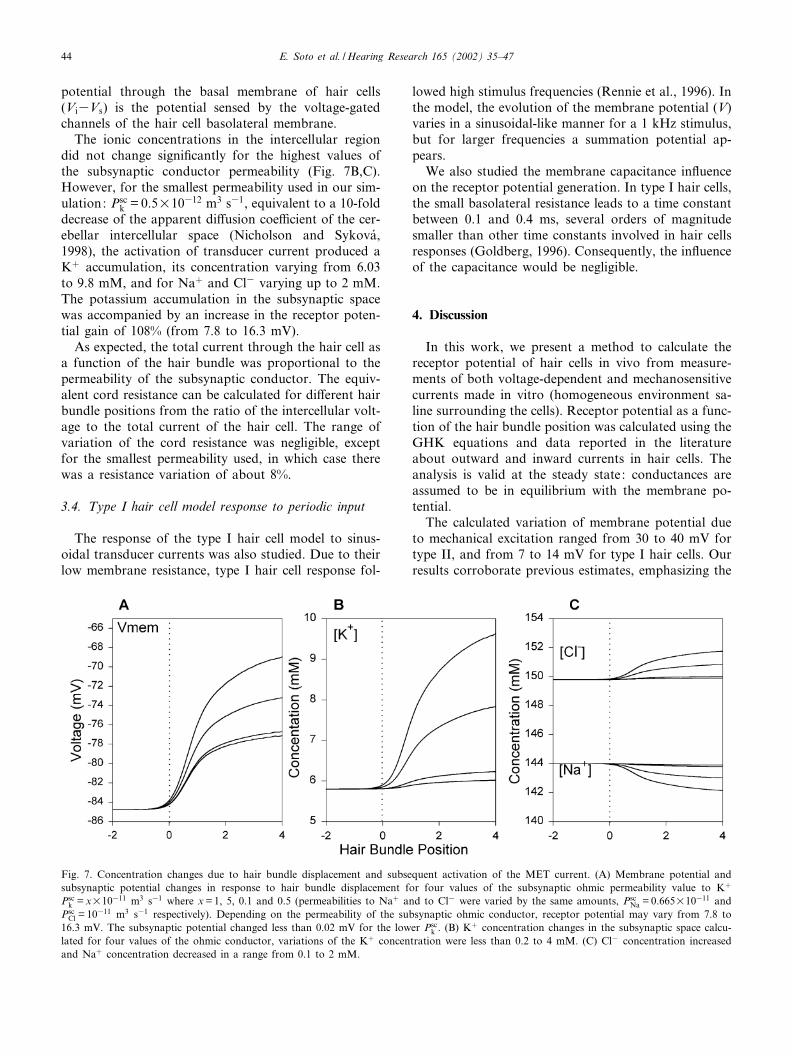
The receptor potentials for di¡erent positions of thehair bundle were calculated, assuming di¡erent perme-abilities through the subsynaptic ionic conductor (Fig.7). Fig. 7A shows the hair bundle position^membranepotential relationship. When the conductor permeabil-ity was relatively high (close to the free Kþ di¡usioncoe⁄cient), the curve was similar to that obtained with-out including the intercellular subsynaptic compart-ment, leading to receptor potentials of less than 8 mV.Since no information is available about di¡usionalproperties of the hair cell calyx, we used values of thesubsynaptic conductor permeability corresponding toan apparent di¡usion coe⁄cient of 2 (equal to theextracellular space in the cerebellum), 10 and 20 (arbi-trary tortuosity values of ¢ve and 10 times those ofcerebellar extracellular space) (Nicholson and Sykova¤,
1998), receptor potentials of 7.8, 9.6, 11.9 and 16.3 mVwere obtained. The voltage of the subsynaptic space(Vs) was negligible in relation to the hair cell potentialreaching a maximum value of 0.02 mV. Notice that the
Fig. 6. E¡ect of the V1=2 of gh and gK;L on receptor potential gener-ation in hair cells with a maximum of 250 pA transducer current in-put. (A) Changing the V1=2 of the gh while maintaining constant theother voltage-dependent conductances similar to those used to mod-el a type II hair cell. Variations of the V1=2 of gh from 3120 to380 mV depolarized the cell, shifting the membrane potential in theresting position of the hair bundle from 360.4 to 59.1 mV. TheCRs varied from 1.1 to 5.3. (1.12, 1.4, 1.9, 3.0 and 5.3 respectively).(B) Simulating a type I hair cell, the V1=2 of gK;L was varied from360 to 3110 mV. The membrane potential in the resting positionvaried from 381.3 to 384.7 mV, the total span of the receptor po-tential decreased from 11.1 to 2.5 mV, and the CR changed from8.6 to 45.7
Fig. 5. Relationship between the hair bundle position and receptorpotential in the presence of the indicated basolateral conductances.Transducer current (of a maximum of 250 pA) is the only input tothe model. (A) gK;DR is the only basolateral conductance. The re-ceptor potential range is 45 mV and CR fell to 0.85. (B) gK;L is theonly conductance present on the basolateral membrane. Receptorpotential span is V7 mV and the CR=13.
HEARES 3806 16-5-02
E. Soto et al. /Hearing Research 165 (2002) 35^47 43
potential through the basal membrane of hair cells(Vi3Vs) is the potential sensed by the voltage-gatedchannels of the hair cell basolateral membrane.
The ionic concentrations in the intercellular regiondid not change signi¢cantly for the highest values ofthe subsynaptic conductor permeability (Fig. 7B,C).However, for the smallest permeability used in our sim-ulation: Psc
k = 0.5U10312 m3 s31, equivalent to a 10-folddecrease of the apparent di¡usion coe⁄cient of the cer-ebellar intercellular space (Nicholson and Sykova¤,1998), the activation of transducer current produced aKþ accumulation, its concentration varying from 6.03to 9.8 mM, and for Naþ and Cl3 varying up to 2 mM.The potassium accumulation in the subsynaptic spacewas accompanied by an increase in the receptor poten-tial gain of 108% (from 7.8 to 16.3 mV).
As expected, the total current through the hair cell asa function of the hair bundle was proportional to thepermeability of the subsynaptic conductor. The equiv-alent cord resistance can be calculated for di¡erent hairbundle positions from the ratio of the intercellular volt-age to the total current of the hair cell. The range ofvariation of the cord resistance was negligible, exceptfor the smallest permeability used, in which case therewas a resistance variation of about 8%.
3.4. Type I hair cell model response to periodic input
The response of the type I hair cell model to sinus-oidal transducer currents was also studied. Due to theirlow membrane resistance, type I hair cell response fol-
lowed high stimulus frequencies (Rennie et al., 1996). Inthe model, the evolution of the membrane potential (V)varies in a sinusoidal-like manner for a 1 kHz stimulus,but for larger frequencies a summation potential ap-pears.
We also studied the membrane capacitance in£uenceon the receptor potential generation. In type I hair cells,the small basolateral resistance leads to a time constantbetween 0.1 and 0.4 ms, several orders of magnitudesmaller than other time constants involved in hair cellsresponses (Goldberg, 1996). Consequently, the in£uenceof the capacitance would be negligible.
4. Discussion
In this work, we present a method to calculate thereceptor potential of hair cells in vivo from measure-ments of both voltage-dependent and mechanosensitivecurrents made in vitro (homogeneous environment sa-line surrounding the cells). Receptor potential as a func-tion of the hair bundle position was calculated using theGHK equations and data reported in the literatureabout outward and inward currents in hair cells. Theanalysis is valid at the steady state: conductances areassumed to be in equilibrium with the membrane po-tential.
The calculated variation of membrane potential dueto mechanical excitation ranged from 30 to 40 mV fortype II, and from 7 to 14 mV for type I hair cells. Ourresults corroborate previous estimates, emphasizing the
Fig. 7. Concentration changes due to hair bundle displacement and subsequent activation of the MET current. (A) Membrane potential andsubsynaptic potential changes in response to hair bundle displacement for four values of the subsynaptic ohmic permeability value to Kþ
Psck = xU10311 m3 s31 where x=1, 5, 0.1 and 0.5 (permeabilities to Naþ and to Cl3 were varied by the same amounts, Psc
Na = 0.665U10311 andPsc
Cl = 10311 m3 s31 respectively). Depending on the permeability of the subsynaptic ohmic conductor, receptor potential may vary from 7.8 to16.3 mV. The subsynaptic potential changed less than 0.02 mV for the lower Psc
k . (B) Kþ concentration changes in the subsynaptic space calcu-lated for four values of the ohmic conductor, variations of the Kþ concentration were less than 0.2 to 4 mM. (C) Cl3 concentration increasedand Naþ concentration decreased in a range from 0.1 to 2 mM.
HEARES 3806 16-5-02
E. Soto et al. /Hearing Research 165 (2002) 35^4744
now inevitable question about how type I hair cellstransmit information to the a¡erent neurons (Goldberg,1996).
Experimentally, it has been found that there are largedi¡erences in the membrane potential between type Iand type II hair cells. For example, in hair cells isolatedfrom the pigeon semicircular canal, the resting mem-brane potential of type I cells was 370W 3 mV, andthat of type II cells was 357W 3 mV (Correia andLang, 1990). Then, MET currents of as much as 1.25nA could depolarize type I cells 13.8 mV, displacing themembrane potential up to 356 mV. Therefore, forthese cells to activate the neurotransmitter release ma-chinery, voltage-dependent Ca2þ channels should havea very negative activating voltage, or there should besome other mechanisms contributing to further depo-larize the cell, as has been proposed by Goldberg(1996).
An alternative mechanism implies that Kþ enteringthrough the MET channels would leave the hair cellthrough the basolateral surface. The calyx terminal ofthe a¡erent neuron, in close apposition to the hair cell,surrounds its basolateral portion forming a reducedextracellular microspace. Thus, Kþ £owing out fromthe hair cell by its basolateral surface (following itsdriving force) would accumulate in the extracellularspace, depolarizing the cell and leading to the activationof high voltage-activated calcium channels and subse-quent neurotransmitter release (Goldberg, 1996). At thesame time, Kþ accumulation in the synaptic cleft mayas well depolarize the calyx ending directly in£uencingthe action potential generation in the a¡erent neuron.In type I hair cells isolated from guinea pig, it has beenshown that perfusion with high Kþ solutions led to asigni¢cant intracellular Ca2þ concentration change, dueto the activation of voltage-sensitive calcium channelsof the L type (Boyer et al., 1998). Another possibilityhas been raised recently, it implies the inhibition of IK;L
by nitric oxide leading to an increase in the input resis-tance of the hair cell, and a subsequent increase in thereceptor potential gain (Chen and Eatock, 2000).
The model shows that the permeability of the path-way for the ions to di¡use out of the calyx may play akey role in determining the ionic changes that wouldtake place at the subsynaptic space. Thus, the calyxstructure seems to be of fundamental importance inthe proposed mechanism, since a very tight or largecalyx determines a very low permeability for the path-way for ions to di¡use out of the subsynaptic space. Inthe model we used permeabilities of the subsynapticohmic conductor in the range of 10311 m3 s31 (closeto the potassium mobility in a water solution) to0.5U10312 m3 s31, that is more than one order of mag-nitude decrease in Kþ permeability, equivalent to atortuosity coe⁄cient of 20 (Nicholson and Sykova¤,
1998) leading to a 62.5% (from 6.03 to 9.79 mM) in-crease of Kþ concentration in the subsynaptic space.
Also, due to the reduced extracellular volume of thesynaptic cleft, it is conceivable that when the Ca2þ in-£ux into the pre- or the postsynaptic cell is large, theconcentration of Ca2þ in the synaptic cleft may drop,leading to a reduction in the amount of Ca2þ availablefor evoking transmitter release. Such a Ca2þ concentra-tion depletion due to synaptic activity has been shownin the synapse between the calyces of Held and the cellsof the medial nucleus of the trapezoid body in the ratauditory brainstem (Borst and Sakmann, 1999).
Important in£uences in the hair cell response couldbe produced by modi¢cations in the characteristics ofgK;L. Cells which express gK;L showed signi¢cantly morenegative resting potential. Depending on gK;L magni-tude and V1=2, the hair cell receptor potential mayvary from a few mV, as in type I cells, to tens of mV,as in type II lacking gK;L. Also the value of the V1=2 ofgK;L contributes to determine the resting membrane po-tential and the total span of the receptor potential, thuscontrolling the gain of type I hair cells.
Cells expressing gK;L maintain the nonlinearities ofthe MET. In contrast, the response to mechanical stim-uli of those cells without gK;L is partially linearized. Weproved that the magnitude and V1=2 of a voltage-depen-dent basolateral conductance might compensate the rec-ti¢cation of the transducer current. The expression ofthe delayed recti¢er gK;DR in the basolateral region de-creased the CR, leading to its inversion (Fig. 5A). Add-ing the slow inward recti¢er gh increased the CR (tovalues between 1 and 10, depending on the V1=2 of ghactivation).
In this work gK;DR and the gK;A-type currents werelumped together. Justi¢cation for doing so is that sincewe used GHK equations to develop the model, the rel-evant parameter is the voltage dependence of the cur-rent and not its particular dynamics. In fact, Hodgkin^Huxley-type models of hair cell dynamics using just onelumped basal Kþ conductance reproduce the hair cellvoltage response trajectories with a high degree of ac-curacy (Alexandrov et al., 2001).
Inward rectifying currents have been found in 50% oftype I hair cells and 61.5% of type II cells in mamma-lian vestibule (Eatock and Hutzler, 1992) and in 86% ofshort oscillatory-type hair cells from the gold¢sh saccu-lus (Sugihara and Furukawa, 1995). Both the rapidgK;IR and the slow non-inactivating gh-type inward cur-rents have been reported in hair cells (Holt and Eatock,1995). In this work, it has been found that a morenegative activation for gh (permeable to Kþ and toNaþ) increased the receptor potential, and decreasedthe CR. This indicates that important regulatory in£u-ences can take place by modi¢cation of the V1=2 of thiscurrent. Therefore, the expression of gh may contribute
HEARES 3806 16-5-02
E. Soto et al. /Hearing Research 165 (2002) 35^47 45
to di¡erentiate hair cell subsets and constitute an ele-ment for plastic and regulatory changes of hair cellresponse.
Bidirectionality is a fundamental property of hair cellsystems that signi¢cantly contributes to sensory codingof mechanical stimuli. Hair cell response to hair bundledisplacements towards the kinocilium is depolarizingwhile displacements of the hair bundle in the oppositedirection produce a hyperpolarization of the cell. How-ever, the transducer current is highly rectifying, the cur-rent produced by hair bundle displacements towardsthe kinocilium is much larger than the current responseto displacements in the opposite direction. It is wellknown that vestibular a¡erent neuron response recti¢es,but this recti¢cation is not as important as that re-ported for the mechanoelectrical transduction (Lowen-stein and Roberts, 1949; Blanks and Precht, 1976; Fer-na¤ndez and Goldberg, 1976; Hartmann and Klinke,1980). Then, there is the problem of how to explainthe bidirectional sensory coding given the strongly rec-tifying transducer current. One possibility is that theexperimental manipulations of hair cells to record thetransducer currents may have some deleterious e¡ect onthe response to mechanical stimuli. The resonance ofthe receptor potential may also contribute to linearizethe receptor potential. However, semicircular canal cellshave a low resonant frequency and low quality of res-onance (Angelaki and Correia, 1991), and type I haircells show practically no resonance (Rennie et al.,1996). Thus, although resonance could explain the bi-directional coding of some hair cells it is not su⁄cientto account for bidirectionality as a general property ofsensory coding in hair cell systems. Our ¢ndings indi-cate that the type I hair cell receptor potential re£ectsthe shape of the transducer current, thus it is stronglyrectifying, and has poor bidirectionality; in contrast,type II hair cells have a high gain bidirectional codingreceptor potential.
Sinusoidal transducer current input to the modelleads to an interesting observation. When the frequencyof stimulation or the cell capacitance increases, the haircell receptor potential modulation by the sinusoidalstimulus decreases, but an accumulative, slowly risingDC potential develops. This type of DC potential hasbeen described in cochlear hair cells (van Emst et al.,1998).
Finally, it should be taken into account that thetransducer current is dynamic, with intracellular Ca2þ
and cyclic nucleotides regulating its activation and theset point of the response^displacement curve. Our mod-el results indicate that basolateral conductances play akey role in de¢ning the resting membrane potential, thegain and the bidirectional sensitivity of the receptorpotential. Also, depending on voltage dependence andthe magnitude of the basolateral Kþ currents, and on
the di¡usional properties of the calyx pathway for ionsto di¡use out from the synaptic cleft, signi¢cant Kþ
accumulation could take place in the synaptic spaceand contribute to increase the gain of the receptor po-tential in type I hair cells.
Acknowledgements
This work was partially supported by Consejo Nacio-nal de Ciencia y Tecnolog|¤a de Me¤xico (CONACyT),Grant 35525-N to E.S. The authors thank Isabel Pe¤rezMontfort for proofreading the English manuscript.
References
Alexandrov, A., Almanza, A., Kulikovskaya, N., Vega, R., Alexan-drova, T.B., Shulenina, N.E., Limo¤n, A., Soto, E., 2001. A math-ematical model of the total current dynamics in hair cells. In:Sadovnichii, V.A., Doger, E. (Eds.), Mathematical Modeling ofComplex Information Processing Systems. Moscow UniversityPress, Moscow, pp. 26^41.
Angelaki, D.E., Correia, M.J., 1991. Models of membrane resonancein pigeon semicircular canal type II hair cells. Biol. Cybern. 65, 1^10.
Blanks, R.H., Precht, W., 1976. Functional characterization of pri-mary vestibular a¡erents in the frog. Exp. Brain Res. 25, 369^390.
Borst, J.G.G., Sakmann, B., 1999. Depletion of calcium in the syn-aptic cleft of a calyx-type synapse in the rat brainstem. J. Physiol.521, 123^133.
Bosher, S.K., Warren, R.L., 1968. Observations on the electrochem-istry of the cochlear endolymph of the rat: a quantitative study ofits electrical potential and ionic composition as determined bymeans of £ame spectrophotometry. Proc. R. Soc. Lond. B 171,227^247.
Boyer, C., Lehouelleur, J., Sans, A., 1998. Potassium depolarizationof mammalian vestibular sensory cells increases [Ca2þ]i throughvoltage-sensitive calcium channels. Eur. J. Neurosci. 10, 971^975.
Bracho, H., Budelli, R., 1978. The generation of resting membranepotentials in an inner ear hair cell system. J. Physiol. 281, 445^465.
Chen, J.W., Eatock, R.A., 2000. Major potassium conductance formhair cell type I from rat semicircular canals: characterization andmodulation by nitric oxide. J. Neurophysiol. 84, 139^151.
Corey, D.P., Hudspeth, A.J., 1979. Response latency of vertebratehair cells. Biophys. J. 26, 499^506.
Corey, D.P., Hudspeth, A.J., 1983. Kinetics of the receptor current inbullfrog saccular hair cells. J. Neurosci. 3, 962^976.
Correia, M.J., 1992. Filtering properties of hair cells. Ann. NY Acad.Sci. 656, 182^203.
Correia, M.J., Lang, D.G., 1990. An electrophysiological comparisonof solitary type I and type II vestibular hair cells. Neurosci. Lett.116, 106^111.
Crawford, A.C., Evans, M.G., Fettiplace, R., 1991. The actions ofcalcium on the mechano-electrical transducer current of turtle haircells. J. Physiol. 434, 369^398.
Eatock, R.A., Hutzler, M.J., 1992. Ionic currents of mammalian ves-tibular hair cells. Ann. NY Acad. Sci. 656, 58^74.
Eatock, R.A., Ru«sch, A., 1997. Developmental changes in the phys-iology of hair cells. Cell Dev. Biol. 8, 265^275.
HEARES 3806 16-5-02
E. Soto et al. /Hearing Research 165 (2002) 35^4746
Fakler, B., Bra«ndle, U., Glowatzki, E., Zenner, H.P., Ruppersberg,J.P., 1994. Kir 2.1 inward recti¢er Kþ channels are regulated in-dependently by protein kinases and ATP hydrolysis. Neuron 13,1413^1420.
Ferna¤ndez, C., Goldberg, J.M., 1976. Physiology of peripheral neu-rons innervating otolith organs of the squirrel monkey II. Direc-tional selectivity and force-response relations. J. Neurophysiol. 39,985^995.
Fuchs, P.A., Evans, M.G., Murrow, B.W., 1990. Calcium currents inhair cells isolated from the cochlea of the chick. J. Physiol. 429,553^568.
Ge¤le¤oc, G.S.G., Lennan, G.W.T., Richardson, G.P., Kros, C.J., 1997.A quantitative comparison of mechanoelectrical transduction investibular and auditory hair cells of neonatal mice. Proc. R. Soc.Lond. B 264, 611^621.
Goldberg, J.M., 1996. Theoretical analysis of intracellular communi-cation between the vestibular type I hair cell and its calyx ending.J. Neurophysiol. 76, 1942^1957.
Goldman, D.E., 1943. Potential, impedance and recti¢cation in mem-branes. J. Gen. Physiol. 27, 37^60.
Guth, P.S., Perin, P., Norris, C.H., Valli, P., 1998. The vestibular haircells : post-transductional signal processing. Prog. Neurobiol. 54,193^247.
Hartmann, R., Klinke, R., 1980. Discharge properties of a¡erent ¢-bres of the gold¢sh semicircular canal with high frequency stim-ulation. P£u«gers Arch. 388, 111^121.
Hodgkin, A.L., Katz, B., 1949. The e¡ect of sodium ions on theelectrical activity of the giant axon of the squid. J. Physiol. 108,37^77.
Holt, R.H., Eatock, R.A., 1995. Inwardly rectifying currents of sac-cular hair cells from the leopard frog. J. Neurophysiol. 73, 1484^1502.
Hudspeth, A.J., 1986. The ionic channels of a vertebrate hair cell.Hear. Res. 22, 21^27.
Hudspeth, A.J., Lewis, R.S., 1988. Kinetic analysis of voltage and iondependent conductances in saccular hair cells of the bull frog,Rana catesbiana. J. Physiol. 400, 237^274.
Hurley, K.M., Eatock, R.A., 1999. Characterization and modulationof a delayed recti¢er in vestibular type I hair cells. Assoc. Res.Otolaryngol. Abstr. 22, 764.
Issa, N.P., Hudspeth, A.J., 1994. Clustering of Ca2þ channels andCa2þ-activated Kþ channels at £uorescently labeled presynapticactive zones of hair cells. Proc. Natl. Acad. Sci. USA 91, 7578^7582.
Lennan, G.W.T., Steinacker, A., Lehouelleur, J., 1999. Ionic currentsand current-clamp depolarisations of type I and type II hair cellsfrom the developing rat utricle. P£u«gers Arch. 438, 40^46.
Lowenstein, O., Roberts, T.D.M., 1949. The equilibrium function ofthe otolith organs of the tornback ray (Raja clavata). J. Physiol.110, 392^415.
Masetto, S., Russo, G., Prigioni, I., 1994. Di¡erential expression ofpotassium currents by hair cells in thin slices of frog crista ampul-laris. J. Neurophysiol. 72, 443^455.
Mo, Z.L., Davis, R.L., 1997. Heterogeneous voltage dependence ofinward recti¢er currents in spiral ganglion neurons. J. Neuro-physiol. 78, 3019^3027.
Nicholson, C., Sykova¤, E., 1998. Extracellular space structure re-vealed by di¡usion analysis. Trends Neurosci. 21, 207^215.
Ohmori, H., 1985. Mechano-electrical transduction currents in iso-lated vestibular hair cells of the chick. J. Physiol. 359, 189^217.
Ohmori, H., 1987. Gating properties of the mechano-electrical trans-ducer channel in the dissociated vestibular hair cell of the chick.J. Physiol. 387, 589^609.
Perin, P., Soto, E., Vega, R., Botta, L., Masetto, S., Zucca, G., Valli,P., 2000. Calcium channels functional roles in the frog semicircularcanal. NeuroReport 11, 417^420.
Rennie, K.J., Ashmore, J.F., 1991. Ionic currents in isolated vestibu-lar hair cells from the guinea pig crista ampullaris. Hear. Res. 51,279^292.
Rennie, K.J., Ricci, A.J., Correia, M.J., 1996. Electrical ¢ltering ingerbil isolated type I semicircular canal hair cells. J. Neurophysiol.75, 2117^2123.
Ricci, A.J., Fettiplace, R., 1997. The e¡ects of calcium bu¡ering andcyclic AMP on mechano-electrical transduction in turtle auditoryhair cells. J. Physiol. 501, 111^124.
Ricci, A.J., Fettiplace, R., 1998. Calcium permeation of the turtle haircell mechanotransducer channel and its relation to the composi-tion of endolymph. J. Physiol. 506, 159^173.
Ricci, A.J., Rennie, K.J., Correia, M.J., 1996. The delayed recti¢er,IKI, is the major conductance in type I vestibular hair cells acrossvestibular end organs. P£u«gers Arch. 432, 34^42.
Ru«sch, A., Eatock, R.A., 1996a. Voltage responses of mouse utricularhair cells to injected currents. Ann. NY Acad. Sci. 781, 71^84.
Ru«sch, A., Eatock, R.A., 1996b. A delayed conductance in type I haircells of the mouse utricle. J. Neurophysiol. 76, 995^1004.
Sauer, G., Richter, C.P., Klinke, R., 1999. Sodium, potassium, chlo-ride and calcium concentrations measured in pigeon perilymphand endolymph. Hear. Res. 129, 1^6.
Scarfone, E., Deme“mes, D., Jahn, R., De Camilli, P., Sans, A., 1988.Secretory function of the vestibular nerve calyx suggested by pres-ence of vesicles, synapsin I, and synaptophysin. J. Neurosci. 8,4640^4645.
Schessel, D.A., Ginzberg, R., Highstein, M., 1991. Morphophysiologyof synaptic transmission between type I hair cells and vestibularprimary a¡erents. An intracellular study employing horseradishperoxidase in the lizard, Calotes versicolor. Brain Res. 544, 1^16.
Soto, E., Budelli, R., Vega, R., 1999. The hair cell receptor potential:a theoretical analysis. Assoc. Res. Otolaryngol. Abstr. 22, 765.
Sterkers, O., Ferrary, E., Amiel, C., 1988. Production of inner ear£uids. Physiol. Rev. 68, 1083^1128.
Sugihara, I., Furukawa, T., 1995. Potassium currents underlying theoscillatory response in hair cells of the gold¢sh sacculus. J. Phys-iol. 489, 443^453.
van Emst, M.G., Gigue're, C., Smoorenburg, F., 1998. The generationof DC potentials in a computational model of the organ of Corti:e¡ects of voltage-dependent Kþ channels in the basolateral mem-brane of the hair cell. Hear. Res. 115, 184^196.
Yamashita, M., Ohmori, H., 1990. Synaptic responses to mechanicalstimulation in calyceal and bouton type vestibular a¡erents studiedin an isolated preparation of semicircular canal ampullae of chick-en. Exp. Brain Res. 80, 475^488.
HEARES 3806 16-5-02
E. Soto et al. /Hearing Research 165 (2002) 35^47 47


