Embed Size (px)
Citation preview
Christopoulos et al. Molecular Cancer (2015) 14:43 DOI 10.1186/s12943-015-0291-7
REVIEW Open Access
The role of the insulin-like growth factor-1 systemin breast cancerPanagiotis F Christopoulos1*, Pavlos Msaouel2 and Michael Koutsilieris1
Abstract
IGF-1 is a potent mitogen of major importance in the mammary gland. IGF-1 binding to the cognate receptor,IGF-1R, triggers a signaling cascade leading to proliferative and anti-apoptotic events. Although many of the relevantmolecular pathways and intracellular cascades remain to be elucidated, a growing body of evidence points to theimportant role of the IGF-1 system in breast cancer development, progression and metastasis. IGF-1 is a point ofconvergence for major signaling pathways implicated in breast cancer growth. In this review, we provide an overviewand concise update on the function and regulation of IGF-1 as well as the role it plays in breast malignancies.
Keywords: Insulin-like growth factor-1 (IGF-1), IGF-1 receptor (IGF-1R), IGF-binding proteins (IGFBPs), IGF-1 signaling,IGF-1 regulation, Estrogens, Estrogen receptor (ER), Breast cancer, Mammary tumorigenesis
IntroductionThe Insulin Like Growth Factor 1 (IGF-1) system which in-cludes IGF-1, IGF-binding proteins (IGFBPs) and the IGF-1receptor (IGF-1R), plays a significant role in human physi-ology, particularly in the development and function of manytissues, including the mammary gland. IGF-1 is a key medi-ator of mammary terminal end bud and ductal formationduring development [1]. Experimental findings have demon-strated that normal rat mammary epithelial cells continue toproliferate in serum-free media in response to IGF-1, sug-gesting that the IGF-1 system plays an important role inmammary gland function and maintenance [2]. In contrast,over the last two decades many studies have implicated theIGF-1 system in the development of several malignancies in-cluding breast cancer. Indeed, breast tumors may aberrantlyexpress each component of the IGF-1 system. To date, an in-creasing number of studies have attempted to elucidate themolecular mechanisms underlying the association of theIGF-1 system with breast malignancy [3-5].
The IGF-1 system in breast cancerIGF-1 is a 7.7 kDa single-chain polypeptide encoded bychromosome 12. The 70 amino acid IGF-1 protein
* Correspondence: [email protected] of Experimental Physiology, Medical School, National andKapodistrian University of Athens, 75 Mikras Asias Street, 11527 Goudi,Athens, GreeceFull list of author information is available at the end of the article
© 2015 Christopoulos et al.; licensee BioMed CCommons Attribution License (http://creativecreproduction in any medium, provided the orDedication waiver (http://creativecommons.orunless otherwise stated.
consists of four domains [6] and is produced primarilyin the liver under the direct stimulation of GrowthHormone (GH). IGF-1 gene expression is controlled byboth transcriptional and post-translational modifications.Distinct IGF-1-peptides may arise via the utilization ofdifferent promoters, alternative splicing, proteolytic pro-cessing and glycosylation events [7]. IGF-1 is expressedin almost every human tissue. In both normal mammarygland and malignant breast tissues, IGF-1 is mainlyexpressed by stromal and only rarely by epithelial cells[8]. Circulating IGF-1 levels vary depending on the per-son’s age: there is initially an increase in serum IGF-1from birth to puberty, followed by a stable decline withage in response to the lower GH levels [9,10]. The mito-genic, anti-apoptotic and other effects of IGF-1 proteinare mainly mediated by the transmembrane tyrosinekinase receptor IGF-1R, which in contrast to IGF-1, ismainly expressed in the mammary epithelium. TetramericIGF-1R consists of two α- identical and two β- identicalsubunits. Ligand binding and subsequent phosphorylationof IGF-1R triggers the activation of two major signalingcascades via the insulin receptor substrate 1 (IRS-1): thephosphatidylinositol 3-kinase/AKT kinase (PI3K/AKT)pathway and the RAF kinase/mitogen activated proteinkinase (RAF/MAPK) pathway which stimulate prolifer-ation and protection from apoptosis [11]. Notably, theIRS-1 has been found to be overexpressed in primarybreast tumors [12]. The bioavailability and half-life of
entral. This is an Open Access article distributed under the terms of the Creativeommons.org/licenses/by/4.0), which permits unrestricted use, distribution, andiginal work is properly credited. The Creative Commons Public Domaing/publicdomain/zero/1.0/) applies to the data made available in this article,
Christopoulos et al. Molecular Cancer (2015) 14:43 Page 2 of 14
circulating IGF-1 is regulated by a family of six IGF-binding proteins (IGFBP1-6) [13]. Each IGFBP canbind to IGF-1 with high affinity and is regulated byseveral specific IGBPB proteases. Approximately 1% ofcirculating IGF-1 remains unbound, while the rest ismainly bound to IGFBP3, forming a complex with anacid-labile subunit [14].Breast Cancer (BrCa) remains one of the leading
causes of cancer-related death worldwide. The hetero-geneity and variability in treatment and survival re-sponse, underscore the need to elucidate the biologicalmechanisms driving BrCa. A proposed molecular pro-file approach for breast tumor classification definesdistinct molecular subtypes of the disease based ondifferences in the expression patterns of estrogen re-ceptor (ER), progesterone receptor (PR) and HER2(ERBB2) [15]. Although BrCa has been intensely stud-ied and multiple reported biomarkers and moleculartargets have been reported in the literature, only a feware of proven relevance to routine clinical practice.Both in vitro and in vivo models, as well as clinical andepidemiological data have indicated the role of theIGF-1 system in BrCa via many diverse endocrine,paracrine and autocrine signaling pathways [16,17].Although some of these findings are conflicting, manycomponents of the IGF-1 system are known to bealtered during BrCa establishment and progression[9,16,18].
Circulating IGF-1 levels and breast cancer (endocrine role)Since the initial report by Peyrat et al. [19], many epi-demiological and prospective studies have attempted toconfirm the positive correlation between plasma IGF-1levels and BrCa risk. A pooled data analysis of seventeenprospective studies from twelve countries by the En-dogenous Hormones and Breast Cancer CollaborativeGroup showed a clear association between circulatingIGF-1 and BrCa risk in Estrogen Receptor positive (ER+)tumors independent of IGFBP3 and menopausal status[20]. This finding is also supported by the data analysisfrom the European Prospective Investigation into Cancerand Nutrition cohort [21]. Serum IGF-1 levels have alsobeen positively associated with increased disease riskamong BRCA gene mutation carriers (hereditary BrCa)in an Italian cohort study [22]. In contrast no correlationwas found between IGF-1 levels and breast cancer devel-opment in a cohort of Brazilian women [23] or womenduring early pregnancy [24]. Mammographic density isone of the strongest predictors of breast cancer develop-ment and may be associated with serum IGF-1 levels inpremenopausal women. Dorio et al., found an associ-ation between mammographic density and serum IGF-1in premenopausal women [25] though recent studies didnot corroborate this finding [26,27].
The association of IGF-1 with disease prognosis fol-lowing tumor establishment is also currently under in-vestigation. High circulating IGF-1 levels have beenpositively correlated with bad prognosis in patientsundergoing endocrine therapy [28], while anotherstudy found that high serum IGF-1 is associated withincreased all-cause mortality in a cohort of women withestablished breast malignancy [29]. Further studies meas-uring both mRNA and protein levels are warranted, inorder to better delineate the role of circulating IGF-1 indisease risk and progression.
IGF-1 polymorphisms in breast cancerOver the last decade, there has been increasing interestin the studying of the genomic analysis of the IGF-1gene for specific alterations involved in cancer formationand progression. One of the most studied genetic varia-tions of IGF-1 is a polymorphic sequence of repeatingcytosine-adenine dinucleotides (CA) ranging from 10 to24 repeats in length, with the CA19 being the most com-mon allele. This repeating sequence is located almost1 kb upstream of the transcription initiation site and isthus considered to be a promoter polymorphism likelyimplicated in regulating IGF-1 protein levels. No signifi-cant association has been found between CA19 and BrCarisk among Arab Omani women in both post- and pre-menopausal status [30]. In contrast, in another casecontrol study among African-American and Hispanicwomen, a significant correlation between the non-19/non-19 allele polymorphisms and breast cancer wasdetected, predominantly in premenopausal women [31].No association between CA 17, 19 and 20 alleles andbreast cancer risk was found in a meta-analysis by Huanget al. [32], whereas a more recent meta-analysis of 11studies by He et al. reported that CA 19/19 may confer adecreased risk for BrCa development in Caucasian but notin Asian women [33]. Indeed, there is growing evidencethat the CA19 allele is associated with increased incidenceof breast and other cancers in Asians [34]. Allelic lengthhas also been found to correlate with disease develop-ment. BrCa risk is increased in Iranian women carryingtwo alleles of CA longer than 19 and decreased in thosecarrying two alleles shorter than 20 [35]. Other geneticvariations including single nucleotide polymorphisms(SNP’s) and SNP combinations (haplotypes) have beenstudied for potential associations with BrCa. Most of theSNPs studied are in areas located in highly evolutionaryconserved regions (ECR) near to the transcription factorbinding domains (BD), thereby affecting transcriptionregulation. Biong et al. reported an association between acommon IGF-1 genetic haplotype, plasma IGF-1 levelsand mammographic density in postmenopausal Norwegianwomen [36] while neither a single SNP, nor any diplo-type (combination of two haplotypes) was associated
Christopoulos et al. Molecular Cancer (2015) 14:43 Page 3 of 14
with circulating IGF-1 levels in a multivariate analysis ofSwedish women. In the same study, a rare diplotype vari-ant found in a small proportion of women (n = 14/325)strongly correlated with development of early-onset BrCa[37]. Another individual SNP (rs 7965399), located in the5′-unstranslated region of IGF-1 gene, near the transcrip-tion initiation site, has been associated with BrCa risk in arecessive model, particularly in estrogen receptor negative(ER−) or early menopause Chinese women [38]. TheBreast and Prostate Cohort Consortium (BPC3), a collab-oration of large US and European Cohorts, genotyped atotal of 1416 SNP’s for 24 genes involved in the IGF-1pathway in 6,292 Caucasian postmenopausal women withdiagnosed BrCa as compared to 8,135 controls and didnot find any SNP associations with BrCa risk [39]. Inaddition other large collaborative studies using data fromthe BPC3, genotyped a total of 302 SNPs in a sample sizeof more than 5,500 Caucasian women and detected aclear association between genetic variations of IGF-1and plasma IGF-1 levels but no association with BrCarisk [40]. Despite the large number of studies identifyingIGF-1 gene polymorphisms in association with BrCarisk, only a few have investigated the relationship ofsuch polymorphisms with disease progression. Homozy-gotes for the non-19/non-19 CA allele with non meta-static BrCa have been found to have favorable prognosticfactors and longer disease-free and overall survival [41],while HER2+ patients carrying the rare SNP rs2946834 al-lele have poorer prognosis and decreased event-free sur-vival probably due to increased IGF-1 circulating levels[42]. Very recently, a meta-analysis of Genome-Wide As-sociation studies (GWAS) found that the SNP rs703556,located 222 kb upstream of IGF-1 gene, correlates withmammographic density [43]. The importance of this find-ing is highlighted by the fact that GWAS utilizes a less“biased” approach by first detecting, among a large pool ofcandidate SNPs, those SNPs that strongly correlate withdisease risk or progression. GWAS then determine if theseSNPs are located near to the IGF-1 gene. This is in con-trast to other studies described above, which use geneticvariants of IGF-1 as candidate risk factors.
Autocrine and paracrine role of IGF-1 in breast cancerIn contrast to the serum IGF-1 expression findings de-scribed above, increased IGF-1 mRNA levels withintissue samples may confer a favorable outcome andhave been associated with increased disease-free sur-vival (DFS) in patients with diagnosed estrogen recep-tor positive (ER+) breast cancer [44]. This finding wasfurther supported by microarray analysis of tumorsamples revealing increased IGF-1 expression in a spe-cific BrCa subtype associated with better prognosis[45]. The contradictory role of circulating and tissueIGF-1 may be partially explained by clinical data showing
a lack of correlation between circulating and tissue IGF-1levels [46].Several factors could explain this discrepancy. Circu-
lating IGF-1 levels may reflect the IGF-1 expressionfrom several organs/tissues and/or metabolic processesand may thus not correlate with BrCa status per se. Thus,tissue IGF-1 levels may be a better marker of tumor IGF-1expression compared to serum levels, as has already beenestablished in mammary gland branching morphogenesis[47]. Within tissue microenvironment, increased IGF-1levels may reflect cell differentiation into a less aggressivephenotype. Ethnic and other differences among the differ-ent groups studied may also account for these conflictingresults. Further studies are warranted in order to delineatethe importance of circulating (endocrine) versus tissue(autocrine/paracrine) levels of IGF-1 in disease risk andprogression. Future investigation should focus on specificethnic groups, measure IGF-1 levels in both serum andbreast tissue of the same patient and correlate those find-ings with disease risk and outcome factors.In addition to the above epidemiological findings,
many pre-clinical laboratory studies have focused on theimpact of IGF-1 in cancer cell proliferation, migration,tumor growth and metastasis using in vitro and in vivomodels to identify the signaling pathways involved inthese processes. Triple-negative cells (negative for PR,ER and HER2) have shown increased proliferation andsurvival in response to exogenous IGF-1 via both AKTand MAPK pathways [48]. IGF-1 release from differenti-ated or precursor adipocytes derived from obese patientswas two fold higher compared to lean individuals. It alsoinduced the proliferation of MCF7 cells in co-cultureexperiments, further supporting the notion that obesityper se could contribute to BrCa progression [49]. Micro-array analysis in an ex vivo model of primary breast fibro-blasts derived from BrCa patients revealed a signature ofgenes associated with proliferation following stimulationwith IGF-1 [50]. An earlier study suggested that IGF-1-mediated stimulation of proliferation might act throughtranscriptional regulation [51]. Administration of IGF-1induces invasion of MDA-MB-231 BrCa cells via the for-mation of cellular protrusions called lamellipodia, a char-acteristic projection at the front edge of motile cellsbelieved to function as the motor pulling the cell forwardduring cell migration [52]. In vivo findings also indicate atumor-promoting role of IGF-1. MCF-7 cells stably over-expressing IGF-1 induce significantly higher tumor vol-umes compared with control or mock cells in mousexenografts [51]. It is also well known that autocrine IGF-1signaling affects mammary development. Indeed, condi-tional and epithelial-specific knockout of IGF-1 results inreduced mammary branching during ductal growth [53].Targeting IGF-1 in mammary epithelium will clarifythe role of autocrine IGF-1 signaling in neoplastic
Christopoulos et al. Molecular Cancer (2015) 14:43 Page 4 of 14
transformation of breast epithelium. Future studies shouldfocus on this approach.Recent research efforts have used animal models to
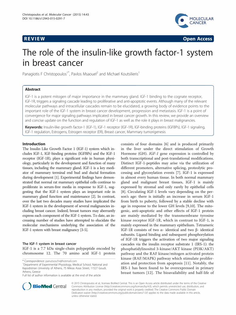
delineate the impact of IGF-1 in complex disease eventsincluding metabolic regulations, angiogenesis and metas-tasis. Prenatal administration of IGF-1 in pregnant wildtype (WT) mice results in increased body weight, higherbreast density with longer ductal elongation and higherbreast stem/progenitor cell populations of prepubescentoffspring compared to phosphate buffered saline (PBS)controls [54]. Transgenic mice specifically overexpressingIGF-1 in mammary epithelium demonstrate upregulationof the Vascular Endothelial Growth Factor (VEGF), a pro-angiogenic factor, in prepubertal glands and induction ofcyclooxygenase-2 (COX-2), an inflammatory moleculethat is also associated with angiogenesis and is responsiblefor formation of prostaglandin (PG), [55]. It is also wellestablished that caloric restriction prevents mammarytumorigenesis in rodents. Mouse models have indicatedthat IGF-1 may play a key role in tumor reduction via theAKT/mTOR pathway following caloric restriction [56].Furthermore, the effect of caloric restriction in IGF-1levels may regulate luminal tumor growth by modulatingthe epithelial-mesenchymal transition (EMT) process andchemokine milieu [57]. This pathway appears to be in-volved in the metastasis of breast tumors. Bone is one ofthe most common distant target sites for BrCa metastasis.Hiraga et al. showed that bone derived IGF-1 stimulatesproliferation and bone localization of breast cancer cellsin vivo through activation of AKT and recruitment oftranscription factor NF-kB [58]. The fact that IGF-1 ismostly expressed by stroma underscores the significanceof the interplay between stroma and epithelium dururingIGF-1 paracrine signaling resulting in disease establish-ment and progression. The excess of paracrine IGF-1signaling via stromal production may trigger epithelialIGF-1 expression, leading to a more malignant phenotype.Within the epithelium, aberrant IGF-1 autocrine signalingcould further contribute to disease aggressiveness. Futurestudies should focus on clarification of the paracrine roleof IGF-1, in establishment and progression of breast car-cinoma, via in vivo models specifically overexpressing orlacking IGF-1 in stroma. In conclusion, IGF-1 plays a keyrole in BrCa development, progression and metastasis. Itachieves this through autocrine, paracrine and endocrineinteractions between stromal and breast cancer cellswithin many microenvironments: the primary tumor site,the circulation and/or host tissue and migrating tumorcells at metastatic sites (Figure 1).
IGF-1 binding proteins (IGFBPs) in breast cancerA wealth of experiment evidence implicates IGFBPs, inBrCa pathophysiology, particularly IGFBP2, IGFBP3 andIGFBP5,. Most studies suggest that elevated systemic or
tissue IGFBP levels are found in breast malignancies.The interaction of IGFBPs with BrCa can be either in-hibitory or stimulatory via both IGF-dependent and-independent pathways. Although many studies report apositive association between BrCa risk and circulatingIGFBPs levels (mainly IGFBP3) calculated from IGF1/IGFBPs ratios, only a few studies have directly measuredfree molecules with conflicting results. Among Brazilianwomen, no association has been detected between serumIGFBP3 and BrCa risk [23], whereas a pooled data ana-lysis of prospective studies revealed a positive (althoughnon-statistically significant) correlation between IGFBP3and BrCa [20]. Furthermore, plasma IGFBP3 levels havebeen shown to be independent of mammographic dens-ity, which is a well-established BrCa risk factor [26,27].The link between IGFBPs and BrCa outcome is also
unclear. Recent studies did not detect a significant asso-ciation between plasma IGFBP3 and overall survival orrisk of all-cause mortality [28,29]. Although other find-ings indicate that a poor BrCa outcome may be associ-ated with systemic and tissue IGFBP3 [59-61], studies onthe correlation between IGFBP2 levels and disease prog-nosis are limited. Hensch et al. reported an associationof IGFBP3 with BrCa variables known to be predictive ofoutcomes, including tumor grade, body mass index(BMI), ER and premenopausal status. The same studyreported a positive association between tissue IGFBP2levels and overall survival, suggesting that protein levelsof both IGFBPs in BrCa are under hormonal and obesitycontrol [62]. In contrast, IGFBP5 tumor tissue levelshave been found to be significantly associated with poordisease outcome and the addition of IGFBP4 (measuredas IGFBP5/IGFBP4 ratio expression levels) further in-creased prognostic power [63].At the DNA level, the genetic polymorphism A-202C
of IGFBP3 (SNP, rs2854744) at the promoter region hasbeen extensively studied for potential associations withcirculating IGFBP3 levels and BrCa risk. Multiple studieshave reported that the A-202C genetic variation of IGFBP3is not associated with BrCa risk among Caucasian, Africanand Asian women [31,34,38,39], whereas others provideevidence for a correlation between A-202C polymorphismand increased IGFBP3 circulating levels. The postulatedcorrelation is thought to be, due to enhanced promoteractivity, indicating a regulation in cis [40,64]. OtherSNPs of both IGFBP1 and IGFBP3 have also been asso-ciated with plasma IGFBP3 levels [36,64]. Additionally,a Mendelian randomization study showed that the aallele of IGFBP3 SNP rs2854744 is associated with in-creased levels of circulating IGFBP3 and decreasedBrCa risk [65].Although the exact mechanism needs to be eluci-
dated, in vitro experiments indicate that IGFBP3 inhibitsBrCa proliferation and induces apoptosis. A recent
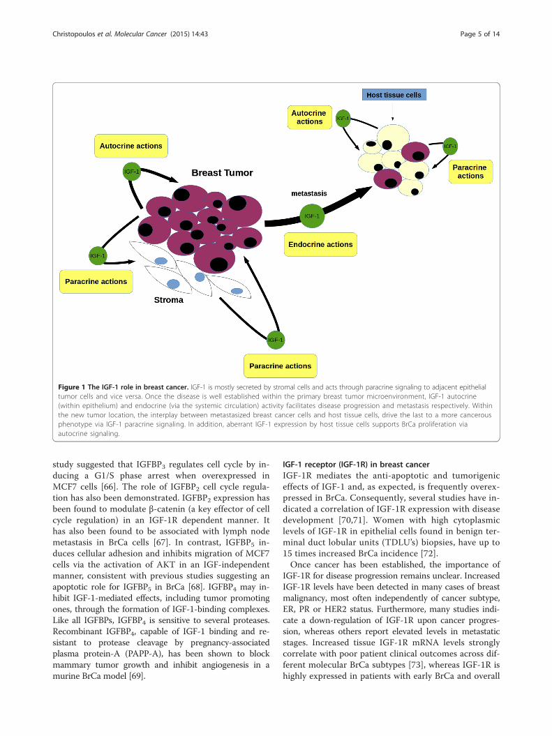
Figure 1 The IGF-1 role in breast cancer. IGF-1 is mostly secreted by stromal cells and acts through paracrine signaling to adjacent epithelialtumor cells and vice versa. Once the disease is well established within the primary breast tumor microenvironment, IGF-1 autocrine(within epithelium) and endocrine (via the systemic circulation) activity facilitates disease progression and metastasis respectively. Withinthe new tumor location, the interplay between metastasized breast cancer cells and host tissue cells, drive the last to a more cancerousphenotype via IGF-1 paracrine signaling. In addition, aberrant IGF-1 expression by host tissue cells supports BrCa proliferation viaautocrine signaling.
Christopoulos et al. Molecular Cancer (2015) 14:43 Page 5 of 14
study suggested that IGFBP3 regulates cell cycle by in-ducing a G1/S phase arrest when overexpressed inMCF7 cells [66]. The role of IGFBP2 cell cycle regula-tion has also been demonstrated. IGFBP2 expression hasbeen found to modulate β-catenin (a key effector of cellcycle regulation) in an IGF-1R dependent manner. Ithas also been found to be associated with lymph nodemetastasis in BrCa cells [67]. In contrast, IGFBP5 in-duces cellular adhesion and inhibits migration of MCF7cells via the activation of AKT in an IGF-independentmanner, consistent with previous studies suggesting anapoptotic role for IGFBP5 in BrCa [68]. IGFBP4 may in-hibit IGF-1-mediated effects, including tumor promotingones, through the formation of IGF-1-binding complexes.Like all IGFBPs, IGFBP4 is sensitive to several proteases.Recombinant IGFBP4, capable of IGF-1 binding and re-sistant to protease cleavage by pregnancy-associatedplasma protein-A (PAPP-A), has been shown to blockmammary tumor growth and inhibit angiogenesis in amurine BrCa model [69].
IGF-1 receptor (IGF-1R) in breast cancerIGF-1R mediates the anti-apoptotic and tumorigeniceffects of IGF-1 and, as expected, is frequently overex-pressed in BrCa. Consequently, several studies have in-dicated a correlation of IGF-1R expression with diseasedevelopment [70,71]. Women with high cytoplasmiclevels of IGF-1R in epithelial cells found in benign ter-minal duct lobular units (TDLU’s) biopsies, have up to15 times increased BrCa incidence [72].Once cancer has been established, the importance of
IGF-1R for disease progression remains unclear. IncreasedIGF-1R levels have been detected in many cases of breastmalignancy, most often independently of cancer subtype,ER, PR or HER2 status. Furthermore, many studies indi-cate a down-regulation of IGF-1R upon cancer progres-sion, whereas others report elevated levels in metastaticstages. Increased tissue IGF-1R mRNA levels stronglycorrelate with poor patient clinical outcomes across dif-ferent molecular BrCa subtypes [73], whereas IGF-1R ishighly expressed in patients with early BrCa and overall
Christopoulos et al. Molecular Cancer (2015) 14:43 Page 6 of 14
positively associated with good prognostic variables.IGF-1R may have differential prognostic impact in BrCamolecular subtypes; IGF-1R has been associated withfavorable outcome in patients with the luminal B BrCamolecular subtype, in contrast to HER2 enriched pa-tients [74]. Additionally, a positive association betweenIGF-1R and better clinical outcomes in hormone receptorpositive, HER2 negative tumors was recently reported[75], while in another luminal subtype group, IGF-1RmRNA has been significantly correlated with improvedBrCa-specific survival (BCSS). The same study found acorrelation between IGF-1R protein levels and prolongedBCSS and association between IGF-1R mRNA expressionand both relapse-free survival (RFS) and BCSS indicatinga concurrence between IGF-1R mRNA and protein levelsin primary breast tumors [76].Several studies have attempted to clarify the relation-
ship between specific genetic variants of IGF-1R anddisease development and progression. Instead of typicalfunctional polymorphisms in the coding region of IGF-1R gene, recent studies aimed to identify novel geneticvariants located in untranslated regions (UTRs), such asmiRNA’s binding sites, that could potentially regulatethe expression patterns of IGF-1R. Although a wealth ofevidence supports the key role of miRNAs in severaltumorigenic processes, only a few studies have evaluatedthe potential association between polymorphisms inmiRNA binding sites and cancers. The specific SNP(rs28674628) is located in a predicted binding site forthe miRNA miR-515-5p, which directly regulates IGF-1Rlevels, in the 3′ UTR of the IGF-1R gene as demonstratedby computational analysis. Gilam et al. showed that thers28674628 is significantly associated with earlier age ofdiagnosis and increased BrCa risk among Jewish BRCA1mutation carriers [77]. The rs2016347 SNP is also locatedin the 3′ UTR of IGF-1R and in silico analysis predicted afunctional role through transcriptional regulation and pos-sibly miRNA binding. Indeed, patients carrying the G al-lele of the IGF-1R rs2016347 polymorphism have poorerprognosis compared with those carrying G/T or T/T andhas been associated with increased risk of tumor progres-sion and death [78]. Seven others SNPs located in intronregions of the IGF-1R gene have been also associated withBrCa risk in Korean women [79]. Future studies mayapply a Mendelian randomization approach to elucidatethe association between functional IGF-1R polymorphismsand BrCa risk.Propelled by the data accumulated by miRNA ap-
proaches, researchers are now focusing on elucidatingthe molecular mechanisms underlying the gene regula-tion of IGF-1R. Recent studies have provided new in-sights into the molecular functions and biologicalsignificance of IGF-1R in BrCa by suggesting the role ofnovel transcription factors and mechanisms implicated
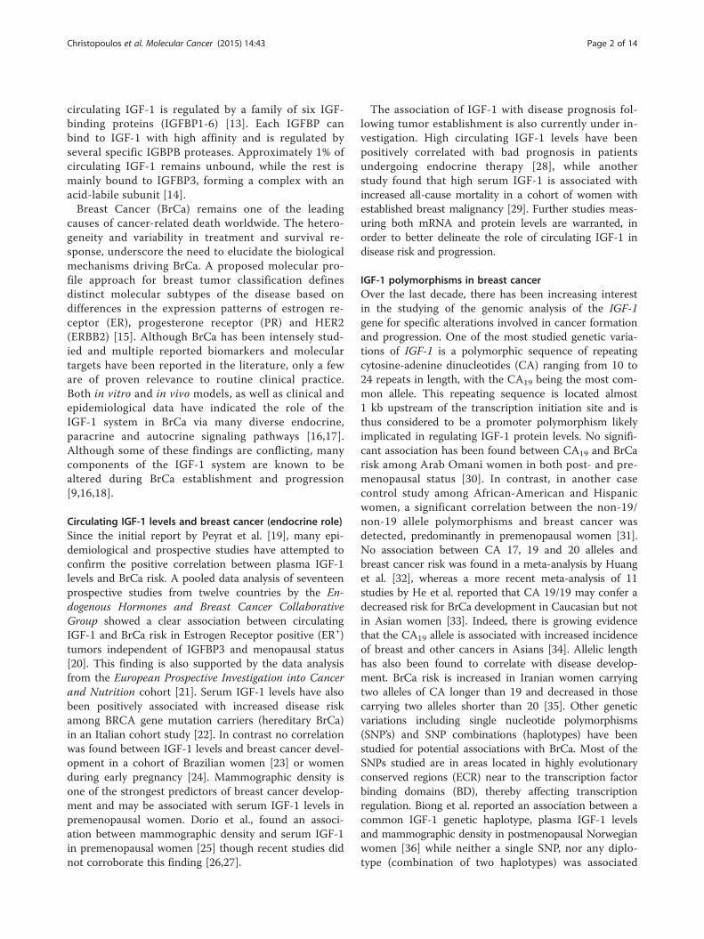
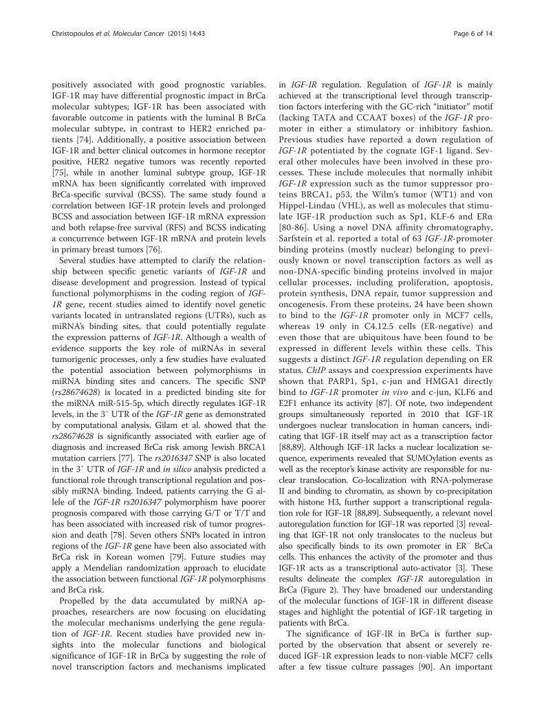
in IGF-IR regulation. Regulation of IGF-1R is mainlyachieved at the transcriptional level through transcrip-tion factors interfering with the GC-rich “initiator” motif(lacking TATA and CCAAT boxes) of the IGF-1R pro-moter in either a stimulatory or inhibitory fashion.Previous studies have reported a down regulation ofIGF-1R potentiated by the cognate IGF-1 ligand. Sev-eral other molecules have been involved in these pro-cesses. These include molecules that normally inhibitIGF-1R expression such as the tumor suppressor pro-teins BRCA1, p53, the Wilm’s tumor (WT1) and vonHippel-Lindau (VHL), as well as molecules that stimu-late IGF-1R production such as Sp1, KLF-6 and ERα[80-86]. Using a novel DNA affinity chromatography,Sarfstein et al. reported a total of 63 IGF-1R-promoterbinding proteins (mostly nuclear) belonging to previ-ously known or novel transcription factors as well asnon-DNA-specific binding proteins involved in majorcellular processes, including proliferation, apoptosis,protein synthesis, DNA repair, tumor suppression andoncogenesis. From these proteins, 24 have been shownto bind to the IGF-1R promoter only in MCF7 cells,whereas 19 only in C4.12.5 cells (ER-negative) andeven those that are ubiquitous have been found to beexpressed in different levels within these cells. Thissuggests a distinct IGF-1R regulation depending on ERstatus. ChIP assays and coexpression experiments haveshown that PARP1, Sp1, c-jun and HMGA1 directlybind to IGF-1R promoter in vivo and c-jun, KLF6 andE2F1 enhance its activity [87]. Of note, two independentgroups simultaneously reported in 2010 that IGF-1Rundergoes nuclear translocation in human cancers, indi-cating that IGF-1R itself may act as a transcription factor[88,89]. Although IGF-1R lacks a nuclear localization se-quence, experiments revealed that SUMOylation events aswell as the receptor’s kinase activity are responsible for nu-clear translocation. Co-localization with RNA-polymeraseII and binding to chromatin, as shown by co-precipitationwith histone H3, further support a transcriptional regula-tion role for IGF-1R [88,89]. Subsequently, a relevant novelautoregulation function for IGF-1R was reported [3] reveal-ing that IGF-1R not only translocates to the nucleus butalso specifically binds to its own promoter in ER− BrCacells. This enhances the activity of the promoter and thusIGF-1R acts as a transcriptional auto-activator [3]. Theseresults delineate the complex IGF-1R autoregulation inBrCa (Figure 2). They have broadened our understandingof the molecular functions of IGF-1R in different diseasestages and highlight the potential of IGF-1R targeting inpatients with BrCa.The significance of IGF-IR in BrCa is further sup-
ported by the observation that absent or severely re-duced IGF-1R expression leads to non-viable MCF7 cellsafter a few tissue culture passages [90]. An important
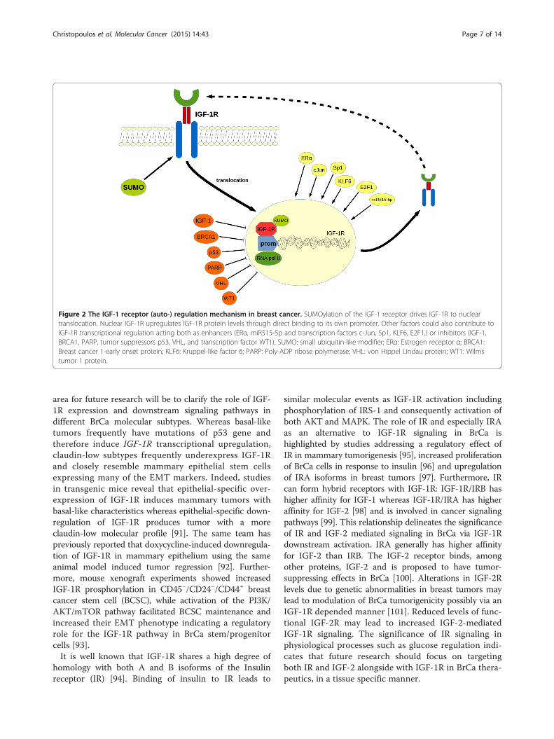
Figure 2 The IGF-1 receptor (auto-) regulation mechanism in breast cancer. SUMOylation of the IGF-1 receptor drives IGF-1R to nucleartranslocation. Nuclear IGF-1R upregulates IGF-1R protein levels through direct binding to its own promoter. Other factors could also contribute toIGF-1R transcriptional regulation acting both as enhancers (ERα, miR515-5p and transcription factors c-Jun, Sp1, KLF6, E2F1,) or inhibitors (IGF-1,BRCA1, PARP, tumor suppressors p53, VHL, and transcription factor WT1). SUMO: small ubiquitin-like modifier; ERα: Estrogen receptor α; BRCA1:Breast cancer 1-early onset protein; KLF6: Kruppel-like factor 6; PARP: Poly-ADP ribose polymerase; VHL: von Hippel Lindau protein; WT1: Wilmstumor 1 protein.
Christopoulos et al. Molecular Cancer (2015) 14:43 Page 7 of 14
area for future research will be to clarify the role of IGF-1R expression and downstream signaling pathways indifferent BrCa molecular subtypes. Whereas basal-liketumors frequently have mutations of p53 gene andtherefore induce IGF-1R transcriptional upregulation,claudin-low subtypes frequently underexpress IGF-1Rand closely resemble mammary epithelial stem cellsexpressing many of the EMT markers. Indeed, studiesin transgenic mice reveal that epithelial-specific over-expression of IGF-1R induces mammary tumors withbasal-like characteristics whereas epithelial-specific down-regulation of IGF-1R produces tumor with a moreclaudin-low molecular profile [91]. The same team haspreviously reported that doxycycline-induced downregula-tion of IGF-1R in mammary epithelium using the sameanimal model induced tumor regression [92]. Further-more, mouse xenograft experiments showed increasedIGF-1R prosphorylation in CD45−/CD24−/CD44+ breastcancer stem cell (BCSC), while activation of the PI3K/AKT/mTOR pathway facilitated BCSC maintenance andincreased their EMT phenotype indicating a regulatoryrole for the IGF-1R pathway in BrCa stem/progenitorcells [93].It is well known that IGF-1R shares a high degree of
homology with both A and B isoforms of the Insulinreceptor (IR) [94]. Binding of insulin to IR leads to
similar molecular events as IGF-1R activation includingphosphorylation of IRS-1 and consequently activation ofboth AKT and MAPK. The role of IR and especially IRAas an alternative to IGF-1R signaling in BrCa ishighlighted by studies addressing a regulatory effect ofIR in mammary tumorigenesis [95], increased proliferationof BrCa cells in response to insulin [96] and upregulationof IRA isoforms in breast tumors [97]. Furthermore, IRcan form hybrid receptors with IGF-1R: IGF-1R/IRB hashigher affinity for IGF-1 whereas IGF-1R/IRA has higheraffinity for IGF-2 [98] and is involved in cancer signalingpathways [99]. This relationship delineates the significanceof IR and IGF-2 mediated signaling in BrCa via IGF-1Rdownstream activation. IRA generally has higher affinityfor IGF-2 than IRB. The IGF-2 receptor binds, amongother proteins, IGF-2 and is proposed to have tumor-suppressing effects in BrCa [100]. Alterations in IGF-2Rlevels due to genetic abnormalities in breast tumors maylead to modulation of BrCa tumorigenicity possibly via anIGF-1R depended manner [101]. Reduced levels of func-tional IGF-2R may lead to increased IGF-2-mediatedIGF-1R signaling. The significance of IR signaling inphysiological processes such as glucose regulation indi-cates that future research should focus on targetingboth IR and IGF-2 alongside with IGF-1R in BrCa thera-peutics, in a tissue specific manner.
Christopoulos et al. Molecular Cancer (2015) 14:43 Page 8 of 14
Recent studies also indicate that IGR-1R may be usedas a molecular target for BrCa imaging. In a panel com-paring other potential imaging targets (EGFR, HER2,GLUT1 and others), IGF-1R was found to be suitable formolecular imaging strategies in 80% of female and 77%of male breast tumors [102,103].
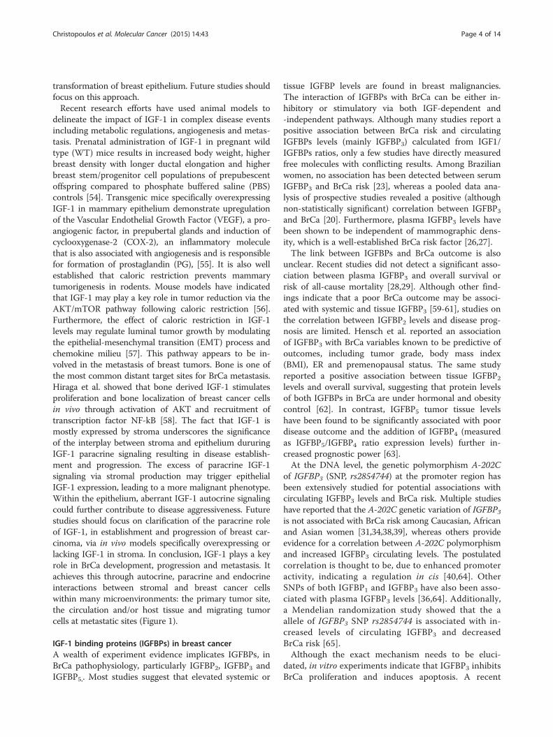
Estrogen receptor (ER) and the IGF-1 system in breast cancerThe importance of steroids and especially the ER statusin disease progression is well established. Its significanceis further supported by the fact that anti-estrogens, suchas tamoxifen are routinely used in BrCa treatment.Crosstalk between growth factors and estrogens may beresponsible for the development of estrogen-independentBrCa tumors [104]. Although the exact mechanism is notcurrently known, it is evident that there is a cross-talk be-tween the IGF-1 system, ER and the cognate ER ligand17β-estradiol (E2). BrCa cells have a differential responseto IGF-1 with regards to both proliferation and survivaldepending on their ER status. Specifically, cells expressingboth IGF-1R and ER demonstrate synergistic or additivegrowth effects in response to simultaneous administrationof ligands (IGF-1, E2) [105]. Many components of theIGF-1 system are under the transcriptional control ofER/E2. A well-established example of this phenomenonis IGF-1R modulation by ERα as described above. A bi-directional regulation has also been proposed wherebyIGF-1 induced pathways regulate ERα- dependent func-tions and vice versa. The mechanism underlying thiscross-talk may be particularly important for the devel-opment of combination treatment strategies. Beckeret al. demonstrated that the IGF-1/IGF-1R axis inducesphosphorylation of ERa through ribosomal S6 kinase 1(S6K1), a downstream molecule of the PI3K/AKT/mTOR pathway. Through chromatin and promoterbinding, activated ERα modulates both the transcrip-tional regulation of ERα induced genes as well as BrCacell growth and proliferation [4]. Other recent studiessuggest that IGF-1 may be regulated by both ERα andBRCA1. IGF-1 expression has been shown to be acti-vated by estrogens whereas breast tumors with BRCA1mutations demonstrate elevated IGF-1 levels. Both ERαand BRCA1 can bind to an estrogen-responsive elementlike site (EREL) in the IGF-1 promoter, thus enhancingor suppressing IGF-1 expression respectively [5]. Aro-matase is a key enzyme during estrogen biosynthesisand is routinely used as a therapeutic target in ER posi-tive BrCa tumors. Prostaglandin E2 (PGE2) along withHER2 and growth factors enhance aromatase activityvia post-transcriptional mechanisms in BrCa cells medi-ated by the IGF-1/IGF-1R axis through both AKT andMAPK pathways. Thus, IGF-1 pathways can enhancearomatase activity via post-transcriptional modificationsthat do not affect aromatase protein levels [106]. The
two subtypes of ER (ERα and ERβ) are found in differentlevels in breast tumors and can produce distinct cellularresponses. However, we still lack a detailed understand-ing of the complex interaction between the ERα andERβ subtypes with the IGF-1/IGF-1R axis and E2.MCF7 cells engineered to express reduced levels of IGF-1R demonstrate decreased proliferation and increasedapoptosis in response to E2 compared to controls. TheERα/ERβ ratio is also impaired in these cells. Specific-ally, ERα is reduced whereas ERβ is elevated resulting inincreased phosphorylation of p38 MAPK and activationof the p53 substrate protein, leading to apoptosis [90].Other findings also support a cross-talk between E2 andthe IGF-1/IGF-1R axis. It has been demonstrated thatin vitro E2 and IGF-1 co-regulate a number of genescomprised mainly of tumor suppressing factors associatedwith poor disease outcome. E2 can also induce IGF-1Rexpression in mouse xenograft models [107,108]. Fur-thermore, IGF-1 alone in MCF7, or both IGF-1 and E2,in BrCa cells overexpressing the IGF-1R, can induce theIGF-1R/ERα association. IGF-1 stimulates activation ofERα, which consequently binds to IGF-1R, inducingdownstream phosphorylation of AKT and ERK1/2 leadingto increased cell growth via enhanced IGF-1-signalingpathways [109]. Tian et al., demonstrated that the IGF-1/ERα cross-talk is altered during development, and thismay corroborate reports indicating an age-depended asso-ciation between IGF-1 levels and BrCa risk. They pro-posed a non-genomic mechanism based in in vivo modelstudy, in which ERα forms a complex with IRS1, triggeringPI3K/AKT pathway in the prepubertal stage. This is incontrast to the postpubertal stage, where the reduced ERαlevels prevent the ERα/IRS1 complex formation, and alterthe signaling via the Raf/MAPK pathway [110]. Addition-ally, a non-genomic crosstalk between ERα and bothAkt and ERK signaling pathways has been reported inobese postmenopausal women [111]. Although theexact mechanism is not clear, estrogens may be modu-lated by the IGF-1 system via both transcriptional andpost-transcriptional mechanisms leading to increasedBrCa proliferation and growth through activation ofthe IGF-1 signaling pathways (Figure 3).
Interaction between the IGF-1 system and other growthfactors in breast cancerThe IGF-1/IGF-1R axis may interact with other growthfactors in the BrCa milieu. IGF-1 may cross-talk withthe transforming growth factor beta-1 (TGFβ1), a mol-ecule known to induce EMT, in BrCa. IGF-1 can activatematrix metalloproteinases through both AKT andMAPK pathways subsequently activating TGFβ1 leadingto nuclear translocation of β-catenin and EMT [112].Other studies also contribute to this crosstalk of IGF-1with b-catenin and the Wnt pathway. Phyllodes tumors
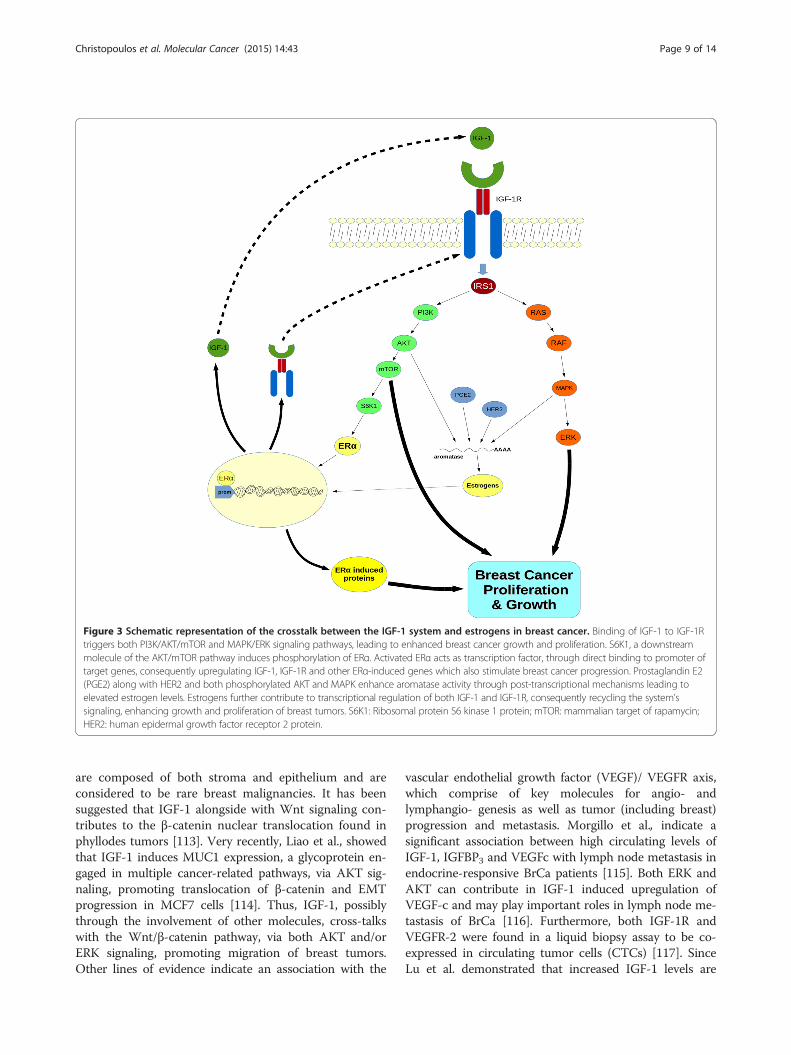
Figure 3 Schematic representation of the crosstalk between the IGF-1 system and estrogens in breast cancer. Binding of IGF-1 to IGF-1Rtriggers both PI3K/AKT/mTOR and MAPK/ERK signaling pathways, leading to enhanced breast cancer growth and proliferation. S6K1, a downstreammolecule of the AKT/mTOR pathway induces phosphorylation of ERα. Activated ERα acts as transcription factor, through direct binding to promoter oftarget genes, consequently upregulating IGF-1, IGF-1R and other ERα-induced genes which also stimulate breast cancer progression. Prostaglandin E2(PGE2) along with HER2 and both phosphorylated AKT and MAPK enhance aromatase activity through post-transcriptional mechanisms leading toelevated estrogen levels. Estrogens further contribute to transcriptional regulation of both IGF-1 and IGF-1R, consequently recycling the system’ssignaling, enhancing growth and proliferation of breast tumors. S6K1: Ribosomal protein S6 kinase 1 protein; mTOR: mammalian target of rapamycin;HER2: human epidermal growth factor receptor 2 protein.
Christopoulos et al. Molecular Cancer (2015) 14:43 Page 9 of 14
are composed of both stroma and epithelium and areconsidered to be rare breast malignancies. It has beensuggested that IGF-1 alongside with Wnt signaling con-tributes to the β-catenin nuclear translocation found inphyllodes tumors [113]. Very recently, Liao et al., showedthat IGF-1 induces MUC1 expression, a glycoprotein en-gaged in multiple cancer-related pathways, via AKT sig-naling, promoting translocation of β-catenin and EMTprogression in MCF7 cells [114]. Thus, IGF-1, possiblythrough the involvement of other molecules, cross-talkswith the Wnt/β-catenin pathway, via both AKT and/orERK signaling, promoting migration of breast tumors.Other lines of evidence indicate an association with the
vascular endothelial growth factor (VEGF)/ VEGFR axis,which comprise of key molecules for angio- andlymphangio- genesis as well as tumor (including breast)progression and metastasis. Morgillo et al., indicate asignificant association between high circulating levels ofIGF-1, IGFBP3 and VEGFc with lymph node metastasis inendocrine-responsive BrCa patients [115]. Both ERK andAKT can contribute in IGF-1 induced upregulation ofVEGF-c and may play important roles in lymph node me-tastasis of BrCa [116]. Furthermore, both IGF-1R andVEGFR-2 were found in a liquid biopsy assay to be co-expressed in circulating tumor cells (CTCs) [117]. SinceLu et al. demonstrated that increased IGF-1 levels are
Christopoulos et al. Molecular Cancer (2015) 14:43 Page 10 of 14
associated with trastuzumab resistance [118], recent focushas turned into clarifying the cross-talk between the IGF-1 system and the Epidermal Growth Factor Receptors(EGFR) family. ERBB2 (HER2) may contribute to de-creased IGF-1R expression during mammary tumorigen-esis [119]. An association between IGFBP and ERBB2 inIGF-1-dependent tumor transformation has been reportedin mammary luminal epithelial cells [120]. Dearth et al.,demostrated that ERBB2-induced mammary tumorigen-esis is independent of circulating IGF-1 levels, indicatingthat the IGF-1/HER2 cross-talk may occur via autocrineand paracrine signaling in BrCa [121]. The IGF-1 systemmay also interact with components of the extracellularmatrix (ECM) affecting tumor growth and progression.Complexes of IGF-1 with IGFBP3 or IGFBP5 and vitro-nectin (VN) have been shown to induce MCF7 survivaland regulation of genes implicated in migration and inva-sion processes [122].
Targeting the IGF-1R in breast cancer: clinical evidenceBoth in vitro and in vivo evidence strongly suggest thatthe IGF-1 system could serve as a promising target can-didate in BrCa therapeutics. Indeed, over the last decade,many strategies targeting several components of theIGF-1 system, have been tested. The majority of theseinvolved targeting the IGF-1R, either with monoclonalantibodies (mAbs) causing internalization of the receptor,or by blocking the receptor’s tyrosine kinase domain acti-vation using receptor tyrosine kinase inhibitors (RTKIs).The first recruited mAb against IGF-1R is CP-751,871(figitumumab) which has demostrated good pre-clinicalantitumor efficacy against several cancers including BrCa[123]. Only mild toxicities induced by figitumumab havebeen reported [124]. Figutumumab has also been tested inphase I (NCT00635245 and NCT01536145) and phase II(NCT00372996) clinical trials against BrCa with appar-ently negative unpublished results. IMC-A12 (cixutumu-mab) is another mAb against IGF-1R. It has been tested ina phase I trial in combination with temsirolimus in pa-tients with resistant ER positive breast tumors and onlymild toxicities were seen [125]. A phase II trial of cixutu-mumab is currently ongoing (NCT00699491). Anotherphase II trial (NCT00684983) is evaluating the effect ofcixutumumab in combination with capecitabine and lapa-nitib in patients with metastatic HER2+ BrCa [126].R1507 (RO4858696) is another monoclonal IgG antibodyspecifically targeting IGF-1R that has demonstrated nodose-limiting toxicities [127]. A phase Ib study showedthat R1507 is well tolerated when added to six standardchemotherapy regiments in patients with various solidtumors, including BrCa [128]. Another phase I trial(NCT00882674) in women with operable breast cancerand a phase II trial (NCT00796107) of R1507 in combin-ation with letrozole in women with advanced breast
tumors have finished, with results pending publication.The same mAb has also successfully been used for im-aging breast cancer in vivo using either SPECT and/orPET imaging techniques [129]. Unlike most of the mAbsagainst IGF-1R, figutumumab and SCH717454 have theability to block signals induced by hybrid receptors[124,130] and this regimen has been tested in a phase IIclinical trial (NCT00954512) in patients with advancedsolid tumors. The reported adverse events common tomost IGF-1R mAbs include anemia, fever, diarrhea, arth-ralgia, leukopenia, thrombocytopenia, rash, fatigue andanorexia with hyperglycemia being the most common.Noticeable, hyperglycemia was not reported for R1507[128].Since the first RTKI (NVP-ADW742) targeting IGF-1R
[131], research efforts have focused in developing ofthese molecules as potential IGF-1R inhibitors. Most ofthese molecules target the tyrosine domain of both IGF-1R or IR, which may compromise specificity. Althoughmany of these molecules have demonstrated good pre-clinical results, only a few have actually been translatedin ongoing clinical trials, probably due to increased cyto-toxicity events. Indeed, production of the NVP-ADW742was halted due to toxicity issues. OSI-906 (linsitinib), aRTKI targeting both IGF-1R and IR, was investigated as atreatment for hormone sensitive breast cancer in combin-ation with endocrine therapy and erlonitib in a phase IIclinical trial (NCT01205685). Unfortunately, this studywas terminated as all patients experienced severe toxicitiesand tumor progression. BMS-754807, another dual RTKIis being tested in a currently ongoing phase II trial(NCT01225172) in combination with letrozole in womenwith andvanced ER positive, non-steroidal aromatase in-hibitor resistant breast tumors [132]. Another phase I/IItrial of BMS-754807 in combination with transtuzumabin patients with advanced or metastatic HER2 positivebreast tumors has been completed (NCT00788333) andcomplete results remain to be published. Except specificityanother limitation of the RTKI approach is, in contrast tomAbs, the lack of receptor internalization, which meansthat IGF-1R could retain it’s activity a while after inhib-ition treatment ends. Targeting the IGF-1 system viainhibiting IGF-1R signal transduction cascade presentslimitations. The complexity of this system, eliminatesthe possibilities for efficient targeting. Even if we couldspecifically block the IGF-1R and not IR, unwanted effectsmay arise such as glucose dysregulation and diabetes dueto the inhibition of hybrid IGF-1R/IR receptors. On theother hand, targeting of hybrid receptors in BrCa thera-peutics, could be desirable as they may act more as IGF-1R rather than IR. In addition, IGF-1R targeting may notinhibit the IGF-2 mediated signal transduction via IR and/or hybrid receptors. Further insights into this complexcrosstalk, may help us overcome some disappointing
Christopoulos et al. Molecular Cancer (2015) 14:43 Page 11 of 14
clinical results and allow the translation of novel mole-cules and strategies to specifically suppress the IGF-1R-induced tumor promoting effects.
ConclusionsA growing body of evidence supports the association ofthe IGF-1 system with BrCa establishment and progression.Conflicting results may arise from discordant methodo-logicals approaches, distinct molecular subtypes studied,genetic differences between different populations andtumor heterogeneity.The complex IGF-1/IGF-1R axis cascade has received
significant attention to date and a wealth of experimen-tal evidence from both in vitro and in vivo models andhuman studies have implicated the IGF-1 system withBrCa biology. The prospect of efficiently targeting theIGF-1 system in BrCa is certainly attractive. Further insighton the molecular mechanisms driving the disease via theIGF-1 system will open new avenues for the diagnosis andtreatment of BrCa. Numerous challenges will have to beovercome prior to reaching the goal of IGF-1 regulation forthe prevention and management of BrCa. Elucidating theIGF-1 expression patterns and diverse molecular pathwaysmay allow the development of effective diagnostic andtreatment strategies against BrCa that may prove beneficialfor selective population subgroups.
Ethical approvalReported research carried on humans are in compliancewith the Helsinki Declaration, whereas experimentalresearch on animals follow internationally recognizedguidelines.
AbbreviationsIGF-1: Insulin-like growth factor-1; GFBP1–5: Insulin-like growth factor bindingproteins 1–5; BrCa: Breast cancer; GH: Growth hormone; IRS-1: Insulin receptorsubstrate 1; PI3K: Phosphatidylinositol 3-kinase; MAPK: Mitogen-activatedprotein kinase; ERα-β: Estrogen receptor α-β; PR: Progesterone receptor;SUMO: Small ubiquitin-like modifier; BRCA1: Breast cancer 1-early onset protein;KLF6: Kruppel-like factor 6; PARP1: Poly-ADP ribose polymerase 1; VHL: Vonhippel lindau protein; WT1: Wilms tumor 1 protein; S6K1: Ribosomal protein S6kinase 1 protein; mTOR: Mammalian target of rapamyci; HER2: Humanepidermal growth factor receptor 2 protein; SNP: Single-nucleotidepolymorphism; PBS: Phosphate buffer saline; EMT: Epithelial to mesenchymaltransition; CTCs: Circulating tumor cells; ECM: Extracellular matrix; BD: Bindingdomain; VEGFc: Vascular endothelial growth factor receptor c; EGFR: Epidermalgrowth factor receptor; TGFβ: Transforming growth factor β; UTR’s: Untranslatedregions; EREL: Estrogen-responsive element-like site; PGE2: Prostaglandin E2;ChIP: Chromatin immunoprecipitation; COX-2: Cyclooxygenase-2;VN: Vitronectin; ECR: Evolutionary conserved regions; DFS: Disease-free survival;TDLU's: Terminal duct lobular units; BCSC: Breast cancer stem cell; BCSS:BrCa-specific survival; mAb: Monoclonal antibody; RTKI: Receptor tyrosine kinaseinhibitor; IR: Insulin receptor.
Competing interestsThe authors declare that they have no competing interests.
Authors’ contributionsPFC: AB, ES, PM: FG, MK: ES, FG. All authors read and approved the finalmanuscript.
Authors’ informationChristopoulos P.F: M.Sc, Ph. D, Research Fellow at Department ofExperimental Physiology, Medical School, National and KapodistrianUniversity of Athens, Athens, Greece.Msaouel P: M.D, Ph. D, Chief Medical Resident at Jacobi Medical Center,Department of Internal Medicine, Albert Einstein College of Medicine, Bronx,NY, USA.Koutsilieris M: M.D, Ph. D, Chairman of the Department of ExperimentalPhysiology, Medical School, National & Kapodistrian University of Athens, 75Micras Asias Str., 11527 Goudi-Athens, Greece, Tel.: +30 210–7462597; Fax:+30 210–7462571.
AcknowledgmentsWe are grateful to Dr. Jenna Cottral and Maria Balta for their helpfulconversations and contributions to conception and drafting of thismanuscript. All authors declare that there was no source of fundingregarding this manuscript.
Author details1Department of Experimental Physiology, Medical School, National andKapodistrian University of Athens, 75 Mikras Asias Street, 11527 Goudi,Athens, Greece. 2Department of Internal Medicine, Jacobi Medical Center,Albert Einstein College of Medicine, Bronx, NY, USA.
Received: 17 July 2014 Accepted: 7 January 2015
References1. Ruan W, Kleinberg DL. Insulin-like growth factor I is essential for terminal
end bud formation and ductal morphogenesis during mammarydevelopment. Endocrinology. 1999;140:5075–81.
2. Deeks S, Richards J, Nandi S. Maintenance of normal rat mammary epithelialcells by insulin and insulin-like growth factor 1. Exp Cell Res.1988;174:448–60.
3. Sarfstein R, Pasmanik-Chor M, Yeheskel A, Edry L, Shomron N, Warman N,et al. Insulin-like Growth Factor-I Receptor (IGF-IR) translocates to nucleusand autoregulates IGF-IR gene expression in breast cancer cells. J Biol Chem.2012;287:2766–76.
4. Becker MA, Ibrahim YH, Cui X, Lee AV, Yee D. The IGF pathway regulatesERα through a S6K1-dependent mechanism in breast cancer cells. MolEndocrinol. 2011;25:516–28.
5. Kang HJ, Yi YW, Kim HJ, Hong YB, Seong YS, Bae I. BRCA1 negativelyregulates IGF-1 expression through an estrogen-responsive element-like site.Cell Death Dis. 2012;3:e336.
6. Denley A, Cosgrove LJ, Booker GW, Wallace JC, Forbes BE. Molecularinteractions of the IGF system. Cytokine Growth Factor Rev. 2005;16:421–39.
7. Philippou A, Maridaki M, Pneumaticos S, Koutsilieris M. The complexity ofthe IGF1 gene splicing, post-translational modification and bioactivity. MolMed Camb Mass. 2014;20:202–14.
8. Macias H, Hinck L. Mammary gland development. Wiley Interdiscip Rev DevBiol. 2012;1:533–57.
9. Yu H, Rohan T. Role of the insulin-like growth factor family in cancerdevelopment and progression. J Natl Cancer Inst. 2000;92:1472–89.
10. Gennigens C, Menetrier-Caux C, Droz JP. Insulin-Like Growth Factor (IGF)family and prostate cancer. Crit Rev Oncol Hematol. 2006;58:124–45.
11. Philippou A, Halapas A, Maridaki M, Koutsilieris M. Type I insulin-like growthfactor receptor signaling in skeletal muscle regeneration and hypertrophy.J Musculoskelet Neuronal Interact. 2007;7:208–18.
12. Lee AV, Jackson JG, Gooch JL, Hilsenbeck SG, Coronado-Heinsohn E,Osborne CK, et al. Enhancement of insulin-like growth factor signaling inhuman breast cancer: estrogen regulation of insulin receptor substrate-1expression in vitro and in vivo. Mol Endocrinol Baltim Md. 1999;13:787–96.
13. LeRoith D, Roberts CT. The insulin-like growth factor system and cancer.Cancer Lett. 2003;195:127–37.
14. Pollak M. Insulin and insulin-like growth factor signalling in neoplasia. NatRev Cancer. 2008;8:915–28.
15. Perou CM, Sørlie T, Eisen MB, van de Rijn M, Jeffrey SS, Rees CA, et al.Molecular portraits of human breast tumours. Nature. 2000;406:747–52.
16. Sachdev D, Yee D. The IGF system and breast cancer. Endocr Relat Cancer.2001;8:197–209.
Christopoulos et al. Molecular Cancer (2015) 14:43 Page 12 of 14
17. Peyrat J-P, Bonneterre J, Beuscart R, Djiane J, Demaille A. Insulin-like growthfactor 1 receptors in human breast cancer and their relation to estradioland progesterone receptors. Cancer Res. 1988;48:6429–33.
18. Maor S, Yosepovich A, Papa MZ, Yarden RI, Mayer D, Friedman E, et al.Elevated insulin-like growth factor-I receptor (IGF-IR) levels in primary breasttumors associated with BRCA1 mutations. Cancer Lett. 2007;257:236–43.
19. Peyrat JP, Bonneterre J, Hecquet B, Vennin P, Louchez MM, Fournier C, et al.Plasma insulin-like growth factor-1 (IGF-1) concentrations in human breastcancer. Eur J Cancer Oxf Engl 1990. 1993;29A:492–7.
20. Endogenous Hormones and Breast Cancer Collaborative Group, Key TJ,Appleby PN, Reeves GK, Roddam AW. Insulin-like growth factor 1 (IGF1), IGFbinding protein 3 (IGFBP3), and breast cancer risk: pooled individual dataanalysis of 17 prospective studies. Lancet Oncol. 2010;11:530–42.
21. Kaaks R, Johnson T, Tikk K, Sookthai D, Tjønneland A, Roswall N, et al.Insulin-like growth factor I and risk of breast cancer by age and hormonereceptor status-A prospective study within the EPIC cohort. Int J Cancer.2014;134(11):2683–90.
22. Pasanisi P, Bruno E, Venturelli E, Manoukian S, Barile M, Peissel B, et al.Serum levels of IGF-I and BRCA penetrance: a case control study in breastcancer families. Fam Cancer. 2011;10:521–8.
23. Trinconi AF, Filassi JR, Soares-Júnior JM, Baracat EC. Evaluation of theinsulin-like growth factors (IGF) IGF-I and IGF binding protein 3 inpatients at high risk for breast cancer. Fertil Steril. 2011;95:2753–5.
24. Toriola AT, Lundin E, Schock H, Grankvist K, Pukkala E, Chen T, et al.Circulating Insulin-like Growth Factor-I in pregnancy and maternal risk ofbreast cancer. Cancer Epidemiol Biomarkers Prev. 2011;20:1798–801.
25. Diorio C, Pollak M, Byrne C, Mâsse B, Hébert-Croteau N, Yaffe M, et al.Insulin-like growth factor-I, IGF-binding protein-3, and mammographicbreast density. Cancer Epidemiol Biomark Prev Publ Am Assoc Cancer ResCosponsored Am Soc Prev Oncol. 2005;14:1065–73.
26. Rice MS, Tworoger SS, Rosner BA, Pollak MN, Hankinson SE, Tamimi RM.Insulin-like growth factor-1, insulin-like growth factor-binding protein-3,growth hormone, and mammographic density in the Nurses’ Health Studies.Breast Cancer Res Treat. 2012;136:805–12.
27. Rinaldi S, Biessy C, Hernandez M, Lesueur F, dos Santos Silva I, Rice MS, et al.Circulating concentrations of insulin-like growth factor-I, insulin-like growthfactor-binding protein-3, genetic polymorphisms and mammographicdensity in premenopausal Mexican women: Results from the ESMaestrascohort: IGF-I, IGFBP-3 and mammographic density in women. Int J Cancer.2014;134:1436–44.
28. Hartog H, Boezen HM, de Jong MM, Schaapveld M, Wesseling J, van derGraaf WTA. Prognostic value of insulin-like growth factor 1 and insulin-likegrowth factor binding protein 3 blood levels in breast cancer. Breast.2013;22:1155–60.
29. Duggan C, Wang C-Y, Neuhouser ML, Xiao L, Smith AW, Reding KW, et al.Associations of insulin-like growth factor and insulin-like growth factorbinding protein-3 with mortality in women with breast cancer. Int J Cancer.2013;132:1191–200.
30. Al-Ajmi K, Ganguly SS, Al-Ajmi A, Mandhari ZA, Al-Moundhri MS.Insulin-like growth factor 1 gene polymorphism and breast cancerrisk among Arab Omani women: a case–control study. Breast Cancer(Auckl). 2012;6:103–12.
31. Sarkissyan M, Mishra DK, Wu Y, Shang X, Sarkissyan S, Vadgama JV. IGF genepolymorphisms and breast cancer in African-American and Hispanicwomen. Int J Oncol. 2011;38:1663–73.
32. Huang Q, Wang C, Qiu L-J, Shao F, Yu J-H. The association between IGF1CA repeat polymorphisms and breast cancer risk: a meta-analysis. BreastCancer Res Treat. 2011;129:191–4.
33. He B, Xu Y, Pan Y, Li R, Gao T, Song G, et al. Differential effects of insulin-likegrowth factor-1 CA repeat polymorphism on breast cancer risk along withrace: a meta-analysis. Gene. 2013;525:92–8.
34. Quan H, Tang H, Fang L, Bi J, Liu Y, Li H. IGF1 (CA)19 and IGFBP-3-202A/Cgene polymorphism and cancer risk: a meta-analysis. Cell Biochem Biophys.2014;69:169–78.
35. Javadi M, Hematti S, Tavassoli M. Polymorphic CA repeat length in insulin-likegrowth factor 1 and risk of breast cancer in Iranian women. Med Oncol.2012;29:516–20.
36. Biong M, Gram IT, Brill I, Johansen F, Solvang HK, Alnaes GIG, et al. Genotypesand haplotypes in the insulin-like growth factors, their receptors and bindingproteins in relation to plasma metabolic levels and mammographic density.BMC Med Genomics. 2010;3:9.
37. Henningson M, Hietala M, Törngren T, Olsson H, Jernström H. IGF1 htSNPsin relation to IGF-1 levels in young women from high-risk breast cancerfamilies: implications for early-onset breast cancer. Fam Cancer.2011;10:173–85.
38. Qian B, Zheng H, Yu H, Chen K. Genotypes and phenotypes of IGF-I andIGFBP-3 in breast tumors among Chinese women. Breast Cancer Res Treat.2011;130:217–26.
39. Canzian F, Cox DG, Setiawan VW, Stram DO, Ziegler RG, Dossus L, et al.Comprehensive analysis of common genetic variation in 61 genes relatedto steroid hormone and insulin-like growth factor-I metabolism and breastcancer risk in the NCI breast and prostate cancer cohort consortium. HumMol Genet. 2010;19:3873–84.
40. Gu F, Schumacher FR, Canzian F, Allen NE, Albanes D, Berg CD, et al.Eighteen insulin-like growth factor pathway genes, circulating levels of IGF-Iand its binding protein, and risk of prostate and breast cancer. CancerEpidemiol Biomark Prev Publ Am Assoc Cancer Res Cosponsored Am SocPrev Oncol. 2010;19:2877–87.
41. Yaren A, Turgut S, Ayada C, Akcilar R, Degirmencioglu S, Gokoz Dogu G.Insulin-like growth factor I (Igf-1) gene polymorphism in patients with non-metastatic breast cancer. Gene. 2012;503:244–7.
42. Muendlein A, Lang AH, Geller-Rhomberg S, Winder T, Gasser K, Drexel H,et al. Association of a common genetic variant of the IGF-1 gene withevent-free survival in patients with HER2-positive breast cancer. J CancerRes Clin Oncol. 2013;139:491–8.
43. Lindström S, Thompson DJ, Paterson AD, Li J, Gierach GL, Scott C, et al.Genome-wide association study identifies multiple loci associated withboth mammographic density and breast cancer risk. Nat Commun.2014;5:5303.
44. Chong KYM, Subramanian A, Mokbel K, Sharma AK. The prognosticsignificance of the insulin-like growth factor-1 ligand and receptorexpression in breast cancer tissue. J Surg Oncol. 2011;104:228–35.
45. Mu L, Tuck D, Katsaros D, Lu L, Schulz V, Perincheri S, et al. Favorableoutcome associated with an IGF-1 ligand signature in breast cancer. BreastCancer Res Treat. 2012;133:321–31.
46. Llanos AA, Brasky TM, Dumitrescu RG, Marian C, Makambi KH, Kallakury BVS,et al. Plasma IGF-1 and IGFBP-3 may be imprecise surrogates for breast con-centrations: an analysis of healthy women. Breast Cancer Res Treat.2013;138:571–9.
47. Richards RG, Klotz DM, Walker MP, Diaugustine RP. Mammary glandbranching morphogenesis is diminished in mice with a deficiency ofinsulin-like growth factor-I (IGF-I), but not in mice with a liver-specificdeletion of IGF-I. Endocrinology. 2004;145:3106–10.
48. Davison Z, de Blacquière GE, Westley BR, May FE. Insulin-like growthfactor-dependent proliferation and survival of triple-negative breast cancercells: implications for therapy. Neoplasia N Y NY. 2011;13:504.
49. D’Esposito V, Passaretti F, Hammarstedt A, Liguoro D, Terracciano D, MoleaG, et al. Adipocyte-released insulin-like growth factor-1 is regulated byglucose and fatty acids and controls breast cancer cell growth in vitro.Diabetologia. 2012;55:2811–22.
50. Rajski M, Zanetti-Dällenbach R, Vogel B, Herrmann R, Rochlitz C, Buess M.IGF-I induced genes in stromal fibroblasts predict the clinical outcome ofbreast and lung cancer patients. BMC Med. 2010;8:1.
51. Pacher M, Seewald MJ, Mikula M, Oehler S, Mogg M, Vinatzer U, et al.Impact of constitutive IGF1/IGF2 stimulation on the transcriptional programof human breast cancer cells. Carcinogenesis. 2007;28:49–59.
52. Morimura S, Takahashi K. Rac1 and Stathmin but not EB1 are required forinvasion of breast cancer cells in response to IGF-I. Int J Cell Biol.2011;2011:1–9.
53. Loladze AV, Stull MA, Rowzee AM, Demarco J, Lantry JH, Rosen CJ, et al.Epithelial-specific and stage-specific functions of insulin-like growth factor-Iduring postnatal mammary development. Endocrinology. 2006;147:5412–23.
54. Chang C-I, Low HP, Qiu L, Strohsnitter WC, Hsieh C-C. Prenatal modulationof breast density and breast stem cells by insulin-like growth factor-1. Am JStem Cells. 2012;1:239.
55. Tian J, Lambertz I, Berton TR, Rundhaug JE, Kiguchi K, Shirley SH, et al.Transgenic insulin-like growth factor-1 stimulates activation of COX-2signaling in mammary glands. Mol Carcinog. 2012;51:973–83.
56. Dogan S, Johannsen A, Grande J, Cleary M. Effects of intermittent andchronic calorie restriction on mammalian target of rapamycin (mTOR) andIGF-I signaling pathways in mammary fat pad tissues and mammary tumors.Nutr Cancer. 2011;63:389–401.
Christopoulos et al. Molecular Cancer (2015) 14:43 Page 13 of 14
57. Ford NA, Nunez NP, Holcomb VB, Hursting SD. IGF1 dependence of dietaryenergy balance effects on murine Met1 mammary tumor progression,epithelial-to-mesenchymal transition, and chemokine expression. EndocrRelat Cancer. 2013;20:39–51.
58. Hiraga T, Myoui A, Hashimoto N, Sasaki A, Hata K, Morita Y, et al. Bone-derivedIGF mediates crosstalk between bone and breast cancer cells in bonymetastases. Cancer Res. 2012;72:4238–49.
59. Vestey SB, Perks CM, Sen C, Calder CJ, Holly JMP, Winters ZE.Immunohistochemical expression of insulin-like growth factor bindingprotein-3 in invasive breast cancers and ductal carcinoma in situ:implications for clinicopathology and patient outcome. Breast Cancer ResBCR. 2005;7:R119–29.
60. Rocha RL, Hilsenbeck SG, Jackson JG, Lee AV, Figueroa JA, Yee D.Correlation of insulin-like growth factor-binding protein-3 messenger RNAwith protein expression in primary breast cancer tissues: detection of higherlevels in tumors with poor prognostic features. J Natl Cancer Inst.1996;88:601–6.
61. Yu H, Levesque MA, Khosravi MJ, Papanastasiou-Diamandi A, Clark GM,Diamandis EP. Insulin-like growth factor-binding protein-3 and breast cancersurvival. Int J Cancer J Int Cancer. 1998;79:624–8.
62. Probst-Hensch NM, Steiner JHB, Schraml P, Varga Z, Zürrer-Härdi U, Storz M,et al. IGFBP2 and IGFBP3 protein expressions in human breast cancer:association with hormonal factors and obesity. Clin Cancer Res Off J AmAssoc Cancer Res. 2010;16:1025–32.
63. Becker MA, Hou X, Harrington SC, Weroha SJ, Gonzalez SE, Jacob KA, et al.IGFBP ratio confers resistance to IGF targeting and correlates with increasedinvasion and poor outcome in breast tumors. Clin Cancer Res.2012;18:1808–17.
64. Rosendahl AH, Hietala M, Henningson M, Olsson H, Jernström H. IGFBP1 andIGFBP3 polymorphisms predict circulating IGFBP-3 levels among women fromhigh-risk breast cancer families. Breast Cancer Res Treat. 2011;127:785–94.
65. Al-Zahrani A, Sandhu MS, Luben RN, Thompson D, Baynes C, Pooley KA, et al.IGF1 and IGFBP3 tagging polymorphisms are associated with circulating levelsof IGF1, IGFBP3 and risk of breast cancer. Hum Mol Genet. 2006;15:1–10.
66. Wu C, Liu X, Wang Y, Tian H, Xie Y, Li Q, et al. Insulin-like factor bindingprotein-3 promotes the G1 cell cycle arrest in several cancer cell lines. Gene.2013;512:127–33.
67. Sehgal P, Kumar N, Kumar VRP, Patil S, Bhattacharya A, Kumar MV, et al.Regulation of protumorigenic pathways by Insulin like growth factorbinding protein2 and its association along with β-catenin in breast cancerlymph node metastasis. Mol Cancer. 2013;12:63.
68. Sureshbabu A, Okajima H, Yamanaka D, Tonner E, Shastri S, Maycock J, et al.IGFBP5 induces cell adhesion, increases cell survival and inhibits cellmigration in MCF-7 human breast cancer cells. J Cell Sci. 2012;125:1693–705.
69. Ryan AJ, Napoletano S, Fitzpatrick PA, Currid CA, O’Sullivan NC, Harmey JH.Expression of a protease-resistant insulin-like growth factor-bindingprotein-4 inhibits tumour growth in a murine model of breast cancer. Br JCancer. 2009;101:278–86.
70. Papa V, Gliozzo B, Clark GM, McGuire WL, Moore D, Fujita-Yamaguchi Y,et al. Insulin-like growth factor-I receptors are overexpressed and predict alow risk in human breast cancer. Cancer Res. 1993;53:3736–40.
71. Jones RA, Campbell CI, Gunther EJ, Chodosh LA, Petrik JJ, Khokha R, et al.Transgenic overexpression of IGF-IR disrupts mammary ductal morphogenesisand induces tumor formation. Oncogene. 2007;26:1636–44.
72. Tamimi RM, Colditz GA, Wang Y, Collins LC, Hu R, Rosner B, et al. Expressionof IGF1R in normal breast tissue and subsequent risk of breast cancer. BreastCancer Res Treat. 2011;128:243–50.
73. Peiró G, Adrover E, Sánchez-Tejada L, Lerma E, Planelles M, Sánchez-Payá J,et al. Increased insulin-like growth factor-1 receptor mRNA expressionpredicts poor survival in immunophenotypes of early breast carcinoma.Mod Pathol. 2011;24:201–8.
74. Yerushalmi R, Gelmon KA, Leung S, Gao D, Cheang M, Pollak M, et al.Insulin-like growth factor receptor (IGF-1R) in breast cancer subtypes. BreastCancer Res Treat. 2012;132:131–42.
75. Mountzios G, Aivazi D, Kostopoulos I, Kourea HP, Kouvatseas G,Timotheadou E, et al. Differential expression of the insulin-like growth factorreceptor among early breast cancer subtypes. PLoS One. 2014;9:e91407.
76. Fu P, Ibusuki M, Yamamoto Y, Hayashi M, Murakami K, Zheng S, et al.Insulin-like growth factor-1 receptor gene expression is associated withsurvival in breast cancer: a comprehensive analysis of gene copy number,mRNA and protein expression. Breast Cancer Res Treat. 2011;130:307–17.
77. Gilam A, Edry L, Mamluk-Morag E, Bar-Ilan D, Avivi C, Golan D, et al.Involvement of IGF-1R regulation by miR-515-5p modifies breast cancerrisk among BRCA1 carriers. Breast Cancer Res Treat. 2013;138:753–60.
78. Winder T, Giamas G, Wilson PM, Zhang W, Yang D, Bohanes P, et al. Insulin-likegrowth factor receptor polymorphism defines clinical outcome in estrogenreceptor-positive breast cancer patients treated with tamoxifen.Pharmacogenomics J. 2014;14:28–34.
79. Kang H-S, Ahn SH, Mishra SK, Hong K-M, Lee ES, Shin KH, et al. Associationof polymorphisms and haplotypes in the insulin-like growth factor 1receptor (IGF1R) gene with the risk of breast cancer in Korean women.PLoS ONE. 2014;9:e84532.
80. Werner H, Re GG, Drummond IA, Sukhatme VP, Rauscher FJ, Sens DA, et al.Increased expression of the insulin-like growth factor I receptor gene, IGF1R,in Wilms tumor is correlated with modulation of IGF1R promoter activity bythe WT1 Wilms tumor gene product. Proc Natl Acad Sci U S A.1993;90:5828–32.
81. Beitner-Johnson D, Werner H, Roberts CT, LeRoith D. Regulation of insulin-likegrowth factor I receptor gene expression by Sp1: physical and functionalinteractions of Sp1 at GC boxes and at a CT element. Mol Endocrinol BaltimMd. 1995;9:1147–56.
82. Abramovitch S, Glaser T, Ouchi T, Werner H. BRCA1-Sp1 interactions intranscriptional regulation of the IGF-IR gene. FEBS Lett. 2003;541:149–54.
83. Rubinstein M, Idelman G, Plymate SR, Narla G, Friedman SL, Werner H.Transcriptional activation of the insulin-like growth factor I receptor gene bythe Kruppel-like factor 6 (KLF6) tumor suppressor protein: potential interactionsbetween KLF6 and p53. Endocrinology. 2004;145:3769–77.
84. Nahor I, Abramovitch S, Engeland K, Werner H. The p53-family membersp63 and p73 inhibit insulin-like growth factor-I receptor gene expression incolon cancer cells. Growth Horm IGF Res Off J Growth Horm Res Soc Int IGFRes Soc. 2005;15:388–96.
85. Maor S, Mayer D, Yarden RI, Lee AV, Sarfstein R, Werner H, et al. Estrogenreceptor regulates insulin-like growth factor-I receptor gene expression inbreast tumor cells: involvement of transcription factor Sp1. J Endocrinol.2006;191:605–12.
86. Yuen JSP, Cockman ME, Sullivan M, Protheroe A, Turner GDH, Roberts IS,et al. The VHL tumor suppressor inhibits expression of the IGF1R and its lossinduces IGF1R upregulation in human clear cell renal carcinoma. Oncogene.2007;26:6499–508.
87. Sarfstein R, Belfiore A, Werner H. Identification of Insulin-Like Growth Factor-IReceptor (IGF-IR) Gene Promoter-Binding Proteins in Estrogen Receptor(ER)-Positive and ER-Depleted Breast Cancer Cells. Cancers. 2010;2:233–61.
88. Aleksic T, Chitnis MM, Perestenko OV, Gao S, Thomas PH, Turner GD, et al.Type 1 insulin-like growth factor receptor translocates to the nucleus ofhuman tumor cells. Cancer Res. 2010;70:6412–9.
89. Sehat B, Tofigh A, Lin Y, Trocmé E, Liljedahl U, Lagergren J, et al.SUMOylation mediates the nuclear translocation and signaling of the IGF-1receptor. Sci Signal. 2010;3:ra10.
90. Mendoza RA, Enriquez MI, Mejia SM, Moody EE, Thordarson G. Interactionsbetween IGF-I, estrogen receptor- (ER ), and ER in regulating growth/apop-tosis of MCF-7 human breast cancer cells. J Endocrinol. 2011;208:1–9.
91. Franks SE, Campbell CI, Barnett EF, Siwicky MD, Livingstone J, Cory S, et al.Transgenic IGF-IR overexpression induces mammary tumors with basal-likecharacteristics, whereas IGF-IR-independent mammary tumors express aclaudin-low gene signature. Oncogene. 2012;31:3298–309.
92. Jones RA, Campbell CI, Wood GA, Petrik JJ, Moorehead RA. Reversibility andrecurrence of IGF-IR-induced mammary tumors. Oncogene.2009;28:2152–62.
93. Chang W-W, Lin R-J, Yu J, Chang W-Y, Fu C-H, Lai AC-Y, et al. The expressionand significance of insulin-like growth factor-1 receptor and its pathway onbreast cancer stem/progenitors. Breast Cancer Res. 2013;15:R39.
94. De Meyts P, Wallach B, Christoffersen CT, Ursø B, Grønskov K, Latus LJ, et al.The insulin-like growth factor-I receptor. Structure, ligand-bindingmechanism and signal transduction. Horm Res. 1994;42:152–69.
95. Papa V, Belfiore A. Insulin receptors in breast cancer: biological and clinicalrole. J Endocrinol Invest. 1996;19:324–33.
96. Osborne CK, Bolan G, Monaco ME, Lippman ME. Hormone responsivehuman breast cancer in long-term tissue culture: effect of insulin. Proc NatlAcad Sci U S A. 1976;73:4536–40.
97. Frasca F, Pandini G, Scalia P, Sciacca L, Mineo R, Costantino A, et al. Insulinreceptor isoform A, a newly recognized, high-affinity insulin-like growthfactor II receptor in fetal and cancer cells. Mol Cell Biol. 1999;19:3278–88.
Christopoulos et al. Molecular Cancer (2015) 14:43 Page 14 of 14
98. Frasca F, Pandini G, Vigneri R, Goldfine ID. Insulin and hybrid insulin/IGFreceptors are major regulators of breast cancer cells. Breast Dis.2003;17:73–89.
99. Pandini G, Frasca F, Mineo R, Sciacca L, Vigneri R, Belfiore A. Insulin/insulin-likegrowth factor I hybrid receptors have different biological characteristicsdepending on the insulin receptor isoform involved. J Biol Chem.2002;277:39684–95.
100. Ellis MJ, Leav BA, Yang Z, Rasmussen A, Pearce A, Zweibel JA, et al. Affinityfor the insulin-like growth factor-II (IGF-II) receptor inhibits autocrine IGF-IIactivity in MCF-7 breast cancer cells. Mol Endocrinol Baltim Md.1996;10:286–97.
101. Lee JS, Weiss J, Martin JL, Scott CD. Increased expression of the mannose6-phosphate/insulin-like growth factor-II receptor in breast cancer cells alterstumorigenic properties in vitro and in vivo. Int J Cancer J Int Cancer.2003;107:564–70.
102. Vermeulen JF, van Brussel AS, van der Groep P, Morsink FH, Bult P, van derWall E, et al. Immunophenotyping invasive breast cancer: paving the roadfor molecular imaging. BMC Cancer. 2012;12:240.
103. Vermeulen JF, Kornegoor R, van der Wall E, van der Groep P, van Diest PJ.Differential expression of growth factor receptors and membrane-boundtumor markers for imaging in male and female breast cancer. PLoS One.2013;8:e53353.
104. Gee JM, Robertson JF, Gutteridge E, Ellis IO, Pinder SE, Rubini M, et al.Epidermal growth factor receptor/HER2/insulin-like growth factor receptorsignalling and oestrogen receptor activity in clinical breast cancer. EndocrRelat Cancer. 2005;12 Suppl 1:S99–111.
105. Karey KP, Sirbasku DA. Differential responsiveness of human breast cancercell lines MCF-7 and T47D to growth factors and 17 beta-estradiol. CancerRes. 1988;48:4083–92.
106. Su B, Wong C, Hong Y, Chen S. Growth factor signaling enhances aromataseactivity of breast cancer cells via post-transcriptional mechanisms. J SteroidBiochem Mol Biol. 2011;123:101–8.
107. Casa AJ, Potter AS, Malik S, Lazard Z, Kuiatse I, Kim H-T, et al. Estrogen andinsulin-like growth factor-I (IGF-I) independently down-regulate criticalrepressors of breast cancer growth. Breast Cancer Res Treat. 2012;132:61–73.
108. Heskamp S, Boerman OC, Molkenboer-Kuenen JDM, Koornstra RHT, Linn SC,Oyen WJG, et al. Dynamics of IGF-1R expression during endocrine breastcancer treatment. Mol Imaging Biol. 2014;16:529–37.
109. Yu Z, Gao W, Jiang E, Lu F, Zhang L, Shi Z, et al. Interaction between IGF-IRand ER Induced by E2 and IGF-I. PLoS ONE. 2013;8:e62642.
110. Tian J, Berton TR, Shirley SH, Lambertz I, Gimenez-Conti IB, DiGiovanni J,et al. Developmental stage determines estrogen receptor alpha expressionand non-genomic mechanisms that control IGF-1 signaling and mammaryproliferation in mice. J Clin Invest. 2012;122:192–204.
111. Bowers LW, Cavazos DA, Maximo IXF, Brenner AJ, Hursting SD,deGraffenried LA. Obesity enhances nongenomic estrogen receptorcrosstalk with the PI3K/Akt and MAPK pathways to promote in vitromeasures of breast cancer progression. Breast Cancer Res. 2013;15:R59.
112. Walsh LA, Damjanovski S. IGF-1 increases invasive potential of MCF 7 breastcancer cells and induces activation of latent TGF-β1 resulting in epithelial tomesenchymal transition. Cell Commun Signal CCS. 2011;9:10.
113. Sawyer EJ, Hanby AM, Poulsom R, Jeffery R, Gillett CE, Ellis IO, et al. Beta-cateninabnormalities and associated insulin-like growth factor overexpression areimportant in phyllodes tumours and fibroadenomas of the breast. J Pathol.2003;200:627–32.
114. Liao G, Wang M, Ou Y, Zhao Y. IGF-1-induced epithelial-mesenchymal transitionin MCF-7 cells is mediated by MUC1. Cell Signal. 2014;26:2131–7.
115. Morgillo F, De Vita F, Antoniol G, Orditura M, Auriemma PP, Diadema MR,et al. Serum insulin-like growth factor 1 correlates with the risk of nodalmetastasis in endocrine-positive breast cancer. Curr Oncol. 2013;20:283.
116. Zhu C, Qi X, Chen Y, Sun B, Dai Y, Gu Y. PI3K/Akt and MAPK/ERK1/2signaling pathways are involved in IGF-1-induced VEGF-C upregulation inbreast cancer. J Cancer Res Clin Oncol. 2011;137:1587–94.
117. Pizon M, Zimon DS, Pachmann U, Pachmann K. Insulin-Like Growth FactorReceptor I (IGF-IR) and Vascular Endothelial Growth Factor Receptor 2(VEGFR-2) are expressed on the circulating epithelial tumor cells of breastcancer patients. PLoS One. 2013;8:e56836.
118. Lu Y, Zi X, Zhao Y, Mascarenhas D, Pollak M. Insulin-like growth factor-Ireceptor signaling and resistance to trastuzumab (Herceptin). J Natl CancerInst. 2001;93:1852–7.
119. Campbell CI, Petrik JJ, Moorehead RA. ErbB2 enhances mammarytumorigenesis, oncogene-independent recurrence and metastasis in amodel of IGF-IR-mediated mammary tumorigenesis. Mol Cancer. 2010;9:235.
120. Worthington J, Bertani M, Chan H-L, Gerrits B, Timms JF. Transcriptionalprofiling of ErbB signalling in mammary luminal epithelial cells-interplayof ErbB and IGF1 signalling through IGFBP3 regulation. BMC Cancer.2010;10:490.
121. Dearth RK, Kuiatse I, Wang Y-F, Liao L, Hilsenbeck SG, Brown PH, et al. Amoderate elevation of circulating levels of IGF-I does not alter ErbB2induced mammary tumorigenesis. BMC Cancer. 2011;11:377.
122. Kashyap AS, Hollier BG, Manton KJ, Satyamoorthy K, Leavesley DI, Upton Z.Insulin-like growth factor-I: vitronectin complex-induced changes in geneexpression effect breast cell survival and migration. Endocrinology.2011;152:1388–401.
123. Cohen BD, Baker DA, Soderstrom C, Tkalcevic G, Rossi AM, Miller PE, et al.Combination therapy enhances the inhibition of tumor growth with thefully human anti-type 1 insulin-like growth factor receptor monoclonalantibody CP-751,871. Clin Cancer Res Off J Am Assoc Cancer Res.2005;11:2063–73.
124. Gualberto A. Figitumumab (CP-751,871) for cancer therapy. Expert Opin BiolTher. 2010;10:575–85.
125. Ma CX, Suman VJ, Goetz M, Haluska P, Moynihan T, Nanda R, et al. A phase Itrial of the IGF-1R antibody Cixutumumab in combination with temsirolimusin patients with metastatic breast cancer. Breast Cancer Res Treat.2013;139:145–53.
126. Haluska P, Reinholz MM, Dueck AC, Linden HM, Lingle WL, Bernath AM,et al. N0733. Phase II trial of capecitabine and lapatinib plus or minuscixutumumab in HER2-positive breast cancer. J Clin Oncol. 2010;28:15s(suppl; abstr TPS129).
127. Kurzrock R, Patnaik A, Aisner J, Warren T, Leong S, Benjamin R, et al. A phaseI study of weekly R1507, a human monoclonal antibody insulin-like growthfactor-I receptor antagonist, in patients with advanced solid tumors. ClinCancer Res Off J Am Assoc Cancer Res. 2010;16:2458–65.
128. Mahadevan D, Sutton GR, Arteta-Bulos R, Bowden CJ, Miller PJE, Swart RE,et al. Phase 1b study of safety, tolerability and efficacy of R1507, a monoclonalantibody to IGF-1R in combination with multiple standard oncology regimensin patients with advanced solid malignancies. Cancer Chemother Pharmacol.2014;73:467–73.
129. Heskamp S, van Laarhoven HWM, Molkenboer-Kuenen JDM, Franssen GM,Versleijen-Jonkers YMH, Oyen WJG, et al. ImmunoSPECT and immunoPET ofIGF-1R expression with the radiolabeled antibody R1507 in a triple-negativebreast cancer model. J Nucl Med Off Publ Soc Nucl Med. 2010;51:1565–72.
130. Bid HK, Zhan J, Phelps DA, Kurmasheva RT, Houghton PJ. Potent inhibitionof angiogenesis by the IGF-1 receptor-targeting antibody SCH717454 isreversed by IGF-2. Mol Cancer Ther. 2012;11:649–59.
131. Mitsiades CS, Mitsiades NS, McMullan CJ, Poulaki V, Shringarpure R, AkiyamaM, et al. Inhibition of the insulin-like growth factor receptor-1 tyrosine kinaseactivity as a therapeutic strategy for multiple myeloma, other hematologicmalignancies, and solid tumors. Cancer Cell. 2004;5:221–30.
132. Haluska P, Dhar A, Hou X, Huang F, Nuyten DSA, Park J, et al. Phase II trial ofthe dual IGF-1R/IR inhibitor BMS-754807 with or without letrozole in aromataseinhibitor-resistant breast cancer. J Clin Oncol. 2011;29:2011. suppl; abstr TPS111.
Submit your next manuscript to BioMed Centraland take full advantage of:
• Convenient online submission
• Thorough peer review
• No space constraints or color figure charges
• Immediate publication on acceptance
• Inclusion in PubMed, CAS, Scopus and Google Scholar
• Research which is freely available for redistribution
Submit your manuscript at www.biomedcentral.com/submit


