Embed Size (px)
Citation preview
INFECTION AND IMMUNITY, July 1995, p. 2755–2761 Vol. 63, No. 70019-9567/95/$04.0010Copyright q 1995, American Society for Microbiology
The Salmonella dublin Virulence Plasmid Mediates Systemic butNot Enteric Phases of Salmonellosis in Cattle
TIM S. WALLIS,* SUE M. PAULIN, JOYCE S. PLESTED, PATRICIA R. WATSON, AND PHILIP W. JONES
Institute for Animal Health, Compton, Newbury, Berkshire, United Kingdom RG20 7NN
Received 19 December 1994/Accepted 3 April 1995
Plasmid-bearing and plasmid-free isolates and a plasmid-cured strain of Salmonella dublin were comparedfor virulence in calves. The plasmid-bearing strains were highly virulent, causing severe enteric and systemicdisease with high mortality. In contrast, the plasmid-free strains caused diarrhea but only low mortality. Theinfection kinetics of a wild-type and a derivative plasmid-cured strain were compared. Both strains wereisolated in high numbers from intestinal sites at 3 and 6 days after oral challenge and were isolated atcomparable frequencies from systemic sites at 3 days, but not at 6 days, when the wild-type strain waspredominant. The strains were equally invasive in intestinal epithelia with and without Peyer’s patch andelicited comparable secretory and inflammatory responses and intestinal pathology in ligated ileal loops. Theeffect of the virulence plasmid on growth kinetics and on the outer membrane protein profile was assessed inan in vivo growth chamber. The virulence plasmid did not influence either extracellular growth or theexpression of major outer membrane proteins. These observations demonstrate that the virulence plasmid isnot involved in either the enteric phase of infection or the systemic dissemination of S. dublin but probablymediates the persistence of S. dublin at systemic sites.
Salmonellosis continues to be a major economic and animalwelfare problem in the cattle industry worldwide. In cattle,salmonellosis is manifested in two main clinical patterns. Inyoung animals, Salmonella typhimurium is the predominantetiological agent of salmonellosis, causing acute enteritis,which results in severe dehydration and high mortality if leftuntreated. In contrast, Salmonella dublin is predominant inolder animals, causing both enteric and systemic infections,including septicemia and abortion (30).The role of the virulence plasmid in the pathogenesis of
salmonellosis remains an enigma. At least 11 serotypes areknown to carry virulence plasmids (27); however, not all iso-lates carry virulence plasmids (29). A virulence plasmid has notbeen detected in Salmonella typhi, the etiological agent oftyphoid fever, a severe systemic infection in humans. The roleof the virulence plasmid in pathogenesis has been studiedmainly with the mouse model of salmonellosis. In mice, plas-mid genes are not required for the translocation of salmonellasthrough the intestinal mucosa (8, 14, 15) but have been impli-cated in controlling the intracellular growth rate (6). Virulenceplasmids are also associated with systemic disease in poultry(2) and in pigs challenged by the intravenous route (5).Plasmid-free strains of Salmonella have been associated with
outbreaks of enteritis in humans (11, 19). However, the preciserole of the virulence plasmid in inducing enteritis has not beenstudied because experimental infection of both mice and chick-ens with salmonellas does not result in the classical Salmonella-induced enteritis seen in large animals and humans. As cattlesuccumb to both enteric and systemic phases of infection, theybecome good models for studying the pathogenesis of salmo-nellosis.Here we report our findings on the role of the S. dublin
virulence plasmid in mediating both the enteric and systemicphases of salmonellosis in calves.
MATERIALS AND METHODS
Calves. Twenty-eight-day-old bull Friesian calves, fed on powdered milk sincethey were 2 days old, were used. Before challenge, calves were screened for theabsence of salmonellas by enrichment culture of their feces in Rappaport me-dium.Bacteria, inoculum preparation, and enumeration of bacteria in samples from
organs. The strains used in this study are described in Table 1. Virulence plasmidcarriage and the presence of the spv locus were confirmed previously by gelelectrophoresis and colony hybridization with an 8-kb SalI-XhoI probe encodingthe spv genes of S. dublin (10). To demonstrate that the attenuation of theplasmid-cured strain was due to the absence of the virulence plasmid, the viru-lence plasmid was reintroduced and virulence in mice was restored (27). Theinocula for orally dosing the calves were prepared as follows. Strains were grownin bactotryptose broth (9) statically for 18 h at 378C. Immediately before dosing,approximately 1.0 ml of the inoculum was diluted in 20 ml of antiacid solution(magnesium trisilicate, 5% [wt/vol]; MgCO3, 5% [wt/vol], NaHCO3, 5% [wt/vol]). For assessing enteropathogenesis in loops, mid-log-phase organisms wereused. Strains were grown in brain heart infusion (BHI) broth (for 16 h at 378Cwith shaking) and this culture was used to inoculate a fresh 10-ml volume of BHIbroth (1:100 dilution). The culture was further incubated (for 41⁄2 h at 378C withshaking), and the optical density at 600 nm was checked. Cells were then har-vested and resuspended in the same volume of BHI broth and kept on ice untilthey were injected into the loops. To determine the level of intestinal invasion,the strains were prepared as described in the accompanying paper (26).Bacteria in samples from organs were enumerated as follows. Tissue samples
(1 g) were homogenized (UltraFurrax homogenizer) in 9 ml of saline and dilutedaccordingly. The samples were plated out in triplicate on brilliant green agar(Unipath, Basingstoke, United Kingdom) and bacteria were counted, as theywere in samples from the in vivo sacs.Infection kinetics following oral challenge. To assess the role of the virulence
plasmid in the systemic distribution of S. dublin, two groups of four calves eachwere orally challenged with approximately 6 3 108 CFU of strain SD2229 orSDM173c. Two animals from each group were killed at 3 and 6 days postchal-lenge. The number and distribution of test strains in the mucosae of the large andsmall intestines and in the liver, spleen, and lungs and their associated lymphnodes were assessed. Following oral challenge, pyrexia, diarrhea, and anorexiawere monitored. Diarrhea was scored by using a cumulative daily scoring schemebased on the consistency and the contents of the feces. Scores for consistencywere as follows: 0, normal; 11, semisolid; 12, liquid; and 13, watery. Scores forthe fecal contents were as follows: 11, fresh blood; 12, sloughed mucosa.Anorexia was defined as the consumption of less than half of the normal amountof food per day. Animals with a diarrhea score of 20 or animals which consumedno food for 2 days or less than half of their food for 4 days were killed tominimize suffering.Quantification of enteropathogenesis. Ileal loops were prepared as follows.
Calves were anesthetized for the duration of the experiment with pentobarbital(Sagatal; 0.44 ml/kg of body weight). The abdominal wall was opened and thedistal ileum was exteriorized. The lumen of the distal ileum proximal to the* Corresponding author. Phone: (1635) 578411. Fax: (1635) 577263.
2755
continuous Peyer’s patch was flushed with saline, and 10-cm loops with 1-cmspacers were ligated with surgical silk. The loops were inoculated with 1 ml of thetest strains or with 1 ml of BHI broth as a negative control. The loops werereplaced within the abdominal cavity and the wound was repaired. A solution of500 ml of glucose (5% [wt/vol]) in saline was administered intravenously by drip.Twelve hours after challenge, the loops were sampled and enteropathogenesis
was assessed with respect to fluid accumulation, neutrophil infiltration, andhistological changes in tissue architecture. The animals were killed by barbiturateoverdose. Fluid secretion was measured as a ratio of volume of fluid accumulatedto loop length. Neutrophils were labelled with 111In (see below) and infiltrationwas measured as a ratio of 111In activity in test loops to that in control loopsinoculated with sterile BHI broth. Samples of intestinal mucosa from ligatedloops were taken for histological analysis. The loops were injected antemortemwith neutral buffered formalin (10% [vol/vol] formaldehyde in phosphate-buff-ered saline [PBS]). Full-thickness biopsy samples were taken from loops andimmersed in neutral buffered formalin overnight before being processed thefollowing day.111In labelling of neutrophils. Approximately 4 h after loop inoculation, a
60-ml sample of blood was taken, from which neutrophils were isolated by themethod described by Carlson and Kaneko (3). The neutrophils were finallyresuspended in 2 ml of Ca21-free Tyrodes buffer and labelled with indium-111oxinate (Mallinckrodt, Northampton, United Kingdom) as described previously(23).Determination of level of intestinal invasion. The technique used for deter-
mination of the level of intestinal invasion is described in the accompanyingpaper (26).In vivo growth kinetics in intraperitoneal sacs. Dialysis sacs were constructed
and implanted as described previously (7). Briefly, dialysis sacs (12 to 14K cutoff;Fisons) containing 40 ml of dextran sulfate solution (6% [wt/vol] in PBS) wereimplanted into the peritoneal cavity through a flank laparotomy involving an8-cm vertical incision in the sublumbar fossa while the animal was under localanesthesia (Wylotox) plus sedation (Rompun; Bayer, United Kingdom). Afterimplantation the wound was repaired around a sampling tube which was closedby a three-way tap. A similar operation was performed on the contralateral side.Forty-eight hours after dialysis sac implantation, the sacs were inoculated withapproximately 103 CFU. At 3-h intervals for a total of 24 h, the sac contents weresampled (100 ml) and viable counts were determined as described above. Twenty-
four hours after inoculation, the sacs were emptied and the cells were harvestedfor outer membrane protein (OMP) analysis.OMP analysis. The outer membranes of strains SD2229 and SDM173c grown
in vivo were prepared by Sarkosyl extraction as described by Achtman et al. (1).Briefly, 1010 cells were harvested by centrifugation (2,500 3 g for 30 min at 48C)and the pellet was resuspended in 10 ml of distilled H2O at 48C. The suspensionwas sonicated for 20 min with a cup-horn sonicator (model W385; Ultrasonics)at 50% cycle, 2-s pulse, output 6. The sonicate was centrifuged (2,400 3 g for 20min at 48C) to remove undisrupted cells, and the supernatant was further cen-trifuged (11,000 3 g for 60 min at 208C). The pellet was resuspended in 100 mlof distilled H2O and stored at 2208C prior to extraction. Fifty microliters of theouter membrane preparation was extracted with 8 volumes of Sarkosyl (sodiumlauryl sarcosinate, 1.67% [vol/vol] in 11 mM Tris-HCl [pH 7.6]) for 20 min at
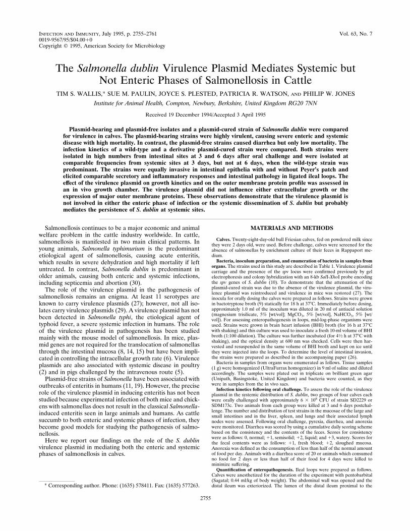
FIG. 1. Mean daily temperatures of four calves challenged with 1010 CFU ofeither SD2229 (F) or SDM173c (E) (two calves per strain).
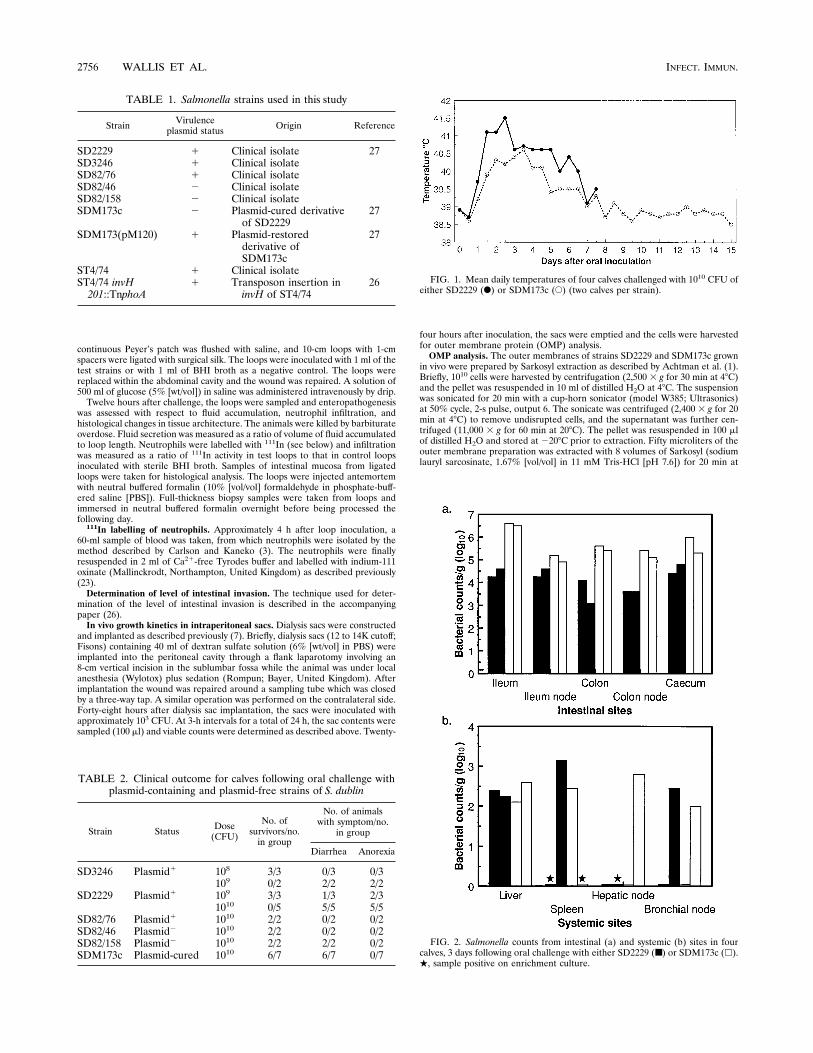
FIG. 2. Salmonella counts from intestinal (a) and systemic (b) sites in fourcalves, 3 days following oral challenge with either SD2229 (■) or SDM173c (h).., sample positive on enrichment culture.
TABLE 1. Salmonella strains used in this study
Strain Virulenceplasmid status Origin Reference
SD2229 1 Clinical isolate 27SD3246 1 Clinical isolateSD82/76 1 Clinical isolateSD82/46 2 Clinical isolateSD82/158 2 Clinical isolateSDM173c 2 Plasmid-cured derivative
of SD222927
SDM173(pM120) 1 Plasmid-restoredderivative ofSDM173c
27
ST4/74 1 Clinical isolateST4/74 invH201::TnphoA
1 Transposon insertion ininvH of ST4/74
26
TABLE 2. Clinical outcome for calves following oral challenge withplasmid-containing and plasmid-free strains of S. dublin
Strain Status Dose(CFU)
No. ofsurvivors/no.in group
No. of animalswith symptom/no.
in group
Diarrhea Anorexia
SD3246 Plasmid1 108 3/3 0/3 0/3109 0/2 2/2 2/2
SD2229 Plasmid1 109 3/3 1/3 2/31010 0/5 5/5 5/5
SD82/76 Plasmid1 1010 2/2 0/2 0/2SD82/46 Plasmid2 1010 2/2 0/2 0/2SD82/158 Plasmid2 1010 2/2 2/2 0/2SDM173c Plasmid-cured 1010 6/7 6/7 0/7
2756 WALLIS ET AL. INFECT. IMMUN.
room temperature. The insoluble outer membrane was pelleted by centrifugation(11,000 3 g for 90 min at 208C) and resuspended in 50 ml of sample buffer (2%[vol/vol] sodium dodecyl sulfate [SDS], 2% [vol/vol] mercaptoethanol, and 1%[vol/vol] glycerol in 62.5 mM Tris-HCl [pH 6.8]) and boiled for 5 min prior toanalysis by SDS-polyacrylamide gel electrophoresis (PAGE). OMPs were sepa-rated on the basis of molecular weight by using a modification of the method ofLaemmli (13). Electrophoresis was carried out at 30 mA per gel at a constantcurrent in stacking and resolving gels containing 4 and 10% acrylamide, respec-tively. Protein (30 mg) was loaded into each lane. After electrophoresis, proteinbands were visualized by immersing gels in Coomassie blue R-250 stainingsolution (0.25% [wt/vol] in 50% [vol/vol] methanol containing 10% acetic acid)for 4 h, and excess stain was removed with destain solution (5% [vol/vol] meth-anol, 7.5% [vol/vol] acetic acid).The resolved proteins were transferred to nitrocellulose by electrophoresis
(300 mA, 48C, overnight; Powerlid; Hoefer). Filters were washed in PBST (PBScontaining 0.05% [vol/vol] Tween 20), blocked with horse serum (50% [vol/vol]in PBST–0.5% [vol/vol] Tween 20) for 2 h at room temperature, and washedagain with PBST. The filters were then probed for 2 h at room temperature withbovine serum (10% [vol/vol] in PBST–0.5% [vol/vol] Tween 20) from convales-cent animals challenged with S. typhimurium and S. dublin and were washed withPBST. The filters were then incubated in anti-bovine immunoglobulin G (wholemolecule)–horseradish peroxidase (Sigma) (diluted 1/2,000 in 5% [vol/vol] horseserum in PBST–0.5% [vol/vol] Tween 20) for 2 h at room temperature andwashed with PBST. Protein bands were visualized with diaminobenzidine withnickel chloride enhancement (0.05% [wt/vol] diaminobenzidine, 0.03% [wt/vol]NiCl2, and 0.3% [vol/vol] H2O2 in PBS) for approximately 5 min, and thereaction was stopped by washing in distilled H2O.Statistical analysis. The data from the invasion assays and secretory and
inflammatory responses were analyzed with Minitab Inc. (State College, Pa.)statistical software. A one-way analysis of variance was carried out. In the eventthat a significant difference was found, strains were compared by using theStudent t test. The variance used in the t test was obtained from the residualmean square calculated in the analysis of variance.
RESULTSVirulence of wild-type and plasmid-free strains of S. dublin
in cattle. Three wild-type plasmid-bearing strains (SD3246,SD2229, and SD82/76), two wild-type naturally plasmid-freestrains (SD82/46 and SD82/158), and a plasmid-cured deriva-tive of SD2229 (SDM173c) were used to infect calves. The out-come of infection is shown in Table 2. Not all plasmid-bearingstrains were equally virulent. Strain SD3246 proved to be themostvirulent, causing diarrhea, anorexia, and mortality in both an-
FIG. 3. Salmonella counts from intestinal (a) and systemic (b) sites in fourcalves, 6 days following oral challenge with either SD2229 (■) or SDM173c (h).., sample positive on enrichment culture.
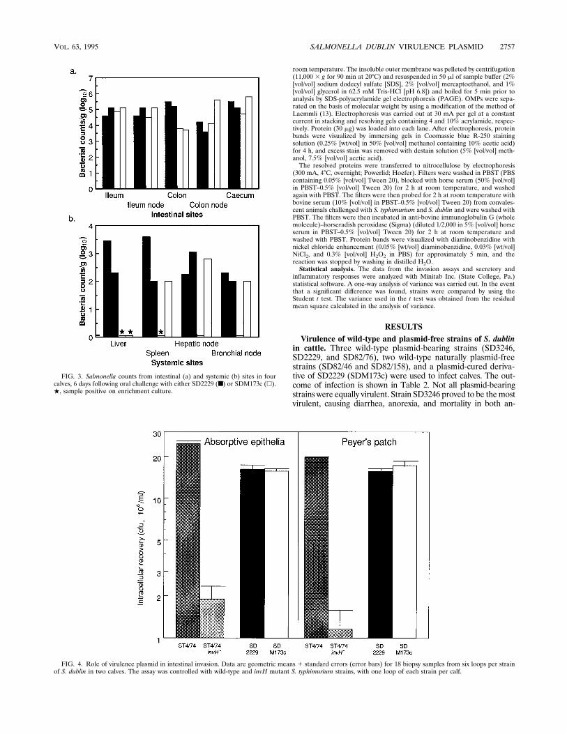
FIG. 4. Role of virulence plasmid in intestinal invasion. Data are geometric means 1 standard errors (error bars) for 18 biopsy samples from six loops per strainof S. dublin in two calves. The assay was controlled with wild-type and invH mutant S. typhimurium strains, with one loop of each strain per calf.
VOL. 63, 1995 SALMONELLA DUBLIN VIRULENCE PLASMID 2757
imals challenged with 109 CFU. Strain SD82/76 was avirulentin this model at a dose of 1010 CFU, indicating that factors inaddition to the virulence plasmid are required for virulence incattle. Ten of 11 animals challenged with the naturally plasmid-free strain or the cured strain survived challenge and did notbecome anorexic; diarrhea occurred in 8 of the 11 animals.The temperature responses in animals infected with SD2229
or its plasmid-cured derivative SDM173c are shown in Fig. 1.Strain SD2229 caused a higher mean rate of daily fever thanstrain SDM173c over the 8 days on which the strains could becompared.Effect of virulence plasmid on infection kinetics in calves.
The distribution of salmonellas within tissues following oralchallenge with 6 3 108 CFU of strains SD2229 and SDM173cwas determined. The gut-associated and systemic counts at 3and 6 days are depicted in Fig. 2 and 3, respectively. Bothstrains were recovered at lower numbers from the systemicsites than from the intestinal sites. Accurately enumeratingbacteria at such concentrations is problematic, and thereforeenrichment culture was carried out to aid in the detection ofsalmonellas. Considerable interanimal variation was seen,which may reflect either inherent variation in host resistance toinfection or the limitations of the assay for detecting salmo-nellas at low numbers.Both strains were primarily associated with the distal ileum
and colon rather than the upper small intestine data notshown. Strain SDM173c was reproducibly reisolated from in-
testinal sites at higher numbers than was strain SD2229 at day3 but not at day 6, when both strains were isolated at similarnumbers. The systemic distribution of wild-type and plasmid-cured strains showed different patterns. Three days after in-fection, the strains were isolated with similar frequencies andat similar numbers. By 6 days, the wild-type strain appearedpredominant, as it was isolated at quantifiable numbers fromall eight systemic sites tested; the cured strain was recovered atsimilar numbers only from three of eight sites.Role of virulence plasmid in intestinal invasion. The obser-
vation that SDM173c was detected at higher numbers at intes-tinal sites than was SD2229 at 3 days following oral challengesuggested that the plasmid-cured strain was possibly more in-vasive than the wild-type strain. The intestinal invasiveness ofthese strains was therefore assessed. The strains were detectedat comparable numbers in epithelia with and without Peyer’spatch mucosa (Fig. 4). The invasion assay was controlled witha wild-type strain and a less invasive invH mutant of S. typhi-murium (26). The invH mutant was recovered from mucosaewith and without Peyer’s patch at significantly lower numbersthan was the wild-type strain (P,0.01), indicating that theassay is able to quantify differences in the magnitude of intes-tinal invasion.Role of virulence plasmid in enteropathogenesis. Induction
of fluid secretion, neutrophil influx, and tissue damage in li-gated loops in the middle ileum was used to assess the entero-pathogenicity of strains SD2229 and SDM173c.
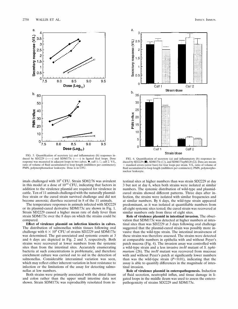
FIG. 5. Quantification of secretory (a) and inflammatory (b) responses in-duced by SD2229 (——) and SDM173c (– – –) in ligated ileal loops. Doseresponse was measured in adjacent loops in two calves. }, calf 1; E, calf 2. V/L,ratio of volume of fluid accumulated to loop length (milliliters per centimeter);PMN, polymorphonuclear leukocyte. Dose is in CFU.
FIG. 6. Quantification of secretory (a) and inflammatory (b) responses in-duced by SD2229 (■), SDM173c (h), and SDM173cpM120 (u). Data are means1 standard errors (error bars) for four loops per strain. V/L, ratio of volume offluid accumulated to loop length (milliliters per centimeter); PMN, polymorpho-nuclear leukocyte.
2758 WALLIS ET AL. INFECT. IMMUN.
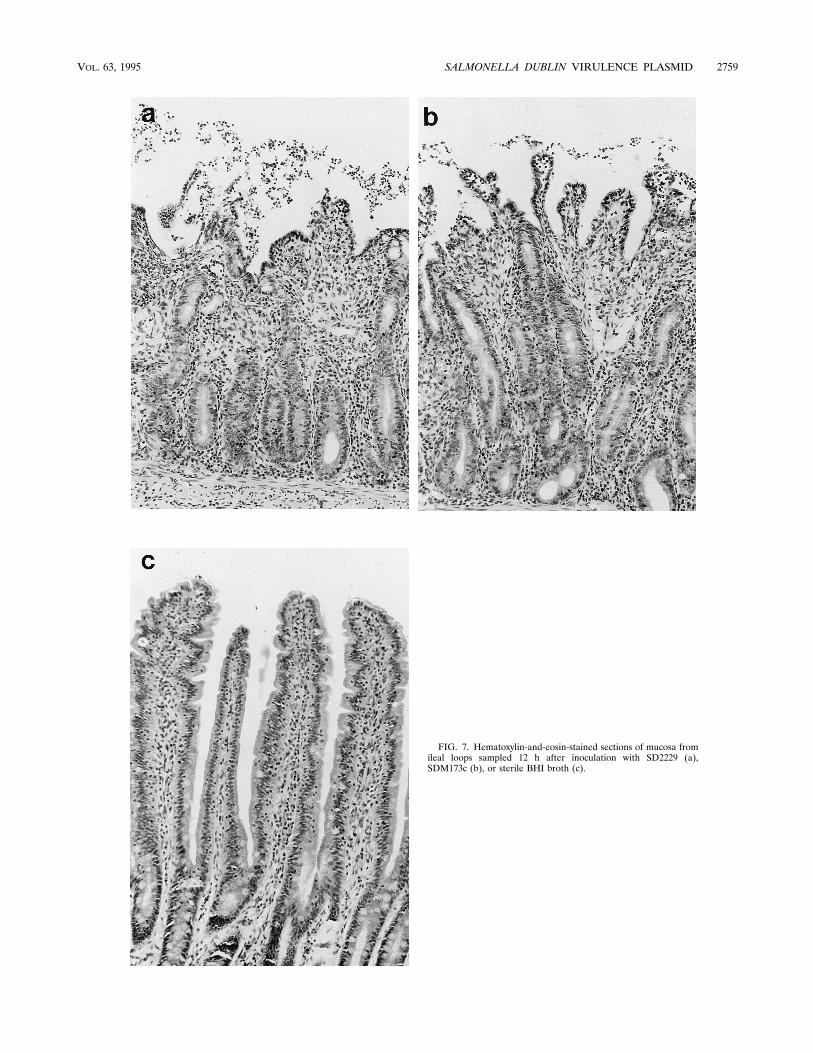
FIG. 7. Hematoxylin-and-eosin-stained sections of mucosa fromileal loops sampled 12 h after inoculation with SD2229 (a),SDM173c (b), or sterile BHI broth (c).
VOL. 63, 1995 SALMONELLA DUBLIN VIRULENCE PLASMID 2759
The dose responses to these strains in two calves were as-sessed (Fig. 5), and considerable interanimal variation wasobserved. In calf 1, potent secretory and inflammatory re-sponses were induced by both strains, and the magnitudes ofthe responses were related to the dose. In calf 2, both strainsevoked secretory and inflammatory responses; less potent re-sponses were recorded for calf 2 than for calf 1, and no in-flammatory dose response was seen (Fig. 5b). In both animals,strain SD2229 evoked slightly greater secretory and inflamma-tory responses at comparable doses than those evoked by strainSDM173c. Both strains were therefore tested in quadruplicatein two other animals at a dose of approximately 5 3 108 CFUper loop to confirm whether these differences were significant(Fig. 6). Interanimal variation was again observed; the mag-nitudes of the inflammatory responses differed significantly.In each animal, however, strains SD2229, SDM173c, andSDM173c(pM120) induced comparable secretory and inflam-matory responses (P $ 0.1).The strains induced comparable pathologies in the intestinal
mucosa. This was typified by exfoliation of villus tip entero-cytes with associated villus blunting, mucosal ulceration, andinfiltration by inflammatory cells (Fig. 7).In vivo extracellular growth kinetics and OMP profile. The
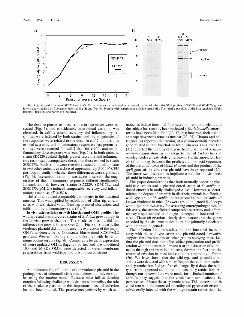
wild-type and plasmid-cured strains of S. dublin grew rapidly inthe in vivo growth chamber. The virulence plasmid did notinfluence the growth kinetics over 24 h (Fig. 8a). Similarly, thevirulence plasmid did not influence the expression of the majorOMPs as detectable by Coomassie blue-stained SDS-PAGEgels and Western blotting (immunoblotting) with hyperim-mune bovine serum (Fig. 8b). Comparable levels of expressionof iron-regulated OMPs, flagellin, porins, and two undefined100- and 66-kDa OMPs were detected in outer membranepreparations from wild-type and plasmid-cured strains.
DISCUSSION
An understanding of the role of the virulence plasmid in thepathogenesis of salmonellosis is based almost entirely on stud-ies using the murine model. Because mice fail to developenteritis following challenge with virulent salmonellas, the roleof the virulence plasmid in this important phase of infectionhas not been studied. The precise mechanisms by which sal-
monellas induce intestinal fluid secretion remain unclear, andthe subject has recently been reviewed (18). Salmonella entero-toxins have been identified (12, 17, 24); however, their role inenteropathogenesis remains unclear (23, 25). Chopra and col-leagues (4) reported the cloning of a chromosomally encodedgene related to that for cholera toxin, whereas Yang and Tan(31) reported the cloning of a gene from plasmids of S. typhi-murium strains showing homology to that of Escherichia coliwhich encodes a heat-labile enterotoxin. Furthermore, low lev-els of homology between the predicted amino acid sequencesof the ace enterotoxin of Vibrio cholerae and the product of thespvB gene of the virulence plasmid have been reported (20).The latter two observations implicate a role for the virulenceplasmid in inducing enteritis.This paper demonstrates that both naturally occurring plas-
mid-free strains and a plasmid-cured strain of S. dublin in-duced enteritis in orally challenged calves. However, as deter-mining the degree of enteritis in diarrheic animals is difficult, awild-type strain of S. dublin and its plasmid-cured derivative ofknown virulence in mice (28) were tested in ligated ileal loopswith a quantitative assay for assessing enteropathogenesis. Inthis assay, the strains elicited comparable secretory and inflam-matory responses and pathological changes in intestinal mu-cosae. These observations clearly demonstrate that the genesencoded by the virulence plasmid are not primarily associatedwith enteropathogenesis.The infection kinetics studies and the intestinal invasion
assay with the wild-type strain and plasmid-cured derivativesupport the observations of other groups studying mice, i.e.,that the plasmid does not affect either penetration and prolif-eration within the intestinal mucosa or translocation of salmo-nellas through the intestinal mucosa, despite the fact that theroutes of invasion in mice and cattle are apparently different(26). We have shown that the wild-type and plasmid-curedstrains were detected with similar frequencies at both intestinaland systemic sites 3 days after challenge. By 6 days, the wild-type strain appeared to be predominant at systemic sites. Al-though our observations were made for a limited number ofanimals, they suggest that the virulence plasmid affects thepersistence of bacteria at systemic sites. This observation isconsistent with the increased mortality and pyrexia observed incalves orally infected with the wild-type strain rather than the
FIG. 8. (a) Growth kinetics of SD2229 and SDM173c in dialysis sacs implanted in peritoneal cavities of calves. (b) OMP profiles of SD2229 and SDM173c grownin vivo and visualized by Coomassie blue staining (I) and Western blotting with hyperimmune bovine serum (II). The relative positions of the iron-regulated OMPs(iromps), flagellin, and porins are indicated.
2760 WALLIS ET AL. INFECT. IMMUN.
plasmid-cured strain, which implicates the plasmid in mediat-ing systemic salmonellosis in cattle. Bacterial persistencewithin a host is determined by two key parameters, bacterialgrowth and death rates. Enumerating bacteria at two timepoints does not enable us to identify which mechanism medi-ates increased persistence. Recently, it was reported that thevirulence plasmid increases the intracellular growth rate butdid not influence the intra- or extracellular location of salmo-nellae in mice (6). However, the extracellular growth rate wasdetermined at only one time point, 48 h, and therefore was notdefinite. Our observations are consistent with the notion thatthe plasmid increases the growth rate, but why this is evidentonly at systemic sites and not intestinal sites is not clear. Oneexplanation is that multiplication of strains within the intestinallumen, possibly unregulated by the virulence plasmid, results incontinuous seeding of the intestinal sites, which would maskthe difference in the abilities of these strains to persist in vivo.Alternatively, it may reflect differences in the abilities of re-ticuloendothelial cells at these various sites to control Salmo-nella growth.We assessed the role of the plasmid in mediating growth
over 24 h in an in vivo growth chamber, a model which facil-itates the study of extracellular growth; no effect on growthrate could be detected.The analysis of bacterial surface components is useful for the
identification of virulence determinants, as it is these compo-nents which directly interact with the host during the diseaseprocess. The expression of such components is determined bybacterial genotype and the microenvironment surrounding thecell. The virulence plasmid is not involved in the regulation oflipopolysaccharide expression (22), but its role in regulatingthe expression of OMPs in vivo has not previously been stud-ied. The virulence plasmid did not affect major OMP expres-sion in this model. Clearly, free Fe21 was not available withinthe peritoneal cavity, as indicated by the expression of the 81-,78-, and 74-kDa iron-regulated OMPs. The virulence plasmidproteins SpvA, -B, and -C have been reported to be expressedunder conditions of iron limitation and in the stationary phasebut only in small amounts in vitro (21). Reagents to specificallydetect the expression of SpvA, -B, and -C in vivo were notavailable at the time of analysis; however, clearly the virulenceplasmid does not regulate expression of major OMPs in theextracellular environment.It has been widely speculated that the virulence plasmid
mediates survival within macrophages; however, to date thereis no evidence to support this (16). Thus, the precise nichewithin the host where the virulence plasmid modifies patho-genesis remains unclear.
ACKNOWLEDGMENTS
This work was funded by the Ministry of Agriculture, Fisheries andFood. J.S.P. is supported by Hoechst.We thank L. H. Thomas for performing the dialysis sac implantation
operations.
REFERENCES
1. Achtman, M., A. Mercer, B. Kusecek, A. Pohl, M. Heuzenroeder, W. Aaron-son, A. Sutton, and R. P. Silver. 1983. Six widespread bacterial clones amongEscherichia coli K1 isolates. Infect. Immun. 39:315–335.
2. Barrow, P. A., J. M. Simpson, M. A. Lovell, and M. M. Binns. 1987. Con-tribution of Salmonella gallinarum large plasmid toward virulence in fowltyphoid. Infect. Immun. 55:388–392.
3. Carlson, G. P., and J. J. Kaneko. 1973. Isolation of leukocytes from bovineperipheral blood. Proc. Soc. Exp. Biol. Med. 142:853–855.
4. Chopra, A. K., C. W. Houston, J. W. Peterson, R. Prasad, and J. J. Mek-alanos. 1987. Cloning and expression of the Salmonella enterotoxin gene. J.Bacteriol. 169:5095–5100.
5. Danbara, H., R. Moriguchi, S. Suzuki, Y. Tamura, M. Kijima, K. Oishi, H.Matsui, A. Abe, and M. Nakamura. 1992. Effect of 50-kilobase plasmid,pkdsc50, of Salmonella cholerae-suis rf-1 strain on pig septicemia. J. Vet.Med. Sci. 54:1175–1178.
6. Gulig, P. A., and T. J. Doyle. 1993. The Salmonella typhimurium virulenceplasmid increases the growth rate of salmonellae in mice. Infect. Immun.61:504–511.
7. Haddock, F. M., L. H. Thomas, and P. Lees.Amodel for the characterisationof the bovine inflammatory response in vivo. Submitted for publication.
8. Heffernan, E. J., J. Fierer, G. Chikami, and D. Guiney. 1987. Natural historyof oral Salmonella dublin infection in Balb/c mice—effect of an 80-kilobase-pair plasmid on virulence. J. Infect. Dis. 155:1254–1259.
9. Jones, P. W. 1975. Effect of the storage in slurry on the virulence of Salmo-nella dublin. J. Hyg. 74:65–70.
10. Jones, P. W. 1986. Characterisation of strains of Salmonella dublin withparticular reference to pathogenicity. Ph.D. thesis. University of Surrey,Guildford, Surrey, United Kingdom.
11. Kapperud, G., S. Gustavsen, I. Hellesnes, A. H. Hansen, J. Lassen, J. Hirn,M. Jahkola, M. A. Montenegro, and R. Helmuth. 1990. Outbreak of Salmo-nella typhimurium infection traced to contaminated chocolate and caused bya strain lacking the 60-megadalton virulence plasmid. J. Clin. Microbiol.28:2597–2601.
12. Koupal, L. R., and R. H. Deibel. 1975. Assay, characterization, and localiza-tion of an enterotoxin produced by Salmonella. Infect. Immun. 11:14–22.
13. Laemmli, U. K. 1970. Cleavage of structural proteins during the assembly ofthe head of bacteriophage T4. Nature (London) 227:680–685.
14. Manning, E. J., G. D. Baird, and P. W. Jones. 1986. The role of plasmidgenes in the pathogenicity of Salmonella dublin. J. Med. Microbiol. 21:239–243.
15. Pardon, P., M. Y. Popoff, C. Coynault, J. Marly, and I. Miras. 1986. Viru-lence-associated plasmids of Salmonella typhimurium in experimental murineinfection. Ann. Inst. Pasteur Microbiol. 137:47–60.
16. Riikonen, P., P. H. Makela, H. Saarilahti, S. Sukupolvi, S. Taira, and M.Rhen. 1992. The virulence plasmid does not contribute to growth of Salmo-nella in cultured murine macrophages. Microb. Pathog. 13:281–291.
17. Sedlock, D. N., and R. H. Deibel. 1978. Detection of Salmonella enterotoxinusing the rabbit ileal loop test. Can. J. Microbiol. 24:268–273.
18. Stephen, J., G. R. Douce, and I. I. Amin. 1991. Experimental salmonellosis inretrospect and prospect, 1990, p. 185–196. In T. Wadstrom (ed.), Molecularpathogenesis of gastrointestinal infections. Plenum Press, New York.
19. Threlfall, E. J., M. D. Hampton, H. Chart, and B. Rowe. 1994. Use ofplasmid profile typing for surveillance of Salmonella enteritidis phage type 4from humans, poultry and eggs. Epidemiol. Infect. 112:25–31.
20. Trucksis, M., J. E. Galen, J. Michalski, A. Fasano, and J. B. Kaper. 1993.Accessory cholera enterotoxin (ace), the 3rd toxin of a Vibrio cholerae viru-lence cassette. Proc. Natl. Acad. Sci. USA 90:5267–5271.
21. Valone, S. E., G. K. Chikami, and V. L. Miller. 1993. Stress induction of thevirulence proteins (SpvA, -B, and -C) from native plasmid pSDL2 of Salmo-nella dublin. Infect. Immun. 61:705–713.
22. Vandenbosch, J. L., D. R. Kurlandsky, R. Urdangaray, and G. W. Jones.1989. Evidence of coordinate regulation of virulence in Salmonella typhi-murium involving the rsk element of the 95-kilobase plasmid. Infect. Immun.57:2566–2568.
23. Wallis, T. S., R. J. H. Hawker, D. C. A. Candy, G. M. Qi, G. J. Clarke, K. J.Worton, M. P. Osborne, and J. Stephen. 1989. Quantification of the leuko-cyte influx into rabbit ileal loops induced by strains of Salmonella typhi-murium of different virulence. J. Med. Microbiol. 30:149–156.
24. Wallis, T. S., W. G. Starkey, J. Stephen, S. J. Haddon, M. P. Osborne, andD. C. A. Candy. 1986. Enterotoxin production by Salmonella typhimuriumstrains of different virulence. J. Med. Microbiol. 21:19–23.
25. Wallis, T. S., A. T. M. Vaughan, G. J. Clarke, G. M. Qi, K. J. Worton, D. C. A.Candy, M. P. Osborne, and J. Stephen. 1990. The role of leukocytes in theinduction of fluid secretion by Salmonella typhimurium. J. Med. Microbiol.31:27–35.
26. Watson, P. R., S. M. Paulin, A. P. Bland, P. W. Jones, and T. S. Wallis. 1995.Characterization of intestinal invasion by Salmonella typhimurium and Sal-monella dublin and effect of a mutation in the invH gene. Infect. Immun.63:2743–2754.
27. Williamson, C. M., G. D. Baird, and E. J. Manning. 1988. A commonvirulence region on plasmids from 11 serotypes of Salmonella. J. Gen. Mi-crobiol. 134:975–982.
28. Williamson, C. M., G. D. Pullinger, and A. J. Lax. 1988. Identification of anessential virulence region on Salmonella plasmids. Microb. Pathog. 5:469–473.
29. Woodward, M. J., I. Mclaren, and C. Wray. 1989. Distribution of virulenceplasmids within salmonellae. J. Gen. Microbiol. 135:503–511.
30. Wray, C. 1991. Salmonellosis in calves. In Pract. 13:13–15.31. Yang, M. K., and M. S. Tan. 1989. Characterisation and cloning of entero-
toxin genes from Salmonella typhimurium. Proc. Natl. Sci. Counc. Repub.China Part B 13:109–118.
VOL. 63, 1995 SALMONELLA DUBLIN VIRULENCE PLASMID 2761


