Embed Size (px)
Citation preview

The Sea Urchin Sperm Receptor for Egg Jelly Is a Modular Protein with Extensive Homology to the Human Polycystic Kidney Disease Protein, PKD1 G.W. Moy,* L.M. Mendoza,* J.R. Schulz,* W.J. Swanson,* C.G. Glabe,* and V.D. Vacquier*
*Marine Biology Research Division, University of California, San Diego, La Jolla, California 92093-0202; and*Department of Molecular Biology and Biochemistry, University of California, Irvine, California 92717-3900
Abstract. During fertilization, the sea urchin sperm acrosome reaction (AR), an ion channel-regulated event, is triggered by glycoproteins in egg jelly (E J). A 210-kD sperm membrane glycoprotein is the receptor for EJ (RE J). This conclusion is based on the following data: purified REJ binds species specifically to EJ dot- ted onto nitrocellulose, an mAb to REJ induces the sperm AR, antibody induction is blocked by purified RE J, and purified REJ absorbs the AR-inducing activ- ity of EJ. Overlapping fragments of REJ cDNA were cloned (total length, 5,596 bp). The sequence was con- firmed by microsequencing six peptides of mature REJ and by Western blotting with antibody to a synthetic peptide designed from the sequence. Complete degly- cosylation of REJ followed by Western blotting yielded
a size estimate in agreement with that of the mature amino acid sequence. REJ is modular in design; it con- tains one EGF module and two C-type lectin carbohy- drate-recognition modules. Most importantly, it con- tains a novel module, herein named the REJ module (700 residues), which shares extensive homology with the human polycystic kidney disease protein (PKD1). Mutations in PKD1 cause autosomal dominant polycys- tic kidney disease, one of the most frequent genetic dis- eases of humans. The lesion in cellular physiology re- suiting from mutations in the PKD1 protein remains unknown. The homology between REJ modules of the sea urchin REJ and human PKD1 suggests that PKD1 could be involved in ionic regulation.
S EA urchin eggs possess an extracellular matrix termed
egg jelly (E J) t. Glycoprotein ligands in EJ induce the sperm acrosome reaction (AR) (Keller and
Vacquier, 1994a; Suzuki, 1995). The AR is required for fertilization; it consists of the exocytosis of the acrosome granule and the polymerization of acrosomal actin to form the bindin-coated acrosomal process used by the sperm to attach to and fuse with the egg (Vacquier et al., 1995). Un- derlying the E J-induced AR, increased sperm respiration,
Address all correspondence to V.D. Vacquier, Marine Biology Research Division, Scripps Institution of Oceanography, University of California, San Diego, La Jolla, CA 92093-0202. Tel.: (619) 534-4803. Fax: (619) 534- 7313. e-mail: [email protected].
L.M. Mendoza's present address is Division of Immunology, LSA 425, Department of Molecular and Cell Biology, University of California, Berkeley, Berkeley, CA 94720-3200.
1. Abbreviat ions used in this paper: ADPKD, autosomal dominant poly- cystic kidney disease; AR, acrosome reaction; CRD, carbohydrate-recog- nition module; E J, egg jelly; Fn3, fibronectin type 3; GA/SW, gluteraldehyde in seawater; HF, hydroflouric acid; HSW, filtered seawater containing 10 mM Hepes; ORF, open reading frame; PKD1, human polycystic kidney disease protein; REJ, receptor for egg jelly; SMV, sperm plasma mem- brane vesicle; WGA, wheat germ agglutinin.
motility, and chemotaxis are signal transduction events that involve Ca 2÷ and Na ÷ influx, H ÷ and K ÷ effiux (Darszon et al., 1989; Schackmann, 1989; Vacquier, 1986a), cyclic nucle- otide changes, and protein kinase activation (Garbers, 1989).
Trypsin treatment of sea urchin sperm blocks AR induc- tion by E J, suggesting that receptor proteins regulate the ion channel events mediating the AR (Moy, G.W., and V.D. Vacquier, unpublished observations). To identify re- ceptor proteins, mice were immunized with sea urchin sperm, and hybridomas were screened for secretion of mAbs re- acting with extracellular epitopes. The majority of mAbs (85 out of 96) reacted with a sperm membrane glycopro- tein of 210-260 kD, which was termed the "210-kD pro- tein" (Trimmer, 1987). Previous work had shown that the isolated 210-kD protein bound to live eggs and to EJ dot- ted onto nitrocellulose (Podell and Vacquier, 1985).
Immunofluorescence (Trimmer et al., 1985) and immu- nogold (Longo et al., 1989) localization showed the 210-kD protein was present on the surface of the sperm flagellum and also on a thin belt (0.2-1xm diam) of membrane directly over the acrosomal granule at the anterior apex of the sperm head. The eightfold increase in immunogold local- ization over the acrosome, as compared with the remain- der of the sperm head, suggested this protein functioned in
© The Rockefeller University Press, 0021-9525/96/05/809/9 $2.00 The Journal of Cell Biology, Volume 133, Number 4, May 1996 809-817 809
on Decem
ber 24, 2015jcb.rupress.org
Dow
nloaded from
Published May 15, 1996

AR induction. Loading sperm with Ca2+-indicator dyes showed that mAbs to the 210-kD protein opened Ca 2÷ channels required for AR induction (Trimmer et al., 1986; Vacquier et al., 1988).
Here we demonstrate that the 210-kD protein is the recep- tor for egg jelly (RE J) ligands inducing the AR. Molecular cloning showed that REJ is modular in design, containing EGF and carbohydrate-recognition modules. In addition, it possesses a new module, the REJ module, which is also found in the human polycystic kidney disease protein (PKD1).
Materials and Methods
Gametes and Miscellaneous Procedures Sperm of Strongylocentrotus purpuratus were spawned and stored undi- luted on ice. Aerosome reactions were scored, and egg jelly was obtained and quantitated (Keller and Vacquier, 1994a; Vacquier, 1986b). Plasma membrane vesicles (Podell et al., 1984) were commercially deglycosylated by hydrofluoric acid (HF) (Immuno-Dynamics, Inc., La Jolla, CA). Meth- ods to produce mAbs and Fab fragments were previously described, and mAb characterization by immunoprecipitation and Western blotting was presented (Trimmer et al., 1985; Trimmer, 1987; Vacquier et al., 1988). Protein was quantitated by the bicinchoninic acid assay (Pierce Chemical Co., Rockford, IL). All Fab, purified REJ protein, and EJ used for treat- ment of live sperm were dialyzed into filtered seawater containing 10 mM Hepes (HSW) (pH 8.0, unless noted otherwise). For Fig. 1, A and B, 10 p.1 Fab J10/14, J17/30 (IgG), EJ, or HSW was placed in 1.5-ml microcentri- fuge tubes. 10 ixl of a 1:100 dilution of undiluted semen ("dry sperm") was added, and after 5 rain the cells were fixed by addition of 40 p.I 5% glu- taraldehyde in seawater (GA/SW). Acrosome reactions were scored by preparing thumb-squashed slides and viewing in phase contrast at a mag- nification of ~1,200 (Vacquier, 1986b). Percentage of A R was rounded to the nearest whole number. In Fig. 1 B, the various pHs were obtained us- ing 10 mM Hepes seawater adjusted to pHs 6.8-8.0, and the sperm were diluted 1:100 into the pH adjusted seawaters. In Fig. 1 B the final concen- tration of EJ was equivalent to 12 ~g fucose per ml (yielding 95% AR), and that of Fab J10/14 was 250 izg/ml (yielding 95% AR). For Fig. 1 C, 10 i~l of Fab J10/14 was diluted 50% per dilution in HSW, pH 7.5, in a series of tubes, and 10 ~l of a 1:100 dilution of dry sperm (in HSW, pH 7.5) was added. After 5 min 20 p.l of EJ was added; the final concentration of EJ was 12 Ixg fucose per ml. After 5 min 80 i~l of GA/SW was added, and the cells were scored for AR. For Fig. 1 D, 5 ILl HSW containing purified REJ protein was diluted into a series of tubes, with the protein varying from 0 to 719 ng. 5 p.l of Fab J10/14 (1.8 ng protein) was added to each tube and incubated 2 h at 22°C. 10 ILl 1:100 diluted sperm in HSW, pH 8.0, was added, and after 5 rain the samples were fixed by addition of 40 p,l GA/ SW. In Fig. 1, each data point represents 200-600 cells.
For Table I, 5 ILl EJ (final concentration after adding sperm was equiv- alent to 780 ng fncose per ml) was mixed with 5 ~.1 of either HSW or HSW containing 12.5 Ixg purified REJ protein (Fig. 3, lane B) for 2 h at 22°C, and 10 izl of 1:100 dilution of sperm was added (final concentration of REJ was 625 p.g/ml). EJ was used in a linear region of the concentration dependence curve, in this case the amount resulting in 25% acrosome- reacted sperm. After 15 s the cells were fixed by addition of 40 I~l GA/SW. 4% A R was observed in the seawater-only control. This value was sub- tracted from the other samples to arrive at 86% inhibition of the EJ- induced AR.
A synthetic peptide (KGVSNEDPDTDA) was conjugated to ovalbumin (Doolittle, 1987), and rabbit antibody was commercially prepared (Cocalico Biologicals Inc., Reamstown, PA). The whole antiserum was used without purification of specific IgGs. SDS-PAGE gels were performed according to the method of Laemmli (1970), and Western blots were performed and developed using the directions in the ECL kit (Amersham Corp., Arling- ton Heights, IL).
Purification of 210-kD Protein from Sperm Sperm plasma membrane vesicles (SMV) were prepared by the pH-9 method (Podell et at., 1984). The proteins found in SMVs are shown in Fig. 3 A (Podell et at., 1984; Vacquier, 1986b). The 200,000 g (60 rain) su- pernatant of SMVs was applied to a wheat germ agglutinin (WGA) aga- rose column (5 ml; E-Y Laboratories, Inc., San Mateo, CA). The column
was washed with a minimum of 10 vol: 500 mM NaC1/50 mM Hepes/5 mM EDTA/I% Triton X-100, followed by 10 vol 150 mM NaC1/50 mM Hepes, pH 8.0/5 mM EDTA. The REJ protein was eluted in a single step of the second wash buffer containing 100 mM GIcNAc. The peak fractions (A280 ~,n) were concentrated using Centricon-50 concentrators (Amicon Corp., Danvers, MA), and the buffer was exchanged for HSW using Cen- tricons (three times, 2 ml). The purity of the isolated REJ protein is shown in Fig. 3, lane B.
Peptide sequences of fragments of REJ were obtained by two methods starting with the WGA-eluted peak. Peptide sequences p2, p3, and p5 were from sperm of the congeneric species, Strongylocentrotus franciscanus. This is because the REJ is more abundant in this species. The W GA elu- ate was carboxymethylated, dialyzed into distilled water, and REJ sepa- rated on 7% acrylamide SDS-PAGE. The REJ band was excised after visu- alization by negative staining in 2 M KC1. CNBr cleavage was done for 2 h at 22°C (Matsudaira, 1989). The gel slices were washed twice in excess 125 mM Tris, pH 6.8 (10 min). Peptides were separated on a 20% acrylamide gel by SDS-PAGE and transferred to a polyvinyldifluoride membrane in 10 mM CAPS buffer, pH 11/10 % methanol. The polyvinyldifluoride mem- brane was stained in Coomassie blue and destained, peptide bands were ex- cised, and gas-phase amino acid sequencing was performed (Matsudaira, 1989). Peptides pl, p4, and p6 were obtained from S. purpuratus REJ by run- ning lanes of 80 Ixg W G A eluate on 5% SDS-PAGE gels, visualizing REJ using the CuC1 z staining kit (Bio Rad Laboratories, Richmond, CA) and ex- cising the center of the REJ band from the gel. The cut-outs were exposed to trypsin, and the tryptic peptides were separated by reverse-phase HPLC (Ferrara et al., 1993). The peak fractions were gas-phase microsequenced by the University of California, San Diego Protein Sequencing Laboratory.
Molecular Cloning, Sequencing, and Expression A Lambda Zap eDNA library of S. purpuratus testis was prepared follow- ing the manufacturer's directions (Stratagene, La Jolla, CA). The library was screened with a polyclonal antibody raised to the REJ protein band cut from SDS-PAGE gels. The antiserum had been absorbed with an ace- tone powder of sea urchin ovaries and reacted with the REJ protein on Western blots (Fig. 3, lane C). Primary screening yielded 22 positive plaques, 14 of which had unique restriction maps. One clone (2 kb) was se- quenced; it contained the sequences of four REJ-derived peptides (Fig. 2 B; p2-p5). All 14 unique clones were screened with an exact 57-mer oligo- nucleotide to the sequence of peptide p2. Six positives were obtained, two of which contained the full-length 210-kD open reading frame (ORF) with an overlap of 1,200 bp. Both ends of the ORF were confirmed by sequenc- ing independently picked phage plaques. The deduced sequence was fur- ther confirmed by obtaining two additional sequences of tryptic fragments of REJ isolated from S. purpuratus sperm (Fig 2 B; pl and p6). Nested de- letions were made using the Erase-a-Base System (Promega, Madison, WI), and T3 and T7 primers were used for sequencing. Gaps in the se- quence were filled in using custom primers. All sequence elements over- lapped, and both strands of all clones were sequenced using Sequenase 2.0 (United States Biochemical Corp., Cleveland, OH).
Computer Analyses Hydrophilicity profiles were generated by the method of Kyte and Doolit- tie (1982) using a window of 14 residues. Homology searches used the BLAST (Altschul et al., 1990) and FASTA (Pearson and Lipman, 1988) algorithms using a variety of scoring matrices. Statistical significance of pairwise comparisons (Tables II and III) used the RFD2 program (500 randomizations, Ktup = 2) (Pearson, 1990). Multiple alignments were made using the NEWAT progressive alignment program (Feng and Doolittle, 1990), except in the case of the putative Fn3 modules of the PKD1 pro- tein, where the published alignments were used (Hughes et al., 1995). Pro- files (Gribskov et at., t990) were computed from the aligned sequences using the Wisconsin Genetics Computer Group's program PROFILEMAKE and searched against GenBank (release 88) using the program PRO- FILESEARCH (Genetics Computer Group, 1991). These programs were accessed using the suite of programs in DNASYSTEM (Smith, 1988).
Results
The 210-kD Protein Is the Sperm REJ
Fab of the 210-kD mAb J10/14 induce the sperm AR in a concentration-dependent manner (Fig. 1 A). Both IgG
The Journal of Cell Biology, Volume 133, 1996 810
on Decem
ber 24, 2015jcb.rupress.org
Dow
nloaded from
Published May 15, 1996
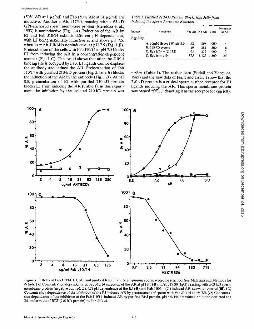
(50% AR at 1 ~g/ml) and Fab (50% AR at 31 Ixg/ml) are inductive. Another mAb, J17/30, reacting with a 63-kD GPI-anchored sperm membrane protein (Mendoza et al., 1993) is noninductive (Fig. 1 A). Induction of the AR by EJ and Fab J10/14 exhibits different pH dependencies, with EJ being maximally inductive at and above pH 7.5, whereas mAb J10/14 is noninductive at pH 7.5 (Fig. 1 B). Preincubation of the cells with Fab J10/14 at pH 7.5 blocks EJ from inducing the A R in a concentration-dependent manner (Fig. 1 C). This result shows that after the J10/14 binding site is occupied by Fab, EJ ligands cannot displace the antibody and induce the AR. Preincubation of Fab J10/14 with purified 210-kD protein (Fig. 3, lane B) blocks the induction of the AR by the antibody (Fig. 1 D). At pH 8.0, preincubation of EJ with purified 210-kD protein blocks EJ from inducing the A R (Table I); in this experi- ment the inhibition by the isolated 210-kD protein was
Table I. Purified 210-kD Protein Blocks Egg Jelly from Inducing the Sperm Acrosome Reaction
Percentage Inducer Condition Yes AR No AR Total of AR
Egg Jelly
A. 10mM Hepes SW, pH 8.0 B. 210-kD protein C. Egg jelly + 210 kD D. Egg jelly only
32 868 900 4 19 281 300 6 63 837 900 7
375 1,125 1,500 25
N86% (Table I). The earlier data (Podell and Vacquier, 1985) and the new data of Fig. 1 and Table I show that the 210-kD protein is a critical sperm surface receptor for EJ ligands inducing the AR. This sperm membrane protein was named "RE J," denoting it as the receptor for egg jelly.
100"
80
60
40'
2o i
A 100 -
80
=¢ 60 < :
40
Z0 G
; ; is 31 s3 lZSZ o o ug/ml .ANTIBODY
6.8
L I
; . . . . . • r ---- " - - - - • T
7.2 7.6 8.0
100'
80"
60" ¢ ¢
< :
4O
20
C -- •
! . o ~ 4
100"
80
60' <C
4 0
ZO
• . g . | . | i . /
G 8 16 31 63 125 ug/ml Fab J10/14
D
| - | - i " | • • " | - i - s - | • i • | •
0.7 2.8 11 44 180 719 ng 210 kDa
Figure 1. Effects of Fab J10/14, E J, pH, and purified REJ on the S. purpuratus sperm acrosome reaction. See Materials and Methods for details. (A) Concentration dependence of Fab J10/14 induction of the AR at pH 8.0 (0). mAb J17/30 (IgG) reacting with a 63-kD sperm membrane protein (negative control, 13). (B) pH dependence of the EJ-(O) and Fab J10/14-(©) induced AR; seawater control ( I ) . (C) Concentration dependence of the inhibition of the E J-induced AR by pretreatment of sperm with Fab J10/14 at pH 7.5. (D) Concentra- tion dependence of the inhibition of the Fab J10/14-induced AR by purified REJ protein, pH 8.0. Half maximal inhibition occurred at a 2:1 molar ratio of REJ (210-kD protein) to Fab J10/14.
Moy et al. Sperm Receptor for Egg Jelly 811
on Decem
ber 24, 2015jcb.rupress.org
Dow
nloaded from
Published May 15, 1996

Sequence of REJ The total nucleotide sequence obtained, 5,596 bp, consists of 201-bp 5' untranslated region, 4,350-bp ORF, and 1,045-bp 3' untranslated region. Northern blots of testis polyA + RNA yielded two closely spaced bands at ~7,200 bp
(not shown). The arrangement of the modules contained in the 1,450 amino acid ORF is shown diagrammatically in Fig. 2 A. These consist of one EGF module (40 residues), two carbohydrate-recognition modules (CRDs) (119 and
A E CRD1 CRD2 REJ MODULE
COOH
] .... , / i l , i i ,,lj ! iaAJiA.J ia I[,II lJ ,,.,, i l l
B 400 800 1200 LLEAVPNSDFAMAT
pl ....................................................................... QG~NAFVYAQIRRYAWIGLSDQVTEGVFDYADGTPvDYLSFPDKNKQs~eTRDCVEVKHLRVDNWSLLDCRANKTSICK~T~FSAT~ J 200
m
I
CA~ATGYCYFYE ERGGMWSKGRE FC LDAGADLAS IHSAEENAF I FDMLTEFVWLGLNDLETEGVYTNF SDGTPADFDNFPADNYQNEDTTDCVS I 1300 I
RHLEKTDRYWFFLGCDDTVTSIC~HEVVATOAPPYLFTTTDVPATTTTI PTTTTTI SATTRTIDAMIL7 ELLLFP~'VS S I DNI FR~]NT I LVTASVL 400
A * * l GSFQALSSQILWS ITNHLSGDSVQYTVYSEDAVLVRFTTVS I FTIKATAVGNSHNLTATANATVKHVC I SSLTTSVEE~DAPHPAVYL~DIH I ST~ 500
P ELNGKC I DPMTPDFKWRI FTSTEADDVVTAFEKITHTRQVMI PRGTLPYGIYSLNLNAKTRLKTSGEVTGEKE I I SWLEI(~PPPLVAVI KGGASRSHGVS
SNL IVDGSNSYDPDVPPGSSSNVTFLWYCVVVDPDVMYSSLDEAIQNTDNACFEDEGIMMNSTS SMI EVIANKLNANVTMNFWLNI SKEC-QI SGLTQQRI
HLTLGLLPEIEI SC ISNCNMYI FTAERLVLHASCTNCDSENEDVSFRWSLESNHTSVIGDLSSQTTTGLDQPYLVLKPLTFDS I SEMGS I I LRVTGSQSD
SDGYAEFSVDLPHNAPPALGSCVVTPDEGYALQTDFTVTCSNFTDVDEPLTYQI I LFSHVDVVDWMFVGRGEGFQLYEGSAP I KDGLYLPVGVGTDDHDI
LLQVNVRIX~NMASTSVY I SATVHPPTLDAVGM~VOELLDMALLVETNVNALLAVGDPGQAAQL I SALGS I LNS I GDEDDSEEGRETRSE IRSFLVDCVA p2
AZ PVE SMT~VVTRITKQE I STHVQMLRASTLSEMTSFVKSKSGSYTQAQENIESAGTI LVEGLSNI LSAAKETERLLSDDTSQEREDHKNLI
600
700
800
900
1000
1100
1200
M KS S IVGIQLEADQMLE FHDLTADI D V Y L P ~ T K T S SDSVLIDHS SLPVDGALHLTVIAENEP~TE SSCVGTDTPL~
. p4 . S p5 A VSNEDPDTDANFTWTVPLVDLKAADGIMI P ~ ~ D N ITLSVFMI4TLQCNFWNEDQQEWDSTGCKVGP LSKPSTTHCLCNHLTGFFGSS I LVP
I p6 V
PNHAQPVI GGHKLTGV~ LI CVLIGYGIYCVALV~VVGC SFAI RVHKVL
1300
1400
Figure 2. Deduced amino acid sequence of the REJ protein. (A) Diagrammatic scheme of the modular design of REJ presented over a hydrophilicity plot of the sequence. E, EGF module; CRD, carbohydrate-recognition domains 1 and 2. (B) The 1,450-residue ORF of REJ. Underlined Met at position 1 marks the putative start of the ORF; the putative NH2 terminus of the mature protein is Lys 27. Shaded box marks the EGF module, followed by the two CRDs with a 14-residue spacer between them (overline). The REJ module is also boxed (positions 480-1,187), as is the 18-residue putative transmembrane segment in the COOH-terminal region. Asterisks mark 17 potential N-linked glycosylation sites, pl-p6 are peptide sequences obtained from fragments of mature RE} isolated from sperm. Thick underline (positions 1,300-1,310) denotes synthetic peptide used for generation of polyclonal antiserum. Six polymorphic sites are labeled by letter above their positions. These sequence data are available from EMBL/GenBank/DDBJ under accession number U40832.
The Journal of Cell Biology, Volume 133, 1996 812
on Decem
ber 24, 2015jcb.rupress.org
Dow
nloaded from
Published May 15, 1996

MOUSE NOTCH DECASTPCKNGAKCLDGPNTYTCVCTEGYTOTHCEVDIDEC BRACHYDANIO DECASTPCKNGAKCTDGPNKYTCEC TPGFS GIHCELDINEC DROS DEVOPM DDCASFPCQNGGTCLDGIGDYSCLCVDGFD@KHCETDINEC SEAURCH FIB NECASIPCLNGGICVDGINQFACTCLPGYTGILCETDINEC XENOPUS XOT DDCQPNPCHNGGSCSDGINMFFCNCPAGFRGPKCEEDINEC SEAURCE REJ VDCASAPCMNGGTCADGFNNPI~FCEEGFTGSMCETEY-GC HUM FACT IX DQCESNPCLNGGSCKDDINSYECWCPFGFEGKNCELDV-TC
Figure 4. Alignments of representative EGF modules compared to the EGF module of REJ. Dashes are included for alignment. These sequence data are available from EMBL/GenBank/DDBJ under the following accession numbers: mouse notch, 483581; Brachydanio, 433867; Drosophila developmental, 157993; S. pur- puratus fibropellin, 310660; Xenopus xotch, 214925; and human factor IX, 182609.
Figure 3. Western blots of REJ. Lanes A and B are silver-stained SDS-PAGE gels, and lanes C-E are Western blots of SMV devel- oped with ECL. Lane A, 4 p+g SMV protein; lane B, 1.2 I~g WGA- purified REJ protein used in Table I and Fig. 1 D. (Lane C) Re- action of the absorbed rabbit antibody to REJ used for primary screening of the lambda Zap library. (Lane D) Reaction of anti- body to the synthetic peptide reacting with SMV. (Lane E) Reac- tion of the antibody to the synthetic peptide reacted with SMV that had been deglycosylated by HF treatment. The reacting band shifts to ~160-170 kD. Numbers on left margin, M~ in kD.
120 residues), and a novel module, the REJ module (707 residues). The hydrophilicity plot of the sequence (Fig. 2 A) shows two hydrophobic regions. The probable signal se- quence is at the NH2 terminus, and the putative mem- brane-spanning sequence is close to the COOH terminus. The orientation of this putative membrane-spanning seg- ment (box, residues 1,418-1,435) follows the '?charge dif- ference rule," having greater positive charge on the COOH- terminal side of the segment as compared with the NH2-terminal side (Sipos and von Heijne, 1993). Detailed analysis of the short, internal, hydrophobic sequence ele- ments throughout the sequence shows they could not span the membrane as alpha helices.
The full-length deduced amino acid sequence of REJ of 1,450 residues is shown in Fig. 2 B with the three types of modules boxed. The start and stop codons were confirmed by sequencing independently picked phage plaques. The putative initiation Met is underlined. This was chosen as the start of the ORF because its sequence of GCAACCAUGA conformed in 8 out of 10 sites to the preferred translation initiation sequence of GCCA/GCCAUGG (Kozak, 1987).
In-frame sequence obtained 5' to the putative Met t is also shown. The precise junction between the signal sequence and the mature protein is speculative. Given the " - 3 , - 1 rule," the most probable junction is between Sei 26 and Lys 27. This assignment would place Pro in the - 6 position, which is optimal for marking the junction (von Heijne, 1990). The six polymorphic positions are noted with the al- ternative residue above the sequence; five are conserva- tive replacements (the cDNA library was made from sev- eral individuals). The sequence was confirmed to be that of the REJ protein by amino acid sequencing of six frag- ments of mature REJ extracted from sperm (underlines, pl-p6). In addition, an antiserum raised to a synthetic peptide corresponding to residues 1,300--1,310 (thick un- derline) reacted with the REJ protein on Western blots of sperm plasma membrane vesicles (Fig. 3, lane D).
Although its mobility on SDS-PAGE is in the 210--260-kD range, the 1,424 amino acids of the putative mature REJ account for only 155,420 units of molecular mass. By chemi- cal analysis, REJ protein is 49% carbohydrate (Podell and Vacquier, 1985). The 17 sites for potential N-linked glyco- sylation are marked with asterisks (Fig. 2 B). Digestion with peptide-N-glycosidase-F decreased the relative mass to ~190 kD. Complete deglycosylation by HF treatment of sperm plasma membrane vesicles (Podell et al., 1984), followed by SDS-PAGE and Western blotting with the synthetic peptide antibody to REJ, yielded a single reac- tion at ~160--170 kD (Fig. 3, lane E).
Sequence Modules of REJ
EGFModule. The EGF module (shaded box, positions 29--68) contains the diagnostic seven Cys residues (Fig. 4). This module is similar to the sea urchin fibropellins and various developmentally regulated proteins such as notch and xotch. Table II presents the quantitative pairwise
Table II. Quantitative Comparisons of EGF Modules
NOTCH BRACH DDEVLP FIBROP XOTCH REJ FACT IX
78.0 58.5 58.5 48.8 52.5 47.5 20.0 61.0 61.0 53.7 50.0 52.5 18.7 15.0 58.5 58.4 55.0 55.0 16.3 18.5 1 6 . 8 ~ 56.1 55.0 50.0 14.9 14.7 14.3 17.1 ~ 50.0 57.5 12.4 11.7 13.1 1 4 . 7 1 2 A ~ ~ 45.0 12.8 13.4 13.5 13.4 14.0 13.0
NOTCH BRACH DDEVLP HBROP XOTCH REJ FACTIX
Values below the diagonal are pairwise alignment scores in SD units, and above the diagonal, percent identity in pairwise comparisons.
Moy et al. Sperm Receptor for Egg Jelly 813
on Decem
ber 24, 2015jcb.rupress.org
Dow
nloaded from
Published May 15, 1996

REJCRDI REJCRD2 HUMCRD8 MBRA 180PPLA RHANS PKDI INTRON+
REJCRDI REJCRD2 HUMCRD8 MBRA 180PPLA RHANS PKDI INTRON+
WFYGPAFGYCYLWERV-DYNWTQARESCIDQGRGAELASIHSAEENAFV-Y--AQI-RRYA--WIGDS WVHNPATGYCYFYEER-GGMWSKGREFCLDA--GADLASIHSAEENAFI-F--DML-TEFV--WLGLN WI--PFHGHCYYIESSYTRNWGQASLECLRM--GSSLVSIESAAESSFLSYRVEPL-KSKTNFWIGLF W--QEYDGHCY WASTYQVRWNDAQLACQTVHPGAYLATIQSQLENAFI .... SET-VSNNRLWIGI~N WLFHQDAEYLFYPHSS---EWSSFEFVCGWL--RSDILTIHSAHEQEFILSKIKALSKYGANWWIGLQ EGSNAYSSYCYYFMED-HLSWAEADLFCQNMN-SGYLVSVLSQAEGNFLASLIKESGTTAANVWIGLH TEIFPGNGHCYRLVVE-KAAWLQAQEQCQAWA-GAALAMVDSPAVQRFL .... VSRVTRSLDVWIGFS WV SC WF W Y AHL V S EQ W L
DQVTEGVF-DYADGTPVDY---LSFPD--KNKQS-ETRDCVYVKHL-RVDN-WSLLD-CRANKTSICK DLETEGVYTTFSDGTPADF---DNFPA--DNYQN-EDHDCVS!RHLEKTDRYWFFLG-CDDTVTSICK RNV-EGTW-LWINNSPVSF---VNWNT--GDPSG-ERNDCVALHA---SSGFWSNIH-CSSYKGYICK DIDLEGHY-VWSNGEATDF---TYWSS--NNPNNWENQDCGVVNY-DTVTGQWDDDD-CNKNKNFLCK EETANDEL-RWRDGTPVIY---QNWDKERDRSMNNQSQRCAFISS---ITGLWDREE-CSVSMPSICK DPKNNRRW-HWSSGSLFLY---KSWDT--GYPNNSNRGYCVSVTS-NSGYKKWRDNS-CDAQLSFVCK TVQGVEVG-PAPQGEAFSLESCQNWLP--GEPHPATAEHCVRL ..... GPTGWCNTDLCSAPHSYVCE
G W KW G Y G D RW
Figure 5. Alignments of rep- resentative CRD modules compared to REJ. Dashes are included for alignment; the two REJ CRD modules are aligned to each other. Bold type denotes identities in five out of seven se- quences. The intron ÷ CRD consensus sequence (Taylor et al., 1990) is shown below the alignments. These se- quence data are available from EMBL/GenBank/DDBJ under the following acces-
sion numbers: human mannose receptor repeat #8 (HUM 8), 187334; acorn barnacle Megabalanus rosa lectin BRA-3 (MBRA), 407227; 180-kD phospholipase A2 receptor precursor (PPLA), 456376; islet regenerating protein (RHANS), 206605; and PKD1, U24497.
comparisons of EGF modules; the numbers below the di- agonal line are pairwise alignment scores in SD units. Val- ues >7 SD units demonstrate homology (Doolittle, 1987). Above the diagonal line are percent sequence identities in the 40-residue modules. The sequences presented are rep- resentative of those retrieved by BLAST and FASTA searches of GenBank. The EGF module of REJ does not match the consensus sequence of a Ca2÷-binding EGF module (Rao et al., 1995).
Carbohydrate-recognition Modules. The two CRD mod- ules (Fig. 2 B, boxed region, positions 72-190 and 205-324) are separated by a 14-residue spacer (overlined; Fig. 2 B). These modules are characteristic of an "intron plus" C-type lectin (Taylor et at., 1990). Alignments with repre- sentative CRD modules retrieved by BLAST and FASTA searches of GenBank (Fig. 5) show the similarity of these sequence elements. A quantitative comparison using pair- wise alignment scores is shown in Table III; numbers be- low the diagonal line are in SD units, and above the diago- nal, percent identities. The consensus sequence of the intron plus C-type lectin CRD (Taylor et al., 1990) is shown below the alignments (Fig. 5). Identity of both CRDs of REJ to the consensus sequence is found in 12 out of 29 po- sitions. The classification of intron plus and intron minus CRDs is also based on genomic structure, of which we know nothing regarding REJ.
The REJ Module. Comparing REJ to GenBank detected similarity to the human PKD1 protein. Closer examination revealed a novel module of 707 amino acids here named "the REJ module" (50% of the mature REJ protein and 17% of PKD1). The alignment of the two REJ modules (Fig. 6; REJ positions 480-1,187; PKD1 positions 2,146-
2,882) (International Polycystic Kidney Disease Consor- tium, 1995) yields 20% sequence identity and 40% se- quence similarity. There are 10 conserved Cys residues in this alignment. The pairwise alignment score of 27 SD units shows the statistical significance of this alignment (Doolit- tie, 1987). The exact boundaries of the REJ module re- main unknown until others are discovered. There is no ev- idence for repeating motifs within the 707 residues. In both sea urchin and human proteins, the REJ module is the closest module to the C O O H terminus. A profile was made using the REJ modules of both proteins; it was com- pared to GenBank and no significant matches were found.
The portion of PKD1 encompassing the REJ module was originally reported to be nonhomologous to any sequence in GenBank (International Polycystic Kidney Disease Con- sortium, 1995). No repeating motifs were reported within this region. However, a second report of the PKD1 se- quence (Hughes et al., 1995) claimed this region contained four fibronectin type-3 (Fn3) modules (Doolittle and Bork, 1993). We constructed a profile of the four putative Fn3 modules of the PKD1 protein (Hughes et al., 1995) and used it to search GenBank. No significant matches were found to proteins containing Fn3 modules. Next, we made a profile of 27 Fn3 modules and compared it to the four putative Fn3 modules of PKD1 protein (Hughes et al., 1995); no significant matches were found. This profile rec- ognized a wide variety of proteins containing Fn3 mod- ules. Based on these analyses, we do not believe that the PKD1 or REJ proteins contain Fn3 modules.
The original description of the PKD1 sequence (Inter- national Polycystic Kidney Disease Consortium, 1995) found only one transmembrane region close to the C O O H
Table IIL Quantitative Comparisons of CRD Modules
REJ I REJ 2 HUM 8 MBRA PPLA RHANS PKD1
REJ 1 50.4 35,7 35.1 24.8 23.9 25.7 REJ 2 39.6 31.0 33.3 28.1 24.6 21.2 HUM 8 15.5 14.9 33.6 23.9 29.6 24.6 MBRA 18.7 18.8 23.6 ~ 23.9 29.7 24.6 PPLA 14.5 12.5 17.2 1 ~ 24.0 14.8 RHANS 9.8 12.6 24.8 22.6 16.8 ~ 26.3 PKD1 9.1 9.3 12.2 11.6 5.0 12.1 ~
Values below the diagonal are pairwise alignment scores in SD units, and above the diagonal, percent identity in pairwise comparisons.
The Journal of Cell Biology, Volume 133, 1996 814
on Decem
ber 24, 2015jcb.rupress.org
Dow
nloaded from
Published May 15, 1996

CDAPHPAVYL ....... RAFDI HI STHMELNGKC IDPMTPDFKWR I FTSTE
CREPEVDVVL PLQVI~QRNYL EAh'VDLRD - CV - TYQTEYRWEVYRTAS
ADDVVTAFEKITHTRQVMI PRGTLPYG I YS LNLNAKTRL/CrSGEVTGEKE
• : .. : .. :I II ,ll ..:. • • : • I ...... CQRPGRPARVAL PGVDVSRPRLVL PRLAL PVGHYC FVFVVS FGDT PLTQ S
IISWLEIQPPPLVAVIKGGASRSHGVSSNLIVDGSNSYDPDVPPGSSSNV
I • :.:.l..il::l.il-I : ..:l::lli:llll::-.l- • : IQANVTVAPERLVPI I EGGSYRVWSDTRDLVLDGSESYDPNLEDGDQTPL
TFLWYCVVVD PDVMYS S LDEAI QNTDNACF EDEG I I'~S~IST S SM I EVIANK
• i I II ...i .... :I : I ..II :.: :: SFHWACV ............ ASTQREAGGCALNFG - --PRGSSTVTIPRER
LNANVTI~FWI~I SKEGQI SGLTQQRI HLTLGLLPEI E I SCI SNCI~4YI F . . . . . : . : : : :.: ° :: l-I I I-I.: I.I. I I I I .l:i
LAAGVEYTFS LTVWKAGRKEEATNQTVL I RSGRVP IVS L ECVS C KAOAVY
TAER- - - L, VLI.IASCTNCDSI~EDVS FRWSLESNHTSVIC, DLS SQTTTGLD
• ..I : I.:.I ll.l .... II . . . . . :- -. I.II • EVSRSSYVYLEGRCLNCSSGS KRG - - RWAARTF SNKTLVLDETTTS TGSA
QPYLVLKPLTFDS I S EMGS I ILRVTGSQSDSDGYAEFSVDLPHNAPPALG
• I [ 1 : " " : " :l - : - I I I" : : . : l : [ I: [ ' ' l Jl I GMRLVLRRGVLRD - GEGI'T FTLTVLGRSGEEEGCA- -S IRLSPNRPPLGG
S~P-DEGYALQTDFTVTCSNFT- - -DVDEPLTYQ I I LFSHVDVVDWM
it : I :.,.II i .... I..:. = • :. I: ,- • SCRLF PLGAVHALTTKVIIFECTALVY.~ r .r .r.RRCRQGHCEEFCVYKGSLS S
FVGR-GEGFQ-LYEGSAP I KDGLYLPVGVGTDDHDI LLQVNVRDCSD4AST
:.." . . l l . "1. ." .'" • I . . : l . - : : . . " • : . - ' . . . ." YGAVL P PG FRPHFEVGLAVVVQDQ [F.,AAVVALNRS L A I TLPE PNGSATGL
SVY--- I SATVHPPTL -DAVGMNLVQ~r .r .r~.IALLVETNVNALLAVGDPGQ
• I: :-I.I I-I :I • :::: I.:-:::. -li-.::f : TVWLHGLTASVLPGLLRQADPQHV I EYS LALVTVLNEY ERALDVAAE PI~-I
AAQL I SALGS I LNS IGDEDDS EEGRETRSE IRS FLVDCVAAI P -VESMTS
• I --: . . . . . . . . . . . . . i- I.:I: ........ ERQHRAQ I RKN T T ETLVS LRVh"I'VDD I Q - Q IAAALAQCMG P SRELVCRS C
LKQS .... SAALAVVTHNKQE I STHVQMLAASTLS EMTSFVKS KSGSYTQ
I- -I : :: ; . . . . . ::-I-I. ..::. I:. ~L ~KQTLHKL~ ILQAETTAGT'/r PTAI GDS I LNI TGDL I HLAS SDV~
AQ~I .......... I ESAGT [ LVEGLSNILSAAK- -ETERL- LSDD - - -T
• • i " I "i II • • f i I " ~PSELGAES PSRMVJkSQAYNLTS~I LM~RVI~EPLTI.~G EEIVA
SQEREDHKNL I EVAVST INDI QDAIVAGKI PS EAAT I ITS PALS IAVGS I
• .i.l.:.f: :--- :.: .I :: :.:. I.:. ,.I : I:I QGKRSDPRSLLCYGGAPGPGCHFS I - P E A F S G ~ S DVVQL I FLVDSN
SRDELAEATFGG PEDLGS FRMPSQDVLNQAMEH -ALGTTVSMKM- SAMKW
• .: ..... ::i .... i ..... I: I ..... I .... I PFPFGY I SNYTVSTKVASMAFQTQAGAQI P I ERLASERAI TVK'VP~SDW
523
2194
573
2244
623
2294
673
2529
723
2579
770
2427
820
2474
866
2534
914
2584
960
2634
1009
2683
1055
2733
1089
2783
1139
2832
1187
2882
Figure 6. Alignments of the REJ modules of sea urchin REJ pro- tein and human PKD1. In each pair of lines, sea urchin REJ (po- sitions 480-1,187; Fig 2 B) is the upper sequence and human PKD1 (positions 2,146--2,882) is the lower sequence. Identical po- sitions are connected by vertical lines, double dots show conser- vative replacements, and single dots show semiconservative re- placements. Dashes are inserted for alignment•
terminus in the sequence of 4,304 amino acids. However, the second report of the PKD1 sequence (Hughes et al., 1995) claimed the existence of 11 transmembrane seg- ments. Two of these putative transmembrane segments lie within the REJ module• Our analysis does not support the existence of transmembrane segments within the REJ module•
Discussion
RE J: Sperm Receptor for Egg Jelly
REJ had been previously identified as the major WGA-bind- ing protein of sea urchin sperm. WGA blocked the EJ-
induced AR, and the block could be overridden by the Ca 2+ ionophore A23187 (Podell and Vacquier, 1984; Sen- dai and Aketa, 1989). Purified 125I-labeled REJ bound to live eggs and species selectively to EJ dotted onto nitrocel- lulose (Podell and Vacquier, 1985). REJ is found on the entire cell surface of round spermatocytes, showing that it is expressed at the earliest stage of spermiogenesis (Nish- ioka et al., 1987). In mature spermatozoa, REJ localizes over the acrosome granule and on the entire flagellum (Trimmer et al., 1985; Longo et al., 1989). This previous work identified REJ as a potential candidate receptor for AR-inducing ligands of EJ (Keller and Vacquier, 1994a).
In the first report on mAb J10/14, the antibody blocked the EJ-induced AR (Trimmer et al., 1985). However, Fig. 1, A and B, show this mAb is a potent AR inducer. The con- tradiction can be explained by comparing the pH-depen- dency curves of AR induction by EJ vs mAb. mAb induc- tion has a sharp pH-dependency curve between pH 7.5 and 8.0, whereas EJ is inductive below pH 7.5 (Fig. 1 B). In the first experiments using mAb J10/14, we did not know that the cells acidified the medium so rapidly when semen was diluted 100-fold into buffered seawater (Vacquier, 1986b). In Fig. 1, the cells were used within 1 min after dilution, which is before the drop occurs to the nonpermissive pH. This explains the difference in our previous results (Trim- mer et al., 1985), compared to the results of Fig. 1, A and B. Results similar to Fig. 1 A have been obtained using sperm of a Japanese sea urchin in which the AR was in- duced by treatment with Fab of an IgG raised against the major WGA-binding membrane protein (260 kD) (Sendai and Aketa, 1989).
The most direct evidence that REJ is the sea urchin sperm receptor for EJ is the neutralization of EJ and mAb as in- ducers of the AR by preincubation with purified REJ pro- tein (Table I; Figs. 1 D and 3 B). Inhibition is not caused by nonspecific interaction with added protein, because the AR and sperm attachment to eggs are quantitatively unaf- fected by 1 mg/ml soybean trypsin inhibitor (Vacquier et al., 1973).
Relationship of REJ to the Cell Membrane
The tentative hypothesis is that REJ is a type I membrane protein with the 18-residue segment near the COOH ter- minus (Fig. 2 B, boxed) spanning the cell membrane. Al- though this 18-residue segment is shorter than ideal, its flanking sequence elements agree with the charge differ- ence rule for membrane-spanning domains (Sipos and von Heijne, 1993). Other experimental evidence indirectly sup- porting the hypothesis that REJ is an integral membrane protein is that it does not wash off the cells when they are suspended for 24 h in pH-8.0 seawater (Vacquier, V.D., unpublished observations). Additional indirect evidence is the eightfold increase in REJ density on the narrow belt of membrane over the acrosome, as compared with the re- mainder of the sperm head (Longo et al., 1989).
When sea urchin sperm are incubated 5-18 h in pH-9 sea- water containing 5 mM benzamidine, right-side-out, tightly sealed, unilamellar plasma membrane vesicles bud from the cells and are sedirnented at 40,000 g centrifugation (SMVs) (Podell et al., 1984) (Fig. 3 A). Approximately 30-40% of total REJ antigenicity remains tightly associated with SMV.
M o y et aL Sperm Receptor for Egg Jelly 815
on Decem
ber 24, 2015jcb.rupress.org
Dow
nloaded from
Published May 15, 1996

REJ is not released from the membrane vesicles by high salt washes or EDTA. Also, REJ is not stripped from SMVs by 0.2 M Na2CO3 at pHs ranging from 7-11. REJ is only released to the supernatant by detergent solubiliza- tion of the vesicles (Podell et al., 1984). However, during the preparation of SMVs, the prolonged exposure of sperm to pH-9.0 seawater/5 mM benzamidine releases ,-~60-70% of the total REJ antigenicity to the 200,000 g supernatant (Trimmer, 1987; Moy, G.W., and V.D. Vacquier, unpub- lished data). The combination of prolonged high pH plus benzamidine may cause the COOH terminus of REJ to pull out of the cell membrane, leaving the protein freely soluble. Conclusive evidence demonstrating the exact rela- tionship of REJ to the membrane requires additional ex- periments. We previously demonstrated that REJ is not GPI-anchored to the cell membrane (Mendoza et al., 1993).
The CRD Module of REJ
The two CRD modules suggest REJ is involved in carbo- hydrate recognition of EJ. This agrees with data demon- strating that glycoproteins of EJ possess the AR-inducing activity. For example, harsh alkaline hydrolysis of EJ in the presence of borohydride (which should break all pep- tide bonds) and proteinase-K digestion release AR-induc- ing glycopeptides from the EJ complex. Digestion of EJ with peptide-N-glycosidase-F, followed by gel filtration chromatography, yields a glycopeptide peak with AR- inducing activity (Keller and Vacquier, 1994b). Further- more, boiling S. purpuratus EJ in 5% SDS/10% mercapto- ethanol, followed by precipitation and washing in 70% ethanol, does not decrease the AR-inducing potency of EJ (Keller and Vacquier, 1994a).
REJ as a Regulator of Sperm Ion Channels
Increases in both intracellular C a 2+ and pH are required for AR induction of sea urchin sperm (for reviews see Darszon et al., 1989; Schackmann, 1989; Garbers, 1989; Vacquier, 1986a). The interrelationship of the activations of the various ion transporters is not established. Blocking Ca 2+ influx through the verapamil-sensitive Ca :+ channel also blocks the stoichiometric Na+/H + exchange that ele- vates cellular pH (Schackmann, 1989; Darszon et al., 1989). When sperm are loaded with fura2 or indol and treated with mAbs to REJ, large increases in intracellular Ca 2÷ oc- cur (Trimmer et al., 1986). Antibodies raised to REJ of a Japanese sea urchin show that the opening of sperm C a 2+
channels is a prerequisite for the Na÷/H + exchange that al- kalinizes the cell and triggers the AR (Sendai and Aketa, 1991). Thus, the induction of the AR by antibodies to REJ shows that REJ is in some way linked to the regulation of ion transport. The primary structure of REJ does not sug- gest how it could be involved in this type of regulation.
REJ is also expressed on the surface of the sea urchin sperm flagellar membrane. The surface area of the flagel- lum is approximately five times the area of the sperm head. Immunofluorescence and immunogold localization show the majority of REJ resides on the flagellum. The re- sponse of fura2-1oaded sperm to egg surface~terived mol- ecules show that both the sperm head and the flagellum exhibit increased Ca 2÷ entry (Schackmann, 1989). If REJ
functions as a regulator of ion transport, it would not be surprising to find it on the flagellar membrane. This mem- brane is extremely active in several ion transport activities required for maintenance of intracellular pH and ATP- dependent motility (Lee, 1984; Gatti and Christen, 1985).
Could REJ Modules Function in Ion Transport Phenomena ?
Autosomal dominant polycystic kidney disease (ADPKD) is one of the most common human hereditary diseases. ADPKD is a systemic disease, affecting not only the kid- neys, but other tubular organs such as the liver and pan- creas. Heart valves are also abnormal, and ~10% of pa- tients suffer cerebral aneurysms (for reviews see Aziz, 1995; Calvet, 1993; Carone et al., 1994, 1995; Gabow, 1993; Grantham, 1990; Ogborn, 1994). ADPKD is caused by mu- tations in the PKD1 gene on human chromosome 16. Al- though an enormous amount of research has been done on ADPKD, the primary molecular lesion in cellular physiol- ogy caused by mutant PKD1 protein remains unknown.
The surprising finding to report here is the homology shared between 50% of the sea urchin REJ protein and 17% of the human PKD1 protein (Fig. 6). The REJ mod- ule of REJ may be the functional portion involved in the regulation of ion transport in sea urchin sperm. Likewise, the REJ module of PKD1 may also function in ion trans- port homeostasis. Abnormal ionic flux could explain many of the molecular changes seen in ADPKD cells (see re- views cited above). Some of these abnormalities are in- creased cAMP concentrations, activation of oncogenes, cyto- skeletal abnormalities, and increased cell proliferation.
Differences in ion transport between normal and ADPKD cells have been documented (see above reviews). In addi- tion, ion transport in RBCs of ADPKD patients is altered (Guarena et al., 1993). Inhibition of Na+/H + transport by amiloride suppresses renal cyst formation in vitro (Woo et al., 1994). Bioelectric properties of ADPKD cells suggest the involvement of anion channels in cyst formation (Grantham et al., 1995). Other ion transport systems implicated in ADPKD are: Na+/K + ATPase, Na+-dependent H + and Ca 2+ transport, and CI-/HCO3- exchange (for review see Woolf and Winyard, 1995). REJ modules of REJ and PKD1 may function in the fine tuning of multiple ion transport phe- nomena. Future research on both REJ and PKD1 proteins should be aimed at identifying, isolating, and characteriz- ing the proteins with which they are associated.
We thank Dr. R.F. Doolittle for consultations regarding the REJ module and Dr. J.S. Trimmer for helpful discussions.
This work was supported by National Institutes of Health grants HD- 21379 to C.G. Glabe and HD-12986 to V.D. Vacquier, and a National Sci- ence Foundation Minority Predoctoral Fellowship to L.M. Mendoza.
Received for publication 15 January 1996 and in revised form 13 March 1996,
References
Altschul, S.F., W. Gish, W. Miller, E.W. Myers, and D.J. Lipman. 1990. Basic local alignment search tool. J. Mol. Biol. 215:403-410.
Aziz, N. 1995. Animal models of polycysfic kidney disease. Bioessays. 17:703-712. Calvet, J.P. 1993. Polycystic kidney disease: primary extracellular matrix abnor-
mality or defective cellular differentiation? Kidney Int. 43:101-108. Carone, F.A., R. Bacallao, and Y.S. Kanwar. 1994. Biology of polycystic kidney
disease. Lab. Invest. 70:437--448.
The Journal of Cell Biology, Volume 133, 1996 816
on Decem
ber 24, 2015jcb.rupress.org
Dow
nloaded from
Published May 15, 1996

Carone, F.A., S. Nakamura, R. Bacallao, W.J. Nelson, M. Khokha, and Y.S. Kanwar. 1995. Impaired tubulogenesis of cyst-derived ceils from autosomal dominant polycystic kidneys. Kidney lnt. 47:861468.
Darszon, A., A. Guerrero, A. Lievano, M. Gonzalez-Martinez, and E. Morales. 1989. Ionic channels in sea urchin sperm physiology. News PhysioL Sci. 3: 181-185.
Doolittle, R.F. 1987. Of URFs and ORFs: A Primer on How to Analyze De- rived Amino Acid Sequences. University Science Books, Mill Valley, CA. 103 pp.
Doolittle, R.F., and P. Bork. 1993. Evolutionarily mobile modules in proteins. Sci. Am. 269:50-56.
Feng, D.-F., and R.F. Doolittle. 1990. Progressive alignment and phylogenetic tree construction of protein sequences. Methods Enzymol. 183:375-387.
Ferrara, P., J. Rosenfeld, J.C. Guillemot, and J. Capdevielle. 1993. Internal pep- tide sequence of proteins digested in-gel after one- or two-dimensional gel electrophoresis. In Techniques in Protein Chemistry IV. R.H. Angeletti, edi- tor. Academic Press, San Diego, CA. 379-387.
Gabow, P.A. 1993. Autosomal dominant polycystic kidney disease. N. Engl. J. Med. 329:332-342.
Garbers, D.L. 1989. Molecular basis of fertilization. Annu. Rev. Biochem. 58: 719-742.
Gatti, J.-L., and R. Christen. 1985. Regulation of internal pH of sea urchin sperm. A role for the Na/K pump. J. Biol. Chem. 260:7599-7602.
Genetics Computer Group. 1991. Program Manual for the GCG Package. Ver- sion 8. University of Wisconsin, Madison.
Grantham, J.J. 1990. Polycystic kidney disease: neoplasia in disguise. Am. J. Kidney Dis. 15:110-116.
Grantham, JJ., M. Ye, V.H. Gattone, and L.P. Sullivan. 1995. In vitro fluid se- cretion by epithelium from polycystic kidneys. Z Clin. Invest. 95:195-202.
Gribskov, M., R. Luthy, and D. Eisenberg. 1990. Profile analysis. Methods En- zymol. 183:146--159.
Guarena, C., R. Boero, F. Quarello, I. Berto, R. Muraca, V. Roux, G. ladarola, and G. Piccoli. 1993. Abnormal erythrocyte sodium transport in patients with adult polycystic kidney disease and hypertension. Arch. Mat. Coeur Vaiss. 86:1241-1243.
Hughes, J., C.J. Ward, B. Peral, R. Aspinwall, K. Clark, J.L. San Millan, V. Gamble, and P.C. Harris. 1995. The polycystic kidney disease 1 (PKD1) gene encodes a novel protein with multiple cell recognition domains. Nat. Genet. 10:151-160.
International Polycystic Kidney Disease Consortium. 1995. Polycystic kidney disease: the complete structure of the PKD1 gene and its protein. Cell 81: 289-298.
Keller, S.H., and V.D. Vacquier. 1994a. The isolation of acrosome reaction in- ducing glycopeptides from sea urchin egg jelly. Dev. Biol. 162:304-312.
Keller, S.H., and V.D. Vacquier. 1994b. N-linked ohgosaccharides of sea urchin egg jelly induce the sperm acrosome reaction. Dev. Growth & Differ. 36:551-557.
Kozak, M. 1987. At least six nucleotides preceding the AUG initiator codon en- hance translation in mammalian cells. J. Mol. Biol. 196:947-950.
Kyte, J., and R.F. Doolittle. 1982. A simple method for displaying the hydro- pathic character of a protein. J. Mot. Biol. 157:105-132.
Laemmli, U.K. 1970. Cleavage of structural proteins during the assembly ot the head of bacteriophage T-4. Nature (Lond.). 277:680-685.
Lee, H.C. 1984. A membrane potential-sensitive Na+-H + exchange system in flagella isolated from sea urchin spermatozoa. J. Biol. Chem. 259:15315- 15319.
Longo, FJ., C. Georgiou, and S. Cook. 1989. Membrane specializations associ- ated with the acrosomal complex of sea urchin sperm as revealed by immu- nocytochemistry and freeze fracture replication. Gamete Res. 23:429-440.
Matsudaira, P. 1989. A Practical Guide to Protein and Peptide Purification for Microsequencing. Academic Press, San Diego. 131 pp.
Mendoza, L.M., D. Nishioka, and V.D. Vacquier. 1993. A GPI-anchored sea ur- chin sperm membrane protein containing EGF domains is related to human uromodulin. J. Cell Biol. 121:1291-1297.
Nishioka, D., J.S. Trimmer, D.L. Poccla, and V.D. Vacquier. 1987. Changing lo- cations of site-specific antigens during sea urchin spermiogenesis. Exp. Cell Res. 173:606~617.
Ogborn, M.R. 1994. Polycystic kidney disease: a truly pediatric problem. Pedi- atr. Nephrol. 8:762-767.
Pearson, W.R. 1990. Rapid and sensitive sequence comparison with FASTP and FASTA. Methods EnzymoL 183:63-98.
Pearson, W.R., and D.J. Lipman. 1988. Improved tools for biological sequence comparison. Proc. Natl. Acad. Sci. USA. 85:2444-2448.
Podell, S.B., and V.D. Vacquier. 1984. Wheat germ agglutinin blocks the acrosome reaction in Strongylocentrotus purpuratus sperm by binding a 210,000-mol-wt membrane protein. J. Cell BioL 99:1598-1604.
Podell, S.B., and V.D. Vacquier. 1985. Purification of the M r 80,000 and M r 210,000 proteins of the sea urchin sperm plasma membrane: evidence that the M r 210,000 protein interacts with egg jelly. J. Biol. Chem. 260:2715-2718.
Podell, S.B., G.W. Moy, and V.D. Vacquier. 1984. Isolation and characteriza- tion of a plasma membrane fraction from sea urchin sperm exhibiting spe- cies-specific recognition of the egg surface. Biochim. Biophys. Acta. 778:25-37.
Rao, Z., P. Handford, M. Mayhew, V. Knott, G.G. Brownlee, and D. Stuart. 1995. The structure of a CaZ+-binding epidermal growth factor-like domain: its role in protein-protein interactions. Cell. 82:131-141.
Schackmann, R.W. 1989. Ionic regulation of the sea urchin sperm acrosome re- action and stimulation by egg-derived peptides. In The Cell Biology of Fer- tilization. H. Schatten and G. Schatten, editors. Academic Press, San Diego. 3-28.
Sendal, Y., and K. Aketa. 1989. Involvement of wheat germ agglutinin (WGA)- binding protein in the induction of the acrosome reaction of the sea urchin Strongylocentrotus intermedius II. Antibody against WGA binding protein induces the acrosome reaction. Dev. Growth & Differ. 31:467-473.
Sendal, Y., and K. Aketa. 1991. Activation of Ca z+ transport system of sea ur- chin sperm by high external pH: 220 kDa membrane glycoprotein is involved in the regulation of the Ca 2+ entry. Dev. Growth & Differ. 33:101-109.
Smith, D.W. 1988. A complete, yet flexible, system for DNA/protein sequence analysis using VAX/VMS computers. Comput. Appl. Biosci. 4:212.
Sipos, L., and G. von Heijne. 1993. Predicting the topology of eukaryotic mem- brane proteins. Eur. J. Biochem. 213:1333-1340.
Suzuki, N. 1995. Structure, function and biosynthesis of sperm-activating pep- tides and fucose sulfate glycoconjugate in the extracellular coat of sea urchin eggs. Zool. Sci. (Tokyo). 12:13-27.
Taylor, M.E., J.T. Conary, M.R. Lennartz, P.D. Stahl, and K. Drickamer. 1990. Primary structure of the mannose receptor contains multiple motifs resem- bling carbohydrate-recognition domains. J. Biol. Chem. 265:12156-12162.
Trimmer, J.S. 1987. Sea urchin sperm antigens mediating the acrosome reac- tion. Ph.D. thesis. University of California, San Diego. 273 pp.
Trimmer, J.S., I.S. Trowbridge, and V.D. Vacquier. 1985. Monoclonal antibody to a membrane glycoprotein inhibits the aerosome reaction and associated Ca ,-+ and H + fluxes of sea urchin sperm. Cell. 40:697-703.
Trimmer, J.S., R.W. Schackmann, and V.D. Vacquier. 1986. Monoclonal anti- bodies increase intracellular Ca ~+ in sea urchin spermatozoa. Proc. Natl. Acad. Sci USA. 83:9055-9059.
Vacquier, V.D. 1986a. Activation of sea urchin spermatozoa during fertiliza- tion. Trends Biochem. Sci 11:77--81.
Vacquier, V.D. 1986b. Handling, labeling, and fractionating sea urchin sperma- tozoa. Methods Cell Biol. 27:15-39.
Vacquier, V.D., M.J. Tegner, and D. Epel. 1973. Protease released from sea ur- chin eggs at fertilization alters the vitelline layer and aids in preventing polyspermy. Exp. Cell Res. 80:111-119.
Vacquier, V.D., G.W. Moy, J.S. Trimmer, Y. Ebina, and D.C. Porter. 1988. Monoclonal antibodies to a membrane glycoprotein induce the phosphoryla- tion of histone H1 in sea urchin sperm. J. Cell Biol. 107:2021-2027.
Vacquier, V.D,, W.J. Swanson, and M.E, Hellberg. 1995. What have we learned about sea urchin sperm bindin? Dev. Growth & Differ. 37:l-10.
yon Heijne, G. 1990. The signal peptide. J. Membr. Biol. 115:195-201. Woo, D.D.L., S.Y.P. Miao, LC. Pelayo, and A.S. Woolf. 1994. Taxol inhibits
progression of congenital polycystic kidney disease. Nature (Lond.). 368: 750-753.
Woolf, A.S., and P.J.D. Winyard. 1995. Unravelling the pathogenesis of cystic kidney diseases. Arch. Dis. Child. 72:103-I05.
Moy et al. Sperm Receptor for Egg Jelly 817
on Decem
ber 24, 2015jcb.rupress.org
Dow
nloaded from
Published May 15, 1996


