Embed Size (px)
Citation preview

at SciVerse ScienceDirect
Neuropharmacology 67 (2013) 155e167
Contents lists available
Neuropharmacology
journal homepage: www.elsevier .com/locate/neuropharm
The serotonin receptor 7 promotes neurite outgrowth via ERK and Cdk5 signalingpathways
L. Speranza a,b, A. Chambery c, M. Di Domenico b, M. Crispino a, V. Severino c, F. Volpicelli b,d, M. Leopoldo e,G.C. Bellenchi b, U. di Porzio b, C. Perrone-Capano a,b,*
aDepartment of Biological Sciences, University of Naples Federico II, Section of Physiology, Via Mezzocannone 8, 80134 Naples, Italyb Institute of Genetics and Biophysics “Adriano Buzzati Traverso”, CNR, Naples, ItalycDepartment of Life Sciences, Second University of Naples, ItalydDepartment of Experimental Pharmacology, University of Naples Federico II, Naples, ItalyeDepartment of Medicinal Chemistry, University of Bari, Italy
a r t i c l e i n f o
Article history:Received 20 June 2012Received in revised form3 August 2012Accepted 9 October 2012
Keywords:5-HTR7Cytoskeletal proteinsMAP1BNeurite outgrowthNeuronal primary cultures
Abbreviations: 5-HT, 5-hydroxytryptamine; 5-HADHD, attention deficit hyperactivity disorder; Cdk5CHX, cycloheximide; CTX, cortex; E, embryonic agregulated kinases 1/2; GABA, gamma-aminobutyric aboxylase 2; HPRT, hypoxanthine phosphoribosyltraassociated protein; NFL, neurofilament light; NFM,striatum.* Corresponding author. Department of Biological S
Federico II, Section of Physiology, Via MezzocannTel.: þ39 081 2535081/2535078; fax: þ39 081 25350
E-mail address: [email protected] (C. Perrone-Cap
0028-3908/$ e see front matter � 2012 Elsevier Ltd.http://dx.doi.org/10.1016/j.neuropharm.2012.10.026
a b s t r a c t
Serotonergic neurotransmission is mediated by at least 14 subtypes of 5-HT receptors. Among these, theCNS serotonin receptor 7 (5-HTR7) is involved in diverse physiological processes. Here we show thattreatment of murine striatal and cortical neuronal cultures with 5-HTR7 agonists (8-OH-DPAT and LP-211)significantly enhances neurite outgrowth. This effect is abolished by the selective 5-HTR7 antagonist SB-269970, by the ERK inhibitor U0126, by the cyclin-dependent kinase 5 (Cdk5) inhibitor roscovitine, as wellas by cycloheximide, an inhibitor of protein synthesis. These data indicate that 5-HTR7 activation stimu-lates extensive neurite elongation in CNS primary cultures, subserved by ERK and Cdk5 activation, and denovo protein synthesis.
Two-dimensional (2D) gel electrophoresis coupled to Western blot analyses reveals both qualita-tive and quantitative expression changes in selected cytoskeletal proteins, following treatment of striatalprimary cultures with LP-211. In particular, the 34 kDa isoform of MAP1B is selectively expressed instimulated cultures, consistent with a role of this protein in tubulin polymerization and neuriteelongation.
In summary, our results show that agonist-dependent activation of the endogenous 5-HTR7 in CNSneuronal primary cultures stimulates ERK- and Cdk5-dependent neurite outgrowth, sustained bymodifications of cytoskeletal proteins. These data support the hypothesis that the 5-HTR7 might playa crucial role in shaping neuronal morphology and behaviorally relevant neuronal networks, paving theway to new approaches able to modulate CNS connectivity.
� 2012 Elsevier Ltd. All rights reserved.
1. Introduction
Serotonin (5-hydroxytryptamine, 5-HT) action in the CentralNervous System is implicated in the regulation of a variety ofphysiological and pathological processes. Mammalian brain
TR7, serotonin receptor 7;, cyclin-dependent kinase 5;e; ERK, extracellular signal-cid; GAD2, glutamate decar-nsferase; MAP, microtubule-neurofilament medium; STR,
ciences, University of Naplesone 8, 80134 Naples, Italy.90.ano).
All rights reserved.
contains extensive serotonergic projections that exert their effectsthrough at least 14 different subtypes of 5-HT receptors (5-HTRs),each encoded by a distinct gene (Barnes and Sharp, 1999; Pytliaket al., 2011).
In addition to its role in neurotransmission, 5-HT regulatesmany processes during neural development, including modulationof neuronal morphology and survival (Ponimaskin et al., 2007).Recent data suggest that the structural organization of behaviorallyrelevant neuronal networks during sensitive developmental stagesmay be modulated by the activation of the serotonin receptor 7 (5-HTR7), which could have a prominent role in regulating theneuronal cytoarchitecture. We have previously shown thatin adolescent rodents 5-HTR7 plays a major role in the modulationof self-control behavior, and may subserve the persistent structuralrearrangements of the brain reward pathways occurring duringpostnatal development, following chronic methyphenidate

L. Speranza et al. / Neuropharmacology 67 (2013) 155e167156
exposure (Leo et al., 2009). Moreover, it has been recently shownthat 5-HTR7 modulates hippocampal neuronal morphology, excit-ability and plasticity in an age-dependent manner (Kobe et al.,2012).
The 5-HTR7 is expressed both in the central and peripheralnervous systems, and has been cloned from several animal speciesincluding human (Hedlund and Sutcliffe, 2004). In adult humanand rodent brains, the highest densities of 5-HTR7 have beenobserved in hypothalamus, thalamus, hippocampus, cortex,amygdala and striatal complex (Cifariello et al., 2008; Leo et al.,2009). Recent data suggest that in vivo the 5-HTR7s exist ashomodimers or heterodimers with the 5-HTR1A and that theseoligomers can affect the biological function of the monomersinvolved in the interaction (Renner et al., 2012; Teitler and Klein,2012).
Pharmacological, genetic and behavioral approaches have clar-ified the physiological role of the 5-HTR7 in homeostasis mainte-nance, acting on circadian rhythms, body temperature, endocrineregulation, and nociception (Hedlund et al., 2003, 2004; Faure et al.,2006; Matthys et al., 2011; Thomas et al., 2003). Moreover, strongevidence supports its involvement in specific aspects ofhippocampus-dependent contextual learning and memory pro-cessing (Gasbarri et al., 2008; Eriksson et al., 2008; Sarkisyan andHedlund, 2009).
An important role of the 5-HTR7 in mood regulation has beensuggested by the observations that several antipsychotics andantidepressants have high affinity for this receptor (Mullins et al.,1999; Roth et al., 1994), and that modulation of this receptormight produce antidepressant and antianxiety effects (Bonaventureet al., 2007; Mnie-Filali et al., 2007, 2011; Nandam et al., 2007). Inaddition, 5-HTR7 knock-out mice exhibit a phenotype similar toantidepressant treated mice (Hedlund et al., 2005; Guscott et al.,2005). All together, these data strengthen the hypothesis that 5-HTR7 mediates key aspects of serotonergic transmission in moodregulation.
Other pathological processes of the CNS involving dysregulationof the 5-HTR7 include anxiety, schizophrenia, epilepsy, andmigraine (Hedlund, 2009; Matthys et al., 2011).
The 5-HTR7 is a seven-transmembrane G-protein-coupledreceptor, positively linked to adenylate cyclase through the stim-ulatory Gs protein. Its prominent downstream effectors are proteinkinase A (PKA) and the extracellular signal-regulated kinases1/2(ERK, Errico et al., 2001). An additional intracellular signalingpathway activated by the 5- HTR7 involves the Ga-12 subunit ofheterotrimeric G-protein, which leads to stimulation of smallGTPases, involved in cytoskeletal rearrangements (Kvachninaet al., 2005). In accordance with these observations, agonist-dependent activation of 5-HTR7/G12 signaling induces filopodiaformation in 5-HTR7 transfected NIH3T3 cells, and increasedneurite length and synaptogenesis in mouse hippocampal neurons(Kobe et al., 2012).
In this work we have used embryonic neuronal primary culturesfrom areas of rat and mouse brain reward circuits (i.e. striatumcomplex and cortex) to show that the stimulation of 5-HTR7regulates neurite outgrowth. We have used a commercially avail-able 5-HTR7 promiscuous agonist (8-OH-DPAT), and a novel highlypotent and selective 5-HTR7 agonist, LP-211 (Hedlund et al., 2010).The specific agonistic action of LP-211 has been recently confirmedin vivo by comparing its ability to induce hypothermia in controlmice (5-HTþ=þ
7 ) and not in their littermates lacking 5-HTR7(5-HT�=�
7 ; Hedlund et al., 2010). Moreover, we have identifiedintracellular signaling kinases required for the 5-HTR7-dependentneurite elongation. Finally we show qualitative and quantitativeexpression changes in selected cytoskeletal proteins, occurring inresponse to agonist-dependent activation of 5-HTR7.
2. Materials and methods
2.1. Neuronal primary cultures
Timed pregnant Sprague-Dawley rats (Charles River, Italy) orC57 BL/6 mice were housed, cared for and sacrificed in accordancewith the recommendations of the European Commission. Theembryonic age (E) was determined by the date of insemination (i.e.the appearance of vaginal plug was considered as day E0). About15e20 embryos from different dams were pooled (at E16 for rats,and at E15 for mice) and were used for preparation of the mono-layer cultures. The striatum (STR) and cortex (CTX) were quicklydissected from embryos, under a stereoscope in sterile conditions,and placed in PBS without calcium and magnesium, supplementedwith 33 mM glucose. Cells were dissociated from embryonic STR orCTX, and cultured as previously described (di Porzio et al., 1980).Briefly, the dissected areas were enzymatically dissociated byincubation for 30 min (min) at 37 �C in a papain solution (War-thington, 20 U/ml) in Earle’s balance salts containing 1 mM EDTA,1 mM cysteine and 0.01% pancreatic DNAse. After addition of 1 mg/ml of bovine serum albumin (Sigma-Aldrich) and 1 mg/ml ovo-mucoid (Sigma-Aldrich) the cells suspensions were centrifuged5 min at 800 g, resuspended in plating medium and counted(Fiszman et al., 1991). For the viable count, cell suspension wasdiluted 1:1 with 0.1% trypan blue dye and loaded into a disposablecell counting chamber-slide. Cell concentration was determined onthe basis of the total cell count, the dilution factor and the trypanblue dye exclusion.
Dissociated cells were plated at a density of 1.5 � 105/cm2 in2 cm2 Lab-Tek chamber slides (Nunc) for morphological analyses,and at a density of 3 � 105/cm2 in 9,5 cm2 cell culture dishes(Corning) for RNA purification and Western blot analyses. Bothchamber slides and cell culture dishes were coated with 15 mg/ml ofpoly-D-Lysine (Sigma-Aldrich).
Cells were grown in serum-free Neurobasal medium (Invi-trogen), supplemented with B27 (Invitrogen), 2 mM L-glutamine,penicillin (50 U/ml) and streptomycin (50 mg/ml). Cultures weremaintained at 37 �C in a humidified incubator for 3 days beforeexperimental manipulation. For each experimental point, cultureswere prepared at least in independent triplicates, and experimentalpoints were repeated using independent cell preparations.
2.2. Drugs and reagents
The cell cultures were treated with 100 nM of the 5-HTR (1a/7)agonist 8-OH-DPAT (Tocris), 100 nM of the selective 5-HTR agonistLP-211 (gift of M. Leopoldo, University of Bari, Italy), 100 nM of theHTR7 antagonist SB-269970 (Tocris), or with a combination of thesedrugs. Pretreatment of cells with the 5-HTR1a antagonist, WAY100635 (Tocris; 30 nM) was performed for 30 min (Corradetti et al.,1998). Pretreatment of the cells with the ERK1/2 inhibitor U0126was performed at the final concentration of 10 mM, for 30 min, asrecommended by manufacturer (Cell Signalling). Pretreatment ofthe cells with cycloheximide (Sigma-Aldrich), an inhibitor ofeukaryotic protein synthesis, was performed at the final concen-tration of 50 mM, for 30 min. Roscovitine (Sigma-Aldrich), a Cdk5inhibitor, was used at the final concentration of 20 mM. Drugs wereadded to cultures 72 h (h) after cell plating and incubated forappropriate time.
2.3. Morphological characterization and measurement of neuriteoutgrowth
For immunofluorescence analysis, cells in culture were fixed in4% paraformaldehyde in phosphate buffered saline (PBS), for

Table 1Primers used for real time PCR analyses.
Genes Primers Annealingtemperature
Length
5-HTR7 F: GCGGTCATGCCTTTCGTTAGT 62 �C 83 bpR: GGCGATGAAGACGTTGCAG
NF-M F: CGTCGCACATCACCGTAGAG 55 �C 113 bpR: CGCCTGGTGCATGTTC
Vimentin F: GCCGAGGAATGGTACAAG 55 �C 105 bpR: TCTCCGGTATTCGTTTGACT
b-III-tubulin F: GTCCACCTTCATCGGCAACAG 59 �C 115 bpR: GCCTTCGCCCGTGTACCAGT
MAP2 F: CTCAGCCGGACACAAGTAAAA 54 �C 89 bpR: CTGCGCAAATGGACTGG
MAP1B F: AGCAACATGCAGGTGACTCT 57 93 bpR: GTGAGGTCTTGCTGCTTCT
HPRT F: TGGGAGGCCATCACATTGT 58 71 bpR: AATCCAGCAGGTCAGCAAAGA
L. Speranza et al. / Neuropharmacology 67 (2013) 155e167 157
30 min at room temperature (RT), washed three times in PBS, andthen permeabilized for 15 min in PBS containing 0.1% Triton-X-100and 10% normal goat serum (NGS). The cells were blocked to roomtemperature with blocking solution (10% NGS, 0.1% BSA in PBS) for1 h and incubated with the primary antibody (in PBS containing0.1% BSA) overnight at 4 �C. The following antibodies were used atthe indicated dilutions: polyclonal GAD2 antibody (Cell Signalling)1:100, monoclonal antibody Tuj1 (Covance) 1:500 and polyclonalantibody 5-HT7 receptor (Imgenex) 1:70.
The cells were washed in PBS, and then incubated withfluorescent-labeled secondary antibodies (Texas red goat anti-rabbit, 1:400, Invitrogen; goat anti-mouse fluorescein-conjugated,1:400, Invitrogen) in PBS containing 5% NGS.
Cells were then counterstained with DAPI (nuclear stain,1:1000) for 10 min, washed with PBS and mounted with oilmounting solution (Vectashield, Vector Lab Inc).
As negative controls, some cells were processed as describedabove, but without primary antibody. For morphological analyses,cells fixed and permeabilized as above, were incubated for 2 h at RTwith the monoclonal antibody against neuron-specific class III b-tubulin (anti-Tuj1, 1:750, Covance) diluted in PBS containing 10%NGS. Cells were then rinsed in PBS and stained according to thestandard avidin-biotin immunocytochemistry procedures (Vectas-tain Elite, Vector Lab Inc) using peroxidase substrate kit(diaminobenzidine).
After Tuj1 staining, cell-culture slideswere analyzedwith a Leicamicroscope (Leica DM6000B) using the software Leica ApplicationSuite (LAS). Images were obtained using a 40� objective andanalyzed by the image-processing software Image J, for the perim-eter and the area of the soma, neurites’ number and length. Onlyclearly visible cells were subjected to analysis to prevent inaccuratescoring. A total of 15 fields for each cell-culture condition wasselected from at least three independently treated culture wells. Atotal of 300 neurites/well were traced from Tuj1 positive neurons tomeasure their length. The analyseswere carried out “blind” to avoidany subjective influences during the measurements.
Morphometric parameters were always compared to controlsfrom the same batch of dissociated cells treated with vehicle alone.Significant agonist-induced increase in neurite length variedalways between 1.2 and 2 fold compared to controls (CTRL). Foreasy comparison of the results among various cell preparations,data were expressed as percentage of the average CTRL.
2.4. RNA isolation and RT-PCR analyses
RNAwas extracted from primary cells cultured in 4 cm2 wells, 3days after seeding, using the Tri-Reagent isolation system (Sigma-Aldrich). The analyses were always carried out in triplicates foreach experimental point. The yield and the integrity of RNA weredetermined by A260 determinations and agarose gel electrophoresisrespectively. For reverse transcriptase (RT)-PCR analyses, RNA wastreated with DNA-free kit (Ambion Inc., Milan, Italy) to preventfalse results by DNA contamination. Briefly, 2 mg of RNA werereverse transcribed using randomhexanucleotides as primers (NewEngland Biolabs Inc., 6 mM) and 200 U of Moloney-murine leukemiavirus reverse transcriptase (New England Biolabs Inc.). Negativeamplification control was performed in the absence of template.
Quantitative real time PCRwas performed by using gene specificprimer sets (Table 1) that were designed with OLIGO 6 software, toobtain amplicons fragments with comparable size (around 100 bp).
SYBR Green real time-PCR reactions were performed in 96-wellplates using 7900HT Fast Real-Time PCR System (Applied Bio-system). Each cDNA sample was amplified in triplicate and everyreaction included a negative control (having no template) toeliminate the possibility of contamination. Thermal cycling
conditions comprised initial step at 50 �C for 2 min and 95 �C for10 min, followed by 40 cycles at 95 �C for 15 s and 60 �C for 1 min.Gene expression levels were quantified by the comparativethreshold cycle (CT) method (Schmittgen and Livak, 2008) usinghypoxanthine phosphoribosyltransferase (HPRT) as an internalcontrol gene. The fractional number of PCR cycles CT required toobtain a given amount of amplified product in the exponentialphase of amplification was determined for the gene of interest andfor HPRT in each cDNA sample. The relative expression level of thegene of interest was then expressed as 2�DCT where DCT ¼ CT geneof interest e CT HPRT.
2.5. Gel electrophoresis and western blot analyses
For mono-dimensional polyacrylamide gel electrophoresis(PAGE), culture dishes were lysed in RIPA Buffer in presence ofprotease inhibitors (Roche, Milan, Italy). Proteins (20 mg/lane) wereseparated on 12% SDS-polyacrilamide gel and transferred to PVDFmembranes (Amersham, Milan, Italy). Filters were blocked for30 min in 5% (w/v) non-fat milk in Tris-buffered saline Tween-20(TBST; 0.1% Tween, 150 mM NaCl, 10 mM TriseHCl, pH 7.5) andprobed for 2 h at room temperature with the following antibodies:anti-5-HT7 receptor (Imgenex, 1:300), anti-b-actin (Sigma-Aldrich,1:1000), anti-p-ERK1/2 (Cell Signalling, 1:750) and anti-ERK1/2(Santa Cruz Biotechnology, 1:1000). After washing, immunoblotswere incubated with goat anti-rabbit IgG (Amersham ECL,1:10000)or anti-mouse IgG antibodies (Amersham ECL,1:10000) conjugatedto horseradish peroxidase (HRP) and visualized on autoradio-graphic film, using enzyme-linked chemiluminescence (ECL;Immobilion Western, Millipore). The relative protein levels weredetermined by densitometry and compared with the protein levelof the appropriate standard (ERK1/2 for p-ERK1/2 blots, and b-actinfor 5-HTR7 blots) probed on the same membrane, after stripping ofthe antibody previously used. Net intensity value of each band wascalculated by subtracting the background of each area from thetotal intensity.
For 2D-PAGE,monolayer cultures of LP-211 treated anduntreatedstriatal cellswereharvestedandcellpelletswerewashed three timeswith PBS and lysed in the lysis buffer (40 mM TriseHCl pH 8.0,8 M urea, 4% CHAPS, 65 mM DTT and 1 mM PMSF). Total proteinswere extracted by repeated freezing and thawing with liquidnitrogen and pooled from 6 independent culture dishes. Sampleswere centrifuged at 17,500 g for 15 min at 4 �C to eliminate cellulardebris and sonicated into an ultrasonic bath for 15 min. Sampleswere then further centrifuged at 17,500 g for 15 min at 4 �C toeliminate cellular debris. The supernatant was collected and proteinconcentration determined by the Bradford method, according to

L. Speranza et al. / Neuropharmacology 67 (2013) 155e167158
manufacturer’s instructions (Biorad). Lysates were aliquoted andstored at�80 �C until use. 50 mg of lysate proteins were analyzed by2D-PAGE, as previously described (Severino et al., 2010). Samples tobe processed by isoelectrofocusing (IEF) were diluted with therehydration buffer (8 M urea, 0.5% CHAPS, 0.2% DTT, 0.5% IPGampholytes and 0.002% bromophenol blue) to a final volume of125 ml. The precast IPG strips (3e10 linear pH gradient, 7 cm long, GEHealthcare), used for the first dimension, were passively rehydratedand loadedwith the sample at roomtemperature for 12hunder low-viscosity paraffin oil. IEF was then performed using an IPGphorisoelectric focusing cell (GE Healthcare), according to the followingprotocol: 50 V for 3 h, 100 V for 2 h, 500 V for 2 h, 1000 V for 2 h,3000 V for 2 h, 4000 V for 2 h, 5000 V for 2 h, 6000 V for 2 h, 8000 Vuntil about 25000 V h total. Strips were then equilibrated twice for15min under gentle shaking in the equilibration solution (6M urea,50 mM TriseHCl buffer pH 8.8, 30% glycerol, 2% SDS, 0.002% bro-mophenolblue) containing1%DTT (to reducedisulfidebonds), in thefirst equilibrationstep, and2.5% iodoacetamide (to alkylate thiols), inthe second step. The second-dimension separation was performedon 12% polyacrylamide gels (0.75 mm � 7.5 � 10 cm) by usinga Miniprotean II apparatus for the detection of Vimentin, Tuj1,Neurofilament L and MAP1B. For the detection of MAP2 andNeurofilament M, 10% polyacrylamide gels were used for proteinseparation. The strips were fixed with 0.5% agarose and 0.002%bromophenol blue dissolved in SDS/Tris running buffer. Therun was carried out at constant power (10 mA/gel for 15 min;20 mA/gel until the end of the run). After the second dimension,2D-gels were electroblotted onto nitrocellulose membranes byusing a TransBlot Turbo system (BioRad, Milan, Italy) followingthe manufacturer’s instructions. The membrane was blocked with5% milk in TBS-0.1%Tween (TTBS) for 1 h at room temperature andwashed with TTBS. Subsequently, membranes were incubatedwith primary antibodies diluted in 2.5% milk in TTBS overnight at4 �C. The following antibodies were used: anti-Tuj1 (Covance,1:1000); anti-Vimentin (Millipore, 1:100); anti-MAP1B (Millipore,1:500); anti-Neurofilament L (Millipore, 1:1000); anti-MAP2(Chemicon, 1:2000) and anti-Neurofilament M (Chemicon,1:1000) Membranes were then washed with TTBS, incubatedwith the appropriate HRP-conjugated secondary antibody diluted1:5000 in 2% milk in TTBS for 1 h at room temperature. Immu-noreactive protein bands were visualized by the ECL Plus WesternBlotting Detection System (GE Healthcare) according to themanufacturer’s instructions.
2.6. Statistical analyses
All the statistical analyses were performed using GraphPadPrism 3.0 (GraphPad Software). Drugs-treated cultures werecompared to corresponding vehicle-treated cultures by un-pairedStudent’s t-test; data were expressed as mean � SEM. Signifi-cance threshold was set at p < 0.05.
3. Results
3.1. Neurite outgrowth is induced by selective 5-HTR7 stimulationin neuronal primary cultures
To examine the role of the 5-HTR7 in neuronal morphology, weused neuronal primary cultures obtained from the striatal complexof rat brains at E16. After 3 days in culture, striatal cells werestimulated with two 5-HTR7 agonists (8-OH-DPAT or LP-211),collected at various time points, stained with the Tuj1 antibody,and analyzed morphometrically (see methods). While 8-OH-DPATis an agonist for both 5-HTR 1a and 7, LP-211 is selective for 5-HTR7(5-HT7 Ki ¼ 0.58 nM; 5-HT1-6 Ki > 70 nM; Leopoldo et al., 2011).
As shown in Fig. 1A, exposure of striatal cultures to 100 nM 8-OH-DPAT resulted in a significant increase in neurite length whencompared to vehicle-treated control cultures (CTRL, set to 100%).The effect was highest after 30 min (150%), remaining significantafter 2 h (120%), and diminished after 24 h. The effect of 8-OH-DPATat 30 min was reversed by the addition to the culture medium ofSB-269970 (100 nM), a highly selective 5-HTR7 receptor antagonist,whereas treatment with the 5-HTR7 receptor antagonist alone hadno effect on the length of neurites. These results extend ourpreliminary data obtained on striatal neurons in vitro (Leo et al.,2009), confirming that the effect of 8-OH-DPAT on neurite growthwas induced by the selective stimulation of the 5-HTR7.
To emphasize the involvement of 5-HTR7 in neurite elongation,subsequent experiments were conducted stimulating striatalcultures with the selective agonist LP-211. The addition of 100 nMLP-211 to the culture medium, when compared to CTRL, did nothave effect on neurite elongation at 30 min and 1 h, but causeda significant increase in the length of neurites after 2 h of stimu-lation (160%); the effect was still significant after 4 h (140%) andpersisted up to 24 h (120%). As expected, co-treatment of the cellswith LP-211 and SB-269970 for 2 h completely abolished neuriteelongation, while addition of the antagonist alone for 2 h had noeffect (Fig. 1B).
We also repeated the application of the agonists to the cultures(at 30 min and 1 h for 8-OH-DPAT; at 2 h and 4 h for LP-211), butwithout significant modification on neurite length compared tosingle application of the agonists (data not shown). These datasuggest that a single application of the agonists is alreadyproducing the maximum stimulatory effect on neurite outhgrowth,at least under our experimental conditions.
Other morphological parameters, such as the number ofprimary neurites, as well as the soma perimeter and the total areaof each neuron were not significantly affected by 8-OH-DPAT or byLP-211 (data not shown).
To exclude the involvement of 5-HT1A receptor on agonists-induced neurite length, we stimulated striatal cultures with 8-OH-DPAT (30 min) or LP-211 (2 h) in presence of a potent andselective antagonist for 5-HT1A receptor (WAY-100635). As shownin Fig. S1, co-treatment of the cells with each agonist and WAY-100635 did not abolish agonists-induced neurite elongation.These results confirm that neurite outgrowth is specifically due to5-HTR7 stimulation.
To test the potential involvement of 5-HTR7 in remodelingcytoarchitecture of neurons from various areas of the CNS, we usedalso cortical primary cultures prepared from E16 rat brains. Simi-larly to striatal neurons, dissociated cortical cells were plated ata density of 1.5 � 105 cells/cm2 in a serum-free culture mediumobtaining neuronal cultures virtually deprived of glial cells (seeMethods). The phenotype of these cells was investigated byimmunofluorescence experiments performed with anti-Tuj1 (awidely used neuronal marker), 5-HTR7 and GAD-65 (GABAergicneuron marker) antibodies, and compared to striatal cultures. Asexpected from previous data (Leo et al., 2009), more than 98% ofstriatal and cortical cells in culture were neurons, as judged by co-localization of the neuronal marker Tuj1 with the nuclear DAPIstaining. In addition, more than 90% of striatal and cortical neuronswere positive to 5-HTR7. Most of neurons obtained from both areaswere GABAergic (about 95% of Tuj1þ/5-HTR7þ in striatal culturesand about 85% in cortical cultures) (Fig. 2A). The abundance of the5-HTR7 protein in E16 striatal and cortical tissues was also evalu-ated by Western blot, and was similar in both areas (Fig. 2B).
Unlike striatal cultures, cortical cultures survived well at celldensity lower than 1.5�105/cm2. Thus, for cortical neurons, neuriteelongation induced by treatment with 100 nM LP-211 for 2 h wasevaluated at various densities (1.5 � 105; 1 � 105; 7.5 � 104/cm2)

Fig. 1. Agonists of 5-HTR7 stimulate neurite outgrowth in striatal primary cultures. (A) Cells were treated for 30 min, 2 h and 24 h with the 5-HTR agonist 8-OH-DPAT (8-OH,100 nM), or for 30 min with the selective 5-HTR7 antagonist SB-269970 (SB, 100 nM) with or without 8-OH-DPAT. (B) Striatal cells were treated for 30 min, 1 h, 2 h, 4 h and 24 hwith the 5-HTR7 selective agonist LP-211 (LP, 100 nM), or for 2 h with the selective 5-HTR7 antagonist SB-269970 (100 nM) with or without LP-211. (A, B) Neurite length wasmeasured on cells stained with anti-Tuj1 antibody, and expressed as percentage of the values measured in the corresponding vehicle-treated cultures (CTRL, set to 100%, dashedline). The bars represent means � SEM from randomly selected fields for each cell culture condition (n ¼ 12). * Significantly different from CTRL by Student’s t-test (p < 0.05). Thesmall box below each bar displays representative Tuj1 immunostaining of the striatal cells cultured in the corresponding condition (magnification 40�).
L. Speranza et al. / Neuropharmacology 67 (2013) 155e167 159
and was significantly increased (Fig. 3). This effect was completelyabolished by co-treatment of the cells with LP-211 and SB-269970(data not shown). As expected, neurites of both CTRL and LP-211-stimulated cells were longer when cells were plated at lowerdensity. However, the extent of the increase of the neurite lengthinduced by 5-HTR7 agonist when compared to the correspondingCTRL, was similar at all cell densities tested (Fig. 3). These datasuggest that stimulation of neurite outgrowth by 5-HTR7 activationis independent from cell density.
We also established striatal and cortical neuronal cultures disso-ciated from E15mouse brain. Similarly towhat observed by using ratprimary cultures, we found that neurite length was significantlyincreased after addition of 100 nM LP-211 for 2 h, whereas the co-
application of LP-211 and SB-269970 did not stimulate neuriteoutgrowth, compared to CTRL (see Supplementary Fig. S2).
3.2. Neurite elongation stimulated by 5-HTR7 activation requiresERK phosphorylation and Cdk5 activation
Intracellular signaling cascades activated by G-protein-coupledreceptors mediate a number of cellular responses to specificstimuli. The ERK signaling pathway is a well known point ofconvergence of distinct neurite outgrowth pathways (ColucciD’Amato et al., 2003; Buchser et al., 2010). We thus testedwhether 8-OH-DPAT and LP-211 were capable of stimulating ERK instriatal neuronal cultures.
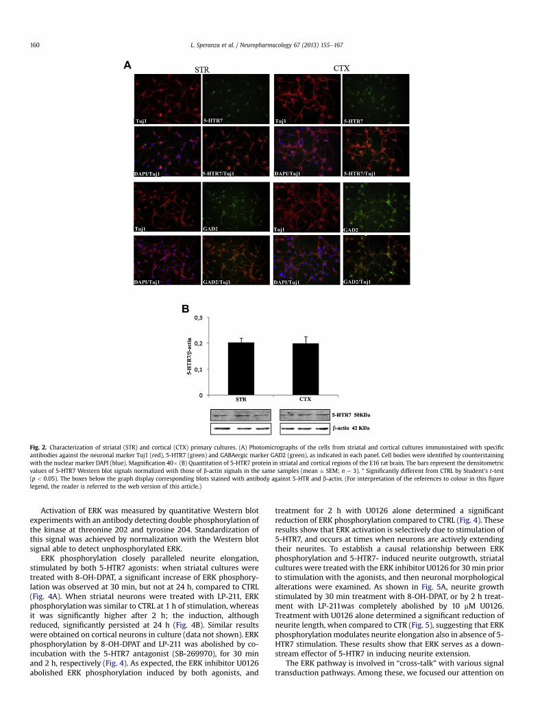
Fig. 2. Characterization of striatal (STR) and cortical (CTX) primary cultures. (A) Photomicrographs of the cells from striatal and cortical cultures immunostained with specificantibodies against the neuronal marker Tuj1 (red), 5-HTR7 (green) and GABAergic marker GAD2 (green), as indicated in each panel. Cell bodies were identified by counterstainingwith the nuclear marker DAPI (blue). Magnification 40� (B) Quantitation of 5-HTR7 protein in striatal and cortical regions of the E16 rat brain. The bars represent the densitometricvalues of 5-HTR7 Western blot signals normalized with those of b-actin signals in the same samples (mean � SEM; n ¼ 3). * Significantly different from CTRL by Student’s t-test(p < 0.05). The boxes below the graph display corresponding blots stained with antibody against 5-HTR and b-actin. (For interpretation of the references to colour in this figurelegend, the reader is referred to the web version of this article.)
L. Speranza et al. / Neuropharmacology 67 (2013) 155e167160
Activation of ERK was measured by quantitative Western blotexperiments with an antibody detecting double phosphorylation ofthe kinase at threonine 202 and tyrosine 204. Standardization ofthis signal was achieved by normalization with the Western blotsignal able to detect unphosphorylated ERK.
ERK phosphorylation closely paralleled neurite elongation,stimulated by both 5-HTR7 agonists: when striatal cultures weretreated with 8-OH-DPAT, a significant increase of ERK phosphory-lation was observed at 30 min, but not at 24 h, compared to CTRL(Fig. 4A). When striatal neurons were treated with LP-211, ERKphosphorylation was similar to CTRL at 1 h of stimulation, whereasit was significantly higher after 2 h; the induction, althoughreduced, significantly persisted at 24 h (Fig. 4B). Similar resultswere obtained on cortical neurons in culture (data not shown). ERKphosphorylation by 8-OH-DPAT and LP-211 was abolished by co-incubation with the 5-HTR7 antagonist (SB-269970), for 30 minand 2 h, respectively (Fig. 4). As expected, the ERK inhibitor U0126abolished ERK phosphorylation induced by both agonists, and
treatment for 2 h with U0126 alone determined a significantreduction of ERK phosphorylation compared to CTRL (Fig. 4). Theseresults show that ERK activation is selectively due to stimulation of5-HTR7, and occurs at times when neurons are actively extendingtheir neurites. To establish a causal relationship between ERKphosphorylation and 5-HTR7- induced neurite outgrowth, striatalcultures were treated with the ERK inhibitor U0126 for 30min priorto stimulation with the agonists, and then neuronal morphologicalalterations were examined. As shown in Fig. 5A, neurite growthstimulated by 30 min treatment with 8-OH-DPAT, or by 2 h treat-ment with LP-211was completely abolished by 10 mM U0126.Treatment with U0126 alone determined a significant reduction ofneurite length, when compared to CTR (Fig. 5), suggesting that ERKphosphorylationmodulates neurite elongation also in absence of 5-HTR7 stimulation. These results show that ERK serves as a down-stream effector of 5-HTR7 in inducing neurite extension.
The ERK pathway is involved in “cross-talk” with various signaltransduction pathways. Among these, we focused our attention on

Fig. 3. Neurite elongation induced by 5-HTR7 stimulation in cortical neurons plated at different cell densities. Neurons dissociated from E16 cortex were plated at 1.5 � 105/cm2 (A),1 � 105/cm2 (B) and 7.5 � 104/cm2 (C), and collected 2 h after the treatment with the 5-HTR agonist LP-211 (LP, 100 nM). The graphs show the neurite length expressed as percentvalues in reference to the control values (CTRL). The bars represent means � SEM from randomly selected fields for each cell culture condition (n ¼ 8). The pixel values of meanneurite length (m. n. l.) for CTRL and LP-211 treated cultures are indicated in the table below each graph. * Significantly different from CTRL by Student’s t-test (p < 0.05).
L. Speranza et al. / Neuropharmacology 67 (2013) 155e167 161
a serine/threonine protein kinase, the cyclin-dependent proteinkinase 5 (Cdk5), that is highly expressed in the brain, and playsa key role in the regulation of microtubule dynamics and cyto-skeletal reorganization necessary for neurite elongation (Nicolicet al., 1996; Cheung and Ip, 2007).
To evaluate the role of Cdk5 on neurite outgrowth induced by 5-HTR7 activation, we used roscovitine, a potent inhibitor of Cdkswith limited selectivity between the Cdk5 and Cdk2 (Otyepka et al.,2006). However, in the Cdk family, Cdk5 is the only member activein post-mitotic terminally differentiated neurons (Otyepka et al.,2006), as those present in our cultures. Interestingly, treatment ofneuronal striatal cultures with 20 mM roscovitine completely abol-ished neurite elongation induced by 2 h of treatment with 100 nMLP-211. Treatment of roscovitine alone for 2 h did not affect neuritelength, compared to CTR (Fig. 6A). Similar resultswere obtained alsowithmouse neuronal primary cultures (see Supplementary Fig. S2).To further exclude the involvement of Cdk2 in the observed effects,we analyzed the gene expression level of Cdk5 and Cdk2 by real-time quantitative RT-PCR analyses in rat striatal cultures. Asshown in Supplementary Fig. S3, we observed that Cdk5 transcriptsare approximately 4 fold more abundant than Cdk2 transcripts.Thus in our experimental conditions roscovitine can be considereda selective inhibitor of Cdk5 (Kim and Ryan, 2010).
To analyze the possible interaction between ERK and Cdk5pathways, we measured ERK phosphorylation by quantitativeWestern blot experiments, in cultures treated with LP-211 with orwithout roscovitine. Interestingly, the co-treatment abolished ERKphosphorylation stimulated by LP-211 (Fig. S4). All together, theseresults suggest that, in our culture conditions, neurite elongation
stimulated by activation of 5-HTR7 requires ERK phosphorylationvia Cdk5 activation.
3.3. Neurite elongation stimulated by 5-HTR7 requires de novoprotein synthesis and changes on the expression of cytoskeletalproteins
To deepen our understanding of the biochemical machinery thatunderlies the 5-HTR7-stimulated neurite outgrowth we investi-gated whether de novo protein synthesis was required. To this aim,neuronal striatal cultures were treated with LP-211 with or without1 h pre-treatment with cycloheximide (CHX, an inhibitor ofeukaryotic protein synthesis), and examined by morphometricanalyses. As shown in Fig. 6B, neurite growth stimulated by 2 htreatment with LP-211 was completely abolished by co-incubationwith 50 mM CHX. Treatment of CHX alone for 2 h did not affectneurite elongation, in comparison with the CTRL.
We thus asked whether activation of the 5-HTR7 by LP-211modified the main components of the neuronal cytoskeleton.Therefore we evaluated the expression of selected genes, such as b-III-tubulin, Vimentin, light and medium neurofilament subunits(NFL and NFM, respectively) and selected microtubule associatedproteins (MAPs) by real-time quantitative RT-PCR analyses. Inter-estingly, significant variation between CTRL and agonist-stimulatedstriatal neurons (LP-211 for 2 h) was observed only for MAP1Btranscripts, that were strongly up-regulated in striatal culturestreated with the agonist (Fig. 7).
Although RT-PCR analysis provides insight on gene expression,changes occurring at the translational and post-translational levels

Fig. 4. Agonist-induced activation of 5-HTR7 stimulates ERK phosphorylation. (A) Striatal cultures were treated either with 5-HTR7 agonist 8-OH-DPAT (8-OH, 100 nM, for 30 minand 24 h) alone, or in combination with the selective 5-HTR7 antagonist SB-269970 (SB, 100 nM, for 30 min). Cells were also treated with the selective ERK inhibitor U0126 (U0,10 mM) with or without 8-OH-DPAT for 30 min (B) Cells were treated either with the selective 5-HTR7 agonist LP-211 (LP, 100 nM, for 1 h, 2 h and 24 h) alone, or in combination withthe selective 5-HTR7 antagonist SB-269970 (100 nM, for 2 h). Cells were also treated with the selective ERK inhibitor U0126 (10 mM) with or without LP-211 for 2 h (A, B) The level ofERK phosphorylation was measured as intensity of phosphorylated ERK (p-ERK1/2) normalized with that of unphosphorylated ERK (ERK1/2) in the same samples. ERK phos-phorylation (means � SEM n ¼ 3) was expressed as percentage of values measured in the corresponding vehicle-treated control cultures (CTRL, set to 100%). * Significantly differentfrom CTRL by Student’s t-test (p < 0.05). The boxes below each graph display representative blots probed with antibody against p-ERK1/2 and ERK1/2.
L. Speranza et al. / Neuropharmacology 67 (2013) 155e167162
are not detected. Therefore, in parallel we analyzed the expressionprofiles of the same neuronal cytoskeleton proteins byWestern blotanalysis coupled to 2D-PAGE. The latter enabled us to obtaina higher resolution profile of these proteins and to evaluate possibleposttranslational modifications. Immunostaining of 2D-gels ofCTRL striatal cultures showed a marked stretching of b-III-tubulinspots (Fig. 8A), attesting a high level of tubulin heterogeneity.Interestingly, an overall down-regulation of b-III-tubulin wasrevealed in LP-211-treated cultures, compared to CTRL (Fig. 8A).This reduction involved mostly the stretching of acidic tubulinspots, resulting in a marked reduction of b-III-tubulin heteroge-neity in stimulated cells compared to CTRL. On the contrary, noexpression changes of Vimentin isoforms were observed betweenLP-211 and vehicle-treated control (Fig. 8A). Similarly, no qualita-tive and quantitative expression changes were revealed for themain form at w68 kDa of NFL (Fig. 8B). However, a protein species
with a slightly lower molecular weight that migrated as a satellitespot of NFL disappeared in LP-211 treated cells (Fig. 8B). On theother hand, a down-regulation of the main component at 145 kDaof NFM was detected in LP-211-treated cultures, in comparison toCTRL (Fig. 8B). On the contrary, a slight up-regulation of the mainisoform of MAP2 (70 kDa) and of a satellite spot at acidic pI wasobserved in LP-211 -treated cells together with the appearance ofa lower molecular weight form (Fig. 8C). Moreover, the high level ofheterogeneity of MAP1B with several high molecular weight iso-forms (ranging from 270 to 380 kDa) detected in control cultures,completely disappeared in LP-211-stimulated cells, whereas the270 kDa isoform was slightly up-regulated (Fig. 8C). Notably, inthese cultures an evident qualitative change was detectedregarding the appearance of an intense spot with molecular weightof about 34 kDa (Fig. 8C). This form corresponds to the MAP1B lightchain (34 kDa), thought to play a role in regulating the neuronal

Fig. 6. Neurite elongation induced by 5-HTR7 stimulation in striatal primary culturesdepends on Cdk5 activation and newly synthesized proteins. (A) Striatal neurons weretreated for 2 h either with the 5-HTR7 selective agonist LP-211 (LP, 100 nM), or withthe Cdk5 inhibitor roscovitine (ROSC, 20 mM), or with a combination of the two. (B)Striatal neurons were treated for 2 h either with the selective 5-HTR agonist LP-211(100 nM), or with the eukaryotic protein synthesis inhibitor cycloheximide (CHX,50 mM) or with a combination of the two. Percent values of the neurite length(means � SEM from randomly selected fields for each cell culture condition, n ¼ 8)were calculated in reference to the control values (CTRL). * Significantly different fromCTRL by Student’s t-test (p < 0.05).
Fig. 5. ERK phosphorylation is required for neurite elongation induced by 5-HTR7stimulation in neuronal primary cultures. (A) Striatal cells were treated for 30 mineither with the 5-HTR7 agonist 8-OH-DPAT (8-OH, 100 nM), or with the selective ERKinhibitor U0126 (U0, 10 mM), or with a combination of the two. (B) Cells were treatedfor 2 h either with the selective 5-HTR7 agonist LP-211 (LP, 100 nM), or with U0126(10 mM), or with a combination of the two. The graphs show the neurite lengthexpressed as percent values in reference to the control values (CTRL). The barsrepresent means � SEM from randomly selected fields for each cell culture condition(n ¼ 8). * Significantly different from CTRL by Student’s t-test (p < 0.05).
L. Speranza et al. / Neuropharmacology 67 (2013) 155e167 163
cytoskeleton by binding to microtubules and inducing tubulinpolymerization (Kuznetsov et al., 1986; Togel et al., 1998).
4. Discussion and conclusions
In neural development, neuronal precursors differentiate,migrate, extend long processes, and finally establish connectionswith their targets. Over the past years, the initial steps of neuriteformation have been widely studied using primarily in vitroapproaches such as dissociated culture of rodent CNS neurons.Using these models important progress has beenmade towards theidentification of the physiological signals and molecular mecha-nisms regulating neurite outgrowth (Arimura and Kaibuchi, 2007;Calabrese, 2008). Along this line, our data show that the endoge-nous 5-HTR7 strongly stimulates neurite elongation in culturedembryonic neurons from rodent striatal complex and cortex. Aselective role of the 5-HTR7 in promoting neurite outgrowth wasconfirmed by specific agonists and by the inhibition of the effects inpresence of a selective 5-HTR7 antagonist (SB-269970; Hagan et al.,2000). Moreover, we could exclude the involvement of 5-HT1Areceptor, since its selective antagonist (WAY-100635; Corradettiet al., 1998) did not alter agonists-induced neurite elongation.
Other parameters, such as the number of primary neurites, aswell as the soma perimeter and the total area of each neuron, werenot affected by agonist treatment, in accordance with the hypoth-esis that mechanisms underlying neurite length determination aredifferent than those controlling other morphological aspects(Shelly et al., 2010).
The stimulation of neurite outgrowth was achieved using thecommonly used promiscuous agonist 8-OH-DPAT and the noveland highly potent selective agonist, LP-211 (Hedlund et al., 2010).Interestingly the effect of LP-211 on neurite outgrowth was morepersistent than that of 8-OH-DPAT, but required more time (2 h) tobe evident. This difference could be due to the action of the twoagonists on different sites of the receptor, which in turn couldactivate the same signal transduction pathways (see below) witha different kinetic.
A role for the 5-HTR7 in the regulation of neuronal morphologyis consistent with previous results obtained in mouse hippocampalneurons in vitro (Kvachnina et al., 2005), and extends our prelim-inary observations on rat striatal cultures (Leo et al., 2009). Alltogether, these results demonstrate that this receptor plays a keyrole in stimulating neurite outgrowth from different neuronsin vitro and suggest its involvement in CNS connectivity. Indeed,a prominent role for the 5-HTR7 in the morphological and func-tional maturation of hippocampal circuits is supported by recentdata by Kobe and coworkers. These authors show that the activa-tion of 5-HTR7 stimulates formation of dendritic spines andneuronal excitability in organotypic preparations from the hippo-campus of juvenile mice, while in preparations from older micewhere the expression of the receptor was down-regulated, theeffect was not present (Kobe et al., 2012). Therefore it is possiblethat critical developmental stages for the modulation of neuronalcircuits depend from the variable expression and activation of 5-HTR7. In addition, psychostimulant administration to adolescentrats triggered a strong and persistent up-regulation of the 5-HTR7in discrete regions of the reward circuits, which paralleled withstructural modifications of synaptic contacts and alterations ofimpulsive behavior, as we showed in previous experiments. Inthese animals, 5-HTR7 modulation caused alterations of reward-based behavior (Cavaliere et al., 2012; Leo et al., 2009).
Our new findings that ERK phosphorylation is required for5-HTR7-dependent neurite elongation are consistent with theknowledge that 5-HTR7 can be functionally coupled to Gs or Ga-12 G-proteins, which in turn can activate ERK (Errico et al., 2001;

Fig. 7. Effect of LP-211 on expression levels of several mRNAs for cytoskeletal proteins in striatal neurons. Striatal cells were collected 2 h after the treatment with the selective5-HTR7 agonist LP-211 (LP, 100 nM). Expression levels of selected mRNAs were determined by real time RT-PCR using specific primers for b-III-tubulin, NF-M, Vimentin, MAP2, andMAP1b. The mRNA levels were normalized with those of the housekeeping gene HPRT (see Methods for details). The bars represent means � SEM (n ¼ 4 for treated samples andn ¼ 3 for CTRL). * Significantly different from CTRL by Student’s t-test (p < 0.05).
L. Speranza et al. / Neuropharmacology 67 (2013) 155e167164
Kvachnina et al., 2005; Lin et al., 2003). It is likely that the activationof the Ga-12 subunit is involved in ERK-dependent neuriteoutgrowth also in our cultures, since in hippocampal neurons fromGa-12-deficient mice the 5-HTR7-mediated effects do not occur(Kobe et al., 2012). It is noteworthy that neurite elongationmirrored the time course of ERK phosphorylation stimulated byeither 8-OH-DPAT or LP-211.
With this work we also unravel the essential role of Cdk5activity in 5-HTR7-dependent neurite elongation in CNS primarycultures, supporting literature data on Cdk5 involvement in neu-rite outgrowth and remodeling (Nikolic et al., 1996; Zhang et al.,2010). To this aim, we used roscovitine, a potent inhibitor ofCdks with limited selectivity between the Cdk5 and Cdk2(Otyepka et al., 2006). However, in our cultures consisting of fullydifferentiated neurons, roscovitine can be considered a selective
inhibitor of Cdk5 (Kim and Ryan, 2010). In addition, we observedthat Cdk5 transcripts were approximately 4 fold more abundantthan Cdk2 transcripts. Indeed, this kinase, differently from otherknown cyclin-dependent family members, is active in non-proliferating cells and is highly expressed in differentiatedneurons, where it can phosphorylate various proteins of theneuronal cytoskeleton. Cdk5 plays important roles in variousaspects of neural development including neuronal migration, axonguidance and regeneration, synapse development, and plasticity(Cheung and Ip, 2007; Lai and Ip, 2009). Its activity is dependentupon association with the p35 cofactor. Interestingly, mice withdysregulation of Cdk5/p35 activity during development displayimproperly established meso-cortico-limbic circuitry and a loco-motor profile and pharmacological responses reminiscent ofADHD (Drerup et al., 2010), a well known developmental disease

Fig. 8. Effect of LP-211 on expression levels of several cytoskeletal proteins in striatal neurons. 2D immunoblot analyses were performed on striatal neurons treated with the 5-HTR7selective agonist LP-211 and on vehicle-treated cultures (CTRL). A, b-III-tubulin and Vimentin; B, low- (NFL) and middle- (NFM) molecular weight neurofilaments; C, microtubule-associated proteins (MAP2 and MAP1B). The molecular weights (kDa) are indicated on the left. Arrows represent qualitatively different spots between CTRL and LP-211 treatedcultures.
L. Speranza et al. / Neuropharmacology 67 (2013) 155e167 165
associated to altered connectivity of neural circuits (Liston et al.,2011). The involvement of altered connectivity in ADHD hasbeen supported by independent genome-wide association studiesshowing that top-ranked ADHD candidate genes, including ERK
and MAP1B, encode proteins regulating neurite outgrowth(Poelmans et al., 2011).
As expected, 5-HTR7-dependent neurite elongation in CNSprimary cultures was accompanied by qualitative and quantitative

L. Speranza et al. / Neuropharmacology 67 (2013) 155e167166
expression changes on selected components of the neuronal cyto-skeleton (i.e. b-III-tubulin, Vimentin, and selected neurofilamentsandmicrotubule associated proteins). In particular, the high level ofheterogeneity of b-tubulin observed in untreated striatal neuronsmatched previous data reporting up to 21 isoelectric variants of thisprotein in the brain and in cultured neurons, mainly due to variablenumber of phosphates that cause variable shifts towards acidic pI(Edde et al., 1989). The absence of stretching of acidic tubulin spotsin cultures treated with the 5-HTR7 agonist suggests a markedreduction of phosphorylated isoforms. This observation confirmsthat neurite elongation requires changes in cytoskeleton functionand dynamics, depending on post-translational modificationsof key proteins (Fukushima et al., 2009). In addition, the down-regulation of the NFM main component and the up-regulationof MAP2 main isoform in stimulated cultures highlights a roleof 5-HTR7 in modulating the amount of proteins involved inneurite outgrowth. On the contrary, no relevant expression changesof spots corresponding to vimentin isoforms or to NFL wereobserved.
The most striking expression changes induced by 5-HTR7 stim-ulation were observed for MAP1B, a protein essential to regulatemicrotubule dynamics, thus playing an important role in dendriticspine formation, synaptic maturation and axonal elongation (Blacket al., 1994; Tortosa et al., 2011; Tymanskyi et al., 2012). MAP1B ispresent as a full length protein, or may be fragmented into a heavychain and a light chain, and its function is modulated by phos-phorylation (Riederer, 2007). The high molecular weight isoforms(above 270 up to 380 kDa) identified in CTRL cultures completelydisappeared in LP-211-treated cultures, whereas the 270 kDa formwas slightly up-regulated. Interestingly, stimulation of the 5-HTR7was able to selectively induce the appearance of a 34 kDa proteincorresponding to the MAP1B light chain (34 kDa; Kuznetsov et al.,1986). This protein plays a key role in regulating the cross-talkbetween microtubules and actin microfilaments during neuriteelongation (Togel et al., 1998). Therefore our findings stronglysuggest that MAP1B light chain may act as an important effector inenhancing neurite outgrowth stimulated by 5-HTR7.
The up-regulation of MAPs in agonist-stimulated neurons isconsistent with the finding that 5-HTR7-dependent neurite elon-gation requires de novo protein synthesis, and with their syner-gistic role in regulating the dynamics of the cytoskeleton inneuronal processes (Matus, 1991). Although the deeper character-ization of modifications of neuronal cytoskeletal proteins is beyondthe scope of this work, our preliminary results suggest the occur-rence of qualitative and quantitative expression changes due to 5-HTR7 stimulation.
In conclusion, our work for the first time shows a direct linkbetween 5-HTR7 activation and enhanced neurite outgrowthmediated by the activation of ERK and Cdk5 intracellular pathways,with the induction of MAP1B and the involvement of other cyto-skeletal proteins, at least in neuronal primary cultures. Future workwill address the role of the 5-HTR7 in the modulation of neuronalconnectivity in vivo.
Acknowledgments
This work was supported by “Fondo per gli Investimenti diRicerca di Base” FIRB-RBIN062YH4, “Medical Research Italy’’MERIT-RBNE08LN4P, “Progetti di Ricerca di Interesse Nazionale’’ PRIN-2009TBCZJB_003, and Ministero della Salute, Under-40 2007. Wethank Dr. W. Adriani, P.I. of the latter project. The funding sourceshad no role in study design, data collection and analysis, decision topublish, or preparation of the manuscript. We are grateful to theIntegrated Microscopy Facility of the Institute of Genetics andBiophysics “Adriano Buzzati Traverso”, CNR, Naples, IT.
Appendix A. Supplementary data
Supplementary data related to this article can be found at http://dx.doi.org/10.1016/j.neuropharm.2012.10.026.
References
Arimura, N., Kaibuchi, K., 2007. Neuronal polarity: from extracellular signals tointracellular mechanisms. Nat. Rev. Neurosci. 8, 194e205.
Barnes, N.M., Sharp, T., 1999. A review of central 5-HT receptors and their function.Neuropharmacol. 38, 1083e1152.
Black, M.M., Slaughter, T., Fischer, I., 1994. Microtubule-associated protein 1b(MAP1b) is concentrated in the distal region of growing axons. J. Neurosci. 14,857e870.
Bonaventure, P., Kelly, L., Aluisio, L., Shelton, J., Lord, B., Galici, R., Miller, K., Atack, J.,Lovenberg, T.W., Dugovic, C., 2007. Selective blockade of 5-hydroxytryptamine(5-HT)7 receptors enhances 5-HT transmission, antidepressant-like behavior,and rapid eye movement sleep suppression induced by citalopram in rodents.J. Pharmacol. Exp. Ther. 321, 690e698.
Buchser, W.J., Slepak, T.I., Gutierrez-Arenas, O., Bixby, J.L., Lemmon, V.P., 2010.Kinase/phosphatase overexpression reveals pathways regulating hippocampalneuron morphology. Mol. Syst. Biol. 6, 391.
Calabrese, E.J., 2008. Enhancing and regulating neurite outgrowth. Crit. Rev. Toxicol.38, 391e418.
Cavaliere, C., Cirillo, G., Bianco, M.R., Adriani, W., De Simone, A., Leo, D., Perrone-Capano, C., Papa, M., 2012. Methylphenidate administration determinesenduring changes in neuroglial network in rats. Eur. Neuropsychopharmacol.22, 53e63.
Cheung, Z.H., Ip, N.Y., 2007. The roles of cyclin-dependent kinase 5 in dendrite andsynapse development. Biotechnol. J. 2, 949e957.
Cifariello, A., Pompili, A., Gasbarri, A., 2008. 5-HT(7) receptors in the modulation ofcognitive processes. Behav. Brain Res. 195, 171e179.
Colucci-D’Amato, G.L., Perrone Capano, C., di Porzio, U., 2003. Chronic activation ofERK and neurodegenerative diseases. BioEssays 25, 1085e1095.
Corradetti, R., Laaris, N., Hanoun, N., Laporte, A.M., Le Poul, E., Hamon, M.,Lanfumey, L., 1998 Feb. Antagonist properties of (-)-pindolol and WAY 100635at somatodendritic and postsynaptic 5-HT1A receptors in the rat brain. Br. J.Pharmacol. 123 (3), 449e462.
di Porzio, U., Daguet, M.C., Glowinski, J., Prochiantz, A., 1980. Effect of striatal cellson in vitro maturation of mesencephalic DA neurons grown in serum-freeconditions. Nature 288, 370e373.
Drerup, J.M., Hayashi, K., Cui, H., Mettlach, G.L., Long, M.A., Marvin, M., Sun, X.,Goldberg, M.S., Lutter, M., Bibb, J.A., 2010. Attention-deficit/hyperactivityphenotype in mice lacking the cyclin-dependent kinase 5 cofactor p35. Biol.Psychiatry 68, 1163e1171.
Edde, B., Denoulet, P., de Nechaud, B., Koulakoff, A., Berwald-Netter, Y., Gros, F., 1989.Posttranslational modifications of tubulin in cultured mouse brain neurons andastroglia. Biol. Cell. 65, 109e117.
Eriksson, T.M., Golkar, A., Ekström, J.C., Svenningsson, P., Ogren, S.O., 2008. 5-HT7receptor stimulation by 8-OH-DPAT counteracts the impairing effect of 5-HT(1A) receptor stimulation on contextual learning in mice. Eur. J. Pharmacol.596, 107e110.
Errico, M., Crozier, R.A., Plummer, M.R., Cowen, D.S., 2001. 5-HT(7) receptors acti-vate the mitogen activated protein kinase extracellular signal related kinase incultured rat hippocampal neurons. Neuroscience 102, 361e367.
Faure, C., Mnie-Filali, O., Scarna, H., Debonnel, G., Haddjeri, N., 2006. Effects of the 5-HT7 receptor antagonist SB-269970 on rat hormonal and temperatureresponses to the 5-HT1A/7 receptor agonist 8-OH-DPAT. Neurosci. Lett. 404,122e126.
Fiszman, M.L., Zuddas, A., Masana, M.I., Barker, J.L., di Porzio, U., 1991. Dopaminesynthesis precedes dopamine uptake in embryonic rat mesencephalic neurons.J. Neurochem. 56, 392e399.
Fukushima, N., Furuta, D., Hidaka, Y., Moriyama, R., Tsujiuchi, T., 2009. Post-trans-lational modifications of tubulin in the nervous system. J. Neurochem. 109,683e693.
Gasbarri, A., Cifariello, A., Pompili, A., Meneses, A., 2008. Effect of 5-HT(7) antago-nist SB-269970 in the modulation of working and reference memory in the rat.Behav. Brain Res. 195, 164e170.
Guscott, M., Bristow, L.J., Hadingham, K., Rosahl, T.W., Beer, M.S., Stanton, J.A.,Bromidge, F., Owens, A.P.,Huscroft, I.,Myers, J., Rupniak,N.M., Patel, S.,Whiting, P.J.,Hutson, P.H., Fone, K.C., Biello, S.M., Kulagowski, J.J., McAllister, G., 2005. Geneticknockout and pharmacological blockade studies of the 5-HT7 receptor suggesttherapeutic potential in depression. Neuropharmacol. 48, 492e502.
Hagan, J.J., Price, G.W., Jeffrey, P., Deeks, N.J., Stean, T., Piper, D., Smith, M.I.,Upton, N., Medhurst, A.D., Middlemiss, D.N., Riley, G.J., Lovell, P.J.,Bromidge, S.M., Thomas, D.R., 2000. Characterization of SB-269970-A, a selec-tive 5-HT7 receptor antagonist. Br. J. Pharmacol. 130, 539e548.
Hedlund, P.B., Danielson, P.E., Thomas, E.A., Slanina, K., Carson, M.J., Sutcliffe, J.G.,2003. No hypothermic response to serotonin in 5-HT7 receptor knockout mice.Proc. Natl. Acad. Sci. U S A 100, 1375e1380.
Hedlund, P.B., Kelly, L., Mazur, C., Lovenberg, T., Sutcliffe, J.G., Bonaventure, P., 2004.8-OH-DPAT acts on both 5-HT1A and 5-HT7 receptors to induce hypothermia inrodents. Eur. J. Pharmacol. 487, 125e132.
L. Speranza et al. / Neuropharmacology 67 (2013) 155e167 167
Hedlund, P.B., Sutcliffe, J.G., 2004. Functional, molecular and pharmacologicaladvances in 5-HT7 receptor research. Trends Pharmacol. Sci. 25, 481e486.
Hedlund, P.B., Huitron-Resendiz, S., Henriksen, S.J., Sutcliffe, J.G., 2005. 5-HT7receptor inhibition and inactivation induce antidepressantlike behavior andsleep pattern. Biol. Psychiatry 58, 831e837.
Hedlund, P.B., 2009. The 5-HT7 receptor and disorders of the nervous system: anoverview. Psychopharmacol. (Berl) 206, 345e354.
Hedlund, P.B., Leopoldo, M., Caccia, S., Sarkisyan, G., Fracasso, C., Martelli, G.,Lacivita, E., Berardi, F., Perrone, R., 2010. LP-211 is a brain penetrant selectiveagonist for the serotonin 5-HT(7) receptor. Neurosci. Lett. 481, 12e16.
Kim, S.H., Ryan, T.A., 2010. CDK5 serves as a major control point in neurotransmitterrelease. Neuron 67, 797e809.
Kobe, F., Guseva, D., Jensen, T.P., Wirth, A., Renner, U., Hess, D., Müller, M.,Medrihan, L., Zhang, W., Zhang, M., Braun, K., Westerholz, S., Herzog, A.,Radyushkin, K., El-Kordi, A., Ehrenreich, H., Richter, D.W., Rusakov, D.A.,Ponimaskin, E., 2012. 5-HT7R/G12 signaling regulates neuronal morphologyand function in an age-dependent manner. J. Neurosci. 32, 2915e2930.
Kuznetsov, S.A., Rodionov, V.I., Nadezhdina, E.S., Murphy, D.B., Gelfand, V.I., 1986.Identification of a 34-kD polypeptide as a light chain of microtubule-associatedprotein-1 (MAP-1) and its association with a MAP-1 peptide that binds tomicrotubules. J. Cell. Biol. 102, 1060e1066.
Kvachnina, E., Liu, G., Dityatev, A., Renner, U., Dumuis, A., Richter, D.W., Dityateva, G.,Schachner, M., Voyno-Yasenetskaya, T.A., Ponimaskin, E.G., 2005. 5-HT7 receptoris coupled to Ga-subunits of heterotrimeric G12-protein to regulate genetranscription and neuronal morphology. J. Neurosci. 25, 7821e7830.
Lai, K.O., Ip, N.Y., 2009. Recent advances in understanding the roles of Cdk5 insynaptic plasticity. Biochim. Biophys. Acta 1792, 741e745.
Leo, D., Adriani, W., Cavaliere, C., Cirillo, G., Marco, E.M., Romano, E., di Porzio, U.,Papa, M., Perrone-Capano, C., Laviola, G., 2009. Methylphenidate to adolescentrats drives enduring changes of accumbal Htr7 expression: implications forimpulsive behavior and neuronal morphology. Genes Brain Behav. 8, 356e368.
Leopoldo, M., Lacivita, E., Berardi, F., Perrone, R., Hedlund, P.B., 2011. Serotonin 5-HT7 receptor agents: structure-activity relationships and potential thera-peutic applications in central nervous system disorders. Pharmacol. Ther. 129,120e148.
Lin, S.L., Johnson-Farley, N.N., Lubinsky, D.R., Cowen, D.S., 2003. Coupling ofneuronal 5-HT7 receptors to activation of extracellular-regulated kinasethrough a protein kinase A-independent pathway that can utilize Epac. J. Neu-rochem. 87, 1076e1085.
Liston, C., Malter Cohen, M., Teslovich, T., Levenson, D., Casey, B.J., 2011. Atypicalprefrontal connectivity in attention-deficit/hyperactivity disorder: pathway todisease or pathological end point? Biol. Psychiatry 69, 1168e1177.
Matthys, A., Haegeman, G., Van Craenenbroeck, K., Vanhoenacker, P., 2011. Role ofthe 5-HT7 receptor in the central nervous system: from current status to futureperspectives. Mol. Neurobiol. 43, 228e253.
Matus, A., 1991. Microtubule-associated proteins and neuronal morphogenesis.J. Cell. Sci. Suppl. 15, 61e67.
Mnie-Filali, O., Dahan, L., Zimmer, L., Haddjeri, N., 2007. Effects of the serotonin 5-HT(7) receptor antagonist SB-269970 on the inhibition of dopamine neuronalfiring induced by amphetamine. Eur. J. Pharmacol. 570, 72e76.
Mnie-Filali, O., Faure, C., Lambás-Señas, L., El Mansari, M., Belblidia, H., Gondard, E.,Etiévant, A., Scarna, H., Didier, A., Berod, A., Blier, P., Haddjeri, N., 2011. Phar-macological blockade of 5-HT7 receptors as a putative fast acting antidepres-sant strategy. Neuropsychopharmacol 36, 1275e1288.
Mullins, U.L., Gianutsos, G., Eison, A.S., 1999. Effects of antidepressants on 5-HT7receptor regulation in therathypothalamus.Neuropsychopharmacol21, 352e367.
Nandam, L.S., Jhaveri, D., Bartlett, P., 2007. 5-HT7, neurogenesis and antidepres-sants: a promising therapeutic axis for treating depression. Clin. Exp. Phar-macol. Physiol. 34, 546e551.
Nikolic, M., Dudek, H., Kwon, Y.T., Ramos, Y.F.M., Tsai, L.H., 1996. The cdk5/p35kinase is essential for neurite outgrowth during neuronal differentiation. GenesDev. 10, 816e825.
Otyepka, M., Bártová, I., Kríz, Z., Koca, J., 2006. Different mechanisms of CDK5 andCDK2 activation as revealed by CDK5/p25 and CDK2/cyclin a dynamics. J. Biol.Chem. 281, 7271e7281.
Poelmans, G., Pauls, D.L., Buitelaar, J.K., Franke, B., 2011. Integrated genome-wideassociation study findings: identification of a neurodevelopmental networkfor attention deficit hyperactivity disorder. Am. J. Psychiatry 168, 365e377.
Ponimaskin, E., Voyno-Yasenetskaya, T., Richter, D.W., Schachner, M., Dityatev, A.,2007. Morphogenic signaling in neurons via neurotransmitter receptors andsmall GTPases. Mol. Neurobiol. 35, 278e287.
Pytliak, M., Vargová, V., Mechírová, V., Fel�söci, M., 2011. Serotonin receptors e frommolecular biology to clinical applications. Physiol. Res. 60, 15e25.
Renner, U., Zeug, A., Woehler, A., Niebert, M., Dityatev, A., Dityateva, G., Gorinski, N.,Guseva, D., Abdel-Galil, D., Fröhlich, M., Döring, F., Wischmeyer, E., Richter, D.W.,Neher, E., Ponimaskin, E.G., 2012. Heterodimerization of serotonin receptors5-HT1A and 5-HT7 differentially regulates receptor signalling and trafficking.J. Cell. Sci. 125, 2486e2499.
Riederer, B.M., 2007. Microtubule-associated protein 1B, a growth-associated andphosphorylated scaffold protein. Brain Res. Bull. 71, 541e558.
Roth, B.L., Craigo, S.C., Choudhary, M.S., Uluer, A., Monsma Jr., F.J., Shen, Y.,Meltzer, H.Y., Sibley, D.R., 1994. Binding of typical and atypical antipsychoticagents to 5-hydroxytryptamine-6 and 5-hydroxytryptamine-7 receptors.J. Pharmacol. Exp. Ther. 268, 1403e1410.
Sarkisyan, G., Hedlund, P.B., 2009. The 5-HT7 receptor is involved in allocentricspatial memory information processing. Behav. Brain Res. 202, 26e31.
Schmittgen, T.D., Livak, K.J., 2008. Analyzing real-time PCR data by the comparativeC(T) method. Nat. Protoc. 3, 1101e1108.
Severino, V., Chambery, A., Vitiello, M., Cantisani, M., Galdiero, S., Galdiero, M.,Malorni, L., Di Maro, A., Parente, A., 2010. Proteomic analysis of human U937cell line activation mediated by Haemophilus influenzae type b P2 porin and itssurface-exposed loop 7. J. Proteome Res. 9, 1050e1062.
Shelly, M., Lim, B.K., Cancedda, L., Heilshorn, S.C., Gao, H., Poo, M.M., 2010. Local andlong-range reciprocal regulation of cAMP and cGMP in axon/dendrite forma-tion. Science 327, 547e552.
Teitler, M., Klein, M.T., 2012. A new approach for studying GPCR dimers: drug-induced inactivation and reactivation to reveal GPCR dimer function in vitro,in primary culture, and in vivo. Pharmacol. Ther. 133, 205e217.
Thomas, D.R., Melotto, S., Massagrande, M., Gribble, A.D., Jeffrey, P., Stevens, A.J.,Deeks, N.J., Eddershaw, P.J., Fenwick, S.H., Riley, G., Stean, T., Scott, C.M.,Hill, M.J., Middlemiss, D.N., Hagan, J.J., Price, G.W., Forbes, I.T., 2003. SB-656104-A, a novel selective 5-HT7 receptor antagonist, modulates REM sleep in rats. Br.J. Pharmacol. 139, 705e714.
Tögel, M., Wiche, G., Propst, F., 1998. Novel features of the light chain ofmicrotubule-associated protein MAP1B: microtubule stabilization, self inter-action, actin filament binding, and regulation by the heavy chain. J. Cell. Biol.143, 695e707.
Tortosa, E., Montenegro-Venegas, C., Benoist, M., Härtel, S., González-Billault, C.,Esteban, J.A., Avila, J., 2011. Microtubule-associated protein 1B (MAP1B) isrequired for dendritic spine development and synaptic maturation. J. Biol.Chem. 286, 40638e40648.
Tymanskyj, S.R., Scales, T.M.E., Gordon-Weeks, P.R., 2012. MAP1B enhances micro-tubule assembly rates and axon extension rates in developing neurons. Mol.Cell. Neurosci. 49, 110e119.
Zhang, P., Yu, P.C., Tsang, A.H.K., Chen, Y., Fu, A.K.Y., Fu, W.Y., Chung, K.K., Ip, N.Y.,2010. S-nitrosylation of cyclin-dependent kinase 5 (cdk5) regulates its kinaseactivity and dendrite growth during neuronal development. J. Neurosci. 30,14366e14370.


