Embed Size (px)
Citation preview
The Skin Microbiome in Healthy and Allergic DogsAline Rodrigues Hoffmann1*, Adam P. Patterson2, Alison Diesel2, Sara D. Lawhon4, Hoai Jaclyn Ly1,
Christine Elkins Stephenson3, Joanne Mansell1, Jorg M. Steiner3, Scot E. Dowd5, Thierry Olivry6,
Jan S. Suchodolski3
1 Dermatopathology Specialty Service, Department of Veterinary Pathobiology, College of Veterinary Medicine & Biomedical Sciences, Texas A&M University, College
Station, Texas, United States of America, 2 Clinical Dermatology Service, College of Veterinary Medicine & Biomedical Sciences, Department of Small Animal Clinical
Sciences, Texas A&M University, College Station, Texas, United States of America, 3 Gastrointestinal Laboratory, Department of Small Animal Clinical Sciences, College of
Veterinary Medicine & Biomedical Sciences, Texas A&M University, College Station, Texas, United States of America, 4 Clinical Microbiology Laboratory, Department of
Veterinary Pathobiology, College of Veterinary Medicine & Biomedical Sciences, Texas A&M University, College Station, Texas, United States of America, 5 MR DNA
Laboratory, Shallowater, Texas, United States of America, 6 Department of Clinical Sciences, College of Veterinary Medicine, and Center for Comparative Medicine and
Translational Research, North Carolina State University, Raleigh, North Carolina, United States of America
Abstract
Background: Changes in the microbial populations on the skin of animals have traditionally been evaluated usingconventional microbiology techniques. The sequencing of bacterial 16S rRNA genes has revealed that the human skin isinhabited by a highly diverse and variable microbiome that had previously not been demonstrated by culture-basedmethods. The goals of this study were to describe the microbiome inhabiting different areas of the canine skin, and tocompare the skin microbiome of healthy and allergic dogs.
Methodology/Principal Findings: DNA extracted from superficial skin swabs from healthy (n = 12) and allergic dogs (n = 6)from different regions of haired skin and mucosal surfaces were used for 454-pyrosequencing of the 16S rRNA gene.Principal coordinates analysis revealed clustering for the different skin sites across all dogs, with some mucosal sites and theperianal regions clustering separately from the haired skin sites. The rarefaction analysis revealed high individual variabilitybetween samples collected from healthy dogs and between the different skin sites. Higher species richness and microbialdiversity were observed in the samples from haired skin when compared to mucosal surfaces or mucocutaneous junctions.In all examined regions, the most abundant phylum and family identified in the different regions of skin and mucosalsurfaces were Proteobacteria and Oxalobacteriaceae. The skin of allergic dogs had lower species richness when compared tothe healthy dogs. The allergic dogs had lower proportions of the Betaproteobacteria Ralstonia spp. when compared to thehealthy dogs.
Conclusions/Significance: The study demonstrates that the skin of dogs is inhabited by much more rich and diversemicrobial communities than previously thought using culture-based methods. Our sequence data reveal high individualvariability between samples collected from different patients. Differences in species richness was also seen between healthyand allergic dogs, with allergic dogs having lower species richness when compared to healthy dogs.
Citation: Rodrigues Hoffmann A, Patterson AP, Diesel A, Lawhon SD, Ly HJ, et al. (2014) The Skin Microbiome in Healthy and Allergic Dogs. PLoS ONE 9(1):e83197. doi:10.1371/journal.pone.003197
Editor: Jose Luis Balcazar, Catalan Institute for Water Research (ICRA), Spain
Received September 21, 2013; Accepted October 30, 2013; Published January 8, 2014
Copyright: � 2014 Rodrigues Hoffmann et al. This is an open-access article distributed under the terms of the Creative Commons Attribution License, whichpermits unrestricted use, distribution, and reproduction in any medium, provided the original author and source are credited.
Funding: The authors have no support or funding to report.
Competing Interests: Co-author Scot E. Dowd is an employee of MR DNA (Molecular Research), Shallowater and co-author Jan Suchodolski is a member of thePLOS ONE Editorial Board. There are no patents, products in development or marketed products to declare. This does not alter the authors’ adherence to all thePLOS ONE policies on sharing data and materials, as detailed online in the guide for authors.
* E-mail: [email protected]
Introduction
The human body is colonized by a wide variety of microor-
ganisms, including bacteria, fungi, and viruses [1]. These resident
microorganisms live in a symbiotic relationship with their host [2].
However, an imbalance of this microbiome may result in damage
to its host. Most of the microorganisms that make up the human
skin microbiome have not been cultured or isolated to date.
Recent molecular-based methods, most commonly targeting the
16S rRNA gene, have now enabled to characterize these highly
complex microbial communities at different sites of the human
body. In veterinary medicine, most knowledge on small animal
microbiome that is based on 16S rRNA is on the microbial
communities present in the gastrointestinal tract [3]. Changes in
the microbial populations in the skin of animals have traditionally
been evaluated using conventional microbiology techniques such
as culture and biochemical methods [4]. The sequencing of
bacterial 16S rRNA genes has revealed that the skin surface of
humans is inhabited by a highly diverse and variable microbiota
that has previously not been demonstrated by culture-based
methods [5,6]. These studies have described the microbial
composition in different skin regions, with Propionibacterium spp.
predominating in sebaceous areas, Staphylococcus and Corynebacterium
spp. predominating in moist areas, and gram-negative organisms
(e.g., Betaproteobacteria) colonizing the dry skin areas such as
forearm or leg [7]. Furthermore, age was shown to influence the
PLOS ONE | www.plosone.org 1 January 2014 | Volume 9 | Issue 1 | e83197

skin microbiome, with infants having a different skin flora than
adults. Similar studies in dogs and other animal species are needed
to better investigate the role of the skin microbiome in health and
disease. There are rare reports on the skin microbiome in dogs.
These studies, however, only investigated a few skin sites in a small
number of dogs [8], or they were mainly focused on human and
dog relationships [9].
The normal skin microbiota is necessary for optimal skin
function, modulating the innate immune response and preventing
colonization with potentially pathogenic microorganisms [10]. In
many skin conditions, it remains unclear if some skin conditions
are caused by alterations in the cutaneous microbiome or whether
these alterations are a result of the skin disease itself [11]. In
humans with atopic dermatitis (AD) and psoriasis, the changes in
the cutaneous microbiota have been proposed to be due to
different mechanisms, such as an altered epidermal barrier
function, Toll-like receptor 2 mutations, reduced levels of
antimicrobial peptides, and/or an increased expression of
extracellular matrix proteins [12]. These proposed mechanisms
are thought to be responsible for an increased prevalence of
Staphylococcus spp. and susceptibility to staphylococcal infections in
human patients with AD [13]. It has also been shown that in
humans with AD, infection with Staphylococcus aureus correlates with
clinical severity of the disease [14]. Furthermore, metagenomics
studies have shown that S. aureus dominates skin lesions in human
patients with AD, although no changes in relative abundance of S.
aureus are identified in nasal samples [15].
Similar to humans, dogs develop AD with hypersensitivity to
environmental allergens such as house dust mites and/or food
allergens [16,17]. AD is considered one of the most common
chronic skin diseases in dogs [18], affecting approximately 10% of
dogs [19]. In most dogs with AD, primary skin lesions are
characterized by intensely pruritic erythematous macules and
patches and the most common sites of lesions are the front and
hind paws, axilla, and abdomen (inguinal region) [20]. Dogs with
AD often suffer from secondary bacterial and/or fungal infections,
most commonly due to Staphylococcus pseudintermedius [21]; these
infections result in an exacerbation of skin lesions with develop-
ment of papules, pustules, crusts, and alopecia [16].
The primary goal of this study was to evaluate and describe the
diversity of the microbiome inhabiting different areas of the canine
skin, including mucosal surfaces, mucocutaneous junctions, and
haired skin sites. A secondary goal of this study was to compare the
skin microbiome of healthy dogs with that of dogs with AD.
Similar to studies described in people, we demonstrate that the
skin microbiome in dogs is highly variable in the different skin sites
evaluated, and that the diversity of the skin microbiome in atopic
dogs is reduced when compared to healthy dogs.
Materials and Methods
Ethics statementThis study had been approved by the Texas A&M University
University (TAMU) Institutional Animal Care and Use Commit-
tee. Informed consent to enroll clinical cases into the study was
obtained from each client.
Study subjects (Table 1)Healthy dogs. Twelve healthy dogs were enrolled into this
study; their age ranged from 8 months to 13 years old (average 7.5
years old) (Table 1). There were 6 male dogs (1 intact and 5
castrated; 3 Labrador, 1 Boston Terrier, 1 Pug and 1 Blue Heeler)
and 6 female dogs (5 spayed and 1 intact; 3 Labrador, 1 Mixed
breed, 1 Pitbull and 1 Terrier). Nine dogs were primarily kept
indoors, two dogs were kept both indoors and outdoors, and one
dog was kept solely outdoors. All dogs co-inhabited with other
animals (dogs and/or cats). The healthy study animals had no
historical or clinical findings suggestive of allergic skin disease, nor
were they treated with antibiotics, anti-inflammatory, or immu-
nosuppressive drugs for at least 6 months prior to sample
collection.
Allergic dogs. Six dogs ranging from 2 to 14 years (average
6.5 years old) with allergic skin diseases were also enrolled into this
study (Table 1). They were all purebred (1 Boston Terrier, 1
Poodle, 1 Shetland Sheepdog, 1 Pitbull, 1 Australian Shepherd
and 1 Golden Retriever), 4 were castrated males and 2 were
spayed females. Five dogs were diagnosed with AD using standard
diagnostic and therapeutic methods including fulfillment of at least
five of Favrot’s criteria and exclusion of other pruritic dermatoses
(e.g., sarcoptic acariasis, flea allergy dermatitis, and cutaneous
adverse food reactions) [22]. One dog was diagnosed with atopic-
like dermatitis, it exhibited signs of AD but IgE-mediated
hypersensitivity to environmental allergens could not be demon-
strated by either allergen-specific intradermal or serological
testing. Allergic dogs were primarily kept indoors, but did receive
monthly adulticidal flea prevention. All dogs co-inhabited with
other animals (dogs and/or cats). To be included, dogs could not
display overt clinical signs of bacterial skin infection or Malassezia
dermatitis, nor could they have received systemic antibiotics for at
least 30 days or have a bath for at least 7 days prior to sample
collection. Three dogs were receiving anti-inflammatory doses of
glucocorticoids (alternate day administration) or the immunomod-
ulatory drug cyclosporine (modified), and three were being treated
with allergen-specific immunotherapy (ASIT). Two of the allergic
dogs (dogs 13 and 18) had not received glucocorticoids within 6
months prior to sample collection, cyclosporine (dog 13 received
cyclosporine 4 years prior to the study), or ASIT. Although dogs
had no skin lesions, most exhibited mild to moderate pruritus at
the time of the study.
Sample collectionSamples were collected from 12 skin sites from 12 healthy dogs,
for a total of 144 samples. The skin sites included the right nasal
mucosa, right dorsal nose, right lip commissure, right conjunctiva,
right periocular area, right ear canal, right concave pinna, dorsal
lumbar area, right axilla, right groin, right dorsal interdigital skin
between digits 4 and 5 from the right front paw, and dorsal
perianal area. Samples were collected from 4 skin sites from 6
allergic dogs for a total of 24 samples. Sites included the right
axilla, right groin, right nasal mucosa, and right dorsal interdigital
skin between digits 4 and 5 from the right front paw.
For each skin site, two sterile culture swab applicators (BD
Biosciences, NJ) were used. Each swab applicator was rubbed on
the skin 40 times, while rotating each swab by one quarter for
every 10 strokes. The two swabs were stored in the same properly
labeled tube and refrigerated at 4uC until further analysis.
DNA extraction and pyrosequencingGenomic DNA was extracted from each set of sterile swabs
collected from each skin site using the Mobio Power soil DNA
isolation Kit (MoBio Laboratories), as recommended by the
manufacturer. Bacterial tag-encoded FLX-titanium amplicon
pyrosequencing (bTEFAP) based upon the V1–V3 region (E. coli
position 27–519) of the 16S rRNA gene was performed at the MR
DNA Laboratory, Shallowater, TX, USA, as described previously,
with primers forward 28F: GAGTTTGATCNTGGCTCAG and
reverse 519R: GTNTTACNGCGGCKGCTG [23]. Raw se-
quence data were screened, trimmed, filtered, denoised, and
The Canine Skin Microbiome in Healthy and Allergy
PLOS ONE | www.plosone.org 2 January 2014 | Volume 9 | Issue 1 | e83197

chimera depleted with default settings using the softwares QIIME
pipeline version 1.6 (http://qiime.sourceforge.net) [24], and
UCHIME (http://www.drive5.com/uchime/) [25]. Operational
taxonomic units were defined as bacterial sequences with at least
97% similarity using QIIME. The sequences obtained in this study
have been deposited in the NCBI Short Read Archive accession
number SRP028524.
Data analysisA total of 779,812 sequences were amplified from all skin
samples from the healthy and allergic dogs. A mean of 4,754
sequences (median 3,450 sequences) were obtained per sample
from each skin site, with a minimum of 195 sequences and a
maximum of 55,956 sequences per site. Due to unequal
sequencing depth between the different sites and samples, and to
standardize sequence counts across samples, data analysis was
performed on a randomly selected subset of 1,000 sequences per
sample. One hundred and thirty samples from the healthy dogs
and 17 samples from the allergic dogs had more than 1,000
sequences, and were considered for data analysis. All samples with
less than 1,000 sequences per sample were removed from further
analysis. Alpha diversity [i.e., rarefaction; the number of different
species (species richness) per sample], and beta diversity (i.e.
microbial communities similarity) measures were calculated and
plotted using the software QIIME v1.6. On samples from the
healthy dogs that had higher numbers of sequences, rarefaction
was also performed on a randomly selected subset of 3,000
sequences, to evaluate species richness at higher sequencing depth
(number of times a specific genomic site is sequenced in a
sequencing run), and a total of 89 samples were evaluated. The
phylogeny-based UniFrac distance metric analysis was used to
investigate differences in microbial communities between skin
sites, as well as between groups (healthy vs. allergic) [26]. Both the
weighted, which accounts for relative abundance of sequences in
different environments, and unweighted, which does not account
for relative abundance, UniFrac analysis were performed.
The analysis of similarities (ANOSIM) function in the statistical
software package PRIMER 6 (PRIMER-E Ltd., Luton, UK) was
used on the weighted and unweighted UniFrac distance matrix to
determine if any groups of samples contained significantly different
bacterial communities. Because of adjustment for multiple
comparisons, p-values equal or above 0.001 were considered for
significance. The R values of the statistical test ANOSIM provide
an estimate of the effect size and range from 1 to 21. R values
closer to 0 indicate that no differences exist between the different
skin sites, whereas values closer to 1 indicate that differences
between skin sites exist. Differences in the proportions of bacterial
taxa (percentage of total sequences) between the different sites, and
between healthy and allergic dogs were tested for normality, and
since data was not normally distributed, a non-parametric
Kruskal-Wallis test was performed, using the statistical package
JMP10 (SAS, Marlow, Buckinghamshire). Resulting p-values were
corrected for multiple comparisons using the Benjamini &
Hochberg’s False Discovery Rate [27]. An adjusted p,0.05 was
considered for statistical significance.
Results
Skin microbiome of healthy dogsSkin microbial composition of healthy dogs. Similarities
in microbial community composition between samples were
evaluated using the unweighted and weighted UniFrac distance
Table 1. Physical and environmental characteristics of healthy and allergic dogs enrolled in this study.
DogHealthstatus Breed Age Sex
AllergyPruritus*
Earproblems* Fleas
Timeindoors
Outdoorenvironment
Indoorenvironment
Allergytreatments
Dog1 Healthy Lab NA M N N N .90% G CTL NA
Dog2 Healthy Lab 6Y CM N N N 0% GW NA NA
Dog3 Healthy Lab 8mo CM N N N 70% TGW CTFB NA
Dog4 Healthy Lab 8Y F N N N .90% TGW NA NA
Dog5 Healthy Lab 4Y SF N N N .90% TG TFB NA
Dog6 Healthy Lab 13Y SF N Y Y .90% TGW NA NA
Dog7 Healthy Bos 5Y CM N N N 80% TGW T NA
Dog8 Healthy Pug 5Y CM N N N .90% TGW CF NA
Dog9 Healthy Hee 13Y CM N N N 40% TGW CFB NA
Dog10 Healthy Mix 11Y SF Y N Y 85% TGW CTFB NA
Dog11 Healthy Pit 7Y SF Y N NA .90% TGW NA NA
Dog12 Healthy Ter 9Y SF N N Y .90% TG CB NA
Dog13 Allergic Bos 6Y CM Y N N .90% TG CTBF N
Dog14 Allergic Poo 14Y SF Y Y N 80% G CTFB ASIT
Dog15 Allergic She 4Y CM N N N .90% GW CTFB CsA, ASIT
Dog16 Allergic Pit 8Y CM Y Y N .90% G CTFB GL
Dog17 Allergic Aus 2Y CM Y N N .90% TGW C GL, ASIT
Dog18 Allergic GR 5Y SF Y Y Y .90% NA CF N
*Pruritus associated with allergy, ear problems and presence of fleas were part of the clinical history of these canine patients, and not necessarily the clinicalpresentation at the time of sample collection. Lab: Labrador Retriver, Bos: Boston Terrier, Hee: Blue Heeler, Mix: Mixed breed, Pit: Pitbull, Ter: Terrier, Poo: Poodle, She:Shetland Sheepdog, Aus: Australian Shepherd, GR: Golden Retriever, NA: Not available, M: male, CM: castrated male, F: female, SF: spayed female, N: no, Y: yes, T: trees,G: grass, W: weeds, C: carpet, T: tile, L: leather, B: bedding, F: furniture, G: glucocorticoid, CsA: cyclosporine, ASIT: allergen-specific immunotherapy.doi:10.1371/journal.pone.0083197.t001
The Canine Skin Microbiome in Healthy and Allergy
PLOS ONE | www.plosone.org 3 January 2014 | Volume 9 | Issue 1 | e83197

metrics. The unweighted UniFrac metric was significantly
different using ANOSIM analysis, when mucosal surfaces and
mucocutaneous zones where compared to haired skin sites
(Table 2). However, when the weighted UniFrac metric was
considered, which gives emphasis to abundance of operational
taxonomic units (bacterial species), fewer sites were considered to
be significantly different.
Principal coordinates analysis plots were constructed using the
unweighted UniFrac metric to evaluate similarities of microbial
communities when considering individual (signalment, pruritus
associated with allergic skin disease, ear problems, and presence of
fleas) and environmental (time spent indoors and type of
immediate environment) characteristics in the healthy dogs. A
clustering, based on similarities of bacterial molecular phylogenetic
trees, was not observed between healthy dogs when breed, age,
sex, presence of fleas, housing habits, indoor and outdoor
environments were compared between the different samples
(Figures 1A and 1B). A large degree of variability was seen across
all samples from the different skin sites from each dog, and from
samples from the same site across all dogs. Clustering was only
seen for the different skin sites across all dogs, with some mucosal
sites and the perianal regions, clustering separately from the haired
skin sites (Figure 1C; Table 2; ANOSIM p = 0.001).Species richness and diversity within skin samples of
healthy dogs. The rarefaction analysis, which evaluates species
richness in the samples, revealed high individual variability between
samples collected from healthy dogs and between the different skin
sites. Higher species richness, evaluated using the number of
observed species, was observed in the samples from haired skin (i.e.,
axilla, concave pinna, dorsal nose, and groin) when compared to
mucosal surfaces or mucocutaneous junctions (i.e., nostril and
conjunctiva; Figure 2; Table 3). The samples from the nostril and
conjunctiva had the lowest species richness; whereas the samples
collected from the axilla and the haired skin from the dorsal aspect
of the nose had higher species abundance. When 1,000 and 3,000
sequences per sample were analyzed, the number of observed
species ranged from 25 and 41 in the nostril, to 486 and 833 in the
dorsal nose, respectively (Figure 3). One sample from the ear had
866 observed species, and was the sample with the largest number of
observed species in the analysis performed on 3,000 sequences per
sample. The Chao 1 index, which is an estimator for species richness
at higher sequencing depth, gave similar results for the different skin
sites evaluated, with most mucosal surfaces having lower Chao 1
index and the haired skin sites having higher Chao 1 index (Figures 2
and 3; Table 3).
The Shannon diversity index (Figures 2 and 3; Table 3), which
takes into account abundance and evenness of species, showed
similar results to those observed with the Chao 1 index and the
number of observed species. Similarly, mucosal sites, including the
nostril and conjunctiva, were less diverse, with lower Shannon
index, when compared to haired skin sites, e.g., axilla, concave
pinna, and dorsal nose, which presented with higher Shannon
index.Most common taxa colonizing the skin of healthy
dogs. A total of seventeen phyla were identified in the samples
from skin and mucosal surfaces (Table S1). In all examined
regions, the most abundant phylum identified in the different
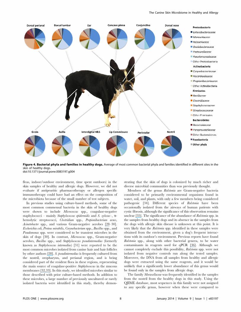
regions of skin and mucosal surfaces was Proteobacteria (Figure 4).
This was followed by Firmicutes, Actinobacteria, Bacteroidetes,
and Cyanobacteria. However, in the samples collected from axilla,
concave pinna, dorsal nose, and interdigital skin, Proteobacteria
were followed by Bacteroidetes and Actinobacteria. The samples
from the perianal skin were slightly different, with Proteobacteria
being followed by Firmicutes, Bacteroidetes, Fusobacteria, and
Actinobacteria.
At the class level, more variability was observed between the
different sites, with Betaproteobacteria being the most common
class identified in the concave pinna, conjunctiva, dorsal lumbar,
ear, and groin; whereas Actinobacteria were most common in the
axilla and interdigital skin; Gammaproteobacteria in the lip
commissure and nostril; Alphaproteobacteria in the dorsal nose,
and Bacilli in the periocular region. The Clostridia and
Bacteroidia were the most common classes in the perianal region,
as would be expected due to the close proximity to the rectum.
The family Oxalobacteriaceae (phylum Proteobacteria; class
Betaproteobacteria; Order Burkholderiales) was the most abun-
dant group in most samples. The genus Ralstonia spp. was the most
abundant genus identified in most samples, ranging from an
Table 2. ANOSIM analysis of unweighted and weighted Unifrac distances.
Skin sites Conjunctiva Dorsal perianal Lip commissure Nostril
R (unwtd) R (wtd) R (unwtd) R (wtd) R (unwtd) R (wtd) R (unwtd) R (wtd)
Axilla 0.364* 0.302* 0.672 0.389* 0.618* 0.367 0.63* 0.503*
Conjunctiva - - 0.465* 0.305* 0.491* 0.302 0.146 0.071
Concave Pinna 0.283 0.227 0.684* 0.284 0.49* 0.252 0.524 0.383
Dorsal Lumbar 20.003 20.047 0.353 0.272 0.471* 0.306 0.256 0.126
Dorsal nose 0.391* 0.269 0.69* 0.333* 0.507* 0.288* 0.592* 0.43*
Dorsal perianal 0.465* 0.305* - - 0.564* 0.097 0.48* 0.285
Ear 0.118 0.069 0.518* 0.355* 0.526* 0.384* 0.418* 0.301
Groin 0.187 0.073 0.332 0.135 0.248* 0.098 0.332 0.183
Interdigital 0.136 0.091 0.564* 0.259 0.549* 0.309 0.419 0.217
Lip commissure 0.491* 0.302 0.564* 0.097 - - 0.366* 0.224
Nostril 0.146 0.071 0.48* 0.285 0.366* 0.224 - -
Periocular 20.002 0.14 0.503* 0.217 0.376 0.262 0.233 0.234
R-values are shown for the healthy skin sites that showed significant differences. R- values closer zero to represent no difference between different sites, whereas valuescloser to 1 indicate that the most similar samples are within the same group.*Significance level p = 0.001.doi:10.1371/journal.pone.0083197.t002
The Canine Skin Microbiome in Healthy and Allergy
PLOS ONE | www.plosone.org 4 January 2014 | Volume 9 | Issue 1 | e83197

average of 5% of the total taxa identified in the lip commissure to
35% of the taxa identified in the conjunctiva. The family
Moraxellaceae was significantly more abundant in the nostril
compared to other sites (median 33.1%; p-value,0.0001; q-
value = 0.0001). The lip commissure was predominantly colonized
by the family Porphyromonadaceae and genus Porphyromonas spp.
(median 7.95%; p-value = 0.0006; q-value,0.001). Other genera
that were commonly present in most samples of skin and
mucocutanous junctions included Bacillus spp., Corynebacterium
spp., Macrococcus spp., and Pseudomonas spp.
Skin microbiome of healthy versus allergic dogsMicrobial community composition in allergic versus
healthy dogs. To compare microbial communities between
samples from allergic versus healthy dogs, the statistical analysis
ANOSIM was performed on the unweighted and weighted
UniFrac distances. Significant differences were not noted in
microbial community composition between allergic and healthy
dogs.
Principal coordinate analysis plots from unweighted UniFrac
metric were constructed to evaluate similarities between individual
(breed, age, sex, pruritus associated with allergic skin disease, ear
problems, and presence of fleas) and environmental (time spent
indoors and type of immediate environment) factors. Principal
coordinate analysis plots were also constructed to evaluate
similarities between the different samples from allergic and healthy
dogs. No significant clustering was noted in the PCoA plots
between allergic and healthy dogs (Figure 5).
Figure 1. Principal coordinates analysis for healthy dogs. Principal coordinates analysis of unweighted Unifrac distances of 16S rRNA genesclusters samples based on similarities of bacterial molecular phylogenetic trees. (A) No clustering differences are observed in 3 healthy dogs with fleascompared to 9 healthy dogs without fleas, demonstrating that the presence of fleas does not appear to influence the microbial diversity. (B) Similarly,there were no clustering differences between male and female dogs. (C) Clustering differences were seen in the samples collected from mucosalsurfaces or mucocutaneous junctions.doi:10.1371/journal.pone.0083197.g001
The Canine Skin Microbiome in Healthy and Allergy
PLOS ONE | www.plosone.org 5 January 2014 | Volume 9 | Issue 1 | e83197

Species richness and diversity in allergic versus healthy
dogs. Diversity analysis performed in a randomly selected 1,000
sequences per sample showed that the samples from the haired
skin of dogs with allergic skin disease (median 125) showed a lower
number of observed bacterial species when compared to the same
skin sites (axilla, groin, and interdigital skin) of healthy dogs
(median 239; p,0.016; Table 4). Significant differences in the
haired skin and nostril mucosa of healthy versus allergic dogs were
also identified for the Chao1 metric (species richness estimator at
higher sequencing depth; p,0.005; Table 4, Figure 6). Although
the median for the Shannon diversity index, which considers
abundance and evenness of species, was lower for the allergic dogs
when compared to the healthy dogs, the difference was not
significant (p = 0.24).
Most common taxa colonizing the skin of allergic versus
healthy dogs. Similar abundances of the most common
bacterial taxa observed in the healthy dogs were also identified
in the samples from allergic dogs (Figure 7). However, taxa that
were minimally represented in the healthy dogs (,1%) were often
absent in allergic dogs (Table S2). Significant differences between
allergic and healthy dogs were identified for a few taxa. One major
difference between allergic and healthy dogs was the proportions
of the Beta Proteobacteria Ralstonia spp., which were significantly
lower in the samples from the allergic dogs (p-value = 0.0001; q-
value = 0.0001). In fact, Ralstonia spp. accounted for less than
0.02% of the total taxa identified in the samples from the allergic
dogs, with the exception of one sample from the axilla, where it
accounted for 45% of the total taxa identified.
In the samples from the axilla from the allergic dogs, some of
the most predominant genera were Bacillus spp. (median 3.8%),
Sphingomonas spp. (median 3.1%), Mycoplasma spp. (median 2.4%),
Rubellimicrobium spp. (median 1.4%) and Propionibacterium spp.
(median 1.3%). The samples from the groin were predominantly
colonized by Staphylococcus spp. (median 2.6%), Sphingomonas spp.
(median 2.4%), Bacillus spp. (median 1.8%), and Roseomonas spp.
(mediam 1.4%). The interdigital skin was predominantly colonized
by Alicyclobacillus spp. (median 1.6%), Staphylococcus spp. (median
1%), Pseudomonas spp. (median 1%) and Corynebacterium spp.
(median 0.7%). The samples from the nostril were predominantly
colonized by Streptococcus spp. (median 0.5%), Diaphrobacter spp.
Figure 2. Alpha diversity in different skin sites for healthy dogs. Alpha diversity measures at 1000 sequences per sample in the different sitesof canine skin (x axis). The y axis represent the data points for the Chao1 index (species predictor estimator) (A), number of observed species (B) andShannon diversity index (diversity index that accounts for species abundance and evenness) (C) data points (y axis) for each skin site. Error barsrepresent the standard deviations. A: Axilla; C: Conjunctiva; CP: Concave pinna, DL: Dorsal lumbar; DN: Dorsal Nose; DP: Dorsal Perianal; E: Ear; G:Groin; I: Interdigital 4&5; LP: Lip commissure; N: Nostril; PO: Periocular.doi:10.1371/journal.pone.0083197.g002
Table 3. Alpha diversity measures at 1000 sequences per sample in the different sites of healthy skin.
Skin site Chao 1 Observed species Shannon
Median (Min-Max) Median (Min-Max) Median (Min-Max)
Axilla 530 (163–859) 277 (97–440) 6 (3–8)
Conjunctiva 151A,N (51–457) 104A,N (38–302) 4A,N (1–7)
Concave pinna 412 (272–620) 260 (153–364) 6 (4–8)
Dorsal lumbar 225 (33–836) 156 (12–423) 4 (1–8)
Dorsal nose 525 (249–945) 291 (115–486) 7 (4–8)
Dorsal perianal 218 (64–359) 109 (45–196) 4 (2–6)
Ear 409 (41–961) 196 (32–476) 5 (1–8)
Groin 433 (21–851) 219 (16–380) 6 (1–8)
Interdigital 364 (85–839) 234 (66–425) 6 (2–8)
Lip comissure 205 (140–371) 126 (57–223) 5 (2–6)
Nostril 101A,CP,DN,G(39–296) 47A,CP,DN (25–154) 3A,CP,DN (1–5)
Periocular 258 (60–492) 172 (45–287) 6 (2–7)
Significant differences between skin sites were mainly observed when comparing mucosal surfaces with haired skin sites, e.g. conjunctiva versus axilla. The Chao 1 indexestimates species richness at higher sequencing depth; the observed species represents the number of observed species in 1,000 sequences; the Shannon is a diversityindex that takes into account abundance and evenness of species.Superscripts represent sites that were significantly different when compared to the skin sites in the first column. A: Axilla; N: Nostril; CP: Concave pinna; DN: Dorsal nose;G: Groin.doi:10.1371/journal.pone.0083197.t003
The Canine Skin Microbiome in Healthy and Allergy
PLOS ONE | www.plosone.org 6 January 2014 | Volume 9 | Issue 1 | e83197

(median 0.25%) and Sphingomonas spp. (median 0.1%). At the
family level, similar to the samples from the healthy dogs, the
nostril was predominantly colonized by Moraxellaceae (median 8%)
(Table S2).
Discussion
In this study, we demonstrate that the skin microbiome in dogs
is much more diverse than has been previously reported based on
culture-based methods. Our sequence data reveal a high
individual variability between samples collected from different
patients. High variability was also observed between different skin
regions within the same dogs, with a higher number of bacterial
species being observed on the haired skin (i.e., axilla, groin,
periocular, pinna, dorsal nose, interdigital, lumbar) when com-
pared to poorly haired skin, mucocutaneous junctions, or mucosal
surfaces (i.e., lips, nose, and conjunctiva). Although this is still a
preliminary study and additional samples from dogs are needed in
order to make any further conclusions, the results suggest that the
composition of the bacterial community in the dogs evaluated
were not influenced by the individual factors tested (e.g., age, sex,
breed, pruritus, ear problems) or by environmental factors (e.g.,
Figure 3. Rarefaction curves from different skin sites from healthy dogs. Rarefaction curves of 16S rRNA gene sequences obtained fromdifferent skin sites from healthy dogs. The analysis was performed on a randomly selected subset of 1000 and 3000 sequences per sample. Hairedskin shows higher Chao1 metric, more observed species, and higher Shannon index compared to the samples from mucosal surfaces, e.g. nostril andconjunctiva. Lines represent average of each skin site, whereas the error bars represent the standard deviations.doi:10.1371/journal.pone.0083197.g003
The Canine Skin Microbiome in Healthy and Allergy
PLOS ONE | www.plosone.org 7 January 2014 | Volume 9 | Issue 1 | e83197
fleas, indoor/outdoor environment, time spent outdoors) in the
skin samples of healthy and allergic dogs. However, we did not
evaluate if antipruritic pharmacotherapy or allergen specific
immunotherapy could have had an effect on the composition of
the microbiota because of the small number of test subjects.
In previous studies using culture-based methods, some of the
most common commensal bacteria in the skin of healthy dogs
were shown to include Micrococcus spp., coagulase-negative
staphylococci – mainly Staphylococcus epidermidis and S. xylosus–, a-
hemolytic streptococci, Clostridium spp., Propionibacterium acnes,
Acinetobacter spp., and various Gram-negative aerobes [28–30].
Escherichia coli, Proteus mirabilis, Corynebacterium spp., Bacillus spp., and
Pseudomonas spp. were considered to be transient microbes in the
skin of dogs [30]. In contrast, Micrococcus spp., Gram-negative
aerobes, Bacillus spp., and Staphylococcus pseudintermedius (formerly
known as Staphylococcus intermedius) [31] were reported to be the
most common microbes isolated from canine hair and hair follicles
by other authors [28]. S. pseudintermedius is frequently cultured from
the nostril, oropharynx, and perianal region, and is being
considered part of the resident flora in these regions, representing
the main source of coagulase-positive Staphylococcus in the mucous
membranes [32,33]. In this study, we identified microbes similar to
those described with prior culture-based methods. In addition to
these microbes, a large number of previously uncultured or rarely
isolated bacteria were identified in this study, thereby demon-
strating that the skin of dogs is colonized by much richer and
diverse microbial communities than was previously thought.
Members of the genus Ralstonia are Gram-negative bacteria
considered to be primarily environmental organisms found in
water, soil, and plants, with only a few members being considered
pathogenic [34]. Different species of Ralstonia have been
occasionally isolated from the airways of human patients with
cystic fibrosis, although the significance of this observation remains
unclear [35]. The significance of the abundance of Ralstonia spp. in
the samples from healthy dogs and its absence in the samples from
the dogs with allergic skin disease is unknown at this point. It is
very likely that the Ralstonia spp. identified in these samples were
obtained from the environment, given a dog’s frequent interac-
tions with its outdoor’s environment. Previous reports have found
Ralstonia spp., along with other bacterial genera, to be water
contaminants in reagents used for qPCR [36]. Although we
cannot completely exclude this possibility, Ralstonia spp. were not
isolated from negative controls run along the tested samples.
Moreover, the DNA from all samples from healthy and allergic
dogs were extracted using the same reagents, and it would be
unlikely that a significantly lower abundance of this genus would
be found only in the samples from allergic dogs.
The family Moraxellaceae was frequently identified in the samples
from the nostril from the healthy dogs in this study. Using the
QIIME database, most sequences in this family were not assigned
to any specific genus, however when these were compared to
Figure 4. Bacterial phyla and families in healthy dogs. Average of most common bacterial phyla and families identified in different sites in theskin of healthy dogs.doi:10.1371/journal.pone.0083197.g004
The Canine Skin Microbiome in Healthy and Allergy
PLOS ONE | www.plosone.org 8 January 2014 | Volume 9 | Issue 1 | e83197

sequences in the NCBI Basic Local Alignment Search Tool
database [37], they usually exhibited a 97–100% identity to
sequences of Moraxella catarrhalis. The genus Moraxella has been
previously isolated from oral swabs from healthy dogs [38] and
from bronchial samples from dogs with tracheal collapse [39]. A
recent metagenomics study evaluating the oral cavity of healthy
dogs also frequently identified the genus Moraxella in evaluated
samples [40]. In cattle, Moraxella bovis, the cause of bovine
keratoconjunctivitis, is frequently isolated from nasal and ocular
secretions [41].
A previous study using samples from children with AD reported
a lower microbial diversity during flares of AD compared to
baseline and post-flare samples.[15] In our study, the skin samples
from allergic dogs in this study similarly showed a lower diversity
when compared to the samples from healthy dogs. Since the
samples collected from the allergic dogs were ‘‘baseline’’ samples
Figure 5. Principal coordinates analysis for allergic versus healthy dogs. Principal coordinates analysis plot of unweighted Unifrac distancesof 16S rRNA genes. No clustering differences are observed between allergic versus healthy dogs in the samples from the nostril, axilla, groin andinterdigital skin.doi:10.1371/journal.pone.0083197.g005
Table 4. Alpha diversity analysis of the nostril mucosa and haired skin including axilla, groin and interdigital area of healthy vsallergic dogs.
Skin site Healthy status Chao 1 Observed species Shannon
Median (Min–Max) Median (Min–Max) Median (Min–Max)
Nostril mucosa Healthy 100 (39–296) 47 (25–154) 2.85 (1.14–5.44)
Allergic 40* (26–45) 31 (21–39) 1.54 (1.04–3.89)
Haired skin Healthy 432 (21–858) 239 (16–440) 6.01 (0.88–8.09)
Allergic 168* (27–585) 125* (23–371) 5.40 (1.14–7.82)
*Significant differences between healthy versus allergic (p,0.05).doi:10.1371/journal.pone.0083197.t004
The Canine Skin Microbiome in Healthy and Allergy
PLOS ONE | www.plosone.org 9 January 2014 | Volume 9 | Issue 1 | e83197

(non flares), we speculate that the lower diversity in the skin of
allergic dogs is possibly a result of frequent antimicrobial
treatments, although none of the allergic dogs in this study had
been treated with antimicrobials for at least one month. Previous
studies have also shown that the visibly normal (i.e. nonlesional)
skin of dogs with AD is not normal (i.e. it is more inflamed than
normal skin), and this inflammation could lead to skin surface
changes leading to lower bacterial diversity [42,43].
Using culture-based methods, the skin and nasal mucous
membranes of atopic human patients [15,44] and dogs [21,45]
are more often colonized with S. aureus and S. pseudintermedius,
respectively, than healthy patients. Based on 16S rRNA pyrose-
quencing data, S. aureus markedly dominated affected skin regions,
more commonly the antecubital and popliteal creases, in children
with AD. Likewise, baseline and post flare samples from children
with AD also had more abundance of S. aureus compared to the
Figure 7. Bacterial phyla and families in allergic versus healthy dogs Average of most common bacterial phyla and familiesidentified in axilla, groin, interdigital skin and nostril in allergic and healthy dogs.doi:10.1371/journal.pone.0083197.g007
Figure 6. Rarefaction curves of 16S rRNA gene from allergic versus healthy dogs. Rarefaction curve of 16S rRNA gene sequences obtainedfrom axilla, groin, interdigital skin and nostril mucosa from allergic and healthy dogs. Lines represent average of each group, whereas the error barsrepresent the standard deviations. The analysis was performed on a randomly selected subset of 1000 sequences per sample.doi:10.1371/journal.pone.0083197.g006
The Canine Skin Microbiome in Healthy and Allergy
PLOS ONE | www.plosone.org 10 January 2014 | Volume 9 | Issue 1 | e83197

skin of healthy children [15]. Staphylococcus spp. was also frequently
identified in the skin of allergic dogs in this study. Although not
significantly different, the proportions of Staphylococcus spp. were
higher in the skin of (post-flare) allergic than in healthy dogs.
Conclusions
A large number of previously uncultured or rarely isolated
microbes were identified in the skin of dogs evaluated in this study,
demonstrating that the skin of dogs is inhabited by much more
rich and diverse microbial communities than was previously
thought, using culture-based methods. The study also shows that
each skin site from each dog evaluated here was inhabited by a
variable and unique microbiome, with significant individual
variability between samples from different dogs and between
different skin sites within the same dog. Differences in species
richness were also seen between healthy and allergic dogs, with
allergic dogs having significantly lower species richness when
compared to healthy dogs. Since the number of allergic dogs
enrolled into this study was small, and significant variability was
observed between individuals and between different skin sites, a
larger cohort of healthy and allergic dogs would have to be
evaluated before drawing any further conclusions on the most
important microbes inhabiting the skin of dogs, and the roles that
these microbes play in health or disease states. A study of the skin
microbiome of allergic dogs during acute flares and chronic skin
lesions might also confirm a lowering of this bacterial diversity.
It is imperative for us to better understand the microbial
populations inhabiting the skin of animals. Being able to describe
the skin microbiome in healthy animals, and identify the changes
that occur in the skin microbiome in disease states, could reveal
the role of the microbiome in the pathogenesis of skin diseases, and
possibly identify better measures to treat skin conditions,
ultimately reducing usage and resistance to systemic antimicrobial
treatment.
Supporting Information
Table S1 Relative percentages of the most abundantbacterial groups on the different skin sites in the healthydogs at the various phylogenetic levels (phylum, class,order, family, genus) based on pyrosequencing.
(PDF)
Table S2 Relative percentages of the most abundantbacterial groups on the different skin sites in the allergicversus healthy dogs at the various phylogenetic levels(phylum, class, order, family, genus) based on pyrose-quencing.
(PDF)
Acknowledgments
The authors would like to thank Ms. Amanda Friedeck for assistance with
collection of skin swabs, Ms. Melissa Markel for assistance with DNA
extraction, and Mr. Matthew Horton and Dr. Felipe Pierezan for technical
support.
Author Contributions
Conceived and designed the experiments: ARH APP AD SDL JM JMS
TO JSS. Performed the experiments: ARH APP AD CES SED. Analyzed
the data: ARH HJL SED JSS. Contributed reagents/materials/analysis
tools: ARH JMS SED JSS. Wrote the paper: ARH APP AD HJL SDL JM
JMS TO JSS.
References
1. Li K, Bihan M, Yooseph S, Methe BA (2012) Analyses of the microbial diversity
across the human microbiome. PLoS One 7: e32118.
2. Grice EA, Segre JA (2011) The skin microbiome. Nat Rev Microbiol 9: 244–
253.
3. Handl S, Dowd SE, Garcia-Mazcorro JF, Steiner JM, Suchodolski JS (2011)Massive parallel 16S rRNA gene pyrosequencing reveals highly diverse fecal
bacterial and fungal communities in healthy dogs and cats. FEMS Microbiol
Ecol 76: 301–310.
4. Whitman WB, Coleman DC, Wiebe WJ (1998) Prokaryotes: the unseen
majority. Proc Natl Acad Sci U S A 95: 6578–6583.
5. Grice EA, Kong HH, Renaud G, Young AC, Program NCS, et al. (2008) Adiversity profile of the human skin microbiota. Genome Res 18: 1043–1050.
6. Capone KA, Dowd SE, Stamatas GN, Nikolovski J (2011) Diversity of the
human skin microbiome early in life. J Invest Dermatol 131: 2026–2032.
7. Grice EA, Kong HH, Conlan S, Deming CB, Davis J, et al. (2009)Topographical and temporal diversity of the human skin microbiome. Science
324: 1190–1192.
8. Sturgeon A, Costa M, Weese JS (2012) Preliminary evaluation of the bacterialmicrobiome of the skin and ear in dogs. Veterinary Dermatology 23: 68.
9. Song SJ, Lauber C, Costello EK, Lozupone CA, Humphrey G, et al. (2013)
Cohabiting family members share microbiota with one another and with theirdogs. Elife 2: e00458.
10. Wanke I, Steffen H, Christ C, Krismer B, Gotz F, et al. (2011) Skin commensals
amplify the innate immune response to pathogens by activation of distinctsignaling pathways. J Invest Dermatol 131: 382–390.
11. Zeeuwen PL, Boekhorst J, van den Bogaard EH, de Koning HD, van de
Kerkhof PM, et al. (2012) Microbiome dynamics of human epidermis followingskin barrier disruption. Genome Biol 13: R101.
12. Hata TR, Gallo RL (2008) Antimicrobial peptides, skin infections, and atopic
dermatitis. Semin Cutan Med Surg 27: 144–150.
13. de Jongh GJ, Zeeuwen PL, Kucharekova M, Pfundt R, van der Valk PG, et al.
(2005) High expression levels of keratinocyte antimicrobial proteins in psoriasis
compared with atopic dermatitis. J Invest Dermatol 125: 1163–1173.
14. Herz U, Bunikowski R, Renz H (1998) Role of T cells in atopic dermatitis. New
aspects on the dynamics of cytokine production and the contribution of bacterial
superantigens. Int Arch Allergy Immunol 115: 179–190.
15. Kong HH, Oh J, Deming C, Conlan S, Grice EA, et al. (2012) Temporal shifts
in the skin microbiome associated with disease flares and treatment in children
with atopic dermatitis. Genome Res 22: 850–859.
16. Olivry T (2012) What can dogs bring to atopic dermatitis research? Chem
Immunol Allergy 96: 61–72.
17. Marsella R, Nicklin C, Lopez J (2006) Studies on the role of routes of allergenexposure in high IgE-producing beagle dogs sensitized to house dust mites. Vet
Dermatol 17: 306–312.
18. Olivry T, Bizikova P (2013) A systematic review of randomized controlled trials
for prevention or treatment of atopic dermatitis in dogs: 2008–2011 update. VetDermatol 24: 97–117 e125–116.
19. Hillier A, Griffin CE (2001) The ACVD task force on canine atopic dermatitis
(I): incidence and prevalence. Vet Immunol Immunopathol 81: 147–151.
20. Favrot C, Steffan J, Seewald W, Picco F (2010) A prospective study on the
clinical features of chronic canine atopic dermatitis and its diagnosis. VetDermatol 21: 23–31.
21. Fazakerley J, Nuttall T, Sales D, Schmidt V, Carter SD, et al. (2009)
Staphylococcal colonization of mucosal and lesional skin sites in atopic and
healthy dogs. Vet Dermatol 20: 179–184.
22. Olivry T, DeBoer DJ, Favrot C, Jackson HA, Mueller RS, et al. (2010)Treatment of canine atopic dermatitis: 2010 clinical practice guidelines from the
International Task Force on Canine Atopic Dermatitis. Vet Dermatol 21: 233–
248.
23. Garcia-Mazcorro JF, Suchodolski JS, Jones KR, Clark-Price SC, Dowd SE,et al. (2012) Effect of the proton pump inhibitor omeprazole on the
gastrointestinal bacterial microbiota of healthy dogs. FEMS Microbiol Ecol80: 624–636.
24. Caporaso JG, Kuczynski J, Stombaugh J, Bittinger K, Bushman FD, et al. (2010)QIIME allows analysis of high-throughput community sequencing data. Nat
Methods 7: 335–336.
25. Edgar RC (2010) Search and clustering orders of magnitude faster than BLAST.Bioinformatics 26: 2460–2461.
26. Lauber CL, Hamady M, Knight R, Fierer N (2009) Pyrosequencing-basedassessment of soil pH as a predictor of soil bacterial community structure at the
continental scale. Appl Environ Microbiol 75: 5111–5120.
27. Benjamini Y HY (1995) Controlling the false discovery rate: a practical andpowerful approach to multiple testing. J Roy Stat Soc B 57: 289–300.
28. Harvey RG, Lloyd DH (1995) The Distribution of Bacteria (Other ThanStaphylococci and Propionibacterium-Acnes) on the Hair, at the Skin Surface
and within the Hair-Follicles of Dogs. Veterinary Dermatology 6: 79–84.
29. SaijonmaaKoulumies LE, Lloyd DH (1996) Colonization of the canine skin withbacteria. Veterinary Dermatology 7: 153–162.
The Canine Skin Microbiome in Healthy and Allergy
PLOS ONE | www.plosone.org 11 January 2014 | Volume 9 | Issue 1 | e83197

30. Miller WH, Griffin CE, Campbell K (2012) Chapter 4: Bacterial skin diseases.
Muller and Kirk’s Small Animal Dermatology, 7th edition. 7th ed. St. ’Louis,
Missouri: Elsevier. pp. 184–222.
31. Sasaki T, Kikuchi K, Tanaka Y, Takahashi N, Kamata S, et al. (2007)
Reclassification of phenotypically identified staphylococcus intermedius strains.
J Clin Microbiol 45: 2770–2778.
32. Allaker RP, Lloyd DH, Bailey RM (1992) Population sizes and frequency of
staphylococci at mucocutaneous sites on healthy dogs. Vet Rec 130: 303–304.
33. Harvey RG, Noble WC (1998) Aspects of nasal, oropharyngeal and anal
carriage of Staphylococcus intermedius in normal dogs and dogs with pyoderma.
Veterinary Dermatology 9: 99–104.
34. Lynch K, Dennis J J (2011) Burkholderia. In: Liu D, editor. Molecular detection of
human bacterial pathogens. Boca Raton, FL: CRC press.
35. Hauser AR, Jain M, Bar-Meir M, McColley SA (2011) Clinical significance of
microbial infection and adaptation in cystic fibrosis. Clin Microbiol Rev 24: 29–
70.
36. Grahn N, Olofsson M, Ellnebo-Svedlund K, Monstein HJ, Jonasson J (2003)
Identification of mixed bacterial DNA contamination in broad-range PCR
amplification of 16S rDNA V1 and V3 variable regions by pyrosequencing of
cloned amplicons. FEMS Microbiol Lett 219: 87–91.
37. Altschul SF, Gish W, Miller W, Myers EW, Lipman DJ (1990) Basic local
alignment search tool. J Mol Biol 215: 403–410.
38. Kasempimolporn S, Benjavongkulchai M, Saengseesom W, Sitprija V (2003)
Oral bacterial flora of dogs with and without rabies: a preliminary study inThailand. J Med Assoc Thai 86: 1162–1166.
39. Johnson LR, Fales WH (2001) Clinical and microbiologic findings in dogs with
bronchoscopically diagnosed tracheal collapse: 37 cases (1990–1995). J Am VetMed Assoc 219: 1247–1250.
40. Sturgeon A, Stull JW, Costa MC, Weese JS (2013) Metagenomic analysis of thecanine oral cavity as revealed by high-throughput pyrosequencing of the 16S
rRNA gene. Vet Microbiol 162: 891–898.
41. Pugh GW Jr, McDonald TJ (1986) Identification of bovine carriers of Moraxellabovis by comparative cultural examinations of ocular and nasal secretions.
Am J Vet Res 47: 2343–2345.42. Olivry T, Naydan DK, Moore PF (1997) Characterization of the cutaneous
inflammatory infiltrate in canine atopic dermatitis. Am J Dermatopathol 19:477–486.
43. Olivry T, Moore PF, Affolter VK, Naydan DK (1996) Langerhans cell
hyperplasia and IgE expression in canine atopic dermatitis. Arch Dermatol Res288: 579–585.
44. Leyden J J, Marples RR, Kligman AM (1974) Staphylococcus aureus in thelesions of atopic dermatitis. Br J Dermatol 90: 525–530.
45. Furiani N, Scarampella F, Martino PA, Panzini I, Fabbri E, et al. (2011)
Evaluation of the bacterial microflora of the conjunctival sac of healthy dogs anddogs with atopic dermatitis. Vet Dermatol 22: 490–496.
The Canine Skin Microbiome in Healthy and Allergy
PLOS ONE | www.plosone.org 12 January 2014 | Volume 9 | Issue 1 | e83197


