Embed Size (px)
Citation preview

Available online at www.sciencedirect.com
7) 299–308www.elsevier.com/locate/yviro
Virology 369 (200
The Vpu-regulated endocytosis of HIV-1 Gag is clathrin-independent
Kirsi Harila a,b,1, Antti Salminen a, Ian Prior c, Jorma Hinkula d, Maarit Suomalainen a,⁎
a Department of Virology, Haartman Institute, PO Box 21, FIN-000 14 University of Helsinki, Finlandb Department of Biosciences at Novum, Karolinska Institutet, S-141 57 Huddinge, Sweden
c The Physiological Laboratory, University of Liverpool, Liverpool L69 3BX, United Kingdomd Department of Virology, Swedish Institute for Infectious Disease Control and MTC, Karolinska Institutet, 171 82 Stockholm
and Department of Molecular Virology, Linkoping University, 581 83 Linkoping, Sweden
Received 16 April 2007; returned to author for revision 11 May 2007; accepted 2 August 2007Available online 5 September 2007
Abstract
Recent results by us and others have shown that the accessory protein Vpu determines plasma membrane versus endosomal accumulation ofthe HIV-1 core protein Gag and progeny virions in the HeLa model of HIV-1 infection, since Vpu suppresses endocytosis of cell surface-associated Gag. In this report, we used pulse-chase studies and subcellular fractionations to investigate endocytosis of newly synthesized Gag inHeLa H1 cells. The uptake of Gag in ΔVpu-virus background was not blocked by inhibitors of clathrin-mediated endocytosis andmacropinocytosis. The cholesterol-sequestering drug filipin inhibited the uptake, but only if the drug was applied before extensive multimerizationof Gag had taken place. Thus, the uptake mechanism most likely is only indirectly dependent on cholesterol. Our results also indicated thattargeting phenotype of Gag was different in confluent versus subconfluent cell cultures, which could perhaps explain some of the controversies inintracellular targeting of Gag.© 2007 Elsevier Inc. All rights reserved.
Keywords: HIV-1; Gag trafficking; Endocytosis; Vpu; Virus assembly
Introduction
Assembly of human immunodeficiency virus type 1 (HIV-1)is directed by the viral core protein precursor Gag (Pr55Gag)(Göttlinger, 2001; Resh, 2005). Pr55Gag is a myristoylated,peripheral membrane protein, and it binds to cellular mem-branes and assembles into enveloped virus-like particles (VLPs)independently of other viral proteins (Bryant and Ratner, 1990;Gheysen et al., 1989; Göttlinger et al., 1989; Spearman et al.,1994; Zhou et al., 1994), but release of progeny virions frominfected cells is enhanced by the viral accessory protein Vpu(Bour and Strebel, 2003), and by the viral protease present in theother core protein precursor, the Gag-Pol (Kaplan et al., 1994).During or shortly after budding, Pr55Gag is cleaved into matrix,
⁎ Corresponding author. Fax: +358 9 19126491.E-mail address: [email protected] (M. Suomalainen).
1 Present address: Department of Medical Microbiology, FIN-90014 Uni-versity of Oulu, Finland.
0042-6822/$ - see front matter © 2007 Elsevier Inc. All rights reserved.doi:10.1016/j.virol.2007.08.009
capsid (CA), nucleocapsid, and p6 proteins and two peptidesSP1 and SP2 by the viral protease.
One intriguing aspect of the Gag-mediated assembly ofHIV-1 is that the assembly phenotype is cell type-dependent.Electron microscopy (EM) and fluorescence microscopystudies have indicated that Gag and progeny virions concen-trate within large internal vacuoles in macrophages (GreeneNguyen et al., 2003; Orenstein et al., 1988; Pelchen-Matthewset al., 2003; Raposo et al., 2002). These vacuoles have beenassumed to represent late endosomes/multivesicular bodies, butrecent data indicate that they in fact are internally sequesteredplasma membrane domains (Deneka et al., 2007; Welsch et al.,2007). In contrast, Gag and virus particles have been foundboth at the plasma membrane and in internal, endosome-likecompartments in T cells and model cell lines such as 293T,HeLa, and Cos (see, for example, Grigorov et al., 2006;Nydegger et al., 2003; Sherer et al., 2003). The intracellularroute by which Gag reaches its cell surface or internallocalization, as well as the intracellular site where virusassembly is initiated, have been subjects of intense debate.

300 K. Harila et al. / Virology 369 (2007) 299–308
Since the virus-filled vacuoles of macrophages have beenfound to be continuous with the plasma membrane, the cellsurface is the primary site for HIV-1 assembly in these cells(Deneka et al., 2007; Welsch et al., 2007). This conclusion issupported by cell imaging and biochemical studies as well(Jouvenet et al., 2006). The plasma membrane appears to be theprimary site for HIV-1 assembly in other cell types as well,since several recent studies have provided strong evidence thatthe internal Gag and virus particles in HeLa and 293T cellsoriginate from uptake of newly assembled virions from the cellsurface (Finzi et al., 2007; Harila et al., 2006; Jouvenet et al.,2006; Neil et al., 2006). We and others recently identified theviral accessory protein Vpu as an important determinant foraccumulation of Gag and progeny virions in internalendosome-like compartments in HeLa H1 and HeLa cells(Harila et al., 2006; Neil et al., 2006). The results from thesetwo studies indicated that a newly synthesized Gag wasinitially inserted into the plasma membrane. When Gag wascoexpressed with Vpu, the protein remained at the plasmamembrane and progeny virions assembled at the cell surfacewere efficiently released into the extracellular medium. Incontrast, Gag expressed in the absence of Vpu producedprogeny virions that remained tethered to the cell surface (Neilet al., 2006), and the plasma membrane-associated newlysynthesized Gag, most likely in the form of a fully assembledvirion, was efficiently retargeted from the cell surface to aninternal endosome-like compartment by an as-yet-uncharacter-ized uptake mechanism (Harila et al., 2006; Neil et al., 2006).However, the initial insertion of newly synthesized Gag intothe plasma membrane is a controversial issue. Biarsenical/tetracysteine labeling and cell imaging studies have favoredeither direct insertion of Pr55Gag into the plasma membrane inHeLa cells (Rudner et al., 2005), or provided evidence fortrafficking of newly synthesized Gag to the plasma membranevia an endosomal intermediate (Perlman and Resh, 2006).Furthermore, cell fractionation studies have indicated that anewly synthesized Gag in 293T cells is first inserted into theplasma membrane (Finzi et al., 2007), or that the proteinsimultaneously reaches the plasma membrane and endosomalmembranes (Grigorov et al., 2006). Studies using Gag-greenfluorescent protein (Gag-GFP) molecules have suggested thatinternalization from the plasma membrane accounts forintracellular Gag in 293T cells and macrophages (Jouvenetet al., 2006).
In the present study, we have investigated the endocyticphenotype of Gag in HeLa H1 cells. Our results indicated thatthe endocytosis of Gag in ΔVpu-background was not blockedby an inhibitor of macropinocytosis [5-(N-ethyl-N-isopropyl)amiloride], or by RNA interference (RNAi)-mediated knock-down of clathrin heavy chain (CHC). The uptake was sensitiveto the cholesterol-sequestering drug filipin, but the effect offilipin was most likely indirect and due to negative impact of thedrug on Gag multimerization and virus assembly. Notably, thetargeting phenotype of Gag in ΔVpu-background was found tobe different in confluent versus subconfluent cell cultures, sincenewly synthesized Gag was simultaneously detected at theplasma membrane and internal membranes in subconfluent cells
whereas targeting to the plasma membrane in confluent cellsclearly preceded the appearance of Gag in internal membranes.
Results
Impact of inhibitors of clathrin-mediated endocytosis andmacropinocytosis on the uptake of cell surface-associated Gag
Our previous results in HeLa H1 cells indicated that a newlysynthesized Pr55Gag was inserted into the plasma membrane,but when expressed in the absence of Vpu, the proteinsubsequently shifted from the cell surface to an internalendosome-like compartment (Harila et al., 2006). Mammaliancells have several endocytic routes which can be classified intothree main uptake modes: clathrin-mediated endocytosis,macropinocytosis, and cholesterol-dependent (raft-mediated)uptake (Marsh and Helenius, 2006; Pelkmans and Helenius,2003). We used inhibitors of these three uptake modes to probethe mechanism of Gag endocytosis in HeLa H1 cells.
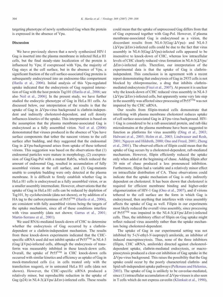
RNAi-mediated knock-down of CHC was used to probewhether the uptake of cell surface-associated Gag in ΔVpu-virus background occurred by clathrin-mediated endocytosis.HeLa H1 cells were transfected with CHC-specific siRNAs ormock-transfected (i.e. treated only with the transfectionreagent), and the following day infected with vesicularstomatitis virus (VSV) G-protein-pseudotyped NL4-3(Gag/ΔVpu), a recombinant virus carrying a genome with a defectivePol region and a defective vpu gene. Western blot analysis ofcell lysates after 24 h postinfection demonstrated that the CHCsiRNA effectively reduced intracellular levels of CHC (Fig.1A). Biotin-tagged transferrin (TF) was used as a probe tomeasure clathrin-dependent endocytosis in the infected CHCknock-down cells. TF was initially bound to cells at cold, andthe cells were then shifted to 37 °C for 0, 5 or 15 min to allowendocytosis. After incubation at 37 °C, cells were placed on ice,and cell surface-associated TF was removed by trypsin. Theamount of trypsin insensitive, endocytosed TF was determinedby Western blot analyses of cell extracts. Both mock- andsiRNA-transfected cells bound ∼equal amounts of TF (Fig. 1B;tot), and the TF at the cell surface was efficiently removed bytrypsin (the 0 min time point in Fig. 1B). After 5 min ofwarming, TF had become fully resistant to trypsin in mock-transfected control cells, whereas the majority of TF (∼70%) inCHC siRNA-treated cells remained susceptible to trypsin evenafter 15 min at 37 °C. This indicated that clathrin-dependentendocytosis was compromised in the CHC siRNA-treated cells.To determine the effect of CHC knock-down on endocytosis ofPr55Gag, siRNA- or mock-transfected cells were infected withNL4-3(Gag/ΔVpu), and at 24 h postinfection cells weremetabolically labeled with [35S]methionine for 30 min andchased for 10 min or 180 min. Intact cells were then coated withcationic silica beads at +4 °C prior to homogenization, andcrude cell extracts were fractionated on a Nycodenz stepgradient. The gradient consisted of a 70% Nycodenz cushionand a 60% sample loading zone overlaid with 50% Nycodenzand buffer solutions. This fractionation method efficientlyseparates the plasma membrane from internal membranes, since

Fig. 2. Effect of CHC-specific siRNAs on virus assembly. Extracellular VLPs(−), or VLPs tethered to the cell surface and released by trypsin treatment of cells(+), were pelleted through a sucrose cushion and analyzed by SDS-polyacrylamide gel electrophoresis. (A) VLPs from cells that expressedunprocessed Pr55Gag in the absence of Vpu [NL4-3(Gag/ΔVpu)-infection].Cells were metabolically labeled for 180 min. (B) VLPs from cells thatexpressed Gag and Gag-Pol, but no Vpu [NL4-3(ΔVpu/ΔEnv)-infected cells].Cells were metabolically labeled for 240 min. Only the CA form is shown, sincethe VLPs did not contain significant amounts of unprocessed Pr55Gag orprocessing intermediates of Gag.
Fig. 1. RNAi-mediated knock-down of CHC does not inhibit uptake of cellsurface-associated Pr55Gag, but reduces the uptake of Gag when Gag and Gag-Pol are coexpressed. (A) The CHC-siRNA used effectively reduced theintracellular levels of CHC. HeLa H1 cells were transfected with CHC-specificsiRNA (+) or mock-transfected (−). CHC levels in postnuclear supernatantswere probed by Western blotting, and GAPDH was used as a control to confirmthat equal amounts of siRNA- and mock-transfected samples were analyzed. (B)Clathrin-mediated endocytosis was inhibited in the siRNA-transfected cells.HeLa H1 cells were transfected with CHC-specific siRNA or mock-transfectedprior to infection with NL4-3(Gag/ΔVpu), and clathrin-mediated endocytosiswas probed by measuring the uptake of biotin-tagged transferrin. Transferrinwas bound to cells at 4 °C, and internalized at 37 °C for the indicated times. Cellsurface-associated transferrin was removed by trypsin prior to homogenization,and the amount of endocytosed, trypsin-resistant transferrin in cell extracts wasdetermined by Western blotting. Tot sample was not trypsin-treated and itindicates the amount of transferrin originally bound to cells. (C) CHC knock-down does not inhibit endocytosis of Pr55Gag expressed in the absence of Vpu.HeLa H1 cells were transfected with CHC-specific siRNA or mock-transfectedprior to infection with NL4-3(Gag/ΔVpu). Cells were metabolically labeledwith [35S]methionine for 30 min and chased for the indicated times. After thechase, intact cells were coated with cationic silica beads at 4 °C prior tohomogenization, and crude cell extracts were fractionated on a Nycodenz stepgradient to separate the plasma membrane (PM) from internal membranes (I).Pr55Gag proteins were immunoprecipitated from the PM and I fractions, andimmunoprecipitates were analyzed by SDS-polyacrylamide gel electrophoresis.The 10 min CHC sample and the 180 min chase samples are from differentexperiments. (D) Intracellular localization of Pr55Gag coexpressed with Vpu isnot affected by the knock-down of CHC. HeLa H1 cells were transfected withCHC-specific siRNA and infected with NL4-3(Gag). Cells were metabolicallylabeled for 30 min and chased for 150 min prior to silica coating andfractionation. (E) Analysis of the effect of CHC knock-down on uptake of Gagwhen Gag and Gag-Pol were coexpressed [NL4-3(ΔVpu/ΔEnv)-infected cells].Cells were metabolically labeled for 30 min and chased for 120 min prior tosilica coating and fractionation.
301K. Harila et al. / Virology 369 (2007) 299–308
due to the bound silica beads, the plasma membrane-derivedvesicles and membrane sheets pellet through the 70% Nycodenzcushion during ultracentrifugation, whereas internal membranesfloat to the 50% Nycodenz-buffer interphase (for controls, seeHarila et al., 2006). Distribution of labeled Pr55Gag between theplasma membrane (PM) and internal membrane (I) fractionswas determined by immunoprecipitation and by gel electro-phoresis analysis of the immunoprecipitates. As shown in Fig.1C, the CHC siRNAs did not block redistribution of labeledPr55Gag from the cell surface to internal membranes. After10 min of chase, the majority of labeled Pr55Gag was in the PMfraction in CHC siRNA-transfected cells, whereas the labeledPr55Gag had shifted to I fraction in the siRNA-transfected cells
as efficiently as in mock-transfected control cells after 180 minof chase. Thus, clathrin-mediated endocytosis is not involved inuptake of plasma membrane-associated Pr55Gag in NL4-3(Gag/ΔVpu)-infected HeLa H1 cells. Lowering intracellular levels ofCHC did not affect intracellular localization of Pr55Gag
coexpressed with Vpu, since the majority of labeled Pr55Gag
in NL4-3(Gag)-infected cells was found in the PM fraction evenafter 150 min of chase (Fig. 1D).
The effect of CHC knock-down was also tested in the contextof Gag and Gag-Pol coexpression. CHC siRNA- or mock-transfected HeLa H1 cells were infected with NL4-3(ΔVpu/ΔEnv), a virus carrying defective vpu and env genes, and at21 h postinfection cells were metabolically labeled with [35S]methionine for 30 min and chased for 120 min. As shown inFig. 1E, only trace amounts of labeled, unprocessed Pr55Gag
remained in mock-transfected and siRNA-transfected cells afterthe 120-min chase. Approximately ∼60% of p24 CA wasinternal in mock-treated cells, whereas the internal p24comprised ∼49% of total cell-associated CA in CHC siRNA-treated cells. This small difference in the intracellular distribu-tion of CA was reproducible.
The results in Figs. 1C and E suggested that the uptake ofplasma membrane-associated Gag in NL4-3(Gag/ΔVpu)-infected cells was clathrin-independent, but in NL4-3(ΔVpu/ΔEnv)-infected cells, a subpopulation of Gag might beinternalized by clathrin-mediated endocytosis. Alternatively,as there is the strong possibility that Gag is endocytosed as afully assembled virion (Neil et al., 2006), the results in Figs. 1Cand E might reflect differential impact of CHC knock-down onvirus assembly in these two cells. To distinguish between thesepossibilities, we analyzed production of VLPs from NL4-3(Gag/ΔVpu)- and NL4-3(ΔVpu/ΔEnv)-infected cells contain-ing normal or low intracellular levels of CHC. The NL4-3(Gag/ΔVpu)- or NL4-3(ΔVpu/ΔEnv)-infected cells were metaboli-cally labeled with [35S]methionine for 180 min and 240 min,respectively, and VLPs released into the culture supernatant, orVLPs tethered to the cell surface (=VLPs released after trypsintreatment of cells) were purified by pelletation through a 20%sucrose cushion. The amount of labeled Gag in the pellets wasdetermined by gel electrophoresis. As shown in Fig. 2A, onlytrace amounts of labeled Pr55Gag were released into extra-cellular VLPs from NL4-3(Gag/ΔVpu)-infected cells, but

Fig. 4. Filipin reduces retargeting of newly synthesized Gag from the cell surfaceto internal membranes in infected HeLa H1 cells. (A) Cells expressingunprocessed Pr55Gag in the absence of Vpu [NL4-3(Gag/ΔVpu)-infection] weremetabolically labeled for 30 min, and chased for 210 min in the presence orabsence of filipin prior to silica coating and fractionation. (B) Filipin does notaffect intracellular localization of Pr55Gag coexpressed with Vpu. NL4-3(Gag)-infected cells were pulsed for 30 min and chased in the presence of filipin for210 min. (C) Analysis of NL4-3(Gag/ΔVpu)-infected cells when filipin wasadded after 30 min of chase. Cells were pulsed for 30 min and chased for 30 minbefore addition of filipin. The chase in the presence or absence of filipin wascontinued for further 180 min. (D) Similar analysis from NL4-3(ΔVpu/ΔEnv)-infected HeLa H1 cells, which express Gag together with Gag-Pol, and no Vpu.Filipin was added to the chase medium after 30 min of chase, and chase wascontinued for further 180 min. The 30 min sample indicates intracellulardistribution of labeled Gag at the time of filipin addition.
302 K. Harila et al. / Virology 369 (2007) 299–308
mock- and CHC siRNA-transfected cells contained similaramounts of labeled Pr55Gag VLPs tethered to the cell surface.Thus, the knock-down of CHC apparently did not affect virusassembly in NL4-3(Gag/ΔVpu)-infected cells. In NL4-3(ΔVpu/ΔEnv)-infection, small amounts of labeled CA weredetected in extracellular VLPs produced from control mock-transfected cells, but the majority of labeled CA was found inVLPs tethered to the cell surface (Fig. 2B). Knock-down ofCHC reduced incorporation of labeled CA into both theextracellular VLPs and into VLPs that were tethered to the cellsurface. Quantitations from two different experiments sug-gested that total VLP production was ∼3-fold higher in mock-transfected control cells than in CHC siRNA-transfected cells.However, this could be an overestimation, since the VLP assaydid not take into account internal (or internalized) VLPs. Sincethe mock-transfected and CHC siRNA-transfected cells hadsimilar amounts of membrane-associated Gag and since therewas no indications that low intracellular levels of CHC causedany Gag processing defects (Fig. 1E), the CHC siRNAsapparently did not reduce membrane targeting or higher-ordermultimerization of newly synthesized Gag in NL4-3(ΔVpu/ΔEnv)-infected cells, but produced an as-yet-undefined mod-erate late assembly defect in these cells. This late assemblydefect might explain why CHC siRNAs gave slightly differentresults in NL4-3(Gag/ΔVpu)- and NL4-3(ΔVpu/ΔEnv)-infected cells in the cell fractionation experiments.
We next tested whether the endocytic uptake of Gag inΔVpu-virus background occurred by macropinocytosis. Ifmacropinocytosis was involved, virus infection would need tostimulate this uptake mechanism, since macropinocytosis is nota constitutive feature of HeLa cells. We measured the uptake offluorescein-conjugated dextran into noninfected, NL4-3(Gag)-or NL4-3(Gag/ΔVpu)-infected HeLa H1 cells in order todetermine whether macropinocytosis was upregulated ininfected HeLa H1 cells. Flow cytometric analyses indicatedsimilar low levels of macropinocytosis for all three cellpopulations (data not shown). The effect of 5-(N-ethyl-N-isopropyl) amiloride, a potent inhibitor of induced macropino-cytosis (Meier et al., 2002; West et al., 1989), was tested in Fig.3. NL4-3(Gag/ΔVpu)-infected cells were pulsed for 30 min,and chased for 180 min in the presence or absence of 100 μMamiloride. As shown in Fig. 3, amiloride did not block the shiftof labeled Pr55Gag from the PM to the I fraction in NL4-3(Gag/ΔVpu)-infected cells. Thus, the uptake of Gag in the ΔVpu-virus background is unlikely to occur by a macropinocytosis-like activity.
Fig. 3. The inhibitor of induced macropinocytosis, 5-(N-ethyl-N-isopropyl)amiloride, did not block shift of newly synthesized Pr55Gag from the plasmamembrane to internal membranes in NL4-3(Gag/ΔVpu)-infected HeLa H1 cells.Infected cells were pulsed for 30 min, and chased for 180 min in the presence orabsence of amiloride prior to silica coating and fractionation.
Effect of filipin on the uptake of cell surface-associated Gag
The possible dependence of Pr55Gag endocytosis oncholesterol was tested by filipin, an antibiotic commonlyused to disrupt cholesterol-dependent raft-like microdomains atthe cell surface. NL4-3(Gag/ΔVpu)-infected HeLa H1 cellswere metabolically labeled with [35S]methionine for 30 minand chased for 210 min in the presence or absence of filipin(3 μg/ml). This amount of filipin did not induce extensiveshedding of microvesicles (a common side-effect of removal ofcell surface cholesterol), but the filipin concentration wassufficient to inhibit cholesterol-dependent endocytic processes,since the clathrin-mediated uptake of transferrin was inhibitedin filipin-treated cells (data not shown; Subtil et al., 1999). Asshown in Fig. 4A, filipin treatment considerably reduced theamount of labeled Pr55Gag in I fraction after 210 min of chase:quantitations from two separate experiments indicated thatapproximately 61% of labeled Pr55Gag was detected in the Ifraction in untreated cells whereas only ∼19% of labeledPr55Gag was in internal membranes in filipin-treated cells.Filipin treatment did not change intracellular localization ofPr55Gag that was coexpressed with Vpu [NL4-3(Gag)-infec-tion], since, as in untreated cells (Harila et al., 2006) themajority of labeled Pr55Gag in filipin-treated cells were plasmamembrane-associated after 210 min of chase (Fig. 4B).
The results in Fig. 4A suggested that the endocytic uptake ofPr55Gag could occur by a cholesterol-dependent mechanism.However, cholesterol is also required for efficient membranebinding and higher-order multimerization of HIV-1 Gag (Onoet al., 2007). Since there are strong indications that Gag isendocytosed as a fully assembled virion (Neil et al., 2006), theobserved effect of filipin could be due to inability of plasma

Fig. 5. Analysis of intracellular targeting of newly synthesized Pr55Gag insubconfluent HeLa H1 cell cultures. (A) Confluent or subconfluent cellsexpressing unprocessed Pr55Gag in the absence of Vpu [HXB2D-(Gag)-infection]. Cells were metabolically labeled for 30 min and chased for theindicated times prior to silica coating and fractionation. In contrast to confluentcells, newly synthesized Pr55Gag was simultaneously detected in PM and Ifractions in the subconfluent cell cultures. (B) Cells coexpressing Vpu andunprocessed Pr55Gag [NL4-3(Gag)-infection] were analyzed after a 30 min pulseand a 5 min chase. Newly synthesized Pr55Gag coexpressed with Vpu wasprimarily targeted to the plasma membrane in subconfluent cells. (C) Cellsexpressing an endocytosis-defective Pr55Gag/HA [HXB2D-(GagHA)-infectedcells]. Cells were analyzed after a 30 min pulse and a 5 min chase. No Vpu wasexpressed in the HXB2D-(GagHA)-infected cells.
303K. Harila et al. / Virology 369 (2007) 299–308
membrane-associated Gag proteins to efficiently multimerize inthe presence of the drug. To test this possibility, we added filipinafter 30 min of chase, i.e. at the time when maximal membraneinsertion, as well as at least some higher-order multimerizationof labeled Pr55Gag has already occurred (Ono et al., 2007; Triteland Resh, 2000). As shown in Fig. 4C, filipin added after 30 minof chase reduced the amount of labeled Pr55Gag in I fraction atthe 210 min chase point, but the reduction was less than if filipinwas present from the beginning of chase. In the experimentshown in Fig. 4C, ∼37% and 49% of labeled Pr55Gag was ininternal membranes in filipin-treated and untreated cells,respectively. However, the efficiency with which filipininhibited the uptake of Pr55Gag varied between differentexperiments; the relative amount of labeled Pr55Gag in the Ifraction was 12%–32% lower in filipin-treated cells than incontrol untreated cells. To further probe the possibility thatfilipin exerted its effect primarily through inhibiting multi-merization of Gag, we also tested the impact of filipin in thecontext of Gag and Gag-Pol coexpression, since processing ofPr55Gag offers a convenient indirect marker for efficientmultimerization of Gag (Kaplan et al., 1994). HeLa H1 cellswere infected with NL4-3(ΔVpu/ΔEnv)-virus, pulsed for30 min, and filipin was added after 30 min of chase, and thechase was continued for further 180 min. At the time of filipinaddition (Fig. 4D; 30 min), the PM fraction contained largeamounts of labeled Pr55Gag, whereas the small amounts ofprocessed CA forms were ∼equally distributed between the PMand I fractions. After 210 min of chase, only trace amounts ofunprocessed Pr55Gag were present in untreated control cells, and68% of labeled CAwas in the I fraction. In contrast, the filipin-treated cells contained significant amounts of plasmamembrane-associated, unprocessed Pr55Gag at the 210 min chase point, thussuggesting that filipin affected proper higher-order multimeriza-tion of Gag. Approximately 47% of total cell-associated labeledCAwas in internal membranes after 210 min of chase. However,in another experiment, CA in filipin-treated cells distributed tothe I fraction as efficiently as in untreated control cells (data notshown). Thus, the effect of filipin varied between differentexperiments, but overall, filipin had a relatively minor effect onintracellular distribution of CA. Therefore, a significant part ofthe apparent reduction in Gag uptake in the filipin-treated cellsmight be attributable to the inhibitory effects of filipin on higher-order oligomerization of Gag.
The apparent targeting phenotype of newly synthesized Gag isdifferent in confluent and subconfluent HeLa H1 cultures
As mentioned in the Introduction, attempts to analyzeintracellular targeting of newly synthesized HIV-1 Gag haveproduced contradictory results. The results in our previouspublication (Harila et al., 2006), as well as for example Figs. 1Cand 4D above, clearly indicate that a newly synthesized Gag isinitially inserted into the plasma membrane in HeLa H1 cells.These results were from confluent HeLa H1 cells, but thetrafficking phenotype of newly synthesized Gag turned out to bedifferent when we repeated the targeting analyses in subcon-fluent HeLa H1 cell cultures (∼30–40% confluency). Fig. 5A
shows analysis of confluent and subconfluent HeLa H1 cellsinfected with HXB2D-(Gag), a recombinant virus with adefective Pol region, a defective vpu gene, and a truncated nefgene. The infected cells were metabolically labeled with [35S]methionine for 30 min, and chased for 5 min or 240 min. Inconfluent cells, the majority of newly synthesized Pr55Gag wasat the plasma membrane after 5 min of chase, but in sub-confluent cells, labeled Pr55Gag was simultaneously detected inPM and I fractions. Whereas there was a clear increase oflabeled Pr55Gag in the I fraction in confluent cells after 240 minof chase, the amount of labeled Pr55Gag in I fraction insubconfluent cells increased only slightly during a longer chase(∼60% was internal after 240 min of chase). When expressed inthe presence of Vpu [NL4-3(Gag)-genome], the majority ofnewly synthesized Pr55Gag was in the PM fraction in bothsubconfluent (Fig. 5B; a 30 min pulse and a 5 min chase) and inconfluent cell cultures (Harila et al., 2006). The simultaneousdetection of Pr55Gag in the PM and I fraction in HXB2D-(Gag)-infected subconfluent cells could mean that membrane insertionof newly synthesized Pr55Gag expressed in the absence of Vpuwas random in subconfluent cultures, or, alternatively, thatuptake of plasma membrane-associated Pr55Gag occurred veryrapidly in these cells. To distinguish between these possibilities,we tested targeting of Pr55Gag/HA in subconfluent cells.Pr55Gag/HA contains a carboxyterminal HA tag, and this Gagvariant is not endocytosed after insertion into the plasmamembrane when expressed in the absence of Vpu (Harila et al.,2006). Fig. 5C shows analysis of HXB2D-(GagHA)-infectedsubconfluent HeLa H1 cells after a 30 min pulse and a 5 minchase. The majority of newly synthesized Pr55Gag/HA wasdetected in the PM fraction. Similar distribution was observedafter 240 min of chase (data not shown). Taken together, theseresults indicate that cell confluency can have an impact on the
304 K. Harila et al. / Virology 369 (2007) 299–308
targeting phenotype of newly synthesized Gag when the proteinis expressed in the absence of Vpu.
Discussion
We have previously shown that a newly synthesized HIV-1Gag is inserted into the plasma membrane in infected HeLa H1cells, but the final steady-state localization of the protein isinfluenced by Vpu; if coexpressed with Vpu, the majority ofGag stays at the cell surface, but in the absence of Vpu, asignificant fraction of the cell surface-associated Gag proteins issubsequently endocytosed into an endosome-like compartment(Harila et al., 2006). Initial analysis of this Vpu-regulateduptake indicated that the endocytosis of Gag required interac-tion of Gag with the host protein Tsg101 (Harila et al., 2006; seealso Neil et al., 2006]. In the present study, we have furtherstudied the endocytic phenotype of Gag in HeLa H1 cells. Asdiscussed below, our interpretation of the results is that theuptake of Gag in ΔVpu-virus background is clathrin-indepen-dent and indirectly cholesterol-dependent, and cell densityinfluences kinetics of the uptake. This interpretation is based onthe assumption that the plasma membrane-associated Gag isendocytosed as a fully assembled virion. Neil et al. (2006)demonstrated that viruses produced in the absence of Vpu havesurface components that tether the nascent virions to the cellsurface after budding, and they suggested that the endosomalGag in ΔVpu-background arises from uptake of these tetheredvirions. This suggestion was based on the observations that (1)endosomal particles were mature in morphology, (2) coexpres-sion of Gag/Gag-Pol with a mutant Rab5a, which reduced theamount of endosomal Gag, resulted in accumulation of fullyassembled virions at the cell surface, and (3) Gag-mutantsunable to complete budding were only detected at the plasmamembrane. It is difficult to firmly establish whether Gag inHeLa H1 cells is endocytosed as a fully assembled virion, or asa smaller assembly intermediate. However, observations that theuptake of Gag in HeLa H1 cells can be reduced by depletion ofTsg101, by cycloheximide (data not shown), or by addition of aHA tag to the carboxyterminus of Pr55Gag (Harila et al., 2006),are consistent with fully assembled virions being the targets ofthe uptake mechanism, since all of these conditions interferewith virus assembly (data not shown; Garrus et al., 2001;Martin-Serrano et al., 2001).
We used RNAi-mediated knock-down of CHC to determinewhether the endocytosis of Gag occurred by a clathrin-dependent or a clathrin-independent mechanism. The resultsfrom these knock-down experiments indicated that the CHC-specific siRNA used did not inhibit uptake of Pr55Gag in NL4-3(Gag/ΔVpu)-infected cells, although the endocytosis of trans-ferrin was measurably inhibited in these knock-down cells.Overall, the uptake of Pr55Gag in CHC knock-down cellsoccurred with similar kinetics and efficiency as uptake of Gag inmock-transfected cells (i.e. in cells treated only with thetransfection reagent), or in untreated HeLa H1 cells (data notshown). However, the CHC-specific siRNA produced arelatively minor, but reproducible reduction in the uptake ofGag (p24) in NL4-3(ΔVpu/ΔEnv)-infected cells. These results
could mean that the uptake of unprocessed Gag differs from thatof Gag expressed together with Gag-Pol. However, if plasmamembrane-associated Gag is endocytosed as a virion, thediscordant results from NL4-3(Gag/ΔVpu)- and NL4-3(ΔVpu/ΔEnv)-infected cells could be due to the fact that virusassembly in NL4-3(Gag/ΔVpu)-infected cells appeared to beinsensitive to knock-down of CHC, whereas low intracellularlevels of CHC clearly reduced virus formation in NL4-3(ΔVpu/ΔEnv)-infected cells. Therefore, our interpretation of theexperimental data is that the uptake of Gag is clathrin-independent. This conclusion is in agreement with a recentreport demonstrating that endocytosis of Gag in 293Tcells is notblocked by chlorpromazine, a drug that inhibits clathrin-mediated endocytosis (Finzi et al., 2007). At present it is unclearwhy the knock-down of CHC reduced virus assembly in NL4-3(ΔVpu/ΔEnv)-infected cells, but apparently some very late stepin the assembly was affected since processing of Pr55Gag was notimpaired by the CHC siRNA.
Our results from filipin-treated cells demonstrate thatinterfering with plasma membrane cholesterol reduces uptakeof cell surface-associated Gag inΔVpu-virus background. HIV-1 Gag is considered to be a raft-associated protein, and raft-likemicrodomains at the plasma membrane have been suggested tofunction as platforms for virus assembly (Ding et al., 2003;Halwani et al., 2003; Holm et al., 2003; Lindwasser and Resh,2001; Nguyen and Hildreth, 2000; Ono and Freed, 2001; Zhenget al., 2001). The observed effects of filipin could mean that theuptake of Gag occurs by a cholesterol-dependent, raft-mediatedmechanism. However, filipin efficiently reduced the uptakeonly when added at the beginning of chase. Adding filipin after30 min of chase produced a less pronounced inhibition.Furthermore, filipin had a variable and a relatively minor effecton intracellular distribution of CA. These observations couldindicate that the uptake mechanism of Gag is only indirectlydependent on cholesterol. It has been shown that cholesterol isrequired for efficient membrane binding and higher-orderoligomerization of HIV-1 Gag (Ono et al., 2007), and if virionstethered to the cell surface are the form in which Gag isendocytosed, then anything that interferes with virus assemblyaffects the uptake of Gag as well. Filipin in our experimentsmost likely reduced the overall virus assembly, since processingof Pr55Gag was impaired in the NL4-3(ΔVpu/ΔEnv)-infectedcells. Thus, the inhibitory effect of filipin on Gag uptake mightreflect reduced virus assembly rather than the uptake mechan-ism being cholesterol-dependent.
The uptake of Gag in our experimental setting was notinhibited by 5-(N-ethyl-N-isopropyl) amiloride, an inhibitor ofinduced macropinocytosis. Thus, none of the three inhibitors(filipin, CHC siRNA, amiloride) directed against cholesterol-dependent uptake, clathrin-mediated endocytosis, or macro-pinocytosis produced a clear-cut inhibition of Gag uptake in theΔVpu-virus background. This raises the possibility that the Gaguptake could occur by the poorly characterized clathrin- andlipid raft-independent uptake pathways (Pelkmans andHelenius,2003). The uptake of Gag is unlikely to be caveolae-mediated,since (1) intracellular accumulation ofΔVpu-viruses is also seenin T cells which do not express caveolin (Klimkait et al., 1990),

305K. Harila et al. / Virology 369 (2007) 299–308
and (2) although HeLa H1 cells express caveolin-1, caveolaestructures are rare in these cells, and genistein, an inhibitor ofcaveolae-mediated endocytosis (Pelkmans and Helenius, 2003),does not affect endocytosis of Gag (data not shown). Our furtherobservations suggest that interaction of the MA domain of Gagwith AP-3 δ subunit, or the Y84G substitution in the MAdomain, does not play a key role in the Vpu-regulatedendocytosis of Gag in HeLa H1 cells (data not shown), althoughboth the AP-3 δ subunit and the Y84G substitution havepreviously been shown to affect targeting of HIV-1 Gag toendosomal compartments (Dong et al., 2005; Ono and Freed,2004). We have not been able to test dependence of the Gaguptake on actin dynamics, since treatment of infected cells withfor example jasplakinolide, latrunculin B, or cytochalasin Ddisrupts the cells during silica coating (data not shown).
Our interpretation that uptake of Gag is mediated by aclathrin-independent mechanism is seemingly contradictory tothe report that dominant negative forms of dynamin, EPS-15,and Rab5a, all known inhibitors of clathrin-mediated endocy-tosis, prevent endocytosis of Gag in HeLa cells (Neil et al.,2006). However, a dominant negative approach and RNAi donot always give the same results, as exemplified by theobservations that dominant-negative CHMP5, but not depletionof endogenous CHMP5, inhibits HIV-1 release (Martin-Serranoet al., 2001; Ward et al., 2005). A dominant-negative mutant caninhibit a cellular function indirectly, for example, by perturbingmore than one cellular pathway due to inactivation of essentialfactors that operate at more than one cellular site. Anotherpossibility is that the uptake mechanism of Gag is cell type-dependent. Assuming that Gag is endocytosed as a fully-assembled virions, our results from e.g. NL4-3(ΔVpu/ΔEnv)-infections indicate that the endocytosis process is directed bycellular factors present in the viral envelope, not the viral Envprotein. The HIV-1 envelope contains several different cellularproteins (Chertova et al., 2006; Hammarstedt and Garoff, 2004),and subtle cell type-dependent changes in the composition ofthe virion envelope could specify the efficiency of ΔVpu-virusuptake, and perhaps also the mechanism of the uptake.
Our previous (Harila et al., 2006) and present results indicatethat the Vpu-regulated endocytosis of Gag is a key determinantfor the steady-state localization of Gag and VLPs in HeLa H1cells. Vpu most likely controls targeting of Gag in infected Tcells as well, since deletion of vpu gene results in accumulationof virions into intracellular compartments of these cells(Klimkait et al., 1990). In contrast, concentration of Gag andprogeny virions into the internally sequestered plasma mem-brane domains in macrophages is Vpu-independent (Deneka etal., 2007; Pelchen-Matthews et al., 2003; Welsch et al., 2007).However, the intracellular virus-containing vacuoles of macro-phages and T cells or HeLa H1 cells are different, since thelimiting membrane of Jurkat and HeLa H1 virus-containingvacuoles is not stained with the cell-impermeant dye rutheniumred (data not shown), and the vacuoles thus belong to theendosomal network. In general, intracellular trafficking routesthat control cell surface versus endosomal localization of Gagand VLPs have been the subject of intense debates. Only fewserious attempts have been made to characterize the intracellular
transport of a newly synthesized Gag (Finzi et al., 2007;Grigorov et al., 2006; Harila et al., 2006; Jouvenet et al., 2006;Neil et al., 2006; Perlman and Resh, 2006; Rudner et al., 2005),and results from these studies are conflicting. Cell fractionationanalyses by us (Harila et al., 2006) and Finzi et al. (2007), aswell as FlAsh/ReAsh imaging studies by Rudner et al. (2005) orfluorescence microscopy analyses of cells expressing Gag-GFP(Jouvenet et al., 2006; Neil et al., 2006), indicated that a newlysynthesized Gag in HeLa H1, HeLa, 293 T cells, ormacrophages was initially inserted into the plasma membrane.In contrast, FlAsh/ReAsh cell imaging studies by Perlman andResh (2006) implied a ‘perinuclear’ and endosomal transportintermediates for cell surface Gag in HeLa, Cos-1, and Jurkat Tcells. Cell fractionation studies by Grigorov et al. (2006) in turndemonstrated that newly synthesized Gag proteins in 293 Tcellswere simultaneously detected at the plasma membrane and onendosomal membranes. One explanation for these contradictoryresults can be the different expression systems used, since theabove-mentioned studies utilized either proviral expression ofGag or codon-optimized plasmid-driven expression of Gag. Atleast three proteins of HIV-1, Nef, Gag, and Vpu, canpotentially modulate membrane trafficking pathways of thehost cell through interactions with cellular partners (Batonick etal., 2005; Costa et al., 2006; Dong et al., 2005; Madrid et al.,2005; Varthakavi et al., 2006), and thus intracellular targeting ofGag could be different in virus-infected cells and in cells inwhich a plasmid-driven expression system is used for synthesisof Gag. Even the multiplicity of infection and/or the time pointpostinfection could impact the apparent targeting phenotype ofGag. Our present study demonstrates that another importantfactor to consider in targeting analyses of Gag is the confluencyof cell cultures; in confluent HeLa H1 cells, newly synthesizedGag was first detected at the plasma membrane in both Vpu+and ΔVpu-virus backgrounds, whereas in subconfluent HeLaH1 cultures, Gag expressed in the absence of Vpu appearedsimultaneously in both the plasma membrane and internalmembrane fractions. The molecular basis of this phenomenon isunclear at present. It is difficult to determine whether the newlysynthesized internal Gag population in subconfluent cellsrepresents Gag proteins directly inserted into the internalmembranes, or proteins (rapidly) endocytosed from the cellsurface. We favor the latter alternative, because (1) the majorityof newly synthesized Gag coexpressed with Vpu fractionatedwith the plasma membrane fraction, and *2) newly synthesizedPr55Gag/HA, which is not endocytosed when expressed in theabsence of Vpu, was detected at the plasma membrane insubconfluent cultures as well. Overall, the results from thesubconfluent cells stress the need to monitor targeting of Gagwith assays that can detect rapid, dynamic changes inintracellular localization of Gag.
Materials and methods
Cell culture, viruses, and plasmid constructs
HeLa H1 and 293T cells were cultured as previouslydescribed (Harila et al., 2006; Holm et al., 2003). Production
306 K. Harila et al. / Virology 369 (2007) 299–308
of VSV G-protein-pseudotyped infectious recombinant HIV-1,as well as plasmid constructs for NL4-3(Gag/ΔVpu), NL4-3(Gag), HXB2D-Gag, and HXB2D-GagHA has been previouslydescribed (Harila et al., 2006). Plasmid pNL4-3(ΔVpu/ΔEnv),a derivative of pNL4-3 (Adachi et al., 1986), contains defectivevpu and env genes, and a truncated vpr gene. The plasmid wasconstructed by replacing the 6459-bp ApaI–BamHI fragment ofpNL4-3 with the equivalent ApaI–BamHI fragment ofpHXB2D-(ΔVpu/ΔEnv) (Harila et al., 2006).
Subcellular fractionations
HeLa H1 cells on 10-cm plates were infected with VSV-G-pseudotyped recombinant HIV-1 (∼1–5 infectious units/cell),and at ∼21 h [NL4-3(ΔVpu/ΔEnv)-virus] or ∼24 h [NL4-3(Gag/ΔVpu)-virus], postinfection cells were metabolicallylabeled with [35S]methionine for 30 min and chased for varioustimes as previously described (Holm et al., 2003). After thechase, cells were brought into suspension by incubation inphosphate-buffered saline (PBS) containing 0.02% EDTA(PBS-EDTA), intact cells were coated at 4 °C with cationicsilica beads, and cell extracts were fractionated on a Nycodenzstep gradient as previously described (Harila et al., 2006). Gagproteins were immunoprecipitated from sodium dodecyl sulfate(SDS)-solubilized plasma membrane and internal membranefractions with mouse anti-HIV-1 CA monoclonal antibody 38:9(Hinkula et al., 1990), or with a mixture of the 38:9 antibodyand a mouse monoclonal anti-p24 antibody 32/5.17.76 (Abcam,Ltd.). The HA-tagged Gag was immunoprecipitated with arabbit polyclonal anti-HA antibody (Sigma).
In filipin treatment, the chase medium contained 3 μg offilipin complex (Sigma) per ml. In 5-(N-ethyl-N-isopropyl)amiloride treatment, 100 μM of the drug (Alexis Biochemicals)was included into the chase medium. To measure macropino-cytosis, cells were first preincubated in minimum essentialmedium containing 0.2% BSA (MEM-BSA) for 30 min, andthen shifted to MEM-BSA containing 1 mg per ml of lysine-fixable fluorescein isothiocyanate-conjugated dextran (70,000Da, Molecular Probes) for 10 min. After extensive washing,cells were incubated in MEM-BSA for further 5 min. To stripplasma membrane-associated dextran, cells were placed on iceand incubated for 5 min in a pH 5.5 buffer containing 100 mMsodium acetate and 50 mM sodium chloride. After washes withice-cold PBS, cells were detached from plates by incubation inPBS-EDTA on ice for 20 min, fixed with 2% paraformaldehyde,and analyzed by a fluorescence-activated cell sorter.
VLPs
To analyze the efficiency of VLP production, NL4-3(Gag/ΔVpu)-infected HeLa H1 cells were metabolically labeled with[35S]methionine for 180 min, and NL4-3(ΔVpu/ΔEnv)-infected cells were metabolically labeled for 240 min. Cellculture supernatants were collected, filtered (0.45 μm), layeredon to a 20% sucrose cushion (w/v, in 25 mM HEPES pH 7.4,150 mMNaCl, 2 mMMgCl2), and centrifuged at 100,000×g for60 min. Pellets were analyzed by gel electrophoresis. These
pellets represented constitutively released extracellular parti-cles. To analyze the amount of VLPs tethered to the cell surface,cells were washed once with PBS after harvesting culturesupernatants, and incubated with trypsin (0.5 mg/ml) at 37 °Cfor 10 min. The reaction was stopped by adding culture mediumcontaining 7% fetal bovine serum, cells were pelleted andculture supernatants were filtered, and VLPs were pelletedthrough sucrose as described above.
RNAi
Short interfering RNAs (siRNAs) directed against theclathrin heavy chain (CCUGCGGUCUGGAGUCAACdTdT;40 pmol per 10-cm plate; Hinrichsen et al., 2003) weretransfected into HeLa H1 cells by using siLentFect (BioRad) inaccordance with the manufacturer's instructions. Mock-trans-fected cells were treated with siLentFect alone. At about 22 hposttransfection, the cells were infected and analyzed about21–24 h postinfection. Intracellular levels of clathrin heavychain in mock- and siRNA-treated cells were determined frompostnuclear supernatants of cell extracts by Western blottingwith a polyclonal goat anti-clathrin heavy chain antibody(C-20; Santa Cruz Biotechnology, Inc.). The polyclonal anti-glyceraldehyde-3-phosphate dehydrogenase (GAPDH) anti-body (FL-335; Santa Cruz Biotechnology, Inc.) was used asa control.
To analyze the effect of CHC knock-down on clathrin-mediated endocytosis, NL4-3(Gag/ΔVpu)-infected HeLa H1cells, mock- or siRNA-treated, were washed once with PBS(with MgCl2 and CaCl2) containing 0.2% bovine serumalbumin (PBS-BSA), and cells were incubated for 30 min onice in PBS-BSA containing 500 ng/ml human biotin-taggedtransferrin (Sigma). After washing the unbound transferrinaway, minimum essential medium containing 0.2% BSA wasadded to cells and cells were shifted to 37 °C for 0 min, 5 min,or 15 min. Cells were then placed on ice, treated with acid washbuffer (0.2 M sodium acetate buffer pH 4.5, 0.5 M NaCl) for2 min, rinsed with the acid wash buffer, and washed with excessPBS-BSA and PBS. Cell surface-associated transferrin wasremoved by trypsin treatment (20 min on ice). Trypsin wasinactivated by culture medium containing 7% fetal bovineserum, cells were homogenized, and postnuclear supernatantsof cell extracts were prepared as previously described (Harila etal., 2006). The amount of trypsin-resistant transferrin–biotin inpostnuclear supernatants was determined by Western blottingusing streptavidin–peroxidase polymer (Sigma).
Acknowledgments
This work was supported by grants from the Academy ofFinland (211130) and Swedish Research Council (K2004-16X-15032-01A) to M.S.
References
Adachi, A., Gendelman, H.E., Koenig, S., Folks, S., Willey, R., Rabson, A.,Martin, M.A., 1986. Production of acquired immunodeficiency syndrome-

307K. Harila et al. / Virology 369 (2007) 299–308
associated retrovirus in human and nonhuman cells transfected with aninfectious molecular retrovirus clone. J. Virol. 59, 284–291.
Batonick, M., Favre, M., Boge, M., Spearman, P., Höning, S., Thali, M., 2005.Interaction of HIV-1 Gag with the clathrin-associated adaptor AP-2.Virology 342, 190–200.
Bour, S., Strebel, K., 2003. The HIV-1 Vpu protein: a multifunctional enhancerof viral particle release. Microbes Infect. 5, 1029–1039.
Bryant,M., Ratner, L., 1990.Myristoylation-dependent replication and assemblyof human immunodeficiency virus 1. Proc. Natl. Acad. Sci. U. S. A. 87,523–527.
Chertova, E.N., Chertov, O., Coren, L.V., Roser, J.D., Trubey, C.M., Bess Jr.,J.W., Sowder II, R.C., Barsov, E., Hood, B.L., Fisher, R.D., Nagashima,K., Conrads, T.P., Veenstra, T.D., Lifson, J.D., Ott, D.E., 2006. Proteomicand biochemical analysis of purified human immunodeficiency virus type1 produced from infected monocyte-derived macrophages. J. Virol. 80,9039–9052.
Costa, L.J., Chen, N., Lopes, A., Aguiar, R.S., Tanuri, A., Plemenitas, A., Peterlin,B.M., 2006. Interactions between Nef and AIP1 proliferate multivesicularbodies and facilitate egress of HIV-1. Retrovirology 2006 3:33.
Deneka, M., Pelchen-Matthews, A., Byland, R., Ruiz-Mateos, E., Marsh, M.,2007. In macrophages, HIV-1 assembles into an intracellular plasmamembrane domain containing the tetraspanins CD81, CD9 and CD53.J. Cell Biol. 177, 329–341.
Ding, L., Derdowski, A., Wang, J.-J., Spearman, P., 2003. Independentsegregation of human immunodeficiency virus type 1 Gag proteincomplexes and lipid rafts. J. Virol. 77, 1916–1926.
Dong, X., Li, H., Derdowski, A., Ding, L., Burnett, A., Chen, X., Peters, T.R.,Dermody, T.S., Woodruff, E., Wang, J.-J., Spearman, P., 2005. AP-3 directsthe intracellular trafficking of HIV-1 Gag and plays a key role in particleassembly. Cell 120, 663–674.
Finzi, A., Orthwein, A., Mercier, J., Cohen, E.A., 2007. Productive humanimmunodeficiency virus type 1 assembly takes place at the plasmamembrane. J. Virol. 81, 7476–7490.
Garrus, J.E., von Schwedler, U.K., Pornillos, O.W., Morham, S.G., Zavitz, K.H.,Wang, H.E., Wettstein, D.A., Stray, K.M., Cöte, M., Rich, R.L., Myszka,D.G., Sundquist, W.I., 2001. Tsg101 and the vacuolar protein sortingpathway are essential for HIV-1 budding. Cell 107, 55–65.
Gheysen, D., Jacobs, E., Foresta, F.d., Thiriart, C., Francotte, M., Thines, D.,Wilde, M.D., 1989. Assembly and release of HIV-1 precursor Pr55gag virus-like particles from recombinant Baculovirus-infected cells. Cell 59,103–112.
Greene Nguyen, D., Booth, A.M., Gould, S.J., Hildreth, J.E.K., 2003. Evidencethat HIV budding in primary macrophages occurs through the exosomerelease pathway. J. Biol. Chem. 278, 52347–52354.
Grigorov, B., Arcanger, F., Roingeard, P., Darlix, J.-L., Muriaux, D., 2006.Assembly of infectious HIV-1 in human epithelial and T-lymphoblastic celllines. J. Mol. Biol. 359, 848–862.
Göttlinger, H.G., 2001. The HIV-1 assembly machine. AIDS 15 (Suppl. 5),S13–S20.
Göttlinger, H.G., Sodroski, J.G., Haseltine, W.A., 1989. Role of capsidprecursor processing and myristoylation in morphogenesis and infectivity ofhuman immunodeficiency virus type 1. Proc. Natl. Acad. Sci. U. S. A. 86,3195–3199.
Halwani, R., Khorchid, A., Cen, S., Kleiman, L., 2003. Rapid localization ofGag/GagPol complexes to detergent-resistant membrane during the assemblyof human immunodeficiency virus type 1. J. Virol. 77, 3973–3984.
Hammarstedt, M., Garoff, H., 2004. Passive and active inclusion of host proteinsin human immunodeficiency virus type 1 gag particles during budding at theplasma membrane. J. Virol. 78, 5686–5697.
Harila, K., Prior, I., Sjöberg, E.M., Salminen, A., Hinkula, J., Suomalainen, M.,2006. Vpu and Tsg101 regulate intracellular targeting of human immuno-deficiency virus type 1 core protein precursor Pr55Gag. J. Virol. 80,3765–3772.
Hinkula, J., Rosen, L., Sundqvist, V.-A., Stigbrand, T., Wahren, B., 1990.Epitope mapping of the HIV-1 Gag region with monoclonal antibodies. Mol.Immunol. 27, 395–403.
Hinrichsen, L., Harborth, J., Andrees, L., Weber, K., Ungewickell, E.J., 2003.Effect of clathrin heavy chain- and a-adaptin-specific small inhibitory RNAs
on endocytic accessory proteins and receptor trafficking in HeLa cells.J. Biol. Chem. 278, 45160–45170.
Holm, K., Weclewicz, K., Hewson, R., Suomalainen, M., 2003. HIV-1 assemblyand lipid rafts: Pr55gag complexes associate with membrane-domains thatare largely resistant to Brij 98, but sensitive to triton X-100. J. Virol. 77,4805–4817.
Jouvenet, N., Neil, S.J.D., Bess, C., Johnson, M.C., Virgen, C.A., Simon, S.M.,Bieniasz, P.D., 2006. Plasma membrane is the site of productive HIV-1particle assembly. PLoS Biol. 4 (12), e435.
Kaplan, A.H., Manchester, M., Swanstrom, R., 1994. The activity of theprotease of human immunodeficiency virus type 1 is initiated at themembrane of infected cells before the release of viral proteins and is requiredfor release to occur with maximum efficiency. J. Virol. 68, 6782–6786.
Klimkait, T., Strebel, K., Hoggan, M.D., Martin, M.A., Orenstein, J.M., 1990.The human immunodeficiency virus type 1-specific protein vpu is requiredfor efficient virus maturation and release. J. Virol. 64, 621–629.
Lindwasser, O.W., Resh, M.D., 2001. Multimerization of human immunode-ficiency virus type 1 Gag promotes its localization to barges, raft-likemembrane microdomains. J. Virol. 75, 7913–7924.
Madrid, R., Janvier, K., Hitchin, D., Day, J., Coleman, S., Noviello, C., Bouchet,J., Benmerah, A., Guatelli, J., Benichou, S., 2005. Nef-induced alteration ofthe early/recycling endosomal compartment correlates with enhancement ofHIV-1 infectivity. J. Biol. Chem. 280, 5032–5044.
Marsh, M., Helenius, A., 2006. Virus entry: open sesame. Cell 124, 729–740.Martin-Serrano, J., Zang, T., Bieniasz, P.D., 2001. HIV-1 and Ebola virus
encode small peptide motifs that recruit Tsg101 to sites of particle assemblyto facilitate egress. Nat. Med. 7, 1313–1319.
Meier, O., Boucke, K., Vig Hammer, S., Keller, S., Stidwill, R.P., Hemmi, S.,Greber, U.F., 2002. Adenovirus triggers macropinocytosis and endosomalleakage together with its clathrin-mediated uptake. J. Cell Biol. 158,1119–1131.
Neil, S.J.D., Eastman, S.W., Jouvenet, N., Bieniasz, P.D., 2006. HIV-1 Vpupromotes release and prevents endocytosis of nascent retrovirus particlesfrom the plasma membrane. PLoS Pathog. 2 (5), e39.
Nguyen, D.H., Hildreth, J.E.K., 2000. Evidence for budding of humanimmunodeficiency virus type 1 selectively from glycolipid-enrichedmembrane lipid rafts. J. Virol. 74, 3264–3272.
Nydegger, S., Foti, M., Derdowski, A., Spearman, P., Thali, M., 2003. HIV-1egress is gated through late endosomal membranes. Traffic 4, 902–910.
Ono, A., Freed, E.O., 2001. Plasma membrane rafts play a critical role in HIV-1assembly and release. Proc. Natl. Acad. Sci. U. S. A. 98, 13925–13930.
Ono, A., Freed, E.O., 2004. Cell-type-dependent targeting of humanimmunodeficiency virus type 1 assembly to the plasma membrane and themultivesicular body. J. Virol. 78, 1552–1563.
Ono, A., Waheed, A.A., Freed, E.O., 2007. Depletion of cellular cholesterolinhibits membrane binding and higher-order multimerization of humanimmunodeficiency virus type 1 Gag. Virology 360, 27–35.
Orenstein, J.M., Meltzer, M.S., Phipps, T., Gendelman, H.E., 1988. Cytoplasmicassembly and accumulation of human immunodeficiency virus types 1 and 2in recombinant human colony-stimulating-factor-1-treated human mono-cytes: an ultrastructural study. J. Virol. 62, 2578–2586.
Pelchen-Matthews, A., Kramer, B., Marsh, M., 2003. Infectious HIV-1assembles in late endosomes in primary macrophages. J. Cell Biol. 162,443–455.
Pelkmans, L., Helenius, A., 2003. Insider information: what viruses tell us aboutendocytosis. Curr. Opin. Cell Biol. 15, 414–422.
Perlman, M., Resh, M.D., 2006. Identification of an intracellular trafficking andassembly pathway for HIV-1 Gag. Traffic 7, 731–745.
Raposo, G., Moore, M.S., Innes, D., Leijendekker, R., Leigh-Brown, A.,Benaroch, P., Geuze, H.J., 2002. Human macrophages accumulate HIV-1particles in MHC II compartments. Traffic 3, 718–729.
Resh, M.D., 2005. Intracellular trafficking of HIV-1 Gag: how Gag interactswith cell membranes and makes viral particles. AIDS Rev. 7, 84–91.
Rudner, L., Nydegger, S., Coren, L.V., Nagashima, K., Thali,M., Ott, D.E., 2005.Dynamic fluorescent imaging of human immunodeficiency virus type 1 Gagin live cells by biarsenical labeling. J. Virol. 79, 4055–4065.
Sherer, N.M., Lehmann, M.J., Jimenez-Soto, L.F., Ingmundson, A., Horner,S.M., Cicchetti, G., Allen, P.G., Pypaert, M., Cunningman, J.M., Mothes,

308 K. Harila et al. / Virology 369 (2007) 299–308
W., 2003. Visualization of retroviral replication in living cells revealsbudding into multivesicular bodies. Traffic 4, 785–801.
Spearman, P., Wang, J.-J., Vander Heyden, N., Ratner, L., 1994.Identification of Human immunodeficiency virus type 1 Gag proteindomains essential to membrane binding and particle assembly. J. Virol.68, 3232–3242.
Subtil, A., Gaidarov, I., Kobylarz, K., Lampson, M.A., Keen, J.H., McGraw,T.E., 1999. Acute cholesterol depletion inhibits clathrin-coated pitbudding. Proc. Natl. Acad. Sci. U. S. A. 96, 6775–6780.
Tritel, M., Resh, M.D., 2000. Kinetic analysis of human immunodeficiencyvirus type 1 assembly reveals the presence of sequential intermediates.J. Virol. 74 (13), 5845–5855.
Varthakavi, V., Smith, R.M., Martin, K.L., Derdowski, A., Lapierre, L.A.,Goldenring, J.R., Spearman, P., 2006. The pericentriolar recyclingendosome plays a key role in Vpu-mediated enhancement of HIV-1 particlerelease. Traffic 7, 298–307.
Ward, D.M., Vaughn, M.B., Shiflett, S.L., White, P.L., Pollock, A.L., Hill, J.,Schnegelberger, R., Sundquist, W.I., Kaplan, J., 2005. The role of LIP5 andCHMP5 in multivesicular body formation and HIV-1 budding inmammalian cells. J. Biol. Chem. 280, 10548–10555.
Welsch, S., Keppler, O.T., Habermann, A., Allespach, I., Krijnse-Locker, J.,Kräusslich, H.-G., 2007. HIV-1 buds predominantly at the plasmamembrane of primary human macrophages. PLoS Pathog. 3 (3), e36.
West, M.A., Bretscher, M.S., Watts, C., 1989. Distinct endocytotic pathways inepidermal growth factor-stimulated human carcinoma A431 cells. J. CellBiol. 109, 2731–2739.
Zheng, Y.-H., Plemenitas, A., Linnemann, T., Fackler, O.T., Peterlin, B.M., 2001.Nef increases infectivity of HIV via lipid rafts. Curr. Biol. 11, 875–879.
Zhou, W., Parent, L.J., Wills, J.W., Resh, M., 1994. Identification of amembrane-binding domain within the amino-terminal region of humanimmunodeficiency virus type 1 Gag protein which interacts with acidicphospholipids. J. Virol. 68, 2556–2569.


