Embed Size (px)
Citation preview

The X Family Portrait: Structural Insights into BiologicalFunctions of X Family Polymerases
Andrea F. Moon1, Miguel Garcia-Diaz1,2, Vinod K. Batra1, William A. Beard1, KatarzynaBebenek1,2, Thomas A. Kunkel1,2, Samuel H. Wilson1, and Lars C. Pedersen1,*
1 Laboratory of Structural Biology, National Institute of Environmental Health Sciences. 111 T.W. AlexanderDrive, MD F3-09, Research Triangle Park, North Carolina 27709
2 Laboratory of Molecular Genetics, National Institute of Environmental Health Sciences. 111 T.W. AlexanderDrive, MD F3-09, Research Triangle Park, North Carolina 27709
AbstractThe mammalian Family X DNA polymerases (DNA polymerases β, λ, μ, and TdT) contribute tobase excision repair and double-strand break repair by virtue of their ability to fill short gaps in DNA.Structural information now exists for all four of these enzymes, making this the first mammalianpolymerase family whose structural portrait is complete. Here we consider how distinctive structuralfeatures of these enzymes contribute to their biological functions in vivo.
KeywordsFamily X Polymerase; DNA polymerase β; DNA polymerase λ; TdT; DNA polymerase μ; gap-filling; misinsertion; misalignment; frameshift; lesion bypass; template-dependence; template-independence; substrate specificity
IntroductionThe mammalian genome is large and complex, requiring multiple processes to faithfullyreplicate and maintain its integrity. Most of these processes require DNA synthesis by a DNApolymerase. The human genome encodes at least 15 different DNA polymerases (reviewed in[1]). They are grouped by primary sequence homology into Family A (Pols γ, ν and θ), FamilyB (Pols α, δ, ε and ζ), Family Y (Pols η, ι, κ, and Rev1) and Family X (Pols β, λ, μ, and terminaldeoxyribonucleotidyltransferase). In general, most polymerases in Families A and B are highfidelity enzymes primarily involved in faithful DNA replication, and in repair of replicationmistakes. Family Y polymerases are less accurate and typically participate in lesion bypass,allowing cells to continue DNA replication where it might otherwise become stalled.Mammalian Family X DNA polymerases are small, relatively inaccurate enzymes involved ina number of DNA repair processes, including base excision repair and repair of double-strandbreaks.
What do all these polymerases do in a cell? Why are multiple DNA polymerases required,especially since they all catalyze essentially the same reaction and often share commonfeatures? Among several approaches to investigate DNA polymerase functions, one that has
*Corresponding author: Phone: 919-541-0444; Fax: 919-541-7880; Email: [email protected]'s Disclaimer: This is a PDF file of an unedited manuscript that has been accepted for publication. As a service to our customerswe are providing this early version of the manuscript. The manuscript will undergo copyediting, typesetting, and review of the resultingproof before it is published in its final citable form. Please note that during the production process errors may be discovered which couldaffect the content, and all legal disclaimers that apply to the journal pertain.
NIH Public AccessAuthor ManuscriptDNA Repair (Amst). Author manuscript; available in PMC 2008 January 1.
Published in final edited form as:DNA Repair (Amst). 2007 December 1; 6(12): 1709–1725.
NIH
-PA Author Manuscript
NIH
-PA Author Manuscript
NIH
-PA Author Manuscript

been particularly successful is structural biology. X-ray crystal structures have been solved forDNA polymerases in each family. While these highly informative structures are often frombacteriophage, viral, eubacterial, or archeal sources, Family X is the first for which structuresare now available for all four mammalian members.
Family X enzymes are relatively small DNA polymerases whose primary role is to fill gaps ofone to a few nucleotides during DNA repair. Pol β and Pol λ have DNA polymerase and 5′-deoxyribose 5′-phosphate (dRP) lyase activity [2,3], both of which are needed for single-nucleotide base excision repair (BER) [4,5]. BER is a repair process involving recognition andremoval of certain DNA lesions (damaged bases and single-stranded nicks), often leaving anapurinic/apyrimidinic (AP) site intermediate. Processing of the AP site generates a singlenucleotide gap with 3′-OH and 5′-deoxyribose phosphate termini (reviewed in [6]).
Pol λ, Pol μ, and terminal deoxyribonucleotidyle transferase (TdT) have an amino-terminalBRCT domain (similar to the BRCA-1 C-terminal protein-protein interaction domain [7]), thatis associated with processing of DNA ends during nonhomologous end-joining (NHEJ) ofdouble-stranded DNA breaks (DSBs) resulting from DNA damage, and/or during V(D)Jrecombination [8–11]. V(D)J recombination is a process by which the V, D, and J regions ofgermline immunoglobulin genes are broken, rearranged, and rejoined to create an array ofimmunoglobulin protein chains with variable specificities [12].
TdT is unique in that it is a template-independent polymerase [13], generating stretches ofrandom sequence (N-regions) at immunoglobulin gene junctions during V(D)J recombination[14], resulting in increased immunological heterogeneity. Pol λ plays a role in V(D)Jrecombination at immunoglobulin heavy-chain loci, in an earlier stage than N-addition by TdT[15]. Conversely, Pol μ is involved in VJ recombination at immunoglobulin κ light-chain loci,after synthesis of the N-regions [16]. The source of these disparate biological functions islargely unknown. Yet, clues to understanding differences in activity may lie in the substratepreferences for each enzyme. It has been recently suggested that, among members of the FamilyX polymerases, there may be a decreasing gradient of template-dependence, correlating to theprescribed physiological functions of these enzymes [11].
In recent years, structural genomics has given rise to a vast array of knowledge for many relatedproteins—orthologous proteins from different species, and for proteins within a species thatare related by primary or tertiary structure. This approach has been used to better understandthe unique properties of each of the Family X DNA polymerases. Though these enzymes donot exhibit a high extent of sequence conservation (Figure 1A), the overall structures of theirenzymatic domains are remarkably similar [17–20]. Conversely, though their structural foldsare nearly identical, their biological activities and substrate preferences markedly differ. Amultitude of NMR and X-ray crystal structures is currently available for these polymerases(Table 1), in complex with substrates chosen to coincide with the biological roles of theseenzymes. Thorough analysis of these structures provides a deeper understanding of the uniqueactivities and specificities attributed to each polymerase.
Structural Comparison of the Family X PolymerasesPrimary sequence alignments (Figure 1A) indicate that Pol β and Pol λ are more similar to eachother than to either TdT or Pol μ. Likewise, TdT and Pol μ are more similar to each other thanthey are to either Pol β or Pol λ. The first x-ray crystal structure of a ternary complex(polymerase with DNA substrate and an incoming nucleotide bound in the active site) of aFamily X DNA polymerase was solved for rat Pol β [21] and demonstrated that, likepolymerases in other families, the polymerase domain is composed of three subdomains thathave been likened to a hand [22]. Also present is an 8 kDa lyase domain that plays an essentialrole in BER [4,5,23]. Structures of ternary substrate complexes of Pol λ [24] and Pol μ [19],
Moon et al. Page 2
DNA Repair (Amst). Author manuscript; available in PMC 2008 January 1.
NIH
-PA Author Manuscript
NIH
-PA Author Manuscript
NIH
-PA Author Manuscript

as well as a binary complex (polymerase with bound oligonucleotide substrate, in the absenceof an incoming nucleotide) of TdT [20], indicate that the overall fold and subdomainorganization of Family X polymerases are very similar (Figure 1B).
An N-terminal BRCT domain present in Pol λ, Pol μ, and TdT allows them to interact withproteins occupying the ends of DSBs, such as Ku, XRCC4, and Ligase IV (reviewed in [25–27]). Deletion of the BRCT domain abolishes the ability of these polymerases to participate inNHEJ [9,11,28]. The BRCT domains of Pol μ (PDB code 2DUN) and TdT (PDB code 2COE)have been recently solved by NMR spectroscopy (Figure 1C). These domains exhibit an α/βmotif that is structurally similar to that of XRCC1 (PDB code 1CDZ [29], r.m.s.d of 2.8Å),another DNA repair protein. In this α/β motif, the core of the structure is comprised of a three-stranded parallel β-sheet, and flanked on either side by three α-helices. There is a high degreeof sequence conservation between the BRCT domains of Pol μ and TdT, which translates totheir tertiary structure (r.m.s.d of 1.4Å over 80 Cα atoms). Interestingly, sequence comparisonwith the BRCT domain of Pol λ shows no significant similarity. Variations in BRCT domainstructure may influence interactions with protein binding factors. For example, Pol μ interactswith the Ku70/Ku80 heterodimer, but this interaction is not sufficient to recruit Pol μ forbinding to a DSB. A complex comprised of Pol μ, Ku, and XRCC4/Ligase IV is required forstable recruitment to DSB ends, and for subsequent gap-filling [8]. TdT forms a similarcomplex with these end-joining factors, and stably binds DNA ends in the presence of Ku andXRCC4/Ligase IV. In contrast, Pol λ can bind and repair a DSB break in complex with XRCC4/Ligase IV alone, in the absence of Ku [9]. Notably, Pol β lacks a BRCT domain and has notbeen associated with NHEJ.
Structural interpretation of the catalytic mechanismThe classical two metal ion mechanism for catalysis by polymerases was first proposed in 1993[30]. Subsequently, a ternary substrate complex structure of Pol β was trapped by employinga dideoxy-terminated primer that prevented insertion of the bound nucleoside triphosphate[17]. Three conserved aspartates (D190, D192, and D256) in the catalytic palm subdomainposition two Mg+2 ions necessary for catalysis [31,32]. Although the topology of the palmsubdomains of Family X polymerses is not homologous to Family A, B, Y and RT polymerases,the catalytic participants (metals, dNTP, and DNA) can be functionally aligned [33]. Indeed,Family X polymerases share many of the general structural and mechanistic features withpolymerases from other families.
A more recent structure of Pol β using a nonhydrolyzable analog at the incoming position hasprovided a comprehensive view of the pre-catalytic complex just prior to catalysis, where allkey atoms are present [34]. Crystal structures of pre- and post-catalytic ternary complexes ofPol λ have also been obtained, providing detail into the subtle but critical changes that occurat the point of bond breakage and formation [24](Garcia-Diaz, in press).
Proper geometry for catalysis is obtained upon binding of the correct incoming nucleotide andtwo divalent metal ions. A nucleotide-binding metal (site B, Figure 2), is coordinated by oxygenatoms from all three phosphates of the incoming nucleotide, and by two aspartate residues. Thecatalytic metal (site A, Figure 2) interacts with the α-phosphate, three aspartate residues, andthe nucleophile for the reaction—the 3′-OH of the terminal deoxyribose sugar of the primerstrand. The catalytic metal positions the O3′ with respect to the α-phosphate of the incomingnucleotide, and may also lower its pKa. Quantum mechanics/molecular mechanics simulationsof the catalytic mechanism suggest that the reaction is associative in nature and proceeds viaan in-line displacement mechanism whereby the proton from the 3′-OH is transferred to anearby aspartate (D256) [35]. The nucleophile can then attack the α-phosphate, leading to atrigonal-bipyramidal pentacoordinated transition state. Creation of the transition state species
Moon et al. Page 3
DNA Repair (Amst). Author manuscript; available in PMC 2008 January 1.
NIH
-PA Author Manuscript
NIH
-PA Author Manuscript
NIH
-PA Author Manuscript

leads to inversion of the α-phosphate stereochemistry [36,37], and release of the pyrophosphateleaving group with its associated nucleotide-binding metal [17,35,38]. The negative chargebuild-up on the α-phosphate and the leaving group during the transition state are believed tobe stabilized by the presence of the two metal ions [39]. The active site then returns to the pre-catalytic state for another round of catalysis.
Though all Family X enzymes likely utilize an identical chemical mechanism, their methodsfor active site assembly appear to vary. In Pol β, active site assembly requires large-scalesubdomain motions (Figure 3A, right). Pol β exists in an ‘open’ conformation until binding ofthe incoming nucleotide, which triggers movement of the carboxyl-terminal thumb subdomain,thereby ‘closing’ the active site. Specifically, α-helix N of the thumb subdomain is repositionedto directly interact with the nascent base pair (i.e. templating and incoming nucleotides),effectively sandwiching the nascent base pair between α-helix N and the template-primerterminus (Figure 3A, middle) [40]. This provides the polymerase an opportunity to monitorgeometric positioning of the incoming nucleotide and the primer terminus.
In addition to large-scale subdomain movement, several side chains rearrange to assemble theactive site. Comparing the binary and ternary complex structures (with correct incoming dNTP)reveals that two of the catalytic aspartate residues (Pol β D190 and D256) occupy very similarconformations before and after binding of the incoming nucleotide. However, Y271 and F272(YF motif) are shifted to lie in the minor groove of the DNA substrate. The third catalyticaspartate, D192, adopts two different conformations, depending on the presence or absence ofthe incoming nucleotide (Figure 3A, left). In the open conformation, D192 hydrogen bondswith R258, restraining D192 in a conformation that prevents interaction with the metals. Uponbinding of the correct nucleotide, movement of the aromatic ring of F272 appears to disruptthe interaction between D192 and R258, allowing a change in the conformation of D192, suchthat it can coordinate the active site metals. The side chain of R258 also rotates away from theactive site, initiating new hydrogen bonding interactions with E295 and/or Y296 [41,42]. Ithas been speculated that regulation of the two different conformations of D192 and R258provides a significant barrier to catalysis, which does not occur until the enzyme enters theclosed conformation [43].
Additional Pol β side chains undergo significant movement upon dNTP binding. The side chainof R283 from α-helix N alters its conformation to interact with the DNA minor groove [44].Such minor groove interactions are necessary to maintain the fidelity of polymerization bythese enzymes [45–47]. Many side chain motions are associated with closing of the carboxyl-terminal thumb subdomain. However, modeling of thumb motions suggests that these motionsare not concerted, but occur in a systematic stepwise fashion [48].
In contrast to Pol β, large-scale domain motions are not observed when Pol λ binds the correctdNTP, suggesting that subdomain motions are not required for catalysis [24]. Pol λ reliesprimarily on correctly positioning the DNA template strand (Figure 3B). In a binary complexwhere the incoming nucleotide has not yet bound, the template strand is moved approximately5Å outward from the DNA binding cleft. However, protein conformation more closelyresembles that of the Pol β ternary complex, where α-helix N is close to the active site. Uponbinding of the nucleotide to form the ternary complex, the template strand is moved into thecorrect position. Like the subtle side chain motions occurring within the active site of Pol β,Pol λ employs a similar pattern of key side chain repositioning (Figure 3B). When comparingthe binary and ternary complexes of Pol λ, the catalytic aspartate residues (Pol λ D427, D429,and D490) clearly occupy very similar conformations, regardless of the presence or absenceof the incoming nucleotide. The key motions involve the Y505 and F506 side chains (YF motif),which shift, forming minor groove contacts with the correctly positioned DNA duplex [49]. Inaddition, R517, a structurally equivalent residue to Pol β R283, occupies the templating
Moon et al. Page 4
DNA Repair (Amst). Author manuscript; available in PMC 2008 January 1.
NIH
-PA Author Manuscript
NIH
-PA Author Manuscript
NIH
-PA Author Manuscript

position in the binary complex [18], and rearranges its conformation to hydrogen bond withinthe minor groove [24,50]. These subtle protein and DNA movements are the only significantchanges observed for Pol λ. A comparison of pre- and post-catalytic structures of Pol λ showsthat active site geometry remains the same throughout the catalytic cycle, and that only theconformation of the α-phosphate changes due to inversion of the stereochemistry as a result ofbond formation with the O3′ on the primer terminus (Figure 3C) [24].
Superpositions of active site residues from Pol β and Pol λ in liganded complexes are nearlyidentical (Figure 4A). In contrast, superpositions of active site residues from Pol β and Pol μdisplay very similar secondary structural architecture, but different side chain identities andpositions (Figure 4B). Note also that the active sites of Pol μ and TdT lack the YF motifcharacteristic of Pol β and Pol λ (Figure 4C). These two residues are replaced by glycine andtryptophan (Pol μ G435/W436, TdT G449/W450). Although a DNA binary complex structureis not available for Pol μ, it seems likely that the GW motif would not be capable of the sameconcerted movement that occurs upon binding of the nucleotide in Pol β and Pol λ. Thishypothesis is supported by crystal structures of unliganded TdT, and with bound single-stranded oligonucleotide or incoming nucleotide, all of which have the GW motif in the sameconformation [20]. In Pol μ and TdT, this motif is implicated in decreased discriminationbetween deoxyribo- versus ribonucleotides [51] [52], as compared to Pol β and Pol λ, whichare much more specific for deoxyribonucleotides [40,49,51,53].
Superpositions of active site residues from Pol μ and TdT (Figure 4C) show a level ofconservation similar to that observed between Pol β and Pol λ [19,20]. TdT is present in a‘closed’ conformation in three different crystal structures, even in the absence of boundsubstrates [20]. Interestingly, differences in active site configuration become more apparentamong the four X family member as distance from the catalytic center increases. This suggestsa ‘gradient’ of protein movements required for catalysis, with Pol β and TdT at opposite endsof the spectrum. Such differences may be relevant to primer-template preferences andbiological activity, as discussed below.
Structural insights into enzyme functionalityThe 8 kDa Domain—DNA Binding and Gap-filling
In accordance with the role of DNA as the genetic material for all living cells, maintaining theintegrity of the DNA sequence is essential. Therefore, damage to the DNA must be detectedand repaired quickly in order to prevent loss or misinterpretation of crucial genetic information.Such DNA damage can occur as a byproduct of misincorporation or polymerase slippageduring the natural course of DNA synthesis, and as a result of exposure to DNA damagingagents (reviewed in [6]). DNA repair processes seek out DNA lesions, removing them fromthe DNA strands, and repairing the genetic sequence at the site of the damaged bases. As abyproduct of these DNA repair processes, single- and/or double-stranded gaps are created atcertain points along the DNA (Figure 5). Therefore, specific enzymes must exist that areequipped to accommodate these nonstandard substrates, and resolve the gaps—the Family Xpolymerases have evolved to serve in this capacity. The unique structural feature that allowsthis family of enzymes to bind single- and/or double-strand gaps is the presence of an N-terminal 8 kDa domain upstream of the polymerization domain (Figure 1). The key role of this8 kDa domain appears to be DNA binding, and global positioning of the enzyme on gapped ornicked substrates [21,41,54]—consistent with the roles of these enzymes in BER, and in repairof DSBs.
The 8 kDa domains of Pol β and Pol λ harbor an intrinsic dRP lyase activity required to removethe 5′-dRP group generated as an intermediate in single-nucleotide BER. The dRP lyasereaction mechanism proceeds through a β-elimination mechanism via a Schiff base
Moon et al. Page 5
DNA Repair (Amst). Author manuscript; available in PMC 2008 January 1.
NIH
-PA Author Manuscript
NIH
-PA Author Manuscript
NIH
-PA Author Manuscript
intermediate, ultimately releasing a 5′-terminal dRP group [2,55]. Recent publications haveexplored the structural aspects of lyase chemistry [44,56,57]. Though all four Family Xenzymes contain the 8 kDa domain, only Pol β and Pol λ have detectable lyase activity [2,3].The amino acid thought to serve as the catalytic nucleophile, Pol β K72 [58] and Pol λ K312[3], is not conserved in Pol μ and TdT (V212 in hPol μ and V224 in hTdT), likely accountingfor the absence of lyase activity.
To repair damaged or broken DNA, Family X DNA polymerases perform DNA synthesis‘between’ two DNA duplex regions separated by one to a few nucleotides of single-strandedDNA. To accomplish this gap filling, these enzymes simultaneously bind to the primer-template junction and to the ‘downstream’ duplex, an interaction mediated by the 8 kDadomain. One of the mechanisms through which the 8 kDa domain aids in DNA binding is likelyby direct interaction with the 5′-phosphate moiety on the downstream end of gapped DNA. Internary complex structures of Pol β and Pol λ, the 5′-phosphate is bound in a positively chargedpocked in the 8 kDa domain (Figure 6A and 6B). Binding is mediated by multiple hydrogenbonding interactions with basic side chains within the pocket. For Pol μ, the concentration ofpositively charged residues in this pocket is decreased relative to those in Pol β and Pol λ(Figure 6C), and there are concomitantly fewer hydrogen bonding interactions to hold thismoiety in position [19]. As of yet, there are no structures of TdT with a DNA substratecontaining a downstream duplex. TdT has a structurally similar pocket on the surface of the 8kDa domain, but this pocket has the lowest concentration of positively charged residues (Figure6D) [20]. The binding affinity of the different Family X polymerases for gapped DNAsubstrates likely correlates to the strength of their interactions with the 5′-phosphate. Thishypothesis is supported by the observation that the polymerase activity of Pol β and Pol λ ismore strongly stimulated by the presence of the 5′-phosphate on the downstream end of thegap [59,60] than is the activity of Pol μ [11].
Family X polymerases contain another structural motif that allows simultaneous binding toboth ends of a gapped DNA substrate—the helix-hairpin-helix (HhH) motif. HhH motifs havebeen described in many proteins that bind either single- or double-stranded DNA in a non-sequence-dependent manner, with the aid of a coordinated metal cation [61,62]. In the Pol β,Pol λ, and Pol μ ternary complex structures, one HhH within the 8 kDa domain (α-helices Cand D) interacts with the downstream end of the gap, while a second HhH within the fingerssubdomain (α-helices F and G) interacts with the upstream duplex (Figure 1). These structuresshow that the trajectory of the downstream duplex is altered 90° with respect to the upstreamduplex. The positions of the 3′-hydroxyl and 5′-phosphate in the structure of a nicked DNAproduct complex with Pol β indicate that these atoms are over 27Å apart. These ends must beligated in the final step of BER. These structures suggest that the function of the HhH motif isstabilization of the bent DNA, thereby facilitating proper positioning of the two free DNAends. Although a structure of TdT with relevant repair intermediates has not been determined,similar HhH motifs have been identified [20].
Interestingly, slight differences exist in the structure of the 8 kDa domain HhH motif amongthe Family X enzymes (Figure 7). In Pol β and Pol λ, this HhH is structurally similar to thosefound in other DNA repair enzymes [61]. Protein residues in the hairpin loop are conserved,with the motif of GϕG (where ϕ is a hydrophobic residue) yielding a specific structural motifbased on relative spatial orientation of the two helices (Figure 7A). However, in Pol μ and TdT,α-helix C is distorted, displaying non-canonical geometry (Figure 7B) [19,20]. This distortionis likely due to a lack of primary sequence conservation of hairpin loop residues. In human andmouse TdT, the sequence of residues in the hairpin is CϕG, where the hydrophobic residue isa leucine. For Pol μ, the hairpin sequence is HFG in the human enzyme, and YFG in the mouseenzyme. In the latter cases, the hydrophobic residue is larger, which could create sterichindrance with α-helix C and alter its geometry. For both Pol μ and TdT, the presence of a
Moon et al. Page 6
DNA Repair (Amst). Author manuscript; available in PMC 2008 January 1.
NIH
-PA Author Manuscript
NIH
-PA Author Manuscript
NIH
-PA Author Manuscript

larger residue (cysteine in TdT and histidine or tyrosine in Pol μ) in place of the first glycinecould alter the interaction of this HhH motif with the downstream end of the DNA duplex(Figure 7C). Distortion of the 8 kDa domain HhH motif, combined with decreased interactionswith the 5′-phosphate suggests that Pol μ and TdT may have decreased affinity for thedownstream DNA, relative to that of Pol β and Pol λ. Consistent with these considerations, Polβ, Pol λ, and Pol μ all exhibit higher activity on gapped DNA duplexes than on template-primersubstrates lacking a downstream primer. Pol β [63] and Pol λ [60] are processive in filling ofsmall gaps, when there is a 5′-phosphate on the downstream end of the gap [59,60]. Pol μactivity is also stimulated by a downstream 5′-phosphate, but gap-filling is less processive[52,64].
Template-dependence versus template-independenceOne of the key differences between Family X polymerases is the issue of template-dependentversus template-independent polymerization. Pol β and Pol λ are primarily template-dependentenzymes [60,65,66]. Conversely, TdT is a template-independent polymerase [13]. Pol μexhibits both template-dependent [8] and template-independent [67] activities. It has beensuggested that the capacity to perform template-independent synthesis may partly reflect thepresence of a loop region between β-strands 3 and 4, referred to as Loop I [11,68]. Loop I isalso present in TdT, occupies the same position in all three TdT structures [20], and is locatedin a region of the DNA binding cleft that would normally be occupied by the template strand(Figure 8A). Therefore, this loop could occlude binding of any DNA substrate possessing atemplate strand. Such a conformation for Loop I is consistent with the apparent affinity of TdTfor single-stranded DNA and double-stranded substrates with a 3′-primer terminal overhang[19]. In Pol μ, Loop I is of a similar length (Figure 8B) to that of TdT. This loop is disorderedin the crystal structure of a ternary complex of Pol μ bound to gapped DNA, suggestingconformational flexibility. However, the DNA duplex—including the template strand—isbound in the usual fashion within the DNA binding cleft. Clearly, Loop I of Pol μ cannot occupythe same position as that of TdT in this configuration. A comparison of the ends of the β-strandsforming the loop shows that TdT’s loop extrudes upward toward the DNA binding cleft, whilethat of Pol μ appears to turn downward, away from the cleft (Figure 8B) [19]. There arecurrently no structures of Pol μ bound only to a substrate used for template-independentsynthesis, where this loop could occupy a position similar to that adopted by TdT. Interestinglythe equivalent regions in Pol β and Pol λ would be less likely to interfere with binding of thetemplate strand because they are much shorter (Figure 8C). Consistent with this idea, whenLoop I in Pol μ is shortened to a length similar to that of Pol β, the altered polymerase hashigher catalytic efficiency on gapped substrates, but is incapable of template-independentsynthesis [11,68].
A second contribution to template-independent activity comes from a histidine residue withinthe active sites of Pol μ and TdT [19]. This side chain is conserved between Pol μ (H329) andTdT (H342) (Figure 1A), but is absent in Pol β (G189) or Pol λ(G426). In the Pol μ ternarystructure, the H329 side chain adopts a conformation that would allow for hydrogen bondingbetween the histidine nitrogens and the phosphate of the primer terminal residue and/or the γ-phosphate of the incoming nucleotide (Figure 4B). These interactions appear to be crucial forproper positioning of the primer terminus and the incoming nucleotide during template-independent polymerization [19]. The conformation adopted by Pol μ’s H329 is not observedfor H342 in any of the TdT structures [20], but could be easily adopted by a simple rotation ofthe side chain. Just as for Pol μ, the H342A mutant of TdT has substantially reduced template-independent activity [19]. Thus, one could imagine that Loop I and the histidine couldcooperatively stabilize the primer terminal nucleotide in a catalytically competentconformation, even when no complementary template strand base is present. This is not onlyrelevant to template-independent synthesis, but also to NHEJ of substrates with short 3′-
Moon et al. Page 7
DNA Repair (Amst). Author manuscript; available in PMC 2008 January 1.
NIH
-PA Author Manuscript
NIH
-PA Author Manuscript
NIH
-PA Author Manuscript
overhangs. These substrates require template-dependent extension of a primer terminus lackingits complementary template strand partners (Figures 5B and 5C). This type of polymerizationreaction is unique to Pol μ in comparison to other polymerases examined to date, and isabrogated by deletion of Loop I or H329A substitution.
Thus, Pol β is most active for template-dependent synthesis with small gaps of fewer than sixnucleotides (Figure 5A) [63]. Pol λ is also active on such gapped DNA substrates, but it alsoassociates with Ku and XRCC4/Ligase IV to repair DSBs with as little as a singlecomplementary nucleotide at the site of the break (Figure 5B) [9,11]. However, neither Pol βnor Pol λ have the active site histidine that allows for stabilization of the primer terminus inthe absence of a template strand nucleotide opposite that position. Pol μ is capable of utilizingDSB substrates with microhomology (Figure 5B), but can also polymerize from a DSB withno homology (Figure 5C) [11,69], a property which requires Loop I and H329 [11,19]. In asimilar fashion, template-independent synthesis by TdT appears to require both Loop I andH342 (Figure 5D) [19,68]. Overall, structural and biochemical analyses of these four FamilyX enzymes reveal a gradient of template-dependence [11].
Fidelity of SynthesisMisinsertion and mispair extension
A gradient of template-dependence is further suggested by considering the fidelity of DNAsynthesis conducted by these four Family X polymerases. Pol β has the highest fidelity ofFamily X polymerases. Pol λ is slightly less accurate for base substitutions and much lessaccurate for single-base deletions [70,71]. Pol μ is highly error-prone for frameshifts [72] andfor substitutions via transient misalignment [73]. TdT, being template-independent, has thelowest biosynthetic ‘fidelity’. Structures of Family X polymerases bound to nonstandardsubstrates provide some insights into their fidelity. Unlike major replicative DNA polymeraseslike Pol δ and Pol ε, Family X members do not possess a 3′ to 5′ exonuclease domain (Figure1) and lack exonuclease activity to proofread errors. Similarly, the major replicativepolymerases that are more accurate interact extensively with the DNA minor groove upstreamof the active site (reviewed in [74]. In contrast, Pol β, Pol λ, and Pol μ have fewer suchinteractions and may therefore be somewhat more tolerant of DNA distortions upstream of theactive site.
DNA polymerases stall or slow at sites of mispaired or adducted nucleotides. Being the mostaccurate and the most structurally well-characterized of the Family X polymerases, studies ofPol β have been extremely informative and have provided molecular insights into DNApolymerase stalling and/or termination. Structures of Pol β in complex with lesion-containingor mismatched DNA substrates have been determined [75–78]. These structures provide insightinto the dynamic interplay between the polymerase and its DNA substrate, and how aberrantbase pairs decrease nucleotide insertion efficiency. For example, the binary and ternarycomplex structures of Pol β with a gapped DNA substrate containing an A-A mismatch at theprimer terminus illustrate the influence of DNA sequence on polymerase conformation andprovide a structural view for accommodation of this mismatch and reduction of nucleotidemisinsertion [77]. In the binary open complex, the mismatched adenines are not planar, butstack with one another, with the template adenine stacking over the primer terminal adenine.The templating (coding) cytidine is flipped extrahelical, permitting the adenines to stack. Polβ binds an incoming nucleotide with high affinity with homopurine mismatches at the primerterminus [79]. Upon addition of the correct nucleotide, dGTP, the structure of the ternarycomplex indicates that the enzyme has ‘closed,’ and there are no overt distortions of the DNAbackbone (Figure 9A). In contrast to the binary complex, the mismatched adenosines are nowplanar, with the template strand A is present in the canonical anti conformation and the primerterminal A adopting a syn conformation to accommodate this mismatched base pair in the DNA
Moon et al. Page 8
DNA Repair (Amst). Author manuscript; available in PMC 2008 January 1.
NIH
-PA Author Manuscript
NIH
-PA Author Manuscript
NIH
-PA Author Manuscript

duplex and to conserve the Watson-Crick hydrogen bonding of the adjacent base pairs. In thisconformation, the sugar-phosphate of the primer terminal A is slightly distorted, displacing the3′-hydroxyl away (>6.5Å from the α-phosphate of the incoming dGTP) from the optimumcatalytic position. The poor position of the primer 3′-hydroxyl likely decreases the frequencyof extension of this mispair, permitting an extrinsic proofreading exonuclease to correct thisbase substitution error. Alternatively, the possibility exists that catalysis may occur from otherless populated, but more catalytically efficient, conformations.
A common oxidative DNA lesion is 8-oxo-7,8-dihydro-2′-deoxyguanosine (8oxodG).Unmodified deoxyguanine prefers an anti glycosidic conformation, whereas 8oxodG favors asyn conformation that can form a Hoogsteen base pair with adenine. Due to ambivalent basepairing properties, DNA polymerases exhibit low fidelity when encountering 8oxodG,inserting dCTP or dATP. The structure of the ternary complex of Pol β with dCTP and gappedDNA, where the templating base is 8oxodG provides a simple explanation (Figure 9B) [75].Pol β prefers to insert dCTP opposite 8oxodG, although dATP is also incorporated in someinstances [80]. To accommodate the keto-group of C8 with 8oxodG in an anti conformation,the 5′-phosphate of the templating nucleotide is repositioned by 3.6Å, thereby permittingcanonical Watson-Crick base pairing with dCTP. Thus, template backbone flexibility is oneparameter that can modulate the anti/syn equilibrium of the templating nucleotide. AlthoughdATP is inserted with a much greater efficiency opposite 8oxodG than unmodified dG, thestructure of the ternary complex indicates that the incoming dATP had been hydrolyzed todAMP. In this case, both 8oxodG and dAMP are in anti conformations and are observedstacking with one another.
More recently, the structure of Pol β bound to a benzo[c]phenanthrene diol epoxide (B[c]PhDE)-adducted templating guanine has been reported [78]. The observed syn conformation ofthis mutagenic adducted guanine provides a structural explanation for the observed preferencefor insertion of incorrect purines over correct dCMP. These structures illustrate the dynamicnature of protein/DNA interactions influencing the coding properties of the templating base,as well as the binding and insertion properties of the incoming nucleotide.
Accomodation of misaligned intermediatesPol β, Pol λ and Pol μ all generate single-base deletions during synthesis [70,72,81]. This typeof error involves misaligned DNA strands with an extra nucleotide in the template strand. Suchan intermediate may be generated by several possible mechanisms (reviewed in [82]), one ofwhich is slippage of the template strand relative to the primer strand, as first proposed byStreisinger [83]. This model predicts that insertions and deletions should occur more frequentlyin repetitive DNA sequences where the misaligned intermediate can be stabilized by correctbase pairing. In accordance with this prediction, the single-base deletion error rate of Pol βincreases with increasing homopolymeric run length (reviewed in [82]). Interestingly, Pol λgenerates single-base deletions at much higher rates than does Pol β [70], a feature that maycorrelate with the fact that Pol λ has fewer contacts with the duplex DNA upstream of thepolymerase active site than does Pol β [18]. Differences in deletion error rates and upstreamDNA contacts are consistent with the higher fidelity of Pol β as it fulfills a role in BER, andwith the ability of Pol λ to accommodate primer-templates with less than perfect homology(e.g., containing unpaired or mismatched bases), contributing to its role in NHEJ.
The deletion error rate of Pol λ is so high that it was possible to obtain crystal structures ofintermediates during formation of single-base deletions [84]. These structures reveal anunpaired nucleotide in the template strand, in an extrahelical position, located one base pairupstream of the polymerase active site (Figure 9C). This base is accommodated with limitedstructural perturbations of the DNA backbone and may be stabilized by interactions with K544in β-strand 8. Superposition of the DNA duplexes from the Pol λ misaligned ternary and
Moon et al. Page 9
DNA Repair (Amst). Author manuscript; available in PMC 2008 January 1.
NIH
-PA Author Manuscript
NIH
-PA Author Manuscript
NIH
-PA Author Manuscript

canonical ternary structures shows that the substrates are nearly identical except for a slightdistortion of the backbone precisely at the location of the 90° bend in the DNA [84]. Thus, noperturbations in protein structure are required in order to accommodate the deletionintermediate, and the positions of all key active site atoms are identical, allowing catalysis tooccur. This accommodation, combined with stabilization of the extrahelical intermediate, mayaccount for Pol λ’s high deletion rate. These features are ideal for a polymerase whose relevantsubstrates may have imperfections in the duplex upstream of the polymerase active site. Polμ is also capable of deletion synthesis [72,73], and the resulting intermediates may be stabilizedin a similar fashion. The stabilizing lysine from Pol λ, K544, is not conserved in Pol μ, but analternate residue, W457, could perform a similar role [19].
Concluding RemarksDespite the fact that mammalian Family X polymerase share considerable homology, a similarorganization of polymerase domains, and the same catalytic mechanism, they differ in a numberof key structural elements and enzymatic properties (as summarized in Table 2). In comparisonto their global structural similarity, some of these differences are perhaps deceptively small(e.g., a single amino acid), yet they are critical in defining different biological functions. Onetheme that develops is that, within the X family, there appear to exist gradients of proteindomain, protein side chain, and DNA strand movements. These gradients appear to correlatewith increasing structural diversity as the distance increases from the active site wherechemistry occurs, which correlates in turn with fidelity, template dependency, and biologicalfunction. Pol β and Pol λ participate in BER to maintain genome stability; Pol λ and Pol μparticipate in NHEJ of damage-induced double-stranded DNA breaks to avoid lethality,sometimes at the expense of mutagenesis; and Pol λ and Pol μ, and TdT participate in V(D)Jrecombination to increase antibody diversity.
Pol β has the strictest guidelines for substrate utilization, with an absolute requirement for anunbroken template strand. This affinity for a template strand is consistent with a highconcentration of positively charged residues in the DNA binding cleft [40], and extensivehydrogen bonding interactions between the protein and the DNA phosphate backbone. Inaddition, Pol β, like more accurate polymerases in Families A and B, undergoes large-scalesubdomain transitions in order to assemble the active site for catalysis [85]. Catalytic activationthrough subdomain motion, rather than template repositioning as observed for Pol λ, mayaccount for the relative high fidelity observed for Pol β compared to other X Family members.
Pol λ is slightly more flexible in its requirements for an undistorted template strand. Thisflexibility likely stems from decreased hydrogen bonding interactions between residues in thebinding cleft and the phosphate backbone of the DNA. As a result, Pol λ exhibits a lower fidelityof synthesis than does Pol β and is capable of accommodating frameshift intermediates wherethe backbone of the template strand is distorted. Frameshift synthesis is accomplished in partby stabilization of the extrahelical nucleotide resulting from misalignment. In addition, Pol λcan fill short gaps so long as there is at least a single complementary base pair upstream of thegap to be filled. Catalysis by Pol λ does not require large-scale protein domain movements.Rather, it accomplishes active site assembly through movement of the DNA template strand,and by subtle alterations in side chain conformations.
Pol μ has even more permissive substrate requirements, fewer hydrogen bonding interactionswith the DNA template strand, and decreased interaction with the downstream 5′-phosphate.Template-dependent synthesis by Pol μ is more distributive and does not require that the primerfor extension be paired with a complementary template strand base. In addition, Pol μ can alsoconduct template-independent synthesis. For these types of reactions, Loop I and the activesite histidine (H329) appear to be critical to stabilize position of the primer terminus.
Moon et al. Page 10
DNA Repair (Amst). Author manuscript; available in PMC 2008 January 1.
NIH
-PA Author Manuscript
NIH
-PA Author Manuscript
NIH
-PA Author Manuscript

Although there are as yet no structural data for the apoprotein or binary complexes of Pol μ, itseems likely that neither Pol λ, Pol μ, nor TdT require major protein domain movements fortheir catalytic cycles. The fact that these are all relatively inaccurate polymerase correlates withthe generally low fidelity of Y Family DNA polymerases, which are also suggested to performcatalysis without major subdomain movements [86]. The four mammalian Family Ypolymerase are strongly implicated in translesion synthesis, and interestingly, several studiesreport that Pol λ and Pol μ can also perform translesion synthesis [87,88].
The human genome encodes three Family A polymerases and four Family B polymerase whosefunctions are only partly understood. As yet, structures do not exist for any of the mammalianFamily A or Family B enzymes. As illustrated by the structural portrait of mammalian FamilyX polymerases discussed here, one can anticipate that having such structures would greatlyenhance our understanding of their biological functions.
Acknowledgements
We thank M. Miller and L. Pedersen for critical reading and thoughtful comments on the manuscript. This researchwas funded by the Division of Intramural Research of the National Institute of Environmental Health Sciences, USNational Institutes of Health.
References1. Bebenek K, Kunkel TA. Functions of DNA Polymerases. Adv Protein Chem 2004;69:137–165.
[PubMed: 15588842]2. Matsumoto Y, Kim K. Excision of deoxyribose phosphate residues by DNA polymerase beta during
DNA repair. Science 1995;269:699–702. [PubMed: 7624801]3. Garcia-Diaz M, Bebenek K, Kunkel TA, Blanco L. Identification of an intrinsic 5′-deoxyribose-5-
phosphate lyase activity in human DNA polymerase lambda: a possible role in base excision repair. JBiol Chem 2001;276:34659–34663. [PubMed: 11457865]
4. Srivastava DK, Berg BJ, Prasad R, Molina JT, Beard WA, Tomkinson AE, Wilson SH. Mammalianabasic site base excision repair. Identification of the reaction sequence and rate-determining steps. JBiol Chem 1998;273:21203–21209. [PubMed: 9694877]
5. Braithwaite EK, Prasad R, Shock DD, Hou EW, Beard WA, Wilson SH. DNA polymerase lambdamediates a back-up base excision repair activity in extracts of mouse embryonic fibroblasts. J BiolChem 2005;280:18469–18475. [PubMed: 15749700]
6. Barnes DE, Lindahl T. Repair and genetic consequences of endogenous DNA base damage inmammalian cells. Annu Rev Genet 2004;38:445–476. [PubMed: 15568983]
7. Bork P, Hofmann K, Bucher P, Neuwald AF, Altschul SF, Koonin EV. A superfamily of conserveddomains in DNA damage-responsive cell cycle checkpoint proteins. Faseb J 1997;11:68–76. [PubMed:9034168]
8. Mahajan KN, Nick McElhinny SA, Mitchell BS, Ramsden DA. Association of DNA polymerase mu(pol mu) with Ku and ligase IV: role for pol mu in end-joining double-strand break repair. Mol CellBiol 2002;22:5194–5202. [PubMed: 12077346]
9. Fan W, Wu X. DNA polymerase lambda can elongate on DNA substrates mimicking non-homologousend joining and interact with XRCC4-ligase IV complex. Biochem Biophys Res Commun2004;323:1328–1333. [PubMed: 15451442]
10. Lee JW, Blanco L, Zhou T, Garcia-Diaz M, Bebenek K, Kunkel TA, Wang Z, Povirk LF. Implicationof DNA polymerase lambda in alignment-based gap filling for nonhomologous DNA end joining inhuman nuclear extracts. J Biol Chem 2004;279:805–811. [PubMed: 14561766]
11. Nick McElhinny SA, Havener JM, Garcia-Diaz M, Juarez R, Bebenek K, Kee BL, Blanco L, KunkelTA, Ramsden DA. A gradient of template dependence defines distinct biological roles for family Xpolymerases in nonhomologous end joining. Mol Cell 2005;19:357–366. [PubMed: 16061182]
12. Schatz DG. V(D)J recombination. Immunol Rev 2004;200:5–11. [PubMed: 15242391]13. Bollum F. Terminal deoxynucleotidyl transferase. The Enzymes 1974;10:145–171.
Moon et al. Page 11
DNA Repair (Amst). Author manuscript; available in PMC 2008 January 1.
NIH
-PA Author Manuscript
NIH
-PA Author Manuscript
NIH
-PA Author Manuscript

14. Gilfillan S, Benoist C, Mathis D. Mice lacking terminal deoxynucleotidyltransferase: adult mice witha fetal antigen receptor repertoire. Immunol Rev 1995;148:201–219. [PubMed: 8825288]
15. Bertocci B, De Smet A, Weill JC, Reynaud CA. Nonoverlapping functions of DNA polymerases mu,lambda, and terminal deoxynucleotidyltransferase during immunoglobulin V(D)J recombination invivo. Immunity 2006;25:31–41. [PubMed: 16860755]
16. Bertocci B, De Smet A, Berek C, Weill JC, Reynaud CA. Immunoglobulin kappa light chain generearrangement is impaired in mice deficient for DNA polymerase mu. Immunity 2003;19:203–211.[PubMed: 12932354]
17. Pelletier H. Polymerase structures and mechanism. Science 1994;266:2025–2026. [PubMed:7801132]
18. Garcia-Diaz M, Bebenek K, Krahn JM, Blanco L, Kunkel TA, Pedersen LC. A structural solution forthe DNA polymerase lambda-dependent repair of DNA gaps with minimal homology. Mol Cell2004;13:561–572. [PubMed: 14992725]
19. Moon AF, Garcia-Diaz M, Bebenek K, Davis BJ, Zhong X, Ramsden DA, Kunkel TA, Pedersen LC.Structural insight into the substrate specificity of DNA polymerase mu. Nat Struct Mol Biol2007;14:45–53. [PubMed: 17159995]
20. Delarue M, Boule JB, Lescar J, Expert-Bezancon N, Jourdan N, Sukumar N, Rougeon F, PapanicolaouC. Crystal structures of a template-independent DNA polymerase: murine terminaldeoxynucleotidyltransferase. Embo J 2002;21:427–439. [PubMed: 11823435]
21. Pelletier H, Sawaya MR, Kumar A, Wilson SH, Kraut J. Structures of ternary complexes of rat DNApolymerase beta, a DNA template-primer, and ddCTP. Science 1994;264:1891–1903. [PubMed:7516580]
22. Beard WA, Wilson SH. Structural insights into the origins of DNA polymerase fidelity. Structure2003;11:489–496. [PubMed: 12737815]
23. Sobol RW, Prasad R, Evenski A, Baker A, Yang XP, Horton JK, Wilson SH. The lyase activity ofthe DNA repair protein beta-polymerase protects from DNA-damage-induced cytotoxicity. Nature2000;405:807–810. [PubMed: 10866204]
24. Garcia-Diaz M, Bebenek K, Krahn JM, Kunkel TA, Pedersen LC. A closed conformation for the Pollambda catalytic cycle. Nat Struct Mol Biol 2005;12:97–98. [PubMed: 15608652]
25. Nick McElhinny SA, Ramsden DA. Sibling rivalry: competition between Pol X family members inV(D)J recombination and general double strand break repair. Immunol Rev 2004;200:156–164.[PubMed: 15242403]
26. Rooney S, Chaudhuri J, Alt FW. The role of the non-homologous end-joining pathway in lymphocytedevelopment. Immunol Rev 2004;200:115–131. [PubMed: 15242400]
27. Ma Y, Lu H, Schwarz K, Lieber MR. Repair of double-strand DNA breaks by the humannonhomologous DNA end joining pathway: the iterative processing model. Cell Cycle 2005;4:1193–1200. [PubMed: 16082219]
28. Ma Y, Lu H, Tippin B, Goodman MF, Shimazaki N, Koiwai O, Hsieh C, Schwarz K, Lieber MR. Abiochemically defined system for mammalian nonhomologous DNA end-joining. Mol Cell2004;16:701–713. [PubMed: 15574326]
29. Zhang X, Morera S, Bates PA, Whitehead PC, Coffer AI, Hainbucher K, Nash RA, Sternberg MJ,Lindahl T, Freemont PS. Structure of an XRCC1 BRCT domain: a new protein-protein interactionmodule. Embo J 1998;17:6404–6411. [PubMed: 9799248]
30. Steitz TA. DNA- and RNA-dependent DNA polymerases. Curr Opin Struct Biol 1993;3:31–38.31. Sawaya MR, Pelletier H, Kumar A, Wilson SH, Kraut J. Crystal structure of rat DNA polymerase
beta: evidence for a common polymerase mechanism. Science 1994;264:1930–1935. [PubMed:7516581]
32. Menge KL, Hostomsky Z, Nodes BR, Hudson GO, Rahmati S, Moomaw EW, Almassy RJ,Hostomska Z. Structure-function analysis of the mammalian DNA polymerase beta active site: roleof aspartic acid 256, arginine 254, and arginine 258 in nucleotidyl transfer. Biochemistry1995;34:15934–15942. [PubMed: 8519750]
33. Steitz TA, Smerdon SJ, Jager J, Joyce CM. A unified polymerase mechanism for nonhomologousDNA and RNA polymerases. Science 1994;266:2022–2025. [PubMed: 7528445]
Moon et al. Page 12
DNA Repair (Amst). Author manuscript; available in PMC 2008 January 1.
NIH
-PA Author Manuscript
NIH
-PA Author Manuscript
NIH
-PA Author Manuscript

34. Batra VK, Beard WA, Shock DD, Krahn JM, Pedersen LC, Wilson SH. Magnesium InducedAssembly of a Complete DNA Polymerase Catalytic Complex. Structure 2006;14:757–766.[PubMed: 16615916]
35. Lin P, Pedersen LC, Batra VK, Beard WA, Wilson SH, Pedersen LG. Energy analysis of chemistryfor correct insertion by DNA polymerase beta. Proc Natl Acad Sci U S A 2006;103:13294–13299.[PubMed: 16938895]
36. Burgers PM, Eckstein F. A study of the mechanism of DNA polymerase I from Escherichia coli withdiastereomeric phosphorothioate analogs of deoxyadenosine triphosphate. J Biol Chem1979;154:6889–6893. [PubMed: 378995]
37. Gupta AP, Benkovic SJ. Stereochemical course of the 3′----5′-exonuclease activity of DNApolymerase I. Biochemistry 1984;23:5874–5881. [PubMed: 6098302]
38. Brautigam CA, Steitz TA. Structural and functional insights provided by crystal structures of DNApolymerases and their substrate complexes. Curr Opin Struct Biol 1998;8:54–63. [PubMed: 9519297]
39. Herschlag D, Jencks WP. The effect of divalent metal ions on the rate and transition-state structureof phosphoryl-transfer reactions. J Am Chem Soc 1987;109:4665–4674.
40. Beard WA, Wilson SH. Structural insights into DNA polymerase beta fidelity: hold tight if you wantit right. Chem Biol 1998;5:R7–13. [PubMed: 9479474]
41. Sawaya MR, Prasad R, Wilson SH, Kraut J, Pelletier H. Crystal structures of human DNA polymerasebeta complexed with gapped and nicked DNA: evidence for an induced fit mechanism. Biochemistry1997;36:11205–11215. [PubMed: 9287163]
42. Yang L, Beard WA, Wilson SH, Broyde S, Schlick T. Highly organized but pliant active site of DNApolymerase beta: compensatory mechanisms in mutant enzymes revealed by dynamics simulationsand energy analyses. Biophys J 2004;86:3392–3408. [PubMed: 15189842]
43. Radhakrishnan R, Arora K, Wang Y, Beard WA, Wilson SH, Schlick T. Regulation of DNA RepairFidelity by Molecular Checkpoints: "Gates" in DNA Polymerase beta’s Substrate Selection.Biochemistry 2006;45:15142–15156. [PubMed: 17176036]
44. Beard WA, Prasad R, Wilson SH. Activities and mechanism of DNA polymerase beta. MethodsEnzymol 2006;408:91–107. [PubMed: 16793365]
45. Beard WA, Osheroff WP, Prasad R, Sawaya MR, Jaju M, Wood TG, Kraut J, Kunkel TA, WilsonSH. Enzyme-DNA interactions required for efficient nucleotide incorporation and discrimination inhuman DNA polymerase beta. J Biol Chem 1996;271:12141–12144. [PubMed: 8647805]
46. Osheroff WP, Beard WA, Wilson SH, Kunkel TA. Base substitution specificity of DNA polymerasebeta depends on interactions in the DNA minor groove. J Biol Chem 1999;274:20749–20752.[PubMed: 10409611]
47. Osheroff WP, Beard WA, Yin S, Wilson SH, Kunkel TA. Minor groove interactions at the DNApolymerase beta active site modulate single-base deletion error rates. J Biol Chem 2000;275:28033–28038. [PubMed: 10851238]
48. Yang L, Beard WA, Wilson SH, Broyde S, Schlick T. Polymerase beta simulations suggest thatArg258 rotation is a slow step rather than large subdomain motions per se. J Mol Biol 2002;317:651–671. [PubMed: 11955015]
49. Shevelev I, Blanca G, Villani G, Ramadan K, Spadari S, Hubscher U, Maga G. Mutagenesis of humanDNA polymerase lambda: essential roles of Tyr505 and Phe506 for both DNA polymerase andterminal transferase activities. Nucleic Acids Res 2003;31:6916–6925. [PubMed: 14627824]
50. Beard WA, Shock DD, Vande Berg BJ, Wilson SH. Efficiency of correct nucleotide insertion governsDNA polymerase fidelity. J Biol Chem 2002;277:47393–47398. [PubMed: 12370169]
51. Boule JB, Rougeon F, Papanicolaou C. Terminal deoxynucleotidyl transferase indiscriminatelyincorporates ribonucleotides and deoxyribonucleotides. J Biol Chem 2001;276:31388–31393.[PubMed: 11406636]
52. Nick McElhinny SA, Ramsden DA. Polymerase mu is a DNA-directed DNA/RNA polymerase. MolCell Biol 2003;23:2309–2315. [PubMed: 12640116]
53. Ruiz JF, Juarez R, Garcia-Diaz M, Terrados G, Picher AJ, Gonzalez-Barrera S, Fernandez deHenestrosa AR, Blanco L. Lack of sugar discrimination by human Pol mu requires a single glycineresidue. Nucleic Acids Res 2003;31:4441–4449. [PubMed: 12888504]
Moon et al. Page 13
DNA Repair (Amst). Author manuscript; available in PMC 2008 January 1.
NIH
-PA Author Manuscript
NIH
-PA Author Manuscript
NIH
-PA Author Manuscript

54. Prasad R, Beard WA, Wilson SH. Studies of gapped DNA substrate binding by mammalian DNApolymerase beta. Dependence on 5′-phosphate group. J Biol Chem 1994;269:18096–18101.[PubMed: 8027071]
55. Piersen CE, Prasad R, Wilson SH, Lloyd RS. Evidence for an imino intermediate in the DNApolymerase beta deoxyribose phosphate excision reaction. J Biol Chem 1996;271:17811–17815.[PubMed: 8663612]
56. Prasad R, Batra VK, Yang XP, Krahn JM, Pedersen LC, Beard WA, Wilson SH. Structural insightinto the DNA polymerase beta deoxyribose phosphate lyase mechanism. DNA Repair (Amst)2005;4:1347–1357. [PubMed: 16172026]
57. Garcia-Diaz M, Bebenek K, Gao G, Pedersen LC, London RE, Kunkel TA. Structure-function studiesof DNA polymerase lambda. DNA Repair (Amst) 2005;4:1358–1367. [PubMed: 16213194]
58. Prasad R, Beard WA, Chyan JY, Maciejewski MW, Mullen GP, Wilson SH. Functional analysis ofthe amino-terminal 8-kDa domain of DNA polymerase beta as revealed by site-directed mutagenesis.DNA binding and 5′-deoxyribose phosphate lyase activities. J Biol Chem 1998;273:11121–11126.[PubMed: 9556598]
59. Singhal RK, Prasad R, Wilson SH. DNA polymerase beta conducts the gap-filling step in uracil-initiated base excision repair in a bovine testis nuclear extract. J Biol Chem 1995;270:949–957.[PubMed: 7822335]
60. Garcia-Diaz M, Bebenek K, Sabariegos R, Dominguez O, Rodriguez J, Kirchhoff T, Garcia-PalomeroE, Picher AJ, Juarez R, Ruiz JF, Kunkel TA, Blanco L. DNA polymerase lambda, a novel DNA repairenzyme in human cells. J Biol Chem 2002;277:13184–13191. [PubMed: 11821417]
61. Doherty AJ, Serpell LC, Ponting CP. The helix-hairpin-helix DNA-binding motif: a structural basisfor non-sequence-specific recognition of DNA. Nucleic Acids Res 1996;24:2488–2497. [PubMed:8692686]
62. Mullen GP, Wilson SH. DNA polymerase beta in abasic site repair: a structurally conserved helix-hairpin-helix motif in lesion detection by base excision repair enzymes. Biochemistry 1997;36:4713–4717. [PubMed: 9125491]
63. Singhal RK, Wilson SH. Short gap-filling synthesis by DNA polymerase beta is processive. J BiolChem 1993;268:15906–15911. [PubMed: 8340415]
64. Roettger M, Fiala K, Sompalli S, Dong Y, Suo Z. Pre-Steady-State Kinetic Studies of the Fidelity ofHuman DNA Polymerase Mu. Biochemistry 2004;43:13827–13838. [PubMed: 15504045]
65. Quintana-Hau JD, Uribe-Luna S, Espinosa-Lara M, Maldonado-Rodriguez R, Logsdon N, BeattieKL. Construction and expression of a chimeric gene encoding human terminaldeoxynucleotidyltransferase and DNA polymerase beta. Gene 1995;163:289–294. [PubMed:7590283]
66. Aoufouchi S, Flatter E, Dahan A, Faili A, Bertocci B, Storck S, Delbos F, Cocea L, Gupta N, WeillJC, Reynaud CA. Two novel human and mouse DNA polymerases of the polX family. Nucleic AcidsRes 2000;28:3684–3693. [PubMed: 10982892]
67. Dominguez O, Ruiz JF, Lain de Lera T, Garcia-Diaz M, Gonzalez MA, Kirchhoff T, Martinez AC,Bernad A, Blanco L. DNA polymerase mu (Pol mu), homologous to TdT, could act as a DNA mutatorin eukaryotic cells. Embo J 2000;19:1731–1742. [PubMed: 10747040]
68. Juarez R, Ruiz JF, McElhinny SA, Ramsden D, Blanco L. A specific loop in human DNA polymerasemu allows switching between creative and DNA-instructed synthesis. Nucleic Acids Res2006;34:4572–4582. [PubMed: 16963491]
69. Lieber MR. The polymerases for V(D)J recombination. Immunity 2006;25:7–9. [PubMed: 16860749]70. Bebenek K, Garcia-Diaz M, Blanco L, Kunkel TA. The frameshift infidelity of human DNA
polymerase lambda. Implications for function. J Biol Chem 2003;278:34685–34690. [PubMed:12829698]
71. Picher AJ, Garcia-Diaz M, Bebenek K, Pedersen LC, Kunkel TA, Blanco L. Promiscuous mismatchextension by human DNA polymerase lambda. Nucleic Acids Res 2006;34:3259–3266. [PubMed:16807316]
72. Zhang Y, Wu X, Yuan F, Xie Z, Wang Z. Highly frequent frameshift DNA synthesis by human DNApolymerase mu. Mol Cell Biol 2001;21:7995–8006. [PubMed: 11689691]
Moon et al. Page 14
DNA Repair (Amst). Author manuscript; available in PMC 2008 January 1.
NIH
-PA Author Manuscript
NIH
-PA Author Manuscript
NIH
-PA Author Manuscript

73. Tippin B, Kobayashi S, Bertram JG, Goodman MF. To slip or skip, visualizing frameshift mutationdynamics for error-prone DNA polymerases. J Biol Chem 2004;279:45360–45368. [PubMed:15339923]
74. Bebenek K, Kunkel TA. Streisinger revisited: DNA synthesis errors mediated by substratemisalignments. Cold Spring Harb Symp Quant Biol 2000;65:81–91. [PubMed: 12760023]
75. Krahn JM, Beard WA, Miller H, Grollman AP, Wilson SH. Structure of DNA polymerase beta withthe mutagenic DNA lesion 8-oxodeoxyguanine reveals structural insights into its coding potential.Structure 2003;11:121–127. [PubMed: 12517346]
76. Krahn JM, Beard WA, Wilson SH. Structural insights into DNA polymerase beta deterrents formisincorporation support an induced-fit mechanism for fidelity. Structure 2004;12:1823–1832.[PubMed: 15458631]
77. Batra VK, Beard WA, Shock DD, Pedersen LC, Wilson SH. Nucleotide-induced DNA polymeraseactive site motions accommodating a mutagenic DNA intermediate. Structure 2005;13:1225–1233.[PubMed: 16084394]
78. Batra VK, Shock DD, Prasad R, Beard WA, Hou EW, Pedersen LC, Sayer JM, Yagi H, Kumar S,Jerina DM, Wilson SH. Structure of DNA polymerase beta with a benzo[c]phenanthrene diolepoxide-adducted template exhibits mutagenic features. Proc Natl Acad Sci U S A 2006;103:17231–17236. [PubMed: 17079493]
79. Beard WA, Shock DD, Wilson SH. Influence of DNA structure on DNA polymerase beta active sitefunction: extension of mutagenic DNA intermediates. J Biol Chem 2004;279:31921–31929.[PubMed: 15145936]
80. Miller H, Prasad R, Wilson SH, Johnson F, Grollman AP. 8-oxodGTP incorporation by DNApolymerase beta is modified by active-site residue Asn279. Biochemistry 2000;39:1029–1033.[PubMed: 10653647]
81. Kunkel TA. The mutational specificity of DNA polymerase-beta during in vitro DNA synthesis.Production of frameshift, base substitution, and deletion mutations. J Biol Chem 1985;260:5787–5796. [PubMed: 3988773]
82. Garcia-Diaz M, Kunkel TA. Mechanism of a genetic glissando: structural biology of indel mutations.Trends Biochem Sci 2006;31:206–214. [PubMed: 16545956]
83. Streisinger G, Okada Y, Emrich J, Newton J, Tsugita A, Terzaghi E, Inouye M. Frameshift mutationsand the genetic code. This paper is dedicated to Professor Theodosius Dobzhansky on the occasionof his 66th birthday. Cold Spring Harb Symp Quant Biol 1966;31:77–84. [PubMed: 5237214]
84. Garcia-Diaz M, Bebenek K, Krahn JM, Pedersen LC, Kunkel TA. Structural analysis of strandmisalignment during DNA synthesis by a human DNA polymerase. Cell 2006;124:331–342.[PubMed: 16439207]
85. Beard WA, Wilson SH. Structure and mechanism of DNA polymerase Beta. Chem Rev2006;106:361–382. [PubMed: 16464010]
86. Yang W. Damage repair DNA polymerases Y. Curr Opin Struct Biol 2003;13:23–30. [PubMed:12581656]
87. Maga G, Villani G, Ramadan K, Shevelev I, Tanguy Le Gac N, Blanco L, Blanca G, Spadari S,Hubscher U. Human DNA polymerase lambda functionally and physically interacts with proliferatingcell nuclear antigen in normal and translesion DNA synthesis. J Biol Chem 2002;277:48434–48440.[PubMed: 12368291]
88. Zhang Y, Wu X, Guo D, Rechkoblit O, Taylor JS, Geacintov NE, Wang Z. Lesion bypass activitiesof human DNA polymerase mu. J Biol Chem 2002;277:44582–44587. [PubMed: 12228225]
89. DeLano, WL. The PyMOL Molecular Graphic System User’s Manual. DeLano Scientif; San Carlos,CA, USA, 2002: 2002.
Moon et al. Page 15
DNA Repair (Amst). Author manuscript; available in PMC 2008 January 1.
NIH
-PA Author Manuscript
NIH
-PA Author Manuscript
NIH
-PA Author Manuscript

Figure 1.Mammalian Family X polymerases. (A) Structure-based primary sequence alignment.Catalytic aspartates, boxed in black; residues involved in minor groove interactions, orange;residues involved in stacking interactions with nucleotides, yellow; dRP lyase nucleophile,green; active site histidines (Pol μ H329, TdT H342), cyan; residue putatively stabilizingframeshift intermediate, magenta. Secondary structural elements are blue (α-helices), green(β-strands), and yellow (310 helix) boxes. (B) Domain organization. α-helices are depicted ascylinders, and labeled alphabetically. β-strands are shown as directional arrows, labelednumerically. The bound DNA duplex is shown in the binding cleft: template (T), orange;upstream primer (P), khaki; and downstream primer (D), brown. The position of the 3′-OH anddownstream 5′-phosphate are marked with green and yellow stars, respectively. The incomingnucleotide is cyan. The ternary complex of Pol β (PDB code 2FMS) was used for thisillustration. (C) NMR solution structure of the Pol μ (maroon) and TdT (purple) BRCTdomains. α-helices are labeled alphabetically and β-strands numerically. Numbering onlyapplies within the BRCT domains.
Moon et al. Page 16
DNA Repair (Amst). Author manuscript; available in PMC 2008 January 1.
NIH
-PA Author Manuscript
NIH
-PA Author Manuscript
NIH
-PA Author Manuscript
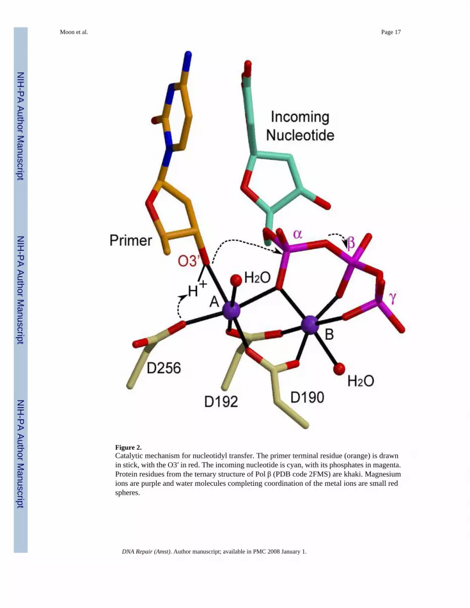
Figure 2.Catalytic mechanism for nucleotidyl transfer. The primer terminal residue (orange) is drawnin stick, with the O3′ in red. The incoming nucleotide is cyan, with its phosphates in magenta.Protein residues from the ternary structure of Pol β (PDB code 2FMS) are khaki. Magnesiumions are purple and water molecules completing coordination of the metal ions are small redspheres.
Moon et al. Page 17
DNA Repair (Amst). Author manuscript; available in PMC 2008 January 1.
NIH
-PA Author Manuscript
NIH
-PA Author Manuscript
NIH
-PA Author Manuscript

Figure 3.Comparison of binary and ternary complexes for Pol β and Pol λ. (A) Superposition of binary(PDB code 1 BPX; protein, light green; DNA, khaki) and ternary (PDB code 2FMS; protein,dark green; DNA, orange) crystal structures of Pol β (right). Left: Amino acid side chainmotions within the active site. Middle: Variations in thumb subdomain position between the‘open’ (light green) and ‘closed’ (dark green) forms of the enzyme. (B) Superposition of binary(PDB code 1XSL; protein, light blue; DNA, khaki) and ternary (PDB code 1XSN; protein,dark blue; DNA, orange) crystal structures of Pol λ (left). Right: Highlights of amino acid sidechain motions in the active site of Pol λ. Binary complex: protein, light blue; DNA khaki.Ternary complex: protein, dark blue; DNA, orange. The incoming nucleotide is drawn in stick(cyan). The magnesium ion in metal site B shown as a purple sphere. (C) Superposition of thepre-catalytic (PDB code 2PFO, orange) and the post-catalytic (PDB code 1XSP, khaki) ternarycomplexes of Pol λ. The dUMPNPP from the pre-catalytic complex is cyan. The manganeseion in metal site A is shown as a yellow sphere, and the magnesium ion in metal site B is shown
Moon et al. Page 18
DNA Repair (Amst). Author manuscript; available in PMC 2008 January 1.
NIH
-PA Author Manuscript
NIH
-PA Author Manuscript
NIH
-PA Author Manuscript

as a purple sphere. The α-phosphate undergoing stereochemical inversion is circled in dashedlight blue.
Moon et al. Page 19
DNA Repair (Amst). Author manuscript; available in PMC 2008 January 1.
NIH
-PA Author Manuscript
NIH
-PA Author Manuscript
NIH
-PA Author Manuscript

Figure 4.Family X polymerase active site organization. (A) Superposition of ternary complexes of Polβ (PDB code 2FMS; protein, green; DNA, khaki) and Pol λ (PDB code 2PFO; protein, blue;DNA, orange). Protein side chains from Pol β are drawn in light green, while those for Pol λare light blue. (B) Superposition of the ternary complexes of Pol β (PDB code 2FMS; protein,green; DNA, khaki) and Pol μ (PDB code 2IHM; protein, maroon; DNA, orange). Protein sidechains from Pol β are drawn in light green, while those for Pol μ are pink. Putative hydrogenbonding interactions between Pol μ H329 and the primer terminal phosphate or the γ-phosphateof the incoming nucleotide are shown as black dashed lines. Magnesium and sodium ions areshown as purple and green spheres, respectively. (C) Superposition of ternary complexes of
Moon et al. Page 20
DNA Repair (Amst). Author manuscript; available in PMC 2008 January 1.
NIH
-PA Author Manuscript
NIH
-PA Author Manuscript
NIH
-PA Author Manuscript

Pol μ (PDB code 2IHM; protein, maroon; DNA, khaki) and TdT apoprotein (PDB code 1JMS;protein, purple). Protein side chains from Pol μ are drawn in pink, while those for TdT are lightpurple. The incoming nucleotide from the structure of Pol μ is drawn in khaki. Magnesium andsodium ions are shown as purple and green spheres, respectively.
Moon et al. Page 21
DNA Repair (Amst). Author manuscript; available in PMC 2008 January 1.
NIH
-PA Author Manuscript
NIH
-PA Author Manuscript
NIH
-PA Author Manuscript

Figure 5.Schematic representation of substrate preferences of Family X polymerases. The primerterminal residue is red and the incoming nucleotide is black. (A) Pol β, Pol λ, and Pol μ canfill small single-strand gaps in a template-dependent manner. (B) Pol λ and Pol μ can fill double-strand gaps with at least one complementary base pair proximal to the gap to be filled. (C) Polμ, aided by Loop I and H329, can fill a gap from a primer terminal residue with nocomplementary template strand nucleotide opposite that position. (D) TdT, with the aid ofLoop I and H342, utilizes DNA substrates with a 3′-primer terminal overhang, and carries outpolymerization in a template-independent manner. Pol μ is also capable of template-independent synthesis in this fashion, but to a lesser extent.
Moon et al. Page 22
DNA Repair (Amst). Author manuscript; available in PMC 2008 January 1.
NIH
-PA Author Manuscript
NIH
-PA Author Manuscript
NIH
-PA Author Manuscript

Figure 6.Comparison of the 8 kDa domain 5′-phosphate binding pocket. Electrostatic surface potentialplots were calculated using the Adaptive Poisson-Boltzmann Solver tool in PyMOL [89]. Thedownstream primer is drawn in orange. All DNA and protein residues are labeled in white.The 5′-phosphate binding pocket in the 8 kDa domain of: (A) Pol β (PDB code 2FMS). (B)Pol λ (PDB code 1XSN). (C) Pol μ (PDB code 2IHM). (D) TdT (PDB code 1JMS). Thedownstream primer from PDB code 2IHM was modeled into the 5′-phosphate binding pocketof TdT, and is drawn in light gray.
Moon et al. Page 23
DNA Repair (Amst). Author manuscript; available in PMC 2008 January 1.
NIH
-PA Author Manuscript
NIH
-PA Author Manuscript
NIH
-PA Author Manuscript

Figure 7.Comparing the 8 kDa domain Helix-hairpin-Helix motifs. (A) HhH motifs from the 8 kDadomains of Pol β (green) and Pol λ (blue) are shown in a ribbon diagram. Side chains in thehairpins of Pol β and Pol λ are light green and light blue, respectively. (B) HhH motifs fromthe 8 kDa domains of Pol μ (maroon) and TdT (purple). Side chains in the hairpins of Pol μand TdT are pink and light purple, respectively. (C) HhH motifs from the 8 kDa domains ofPol μ (maroon) and Pol β (green). Side chains from residues in the hairpins of Pol μ and Polβ are pink and light green, respectively. Distortions in α-helix C are highlighted by a dashedcircle (orange).
Moon et al. Page 24
DNA Repair (Amst). Author manuscript; available in PMC 2008 January 1.
NIH
-PA Author Manuscript
NIH
-PA Author Manuscript
NIH
-PA Author Manuscript

Figure 8.Comparing Loop I in Family X polymerases. Secondary structural elements of Pol β are shownin the background in light gray. The DNA is orange. β-strands 3 and 4 are colored ribbons andlabeled in black. (A) Superposition showing the Loop I region of Pol β (PDB code 2FMS,green) and TdT (PDB code 1JMS, purple). (B) Superposition showing the Loop I region ofTdT (PDB code 1JMS, purple) and Pol μ (PDB code 2IHM, maroon). Loop I is disordered inPol μ. The dashed maroon line shows a hypothetical path of the loop in relation to the DNAbinding cleft. (C) Superposition of ternary complexes showing the Loop I region of Pol β(PDB code 2FMS, green) and Pol λ (PDB code 1XSN, blue).
Moon et al. Page 25
DNA Repair (Amst). Author manuscript; available in PMC 2008 January 1.
NIH
-PA Author Manuscript
NIH
-PA Author Manuscript
NIH
-PA Author Manuscript

Figure 9.Accommodating distorted DNA substrates. (A) Superposition of a gapped substrate with anA-A mismatch at the primer terminus (PDB code 1ZJN, orange) and a normal gapped DNAsubstrate (PDB code 2FMS, khaki). The identities of the primer terminal and nascent base pairsare indicated (normal ternary in khaki, and mismatched ternary in orange). Identities of theDNA bases in the mismatch structure are shown to the right of each base (orange). (B)Superposition of a normal gapped DNA substrate (PDB code 2FMS, khaki) with canonical G-C base pair and a gapped DNA substrate containing an 8oxodG at the templating position (PDBcode 1MQ3, orange), paired with incoming dCTP. (C) Pol λ accommodating an extra base.Superposition of a normal ternary complex of Pol λ (PDB code 1XSN; DNA, orange) with the
Moon et al. Page 26
DNA Repair (Amst). Author manuscript; available in PMC 2008 January 1.
NIH
-PA Author Manuscript
NIH
-PA Author Manuscript
NIH
-PA Author Manuscript
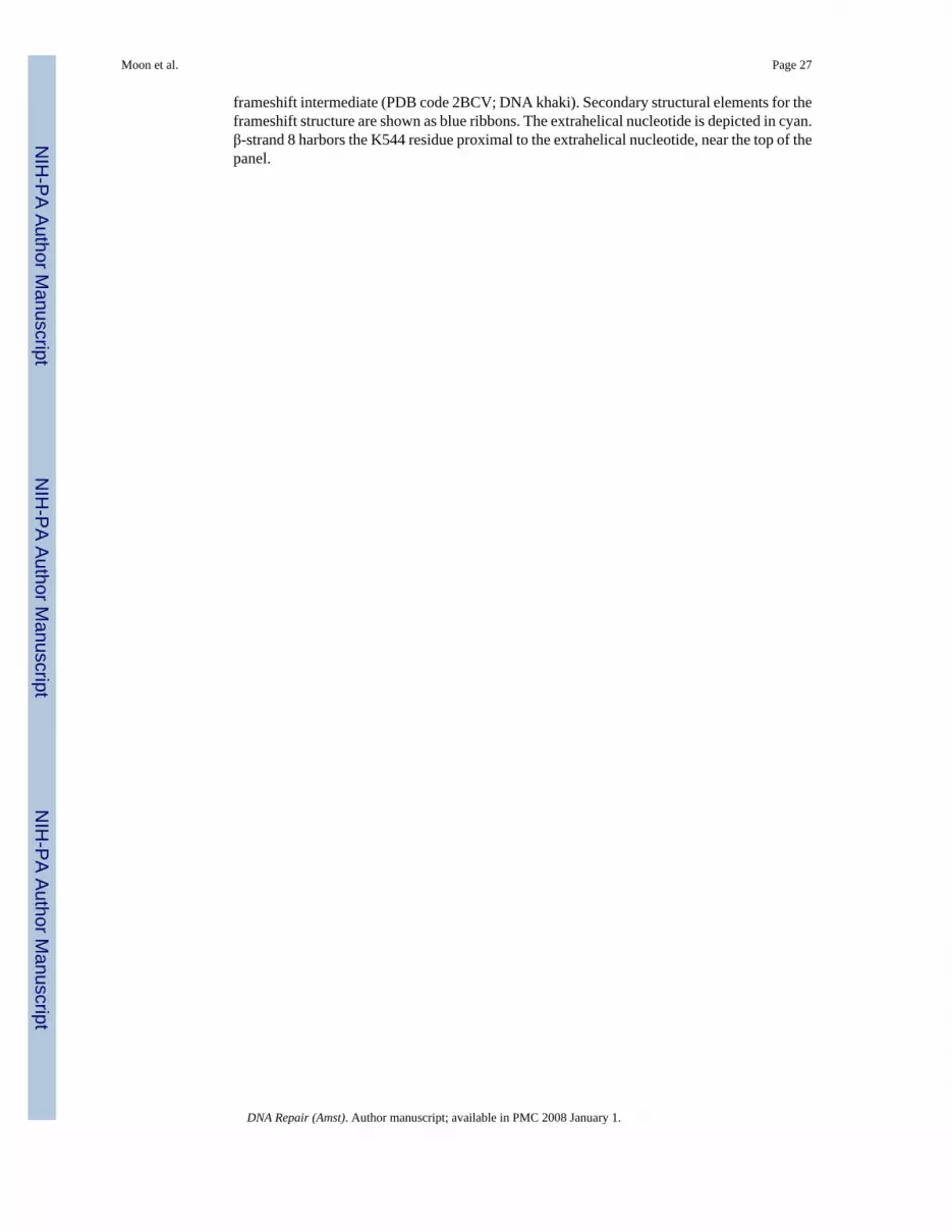
frameshift intermediate (PDB code 2BCV; DNA khaki). Secondary structural elements for theframeshift structure are shown as blue ribbons. The extrahelical nucleotide is depicted in cyan.β-strand 8 harbors the K544 residue proximal to the extrahelical nucleotide, near the top of thepanel.
Moon et al. Page 27
DNA Repair (Amst). Author manuscript; available in PMC 2008 January 1.
NIH
-PA Author Manuscript
NIH
-PA Author Manuscript
NIH
-PA Author Manuscript

NIH
-PA Author Manuscript
NIH
-PA Author Manuscript
NIH
-PA Author Manuscript
Moon et al. Page 28
Table 1Selective list of NMR and X-ray crystal structures for Family X DNA polymerases
Polymerase PDB Code DNA Incoming Nucleotide
hPol β 1BPX 1 nt gap1BPY 1 nt gap ddCTP1BPZ nicked DNA1TV9 mismatch A:C1TVA mismatch T:C1ZJM primer A:A mismatch1ZJN primer A:A mismatch dGTP2FMP 1 nt gap (Na+/Mg+2) ddCTP2FMQ 1 nt gap (Na+/Mg+2) dUMPNPP2FMS 1 nt gap (Mg+2/Mg+2) dUMPNPP1MQ2 gapped DNA 8oxodG dAMP1MQ3 gapped DNA 8oxodG dCTP2I9G B[c]Ph DE adduct ddCTP
hPol λ 1RZT 2nt gap1XSL 1 nt gap1XSN 1 nt gap ddTTP2PFO 1 nt gap (Mn+2/Mg+2) dUMPNPP1XSP nicked DNA PPi2BCQ frameshift (extrahelical dTMP)2BCR frameshift (extrahelical dAMP)2BCS frameshift (extrahelical dCMP)2BCV frameshift (extrahelical dTMP) ddTTP2BCU T:T mismatch (unpaired dAMP)
mTdT 1JMS1KEJ ddATP1KDH brominated 4 nt ssDNA2COE*
mPol μ 2IHM 1nt gap ddTTP2DUN*
Pol β and Pol λ X-ray crystal structures with 39 kDa polymerase domain. Pol μ and TdT X-ray crystal structures with 40kDa polymerase domain. 2COEand 2DUN (marked by *) are NMR spectroscopy structures of the hTdT and hPol μ BRCT domains, respectively.
DNA Repair (Amst). Author manuscript; available in PMC 2008 January 1.

NIH
-PA Author Manuscript
NIH
-PA Author Manuscript
NIH
-PA Author Manuscript
Moon et al. Page 29Ta
ble
2C
ompa
rison
of k
ey st
ruct
ural
and
beh
avio
ral a
spec
ts o
f Fam
ily X
DN
A p
olym
eras
esPo
l βPo
l λPo
l μT
dT8
kDa
dom
ain
8 kD
a do
mai
nLy
ase
activ
ity5′
-pho
spha
te p
ocke
tH
hH1
Yes
Yes
Yes
Yes
Yes
Yes
Yes
Yes
Yes
No
Yes
, few
er p
ositi
vely
cha
rged
resi
dues
Yes
, but
dis
torte
d
Yes
No
Few
pos
itive
ly c
harg
ed re
sidu
esY
es, b
ut d
isto
rted
Cat
alys
is (N
ucle
otid
yl T
rans
fer)
Two-
met
al m
echa
nism
Act
ive
site
ass
embl
yPr
oces
sivi
tyY
F m
otif
Nuc
leot
ide
sele
ctiv
ityD
NA
stra
nd in
tera
ctio
nsM
inor
gro
ove
inte
ract
ions
HhH
2
Yes
Subd
omai
n m
otio
ns &
side
cha
inm
otio
nsPr
oces
sive
with
5′-p
hosp
hate
Yes
dNTP
sM
any,
mos
tly w
ith te
mpl
ate
Y27
1, F
272,
N27
9, R
283
Yes
Yes
DN
A re
posi
tioni
ng, s
ide c
hain
mot
ions
Proc
essi
ve w
ith 5′-p
hosp
hate
Yes
dNTP
sFe
wer
, mos
t with
tem
plat
e op
posi
tepr
imer
Y50
5, F
506,
N51
3, R
517
Yes
Yes
Unk
now
n, b
ut li
kely
few
Dis
tribu
tive
No,
GW
mot
ifdN
TPs &
rNTP
sFe
wer
, mos
t with
ups
tream
prim
erR
447,
pos
sibl
eY
es
Unk
now
n, b
ut li
kely
Unk
now
n, b
ut li
kely
few
Dis
tribu
tive
No,
GW
mot
ifdN
TPs &
rNTP
sU
nkno
wn
Unk
now
nY
es
Subs
trat
e Sp
ecifi
city
Gap
-fill
ing
Tem
plat
e-de
pend
ence
Tem
plat
e-in
depe
nden
ce
Yes
Yes
No
Yes
Yes
No
Yes
Yes
Yes
No
No
Yes
DN
A R
epai
rPa
rtici
patio
n in
BER
Ser/P
ro-r
ich
dom
ain
BR
CT
dom
ain
NH
EJD
SB m
icro
hom
olog
yV
(D)J
reco
mbi
natio
nA
ctiv
e si
te h
istid
ine
Yes
No
No
No
N/A
No
No
Yes
Yes
Yes
Yes
Yes
, nee
ds a
t lea
st o
ne c
ompl
emen
tary
base
pai
rY
es, i
mm
unog
lobu
lin h
eavy
cha
inN
o
No
No
Yes
Yes
No
hom
olog
y re
quire
dY
es, i
mm
unog
lobu
lin li
ght c
hain
Yes
No
No
Yes
Perh
aps
Tem
plat
e-in
depe
nden
tY
es, N
-add
ition
Yes
Fide
lity
Loop
IM
isal
ignm
ent s
ynth
esis
Tran
sles
ion
synt
hesi
sM
ism
atch
ext
ensi
on
No
Yes
, hig
h in
hom
opol
ymer
ic ru
nsSo
me
Som
e
Smal
lY
es, h
igh
erro
r rat
eSo
me
Som
e
Larg
er, n
ot se
en o
cclu
ding
tem
plat
e stra
ndY
es, e
rror
rate
unk
now
nSo
me,
err
or ra
te u
nkno
wn
Som
e, e
rror
rate
unk
now
n
Larg
er, l
ikel
y oc
clud
es te
mpl
ate
stra
nd b
indi
ngTe
mpl
ate-
inde
pend
ent
Tem
plat
e-in
depe
nden
tTe
mpl
ate-
inde
pend
ent
DNA Repair (Amst). Author manuscript; available in PMC 2008 January 1.


