Embed Size (px)
Citation preview
Therapeutic implication ofL-phenylalanine aggregation mechanismand its modulation by D-phenylalanine inphenylketonuriaVirender Singh1, Ratan Kumar Rai2, Ashish Arora3, Neeraj Sinha2 & Ashwani Kumar Thakur1
1Department of Biological Sciences and Bioengineering, Indian Institute of Technology Kanpur 208016, Uttar Pradesh, India,2Centre of Biomedical Research, SGPGIMS Campus, Raibarelly Road, Lucknow 226014, Uttar Pradesh, India, 3CSIR-Central DrugResearch Institute, 10/1, Sector 10, Jankipuram Extension, Sitapur Road, Lucknow 226031, Uttar Pradesh, India.
Self-assembly of phenylalanine is linked to amyloid formation toxicity in phenylketonuria disease. We aredemonstrating that L-phenylalanine self-assembles to amyloid fibrils at varying experimental conditionsand transforms to a gel state at saturated concentration. Biophysical methods including nuclear magneticresonance, resistance by alpha-phenylglycine to fibril formation and preference of protected phenylalanineto self-assemble show that this behaviour of L-phenylalanine is governed mainly by hydrophobicinteractions. Interestingly, D-phenylalanine arrests the fibre formation by L-phenylalanine and gives rise toflakes. These flakes do not propagate further and prevent fibre formation by L-phenylalanine. This suggeststhe use of D-phenylalanine as modulator of L-phenylalanine amyloid formation and may qualify as atherapeutic molecule in phenylketonuria.
Phenylketonuria (PKU) is an inborn metabolic disorder linked with inability of infants to utilize phenyla-lanine (Phe) due to lack of phenylalanine hydroxylase (PAH) enzyme activity caused by a genetic mutation1.This drastically increases blood Phe concentration from normal 50 mM L21 to toxic 1200 mM L21 level2–4. As
a result there is a deficit of tyrosine, dopamine, epinephrine and norepinephrine molecules1. This hampers properbrain development and results in psychiatric disorder in patients2,3. Currently, restricted Phe diet along with largeneutral amino acids (LNAA) and tetrahydrobiopterin therapy are used to reduce toxic effects in PKU patients2.
Recently, Adler et al. for the first time linked Phe self-assembly with PKU5. High build-up of Phe in hip-pocampus and the parietal cortex area of the brain, results in self-assembly and fibril formation. Birefringencecharacteristic upon congo red dye binding, thioflavin T (ThT) binding and electron diffraction confirmed thatthese fibres are amyloidic in nature5. Phe fibres have shown the characteristic cross b-fibre diffraction patternpresent in amyloid fibrils6, associated with several neurodegenerative diseases like Alzheimer’s disease,Huntington’s disease and Atrial amyloidosis7. Presence of these amyloids and intermediate oligomers in differentregions of brain are associated with toxicity, leading to neuronal death6,8. Phe fibres showed similar cell toxicity onPC12 and Chinese Hamster Ovary (CHO) cells5. Importantly, the antibody and histological staining of braintissues from PKU patients have shown the presence of Phe fibrils, indicating amyloid fibril associated toxicity5.Similar, needle like fibrillar morphology was observed under optical microscope for Phe crystals while studyingaspartame gel crystallization9. Phe also form dimers in presence of its cationic and zwitterionic state in aqueoussolution10.
It was estimated by using infrared spectroscopy that the interactions responsible for Phe assembly are mainlyhydrogen bonding between symmetrical –C5O???HOOC– and electrostatic between –NH3
1???2OOC–groups5,11. However, it seems a reasonable assumption that Phe being an aromatic amino acid can also formhydrophobic interactions through p-p stacking. Involvement of these interactions are not established completely,but are predicted to favour self-assembly10. Also, self-assembly of diphenylalanine suggests the importance ofstacking interactions in stabilizing nanostructures12. The p-p stacking interactions leading to association ofaromatic side chain of Phe are involved in molecular recognition13, peptide self-assembly to fibril formation14–16
and neurotoxicity17.
OPEN
SUBJECT AREAS:SMALL MOLECULES
DRUG DISCOVERY
BIOPHYSICAL CHEMISTRY
Received7 October 2013
Accepted6 January 2014
Published27 January 2014
Correspondence andrequests for materials
should be addressed toA.K.T. (akthakur@iitk.
ac.in)
SCIENTIFIC REPORTS | 4 : 3875 | DOI: 10.1038/srep03875 1
The studies mentioned above qualify that Phe forms fibrousmaterial and its importance in disease association. However, themechanism of their formation is not completely deciphered. In thispaper, Phe self-assembly and mechanism leading to fibre formationare reported. Apart from earlier known interactions in Phe self-assembly, we present here the major role played by hydrophobicinteractions involving Phe ring in this process. We have furtherdemonstrated inhibition of L-Phe fibrillar assemblies by D-Pheenantiomer to non-propagating flakes. This study provides one use-ful strategy for arresting the amyloid fibre formation in phenylketo-nuria disease.
ResultsKinetics of phe self-assembly. L-Phe self-assembly was monitoredby light scattering (LS) and thioflavin t (ThT) binding assays.Samples in water (pH 5.8 6 0.3) at different concentrations were
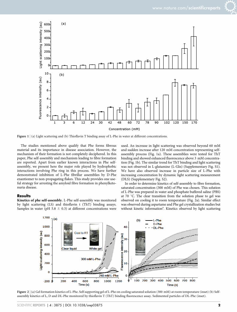
used. An increase in light scattering was observed beyond 60 mMand sudden increase after 120 mM concentration representing self-assembly process (Fig. 1a). These assemblies were tested for ThTbinding and showed enhanced fluorescence above 3 mM concentra-tion (Fig. 1b). The similar trend for ThT binding and light scatteringwas not observed in L-glutamine (L-Gln) (Supplementary Fig. S1).We have also observed increase in particle size of L-Phe withincreasing concentration by dynamic light scattering measurement(DLS) (Supplementary Fig. S2).
In order to determine kinetics of self-assembly to fibre formation,saturated concentration (300 mM) of Phe was chosen. This solutionof L-Phe was prepared in water and phosphate buffered saline (PBS)at 70 uC. The clear transition from the solution phase to gel wasobserved on cooling it to room temperature (Fig. 2a). Similar effectwas observed during aspartame and Phe gel crystallization studies butwithout kinetic information9. Kinetics observed by light scattering
Figure 1 | (a) Light scattering and (b) Thioflavin T binding assay of L-Phe in water at different concentrations.
Figure 2 | (a) Gel formation kinetics of L-Phe. Self supporting gel of L-Phe on cooling saturated solution (300 mM) at room temperature (inset) (b) Self-
assembly kinetics of L, D and DL-Phe monitored by thioflavin T (ThT) binding fluorescence assay. Sedimented particles of DL-Phe (inset).
www.nature.com/scientificreports
SCIENTIFIC REPORTS | 4 : 3875 | DOI: 10.1038/srep03875 2
analysis showed the appearance of a lag phase followed by an expo-nential phase (Fig. 2a). An initial lag might be due to nucleationevents where molecules associate to form productive nucleus forfurther elongation into mature fibres or the associated moleculesare insensitive to light scattering. In such case, seeding with pre-formed aggregates could abrogate this phase and the reaction willseed quickly for elongation18.
When 10% (w/w) preformed L-Phe aggregates were incubatedwith saturated L-Phe solution, it abolished lag phase and gel forma-tion was so quick that it was practically difficult to monitor it by lightscattering and ThT binding due to signal saturation. This is an inter-esting finding in Phe assembly. Similar behaviour of lag and elonga-tion phase was reported for amyloid forming peptides19 such aspolyglutamine in Huntington’s disease18. Interestingly, ThT bindingassay showed enhanced fluorescence even at earlier time points andlower concentrations suggesting early formation of ThT positiveassemblies (Fig. 1b and Fig. 2b) which corroborates well with DLSdata (Supplementary Fig. S2). L-Phe in PBS buffer showed similarkinetics and gel formation behaviour as in water (Supplementary Fig.S3). Hence, water was used for further molecular interaction studiesas also used by Adler et al. for most experiments5.
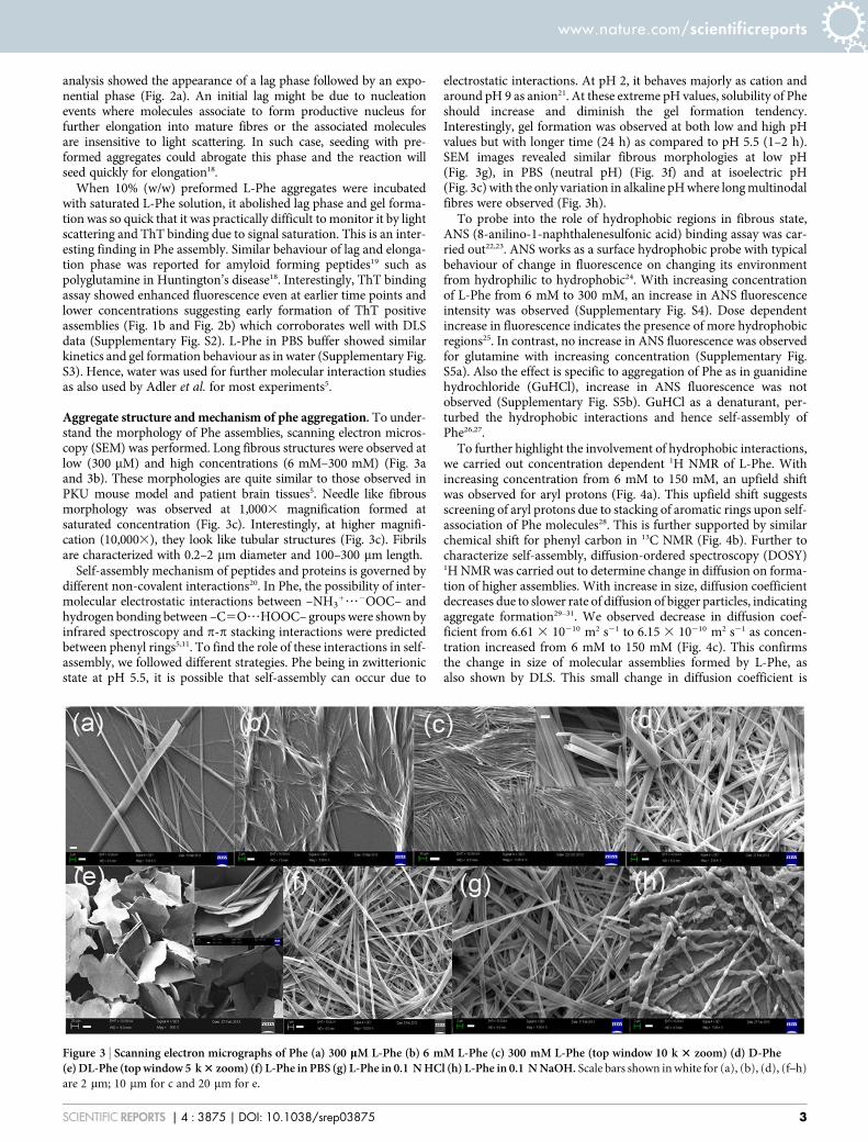
Aggregate structure and mechanism of phe aggregation. To under-stand the morphology of Phe assemblies, scanning electron micros-copy (SEM) was performed. Long fibrous structures were observed atlow (300 mM) and high concentrations (6 mM–300 mM) (Fig. 3aand 3b). These morphologies are quite similar to those observed inPKU mouse model and patient brain tissues5. Needle like fibrousmorphology was observed at 1,0003 magnification formed atsaturated concentration (Fig. 3c). Interestingly, at higher magnifi-cation (10,0003), they look like tubular structures (Fig. 3c). Fibrilsare characterized with 0.2–2 mm diameter and 100–300 mm length.
Self-assembly mechanism of peptides and proteins is governed bydifferent non-covalent interactions20. In Phe, the possibility of inter-molecular electrostatic interactions between –NH3
1???2OOC– andhydrogen bonding between –C5O???HOOC– groups were shown byinfrared spectroscopy and p-p stacking interactions were predictedbetween phenyl rings5,11. To find the role of these interactions in self-assembly, we followed different strategies. Phe being in zwitterionicstate at pH 5.5, it is possible that self-assembly can occur due to
electrostatic interactions. At pH 2, it behaves majorly as cation andaround pH 9 as anion21. At these extreme pH values, solubility of Pheshould increase and diminish the gel formation tendency.Interestingly, gel formation was observed at both low and high pHvalues but with longer time (24 h) as compared to pH 5.5 (1–2 h).SEM images revealed similar fibrous morphologies at low pH(Fig. 3g), in PBS (neutral pH) (Fig. 3f) and at isoelectric pH(Fig. 3c) with the only variation in alkaline pH where long multinodalfibres were observed (Fig. 3h).
To probe into the role of hydrophobic regions in fibrous state,ANS (8-anilino-1-naphthalenesulfonic acid) binding assay was car-ried out22,23. ANS works as a surface hydrophobic probe with typicalbehaviour of change in fluorescence on changing its environmentfrom hydrophilic to hydrophobic24. With increasing concentrationof L-Phe from 6 mM to 300 mM, an increase in ANS fluorescenceintensity was observed (Supplementary Fig. S4). Dose dependentincrease in fluorescence indicates the presence of more hydrophobicregions25. In contrast, no increase in ANS fluorescence was observedfor glutamine with increasing concentration (Supplementary Fig.S5a). Also the effect is specific to aggregation of Phe as in guanidinehydrochloride (GuHCl), increase in ANS fluorescence was notobserved (Supplementary Fig. S5b). GuHCl as a denaturant, per-turbed the hydrophobic interactions and hence self-assembly ofPhe26,27.
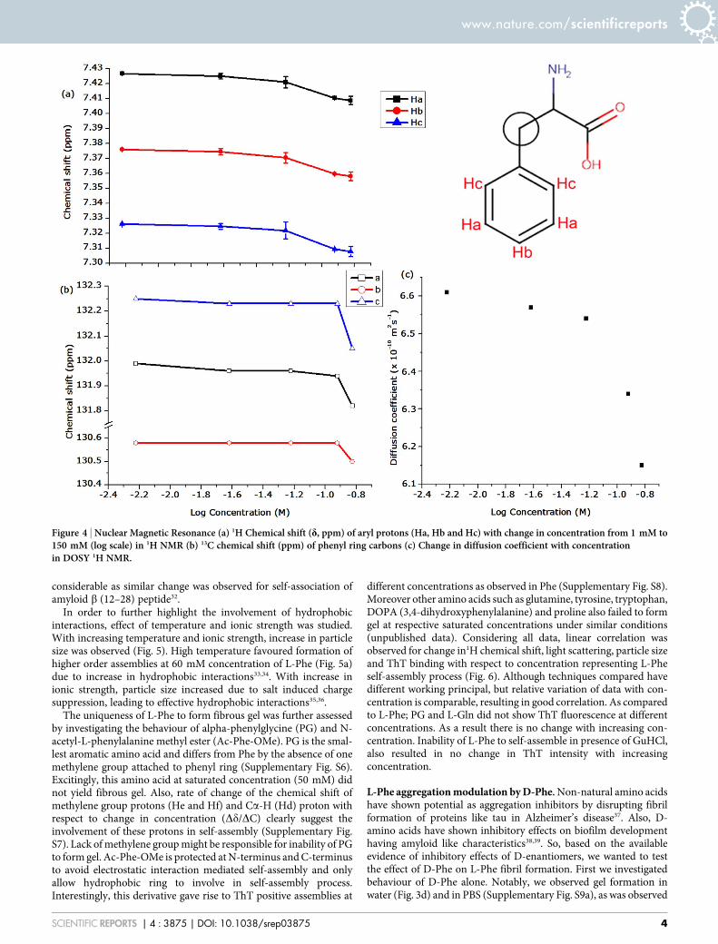
To further highlight the involvement of hydrophobic interactions,we carried out concentration dependent 1H NMR of L-Phe. Withincreasing concentration from 6 mM to 150 mM, an upfield shiftwas observed for aryl protons (Fig. 4a). This upfield shift suggestsscreening of aryl protons due to stacking of aromatic rings upon self-association of Phe molecules28. This is further supported by similarchemical shift for phenyl carbon in 13C NMR (Fig. 4b). Further tocharacterize self-assembly, diffusion-ordered spectroscopy (DOSY)1H NMR was carried out to determine change in diffusion on forma-tion of higher assemblies. With increase in size, diffusion coefficientdecreases due to slower rate of diffusion of bigger particles, indicatingaggregate formation29–31. We observed decrease in diffusion coef-ficient from 6.61 3 10210 m2 s21 to 6.15 3 10210 m2 s21 as concen-tration increased from 6 mM to 150 mM (Fig. 4c). This confirmsthe change in size of molecular assemblies formed by L-Phe, asalso shown by DLS. This small change in diffusion coefficient is
Figure 3 | Scanning electron micrographs of Phe (a) 300 mM L-Phe (b) 6 mM L-Phe (c) 300 mM L-Phe (top window 10 k 3 zoom) (d) D-Phe(e) DL-Phe (top window 5 k 3 zoom) (f) L-Phe in PBS (g) L-Phe in 0.1 N HCl (h) L-Phe in 0.1 N NaOH. Scale bars shown in white for (a), (b), (d), (f–h)
are 2 mm; 10 mm for c and 20 mm for e.
www.nature.com/scientificreports
SCIENTIFIC REPORTS | 4 : 3875 | DOI: 10.1038/srep03875 3
considerable as similar change was observed for self-association ofamyloid b (12–28) peptide32.
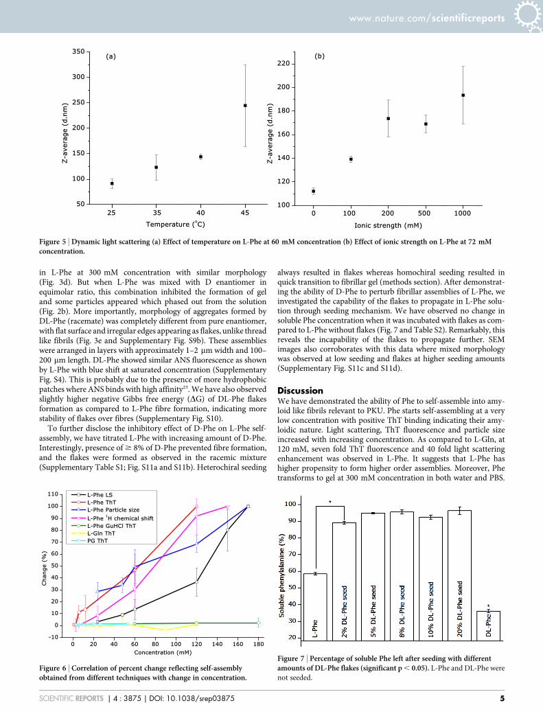
In order to further highlight the involvement of hydrophobicinteractions, effect of temperature and ionic strength was studied.With increasing temperature and ionic strength, increase in particlesize was observed (Fig. 5). High temperature favoured formation ofhigher order assemblies at 60 mM concentration of L-Phe (Fig. 5a)due to increase in hydrophobic interactions33,34. With increase inionic strength, particle size increased due to salt induced chargesuppression, leading to effective hydrophobic interactions35,36.
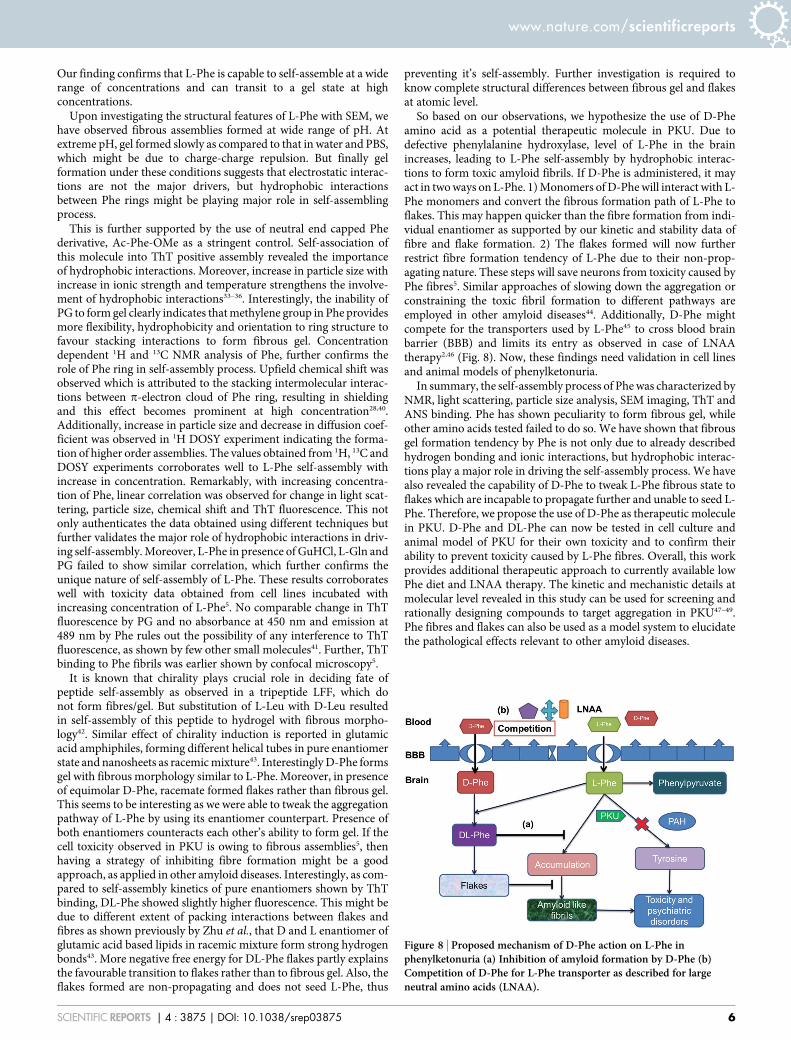
The uniqueness of L-Phe to form fibrous gel was further assessedby investigating the behaviour of alpha-phenylglycine (PG) and N-acetyl-L-phenylalanine methyl ester (Ac-Phe-OMe). PG is the smal-lest aromatic amino acid and differs from Phe by the absence of onemethylene group attached to phenyl ring (Supplementary Fig. S6).Excitingly, this amino acid at saturated concentration (50 mM) didnot yield fibrous gel. Also, rate of change of the chemical shift ofmethylene group protons (He and Hf) and Ca-H (Hd) proton withrespect to change in concentration (Dd/DC) clearly suggest theinvolvement of these protons in self-assembly (Supplementary Fig.S7). Lack of methylene group might be responsible for inability of PGto form gel. Ac-Phe-OMe is protected at N-terminus and C-terminusto avoid electrostatic interaction mediated self-assembly and onlyallow hydrophobic ring to involve in self-assembly process.Interestingly, this derivative gave rise to ThT positive assemblies at
different concentrations as observed in Phe (Supplementary Fig. S8).Moreover other amino acids such as glutamine, tyrosine, tryptophan,DOPA (3,4-dihydroxyphenylalanine) and proline also failed to formgel at respective saturated concentrations under similar conditions(unpublished data). Considering all data, linear correlation wasobserved for change in1H chemical shift, light scattering, particle sizeand ThT binding with respect to concentration representing L-Pheself-assembly process (Fig. 6). Although techniques compared havedifferent working principal, but relative variation of data with con-centration is comparable, resulting in good correlation. As comparedto L-Phe; PG and L-Gln did not show ThT fluorescence at differentconcentrations. As a result there is no change with increasing con-centration. Inability of L-Phe to self-assemble in presence of GuHCl,also resulted in no change in ThT intensity with increasingconcentration.
L-Phe aggregation modulation by D-Phe. Non-natural amino acidshave shown potential as aggregation inhibitors by disrupting fibrilformation of proteins like tau in Alzheimer’s disease37. Also, D-amino acids have shown inhibitory effects on biofilm developmenthaving amyloid like characteristics38,39. So, based on the availableevidence of inhibitory effects of D-enantiomers, we wanted to testthe effect of D-Phe on L-Phe fibril formation. First we investigatedbehaviour of D-Phe alone. Notably, we observed gel formation inwater (Fig. 3d) and in PBS (Supplementary Fig. S9a), as was observed
Figure 4 | Nuclear Magnetic Resonance (a) 1H Chemical shift (d, ppm) of aryl protons (Ha, Hb and Hc) with change in concentration from 1 mM to150 mM (log scale) in 1H NMR (b) 13C chemical shift (ppm) of phenyl ring carbons (c) Change in diffusion coefficient with concentrationin DOSY 1H NMR.
www.nature.com/scientificreports
SCIENTIFIC REPORTS | 4 : 3875 | DOI: 10.1038/srep03875 4
in L-Phe at 300 mM concentration with similar morphology(Fig. 3d). But when L-Phe was mixed with D enantiomer inequimolar ratio, this combination inhibited the formation of geland some particles appeared which phased out from the solution(Fig. 2b). More importantly, morphology of aggregates formed byDL-Phe (racemate) was completely different from pure enantiomer,with flat surface and irregular edges appearing as flakes, unlike threadlike fibrils (Fig. 3e and Supplementary Fig. S9b). These assemblieswere arranged in layers with approximately 1–2 mm width and 100–200 mm length. DL-Phe showed similar ANS fluorescence as shownby L-Phe with blue shift at saturated concentration (SupplementaryFig. S4). This is probably due to the presence of more hydrophobicpatches where ANS binds with high affinity23. We have also observedslightly higher negative Gibbs free energy (DG) of DL-Phe flakesformation as compared to L-Phe fibre formation, indicating morestability of flakes over fibres (Supplementary Fig. S10).
To further disclose the inhibitory effect of D-Phe on L-Phe self-assembly, we have titrated L-Phe with increasing amount of D-Phe.Interestingly, presence of $ 8% of D-Phe prevented fibre formation,and the flakes were formed as observed in the racemic mixture(Supplementary Table S1; Fig. S11a and S11b). Heterochiral seeding
always resulted in flakes whereas homochiral seeding resulted inquick transition to fibrillar gel (methods section). After demonstrat-ing the ability of D-Phe to perturb fibrillar assemblies of L-Phe, weinvestigated the capability of the flakes to propagate in L-Phe solu-tion through seeding mechanism. We have observed no change insoluble Phe concentration when it was incubated with flakes as com-pared to L-Phe without flakes (Fig. 7 and Table S2). Remarkably, thisreveals the incapability of the flakes to propagate further. SEMimages also corroborates with this data where mixed morphologywas observed at low seeding and flakes at higher seeding amounts(Supplementary Fig. S11c and S11d).
DiscussionWe have demonstrated the ability of Phe to self-assemble into amy-loid like fibrils relevant to PKU. Phe starts self-assembling at a verylow concentration with positive ThT binding indicating their amy-loidic nature. Light scattering, ThT fluorescence and particle sizeincreased with increasing concentration. As compared to L-Gln, at120 mM, seven fold ThT fluorescence and 40 fold light scatteringenhancement was observed in L-Phe. It suggests that L-Phe hashigher propensity to form higher order assemblies. Moreover, Phetransforms to gel at 300 mM concentration in both water and PBS.
Figure 5 | Dynamic light scattering (a) Effect of temperature on L-Phe at 60 mM concentration (b) Effect of ionic strength on L-Phe at 72 mMconcentration.
Figure 6 | Correlation of percent change reflecting self-assemblyobtained from different techniques with change in concentration.
Figure 7 | Percentage of soluble Phe left after seeding with differentamounts of DL-Phe flakes (significant p , 0.05). L-Phe and DL-Phe were
not seeded.
www.nature.com/scientificreports
SCIENTIFIC REPORTS | 4 : 3875 | DOI: 10.1038/srep03875 5
Our finding confirms that L-Phe is capable to self-assemble at a widerange of concentrations and can transit to a gel state at highconcentrations.
Upon investigating the structural features of L-Phe with SEM, wehave observed fibrous assemblies formed at wide range of pH. Atextreme pH, gel formed slowly as compared to that in water and PBS,which might be due to charge-charge repulsion. But finally gelformation under these conditions suggests that electrostatic interac-tions are not the major drivers, but hydrophobic interactionsbetween Phe rings might be playing major role in self-assemblingprocess.
This is further supported by the use of neutral end capped Phederivative, Ac-Phe-OMe as a stringent control. Self-association ofthis molecule into ThT positive assembly revealed the importanceof hydrophobic interactions. Moreover, increase in particle size withincrease in ionic strength and temperature strengthens the involve-ment of hydrophobic interactions33–36. Interestingly, the inability ofPG to form gel clearly indicates that methylene group in Phe providesmore flexibility, hydrophobicity and orientation to ring structure tofavour stacking interactions to form fibrous gel. Concentrationdependent 1H and 13C NMR analysis of Phe, further confirms therole of Phe ring in self-assembly process. Upfield chemical shift wasobserved which is attributed to the stacking intermolecular interac-tions between p-electron cloud of Phe ring, resulting in shieldingand this effect becomes prominent at high concentration28,40.Additionally, increase in particle size and decrease in diffusion coef-ficient was observed in 1H DOSY experiment indicating the forma-tion of higher order assemblies. The values obtained from 1H, 13C andDOSY experiments corroborates well to L-Phe self-assembly withincrease in concentration. Remarkably, with increasing concentra-tion of Phe, linear correlation was observed for change in light scat-tering, particle size, chemical shift and ThT fluorescence. This notonly authenticates the data obtained using different techniques butfurther validates the major role of hydrophobic interactions in driv-ing self-assembly. Moreover, L-Phe in presence of GuHCl, L-Gln andPG failed to show similar correlation, which further confirms theunique nature of self-assembly of L-Phe. These results corroborateswell with toxicity data obtained from cell lines incubated withincreasing concentration of L-Phe5. No comparable change in ThTfluorescence by PG and no absorbance at 450 nm and emission at489 nm by Phe rules out the possibility of any interference to ThTfluorescence, as shown by few other small molecules41. Further, ThTbinding to Phe fibrils was earlier shown by confocal microscopy5.
It is known that chirality plays crucial role in deciding fate ofpeptide self-assembly as observed in a tripeptide LFF, which donot form fibres/gel. But substitution of L-Leu with D-Leu resultedin self-assembly of this peptide to hydrogel with fibrous morpho-logy42. Similar effect of chirality induction is reported in glutamicacid amphiphiles, forming different helical tubes in pure enantiomerstate and nanosheets as racemic mixture43. Interestingly D-Phe formsgel with fibrous morphology similar to L-Phe. Moreover, in presenceof equimolar D-Phe, racemate formed flakes rather than fibrous gel.This seems to be interesting as we were able to tweak the aggregationpathway of L-Phe by using its enantiomer counterpart. Presence ofboth enantiomers counteracts each other’s ability to form gel. If thecell toxicity observed in PKU is owing to fibrous assemblies5, thenhaving a strategy of inhibiting fibre formation might be a goodapproach, as applied in other amyloid diseases. Interestingly, as com-pared to self-assembly kinetics of pure enantiomers shown by ThTbinding, DL-Phe showed slightly higher fluorescence. This might bedue to different extent of packing interactions between flakes andfibres as shown previously by Zhu et al., that D and L enantiomer ofglutamic acid based lipids in racemic mixture form strong hydrogenbonds43. More negative free energy for DL-Phe flakes partly explainsthe favourable transition to flakes rather than to fibrous gel. Also, theflakes formed are non-propagating and does not seed L-Phe, thus
preventing it’s self-assembly. Further investigation is required toknow complete structural differences between fibrous gel and flakesat atomic level.
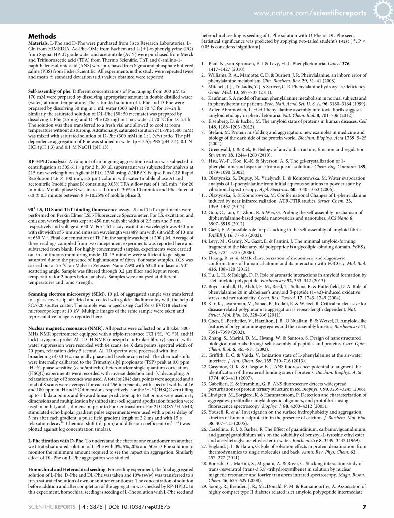
So based on our observations, we hypothesize the use of D-Pheamino acid as a potential therapeutic molecule in PKU. Due todefective phenylalanine hydroxylase, level of L-Phe in the brainincreases, leading to L-Phe self-assembly by hydrophobic interac-tions to form toxic amyloid fibrils. If D-Phe is administered, it mayact in two ways on L-Phe. 1) Monomers of D-Phe will interact with L-Phe monomers and convert the fibrous formation path of L-Phe toflakes. This may happen quicker than the fibre formation from indi-vidual enantiomer as supported by our kinetic and stability data offibre and flake formation. 2) The flakes formed will now furtherrestrict fibre formation tendency of L-Phe due to their non-prop-agating nature. These steps will save neurons from toxicity caused byPhe fibres5. Similar approaches of slowing down the aggregation orconstraining the toxic fibril formation to different pathways areemployed in other amyloid diseases44. Additionally, D-Phe mightcompete for the transporters used by L-Phe45 to cross blood brainbarrier (BBB) and limits its entry as observed in case of LNAAtherapy2,46 (Fig. 8). Now, these findings need validation in cell linesand animal models of phenylketonuria.
In summary, the self-assembly process of Phe was characterized byNMR, light scattering, particle size analysis, SEM imaging, ThT andANS binding. Phe has shown peculiarity to form fibrous gel, whileother amino acids tested failed to do so. We have shown that fibrousgel formation tendency by Phe is not only due to already describedhydrogen bonding and ionic interactions, but hydrophobic interac-tions play a major role in driving the self-assembly process. We havealso revealed the capability of D-Phe to tweak L-Phe fibrous state toflakes which are incapable to propagate further and unable to seed L-Phe. Therefore, we propose the use of D-Phe as therapeutic moleculein PKU. D-Phe and DL-Phe can now be tested in cell culture andanimal model of PKU for their own toxicity and to confirm theirability to prevent toxicity caused by L-Phe fibres. Overall, this workprovides additional therapeutic approach to currently available lowPhe diet and LNAA therapy. The kinetic and mechanistic details atmolecular level revealed in this study can be used for screening andrationally designing compounds to target aggregation in PKU47–49.Phe fibres and flakes can also be used as a model system to elucidatethe pathological effects relevant to other amyloid diseases.
Figure 8 | Proposed mechanism of D-Phe action on L-Phe inphenylketonuria (a) Inhibition of amyloid formation by D-Phe (b)Competition of D-Phe for L-Phe transporter as described for largeneutral amino acids (LNAA).
www.nature.com/scientificreports
SCIENTIFIC REPORTS | 4 : 3875 | DOI: 10.1038/srep03875 6
MethodsMaterials. L-Phe and D-Phe were purchased from Sisco Research Laboratories, L-Gln from HIMEDIA, Ac-Phe-OMe from Bachem and L-(1)-a-phenylglycine (PG)from Sigma. HPLC grade water and acetonitrile (ACN) were purchased from Merckand Trifluoroacetic acid (TFA) from Thermo Scientific. ThT and 8-anilino-1-naphthalenesulfonic acid (ANS) were purchased from Sigma and phosphate bufferedsaline (PBS) from Fisher Scientific. All experiments in this study were repeated twiceand mean 6 standard deviation (s.d.) values obtained were reported.
Self-assembly of phe. Different concentrations of Phe ranging from 300 mM to170 mM were prepared by dissolving appropriate amount in double distilled water(water) at room temperature. The saturated solution of L-Phe and D-Phe wereprepared by dissolving 50 mg in 1 mL water (300 mM) at 70 uC for 18–24 h.Similarly the saturated solution of DL-Phe (50550 racemate) was prepared bydissolving L-Phe (25 mg) and D-Phe (25 mg) in 1 mL water at 70 uC for 18–24 h.The solution was then transferred to a fresh vial and allowed to cool at roomtemperature without disturbing. Additionally, saturated solution of L-Phe (300 mM)was mixed with saturated solution of D-Phe (300 mM) in 151 (v/v) ratio. The pHdependence aggregation of Phe was studied in water (pH 5.5); PBS (pH 7.4); 0.1 NHCl (pH 1.3) and 0.1 M NaOH (pH 13).
RP-HPLC analysis. An aliquot of an ongoing aggregation reaction was subjected tocentrifugation at 305,6113g for 2 h. 30 mL supernatant was subjected for analysis at215 nm wavelength on Agilent HPLC 1260 using ZORBAX Eclipse Plus C18 RapidResolution (4.6 3 100 mm, 3.5 mm) column with water (mobile phase A) andacetonitrile (mobile phase B) containing 0.05% TFA at flow rate of 1 mL min21 for 20minutes. Mobile phase B was increased from 0–30% in 10 minutes and Phe eluted at6.0 6 0.3 minute between 8.8–10.25% of mobile phase B.
906 LS, DLS and ThT binding fluorescence assay. LS and ThT experiments wereperformed on Perkin Elmer LS55 Fluorescence Spectrometer. For LS, excitation andemission wavelength was kept at 450 nm with slit width of 2.5 nm and 5 nmrespectively and voltage at 650 V. For ThT assay, excitation wavelength was 450 nmwith slit width of 5 nm and emission wavelength was 489 nm with slit width of 10 nmat 650 V19. Final concentration of ThT in the sample was kept at 200 mM. Average ofthree readings compiled from two independent experiments was reported here andsubtracted from blank. For highly concentrated samples, experiments were carriedout in continuous monitoring mode. 10–15 minutes were sufficient to get signalsaturated due to the presence of high amount of fibres. For same samples, DLS wascarried out at 25 uC on Malvern Zetasizer Nano ZS90 with 632.8 nm laser at 90uscattering angle. Sample was filtered through 0.2 mm filter and kept at roomtemperature for 2 hours before analysis. Samples were analysed at differenttemperatures and ionic strength.
Scanning electron microscopy (SEM). 10 mL of aggregated sample was transferredto a glass cover slip, air dried and coated with gold/palladium alloy with the help ofSC7620 sputter coater. The sample was imaged using Carl Zeiss EVO18 electronmicroscope kept at 10 kV. Multiple images of the same sample were taken andrepresentative image is reported here.
Nuclear magnetic resonance (NMR). All spectra were collected on a Bruker 800-MHz NMR spectrometer equipped with a triple-resonance TCI (1H, 13C,15N, and2Hlock) cryogenic probe. All 1D 1H NMR (noesypr1d in Bruker library) spectra withwater suppression were recorded with 64 scans, 64 K data points, spectral width of20 ppm, relaxation delay 5 second. All 1D spectra were processed with linebroadening of 0.3 Hz, manually phase and baseline corrected. The chemical shiftswere internally calibrated to the Trimethylsilyl propionate (TSP) peak at 0.0 ppm.1H-13C phase sensitive (echo/antiecho) heteronuclear single quantum correlation(HSQC) experiments were recorded with inverse detection and 13C decoupling. Arelaxation delay of 2 seconds was used. A total of 2048 data points were acquired and atotal of 8 scans were averaged for each of 256 increments, with spectral widths of 16and 180 ppm in 1H and 13C dimensions respectively. For the 1H-13C HSQC zero fillingup to 1 k data points and forward linear prediction up to 128 points were used in t1
dimensions and multiplication by shifted sine-bell squared apodization function wereused in both t2 and t1 dimension prior to Fourier transform. For 2D DOSY 1H NMR,stimulated echo bipolar gradient pulse experiments were used with a pulse delay of5 ms after each gradient, a pulse field gradient length of 2.2 ms and with 15 srelaxation decay50. Chemical shift ( d, ppm) and diffusion coefficient (m2 s21) wasplotted against log concentration (molar).
L-Phe titration with D-Phe. To understand the effect of one enantiomer on another,we titrated saturated solution of L-Phe with 0%, 5%, 20% and 50% D-Phe solution tomonitor the minimum amount required to see the impact on aggregation. Similarlyeffect of DL-Phe on L-Phe aggregation was studied.
Homochiral and Heterochiral seeding. For seeding experiment, the final aggregatedsolution of L-Phe, D-Phe and DL-Phe was taken and 10% (w/w) was transferred to afresh saturated solution of own or another enantiomer. The concentration of solutionbefore addition and after completion of the aggregation was checked by RP-HPLC. Inthis experiment, homochiral seeding is seeding of L-Phe solution with L-Phe seed and
heterochiral seeding is seeding of L-Phe solution with D-Phe or DL-Phe seed.Statistical significance was predicted by applying two-tailed student’s t-test [ *, P ,
0.05 is considered significant].
1. Blau, N., van Spronsen, F. J. & Levy, H. L. Phenylketonuria. Lancet 376,1417–1427 (2010).
2. Williams, R. A., Mamotte, C. D. & Burnett, J. R. Phenylalanine: an inborn error ofphenylalanine metabolism. Clin. Biochem. Rev. 29, 31–41 (2008).
3. Mitchell, J. J., Trakadis, Y. J. & Scriver, C. R. Phenylalanine hydroxylase deficiency.Genet. Med. 13, 697–707 (2011).
4. Kaufman, S. A model of human phenylalanine metabolism in normal subjects andin phenylketonuric patients. Proc. Natl. Acad. Sci. U. S. A. 96, 3160–3164 (1999).
5. Adler-Abramovich, L. et al. Phenylalanine assembly into toxic fibrils suggestsamyloid etiology in phenylketonuria. Nat. Chem. Biol. 8, 701–706 (2012).
6. Eisenberg, D. & Jucker, M. The amyloid state of proteins in human diseases. Cell148, 1188–1203 (2012).
7. Stefani, M. Protein misfolding and aggregation: new examples in medicine andbiology of the dark side of the protein world. Biochim. Biophys. Acta 1739, 5–25(2004).
8. Greenwald, J. & Riek, R. Biology of amyloid: structure, function and regulation.Structure 18, 1244–1260 (2010).
9. Hsu, W.-P., Koo, K.-K. & Myerson, A. S. The gel-crystallization of 1-phenylalanine and aspartame from aqueous solutions. Chem. Eng. Commun. 189,1079–1090 (2002).
10. Olsztynska, S., Dupuy, N., Vrielynck, L. & Komorowska, M. Water evaporationanalysis of L-phenylalanine from initial aqueous solutions to powder state byvibrational spectroscopy. Appl. Spectrosc. 60, 1040–1053 (2006).
11. Olsztynska, S. & Komorowska, M. Conformational Changes of L-phenylalanineinduced by near infrared radiation. ATR-FTIR studies. Struct. Chem. 23,1399–1407 (2012).
12. Guo, C., Luo, Y., Zhou, R. & Wei, G. Probing the self-assembly mechanism ofdiphenylalanine-based peptide nanovesicles and nanotubes. ACS Nano 6,3907–3918 (2012).
13. Gazit, E. A possible role for pi-stacking in the self-assembly of amyloid fibrils.FASEB J. 16, 77–83 (2002).
14. Levy, M., Garmy, N., Gazit, E. & Fantini, J. The minimal amyloid-formingfragment of the islet amyloid polypeptide is a glycolipid-binding domain. FEBS J.273, 5724–5735 (2006).
15. Huang, R. et al. NMR characterization of monomeric and oligomericconformations of human calcitonin and its interaction with EGCG. J. Mol. Biol.416, 108–120 (2012).
16. Tu, L. H. & Raleigh, D. P. Role of aromatic interactions in amyloid formation byislet amyloid polypeptide. Biochemistry 52, 333–342 (2013).
17. Boyd-kimball, D., Abdul, H. M., Reed, T., Sultana, R. & Butterfield, D. A. Role ofphenylalanine 20 in alzheimer’s amyloid b-peptide (1–42)-induced oxidativestress and neurotoxicity. Chem. Res. Toxicol. 17, 1743–1749 (2004).
18. Kar, K., Jayaraman, M., Sahoo, B., Kodali, R. & Wetzel, R. Critical nucleus size fordisease-related polyglutamine aggregation is repeat-length dependent. Nat.Struct. Mol. Biol. 18, 328–336 (2011).
19. Chen, S., Berthelier, V., Hamilton, J. B., O’Nuallain, B. & Wetzel, R. Amyloid-likefeatures of polyglutamine aggregates and their assembly kinetics. Biochemistry 41,7391–7399 (2002).
20. Zhang, S., Marini, D. M., Hwang, W. & Santoso, S. Design of nanostructuredbiological materials through self-assembly of peptides and proteins. Curr. Opin.Chem. Biol. 6, 865–871 (2002).
21. Griffith, E. C. & Vaida, V. Ionization state of L-phenylalanine at the air-waterinterface. J. Am. Chem. Soc. 135, 710–716 (2013).
22. Gasymov, O. K. & Glasgow, B. J. ANS fluorescence: potential to augment theidentification of the external binding sites of proteins. Biochim. Biophys. Acta1774, 403–411 (2007).
23. Gabellieri, E. & Strambini, G. B. ANS fluorescence detects widespreadperturbations of protein tertiary structure in ice. Biophys. J. 90, 3239–3245 (2006).
24. Lindgren, M., Sorgjerd, K. & Hammarstrom, P. Detection and characterization ofaggregates, prefibrillar amyloidogenic oligomers, and protofibrils usingfluorescence spectroscopy. Biophys. J. 88, 4200–4212 (2005).
25. Yousefi, R. et al. Investigation on the surface hydrophobicity and aggregationkinetics of human calprotectin in the presence of calcium. J. Biochem. Mol. Biol.38, 407–413 (2005).
26. Castellino, F. J. & Barker, R. The Effect of guanidinium, carbamoylguanidinium,and guanylguanidinium salts on the solubility of benzoyl-L-tyrosine ethyl esterand acetyltetraglycine ethyl ester in water. Biochemistry 8, 3439–3442 (1969).
27. England, J. L. & Haran, G. Role of solvation effects in protein denaturation: fromthermodynamics to single molecules and back. Annu. Rev. Phys. Chem. 62,257–277 (2011).
28. Bonechi, C., Martini, S., Magnani, A. & Rossi, C. Stacking interaction study oftrans-resveratrol (trans-3,5,49-trihydroxystilbene) in solution by nuclearmagnetic resonance and fourier transform infrared spectroscopy. Magn. Reson.Chem. 46, 625–629 (2008).
29. Soong, R., Brender, J. R., MacDonald, P. M. & Ramamoorthy, A. Association ofhighly compact type II diabetes related islet amyloid polypeptide intermediate
www.nature.com/scientificreports
SCIENTIFIC REPORTS | 4 : 3875 | DOI: 10.1038/srep03875 7
species at physiological temperature revealed by diffusion NMR spectroscopy.J. Am. Chem. Soc. 131, 7079–7085 (2008).
30. Cohen, Y., Avram, L. & Frish, L. Diffusion NMR spectroscopy in supramolecularand combinatorial chemistry: an old parameter-new insights. Angew. Chem. Int.Ed. 44, 520–554 (2005).
31. Macchioni, A., Ciancaleoni, G., Zuccaccia, C. & Zuccaccia, D. Determiningaccurate molecular sizes in solution through NMR diffusion spectroscopy. Chem.Soc. Rev. 37, 479–489 (2008).
32. Mansfield, S. L., Jayawickrama, D. A., Timmons, J. S. & Larive, C. K. Measurementof peptide aggregation with pulsed-field gradient nuclear magnetic resonancespectroscopy. Biochim. Biophys. Acta 1382, 257–265 (1998).
33. Schellman, J. A. Temperature, stability, and the hydrophobic interaction. Biophys.J. 73, 2960–2964 (1997).
34. Chandler, D. Interfaces and the driving force of hydrophobic assembly. Nature437, 640–647 (2005).
35. Castelletto, V., Hamley, I. W., Cenker, C. & Olsson, U. Influence of salt on the self-assembly of two model amyloid heptapeptides. J. Phys. Chem. B 114, 8002–8008(2010).
36. Hu, Y. et al. Dye Adsorption by resins: Effect of ionic strength on hydrophobic andelectrostatic interactions. Chem. Eng. J. 228, 392–397 (2013).
37. Sievers, S. A. et al. Structure-based design of non-natural amino-acid inhibitors ofamyloid fibril formation. Nature 475, 96–100 (2011).
38. Hochbaum, A. I. et al. Inhibitory effects of D-amino acids on Staphylococcusaureus biofilm development. J. Bacteriol. 193, 5616–5622 (2011).
39. Cegelski, L. et al. Small-molecule inhibitors target Escherichia coli amyloidbiogenesis and biofilm formation. Nat. Chem. Biol. 5, 913–919 (2009).
40. Sanna, C. et al. New class of aggregates in aqueous solution: An NMR,thermodynamic, and dynamic light scattering study. Langmuir 22, 6031–6041(2006).
41. Suzuki, Y., Brender, J. R., Hartman, K., Ramamoorthy, A. & Marsh, E. N.Alternative pathways of human islet amyloid polypeptide aggregationdistinguished by (19)f nuclear magnetic resonance-detected kinetics of monomerconsumption. Biochemistry 51, 8154–8162 (2012).
42. Marchesan, S. et al. Unzipping the role of chirality in nanoscale self-assembly oftripeptide hydrogels. Nanoscale 4, 6752–6760 (2012).
43. Zhu, X., Li, Y., Duan, P. & Liu, M. Self-assembled ultralong chiral nanotubes andtuning of their chirality through the mixing of enantiomeric components.Chemistry 16, 8034–8040 (2010).
44. Cheng, B., Gong, H., Xiao, H., Petersen, R. B., Zheng, L. & Huang, K. Inhibitingtoxic aggregation of amyloidogenic proteins: A therapeutic strategy for proteinmisfolding diseases. Biochim. Biophys. Acta 1830, 4860–4871 (2013).
45. Yanagida, O. et al. Human L-type amino acid transporter 1 (LAT1):characterization of function and expression in tumor cell lines. Biochim. Biophys.Acta 1514, 291–302 (2001).
46. Van Spronsen, F. J., de Groot, M. J., Hoeksma, M., Reijngoud, D. & van Rijn, M.Large neutral amino acids in the treatment of PKU: from theory to practice.J. Inherit. Metab. Dis. 33, 671–676 (2010).
47. Ramamoorthy, A. & Lim, M. H. Structural characterization and inhibition of toxicamyloid-beta oligomeric intermediates. Biophys. J. 105, 287–288 (2013).
48. Braymer, J. et al. Development of bifunctional stillbene derivatives for targetingand modulating metal-amyloid-beta species. Inorg. Chem. 50, 10724–10734(2011).
49. Detoma, A. S., Salamekh, S., Ramamoorthy, A. & Lim, M. H. Misfolded proteins inalzheimer’s disease and type II diabetes. Chem. Soc. Rev. 41, 608–621 (2012).
50. Wu, D. H., Chen, A. D. & Johnson, C. S. An improved diffusion-orderedspectroscopy experiment incorporating bipolar-gradient pulses. J. Magn. Reson.Series A 115, 260–264 (1995).
AcknowledgmentsThis work is done with the financial support given to A.K.T. by Department ofBiotechnology, Government of India Grant (BT/PR3041/NNT/28/545/2011) and IITKanpur research initiating grant (IITK/BSBE/20100226). V.S. and R.K.R gratefullyacknowledges financial support from the Council of Scientific and Industrial Research,India. We thank Prof. DS Katti for providing SEM facility procured under CARE grant ofIITK. A.K.T. thanks his former M.Tech student Itika Saha for timely highlighting the role ofPhe amyloid formation in PKU to V.S. A.K.T. gratefully acknowledges Prof. Pradip Sinhaand Mainak Das for consistent encouragement.
Author contributionsThe experiments were designed by V.S. and A.K.T. The experimental work was performedby V.S. NMR experiments were done by R.K.R, N.S. and A.A. The manuscript was writtenby V.S. and A.K.T. A.K.T. conceived & directed the ideas, planning and overall execution.
Additional informationSupplementary information accompanies this paper at http://www.nature.com/scientificreports
Competing financial interests: The authors declare no competing financial interests.
How to cite this article: Singh, V., Rai, R.K., Arora, A., Sinha, N. & Thakur, A.K.Therapeutic implication of L-phenylalanine aggregation mechanism and its modulation byD-phenylalanine in phenylketonuria. Sci. Rep. 4, 3875; DOI:10.1038/srep03875 (2014).
This work is licensed under a Creative Commons Attribution-NonCommercial-NoDerivs 3.0 Unported license. To view a copy of this license,
visit http://creativecommons.org/licenses/by-nc-nd/3.0
www.nature.com/scientificreports
SCIENTIFIC REPORTS | 4 : 3875 | DOI: 10.1038/srep03875 8


