Embed Size (px)
Citation preview
Thermal and Sedimentation Stress Are Unlikely Causes ofBrown Spot Syndrome in the Coral Reef Sponge,Ianthella bastaHeidi M. Luter1,2,3,4*, Steve Whalan5, Nicole S. Webster3
1 Australian Institute of Marine Science James Cook University, James Cook University, Townsville, Queensland, Australia, 2 School of Marine and Tropical Biology, James
Cook University, Townsville, Queensland, Australia, 3 Australian Institute of Marine Science, Townsville, Queensland, Australia, 4 Research Institute for the Environment
and Livelihoods, Charles Darwin University, Darwin, Northern Territory, Australia, 5 School of Environment, Science and Engineering, Southern Cross University, Lismore,
New South Wales, Australia
Abstract
Background: Marine diseases are being increasingly linked to anthropogenic factors including global and local stressors. Onthe Great Barrier Reef, up to 66% of the Ianthella basta population was recently found to be afflicted by a syndromecharacterized by brown spot lesions and necrotic tissue.
Methodology/Principal Findings: Manipulative experiments were undertaken to ascertain the role of environmentalstressors in this syndrome. Specifically, the effects of elevated temperature and sedimentation on sponge health andsymbiont stability in I. basta were examined. Neither elevated temperature nor increased sedimentation were responsiblefor the brown spot lesions, but sponges exposed to 32uC developed substantial discoloration and deterioration of theirtissues, resulting in death after eight days and a higher microbial diversity in those samples. No shifts in the microbialcommunity of I. basta were observed across a latitudinal gradient or with increased sedimentation, with three previouslydescribed symbionts dominating the community of all sponges (Alphaproteobacteria, Gammaproteobacteria andThaumarchaea).
Conclusions/Significance: Results from this study highlight the stable microbial community of I. basta and indicate thatthermal and sedimentation stress are not responsible for the brown spot lesions currently affecting this abundant andecologically important sponge species.
Citation: Luter HM, Whalan S, Webster NS (2012) Thermal and Sedimentation Stress Are Unlikely Causes of Brown Spot Syndrome in the Coral Reef Sponge,Ianthella basta. PLoS ONE 7(6): e39779. doi:10.1371/journal.pone.0039779
Editor: Caroline P. Slomp, Utrecht University, Netherlands
Received November 3, 2011; Accepted May 25, 2012; Published June 22, 2012
Copyright: � 2012 Luter et al. This is an open-access article distributed under the terms of the Creative Commons Attribution License, which permitsunrestricted use, distribution, and reproduction in any medium, provided the original author and source are credited.
Funding: This study was supported by an Australian Institute of Marine Science @ James Cook University and Marine and a Tropical Sciences ResearchFoundation postgraduate award to H.M.L. The funders had no role in study design, data collection and analysis, decision to publish, or preparation of themanuscript.
Competing Interests: The authors have declared that no competing interests exist.
* E-mail: [email protected]
Introduction
Marine sponges host a diverse range of microorganisms,
including 32 bacterial phyla and both major lineages of Archaea
[1,2,3]. Coupled with their high diversity, sponge microorganisms
are involved in important functional processes including nitrifica-
tion [4,5,6,7,8,9,10], denitrification [7,11] and Anammox [11,12].
Given the host specificity of sponge microbes, evidence of vertical
transmission of symbionts to larvae (reviewed in [2]) and their
involvement in important functional processes, it is clear that
sponge-associated bacteria are critical to the health and survival of
their host.
The stability of sponge-microbial associations appears to vary
between species and environments. For instance, whilst the stable
microbial community in Aplysina aerophoba is disrupted by disease
[13], the symbionts are unaffected by both starvation conditions
and antibiotic exposure [14]. Similarly, transplantation of Aplysina
cavernicola from its original habitat (.40 m) to a shallower, more
illuminated environment does not affect the microbial community
[15]. In contrast, other species exposed to environmental stress
exhibit a disruption of the microbial community including the loss
of sponge symbionts and the appearance of putative pathogens.
Environmental stressors such as increased temperature [16,17,18]
and heavy metal exposure [19,20] have caused shifts in typically
stable microbial communities with cascading effects on host
health.
Rising temperatures and anthropogenic stressors are being
increasingly linked with disease in marine and terrestrial organisms
[21,22]. Disease outbreaks can have extensive impacts on sponge
populations and thereby the ecology of reef environments
(reviewed in [23]). Notably, an epidemic in 1938 affected 70–
95% of the total Caribbean sponge population [24] and disease in
commercial Mediterranean species caused the complete collapse
of the sponge fishery in the 19809s [25,26]. Mass sponge
mortalities have also occurred in association with abnormally
high seawater temperatures [27,28], including a recent mass
mortality event affecting 80-100% of the Ircinia fasciculata
populations in the western Mediterranean [29]. The effects of
PLoS ONE | www.plosone.org 1 June 2012 | Volume 7 | Issue 6 | e39779
at
increased sedimentation on sponge communities are not well
documented, but there are very clear impacts on host pumping
rates [30,31,32], metabolism [33], reproductive output [34,35]
and survival of larval recruits [36,37]. Climate change predicted
by the Intergovernmental Panel on Climate Change (IPCC) [38] is
likely to have widespread effects on ecosystems such as coral reefs
[39,40] with elevated temperature and terrestrial runoff contrib-
uting to disease, as seen in corals exposed to higher temperatures
[41,42,43] and sedimentation/runoff [21,44]. While there is
compelling evidence for the effects of elevated sea surface
temperature and sedimentation on the symbiotic partnerships
between coral, zooxanthellae and associated microbes [45,46],
there is a conspicuous absence of knowledge on other benthic
groups, such as sponges [47].
A disease-like syndrome is currently affecting a large percentage
of I. basta in Torres Strait and the Palm Islands in the Great
Barrier Reef [48]. However, despite extensive microbial and
molecular characterization, no pathogen(s) have been identified or
implicated in the formation of brown spot lesions and necrosis in
these populations [49] raising questions of whether environmental
factors are responsible. This study will explore the role of
environmental parameters in the formation of brown spot lesions
by assessing the effects of elevated seawater temperature and
sedimentation on the sponge holobiont (host and associated
microbial communities).
Materials and Methods
Sponge collection for analysis of microbial stabilityTo assess the microbial stability in different populations of I.
basta, specimens were collected from three different locations in
eastern Australia: (1) Orpheus Island, central Great Barrier Reef
(18u 36.878’ S, 146u 29.9909E), (2) Masig Island, Torres Strait (9u44.260’ S, 143u 25.2759E) and (3) Davies Reef (18u 49.246’ S, 147u37.9189E) representing a geographic range of 1100 km. Seven
samples from each location were photographed in situ and
preserved in 100% ethanol for molecular analysis. All individuals
were collected from a 12–15 m depth range.
Sponge collection for environmental stress experimentsHaving determined a uniform microbial community in different
I. basta populations, fourteen individuals were collected from the
fringing reefs of the Palm Islands, central GBR. These donor
sponges were cut into smaller explants (approximately 10 cm3)
using a scalpel blade and transferred to four Aquapurse baskets
(TTP plastics by design; Brisbane, Queensland, Australia) that
were secured to the reef (18u 35.5959S, 146u 28.9559E). This
procedure is well established with sponge explants generally
recovering within two months [50]. I. basta explants recovered for
12 weeks before collection and transportation to climate-con-
trolled aquaria at the Australian Institute of Marine Science
(AIMS), Townsville. I. basta explants were placed into 12630 l
flow-through aquaria (flow rate of 600 ml min21) and maintained
under a diel cycle of 12:12 h at 40 mmol quanta m22s21. Seawater
was pumped from a pipe 400 m off the coast at the AIMS Cape
Cleveland site and filtered to 5 mm to remove large particulates,
but still provide sponges with a sufficient food source [51].
Explants were left to acclimate under these aquarium conditions
for 48 h before experiments commenced.
Temperature experimentTo assess the effect of thermal stress on the I. basta holobiont,
sponge explants were exposed to four different temperature
treatments: 27, 30, 31 and 32uC (range +/2 0.2uC). Both 27
and 30uC represent ambient temperatures commonly recorded on
inshore reefs of the GBR during the summer period [52] and
therefore serve as control temperatures for this experiment. The
treatment temperatures of 31 and 32uC represent temperatures
which have been linked with mass coral bleaching [52] and the
disruption of sponge symbiosis [18].
The experimental design comprised three replicate tanks per
temperature treatment, each holding seven explants to allow the
destructive sampling of 1 clone per tank for each of the time
points. Initially, all tanks were left at 27uC for 72 h and then
temperatures were adjusted gradually (0.2uC h21) until reaching
the final temperature treatments, to allow sponges to acclimate
[18]. Following the acclimation period, explants were randomly
selected and removed from each temperature treatment at 0, 1, 4,
7, 14 and 18 days. Day one sampling commenced 24 h after the
32uC treatment temperature had been reached. All sampled
explants were photographed to visually assess tissue health and
frozen in liquid nitrogen for molecular analysis. After seven days,
sponges in the 32uC treatment displayed substantial discoloration
and deterioration of the tissues and were considered dead after
8 days. After 14 days, the three remaining temperature treatments
were returned to 27uC for the final four days to serve as a recovery
period, at which point the remaining explants were sampled.
Sediment experimentTo examine the effects of increased sedimentation on the I. basta
holobiont, sediment was collected from the reef slope where I. basta
was cloned at Orpheus Island (18u 35.5959S, 146u 28.9559E).
Table 1. Sediment particle compositions added toconcentrated stock tanks and used to dose sponges.
Percentage Particle size
47 .125 mm ,180 mm
43 .63 mm ,125 mm
10 ,63 mm
doi:10.1371/journal.pone.0039779.t001
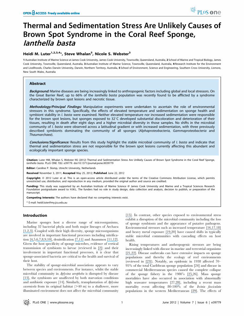
Figure 1. Principal components analysis (PCA) of I. bastacommunity composition, using DGGE banding pattern datato construct a similarity matrix of sponges from each of thethree geographic locations.doi:10.1371/journal.pone.0039779.g001
Environmental Stressors and the Sponge Holobiont
PLoS ONE | www.plosone.org 2 June 2012 | Volume 7 | Issue 6 | e39779
Sediment was subsequently dried at 100uC and sieved to a final
composition of particles that ranged between ,63 and #180 mm
(Table 1). These particle sizes are consistent with suspended
sediment compositions for the inner shelf of the GBR [32]. The
experimental design comprised four different sediment treatments:
0, 13, 30 and 100 mg l21 (+/22 mg l21). Following procedures of
[53], fiberglass stock tanks (240 l) were used to deliver short,
frequent pulses of concentrated sediment stocks to treatment tanks.
The stock tanks were designed with a 45u taper at their base to
prevent sediment accumulation and ensure constant rates of
delivery. To maintain sediment suspension throughout the
experiment, an external 2400 l h21 pump (Eheim 1260) was used
to circulate the water/sediment suspension from the base to the
top of the tank. The delivery of sediment pulses was achieved using
a second Eheim 1260 pump, which was set on a timer to pulse for
8 sec every 8 min and delivered approximately 120 ml of
concentrated sediment stock to the treatment tanks. A second
inverted pump (Eheim Compact+3000 pump) was suspended at
the waterline of each experimental tank to further improve re-
suspension of sediments. The frequent pulsing and partial re-
suspension of sediments ensured that turbidity (NTU) within tanks
was typically 10% CV [coefficient of variation, CV = (Std Dev
x100)/mean] when logged every 30 s and within 5% CV on a
daily basis (A. Negri pers. comm.). Total suspended sediment
Table 2. Sequence similarity in excised 16S rRNA DGGE bands from I. basta using BLAST.
Band ID Accession Number Percent Similarity Description
A* GQ347593 91 a-proteobacteria (Oceanic dead zone)
B FJ205252 89 a-proteobacteria (Deep sea hydrothermal region)
C GQ347593 91 a-proteobacteria, (Oceanic dead zone)
D FJ205252 90 a-proteobacteria (Deep sea hydrothermal region)
E GQ274301 94 c-proteobacteria (marine biofouling sample)
F* GQ348745 95 c-proteobacteria (oceanic dead zone)
G FM242455 94 c-proteobacteria (coastal sediment)
H GQ204920 93 c-proteobacteria (hard coral)
I JF733387 99 Pseudomonas sp. (adult black fly)
J* EU283427 96 Thaumarchaeota (ascidian)
K AJ876989 96 Thaumarchaeota (soft coral )
L EU283427 95 Thaumarchaeota (ascidian)
M EU182114 99 unidentified bacterium (sediment and seawater, South China Sea)
N HQ436843 96 unidentified bacterium (Lake Bosten, China)
The asterisks (*) denotes bands from the three dominant symbionts previously reported [47].doi:10.1371/journal.pone.0039779.t002
Figure 2. Representative photos of I. basta explants from thetemperature stress experiment: control explant from 27uC (a), explantfrom 30uC treatment at day 14 (b) and explant from 31uC treatment atday 14 (c).doi:10.1371/journal.pone.0039779.g002
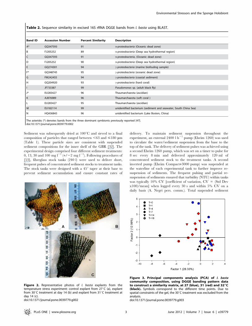
Figure 3. Principal components analysis (PCA) of I. bastacommunity composition, using DGGE banding pattern datato construct a similarity matrix, at 27 (blue), 31 (red) and 326C(black). Symbols correspond to the different time points. Due tospatial constraints of the gel, the 30uC treatment was excluded from theanalysis.doi:10.1371/journal.pone.0039779.g003
Environmental Stressors and the Sponge Holobiont
PLoS ONE | www.plosone.org 3 June 2012 | Volume 7 | Issue 6 | e39779
(TSS) concentrations in the treatment tanks were achieved and
maintained by measuring turbidity (NTU) with a TPS 90 FL-T
water quality logger (Springwood, QLD, Australia). Turbidity
measurements were taken daily throughout the experiment and
the amount of sediment added to the concentrated stock tanks was
adjusted according to the following turbidity calibration equation:
NTU = 0.2646TSS +0.05, r2 = 0.99.
Consistent with the temperature experiment, there were three
replicate tanks per treatment, each tank holding seven explants.
Single explants were randomly selected and removed from each
sediment treatment tank at 0, 1, 3, 7 and 9 days. All sampled
explants were photographed to visually assess tissue health and
frozen in liquid nitrogen for molecular analysis. After seven days
the sediment influx was turned off and a 48 h recovery period was
established.
DNA extraction and DGGEDNA was extracted from all sponge tissue samples from each
experiment using the Power Plant DNA Isolation kit, MoBio
Laboratories (Carlsbad, CA) according to the manufacturer’s
protocol. The 16S rRNA gene was amplified by PCR with
bacterial primers (1055f: 5’-ATGGCTGTCGTCAGC T-3’ and
1392r: 5’-ACGGGCGGTGTGTRC-3’) [54]. The reverse primer
was modified to contain a 40 bp GC clamp [55]. PCR reactions
contained 5 ml dNTP (2.5 mM), 5 ml 106OptiBuffer, 0.15 ml of
each primer (100 pmol ml21), 0.4 ml BSA (10 mg ml21), 3 ml
MgCl2 (50 mM), 0.5 ml Bio-X-ACT Taq polymerase (Bioline,
London, UK) and 1 ml DNA template. Reactions were made up to
50 ml total volume with Milli-Q water. The PCR conditions were:
1 cycle at 95uC for 5 min; 30 cycles at 95uC for 30 sec, 55uC for
1 min, 70uC for 1 min; and a final elongation at 70uC for 10 min.
Twenty ml of each sample was added to an 8% w/v polyacryl-
amide gel containing a 50–70% denaturing gradient of formamide
and urea. The gels were run at 60uC for 16 h in 1 x TAE buffer at
75 V using the Ingeny D-code system, stained with 1 x sybr gold
for 10 min, visualized under UV illumination and photographed
with the Vilber Lourmat ChemiSmart 3000 system. Reference
bands from each gel were excised, re-amplified with PCR and
checked for correct mobility on a 50–70% gel. PCR products were
sequenced by Macrogen Inc. (Seoul, Korea) using the forward
primer (1055f).
Due to spatial constraints of the gel, only 46 samples can be
visualized at once. Therefore, the 30uC treatment from the
temperature experiment and the 13 mg l21 treatment from the
sediment experiment were excluded from the DGGE analysis.
Cloning and sequencing (temperature experiment)The 16S rRNA gene from three replicate sponges, from the
32uC treatment at day 7, and three replicate control samples at
27uC, was amplified by PCR with universal bacterial primers: 63f
5’-CAGGCCTAACACATG CAA GTC-3’ [56] and 1492r 5’-
GGT TACCTTGTTACGACT T 23’ [57]. PCR reactions
contained 10 ml 5x MyTaq Buffer, 0.15 ml of each primer (100
pmol ml21), 0.4 ml BSA (10 mg ml21), 0.25 ml MyTaq DNA
polymerase (Bioline, London, UK) and 1 ml DNA. Reactions were
made up to 50 ml total volume with Milli-Q water. The PCR
conditions were: 1 cycle at 95uC for 1 min; 32 cycles at 95uC for
30 sec, 56uC for 30 sec, 72uC for 2 min; and a final elongation at
72uC for 10 min. PCR products from the control and 32uCtreatments were pooled and gel purified following the manufac-
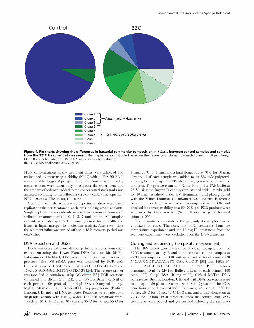
Figure 4. Pie charts showing the differences in bacterial community composition in I. basta between control samples and samplesfrom the 326C treatment at day seven. The graphs were constructed based on the frequency of clones from each library (n = 88 per library).Clone 8 and 5 had identical 16S rRNA sequences in both libraries.doi:10.1371/journal.pone.0039779.g004
Environmental Stressors and the Sponge Holobiont
PLoS ONE | www.plosone.org 4 June 2012 | Volume 7 | Issue 6 | e39779
turers protocol (NucleoSpin Extract II, Scientifix). Purified PCR
products were cloned with a TOPO TA cloning kit (Invitrogen,
Carlsbad, CA) according to the manufacturer’s instructions.
Plasmids were checked for inserts by PCR amplification using
M13 forward and reverse primers. Restriction digests using HhaI
and HaeIII (New England Biolabs Inc.) were performed to
determine operational taxonomic units (OTUs) for each library.
Eighty-eight clones from each library were screened and triplicates
of each representative OTU from both libraries were sent to
Magrogen Inc. (Seoul, Korea) for sequencing using 63f and 1492r
as the sequencing primers.
Phylogenetic analysisDGGE and clone sequences were compared with available
databases using the Basic Local Alignment Search Tool (BLAST)
[58] to determine nearest relatives and percent similarity. Clone
sequences were submitted to Genbank under the accession
numbers JN388026-JN388033. Due to the repetition of DGGE
sequences from each of the experiments and the latitudinal
comparison, only one representative sequence corresponding to
each band was submitted to Genbank (accession numbers
JN388012-JN388025). Sequences were checked for chimera
formation using Greengenes [59] and all chimeric sequences were
removed before tree construction. Sequences were compiled,
automatically aligned and manually edited in the ARB software
0.1
Ianthella basta clone 1, JN388026 (12% 32ºC) *Montiproa sp. clone AB470941 Marine bacterium EU513001
Pseudoalteromonas sp. EU930871Pseudoalteromonas sp. AM941186
Spongiobacter nickelotolerans AB205011 *Ianthella basta clone 2, JN388027 (9% 32ºC) *
Marine sediment clone AM292385Alteromonadaceae bacterium AM935070
Ianthella basta clone 3, JN388028 (13% 32ºC) * Seawater clone GU061307
Ianthella basta clone 4, JN388029 (12% 32ºC) *Seawater clone FJ347760
Arctic sediment clone EU050809Axinella verrucosa clone AJ581351 *
Ianthella basta clone H4 GU784985 *Ianthella basta clone 5, JN388030 (7% Control, 14% 32ºC) *
Petroleum-contaminated soil clone HQ727620Ianthella basta clone 6, JN388031 (5% 32ºC) *
Montipora sp. clone AB470948Marine sediment clone GU176617
Montastraea favelota clone FJ202499Hydrothermal vent clone GU369921Rhodobacteraceae bacterium EF620870 *Ianthella basta clone 7, JN388032 (7% 32ºC) * Roseobacter sp. EF092256 *
Ruegeria lacusaerulensis HQ908678 *Seawater clone GQ349430
Ianthella basta clone H1 GU784988 *Ianthella basta clone 8, JN388033 (93% Control, 28% 32ºC) *100
100
100
100
100100
100
100
100
10099
99
88
85
50
77
62
92
53
Gam
maproteobacteria
Alphaproteobacteria
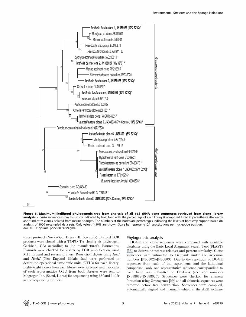
Figure 5. Maximum-likelihood phylogenetic tree from analysis of all 16S rRNA gene sequences retrieved from clone libraryanalysis. I. basta sequences from this study indicated by bold font, with the percentage of each library it comprised listed in parenthesis afterwardsand * indicates clones isolated from marine sponges. The numbers at the nodes are percentages indicating the levels of bootstrap support based onanalysis of 1000 re-sampled data sets. Only values .50% are shown. Scale bar represents 0.1 substitutions per nucleotide position.doi:10.1371/journal.pone.0039779.g005
Environmental Stressors and the Sponge Holobiont
PLoS ONE | www.plosone.org 5 June 2012 | Volume 7 | Issue 6 | e39779
package (http://www.arb-home.de [60]). Initially, trees were
calculated with almost complete 16S rRNA (1400 bp) sequences
for all close relatives of target sequences using the neighbor-joining
and maximum parsimony methods in ARB. Partial sequences
were subsequently imported to the tree without changing branch
topology using the ARB parsimony-interactive method. The
robustness of inferred tree topologies was evaluated after 1000
bootstrap re-samplings of the neighbor-joining data in the
PHYLIP program [61]. Synechoccocus elongatus was used as an out
group for the tree.
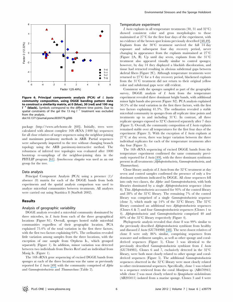
Data analysisPrincipal Component Analysis (PCA) using a presence (1)/
absence (0) matrix for each of the DGGE bands from both
experiments and the spatial analysis comparison was used to
analyze microbial communities between treatments. All analyses
were carried out using Stastitica 8 (StatSoft 2002).
Results
Analysis of geographic variabilityDGGE analysis revealed a microbial community dominated by
three microbes, in I. basta from each of the three geographical
locations (Figure S1). Overall, sponges hosted stable microbial
communities regardless of their geographic location. PCA
explained 75.4% of the total variation in the first three factors,
with the first two factors explaining 64%. The ordination revealed
little variation among samples from the three locations, with the
exception of one sample from Orpheus Is., which grouped
separately (Figure 1). In addition, minor variation was detected
between two individuals from Davies Reef and an individual from
Masig Is. (Figure 1).
The 16S rRNA gene sequencing of excised DGGE bands from
sponges at each of the three locations was the same as previously
reported for I. basta [49] with the community comprised of Alpha
and Gammaproteobacteria and Thaumarchaea (Table 2).
Temperature experimentI. basta explants in all temperature treatments (30, 31 and 32uC)
showed consistent color and gross morphologies to those
maintained at 27uC for the first four days of the experiment, with
no evidence of the brown spot lesions previously described [48,49].
Explants from the 30uC treatment survived the full 14 day
exposure and subsequent four day recovery period, never
changing in appearance from the explants maintained at 27uC(Figure 2A, B). Up until day seven, explants from the 31uCtreatment also appeared visually similar to control sponges;
however, by day 14 they displayed a blackish discoloration, and
tissue had retracted resulting in obvious subdermal gaps between
skeletal fibers (Figure 2C). Although temperature treatments were
returned to 27uC for a 4 day recovery period, blackened explants
from the 31uC treatment did not return to their original yellow
color and subdermal gaps were still evident.
Consistent with the sponges sampled as part of the geographic
survey, DGGE analysis of I. basta from the temperature
experiment revealed three dominant bright bands, with additional
minor light bands also present (Figure S2). PCA analysis explained
50.5% of the total variation in the first three factors, with the first
two factors explaining 41.3%. The ordination revealed a stable
microbial community in sponges from all replicate time points and
treatments up to and including 31uC. In contrast, all three
replicate sponges exposed to 32uC clustered separately after 7 days
(Figure 3). Overall, the community composition of I. basta explants
remained stable over all temperatures for the first four days of the
experiment (Figure 3). With the exception of I. basta explants at
27uC at day seven, there was larger variation between the three
individual replicates for each of the temperature treatments after
day four (Figure 3).
The 16S rRNA sequencing of excised DGGE bands from the
temperature experiment confirmed the same community previ-
ously reported for I. basta [49], with the three dominant symbionts
present in all treatments (Alphaproteobacteria, Gammaproteobacteria, and
Thaumarchaea).
Clone library analysis of I. basta from the 32uC treatment at day
seven and control samples confirmed the presence of only a few
dominant symbionts indicated by DGGE. All clone sequences fell
into only two classes, the Alpha- and Gammaproteobacteria, with both
libraries dominated by a single Alphaproteobacteria sequence (clone
8). This Alphaproteobacteria accounted for 93% of the control library
and 28% of the 32uC library. The remaining 7% of the control
library was comprised of a single Gammaproteobacteria sequence
(clone 5), which made up 14% of the 32uC library. The 32uClibrary contained an additional two Alphaproteobacteria sequences
(Clones 6 & 7) and four Gammaproteobacteria sequences (Clones 1 to
4). Alphaproteobacteria and Gammaproteobacteria comprised 40 and
60% of the 32uC library respectively (Figure 4).
Phylogenetic analysis revealed that clone 8 was 99% similar to
the previously described Alphaproteobacteria symbiont from healthy
and diseased I. basta (GU784988) [48]. The next closest relatives of
clone 8 were only 86% similar, comprising sequences from
seawater and sediment samples, as well as other sponge and coral-
derived sequences (Figure 5). Clone 5 was identical to the
previously described Gammaproteobacteria symbiont from I. basta
(GU784985). Clones 6 and 7, exclusively detected in the 32uClibrary, were both most closely related to other sponge and coral-
derived sequences (Figure 5). The additional Gammaproteobacteria
sequences observed in the 32uC library were most closely related
to other environmental sequences. Specifically, clone 1 was related
to a sequence retrieved from the coral Montipora sp. (AB470941)
while clone 2 was most closely related to Spongiobacter nickelotolerans
(AB205011) isolated from a marine sponge. Clones 3 and 4 were
Figure 6. Principal components analysis (PCA) of I. bastacommunity composition, using DGGE banding pattern datato construct a similarity matrix, at 0 (blue), 30 (red) and 100 mgl21 (black). Symbols correspond to the different time points. Due tospatial constraints of the gel the 13 mg l21 treatment was excludedfrom the analysis.doi:10.1371/journal.pone.0039779.g006
Environmental Stressors and the Sponge Holobiont
PLoS ONE | www.plosone.org 6 June 2012 | Volume 7 | Issue 6 | e39779
most closely related to soil and seawater bacteria respectively
(Figure 5).
Sediment experimentI. basta explants in all sediment treatments (13 to 100 mg l21)
survived the seven day exposure and further two day recovery
period and appeared visually similar to those explants maintained
at 0 mg l21. Explants from the 30 and 100 mg l21 treatment
accumulated sediments on their surfaces; however, tissue health
remained similar to explants maintained at 0 mg l21 when
sediments were manually removed after 7 days. None of the
sponge clones developed brown spot lesions during the course of
the sedimentation experiment.
DGGE analysis of sponges in the sediment treatments at each
time point confirmed the presence of three dominant community
players with other low abundance microbes also present. PCA
analysis explained 47.8% of the total variation in the first three
factors, with the first two factors explaining 37.7%. In general,
there was a large amount of variation between individuals,
regardless of the sediment treatment or time point (Figure 6). This
variation was attributed to the absence of bands in some samples
(see Figure S3) rather than the appearance of novel microbes not
normally detected in I. basta. 16S rRNA sequencing of excised
DGGE bands revealed the same microbes detected in sponges
from the temperature experiment with all sequences identical to
those previously reported for I. basta [49].
Discussion
Elevated seawater temperature and sedimentation do not
appear to be responsible for the syndrome of brown spot lesions
currently affecting a large portion of the I. basta population on the
Great Barrier Reef [48]. Sponges exposed to elevated temperature
and sedimentation did not develop symptoms of the syndrome and
the microbial symbionts were not affected by the environmental
stressors. However, I. basta do not tolerate seawater temperatures
of 32uC, which is only 2–4 degrees higher than the average
summer temperature at Orpheus Is. [52]. This indicates a very
narrow thermal threshold for I. basta as no morphological changes
were evident at 30uC, but sponges at 32uC exhibited substantial
discolouration and deterioration of the tissue with death occurring
after 8 days. In addition, sponges exposed to 31uC showed
symptoms of tissue regression and discolouration after 14 days.
Accompanied with the decline in host health at 32uC, sponges
from this treatment had a greater diversity of microbes within the
Alpha- and Gammaproteobacteria. Whilst other studies have reported
microbial shifts in sponges exposed to elevated seawater temper-
atures [16,18,62], these studies have described both the loss of
symbionts and the appearance of foreign microbes including
putative pathogens. Bacteria within the Bacteroidetes, Epsilon- and
Deltaproteobacteria commonly occur after stress in both sponges and
corals [18,63,64]. In contrast, all three dominant I. basta symbionts
were maintained in sponges at 32uC after 7 days and all of the
microbes exclusively detected in the 32uC treated sponges were
members of the Alpha and Gammaproteobacteria. Given this apparent
stability in the microbial community of I. basta, it is likely these
microbes play an important functional role(s) in this sponge.
Sponges can produce secondary metabolites to maintain their
inherent microbial community, as well as protecting themselves
from microbial attack [65]. I. basta produces both araplysillin and
bastadin compounds that are known to have strong biological
activity and antimicrobial properties [66,67]. DGGE analysis
revealed higher diversity in moribund samples from 32uC at day
seven than from 32uC at day four. It is possible that this increased
diversity coincides with the breakdown in I. basta’s antimicrobial
compound production, allowing the proliferation of a greater
number of opportunistic bacteria prior to sponge mortality.
I. basta explants exposed to high levels of sediments (100 mg l21)
appeared visually similar to those maintained at 0 mg l21,
indicating this sponge is capable of withstanding high levels of
sediment exposure, at least in the short term. Despite the potential
pumping implications from the high amounts of sediment on the
sponge surface [30,31,33], I. basta showed no visible adverse
effects. Conversely, coral tissues exposed to high sedimentation
rates show decreased photosynthetic activity and necrosis [68].
The ability of I. basta to cope with short term sediment stress is
likely linked to their habitat and distribution, which includes
inshore silty patches and fringing reef slopes [48,69]. For instance,
the mean max turbidity of Orpheus Island in the summer is 26 +/
211 NTU (AIMS Water Quality Monitoring Data), which is
equivalent to the turbidity experienced by I. basta in the highest
treatment of this study. No microbial community shifts were
detected in sponges from any of the sediment treatments or time
points. While previous studies have documented the negative
impacts of water quality and increased sedimentation on sponge
pumping rates [30,31,32], reproduction [34,35] and metabolism
[33], this is the first study to assess the effects of increased
sedimentation on sponge symbiosis.
Sponge microbial communities are generally stable across
temporal and spatial scales [14,70,71,72]. For instance, the sponge
Cymbastela concentrica maintains a similar microbial community
composition across a 500 km region of its temperate range [71].
The microbial community of I. basta is no exception, with
consistent communities observed in sponges from three separate
locations across an 1100 km latitudinal gradient.
I. basta’s microbial community is consistently dominated by
three symbionts, regardless of its disease status [49], the
environmental stress or the geographic location it inhabits. In all
instances, the community is dominated by three symbionts within
the Alphaproteobacteria, Gammaproteobacteria and Thaumarchaea al-
though other low abundance microbes within the Cyanobacteria,
Chloroflexi and Verrucomicrobia are also present [65,73]. To date,
none of the three dominant symbionts have been successfully
cultivated [49]. Neither increased temperature nor sedimentation
induced the disease-like symptoms (brown spot lesions) observed in
I. basta in the field [48], indicating that these two environmental
factors are unlikely causes of the syndrome. In addition, multiple
techniques (e.g. bacterial cultivation, molecular community
analysis and electron microscopy) have failed to identify any
microbial community shifts or putative pathogens responsible for
this syndrome [49]. Given these results, the cause of the syndrome
is still uncertain. It cannot be ruled out that other environmental
factors (e.g. elevated nutrients) may be responsible, or that it is a
combination of different environmental factors working together
to induce the lesions. In addition, the pre-filtration of the
experimental water (5 mm) may have removed potential protist
vectors and/or disease causing protists. Future work will assess the
role of protists in disease aetiology and examine immune
dysfunction and senescence in the sponge host by identifying
differentially expressed genes in sponges displaying the syndrome.
This may provide further insight into the cause of the brown spot
lesions in this abundant and ecologically important species.
Supporting Information
Figure S1 DGGE gel image of 16S rRNA-defined bacte-rial populations from I. basta from three geographicallocations. Bands excised for sequencing are labeled on the right
Environmental Stressors and the Sponge Holobiont
PLoS ONE | www.plosone.org 7 June 2012 | Volume 7 | Issue 6 | e39779
hand side of the bands, and asterisks (*) denote bands that yielded
low sequence quality.
(PPT)
Figure S2 DGGE gel image of 16S rRNA-defined bacte-rial populations from I. basta explants in the 27, 31 and326C treatments over the course of the experiment(T = 0, 1, 4, 7, 14 and 18). Bands excised for sequencing are
labeled on the right hand side of the bands, and asterisks (*) denote
bands that yielded low sequence quality. Due to spatial constraints
of the gel, only 46 samples can be visualized at once. Therefore,
the 30uC treatment was excluded from the analysis.
(PPT)
Figure S3 DGGE gel image of 16S rRNA-defined bacte-rial populations from I. basta explants in the 0, 30 and100 mg l21 treatments over the course of the experiment(T = 0, 1, 4 and 7). Bands excised for sequencing are labeled on
the right hand side of the bands, and asterisks (*) denote bands that
yielded low sequence quality. Due to spatial constraints of the gel,
only 46 samples can be visualized at once. Therefore, the 13 mg
l21 treatment was excluded from the analysis.
(PPT)
Acknowledgments
We thank A. Negri and F. Flores for their assistance with the sediment
delivery system and aquarium facilities. All work was completed under the
Great Barrier Reef Marine Park Authority Permit Number G06/15571.1.
Author Contributions
Conceived and designed the experiments: HML SW NSW. Performed the
experiments: HML. Analyzed the data: HML. Contributed reagents/
materials/analysis tools: HML SW NSW. Wrote the paper: HML SW
NSW.
References
1. Schmitt S, Tsai P, Bell J, Fromont J, Ilan M, et al. (2011) Assessing the complex
sponge microbiota-core, variable and species-specific bacterial communities in
marine sponges. ISME Journal doi:10.1038/ismej.2011.116.
2. Taylor MW, Radax R, Steger D, Wagner M (2007) Sponge-associated
microorganisms: Evolution, ecology, and biotechnological potential. Microbiol-
ogy and Molecular Biology Reviews 71: 295–347.
3. Webster NS, Taylor MW (2012) Marine sponges and their microbial symbionts:
love and other relationships. Environmental Microbiology 14: 335–346.
4. Bayer K, Schmitt S, Hentschel U (2008) Physiology, phylogeny and in situ
evidence for bacterial and archaeal nitrifiers in the marine sponge Aplysina
aerophoba. Environmental Microbiology 10: 2942–2955.
5. Mohamed NM, Rao V, Hamann MT, Kelly M, Hill RT (2008) Changes in
bacterial communities of the marine sponge Mycale laxissima on transfer into
aquaculture. Applied and Environmental Microbiology 74: 1209–1222.
6. Off S, Alawi M, Spieck E (2010) Enrichment and physiological characterization
of a novel Nitrospira-like bacterium obtained from a marine sponge. Applied and
Environmental Microbiology 76: 4640–4646.
7. Schlappy M, Schottner SI, Lavik G, Kuypers MMM, de Beer D, et al. (2010)
Evidence of nitrification and denitrification in high and low microbial
abundance sponges. Marine Biology 157: 593–602.
8. Southwell MW, Popp BN, Martens CS (2008) Nitrification controls on fluxes
and isotopic composition of nitrate from Florida Keys sponges. Marine
Chemistry 108: 96–108.
9. Southwell MW, Weisz J, Martens CS, Lindquist N (2008) In situ fluxes of
dissolved inorganic nitrogen from the sponge community on Conch Reef, Key
Largo, Florida. Limnology and Oceanography 53: 986–996.
10. Steger D, Ettinger-Epstein P, Whalan S, Hentschel U, de Nys R, et al. (2008)
Diversity and mode of transmission of ammonia-oxidizing archaea in marine
sponges. Environmental Microbiology 10: 1087–1094.
11. Hoffmann F, Radax R, Woebken D, Holtappels M, Lavik G, et al. (2009)
Complex nitrogen cycling in the sponge Geodia barretti. Environmental
Microbiology 11: 2228–2249.
12. Mohamed NM, Saito K, Tal Y, Hill RT (2010) Diversity of aerobic and
anaerobic ammonia-oxidizing bacteria in marine sponges. ISME Journal 4: 38–
48.
13. Webster NS, Xavier JR, Freckelton M, Motti CA, Cobb R (2008) Shifts in
microbial and chemical patterns within the marine sponge Aplysina aerophoba
during a disease outbreak. Environmental Microbiology 10: 3366–3376.
14. Friedrich AB, Fischer I, Proksch P, Hacker J, Hentschel U (2001) Temporal
variation of the microbial community associated with the Mediterranean sponge
Aplysina aerophoba. FEMS Microbiology Ecology 38: 105–113.
15. Thoms C, Horn M, Wagner M, Hentschel U, Proksch P (2003) Monitoring
microbial diversity and natural product profiles of the sponge Aplysina cavernicola
following transplantation. Marine Biology 142: 685–692.
16. Lemoine N, Buell N, Hill A, Hill M (2007) Assessing the utility of sponge
microbial symbiont communities as models to study global climate change: a
case study with Halichondria bowerbanki. In: Custodio MR, G L-H, Hajdu E,
Muricy G, editors. 7th International Sponge Symposium. Rio de Janeiro. 419–
425.
17. Lopez-Legentil S, Song B, McMurray SE, Pawlik JR (2008) Bleaching and stress
in coral reef ecosystems: hsp70 expression by the giant barrel sponge Xestospongia
muta. Molecular Ecology 17: 1840–1849.
18. Webster NS, Cobb RE, Negri AP (2008) Temperature thresholds for bacterial
symbiosis with a sponge. ISME Journal 2: 830–842.
19. Selvin J, Shangmugha Priya S, Seghal Kiran G, Thangavelu T, Sapna Bai N
(2009) Sponge-associated marine bacteria as indicators of heavy metal pollution.
Microbiological Research 164: 352–363.
20. Webster NS, Webb RI, Ridd MJ, Hill RT, Negri AP (2001) The effects ofcopper on the microbial community of a coral reef sponge. Environmental
Microbiology 3: 19–31.
21. Haapkyla J, Unsworth RKF, Flavell M, Bourne DG, Schaffelke B, et al. (2011)Seasonal rainfall and runoff promote coral disease on an inshore reef. PLoS
ONE 6: e16893.
22. Harvell CD, Altizer S, Cattadori IM, Harrington L, Weil E (2009) Climate
change and wildlife diseases: When does the host matter the most? Ecology 90:912–920.
23. Webster NS (2007) Sponge disease: a global threat? Environmental Microbi-
ology 9: 1363–1375.
24. Galstoff PS (1942) Wasting disease causing mortality of sponges in the WestIndies and Gulf of Mexico. Proceedings VIII American Science Congress.
Wahington, D.C. 411–421.
25. Gaino E, Pronzato R, Corriero G, Buffa P (1992) Mortality of commercialsponges – incidence in 2 Mediterranean areas. Bollettino Di Zoologia 59: 79–85.
26. Vacelet J (1994) Control of the severe sponge epidemic. Near East and Europe:
Algeria, Cyprus, Egypt, Lebanon, Malta, Morocco, Syria, Tunisia, Turkey,
Yugoslavia. Technical Report – the struggle against the epidemic which isdecimating Mediterranean sponges FI: TCP/RAB/8853. Rome, Italy. 1–39 p.
27. Cerrano C, Bavestrello G, Bianchi CN, Cattaneovietti R, Bava S, et al. (2000) A
catstrophic mass-mortality episode of gorgonians and other organisms in theLigurian Sea (North-Western Mediterranean), summer 1999. Ecology Letters 3:
284–293.
28. Vicente VP (1989) Regional commercial sponge extinction in the West Indies:are recent climatic changes responsible? Marine Ecology-Progress Series 10:
179–191.
29. Cebrian E, Uriz MJ, Garrabou J, Ballesteros E (2011) Sponge mass mortalities in
a warming Mediterranean Sea: Are cyanobacteria-harboring species worse off?PLoS ONE 6: e20211.
30. Gerrodette T, Flechsig AO (1979) Sediment-induced reduction in the pumping
rate of the tropical sponge Verongia lacunosa. Marine Biology 55: 103–110.
31. Reiswig HM (1971) In situ pumping activities of tropical demospongiae. MarineBiology 9: 38–50.
32. Tompkins-MacDonald G, Leys S (2008) Glass sponges arrest pumping in
response to sediment: implications for the physiology of the Hexactinellidconduction system. Marine Biology 154: 973–984.
33. Bannister RJ, Battershill CN, de Nys R (2012) Suspended sediment grain size
and mineralogy across the continental shelf of the Great Barrier Reef: Impacts
on the physiology of a coral reef sponge. Continental Shelf Research 32: 86–95.
34. Roberts DE, Davis AR, Cummins SP (2006) Experimental manipulation ofshade, silt, nutrients and salinity on the temperate reef sponge Cymbastela
concentrica. Marine Ecology Progress Series 307: 143–154.
35. Whalan S, Battershill C, de Nys R (2007) Variability in reproductive outputacross a water quality gradient for a tropical marine sponge. Marine Biology
153: 163–169.
36. Maldonado M, Giraud K, Carmona C (2008) Effects of sediment on the survivalof asexually produced sponge recruits. Marine Biology 154: 631–641.
37. Abdul Wahab MA, de Nys R, Whalan S (2012) Closing the lifecycle for the
sustainable aquaculture of the bath sponge Coscinoderma matthewsi. Aquaculture
324–325: 281–285.
38. Solomon S, Qin D, Manning M, Chen Z, Marquis M, et al. (2007) ClimateChange 2007: The Physical Science Basis. Contribution of working group 1 to
the Fourth Assessment Report of the Intergovernmental Panel on ClimateChange Cambridge.
39. Hoegh-Gulberg O, Mumby PJ, Hooten AJ, Steneck RS, Greenfield P, et al.
(2007) Coral reefs under rapid climate change and ocean acidification. Science318: 1737–1742.
Environmental Stressors and the Sponge Holobiont
PLoS ONE | www.plosone.org 8 June 2012 | Volume 7 | Issue 6 | e39779
40. Pandolfi JM, Connolly SR, Marshall DJ, Cohen AL (2011) Projecting coral reef
futures under global warming and ocean acidification. Science 333: 418–422.41. Bruno JF, Selig ER, Casey KS, Page CA, Willis BL, et al. (2007) Thermal stress
and coral cover as drivers of coral disease outbreaks. PLoS Biology 5: 1220–
1227.42. Dalton SJ, Godwin S, Smith SDA, Pereg L (2010) Australian subtropical white
syndrome: a transmissible, temperature-dependent coral disease. Marine andFreshwater Research 61: 342–350.
43. Sato Y, Bourne DG, Willis BL (2011) Effects of temperature and light on the
progression of black band disease on the reef coral, Montipora hispida. Coral Reefs30: 753–761.
44. Weber M, Lott C, Fabricius KE (2006) Sedimentation stress in a scleractiniancoral exposed to terrestrial and marine sediments with contrasting physical,
organic and geochemical properties. Journal of Experimental Marine Biologyand Ecology 336: 18–32.
45. Littman R, Willis BL, Bourne DG (2011) Metagenomic analysis of the coral
holobiont during a natural bleaching even on the Great Barrier Reef.Environmental Microbiology Reports 3: 651–660.
46. Thurber RV, Willner-Hall D, Rodriguez-Mueller B, Desnues C, Edwards RA,et al. (2009) Metagenomic analysis of stressed coral holobionts. Environmental
Microbiology 11: 2148–2163.
47. Przeslawski R, Ahyong S, Byrne M, Worheide G, Hutchings P (2008) Beyondcoral and fish: the effects of climate change on non-coral benthic invertebrates of
tropical reefs. Global Climate Change 14: 2773–2795.48. Luter HM, Whalan S, Webster NS (2010) Prevalence of tissue necrosis and
brown spot lesions in a common marine sponge. Marine and FreshwaterResearch 61: 484–489.
49. Luter HM, Whalan S, Webster NS (2010) Exploring the role of microorganisms
in the disease-like syndrome affecting the sponge Ianthella basta. Applied andEnvironmental Microbiology 76: 5736–5744.
50. Louden D, Whalan S, Evans-Illidge E, Wolff C, de Nys R (2007) An assessmentof the aquaculture potential of the tropical sponges Rhopaloeides odorabile and
Coscinoderma sp. Aquaculture 270: 57–67.
51. Reiswig HM (1971) Particle feeding in natural populations of 3 marineDemosponges. Biological Bulletin 141: 568–591.
52. Berkelmans R, De’ ath G, Kininmonth S, Skirving WJ (2004) A comparison ofthe 1998 and 2002 coral bleaching events on the Great Barrier Reef: spatial
correlation, patterns, and predictions. Coral Reefs 23: 74–83.53. Flores F, Hoogenboom MO, Smith LD, Cooper TF, Abrego D, et al. (2012)
Chronic exposure of corals to fine sediments: lethal and sub-lethal impacts. PLoS
ONE. DOI: 10.1371/journal.pone.0037795.54. Ferris MJ, Muyzer G, Ward DM (1996) Denaturing gradient gel electrophoresis
profiles of 16S rRNA-defined populations inhabiting a hot spring microbial matcommunity. Applied and Environmental Microbiology 62: 340–346.
55. Muyzer G, de Waal EC, Uitterlinden AG (1993) Profiling of complex microbial
populations by denaturing gradient gel electrophoresis analysis of polymerasechain reaction-amplified genes coding for 16S rRNA. Applied and Environ-
mental Microbiology 59: 695–700.56. Marchesi JR, Sata T, Weightman AJ, Martine TA, Fry JC, et al. (1998) Design
and Evaluation of useful bacterium-specific PCR primers that amplify genescoding for bacterial 16S rRNA. Applied and Environmental Microbiology 64:
795–799.
57. Lane DJ (1991) 16S rRNA sequencing. In: Stackerbrandt E, Goodfellow M,
editors. Nucleic acid techniques in bacterial systematics. New York: John Wileyand Sons, Inc. 115–148.
58. Altschul SF, Madden TL, Schaffer AA, Zhang J, Zhang Z, et al. (1997) Gapped
BLAST and PSI-BLAST: a new generation of protein database searchprograms. Nucleic Acids Research 25: 3389–3402.
59. DeSantis TZ, Hugenholtz P, Larsen N, Rojas M, Brodie EL, et al. (2006)Greengenes, a Chimera-Checked 16S rRNA Gene Database and Workbench
Compatible with ARB. Applied and Environmental Microbiology 172: 5069–
5072.60. Ludwig W, Strunk O, Westram R, Richter L, Meier H, et al. (2004) ARB: a
software environment for sequence data. Nucleic Acids Research 32: 1363–1371.
61. Felsenstein J (1993) PHYLIP (Phylogenetic Inference Package) version 3.5c.Department of Genetics, University of Washington, Seattle.
62. Webster NS, Botte ES, Soo RM, Whalan S (2011) The larval sponge holobiont
exhibits high thermal tolerance. Environmental Microbiology Reports 3: 756–762.
63. Frias-Lopez J, Zerkle AL, Bonheyo GT, Fouke BW (2002) Partitioning ofbacterial communities between seawater and healthy, black band diseased, and
dead coral surfaces. Applied and Environmental Microbiology 68: 2214–2228.
64. Pantos O, Bythell JC (2006) Bacterial community structure associated with whiteband disease in the elkhorn coral Acropora palmata determined using culture
independent 16S rRNA techniques. Diseases of Aquatic Organisms 69: 79–88.65. Kelman D, Kashman Y, Rosenberg E, Ilan M, Ifrach I, et al. (2001)
Antimicrobial activity of the reef sponge Amphimedon viridis from the Red Sea:evidence for selective toxicity. Aquatic Microbial Ecology 24: 9–16.
66. Freckelton ML, Luter HM, Andreakis N, Webster NS, Motti CA (2011)
Qualitative variation in colour morphotypes of Ianthella basta (Porifera:Verongida). Hydrobiologia. DOI: 10.1007/s10750-011-0818-x.
67. Pettit GR, Butler MS, Williams MD, Filiatrault MJ, Pettit RK (1996) Isolationand structure of hemibastadinols 1–3 from the Papua New Guinea marine
sponge Ianthella basta. Journal of Natural Products 59: 927–934.
68. Philipp E, Fabricius K (2003) Photophysiological stress in scleractinian corals inresponse to short-term sedimentation. Journal of Experimental Marine Biology
and Ecology 287: 57–78.69. Bergquist PR, Kelly-Borges M (1995) Systematics and biogeography of the genus
Ianthella (Demospongiae: Verongida: Ianthellidae) in the south-west Pacific.Records of the Museums and Art Galleries of the Northern Territory 12: 151–
176.
70. Taylor MW, Schupp PJ, Dahllof I, Kjelleberg S, Steinberg PD (2004) Hostspecificity in marine sponge-associated bacteria, and potential implications for
marine microbial diversity. Environmental Microbiology 6: 121–130.71. Taylor MW, Schupp PJ, de Nys R, Kjelleberg S, Steinberg PD (2005)
Biogeography of bacteria associated with the marine sponge Cymbastela concentrica.
Environmental Microbiology 7: 419–433.72. Webster NS, Negri AP, Munro M, Battershill CN (2004) Diverse microbial
communities inhabit Antarctic sponges. Environmental Microbiology 6: 288–300.
73. Webster NS, Taylor MW, Behnam F, Lucker S, Rattel T, et al. (2010) Deepsequencing reveals exceptional diver- sity and modes of transmission for bacterial
sponge symbionts. Environmental Microbiology 12: 2070–2082.
Environmental Stressors and the Sponge Holobiont
PLoS ONE | www.plosone.org 9 June 2012 | Volume 7 | Issue 6 | e39779


