Embed Size (px)
Citation preview
materials
Article
Thermosensitive Drug Delivery System SBA-15-PEI forControlled Release of Nonsteroidal Anti-Inflammatory DrugDiclofenac Sodium Salt: A Comparative Study
Lubos Zauska 1, Stefan Bova 2, Eva Benova 1, Jozef Bednarcik 3 , Matej Balaz 4 , Vladimir Zelenak 1 ,Virginie Hornebecq 5 and Miroslav Almasi 1,*
�����������������
Citation: Zauska, L.; Bova, S.;
Benova, E.; Bednarcik, J.; Balaz, M.;
Zelenak, V.; Hornebecq, V.; Almasi, M.
Thermosensitive Drug Delivery
System SBA-15-PEI for Controlled
Release of Nonsteroidal
Anti-Inflammatory Drug Diclofenac
Sodium Salt: A Comparative Study.
Materials 2021, 14, 1880. https://
doi.org/10.3390/ma14081880
Academic Editor: Eun-Kyung Lim
Received: 26 February 2021
Accepted: 6 April 2021
Published: 9 April 2021
Publisher’s Note: MDPI stays neutral
with regard to jurisdictional claims in
published maps and institutional affil-
iations.
Copyright: © 2021 by the authors.
Licensee MDPI, Basel, Switzerland.
This article is an open access article
distributed under the terms and
conditions of the Creative Commons
Attribution (CC BY) license (https://
creativecommons.org/licenses/by/
4.0/).
1 Department of Inorganic Chemistry, Faculty of Science, P. J. Šafárik University, Moyzesova 11,SK-041 01 Košice, Slovakia; [email protected] (L.Z.); [email protected] (E.B.);[email protected] (V.Z.)
2 BovaChem s.r.o, Garbiarska 1919/14, SK-048 01 Rožnava, Slovakia; [email protected] Institute of Experimental Physics, Slovak Academy of Sciences, Watsonova 47, SK-040 01 Košice, Slovakia;
[email protected] Institute of Geotechnics, Slovak Academy of Sciences, Watsonova 45, SK-040 01 Košice, Slovakia;
[email protected] Aix-Marseille University, CNRS, MADIREL, F-133 97 Marseille, France; [email protected]* Correspondence: [email protected]; Tel.: +421 552-342-366
Abstract: Mesoporous SBA-15 silica material was prepared by the sol–gel method and functional-ized with thermosensitive polyethylenimine polymers with different molecular weight (g·mol−1):800 (SBA-15(C)-800), 1300 (SBA-15(C)-1300) and 2000 (SBA-15(C)-2000). The nonsteroidal anti-inflammatory drug (NSAID) diclofenac sodium was selected as a model drug and encapsulatedinto the pores of prepared supports. Materials were characterized by the combination of infraredspectroscopy (IR), atomic force microscopy (AFM), transmission electron microscopy (TEM), photoncross-correlation spectroscopy (PCCS), nitrogen adsorption/desorption analysis, thermogravimetry(TG), differential scanning calorimetry (DSC) and small-angle X-ray diffraction (SA-XRD) experi-ments. The drug release from prepared matrixes was realized in two model media differing in pH,namely small intestine environment/simulated body fluid (pH = 7.4) and simulated gastric fluid(pH = 2), and at different temperatures, namely normal body temperature (T = 37 ◦C) and inflam-matory temperature (T = 42 ◦C). The process of drug loading into the pores of prepared materialsfrom the diclofenac sodium salt solutions with different concentrations and subsequent quantitativedetermination of released drugs was analyzed by UV-VIS spectroscopy. Analysis of prepared SBA-15materials modified with polyethylenimines in solution showed a high ability to store large amountsof the drug, up to 230 wt.%. Experimental results showed their high drug release into the solutionat pH = 7.4 for both temperatures, which is related to the high solubility of diclofenac sodium in aslightly alkaline environment. At pH = 2, a difference in drug release rate was observed betweenboth temperatures. Indeed, at a higher temperature, the release rates and the amount of releaseddrug were 2–3 times higher than those observed at a lower temperature. Different kinetic modelswere used to fit the obtained drug release data to determine the drug release rate and its releasemechanism. Moreover, the drug release properties of prepared compounds were compared to acommercially available medicament under the same experimental conditions.
Keywords: mesoporous silica; surface modification; polyethylenimines; diclofenac sodium; tempera-ture and pH; kinetic models
1. Introduction
Oncological and/or infectious diseases often cause inflammation in the human body [1].Inflammation can have a local character or spread throughout the whole human body,accompanied by symptoms such as pain, fever and other complications. Anti-inflammatory
Materials 2021, 14, 1880. https://doi.org/10.3390/ma14081880 https://www.mdpi.com/journal/materials
Materials 2021, 14, 1880 2 of 25
medications such as nonsteroidal anti-inflammatory drugs (NSAIDs) and analgesics [2] areeffective inflammatory suppressants. However, these medications are often invasive andaddictive and can exhibit negative long-term side effects such as liver, kidney, heart andbone marrow damage. A method that has proven to be relatively significant is the use of atargeted drug delivery system (DDS) [3]. DDSs are biocompatible structures in which thedrug is encapsulated and then released only at the targeted site is reached. Carriers can beinorganic materials, such as metal–organic frameworks (MOFs) [4,5], carbon nanotubes [6],fullerenes [7], zeolites [8] and silica [9], or organic materials, such as polymers [10], mi-celles [11] and dendrimers, containing different functional groups. It is also possible tomake DDSs from proteins, lipids and polysaccharides [12], but the most common materialsare combinations of mentioned structures [13].
In bioinorganic chemistry and pharmacology, mesoporous silica materials have foundapplication due to their excellent biocompatibility, low cytotoxicity, uniform particle size,resistance and stability [14]. The surface of silica can be modified by various organic andinorganic groups that are called “plug” and that respond to external stimuli. The mentionedsurface plug prevents the uncontrolled release of a drug from the porous structure ofsilica [15]. Different molecules have been studied as a plug, from the smallest binarycompounds [16] to polymeric [17–19] and macrocyclic compounds [20,21]. All substancesare sensitive to changes in external stimuli such as temperature [22,23], pH [24–27], UVradiation [28,29], magnetic field [30], electric current [27,31] and others [32].
Polyethyleneimines (PEIs) have interesting properties and have been shown to bepH- and temperature-sensitive polymers [33]. PEI molecules (linear or branched form) arewidely studied and applied in DDSs as coated monolayers on inorganic nanoparticles, suchas in the case of Fe3O4/Gd2O3 where cisplatin was used as a model drug [34], or bioorganicsubstrates such as cellulose for encapsulation of salicylate sodium [33]. PEI was also studiedas a DDS for doxorubicin without any supportive substrate, which was assembled intospherical nanogel via cross-linking [35]. It can be possible to bind PEI molecules viadisulfide bonds as an enzyme-sensitive DDS and entrap ceftriaxone sodium [36]. Theversatility of PEI allows merging with other polymers such as polyethyleneglycol [37]and poly(N-isopropylacrylamide) [38] to synthesize multifunctional polymers for DDSs.According to database search and the best of our knowledge, silica modified with PEI(linear form) has only been studied in one article as a DDS for anticancer therapy [39].In the mentioned study, mesoporous silica with ellipsoidal-shaped grains with a particlesize of 40 to 120 nm was used as a support. The surface of support was modified withlinear PEI molecules and folic acid. Curcumin as the drug was encapsulated into thepores of surface-modified material with a maximal storage capacity of about 80 wt.%. Thedrug release was assessed at different pH values, and the amount of drug released after120 h was 54.6 wt.% at pH = 5.4, 18.52 wt.% at pH = 6.8 and 8.87 wt.% at pH = 7.4. Theuniqueness of the presented study lies in the investigation of thermosensitive properties ofPEI polymers, which were first studied on the porous silica material SBA-15.
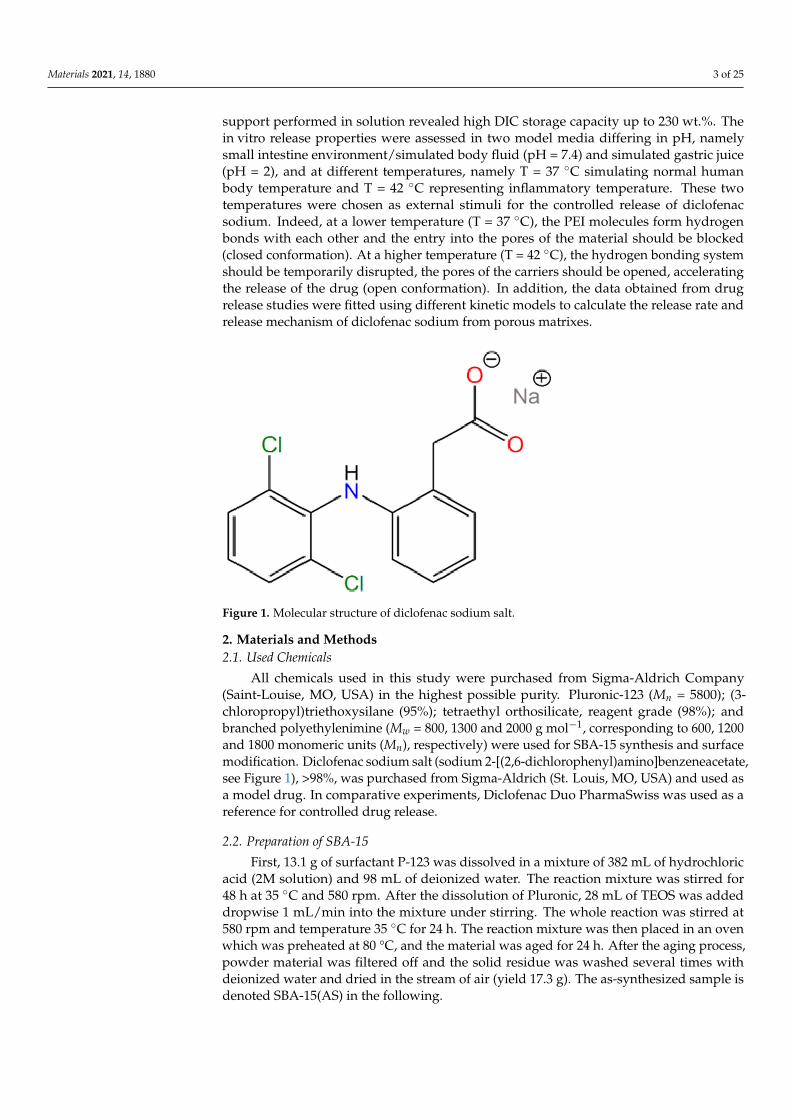
In our study, the drug diclofenac sodium, which belongs to the group of nons-teroidal anti-inflammatory drugs (NSAIDs) was used. Diclofenac sodium, or 2-[2-(2,6-dichloroanilino)phenyl]acetate sodium (according to IUPAC, see Figure 1) is used in non-rheumatic, rheumatic and inflammatory diseases. It acts as a mediator in the inhibitionof prostaglandin synthesis in cyclooxygenase. In an in vivo study, diclofenac did not in-hibit phospholipase A2 at a high concentration that controlled arachidonic acid synthesisand had low to no effect on 5- and 15-lipoxygenases. Nevertheless, the concentrations ofproducts derived from the lipoxygenase pathway were reduced precisely due to the highconcentrations of diclofenac in vivo and exclusively in leukocytes. This effect is likely to becaused by the availability of intracellular arachidonic acid [40].
In the present study, thermosensitive DDSs based on mesoporous silica SBA-15 graftedwith branched polyethyleneimines (PEIs) with different molecular weights (Mw = 800, 1000and 1200 g·mol−1) were prepared, characterized and studied as supports for nonsteroidalanti-inflammatory drug diclofenac sodium (DIC). Drug adsorption into the prepared
Materials 2021, 14, 1880 3 of 25
support performed in solution revealed high DIC storage capacity up to 230 wt.%. Thein vitro release properties were assessed in two model media differing in pH, namelysmall intestine environment/simulated body fluid (pH = 7.4) and simulated gastric juice(pH = 2), and at different temperatures, namely T = 37 ◦C simulating normal humanbody temperature and T = 42 ◦C representing inflammatory temperature. These twotemperatures were chosen as external stimuli for the controlled release of diclofenacsodium. Indeed, at a lower temperature (T = 37 ◦C), the PEI molecules form hydrogenbonds with each other and the entry into the pores of the material should be blocked(closed conformation). At a higher temperature (T = 42 ◦C), the hydrogen bonding systemshould be temporarily disrupted, the pores of the carriers should be opened, acceleratingthe release of the drug (open conformation). In addition, the data obtained from drugrelease studies were fitted using different kinetic models to calculate the release rate andrelease mechanism of diclofenac sodium from porous matrixes.
Figure 1. Molecular structure of diclofenac sodium salt.
2. Materials and Methods2.1. Used Chemicals
All chemicals used in this study were purchased from Sigma-Aldrich Company(Saint-Louise, MO, USA) in the highest possible purity. Pluronic-123 (Mn = 5800); (3-chloropropyl)triethoxysilane (95%); tetraethyl orthosilicate, reagent grade (98%); andbranched polyethylenimine (Mw = 800, 1300 and 2000 g mol−1, corresponding to 600, 1200and 1800 monomeric units (Mn), respectively) were used for SBA-15 synthesis and surfacemodification. Diclofenac sodium salt (sodium 2-[(2,6-dichlorophenyl)amino]benzeneacetate,see Figure 1), >98%, was purchased from Sigma-Aldrich (St. Louis, MO, USA) and used asa model drug. In comparative experiments, Diclofenac Duo PharmaSwiss was used as areference for controlled drug release.
2.2. Preparation of SBA-15
First, 13.1 g of surfactant P-123 was dissolved in a mixture of 382 mL of hydrochloricacid (2M solution) and 98 mL of deionized water. The reaction mixture was stirred for48 h at 35 ◦C and 580 rpm. After the dissolution of Pluronic, 28 mL of TEOS was addeddropwise 1 mL/min into the mixture under stirring. The whole reaction was stirred at580 rpm and temperature 35 ◦C for 24 h. The reaction mixture was then placed in an ovenwhich was preheated at 80 °C, and the material was aged for 24 h. After the aging process,powder material was filtered off and the solid residue was washed several times withdeionized water and dried in the stream of air (yield 17.3 g). The as-synthesized sample isdenoted SBA-15(AS) in the following.
Materials 2021, 14, 1880 4 of 25
2.3. Extraction/Calcination Process
After the material drying procedure, surfactant molecules located in pores wereremoved by the combination of extraction and calcination processes. The first step wasan extraction with dry toluene to purify the silica from contamination of impurities thatoccurred during the synthesis. The second step was sample extraction with HCl (2M) toremove the surfactant from the pores. The third step of the extraction was performed withtetrahydrofuran to remove the remaining surfactant molecules and other impurities. Afterextraction processes, the sample was dried in an oven at 70 ◦C and further calcined. Thecalcination process was performed in an oven with the following temperature program:First, the sample was slowly heated, with a heating rate of 0.5 ◦C·min−1, to 150 ◦C for 2 h.Slow heating was chosen to remove residual solvents from the mesopores of the material.The next step was thermolysis of the residual surfactant by heating to 600 ◦C with a slowheating rate of 1.25 ◦C·min−1. After heating the material at 600 ◦C for 8 h, the materialwas slowly cooled down to ambient temperature. The weight of the calcined material was3.585 g, and the sample after the calcination process was denoted as SBA-15(C).
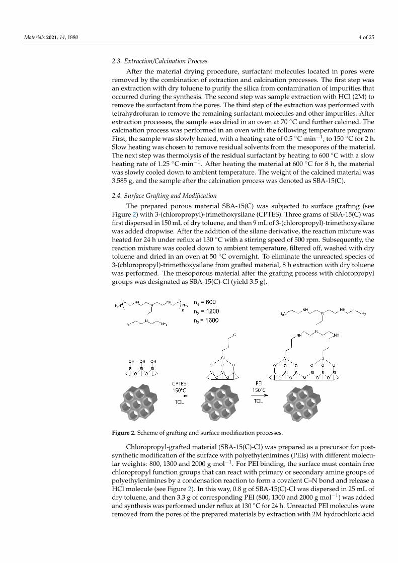
2.4. Surface Grafting and Modification
The prepared porous material SBA-15(C) was subjected to surface grafting (seeFigure 2) with 3-(chloropropyl)-trimethoxysilane (CPTES). Three grams of SBA-15(C) wasfirst dispersed in 150 mL of dry toluene, and then 9 mL of 3-(chloropropyl)-trimethoxysilanewas added dropwise. After the addition of the silane derivative, the reaction mixture washeated for 24 h under reflux at 130 ◦C with a stirring speed of 500 rpm. Subsequently, thereaction mixture was cooled down to ambient temperature, filtered off, washed with drytoluene and dried in an oven at 50 ◦C overnight. To eliminate the unreacted species of3-(chloropropyl)-trimethoxysilane from grafted material, 8 h extraction with dry toluenewas performed. The mesoporous material after the grafting process with chloropropylgroups was designated as SBA-15(C)-Cl (yield 3.5 g).
Figure 2. Scheme of grafting and surface modification processes.
Chloropropyl-grafted material (SBA-15(C)-Cl) was prepared as a precursor for post-synthetic modification of the surface with polyethylenimines (PEIs) with different molecu-lar weights: 800, 1300 and 2000 g·mol−1. For PEI binding, the surface must contain freechloropropyl function groups that can react with primary or secondary amine groups ofpolyethylenimines by a condensation reaction to form a covalent C–N bond and release aHCl molecule (see Figure 2). In this way, 0.8 g of SBA-15(C)-Cl was dispersed in 25 mL ofdry toluene, and then 3.3 g of corresponding PEI (800, 1300 and 2000 g mol−1) was addedand synthesis was performed under reflux at 130 ◦C for 24 h. Unreacted PEI molecules wereremoved from the pores of the prepared materials by extraction with 2M hydrochloric acid
Materials 2021, 14, 1880 5 of 25
solution and then dried in an oven at 70 ◦C overnight. Materials after surface modificationwith polyethylenimines were designated as SBA-15(C)-800 (yield 3.30 g), SBA-15(C)-1300(yield 3.47 g) and SBA-15(C)-2000 (yield 3.64 g).
2.5. Drug Encapsulation
Modified samples SBA-15(C)-800, SBA-15(C)-1300 and SBA-15(C)-2000 and calcinedmaterial SBA-15(C) were used as carriers for diclofenac sodium salt. The affinity of the drugto the surface of carriers was assessed using methanolic solutions of diclofenac sodium withdifferent concentrations: 0.1, 0.05, 0.025, 0.0125 and 0.00625 mol·dm−3. Methanol was usedas a solvent due to the high solubility of diclofenac sodium salt (35 mg·cm−3). In general,10 mg of samples SBA-15(C)-PEI-X (X = 800, 1300 and 2000)/SBA-15(C) were dispersedin 1 mL of drug solution in a plastic vial. Suspensions were stirred at 40 rpm (rotator)for 24 h at 42 ◦C (open conformation) while the evaporation of methanol was prevented.The suspensions were then centrifugated at 6000 rpm for 10 min; after centrifugation,the supernatants were separated, and the amount of adsorbed drugs was determined byUV-VIS spectroscopy, based on the calibration curve of DIC in methanol (see Figure S1 inSupplementary Materials).
2.6. Drug Release
Drug-loaded samples (~3 mg) were packed in a semipermeable membrane VISKINGand inserted into 50 mL polypropylene vials covered with caps. The vials were filled with50 mL of saline solutions with pH = 2 and pH = 7.4 and transferred into a preheated ovenat 37 ◦C (closed conformation) or 42 ◦C (open conformation), and mixtures were gentlystirred on a magnetic stirrer. In general, the release amount of the drug was analyzed atselected time intervals: 0.5, 1.5, 3.5, 5.5, 7.5, 9.5 and 24 h.
The amount of the drug released was determined using UV-VIS spectroscopy. Beforedetermining the concentration of DIC, calibration curves were constructed based on so-lutions (saline at pH = 7.4/2/5.5) with various concentrations of diclofenac sodium (seeFigure S2 in Supplementary Materials). The correlation coefficient r2 = 0.9999 for salinesolution at pH = 7.4, r2 = 0.9966 for saline solution at pH = 2 and r2 = 0.9999 for salinesolution at pH = 5.5 confirmed the linearity of calibration curves. pH = 5.5 was used indrug release experiments performed under dynamic conditions.
2.7. Drug Solubility
The diclofenac sodium solubility was studied at 37 and 42 ◦C and pH 2 and 7.4. Onehundred milligrams of drug was dispersed in 10 mL of saline solution with different pHvalues in glass vials. Vials were sealed and sonicated for 3 min. After sonification, vialswere put in the preheated ovens at 37 and 42 ◦C. The prepared suspensions were stirredunder selected conditions (1. 37 ◦C, pH = 2; 2. 37 ◦C, pH = 7.4; 3. 42 ◦C, pH = 2; 4.42 ◦C, pH = 7.4) for 24 h. After that, the remaining undissolved drug was filtered withpolytetrafluoroethylene (PTFE) micro filter with 0.2 µm porosity and 20 mm diameter.Subsequently, 100 µL of supernatant from the individual solutions was diluted in waterin a 1:1000 ratio (drug solution/saline solution) and analyzed by UV-VIS spectroscopy(Analytik Jena GmbH, Jena, Germany). The amount of drug in the samples was determinedbased on absorbance using calibration curves.
2.8. Characterization
Infrared spectra of prepared materials were measured by FT-IR spectroscopy onNicolet 6700 from Thermo Scientific (Thermo Scientific, Waltham, MA, USA) using KBrtechnique in the wavelength range of 4000–400 cm−1. Samples for IR analysis wereprepared in the form of KBr pellets with KBr/sample mass ratio 100:1 or 100:5. For selectedsamples, a higher mass ratio was chosen for better visibility of characteristic absorptionbands. Before IR measurements, potassium bromide was dried at 700 ◦C for 4 h in an ovento remove water and freely cooled in a desiccator. All IR spectra of prepared materials
Materials 2021, 14, 1880 6 of 25
were recorded by collecting 64 scans with a resolution of 4 cm−1 for a single spectrum atambient temperature.
Nitrogen adsorption/desorption isotherms were measured at −196 ◦C and experi-ments were carried out using Nova 1200e from Quantachrome Instruments (Quantachrome,Miami, FL, USA) and ASAP 2020 Micromeritics apparatus (Micromeritics, Norcross, GA,USA). Before nitrogen adsorption/desorption measurements, samples were outgassed atdifferent temperatures (150 ◦C for SBA-15(C) and extracted samples, 60 ◦C for surface-modified materials). The Brunauer–Emmett–Teller (BET) specific surface area (SBET) ofeach sample was calculated using nitrogen adsorption data in a p/p0 = 0.05–0.20. Thetextural properties such as pore size diameter (d) and pore volume (Vp) and were evaluatedusing the Barrett–Joyner–Halenda (BJH) model from the nitrogen desorption isotherm.
The shape and surface morphology of prepared materials were analyzed using atomicforce microscopy (AFM) on a Solver PRO instrument from NT-MDT Spectrum Instruments(NT-MDT, Moscow, Russia). The measurements were performed in contact and semi-contact mode on samples attached to a surface-treated mica plate with polylysine.
Transmission electron microscopy was carried out on a microscope JEOL 2000FX(JEOL, Pleasanton, FA, USA). Prepared materials were ground and dispersed in ethanol.The material’s suspension was dripped onto a carbon grid and freely air-dried.
The particle size distribution in the nano-range was determined by a photon cross-correlation spectroscopy, which was carried out on a Nanophox particle size analyzer(Sympatec, Clausthal-Zellerfeld, Germany). A small amount of powder product was dis-persed in the deionized water and ultrasonicated for 15 min. After that, the obtainedsuspensions were left standing for 10 min, and subsequently, part of the top fraction con-taining a significant amount of fine particles was transferred into the measurement cuvetteand diluted with distilled water to obtain a suitable concentration for the experiment. Adispersant refractive index of 1.33 was used for the analysis, and the measurements wererepeated three times for each sample. The polydispersity index (PDI) was calculated bydividing the square of the standard deviation of the corresponding peak by the square ofthe value of its central position on the x-axis.
Small-angle X-ray diffraction (SA-XRD) experiments (Rigaku, Tokyo, Japan) wereperformed on a Rigaku Ultima IV multipurpose diffractometer in transmission geometryusing CuKα radiation (λ = 1.54056 Å). The thickness of the sample during the measurementwas 2 mm, which was achieved by placing the sample in a metal holder, which was sealedon both sides with Kapton tape. SA-XRD measurements were realized by 2θ continuousscan at 0.2◦ min−1 in the 2θ range from 0.1 to 3◦, and scattered photons from the sampleswere detected every 0.02◦ using a sodium iodide scintillation detector.
TGA Q500 apparatus from TA Instruments (TA Instruments, New Castle, DE, USA)was used for determining the thermal properties of prepared materials. The thermogravi-metric analysis was performed in the temperature range of 30–800 ◦C with a heating rateof 10 ◦C min−1 in an air atmosphere with a flow rate of 60 cm3·min−1 using platinumcrucibles.
The volume phase transition behavior of PEI-modified samples was investigated bydifferential scanning calorimetry (DSC) on a DSC Q2000 apparatus from TA Instruments(TA Instruments, New Castle, DE, USA). The samples were heated with a heating rate of1 ◦C min−1 in a temperature range of 10–60 ◦C and nitrogen atmosphere.
UV-VIS spectroscopy was performed on a Specord 250 UV-VIS spectrometer from Ana-lytik Jena AG (Analytik Jena GmbH, Jena, Germany) in a wavelength range of 240–350 nm.UV-VIS spectroscopy was used to determine the amount of adsorbed/released drug.
3. Results and Discussion3.1. Synthesis of SBA-15 and Surface Modification
The sol–gel process was used for the synthesis of SBA-15 by hydrolysis and poly-condensation reactions of TEOS in water using Pluronic-123 as surfactant and HCl as acatalyst [41]. Sol is a low viscosity liquid that can be transformed into a solid form. Over
Materials 2021, 14, 1880 7 of 25
time, the colloidal and condensed silica are combined into a 3D structure to form a mesh.The gel form and its physical properties depend mainly on the particle size. When thegel is forming, the viscosity increases the strength and integrity of the silica material. Thestructure and textural properties of the gel depend on the time of its formation. Otherprocesses such as aging, drying, stabilization and thickening depend on the structure of thegel [42]. Achieving the largest silica surface area, pore size and pore volume depend on theremoval of surfactant molecules. Calcination of silica nanoparticles at high temperatureis a fast and efficient way to remove surfactant from pores. However, the calcinationconditions must be chosen carefully because this process can perforate the silica pores. Theas-synthesized silica contains not only surfactant molecules but also molecules of solventin the pores. With rapid calcination, there is a risk that the solvent molecules will startto evaporate quickly and perforate the pores. Surfactant extraction is a slower but moregentle removal process that, on the other hand, does not lead to the complete removal ofsurfactant molecules. For the mentioned reasons, we have chosen in our work a combina-tion of extraction and calcination of SBA-15(AS) material. The extraction was performedusing solvents with different polarity (toluene, THF and 2M HCl solution), and then thematerial was calcined by slow heating at 600 ◦C. For modification with organic compounds,the surface was treated by silylation reaction with alkoxysilanes and similar silane com-pounds. Mentioned surface grafting is suitable for the subsequent attachment of organiccompounds with different functional groups. The surface of SBA-15(C), which containsfree hydroxyl groups, was subsequently grafted with 3-(chloropropyl)-trimethoxysilanemolecules in dry toluene. This step was necessary for the formation of the covalent bondbetween the surface of SBA-15(C) and polyethylenimine molecules, via a condensationreaction. PEI molecules with different molecular weights (800, 1300 and 2000 g·mol−1)were used for surface modification. The materials prepared by the described processeswere subsequently used as carriers for the anti-inflammatory drug diclofenac sodium andtested as thermosensitive DDSs.
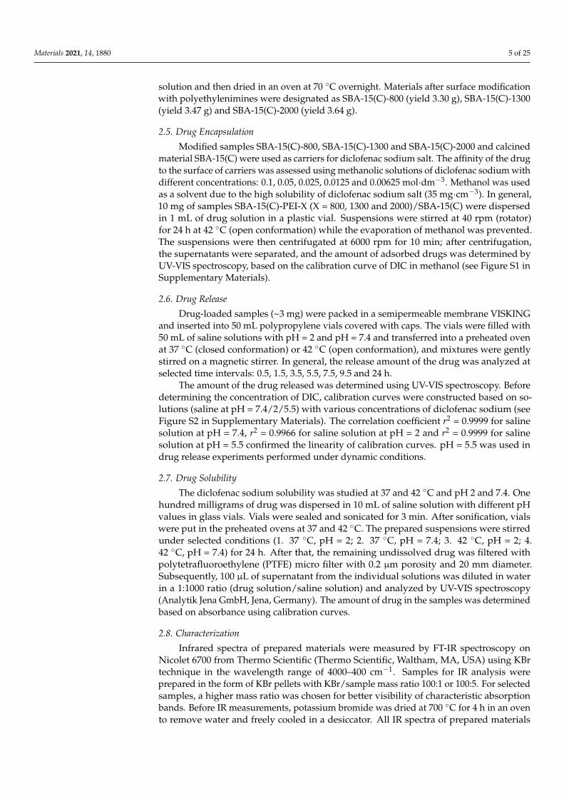
3.2. Infrared Spectroscopy3.2.1. SBA-15 and Surface-Modified Samples
Infrared spectra of prepared materials are presented in Figure 3, and the assignmentof characteristic bands is summarized in Table 1. The infrared spectrum of SBA-15(C) (seeFigure 3a) shows a broad band at 3437 cm−1 corresponding to stretching ν(OH) vibrationof silanol groups located on the surface of silica (Si–OH) and physisorbed water molecules.Bending vibrations δ(OH) of mentioned groups was found at 1638 cm−1. The presence ofsilica framework is reflected by broad and intense bands at 1088 and 809 cm−1, which canbe attributed to the asymmetric (νas(SiOSi)) and symmetric (νs(SiOSi)) stretching vibrations,respectively. Bending vibrations of δ(SiOSi) groups were detected at 462 cm−1.
Table 1. Assignment of vibrations and corresponding wavenumbers to characteristic bands in IR spectra of surface-modifiedmaterials.
Sample ν(OH) ν(SiOSi)s ν(SiOSi)as δ(SiOSi) δ(OH) ν(CH)aliph δ(CH) δ(NH2) ν(NH2)
SBA-15(C) 3437 809 1088 462 1638 - - - -SBA-15(C)-Cl 3427 800 1100 466 1638 2981 1453 - -
29312900
SBA-15(C)-800 - 800 1100 475 1656 2954 1462 1602 33602837 3300
SBA-15(C)-1300 - 800 1124 475 1638 2952 1462 1603 32832837 3301
SBA-15(C)-2000 - 804 1098 475 1629 2953 1462 1602 32512838 3300
ν—stretching; δ—scissoring; as—asymmetric; s—symmetric; ar—aromatic; aliph—aliphatic.
Materials 2021, 14, 1880 8 of 25
Figure 3. Infrared spectra of (a) SBA-15(C), (b) SBA-15(C)-Cl, (c) polyethyleneimine (PEI)-modified samples and (d)drug-loaded materials.
The IR spectrum of SBA-15(C)-Cl after grafting with 3-(chloropropyl)-trimethoxysilaneis shown in Figure 3b. Bands observed at the wavelengths of 2981, 2931 and 2900 cm−1
are attributed to the valence vibrations of ν(CH2) groups of propyl chain. Deformationvibration (δ(CH2)) of mentioned groups appeared at 1453 cm−1.
All IR spectra (see Figure 3c) of surface-modified samples with polyethylenimines(SBA-15(C)-PEI-X (X = 800, 1300 and 2000)) contain characteristic bands in the wavenum-ber range of 3360–3251 cm−1 due to stretching vibrations of primary/secondary amines(ν(NH2)/ν(NH)) of PEI molecules. In the IR spectra, deformation bands δ(NH2) in therange from 1602 to 1603 cm−1 are present, which also confirm the presence of aminegroups. Moreover, the presence of PEI molecules is also evidenced by the presence of twoweak stretching vibrations of aliphatic ν(CH) groups of ethylene bridges located below3000 cm−1 (see Table 1, Figure 3c).
3.2.2. Diclofenac Sodium and Drug-Loaded Samples
The structure of diclofenac sodium is presented in Figure 1, and the following bandswere observed in its IR spectrum (see Figure S3 in Supplementary Materials): a weak bandat 3080 cm−1 belonging to the stretching vibration of aromatic CH groups of phenyl scaffold;sharp, medium-intensity bands at 1496, 1573 and 1604 cm−1 corresponding to the valencevibrations of the conjugated system of C=C bonds in the phenyl ring; a low-intensity, sharpband at about 717 cm−1 related to the deformation vibrations for monosubstituted phenylring; and three low-intensity, sharp bands at 767, 951 and 1170 cm−1 corresponding to thedeformation vibrations for 1,2,3-trisubstituted phenyl ring. Moreover, in the IR spectrumof diclofenac sodium salt, a strong and sharp band was found at 747 cm−1, which could beassigned to the vibration of the C–Cl bond. The presence of a secondary amine group isobvious from the weak and broad band at 3386 cm−1 (ν(NH)), and its deformation vibrationδ(NH) was detected at 1556 cm−1. Other characteristic vibrations in the IR spectrum of
Materials 2021, 14, 1880 9 of 25
diclofenac sodium are symmetric and asymmetric vibrations of the carboxylate group(ν(COO−)), located at 1400 and 1550 cm−1, respectively.
The IR spectra of samples with encapsulated DIC are shown in Figure 3d, and charac-teristic bands are listed in Table 2. The presence of diclofenac sodium salt encapsulated inprepared materials is evidenced by the presence of two sharp, medium-intensity bandsat 1555 and 1580 cm−1, which correspond to the valence vibrations of the aromatic ring(ν(C=C)ar). Typical vibrations of carboxylate group ν(COO−)s and ν(COO−)as were foundat about 1450 and 1510 cm−1. Another low-intensity, sharp band found at 747 cm−1
corresponds to the valence vibration of the C–Cl bond.
Table 2. Assignment of vibrations and corresponding wavenumbers to characteristic bands in IR spectra of preparedmaterials with loaded drug.
Sample ν(CH)ar δ(CH) ν(CCl) ν(C=C)ar ν(NH) ν(COO−)s ν(COO−)as δ(NH)
SBA-15(C)/DIC 3080 1454 746 1555 3420 1450 1509 16111580 3274
SBA-15(C)-800/DIC 3082 1454 748 1555 3413 1451 1510 16091580 3280
SBA-15(C)-1300/DIC 3081 1453 746 1555 3421 1450 1511 16121580 3274
SBA-15(C)-2000/DIC 3080 1454 748 1555 3418 1453 1510 16101580 3271
3.3. Nitrogen Adsorption/Desorption Measurements
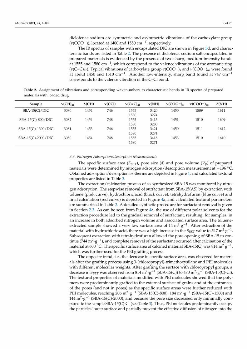
The specific surface area (SBET), pore size (d) and pore volume (Vp) of preparedmaterials were determined by nitrogen adsorption/desorption measurement at −196 ◦C.Obtained adsorption/desorption isotherms are depicted in Figure 4, and calculated texturalproperties are listed in Table 3.
The extraction/calcination process of as-synthesized SBA-15 was monitored by nitro-gen adsorption. The stepwise removal of surfactant from SBA-15(AS) by extraction withtoluene (pink curve), hydrochloric acid (black curve), tetrahydrofuran (blue curve) andfinal calcination (red curve) is depicted in Figure 4a, and calculated textural parametersare summarized in Table 3. A detailed synthetic procedure for surfactant removal is givenin Section 2.3. As can be seen from Figure 4a, the use of different polar solvents for theextraction procedure led to the gradual removal of surfactant, resulting, for samples, inan increase in both adsorbed nitrogen volume and associated surface area. The toluene-extracted sample showed a very low surface area of 14 m2 g−1. After extraction of thematerial with hydrochloric acid, there was a high increase in the SBET value to 547 m2 g−1.Subsequent extraction with tetrahydrofuran allowed the pore opening of SBA-15 to con-tinue (744 m2·g−1), and complete removal of the surfactant occurred after calcination of thematerial at 600 ◦C. The specific surface area of calcined material SBA-15(C) was 814 m2 g−1,which was further used for the PEI grafting process.
The opposite trend, i.e., the decrease in specific surface area, was observed for materi-als after the grafting process using 3-(chloropropyl)-trimethoxysilane and PEI moleculeswith different molecular weights. After grafting the surface with chloropropyl groups, adecrease in SBET was observed from 814 m2·g−1 (SBA-15(C)) to 470 m2·g−1 (SBA-15(C)-Cl).The textural properties of materials modified with PEI molecules showed that the poly-mers were predominantly grafted to the external surface of grains and at the entrancesof the pores (and not in pores) as the specific surface areas were further reduced withPEI molecules, reaching 206 m2·g−1 (SBA-15(C)-800), 184 m2·g−1 (SBA-15(C)-1300) and144 m2·g−1 (SBA-15(C)-2000), and because the pore size decreased only minimally com-pared to the sample SBA-15(C)-Cl (see Table 3). Thus, PEI molecules predominantly occupythe particles’ outer surface and partially prevent the effective diffusion of nitrogen into the
Materials 2021, 14, 1880 10 of 25
internal pores. This fact was also confirmed by AFM spectroscopy, which clearly showedthe PEI overlapping the entrances to the pores (see Section 3.4).
Figure 4. N2 adsorption/desorption isotherms measured at −196 ◦C of (a) the SBA-15(AS) during the surfactant removalprocess and (b) SBA-15(C) after chloropropyl grafting and PEI modification.
Table 3. The textural properties of prepared SBA-15 materials determined from N2 adsorp-tion/desorption isotherms.
Sample Surface Area Pore Size Pore Volume(m2.g−1) (nm) (cm3.g−1)
SBA-15(C)-TOL 14 - 0.21SBA-15(C)-HCl 547 7.2 0.64SBA-15(C)-THF 744 8.1 0.82
SBA-15(C) 814 8.6 0.86SBA-15(C)-Cl 470 6.1 0.41
SBA-15(C)-800 206 5.9 0.28SBA-15(C)-1300 184 5.6 0.24SBA-15(C)-2000 144 5.5 0.24
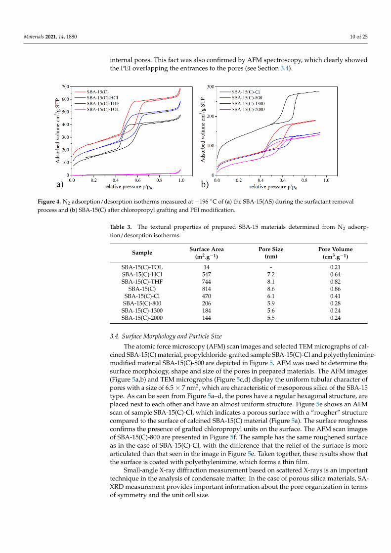
3.4. Surface Morphology and Particle Size
The atomic force microscopy (AFM) scan images and selected TEM micrographs of cal-cined SBA-15(C) material, propylchloride-grafted sample SBA-15(C)-Cl and polyethylenimine-modified material SBA-15(C)-800 are depicted in Figure 5. AFM was used to determine thesurface morphology, shape and size of the pores in prepared materials. The AFM images(Figure 5a,b) and TEM micrographs (Figure 5c,d) display the uniform tubular character ofpores with a size of 6.5× 7 nm2, which are characteristic of mesoporous silica of the SBA-15type. As can be seen from Figure 5a–d, the pores have a regular hexagonal structure, areplaced next to each other and have an almost uniform structure. Figure 5e shows an AFMscan of sample SBA-15(C)-Cl, which indicates a porous surface with a “rougher” structurecompared to the surface of calcined SBA-15(C) material (Figure 5a). The surface roughnessconfirms the presence of grafted chloropropyl units on the surface. The AFM scan imagesof SBA-15(C)-800 are presented in Figure 5f. The sample has the same roughened surfaceas in the case of SBA-15(C)-Cl, with the difference that the relief of the surface is morearticulated than that seen in the image in Figure 5e. Taken together, these results show thatthe surface is coated with polyethylenimine, which forms a thin film.
Small-angle X-ray diffraction measurement based on scattered X-rays is an importanttechnique in the analysis of condensate matter. In the case of porous silica materials, SA-XRD measurement provides important information about the pore organization in termsof symmetry and the unit cell size.
Materials 2021, 14, 1880 11 of 25
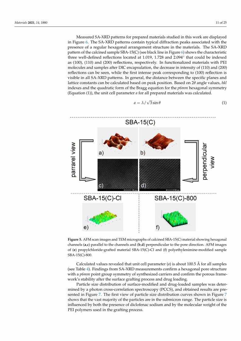
Measured SA-XRD patterns for prepared materials studied in this work are displayedin Figure 6. The SA-XRD patterns contain typical diffraction peaks associated with thepresence of a regular hexagonal arrangement structure in the materials. The SA-XRDpattern of the calcined sample SBA-15(C) (see black line in Figure 6) shows the characteristicthree well-defined reflections located at 1.019, 1.728 and 2.094◦ that could be indexedas (100), (110) and (200) reflections, respectively. In functionalized materials with PEImolecules and samples after DIC encapsulation, the decrease in intensity of (110) and (200)reflections can be seen, while the first intense peak corresponding to (100) reflection isvisible in all SA-XRD patterns. In general, the distance between the specific planes andlattice constants can be calculated based on peak position. Based on 2θ angle values, hklindexes and the quadratic form of the Bragg equation for the p6mm hexagonal symmetry(Equation (1)), the unit cell parameter a for all prepared materials was calculated.
a = λ/√
3 sin θ (1)
Figure 5. AFM scan images and TEM micrographs of calcined SBA-15(C) material showing hexagonalchannels (a,c) parallel to the channels and (b,d) perpendicular to the pore direction. AFM imagesof (e) propylchloride-grafted material SBA-15(C)-Cl and (f) polyethylenimine-modified sampleSBA-15(C)-800.
Calculated values revealed that unit cell parameter (a) is about 100.5 Å for all samples(see Table 4). Findings from SA-XRD measurements confirm a hexagonal pore structurewith a p6mm point group symmetry of synthesized carriers and confirm the porous frame-work’s stability after the surface grafting process and drug loading.
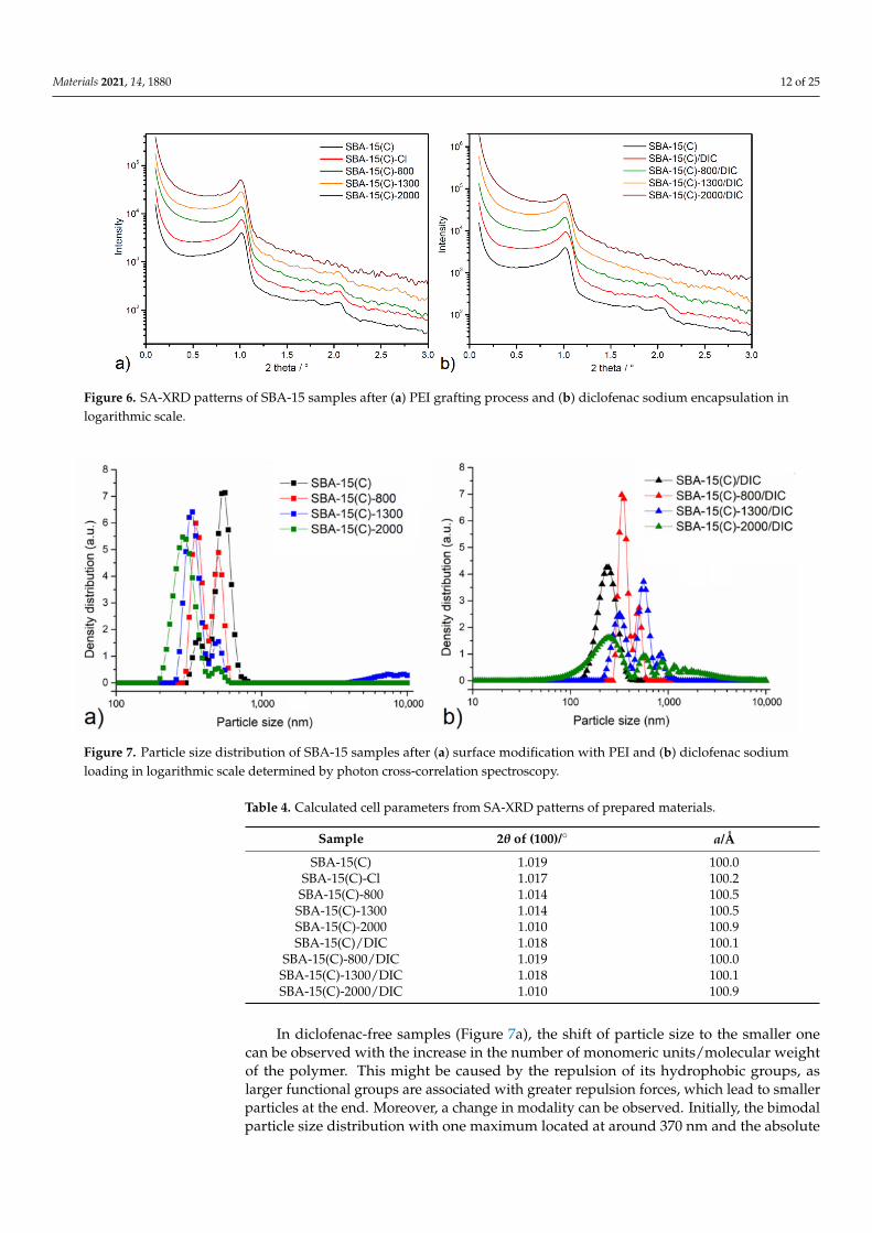
Particle size distribution of surface-modified and drug-loaded samples was deter-mined by a photon cross-correlation spectroscopy (PCCS), and obtained results are pre-sented in Figure 7. The first view of particle size distribution curves shown in Figure 7shows that the vast majority of the particles are in the submicron range. The particle size isinfluenced by both the presence of diclofenac sodium and by the molecular weight of thePEI polymers used in the grafting process.
Materials 2021, 14, 1880 12 of 25
Figure 6. SA-XRD patterns of SBA-15 samples after (a) PEI grafting process and (b) diclofenac sodium encapsulation inlogarithmic scale.
Figure 7. Particle size distribution of SBA-15 samples after (a) surface modification with PEI and (b) diclofenac sodiumloading in logarithmic scale determined by photon cross-correlation spectroscopy.
Table 4. Calculated cell parameters from SA-XRD patterns of prepared materials.
Sample 2θ of (100)/◦ a/Å
SBA-15(C) 1.019 100.0SBA-15(C)-Cl 1.017 100.2
SBA-15(C)-800 1.014 100.5SBA-15(C)-1300 1.014 100.5SBA-15(C)-2000 1.010 100.9SBA-15(C)/DIC 1.018 100.1
SBA-15(C)-800/DIC 1.019 100.0SBA-15(C)-1300/DIC 1.018 100.1SBA-15(C)-2000/DIC 1.010 100.9
In diclofenac-free samples (Figure 7a), the shift of particle size to the smaller onecan be observed with the increase in the number of monomeric units/molecular weightof the polymer. This might be caused by the repulsion of its hydrophobic groups, aslarger functional groups are associated with greater repulsion forces, which lead to smallerparticles at the end. Moreover, a change in modality can be observed. Initially, the bimodalparticle size distribution with one maximum located at around 370 nm and the absolute
Materials 2021, 14, 1880 13 of 25
maximum at around 550 nm can be observed. With increasing the PEI molecular weight,the contribution of smaller particles increases. Namely, for the SBA-15(C)-800 sample,the bimodal size distribution with the smaller fraction being slightly more abundantwas evidenced. Further increasing the PEI molecular weight leads to almost completeeradication of the larger fraction of particles and reduction of particle size. Namely, for theSBA-15(C)-2000 sample, almost unimodal distribution with an absolute maximum at theparticle size of 287 nm was evidenced.
For the samples containing the therapeutics, the trend seems to be the other wayaround; i.e., with the increase in PEI molecular weight, the particle size distributionchanges from unimodal to polymodal. Namely, for the PEI-free sample, the unimodaldistribution with the maximum around 240 nm was evidenced. For this sample, thepolydispersity index was calculated to be 0.096, which means that the presented data arerelevant [43]. Introducing PEI and increasing its molecular weight leads to a change intobimodal (SBA-15(C)-800), trimodal (SBA-15(C)-1300) and polymodal (SBA-15(C)-2000)particle size distribution. The observed effect might be connected with the interaction ofPEI with the therapeutic or with the efficient elimination of diclofenac sodium from thestructure of the porous material.
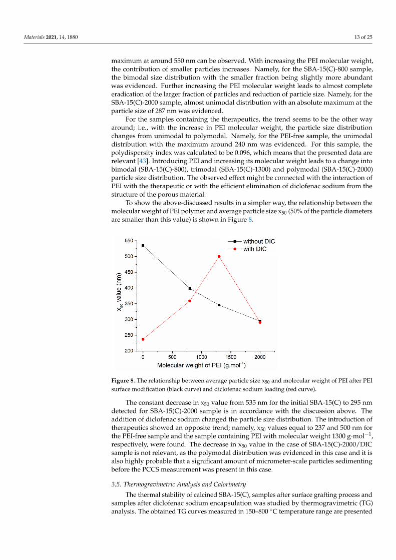
To show the above-discussed results in a simpler way, the relationship between themolecular weight of PEI polymer and average particle size x50 (50% of the particle diametersare smaller than this value) is shown in Figure 8.
Figure 8. The relationship between average particle size x50 and molecular weight of PEI after PEIsurface modification (black curve) and diclofenac sodium loading (red curve).
The constant decrease in x50 value from 535 nm for the initial SBA-15(C) to 295 nmdetected for SBA-15(C)-2000 sample is in accordance with the discussion above. Theaddition of diclofenac sodium changed the particle size distribution. The introduction oftherapeutics showed an opposite trend; namely, x50 values equal to 237 and 500 nm forthe PEI-free sample and the sample containing PEI with molecular weight 1300 g·mol−1,respectively, were found. The decrease in x50 value in the case of SBA-15(C)-2000/DICsample is not relevant, as the polymodal distribution was evidenced in this case and it isalso highly probable that a significant amount of micrometer-scale particles sedimentingbefore the PCCS measurement was present in this case.
3.5. Thermogravimetric Analysis and Calorimetry
The thermal stability of calcined SBA-15(C), samples after surface grafting process andsamples after diclofenac sodium encapsulation was studied by thermogravimetric (TG)analysis. The obtained TG curves measured in 150–800 ◦C temperature range are presented
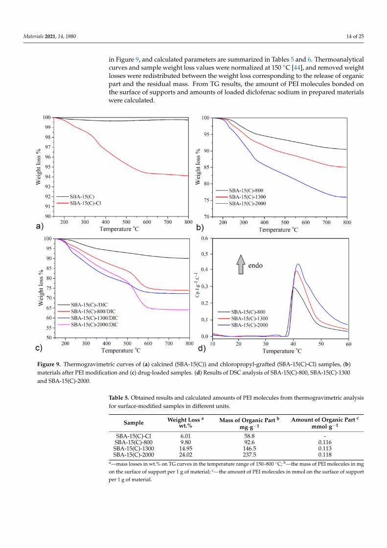
Materials 2021, 14, 1880 14 of 25
in Figure 9, and calculated parameters are summarized in Tables 5 and 6. Thermoanalyticalcurves and sample weight loss values were normalized at 150 ◦C [44], and removed weightlosses were redistributed between the weight loss corresponding to the release of organicpart and the residual mass. From TG results, the amount of PEI molecules bonded onthe surface of supports and amounts of loaded diclofenac sodium in prepared materialswere calculated.
Figure 9. Thermogravimetric curves of (a) calcined (SBA-15(C)) and chloropropyl-grafted (SBA-15(C)-Cl) samples, (b)materials after PEI modification and (c) drug-loaded samples. (d) Results of DSC analysis of SBA-15(C)-800, SBA-15(C)-1300and SBA-15(C)-2000.
Table 5. Obtained results and calculated amounts of PEI molecules from thermogravimetric analysisfor surface-modified samples in different units.
Sample Weight Loss a
wt.%Mass of Organic Part b
mg·g−1Amount of Organic Part c
mmol·g−1
SBA-15(C)-Cl 6.01 58.8 -SBA-15(C)-800 9.80 92.6 0.116
SBA-15(C)-1300 14.95 146.5 0.113SBA-15(C)-2000 24.02 237.5 0.118
a—mass losses in wt.% on TG curves in the temperature range of 150–800 ◦C; b—the mass of PEI molecules in mgon the surface of support per 1 g of material; c—the amount of PEI molecules in mmol on the surface of supportper 1 g of material.
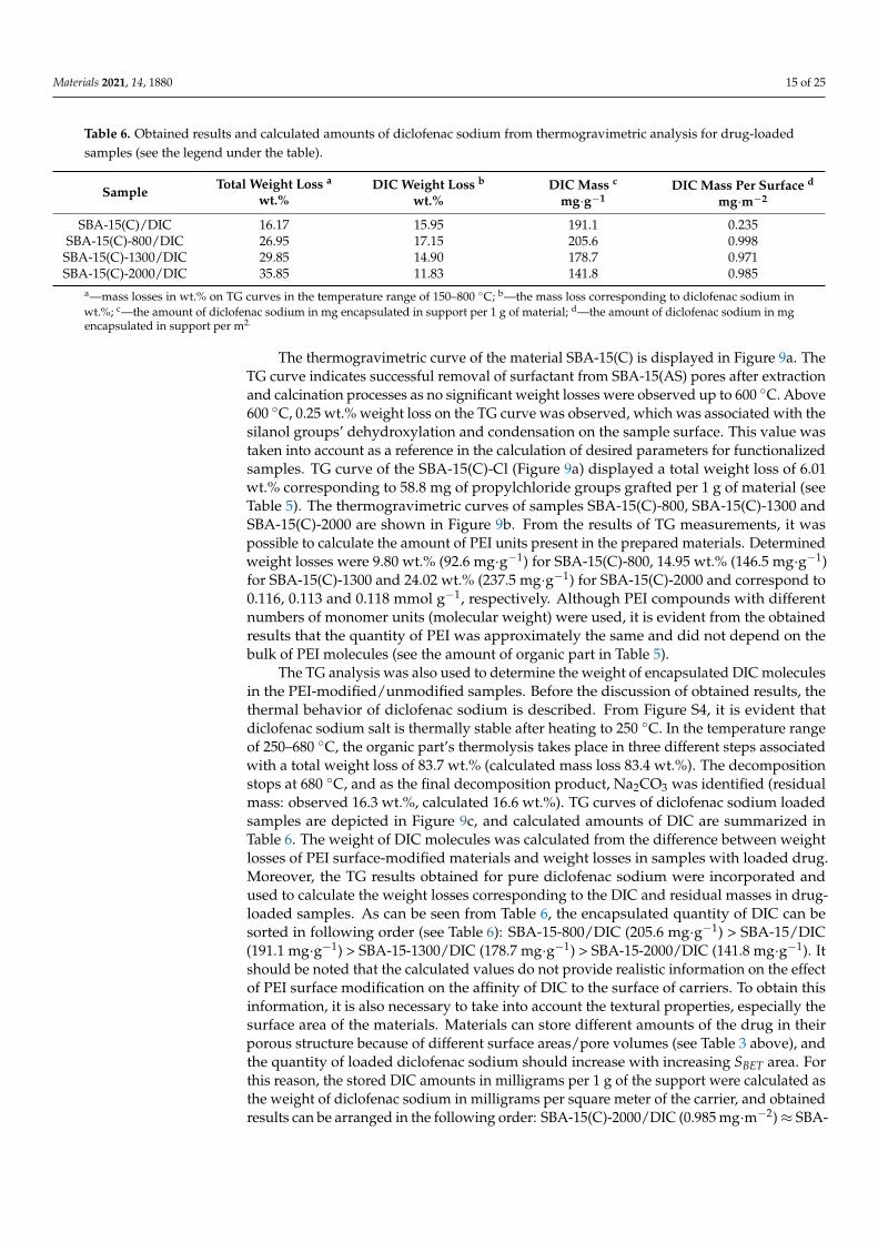
Materials 2021, 14, 1880 15 of 25
Table 6. Obtained results and calculated amounts of diclofenac sodium from thermogravimetric analysis for drug-loadedsamples (see the legend under the table).
Sample Total Weight Loss a
wt.%DIC Weight Loss b
wt.%DIC Mass c
mg·g−1DIC Mass Per Surface d
mg·m−2
SBA-15(C)/DIC 16.17 15.95 191.1 0.235SBA-15(C)-800/DIC 26.95 17.15 205.6 0.998SBA-15(C)-1300/DIC 29.85 14.90 178.7 0.971SBA-15(C)-2000/DIC 35.85 11.83 141.8 0.985
a—mass losses in wt.% on TG curves in the temperature range of 150–800 ◦C; b—the mass loss corresponding to diclofenac sodium inwt.%; c—the amount of diclofenac sodium in mg encapsulated in support per 1 g of material; d—the amount of diclofenac sodium in mgencapsulated in support per m2.
The thermogravimetric curve of the material SBA-15(C) is displayed in Figure 9a. TheTG curve indicates successful removal of surfactant from SBA-15(AS) pores after extractionand calcination processes as no significant weight losses were observed up to 600 ◦C. Above600 ◦C, 0.25 wt.% weight loss on the TG curve was observed, which was associated with thesilanol groups’ dehydroxylation and condensation on the sample surface. This value wastaken into account as a reference in the calculation of desired parameters for functionalizedsamples. TG curve of the SBA-15(C)-Cl (Figure 9a) displayed a total weight loss of 6.01wt.% corresponding to 58.8 mg of propylchloride groups grafted per 1 g of material (seeTable 5). The thermogravimetric curves of samples SBA-15(C)-800, SBA-15(C)-1300 andSBA-15(C)-2000 are shown in Figure 9b. From the results of TG measurements, it waspossible to calculate the amount of PEI units present in the prepared materials. Determinedweight losses were 9.80 wt.% (92.6 mg·g−1) for SBA-15(C)-800, 14.95 wt.% (146.5 mg·g−1)for SBA-15(C)-1300 and 24.02 wt.% (237.5 mg·g−1) for SBA-15(C)-2000 and correspond to0.116, 0.113 and 0.118 mmol g−1, respectively. Although PEI compounds with differentnumbers of monomer units (molecular weight) were used, it is evident from the obtainedresults that the quantity of PEI was approximately the same and did not depend on thebulk of PEI molecules (see the amount of organic part in Table 5).
The TG analysis was also used to determine the weight of encapsulated DIC moleculesin the PEI-modified/unmodified samples. Before the discussion of obtained results, thethermal behavior of diclofenac sodium is described. From Figure S4, it is evident thatdiclofenac sodium salt is thermally stable after heating to 250 ◦C. In the temperature rangeof 250–680 ◦C, the organic part’s thermolysis takes place in three different steps associatedwith a total weight loss of 83.7 wt.% (calculated mass loss 83.4 wt.%). The decompositionstops at 680 ◦C, and as the final decomposition product, Na2CO3 was identified (residualmass: observed 16.3 wt.%, calculated 16.6 wt.%). TG curves of diclofenac sodium loadedsamples are depicted in Figure 9c, and calculated amounts of DIC are summarized inTable 6. The weight of DIC molecules was calculated from the difference between weightlosses of PEI surface-modified materials and weight losses in samples with loaded drug.Moreover, the TG results obtained for pure diclofenac sodium were incorporated andused to calculate the weight losses corresponding to the DIC and residual masses in drug-loaded samples. As can be seen from Table 6, the encapsulated quantity of DIC can besorted in following order (see Table 6): SBA-15-800/DIC (205.6 mg·g−1) > SBA-15/DIC(191.1 mg·g−1) > SBA-15-1300/DIC (178.7 mg·g−1) > SBA-15-2000/DIC (141.8 mg·g−1). Itshould be noted that the calculated values do not provide realistic information on the effectof PEI surface modification on the affinity of DIC to the surface of carriers. To obtain thisinformation, it is also necessary to take into account the textural properties, especially thesurface area of the materials. Materials can store different amounts of the drug in theirporous structure because of different surface areas/pore volumes (see Table 3 above), andthe quantity of loaded diclofenac sodium should increase with increasing SBET area. Forthis reason, the stored DIC amounts in milligrams per 1 g of the support were calculated asthe weight of diclofenac sodium in milligrams per square meter of the carrier, and obtainedresults can be arranged in the following order: SBA-15(C)-2000/DIC (0.985 mg·m−2)≈ SBA-
Materials 2021, 14, 1880 16 of 25
15(C)-1300/DIC (0.971 mg·m−2) ≈ SBA-15(C)-800/DIC (0.998 mg·m−2) > SBA-15(C)/DIC(0.235 mg·m−2) (see Table 6). From the obtained values, it can be concluded that the affinityof diclofenac sodium is similar for all PEI-modified materials, due to the formation ofelectrostatic interactions or intermolecular hydrogen bonds between primary/secondarygroups of bonded molecules and diclofenac sodium molecules. PEI-modified samples areable to encapsulate four times more amount of drug compared to SBA-15(C).
The lower critical solution temperature (LCST) of an aqueous solution of PEI-modifiedsamples was investigated by DSC measurements. Figure 9d clearly confirms LCST transi-tion of polyethyleneimine molecules in SBA-15(C)-800, SBA-15(C)-1300 and SBA-15(C)-2000samples by the presence of endothermic peaks with a maximum at 40.2, 41.1 and 41.7 ◦C,respectively. Because the thermal effects are accompanied by low intensity [45,46], it can beassumed that there is only a partial change in the conformation of the PEI molecules at theobserved temperatures.
3.6. Drug Adsorption Properties
The drug adsorption properties of prepared materials were monitored in solution byUV-VIS spectroscopy based on a calibration curve of DIC in methanol (see Figure S1 inSupplementary Materials). From obtained UV-VIS spectra, diclofenac sodium containsa characteristic absorption band at 280 nm, which corresponds to an electron transitionn→ π*. Diclofenac sodium was loaded into the supports by the impregnation methodusing the methanolic drug solutions with different initial concentrations: 6.25 × 10−3,1.25 × 10−2, 2.5 × 10⁻², 5 × 10⁻² and 10−1 mol.dm−3. The determination of the super-natant concentration after adsorption, defined as the equilibrium concentration, allows thecalculation of the amount adsorbed, QADS, by the following equation:
QADS =V(Ci − Ceq)
ms(2)
where V is the volume of the drug solution (dm3), Ci (mol·dm−3) is the initial concentration,Ceq (mol·dm−3) is the equilibrium concentration and ms is the mass (g) of adsorbent used.
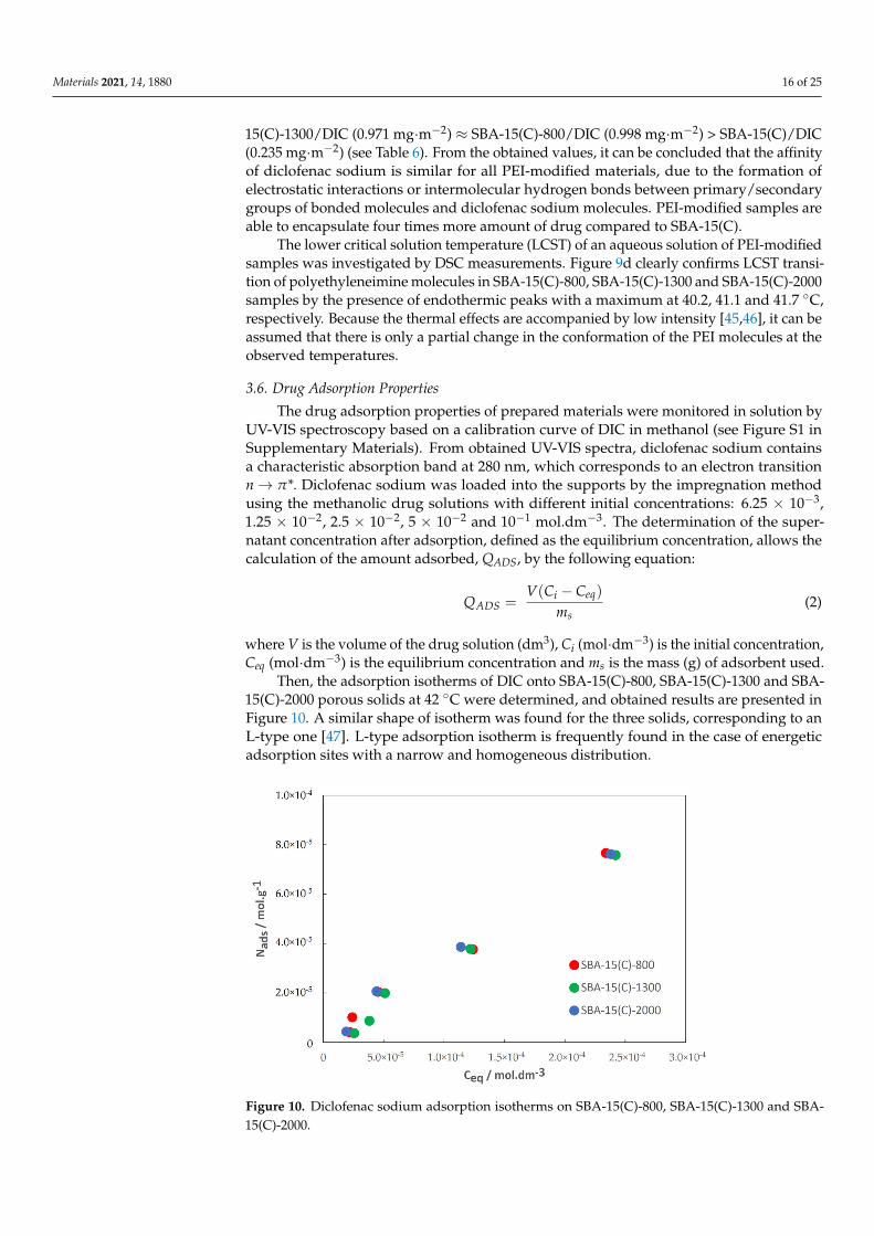
Then, the adsorption isotherms of DIC onto SBA-15(C)-800, SBA-15(C)-1300 and SBA-15(C)-2000 porous solids at 42 ◦C were determined, and obtained results are presented inFigure 10. A similar shape of isotherm was found for the three solids, corresponding to anL-type one [47]. L-type adsorption isotherm is frequently found in the case of energeticadsorption sites with a narrow and homogeneous distribution.
Figure 10. Diclofenac sodium adsorption isotherms on SBA-15(C)-800, SBA-15(C)-1300 and SBA-15(C)-2000.
Materials 2021, 14, 1880 17 of 25
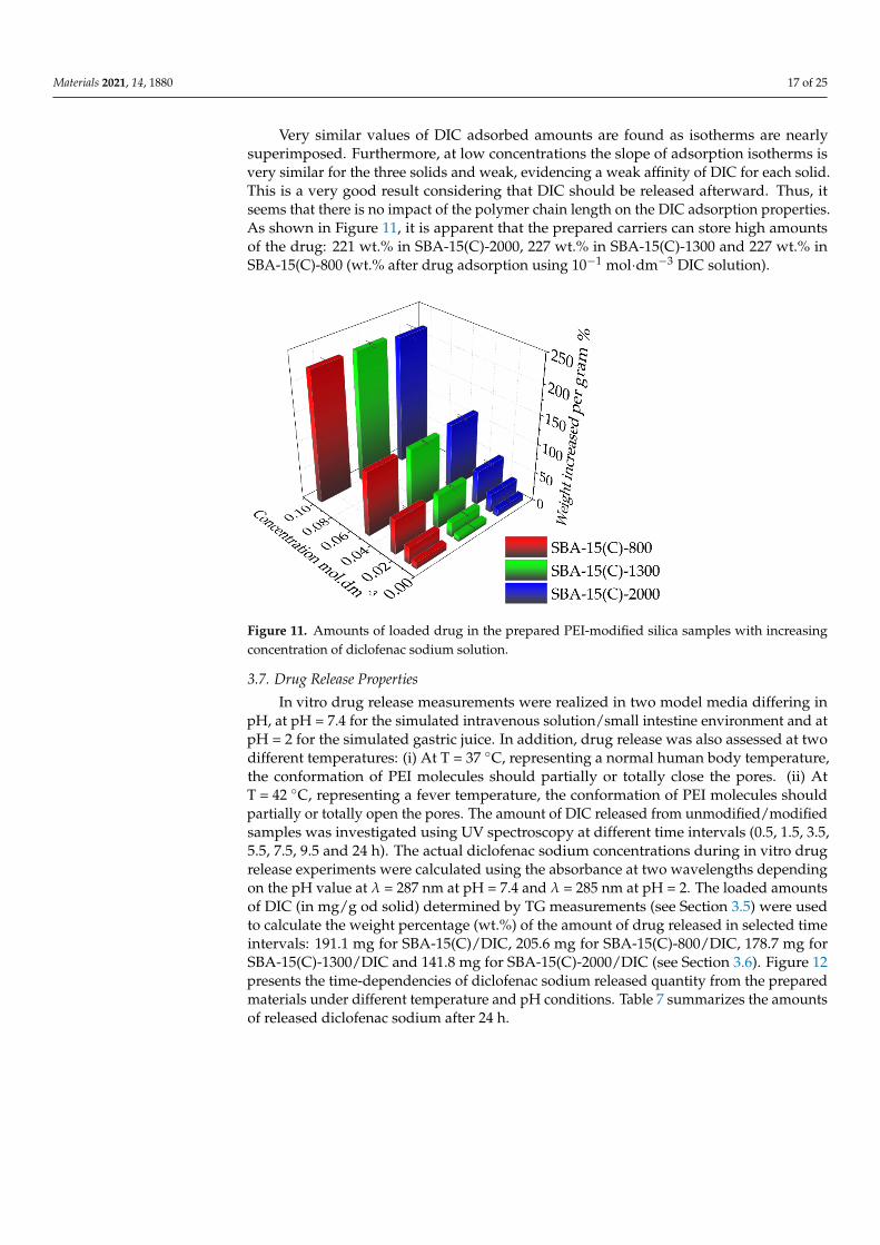
Very similar values of DIC adsorbed amounts are found as isotherms are nearlysuperimposed. Furthermore, at low concentrations the slope of adsorption isotherms isvery similar for the three solids and weak, evidencing a weak affinity of DIC for each solid.This is a very good result considering that DIC should be released afterward. Thus, itseems that there is no impact of the polymer chain length on the DIC adsorption properties.As shown in Figure 11, it is apparent that the prepared carriers can store high amountsof the drug: 221 wt.% in SBA-15(C)-2000, 227 wt.% in SBA-15(C)-1300 and 227 wt.% inSBA-15(C)-800 (wt.% after drug adsorption using 10−1 mol·dm−3 DIC solution).
Figure 11. Amounts of loaded drug in the prepared PEI-modified silica samples with increasingconcentration of diclofenac sodium solution.
3.7. Drug Release Properties
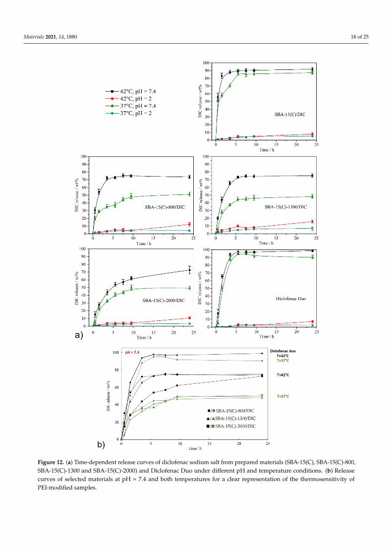
In vitro drug release measurements were realized in two model media differing inpH, at pH = 7.4 for the simulated intravenous solution/small intestine environment and atpH = 2 for the simulated gastric juice. In addition, drug release was also assessed at twodifferent temperatures: (i) At T = 37 ◦C, representing a normal human body temperature,the conformation of PEI molecules should partially or totally close the pores. (ii) AtT = 42 ◦C, representing a fever temperature, the conformation of PEI molecules shouldpartially or totally open the pores. The amount of DIC released from unmodified/modifiedsamples was investigated using UV spectroscopy at different time intervals (0.5, 1.5, 3.5,5.5, 7.5, 9.5 and 24 h). The actual diclofenac sodium concentrations during in vitro drugrelease experiments were calculated using the absorbance at two wavelengths dependingon the pH value at λ = 287 nm at pH = 7.4 and λ = 285 nm at pH = 2. The loaded amountsof DIC (in mg/g od solid) determined by TG measurements (see Section 3.5) were usedto calculate the weight percentage (wt.%) of the amount of drug released in selected timeintervals: 191.1 mg for SBA-15(C)/DIC, 205.6 mg for SBA-15(C)-800/DIC, 178.7 mg forSBA-15(C)-1300/DIC and 141.8 mg for SBA-15(C)-2000/DIC (see Section 3.6). Figure 12presents the time-dependencies of diclofenac sodium released quantity from the preparedmaterials under different temperature and pH conditions. Table 7 summarizes the amountsof released diclofenac sodium after 24 h.
Materials 2021, 14, 1880 18 of 25
Materials 2021, 14, x FOR PEER REVIEW 20 of 26
and the drug molecules and the thermosensitivity of prepared DDSs. Moreover, lower amounts of DIC released from supports at pH = 2 when compared to pure diclofenac sodium could be an advantage of our prepared materials. It is known from clinical studies that one of the side effects of DIC with its long-term use or at high doses is the formation of gastric ulcers. It is estimated that NSAIDs, including DIC, cause around 2000 deaths a year due to the perforation of stomach ulcers [50,51]. SBA-15(C)-2000/DIC material suppresses the solubility of NSAIDs in gastric fluid (pH = 2) by 92% at T = 37 °C or 51% at T = 42 °C in comparison to the pure drug, which may suppress the risk of gastric ulcers.
Finally, the DIC release was also studied using commercial support, named Diclofenac Duo, from the company PharmaSwiss Figure 12. This product was reported to have a controlled release of the drug. A qualitative analysis was performed on a sample of the commercial product, which confirmed the presence of the undefined silicate component. The controlled release indicated that the material was unlikely to be porous, and thus, the drug was only granulated. The drug was released in less than 5 h, and the equilibrium drug concentrations equalized almost immediately. No effect of temperature was evidenced, as also found for the SBA-15(C) support.
Figure 12. (a) Time-dependent release curves of diclofenac sodium salt from prepared materials (SBA-15(C), SBA-15(C)-800,SBA-15(C)-1300 and SBA-15(C)-2000) and Diclofenac Duo under different pH and temperature conditions. (b) Releasecurves of selected materials at pH = 7.4 and both temperatures for a clear representation of the thermosensitivity ofPEI-modified samples.
Materials 2021, 14, 1880 19 of 25
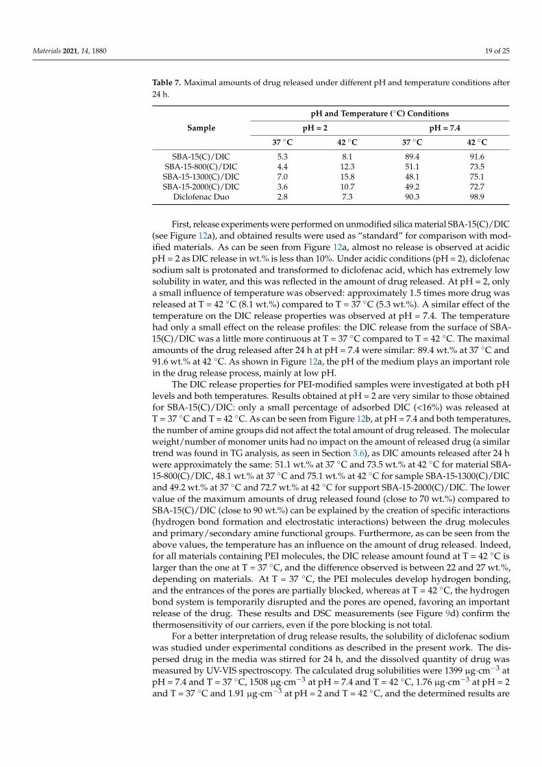
Table 7. Maximal amounts of drug released under different pH and temperature conditions after24 h.
pH and Temperature (◦C) Conditions
Sample pH = 2 pH = 7.4
37 ◦C 42 ◦C 37 ◦C 42 ◦C
SBA-15(C)/DIC 5.3 8.1 89.4 91.6SBA-15-800(C)/DIC 4.4 12.3 51.1 73.5SBA-15-1300(C)/DIC 7.0 15.8 48.1 75.1SBA-15-2000(C)/DIC 3.6 10.7 49.2 72.7
Diclofenac Duo 2.8 7.3 90.3 98.9
First, release experiments were performed on unmodified silica material SBA-15(C)/DIC(see Figure 12a), and obtained results were used as “standard” for comparison with mod-ified materials. As can be seen from Figure 12a, almost no release is observed at acidicpH = 2 as DIC release in wt.% is less than 10%. Under acidic conditions (pH = 2), diclofenacsodium salt is protonated and transformed to diclofenac acid, which has extremely lowsolubility in water, and this was reflected in the amount of drug released. At pH = 2, onlya small influence of temperature was observed: approximately 1.5 times more drug wasreleased at T = 42 ◦C (8.1 wt.%) compared to T = 37 ◦C (5.3 wt.%). A similar effect of thetemperature on the DIC release properties was observed at pH = 7.4. The temperaturehad only a small effect on the release profiles: the DIC release from the surface of SBA-15(C)/DIC was a little more continuous at T = 37 ◦C compared to T = 42 ◦C. The maximalamounts of the drug released after 24 h at pH = 7.4 were similar: 89.4 wt.% at 37 ◦C and91.6 wt.% at 42 ◦C. As shown in Figure 12a, the pH of the medium plays an important rolein the drug release process, mainly at low pH.
The DIC release properties for PEI-modified samples were investigated at both pHlevels and both temperatures. Results obtained at pH = 2 are very similar to those obtainedfor SBA-15(C)/DIC: only a small percentage of adsorbed DIC (<16%) was released atT = 37 ◦C and T = 42 ◦C. As can be seen from Figure 12b, at pH = 7.4 and both temperatures,the number of amine groups did not affect the total amount of drug released. The molecularweight/number of monomer units had no impact on the amount of released drug (a similartrend was found in TG analysis, as seen in Section 3.6), as DIC amounts released after 24 hwere approximately the same: 51.1 wt.% at 37 ◦C and 73.5 wt.% at 42 ◦C for material SBA-15-800(C)/DIC, 48.1 wt.% at 37 ◦C and 75.1 wt.% at 42 ◦C for sample SBA-15-1300(C)/DICand 49.2 wt.% at 37 ◦C and 72.7 wt.% at 42 ◦C for support SBA-15-2000(C)/DIC. The lowervalue of the maximum amounts of drug released found (close to 70 wt.%) compared toSBA-15(C)/DIC (close to 90 wt.%) can be explained by the creation of specific interactions(hydrogen bond formation and electrostatic interactions) between the drug moleculesand primary/secondary amine functional groups. Furthermore, as can be seen from theabove values, the temperature has an influence on the amount of drug released. Indeed,for all materials containing PEI molecules, the DIC release amount found at T = 42 ◦C islarger than the one at T = 37 ◦C, and the difference observed is between 22 and 27 wt.%,depending on materials. At T = 37 ◦C, the PEI molecules develop hydrogen bonding,and the entrances of the pores are partially blocked, whereas at T = 42 ◦C, the hydrogenbond system is temporarily disrupted and the pores are opened, favoring an importantrelease of the drug. These results and DSC measurements (see Figure 9d) confirm thethermosensitivity of our carriers, even if the pore blocking is not total.
For a better interpretation of drug release results, the solubility of diclofenac sodiumwas studied under experimental conditions as described in the present work. The dis-persed drug in the media was stirred for 24 h, and the dissolved quantity of drug wasmeasured by UV-VIS spectroscopy. The calculated drug solubilities were 1399 µg·cm−3 atpH = 7.4 and T = 37 ◦C, 1508 µg·cm−3 at pH = 7.4 and T = 42 ◦C, 1.76 µg·cm−3 at pH = 2and T = 37 ◦C and 1.91 µg·cm−3 at pH = 2 and T = 42 ◦C, and the determined results are
Materials 2021, 14, 1880 20 of 25
in very good agreement with the published data [48,49]. As can be seen from obtainedvalues, the temperature had a weak effect on the drug solubility. However, with increasingtemperature, the amount of dissolved drug increased. The effect of temperature and pH onthe amount of drug released can be demonstrated with the SBA-15(C)-2000/DIC sampleand a pure drug. Three milligrams of support SBA-15(C)-2000/DIC (containing 0.43 mgof diclofenac sodium, based on TG) was used in the drug release studies from the carrier,which was dispersed in 50 cm3 of saline solution. For pure diclofenac sodium, completedissolution of the drug would occur at pH = 7.4 (solubility of DIC is 69.95 mg·cm−3 atT = 37 ◦C and 75.40 mg·cm−3 at T = 42 ◦C in 50 cm3). At pH = 2, the amount of drugreleased would be 20.5 wt.% at T = 37 ◦C and 22.2 wt.% at T = 42 ◦C (m100% = 0.43 mg).However, the SBA-15-2000/DIC sample showed a gradual release of the drug, and themaximum amounts determined after 24 h were lower (at pH = 7.4, 49.2 wt.% (T = 37 ◦C)and 72.7 wt.% (T = 42 ◦C); at pH = 2, 3.6 wt.% (T = 37 ◦C) and 10.7 wt.% (T = 42 ◦C)). Thisexperimental result also points to the creation of intermolecular interactions between PEIand the drug molecules and the thermosensitivity of prepared DDSs. Moreover, loweramounts of DIC released from supports at pH = 2 when compared to pure diclofenacsodium could be an advantage of our prepared materials. It is known from clinical studiesthat one of the side effects of DIC with its long-term use or at high doses is the formation ofgastric ulcers. It is estimated that NSAIDs, including DIC, cause around 2000 deaths a yeardue to the perforation of stomach ulcers [50,51]. SBA-15(C)-2000/DIC material suppressesthe solubility of NSAIDs in gastric fluid (pH = 2) by 92% at T = 37 ◦C or 51% at T = 42 ◦Cin comparison to the pure drug, which may suppress the risk of gastric ulcers.
Finally, the DIC release was also studied using commercial support, named DiclofenacDuo, from the company PharmaSwiss Figure 12. This product was reported to have acontrolled release of the drug. A qualitative analysis was performed on a sample of thecommercial product, which confirmed the presence of the undefined silicate component.The controlled release indicated that the material was unlikely to be porous, and thus, thedrug was only granulated. The drug was released in less than 5 h, and the equilibrium drugconcentrations equalized almost immediately. No effect of temperature was evidenced, asalso found for the SBA-15(C) support.
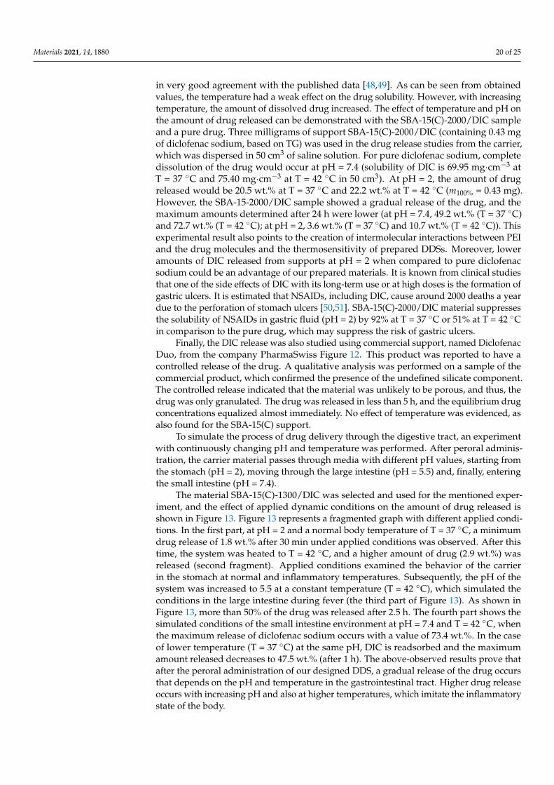
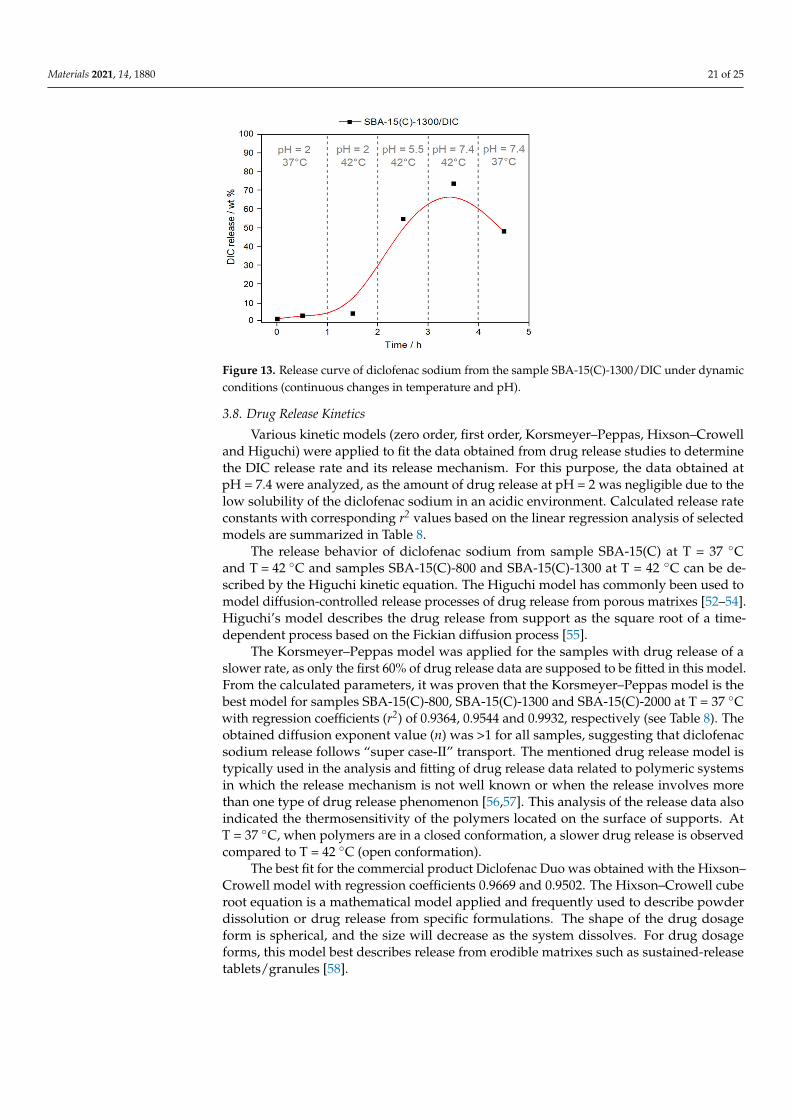
To simulate the process of drug delivery through the digestive tract, an experimentwith continuously changing pH and temperature was performed. After peroral adminis-tration, the carrier material passes through media with different pH values, starting fromthe stomach (pH = 2), moving through the large intestine (pH = 5.5) and, finally, enteringthe small intestine (pH = 7.4).
The material SBA-15(C)-1300/DIC was selected and used for the mentioned exper-iment, and the effect of applied dynamic conditions on the amount of drug released isshown in Figure 13. Figure 13 represents a fragmented graph with different applied condi-tions. In the first part, at pH = 2 and a normal body temperature of T = 37 ◦C, a minimumdrug release of 1.8 wt.% after 30 min under applied conditions was observed. After thistime, the system was heated to T = 42 ◦C, and a higher amount of drug (2.9 wt.%) wasreleased (second fragment). Applied conditions examined the behavior of the carrierin the stomach at normal and inflammatory temperatures. Subsequently, the pH of thesystem was increased to 5.5 at a constant temperature (T = 42 ◦C), which simulated theconditions in the large intestine during fever (the third part of Figure 13). As shown inFigure 13, more than 50% of the drug was released after 2.5 h. The fourth part shows thesimulated conditions of the small intestine environment at pH = 7.4 and T = 42 ◦C, whenthe maximum release of diclofenac sodium occurs with a value of 73.4 wt.%. In the caseof lower temperature (T = 37 ◦C) at the same pH, DIC is readsorbed and the maximumamount released decreases to 47.5 wt.% (after 1 h). The above-observed results prove thatafter the peroral administration of our designed DDS, a gradual release of the drug occursthat depends on the pH and temperature in the gastrointestinal tract. Higher drug releaseoccurs with increasing pH and also at higher temperatures, which imitate the inflammatorystate of the body.
Materials 2021, 14, 1880 21 of 25
Figure 13. Release curve of diclofenac sodium from the sample SBA-15(C)-1300/DIC under dynamicconditions (continuous changes in temperature and pH).
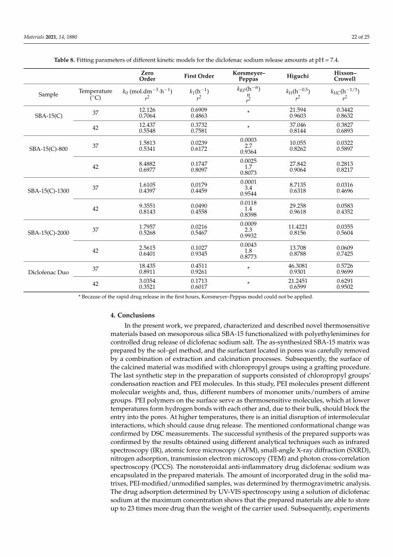
3.8. Drug Release Kinetics
Various kinetic models (zero order, first order, Korsmeyer–Peppas, Hixson–Crowelland Higuchi) were applied to fit the data obtained from drug release studies to determinethe DIC release rate and its release mechanism. For this purpose, the data obtained atpH = 7.4 were analyzed, as the amount of drug release at pH = 2 was negligible due to thelow solubility of the diclofenac sodium in an acidic environment. Calculated release rateconstants with corresponding r2 values based on the linear regression analysis of selectedmodels are summarized in Table 8.
The release behavior of diclofenac sodium from sample SBA-15(C) at T = 37 ◦Cand T = 42 ◦C and samples SBA-15(C)-800 and SBA-15(C)-1300 at T = 42 ◦C can be de-scribed by the Higuchi kinetic equation. The Higuchi model has commonly been used tomodel diffusion-controlled release processes of drug release from porous matrixes [52–54].Higuchi’s model describes the drug release from support as the square root of a time-dependent process based on the Fickian diffusion process [55].
The Korsmeyer–Peppas model was applied for the samples with drug release of aslower rate, as only the first 60% of drug release data are supposed to be fitted in this model.From the calculated parameters, it was proven that the Korsmeyer–Peppas model is thebest model for samples SBA-15(C)-800, SBA-15(C)-1300 and SBA-15(C)-2000 at T = 37 ◦Cwith regression coefficients (r2) of 0.9364, 0.9544 and 0.9932, respectively (see Table 8). Theobtained diffusion exponent value (n) was >1 for all samples, suggesting that diclofenacsodium release follows “super case-II” transport. The mentioned drug release model istypically used in the analysis and fitting of drug release data related to polymeric systemsin which the release mechanism is not well known or when the release involves morethan one type of drug release phenomenon [56,57]. This analysis of the release data alsoindicated the thermosensitivity of the polymers located on the surface of supports. AtT = 37 ◦C, when polymers are in a closed conformation, a slower drug release is observedcompared to T = 42 ◦C (open conformation).
The best fit for the commercial product Diclofenac Duo was obtained with the Hixson–Crowell model with regression coefficients 0.9669 and 0.9502. The Hixson–Crowell cuberoot equation is a mathematical model applied and frequently used to describe powderdissolution or drug release from specific formulations. The shape of the drug dosageform is spherical, and the size will decrease as the system dissolves. For drug dosageforms, this model best describes release from erodible matrixes such as sustained-releasetablets/granules [58].
Materials 2021, 14, 1880 22 of 25
Table 8. Fitting parameters of different kinetic models for the diclofenac sodium release amounts at pH = 7.4.
ZeroOrder First Order Korsmeyer–
Peppas Higuchi Hixson–Crowell
Sample Temperature(◦C)
k0 (mol.dm−3·h−1)r2
k1(h−1)r2
kKP(h−n)nr2
kH(h−0.5)r2
kHC(h−1/3)r2
SBA-15(C) 37 12.1260.7064
0.69090.4863 * 21.594
0.96030.34420.8632
42 12.4370.5548
0.37320.7581 * 37.046
0.81440.38270.6893
SBA-15(C)-800 37 1.58130.5341
0.02390.6172
0.00032.7
0.936410.0550.8262
0.03220.5897
42 8.48820.6977
0.17470.8097
0.00251.7
0.807327.8420.9064
0.28130.8217
SBA-15(C)-1300 37 1.61050.4397
0.01790.4459
0.00013.4
0.95448.71350.6318
0.03160.4696
42 9.35510.8143
0.04900.4558
0.01181.4
0.839829.2580.9618
0.05830.4352
SBA-15(C)-2000 37 1.79570.5268
0.02160.5467
0.00092.3
0.993211.42210.8156
0.03550.5604
42 2.56150.6401
0.10270.9345
0.00431.8
0.877313.7080.8788
0.06090.7425
Diclofenac Duo 37 18.4350.8911
0.45110.9261 * 46.3081
0.93010.57260.9699
42 3.03540.3521
0.17130.6017 * 21.2451
0.65990.62910.9502
* Because of the rapid drug release in the first hours, Korsmeyer–Peppas model could not be applied.
4. Conclusions
In the present work, we prepared, characterized and described novel thermosensitivematerials based on mesoporous silica SBA-15 functionalized with polyethylenimines forcontrolled drug release of diclofenac sodium salt. The as-synthesized SBA-15 matrix wasprepared by the sol–gel method, and the surfactant located in pores was carefully removedby a combination of extraction and calcination processes. Subsequently, the surface ofthe calcined material was modified with chloropropyl groups using a grafting procedure.The last synthetic step in the preparation of supports consisted of chloropropyl groups’condensation reaction and PEI molecules. In this study, PEI molecules present differentmolecular weights and, thus, different numbers of monomer units/numbers of aminegroups. PEI polymers on the surface serve as thermosensitive molecules, which at lowertemperatures form hydrogen bonds with each other and, due to their bulk, should block theentry into the pores. At higher temperatures, there is an initial disruption of intermolecularinteractions, which should cause drug release. The mentioned conformational change wasconfirmed by DSC measurements. The successful synthesis of the prepared supports wasconfirmed by the results obtained using different analytical techniques such as infraredspectroscopy (IR), atomic force microscopy (AFM), small-angle X-ray diffraction (SXRD),nitrogen adsorption, transmission electron microscopy (TEM) and photon cross-correlationspectroscopy (PCCS). The nonsteroidal anti-inflammatory drug diclofenac sodium wasencapsulated in the prepared materials. The amount of incorporated drug in the solid ma-trixes, PEI-modified/unmodified samples, was determined by thermogravimetric analysis.The drug adsorption determined by UV-VIS spectroscopy using a solution of diclofenacsodium at the maximum concentration shows that the prepared materials are able to storeup to 23 times more drug than the weight of the carrier used. Subsequently, experiments
Materials 2021, 14, 1880 23 of 25
were performed to assess drug release from the prepared materials into media with differ-ent pH values (simulated gastric fluid (pH = 2) and small intestine environment/simulatedbody fluid (pH = 7.4)) and temperatures (T = 37 ◦C representing normal body temperatureand T = 42 ◦C simulating inflammatory temperature). From the obtained release curves,it could be concluded that in the case of pH, a higher amount of drug was liberated atpH = 7.4 compared to pH = 2 due to the drug solubility. In the case of temperature, ahigher amount of DIC released was observed at a higher temperature, and the observedresults confirmed thermosensitivity of the PEI-modified materials. Moreover, the drugrelease properties of prepared compounds were compared to a commercial product underthe same experimental conditions. Various kinetic models were applied to fit the drugrelease data to study the mechanism of diclofenac sodium release. It was demonstrated thatdrug release at pH = 7.4 could be described by applying Higuchi and Korsmeyer–Peppasmodels.
Supplementary Materials: The following are available online at https://www.mdpi.com/article/10.3390/ma14081880/s1: Figure S1: (a) UV spectra of diclofenac sodium methanolic solutions and(b) corresponding calibration curve. Figure S2: Calibration curve of diclofenac sodium in salinesolutions at (a) pH = 2 and (b) pH = 7.4. Figure S3: IR spectrum of diclofenac sodium salt. Figure S4:Thermogravimetric curve of diclofenac sodium salt.
Author Contributions: Data curation, L.Z., S.B., J.B., M.B. and M.A.; formal analysis, L.Z., S.B., E.B.,M.B., V.H. and M.A.; investigation, L.Z., S.B., E.B., J.B., M.B., V.Z., V.H. and M.A.; visualization, L.Z.and M.A; writing—original draft preparation, L.Z., S.B., E.B., M.B., V.H. and M.A.; writing—reviewand editing, L.Z. and M.A.; funding acquisition, V.Z.; supervision, M.A. All authors have read andagreed to the published version of the manuscript.
Funding: This work was supported by the Slovak Research and Development Agency project“Intelligent nanoporous systems as carriers of drug” under contract No. APVV-15-0520 andby theDevelopment Operational Programme Integrated Infrastructure for the project “Nanoparticles forsolving diagnostic-therapeutic problems with COVID-19 (NANOVIR)”, ITMS 2014+:313011AUW7,co-founded by the European Regional Development Fund (ERDF). This research was also supportedby Slovak Grant Agency VEGA, grant No. 2/0044/18 “High-energy milling for the synthesis ofnanomaterials by a bio-approach and selected environmental applications”.
Institutional Review Board Statement: Not applicable.
Informed Consent Statement: Not applicable.
Data Availability Statement: The data presented in this study are available on request from thecorresponding author.
Acknowledgments: The authors acknowledge the help of Martin Stahorský from the Institute ofGeotechnics, Slovak Academy of Sciences, in performing the photon cross-correlation measurements.
Conflicts of Interest: The authors declare no conflict of interest.
References1. Ferrero-Miliani, L.; Nielsen, O.H.; Andersen, P.S.; Girardin, S.E. Chronic inflammation: Importance of NOD2 and NALP3 in
interleukin-1β generation. Clin. Exp. Immunol. 2007, 147, 227–235. [CrossRef] [PubMed]2. Balfour, J.A.; Buckley, M.M.T. Etodolac: A reappraisal of its pharmacology and therapeutic use in rheumatic diseases and pain
states. Drugs 1991, 42, 274–299. [CrossRef]3. Hu, M.; Ge, X.; Chen, X.; Mao, W.; Qian, X.; Yuan, W.E. Micro/nanorobot: A promising targeted drug delivery system.
Pharmaceutics 2020, 12, 665. [CrossRef]4. Cao, J.; Li, X.; Tian, H. Metal-organic framework (MOF)-based drug delivery. Curr. Med. Chem. 2020, 27, 5949–5969. [CrossRef]5. Almáši, M.; Zelenák, V.; Palotai, P.; Benová, E.; Zelenáková, A. Metal-organic framework MIL-101(Fe)-NH2 functionalized with
different long-chain polyamines as drug delivery system. Inorg. Chem. Commun. 2018, 93, 115–120. [CrossRef]6. Bianco, A.; Kostarelos, K.; Prato, M. Applications of carbon nanotubes in drug delivery. Curr. Opin. Chem. Biol. 2005, 9, 674–679.
[CrossRef] [PubMed]7. Murakami, T.; Tsuchida, K. Recent advances in inorganic nanoparticle-based drug delivery systems. Mini Rev. Med. Chem. 2008,
8, 175–183. [PubMed]
Materials 2021, 14, 1880 24 of 25
8. Shi, J.; Zhang, H.; Wang, L.; Li, L.; Wang, H.; Wang, Z.; Li, Z.; Chen, C.; Hou, L.; Zhang, C.; et al. PEI-derivatized fullerene drugdelivery using folate as a homing device targeting to tumor. Biomaterials 2013, 34, 251–261. [CrossRef] [PubMed]
9. Almomen, A.; El-Toni, A.M.; Badran, M.; Alhowyan, A.; Kalam, M.A.; Alshamsan, A.; Alkholief, M. The design of anionicsurfactant-based amino-functionalized mesoporous silica nanoparticles and their application in transdermal drug delivery.Pharmaceutics 2020, 12, 1035. [CrossRef] [PubMed]
10. Mondal, S.; Das, S.; Nandi, A.K. A review on recent advances in polymer and peptide hydrogels. Soft Matter 2020, 16, 1404–1454.[CrossRef] [PubMed]
11. Akash, M.S.H.; Rehman, K. Recent progress in biomedical applications of pluronic (P-127): Pharmaceutical perspectives. J.Control. Release 2015, 209, 120–138. [CrossRef]
12. Venkatasubbu, G.D.; Ramasamy, S.; Ramakrishnan, V.; Kumar, J. Folate targeted PEGylated titanium dioxide nanoparticles as ananocarrier for targeted paclitaxel drug delivery. Adv. Powder Technol. 2013, 24, 947–954. [CrossRef]
13. Raza, A.; Hayat, U.; Bilal, M.; Iqbal, H.M.N.; Wang, J.Y. Zein-based micro- and nano-constructs and biologically therapeutic cueswith multi-functionalities for oral drug delivery systems. J. Drug Deliv. Sci. Technol. 2020, 58, 101818. [CrossRef]
14. Cho, I.; Shim, M.K.; Jung, N.; Jang, E.H.; Park, M.; Kang, H.C.; Kim, J. Heat shock responsive drug delivery system based onmesoporous silica nanoparticles coated with temperature sensitive gatekeeper. Microporous Mesoporous Mater. 2017, 253, 96–101.[CrossRef]
15. Abdo, G.G.; Zagho, M.M.; Khalil, A. Recent advances in stimuli-responsive drug release and targeting concepts using mesoporoussilica nanoparticles. Emergent. Mater. 2020, 3, 407–425. [CrossRef]
16. Abedi, M.; Abolmaali, S.S.; Abedanzadeh, M.; Farjadian, F.; Samani, S.M.; Tamaddon, A.M. Core-shell imidazoline-functionalizedmesoporous silica superparamagnetic hybrid manoparticles as a potential theranostic agent for controlled delivery of platinum(II)compound. Int. J. Nanomed. 2020, 15, 2617–2631. [CrossRef] [PubMed]
17. Xu, C.; Cao, Y.; Lei, C.; Li, Z.; Kumeria, T.; Meka, A.K.; Xu, J.; Liu, J.; Yan, C.; Luo, L.; et al. Polymer–mesoporous silica nanoparticlecore–shell nanofibers as a dual-drug-delivery system for guided tissue regeneration. ACS Appl. Nano Mater. 2020, 3, 1457–1467.[CrossRef]
18. Constantin, M.; Bucatariu, S.M.; Doroftei, F.; Fundueanu, G. Smart composite materials based on chitosan microspheres embeddedin thermosensitive hydrogel for controlled delivery of drugs. Carbohydr. Polym. 2017, 157, 493–502. [CrossRef]
19. Zhao, J.; Zhao, X.; Guo, B.; Ma, P.X. Multifunctional interpenetrating polymer network hydrogels based on methacrylated alginatefor the delivery of small molecule drugs and sustained release of protein. Biomacromolecules 2014, 15, 3246–3252. [CrossRef][PubMed]
20. Hu, W.; Bai, X.; Wang, Y.; Lei, Z.; Luo, H.; Tong, Z. Upper critical solution temperature polymer-grafted hollow mesoporous silicananoparticles for near-infrared-irradiated drug release. J. Mater. Chem. B 2019, 7, 5789–5796. [CrossRef] [PubMed]
21. Yang, J.; Dai, D.; Lou, X.; Ma, L.; Wang, B.; Yang, Y.W. Supramolecular nanomaterials based on hollow mesoporous drug carriersand macrocycle-capped CuS nanogates for synergistic chemo-photothermal therapy. Theranostics 2020, 10, 615–629. [CrossRef][PubMed]
22. Cui, Y.; Deng, R.; Li, X.; Wang, X.; Jia, Q.; Bertrand, E.; Meguellati, K.; Yang, Y. Temperature-sensitive polypeptide brushes-coatedmesoporous silica nanoparticles for dual-responsive drug release. Chin. Chem. Lett. 2019, 30, 2291–2294. [CrossRef]
23. Yu, F.; Wu, H.; Tang, Y.; Xu, Y.; Qian, X.; Zhu, W. Temperature-sensitive copolymer-coated fluorescent mesoporous silicananoparticles as a reactive oxygen species activated drug delivery system. Int. J. Pharm. 2018, 536, 11–20. [CrossRef] [PubMed]
24. Benová, E.; Bergé-Lefranc, D.; Zelenák, V.; Almáši, M.; Huntošová, V.; Hornebecq, V. Adsorption properties, the pH-sensitiverelease of 5-fluorouracil and cytotoxicity studies of mesoporous silica drug delivery matrix. Appl. Surf. Sci. 2020, 504, 1–12.[CrossRef]
25. Almáši, M.; Benová, E.; Zelenák, V.; Madaj, B.; Huntošová, V.; Brus, J.; Urbanová, M.; Bednarcík, J.; Hornebecq, V. Cytotoxicitystudy and influence of SBA-15 surface polarity and pH on adsorption and release properties of anticancer agent pemetrexed.Mater. Sci. Eng. C 2019, 109, 110552. [CrossRef] [PubMed]
26. Žid, L.; Zelenák, V.; Almáši, M.; Zelenáková, A.; Szücsová, J.; Bednarcík, J.; Šuleková, M.; Hudák, A.; Váhovská, L. Mesoporoussilica as a drug delivery system for naproxen: Influence of surface functionalization. Molecules 2020, 25, 4722. [CrossRef]
27. Qu, J.; Zhao, X.; Ma, P.X.; Guo, B. Injectable antibacterial conductive hydrogels with dual response to an electric field and pH forlocalized “smart” drug release. Acta Biomater. 2018, 72, 55–69. [CrossRef] [PubMed]
28. Zelenák, V.; Benová, E.; Almáši, M.; Halamová, D.; Hornebecq, V.; Hronský, V. Photo-switchable nanoporous silica supports forcontrolled drug delivery. New J. Chem. 2018, 42, 13263–13271. [CrossRef]
29. Benová, E.; Zelenák, V.; Halamová, D.; Almáši, M.; Petrul’ová, V.; Psotka, M. A drug delivery system based on switchablephoto-controlled p-coumaric acid derivatives anchored on mesoporous silica. J. Mater. Chem. B 2017, 5, 817–825. [CrossRef][PubMed]
30. Kapusta, O.; Zelenáková, A.; Hrubovcák, P.; Girman, V.; Zelenák, V. Fe2O3 and Gd2O3 nanoparticles embedded in mesoporoussilica: Magnetic properties comparison. Acta Phys. Pol. A 2017, 131, 860–862. [CrossRef]
31. Vila, M.; Cicuéndez, M.; Sánchez-Marcos, J.; Fal-Miyar, V.; Manzano, M.; Prieto, C.; Vallet-Regi, M. Electrical stimuli to increasecell proliferation on carbon nanotubes/mesoporous silica composites for drug delivery. J. Biomed. Mater. Res. Part A 2013, 101,213–221. [CrossRef]
Materials 2021, 14, 1880 25 of 25
32. Gu, M.; Wang, X.; Toh, T.B.; Chow, E.K.H. Applications of stimuli-responsive nanoscale drug delivery systems in translationalresearch. Drug Discov. Today 2018, 23, 1043–1052. [CrossRef] [PubMed]
33. Zhao, J.; Lu, C.; He, X.; Zhang, X.; Zhang, W.; Zhang, X. Polyethylenimine-grafted cellulose nanofibril aerogels as versatilevehicles for drug delivery. ACS Appl. Mater. Interfaces 2015, 7, 2607–2615. [CrossRef]
34. Sun, X.; Cai, C.; Wang, Q.; Cai, D.; Qian, J.; Chi, Y.; Zheng, K.; Zhang, X.; Zhang, G.; Zhong, K. A polyethylenimine functionalizedporous/hollow nanoworm as a drug delivery system and a bioimaging agent. Phys. Chem. Chem. Phys. 2016, 18, 7820–7828.[CrossRef] [PubMed]
35. Zou, Y.; Li, D.; Shen, M.; Shi, X. Polyethylenimine-based nanogels for biomedical applications. Macromol. Biosci. 2019, 19, 1900272.[CrossRef] [PubMed]
36. Han, S.C.; He, W.D.; Li, J.; Li, L.Y.; Sun, X.L.; Zhang, B.Y.; Pan, T.T. Reducible polyethylenimine hydrogels with disulfidecrosslinkers prepared by Michael addition chemistry as drug delivery carriers: Synthesis, properties, and in vitro release. J. Polym.Sci. Part A Polym. Chem. 2009, 47, 4074–4082. [CrossRef]
37. Zhou, C.B.; Zhao, L.; Shen, M.; Zhao, J.; Shi, X. Multifunctional polyethylenimine-based nanoplatform for targeted anticancerdrug delivery to tumors in vivo. J. Mater. Chem. C 2015, 3, 10715–10722. [CrossRef]
38. Zhang, J.T.; Xue, Y.N.; Gao, F.Z.; Huang, S.W.; Zhuo, R.X. Preparation of temperature-sensitive poly(N-isopropylacrylamide)/b-cyclodextrin-grafted polyethylenimine hydrogels for drug delivery. J. Appl. Polym. Sci. 2008, 108, 3031–3037. [CrossRef]
39. Sun, X.; Wang, N.; Yang, L.Y.; Ouyang, X.K.; Huang, F. Folic acid and PEI modified mesoporous silica for targeted delivery ofcurcumin. Pharmaceutics 2019, 11, 623. [CrossRef] [PubMed]
40. Todd, P.A.; Sorkin, E.M. Diclofenac sodium. A reappraisal of its pharmacodynamic and pharmacokinetic properties, andtherapeutic efficacy. Drugs 1988, 35, 244–285. [CrossRef]
41. Hench, L.; Larry, L.; West, J.K. The sol-gel process. Chem. Rev. 1990, 90, 33–72. [CrossRef]42. Pirez, C.; Morin, J.C.; Manayil, J.C.; Lee, A.F.; Wilson, K. Sol-gel synthesis of SBA-15: Impact of HCl on surface chemistry.
Microporous Mesoporous Mater. 2018, 271, 196–202. [CrossRef]43. Nobbmann, U.; Morfesis, A. Light scattering and nanoparticles. Mater. Today 2009, 12, 52–54. [CrossRef]44. Almáši, M.; Zelenák, V.; Opanasenko, M.; Cejka, J. Efficient and reusable Pb(II) metal–organic framework for Knoevenagel
condensation. Catal. Lett. 2018, 148, 2263–2273. [CrossRef]45. Lien, Y.H.; Wu, T.M. Preparation and characterization of thermosensitive polymers grafted onto silica-coated iron oxide nanopar-
ticles. J. Colloid Interface. Sci. 2008, 326, 517–521. [CrossRef]46. Gandhi, A.; Paul, A.; Sen, S.O.; Sen, K.K. Studies on thermoresponsive polymers: Phase behaviour, drug delivery and biomedical
applications. Asian J. Pharm. Sci. 2015, 10, 99–107. [CrossRef]47. Giles, C.H.; Smith, D.; Huitson, A.A. General treatment and classification of the solute adsorption isotherm I. theoretical. J. Colloid
Interface Sci. 1974, 47, 755–765. [CrossRef]48. Llinas, A.; Burley, J.C.; Box, K.J.; Glen, R.C.; Goodman, J.M. Diclofenac solubility: Independent determination of the intrinsic
solubility of three crystal forms. J. Med. Chem. 2007, 50, 979–983. [CrossRef]49. Fini, A.; Laus, M.; Orienti, I.; Zecchi, V. Dissolution and partition thermodynamic functions of some nonsteroidal anti-
inflammatory drugs. J. Pharm. Sci. 1986, 75, 23–25. [CrossRef]50. Žilnik, L.F.; Jazbinšek, A.; Hvala, A.; Vrecer, F.; Klamt, A. Solubility of sodium diclofenac in different solvents. Fluid Phase
Equilibria 2007, 261, 140–145. [CrossRef]51. Zacher, J.; Altman, R.; Bellamy, N.; Bruhlmann, P.; Da Silva, J.; Huskisson, E.; Taylor, R.S. Topical diclofenac and its role in pain
and inflammation: An evidence-based review. Curr. Med. Res. Opin. 2008, 24, 925–950. [CrossRef] [PubMed]52. Pathan, S.; Solanki, P.; Patel, A. Functionalized SBA-15 for controlled release of poorly soluble drug, Erythromycin. Microporous
Mesoporous Mater. 2018, 258, 114–121. [CrossRef]53. Polo, L.; Gómez-Cerezo, N.; Garcia-Fernández, A.; Aznar, E.; Vivancos, J.L.; Arcos, D.; Vallet-Regi, M.; Martinez-Mánez, R.
Mesoporous bioactive glasses equipped with stimuli-responsive molecular gates for controlled delivery of levofloxacin againstbacteria. Chem. Eur. J. 2018, 24, 18944–18951. [CrossRef] [PubMed]
54. Doadrio, C.; Sousa, E.M.B.; Izquierdo-Barba, I.; Doadrio, A.L.; Perez-Pariente, J.; Vallet-Regí, M. Functionalization of mesoporousmaterials with long alkyl chains as a strategy for controlling drug delivery pattern. J. Mater. Chem. 2006, 16, 462–466. [CrossRef]
55. Merchant, H.A.; Shoaib, H.M.; Tazeen, J.; Yousuf, R. I Once-daily tablet formulation and in vitro release evaluation of cefpodoximeusing hydroxypropyl methylcellulose: A technical note. AAPS PharmSciTech 2006, 7, 178–183. [CrossRef] [PubMed]
56. Madhavi, M.; Madhavi, K.; Jithan, A.V. Preparation and in vitro/in vivo characterization of curcumin microspheres intended totreat colon cancer. J. Pharm. Bioallied Sci. 2012, 4, 164–171.
57. Marini, V.G.; Martelli, S.M.; Zornio, C.F.; Caon, T.; Simoes, C.M.O.; Micke, G.A.; Oliviera, M.A.L.; Machado, V.G.; Soldi, V.Biodegradable nanoparticles obtained from zein as a drug delivery system for terpinen-4-ol. Quím. Nova 2014, 37, 839–843.[CrossRef]
58. Gouda, R.; Baishya, H.; Qing, Z. Application of mathematical models in drug release kinetics of Carbidopa and Levodopa ERtablets. J. Dev. Drugs 2017, 6, 1000171.


