Embed Size (px)
Citation preview
this is what the future looks like 1this is what the future looks like
2022
this is what the future looks like 3
progress works by leaps and bounds.and your position is clear: at the top.
as a renowned eye surgeon, you want the best of the best.for yourself. for your reputation. and for your patients.
because there is no room for error in your profession. no understanding for mistakes.
there is only the urge for perfection.the challenge to succeed, every time.
that requires state of the art material.top notch interlocutors
knowledge of the latest technologies
and uncompromising quality.
ophtalmo is as ambitious as you are.we understand your demands,
share your high standards,speak your language and defend your interests.
that is why we offer you a wealth of experience,we only work with premium brands,
we are already looking for tomorrow’s solutions and we team up with our partners to produce more meaning and value.
because there is only one word that belongs to your profession: perfection.
T H I S I S W H A T T H E F U T U R E L O O K S L I K E
this is what the future looks like 5
Ophtalmo, with a clear focus on quality and innovation
We are more than a partner who is knowledgeable. We build lasting relationships with our customers, we genuinely value their feedback and we actively contribute to innovative solutions. We do all this in order to achieve our main goal: to achieve the best patient care. We work with carefully selected partners to strive for the best solution and technology. We offer a specialised portfolio of medical devices, services, supplies and accessories in selected medical fields in order to help create a complete, customised solution.
We only work with suppliers that show exceptional product and service performance levels to ensure we only deliver the best quality, anytime, anywhere. We look ahead, and we don’t miss any opportunities to perform even better. We listen to our customers’ needs in order to lead the way in the advancement of ophthalmology.
Ophtalmo makes proactive contributions
and offers you an innovative eye care
approach
Ophtalmo started as a distributor of contact lenses in 1993, and over the years we have developed our wide range of innovative products and technologies for the various segments of the BELUX ophthalmology market.
We are now a major player that offers solutions for cataract, retinal issues, refractive surgery, glaucoma and so on. In addition to diagnostic equipment, instruments and consumables, we also offer a broad and attractive spectrum of viscoelastics and intraocular lenses. Our extensive range of high-quality products has made Ophtalmo the top supplier in Belgium and Luxembourg, where personal service and flexibility are highly valued.
It is our ambition to increase
our customers’ performance
as much as possible
Our close-knit team of experienced staff and network of high-quality suppliers all share the same ambition: to be the number one for our customers. We continue to invest in our business and the right infrastructure, so that we can help our customers in an even better way, with more space, more possibilities and more innovation, now and in the future.
DEDICATION We are available to our customers 24/7 in order to offer the best care to patients anytime and anywhere.
RELIABILITY Customers can rely on us. We respect our promises and the agreed delivery dates.
FLEXIBILITY As a distributor, we maintain close relationships with our regular suppliers. We monitor everything closely to achieve the shortest possible response times.
Would you like to find out more about our innovations and projects? Jorg De Troyer [email protected] I T. +32 485 37 27 57
Bob De Meyer [email protected] I T. +32 470 53 85 29
Laurent Pironnet [email protected] I T. +32 473 69 57 62
Customer Service:
Inge Boelaert [email protected] +32 (0)55 313 038
this is what the future looks like 7
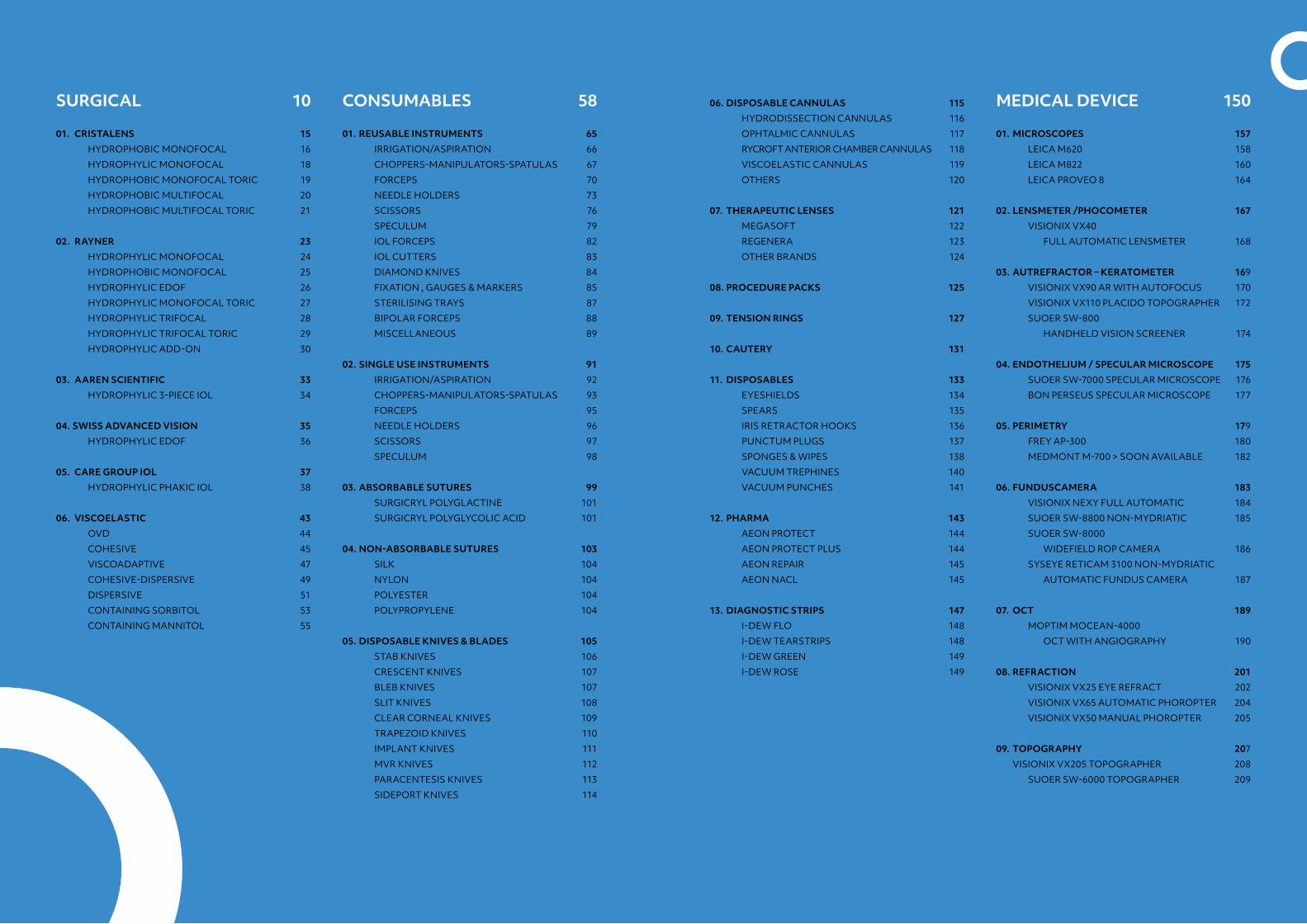
SURGICAL 10
01. CRISTALENS 15
HYDROPHOBIC MONOFOCAL 16
HYDROPHYLIC MONOFOCAL 18
HYDROPHOBIC MONOFOCAL TORIC 19
HYDROPHOBIC MULTIFOCAL 20
HYDROPHOBIC MULTIFOCAL TORIC 21
02. RAYNER 23
HYDROPHYLIC MONOFOCAL 24
HYDROPHOBIC MONOFOCAL 25
HYDROPHYLIC EDOF 26
HYDROPHYLIC MONOFOCAL TORIC 27
HYDROPHYLIC TRIFOCAL 28
HYDROPHYLIC TRIFOCAL TORIC 29
HYDROPHYLICADD-ON 30
03. AAREN SCIENTIFIC 33
HYDROPHYLIC3-PIECEIOL 34
04. SWISS ADVANCED VISION 35
HYDROPHYLICEDOF 36
05. CARE GROUP IOL 37
HYDROPHYLICPHAKICIOL 38
06. VISCOELASTIC 43
OVD 44
COHESIVE 45
VISCOADAPTIVE 47
COHESIVE-DISPERSIVE 49
DISPERSIVE 51
CONTAININGSORBITOL 53
CONTAINING MANNITOL 55
CONSUMABLES 58
01. REUSABLE INSTRUMENTS 65
IRRIGATION/ASPIRATION 66
CHOPPERS-MANIPULATORS-SPATULAS 67
FORCEPS 70
NEEDLEHOLDERS 73
SCISSORS 76
SPECULUM 79
IOL FORCEPS 82
IOLCUTTERS 83
DIAMOND KNIVES 84
FIXATION , GAUGES & MARKERS 85
STERILISING TRAYS 87
BIPOLAR FORCEPS 88
MISCELLANEOUS 89
02. SINGLE USE INSTRUMENTS 91
IRRIGATION/ASPIRATION 92
CHOPPERS-MANIPULATORS-SPATULAS 93
FORCEPS 95
NEEDLE HOLDERS 96
SCISSORS 97
SPECULUM 98
03. ABSORBABLE SUTURES 99
SURGICRYL POLYGLACTINE 101
SURGICRYL POLYGLYCOLIC ACID 101
04. NON-ABSORBABLE SUTURES 103
SILK 104
NYLON 104
POLYESTER 104
POLYPROPYLENE 104
05. DISPOSABLE KNIVES & BLADES 105
STAB KNIVES 106
CRESCENT KNIVES 107
BLEB KNIVES 107
SLIT KNIVES 108
CLEAR CORNEAL KNIVES 109
TRAPEZOID KNIVES 110
IMPLANT KNIVES 111
MVR KNIVES 112
PARACENTESISKNIVES 113
SIDEPORT KNIVES 114
06. DISPOSABLE CANNULAS 115
HYDRODISSECTION CANNULAS 116
OPHTALMIC CANNULAS 117
RYCROFT ANTERIOR CHAMBER CANNULAS 118
VISCOELASTIC CANNULAS 119
OTHERS 120
07. THERAPEUTIC LENSES 121
MEGASOFT 122
REGENERA 123
OTHER BRANDS 124
08. PROCEDURE PACKS 125
09. TENSION RINGS 127
10. CAUTERY 131
11. DISPOSABLES 133
EYESHIELDS 134
SPEARS 135
IRISRETRACTORHOOKS 136
PUNCTUMPLUGS 137
SPONGES&WIPES 138
VACUUM TREPHINES 140
VACUUM PUNCHES 141
12. PHARMA 143
AEON PROTECT 144
AEON PROTECT PLUS 144
AEON REPAIR 145
AEON NACL 145
13. DIAGNOSTIC STRIPS 147
I-DEWFLO 148
I-DEWTEARSTRIPS 148
I-DEWGREEN 149
I-DEWROSE 149
MEDICAL DEVICE 150
01. MICROSCOPES 157
LEICA M620 158
LEICA M822 160
LEICA PROVEO 8 164
02. LENSMETER /PHOCOMETER 167
VISIONIX VX40
FULL AUTOMATIC LENSMETER 168
03. AUTREFRACTOR – KERATOMETER 169
VISIONIX VX90 AR WITH AUTOFOCUS 170
VISIONIX VX110 PLACIDO TOPOGRAPHER 172
SUOERSW-800
HANDHELD VISION SCREENER 174
04. ENDOTHELIUM / SPECULAR MICROSCOPE 175
SUOERSW-7000SPECULARMICROSCOPE 176
BON PERSEUS SPECULAR MICROSCOPE 177
05. PERIMETRY 179
FREYAP-300 180
MEDMONTM-700>SOONAVAILABLE 182
06. FUNDUSCAMERA 183
VISIONIX NEXY FULL AUTOMATIC 184
SUOERSW-8800NON-MYDRIATIC 185
SUOERSW-8000
WIDEFIELD ROP CAMERA 186
SYSEYERETICAM3100NON-MYDRIATIC
AUTOMATIC FUNDUS CAMERA 187
07. OCT 189
MOPTIMMOCEAN-4000
OCT WITH ANGIOGRAPHY 190
08. REFRACTION 201
VISIONIX VX25 EYE REFRACT 202
VISIONIX VX65 AUTOMATIC PHOROPTER 204
VISIONIX VX50 MANUAL PHOROPTER 205
09. TOPOGRAPHY 207
VISIONIX VX205 TOPOGRAPHER 208
SUOERSW-6000TOPOGRAPHER 209
this is what the future looks like 9
10. TONOMETRY 211
TAKAGISNT-700
NON-CONTACTTONOMETER 212
FREYMEDICALTN-100
NON-CONTACTTONOMETER 215
SUOERSW-500
HANDHELD REBOUND TONOMETER 216
SUOERSW-5000
NON-CONTACTTONOMETER 217
11. DIAGNOSTIC 219
VISIONIX VX120+
ANTERIOR SCREENING 220
VISIONIXVX130+
ANTERIOR SCREENING 224
VISIONIX VX210
SCHEIMPFLUG CAMERA 228
TAKAGICGT-2000
CONTRAST GLARETESTER 229
12. LASER 233
LIGHTMEDLIGHTLASYAG 238
LIGHTMED LIGHTLAS SLT 240
LIGHTMEDLIGHTLAS532/577/810 242
LIGHTMED TRUSCAN 244
13. SLIT LAMPS 237
VISIONIXVX70 238
VISIONIX VX75 240
VISIONIX VX80 242
VISIONIX VX85 244
HEINEHSL150HAND-HELDSLITLAMP 246
14. BIOMETRY 247
SUOERSW-9000OPTICALBIOMETER 248
15. ULTRASOUND 249
SUOERSW-21SCANNERA-B-UBM 250
16. OPHTALMOSCOPE 251
HEINEHAND-HELDDIRECT
BETA200/BETA200S(LED) 252
HEINEHAND-HELDDIRECTMINI3000 253
HEINEHAND-HELDINDIRECT 254
HEINE OMEGA 600 INDIRECT 255
17. RETINOSCOPY / RETINOMETER 257
HEINE BETA 200 RETINOSCOPE 258
HEINE LAMBDA 100 RETINOMETER 259
18. EXAMINATION UNITS 261
VISIONIX VX1200 262
VISIONIXVX2000 263
VISIONIXVX3000 264
19. OPERATION & PATIENT CHAIRS 265
UFSK 500XLE 266
UFSK EYEFORCE 268
20. AUTOCLAVES 271
STATIM G4 272
21. TRAIL LENSES & SETS 275
CONTACT LENSES & CARE 278
CONTACT LENSES & CARE 281
OUR BRANDS 282
OVERVIEW 283

this is what the future looks like 11
SURGICAL
this is what the future looks like 13
01. CRISTALENS 15 HYDROPHOBIC MONOFOCAL 16 HYDROPHYLIC MONOFOCAL 18 HYDROPHOBIC MONOFOCAL TORIC 19 HYDROPHOBIC MULTIFOCAL 20 HYDROPHOBIC MULTIFOCAL TORIC 21
02. RAYNER 23 HYDROPHYLIC MONOFOCAL 24 HYDROPHOBIC MONOFOCAL 25 HYDROPHYLIC EDOF 26 HYDROPHYLIC MONOFOCAL TORIC 27 HYDROPHYLIC TRIFOCAL 28 HYDROPHYLIC TRIFOCAL TORIC 29 HYDROPHYLICADD-ON 30
03. AAREN SCIENTIFIC 33 HYDROPHYLIC3-PIECEIOL 34
04. SWISS ADVANCED VISION 35 HYDROPHYLICEDOF 36
05. CARE GROUP IOL 37 HYDROPHYLICPHAKICIOL 38
06. VISCOELASTIC 43 OVD 44 COHESIVE 45 VISCOADAPTIVE 47 COHESIVE-DISPERSIVE 49 DISPERSIVE 51 CONTAININGSORBITOL 53 CONTAINING MANNITOL 55
01CRISTALENS
Founded in 1994, CRISTALENS was initially a distributor of medical devices for cataract surgery. In 2006, the company created its own production unit of hydrophilic and hydrophobic intraocular lenses. In 2008, it
has developed a new hydrophobic raw material (phenoxy ethyl acrylate) that allows less than 1.8 mm micro incisions. In 2013, Cristalens received the Industrial Innovation prize with this invention.
By choosing CRISTALENS you are collaborating with a laboratory that has a total mastery of its profession: from raw material production to packaging, with three key words:
Patient safety | Innovation | Proximity
this is what the future looks like 17
CRISTALENS HYDROPHOBICMonofocal
CRISTALENS HYDROPHOBICMonofocal
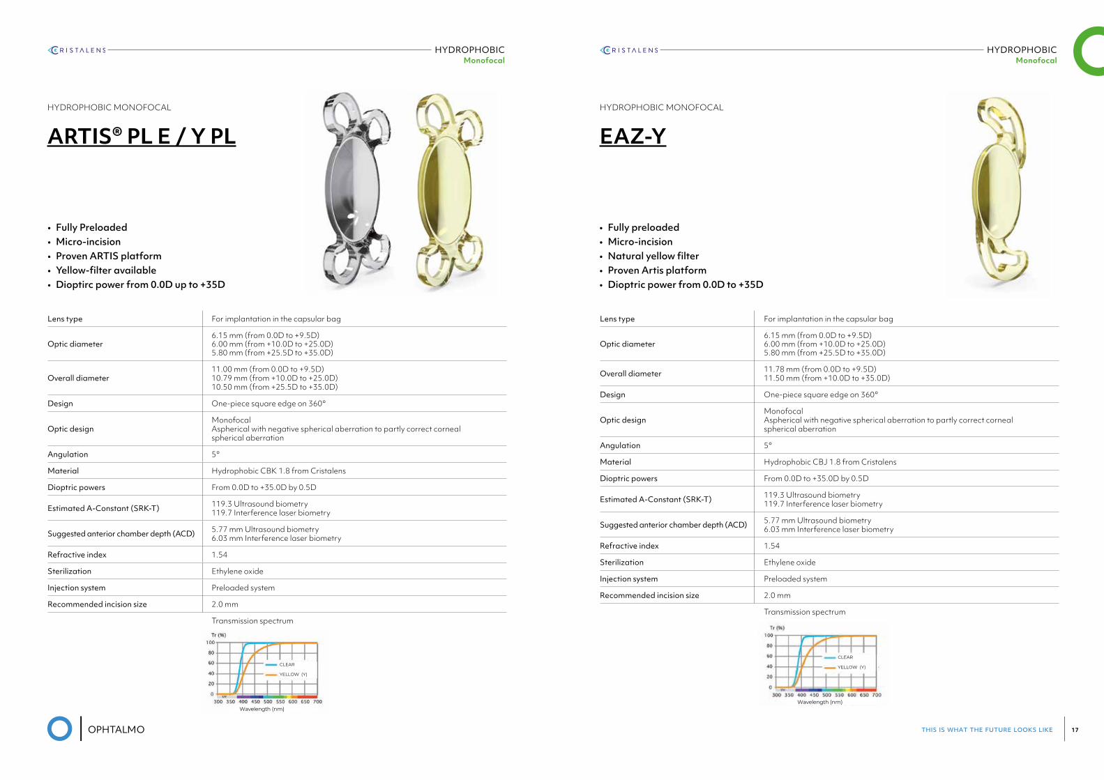
HYDROPHOBIC MONOFOCAL
ARTIS® PL E / Y PL
Lens type For implantation in the capsular bag
Optic diameter6.15 mm (from 0.0D to +9.5D) 6.00 mm (from +10.0D to +25.0D) 5.80 mm (from +25.5D to +35.0D)
Overall diameter11.00 mm (from 0.0D to +9.5D)10.79 mm (from +10.0D to +25.0D)10.50 mm (from +25.5D to +35.0D)
Design One-piece square edge on 360°
Optic designMonofocalAspherical with negative spherical aberration to partly correct cornealspherical aberration
Angulation 5°
Material Hydrophobic CBK 1.8 from Cristalens
Dioptric powers From 0.0D to +35.0D by 0.5D
Estimated A-Constant (SRK-T) 119.3 Ultrasound biometry119.7 Interference laser biometry
Suggested anterior chamber depth (ACD) 5.77 mm Ultrasound biometry6.03 mm Interference laser biometry
Refractive index 1.54
Sterilization Ethylene oxide
Injection system Preloaded system
Recommended incision size 2.0 mm
Transmission spectrum
10910
ARTIS® MONOFOCAL TECHNICAL SPECIFICATIONS
• PRELOADED HYDROPHOBIC LENSES
➤ Monofocal
Lens type For implantation in the capsular bag
Optic diameter6.15 mm (from 0.0D to +9.5D)6.00 mm (from +10.0D to +25.0D)5.80 mm (from +25.5D to +35.0D)
Overall diameter11.00 mm (from 0.0D to +9.5D)10.79 mm (from +10.0D to +25.0D)10.50 mm (from +25.5D to +35.0D)
Design One-piece square edge on 360°
Optic designMonofocalAspherical with negative spherical aberration to partly correct corneal spherical aberration
Angulation 5°
Material Hydrophobic CBK 1.8 / CBJ 1.8 from Cristalens
Dioptric powers From 0.0D to +35.0D by 0.5D
Estimated A-Constant (SRK-T) 119.3 Ultrasound biometry119.7 Interference laser biometry
Suggested anterior chamber depth (ACD)
5.77 mm Ultrasound biometry6.03 mm Interference laser biometry
Refractive index 1.54
Sterilization Ethylene oxide
Injection system Preloaded system
Recommended incision size 2.0 mm
Natural yellow filter
Reference: ARTIS® Y PL
Reference: ARTIS® PL E
PRELOADED
IN
CLEAR
YELLOW (Y)
Wavelength (nm)
Transmission spectrum
• Fully Preloaded • Micro-incision • Proven ARTIS platform • Yellow-filter available • Dioptirc power from 0.0D up to +35D
HYDROPHOBIC MONOFOCAL
EAZ-Y
Lens type For implantation in the capsular bag
Optic diameter6.15 mm (from 0.0D to +9.5D) 6.00 mm (from +10.0D to +25.0D) 5.80 mm (from +25.5D to +35.0D)
Overall diameter 11.78 mm (from 0.0D to +9.5D)11.50 mm (from +10.0D to +35.0D)
Design One-piece square edge on 360°
Optic designMonofocalAspherical with negative spherical aberration to partly correct cornealspherical aberration
Angulation 5°
Material Hydrophobic CBJ 1.8 from Cristalens
Dioptric powers From 0.0D to +35.0D by 0.5D
Estimated A-Constant (SRK-T) 119.3 Ultrasound biometry119.7 Interference laser biometry
Suggested anterior chamber depth (ACD) 5.77 mm Ultrasound biometry6.03 mm Interference laser biometry
Refractive index 1.54
Sterilization Ethylene oxide
Injection system Preloaded system
Recommended incision size 2.0 mm
Transmission spectrum
CRISTALENS INDUSTRIE4 rue Louis de Broglie - 22300 Lannion - FRANCETel +33 (0)2 96 48 92 92 - Fax +33 (0)2 96 48 97 87
TECHNICAL SPECIFICATIONS
Ref
: BR
OC
HU
RE
_10
12_E
N- v
2.0
- E
dit
ion
: 06/
09/
2019
CLEAR
YELLOW (Y)
Wavelength (nm)
Transmission spectrum
Lens type For implantation in the capsular bag
Optic diameter6.15 mm (from 0.0D to +9.5D)6.00 mm (from +10.0D to +25.0D)5.80 mm (from +25.5D to +35.0D)
Overall diameter 11.78 mm (from 0.0D to +9.5D)11.50 mm (from +10.0D to +35.0D)
Design One-piece square edge on 360°
Optic designMonofocalAspherical with negative spherical aberration to partly correct corneal spherical aberration
Angulation 5°
Material Hydrophobic CBJ 1.8 from Cristalens
Dioptric powers From 0.0D to +35.0D by 0.5D
Estimated A-Constant (SRK-T) 119.3 Ultrasound biometry119.7 Interference laser biometry
Suggested anterior chamber depth (ACD)
5.77 mm Ultrasound biometry6.03 mm Interference laser biometry
Refractive index 1.54
Sterilization Ethylene oxide
Injection system Preloaded system
Recommended incision size 2.0 mm
www.cristalens.f r
• Fully preloaded• Micro-incision• Natural yellow filter• Proven Artis platform• Dioptric power from 0.0D to +35D
this is what the future looks like 19
HYDROPHYLICMonofocal
HYDROPHOBICMonofocal Toric
HYDROPHYLIC MONOFOCAL
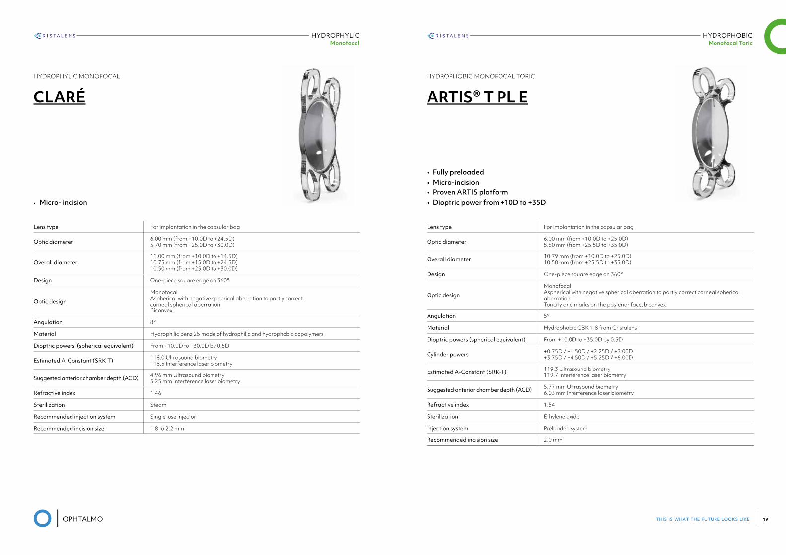
CLARÉ
Lens type For implantation in the capsular bag
Optic diameter 6.00 mm (from +10.0D to +24.5D) 5.70 mm (from +25.0D to +30.0D)
Overall diameter11.00 mm (from +10.0D to +14.5D)10.75 mm (from +15.0D to +24.5D)10.50 mm (from +25.0D to +30.0D)
Design One-piece square edge on 360°
Optic design
MonofocalAspherical with negative spherical aberration to partly correctcorneal spherical aberrationBiconvex
Angulation 8°
Material Hydrophilic Benz 25 made of hydrophilic and hydrophobic copolymers
Dioptric powers (spherical equivalent) From +10.0D to +30.0D by 0.5D
Estimated A-Constant (SRK-T) 118.0 Ultrasound biometry118.5 Interference laser biometry
Suggested anterior chamber depth (ACD) 4.96 mm Ultrasound biometry5.25 mm Interference laser biometry
Refractive index 1.46
Sterilization Steam
Recommended injection system Single-use injector
Recommended incision size 1.8 to 2.2 mm
• Micro- incision
• Fully preloaded• Micro-incision• Proven ARTIS platform• Dioptric power from +10D to +35D
HYDROPHOBIC MONOFOCAL TORIC
ARTIS® T PL E
Lens type For implantation in the capsular bag
Optic diameter 6.00 mm (from +10.0D to +25.0D) 5.80 mm (from +25.5D to +35.0D)
Overall diameter 10.79 mm (from +10.0D to +25.0D) 10.50 mm (from +25.5D to +35.0D)
Design One-piece square edge on 360°
Optic design
Monofocal Aspherical with negative spherical aberration to partly correct corneal spherical aberration Toricity and marks on the posterior face, biconvex
Angulation 5°
Material Hydrophobic CBK 1.8 from Cristalens
Dioptric powers (spherical equivalent) From +10.0D to +35.0D by 0.5D
Cylinder powers +0.75D / +1.50D / +2.25D / +3.00D +3.75D / +4.50D / +5.25D / +6.00D
Estimated A-Constant (SRK-T) 119.3 Ultrasound biometry 119.7 Interference laser biometry
Suggested anterior chamber depth (ACD) 5.77 mm Ultrasound biometry 6.03 mm Interference laser biometry
Refractive index 1.54
Sterilization Ethylene oxide
Injection system Preloaded system
Recommended incision size 2.0 mm
this is what the future looks like 21
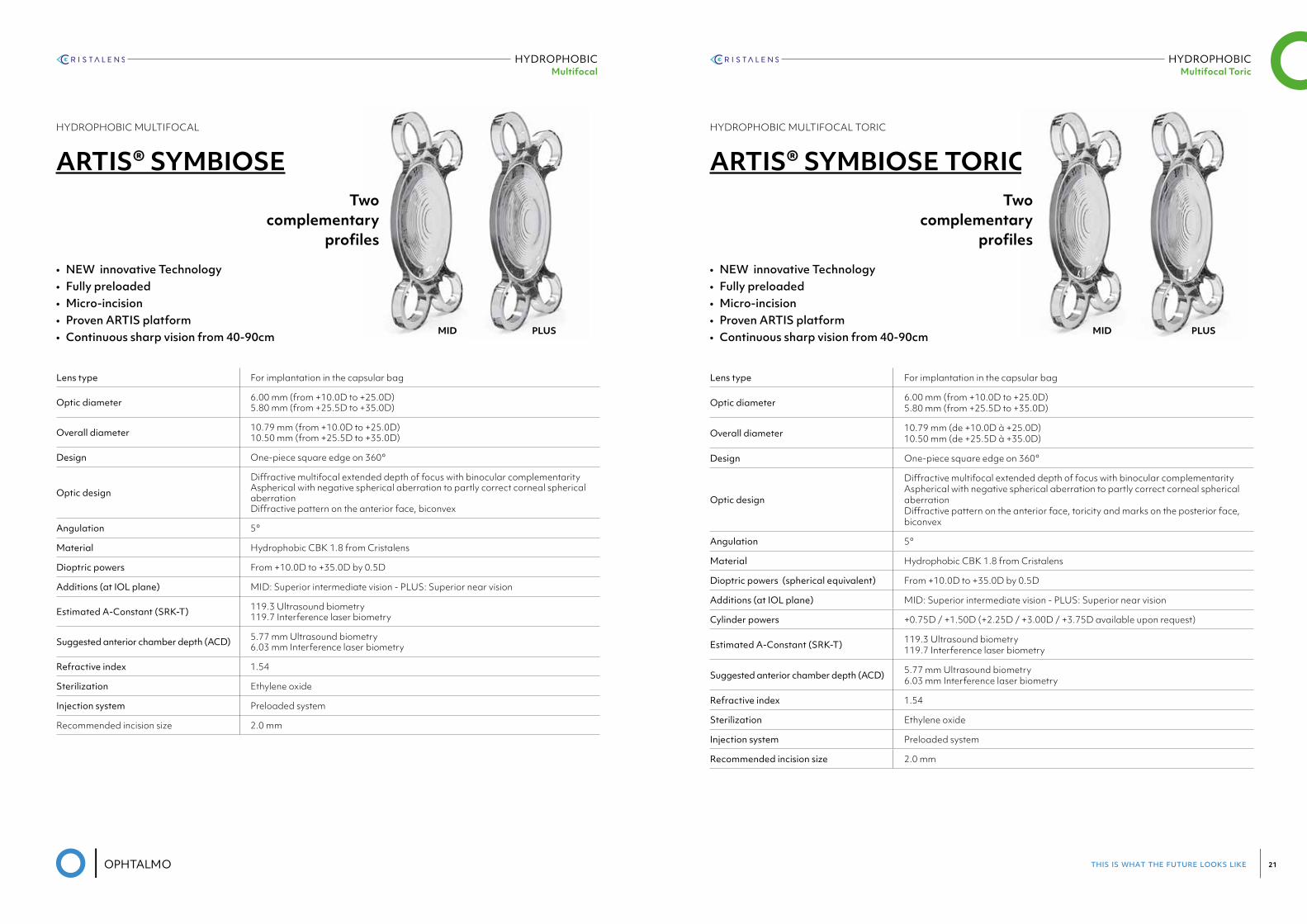
HYDROPHOBICMultifocal
• NEW innovative Technology• Fully preloaded• Micro-incision• Proven ARTIS platform• Continuous sharp vision from 40-90cm
HYDROPHOBIC MULTIFOCAL
ARTIS® SYMBIOSE
Lens type For implantation in the capsular bag
Optic diameter 6.00 mm (from +10.0D to +25.0D) 5.80 mm (from +25.5D to +35.0D)
Overall diameter 10.79 mm (from +10.0D to +25.0D) 10.50 mm (from +25.5D to +35.0D)
Design One-piece square edge on 360°
Optic design
Diffractive multifocal extended depth of focus with binocular complementarity Aspherical with negative spherical aberration to partly correct corneal spherical aberration Diffractive pattern on the anterior face, biconvex
Angulation 5°
Material Hydrophobic CBK 1.8 from Cristalens
Dioptric powers From +10.0D to +35.0D by 0.5D
Additions (at IOL plane) MID: Superior intermediate vision - PLUS: Superior near vision
Estimated A-Constant (SRK-T) 119.3 Ultrasound biometry 119.7 Interference laser biometry
Suggested anterior chamber depth (ACD) 5.77 mm Ultrasound biometry 6.03 mm Interference laser biometry
Refractive index 1.54
Sterilization Ethylene oxide
Injection system Preloaded system
Recommended incision size 2.0 mm
Two complementary
profiles
MID PLUS
HYDROPHOBICMultifocal Toric
HYDROPHOBIC MULTIFOCAL TORIC
ARTIS® SYMBIOSE TORIC
Lens type For implantation in the capsular bag
Optic diameter 6.00 mm (from +10.0D to +25.0D) 5.80 mm (from +25.5D to +35.0D)
Overall diameter 10.79 mm (de +10.0D à +25.0D) 10.50 mm (de +25.5D à +35.0D)
Design One-piece square edge on 360°
Optic design
Diffractive multifocal extended depth of focus with binocular complementarity Aspherical with negative spherical aberration to partly correct corneal spherical aberration Diffractive pattern on the anterior face, toricity and marks on the posterior face, biconvex
Angulation 5°
Material Hydrophobic CBK 1.8 from Cristalens
Dioptric powers (spherical equivalent) From +10.0D to +35.0D by 0.5D
Additions (at IOL plane) MID: Superior intermediate vision - PLUS: Superior near vision
Cylinder powers +0.75D / +1.50D (+2.25D / +3.00D / +3.75D available upon request)
Estimated A-Constant (SRK-T) 119.3 Ultrasound biometry 119.7 Interference laser biometry
Suggested anterior chamber depth (ACD) 5.77 mm Ultrasound biometry 6.03 mm Interference laser biometry
Refractive index 1.54
Sterilization Ethylene oxide
Injection system Preloaded system
Recommended incision size 2.0 mm
• NEW innovative Technology• Fully preloaded• Micro-incision• Proven ARTIS platform• Continuous sharp vision from 40-90cm
Two complementary
profiles
MID PLUS
02RAYNER
Rayner manufactured the world’s first IOL in 1949, and has remained at the forefront of innovation for 70 years, focused on providing you and your patients with the best IOLs and ophthalmic solutions - always
driven by science to improve patient outcomes and safety.
RayOne fully preloaded IOL injection system, designed to deliver without compromise.
At Rayner, we believe that the only way to create a true, fully preloaded micro incision cataract surgery (MICS) injection system that works consistently without compromise, is to design the system as one – both
lens and injector. This was the inspiration behind RayOne.
RayOne Aspheric
RayOne Spheric
RayOne Toric
RayOne Trifocal
RayOne Hydrophobic
RayOne Trifocal Toric
this is what the future looks like 25
HYDROPHOBICMonofocal
HYDROPHYLICMonofocal
HYDROPHYLIC MONOFOCAL
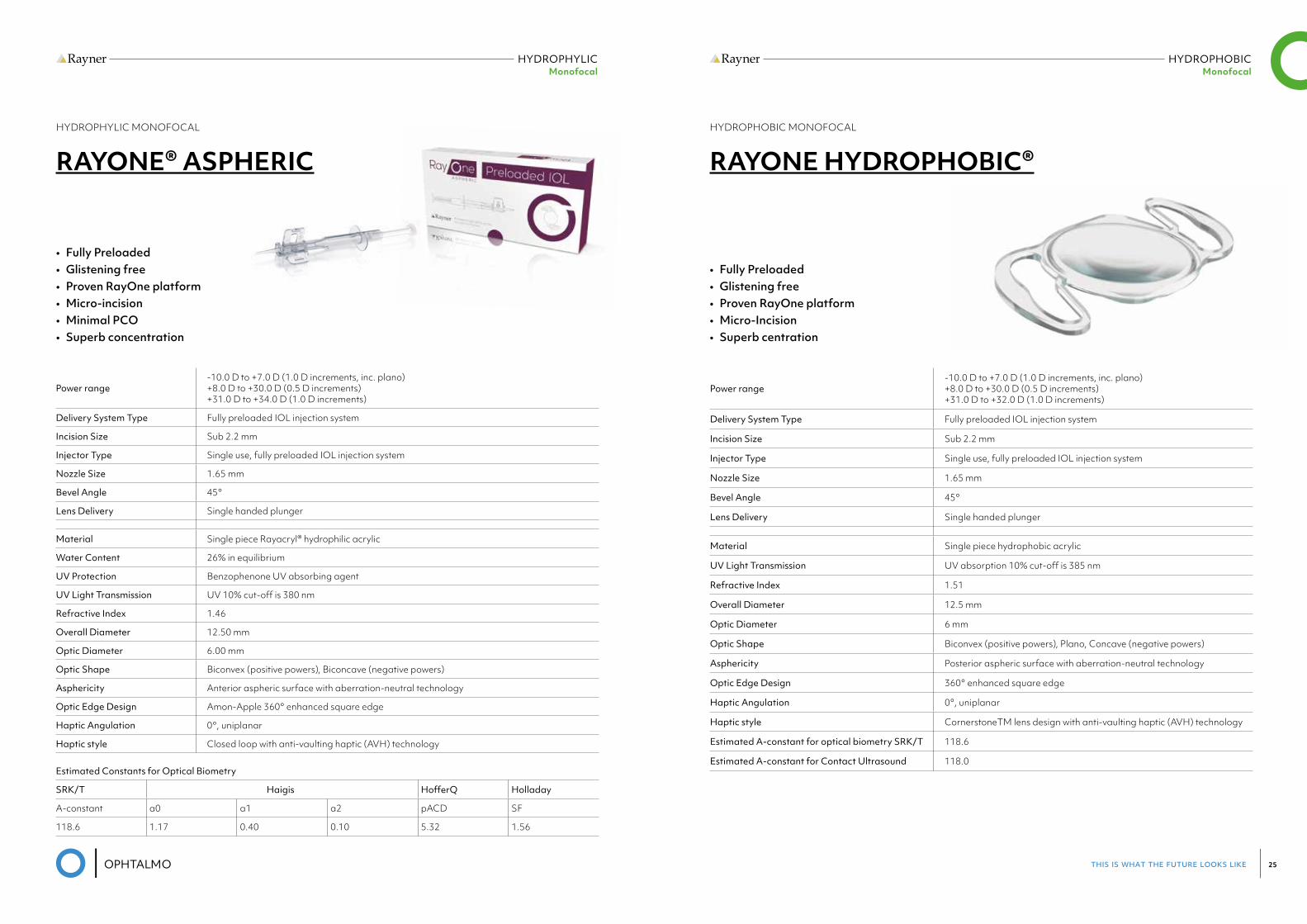
RAYONE® ASPHERIC
Power range-10.0 D to +7.0 D (1.0 D increments, inc. plano) +8.0 D to +30.0 D (0.5 D increments) +31.0 D to +34.0 D (1.0 D increments)
Delivery System Type Fully preloaded IOL injection system
Incision Size Sub 2.2 mm
Injector Type Single use, fully preloaded IOL injection system
Nozzle Size 1.65 mm
Bevel Angle 45°
Lens Delivery Single handed plunger
Material Single piece Rayacryl® hydrophilic acrylic
Water Content 26% in equilibrium
UV Protection Benzophenone UV absorbing agent
UV Light Transmission UV 10% cut-off is 380 nm
Refractive Index 1.46
Overall Diameter 12.50 mm
Optic Diameter 6.00 mm
Optic Shape Biconvex (positive powers), Biconcave (negative powers)
Asphericity Anterior aspheric surface with aberration-neutral technology
Optic Edge Design Amon-Apple 360° enhanced square edge
Haptic Angulation 0°, uniplanar
Haptic style Closed loop with anti-vaulting haptic (AVH) technology
Estimated Constants for Optical Biometry
SRK/T Haigis HofferQ Holladay
A-constant a0 a1 a2 pACD SF
118.6 1.17 0.40 0.10 5.32 1.56
• Fully Preloaded • Glistening free• Proven RayOne platform • Micro-incision • Minimal PCO • Superb concentration
HYDROPHOBIC MONOFOCAL
RAYONE HYDROPHOBIC®
Power range-10.0 D to +7.0 D (1.0 D increments, inc. plano) +8.0 D to +30.0 D (0.5 D increments) +31.0 D to +32.0 D (1.0 D increments)
Delivery System Type Fully preloaded IOL injection system
Incision Size Sub 2.2 mm
Injector Type Single use, fully preloaded IOL injection system
Nozzle Size 1.65 mm
Bevel Angle 45°
Lens Delivery Single handed plunger
Material Single piece hydrophobic acrylic
UV Light Transmission UV absorption 10% cut-off is 385 nm
Refractive Index 1.51
Overall Diameter 12.5 mm
Optic Diameter 6 mm
Optic Shape Biconvex (positive powers), Plano, Concave (negative powers)
Asphericity Posterior aspheric surface with aberration-neutral technology
Optic Edge Design 360° enhanced square edge
Haptic Angulation 0°, uniplanar
Haptic style CornerstoneTM lens design with anti-vaulting haptic (AVH) technology
Estimated A-constant for optical biometry SRK/T 118.6
Estimated A-constant for Contact Ultrasound 118.0
• Fully Preloaded• Glistening free• Proven RayOne platform • Micro-Incision• Superb centration
this is what the future looks like 27
HYDROPHYLIC Monofocal Toric
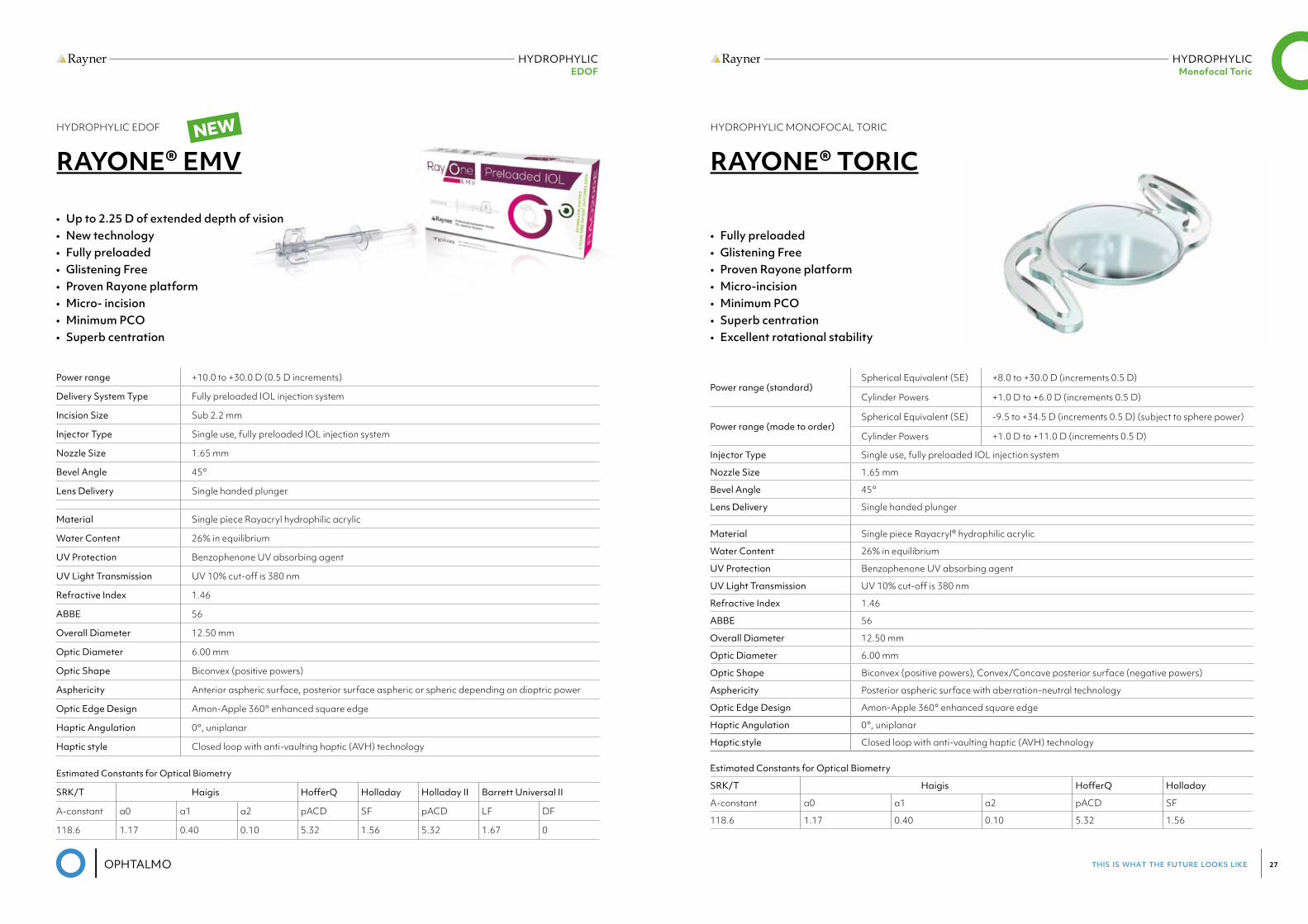
HYDROPHYLIC EDOF
HYDROPHYLIC EDOF
RAYONE® EMV
Estimated Constants for Optical Biometry
SRK/T Haigis HofferQ Holladay Holladay II Barrett Universal II
A-constant a0 a1 a2 pACD SF pACD LF DF
118.6 1.17 0.40 0.10 5.32 1.56 5.32 1.67 0
Power range +10.0 to +30.0 D (0.5 D increments)
Delivery System Type Fully preloaded IOL injection system
Incision Size Sub 2.2 mm
Injector Type Single use, fully preloaded IOL injection system
Nozzle Size 1.65 mm
Bevel Angle 45°
Lens Delivery Single handed plunger
Material Single piece Rayacryl hydrophilic acrylic
Water Content 26% in equilibrium
UV Protection Benzophenone UV absorbing agent
UV Light Transmission UV 10% cut-off is 380 nm
Refractive Index 1.46
ABBE 56
Overall Diameter 12.50 mm
Optic Diameter 6.00 mm
Optic Shape Biconvex (positive powers)
Asphericity Anterior aspheric surface, posterior surface aspheric or spheric depending on dioptric power
Optic Edge Design Amon-Apple 360° enhanced square edge
Haptic Angulation 0°, uniplanar
Haptic style Closed loop with anti-vaulting haptic (AVH) technology
• Up to 2.25 D of extended depth of vision• New technology • Fully preloaded • Glistening Free • Proven Rayone platform • Micro- incision • Minimum PCO • Superb centration
NEW HYDROPHYLIC MONOFOCAL TORIC
RAYONE® TORIC
Power range (standard)Spherical Equivalent (SE) +8.0 to +30.0 D (increments 0.5 D)
Cylinder Powers +1.0 D to +6.0 D (increments 0.5 D)
Power range (made to order)Spherical Equivalent (SE) -9.5 to +34.5 D (increments 0.5 D) (subject to sphere power)
Cylinder Powers +1.0 D to +11.0 D (increments 0.5 D)
Injector Type Single use, fully preloaded IOL injection system
Nozzle Size 1.65 mm
Bevel Angle 45°
Lens Delivery Single handed plunger
Material Single piece Rayacryl® hydrophilic acrylic
Water Content 26% in equilibrium
UV Protection Benzophenone UV absorbing agent
UV Light Transmission UV 10% cut-off is 380 nm
Refractive Index 1.46
ABBE 56
Overall Diameter 12.50 mm
Optic Diameter 6.00 mm
Optic Shape Biconvex (positive powers), Convex/Concave posterior surface (negative powers)
Asphericity Posterior aspheric surface with aberration-neutral technology
Optic Edge Design Amon-Apple 360° enhanced square edge
Haptic Angulation 0°, uniplanar
Haptic style Closed loop with anti-vaulting haptic (AVH) technology
• Fully preloaded • Glistening Free • Proven Rayone platform • Micro-incision • Minimum PCO • Superb centration • Excellent rotational stability
Estimated Constants for Optical Biometry
SRK/T Haigis HofferQ Holladay
A-constant a0 a1 a2 pACD SF
118.6 1.17 0.40 0.10 5.32 1.56
this is what the future looks like 29
HYDROPHYLIC Trifocal Toric
HYDROPHYLIC Trifocal
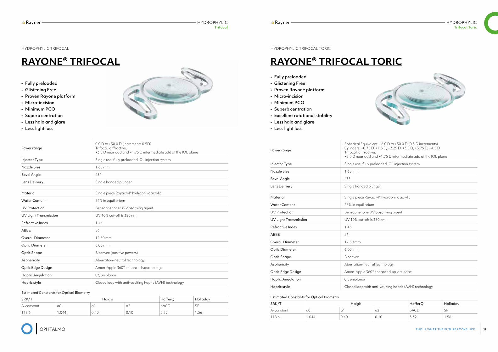
HYDROPHYLIC TRIFOCAL
RAYONE® TRIFOCAL
Power range0.0 D to +30.0 D (increments 0.5D) Trifocal, diffractive, +3.5 D near add and +1.75 D intermediate add at the IOL plane
Injector Type Single use, fully preloaded IOL injection system
Nozzle Size 1.65 mm
Bevel Angle 45°
Lens Delivery Single handed plunger
Material Single piece Rayacryl® hydrophilic acrylic
Water Content 26% in equilibrium
UV Protection Benzophenone UV absorbing agent
UV Light Transmission UV 10% cut-off is 380 nm
Refractive Index 1.46
ABBE 56
Overall Diameter 12.50 mm
Optic Diameter 6.00 mm
Optic Shape Biconvex (positive powers)
Asphericity Aberration-neutral technology
Optic Edge Design Amon-Apple 360° enhanced square edge
Haptic Angulation 0°, uniplanar
Haptic style Closed loop with anti-vaulting haptic (AVH) technology
Estimated Constants for Optical Biometry
SRK/T Haigis HofferQ Holladay
A-constant a0 a1 a2 pACD SF
118.6 1.044 0.40 0.10 5.32 1.56
• Fully preloaded • Glistening Free • Proven Rayone platform • Micro-incision • Minimum PCO • Superb centration • Less halo and glare • Less light loss
HYDROPHYLIC TRIFOCAL TORIC
RAYONE® TRIFOCAL TORIC
Power range
Spherical Equivalent: +6.0 D to +30.0 D (0.5 D increments) Cylinders: +0.75 D, +1.5 D, +2.25 D, +3.0 D, +3.75 D, +4.5 D Trifocal, diffractive, +3.5 D near add and +1.75 D intermediate add at the IOL plane
Injector Type Single use, fully preloaded IOL injection system
Nozzle Size 1.65 mm
Bevel Angle 45°
Lens Delivery Single handed plunger
Material Single piece Rayacryl® hydrophilic acrylic
Water Content 26% in equilibrium
UV Protection Benzophenone UV absorbing agent
UV Light Transmission UV 10% cut-off is 380 nm
Refractive Index 1.46
ABBE 56
Overall Diameter 12.50 mm
Optic Diameter 6.00 mm
Optic Shape Biconvex
Asphericity Aberration-neutral technology
Optic Edge Design Amon-Apple 360° enhanced square edge
Haptic Angulation 0°, uniplanar
Haptic style Closed loop with anti-vaulting haptic (AVH) technology
Estimated Constants for Optical Biometry
SRK/T Haigis HofferQ Holladay
A-constant a0 a1 a2 pACD SF
118.6 1.044 0.40 0.10 5.32 1.56
• Fully preloaded • Glistening Free • Proven Rayone platform • Micro-incision • Minimum PCO • Superb centration • Excellent rotational stability• Less halo and glare • Less light loss
this is what the future looks like 31
HYDROPHYLIC ADD-ON
HYDROPHYLIC ADD-ON
HYDROPHYLIC ADD-ON
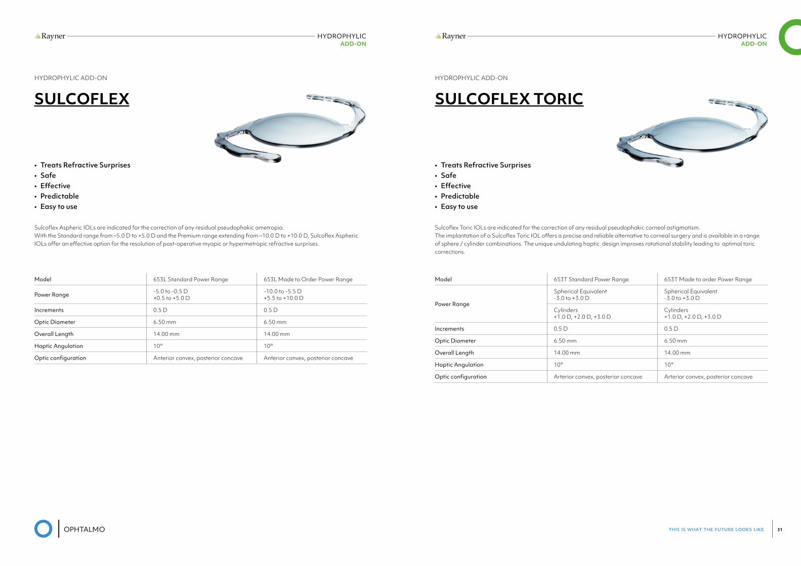
SULCOFLEX
Sulcoflex Aspheric IOLs are indicated for the correction of any residual pseudophakic ametropia. With the Standard range from –5.0 D to +5.0 D and the Premium range extending from –10.0 D to +10.0 D, Sulcoflex Aspheric IOLs offer an effective option for the resolution of post-operative myopic or hypermetropic refractive surprises.
Model 653L Standard Power Range 653L Made to Order Power Range
Power Range -5.0 to -0.5 D +0.5 to +5.0 D
-10.0 to -5.5 D +5.5 to +10.0 D
Increments 0.5 D 0.5 D
Optic Diameter 6.50 mm 6.50 mm
Overall Length 14.00 mm 14.00 mm
Haptic Angulation 10° 10°
Opticconfiguration Anterior convex, posterior concave Anterior convex, posterior concave
• Treats Refractive Surprises • Safe • Effective• Predictable • Easy to use
HYDROPHYLIC ADD-ON
SULCOFLEX TORIC
Sulcoflex Toric IOLs are indicated for the correction of any residual pseudophakic corneal astigmatism. The implantation of a Sulcoflex Toric IOL offers a precise and reliable alternative to corneal surgery and is available in a range of sphere / cylinder combinations. The unique undulating haptic design improves rotational stability leading to optimal toric corrections.
Model 653T Standard Power Range 653T Made to order Power Range
Power Range
Spherical Equivalent -3.0 to +3.0 D
Spherical Equivalent -3.0 to +3.0 D
Cylinders +1.0 D, +2.0 D, +3.0 D
Cylinders +1.0 D, +2.0 D, +3.0 D
Increments 0.5 D 0.5 D
Optic Diameter 6.50 mm 6.50 mm
Overall Length 14.00 mm 14.00 mm
Haptic Angulation 10° 10°
Opticconfiguration Arterior convex, posterior concave Arterior convex, posterior concave
• Treats Refractive Surprises • Safe • Effective• Predictable • Easy to use
this is what the future looks like 33
HYDROPHYLIC ADD-ON
HYDROPHYLIC ADD-ON
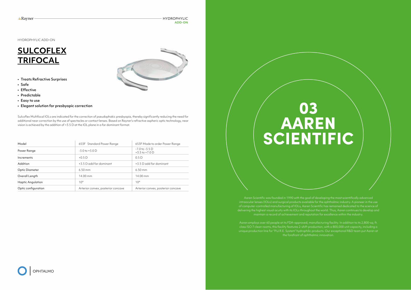
SULCOFLEX TRIFOCAL
Sulcoflex Multifocal IOLs are indicated for the correction of pseudophakic presbyopia, thereby significantly reducing the need for additional near correction by the use of spectacles or contact lenses. Based on Rayner’s refractive aspheric optic technology, near vision is achieved by the addition of +3.5 D at the IOL plane in a far dominant format.
Model 653F Standard Power Range 653F Made to order Power Range
Power Range -3.0 to +3.0 D -7.0 to -3.5 D +3.5 to +7.0 D
Increments +0.5 D 0.5 D
Addition +3.5 D add far dominant +3.5 D add far dominant
Optic Diameter 6.50 mm 6.50 mm
Overall Length 14.00 mm 14.00 mm
Haptic Angulation 10° 10°
Opticconfiguration Arterior convex, posterior concave Arterior convex, posterior concave
• Treats Refractive Surprises • Safe • Effective• Predictable • Easy to use • Elegant solution for presbyopic correction 03
AAREN SCIENTIFIC
Aaren Scientific was founded in 1990 with the goal of developing the most scientifically advanced intraocular lenses (IOLs) and surgical products available for the ophthalmic industry. A pioneer in the use of computer-controlled manufacturing of IOLs, Aaren Scientific has remained dedicated to the science of
delivering the highest visual acuity with its IOLs throughout the world. Thus, Aaren continues to develop and maintain a record of achievement and reputation for excellence within the industry.
Aaren employs over 60 people at its FDA-approved, manufacturing facility. In addition to its 2,800-sq. ft. class ISO 7 clean rooms, this facility features 2-shift production, with a 800,000 unit capacity, including a
unique production line for “P.U.R.E. System” hydrophilic products. Our exceptional R&D team put Aaren at the forefront of ophthalmic innovation.
this is what the future looks like 35
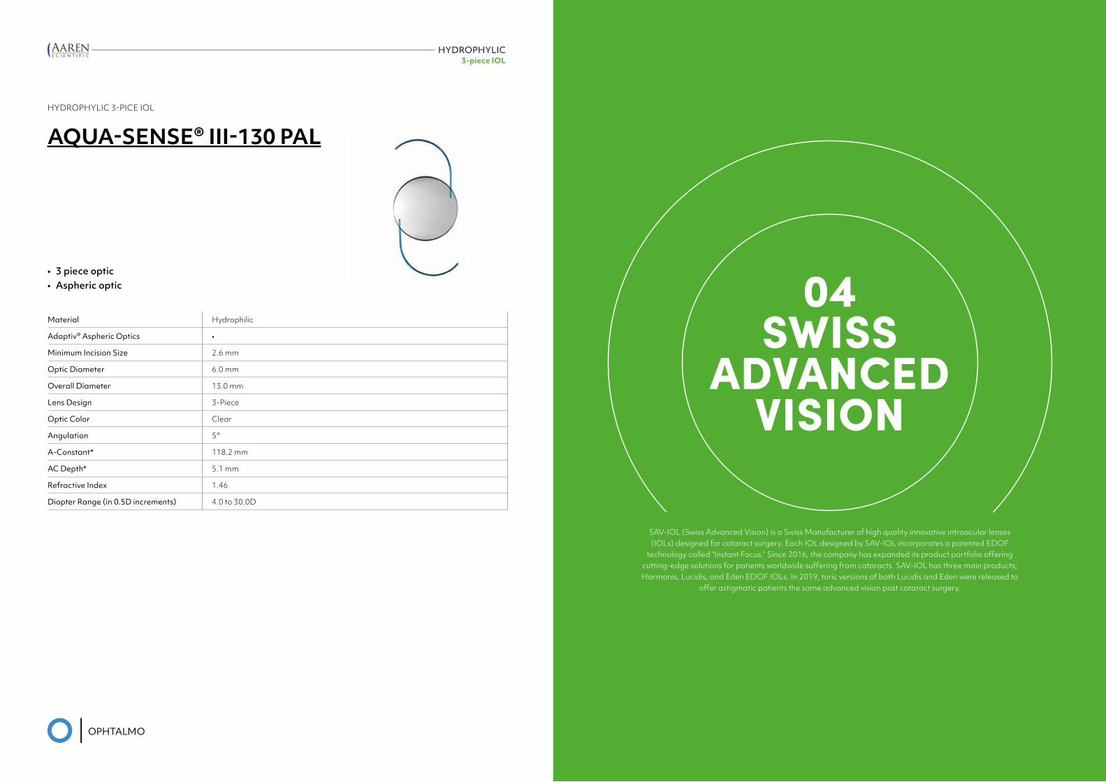
HYDROPHYLIC3-piece IOL
HYDROPHYLIC 3-PICE IOL
AQUA-SENSE® III-130 PAL
Material Hydrophilic
Adaptiv® Aspheric Optics •
Minimum Incision Size 2.6 mm
Optic Diameter 6.0 mm
Overall Diameter 13.0 mm
Lens Design 3-Piece
Optic Color Clear
Angulation 5°
A-Constant* 118.2 mm
AC Depth* 5.1 mm
Refractive Index 1.46
Diopter Range (in 0.5D increments) 4.0 to 30.0D
• 3 piece optic • Aspheric optic 04
SWISS ADVANCED
VISION
SAV-IOL (Swiss Advanced Vision) is a Swiss Manufacturer of high quality innovative intraocular lenses (IOLs) designed for cataract surgery. Each IOL designed by SAV-IOL incorporates a patented EDOF
technology called “Instant Focus.” Since 2016, the company has expanded its product portfolio offering cutting-edge solutions for patients worldwide suffering from cataracts. SAV-IOL has three main products; Harmonis, Lucidis, and Eden EDOF IOLs. In 2019, toric versions of both Lucidis and Eden were released to
offer astigmatic patients the same advanced vision post cataract surgery.
this is what the future looks like 37
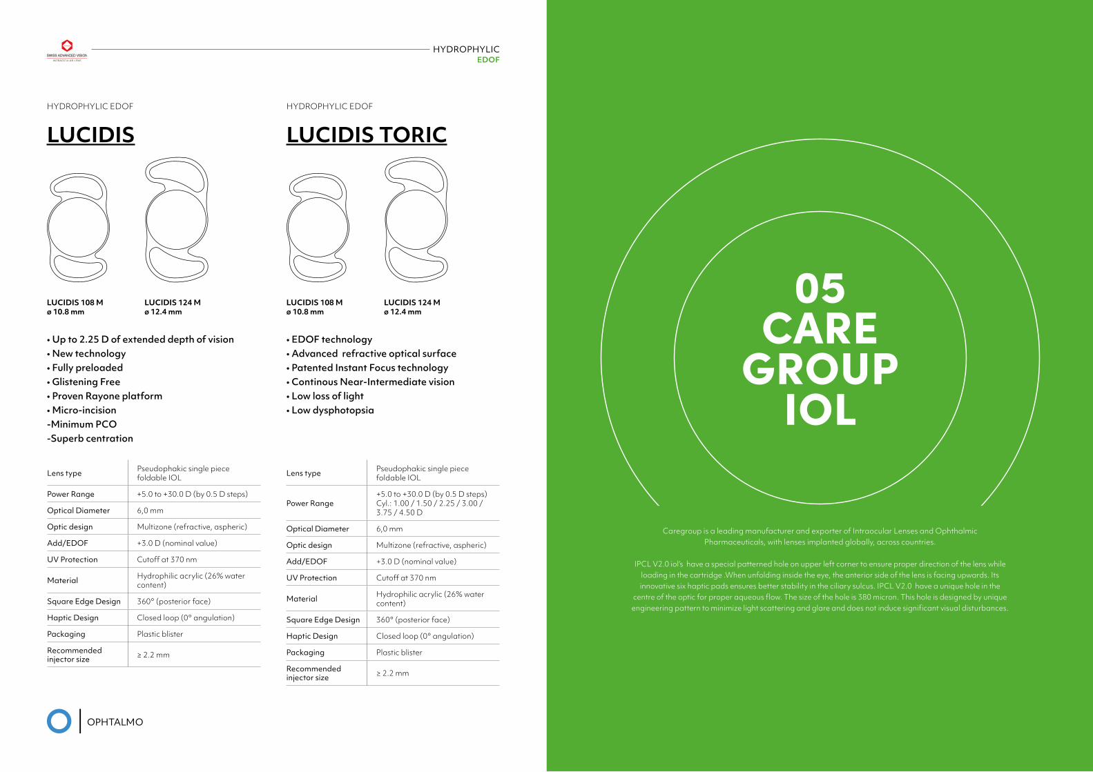
HYDROPHYLIC EDOF
HYDROPHYLIC EDOF
LUCIDIS
Lens type Pseudophakic single piece foldable IOL
Power Range +5.0 to +30.0 D (by 0.5 D steps)
Optical Diameter 6,0 mm
Optic design Multizone (refractive, aspheric)
Add/EDOF +3.0 D (nominal value)
UV Protection Cutoff at 370 nm
Material Hydrophilic acrylic (26% water content)
Square Edge Design 360° (posterior face)
Haptic Design Closed loop (0° angulation)
Packaging Plastic blister
Recommended injector size ≥ 2.2 mm
LUCIDIS 108 M ø 10.8 mm
LUCIDIS 108 M ø 10.8 mm
LUCIDIS 124 M ø 12.4 mm
LUCIDIS 124 M ø 12.4 mm
HYDROPHYLIC EDOF
LUCIDIS TORIC
Lens type Pseudophakic single piece foldable IOL
Power Range+5.0 to +30.0 D (by 0.5 D steps) Cyl.: 1.00 / 1.50 / 2.25 / 3.00 / 3.75 / 4.50 D
Optical Diameter 6,0 mm
Optic design Multizone (refractive, aspheric)
Add/EDOF +3.0 D (nominal value)
UV Protection Cutoff at 370 nm
Material Hydrophilic acrylic (26% water content)
Square Edge Design 360° (posterior face)
Haptic Design Closed loop (0° angulation)
Packaging Plastic blister
Recommended injector size ≥ 2.2 mm
• Up to 2.25 D of extended depth of vision• New technology • Fully preloaded • Glistening Free • Proven Rayone platform • Micro-incision -Minimum PCO -Superb centration
• EDOF technology • Advanced refractive optical surface• Patented Instant Focus technology • Continous Near-Intermediate vision • Low loss of light • Low dysphotopsia
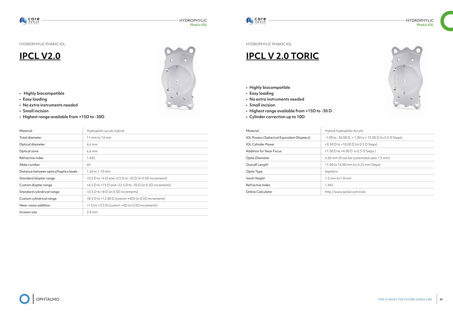
05CARE
GROUP IOL
Caregroup is a leading manufacturer and exporter of Intraocular Lenses and Ophthalmic Pharmaceuticals, with lenses implanted globally, across countries.
IPCL V2.0 iol’s have a special patterned hole on upper left corner to ensure proper direction of the lens while loading in the cartridge .When unfolding inside the eye, the anterior side of the lens is facing upwards. Its innovative six haptic pads ensures better stability in the ciliary sulcus. IPCL V2.0 have a unique hole in the
centre of the optic for proper aqueous flow. The size of the hole is 380 micron. This hole is designed by unique engineering pattern to minimize light scattering and glare and does not induce significant visual disturbances.
this is what the future looks like 39
HYDROPHYLIC Phakic IOL
HYDROPHYLIC Phakic IOL
HYDROPHYLIC PHAKIC IOL
IPCL V2.0
Material Hydrophilic acrylic hybrid
Total diameter 11 mm to 14 mm
Optical diameter 6,6 mm
Optical zone 6,6 mm
Refractive index 1.465
Abbe number 60
Distance between optics/haptics levels 1.20 to 1.70 mm
Standard diopter range +0,5 D to +6 D and -0.5 D to -22 D (in 0.5D increments)
Custom diopter range +6.5 D to +15 D and -22.5 D to -30 D (in 0.5D increments)
Standard cylindrical range +0.5 D to +8 D (in 0,5D increments)
Custom cylindrical range +8.5 D to +12.00 D (custom +4D) (in 0,5D increments)
Near-vision addition +1 D to +3.5 D (custom +4D (in 0,5D increments)
Incision size 2.8 mm
• Highly biocompatible • Easy loading • No extra instruments needed • Small incision • Highest range available from +15D to -30D
HYDROPHYLIC PHAKIC IOL
IPCL V 2.0 TORIC
Material Hybrid Hydrophilic Acrylic
IOL Powers (Spherical Equivalent Diopters) -1.00 to -30.00 D, + 1.00 to + 15.00 D (in 0.5 D Steps)
IOL Cylinder Power +0.50 D to +10.00 D (in 0.5 D Steps)
Addition for Near Focus +1.00 D to +4.00 D in 0.5 D Steps )
Optie Diameter 6.60 mm (lt can be customized upto 7.5 mm)
Overall Length 11.00 to 14.00 mm (in 0.25 mm Steps)
Optie Type Aspheric
Vault Height 1.2 mm to 1.8 mm
Refractive Index 1.465
Online Calculator http://www.ipcliol.com/calc
• Highly biocompatible • Easy loading • No extra instruments needed • Small incision • Highest range available from +15D to -30 D . • Cylinder correction up to 10D
this is what the future looks like 41
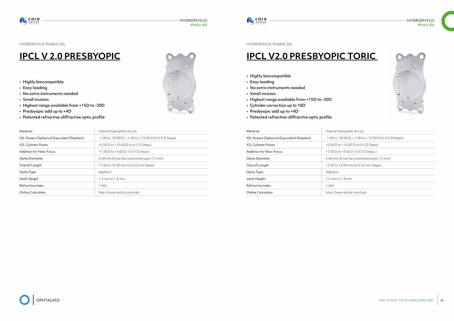
HYDROPHYLIC Phakic IOL
HYDROPHYLIC Phakic IOL
HYDROPHYLIC PHAKIC IOL
IPCL V 2.0 PRESBYOPIC
Material Hybrid Hydrophilic Acrylic
IOL Powers (Spherical Equivalent Diopters) -1.00 to -30.00 D, + 1.00 to + 15.00 D (in 0.5 D Steps)
IOL Cylinder Power +0.50 D to +10.00 D (in 0.5 D Steps)
Addition for Near Focus +1.00 D to +4.00 D in 0.5 D Steps )
Optie Diameter 6.60 mm (lt can be customized upto 7.5 mm)
Overall Length 11.00 to 14.00 mm (in 0.25 mm Steps)
Optie Type Aspheric
Vault Height 1.2 mm to 1.8 mm
Refractive Index 1.465
Online Calculator http://www.ipcliol.com/calc
• Highly biocompatible • Easy loading • No extra instruments needed • Small incision • Highest range available from +15D to -30D • Presbyopic add up to +4D• Patented refractive-diffractive optic profile
HYDROPHYLIC PHAKIC IOL
IPCL V2.0 PRESBYOPIC TORIC
Material Hybrid Hydrophilic Acrylic
IOL Powers (Spherical Equivalent Diopters) -1.00 to -30.00 D, + 1.00 to + 15.00 D (in 0.5 D Steps)
IOL Cylinder Power +0.50 D to +10.00 D (in 0.5 D Steps)
Addition for Near Focus +1.00 D to +4.00 D in 0.5 D Steps )
Optie Diameter 6.60 mm (lt can be customized upto 7.5 mm)
Overall Length 11.00 to 14.00 mm (in 0.25 mm Steps)
Optie Type Aspheric
Vault Height 1.2 mm to 1.8 mm
Refractive Index 1.465
Online Calculator http://www.ipcliol.com/calc
• Highly biocompatible • Easy loading • No extra instruments needed • Small incision • Highest range available from +15D to -30D • Cylinder correction up to 10D• Presbyopic add up to +4D• Patented refractive-diffractive optic profile
this is what the future looks like 43
06VISCOELASTIC
this is what the future looks like 45
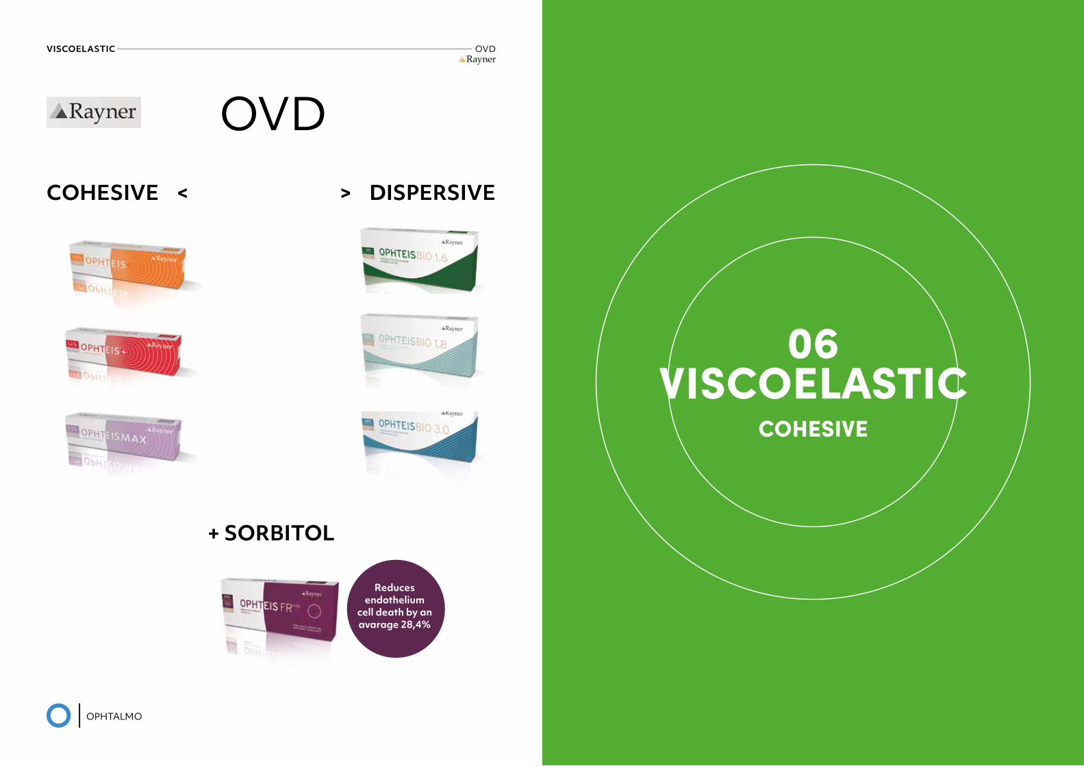
VISCOELASTIC OVDRayner
COHESIVE < > DISPERSIVE
OVD
+ SORBITOL
COHESIVE < > DISPERSIVE
+ SORBITOL
OVD
Reducesendothelium
cell death by anavarage 28,4%
06VISCOELASTIC
COHESIVE
this is what the future looks like 47
VISCOELASTIC COHESIVERayner
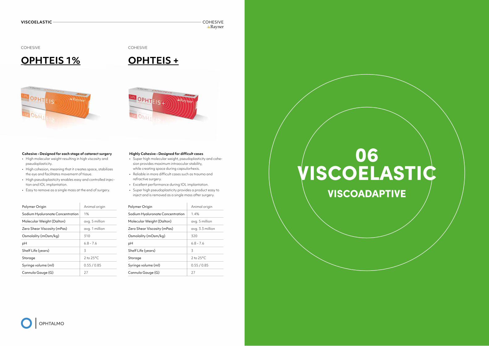
COHESIVE
OPHTEIS 1%
Cohesive • Designed for each stage of cataract surgery• High molecular weight resulting in high viscosity and
pseudoplasticity.• High cohesion, meaning that it creates space, stabilizes
the eye and facilitates movement of tissue.• High pseudoplasticity enables easy and controlled injec-
tion and IOL implantation.• Easy to remove as a single mass at the end of surgery.
Polymer Origin Animal origin
Sodium Hyaluronate Concentration 1%
Molecular Weight (Dalton) avg. 5 million
Zero Shear Viscosity (mPas) avg. 1 million
Osmolality (mOsm/kg) 310
pH 6.8 - 7.6
Shelf Life (years) 3
Storage 2 to 25°C
Syringe volume (ml) 0.55 / 0.85
Cannula Gauge (G) 27
COHESIVE
OPHTEIS +
Highly Cohesive • Designed for difficult cases• Super high molecular weight, pseudoplasticity and cohe-
sion provides maximum intraocular stability, while creating space during capsulorhexis.
• Reliable in more difficult cases such as trauma and refractive surgery.
• Excellent performance during IOL implantation.• Super high pseudoplasticity provides a product easy to
inject and is removed as a single mass after surgery.
Polymer Origin Animal origin
Sodium Hyaluronate Concentration 1.4%
Molecular Weight (Dalton) avg. 5 million
Zero Shear Viscosity (mPas) avg. 3.3 million
Osmolality (mOsm/kg) 320
pH 6.8 - 7.6
Shelf Life (years) 3
Storage 2 to 25°C
Syringe volume (ml) 0.55 / 0.85
Cannula Gauge (G) 27
06VISCOELASTIC
VISCOADAPTIVE
this is what the future looks like 49
VISCOELASTIC VISCOADAPTIVERayner
06VISCOELASTIC
COHESIVE-DISPERSIVE
VISCOADAPTIVE
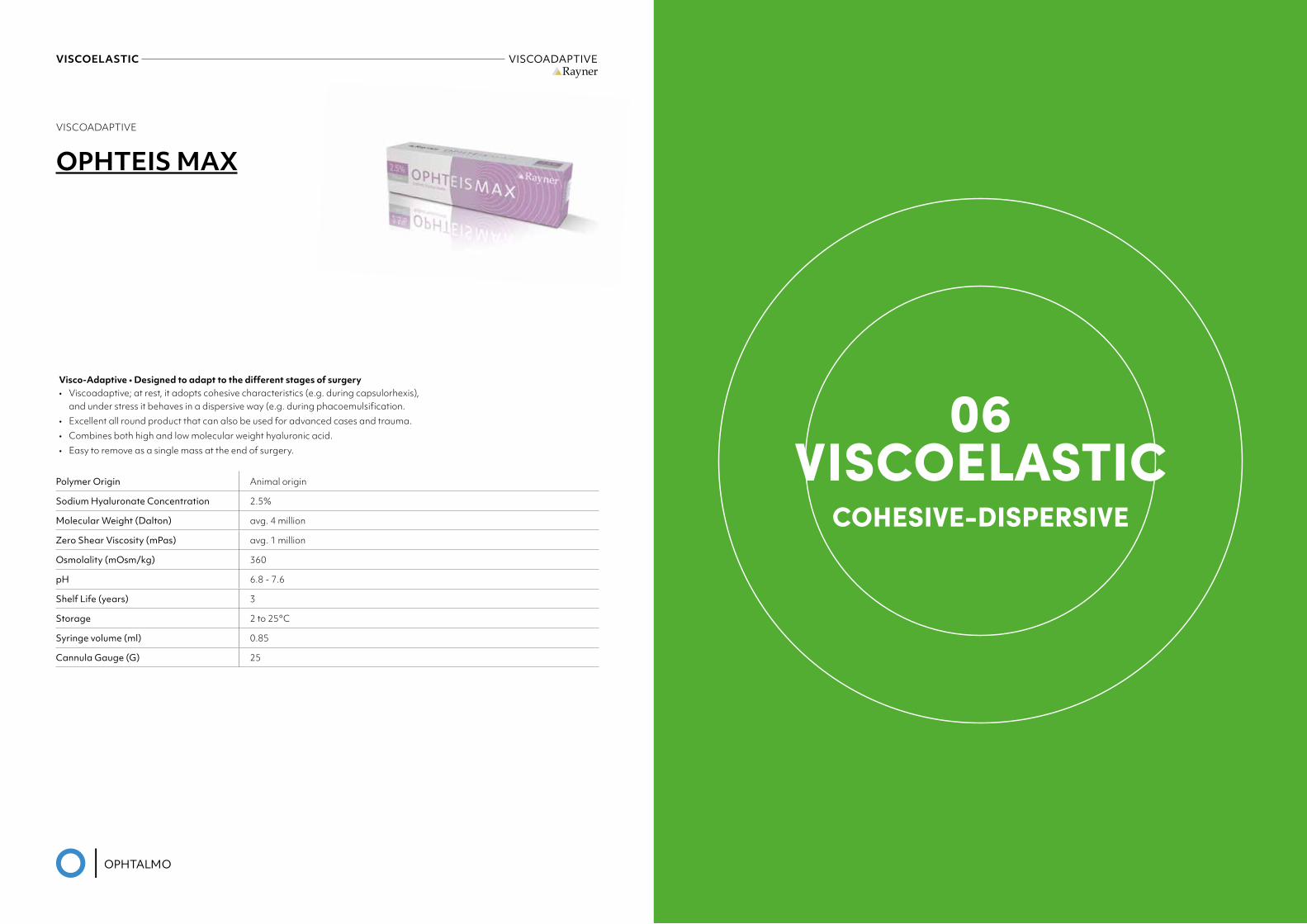
OPHTEIS MAX
Visco-Adaptive • Designed to adapt to the different stages of surgery• Viscoadaptive; at rest, it adopts cohesive characteristics (e.g. during capsulorhexis),
and under stress it behaves in a dispersive way (e.g. during phacoemulsification.• Excellent all round product that can also be used for advanced cases and trauma.• Combines both high and low molecular weight hyaluronic acid.• Easy to remove as a single mass at the end of surgery.
Polymer Origin Animal origin
Sodium Hyaluronate Concentration 2.5%
Molecular Weight (Dalton) avg. 4 million
Zero Shear Viscosity (mPas) avg. 1 million
Osmolality (mOsm/kg) 360
pH 6.8 - 7.6
Shelf Life (years) 3
Storage 2 to 25°C
Syringe volume (ml) 0.85
Cannula Gauge (G) 25
this is what the future looks like 51
VISCOELASTIC COHESIVE-DISPERSIVERayner
COHESIVE-DISPERSIVE
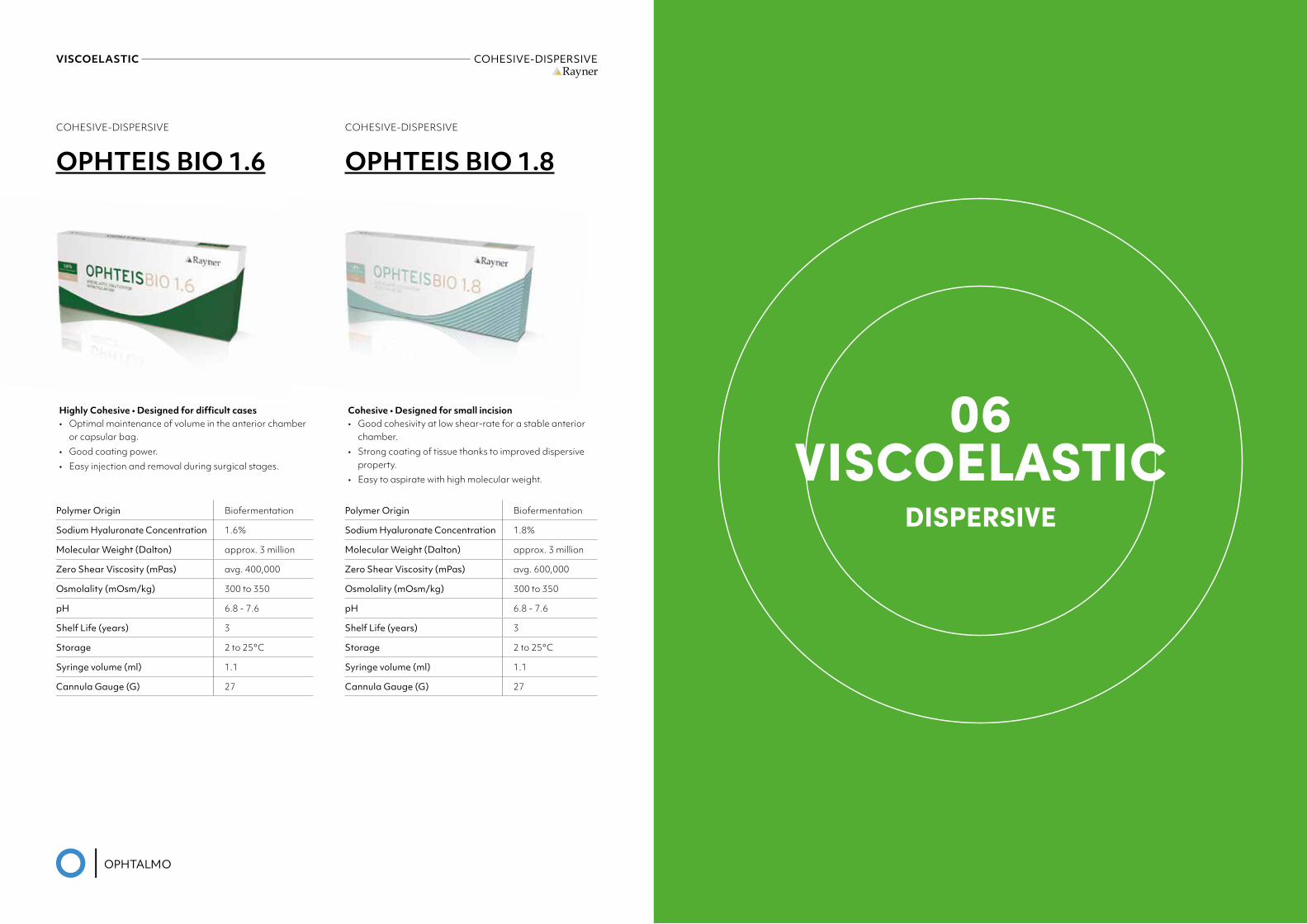
OPHTEIS BIO 1.6
Highly Cohesive • Designed for difficult cases• Optimal maintenance of volume in the anterior chamber
or capsular bag.• Good coating power.• Easy injection and removal during surgical stages.
Polymer Origin Biofermentation
Sodium Hyaluronate Concentration 1.6%
Molecular Weight (Dalton) approx. 3 million
Zero Shear Viscosity (mPas) avg. 400,000
Osmolality (mOsm/kg) 300 to 350
pH 6.8 - 7.6
Shelf Life (years) 3
Storage 2 to 25°C
Syringe volume (ml) 1.1
Cannula Gauge (G) 27
COHESIVE-DISPERSIVE
OPHTEIS BIO 1.8
Cohesive • Designed for small incision• Good cohesivity at low shear-rate for a stable anterior
chamber.• Strong coating of tissue thanks to improved dispersive
property.• Easy to aspirate with high molecular weight.
Polymer Origin Biofermentation
Sodium Hyaluronate Concentration 1.8%
Molecular Weight (Dalton) approx. 3 million
Zero Shear Viscosity (mPas) avg. 600,000
Osmolality (mOsm/kg) 300 to 350
pH 6.8 - 7.6
Shelf Life (years) 3
Storage 2 to 25°C
Syringe volume (ml) 1.1
Cannula Gauge (G) 27
06VISCOELASTIC
DISPERSIVE
this is what the future looks like 53
VISCOELASTIC DISPERSIVERayner
DISPERSIVE
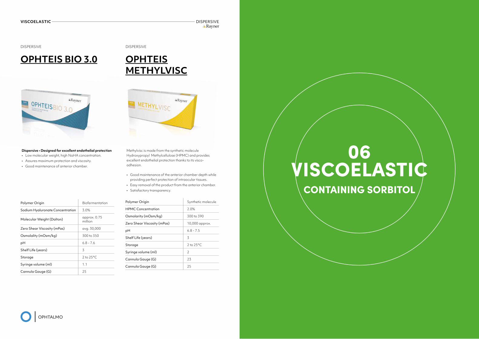
OPHTEIS BIO 3.0
Dispersive • Designed for excellent endothelial protection• Low molecular weight, high NaHA concentration.• Assures maximum protection and viscosity.• Good maintenance of anterior chamber.
Polymer Origin Biofermentation
Sodium Hyaluronate Concentration 3.0%
Molecular Weight (Dalton) approx. 0.75 million
Zero Shear Viscosity (mPas) avg. 30,000
Osmolality (mOsm/kg) 300 to 350
pH 6.8 - 7.6
Shelf Life (years) 3
Storage 2 to 25°C
Syringe volume (ml) 1.1
Cannula Gauge (G) 25
Methylvisc is made from the synthetic molecule Hydroxypropyl Methylcellulose (HPMC) and provides excellent endothelial protection thanks to its visco-adhesion.
• Good maintenance of the anterior chamber depth while providing perfect protection of intraocular tissues.
• Easy removal of the product from the anterior chamber.• Satisfactory transparency.
Polymer Origin Synthetic molecule
HPMC Concentration 2.0%
Osmolarity (mOsm/kg) 300 to 390
Zero Shear Viscosity (mPas) 10,000 approx.
pH 6.8 - 7.5
Shelf Life (years) 3
Storage 2 to 25°C
Syringe volume (ml) 2
Cannula Gauge (G) 23
Cannula Gauge (G) 25
DISPERSIVE
OPHTEISMETHYLVISC
06VISCOELASTIC
CONTAINING SORBITOL
this is what the future looks like 55
VISCOELASTIC CONTAINING SORBITOLRayner
CONTAINING SORBITOL
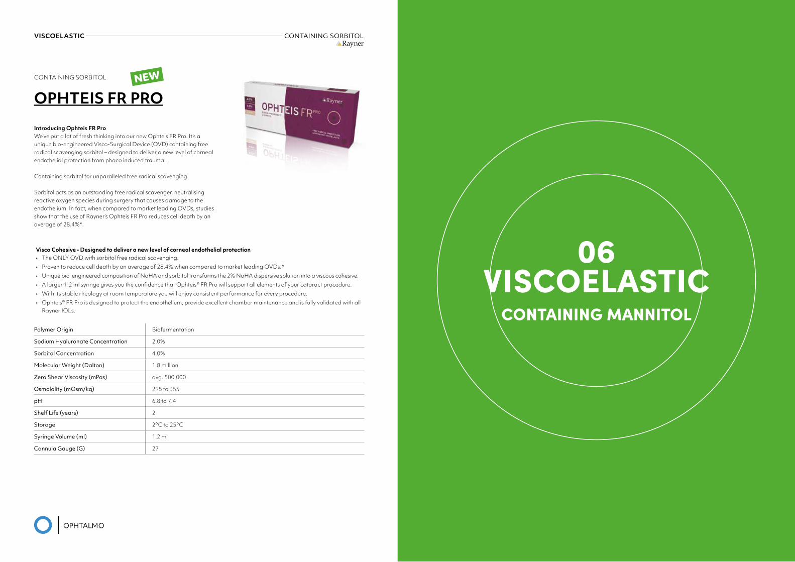
OPHTEIS FR PRO
Visco Cohesive • Designed to deliver a new level of corneal endothelial protection• The ONLY OVD with sorbitol free radical scavenging.• Proven to reduce cell death by an average of 28.4% when compared to market leading OVDs.*• Unique bio-engineered composition of NaHA and sorbitol transforms the 2% NaHA dispersive solution into a viscous cohesive.• A larger 1.2 ml syringe gives you the confidence that Ophteis® FR Pro will support all elements of your cataract procedure.• With its stable rheology at room temperature you will enjoy consistent performance for every procedure.• Ophteis® FR Pro is designed to protect the endothelium, provide excellent chamber maintenance and is fully validated with all
Rayner IOLs.
Polymer Origin Biofermentation
Sodium Hyaluronate Concentration 2.0%
Sorbitol Concentration 4.0%
Molecular Weight (Dalton) 1.8 million
Zero Shear Viscosity (mPas) avg. 500,000
Osmolality (mOsm/kg) 295 to 355
pH 6.8 to 7.4
Shelf Life (years) 2
Storage 2°C to 25°C
Syringe Volume (ml) 1.2 ml
Cannula Gauge (G) 27
Introducing Ophteis FR ProWe’ve put a lot of fresh thinking into our new Ophteis FR Pro. It’s a unique bio-engineered Visco-Surgical Device (OVD) containing free radical scavenging sorbitol – designed to deliver a new level of corneal endothelial protection from phaco induced trauma.
Containing sorbitol for unparalleled free radical scavenging
Sorbitol acts as an outstanding free radical scavenger, neutralising reactive oxygen species during surgery that causes damage to the endothelium. In fact, when compared to market leading OVDs, studies show that the use of Rayner’s Ophteis FR Pro reduces cell death by an average of 28.4%*.
NEW
06VISCOELASTIC
CONTAINING MANNITOL
this is what the future looks like 57
VISCOELASTIC CONTAINING MANNITOLRayner
CONTAINING MANNITOL
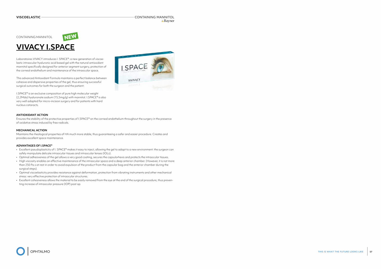
VIVACY I.SPACE
ANTIOXIDANT ACTION Ensures the stability of the protective properties of I.SPACE® on the corneal endothelium throughout the surgery in the presence of oxidative stress induced by free radicals.
MECHANICAL ACTION Maintains the rheological properties of HA much more stable, thus guaranteeing a safer and easier procedure. Creates and provides excellent space maintenance.
ADVANTAGES OF I.SPACE® • Excellent pseudoplasticity of I. SPACE® makes it easy to inject, allowing the gel to adapt to a new environment: the surgeon can
safely manipulate delicate intraocular tissues and intraocular lenses (IOLs).• Optimal adhesiveness of the gel allows a very good coating, secures the capsulorhexis and protects the intraocular tissues.• High viscosity enables an effective maintenance of the intraocular space and a deep anterior chamber. (However, it is not more
than 250 Pa.s at rest in order to avoid expulsion of the product from the capsular bag and the anterior chamber during the surgical steps).
• Optimal viscoelasticity provides resistance against deformation, protection from vibrating instruments and other mechanical stress: very effective protection of intraocular structures.
• Excellent cohesiveness allows the material to be easily removed from the eye at the end of the surgical procedure, thus preven-ting increase of intraocular pressure (IOP) post op.
Laboratoires VIVACY introduces I. SPACE®: a new generation of viscoe-lastic intraocular hyaluronic acid based gel with the natural antioxidant mannitol specifically designed for anterior segment surgery, protection of the corneal endothelium and maintenance of the intraocular space.
This advanced Antioxidant Formula maintains a perfect balance between cohesive and dispersive properties of the gel, thus ensuring successful surgical outcomes for both the surgeon and the patient:
I.SPACE® is an exclusive composition of pure high molecular weight (2,2Mda) hyaluronate sodium (15,5mg/g) with mannitol. I.SPACE® is also very well adapted for micro-incision surgery and for patients with hard nucleus cataracts.
NEW

this is what the future looks like 59
CONSUMABLES
this is what the future looks like 61
01. REUSABLE INSTRUMENTS 65 IRRIGATION/ASPIRATION 66 CHOPPERS-MANIPULATORS-SPATULAS 67 FORCEPS 70 NEEDLEHOLDERS 73 SCISSORS 76 SPECULUM 79 IOL FORCEPS 82 IOLCUTTERS 83 DIAMOND KNIVES 84 FIXATION , GAUGES & MARKERS 85 STERILISING TRAYS 87 BIPOLAR FORCEPS 88 MISCELLANEOUS 89
02. SINGLE USE INSTRUMENTS 91 IRRIGATION/ASPIRATION 92 CHOPPERS-MANIPULATORS-SPATULAS 93 FORCEPS 95 NEEDLE HOLDERS 96 SCISSORS 97 SPECULUM 98
03. ABSORBABLE SUTURES 99 SURGICRYL POLYGLACTINE 101 SURGICRYL POLYGLYCOLIC ACID 101
04. NON-ABSORBABLE SUTURES 103 SILK 104 NYLON 104 POLYESTER 104 POLYPROPYLENE 104
this is what the future looks like 63
05. DISPOSABLE KNIVES & BLADES 105 STAB KNIVES 106 CRESCENT KNIVES 107 BLEB KNIVES 107 SLIT KNIVES 108 CLEAR CORNEAL KNIVES 109 TRAPEZOID KNIVES 110 IMPLANT KNIVES 111 MVR KNIVES 112 PARACENTESISKNIVES 113 SIDEPORT KNIVES 114 06. DISPOSABLE CANNULAS 115 HYDRODISSECTION CANNULAS 116 OPHTALMIC CANNULAS 117 RYCROFT ANTERIOR CHAMBER CANNULAS 118 VISCOELASTIC CANNULAS 119 OTHERS 120
07. THERAPEUTIC LENSES 121 MEGASOFT 122 REGENERA 123 OTHER BRANDS 124 08. PROCEDURE PACKS 125
09. TENSION RINGS 127 10. CAUTERY 131
11. DISPOSABLES 133 EYESHIELDS 134 SPEARS 135 IRISRETRACTORHOOKS 136 PUNCTUMPLUGS 137 SPONGES&WIPES 138 VACUUM TREPHINES 140 VACUUM PUNCHES 141
12. PHARMA 143 AEON PROTECT 144 AEON PROTECT PLUS 144 AEON REPAIR 145 AEON NACL 145 13. DIAGNOSTIC STRIPS 147 I-DEWFLO 148 I-DEWTEARSTRIPS 148 I-DEWGREEN 149 I-DEWROSE 149
this is what the future looks like 65
01REUSABLE
INSTRUMENTS
this is what the future looks like 67
REUSABLE INSTRUMENTS CHOPPERS - MANIPULATORS - SPATULASREUSABLE INSTRUMENTS IRRIGATION/ASPIRATIONMedicel
I/A INSTRUMENTS
REUSABLE SWISS BIMANUAL I/A SYSTEMS, NON-STERILE
The Medicel bimanual I/A systems can be combined in various configurations. All models are made of titanium. Thanks to the aeroplane-nose design, cannulas can be introduced easily through the smallest of incisions.
consisting of: Aspiration Instrument (Box of 1)
I/A ASPIRATION INSTRUMENTS
ITEM CODE SIZE INCISION SIZE ASPIRATION CANNULA
ASPIRATION PORT
ASPIRATION HEAD
SBA100A 21G / 0.8mm 1.1mm aeroplane shape 0.35mm polished
SBA103A 21G / 0.8mm 1.1mm aeroplane shape 0.40mm polished
SBA104A 21G / 0.8mm 1.1mm aeroplane shape 0.40mm textured
SBA105A 21G / 0.8mm 1.1mm aeroplane shape 0.35mm textured
SBA101B 23G / 0.6mm 0.8mm bullet shape 0.30mm polished
SBA106B 23G / 0.6mm 0.8mm bullet shape 0.30mm textured
consisting of: Irrigation Instrument (Box of 1)
I/A IRRIGATION INSTRUMENTS
ITEM CODE SIZE INCISION SIZE IRRIGATION CANNULA
IRRIGATION PORT IRRIGATION HEAD
SBI120A 21G / 0.8mm 1.1mm aeroplane shape 2 ports, 0.45mm polished
SBI125A 21G / 0.8mm 1.1mm aeroplane shape 2 ports, 0.45mm textured
SBI121B 23G / 0.6mm 0.8mm bullet shape 2 ports, 0.30mm polished
SBI126B 23G / 0.6mm 0.8mm bullet shape 2 ports, 0.30mm textured
REUSABLE SWISS BIMANUAL I/A SYSTEMS, NON-STERILE
The Medicel bimanual I/A systems can be combined in various configurations. All models are made of titanium. Thanks to the aeroplane-nose design, cannulas can be introduced easily through the smallest of incisions.
consisting of: Aspiration Instrument (Box of 1)
I/A ASPIRATION INSTRUMENTS
ITEM CODE SIZE INCISION SIZE ASPIRATION CANNULA ASPIRATION PORT ASPIRATION HEADSBA100A 21G / 0.8mm 1.1mm aeroplane shape 0.35mm polished
SBA103A 21G / 0.8mm 1.1mm aeroplane shape 0.40mm polishedSBA104A 21G / 0.8mm 1.1mm aeroplane shape 0.40mm texturedSBA105A 21G / 0.8mm 1.1mm aeroplane shape 0.35mm texturedSBA101B 23G / 0.6mm 0.8mm bullet shape 0.30mm polishedSBA106B 23G / 0.6mm 0.8mm bullet shape 0.30mm textured
consisting of: Irrigation Instrument (Box of 1)
I/A IRRIGATION INSTRUMENTS
ITEM CODE SIZE INCISION SIZE IRRIGATION CANNULA IRRIGATION PORT IRRIGATION HEADSBI120A 21G / 0.8mm 1.1mm aeroplane shape 2 ports, 0.45mm polished
SBI125A 21G / 0.8mm 1.1mm aeroplane shape 2 ports, 0.45mm texturedSBI121B 23G / 0.6mm 0.8mm bullet shape 2 ports, 0.30mm polishedSBI126B 23G / 0.6mm 0.8mm bullet shape 2 ports, 0.30mm textured
Bullet-shape aeroplane-shape
18
I/A INSTRUMENTS
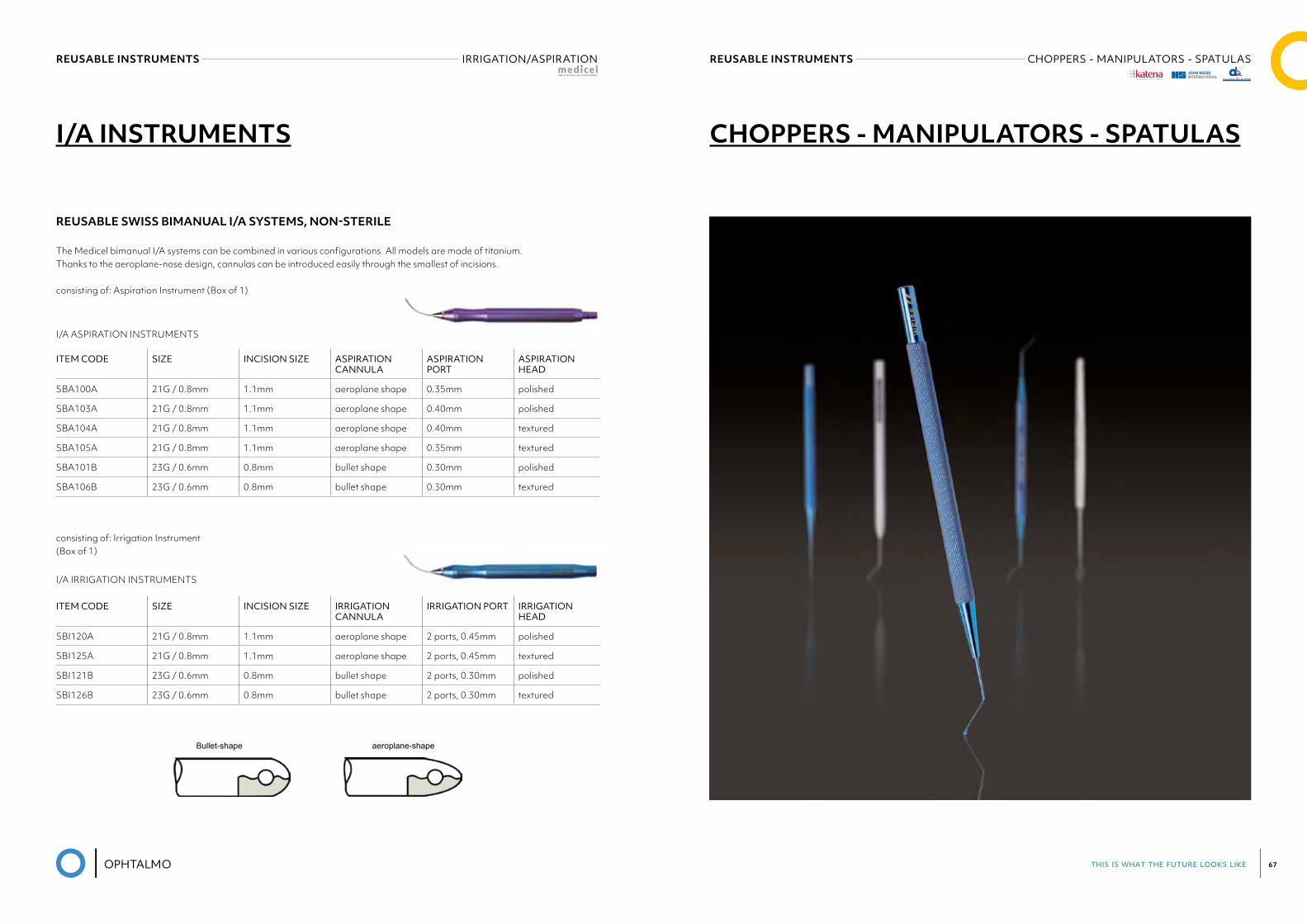
CHOPPERS - MANIPULATORS - SPATULAS
®
this is what the future looks like 69
REUSABLE INSTRUMENTS CHOPPERS - MANIPULATORS - SPATULASREUSABLE INSTRUMENTS CHOPPERS - MANIPULATORS - SPATULAS
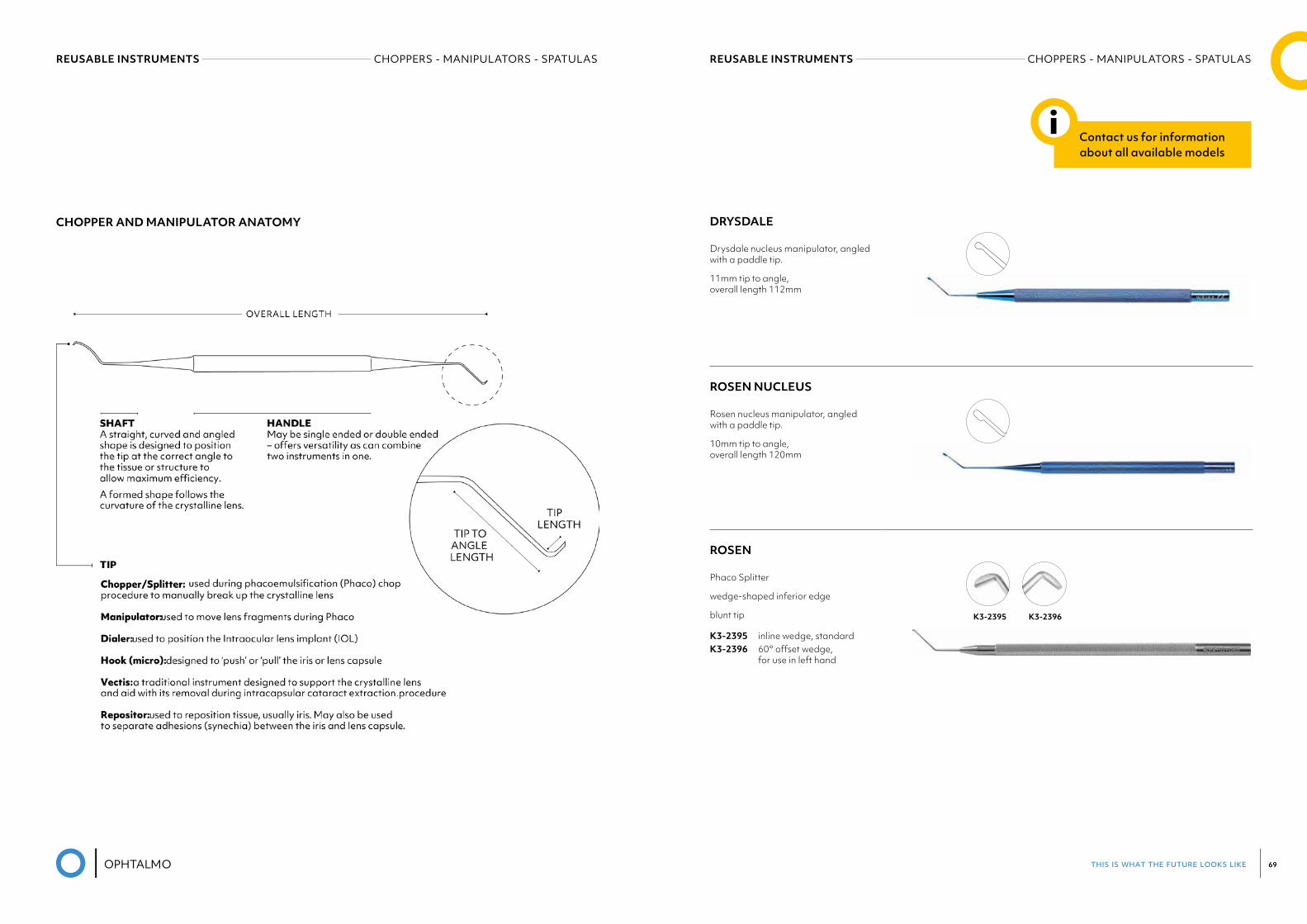
CHOPPER AND MANIPULATOR ANATOMY DRYSDALE
Drysdale nucleus manipulator, angled with a paddle tip.
11mm tip to angle, overall length 112mm
ROSEN NUCLEUS
Rosen nucleus manipulator, angled with a paddle tip.
10mm tip to angle, overall length 120mm
ROSEN
Phaco Splitter
wedge-shaped inferior edge
blunt tip
K3-2395 inline wedge, standard K3-2396 60° offset wedge, for use in left hand
K3-2395 K3-2396
Contact us for information about all available models
i
this is what the future looks like 71
REUSABLE INSTRUMENTS FORCEPSREUSABLE INSTRUMENTS FORCEPS
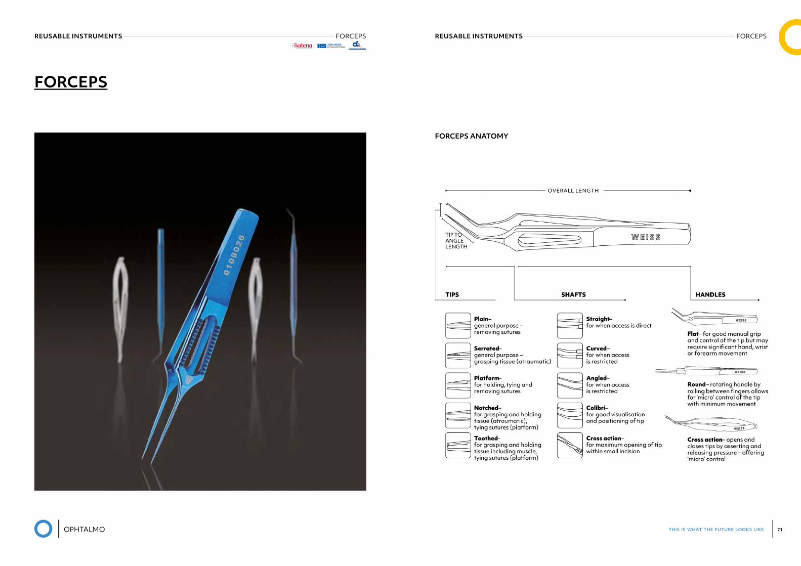
FORCEPS
FORCEPS ANATOMY
®
this is what the future looks like 73
BONN
Forceps
1x2 teeth, 0.12mm
with tying platform
K5-2020 stainless steelKT5-2020 titanium
UTRATA
Capsulorrhexis Forceps
very delicate, short handle
triangular grasping tips
extremely thin with landmarks at 2.5mm and 5.0mm
K5-5071 straight
MICS CAPSULORRHEXIS FORCEPS
23 gauge curved shaft with short micro-grasping tips
Designed for use through a very small paracentesis.
K5-7651 AlioK5-7655 Fine-Ikeda
REUSABLE INSTRUMENTS NEEDLE HOLDERSREUSABLE INSTRUMENTS FORCEPS
®
ForcepsK5
99
Harms-Colibri Forcepsvery delicate 1x2 teeth, 0.12mm with tying platformK5-1902
Hyde Corneal Forceps 2x2 teeth, 0.12mm with tying platformK5-1920
Bonn Forcepsvery delicate1x2 teeth, 0.12mm K5-2000
Bonn Forceps1x2 teeth, 0.12mm with tying platformK5-2020 stainless steelKT5-2020 titanium
®
ForcepsK5
108
Utrata Capsulorrhexis Forceps very delicate triangular grasping tips extremely thin 11mm long shanksK5-5081 stainless steel KT5-5081 titanium
Also available for MICS, see K5-5091.
Also available for MICS, see K5-5093.
Utrata Capsulorrhexis Forceps very delicate triangular grasping tips extremely thin 11mm long shanks round handle with guide pinfor alignmentK5-5082
Nevyas Capsulorrhexis Forceps cystotome shaped tipsextremely thin 11mm long shanksFor tearing and grasping the anterior capsule.K5-5083
Masket Capsulorrhexis Forceps very delicate triangular grasping tips extremely thin 11mm long vaulted shanks K5-5084
®
ForcepsK5
121
MICS Capsulorrhexis Forceps23 gauge curved shaft withshort micro-grasping tipsDesigned for use through a very small paracentesis.K5-7651 AlioK5-7655 Fine-Ikeda
Katena“Squeeze Handle” ForcepsThese “squeeze handle” instruments feature titanium handles to reduce overall weight, and front ends of hardened stainless steel for secure grasping.
horizontal opening serrated jaws K5-7600 20ga.K5-7703 23ga.
horizontal opening end gripping jawsK5-7610 20ga.K5-7713 23ga.K5-7715 25ga.
vertical opening smooth side gripping jawsK5-7620 20ga.
membrane peeling smooth 45° angled jaws K5-7630 20ga.
membrane peeling grooved 45° angled jawsK5-7631 20ga.
asymmetricalK5-7723 23ga.K5-7725 25ga.
ILMK5-7733 23ga.K5-7735 25ga.
K5-7651 K5-7655
Contact us for information about all available models
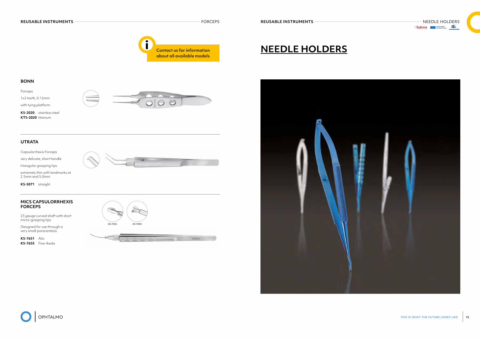
i NEEDLE HOLDERS
®
this is what the future looks like 75
REUSABLE INSTRUMENTS NEEDLE HOLDERSREUSABLE INSTRUMENTS NEEDLE HOLDERS
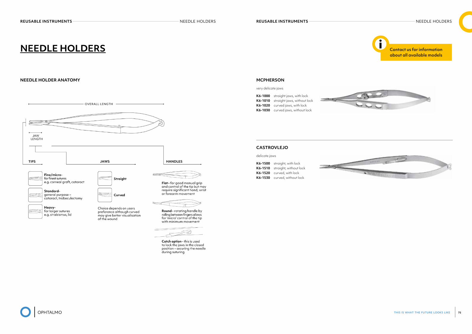
NEEDLE HOLDERS
NEEDLE HOLDER ANATOMY MCPHERSON
very delicate jaws
K6-1000 straight jaws, with lock K6-1010 straight jaws, without lock K6-1020 curved jaws, with lock K6-1030 curved jaws, without lock
CASTROVLEJO
delicate jaws
K6-1500 straight, with lock K6-1510 straight, without lock K6-1520 curved, with lock K6-1530 curved, without lock
Contact us for information about all available models
i
this is what the future looks like 77
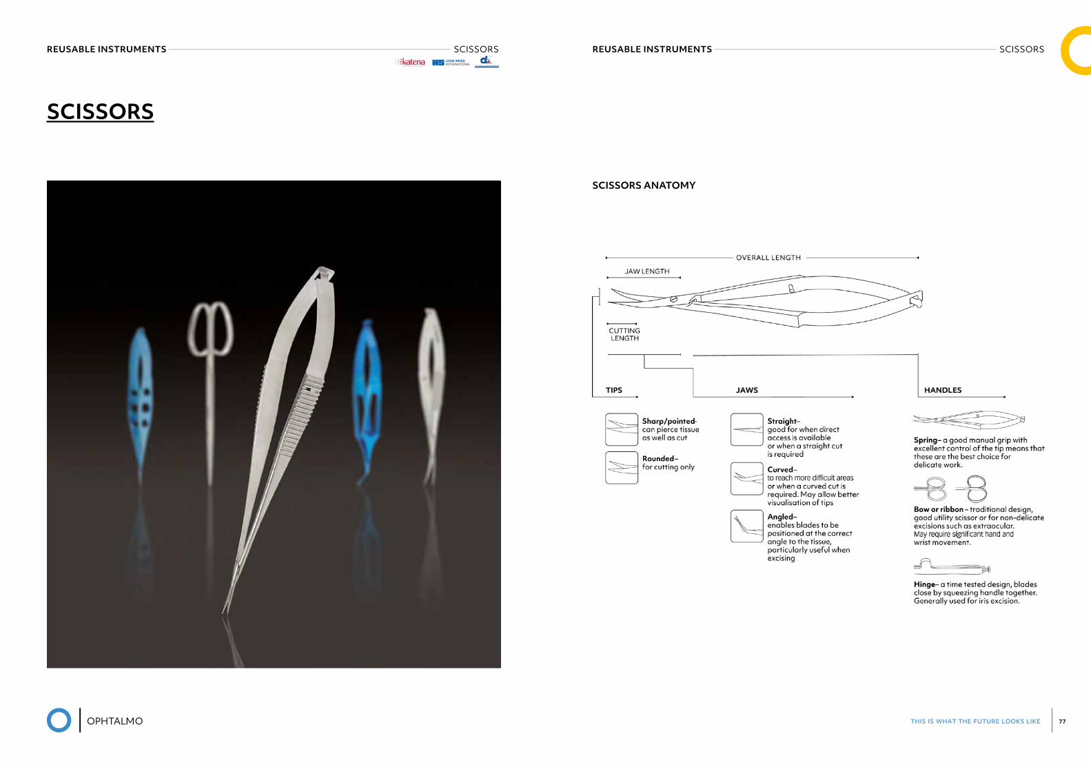
REUSABLE INSTRUMENTS SCISSORSREUSABLE INSTRUMENTS SCISSORS
SCISSORS
SCISSORS ANATOMY
®
this is what the future looks like 79
REUSABLE INSTRUMENTS SPECULUMREUSABLE INSTRUMENTS SCISSORS
WESTCOTT
Tenotomy Scissors
blunt tips, standard blades
curved
K4-3100 right (shown) K4-3110 left
CAPSULOTOMY SCISSORS
VANNAS
5mm long blades, sharp tips
K4-5000 straight K4-5010 curved K4-5020 angled forward K4-5030 angled to side
GILLS-VANNAS
7mm long blades, sharp tips
K4-5050 straight K4-5060 curved K4-5070 angled forward
HOLLAND DALK SCISSORS
4.5mm angled blades
K4-5075 blunted tips
MCPHERSON-VANNAS
8mm long blades, sharp tips
K4-5100 curved
Contact us for information about all available models
i SPECULUM
K4-5000
®
this is what the future looks like 81
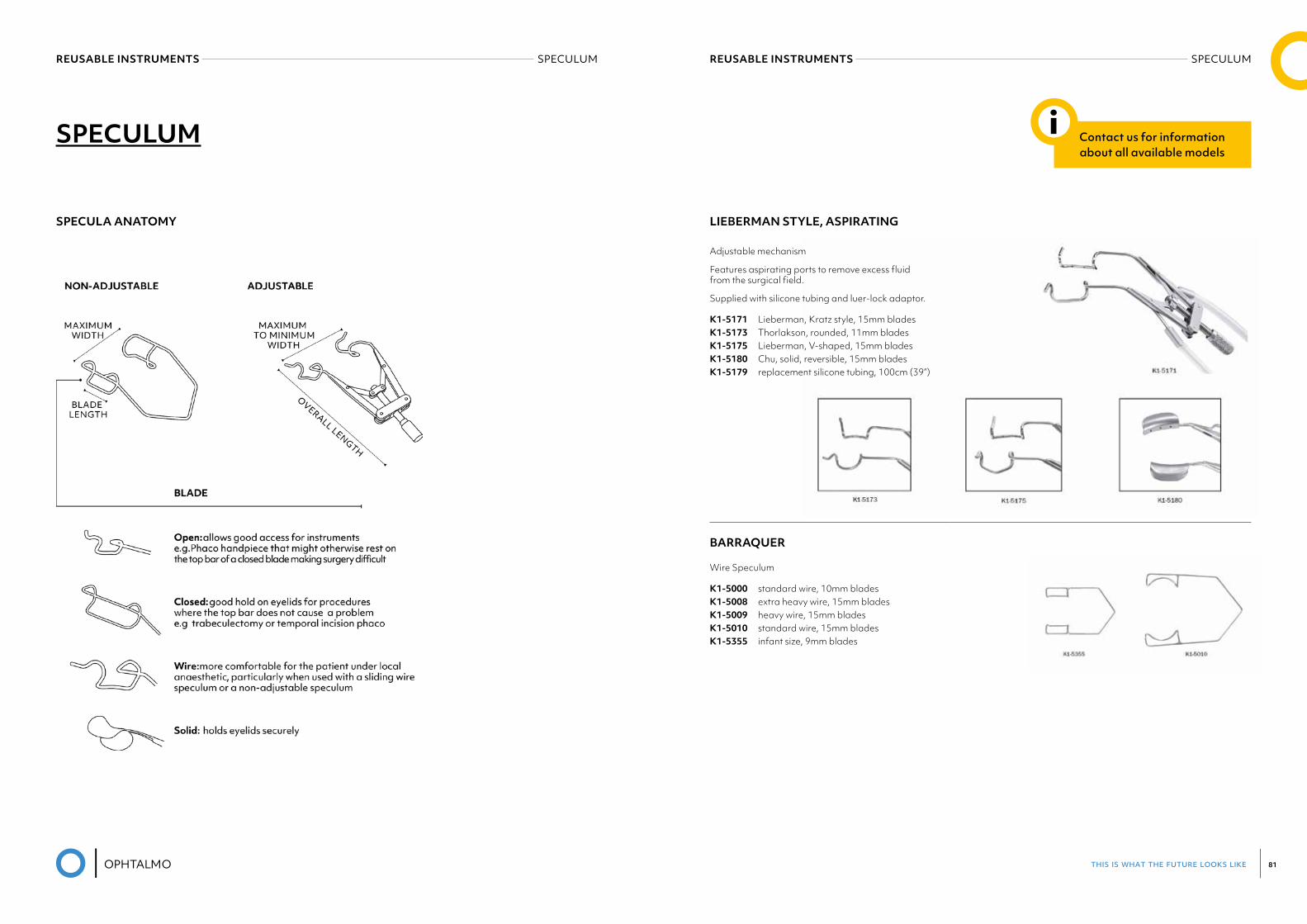
LIEBERMAN STYLE, ASPIRATING
Adjustable mechanism
Features aspirating ports to remove excess fluid from the surgical field.
Supplied with silicone tubing and luer-lock adaptor.
K1-5171 Lieberman, Kratz style, 15mm blades K1-5173 Thorlakson, rounded, 11mm blades K1-5175 Lieberman, V-shaped, 15mm blades K1-5180 Chu, solid, reversible, 15mm blades K1-5179 replacement silicone tubing, 100cm (39”)
BARRAQUER
Wire Speculum
K1-5000 standard wire, 10mm blades K1-5008 extra heavy wire, 15mm blades K1-5009 heavy wire, 15mm blades K1-5010 standard wire, 15mm blades K1-5355 infant size, 9mm blades
REUSABLE INSTRUMENTS SPECULUMREUSABLE INSTRUMENTS SPECULUM
SPECULUM
SPECULA ANATOMY
Contact us for information about all available models
i
this is what the future looks like 83
REUSABLE INSTRUMENTS IOL CUTTERSREUSABLE INSTRUMENTS IOL FORCEPS
IOL FORCEPS
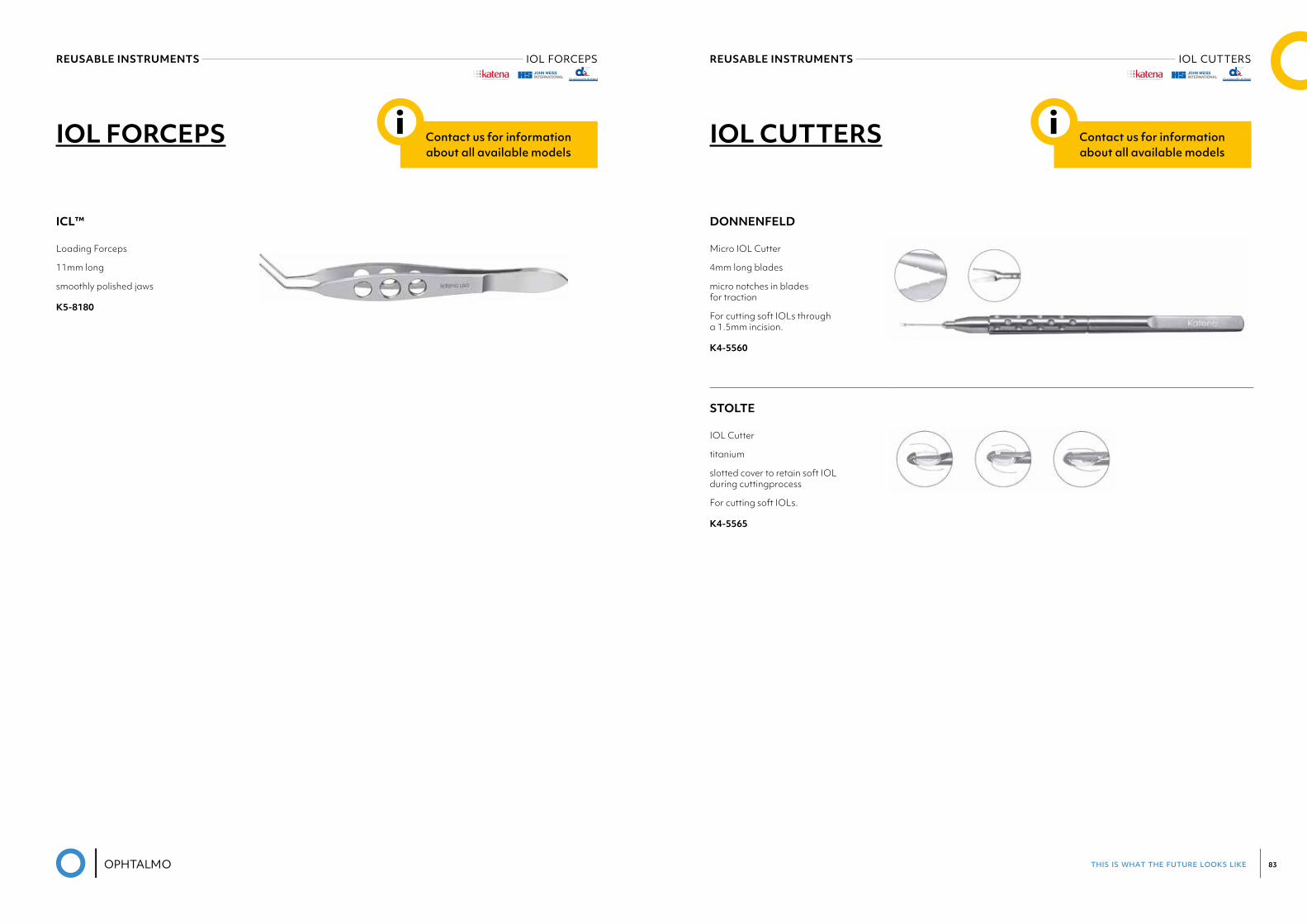
ICL™
Loading Forceps
11mm long
smoothly polished jaws
K5-8180
Contact us for information about all available models
i IOL CUTTERS
DONNENFELD
Micro IOL Cutter
4mm long blades
micro notches in blades for traction
For cutting soft IOLs through a 1.5mm incision.
K4-5560
STOLTE
IOL Cutter
titanium
slotted cover to retain soft IOL during cuttingprocess
For cutting soft IOLs.
K4-5565
Contact us for information about all available models
i
® ®
this is what the future looks like 85
REUSABLE INSTRUMENTS FIXATION, GAUGES & MARKERSREUSABLE INSTRUMENTS DIAMOND KNIVES
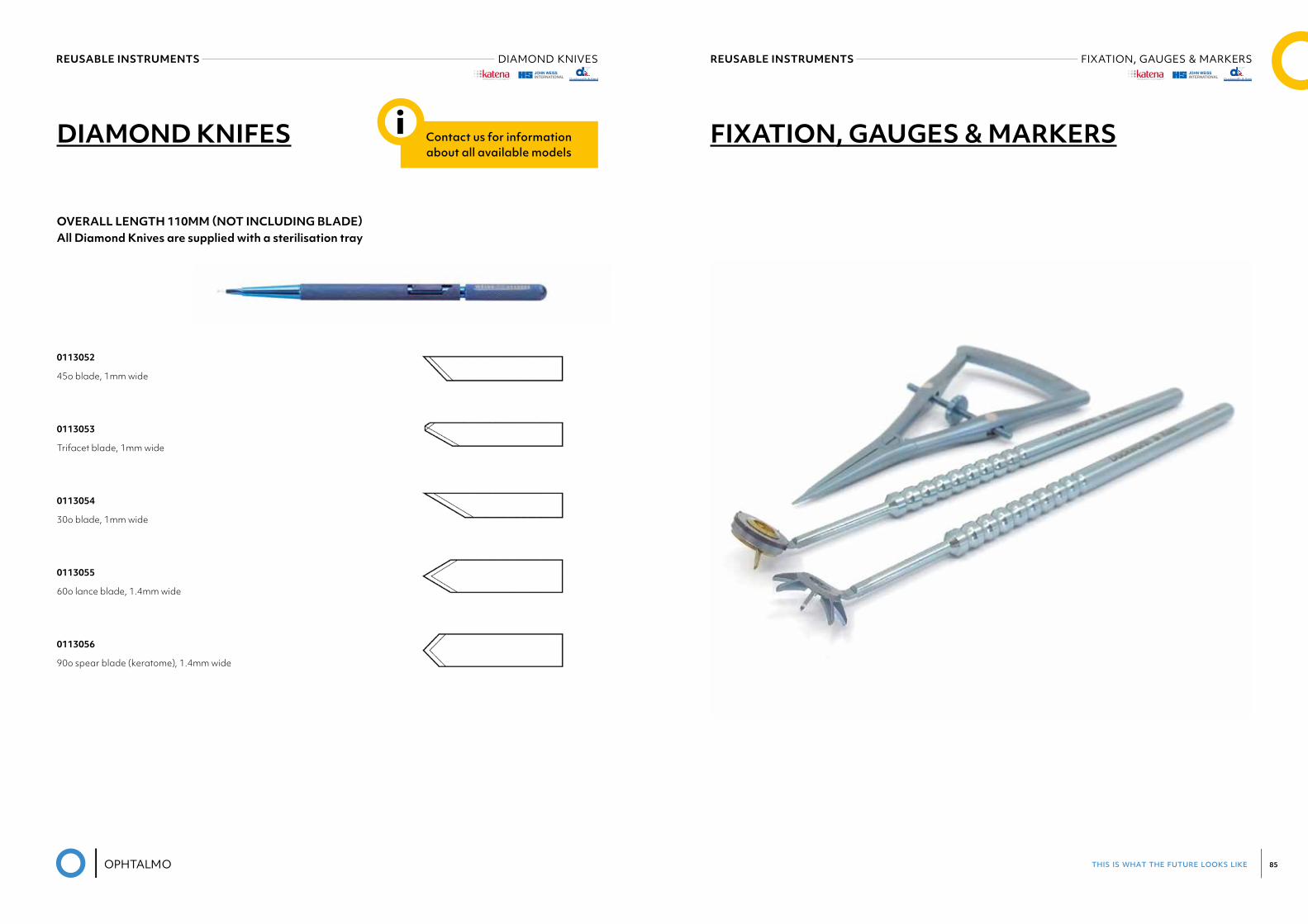
OVERALL LENGTH 110MM (NOT INCLUDING BLADE) All Diamond Knives are supplied with a sterilisation tray
0113052
45o blade, 1mm wide
0113053
Trifacet blade, 1mm wide
0113054
30o blade, 1mm wide
0113055
60o lance blade, 1.4mm wide
0113056
90o spear blade (keratome), 1.4mm wide
DIAMOND KNIFES Contact us for information about all available models
i FIXATION, GAUGES & MARKERS
® ®
this is what the future looks like 87
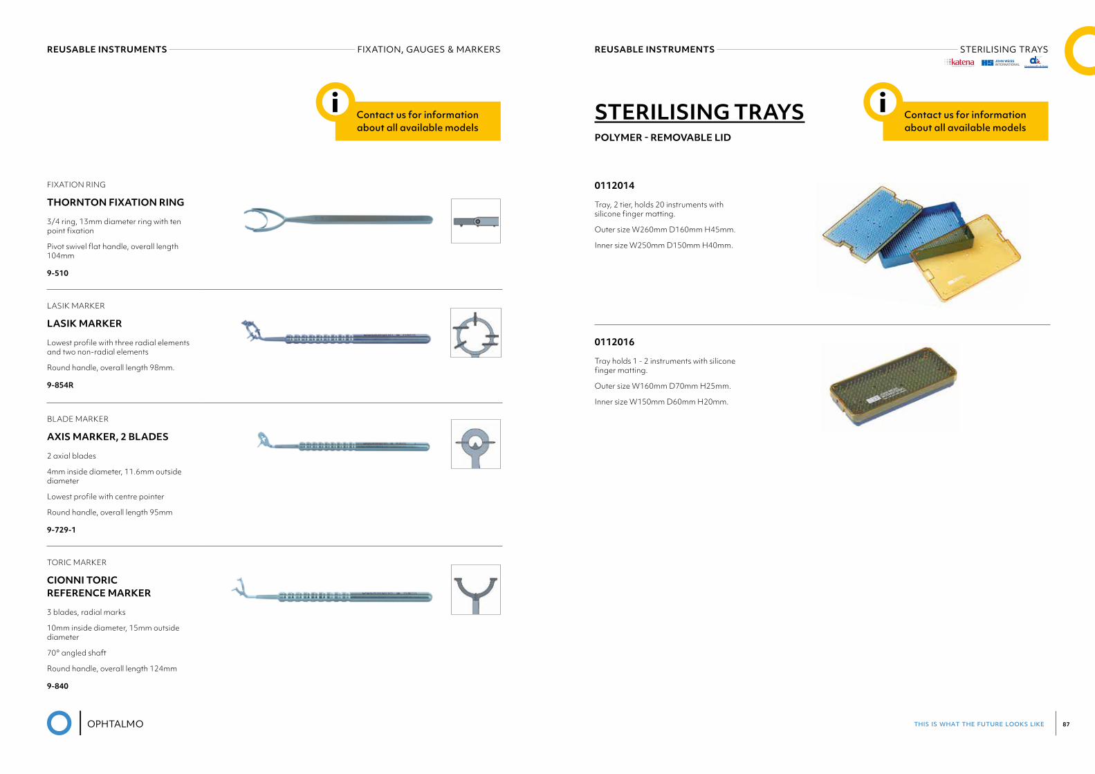
FIXATION RING
THORNTON FIXATION RING
3/4 ring, 13mm diameter ring with ten point fixation
Pivot swivel flat handle, overall length 104mm
9-510
LASIK MARKER
LASIK MARKER
Lowest profile with three radial elements and two non-radial elements
Round handle, overall length 98mm.
9-854R
BLADE MARKER
AXIS MARKER, 2 BLADES
2 axial blades
4mm inside diameter, 11.6mm outside diameter
Lowest profile with centre pointer
Round handle, overall length 95mm
9-729-1
TORIC MARKER
CIONNI TORIC REFERENCE MARKER
3 blades, radial marks
10mm inside diameter, 15mm outside diameter
70° angled shaft
Round handle, overall length 124mm
9-840
REUSABLE INSTRUMENTS STERILISING TRAYSREUSABLE INSTRUMENTS FIXATION, GAUGES & MARKERS
Contact us for information about all available models
iPOLYMER - REMOVABLE LID
0112014
Tray, 2 tier, holds 20 instruments with silicone finger matting.
Outer size W260mm D160mm H45mm.
Inner size W250mm D150mm H40mm.
0112016
Tray holds 1 - 2 instruments with silicone finger matting.
Outer size W160mm D70mm H25mm.
Inner size W150mm D60mm H20mm.
STERILISING TRAYS Contact us for information about all available models
i
®
this is what the future looks like 89
REUSABLE INSTRUMENTS MISCELLANOUSREUSABLE INSTRUMENTS BIPOLAR FORCEPS
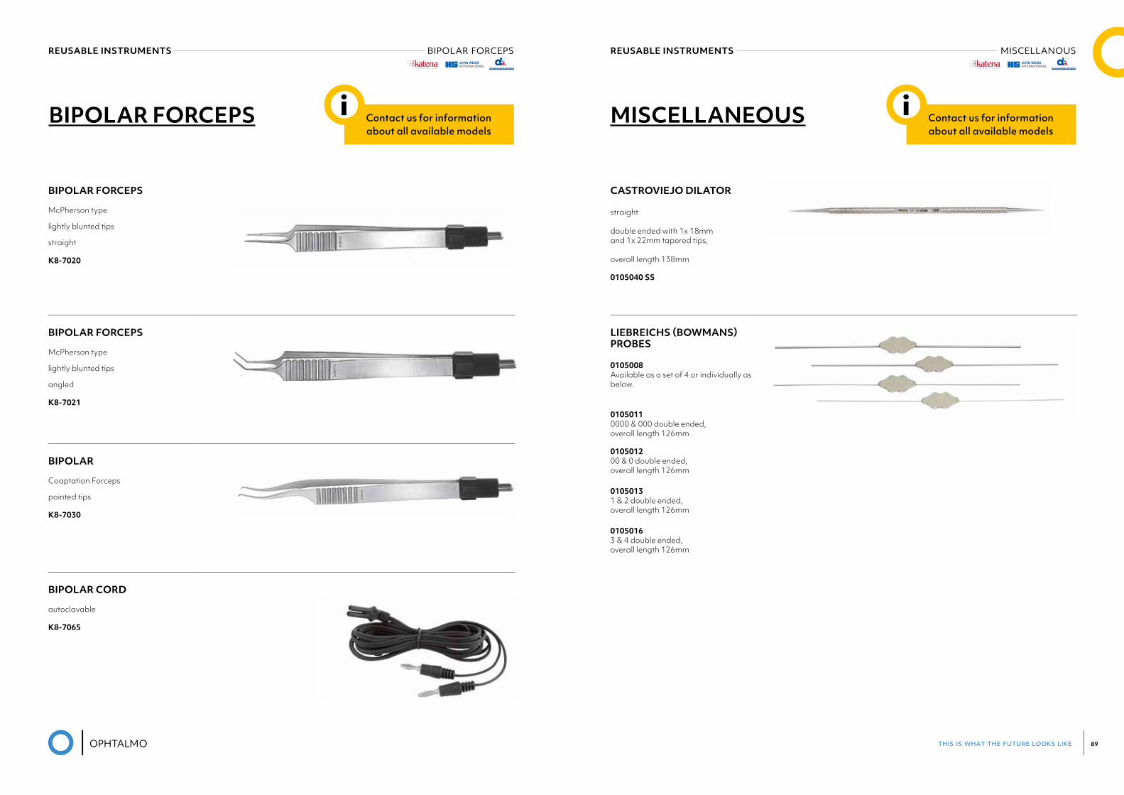
BIPOLAR FORCEPS
McPherson type
lightly blunted tips
straight
K8-7020
BIPOLAR FORCEPS
McPherson type
lightly blunted tips
angled
K8-7021
BIPOLAR
Coaptation Forceps
pointed tips
K8-7030
BIPOLAR CORD
autoclavable
K8-7065
BIPOLAR FORCEPS Contact us for information about all available models
i
CASTROVIEJO DILATOR
straight
double ended with 1x 18mm and 1x 22mm tapered tips,
overall length 138mm
0105040 SS
LIEBREICHS (BOWMANS) PROBES
0105008 Available as a set of 4 or individually as below.
0105011 0000 & 000 double ended, overall length 126mm
010501200 & 0 double ended, overall length 126mm
0105013 1 & 2 double ended, overall length 126mm
0105016 3 & 4 double ended, overall length 126mm
MISCELLANEOUS Contact us for information about all available models
i
®®
this is what the future looks like 91
REUSABLE INSTRUMENTS MISCELLANOUS
Cataract• Iris Retractor and Cataract Support System• Capsule Polisher• Lens Loop and Expressor
Corneal• Lamellar Dissector• Trephine Guide and Epithelial Disruptor• Keratometer and Scleral Support Rings 1
Glaucoma• Punches• Conjunctival Clamps
Lacrimal• Lacrimal Probes and Dilators
Oculoplastics• Curettes• Eye Shields• Retractors• Clamps
Paediatric• Muscle Hooks
Vitreoretinal• Scleral Depressors• Membrane Spatula• Cannula Forceps
MACKOOL CAPSULE POLISHER
0.3mm diameter olive shaped textured tip
45° angled shaft, tip to angle length 11mm
Round handle, overall length 119mm
6-510
KHAW DESCEMET’S MEMBRANE PUNCH, MEDIUM
Designed to punch 0.75mm x 0.5mm
• Round squeeze handle, overall length 117mm
7-102
02SINGLE USE
INSTRUMENTS
this is what the future looks like 93
ITEM CODE SIZE INCISION SIZE
ASPIRATION CANNULA
ASPIRATION PORT
ASPIRATION HEAD
IRRIGATION CANNULA
IRRIGATION PORT
SBS105 21G / 0.8mm
1.1mm curved, aeroplane shape
0.35mm textured open 1 port, 0.5mm
SBS110A 21G / 0.8mm
1.1mm curved, aeroplane shape
0.35mm textured curved, aeroplane shape
2 ports, 0.45mm
SBS122A 22G / 0.7mm
1.0mm aeroplane shape
0.35mm textured curved, aeroplane shape
2 ports, 0.45mm
SBS123A 23G / 0.65mm
0.8mm aeroplane shape
0.30mm textured curved, aeroplane shape
2 ports, 0.35mm
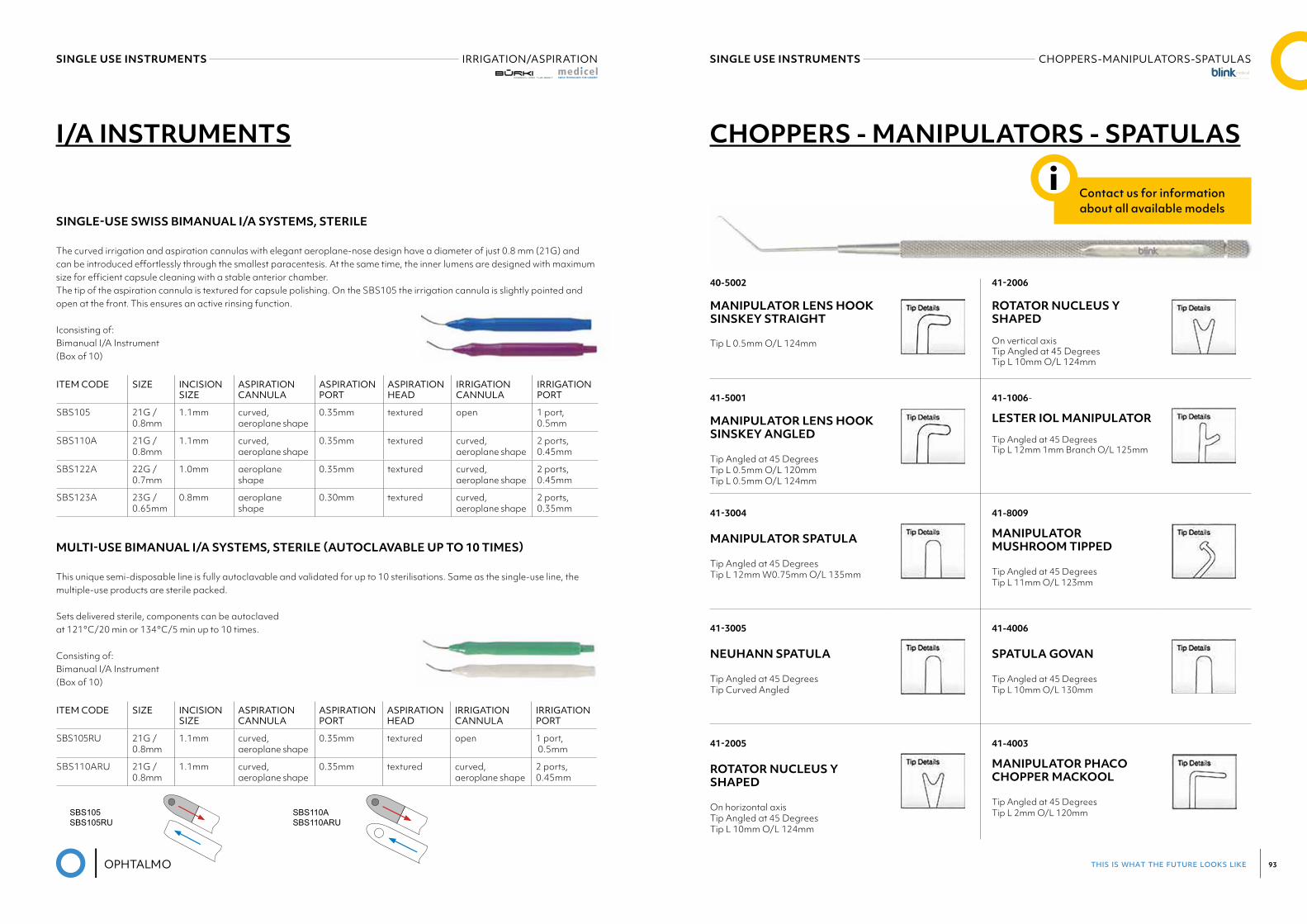
MULTI-USE BIMANUAL I/A SYSTEMS, STERILE (AUTOCLAVABLE UP TO 10 TIMES)
This unique semi-disposable line is fully autoclavable and validated for up to 10 sterilisations. Same as the single-use line, the multiple-use products are sterile packed.
Sets delivered sterile, components can be autoclaved at 121°C/20 min or 134°C/5 min up to 10 times.
Consisting of: Bimanual I/A Instrument (Box of 10)
SINGLE USE INSTRUMENTS CHOPPERS-MANIPULATORS-SPATULASSINGLE USE INSTRUMENTS IRRIGATION/ASPIRATION
SINGLE-USE SWISS BIMANUAL I/A SYSTEMS, STERILE
The curved irrigation and aspiration cannulas with elegant aeroplane-nose design have a diameter of just 0.8 mm (21G) and can be introduced effortlessly through the smallest paracentesis. At the same time, the inner lumens are designed with maximum size for efficient capsule cleaning with a stable anterior chamber. The tip of the aspiration cannula is textured for capsule polishing. On the SBS105 the irrigation cannula is slightly pointed and open at the front. This ensures an active rinsing function.
Iconsisting of: Bimanual I/A Instrument (Box of 10)
SINGLE-USE SWISS BIMANUAL I/A SYSTEMS, STERILE
The curved irrigation and aspiration cannulas with elegant aeroplane-nose design have a diameter of just 0.8 mm (21G) and can be introduced effortlessly through the smallest paracentesis. At the same time, the inner lumens are designed with maximum size for efficient capsule cleaning with a stable anterior chamber.
The tip of the aspiration cannula is textured for capsule polishing. On the SBS105 the irrigation cannula is slightly poin-ted and open at the front. This ensures an active rinsing function.
consisting of: Bimanual I/A Instrument (Box of 10)
ITEM CODE SIZE INCISIONSIZE
ASPIRATIONCANNULA
ASPIRATIONPORT
ASPIRATIONHEAD
IRRIGATIONCANNULA
IRRIGATIONPORT
SBS105 21G / 0.8mm 1.1mm curved,aeroplane shape 0.35mm textured open 1 port,
0.5mm
SBS110A 21G / 0.8mm 1.1mm curved,aeroplane shape 0.35mm textured curved,
aeroplane shape2 ports,0.45mm
SBS122A 22G / 0.7mm 1.0mm aeroplane shape 0.35mm textured curved,aeroplane shape
2 ports, 0.45mm
SBS123A 23G / 0.65mm 0.8mm aeroplane shape 0.30mm textured curved,aeroplane shape
2 ports, 0.35mm
MULTI-USE BIMANUAL I/A SYSTEMS, STERILE (AUTOCLAVABLE UP TO 10 TIMES)
This unique semi-disposable line is fully autoclavable and validated for up to 10 sterilisations. Same as the single-use line, the multiple-use products are sterile packed.
Sets delivered sterile, components can be autoclavedat 121°C/20 min or 134°C/5 min up to 10 times.
consisting of: Bimanual I/A Instrument (Box of 10)
ITEM CODE SIZE INCISIONSIZE
ASPIRATIONCANNULA
ASPIRATIONPORT
ASPIRATIONHEAD
IRRIGATIONCANNULA
IRRIGATIONPORT
SBS105RU 21G / 0.8mm 1.1mm curved,aeroplane shape 0.35mm textured open 1 port,
0.5mm
SBS110ARU 21G / 0.8mm 1.1mm curved,aeroplane shape 0.35mm textured curved,
aeroplane shape2 ports,0.45mm
17
I/A INSTRUMENTS
SBS105SBS105RU
SBS110ASBS110ARU
I/A INSTRUMENTS
ITEM CODE SIZE INCISION SIZE
ASPIRATION CANNULA
ASPIRATION PORT
ASPIRATION HEAD
IRRIGATION CANNULA
IRRIGATION PORT
SBS105RU 21G / 0.8mm
1.1mm curved, aeroplane shape
0.35mm textured open 1 port, 0.5mm
SBS110ARU 21G / 0.8mm
1.1mm curved, aeroplane shape
0.35mm textured curved, aeroplane shape
2 ports, 0.45mm
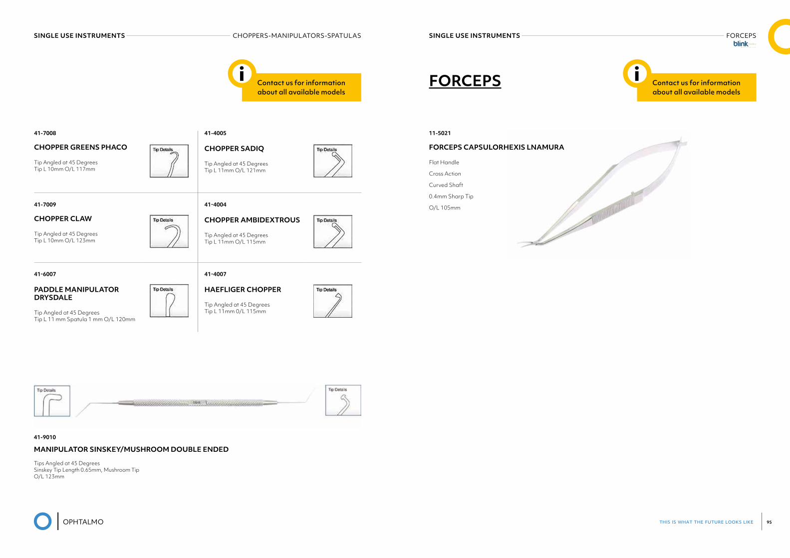
CHOPPERS - MANIPULATORS - SPATULAS
40-5002
MANIPULATOR LENS HOOK SINSKEY STRAIGHT
Tip L 0.5mm O/L 124mm
41-2006 ROTATOR NUCLEUS Y SHAPED
On vertical axis Tip Angled at 45 Degrees Tip L 10mm O/L 124mm
41-5001
MANIPULATOR LENS HOOK SINSKEY ANGLED
Tip Angled at 45 Degrees Tip L 0.5mm O/L 120mm Tip L 0.5mm O/L 124mm
41-1006-
LESTER IOL MANIPULATOR
Tip Angled at 45 Degrees Tip L 12mm 1mm Branch O/L 125mm
41-3004 MANIPULATOR SPATULA Tip Angled at 45 Degrees Tip L 12mm W0.75mm O/L 135mm
41-8009
MANIPULATOR MUSHROOM TIPPED Tip Angled at 45 Degrees Tip L 11mm O/L 123mm
41-3005 NEUHANN SPATULA Tip Angled at 45 Degrees Tip Curved Angled
41-4006
SPATULA GOVAN
Tip Angled at 45 Degrees Tip L 10mm O/L 130mm
41-2005 ROTATOR NUCLEUS Y SHAPED On horizontal axis Tip Angled at 45 Degrees Tip L 10mm O/L 124mm
41-4003
MANIPULATOR PHACO CHOPPER MACKOOL Tip Angled at 45 Degrees Tip L 2mm O/L 120mm
Contact us for information about all available models
i
this is what the future looks like 95
SINGLE USE INSTRUMENTS FORCEPSSINGLE USE INSTRUMENTS CHOPPERS-MANIPULATORS-SPATULAS
41-7008
CHOPPER GREENS PHACO
Tip Angled at 45 Degrees Tip L 10mm O/L 117mm
41-4005
CHOPPER SADIQ
Tip Angled at 45 Degrees Tip L 11mm O/L 121mm
41-7009
CHOPPER CLAW
Tip Angled at 45 Degrees Tip L 10mm O/L 123mm
41-4004 CHOPPER AMBIDEXTROUS Tip Angled at 45 Degrees Tip L 11mm O/L 115mm
41-6007 PADDLE MANIPULATOR DRYSDALE Tip Angled at 45 Degrees Tip L 11 mm Spatula 1 mm O/L 120mm
41-4007 HAEFLIGER CHOPPER Tip Angled at 45 Degrees Tip L 11mm 0/L 115mm
41-9010
MANIPULATOR SINSKEY/MUSHROOM DOUBLE ENDED
Tips Angled at 45 Degrees Sinskey Tip Length 0.65mm, Mushroom Tip O/L 123mm
Contact us for information about all available models
i FORCEPS
11-5021
FORCEPS CAPSULORHEXIS LNAMURA
Flat Handle
Cross Action
Curved Shaft
0.4mm Sharp Tip
O/L 105mm
Contact us for information about all available models
i
this is what the future looks like 97
20-7001
SCISSORS IRIS STRAIGHT
Rounded Tip 0/L 100mmm
20-6003
SCISSORS VANNAS STRAIGHT
0/L 95mm
SINGLE USE INSTRUMENTS SCISSORSSINGLE USE INSTRUMENTS NEEDLE HOLDERS
NEEDLE HOLDERS
MCPHERSONvery delicate jaws
K6-1000 straight jaws, with lockK6-1010 straight jaws, without lockK6-1020 curved jaws, with lockK6-1030 curved jaws, without lock
CASTROVIEJOvery delicate jaws
K6-1500 straight, with lockK6-1510 straight, without lockK6-1520 curved, with lockK6-1530 curved, without lock
Contact us for information about all available models
i SCISSORS Contact us for information about all available models
i
this is what the future looks like 99
31-2003
SPECULUM BARRAQUER
Angled Tempora! L40mm W30mm
30-2004
SPECULUM BARRAQUER STRAIGHT
L40mmW30mm
31-1006
SPECULUM KRATZ-BARRAQUER
Angled Tempora! L40mm W30mm
SINGLE USE INSTRUMENTS SPECULUM
SPECULUM Contact us for information about all available models
i
03ABSORBABLE
SUTURESSMI AG was established in 1987 – the first Belgian company to manufacture surgical sutures – since when it has grown rapidly. Today it is recognized as an experienced and world-wide supplier of surgical sutures.
The company is situated in St. Vith in the east of Belgium, near the point where the borders of Belgium, Germany and Luxembourg meet.
SMI complies with international quality standards such as EN ISO 13485 as well as Good Manufacturing Practices (GMP). Our products are manufactured with newest techniques and all compounds are of highest technical level. All raw materials and finished products are submitted to stringent quality control by a highly
qualified team.
SMI is dedicated to customer satisfaction by ensuring high quality products and an excellent service based on customer focus, reliability and delivery performance.
this is what the future looks like 101
ABSORBABLE SUTURES SURGICRYLSMI
ABSORBABLE SUTURES SURGICRYLSMI
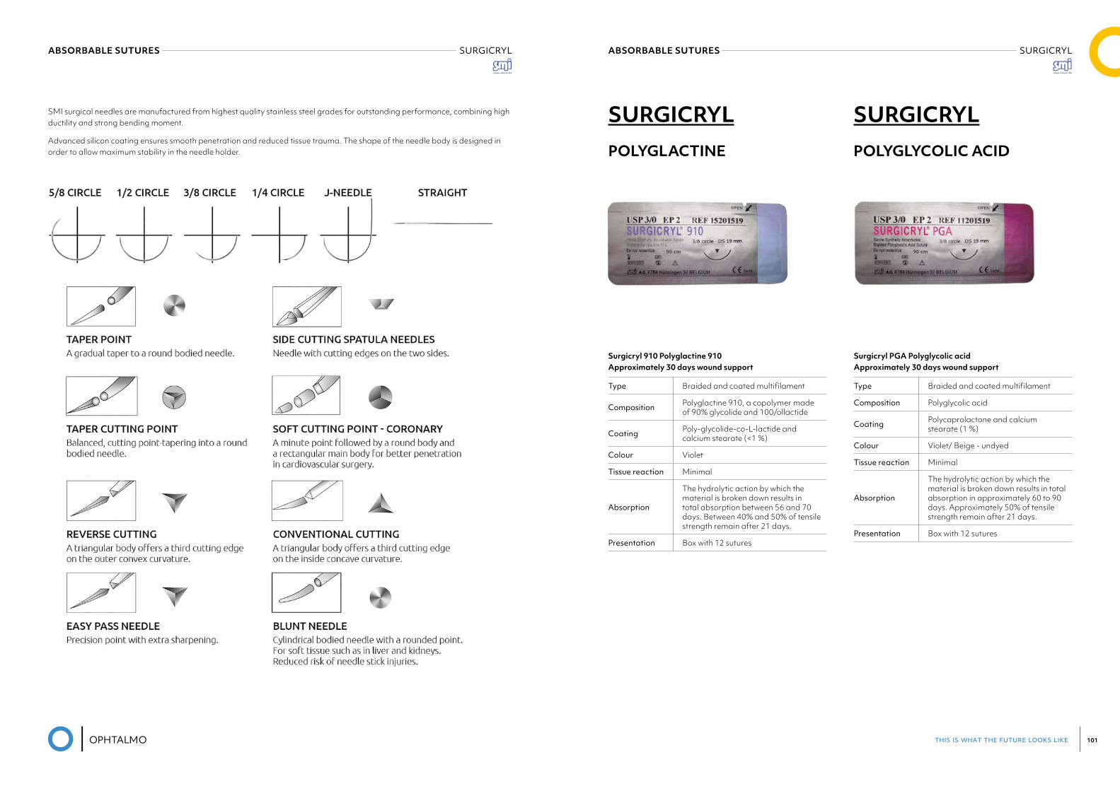
SURGICRYLPOLYGLACTINE
Surgicryl 910 Polyglactine 910 Approximately 30 days wound support
Type Braided and coated multifilament
Composition Polyglactine 910, a copolymer made of 90% glycolide and 100/ol lactide
Coating Poly-glycolide-co-L-lactide and calcium stearate (<1 %)
Colour Violet
Tissue reaction Minimal
Absorption
The hydrolytic action by which the material is broken down results in total absorption between 56 and 70 days. Between 40% and 50% of tensile strength remain after 21 days.
Presentation Box with 12 sutures
Surgicryl PGA Polyglycolic acid Approximately 30 days wound support
Type Braided and coated multifilament
Composition Polyglycolic acid
Coating Polycaprolactone and calcium stearate (1 %)
Colour Violet/ Beige - undyed
Tissue reaction Minimal
Absorption
The hydrolytic action by which the material is broken down results in total absorption in approximately 60 to 90 days. Approximately 50% of tensile strength remain after 21 days.
Presentation Box with 12 sutures
SURGICRYLPOLYGLYCOLIC ACID
SMI surgical needles are manufactured from highest quality stainless steel grades for outstanding performance, combining high ductility and strong bending moment.
Advanced silicon coating ensures smooth penetration and reduced tissue trauma. The shape of the needle body is designed in order to allow maximum stability in the needle holder.
this is what the future looks like 103
04NON-ABSORBABLE
SUTURES
this is what the future looks like 105
NON-ABSORBABLE SUTURES SMI
SILK
POLYPROPYLENE
Type Braided multifilament
Composition Braided fibres from the cocoon of the silkworm
Coating Silicone
Colour Black
Tissue reaction Moderate
Absorption
Silk suture elicits an initial inflammatory reaction in tissues, which is followed by gradual encapsulation of the suture by fibrous connective tissues.
Presentation Box with 12 sutures
Type Monofilament
Composition Polypropylene, a polymer of propylene
Coating None
Colour Blue
Tissue reaction Minimal
Absorption Non absorbable
Presentation Box with 12 sutures
Type Monofilament
Composition Extrusion of polyamide 6.0 or 6.6
Coating None
Colour Blue Black
Tissue reaction Minimal
Absorption
Non absorbable, gradually encapsulated by connective tissue. The thread mass diminishes, approximately 10% a year by rupture of chemica! links (hydrolytic action).
Presentation Box with 12 sutures
Type Braided multifilament
Composition Polyester - a polymer of polyethylene terephthalate
Coating Silicone
Colour Green White
Tissue reaction Minimal
Absorption Non absorbable, gradually encapsulated by connective tissue
Presentation Box with 12 sutures
NYLON
POLYESTER
05DISPOSABLE
KNIVES & BLADES
this is what the future looks like 107
DISPOSABLE KNIVES & BLADES CRESCENT KNIVES - BLEB KNIVESDISPOSABLE KNIVES & BLADES STAB KNIVES
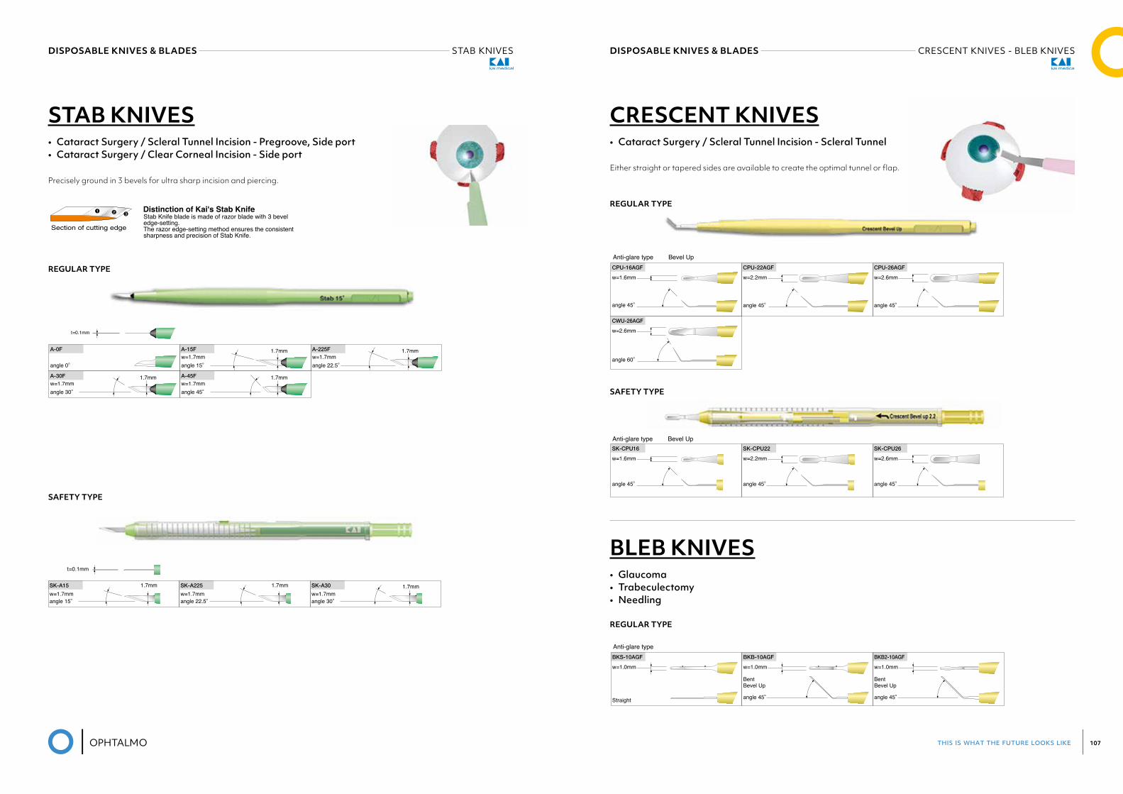
STAB KNIVES• Cataract Surgery / Scleral Tunnel Incision - Pregroove, Side port• Cataract Surgery / Clear Corneal Incision - Side port
Precisely ground in 3 bevels for ultra sharp incision and piercing.
SAFETY TYPE
REGULAR TYPE
t=0.1mm
SK-A15
w=1.7mmangle 15°
SK-A225
w=1.7mmangle 22.5°
SK-A30
w=1.7mm1.7mm 1.7mm 1.7mm
angle 30°
Safety Type
STAB KNIVESCataract Surgery / Scleral Tunnel Incision - Pregroove, Side port / Clear Corneal Incision - Side port
Precisely ground in 3 bevels for ultra sharp incision and piercing.
Regular Type
Stab Knife blade is made of razor blade with 3 bevel edge-setting. The razor edge-setting method ensures the consistent sharpness and precision of Stab Knife.
Distinction of Kai's Stab Knife
t=0.1mm
A-0F
angle 0°
A-15F 1.7mm
angle 15°
A-45F 1.7mm
angle 45°
A-30F 1.7mm
angle 30°
A-225F 1.7mmw=1.7mm
w=1.7mmw=1.7mm
w=1.7mmangle 22.5°
3
t=0.1mm
SK-A15
w=1.7mmangle 15°
SK-A225
w=1.7mmangle 22.5°
SK-A30
w=1.7mm1.7mm 1.7mm 1.7mm
angle 30°
Safety Type
STAB KNIVESCataract Surgery / Scleral Tunnel Incision - Pregroove, Side port / Clear Corneal Incision - Side port
Precisely ground in 3 bevels for ultra sharp incision and piercing.
Regular Type
Stab Knife blade is made of razor blade with 3 bevel edge-setting. The razor edge-setting method ensures the consistent sharpness and precision of Stab Knife.
Distinction of Kai's Stab Knife
t=0.1mm
A-0F
angle 0°
A-15F 1.7mm
angle 15°
A-45F 1.7mm
angle 45°
A-30F 1.7mm
angle 30°
A-225F 1.7mmw=1.7mm
w=1.7mmw=1.7mm
w=1.7mmangle 22.5°
3
Regular Type
Safety Type
SK-CPU16
w=1.6mm
SK-CPU22
w=2.2mm
SK-CPU26
w=2.6mm
angle 45° angle 45° angle 45°
CRESCENT KNIVESCataract Surgery / Scleral Tunnel Incision - Scleral Tunnel
BLEB KNIVESGlaucoma / Trabeculectomy / Needling
Regular Type
Either straight or tapered sides are available to create the optimal tunnel or flap.
CPU-16AGF
w=1.6mm
angle 45°
CPU-22AGF
w=2.2mm
angle 45°
CPU-26AGF
w=2.6mm
angle 45°
CWU-26AGF
w=2.6mm
angle 60°
BKB2-10AGF
w=1.0mm
angle 45°
BentBevel Up
BKS-10AGF
w=1.0mm
Straight
BKB-10AGF
w=1.0mm
angle 45°
BentBevel Up
Anti-glare type Bevel Up
Anti-glare type
Anti-glare type Bevel Up
4
t=0.1mm
SK-A15
w=1.7mmangle 15°
SK-A225
w=1.7mmangle 22.5°
SK-A30
w=1.7mm1.7mm 1.7mm 1.7mm
angle 30°
Safety Type
STAB KNIVESCataract Surgery / Scleral Tunnel Incision - Pregroove, Side port / Clear Corneal Incision - Side port
Precisely ground in 3 bevels for ultra sharp incision and piercing.
Regular Type
Stab Knife blade is made of razor blade with 3 bevel edge-setting. The razor edge-setting method ensures the consistent sharpness and precision of Stab Knife.
Distinction of Kai's Stab Knife
t=0.1mm
A-0F
angle 0°
A-15F 1.7mm
angle 15°
A-45F 1.7mm
angle 45°
A-30F 1.7mm
angle 30°
A-225F 1.7mmw=1.7mm
w=1.7mmw=1.7mm
w=1.7mmangle 22.5°
3
Regular Type
Safety Type
SK-CPU16
w=1.6mm
SK-CPU22
w=2.2mm
SK-CPU26
w=2.6mm
angle 45° angle 45° angle 45°
CRESCENT KNIVESCataract Surgery / Scleral Tunnel Incision - Scleral Tunnel
BLEB KNIVESGlaucoma / Trabeculectomy / Needling
Regular Type
Either straight or tapered sides are available to create the optimal tunnel or flap.
CPU-16AGF
w=1.6mm
angle 45°
CPU-22AGF
w=2.2mm
angle 45°
CPU-26AGF
w=2.6mm
angle 45°
CWU-26AGF
w=2.6mm
angle 60°
BKB2-10AGF
w=1.0mm
angle 45°
BentBevel Up
BKS-10AGF
w=1.0mm
Straight
BKB-10AGF
w=1.0mm
angle 45°
BentBevel Up
Anti-glare type Bevel Up
Anti-glare type
Anti-glare type Bevel Up
4
CRESCENT KNIVES• Cataract Surgery / Scleral Tunnel Incision - Scleral Tunnel
Either straight or tapered sides are available to create the optimal tunnel or flap.
REGULAR TYPE
BLEB KNIVES• Glaucoma• Trabeculectomy• Needling
REGULAR TYPE
SAFETY TYPE
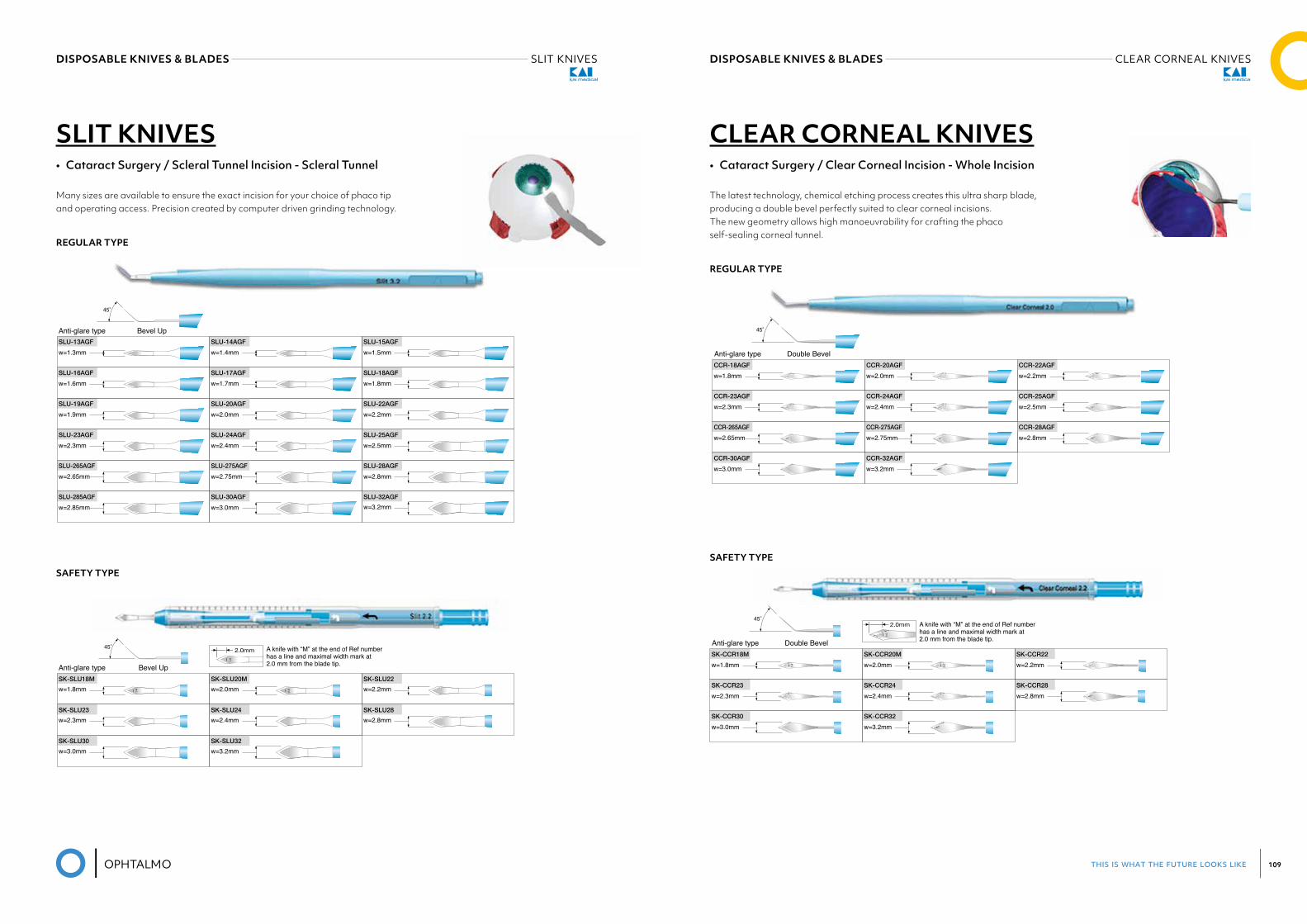
this is what the future looks like 109
DISPOSABLE KNIVES & BLADES CLEAR CORNEAL KNIVESDISPOSABLE KNIVES & BLADES SLIT KNIVES
SLIT KNIVES• Cataract Surgery / Scleral Tunnel Incision - Scleral Tunnel
Many sizes are available to ensure the exact incision for your choice of phaco tip and operating access. Precision created by computer driven grinding technology.
REGULAR TYPE
SAFETY TYPE
1.8
2.0mm45°
SK-SLU22
w=2.2mm
SK-SLU23
w=2.3mmSK-SLU24
w=2.4mmSK-SLU28
w=2.8mm
SK-SLU30
w=3.0mmSK-SLU32
w=3.2mm
SK-SLU20M
w=2.0mm
2.0
SK-SLU18M
w=1.8mm
1.8
SLIT KNIVESCataract Surgery / Scleral Tunnel Incision - Anterior Chamber Entry
Many sizes are available to ensure the exact incision for your choice of phaco tip and operating access. Precision created by computer driven grinding technology.
45°
SLU-23AGF
w=2.3mm
SLU-13AGF
w=1.3mmSLU-15AGF
w=1.5mm
SLU-17AGF
w=1.7mm
SLU-19AGF
w=1.9mmSLU-22AGF
w=2.2mm
SLU-14AGF
w=1.4mm
SLU-16AGF
w=1.6mmSLU-18AGF
w=1.8mm
SLU-20AGF
w=2.0mm
SLU-24AGF
w=2.4mm
SLU-265AGF
w=2.65mm
SLU-25AGF
w=2.5mm
SLU-275AGF
w=2.75mm
SLU-285AGF
w=2.85mm
SLU-28AGF
w=2.8mm
SLU-30AGF
w=3.0mm
Safety Type
Regular Type
5
Anti-glare type Bevel Up
Anti-glare type Bevel Up
SLU-32AGF
w=3.2mm
A knife with “M” at the end of Ref number has a line and maximal width mark at 2.0 mm from the blade tip.
1.8
2.0mm45°
SK-SLU22
w=2.2mm
SK-SLU23
w=2.3mmSK-SLU24
w=2.4mmSK-SLU28
w=2.8mm
SK-SLU30
w=3.0mmSK-SLU32
w=3.2mm
SK-SLU20M
w=2.0mm
2.0
SK-SLU18M
w=1.8mm
1.8
SLIT KNIVESCataract Surgery / Scleral Tunnel Incision - Anterior Chamber Entry
Many sizes are available to ensure the exact incision for your choice of phaco tip and operating access. Precision created by computer driven grinding technology.
45°
SLU-23AGF
w=2.3mm
SLU-13AGF
w=1.3mmSLU-15AGF
w=1.5mm
SLU-17AGF
w=1.7mm
SLU-19AGF
w=1.9mmSLU-22AGF
w=2.2mm
SLU-14AGF
w=1.4mm
SLU-16AGF
w=1.6mmSLU-18AGF
w=1.8mm
SLU-20AGF
w=2.0mm
SLU-24AGF
w=2.4mm
SLU-265AGF
w=2.65mm
SLU-25AGF
w=2.5mm
SLU-275AGF
w=2.75mm
SLU-285AGF
w=2.85mm
SLU-28AGF
w=2.8mm
SLU-30AGF
w=3.0mm
Safety Type
Regular Type
5
Anti-glare type Bevel Up
Anti-glare type Bevel Up
SLU-32AGF
w=3.2mm
A knife with “M” at the end of Ref number has a line and maximal width mark at 2.0 mm from the blade tip.
CLEAR CORNEAL KNIVES• Cataract Surgery / Clear Corneal Incision - Whole Incision
The latest technology, chemical etching process creates this ultra sharp blade, producing a double bevel perfectly suited to clear corneal incisions. The new geometry allows high manoeuvrability for crafting the phaco self-sealing corneal tunnel.
REGULAR TYPE
SAFETY TYPE
1.8
2.0mm45°
SK-SLU22
w=2.2mm
SK-SLU23
w=2.3mmSK-SLU24
w=2.4mmSK-SLU28
w=2.8mm
SK-SLU30
w=3.0mmSK-SLU32
w=3.2mm
SK-SLU20M
w=2.0mm
2.0
SK-SLU18M
w=1.8mm
1.8
SLIT KNIVESCataract Surgery / Scleral Tunnel Incision - Anterior Chamber Entry
Many sizes are available to ensure the exact incision for your choice of phaco tip and operating access. Precision created by computer driven grinding technology.
45°
SLU-23AGF
w=2.3mm
SLU-13AGF
w=1.3mmSLU-15AGF
w=1.5mm
SLU-17AGF
w=1.7mm
SLU-19AGF
w=1.9mmSLU-22AGF
w=2.2mm
SLU-14AGF
w=1.4mm
SLU-16AGF
w=1.6mmSLU-18AGF
w=1.8mm
SLU-20AGF
w=2.0mm
SLU-24AGF
w=2.4mm
SLU-265AGF
w=2.65mm
SLU-25AGF
w=2.5mm
SLU-275AGF
w=2.75mm
SLU-285AGF
w=2.85mm
SLU-28AGF
w=2.8mm
SLU-30AGF
w=3.0mm
Safety Type
Regular Type
5
Anti-glare type Bevel Up
Anti-glare type Bevel Up
SLU-32AGF
w=3.2mm
A knife with “M” at the end of Ref number has a line and maximal width mark at 2.0 mm from the blade tip.
Regular Type
Anti-glare type
Anti-glare type
2.0
2.0mm A knife with “M” at the end of Ref number has a line and maximal width mark at 2.0 mm from the blade tip.
45°
SK-CCR32
w=3.2mm
SK-CCR22
w=2.2mm
SK-CCR23
w=2.3mmSK-CCR24
w=2.4mmSK-CCR28
w=2.8mm
SK-CCR30
w=3.0mm
SK-CCR18M
w=1.8mm 1.8
SK-CCR20M
w=2.0mm 2.0
45°
CLEAR CORNEAL KNIVESCataract Surgery / Clear Corneal Incision - Whole Incision
The latest technology, chemical etching process creates this ultra sharp blade, producing a double bevel perfectly suited to clear corneal incisions. The new geometry allows high manoeuvrability for crafting the phaco self-sealing corneal tunnel.
CCR-18AGF
w=1.8mmCCR-20AGF
w=2.0mmCCR-22AGF
w=2.2mm
CCR-23AGF
w=2.3mmCCR-24AGF
w=2.4mmCCR-25AGF
w=2.5mm
CCR-265AGF
w=2.65mm
w=3.0mm
CCR-275AGF
w=2.75mmCCR-28AGF
w=2.8mm
CCR-30AGF CCR-32AGF
w=3.2mm
6
Safety Type
Double Bevel
Double Bevel
1.8
2.0mm45°
SK-SLU22
w=2.2mm
SK-SLU23
w=2.3mmSK-SLU24
w=2.4mmSK-SLU28
w=2.8mm
SK-SLU30
w=3.0mmSK-SLU32
w=3.2mm
SK-SLU20M
w=2.0mm
2.0
SK-SLU18M
w=1.8mm
1.8
SLIT KNIVESCataract Surgery / Scleral Tunnel Incision - Anterior Chamber Entry
Many sizes are available to ensure the exact incision for your choice of phaco tip and operating access. Precision created by computer driven grinding technology.
45°
SLU-23AGF
w=2.3mm
SLU-13AGF
w=1.3mmSLU-15AGF
w=1.5mm
SLU-17AGF
w=1.7mm
SLU-19AGF
w=1.9mmSLU-22AGF
w=2.2mm
SLU-14AGF
w=1.4mm
SLU-16AGF
w=1.6mmSLU-18AGF
w=1.8mm
SLU-20AGF
w=2.0mm
SLU-24AGF
w=2.4mm
SLU-265AGF
w=2.65mm
SLU-25AGF
w=2.5mm
SLU-275AGF
w=2.75mm
SLU-285AGF
w=2.85mm
SLU-28AGF
w=2.8mm
SLU-30AGF
w=3.0mm
Safety Type
Regular Type
5
Anti-glare type Bevel Up
Anti-glare type Bevel Up
SLU-32AGF
w=3.2mm
A knife with “M” at the end of Ref number has a line and maximal width mark at 2.0 mm from the blade tip.
Regular Type
Anti-glare type
Anti-glare type
2.0
2.0mm A knife with “M” at the end of Ref number has a line and maximal width mark at 2.0 mm from the blade tip.
45°
SK-CCR32
w=3.2mm
SK-CCR22
w=2.2mm
SK-CCR23
w=2.3mmSK-CCR24
w=2.4mmSK-CCR28
w=2.8mm
SK-CCR30
w=3.0mm
SK-CCR18M
w=1.8mm 1.8
SK-CCR20M
w=2.0mm 2.0
45°
CLEAR CORNEAL KNIVESCataract Surgery / Clear Corneal Incision - Whole Incision
The latest technology, chemical etching process creates this ultra sharp blade, producing a double bevel perfectly suited to clear corneal incisions. The new geometry allows high manoeuvrability for crafting the phaco self-sealing corneal tunnel.
CCR-18AGF
w=1.8mmCCR-20AGF
w=2.0mmCCR-22AGF
w=2.2mm
CCR-23AGF
w=2.3mmCCR-24AGF
w=2.4mmCCR-25AGF
w=2.5mm
CCR-265AGF
w=2.65mm
w=3.0mm
CCR-275AGF
w=2.75mmCCR-28AGF
w=2.8mm
CCR-30AGF CCR-32AGF
w=3.2mm
6
Safety Type
Double Bevel
Double Bevel
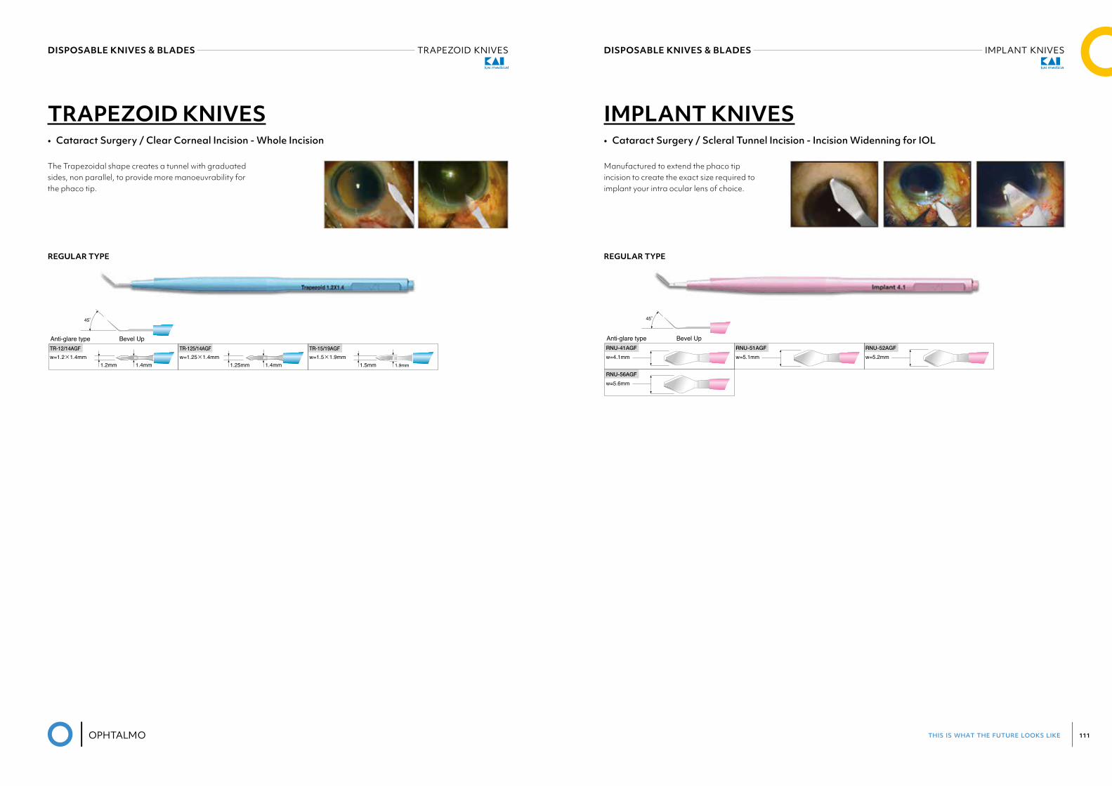
this is what the future looks like 111
DISPOSABLE KNIVES & BLADES IMPLANT KNIVESDISPOSABLE KNIVES & BLADES TRAPEZOID KNIVES
TRAPEZOID KNIVES• Cataract Surgery / Clear Corneal Incision - Whole Incision
The Trapezoidal shape creates a tunnel with graduated sides, non parallel, to provide more manoeuvrability for the phaco tip.
REGULAR TYPE
Bevel Up
TRAPEZOID KNIVESCataract Surgery / Clear Corneal Incision - Whole Incision
The Trapezoidal shape creates a tunnel with graduated sides, non parallel, to provide more manoeuvrability for the phaco tip.
45°
TR-15/19AGF
w=1.5×1.9mm1.9mm1.5mm
TR-125/14AGF
w=1.25×1.4mm1.4mm1.25mm
TR-12/14AGF
w=1.2×1.4mm1.4mm1.2mm
7
Regular Type
Anti-glare type
IMPLANT KNIVES• Cataract Surgery / Scleral Tunnel Incision - Incision Widenning for IOL
Manufactured to extend the phaco tip incision to create the exact size required to implant your intra ocular lens of choice.
REGULAR TYPE
Bevel Up
TRAPEZOID KNIVESCataract Surgery / Clear Corneal Incision - Whole Incision
The Trapezoidal shape creates a tunnel with graduated sides, non parallel, to provide more manoeuvrability for the phaco tip.
45°
TR-15/19AGF
w=1.5×1.9mm1.9mm1.5mm
TR-125/14AGF
w=1.25×1.4mm1.4mm1.25mm
TR-12/14AGF
w=1.2×1.4mm1.4mm1.2mm
7
Regular Type
Anti-glare type
RNU-41AGF
w=4.1mmRNU-52AGF
w=5.2mm
RNU-56AGF
w=5.6mm
45°
RNU-51AGF
w=5.1mm
Regular Type
Anti-glare type Bevel Up
IMPLANT KNIVESCataract Surgery / Scleral Tunnel Incision - Incision Widenning for IOL Manufactured to extend the phaco tip incision to create the exact size required to implant your intra ocular lens of choice.
8
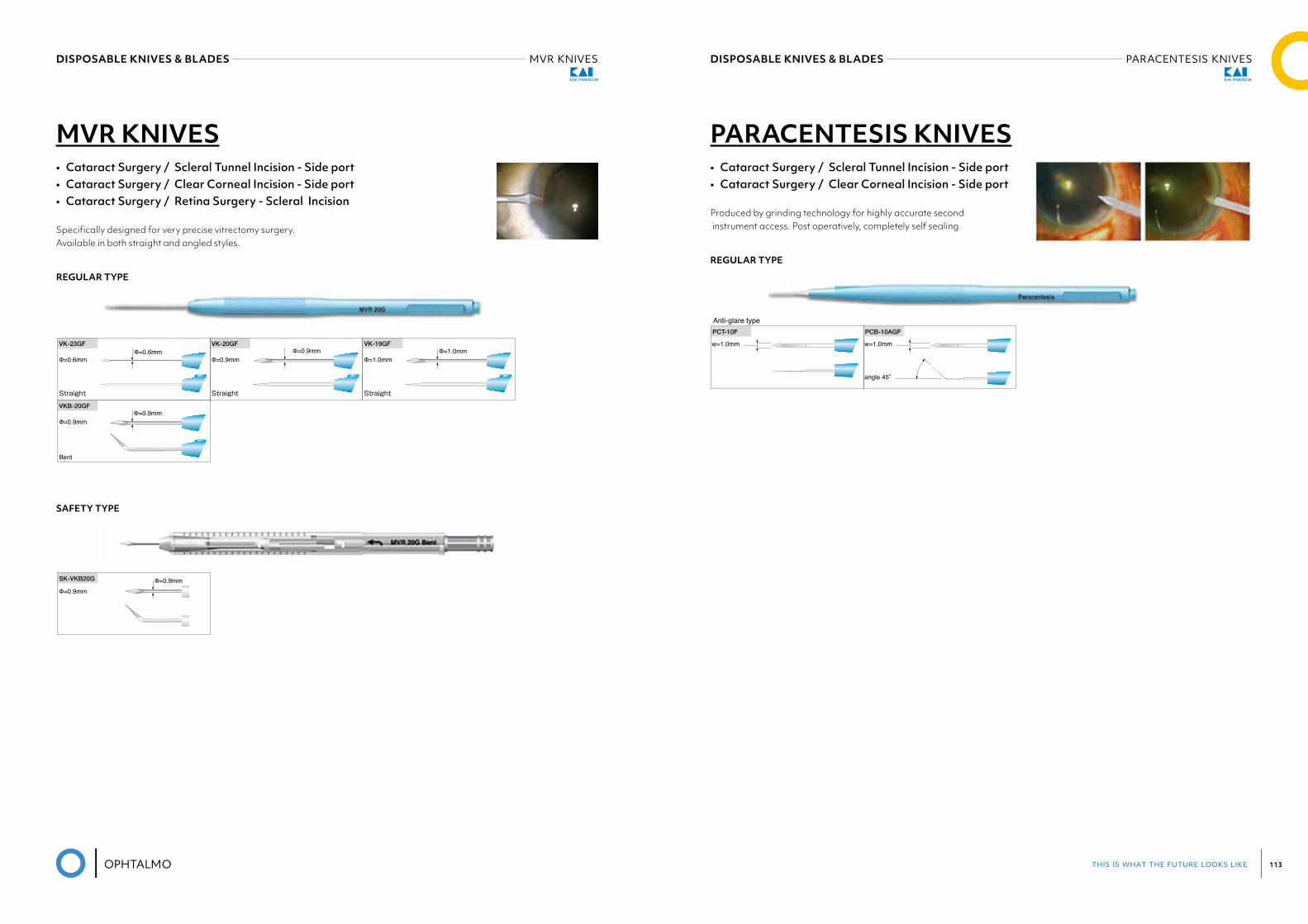
this is what the future looks like 113
DISPOSABLE KNIVES & BLADES PARACENTESIS KNIVESDISPOSABLE KNIVES & BLADES MVR KNIVES
MVR KNIVES• Cataract Surgery / Scleral Tunnel Incision - Side port• Cataract Surgery / Clear Corneal Incision - Side port• Cataract Surgery / Retina Surgery - Scleral Incision
Specifically designed for very precise vitrectomy surgery. Available in both straight and angled styles.
REGULAR TYPE
SAFETY TYPE
PARACENTESIS KNIVESCataract Surgery / Scleral Tunnel Incision - Side port / Clear Corneal Incision - Side port
Produced by grinding technology for highly accurate second instrument access. Post operatively, completely self sealing.
PCT-10F
w=1.0mm
PCB-10AGF
w=1.0mm
angle 45°
Φ=0.9mm
SK-VKB20G Φ=0.9mm
Φ=0.9mm Φ=0.9mm
VK-20GF
Φ=1.0mmΦ=1.0mm
VK-19GF
Φ=0.6mmΦ=0.6mm
VK-23GF
Straight StraightStraight
Φ=0.9mmΦ=0.9mm
VKB-20GF
Bent
Regular Type
Regular Type
Safety Type
Anti-glare type
MVR KNIVESCataract Surgery / Scleral Tunnel Incision - Side port / Clear Corneal Incision - Side port / Retina Surgery - Scleral IncisionSpecifically designed for very precise vitrectomy surgery.Available in both straight and angled styles.
9
PARACENTESIS KNIVES• Cataract Surgery / Scleral Tunnel Incision - Side port• Cataract Surgery / Clear Corneal Incision - Side port
Produced by grinding technology for highly accurate second instrument access. Post operatively, completely self sealing.
REGULAR TYPE
PARACENTESIS KNIVESCataract Surgery / Scleral Tunnel Incision - Side port / Clear Corneal Incision - Side port
Produced by grinding technology for highly accurate second instrument access. Post operatively, completely self sealing.
PCT-10F
w=1.0mm
PCB-10AGF
w=1.0mm
angle 45°
Φ=0.9mm
SK-VKB20G Φ=0.9mm
Φ=0.9mm Φ=0.9mm
VK-20GF
Φ=1.0mmΦ=1.0mm
VK-19GF
Φ=0.6mmΦ=0.6mm
VK-23GF
Straight StraightStraight
Φ=0.9mmΦ=0.9mm
VKB-20GF
Bent
Regular Type
Regular Type
Safety Type
Anti-glare type
MVR KNIVESCataract Surgery / Scleral Tunnel Incision - Side port / Clear Corneal Incision - Side port / Retina Surgery - Scleral IncisionSpecifically designed for very precise vitrectomy surgery.Available in both straight and angled styles.
9
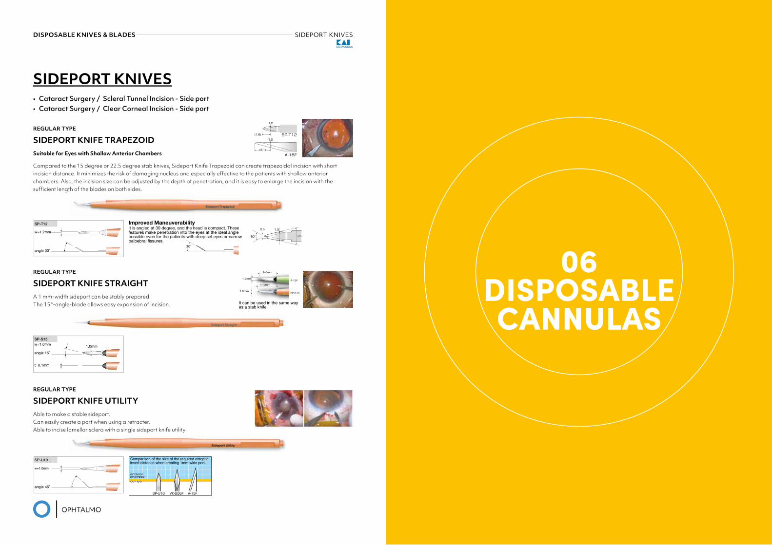
this is what the future looks like 115
DISPOSABLE KNIVES & BLADES SIDEPORT KNIVES
REGULAR TYPE
SIDEPORT KNIFE STRAIGHTA 1 mm-width sideport can be stably prepared. The 15°-angle-blade allows easy expansion of incision.
REGULAR TYPE
SIDEPORT KNIFE UTILITYAble to make a stable sideport. Can easily create a port when using a retracter. Able to incise lamellar sclera with a single sideport knife utility
PARACENTESIS KNIVESCataract Surgery / Scleral Tunnel Incision - Side port / Clear Corneal Incision - Side port
Produced by grinding technology for highly accurate second instrument access. Post operatively, completely self sealing.
PCT-10F
w=1.0mm
PCB-10AGF
w=1.0mm
angle 45°
Φ=0.9mm
SK-VKB20G Φ=0.9mm
Φ=0.9mm Φ=0.9mm
VK-20GF
Φ=1.0mmΦ=1.0mm
VK-19GF
Φ=0.6mmΦ=0.6mm
VK-23GF
Straight StraightStraight
Φ=0.9mmΦ=0.9mm
VKB-20GF
Bent
Regular Type
Regular Type
Safety Type
Anti-glare type
MVR KNIVESCataract Surgery / Scleral Tunnel Incision - Side port / Clear Corneal Incision - Side port / Retina Surgery - Scleral IncisionSpecifically designed for very precise vitrectomy surgery.Available in both straight and angled styles.
9
It is angled at 30 degree, and the head is compact. These features make penetration into the eyes at the ideal angle possible even for the patients with deep set eyes or narrow palbebral fissures.
10
It can be used in the same way as a stab knife.
9.0mm
11.0mm
1.7mm
1.0mm
A-15F
SP-S15
SP-T12
A-15F
1.0
1.0
(3.1)
(1.6)
0.5 1.2
25°60°
Comparison of the size of the required entoptic insert distance when creating 1mm wide port.
corneacornea
anterior chamberanterior chamber
SP-U10 VK-20GF A-15F
SIDEPORT KNIVESCataract Surgery / Scleral Tunnel Incision - Side port / Clear Corneal Incision - Side port
SIDEPORT KNIFE TRAPEZOID
SIDEPORT KNIFE STRAIGHTA 1 mm-width sideport can be stably prepared.The 15°-angle-blade allows easy expansion of incision.
SIDEPORT KNIFE UTILITYAble to make a stable sideport.Can easily create a port when using a retracter. Able to incise lamellar sclera with a single sideport knife utility.
SP-S15w=1.0mm 1.0mmangle 15°
t=0.1mm
SP-U10
w=1.0mm
angle 45°
Regular Type
Regular Type
Regular Type
SP-T12
w=1.2mm
angle 30°
Compared to the 15 degree or 22.5 degree stab knives, Sideport Knife Trapezoid can create trapezoidal incision with short incision distance. It minimizes the risk of damaging nucleus and especially effective to the patients with shallow anterior chambers. Also, the incision size can be adjusted by the depth of penetration, and it is easy to enlarge the incision with the sufficient length of the blades on both sides.
Suitable for Eyes with Shallow Anterior Chambers
Improved Maneuverability
SIDEPORT KNIVES• Cataract Surgery / Scleral Tunnel Incision - Side port• Cataract Surgery / Clear Corneal Incision - Side port
PARACENTESIS KNIVESCataract Surgery / Scleral Tunnel Incision - Side port / Clear Corneal Incision - Side port
Produced by grinding technology for highly accurate second instrument access. Post operatively, completely self sealing.
PCT-10F
w=1.0mm
PCB-10AGF
w=1.0mm
angle 45°
Φ=0.9mm
SK-VKB20G Φ=0.9mm
Φ=0.9mm Φ=0.9mm
VK-20GF
Φ=1.0mmΦ=1.0mm
VK-19GF
Φ=0.6mmΦ=0.6mm
VK-23GF
Straight StraightStraight
Φ=0.9mmΦ=0.9mm
VKB-20GF
Bent
Regular Type
Regular Type
Safety Type
Anti-glare type
MVR KNIVESCataract Surgery / Scleral Tunnel Incision - Side port / Clear Corneal Incision - Side port / Retina Surgery - Scleral IncisionSpecifically designed for very precise vitrectomy surgery.Available in both straight and angled styles.
9
It is angled at 30 degree, and the head is compact. These features make penetration into the eyes at the ideal angle possible even for the patients with deep set eyes or narrow palbebral fissures.
10
It can be used in the same way as a stab knife.
9.0mm
11.0mm
1.7mm
1.0mm
A-15F
SP-S15
SP-T12
A-15F
1.0
1.0
(3.1)
(1.6)
0.5 1.2
25°60°
Comparison of the size of the required entoptic insert distance when creating 1mm wide port.
corneacornea
anterior chamberanterior chamber
SP-U10 VK-20GF A-15F
SIDEPORT KNIVESCataract Surgery / Scleral Tunnel Incision - Side port / Clear Corneal Incision - Side port
SIDEPORT KNIFE TRAPEZOID
SIDEPORT KNIFE STRAIGHTA 1 mm-width sideport can be stably prepared.The 15°-angle-blade allows easy expansion of incision.
SIDEPORT KNIFE UTILITYAble to make a stable sideport.Can easily create a port when using a retracter. Able to incise lamellar sclera with a single sideport knife utility.
SP-S15w=1.0mm 1.0mmangle 15°
t=0.1mm
SP-U10
w=1.0mm
angle 45°
Regular Type
Regular Type
Regular Type
SP-T12
w=1.2mm
angle 30°
Compared to the 15 degree or 22.5 degree stab knives, Sideport Knife Trapezoid can create trapezoidal incision with short incision distance. It minimizes the risk of damaging nucleus and especially effective to the patients with shallow anterior chambers. Also, the incision size can be adjusted by the depth of penetration, and it is easy to enlarge the incision with the sufficient length of the blades on both sides.
Suitable for Eyes with Shallow Anterior Chambers
Improved Maneuverability
REGULAR TYPE
SIDEPORT KNIFE TRAPEZOIDSuitable for Eyes with Shallow Anterior Chambers
Compared to the 15 degree or 22.5 degree stab knives, Sideport Knife Trapezoid can create trapezoidal incision with short incision distance. It minimizes the risk of damaging nucleus and especially effective to the patients with shallow anterior chambers. Also, the incision size can be adjusted by the depth of penetration, and it is easy to enlarge the incision with the sufficient length of the blades on both sides.
06DISPOSABLECANNULAS
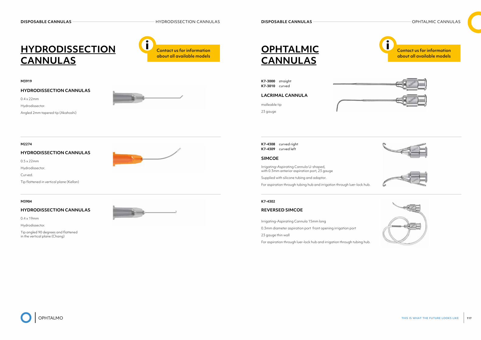
this is what the future looks like 117
DISPOSABLE CANNULAS OPHTALMIC CANNULASDISPOSABLE CANNULAS HYDRODISSECTION CANNULAS
M3919
HYDRODISSECTION CANNULAS
0.4 x 22mm
Hydrodissector.
Angled 2mm tapered tip (Akahoshi)
M2274
HYDRODISSECTION CANNULAS
0.5 x 22mm
Hydrodissector.
Curved.
Tip flattened in vertical plane (Kellan)
M3904
HYDRODISSECTION CANNULAS
0.4 x 19mm
Hydrodissector.
Tip angled 90 degrees and flattened in the vertical plane (Chang)
HYDRODISSECTION CANNULAS
Contact us for information about all available models
i
K7-3000 straightK7-3010 curved
LACRIMAL CANNULA
malleable tip
23 gauge
K7-4308 curved rightK7-4309 curved left
SIMCOE
Irrigating–Aspirating Cannula U-shaped, with 0.3mm anterior aspiration port, 23 gauge
Supplied with silicone tubing and adaptor.
For aspiration through tubing hub and irrigation through luer-lock hub.
K7-4302
REVERSED SIMCOE
Irrigating-Aspirating Cannula 15mm long
0.3mm diameter aspiration port front opening irrigation port
23 gauge thin wall
For aspiration through luer-lock hub and irrigation through tubing hub.
OPHTALMIC CANNULAS
Contact us for information about all available models
i
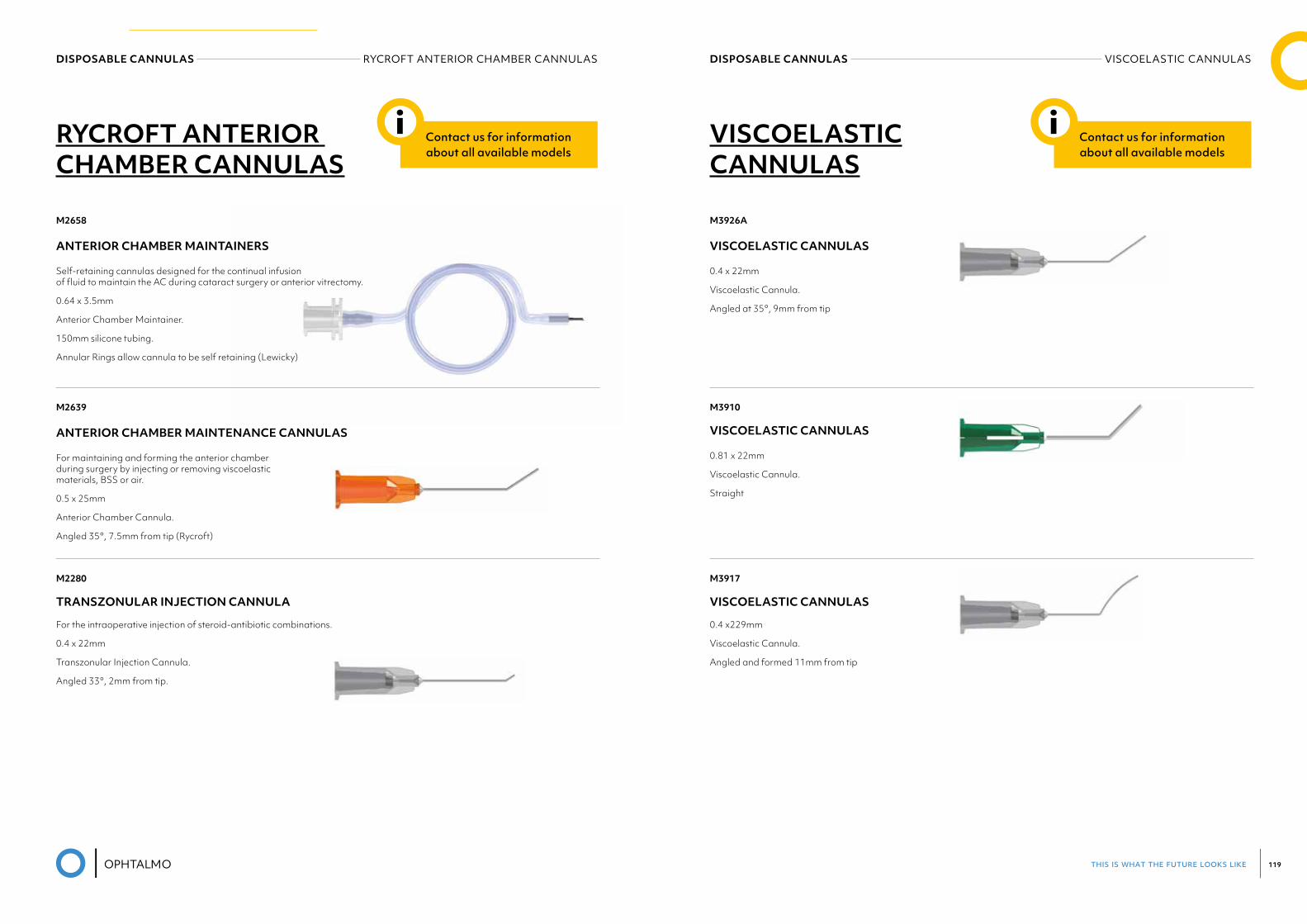
this is what the future looks like 119
DISPOSABLE CANNULAS VISCOELASTIC CANNULASDISPOSABLE CANNULAS RYCROFT ANTERIOR CHAMBER CANNULAS
M2658
ANTERIOR CHAMBER MAINTAINERS
Self-retaining cannulas designed for the continual infusion of fluid to maintain the AC during cataract surgery or anterior vitrectomy.
0.64 x 3.5mm
Anterior Chamber Maintainer.
150mm silicone tubing.
Annular Rings allow cannula to be self retaining (Lewicky)
M2639
ANTERIOR CHAMBER MAINTENANCE CANNULAS
For maintaining and forming the anterior chamber during surgery by injecting or removing viscoelastic materials, BSS or air.
0.5 x 25mm
Anterior Chamber Cannula.
Angled 35°, 7.5mm from tip (Rycroft)
M2280
TRANSZONULAR INJECTION CANNULA
For the intraoperative injection of steroid-antibiotic combinations.
0.4 x 22mm
Transzonular Injection Cannula.
Angled 33°, 2mm from tip.
RYCROFT ANTERIOR CHAMBER CANNULAS
Contact us for information about all available models
i
M3926A
VISCOELASTIC CANNULAS
0.4 x 22mm
Viscoelastic Cannula.
Angled at 35°, 9mm from tip
M3910
VISCOELASTIC CANNULAS
0.81 x 22mm
Viscoelastic Cannula.
Straight
M3917
VISCOELASTIC CANNULAS
0.4 x229mm
Viscoelastic Cannula.
Angled and formed 11mm from tip
VISCOELASTIC CANNULAS
Contact us for information about all available models
i
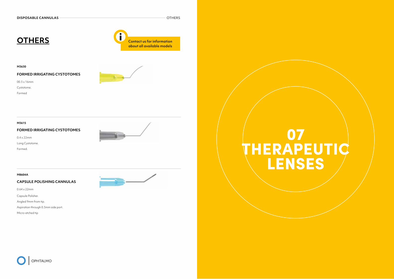
this is what the future looks like 121
DISPOSABLE CANNULAS OTHERS
OTHERS
M3630
FORMED IRRIGATING CYSTOTOMES
00.3 x 16mm
Cystotome.
Formed
M3615
FORMED IRRIGATING CYSTOTOMES
0.4 x 22mm
Long Cystotome.
Formed.
M8604A
CAPSULE POLISHING CANNULAS
0.64 x 22mm
Capsule Polisher.
Angled 9mm from tip.
Aspiration through 0.3mm side port.
Micro-etched tip
Contact us for information about all available models
i
07THERAPEUTIC
LENSES
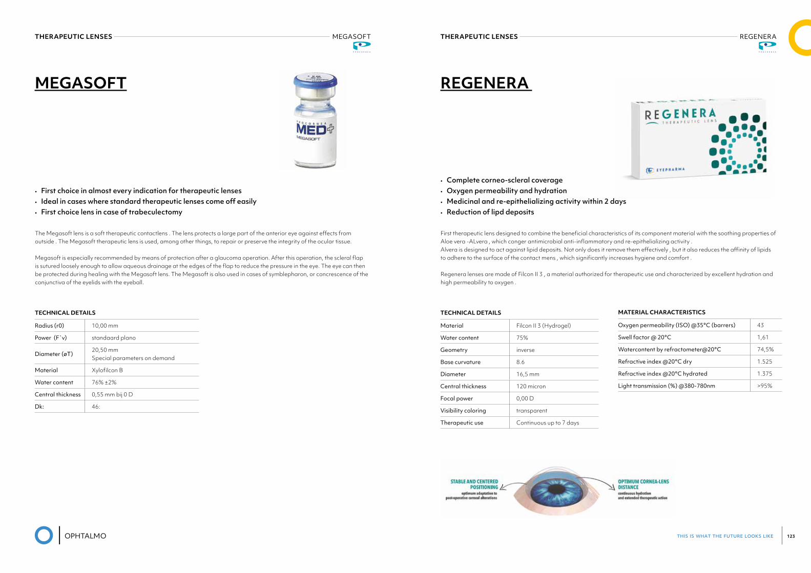
this is what the future looks like 123
THERAPEUTIC LENSES MEGASOFTProcornea Med+
THERAPEUTIC LENSES REGENERAEyepharma
MEGASOFT
TECHNICAL DETAILS
Radius (r0) 10,00 mm
Power (F`v) standaard plano
Diameter (øT)20,50 mmSpecial parameters on demand
Material Xylofilcon B
Water content 76% ±2%
Central thickness 0,55 mm bij 0 D
Dk: 46:
• First choice in almost every indication for therapeutic lenses • Ideal in cases where standard therapeutic lenses come off easily • First choice lens in case of trabeculectomy
The Megasoft lens is a soft therapeutic contactlens . The lens protects a large part of the anterior eye against effects from outside . The Megasoft therapeutic lens is used, among other things, to repair or preserve the integrity of the ocular tissue.
Megasoft is especially recommended by means of protection after a glaucoma operation. After this operation, the scleral flap is sutured loosely enough to allow aqueous drainage at the edges of the flap to reduce the pressure in the eye. The eye can then be protected during healing with the Megasoft lens. The Megasoft is also used in cases of symblepharon, or concrescence of the conjunctiva of the eyelids with the eyeball.
REGENERA
MATERIAL CHARACTERISTICS
Oxygenpermeability(ISO)@35°C(barrers) 43
Swellfactor@20°C 1,61
Watercontentbyrefractometer@20°C 74,5%
Refractiveindex@20°Cdry 1.525
Refractiveindex@20°Chydrated 1.375
Lighttransmission(%)@380-780nm >95%
• Complete corneo-scleral coverage • Oxygen permeability and hydration • Medicinal and re-epithelializing activity within 2 days • Reduction of lipd deposits
First therapeutic lens designed to combine the beneficial characteristics of its component material with the soothing properties of Aloe vera -ALvera , which conger antimicrobial anti-inflammatory and re-epithelializing activity . Alvera is designed to act against lipid deposits. Not only does it remove them effectively , but it also reduces the affinity of lipids to adhere to the surface of the contact mens , which significantly increases hygiene and comfort .
Regenera lenses are made of Filcon II 3 , a material authorized for therapeutic use and characterized by excellent hydration and high permeability to oxygen .
TECHNICAL DETAILS
Material Filcon II 3 (Hydrogel)
Water content 75%
Geometry inverse
Base curvature 8.6
Diameter 16,5 mm
Central thickness 120 micron
Focal power 0,00 D
Visibility coloring transparent
Therapeutic use Continuous up to 7 days
this is what the future looks like 125
THERAPEUTIC LENSES OTHER BRANDS
PureVision and PureVision2 contact lenses are indicated for therapeutic use. When placed on the cornea for therapeutic use, these lenses act as a bandage to protect the cornea and relieve pain during treatment of ocular pathologies as well as post-surgical conditions.
Applications include, but are not limited to, conditions such as the following:• For corneal protection - in conditions such as entropion, trichiasis, tarsal scars, recurrent corneal erosion and post surgical
ptosis for corneal protection;• For corneal pain relief - in conditions such as bullous keratopathy, epithelial erosion and abrasion, filamentary keratitis,
post-keratoplasty;• For use as a bandage during the healing process of conditions such as chronic epithelial defects, corneal ulcer, neurotrophic
keratitis, neuroparalytic keratitis, chemical burns, and post surgical epithelial defects.• For post-surgical conditions that include bandage use such as LASIK, PRK, PK, PTK, lamellar grafts, corneal flaps, and additi-
onal corneal surgical conditions.
08PROCEDURE PACKS
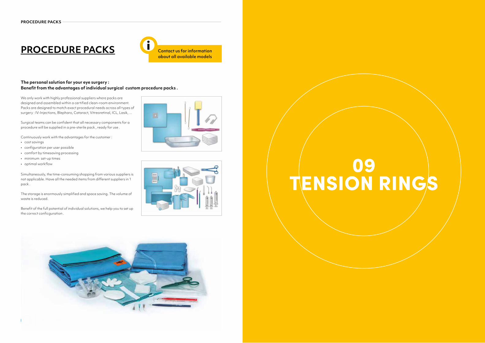
this is what the future looks like 127
PROCEDURE PACKS
PROCEDURE PACKS
The personal solution for your eye surgery : Benefit from the advantages of individual surgical custom procedure packs .
We only work with highly professional suppliers where packs are designed and assembled within a certified clean-room environment. Packs are designed to match exact procedural needs across all types of surgery : IV-Injections, Blepharo, Cataract, Vitreoretinal, ICL, Lasik, ...
Surgical teams can be confident that all necessary components for a procedure will be supplied in a pre-sterile pack , ready for use .
Continuously work with the advantages for the customer : • cost savings • configuration per user possible • comfort by timesaving processing• minimum set-up times• optimal workflow
Simultaneously, the time-consuming shopping from various suppliers is not applicable. Have all the needed items from different suppliers in 1 pack .
The storage is enormously simplified and space saving. The volume of waste is reduced.
Benefit of the full potential of individual solutions, we help you to set up the correct conficguration .
Contact us for information about all available models
i
09TENSION RINGS
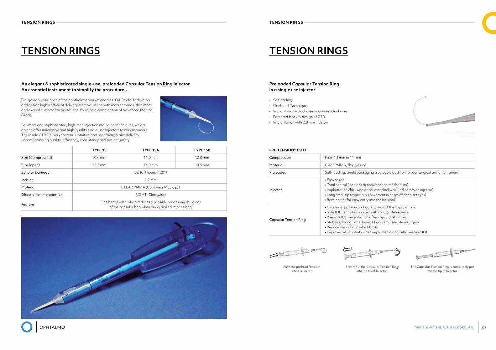
this is what the future looks like 129
TENSION RINGS TENSION RINGS
TENSION RINGS TENSION RINGS
An elegant & sophisticated single-use, preloaded Capsular Tension Ring Injector.An essential instrument to simplify the procedure…
On-going surveillance of the ophthalmic market enables “O&Omdc” to develop and design highly efficient delivery systems, in line with market trends, that meet and exceed customer expectations. By using a combination of advanced Medical Grade
Polymers and sophisticated, high-tech injection moulding techniques, we are able to offer innovative and high-quality single-use Injectors to our customers. The Inside CTR Delivery System is intuitive and user friendly and delivers uncompromising quality, efficiency, consistency and patient safety.
Preloaded Capsular Tension Ring in a single use injector
• Selfloading• Onehand-Technique• Implantation – clockwise or counter clockwise• Patented Hockey design of CTR• Implantation with 2.0 mm incision
TYPE 15 TYPE 15A TYPE 15B
Size (Compressed) 10.0 mm 11.0 mm 12.0 mm
Size (open) 12.3 mm 13.0 mm 14.5 mm
Zonular Damage Up to 4 hours (120°)
Incision 2.2 mm
Material CLEAR PMMA (Compress Moulded)
Direction of Implantation RIGHT (Clockwise)
FeatureOne bent eyelet, which reduces a possible puncturing (bulging)
of the capsular bag when being dialled into the bag
PRE-TENSION® 13/11
Compression From 13 mm to 11 mm
Material Clear PMMA, flexible ring
Preloaded Self-loading, single packaging a valuable addition to your surgical armamentarium
Injector
• Easy to use• Total control (includes action/reaction mechanism)• Implantation clockwise or counter clockwise (indicators on injector)• Long small tip (especially convenient in cases of deep set eyes)• Beveled tip (for easy entry into the incision)
Capsular Tension Ring
• Circular expansion and stabilization of the capsular bag• Safe IOL centration in eyes with zonular dehiscence• Prevents IOL decentration after capsular shrinking• Stabilized conditions during Phaco-emulsification surgery• Reduced risk of capsular fibrosis• Improves visual acuity when implanted along with premium IOL
Model Pre-Tension® 13/11
Compression From 13 mm to 11 mm
Material Clear PMMA, flexible ring
Preloaded Self-loading, single packaging a valuable addition to your surgical armamentarium
Injector
• Easy to use• Total control (includes action/reaction mechanism)• Implantation clockwise or counter clockwise (indicators on injector)• Long small tip (especially convenient in cases of deep set eyes)• Beveled tip (for easy entry into the incision)
Capsular Tension Ring
• Circular expansion and stabilization of the capsular bag• Safe IOL centration in eyes with zonular dehiscence• Prevents IOL decentration after capsular shrinking• Stabilized conditions during Phaco-emulsification surgery• Reduced risk of capsular fibrosis• Improves visual acuity when implanted along with premium IOL
Preloaded Capsular Tension Ring in a single use injector
Technical Data
Push the pushrod forward until it is limited.
Slowly put the Capsular Tension Ring into the tip of Injector.
The Capsular Tension Ring is completely put into the tip of Injector.
Pre-Tension®
Products for ophthalmic surgery
OPHTHALMO Pro GmbHIm Reihersbruch 1, 66386 Sankt Ingbert Owner: Thomas ZimmerTel.: +49 (68 94) 99 88 770Fax: +49 (68 94) 99 88 777Email: [email protected]/en
Contact:
Push the pushrod forward until it is limited.
Slowly put the Capsular Tension Ringinto the tip of Injector.
The Capsular Tension Ring is completely put into the tip of Injector.
this is what the future looks like 131
10CAUTERY
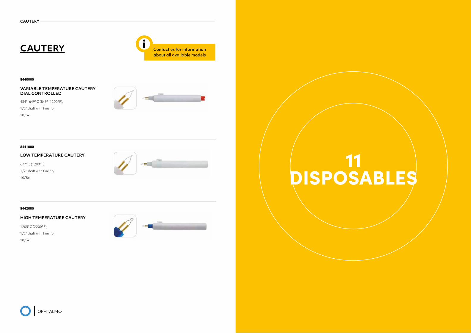
this is what the future looks like 133
CAUTERY
CAUTERY
8440000
VARIABLE TEMPERATURE CAUTERY DIAL CONTROLLED
454°-649°C (849°-1200°F),
1/2” shaft with fine tip,
10/bx
8441000
LOW TEMPERATURE CAUTERY
677°C (1200°F),
1/2” shaft with fine tip,
10/Bx
8442000
HIGH TEMPERATURE CAUTERY
1205°C (2200°F),
1/2” shaft with fine tip,
10/bx
Contact us for information about all available models
i
11DISPOSABLES
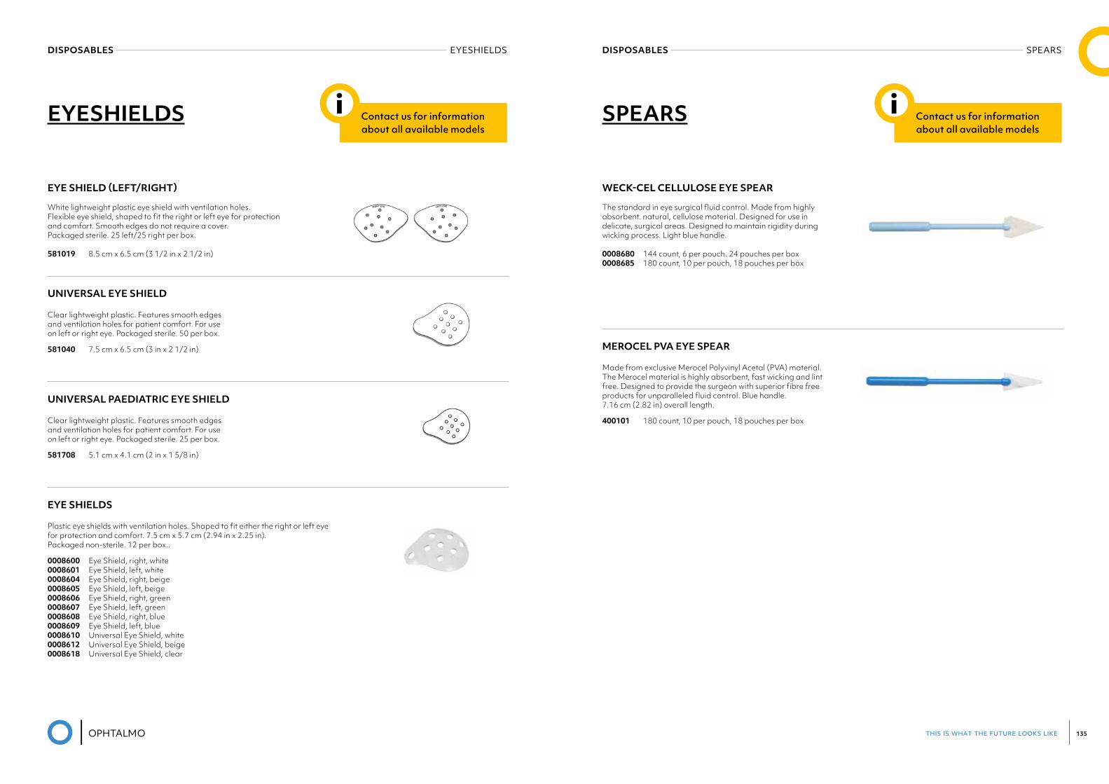
this is what the future looks like 135
DISPOSABLES EYESHIELDS DISPOSABLES SPEARS
EYESHIELDS Contact us for information about all available models
i
EYE SHIELD (LEFT/RIGHT)
White lightweight plastic eye shield with ventilation holes. Flexible eye shield, shaped to fit the right or left eye for protection and comfort. Smooth edges do not require a cover. Packaged sterile. 25 left/25 right per box.
581019 8.5 cm x 6.5 cm (3 1/2 in x 2 1/2 in)
UNIVERSAL EYE SHIELD
Clear lightweight plastic. Features smooth edges and ventilation holes for patient comfort. For use on left or right eye. Packaged sterile. 50 per box.
581040 7.5 cm x 6.5 cm (3 in x 2 1/2 in)
UNIVERSAL PAEDIATRIC EYE SHIELD
Clear lightweight plastic. Features smooth edges and ventilation holes for patient comfort. For use on left or right eye. Packaged sterile. 25 per box.
581708 5.1 cm x 4.1 cm (2 in x 1 5/8 in)
EYE SHIELDS
Plastic eye shields with ventilation holes. Shaped to fit either the right or left eye for protection and comfort. 7.5 cm x 5.7 cm (2.94 in x 2.25 in). Packaged non-sterile. 12 per box..
0008600 Eye Shield, right, white0008601 Eye Shield, left, white0008604 Eye Shield, right, beige0008605 Eye Shield, left, beige0008606 Eye Shield, right, green0008607 Eye Shield, left, green 0008608 Eye Shield, right, blue0008609 Eye Shield, left, blue0008610 Universal Eye Shield, white0008612 Universal Eye Shield, beige0008618 Universal Eye Shield, clear
SPEARS
WECK-CEL CELLULOSE EYE SPEAR
The standard in eye surgical fluid control. Made from highly absorbent. natural, cellulose material. Designed for use in delicate, surgical areas. Designed to maintain rigidity during wicking process. Light blue handle.
0008680 144 count, 6 per pouch. 24 pouches per box 0008685 180 count, 10 per pouch, 18 pouches per box
MEROCEL PVA EYE SPEAR
Made from exclusive Merocel Polyvinyl Acetal (PVA) material. The Merocel material is highly absorbent, fast wicking and lint free. Designed to provide the surgeon with superior fibre free products for unparalleled fluid control. Blue handle. 7.16 cm (2.82 in) overall length.
400101 180 count, 10 per pouch, 18 pouches per box
Contact us for information about all available models
i
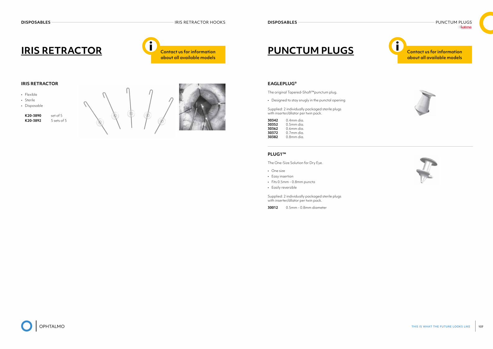
this is what the future looks like 137
DISPOSABLES PUNCTUM PLUGSDISPOSABLES IRIS RETRACTOR HOOKS
IRIS RETRACTOR
IRIS RETRACTOR
• Flexible• Sterile • Disposable
K20-3890 set of 5 K20-3892 5 sets of 5
Contact us for information about all available models
i PUNCTUM PLUGS
EAGLEPLUG®
The original Tapered-Shaft™punctum plug.
• Designed to stay snugly in the punctal opening
Supplied: 2 individually packaged sterile plugs with inserter/dilator per twin pack.
30342 0.4mm dia. 30352 0.5mm dia. 30362 0.6mm dia. 30372 0.7mm dia. 30382 0.8mm dia.
PLUG1™
The One-Size Solution for Dry Eye.
• One size• Easy insertion• Fits 0.5mm - 0.8mm puncta• Easily reversible
Supplied: 2 individually packaged sterile plugs with inserter/dilator per twin pack.
30012 0.5mm - 0.8mm diameter
Contact us for information about all available models
i
®
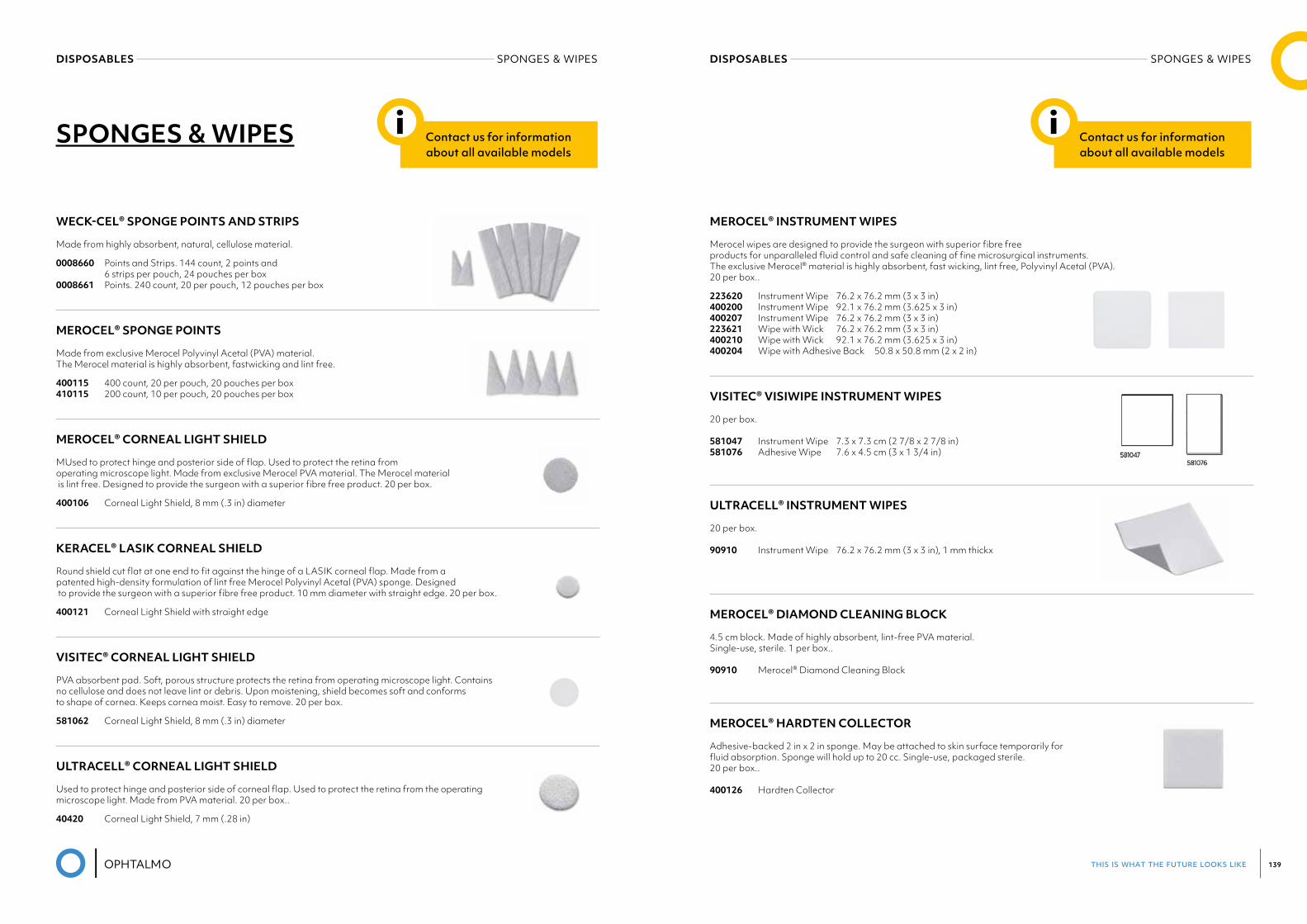
this is what the future looks like 139
DISPOSABLES SPONGES & WIPESDISPOSABLES SPONGES & WIPES
SPONGES & WIPES Contact us for information about all available models
i
WECK-CEL® SPONGE POINTS AND STRIPS
Made from highly absorbent, natural, cellulose material.
0008660 Points and Strips. 144 count, 2 points and 6 strips per pouch, 24 pouches per box 0008661 Points. 240 count, 20 per pouch, 12 pouches per box
MEROCEL® SPONGE POINTS
Made from exclusive Merocel Polyvinyl Acetal (PVA) material. The Merocel material is highly absorbent, fastwicking and lint free.
400115 400 count, 20 per pouch, 20 pouches per box 410115 200 count, 10 per pouch, 20 pouches per box
MEROCEL® CORNEAL LIGHT SHIELD
MUsed to protect hinge and posterior side of flap. Used to protect the retina from operating microscope light. Made from exclusive Merocel PVA material. The Merocel material is lint free. Designed to provide the surgeon with a superior fibre free product. 20 per box.
400106 Corneal Light Shield, 8 mm (.3 in) diameter
KERACEL® LASIK CORNEAL SHIELD
Round shield cut flat at one end to fit against the hinge of a LASIK corneal flap. Made from a patented high-density formulation of lint free Merocel Polyvinyl Acetal (PVA) sponge. Designed to provide the surgeon with a superior fibre free product. 10 mm diameter with straight edge. 20 per box.
400121 Corneal Light Shield with straight edge
VISITEC® CORNEAL LIGHT SHIELD
PVA absorbent pad. Soft, porous structure protects the retina from operating microscope light. Contains no cellulose and does not leave lint or debris. Upon moistening, shield becomes soft and conforms to shape of cornea. Keeps cornea moist. Easy to remove. 20 per box.
581062 Corneal Light Shield, 8 mm (.3 in) diameter
ULTRACELL® CORNEAL LIGHT SHIELD
Used to protect hinge and posterior side of corneal flap. Used to protect the retina from the operating microscope light. Made from PVA material. 20 per box..
40420 Corneal Light Shield, 7 mm (.28 in)
Contact us for information about all available models
i
MEROCEL® INSTRUMENT WIPES
Merocel wipes are designed to provide the surgeon with superior fibre free products for unparalleled fluid control and safe cleaning of fine microsurgical instruments. The exclusive Merocel® material is highly absorbent, fast wicking, lint free, Polyvinyl Acetal (PVA). 20 per box..
223620 Instrument Wipe 76.2 x 76.2 mm (3 x 3 in) 400200 Instrument Wipe 92.1 x 76.2 mm (3.625 x 3 in) 400207 Instrument Wipe 76.2 x 76.2 mm (3 x 3 in) 223621 Wipe with Wick 76.2 x 76.2 mm (3 x 3 in) 400210 Wipe with Wick 92.1 x 76.2 mm (3.625 x 3 in) 400204 Wipe with Adhesive Back 50.8 x 50.8 mm (2 x 2 in)
VISITEC® VISIWIPE INSTRUMENT WIPES
20 per box.
581047 Instrument Wipe 7.3 x 7.3 cm (2 7/8 x 2 7/8 in) 581076 Adhesive Wipe 7.6 x 4.5 cm (3 x 1 3/4 in)
ULTRACELL® INSTRUMENT WIPES
20 per box.
90910 Instrument Wipe 76.2 x 76.2 mm (3 x 3 in), 1 mm thickx
MEROCEL® DIAMOND CLEANING BLOCK
4.5 cm block. Made of highly absorbent, lint-free PVA material. Single-use, sterile. 1 per box..
90910 Merocel® Diamond Cleaning Block
MEROCEL® HARDTEN COLLECTOR
Adhesive-backed 2 in x 2 in sponge. May be attached to skin surface temporarily for fluid absorption. Sponge will hold up to 20 cc. Single-use, packaged sterile. 20 per box..
400126 Hardten Collector
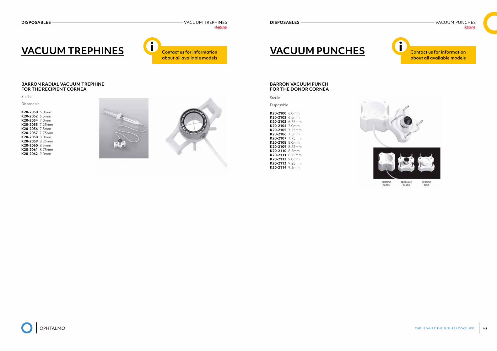
this is what the future looks like 141
DISPOSABLES VACUUM TREPHINES DISPOSABLES VACUUM PUNCHES
VACUUM TREPHINES
BARRON RADIAL VACUUM TREPHINE FOR THE RECIPIENT CORNEASterile
Disposable
K20-2050 6.0mm K20-2052 6.5mm K20-2054 7.0mm K20-2055 7.25mm K20-2056 7.5mm K20-2057 7.75mm K20-2058 8.0mm K20-2059 8.25mm K20-2060 8.5mm K20-2061 8.75mm K20-2062 9.0mm
Contact us for information about all available models
i VACUUM PUNCHES
BARRON VACUUM PUNCH FOR THE DONOR CORNEA
Sterile
Disposable
K20-2100 6.0mm K20-2102 6.5mm K20-2103 6.75mm K20-2104 7.0mm K20-2105 7.25mm K20-2106 7.5mm K20-2107 7.75mm K20-2108 8.0mm K20-2109 8.25mm K20-2110 8.5mm K20-2111 8.75mm K20-2112 9.0mm K20-2113 9.25mmK20-2114 9.5mm
®
Contact us for information about all available models
i
®
this is what the future looks like 143
12PHARMA
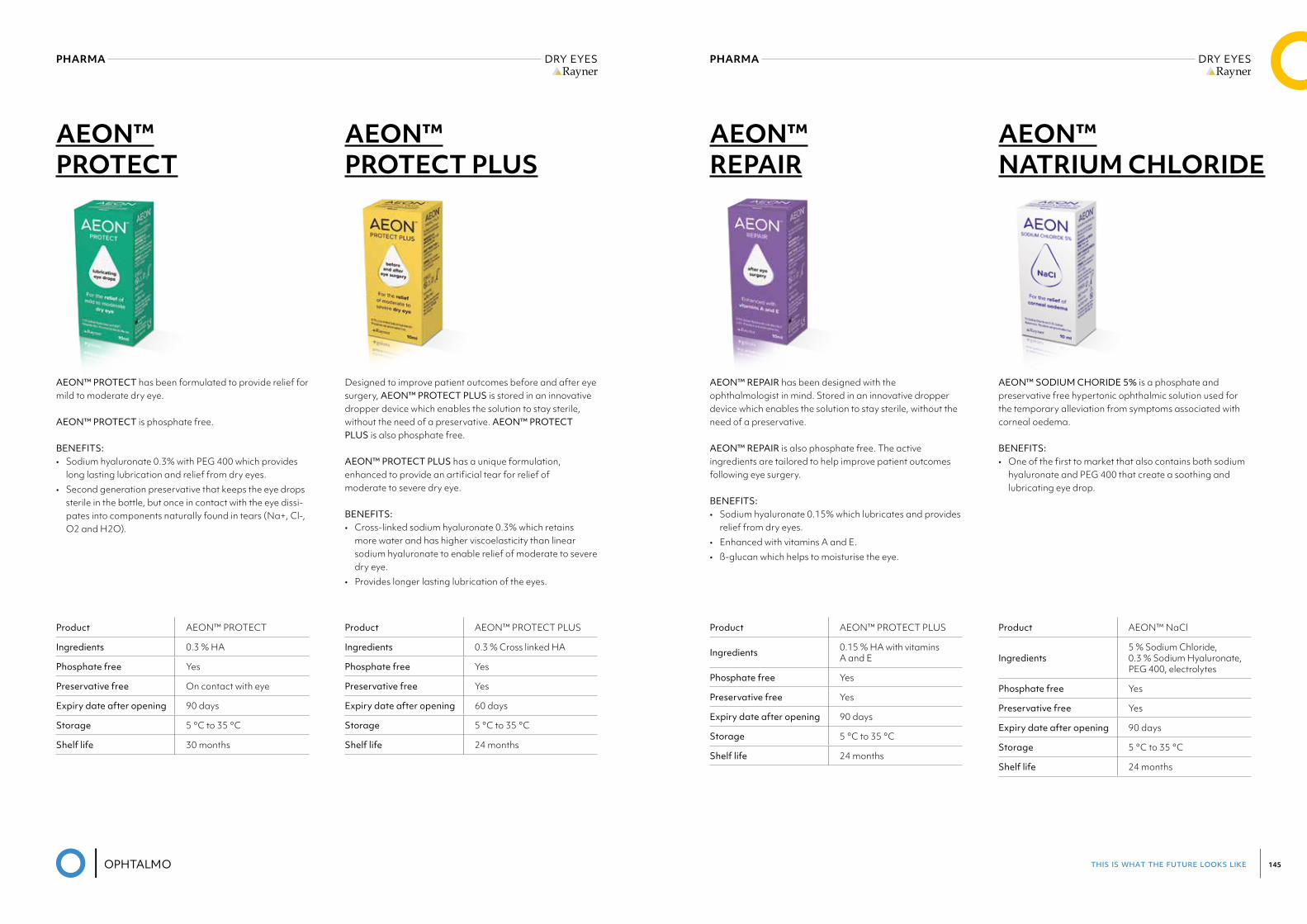
this is what the future looks like 145
PHARMA DRY EYESRayner
PHARMA DRY EYESRayner
AEON™ PROTECT PLUS
AEON™ PROTECT
Product AEON™ PROTECT PLUS
Ingredients 0.3 % Cross linked HA
Phosphate free Yes
Preservative free Yes
Expiry date after opening 60 days
Storage 5 °C to 35 °C
Shelf life 24 months
Product AEON™ PROTECT
Ingredients 0.3 % HA
Phosphate free Yes
Preservative free On contact with eye
Expiry date after opening 90 days
Storage 5 °C to 35 °C
Shelf life 30 months
Designed to improve patient outcomes before and after eye surgery, AEON™ PROTECT PLUS is stored in an innovative dropper device which enables the solution to stay sterile, without the need of a preservative. AEON™ PROTECT PLUS is also phosphate free.
AEON™ PROTECT PLUS has a unique formulation,enhanced to provide an artificial tear for relief ofmoderate to severe dry eye.
BENEFITS:• Cross-linked sodium hyaluronate 0.3% which retains
more water and has higher viscoelasticity than linear sodium hyaluronate to enable relief of moderate to severe dry eye.
• Provides longer lasting lubrication of the eyes.
AEON™ PROTECT has been formulated to provide relief for mild to moderate dry eye.
AEON™ PROTECT is phosphate free.
BENEFITS:• Sodium hyaluronate 0.3% with PEG 400 which provides
long lasting lubrication and relief from dry eyes.• Second generation preservative that keeps the eye drops
sterile in the bottle, but once in contact with the eye dissi-pates into components naturally found in tears (Na+, Cl-, O2 and H2O).
AEON™ REPAIR
AEON™ NATRIUM CHLORIDE
Product AEON™ PROTECT PLUS
Ingredients 0.15 % HA with vitamins A and E
Phosphate free Yes
Preservative free Yes
Expiry date after opening 90 days
Storage 5 °C to 35 °C
Shelf life 24 months
Product AEON™ NaCl
Ingredients5 % Sodium Chloride, 0.3 % Sodium Hyaluronate, PEG 400, electrolytes
Phosphate free Yes
Preservative free Yes
Expiry date after opening 90 days
Storage 5 °C to 35 °C
Shelf life 24 months
AEON™ REPAIR has been designed with the ophthalmologist in mind. Stored in an innovative dropper device which enables the solution to stay sterile, without the need of a preservative.
AEON™ REPAIR is also phosphate free. The active ingredients are tailored to help improve patient outcomes following eye surgery.
BENEFITS:• Sodium hyaluronate 0.15% which lubricates and provides
relief from dry eyes.• Enhanced with vitamins A and E.• ß-glucan which helps to moisturise the eye.
AEON™ SODIUM CHORIDE 5% is a phosphate and preservative free hypertonic ophthalmic solution used for the temporary alleviation from symptoms associated with corneal oedema.
BENEFITS:• One of the first to market that also contains both sodium
hyaluronate and PEG 400 that create a soothing and lubricating eye drop.
this is what the future looks like 147
13DIAGNOSTIC
STRIPS
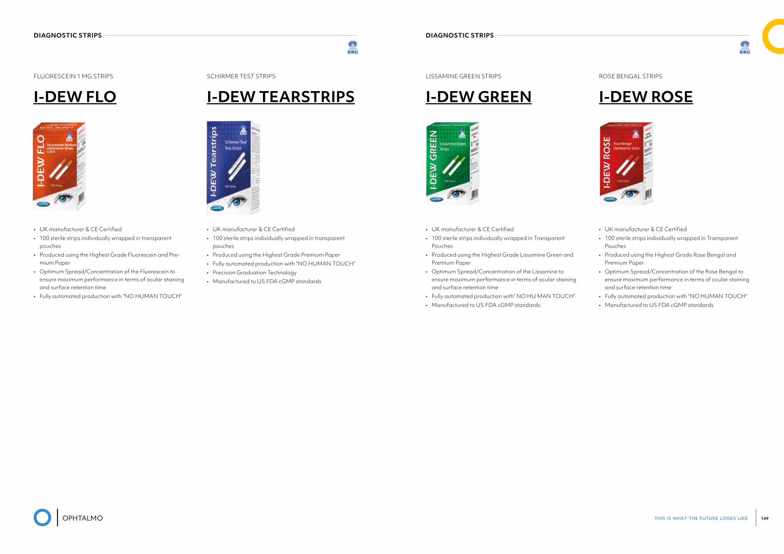
this is what the future looks like 149
DIAGNOSTIC STRIPS ERC
DIAGNOSTIC STRIPS ERC
SCHIRMER TEST STRIPS
I-DEW TEARSTRIPSFLUORESCEIN 1 MG STRIPS
I-DEW FLO
• UK manufacturer & CE Certified• 100 sterile strips individually wrapped in transparent
pouches• Produced using the Highest Grade Premium Paper• Fully automated production with “NO HUMAN TOUCH”• Precision Graduation Technology• Manufactured to US FDA cGMP standards
• UK manufacturer & CE Certified• 100 sterile strips individually wrapped in transparent
pouches• Produced using the Highest Grade Fluorescein and Pre-
mium Paper• Optimum Spread/Concentration of the Fluorescein to
ensure maximum performance in terms of ocular staining and surface retention time
• Fully automated production with “NO HUMAN TOUCH”
ROSE BENGAL STRIPS
I-DEW ROSELISSAMINE GREEN STRIPS
I-DEW GREEN
• UK manufacturer & CE Certified• 100 sterile strips individually wrapped in Transparent
Pouches• Produced using the Highest Grade Rose Bengal and
Premium Paper• Optimum Spread/Concentration of the Rose Bengal to
ensure maximum performance in terms of ocular staining and surface retention time
• Fully automated production with “NO HUMAN TOUCH”• Manufactured to US FDA cGMP standards
• UK manufacturer & CE Certified• 100 sterile strips individually wrapped in Transparent
Pouches• Produced using the Highest Grade Lissamine Green and
Premium Paper• Optimum Spread/Concentration of the Lissamine to
ensure maximum performance in terms of ocular staining and surface retention time
• Fully automated production with” NO HU MAN TOUCH”• Manufactured to US FDA cGMP standards

this is what the future looks like 151
MEDICAL DEVICE
this is what the future looks like 153
01. MICROSCOPES 157 LEICA M620 158 LEICA M822 160 LEICA PROVEO 8 164
02. LENSMETER /PHOCOMETER 167 VISIONIX VX40 FULL AUTOMATIC LENSMETER 168
03. AUTREFRACTOR – KERATOMETER 169 VISIONIX VX90 AR WITH AUTOFOCUS 170 VISIONIX VX110 PLACIDO TOPOGRAPHER 172 SUOERSW-800HANDHELDVISIONSCREENER 174
04. ENDOTHELIUM / SPECULAR MICROSCOPE 175 SUOERSW-7000SPECULARMICROSCOPE 176 BON PERSEUS SPECULAR MICROSCOPE 177
05. PERIMETRY 179 FREYAP-300 180 MEDMONTM-700>SOONAVAILABLE 182
06. FUNDUSCAMERA 183 VISIONIX NEXY FULL AUTOMATIC 184 SUOERSW-8800NON-MYDRIATIC 185 SUOERSW-8000WIDEFIELDROPCAMERA 186 SYSEYERETICAM3100NON-MYDRIATIC AUTOMATIC FUNDUS CAMERA 187
07. OCT 189 MOPTIMMOCEAN-4000OCTWITHANGIOGRAPHY 190
08. REFRACTION 201 VISIONIX VX25 EYE REFRACT 202 VISIONIX VX65 AUTOMATIC PHOROPTER 204 VISIONIX VX50 MANUAL PHOROPTER 205
this is what the future looks like 155
16. OPHTALMOSCOPE 251 HEINEHAND-HELDDIRECTBETA200/BETA200S(LED) 252 HEINEHAND-HELDDIRECTMINI3000 253 HEINEHAND-HELDINDIRECT 254 HEINE OMEGA 600 INDIRECT 255
17. RETINOSCOPY / RETINOMETER 257 HEINE BETA 200 RETINOSCOPE 258 HEINE LAMBDA 100 RETINOMETER 259
18. EXAMINATION UNITS 261 VISIONIX VX1200 262 VISIONIXVX2000 263 VISIONIXVX3000 264
19. OPERATION & PATIENT CHAIRS 265 UFSK 500XLE 266 UFSK EYEFORCE 268
20. AUTOCLAVES 271 STATIM G4 272
21. TRAIL LENSES & SETS 275
09. TOPOGRAPHY 207 VISIONIX VX205 TOPOGRAPHER 208 SUOERSW-6000TOPOGRAPHER 209
10. TONOMETRY 211 TAKAGISNT-700NON-CONTACTTONOMETER 212 FREYMEDICALTN-100NON-CONTACTTONOMETER 215 SUOERSW-500HANDHELDREBOUNDTONOMETER 216 SUOERSW-5000NON-CONTACTTONOMETER 217
11. DIAGNOSTIC 219 VISIONIX VX120+ ANTERIOR SCREENING 220 VISIONIXVX130+ANTERIORSCREENING 224 VISIONIX VX210 SCHEIMPFLUG CAMERA 228 TAKAGICGT-2000CONTRASTGLARETESTER 229
12. LASER 233 LIGHTMEDLIGHTLASYAG 238 LIGHTMED LIGHTLAS SLT 240 LIGHTMEDLIGHTLAS532/577/810 242 LIGHTMED TRUSCAN 244
13. SLIT LAMPS 237 VISIONIXVX70 238 VISIONIX VX75 240 VISIONIX VX80 242 VISIONIX VX85 244 HEINEHSL150HAND-HELDSLITLAMP 246
14. BIOMETRY 247 SUOERSW-9000OPTICALBIOMETER 248
15. ULTRASOUND 249 SUOERSW-21SCANNERA-B-UBM 250
OPHTALMO CONSUMABLES
01MICROSCOPES
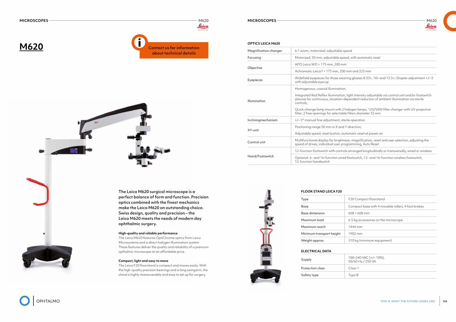
this is what the future looks like 159
MICROSCOPES M620Leica
MICROSCOPES M620Leica
M620
The Leica M620 surgical microscope is a perfect balance of form and function. Precision optics combined with the finest mechanics make the Leica M620 an outstanding choice. Swiss design, quality and precision – the Leica M620 meets the needs of modern day ophthalmic surgery.
High-quality and reliable performanceThe Leica M620 features OptiChrome optics from Leica Microsystems and a direct halogen Illumination system. These features deliver the quality and reliability of a premium opthalmic microscope at an affordable price.
Compact, light and easy to moveThe Leica F20 floorstand is compact and moves easily. With the high-quality precision bearings and a long swingarm, the stand is highly maneuverable and easy to set up for surgery.
Contact us for information about technical details
i OPTICS LEICA M620
Magnificationchanger 6:1 zoom, motorized, adjustable speed
Focusing Motorized, 50 mm, adjustable speed, with automatic reset
ObjectiveAPO Leica WD = 175 mm, 200 mm
Achromatic Leica f = 175 mm, 200 mm and 225 mm
Eyepieces Widefield eyepieces for those wearing glasses 8.33×, 10× and 12.5×; Diopter adjustment +/– 5 with adjustable eyecup
Illumination
Homogenous, coaxial illumination;
Integrated Red Reflex illumination, light intensity adjustable via control unit and/or footswitch jalousie for continuous, situation-dependent reduction of ambient illumination via sterile controls;
Quick-change lamp mount with 2 halogen lamps, 12V/50W filter changer with UV projective filter; 2 free openings for selectable filters diameter 32 mm
Incliningmechanism +/– 5° manual fine adjustment, sterile operation
XY-unitPositioning range 50 mm in X and Y direction;
Adjustable speed, reset button, automatic reset at power on
Control unit Multifunctional display for brightness, magnification, reset and user selection, adjusting the speed of drives, individual user programming, Auto Reset
Hand/Footswitch
12-function footswitch with controls arranged longitudinally or transversally, wired or wireless
Optional: 6- and 16-function wired footswitch, 12- and 16-function wireless footswitch, 12-function handswitch
FLOOR STAND LEICA F20
Type F20 Compact floorstand
Base Compact base with 4 movable rollers, 4 foot brakes
Base dimension 608 × 608 mm
Maximum load 6.5 kg accessories on the microscope
Maximum reach 1444 mm
Minimum transport height 1902 mm
Weight approx. 210 kg (minimum equipment)
ELECTRICAL DATA
Supply 100–240 VAC (+/– 10%), 50/60 Hz / 250 VA
Protection class Class 1
Safety type Type B
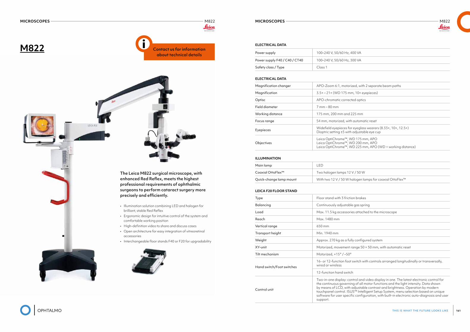
this is what the future looks like 161
MICROSCOPES M822Leica
MICROSCOPES M822Leica
M822
The Leica M822 surgical microscope, with enhanced Red Reflex, meets the highest professional requirements of ophthalmic surgeons to perform cataract surgery more precisely and efficiently.
• Illumination solution combining LED and halogen for brilliant, stable Red Reflex
• Ergonomic design for intuitive control of the system and comfortable working position
• High-definition video to share and discuss cases• Open architecture for easy integration of vitreoretinal
accessories• Interchangeable floor stands F40 or F20 for upgradability
Contact us for information about technical details
i ELECTRICAL DATA
Power supply 100–240 V, 50/60 Hz, 400 VA
Power supply F40 / C40 / CT40 100–240 V, 50/60 Hz, 300 VA
Safety class / Type Class 1
ELECTRICAL DATA
Magnificationchanger APO-Zoom 6:1, motorized, with 2 separate beam paths
Magnification 3.5× – 21× (WD 175 mm, 10× eyepieces)
Optisc APO-chromatic corrected optics
Field diameter 7 mm – 80 mm
Working distance 175 mm, 200 mm and 225 mm
Focus range 54 mm, motorized, with automatic reset
Eyepieces Widefield eyepieces for eyeglass wearers (8.33×, 10×, 12.5×) Dioptric setting ±5 with adjustable eye cup
ObjectivesLeica OptiChrome™, WD 175 mm, APO Leica OptiChrome™, WD 200 mm, APO Leica OptiChrome™, WD 225 mm, APO (WD = working distance)
ILLUMINATION
Main lamp LED
Coaxial OttoFlex™ Two halogen lamps 12 V / 50 W
Quick-changelampmount With two 12 V / 50 W halogen lamps for coaxial OttoFlex™
LEICA F20 FLOOR STAND
Type Floor stand with 3 friction brakes
Balancing Continuously adjustable gas spring
Load Max. 11.5 kg accessories attached to the microscope
Reach Max. 1480 mm
Vertical range 650 mm
Transport height Min. 1940 mm
Weight Approx. 270 kg as a fully configured system
XY-unit Motorized, movement range 50 × 50 mm, with automatic reset
Tilt mechanism Motorized, +15° / –50°
Hand switch/Foot switches
16- or 12-function foot switch with controls arranged longitudinally or transversally, wired or wireless
12-function hand switch
Control unit
Two-in-one display: control and video display in one. The latest electronic control for the continuous governing of all motor functions and the light intensity. Data shown by means of LCD, with adjustable contrast and brightness. Operation by modern touchpanel control. ISUS™ Intelligent Setup System, menu selection based on unique software for user specific configuration, with built-in electronic auto-diagnosis and user support.
this is what the future looks like 163
MICROSCOPES M822Leica
MICROSCOPES M822Leica
LEICA F40 FLOOR STAND, LEICA C40 CEILING MOUNT, LEICA CT40 TELESCOPE CEILING MOUNT
Type Floor stand with 4 electromagnetic brakes
Balancing Continuously adjustable gas spring
Load Max. 12.2 kg accessories attached to the microscope
Reach Max. 1492 mm
Vertical range 846 mm
Transport height Min. 1949 mm
Weight Approx. 330 kg as a fully configured system
XY-unit Motorized, movement range 50 × 50 mm, with automatic reset
Tilt mechanism Motorized, +15° / –50°
Hand / foot switches
16- or 12-function foot switch with controls arranged longitudinally or transversally, wired or wireless
12-function hand switch
Control unit
Two-in-one display: control and video display in one. The latest electronic control for the continous governing of all motor functions and the light intensity. Data shown by means of LCD, with adjustable contrast and brightness. Operation by modern touchpanel control. ISUS™ Intelligent Setup System, menu selection based on unique software for user specific configuration, with built-in electronic auto-diagnosis and user support.
HD IMAGING
For more information, please refer to your local Leica Microsystems Sales Representative.
STANDARDS
Council Directive 93/42/EEC on Medical Devices (MDD) and its amendments. Classification: Class I, in compliance with Annex IX, rule 1 and rule 12 of the directive. Medical Electrical Equipment, Part 1: General Requirements for Safety IEC 60601-1; EN 60601-1; UL60601-1; CAN/CSA-C22.2 NO. 601.1-M90. Electromagnetic compatibility IEC 60601-1-2; EN 60601-1-2. The Medical Division, within Leica Microsystems (Schweiz) AG, holds the management system certificates for the international standards ISO 9001, ISO 13485 and ISO 14001 relating to quality management, quality assurance and environmental management.
ACCESSORIES
Assistant attachment Stereo assistant attachment
Beamsplitter 50% / 50%, 70% / 30%, Leica Rotatable Beamsplitter
TV / Photo
Leica 2D video systems
Leica MDRS4 digital recording system
Leica Zoom Video Adapter (VA) f = 35 – 100 mm, Manual VA 55/70 mm, Remote VA 55/70 mm
Photo/TV dual attachment: f = 60/85/107 mm for TV, f = 250/350 mm for 35 mm camera
Photo tube f = 250/350 mm
Wide-angle observationLeica RUV800
Interface for Oculus SDI / BIOM
Inverters (interface) AVI, SDI, OIVSL, ROLS
Laser interface Interface to fit various commercially available lasers, adapters available from laser suppliers
Slit lamp Motorized travel ±23°, slit width 0.01 – 15 mm, length 3 – 15 mm, rotatable 180°, quick-lamp changer
Imaging Leica DI C800 high resolution true color digital imaging module for data display, 1024 × 768 pixel resolution
IOL Placement Leica ToricEyePiece
Binocular tubes
Variable angle 0° – 180°
Variable angle 30° – 150°
Variable angle 10° – 50° UltraLow™ II
Variable angle 10° – 50°
Variable angle 5° – 25°
Inclined angle 45°
Hand / foot switches
wired 16- or 12-function foot switch with controls arranged longitudinally or transversally
wireless 16- or 12-function foot switch with controls arranged longitudinally or transversally (optional)
12-function hand switch
Asepsis Sterilizable protective glass for the objective, sterilizable components for all drive knobs, commercially available drapes
Network (optional) DICOM compatibility for both videos and still images, archiving to patient records with Leica MDRS4 / Med X Change HDMD®
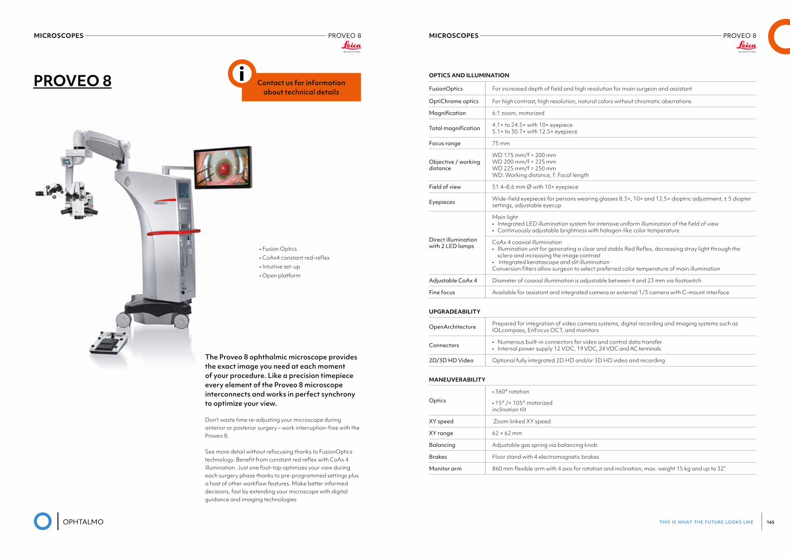
this is what the future looks like 165
MICROSCOPES PROVEO 8Leica
MICROSCOPES PROVEO 8Leica
PROVEO 8
The Proveo 8 ophthalmic microscope provides the exact image you need at each moment of your procedure. Like a precision timepiece every element of the Proveo 8 microscope interconnects and works in perfect synchrony to optimize your view.
Don’t waste time re-adjusting your microscope during anterior or posterior surgery – work interruption-free with the Proveo 8.
See more detail without refocusing thanks to FusionOptics technology. Benefit from constant red reflex with CoAx 4 illumination. Just one foot-tap optimizes your view during each surgery phase thanks to pre-programmed settings plus a host of other workflow features. Make better informed decisions, fast by extending your microscope with digital guidance and imaging technologies
• Fusion Optics • CoAx4 constant red-reflex • Intuitive set-up • Open platform
Contact us for information about technical details
i OPTICS AND ILLUMINATION
FusionOptics For increased depth of field and high resolution for main surgeon and assistant
OptiChrome optics For high contrast, high resolution, natural colors without chromatic aberrations
Magnification 6:1 zoom, motorized
Totalmagnification 4.1× to 24.5× with 10× eyepiece 5.1× to 30.7× with 12.5× eyepiece
Focus range 75 mm
Objective / working distance
WD 175 mm/f = 200 mm WD 200 mm/f = 225 mm WD 225 mm/f = 250 mm WD: Working distance, f: Focal length
Field of view 51.4–8.6 mm Ø with 10× eyepiece
Eyepieces Wide-field eyepieces for persons wearing glasses 8.3×, 10× and 12.5× dioptric adjustment, ± 5 diopter settings, adjustable eyecup
Direct illumination with 2 LED lamps
Main light • Integrated LED illumination system for intensive uniform illumination of the field of view • Continuously adjustable brightness with halogen-like color temperature
CoAx 4 coaxial illumination • Illumination unit for generating a clear and stable Red Reflex, decreasing stray light through the sclera and increasing the image contrast • Integrated keratascope and slit illumination Conversion filters allow surgeon to select preferred color temperature of main illumination
Adjustable CoAx 4 Diameter of coaxial illumination is adjustable between 4 and 23 mm via footswitch
Fine focus Available for assistant and integrated camera or external 1/3 camera with C-mount interface
UPGRADEABILITY
OpenArchitecture Prepared for integration of video camera systems, digital recording and imaging systems such as IOLcompass, EnFocus OCT, and monitors
Connectors • Numerous built-in connectors for video and control data transfer • Internal power supply 12 VDC, 19 VDC, 24 VDC and AC terminals
2D/3DHDVideo Optional fully integrated 2D HD and/or 3D HD video and recording
MANEUVERABILITY
Optics• 360° rotation
• 15° /+ 105° motorized inclination tilt
XY speed Zoom linked XY speed
XY range 62 × 62 mm
Balancing Adjustable gas spring via balancing knob
Brakes Floor stand with 4 electromagnetic brakes
Monitor arm 860 mm flexible arm with 4 axis for rotation and inclination, max. weight 15 kg and up to 32”
MICROSCOPES PROVEO 8Leica
CONTROL
Control unit
• User-friendly, individually programmable touch-screen (up to 30 surgeons) for control of motor functions and light intensity • Menu selection based on unique software for user-specific configuration • Built-in electronic auto-diagnosis and user support • Software independent hard keys and indicator for illumination • Data shown by means of LCD
Control elements Rotary handles • 14-function and 12-function wireless footswitch with optional back-up cable
IR sensor Remote control of the HDR recorder
Indicators • LED for video record status • Surgeon information panel for setting status
CONSTRUCTION
Floor stand Four 360° rotating castors (Ø150 mm), parking brake
Materials • Coated with antimicrobial paint • Conforming with RoHS
Load • Floor stand max. 8.0 kg from microscope dovetail ring interface • C42/CT42 max. 8.0 kg from dovetail ring interface
Weight• Floor stand approx. 350 kg without load • C42 ceiling mount total approx. 260 kg • CT42 telescope mount total approx. 200 kg
TECHNICAL DATA
Power connection• 1100 VA 50/60 Hz • 100–240 V~ 50/60 Hz • 2 × T10 AH 250 V
Protection class Class 1
02LENSMETER
PHOCOMETER
LENSMETER/PHOCOMETER VX40 FULL AUTOMATIC LENSMETERLuneau Visionix
VX40 FULL AUTOMATIC LENSMETER
Visionix wave front technology allows you to have a full detailed analysis of a frame at the push of the button. Experience the next generation autolensmeter with the VX40. The VX40 can detect and analyze single vision, bifocals, and progressive lenses and is compatible with all lens technologies and brands.
MEASURABLE RANGE
Number of analyzed points Up to 1350
Sphere power -15 ~ +10D (step 0.01, 0.06, 0.125, 0.25D)
Cylinder power 0~10D (step 0.01, 0.06, 0.125, 0.25D)
Cylinder axis 0~180° (step 1°)
Addition power 0~± 3.5D (step 0.01, 0.06, 0.125, 0.25D)
Prism power 0~± 10 Δ (step 0.01 Δ)
PD measurement Mono / Bino
Cylinder -, +
GENERAL
Printer Internal
Screen LCD/16M colours, 7’’
Light source LED – 730 nm
Working conditions 10 to 40°C
Data output RS-232, Bluetooth
GENERAL DATA
Height 455 mm (17,90 in.)
Width 220 mm (8,66 in.)
Depth 240 mm (9,44 in.)
Weight 9 kg (20 lbs)
Power supply 115/230V
Standards CE
FEATURES AND BENEFITS
Fully AutomatedThe VX40 is fully automated. After the operator places the frame and initializes the start procedure, lens analysis and measurement is completely automatic. Even left / right eye movement requires no user input.
Easy to useThe unique lens holder system allows for fast insertion of the frame with only one hand! After pushing the start button the whole process works on it’s own. The VX40 allows you to spend more time with your patients and less time with analysis.
Automatic Lens AnalysisThe VX40 detects all types of lenses including bifocals, progressives, freeform, and single vision lenses. It is compatible with virtually every lens technology and brand.
Complete Visual AnalysisThe VX40 analyzes progressive and freeform lenses and then conveys the measurements to the operator in the form of a visual topographic map of the power ranges across the lens. These lens types no longer have to be a mystery to you.
Side By Side Progressive Lens AnalysisThe lens is studied completely which allows for the operator to pick any point on the lens to verify the power and accuracy of the manufacturing. Compare and analyze even freeform progressive lenses and show your patients the differences between different progressive lens designs.
03AUTOREFRACTOR-
KERATOMETER
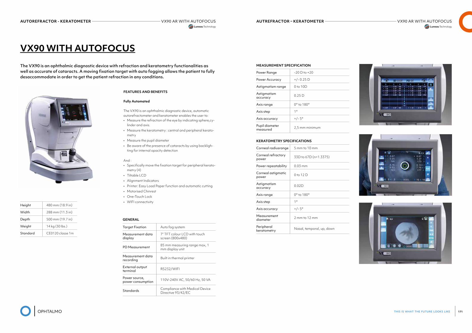
this is what the future looks like 171
AUTREFRACTOR – KERATOMETER VX90 AR WITH AUTOFOCUSLuneau Visionix
AUTOREFRACTOR - KERATOMETER VX90 AR WITH AUTOFOCUSLuneau Visionix
VX90 WITH AUTOFOCUS
The VX90 is an ophthalmic diagnostic device with refraction and keratometry functionalities as well as accurate of cataracts. A moving fixation target with auto fogging allows the patient to fully desaccommodate in order to get the patient refraction in any conditions.
FEATURES AND BENEFITS
Fully Automated
The VX90 is an ophthalmic diagnostic device, automatic autorefractometer and keratometer enables the user to:• Measure the refraction of the eye by indicating sphere,cy-
linder and axis.• Measure the keratometry : central and peripheral kerato-
metry• Measure the pupil diameter• Be aware of the presence of cataracts by using backligh-
ting for internal opacity detection
And :• Specifically move the fixation target for peripheral kerato-
metry (4)• Tiltable LCD• Alignment Indicators• Printer: Easy Load Paper function and automatic cutting• Motorised Chinrest• One-Touch Lock• WIFI connectivity
GENERAL
Target Fixation Auto fog system
Measurement data display
7” TFT colour LCD with touch screen (800x480)
PD Measurement 85 mm measuring range max, 1 mm display unit
Measurement data recording Built in thermal printer
External output terminal RS232/WIFI
Power source, power consumption 110V-240V AC, 50/60 Hz, 50 VA
Standards Compliance with Medical Device Directive 93/42/EC
Height 480 mm (18.9 in)
Width 288 mm (11.3 in)
Depth 500 mm (19.7 in)
Weight 14 kg (30 lbs.)
Standard CE0120 classe 1m
MEASUREMENT SPECIFICATION
Power Range -20 D to +20
Power Accuracy +/- 0.25 D
Astigmatism range 0 to 10D
Astigmatism accuracy 0.25 D
Axis range 0° to 180°
Axis step 1°
Axis accuracy +/- 5°
Pupil diameter measured 2,5 mm minimum
KERATOMETRY SPECIFICATIONS
Corneal radiusrange 5 mm to 10 mm
Corneal refractory power 33D to 67D (n=1.3375)
Power repeatability 0.03 mm
Corneal astigmatic power 0 to 12 D
Astigmatism accuracy 0.02D
Axis range 0° to 180°
Axis step 1°
Axis accuracy +/- 5°
Measurement diameter 2 mm to 12 mm
Peripheral keratometry Nasal, temporal, up, down
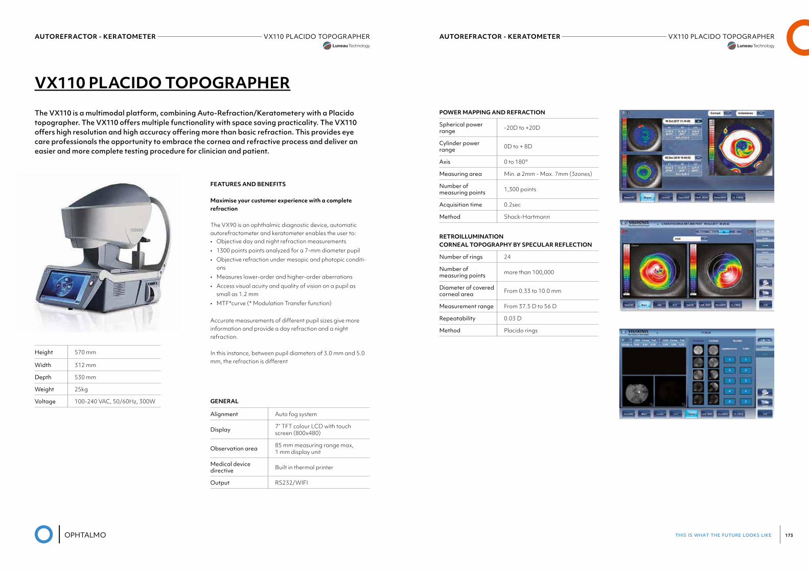
this is what the future looks like 173
AUTOREFRACTOR - KERATOMETER VX110 PLACIDO TOPOGRAPHERLuneau Visionix
AUTOREFRACTOR - KERATOMETER VX110 PLACIDO TOPOGRAPHERLuneau Visionix
VX110 PLACIDO TOPOGRAPHER
The VX110 is a multimodal platform, combining Auto-Refraction/Keratometery with a Placido topographer. The VX110 offers multiple functionality with space saving practicality. The VX110 offers high resolution and high accuracy offering more than basic refraction. This provides eye care professionals the opportunity to embrace the cornea and refractive process and deliver an easier and more complete testing procedure for clinician and patient.
FEATURES AND BENEFITS
Maximise your customer experience with a complete refraction
The VX90 is an ophthalmic diagnostic device, automatic autorefractometer and keratometer enables the user to:• Objective day and night refraction measurements• 1300 points points analyzed for a 7-mm diameter pupil• Objective refraction under mesopic and photopic conditi-
ons• Measures lower-order and higher-order aberrations• Access visual acuity and quality of vision on a pupil as
small as 1.2 mm• MTF*curve (* Modulation Transfer function)
Accurate measurements of different pupil sizes give more information and provide a day refraction and a night refraction.
In this instance, between pupil diameters of 3.0 mm and 5.0 mm, the refraction is different
Height 570 mm
Width 312 mm
Depth 530 mm
Weight 25kg
Voltage 100-240 VAC, 50/60Hz, 300W GENERAL
Alignment Auto fog system
Display 7” TFT colour LCD with touch screen (800x480)
Observation area 85 mm measuring range max, 1 mm display unit
Medical device directive Built in thermal printer
Output RS232/WIFI
POWER MAPPING AND REFRACTION
Spherical power range -20D to +20D
Cylinder power range 0D to + 8D
Axis 0 to 180°
Measuring area Min. ø 2mm - Max. 7mm (3zones)
Number of measuring points 1,300 points
Acquisition time 0.2sec
Method Shack-Hartmann
RETROILLUMINATION CORNEAL TOPOGRAPHY BY SPECULAR REFLECTION
Number of rings 24
Number of measuring points more than 100,000
Diameter of covered corneal area From 0.33 to 10.0 mm
Measurement range From 37.5 D to 56 D
Repeatability 0.03 D
Method Placido rings
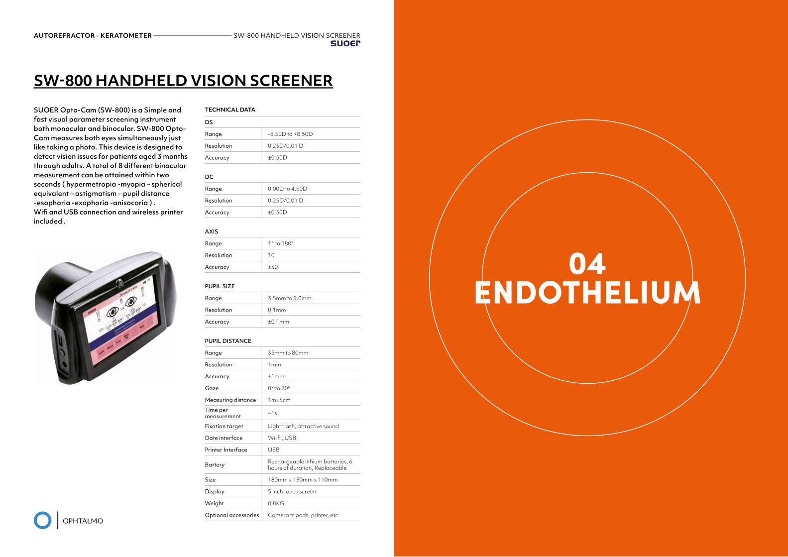
this is what the future looks like 175
04ENDOTHELIUM
AUTOREFRACTOR - KERATOMETER SW-800 HANDHELD VISION SCREENERSuoer
SW-800 HANDHELD VISION SCREENER
SUOER Opto-Cam (SW-800) is a Simple and fast visual parameter screening instrument both monocular and binocular. SW-800 Opto-Cam measures both eyes simultaneously just like taking a photo. This device is designed to detect vision issues for patients aged 3 months through adults. A total of 8 different binocular measurement can be attained within two seconds ( hypermetropia -myopia – spherical equivalent – astigmatism – pupil distance -esophoria -exophoria -anisocoria ) .Wifi and USB connection and wireless printer included .
TECHNICAL DATA
DS
Range -8.50D to +8.50D
Resolution 0.25D/0.01 D
Accuracy ±0.50D
DC
Range 0.00D to 4.50D
Resolution 0.25D/0.01 D
Accuracy ±0.50D
AXIS
Range 1° to 180°
Resolution 10
Accuracy ±50
PUPIL SIZE
Range 3.5mm to 9.0mm
Resolution 0.1mm
Accuracy ±0.1mm
PUPIL DISTANCE
Range 35mm to 80mm
Resolution 1mm
Accuracy ±1mm
Gaze 0° to 20°
Measuring distance 1m±5cm
Time per measurement ~1s
Fixation target Light flash, attractive sound
Date interface Wi-Fi, USB
Printer Interface USB
Battery Rechargeable lithium batteries, 6 hours of duration, Replaceable
Size 180mm x 130mm x 110mm
Display 5 inch touch screen
Weight 0.8KG
Optional accessories Camera tripods, printer, etc
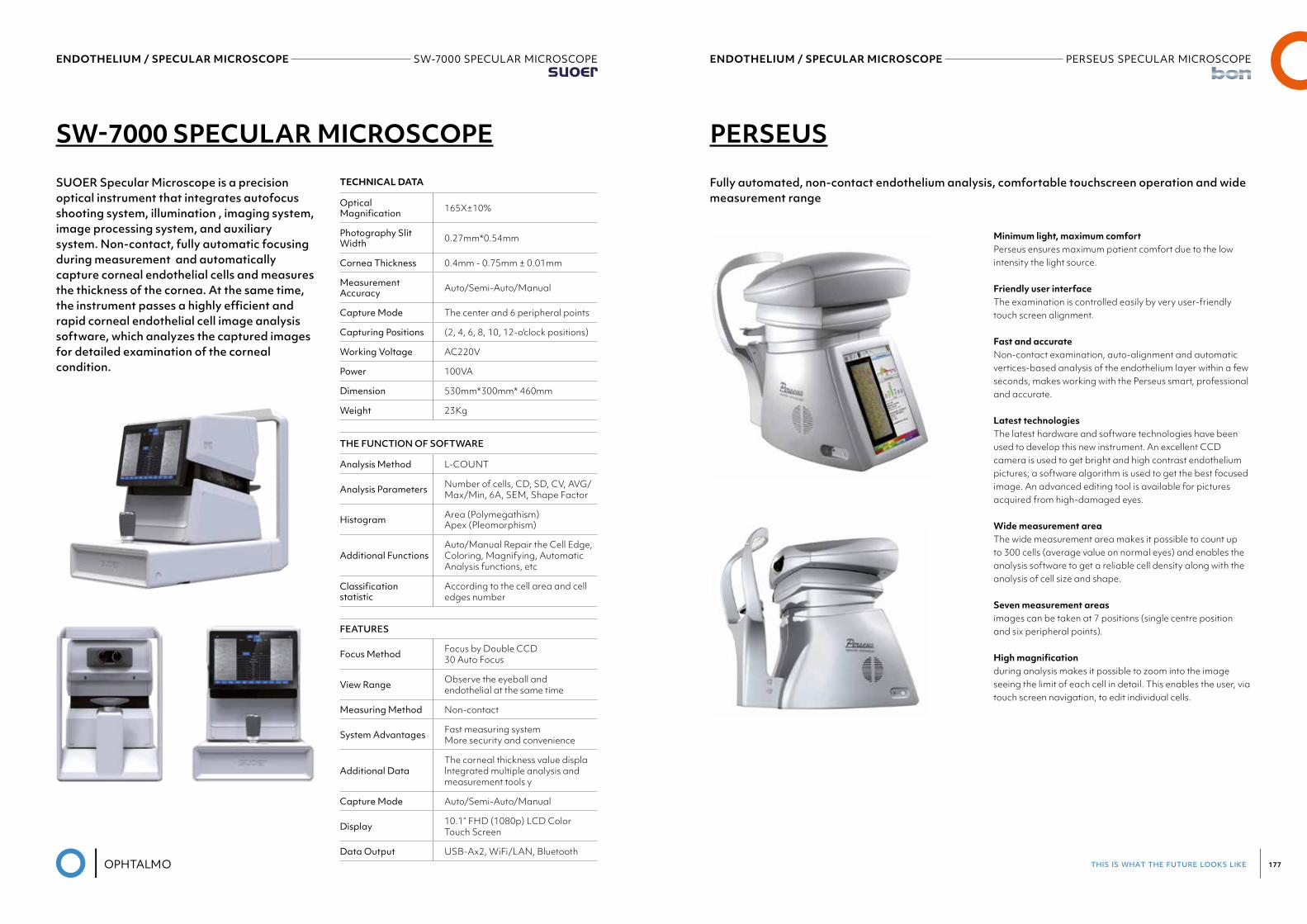
this is what the future looks like 177
ENDOTHELIUM / SPECULAR MICROSCOPE SW-7000 SPECULAR MICROSCOPESuoer
ENDOTHELIUM / SPECULAR MICROSCOPE PERSEUS SPECULAR MICROSCOPEBon
SW-7000 SPECULAR MICROSCOPE
SUOER Specular Microscope is a precision optical instrument that integrates autofocus shooting system, illumination , imaging system, image processing system, and auxiliary system. Non-contact, fully automatic focusing during measurement and automatically capture corneal endothelial cells and measures the thickness of the cornea. At the same time, the instrument passes a highly efficient and rapid corneal endothelial cell image analysis software, which analyzes the captured images for detailed examination of the corneal condition.
TECHNICAL DATA
Optical Magnification 165X±10%
Photography Slit Width 0.27mm*0.54mm
Cornea Thickness 0.4mm - 0.75mm ± 0.01mm
Measurement Accuracy Auto/Semi-Auto/Manual
Capture Mode The center and 6 peripheral points
Capturing Positions (2, 4, 6, 8, 10, 12-o'clock positions)
Working Voltage AC220V
Power 100VA
Dimension 530mm*300mm* 460mm
Weight 23Kg
THE FUNCTION OF SOFTWARE
Analysis Method L-COUNT
Analysis Parameters Number of cells, CD, SD, CV, AVG/Max/Min, 6A, SEM, Shape Factor
Histogram Area (Polymegathism) Apex (Pleomorphism)
Additional FunctionsAuto/Manual Repair the Cell Edge, Coloring, Magnifying, Automatic Analysis functions, etc
Classificationstatistic
According to the cell area and cell edges number
FEATURES
Focus Method Focus by Double CCD 30 Auto Focus
View Range Observe the eyeball and endothelial at the same time
Measuring Method Non-contact
System Advantages Fast measuring system More security and convenience
Additional DataThe corneal thickness value displa lntegrated multiple analysis and measurement tools y
Capture Mode Auto/Semi-Auto/Manual
Display 10.1” FHD (1080p) LCD Color Touch Screen
Data Output USB-Ax2, WiFi/LAN, Bluetooth
PERSEUS
Fully automated, non-contact endothelium analysis, comfortable touchscreen operation and wide measurement range
Minimum light, maximum comfortPerseus ensures maximum patient comfort due to the low intensity the light source.
Friendly user interfaceThe examination is controlled easily by very user-friendly touch screen alignment.
Fast and accurateNon-contact examination, auto-alignment and automatic vertices-based analysis of the endothelium layer within a few seconds, makes working with the Perseus smart, professional and accurate.
Latest technologiesThe latest hardware and software technologies have been used to develop this new instrument. An excellent CCD camera is used to get bright and high contrast endotheliumpictures; a software algorithm is used to get the best focused image. An advanced editing tool is available for pictures acquired from high-damaged eyes.
Wide measurement areaThe wide measurement area makes it possible to count up to 300 cells (average value on normal eyes) and enables the analysis software to get a reliable cell density along with the analysis of cell size and shape.
Seven measurement areasimages can be taken at 7 positions (single centre position and six peripheral points).
High magnificationduring analysis makes it possible to zoom into the image seeing the limit of each cell in detail. This enables the user, via touch screen navigation, to edit individual cells.
ENDOTHELIUM / SPECULAR MICROSCOPE PERSEUS SPECULAR MICROSCOPEBon
Software featuresthe intuitive software evaluates all relevant endothelium data such as:• Number of counted cells• Cell density (number of cells per 1 mm²)• Average dimension of analysed cells• Standard deviation of analysed cell dimension• Coefficient of variation• Statistical distribution of sizes (polymegathism) and sha-
pes (pleomorphism)
Data managementis integrated stand-alone in Perseus software on via network to share the data management with existing b o n archives.
Data can also be transferred – via network connection – to a dicom (Digital Imaging and Communications in Medicine) compatible server.
Pachymetrynon-contact pachymetry is performed on every picture acquisition.
05PERIMETRY
this is what the future looks like 181
PERIMETRY AP-300Frey Medical
PERIMETRY AP-300Frey Medical
AP-300
Automated perimeter
AP 300 is a modern and innovative Automated Perimeter that meets global standards for excellence around visual sensitivity testing, diagnosis and management of eye disease. AP-300 platform of advanced diagnostics uses Kinetic and Static Perimetry, including white/white perimetry, blue/yellow (SWAP - Short wavelength automated perimetry) and Flicker (critical flicker fusion perimetry) for early glaucoma detection plus real Goldmann Kinetic Perimetry.
AP-300 comes standard with an extensive range of strategies, fields and test parameters. Built-in camera completed by automated eye tacking provides reliable automated fixation control. Standard built-in data capture and analysis include regression analysis of the visual field on the basis of examination results. Easy to navigate, intuitive software allows easy operation and is designed to be operated using the touch screen. AP-300 design includes built-in high quality PC computer
• Frey Perimeter• Rapid testing times• Accurate results• User friendly software• lmproved patient comfort• Complete analysis modes• Multiple test capabilities• Networking
Height 637 mm
Width 566 mm
Depth 420 mm
Weight 23 kg
TEST SPECIFICATIONS
Maximum temporal range (degrees) 80
Stimulus duration 0.1 - 9.9s
Visualfieldtestingdistance 30 cm
Background illumination 31,5 ASB
TEST MODES
Supra threshold age corrected (Screening) •
Single intesity •
Full intensity •
Fast threshold •
Smart threshold •
2-Zone,3-Zone,QuantifyDefect,Neurological •
SPECIAL TEST LIBRARY
Bi-Driving, Industrial Medicine, monoular, binocular •
Peripheral •
Kinetic testing •
Blue-on-Yellow (SWAP) •
Custom testing •
TEST FIELD LIBRARY
24-2,30-2,10-2,Macula •
Nasal step (Glaucoma) •
Central10,Central20,Central30,Macula •
Peripheral •
COMPUTER
Build-in PC •
Touch screen suport •
Fixation control •
HeijlKrakaublindspotmonitor •
Eye tracking (video camera) •
Eye preview (video camera •
STIMULUS
White on white •
Green on white •
Red on white •
Blue on white •
GENERAL TESTING FEATURES
Stimulus size (Goldman size) I-V
Fovea threshold testing •
Automatic piupil measurement •
ADDITIONAL SOFTWARE FEATURES
Network connectivity •
DICOM export •
Targeted perimetry (merging tests with fundus images) •
Fast threshold strategies •
Time adaptive algorithms
Regression analysis
Printer
External or network printer
PERIMETRY M-700Medmont
PERIMETRY M-700Medmont
M-700
06FUNDUSCAMERA
Soon available i
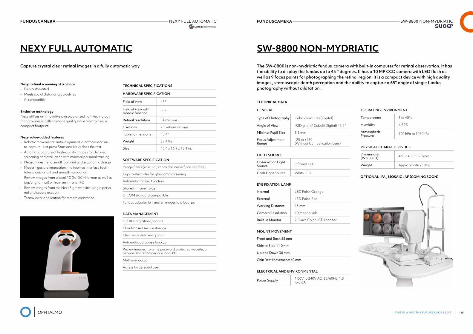
this is what the future looks like 185
FUNDUSCAMERA SW-8800 NON-MYDRIATICSuoer
FUNDUSCAMERA NEXY FULL AUTOMATICLuneau Visionix
NEXY FULL AUTOMATIC SW-8800 NON-MYDRIATIC
Capture crystal clear retinal images in a fully automatic way The SW-8800 is non-mydriatic fundus camera with built-in computer for retinal observation. It has the ability to display the fundus up to 45 ° degrees. It has a 10 MP CCD camera with LED flash as well as 9 focus points for photographing the retinal region. It is a compact device with high quality images , stereoscopic depth perception and the ability to capture a 65° angle of single fundus photography without dilatation .
Nexy: retinal screening at a glance• Fully automated• Meets social distancing guidelines• AI compatible
Exclusive technologyNexy utilises an innovative cross-polarized light technology that provides excellent image quality while maintaining a compact footprint.
Nexy value-added features• Robotic movements: auto-alignment, autofocus and au-
to-capture. Just press Start and Nexy does the rest• Automatic capture of high-quality images for detailed
screening and evaluation with minimal personal training• Pleasant aesthetic: small footprint and ergonomic design• Modern gesture interaction: the intuitive interface facili-
tates a quick start and smooth navigation• Review images from a local PC (in .DCM format as well as
jpg/png format) or from an intranet PC• Review images from the Next Sight website using a perso-
nal and secure account• Teamviewer application for remote assistance
TECHNICAL SPECIFICATIONS
HARDWARE SPECIFICATION
Field of view 45°
Field of view with mosaic function 90°
Retinal resolution 14 microns
Fixations 7 fixations set-ups
Tablet dimensions 10.4’’
Weight 32,4 lbs
Size 13,4 x 16,9 x 18,1 in.
SOFTWARE SPECIFICATION
Image filters (vascular, choroidal, nerve fibre, red free)
Cup-to-disc ratio for glaucoma screening
Automatic mosaic function
Shared intranet folder
DICOM standard compatible
Fundus adapter to transfer images to a local pc
DATA MANAGEMENT
Full AI integration (option)
Cloud-based secure storage
Client-side data encryption
Automatic database backup
Review images from the password protected website, a network shared folder or a local PC
Multilevel account
Access by personal user
TECHNICAL DATA
GENERAL
Type of Photography Color / Red-Free(Digital)
Angle of View IR(Digital) / Cobalt(Digital) 46.5°
Minimal Pupil Size 3.5 mm
Focus Adjustment Range
-25 to +250(Without Compensation Lens)
LIGHT SOURCE
Observation Light Source lnfrared LED
Flash Light Source White LED
EYE FIXATION LAMP
lnternal LED Point, Orange
External LED Point, Red
Working Distance 15 mm
Camera Resolution 10 Megapixels
Built-in Monitor 7.0 inch Color LCD Monitor
MOUNT MOVEMENT
Front and Back 85 mm
Side to Side 11 0 mm
UpandDown30mm
Chin Rest Movement: 60 mm
ELECTRICAL AND ENVIRONMENTAL
Power Supply 1 00V to 240V AC, 50/60Hz, 1.3 to 0.6A
OPERATING ENVIRONMENT
Temperature 5 to 40°c
Humidity ≤ 80%
Atmospheric Pressure 700 hPa to 1060hPa
PHYSICAL CHARACTERISTICS
Dimensions (W x D x H): 430 x 450 x 570 mm
Weight Approximately 10Kg
OPTIONAL : FA , MOSAIC , AF (COMING SOON)
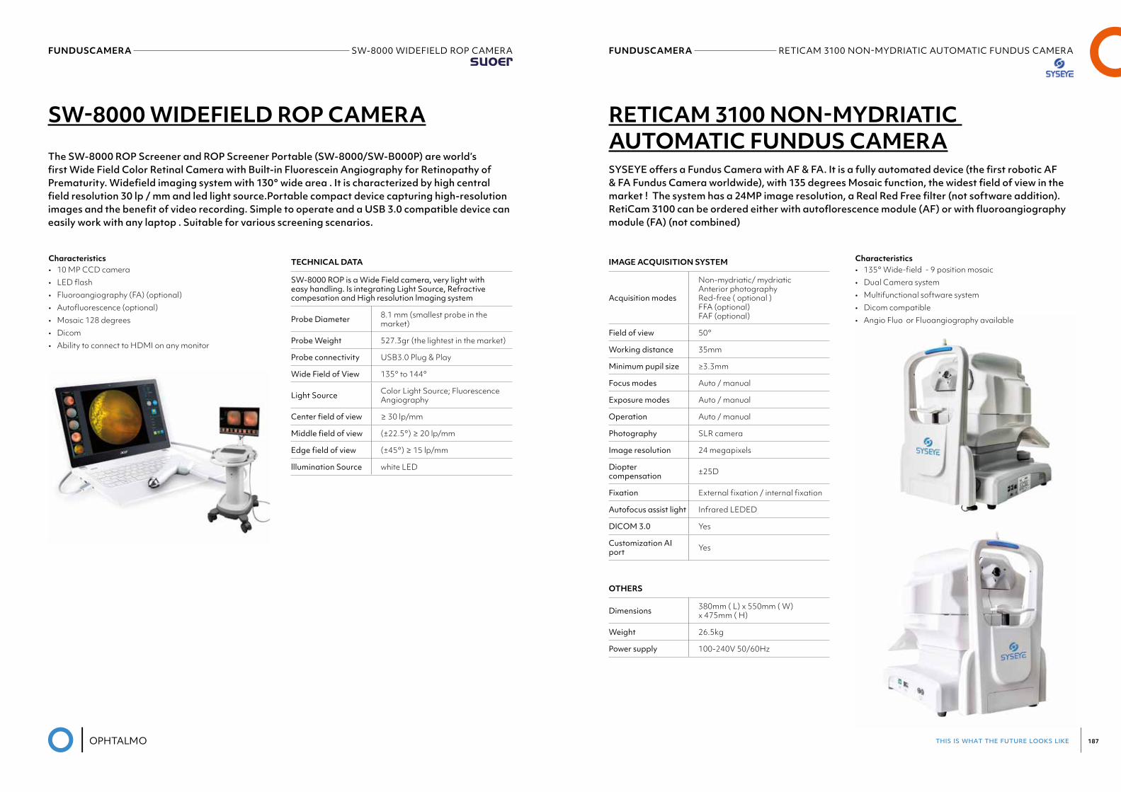
this is what the future looks like 187
FUNDUSCAMERA RETICAM3100NON-MYDRIATICAUTOMATICFUNDUSCAMERASyseye
FUNDUSCAMERA SW-8000 WIDEFIELD ROP CAMERASuoer
SW-8000 WIDEFIELD ROP CAMERA RETICAM 3100 NON-MYDRIATIC AUTOMATIC FUNDUS CAMERA
The SW-8000 ROP Screener and ROP Screener Portable (SW-8000/SW-B000P) are world’s first Wide Field Color Retinal Camera with Built-in Fluorescein Angiography for Retinopathy of Prematurity. Widefield imaging system with 130° wide area . It is characterized by high central field resolution 30 lp / mm and led light source.Portable compact device capturing high-resolution images and the benefit of video recording. Simple to operate and a USB 3.0 compatible device can easily work with any laptop . Suitable for various screening scenarios.
SYSEYE offers a Fundus Camera with AF & FA. It is a fully automated device (the first robotic AF & FA Fundus Camera worldwide), with 135 degrees Mosaic function, the widest field of view in the market ! The system has a 24MP image resolution, a Real Red Free filter (not software addition).RetiCam 3100 can be ordered either with autoflorescence module (AF) or with fluoroangiography module (FA) (not combined)
Characteristics• 10 MP CCD camera• LED flash• Fluoroangiography (FA) (optional)• Αutofluorescence (optional)• Mosaic 128 degrees• Dicom• Ability to connect to HDMI on any monitor
Characteristics• 135° Wide-field - 9 position mosaic • Dual Camera system • Multifunctional software system • Dicom compatible • Angio Fluo or Fluoangiography available
TECHNICAL DATA
SW-8000 ROP is a Wide Field camera, very light with easy handling. Is integrating Light Source, Refractive compesation and High resolution lmaging system
Probe Diameter 8.1 mm (smallest probe in the market)
Probe Weight 527.3gr (the lightest in the market)
Probe connectivity USB3.0 Plug & Play
Wide Field of View 135° to 144°
Light Source Color Light Source; Fluorescence Angiography
Centerfieldofview ≥ 30 lp/mm
Middlefieldofview (±22.5°) ≥ 20 lp/mm
Edgefieldofview (±45°) ≥ 15 lp/mm
lllumination Source white LED
IMAGE ACQUISITION SYSTEM
Acquisition modes
Non-mydriatic/ mydriaticAnterior photographyRed-free ( optional )FFA (optional)FAF (optional)
Field of view 50°
Working distance 35mm
Minimum pupil size ≥3.3mm
Focus modes Auto / manual
Exposure modes Auto / manual
Operation Auto / manual
Photography SLR camera
Image resolution 24 megapixels
Diopter compensation ±25D
Fixation External fixation / internal fixation
Autofocus assist light Infrared LEDED
DICOM3.0 Yes
Customization AI port Yes
OTHERS
Dimensions 380mm ( L) x 550mm ( W) x 475mm ( H)
Weight 26.5kg
Power supply 100-240V 50/60Hz
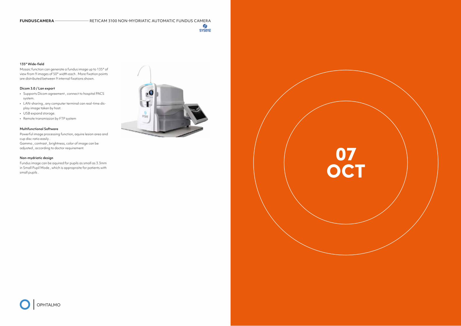
FUNDUSCAMERA RETICAM3100NON-MYDRIATICAUTOMATICFUNDUSCAMERASyseye
07OCT
135° Wide-fieldMosaic function can generate a fundus image up to 135° of view from 9 images of 50° width each . More fixation points are distributed between 9 internal fixations shown.
Dicom 3.0 / Lan export• Supports Dicom agreement , connect to hospital PACS
system.• LAN-sharing , any computer terminal can real-time dis-
play image taken by host.• USB expand storage.• Remote transmission by FTP system
Multifunctional SoftwarePowerful image processing function, aquire lesion area and cup disc ratio easily .Gamma , contrast , brightness, color of image can be adjusted , according to doctor requirement.
Non-mydriatic designFundus image can be aquired for pupils as small as 3.3mm in Small Pupil Mode , which is appropraite for patients with small pupils .
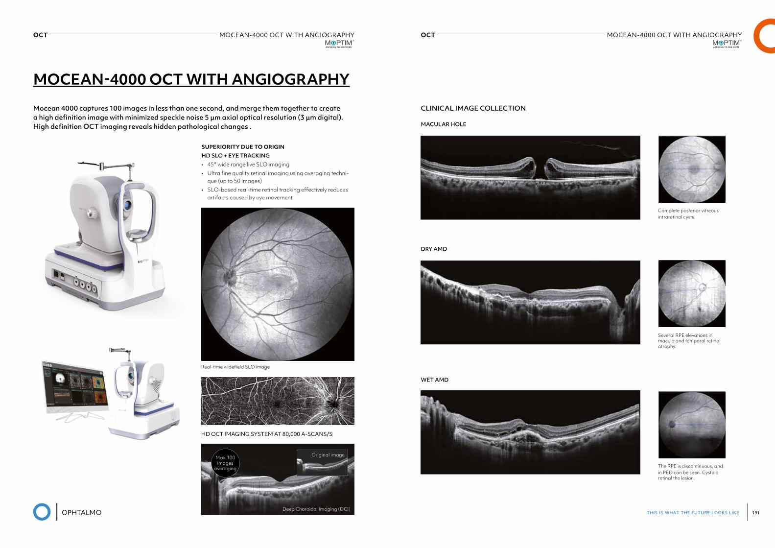
this is what the future looks like 191
OCT MOCEAN-4000 OCT WITH ANGIOGRAPHYMoptim
OCT MOCEAN-4000 OCT WITH ANGIOGRAPHYMoptim
MOCEAN-4000 OCT WITH ANGIOGRAPHY
Mocean 4000 captures 100 images in less than one second, and merge them together to create a high definition image with minimized speckle noise 5 μm axial optical resolution (3 μm digital). High definition OCT imaging reveals hidden pathological changes .
CLINICAL IMAGE COLLECTION
SUPERIORITY DUE TO ORIGINHD SLO + EYE TRACKING• 45° wide range live SLO imaging• Ultra fine quality retinal imaging using averaging techni-
que (up to 50 images)• SLO-based real-time retinal tracking effectively reduces
artifacts caused by eye movement
MACULAR HOLE
DRY AMD
WET AMD
Real-time widefield SLO image
Complete posterior vitreousintraretinal cysts.
Several RPE elevations in macula and temporal retinal atrophy.
The RPE is discontinuous, and in PED can be seen. Cystoid retinal the lesion.
HD OCT IMAGING SYSTEM AT 80,000 A-SCANS/S
Deep Choroidal Imaging (DCI)
Original imageMax.100images
averaging
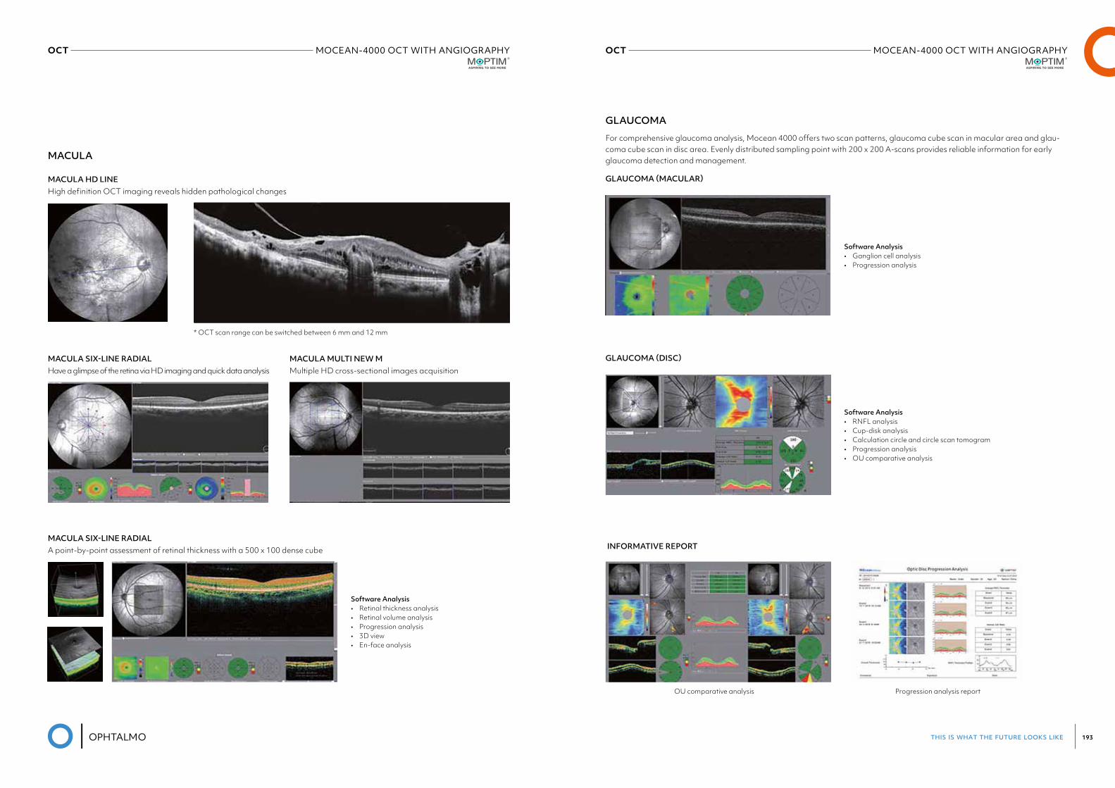
this is what the future looks like 193
OCT MOCEAN-4000 OCT WITH ANGIOGRAPHYMoptim
OCT MOCEAN-4000 OCT WITH ANGIOGRAPHYMoptim
MACULA
GLAUCOMA
GLAUCOMA (MACULAR)
MACULA SIX-LINE RADIAL
MACULA SIX-LINE RADIAL
MACULA HD LINE
MACULA MULTI NEW M GLAUCOMA (DISC)
INFORMATIVE REPORT
Have a glimpse of the retina via HD imaging and quick data analysis
A point-by-point assessment of retinal thickness with a 500 x 100 dense cube
High definition OCT imaging reveals hidden pathological changes
For comprehensive glaucoma analysis, Mocean 4000 offers two scan patterns, glaucoma cube scan in macular area and glau-coma cube scan in disc area. Evenly distributed sampling point with 200 x 200 A-scans provides reliable information for early glaucoma detection and management.
Multiple HD cross-sectional images acquisition
* OCT scan range can be switched between 6 mm and 12 mm
OU comparative analysis Progression analysis report
Software Analysis• Retinal thickness analysis• Retinal volume analysis• Progression analysis• 3D view• En-face analysis
Software Analysis• Ganglion cell analysis• Progression analysis
Software Analysis• RNFL analysis• Cup-disk analysis• Calculation circle and circle scan tomogram• Progression analysis• OU comparative analysis
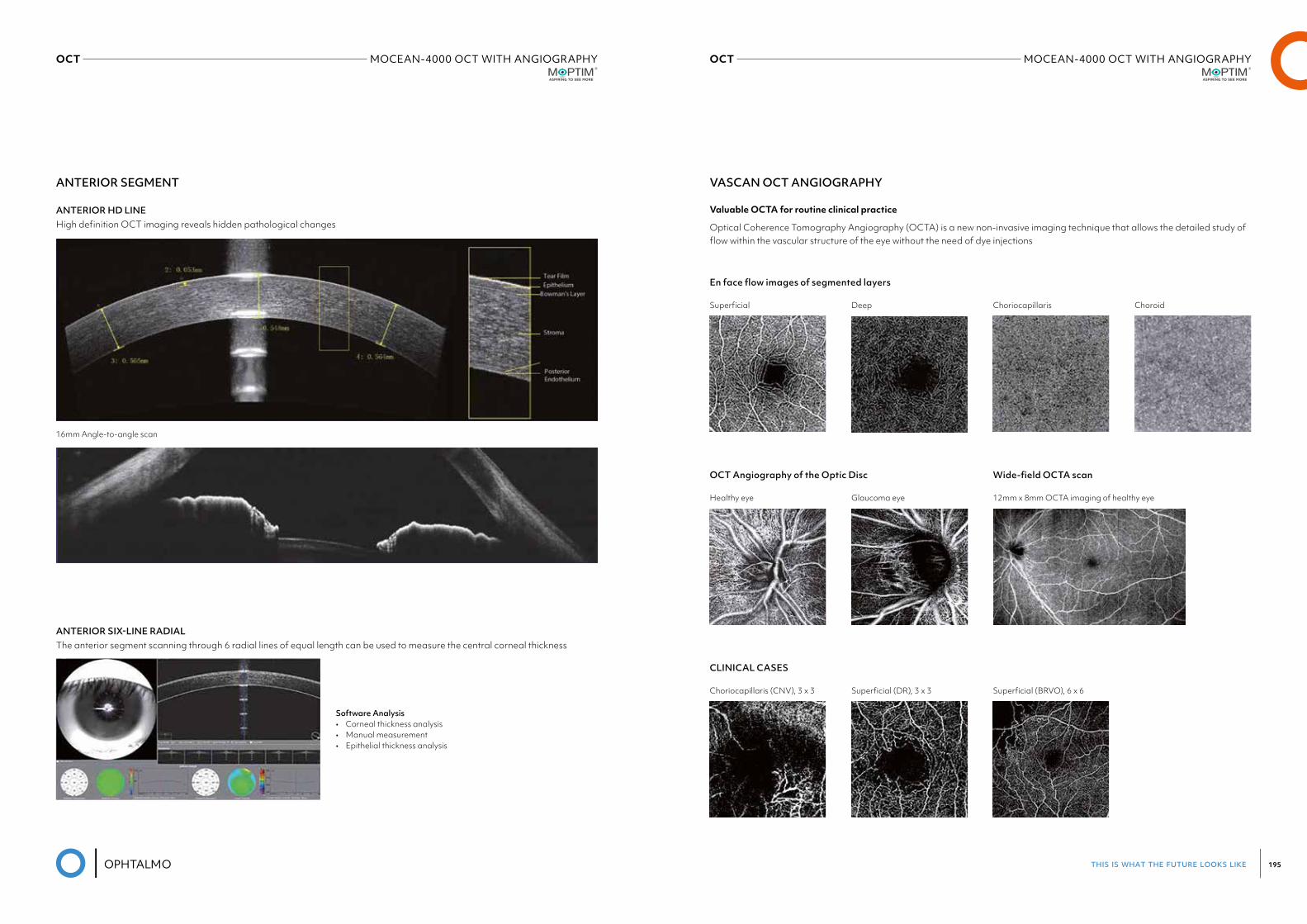
this is what the future looks like 195
OCT MOCEAN-4000 OCT WITH ANGIOGRAPHYMoptim
OCT MOCEAN-4000 OCT WITH ANGIOGRAPHYMoptim
VASCAN OCT ANGIOGRAPHY
Valuable OCTA for routine clinical practice
En face flow images of segmented layers
OCT Angiography of the Optic Disc Wide-field OCTA scan
CLINICAL CASES
ANTERIOR SEGMENT
ANTERIOR SIX-LINE RADIAL
ANTERIOR HD LINE
16mm Angle-to-angle scan
The anterior segment scanning through 6 radial lines of equal length can be used to measure the central corneal thickness
High definition OCT imaging reveals hidden pathological changes Optical Coherence Tomography Angiography (OCTA) is a new non-invasive imaging technique that allows the detailed study of flow within the vascular structure of the eye without the need of dye injections
Software Analysis• Corneal thickness analysis• Manual measurement• Epithelial thickness analysis
Superficial
Healthy eye
Choriocapillaris (CNV), 3 x 3
Deep
Glaucoma eye
Superficial (DR), 3 x 3
Choriocapillaris
12mm x 8mm OCTA imaging of healthy eye
Superficial (BRVO), 6 x 6
Choroid
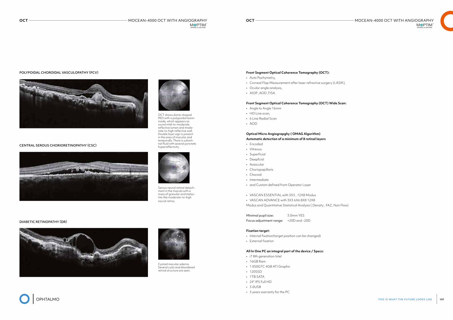
this is what the future looks like 197
OCT MOCEAN-4000 OCT WITH ANGIOGRAPHYMoptim
OCT MOCEAN-4000 OCT WITH ANGIOGRAPHYMoptim
POLYPOIDAL CHOROIDAL VASCULOPATHY (PCV)
CENTRAL SEROUS CHORIORETINOPATHY (CSC)
DIABETIC RETINOPATHY (DR)
OCT shows dome-shaped PED with a polypoidal lesion inside, which appears as round mild-to-moderate reflective lumen and mode-rate-to-high reflective wall. Double layer sign is present in the area of macular and temporally. There is subreti-nal fluid with several punctate hyperreflectivity.
Serous neural retinal detach-ment in the macula with a mass of granular and stalac-tite-like moderate-to-high neural retina.
Cystoid macular edema. Several cysts and disordered retinal structure are seen.
Front Segment Optical Coherence Tomography (OCT): • Auto Pachymetry,• Corneal Flap Measurement after laser refractive surgery (LASIK),• Ocular angle analysis,• AIOP ,AOD ,TISA
Front Segment Optical Coherence Tomography (OCT) Wide Scan: • Angle to Angle 16mm• HD Line scan,• 6 Line Radial Scan• AOD
Optical Micro Angiograpghy ( OMAG Algorithm) Automatic detection of a minimum of 8 retinal layers • Encoded• Vitreous• Superficial• Deepfcial• Avascular• Choriopapillaris• Choroid• Intermediate• and Custom defined from Operator Layer
• VASCAN ESSENTIAL with 3X3 , 12X8 Modus• VASCAN ADVANCE with 3X3 6X6 8X8 12X8Modus and Quantitative Statistical Analysis ( Density , FAZ, Non Flow)
Minimal pupil size: 3.0mm YESFocus adjustment range: +20D and -20D
Fixation target: • Internal fixation(target position can be changed)• External fixation
All In One PC an integral part of the device / Specs: • i7 8th generation Intel• 16GB Ram• 1 0S0GTC 4GB ATI Graphic• 120SSD• 1TB SATA• 24” IPS Full HD• 3.0USB• 3 years warranty for the PC
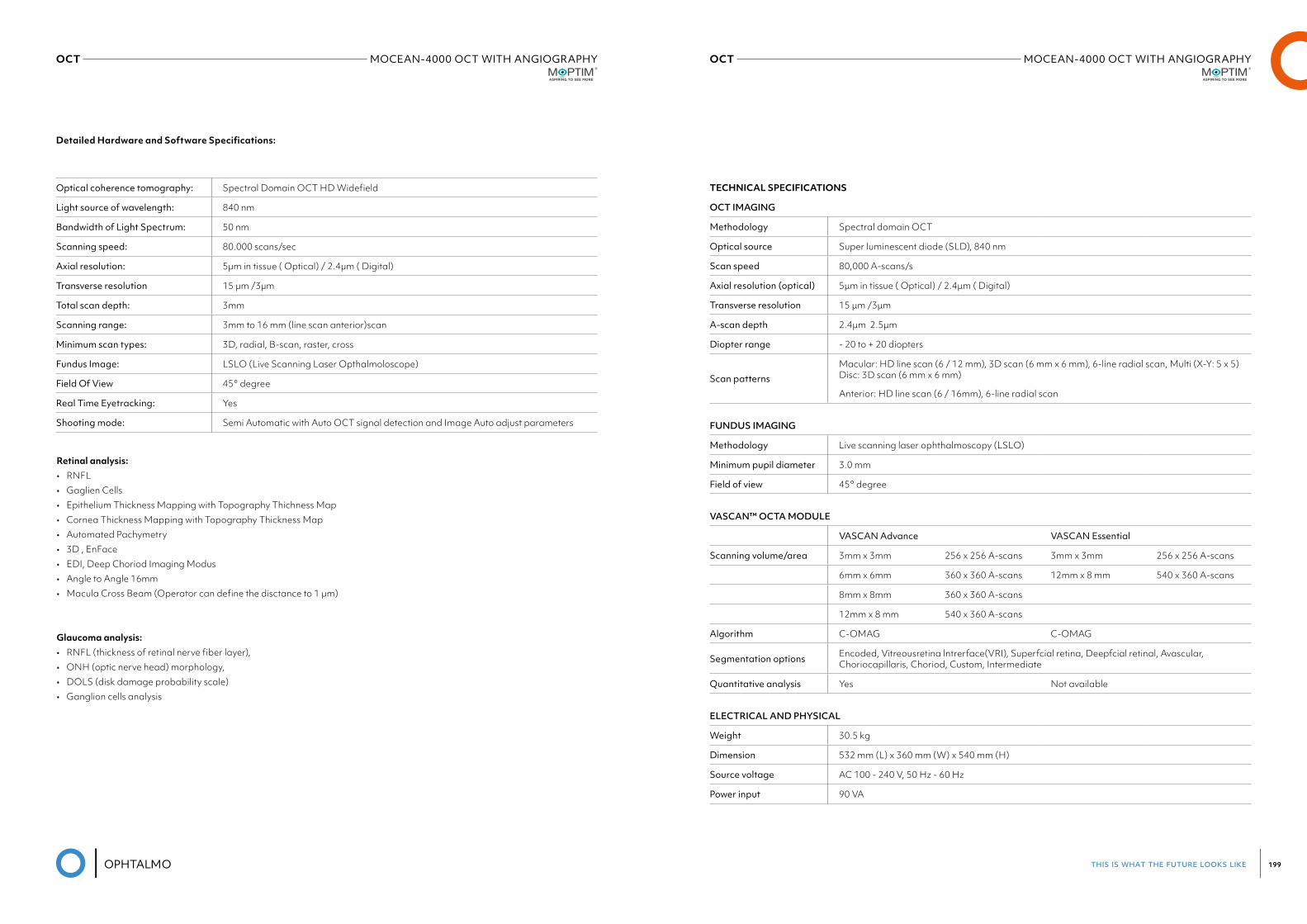
this is what the future looks like 199
OCT MOCEAN-4000 OCT WITH ANGIOGRAPHYMoptim
OCT MOCEAN-4000 OCT WITH ANGIOGRAPHYMoptim
TECHNICAL SPECIFICATIONS
OCT IMAGING
Methodology Spectral domain OCT
Optical source Super luminescent diode (SLD), 840 nm
Scan speed 80,000 A-scans/s
Axial resolution (optical) 5μm in tissue ( Optical) / 2.4μm ( Digital)
Transverse resolution 15 μm /3μm
A-scan depth 2.4μm 2.5μm
Diopter range - 20 to + 20 diopters
Scan patternsMacular: HD line scan (6 / 12 mm), 3D scan (6 mm x 6 mm), 6-line radial scan, Multi (X-Y: 5 x 5) Disc: 3D scan (6 mm x 6 mm)
Anterior: HD line scan (6 / 16mm), 6-line radial scan
FUNDUS IMAGING
Methodology Live scanning laser ophthalmoscopy (LSLO)
Minimum pupil diameter 3.0 mm
Field of view 45° degree
VASCAN™ OCTA MODULE
VASCAN Advance VASCAN Essential
Scanning volume/area 3mm x 3mm 256 x 256 A-scans 3mm x 3mm 256 x 256 A-scans
6mm x 6mm 360 x 360 A-scans 12mm x 8 mm 540 x 360 A-scans
8mm x 8mm 360 x 360 A-scans
12mm x 8 mm 540 x 360 A-scans
Algorithm C-OMAG C-OMAG
Segmentation options Encoded, Vitreousretina lntrerface(VRI), Superfcial retina, Deepfcial retinal, Avascular, Choriocapillaris, Choriod, Custom, Intermediate
Quantitativeanalysis Yes Not available
ELECTRICAL AND PHYSICAL
Weight 30.5 kg
Dimension 532 mm (L) x 360 mm (W) x 540 mm (H)
Source voltage AC 100 - 240 V, 50 Hz - 60 Hz
Power input 90 VA
Optical coherence tomography: Spectral Domain OCT HD Widefield
Light source of wavelength: 840 nm
Bandwidth of Light Spectrum: 50 nm
Scanning speed: 80.000 scans/sec
Axial resolution: 5µm in tissue ( Optical) / 2.4µm ( Digital)
Transverse resolution 15 µm /3µm
Total scan depth: 3mm
Scanning range: 3mm to 16 mm (line scan anterior)scan
Minimum scan types: 3D, radial, B-scan, raster, cross
Fundus Image: LSLO (Live Scanning Laser Opthalmoloscope)
Field Of View 45° degree
Real Time Eyetracking: Yes
Shooting mode: Semi Automatic with Auto OCT signal detection and Image Auto adjust parameters
Detailed Hardware and Software Specifications:
Retinal analysis: • RNFL• Gaglien Cells• Epithelium Thickness Mapping with Topography Thichness Map• Cornea Thickness Mapping with Topography Thickness Map• Automated Pachymetry• 3D , EnFace• EDI, Deep Choriod Imaging Modus• Angle to Angle 16mm• Macula Cross Beam (Operator can define the disctance to 1 µm)
Glaucoma analysis: • RNFL (thickness of retinal nerve fiber layer),• ONH (optic nerve head) morphology,• DOLS (disk damage probability scale)• Ganglion cells analysis

08REFRACTION
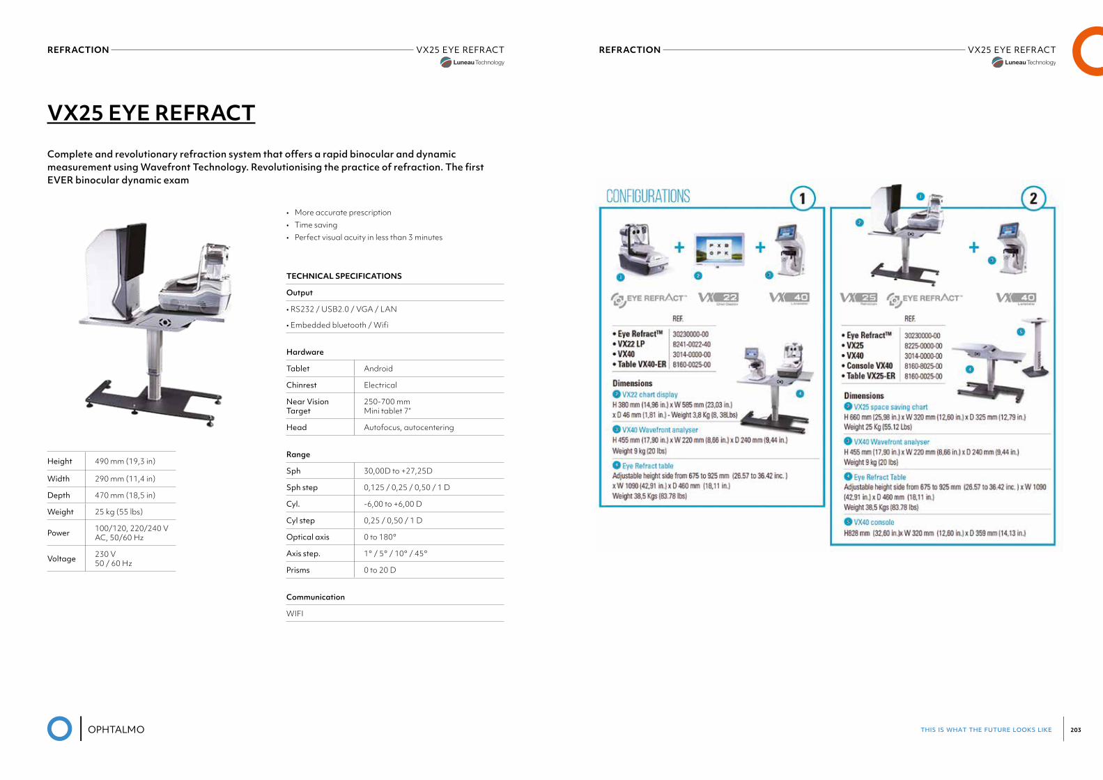
this is what the future looks like 203
REFRACTION VX25 EYE REFRACTLuneau Visionix
REFRACTION VX25 EYE REFRACTLuneau Visionix
VX25 EYE REFRACT
Complete and revolutionary refraction system that offers a rapid binocular and dynamic measurement using Wavefront Technology. Revolutionising the practice of refraction. The first EVER binocular dynamic exam
• More accurate prescription • Time saving • Perfect visual acuity in less than 3 minutes
TECHNICAL SPECIFICATIONS
Output
• RS232 / USB2.0 / VGA / LAN
• Embedded bluetooth / Wifi
Hardware
Tablet Android
Chinrest Electrical
Near Vision Target
250-700 mm Mini tablet 7“
Head Autofocus, autocentering
Range
Sph 30,00D to +27,25D
Sph step 0,125 / 0,25 / 0,50 / 1 D
Cyl. -6,00 to +6,00 D
Cyl step 0,25 / 0,50 / 1 D
Optical axis 0 to 180°
Axis step. 1° / 5° / 10° / 45°
Prisms 0 to 20 D
Communication
WIFI
Height 490 mm (19,3 in)
Width 290 mm (11,4 in)
Depth 470 mm (18,5 in)
Weight 25 kg (55 lbs)
Power 100/120, 220/240 V AC, 50/60 Hz
Voltage 230 V 50 / 60 Hz
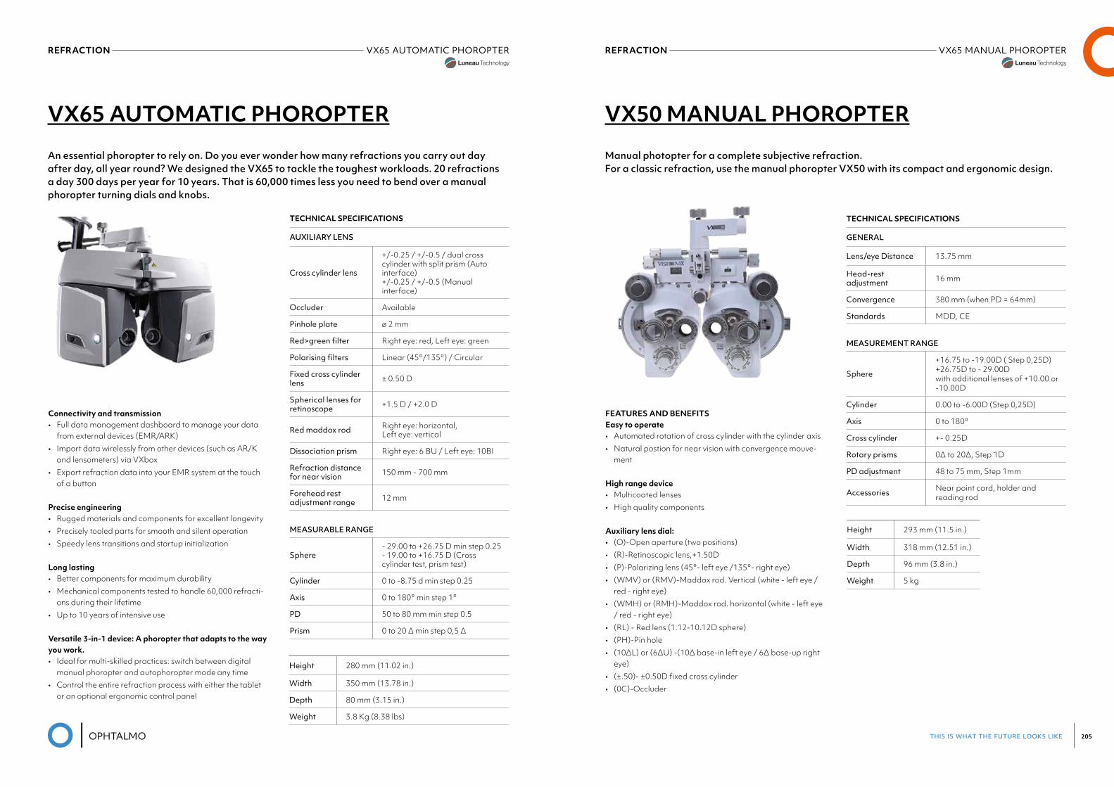
this is what the future looks like 205
REFRACTION VX65 MANUAL PHOROPTERREFRACTION VX65 AUTOMATIC PHOROPTER
VX50 MANUAL PHOROPTER
Manual photopter for a complete subjective refraction.For a classic refraction, use the manual phoropter VX50 with its compact and ergonomic design.
FEATURES AND BENEFITS Easy to operate• Automated rotation of cross cylinder with the cylinder axis• Natural postion for near vision with convergence mouve-
ment
High range device• Multicoated lenses• High quality components
Auxiliary lens dial:• (O)-Open aperture (two positions)• (R)-Retinoscopic lens,+1.50D• (P)-Polarizing lens (45°- left eye /135°- right eye)• (WMV) or (RMV)-Maddox rod. Vertical (white - left eye /
red - right eye)• (WMH) or (RMH)-Maddox rod. horizontal (white - left eye
/ red - right eye)• (RL) - Red lens (1.12-10.12D sphere)• (PH)-Pin hole• (10ΔL) or (6ΔU) -(10Δ base-in left eye / 6Δ base-up right
eye)• (±.50)- ±0.50D fixed cross cylinder• (0C)-Occluder
Height 293 mm (11.5 in.)
Width 318 mm (12.51 in.)
Depth 96 mm (3.8 in.)
Weight 5 kg
TECHNICAL SPECIFICATIONS
GENERAL
Lens/eye Distance 13.75 mm
Head-rest adjustment 16 mm
Convergence 380 mm (when PD = 64mm)
Standards MDD, CE
MEASUREMENT RANGE
Sphere
+16.75 to -19.00D ( Step 0,25D) +26.75D to - 29.00D with additional lenses of +10.00 or -10.00D
Cylinder 0.00 to -6.00D (Step 0,25D)
Axis 0 to 180°
Cross cylinder +- 0.25D
Rotary prisms 0∆ to 20∆, Step 1D
PD adjustment 48 to 75 mm, Step 1mm
Accessories Near point card, holder and reading rod
VX65 AUTOMATIC PHOROPTER
An essential phoropter to rely on. Do you ever wonder how many refractions you carry out day after day, all year round? We designed the VX65 to tackle the toughest workloads. 20 refractions a day 300 days per year for 10 years. That is 60,000 times less you need to bend over a manual phoropter turning dials and knobs.
Connectivity and transmission• Full data management dashboard to manage your data
from external devices (EMR/ARK)• Import data wirelessly from other devices (such as AR/K
and lensometers) via VXbox• Export refraction data into your EMR system at the touch
of a button
Precise engineering• Rugged materials and components for excellent longevity• Precisely tooled parts for smooth and silent operation• Speedy lens transitions and startup initialization
Long lasting• Better components for maximum durability• Mechanical components tested to handle 60,000 refracti-
ons during their lifetime• Up to 10 years of intensive use
Versatile 3-in-1 device: A phoropter that adapts to the way you work.• Ideal for multi-skilled practices: switch between digital
manual phoropter and autophoropter mode any time• Control the entire refraction process with either the tablet
or an optional ergonomic control panel
TECHNICAL SPECIFICATIONS
AUXILIARY LENS
Cross cylinder lens
+/-0.25 / +/-0.5 / dual cross cylinder with split prism (Auto interface) +/-0.25 / +/-0.5 (Manual interface)
Occluder Available
Pinhole plate ø 2 mm
Red>greenfilter Right eye: red, Left eye: green
Polarisingfilters Linear (45°/135°) / Circular
Fixed cross cylinder lens ± 0.50 D
Spherical lenses for retinoscope +1.5 D / +2.0 D
Red maddox rod Right eye: horizontal, Left eye: vertical
Dissociation prism Right eye: 6 BU / Left eye: 10BI
Refraction distance for near vision 150 mm - 700 mm
Forehead rest adjustment range 12 mm
MEASURABLE RANGE
Sphere- 29.00 to +26.75 D min step 0.25 - 19.00 to +16.75 D (Cross cylinder test, prism test)
Cylinder 0 to -8.75 d min step 0.25
Axis 0 to 180° min step 1°
PD 50 to 80 mm min step 0.5
Prism 0 to 20 ∆ min step 0,5 ∆
Height 280 mm (11.02 in.)
Width 350 mm (13.78 in.)
Depth 80 mm (3.15 in.)
Weight 3.8 Kg (8.38 lbs)

09TOPOGRAPHY
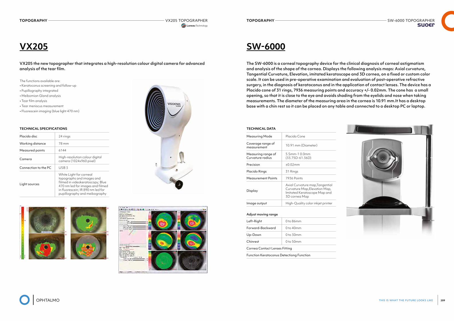
this is what the future looks like 209
TOPOGRAPHY VX205 TOPOGRAPHERLuneau Visionix
TOPOGRAPHY SW-6000 TOPOGRAPHERSuoer
VX205 SW-6000
VX205 the new topographer that integrates a high-resolution colour digital camera for advanced analysis of the tear film.
The SW-6000 is a corneal topography device for the clinical diagnosis of corneal astigmatism and analysis of the shape of the cornea. Displays the following analysis maps: Axial curvature, Tangential Curvature, Elevation, imitated keratoscope and 3D cornea, on a fixed or custom color scale. It can be used in pre-operative examination and evaluation of post-operative refractive surgery, in the diagnosis of keratoconus and in the application of contact lenses. The device has a Placido cone of 31 rings, 7936 measuring points and accuracy +/- 0.02mm. The cone has a small opening, so that it is close to the eye and avoids shading from the eyelids and nose when taking measurements. The diameter of the measuring area in the cornea is 10.91 mm.It has a desktop base with a chin rest so it can be placed on any table and connected to a desktop PC or laptop.
The functions available are:• Keratoconus screening and follow-up• Pupillography integrated• Meibomian Gland analysis• Tear film analysis• Tear meniscus measurement• Fluorescein imaging (blue light 470 nm)
TECHNICAL SPECIFICATIONS
Placido disc 24 rings
Working distance 78 mm
Measured points 6144
Camera High-resolution colour digital camera (1024x960 pixel)
Connection to the PC USB 3
Light sources
White Light for corneal topographs and images and filmed in videokeratoscopy; Blue 470 nm led for images and filmed in fluorescein; IR 890 nm led for pupillography and meibography
TECHNICAL DATA
Measuring Mode Placido Cone
Coverage range of measurement 10.91 mm (Diameter)
Measuring range of Curvature radius
5.Smm-1 0.0mm (33.75D-61.36D)
Precision ±0.02mm
Placido Rings 31 Rings
Measurement Points 7936 Points
Display
Axial Curvature map,Tangential Curvature Map,Elevation Map, lmitated Keratoscope Map and 3D cornea Map
Image output High-Quality color inkjet printer
Adjust moving range
Left-Right 0 to 86mm
Forward-Backward 0 to 40mm
Up-Down 0 to 30mm
Chinrest 0 to 50mm
Cornea Contact Lenses Fitting
Function Keratoconus Detectiong Function
10TONOMETRY
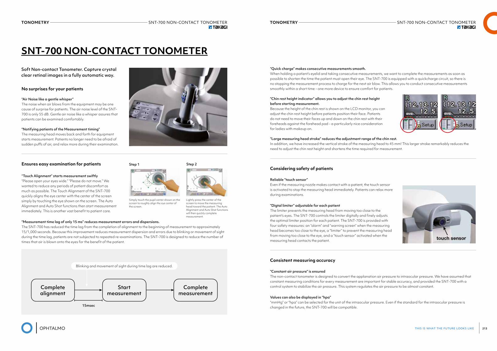
this is what the future looks like 213
TONOMETRY SNT-700 NON-CONTACT TONOMETER TONOMETRY SNT-700 NON-CONTACT TONOMETER
SNT-700 NON-CONTACT TONOMETER
Soft Non-contact Tonometer. Capture crystal clear retinal images in a fully automatic way.
No surprises for your patients
“Air Noise like a gentle whisper”The noise when air blows from the equipment may be one cause of surprise for patients. The air noise level of the SNT-700 is only 55 dB. Gentle air noise like a whisper assures that patients can be examined comfortably.
“Notifying patients of the Measurement timing”The measuring head moves back and forth for equipment starts measurement. Patients no longer need to be afraid of sudden puffs of air, and relax more during their examination.
Ensures easy examination for patients
“Touch Alignment” starts measurement swiftly“Please open your eyes wide.” “Please do not move.” We wanted to reduce any periods of patient discomfort as much as possible. The Touch Alignment of the SNT-700 quickly aligns the eye center with the center of the screen simply by touching the eye shown on the screen. The Auto Alignment and Auto Shot functions then start measurement immediately. This is another vast benefit to patient care.
“Measurement time lag of only 15 ms” reduces measurement errors and dispersions. The SNT-700 has reduced the time lag from the completion of alignment to the beginning of measurement to approximately 15/1,000 seconds. Because this improvement reduces measurement dispersion and errors due to blinking or movement of sight during the time lag, patients are not subjected to repeated re-examinations. The SNT-700 is designed to reduce the number of times that air is blown onto the eyes for the benefit of the patient.
Simply touch the pupil center shown on the screen to roughly align the eye center of the screen.
Lightly press the center of the screen to move the measuring head toward the patient. The AutoAlignment and Auto Shot functions will then quickly complete measurement.
Step 1 Step 2
Blinking and movement of sight during time lag are reduced.
Completealignment
Startmeasurement
Completemeasurement
15msec
“Quick charge” makes consecutive measurements smooth.When holding a patient’s eyelid and taking consecutive measurements, we want to complete the measurements as soon as possible to shorten the time the patient must open their eye. The SNT-700 is equipped with a quickcharge circuit, so there is no stopping the measurement process to charge for the next air blow. This allows you to conduct consecutive measurements smoothly within a short time - one more device to ensure comfort for patients.
“Chin rest height indicator” allows you to adjust the chin rest height before starting measurement.Because the height of the chin rest is shown on the LCD monitor, you can adjust the chin rest height before patients position their face. Patients do not need to move their faces up and down on the chin rest with their foreheads against the forehead pad - a particularly nice consideration for ladies with makeup on.
“Large measuring head stroke” reduces the adjustment range of the chin rest.In addition, we have increased the vertical stroke of the measuring head to 45 mm! This larger stroke remarkably reduces the need to adjust the chin rest height and shortens the time required for measurement.
Considering safety of patients
Reliable “touch sensor”Even if the measuring nozzle makes contact with a patient, the touch sensor is activated to stop the measuring head immediately. Patients can relax more during examinations.
“Digtal limiter” adjustable for each patientThe limiter prevents the measuring head from moving too close to the patient’s eyes. The SNT-700 controls the limiter digitally and finely adjusts the optimal limiter position for each patient. The SNT-700 is provided with four safety measures: an “alarm” and “warning screen” when the measuring head becomes too close to the eye, a “limiter” to prevent the measuring head from moving too close to the eye, and a “touch sensor” activated when the measuring head contacts the patient.
Consistent measuring accuracy
“Constant air pressure” is ensuredThe non-contact tonometer is designed to convert the applanation air pressure to intraocular pressure. We have assumed that constant measuring conditions for every measurement are important for stable accuracy, and provided the SNT-700 with a control system to stabilize the air pressure. This system regulates the air pressure to be almost constant.
Values can also be displayed in “hpa”“mmHg” or “hpa” can be selected for the unit of the intraocular pressure. Even if the standard for the intraocular pressure is changed in the future, the SNT-700 will be compatible.
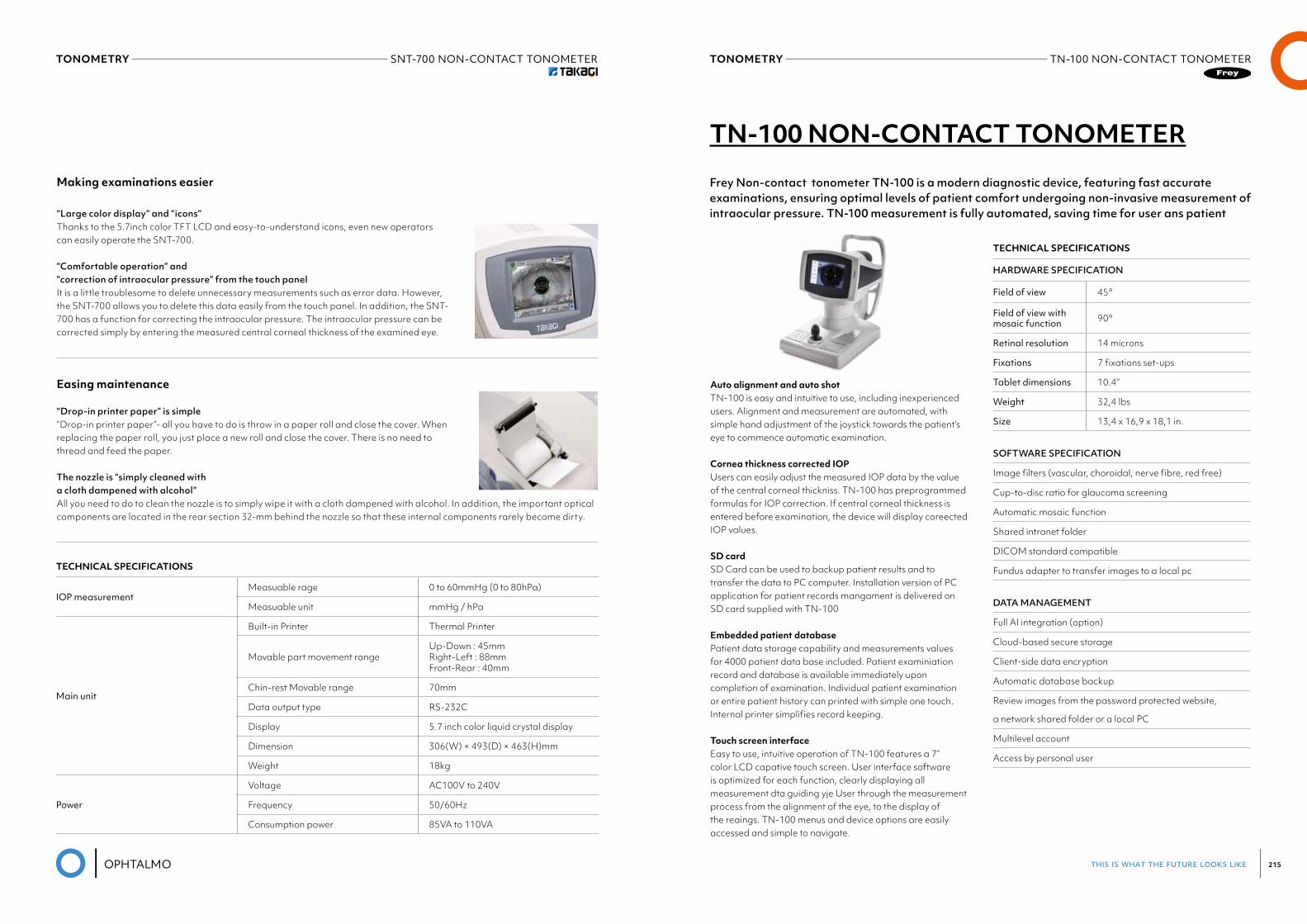
this is what the future looks like 215
TONOMETRY SNT-700 NON-CONTACT TONOMETERTakagi
TONOMETRY TN-100 NON-CONTACT TONOMETER
Making examinations easier
“Large color display” and “icons”Thanks to the 5.7inch color TFT LCD and easy-to-understand icons, even new operators can easily operate the SNT-700.
“Comfortable operation” and “correction of intraocular pressure” from the touch panelIt is a little troublesome to delete unnecessary measurements such as error data. However, the SNT-700 allows you to delete this data easily from the touch panel. In addition, the SNT-700 has a function for correcting the intraocular pressure. The intraocular pressure can be corrected simply by entering the measured central corneal thickness of the examined eye.
Easing maintenance
“Drop-in printer paper” is simple“Drop-in printer paper”- all you have to do is throw in a paper roll and close the cover. When replacing the paper roll, you just place a new roll and close the cover. There is no need to thread and feed the paper.
The nozzle is “simply cleaned with a cloth dampened with alcohol”All you need to do to clean the nozzle is to simply wipe it with a cloth dampened with alcohol. In addition, the important optical components are located in the rear section 32-mm behind the nozzle so that these internal components rarely become dirty.
TECHNICAL SPECIFICATIONS
IOP measurementMeasuable rage 0 to 60mmHg (0 to 80hPa)
Measuable unit mmHg / hPa
Main unit
Built-in Printer Thermal Printer
Movable part movement rangeUp-Down : 45mm Right-Left : 88mm Front-Rear : 40mm
Chin-rest Movable range 70mm
Data output type RS-232C
Display 5.7 inch color liquid crystal display
Dimension 306(W) × 493(D) × 463(H)mm
Weight 18kg
Power
Voltage AC100V to 240V
Frequency 50/60Hz
Consumption power 85VA to 110VA
TN-100 NON-CONTACT TONOMETER
Frey Non-contact tonometer TN-100 is a modern diagnostic device, featuring fast accurate examinations, ensuring optimal levels of patient comfort undergoing non-invasive measurement of intraocular pressure. TN-100 measurement is fully automated, saving time for user ans patient
Auto alignment and auto shotTN-100 is easy and intuitive to use, including inexperienced users. Alignment and measurement are automated, with simple hand adjustment of the joystick towards the patient’s eye to commence automatic examination.
Cornea thickness corrected IOPUsers can easily adjust the measured IOP data by the value of the central corneal thickniss. TN-100 has preprogrammed formulas for IOP correction. If central corneal thickness is entered before examination, the device will display coreected IOP values.
SD cardSD Card can be used to backup patient results and to transfer the data to PC computer. Installation version of PC application for patient records mangament is delivered on SD card supplied with TN-100
Embedded patient databasePatient data storage capability and measurements values for 4000 patient data base included. Patient examiniation record and database is available immediately upon completion of examination. Individual patient examination or entire patient history can printed with simple one touch. Internal printer simplifies record keeping.
Touch screen interfaceEasy to use, intuitive operation of TN-100 features a 7” color LCD capative touch screen. User interface software is optimized for each function, clearly displaying all measurement dta guiding yje User through the measurement process from the alignment of the eye, to the display of the reaings. TN-100 menus and device options are easily accessed and simple to navigate.
TECHNICAL SPECIFICATIONS
HARDWARE SPECIFICATION
Field of view 45°
Field of view with mosaic function 90°
Retinal resolution 14 microns
Fixations 7 fixations set-ups
Tablet dimensions 10.4’’
Weight 32,4 lbs
Size 13,4 x 16,9 x 18,1 in.
SOFTWARE SPECIFICATION
Image filters (vascular, choroidal, nerve fibre, red free)
Cup-to-disc ratio for glaucoma screening
Automatic mosaic function
Shared intranet folder
DICOM standard compatible
Fundus adapter to transfer images to a local pc
DATA MANAGEMENT
Full AI integration (option)
Cloud-based secure storage
Client-side data encryption
Automatic database backup
Review images from the password protected website,
a network shared folder or a local PC
Multilevel account
Access by personal user

this is what the future looks like 217
TONOMETRY SW-5000NON-CONTACTTONOMETERSuoer
TONOMETRY SW-500HANDHELDREBOUNDTONOMETERSuoer
SW-500 HANDHELD REBOUND TONOMETER SW-5000 NON-CONTACT TONOMETER
SUOER rebound Tonometer SW-500 is used to measure intraocular pressure. There are two working modes: vertical and horizontal. When the probe contacts the surface of cornea , depending on hardness at a certain speed, it has a different reaction of the probe rebounding.
Advantage: high accuracy, portable, without anesthesia, without cross-infection etc. Wireless data output.
SUOER IOPro (SW-5000) using an air puff, an optical non-contact method, to measure intraocular pressure. The advantage includes auto-focusing, fast measurement speed, comfortable measurement process, and no cross-infection.
TECHNICAL DATA
Measure Range 3mmHg"' 70mmHg
Precision ± 1.5mmHg(3mmHg≤IOP≤25mmHg);
Measurment Mode ±2.5mmHg(25mmHg<IOP≤70mmHg);
Output Bath vertical and horizontal measurment
Easy to use Wireless lnfrared Thermal Printer
Small size, Easy to carry
No need Anesthesia, No Discomfortableness
TECHNICAL DATA
Measuring Range 1 mmHg~60mmHg
Measuring Scale 30mmHg, 60mmHg
Measuring Accuracy 1mmHg;
Measuring Distance 11mm
Focus Method focus points + focus notification
Focus Mode three-dimensional auto-focus/ma-nual focus/touch screen focus
lnterior Light Fixation Green LED
Stroke of Moving Track
Left-Right: 80mmForward-Backward: 40mmUp-Down: 20mm
Display large colored LCD screen
Output high speed thermal printer
Unique Features • lntegrated ORA (Ocular Response Analyzer) • Unique collection of waveform confidence interval data by
weight average of three readings, and indicate low confi-dence interval results
• Manually focus by touching screen • Non-contact measurements to avoid cross infection• lntegrated 24 hours IOP trend analysis system
11DIAGNOSTIC
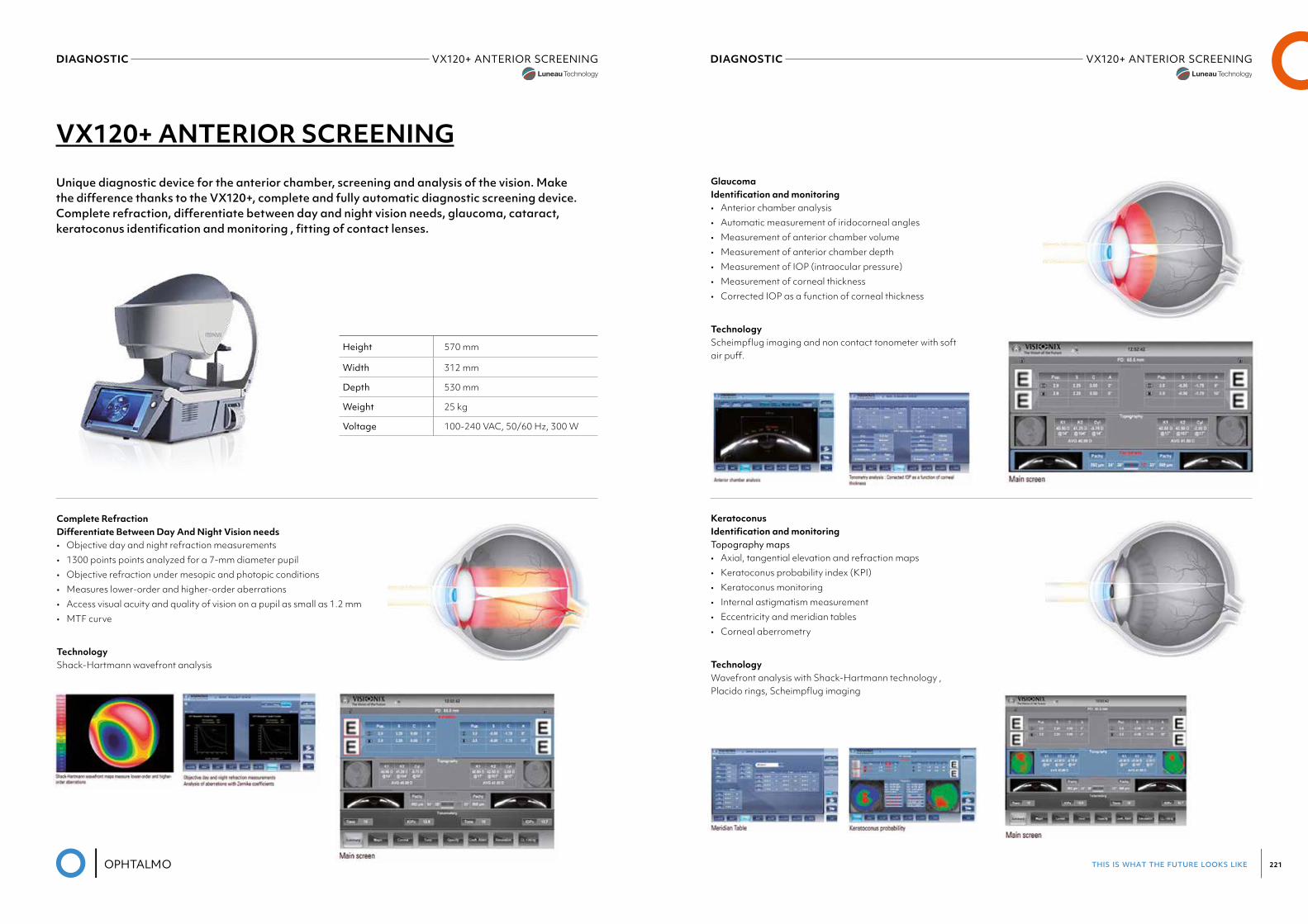
this is what the future looks like 221
DIAGNOSTIC VX120+ ANTERIOR SCREENINGLuneau Visionix
DIAGNOSTIC VX120+ ANTERIOR SCREENING
VX120+ ANTERIOR SCREENING
Unique diagnostic device for the anterior chamber, screening and analysis of the vision. Make the difference thanks to the VX120+, complete and fully automatic diagnostic screening device. Complete refraction, differentiate between day and night vision needs, glaucoma, cataract, keratoconus identification and monitoring , fitting of contact lenses.
Complete Refraction Differentiate Between Day And Night Vision needs• Objective day and night refraction measurements• 1300 points points analyzed for a 7-mm diameter pupil• Objective refraction under mesopic and photopic conditions• Measures lower-order and higher-order aberrations• Access visual acuity and quality of vision on a pupil as small as 1.2 mm• MTF curve
TechnologyShack-Hartmann wavefront analysis
Height 570 mm
Width 312 mm
Depth 530 mm
Weight 25 kg
Voltage 100-240 VAC, 50/60 Hz, 300 W
Glaucoma Identification and monitoring• Anterior chamber analysis• Automatic measurement of iridocorneal angles• Measurement of anterior chamber volume• Measurement of anterior chamber depth• Measurement of IOP (intraocular pressure)• Measurement of corneal thickness• Corrected IOP as a function of corneal thickness
TechnologyScheimpflug imaging and non contact tonometer with soft air puff.
Keratoconus Identification and monitoringTopography maps• Axial, tangential elevation and refraction maps• Keratoconus probability index (KPI)• Keratoconus monitoring• Internal astigmatism measurement• Eccentricity and meridian tables• Corneal aberrometry
TechnologyWavefront analysis with Shack-Hartmann technology , Placido rings, Scheimpflug imaging
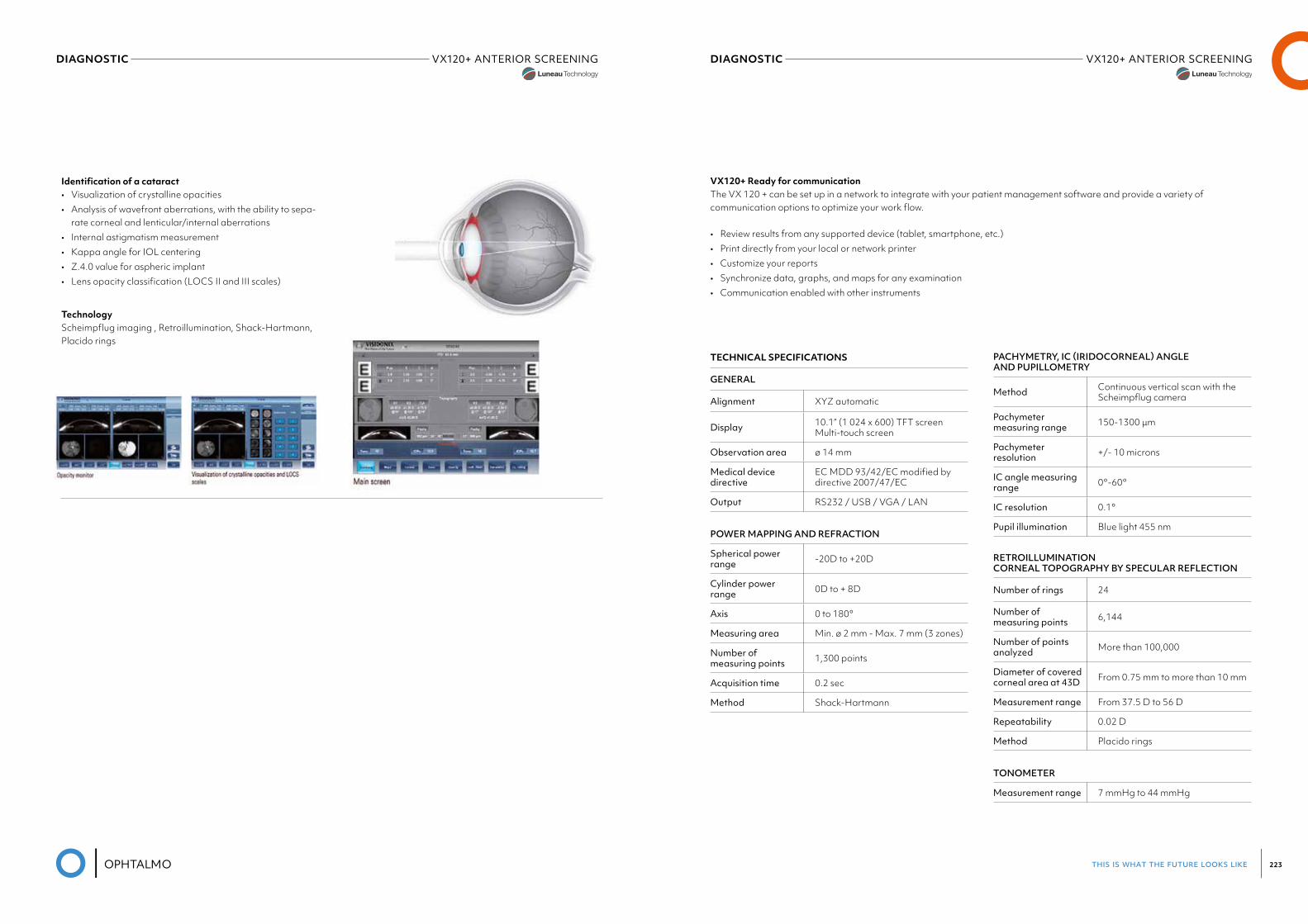
this is what the future looks like 223
DIAGNOSTIC VX120+ ANTERIOR SCREENINGLuneau Visionix
DIAGNOSTIC VX120+ ANTERIOR SCREENINGLuneau Visionix
Identification of a cataract• Visualization of crystalline opacities• Analysis of wavefront aberrations, with the ability to sepa-
rate corneal and lenticular/internal aberrations• Internal astigmatism measurement• Kappa angle for IOL centering• Z.4.0 value for aspheric implant• Lens opacity classification (LOCS II and III scales)
TechnologyScheimpflug imaging , Retroillumination, Shack-Hartmann, Placido rings
PACHYMETRY, IC (IRIDOCORNEAL) ANGLE AND PUPILLOMETRY
Method Continuous vertical scan with the Scheimpflug camera
Pachymeter measuring range 150-1300 μm
Pachymeter resolution +/- 10 microns
IC angle measuring range 0°-60°
IC resolution 0.1°
Pupil illumination Blue light 455 nm
RETROILLUMINATION CORNEAL TOPOGRAPHY BY SPECULAR REFLECTION
Number of rings 24
Number of measuring points 6,144
Number of points analyzed More than 100,000
Diameter of covered cornealareaat43D From 0.75 mm to more than 10 mm
Measurement range From 37.5 D to 56 D
Repeatability 0.02 D
Method Placido rings
TONOMETER
Measurement range 7 mmHg to 44 mmHg
VX120+ Ready for communicationThe VX 120 + can be set up in a network to integrate with your patient management software and provide a variety of communication options to optimize your work flow.
• Review results from any supported device (tablet, smartphone, etc.)• Print directly from your local or network printer• Customize your reports• Synchronize data, graphs, and maps for any examination• Communication enabled with other instruments
TECHNICAL SPECIFICATIONS
GENERAL
Alignment XYZ automatic
Display 10.1” (1 024 x 600) TFT screen Multi-touch screen
Observation area ø 14 mm
Medical device directive
EC MDD 93/42/EC modified by directive 2007/47/EC
Output RS232 / USB / VGA / LAN
POWER MAPPING AND REFRACTION
Spherical power range -20D to +20D
Cylinder power range 0D to + 8D
Axis 0 to 180°
Measuring area Min. ø 2 mm - Max. 7 mm (3 zones)
Number of measuring points 1,300 points
Acquisition time 0.2 sec
Method Shack-Hartmann
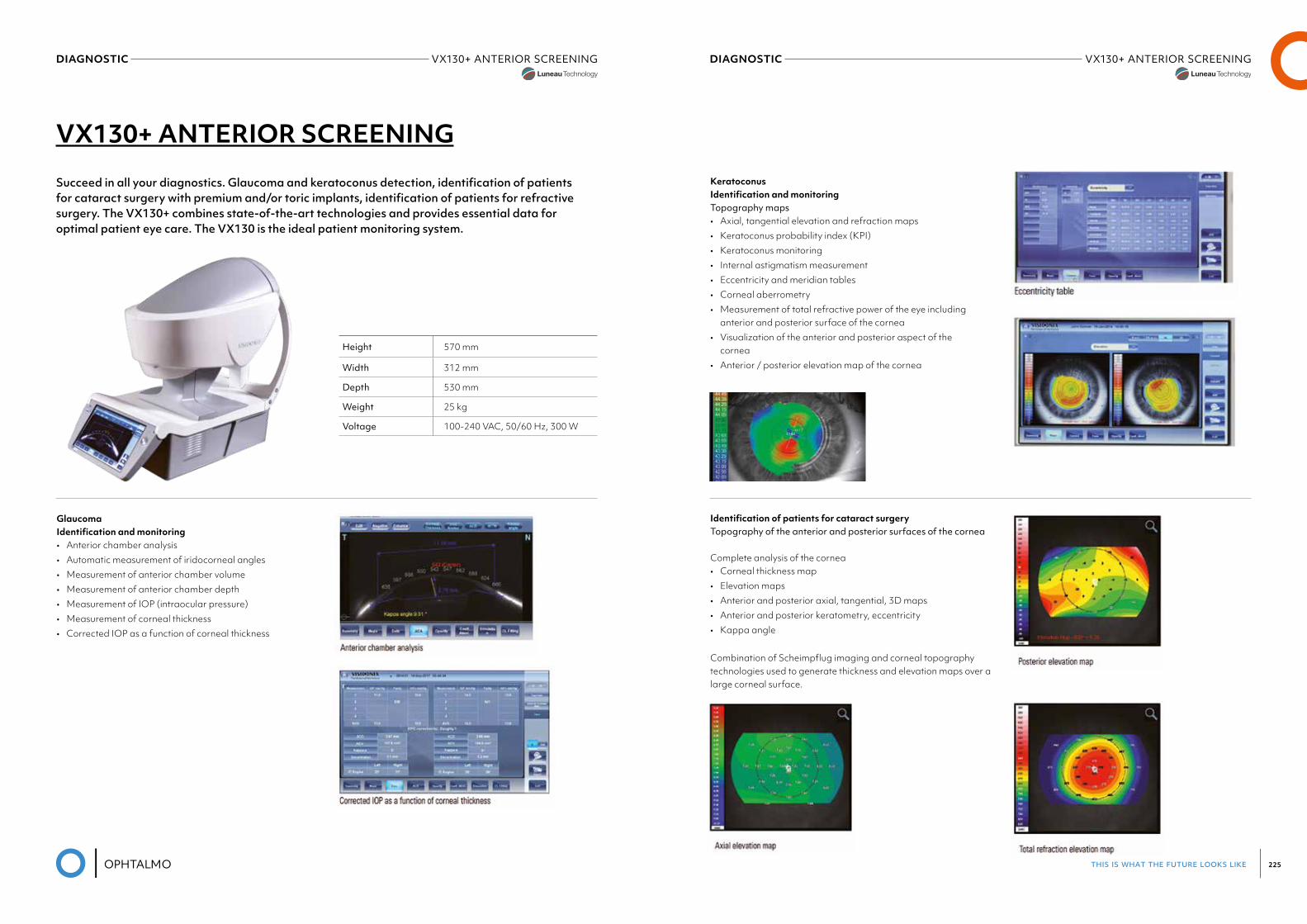
this is what the future looks like 225
DIAGNOSTIC VX130+ANTERIORSCREENINGLuneau Visionix
DIAGNOSTIC VX130+ANTERIORSCREENINGLuneau Visionix
VX130+ ANTERIOR SCREENING
Succeed in all your diagnostics. Glaucoma and keratoconus detection, identification of patients for cataract surgery with premium and/or toric implants, identification of patients for refractive surgery. The VX130+ combines state-of-the-art technologies and provides essential data for optimal patient eye care. The VX130 is the ideal patient monitoring system.
Glaucoma Identification and monitoring• Anterior chamber analysis• Automatic measurement of iridocorneal angles• Measurement of anterior chamber volume• Measurement of anterior chamber depth• Measurement of IOP (intraocular pressure)• Measurement of corneal thickness• Corrected IOP as a function of corneal thickness
Height 570 mm
Width 312 mm
Depth 530 mm
Weight 25 kg
Voltage 100-240 VAC, 50/60 Hz, 300 W
Keratoconus Identification and monitoringTopography maps• Axial, tangential elevation and refraction maps• Keratoconus probability index (KPI)• Keratoconus monitoring• Internal astigmatism measurement• Eccentricity and meridian tables• Corneal aberrometry• Measurement of total refractive power of the eye including
anterior and posterior surface of the cornea• Visualization of the anterior and posterior aspect of the
cornea• Anterior / posterior elevation map of the cornea
Identification of patients for cataract surgeryTopography of the anterior and posterior surfaces of the cornea
Complete analysis of the cornea• Corneal thickness map• Elevation maps• Anterior and posterior axial, tangential, 3D maps• Anterior and posterior keratometry, eccentricity• Kappa angle
Combination of Scheimpflug imaging and corneal topography technologies used to generate thickness and elevation maps over a large corneal surface.
this is what the future looks like 227
DIAGNOSTIC VX130+ANTERIORSCREENINGLuneau Visionix
DIAGNOSTIC VX130+ANTERIORSCREENINGLuneau Visionix
PRE-OP cataract surgery
• Visualization of crystalline opacities• Analysis of wavefront aberrations, with the ability to sepa-
rate corneal and lenticular/internal aberrations• Internal astigmatism measurement• Kappa angle for IOL centering• Z.4.0 value for aspheric implant• Lens opacity classification (LOCS II and III scales)• Premium IOL candidate identification and monitoring Aspheric or Spheric IOL Multifocal Toric Multifocal toric
POST-OP cataract surgery
• Post-operative IOL check• Toric lens implant axis alignment check• Analysis of post-op output to improve surgery protocols• Analysis of the total correction including the IOL
Vx130+ Complete refraction Differentiate between day and night vision needs
• Objective day and night refraction measurements• 1300 points points analyzed for a 7-mm diameter pupil• Objective refraction under mesopic and photopic conditi-
ons• Measures lower-order and higher-order aberrations• Access visual acuity and quality of vision on a pupil as
small as 1.2 mm• MTF curve
PACHYMETRY, IC (IRIDOCORNEAL) ANGLE AND PUPILLOMETRY
Method Continuous vertical scan with the Scheimpflug camera
Pachymeter measuring range 150-1300 μm
Pachymeter resolution +/- 10 microns
IC angle measuring range 0°-60°
IC resolution 0.1°
Pupil illumination Blue light 455 nm
RETROILLUMINATION CORNEAL TOPOGRAPHY BY SPECULAR REFLECTION
Number of rings 24
Number of measuring points 6,144
Number of points analyzed More than 100,000
Diameter of covered cornealareaat43D From 0.75 mm to more than 10 mm
Measurement range From 37.5 D to 56 D
Repeatability 0.02 D
Method Placido rings
TONOMETER
Measurement range 7 mmHg to 44 mmHg
TECHNICAL SPECIFICATIONS
GENERAL
Alignment XYZ automatic
Display 10.1” (1 024 x 600) TFT screen Multi-touch screen
Observation area ø 14 mm
Medical device directive
EC MDD 93/42/EC modified by directive 2007/47/EC
Output RS232 / USB / VGA / LAN
POWER MAPPING AND REFRACTION
Spherical power range -20D to +20D
Cylinder power range 0D to + 8D
Axis 0 to 180°
Measuring area Min. ø 2 mm - Max. 7 mm (3 zones)
Number of measuring points 1,300 points
Acquisition time 0.2 sec
Method Shack-Hartmann
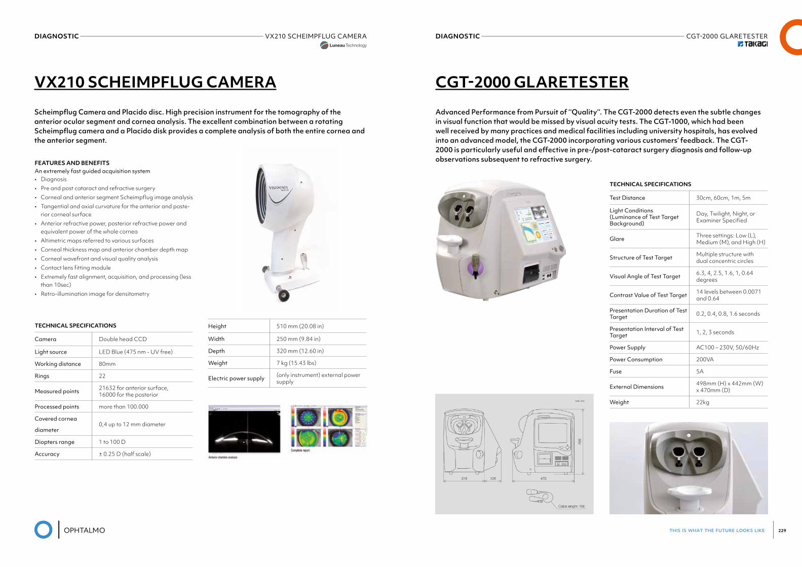
this is what the future looks like 229
DIAGNOSTIC CGT-2000GLARETESTERDIAGNOSTIC VX210 SCHEIMPFLUG CAMERALuneau Visionix
Height 510 mm (20.08 in)
Width 250 mm (9.84 in)
Depth 320 mm (12.60 in)
Weight 7 kg (15.43 lbs)
Electric power supply (only instrument) external power supply
TECHNICAL SPECIFICATIONS
Camera Double head CCD
Light source LED Blue (475 nm - UV free)
Working distance 80mm
Rings 22
Measured points 21632 for anterior surface, 16000 for the posterior
Processed points more than 100.000
Covered cornea
diameter0,4 up to 12 mm diameter
Diopters range 1 to 100 D
Accuracy ± 0.25 D (half scale)
TECHNICAL SPECIFICATIONS
Test Distance 30cm, 60cm, 1m, 5m
Light Conditions (Luminance of Test Target Background)
Day, Twilight, Night, or Examiner Specified
Glare Three settings: Low (L), Medium (M), and High (H)
Structure of Test Target Multiple structure with dual concentric circles
Visual Angle of Test Target 6.3, 4, 2.5, 1.6, 1, 0.64 degrees
Contrast Value of Test Target 14 levels between 0.0071 and 0.64
Presentation Duration of Test Target 0.2, 0.4, 0.8, 1.6 seconds
Presentation Interval of Test Target 1, 2, 3 seconds
Power Supply AC100 – 230V, 50/60Hz
Power Consumption 200VA
Fuse 5A
External Dimensions 498mm (H) x 442mm (W) x 470mm (D)
Weight 22kg
VX210 SCHEIMPFLUG CAMERA CGT-2000 GLARETESTER
Scheimpflug Camera and Placido disc. High precision instrument for the tomography of the anterior ocular segment and cornea analysis. The excellent combination between a rotating Scheimpflug camera and a Placido disk provides a complete analysis of both the entire cornea and the anterior segment.
Advanced Performance from Pursuit of ‘‘Quality’’. The CGT-2000 detects even the subtle changes in visual function that would be missed by visual acuity tests. The CGT-1000, which had been well received by many practices and medical facilities including university hospitals, has evolved into an advanced model, the CGT-2000 incorporating various customers’ feedback. The CGT-2000 is particularly useful and effective in pre-/post-cataract surgery diagnosis and follow-up observations subsequent to refractive surgery.FEATURES AND BENEFITS
An extremely fast guided acquisition system• Diagnosis• Pre and post cataract and refractive surgery• Corneal and anterior segment Scheimpflug image analysis• Tangential and axial curvature for the anterior and poste-
rior corneal surface• Anterior refractive power, posterior refractive power and
equivalent power of the whole cornea• Altimetric maps referred to various surfaces• Corneal thickness map and anterior chamber depth map• Corneal wavefront and visual quality analysis• Contact lens fitting module• Extremely fast alignment, acquisition, and processing (less
than 10sec)• Retro-illumination image for densitometry
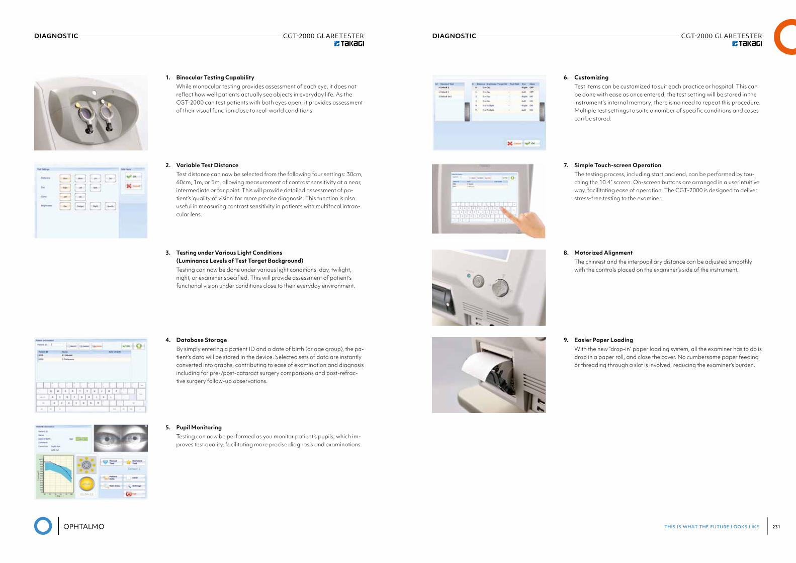
this is what the future looks like 231
DIAGNOSTIC CGT-2000GLARETESTERDIAGNOSTIC CGT-2000GLARETESTER
1. Binocular Testing Capability While monocular testing provides assessment of each eye, it does not
reflect how well patients actually see objects in everyday life. As the CGT-2000 can test patients with both eyes open, it provides assessment of their visual function close to real-world conditions.
6. Customizing Test items can be customized to suit each practice or hospital. This can
be done with ease as once entered, the test setting will be stored in the instrument’s internal memory; there is no need to repeat this procedure. Multiple test settings to suite a number of specific conditions and cases can be stored.
2. Variable Test Distance Test distance can now be selected from the following four settings: 30cm,
60cm, 1m, or 5m, allowing measurement of contrast sensitivity at a near, intermediate or far point. This will provide detailed assessment of pa-tient’s ‘quality of vision’ for more precise diagnosis. This function is also useful in measuring contrast sensitivity in patients with multifocal intrao-cular lens.
7. Simple Touch-screen Operation The testing process, including start and end, can be performed by tou-
ching the 10.4” screen. On-screen buttons are arranged in a userintuitive way, facilitating ease of operation. The CGT-2000 is designed to deliver stress-free testing to the examiner.
3. Testing under Various Light Conditions (Luminance Levels of Test Target Background)
Testing can now be done under various light conditions: day, twilight, night, or examiner specified. This will provide assessment of patient’s functional vision under conditions close to their everyday environment.
8. Motorized Alignment The chinrest and the interpupillary distance can be adjusted smoothly
with the controls placed on the examiner’s side of the instrument.
4. Database Storage By simply entering a patient ID and a date of birth (or age group), the pa-
tient’s data will be stored in the device. Selected sets of data are instantly converted into graphs, contributing to ease of examination and diagnosis including for pre-/post-cataract surgery comparisons and post-refrac-tive surgery follow-up observations.
9. Easier Paper Loading With the new “drop-in” paper loading system, all the examiner has to do is
drop in a paper roll, and close the cover. No cumbersome paper feeding or threading through a slot is involved, reducing the examiner’s burden.
5. Pupil Monitoring Testing can now be performed as you monitor patient’s pupils, which im-
proves test quality, facilitating more precise diagnosis and examinations.
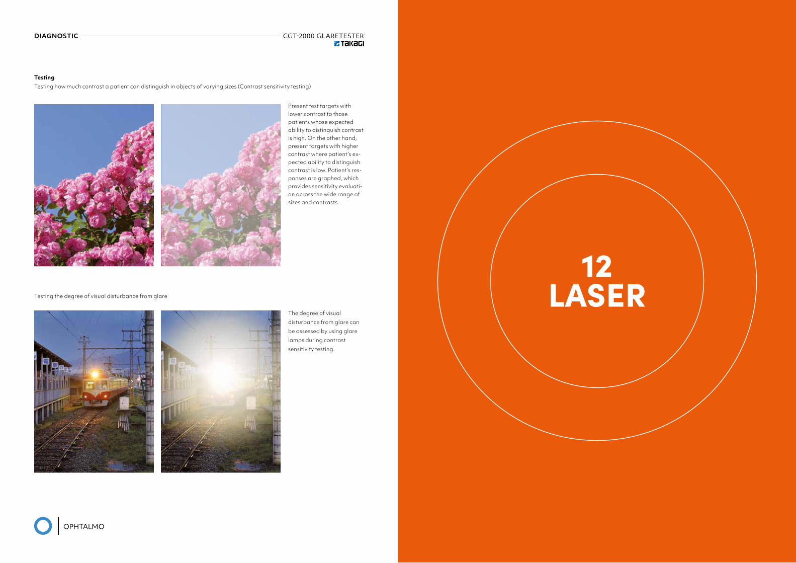
DIAGNOSTIC CGT-2000GLARETESTER
12LASER
TestingTesting how much contrast a patient can distinguish in objects of varying sizes (Contrast sensitivity testing)
Testing the degree of visual disturbance from glare
Present test targets with lower contrast to those patients whose expected ability to distinguish contrast is high. On the other hand, present targets with higher contrast where patient’s ex-pected ability to distinguish contrast is low. Patient’s res-ponses are graphed, which provides sensitivity evaluati-on across the wide range of sizes and contrasts.
The degree of visualdisturbance from glare canbe assessed by using glarelamps during contrastsensitivity testing.
this is what the future looks like 235
LASER LIGHTLAS532/577/810/LIGHTLASTRUSCANLASER LIGHTLAS YAG / LIGHTLAS SLT
LIGHTLAS YAG
LIGHTLAS SLT
Laser photodisruptor and vitreolysis
Selective Laser Trabeculoplasty
• Solution For: Cataract, Glaucoma, Anterior Segment/Cor-nea, Vitreous Floaters
• Clinical Applications: Posterior Capsulotomy, Posterior Membranectomy, YAG Vitreolysis, YAG Laser Iridotomy, YAG Sweeping, YAG Corticolysis, YAG Anterior Stromal Puncture
• Treats: Secondary Cataract, Closed Angle Glaucoma, Corneal Erosion Syndrome
• Clinical Applications: Selective Laser Trabeculoplasty• Treats: Open Angle Glaucoma
LIGHTLAS 532/577/810®
LIGHTLAS TRUSCAN
Laser Photocoagulators with SP-Mode®
Pattern Scanning Photocoagulator with SP-Mode®
• Solution For: Retina, Glaucoma Clinical Applications: Laser Photocoagulation, SP-Mode® Microsecond Laser Techno-logy
• Treats: Diabetic Retinopathy, Pan-Retinal Photocoagu-lation, Retinal Tears, Holes & Detachment, Endo-Photo-coagulation, Proliferative Diabetic Retinopathy, Diabetic Macular Edema, Sub-Retinal Neovascularization, Central Retinal Vein Occlusion, Branch Retinal Vein Occlusion, Central Serous Chorioretinopathy, Sub-Threshold Laser Trabeculoplasty,
• Solution For: Retina, Glaucoma • Clinical Applications: Laser Photocoagulation, SP-Mode®
Microsecond Laser Technology• Treats: Diabetic Retinopathy, Pan-Retinal Photocoagu-
lation, Retinal Tears, Holes & Detachment, Endo-Photo-coagulation, Proliferative Diabetic Retinopathy, Diabetic Macular Edema, Sub-Retinal Neovascularization, Central Retinal Vein Occlusion, Branch Retinal Vein Occlusion, Central Serous Chorioretinopathy, Sub-Threshold Laser Trabeculoplasty, Retinopathy of Prematurity, Transscleral Cyclophotocoagulation
13SLIT LAMPS
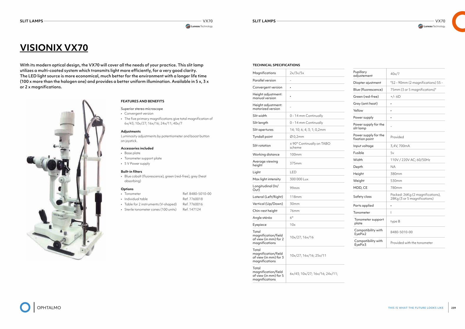
this is what the future looks like 239
SLIT LAMPS VX70Luneau Visionix
SLIT LAMPS VX70Luneau Visionix
VISIONIX VX70
With its modern optical design, the VX70 will cover all the needs of your practice. This slit lamp utilizes a multi-coated system which transmits light more efficiently, for a very good clarity.The LED light source is more economical, much better for the environment with a longer life time (100 x more than the halogen one) and provides a better uniform illumination. Available in 5 x, 3 x or 2 x magnifications.
FEATURES AND BENEFITS
Superior stereo microscope• Convergent version• The five primary magnifications give total magnification of
6x/43; 10x/27; 16x/16; 24x/11; 40x/7
AdjustmentsLuminosity adjustments by potentiometer and boost button on joystick.
Accessories included• Base plate• Tonometer support plate• 5 V Power supply
Built-in filters• Blue cobalt (fluorescence), green (red-free), grey (heat
absorbing)
Options• Tonometer Ref. 8480-5010-00• Individual table Ref. 7760018• Table for 2 instruments (V-shaped) Ref. 7760016• Sterile tonometer cones (100 units) Ref. 147124
TECHNICAL SPECIFICATIONS
Magnifications 2x/3x/5x
Parallel version -
Convergent version •
Height adjustment: manual version •
Height adjustment: motorized version -
Slit width 0 - 14 mm Continually
Slit length 0 - 14 mm Continually
Slit apertures 14; 10; 6; 4; 3; 1; 0,2mm
Tyndall point Ø 0,2mm
Slit rotation ± 90° Continually on TABO scheme
Working distance 100mm
Average viewing height 375mm
Light LED
Max light intensity 300 000 Lux
Longitudinal (In/Out) 99mm
Lateral (Left/Right) 118mm
Vertical (Up/Down) 30mm
Chin-rest height 76mm
Angle stéréo 6°
Eyepiece 10x
Total magnification/fieldof view (in mm) for 2 magnifications
10x/27; 16x/16
Total magnification/fieldofview(inmm)for3magnifications
10x/27; 16x/16; 25x/11
Total magnification/fieldof view (in mm) for 5 magnifications
6x/43; 10x/27; 16x/16; 24x/11;
Pupillary adjustement 40x/7
Diopter ajustment "52 - 90mm (2 magnifications) 55 -
Blue(fluorescence) 75mm (3 or 5 magnifications)"
Green (red-free) +/- 6D
Grey (anti heat) •
Yellow •
Power supply •
Power supply for the slit lamp -
Power supply for the fixationpoint Provided
Input voltage 3,4V, 700mA
Fusible 5v
Width 110V / 220V AC; 60/50Hz
Depth NA
Height 380mm
Weight 530mm
MDD, CE 780mm
Safety class Packed: 26Kg (2 magnifications), 28Kg (3 or 5 magnifications)
Parts applied •
Tonometer I
Tonometer support plate type B
Compatibility with EyePix2 8480-5010-00
Compatibility with EyePix3 Provided with the tonometer
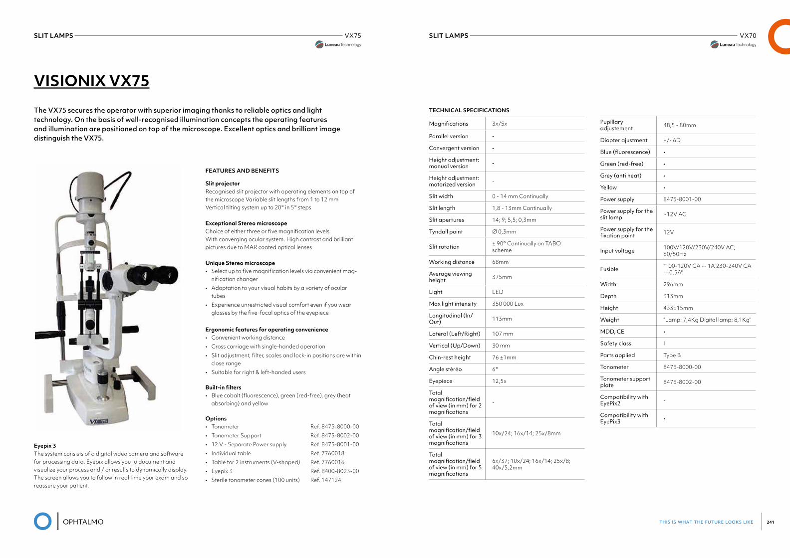
this is what the future looks like 241
SLIT LAMPS VX70Luneau Visionix
SLIT LAMPS VX75
VISIONIX VX75
The VX75 secures the operator with superior imaging thanks to reliable optics and light technology. On the basis of well-recognised illumination concepts the operating features and illumination are positioned on top of the microscope. Excellent optics and brilliant image distinguish the VX75.
FEATURES AND BENEFITS
Slit projectorRecognised slit projector with operating elements on top of the microscope Variable slit lengths from 1 to 12 mmVertical tilting system up to 20° in 5° steps
Exceptional Stereo microscopeChoice of either three or five magnification levels With converging ocular system. High contrast and brilliant pictures due to MAR coated optical lenses
Unique Stereo microscope• Select up to five magnification levels via convenient mag-
nification changer• Adaptation to your visual habits by a variety of ocular
tubes• Experience unrestricted visual comfort even if you wear
glasses by the five-focal optics of the eyepiece
Ergonomic features for operating convenience• Convenient working distance• Cross carriage with single-handed operation• Slit adjustment, filter, scales and lock-in positions are within
close range• Suitable for right & left-handed users
Built-in filters• Blue cobalt (fluorescence), green (red-free), grey (heat
absorbing) and yellow
Options• Tonometer Ref. 8475-8000-00• Tonometer Support Ref. 8475-8002-00• 12 V - Separate Power supply Ref. 8475-8001-00• Individual table Ref. 7760018• Table for 2 instruments (V-shaped) Ref. 7760016• Eyepix 3 Ref. 8400-8023-00• Sterile tonometer cones (100 units) Ref. 147124
Eyepix 3The system consists of a digital video camera and software for processing data. Eyepix allows you to document and visualize your process and / or results to dynamically display.The screen allows you to follow in real time your exam and so reassure your patient.
TECHNICAL SPECIFICATIONS
Magnifications 3x/5x
Parallel version •
Convergent version •
Height adjustment: manual version •
Height adjustment: motorized version -
Slit width 0 - 14 mm Continually
Slit length 1,8 - 13mm Continually
Slit apertures 14; 9; 5,5; 0,3mm
Tyndall point Ø 0,3mm
Slit rotation ± 90° Continually on TABO scheme
Working distance 68mm
Average viewing height 375mm
Light LED
Max light intensity 350 000 Lux
Longitudinal (In/Out) 113mm
Lateral (Left/Right) 107 mm
Vertical (Up/Down) 30 mm
Chin-rest height 76 ±1mm
Angle stéréo 6°
Eyepiece 12,5x
Total magnification/fieldof view (in mm) for 2 magnifications
-
Total magnification/fieldofview(inmm)for3magnifications
10x/24; 16x/14; 25x/8mm
Total magnification/fieldof view (in mm) for 5 magnifications
6x/37; 10x/24; 16x/14; 25x/8; 40x/5,2mm
Pupillary adjustement 48,5 - 80mm
Diopter ajustment +/- 6D
Blue(fluorescence) •
Green (red-free) •
Grey (anti heat) •
Yellow •
Power supply 8475-8001-00
Power supply for the slit lamp ~12V AC
Power supply for the fixationpoint 12V
Input voltage 100V/120V/230V/240V AC; 60/50Hz
Fusible "100-120V CA -- 1A 230-240V CA -- 0,5A"
Width 296mm
Depth 313mm
Height 433±15mm
Weight "Lamp: 7,4Kg Digital lamp: 8,1Kg"
MDD, CE •
Safety class I
Parts applied Type B
Tonometer 8475-8000-00
Tonometer support plate 8475-8002-00
Compatibility with EyePix2 -
Compatibility with EyePix3 •
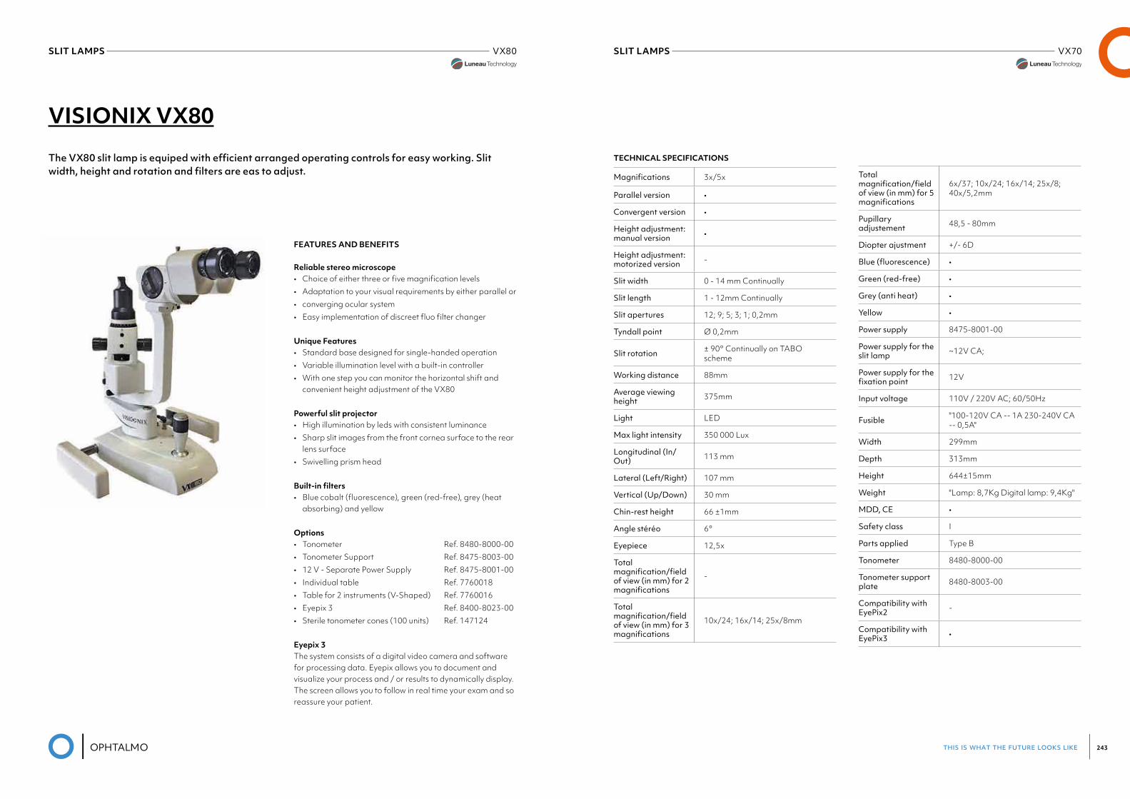
this is what the future looks like 243
SLIT LAMPS VX70Luneau Visionix
SLIT LAMPS VX80
VISIONIX VX80
The VX80 slit lamp is equiped with efficient arranged operating controls for easy working. Slit width, height and rotation and filters are eas to adjust.
FEATURES AND BENEFITS
Reliable stereo microscope• Choice of either three or five magnification levels• Adaptation to your visual requirements by either parallel or• converging ocular system• Easy implementation of discreet fluo filter changer
Unique Features• Standard base designed for single-handed operation• Variable illumination level with a built-in controller• With one step you can monitor the horizontal shift and
convenient height adjustment of the VX80
Powerful slit projector• High illumination by leds with consistent luminance• Sharp slit images from the front cornea surface to the rear
lens surface• Swivelling prism head
Built-in filters• Blue cobalt (fluorescence), green (red-free), grey (heat
absorbing) and yellow
Options• Tonometer Ref. 8480-8000-00• Tonometer Support Ref. 8475-8003-00• 12 V - Separate Power Supply Ref. 8475-8001-00• Individual table Ref. 7760018• Table for 2 instruments (V-Shaped) Ref. 7760016• Eyepix 3 Ref. 8400-8023-00• Sterile tonometer cones (100 units) Ref. 147124
Eyepix 3The system consists of a digital video camera and software for processing data. Eyepix allows you to document and visualize your process and / or results to dynamically display.The screen allows you to follow in real time your exam and so reassure your patient.
TECHNICAL SPECIFICATIONS
Magnifications 3x/5x
Parallel version •
Convergent version •
Height adjustment: manual version •
Height adjustment: motorized version -
Slit width 0 - 14 mm Continually
Slit length 1 - 12mm Continually
Slit apertures 12; 9; 5; 3; 1; 0,2mm
Tyndall point Ø 0,2mm
Slit rotation ± 90° Continually on TABO scheme
Working distance 88mm
Average viewing height 375mm
Light LED
Max light intensity 350 000 Lux
Longitudinal (In/Out) 113 mm
Lateral (Left/Right) 107 mm
Vertical (Up/Down) 30 mm
Chin-rest height 66 ±1mm
Angle stéréo 6°
Eyepiece 12,5x
Total magnification/fieldof view (in mm) for 2 magnifications
-
Total magnification/fieldofview(inmm)for3magnifications
10x/24; 16x/14; 25x/8mm
Total magnification/fieldof view (in mm) for 5 magnifications
6x/37; 10x/24; 16x/14; 25x/8; 40x/5,2mm
Pupillary adjustement 48,5 - 80mm
Diopter ajustment +/- 6D
Blue(fluorescence) •
Green (red-free) •
Grey (anti heat) •
Yellow •
Power supply 8475-8001-00
Power supply for the slit lamp ~12V CA;
Power supply for the fixationpoint 12V
Input voltage 110V / 220V AC; 60/50Hz
Fusible "100-120V CA -- 1A 230-240V CA -- 0,5A"
Width 299mm
Depth 313mm
Height 644±15mm
Weight "Lamp: 8,7Kg Digital lamp: 9,4Kg"
MDD, CE •
Safety class I
Parts applied Type B
Tonometer 8480-8000-00
Tonometer support plate 8480-8003-00
Compatibility with EyePix2 -
Compatibility with EyePix3 •
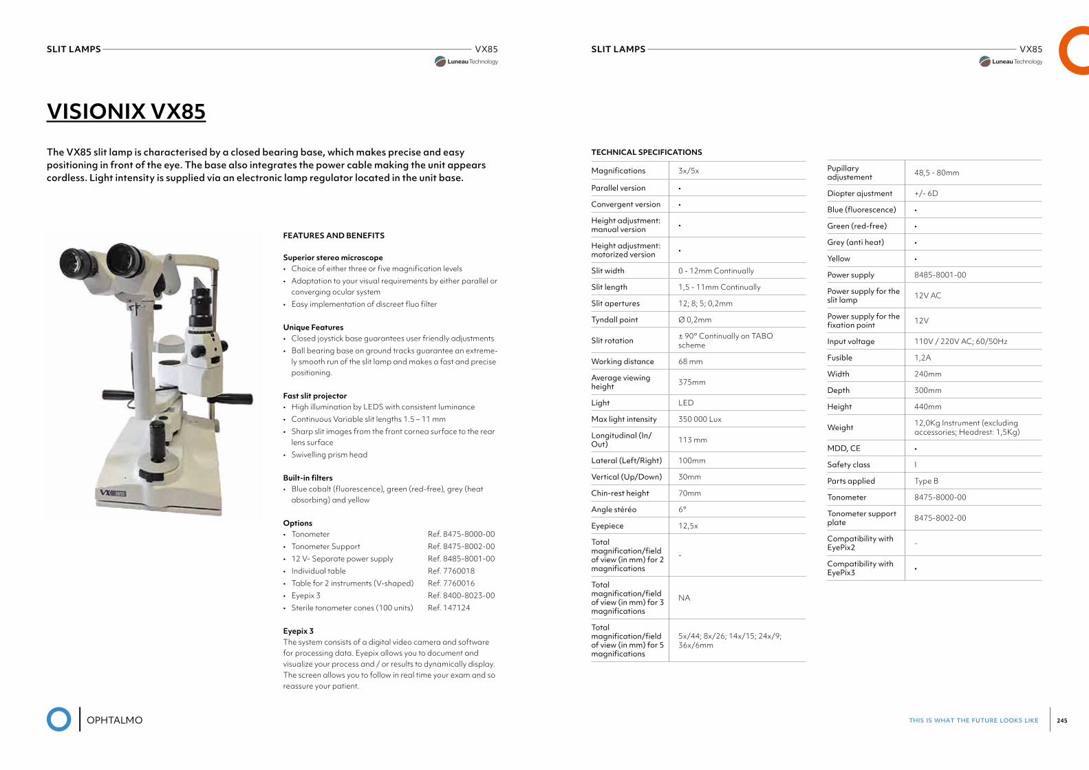
this is what the future looks like 245
SLIT LAMPS VX85SLIT LAMPS VX85
VISIONIX VX85
The VX85 slit lamp is characterised by a closed bearing base, which makes precise and easy positioning in front of the eye. The base also integrates the power cable making the unit appears cordless. Light intensity is supplied via an electronic lamp regulator located in the unit base.
FEATURES AND BENEFITS
Superior stereo microscope• Choice of either three or five magnification levels• Adaptation to your visual requirements by either parallel or
converging ocular system• Easy implementation of discreet fluo filter
Unique Features• Closed joystick base guarantees user friendly adjustments• Ball bearing base on ground tracks guarantee an extreme-
ly smooth run of the slit lamp and makes a fast and precise positioning.
Fast slit projector• High illumination by LEDS with consistent luminance• Continuous Variable slit lengths 1.5 – 11 mm• Sharp slit images from the front cornea surface to the rear
lens surface• Swivelling prism head
Built-in filters• Blue cobalt (fluorescence), green (red-free), grey (heat
absorbing) and yellow
Options• Tonometer Ref. 8475-8000-00• Tonometer Support Ref. 8475-8002-00• 12 V- Separate power supply Ref. 8485-8001-00• Individual table Ref. 7760018• Table for 2 instruments (V-shaped) Ref. 7760016• Eyepix 3 Ref. 8400-8023-00• Sterile tonometer cones (100 units) Ref. 147124
Eyepix 3The system consists of a digital video camera and software for processing data. Eyepix allows you to document and visualize your process and / or results to dynamically display.The screen allows you to follow in real time your exam and so reassure your patient.
TECHNICAL SPECIFICATIONS
Magnifications 3x/5x
Parallel version •
Convergent version •
Height adjustment: manual version •
Height adjustment: motorized version •
Slit width 0 - 12mm Continually
Slit length 1,5 - 11mm Continually
Slit apertures 12; 8; 5; 0,2mm
Tyndall point Ø 0,2mm
Slit rotation ± 90° Continually on TABO scheme
Working distance 68 mm
Average viewing height 375mm
Light LED
Max light intensity 350 000 Lux
Longitudinal (In/Out) 113 mm
Lateral (Left/Right) 100mm
Vertical (Up/Down) 30mm
Chin-rest height 70mm
Angle stéréo 6°
Eyepiece 12,5x
Total magnification/fieldof view (in mm) for 2 magnifications
-
Total magnification/fieldofview(inmm)for3magnifications
NA
Total magnification/fieldof view (in mm) for 5 magnifications
5x/44; 8x/26; 14x/15; 24x/9; 36x/6mm
Pupillary adjustement 48,5 - 80mm
Diopter ajustment +/- 6D
Blue(fluorescence) •
Green (red-free) •
Grey (anti heat) •
Yellow •
Power supply 8485-8001-00
Power supply for the slit lamp 12V AC
Power supply for the fixationpoint 12V
Input voltage 110V / 220V AC; 60/50Hz
Fusible 1,2A
Width 240mm
Depth 300mm
Height 440mm
Weight 12,0Kg Instrument (excluding accessories; Headrest: 1,5Kg)
MDD, CE •
Safety class I
Parts applied Type B
Tonometer 8475-8000-00
Tonometer support plate 8475-8002-00
Compatibility with EyePix2 -
Compatibility with EyePix3 •
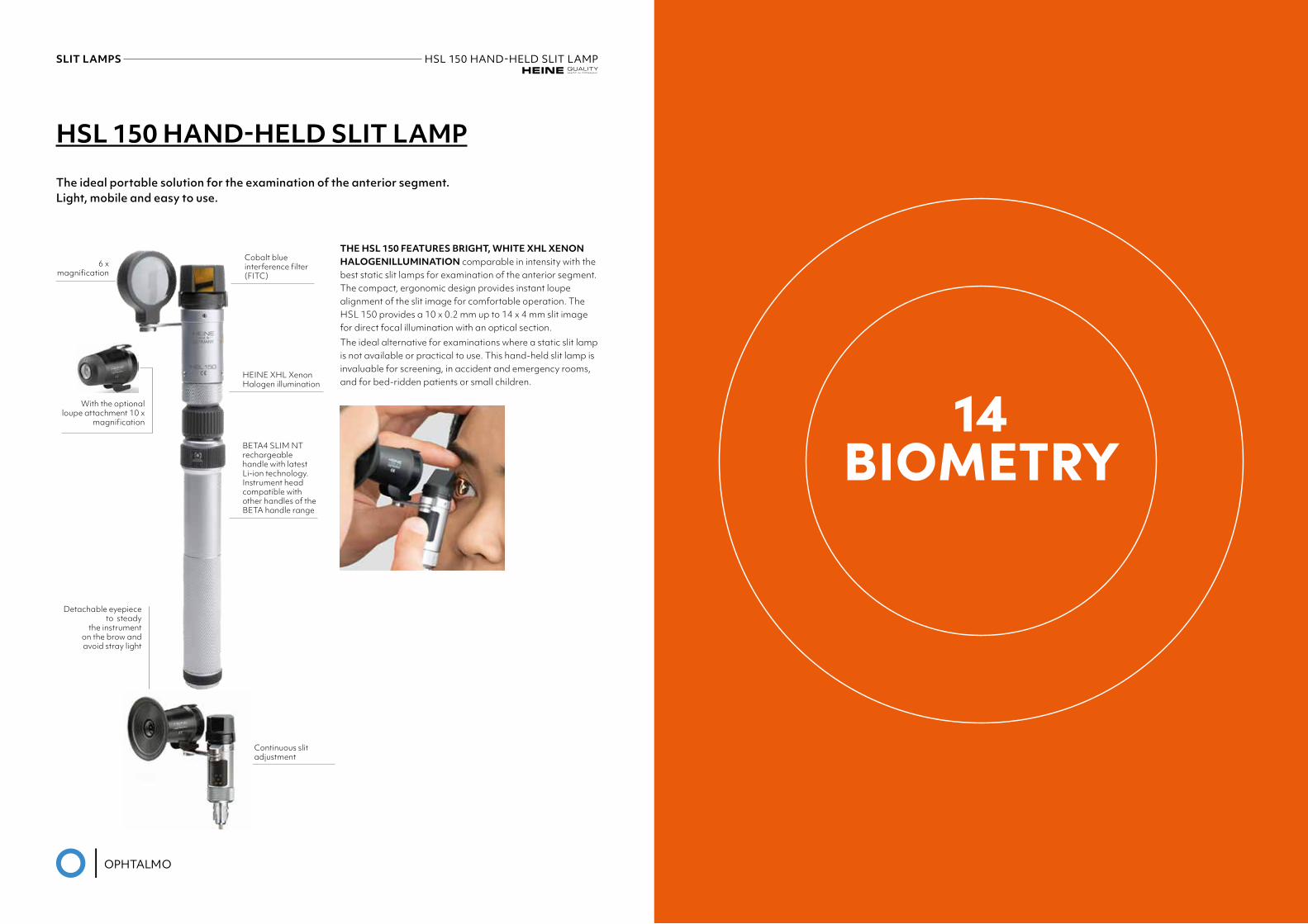
14BIOMETRY
HSL 150 HAND-HELD SLIT LAMP
The ideal portable solution for the examination of the anterior segment.Light, mobile and easy to use.
THE HSL 150 FEATURES BRIGHT, WHITE XHL XENON HALOGENILLUMINATION comparable in intensity with the best static slit lamps for examination of the anterior segment. The compact, ergonomic design provides instant loupe alignment of the slit image for comfortable operation. The HSL 150 provides a 10 x 0.2 mm up to 14 x 4 mm slit image for direct focal illumination with an optical section.The ideal alternative for examinations where a static slit lamp is not available or practical to use. This hand-held slit lamp is invaluable for screening, in accident and emergency rooms, and for bed-ridden patients or small children.
With the optional loupe attachment 10 x
magnification
6 x magnification
Detachable eyepiece to steady
the instrument on the brow and avoid stray light
Cobalt blue interference filter (FITC)
HEINE XHL Xenon Halogen illumination
Continuous slit adjustment
BETA4 SLIM NT rechargeable handle with latest Li-ion technology. Instrument head compatible with other handles of the BETA handle range
SLIT LAMPS HSL150HAND-HELDSLITLAMPHeine
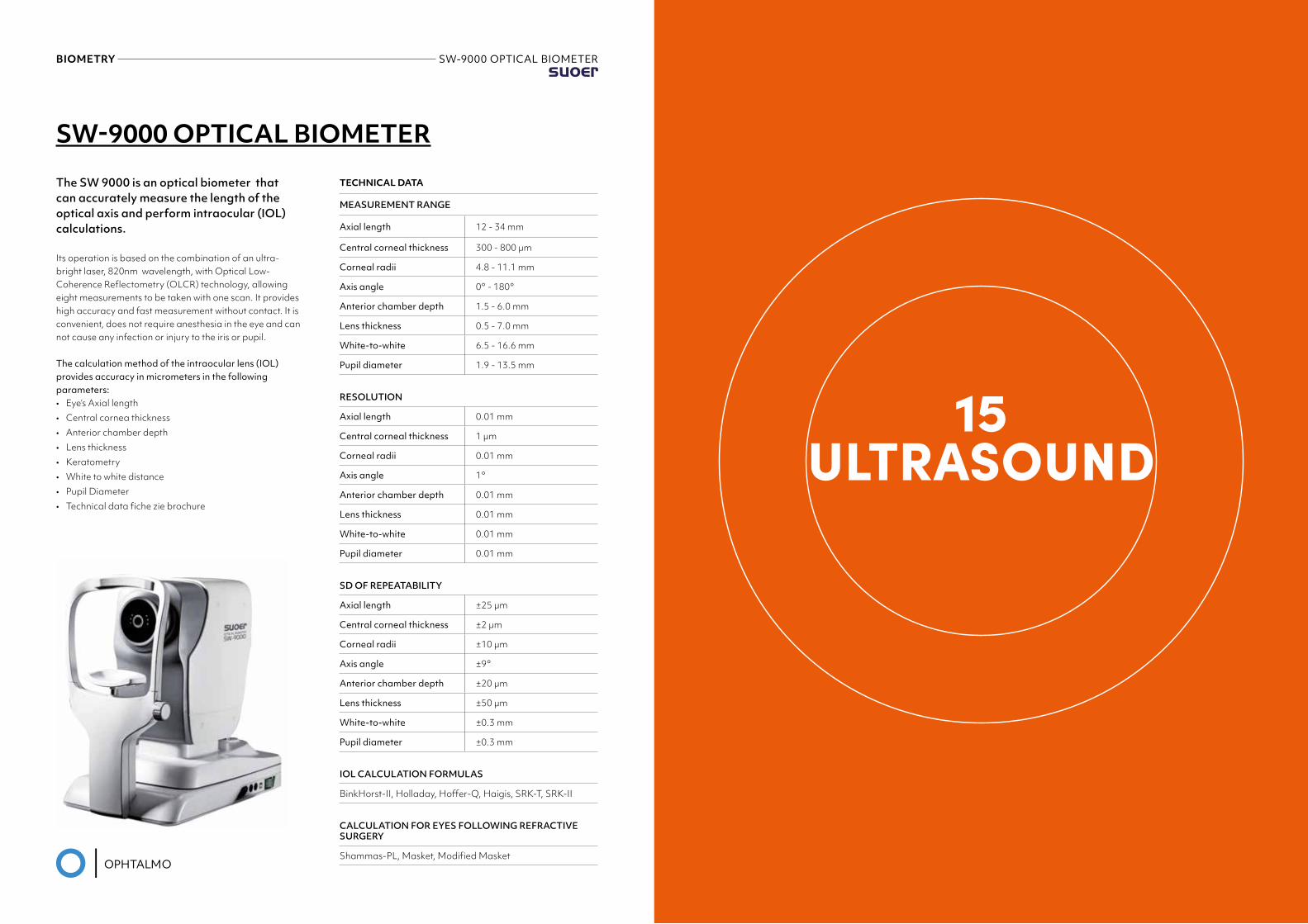
BIOMETRY SW-9000 OPTICAL BIOMETERSuoer
15ULTRASOUND
TECHNICAL DATA
MEASUREMENT RANGE
Axial length 12 - 34 mm
Central corneal thickness 300 - 800 µm
Corneal radii 4.8 - 11.1 mm
Axis angle 0° - 180°
Anterior chamber depth 1.5 - 6.0 mm
Lens thickness 0.5 - 7.0 mm
White-to-white 6.5 - 16.6 mm
Pupil diameter 1.9 - 13.5 mm
RESOLUTION
Axial length 0.01 mm
Central corneal thickness 1 µm
Corneal radii 0.01 mm
Axis angle 1°
Anterior chamber depth 0.01 mm
Lens thickness 0.01 mm
White-to-white 0.01 mm
Pupil diameter 0.01 mm
SD OF REPEATABILITY
Axial length ±25 µm
Central corneal thickness ±2 µm
Corneal radii ±10 µm
Axis angle ±9°
Anterior chamber depth ±20 µm
Lens thickness ±50 µm
White-to-white ±0.3 mm
Pupil diameter ±0.3 mm
IOL CALCULATION FORMULAS
BinkHorst-II, Holladay, Hoffer-Q, Haigis, SRK-T, SRK-II
CALCULATION FOR EYES FOLLOWING REFRACTIVE SURGERY
Shammas-PL, Masket, Modified Masket
SW-9000 OPTICAL BIOMETER
The SW 9000 is an optical biometer that can accurately measure the length of the optical axis and perform intraocular (IOL) calculations.
Its operation is based on the combination of an ultra-bright laser, 820nm wavelength, with Optical Low-Coherence Reflectometry (OLCR) technology, allowing eight measurements to be taken with one scan. It provides high accuracy and fast measurement without contact. It is convenient, does not require anesthesia in the eye and can not cause any infection or injury to the iris or pupil.
The calculation method of the intraocular lens (IOL)provides accuracy in micrometers in the following parameters:• Eye’s Axial length• Central cornea thickness• Anterior chamber depth• Lens thickness• Keratometry• White to white distance• Pupil Diameter• Technical data fiche zie brochure
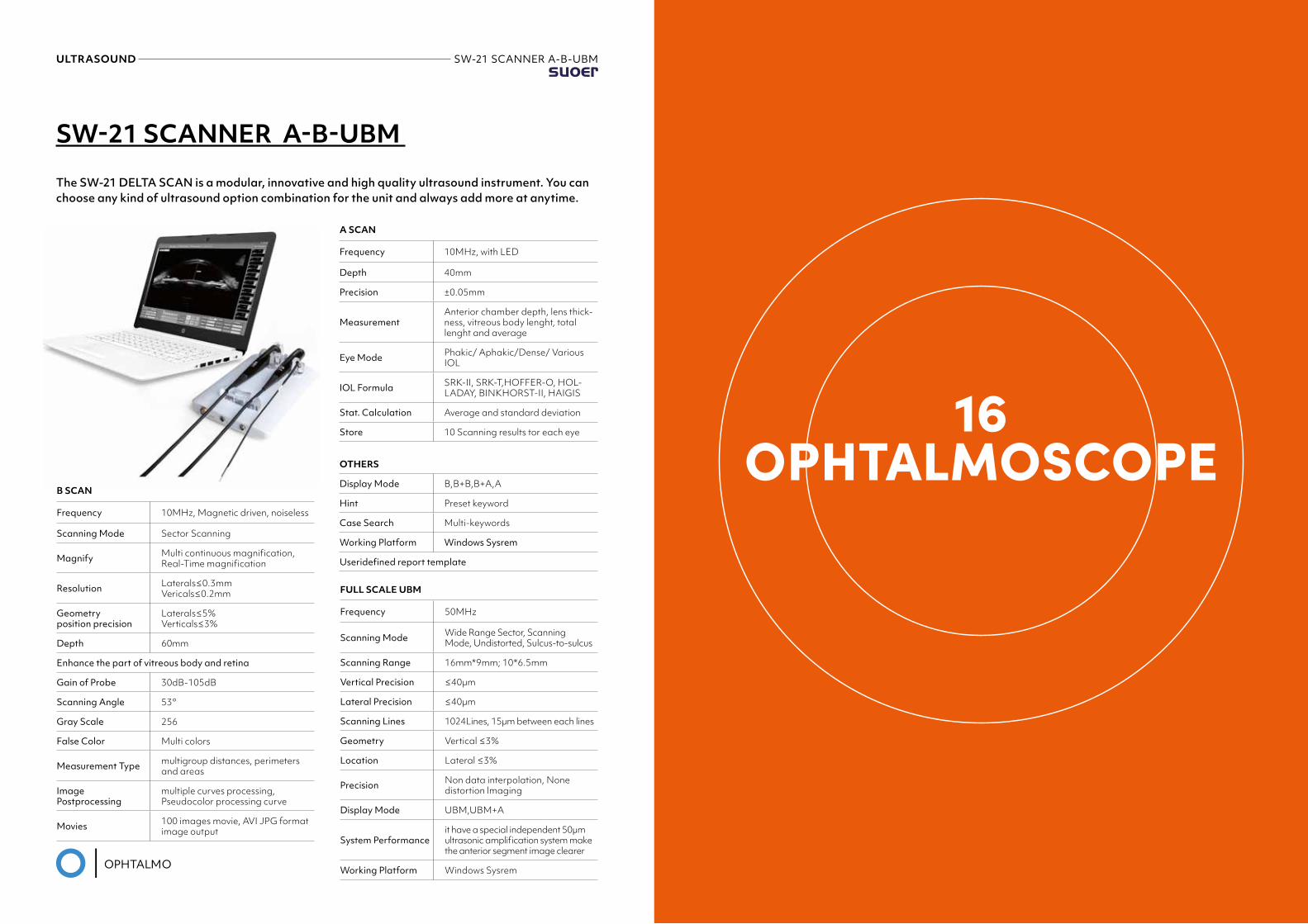
ULTRASOUND SW-21 SCANNER A-B-UBMSuoer
SW-21 SCANNER A-B-UBM
The SW-21 DELTA SCAN is a modular, innovative and high quality ultrasound instrument. You can choose any kind of ultrasound option combination for the unit and always add more at anytime.
A SCAN
Frequency 10MHz, with LED
Depth 40mm
Precision ±0.05mm
MeasurementAnterior chamber depth, lens thick-ness, vitreous body lenght, total lenght and average
Eye Mode Phakic/ Aphakic/Dense/ Various IOL
IOL Formula SRK-II, SRK-T,HOFFER-O, HOL-LADAY, BINKHORST-II, HAIGIS
Stat. Calculation Average and standard deviation
Store 10 Scanning results tor each eye
OTHERS
Display Mode B,B+B,B+A,A
Hint Preset keyword
Case Search Multi-keywords
Working Platform Windows Sysrem
Useridefinedreporttemplate
FULL SCALE UBM
Frequency 50MHz
Scanning Mode Wide Range Sector, Scanning Mode, Undistorted, Sulcus-to-sulcus
Scanning Range 16mm*9mm; 10*6.5mm
Vertical Precision ≤40µm
Lateral Precision ≤40µm
Scanning Lines 1024Lines, 15µm between each lines
Geometry Vertical ≤3%
Location Lateral ≤3%
Precision Non data interpolation, None distortion lmaging
Display Mode UBM,UBM+A
System Performanceit have a special independent 50µm ultrasonic amplification system make the anterior segment image clearer
Working Platform Windows Sysrem
B SCAN
Frequency 10MHz, Magnetic driven, noiseless
Scanning Mode Sector Scanning
Magnify Multi continuous magnification, Real-Time magnification
Resolution Laterals≤0.3mm Vericals≤0.2mm
Geometry position precision
Laterals≤5% Verticals≤3%
Depth 60mm
Enhance the part of vitreous body and retina
Gain of Probe 30dB-105dB
Scanning Angle 53°
Gray Scale 256
False Color Multi colors
Measurement Type multigroup distances, perimeters and areas
Image Postprocessing
multiple curves processing, Pseudo color processing curve
Movies 100 images movie, AVI JPG format image output
16OPHTALMOSCOPE
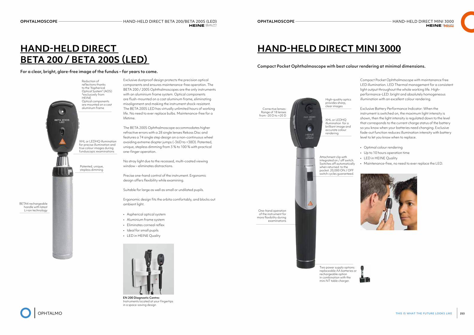
this is what the future looks like 253
OPHTALMOSCOPE HAND-HELD DIRECT BETA 200/BETA 200S (LED) Heine
OPHTALMOSCOPE HAND-HELDDIRECTMINI3000Heine
HAND-HELD DIRECT BETA 200 / BETA 200S (LED)
HAND-HELD DIRECT MINI 3000
For a clear, bright, glare-free image of the fundus – for years to come.Compact Pocket Ophthalmoscope with best colour rendering at minimal dimensions.
Exclusive dustproof design protects the precision optical components and ensures maintenance-free operation. The BETA 200 / 200S Ophthalmoscopes are the only instruments with an aluminium frame system. Optical components are flush-mounted on a cast aluminum frame, eliminating misalignment and making the instrument shock-resistant. The BETA 200S LED has virtually unlimited hours of working life. No need to ever replace bulbs. Maintenance-free for a lifetime.
The BETA 200S Ophthalmoscope accommodates higher refractive errors with a 28 single lenses Rekoss Disc and features a 74 single step design on a non-continuous wheel avoiding extreme diopter jumps (-36D to +38D). Patented, unique, stepless dimming from 3 % to 100 % with practical one-finger operation.
No stray light due to the recessed, multi-coated viewing window – eliminates distractions.
Precise one-hand control of the instrument. Ergonomic design offers flexibility while examining.
Suitable for large as well as small or undilated pupils.
Ergonomic design fits the orbita comfortably, and blocks out ambient light.
• Aspherical optical system• Aluminium frame system• Eliminates corneal reflex• Ideal for small pupils• LED in HEINE Quality
BETA4 rechargeablehandle with latest Li-ion technology
Corrective lenses: Range of 18 lenses
from -20 D to +20 D
One-hand operation of the instrument for
more flexibility during examinations
Reduction of reflections thanks to the “Aspherical Optical System” (AOS) *exclusively from HEINEOptical components are mounted on a cast aluminum frame
XHL or LEDHQ illumination for precise illumination and true colour images during funduscopic examinations
Attachment clip with integrated on / off switch. Switches off automatically when returned to the pocket. 20,000 ON / OFF switch cycles guaranteed.
Two power supply options: replaceable AA batteries or rechargeable option in combination with the mini NT table charger.
Patented, unique,stepless dimming
High-quality optics provides sharp, clear images
XHL or LEDHQ illumination for a brilliant image and accurate colour rendering
Compact Pocket Ophthalmoscope with maintanance free LED illumination. LED Thermal management for a consistent light output throughout the whole working life. High-performance-LED: bright and absolutely homogeneous illumination with an excellent colour rendering.
Exclusive Battery Performance Indicator: When the instrument is switched on, the maximum light intensity is shown, then the light intensity is regulated down to the level that corresponds to the current charge status of the battery so you know when your batteries need changing. Exclusive fade-out function reduces illumination intensity with battery level to let you know when to replace.
• Optimal colour rendering• Up to 10 hours operation time• LED in HEINE Quality • Maintenance-free, no need to ever replace the LED.
EN 200 Diagnostic Centre: Instruments located at your fingertips in a space-saving design
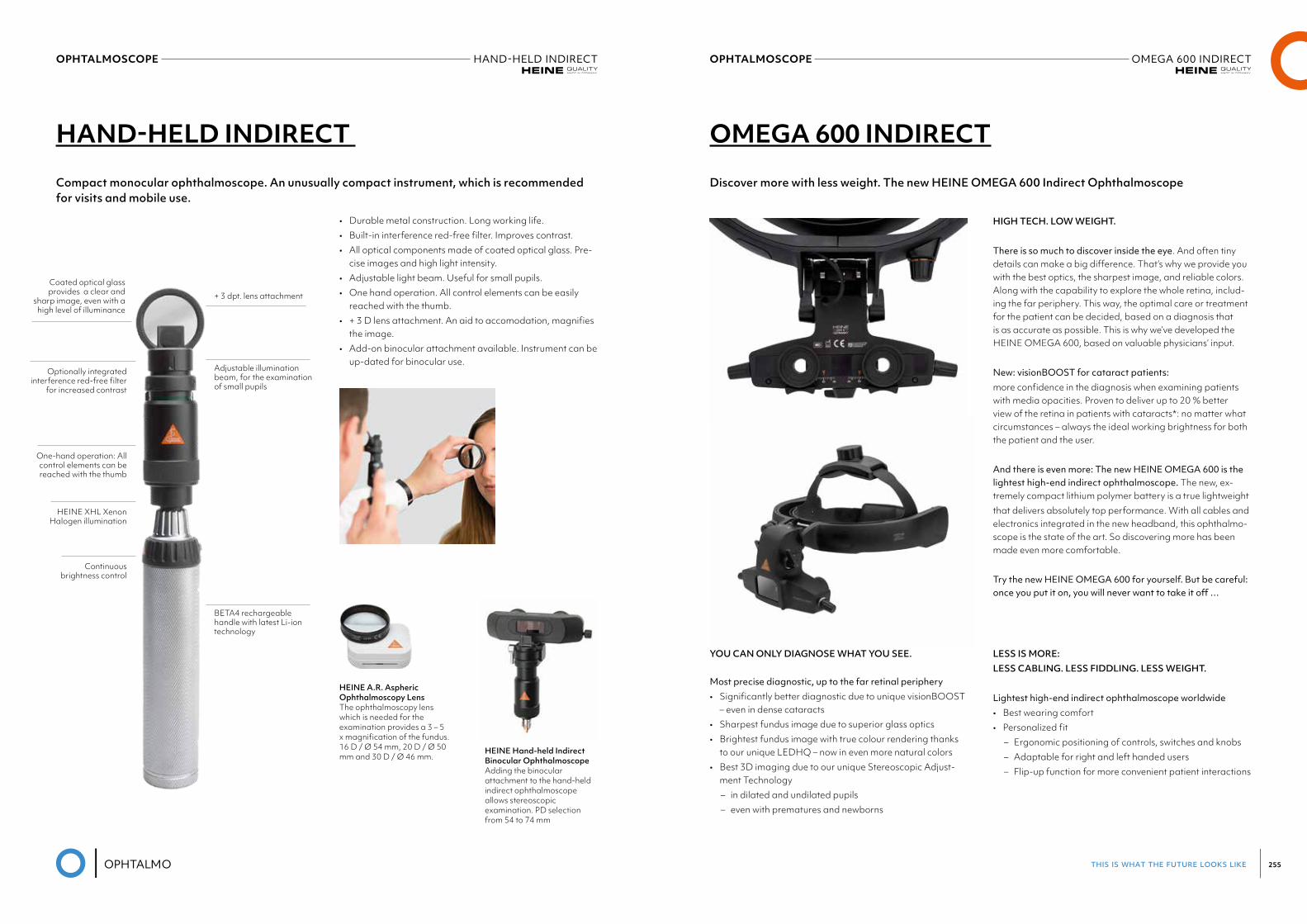
this is what the future looks like 255
OPHTALMOSCOPE HAND-HELDINDIRECT
HAND-HELD INDIRECT
Compact monocular ophthalmoscope. An unusually compact instrument, which is recommended for visits and mobile use.
• Durable metal construction. Long working life.• Built-in interference red-free filter. Improves contrast.• All optical components made of coated optical glass. Pre-
cise images and high light intensity.• Adjustable light beam. Useful for small pupils.• One hand operation. All control elements can be easily
reached with the thumb.• + 3 D lens attachment. An aid to accomodation, magnifies
the image.• Add-on binocular attachment available. Instrument can be
up-dated for binocular use.
HEINE A.R. Aspheric Ophthalmoscopy Lens The ophthalmoscopy lens which is needed for the examination provides a 3 – 5 x magnification of the fundus. 16 D / Ø 54 mm, 20 D / Ø 50 mm and 30 D / Ø 46 mm.
HEINE Hand-held Indirect Binocular Ophthalmoscope Adding the binocular attachment to the hand-held indirect ophthalmoscope allows stereoscopic examination. PD selection from 54 to 74 mm
Coated optical glass provides a clear and
sharp image, even with a high level of illuminance
+ 3 dpt. lens attachment
Adjustable illumination beam, for the examination of small pupils
BETA4 rechargeable handle with latest Li-ion technology
Optionally integrated interference red-free filter
for increased contrast
One-hand operation: All control elements can be reached with the thumb
HEINE XHL Xenon Halogen illumination
Continuous brightness control
OPHTALMOSCOPE OMEGA 600 INDIRECT
OMEGA 600 INDIRECT
Discover more with less weight. The new HEINE OMEGA 600 Indirect Ophthalmoscope
HIGH TECH. LOW WEIGHT.
There is so much to discover inside the eye. And often tiny details can make a big difference. That’s why we provide you with the best optics, the sharpest image, and reliable colors. Along with the capability to explore the whole retina, includ-ing the far periphery. This way, the optimal care or treatment for the patient can be decided, based on a diagnosis that is as accurate as possible. This is why we’ve developed the HEINE OMEGA 600, based on valuable physicians’ input.
New: visionBOOST for cataract patients:more confidence in the diagnosis when examining patients with media opacities. Proven to deliver up to 20 % better view of the retina in patients with cataracts*: no matter what circumstances – always the ideal working brightness for both the patient and the user.
And there is even more: The new HEINE OMEGA 600 is the lightest high-end indirect ophthalmoscope. The new, ex-tremely compact lithium polymer battery is a true lightweightthat delivers absolutely top performance. With all cables and electronics integrated in the new headband, this ophthalmo-scope is the state of the art. So discovering more has been made even more comfortable.
Try the new HEINE OMEGA 600 for yourself. But be careful: onceyouputiton,youwillneverwanttotakeitoff…
YOU CAN ONLY DIAGNOSE WHAT YOU SEE.
Most precise diagnostic, up to the far retinal periphery• Significantly better diagnostic due to unique visionBOOST
– even in dense cataracts• Sharpest fundus image due to superior glass optics• Brightest fundus image with true colour rendering thanks
to our unique LEDHQ – now in even more natural colors• Best 3D imaging due to our unique Stereoscopic Adjust-
ment Technology– in dilated and undilated pupils– even with prematures and newborns
LESS IS MORE: LESS CABLING. LESS FIDDLING. LESS WEIGHT.
Lightest high-end indirect ophthalmoscope worldwide• Best wearing comfort• Personalized fit
– Ergonomic positioning of controls, switches and knobs– Adaptable for right and left handed users– Flip-up function for more convenient patient interactions
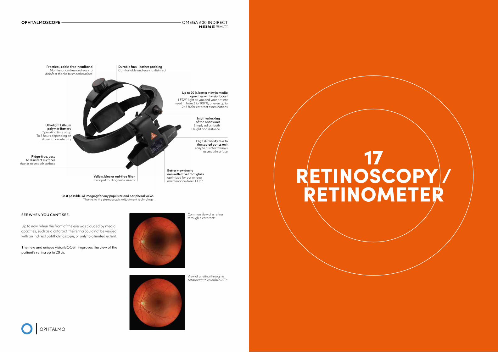
OPHTALMOSCOPE OMEGA 600 INDIRECT
17RETINOSCOPY / RETINOMETER
SEE WHEN YOU CAN’T SEE.
Up to now, when the front of the eye was clouded by media opacities, such as a cataract, the retina could not be viewed with an indirect ophthalmoscope, or only to a limited extent.
The new and unique visionBOOST improves the view of the patient’s retina up to 20 %.
Common view of a retina through a cataract*
View of a retina through a cataract with visionBOOST*
Practical, cable-free headbandMaintenance-free and easy to
disinfect thanks to smoothsurface
Durable faux leather padding Comfortable and easy to disinfect
Yellow, blue or red-free filter To adjust to diagnostic needs
Best possible 3d imaging for any pupil size and peripheral viewsThanks to the stereoscopic adjustment technology
Ultralight Lithium polymer Battery
Operating time of up To 8 hours depending on
illumination intensity
Ridge-free, easy to disinfect surfaces
thanks to smooth surface
Intuitive locking of the optics unit
Simply adjust both Height and distance
High durability due to the sealed optics unit
easy to disinfect thanks to smoothsurface
Better view due to non-reflective front glass optimized for our unique, maintenance-free LEDHQ
Up to 20 % better view in media opacities with visionboost
LEDHQ light as you and your patient need it: from 3 to 100 %, or even up to
245 % for cataract examinations
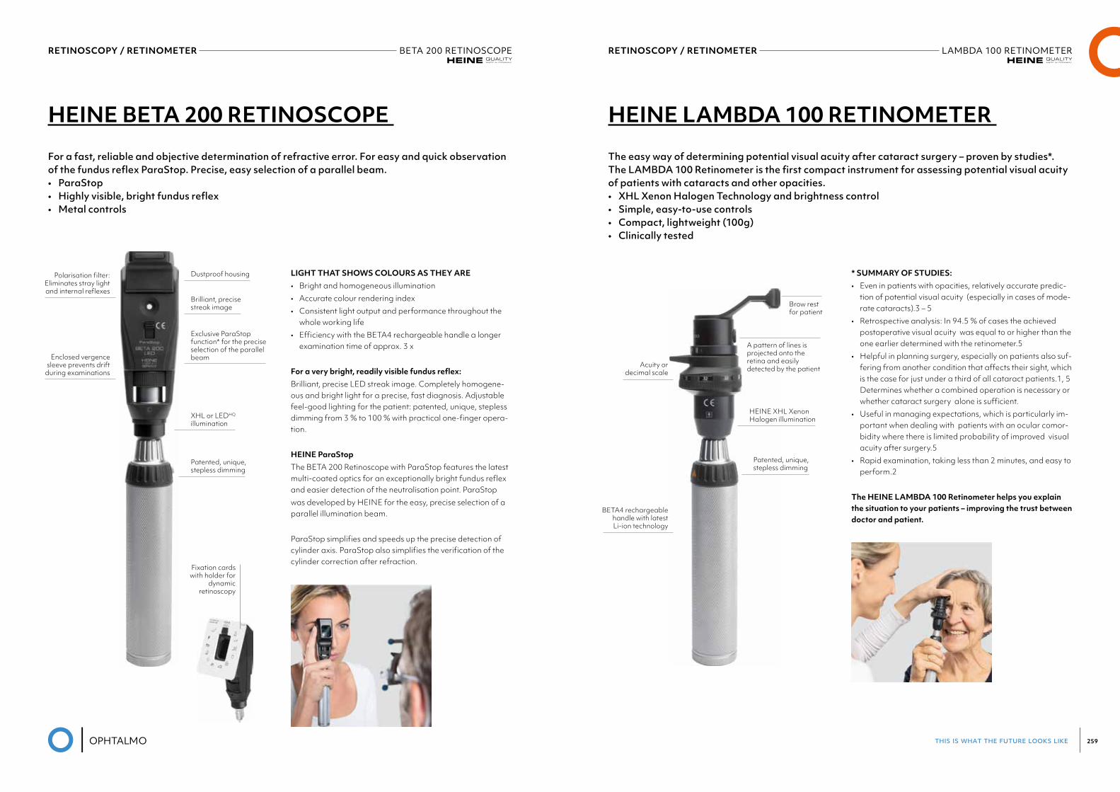
this is what the future looks like 259
RETINOSCOPY / RETINOMETER BETA 200 RETINOSCOPEHeine
RETINOSCOPY / RETINOMETER LAMBDA 100 RETINOMETERHeine
HEINE BETA 200 RETINOSCOPE HEINE LAMBDA 100 RETINOMETER
For a fast, reliable and objective determination of refractive error. For easy and quick observation of the fundus reflex ParaStop. Precise, easy selection of a parallel beam.• ParaStop• Highly visible, bright fundus reflex• Metal controls
The easy way of determining potential visual acuity after cataract surgery – proven by studies*. The LAMBDA 100 Retinometer is the first compact instrument for assessing potential visual acuity of patients with cataracts and other opacities. • XHL Xenon Halogen Technology and brightness control• Simple, easy-to-use controls• Compact, lightweight (100g)• Clinically tested
LIGHT THAT SHOWS COLOURS AS THEY ARE• Bright and homogeneous illumination• Accurate colour rendering index• Consistent light output and performance throughout the
whole working life• Efficiency with the BETA4 rechargeable handle a longer
examination time of approx. 3 x
For a very bright, readily visible fundus reflex:Brilliant, precise LED streak image. Completely homogene-ous and bright light for a precise, fast diagnosis. Adjustable feel-good lighting for the patient: patented, unique, stepless dimming from 3 % to 100 % with practical one-finger opera-tion.
HEINE ParaStopThe BETA 200 Retinoscope with ParaStop features the latest multi-coated optics for an exceptionally bright fundus reflex and easier detection of the neutralisation point. ParaStopwas developed by HEINE for the easy, precise selection of a parallel illumination beam.
ParaStop simplifies and speeds up the precise detection of cylinder axis. ParaStop also simplifies the verification of the cylinder correction after refraction.
* SUMMARY OF STUDIES:• Even in patients with opacities, relatively accurate predic-
tion of potential visual acuity (especially in cases of mode-rate cataracts).3 – 5
• Retrospective analysis: In 94.5 % of cases the achieved postoperative visual acuity was equal to or higher than the one earlier determined with the retinometer.5
• Helpful in planning surgery, especially on patients also suf-fering from another condition that affects their sight, which is the case for just under a third of all cataract patients.1, 5 Determines whether a combined operation is necessary or whether cataract surgery alone is sufficient.
• Useful in managing expectations, which is particularly im-portant when dealing with patients with an ocular comor-bidity where there is limited probability of improved visual acuity after surgery.5
• Rapid examination, taking less than 2 minutes, and easy to perform.2
The HEINE LAMBDA 100 Retinometer helps you explain the situation to your patients – improving the trust between doctor and patient.
Polarisation filter:Eliminates stray lightand internal reflexes
Acuity ordecimal scale
Enclosed vergencesleeve prevents drift
during examinations
BETA4 rechargeablehandle with latest Li-ion technology
Dustproof housing
A pattern of lines is projected onto the retina and easilydetected by the patient
Brow rest for patient
Brilliant, precisestreak image
Exclusive ParaStop function* for the precise selection of the parallel beam
XHL or LEDHQ
illumination
HEINE XHL XenonHalogen illumination
Patented, unique,stepless dimming
Patented, unique,stepless dimming
Fixation cardswith holder for
dynamicretinoscopy

18EXAMINATION
UNITS
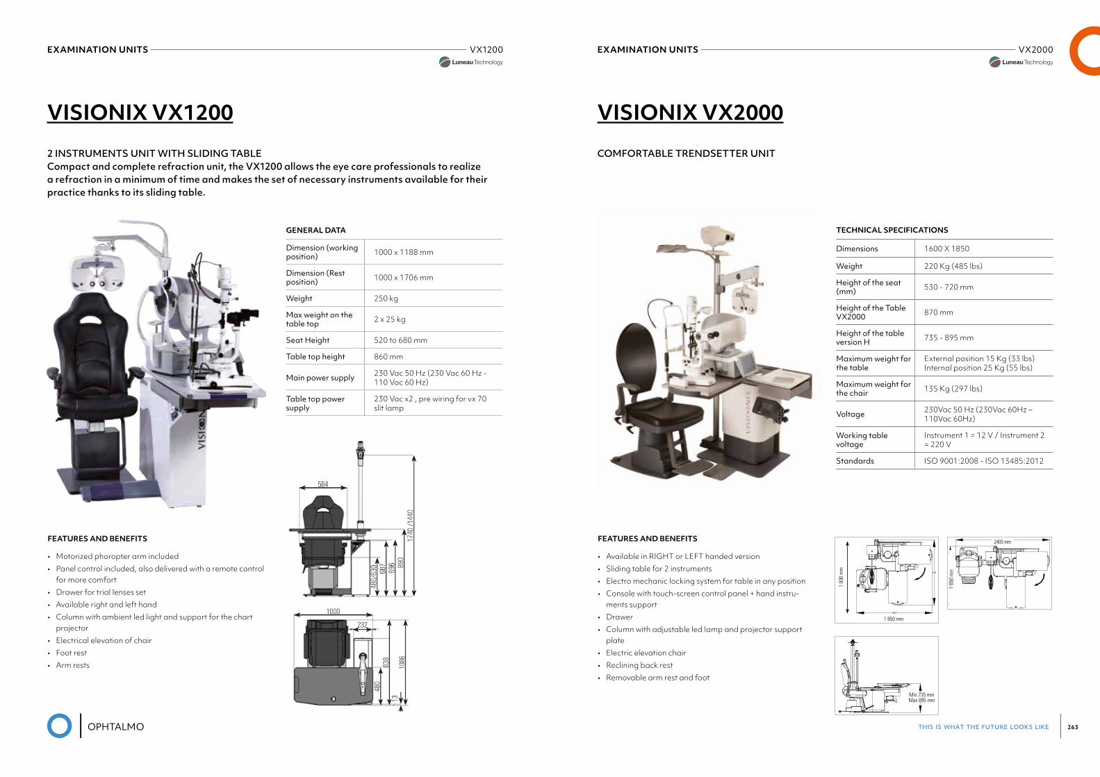
this is what the future looks like 263
EXAMINATION UNITS VX1200Luneau Visionix
EXAMINATION UNITS VX2000
VISIONIX VX1200
2 INSTRUMENTS UNIT WITH SLIDING TABLECompact and complete refraction unit, the VX1200 allows the eye care professionals to realize a refraction in a minimum of time and makes the set of necessary instruments available for their practice thanks to its sliding table.
GENERAL DATA
Dimension (working position) 1000 x 1188 mm
Dimension (Rest position) 1000 x 1706 mm
Weight 250 kg
Max weight on the table top 2 x 25 kg
Seat Height 520 to 680 mm
Table top height 860 mm
Main power supply 230 Vac 50 Hz (230 Vac 60 Hz - 110 Vac 60 Hz)
Table top power supply
230 Vac x2 , pre wiring for vx 70 slit lamp
FEATURES AND BENEFITS
• Motorized phoropter arm included• Panel control included, also delivered with a remote control
for more comfort• Drawer for trial lenses set• Available right and left hand• Column with ambient led light and support for the chart
projector• Electrical elevation of chair• Foot rest• Arm rests
VISIONIX VX2000
COMFORTABLE TRENDSETTER UNIT
FEATURES AND BENEFITS
• Available in RIGHT or LEFT handed version• Sliding table for 2 instruments• Electro mechanic locking system for table in any position• Console with touch-screen control panel + hand instru-
ments support• Drawer• Column with adjustable led lamp and projector support
plate• Electric elevation chair• Reclining back rest• Removable arm rest and foot
TECHNICAL SPECIFICATIONS
Dimensions 1600 X 1850
Weight 220 Kg (485 lbs)
Height of the seat (mm) 530 - 720 mm
Height of the Table VX2000 870 mm
Height of the table version H 735 - 895 mm
Maximum weight for the table
External position 15 Kg (33 lbs) Internal position 25 Kg (55 lbs)
Maximum weight for the chair 135 Kg (297 lbs)
Voltage 230Vac 50 Hz (230Vac 60Hz – 110Vac 60Hz)
Working table voltage
Instrument 1 = 12 V / Instrument 2 = 220 V
Standards ISO 9001:2008 - ISO 13485:2012
1 60
0 m
m
1 850 mm
Min 735 mmMax 895 mm
2400 mm
1 65
0 m
m
1 60
0 m
m
1 850 mm
Min 735 mmMax 895 mm
2400 mm
1 65
0 m
m
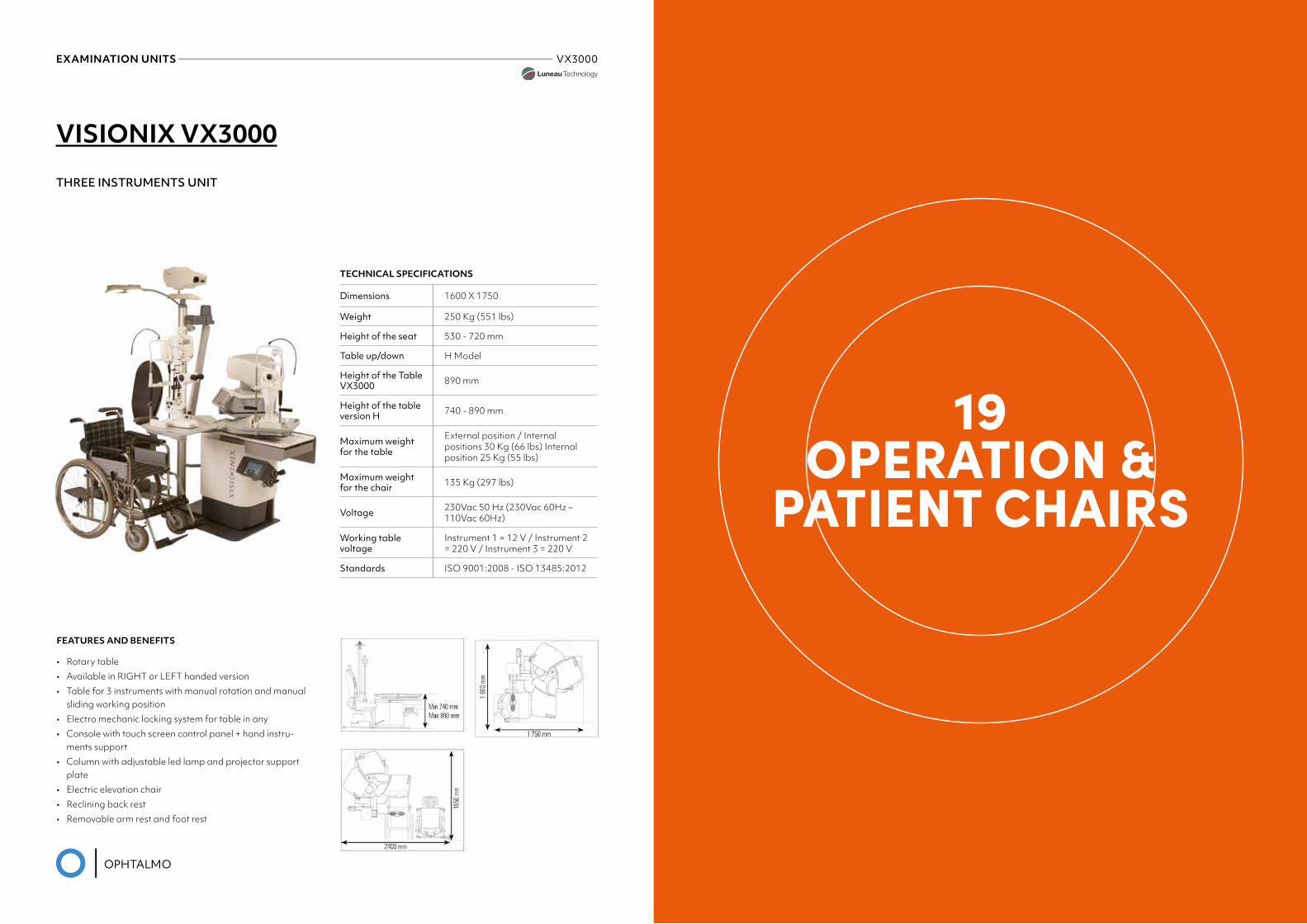
EXAMINATION UNITS VX3000
VISIONIX VX3000
THREE INSTRUMENTS UNIT
FEATURES AND BENEFITS
• Rotary table• Available in RIGHT or LEFT handed version• Table for 3 instruments with manual rotation and manual
sliding working position• Electro mechanic locking system for table in any• Console with touch screen control panel + hand instru-
ments support• Column with adjustable led lamp and projector support
plate• Electric elevation chair• Reclining back rest• Removable arm rest and foot rest
TECHNICAL SPECIFICATIONS
Dimensions 1600 X 1750
Weight 250 Kg (551 lbs)
Height of the seat 530 - 720 mm
Table up/down H Model
Height of the Table VX3000 890 mm
Height of the table version H 740 - 890 mm
Maximum weight for the table
External position / Internal positions 30 Kg (66 lbs) Internal position 25 Kg (55 lbs)
Maximum weight for the chair 135 Kg (297 lbs)
Voltage 230Vac 50 Hz (230Vac 60Hz – 110Vac 60Hz)
Working table voltage
Instrument 1 = 12 V / Instrument 2 = 220 V / Instrument 3 = 220 V
Standards ISO 9001:2008 - ISO 13485:2012
19OPERATION &
PATIENT CHAIRS
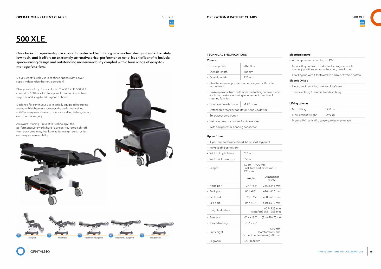
this is what the future looks like 267
OPERATION & PATIENT CHAIRS 500 XLE OPERATION & PATIENT CHAIRS 500 XLE
500 XLE
Our classic. lt represents proven and time-tested technology in a modern design, it is deliberately low-tech, and it offers an extremely attractive price-performance ratio. lts chief benefits include space-saving design and outstanding maneuverability coupled with a lean range of easy-to-manage functions.
Do you want flexible use in confined spaces with power-supply independent battery operation?
Then you should go for our classic: The 500 XLE, 500 XLE comfort or 500 bariatric, for optimal combination with our surgiLine and surgiTrend surgeon·s chairs.
Designed for continuous use in serially equipped operating rooms with high patient turnover, the performanceLine satisfies every user thanks to its easy handling before, during and after the surgery.
An award-winning “Preventive Technology”, the performanceLine works hard to proleet your surgical staff from back problems, thanks to its lightweight construction and easy maneuverability.
TECHNICAL SPECIFICATIONS
Chassis
- Frame profile 90x 50 mm
- Outside length 785mm
- Outside width 720mm
- Steel tube frame, powder-coated elegant anthracite matte finish
- Brake operable from both sides and acting on two castors each; two castors featuring independent directional steering function
- Double rimmed castors Ø 125 mm
- Detachable foot keypad (total. head up/down)
- Emergency stop button
- Visible screws are made of stainless steel
- With equipotential bonding connection
Upper frame
- 4-part support frame (head, back, seat. leg part)
- Removeable upholstery
- Width of upholstery 610mm
- Width incl . armrests 850mm
- Length1.760 - 1.900 mm (incl. foot part extension) + 190 mm
Angle Dimensions (Lx W)
- Head part - 2° / +35° 255 x 245 mm
- Back part 0° / +83° 610 x 610 mm
- Seat part -5° / +35° 450 x 610 mm
- Leg part 0° / +77° 510 x 610 mm
- Height adjustment 625- 925 mm (comfort) 655 - 955 mm
- Armrests 0° / +180° (2x) 470x 75 mm
- Trendelenburg -12° / +5”
- Entry hight580 mm
(comfort) 610 mm (incl. foot part extension) - 80 mm
- Legroom 520- 820 mm
Electrical control
- All components according to IPX4
- Manual keypad with 8 individually programmable memory positions, auto run function, reset button
- Foot keypad with 4 footswitches and reactivation button
Electric Drives
- Head, back, seat. leg part. total up/ down
- Trendelenburg / Reverse Trendelenburg
Lifting column
- Max. lifting 300 mm
- Max. patient weight 250 kg
- Motors IPX4 with HAL sensors, to be memorized
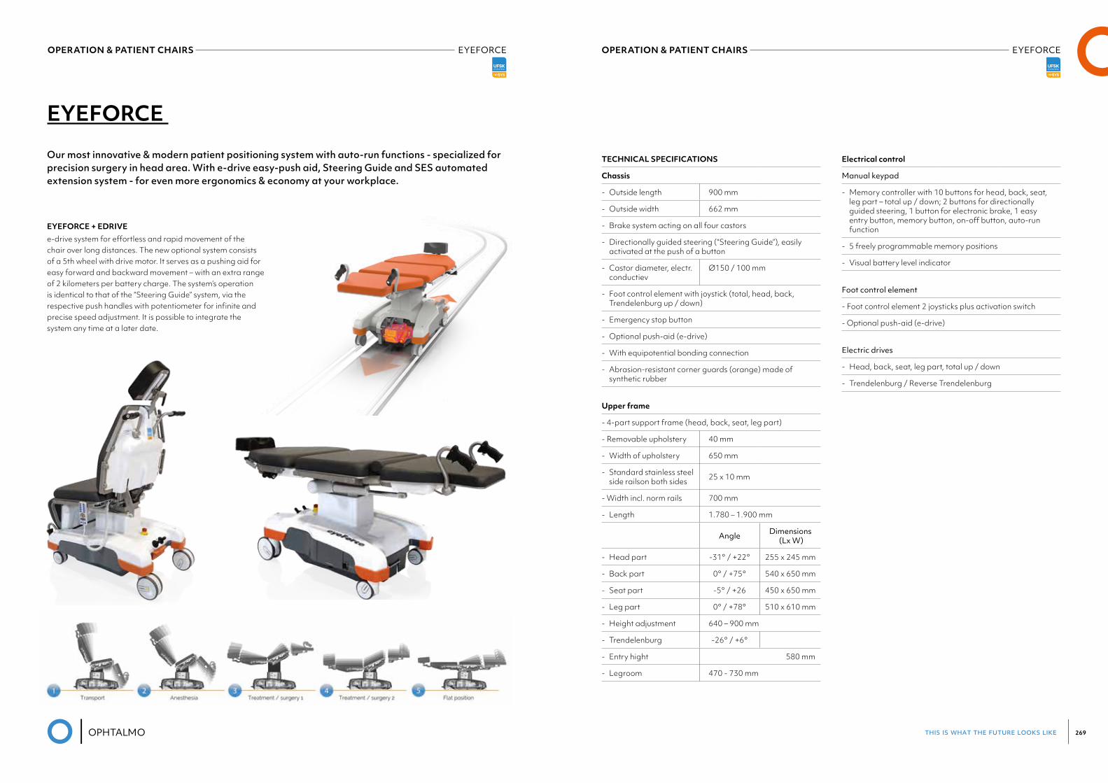
this is what the future looks like 269
OPERATION & PATIENT CHAIRS EYEFORCE OPERATION & PATIENT CHAIRS EYEFORCE
EYEFORCE
Our most innovative & modern patient positioning system with auto-run functions - specialized forprecision surgery in head area. With e-drive easy-push aid, Steering Guide and SES automated extension system - for even more ergonomics & economy at your workplace.
EYEFORCE + EDRIVEe-drive system for effortless and rapid movement of the chair over long distances. The new optional system consists of a 5th wheel with drive motor. It serves as a pushing aid for easy forward and backward movement – with an extra range of 2 kilometers per battery charge. The system’s operation is identical to that of the “Steering Guide” system, via the respective push handles with potentiometer for infinite and precise speed adjustment. It is possible to integrate the system any time at a later date.
TECHNICAL SPECIFICATIONS
Chassis
- Outside length 900 mm
- Outside width 662 mm
- Brake system acting on all four castors
- Directionally guided steering (“Steering Guide”), easily activated at the push of a button
- Castor diameter, electr. conductiev
Ø150 / 100 mm
- Foot control element with joystick (total, head, back, Trendelenburg up / down)
- Emergency stop button
- Optional push-aid (e-drive)
- With equipotential bonding connection
- Abrasion-resistant corner guards (orange) made of synthetic rubber
Upper frame
- 4-part support frame (head, back, seat, leg part)
- Removable upholstery 40 mm
- Width of upholstery 650 mm
- Standard stainless steel side railson both sides 25 x 10 mm
- Width incl. norm rails 700 mm
- Length 1.780 – 1.900 mm
Angle Dimensions (Lx W)
- Head part -31° / +22° 255 x 245 mm
- Back part 0° / +75° 540 x 650 mm
- Seat part -5° / +26 450 x 650 mm
- Leg part 0° / +78° 510 x 610 mm
- Height adjustment 640 – 900 mm
- Trendelenburg -26° / +6°
- Entry hight 580 mm
- Legroom 470 - 730 mm
Electrical control
Manual keypad
- Memory controller with 10 buttons for head, back, seat, leg part – total up / down; 2 buttons for directionally guided steering, 1 button for electronic brake, 1 easy entry button, memory button, on-off button, auto-run function
- 5 freely programmable memory positions
- Visual battery level indicator
Foot control element
- Foot control element 2 joysticks plus activation switch
- Optional push-aid (e-drive)
Electric drives
- Head, back, seat, leg part, total up / down
- Trendelenburg / Reverse Trendelenburg
OPERATION & PATIENT CHAIRS EYEFORCE
20AUTOCLAVES
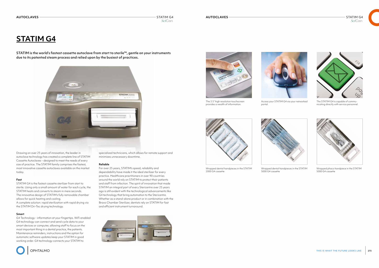
this is what the future looks like 273
AUTOCLAVES STATIM G4Scican
AUTOCLAVES STATIM G4Scican
STATIM G4
STATIM is the world’s fastest cassette autoclave from start to sterile™, gentle on your instruments due to its patented steam process and relied upon by the busiest of practices.
Drawing on over 25 years of innovation, the leader in autoclave technology has created a complete line of STATIM Cassette Autoclaves – designed to meet the needs of every size of practice. The STATIM family comprises the fastest, most innovative cassette autoclaves available on the market today.
FastSTATIM G4 is the fastest cassette sterilizer from start to sterile. Using only a small amount of water for each cycle, the STATIM heats and converts to steam in mere seconds.The innovative design of STATIM’s fully removable chamber allows for quick heating and cooling.A complete solution: rapid sterilization with rapid drying via the STATIM Dri-Tec drying technology.
SmartG4 Technology - information at your fingertips. WiFi enabled G4 technology can connect and send cycle data to your smart devices or computer, allowing staff to focus on the most important thing in a dental practice, the patients.Maintenance reminders, instructions and the option for automatic software updates keep your STATIM in good working order. G4 technology connects your STATIM to
specialized technicians, which allows for remote support and minimizes unnecessary downtime.
ReliableFor over 25 years, STATIM’s speed, reliability and dependability have made it the ideal sterilizer for every practice. Healthcare practitioners in over 90 countries around the world rely on STATIM to protect their patients and staff from infection. The spirit of innovation that made STATIM an integral part of every Stericentre over 25 years ago is still evident with the technological advancements like G4 technology that bring automation to the Stericentre.Whether as a stand-alone product or in combination with the Bravo Chamber Sterilizer, dentists rely on STATIM for fast and efficient instrument turnaround.
The 3.5” high resolution touchscreen provides a wealth of information.
Access your STATIM G4 via your networked portal.
The STATIM G4 is capable of commu-nicating directly with service personnel.
Wrapped dental handpieces in the STATIM 2000 G4 cassette
Wrapped dental handpieces in the STATIM 5000 G4 cassette
Wrapped phaco handpiece in the STATIM 5000 G4 cassette
21TRIAL LENSES
& SETS
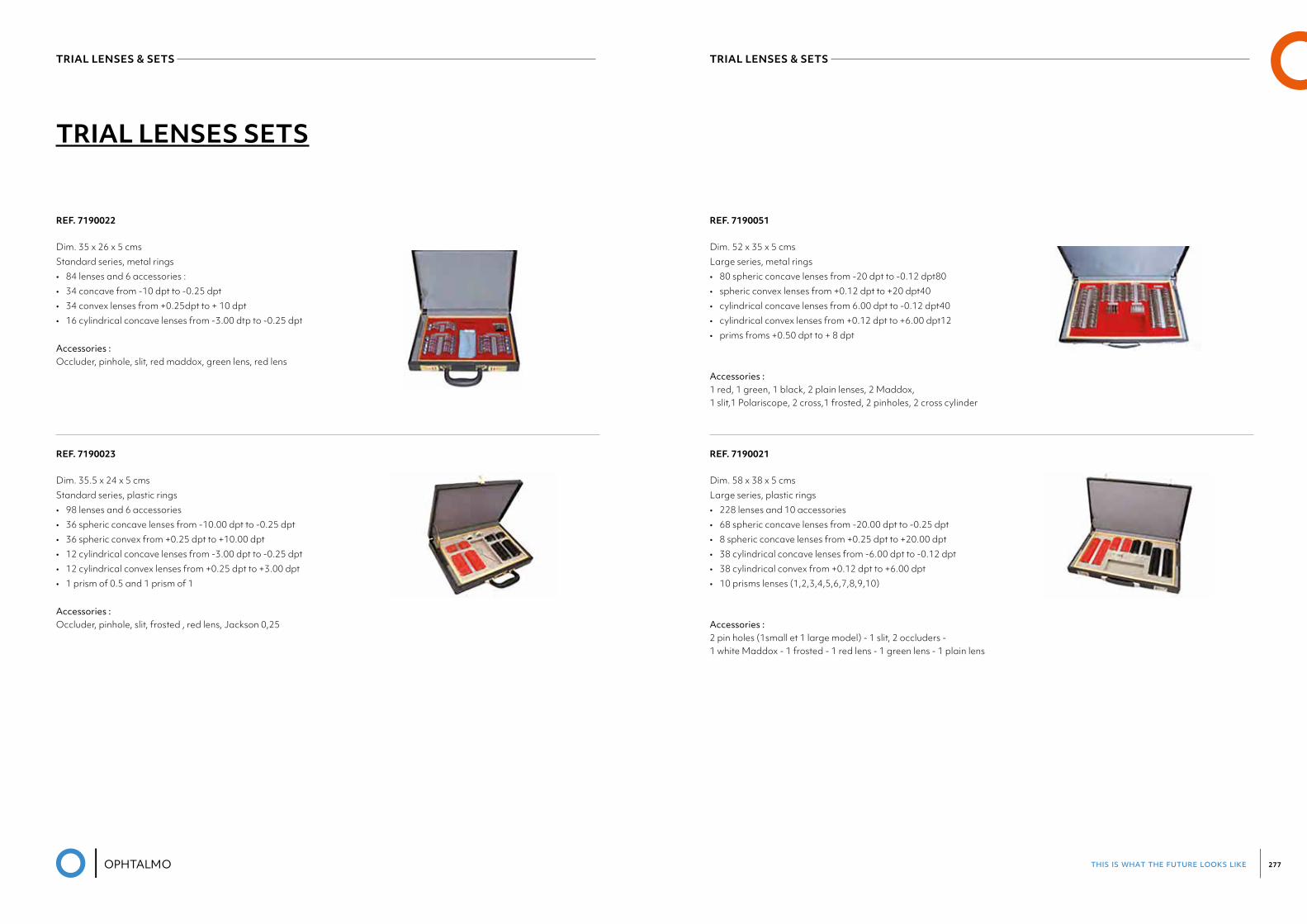
this is what the future looks like 277
TRIAL LENSES & SETS TRIAL LENSES & SETS
REF. 7190022
Dim. 35 x 26 x 5 cmsStandard series, metal rings• 84 lenses and 6 accessories :• 34 concave from -10 dpt to -0.25 dpt• 34 convex lenses from +0.25dpt to + 10 dpt• 16 cylindrical concave lenses from -3.00 dtp to -0.25 dpt Accessories : Occluder, pinhole, slit, red maddox, green lens, red lens
REF. 7190023
Dim. 35.5 x 24 x 5 cmsStandard series, plastic rings• 98 lenses and 6 accessories• 36 spheric concave lenses from -10.00 dpt to -0.25 dpt• 36 spheric convex from +0.25 dpt to +10.00 dpt• 12 cylindrical concave lenses from -3.00 dpt to -0.25 dpt• 12 cylindrical convex lenses from +0.25 dpt to +3.00 dpt• 1 prism of 0.5 and 1 prism of 1 Accessories : Occluder, pinhole, slit, frosted , red lens, Jackson 0,25
TRIAL LENSES SETS
REF. 7190051
Dim. 52 x 35 x 5 cmsLarge series, metal rings • 80 spheric concave lenses from -20 dpt to -0.12 dpt80• spheric convex lenses from +0.12 dpt to +20 dpt40• cylindrical concave lenses from 6.00 dpt to -0.12 dpt40• cylindrical convex lenses from +0.12 dpt to +6.00 dpt12• prims froms +0.50 dpt to + 8 dpt
Accessories : 1 red, 1 green, 1 black, 2 plain lenses, 2 Maddox, 1 slit,1 Polariscope, 2 cross,1 frosted, 2 pinholes, 2 cross cylinder
REF. 7190021
Dim. 58 x 38 x 5 cmsLarge series, plastic rings• 228 lenses and 10 accessories• 68 spheric concave lenses from -20.00 dpt to -0.25 dpt• 8 spheric concave lenses from +0.25 dpt to +20.00 dpt• 38 cylindrical concave lenses from -6.00 dpt to -0.12 dpt• 38 cylindrical convex from +0.12 dpt to +6.00 dpt• 10 prisms lenses (1,2,3,4,5,6,7,8,9,10)
Accessories :2 pin holes (1small et 1 large model) - 1 slit, 2 occluders - 1 white Maddox - 1 frosted - 1 red lens - 1 green lens - 1 plain lens

this is what the future looks like 279
CONTACT LENSES& CARE
this is what the future looks like 281
CONTACT LENSES & CARE CONTACT LENSES & CARE
Ophtalmo meets a variety of eye treatment needs and supplies ophthalmologists and hospitals with a wide range of custom hard contact lenses, soft contact lenses and contact lens solutions.
Right from the very beginning in 1991, we decided to complement our quality products with matching quality service and advice. We work with almost all contact lens suppliers. They help us to offer the best quality and latest innovations and developments. We use our knowledge, experience and expertise to combine the best products, techniques and devices. Many ophthalmologists rely on us to provide the right contact lens solution for their patients.
Soft contact lenses • Daily contact lenses• 14-day contact lenses• Monthly contact lenses• 90-day contact lenses• Custom contact lenses• Medical soft/therapeutic contact lenses• Coloured contact lenses
Hard contact lenses• Rigid corneal contact lenses• Medical corneal contact lenses• Semi-scleral contact lenses• Scleral contact lenses (mini sclera or full sclera)
Treatment contact lensesTreatment contact lenses are also referred to as therapeutic contact lenses. They are used to slow down myopia in children and adolescents. They can be hard or soft contact lenses:• Soft non-standard or custom contact lenses• Hard contact lenses• Scleral contact lenses
Medical contact lenses• Medical soft contact lenses• Therapeutic / Hand-painted soft contact lenses• Hand-painted scleral contact lenses • Medical corneal contact lenses• Scleral contact lenses• Bandage contact lenses• Hybrid contact lenses
All-In-One Solutions
Peroxides
Cleaners
Saline Solutions
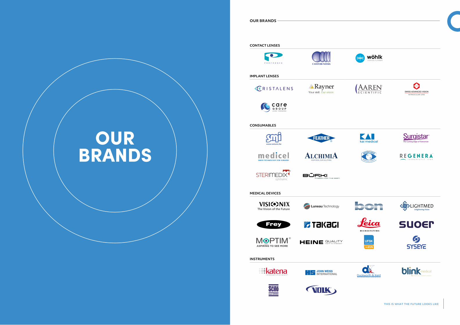
OSYS
this is what the future looks like
OUR BRANDS
OURBRANDS
CONTACT LENSES
IMPLANT LENSES
CONSUMABLES
MEDICAL DEVICES
INSTRUMENTS
®

OPHTALMO I Industriepark De Bruwaan 5 I B-9700 Oudenaarde I T+32(0)55313038 I ophtalmoservice.be
SURGICAL
CONSUMABLES
MEDICAL DEVICET H I S I S W H A T T H E F U T U R E L O O K S L I K E


