Embed Size (px)
Citation preview
Page 1/15
Thromboembolism in COVID-19 Patient withUnilateral Congenital Absent ICA with IntracranialAneurysm: First Case ReportDiwakar Shankar ( [email protected] )
Dr Ram Manohar Lohia Institute of Medical Sciences https://orcid.org/0000-0003-4752-6168Deepak Kumar Singh
Dr Ram Manohar Lohia Institute of Medical SciencesVipin Chand
Dr Ram Manohar Lohia Institute of Medical SciencesKuldeep Yadav
Dr Ram Manohar Lohia Institute of Medical Sciences
Research Article
Keywords: COVID-19, Intracranial aneurysm, Thromboembolism in COVID-19, Absent ICA, Giantaneurysm, Vascular anomaly
Posted Date: August 13th, 2021
DOI: https://doi.org/10.21203/rs.3.rs-741390/v1
License: This work is licensed under a Creative Commons Attribution 4.0 International License. Read Full License
Page 2/15
AbstractWe here discuss an interesting case of COVID-19 patient suffering from ruptured right supra-clinoidintracranial aneurysm with congenital absence of right ICA. COVID-19 has been responsible for over 175million reported cases and over 3.8 million deaths world-wide. Severe cases of COVID-19 is characterizedwith cytokine outburst and hyperin�ammation, platelet activation, endothelial dysfunction and sepsisrelated coagulopathy. This predisposes for thromboembolic events and aneurysm formation and rupture.Agenesis, aplasia and hypoplasia of internal carotid artery (ICA) is a rare congenital anomaly. ICAagenesis is associated with increased incidence of intracranial aneurysm as compared with generalpopulation.
IntroductionAccording to World Health Organization (WHO), as of now COVID-19 has been responsible for over 191million reported cases and over 4.1 million deaths world-wide. Severe cases of COVID-19 arecharacterized with cytokine outburst and hyperin�ammation, platelet activation, endothelial dysfunctionand sepsis related coagulopathy. This predisposes for thromboembolic events and aneurysm formationand rupture. Though venous thromboembolism (VTE) is more common than arterial, there had been caseseries suggesting increased incidence of arterial thromboembolic events (ATE) in COVID-19 patients.Also, agenesis, aplasia and hypoplasia of internal carotid artery (ICA) is a rare congenital anomaly withincidence of <0.01% in general population[11]. ICA agenesis has been associated with increasedincidence of intracranial aneurysm (25-43%) as compared with general population (2-4%). We herediscuss one interesting case of COVID-19 patient suffering from ruptured right supra-clinoid intracranialaneurysm with congenital absence of right ICA. A rare case that has never been reported before.
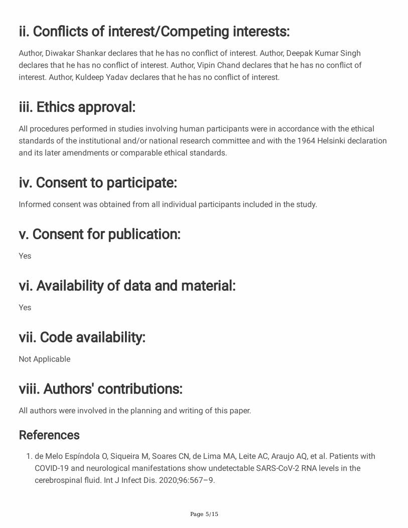
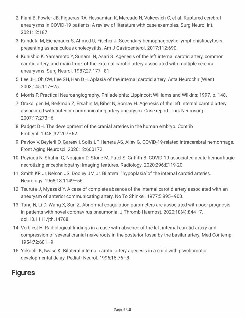
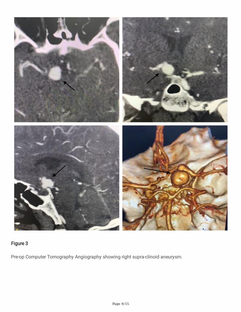
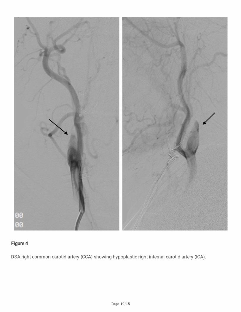
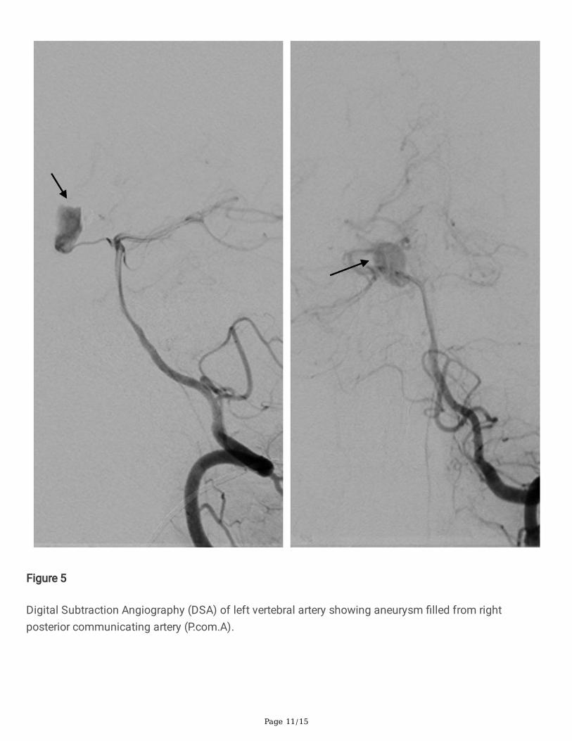
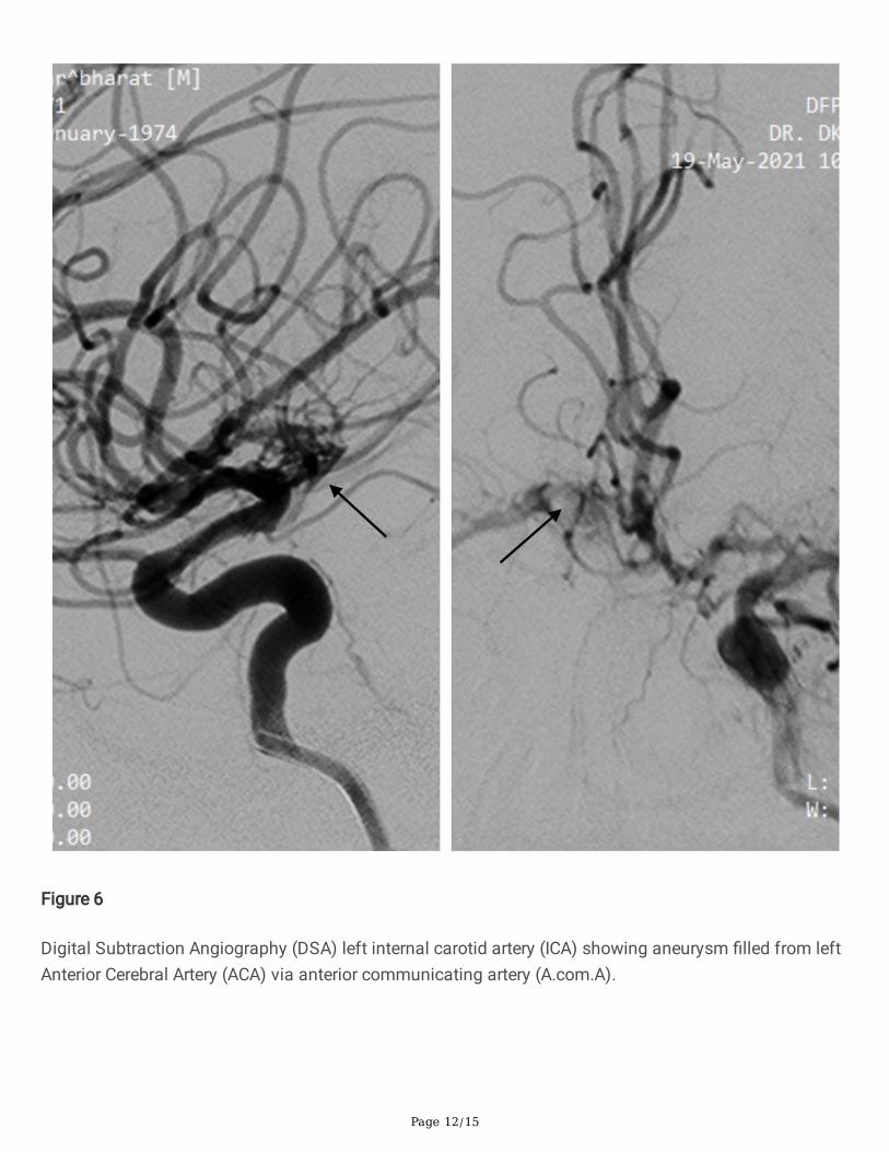
Case ReportA 47-years-old male patient complained of sudden onset severe headache associated with loss ofconsciousness eight days prior to presentation. At the time of presentation he was COVID-19 RT-PCRpositive. On admission GCS was E4V5M6, Hunt and Hess grade 2 with no sensory / motor / autonomicneurological de�cit. High resolution CT thorax and chest skiagram was done which showed post-covidchanges in lungs (Figure 1). NCCT Brain suggestive of minimal SAH in basal cistern (Figure 2). CTAngiography Brain vessels showed right supra-clinoid aneurysm (Figure 3). Patient was taken for digitalsubtraction angiography (DSA) to better delineate anatomy following all necessary COVID-19 guidelinefor operation theatre. A 6-French 11cm femoral access sheath (Medtronic Inc, Minneapolis, USA) wasused for groin access on the right side. A 6-French 100cm Envoy guide catheter (Codman, Wokingham,UK) was used to access bilateral common carotid artery (CCA) and vertebral arteries (VA). A Terumo150cm guide wire (Terumo Europe, Leuven, Belgium) was used to guide the catheter. Selective angiogramof the right common carotid artery (CCA) showed hypoplastic stump of the right ICA (Figure 4). Selectiveangiogram of the left vertebral artery showed an aneurysm measuring 5.6mm x 6.5mm x 9.8mm �llingfrom right posterior communicating artery (P.com.) (Figure 5). Aneurysm also �lled from the left anterior
Page 3/15
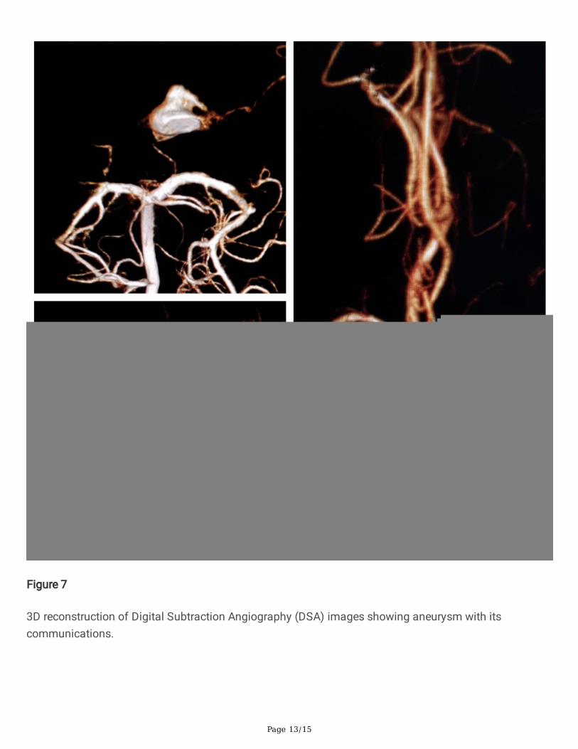
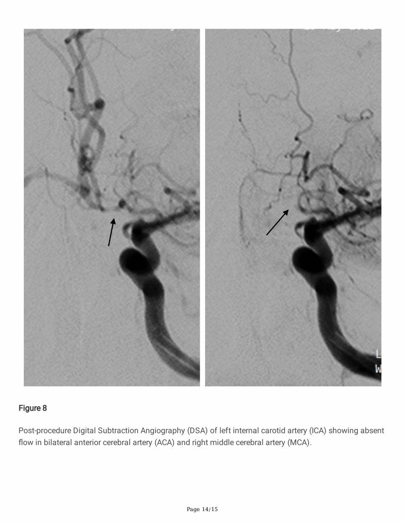
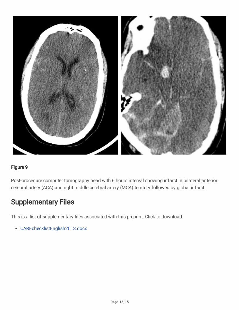
cerebral artery (ACA) through anterior communicating artery (A.com) (Figure 6). Also the right ACA andmiddle cerebral artery (MCA) was supplies by left ACA via A.com (Figure 7). Post-procedure DSA showedabsent �ow in the bilateral ACA and right MCA (Figure 8). Immediately intra-arterial administration of rtPA(alteplase (0.9 mg/kg) along with Abciximab (4mg) was given but with no success. Post-procedure CTHead showed right MCA and bilateral ACA territory infarct (Figure 9). Patient was taken emergencydecompressive craniectomy but he sustained cardiac arrest couple of minutes thereafter and didn’tsurvive.
DiscussionAgenesis, aplasia and hypoplasia of internal carotid artery (ICA) is a rare congenital anomaly withincidence of < 0.01% in general population. Tode, in 1787, was �rst to report ICA agenesis on post-mortemexam[7]. Verbiest was �rst to report the same on angiography in 1954[14].
Some more than 100 cases have been reported in the past. Padget described that ICA arises from thedorsal aorta and the third aortic arch at 4–5 mm embryonic stage and entirely develops by 6 weeks[8].This developmental anomaly occurs due to mechanical stress resulting from pressure effects andexcessive folding of cephalic portion of embryo and amniotic bands[5]. Also the carotid canal develops inassociation with the ICA. The skull-base begin to form during the 5th–6th weeks of fetal life. Thus, if ICAdoes not develop or fails to develop before the 5th embryonic week, the ICA and the carotid canal remainsundeveloped[5, 6]. The external carotid artery (ECA) and common carotid artery (CCA) arise from theaortic sac independently and can present normally in a case of ICA agenesis as was seen in our case[15].
Diagnosis of an absent ICA is made on CT/CT angiography and digital subtraction angiography (DSA).Non-visualization of an ICA on angiography and absent bony carotid canal in the base of the skull on CTis helps in the diagnosis of congenital absence of an ICA.
Most of the patients are asymptomatic because of the collateral circulation. Tsuruta and Myazakiproposed three types of collateral channels through the circle of Willis[12]. In Type I, the ipsilateral ACA issupplied by the contralateral ICA, opposite to the ICA agenesis, through the anterior communicating artery(A.Com.). The MCA is supplied by the basilar artery through the posterior communicating artery. In Type II,the ipsilateral ACA and MCA are supplied by the contralateral ICA through patent A.Com. In Type III, theipsilateral ACA and MCA are supplied by the transcranial anastomoses that develop from ECA orcontralateral ICA or primitive vessels. Our case had Type II anomaly.
Sometimes patients with congenital absent ICA develops subarachnoid hemorrhage (SAH) due toruptured aneurysm or transient ischemic attack (TIA) due to vascular insu�ciency. ICA agenesis has beenassociated with increased incidence of intracranial aneurysm (25–43%) as compared with generalpopulation (2–4%)[12, 4]. Lee et.al. found that aneurysm develops ipsilateral to absent ICA as was in ourcase, supporting a congenital origin of aneurysm as opposed to hemodynamic factors.
Page 4/15
COVID-19 infection occurs through the severe acute respiratory syndrome coronavirus 2 (SARS-CoV-2)virion binding to Angiotensin Converting Enzyme 2 (ACE-2), an enzyme responsible for regulation ofblood pressure and has anti-atherosclerotic effects. SARS-CoV-2 binds to Angiotensin ConvertingEnzyme-2 (ACE-2) and inactivates it. Also, SARS-CoV-2- ACE-2 binding is responsible for direct damage tothe BBB[9, 10].
COVID-19 is characterized by cytokine outburst and hyperin�ammation, platelet activation, endothelialdysfunction and sepsis related coagulopathy leading to increased risk of aneurysm formation or rupture.Pro-in�ammatory cytokines in COVID-19 such as interleukin (IL)-1, IL-6, and TNF are found to beresponsible for the loss of vascular integrity. Kandula et al. in his study found that the hypercytokinemiaof sHLH (Secondary Hemophagocytic Lymph Histiocytosis) may result in endothelial injury throughincreased vascular permeability, resultant ischemia of the vascular endothelium, and cell damage. SevereCOVID-19 infections have a similar cytokine pro�le as sHLH[3]. Moriguchi et al. found that cytokinecascade has been directly demonstrated to be responsible for neurological disorders and acutecerebrovascular disease[1]. Brian et. al. in his study suggested that the state of hyperin�ammation isresponsible for increased chance of aneurysm formation and rupture in patients with COVID-19infection[2].
COVID-19 is also responsible for thromboembolic event which was seen in our patient. COVID-19 has anincreased incidence of thromboembolic events (TE), Venous TE (VTE) more common than arterial TE(ATE)[15]. Some report incidence of (TE) to be 20–30% in COVID-19 patients and some reported up to40–70% of their cases. This was associated with higher rate of mortality patients with COVID-19 as wasin our case[13].
ConclusionCongenital absence of internal carotid artery is an extremely rare disorder which is associated withintracranial aneurysm formation, more commonly on the ipsilateral side. This when combined with thein�ammatory response of hypercytokinemia in COVID-19 leads to degradation of cerebral vasculatureand aneurysm formation or rupture. It was also seen that COVID-19 has increased incidence ofthromboembolic events which is associated with increased mortality. Though this case was �rst of itskind where COVID-19 was associated with intracranial aneurysm rupture with congenital absent ICA andthromboembolism. Further, long-term retrospective studies will be needed to determine the effects ofCOVID-19 on intracranial aneurysm and thromboembolism and postulate guidelines to prevent and treatthe same.
Declarations
i. Funding: Not Applicable
Page 5/15
ii. Con�icts of interest/Competing interests: Author, Diwakar Shankar declares that he has no con�ict of interest. Author, Deepak Kumar Singhdeclares that he has no con�ict of interest. Author, Vipin Chand declares that he has no con�ict ofinterest. Author, Kuldeep Yadav declares that he has no con�ict of interest.
iii. Ethics approval: All procedures performed in studies involving human participants were in accordance with the ethicalstandards of the institutional and/or national research committee and with the 1964 Helsinki declarationand its later amendments or comparable ethical standards.
iv. Consent to participate: Informed consent was obtained from all individual participants included in the study.
v. Consent for publication: Yes
vi. Availability of data and material: Yes
vii. Code availability: Not Applicable
viii. Authors' contributions: All authors were involved in the planning and writing of this paper.
References1. de Melo Espíndola O, Siqueira M, Soares CN, de Lima MA, Leite AC, Araujo AQ, et al. Patients with
COVID-19 and neurological manifestations show undetectable SARS-CoV-2 RNA levels in thecerebrospinal �uid. Int J Infect Dis. 2020;96:567–9.
Page 6/15
2. Fiani B, Fowler JB, Figueras RA, Hessamian K, Mercado N, Vukcevich O, et al. Ruptured cerebralaneurysms in COVID-19 patients: A review of literature with case examples. Surg Neurol Int.2021;12:187.
3. Kandula M, Eichenauer S, Ahmed U, Fischer J. Secondary hemophagocytic lymphohistiocytosispresenting as acalculous cholecystitis. Am J Gastroenterol. 2017;112:690.
4. Kunishio K, Yamamoto Y, Sunami N, Asari S. Agenesis of the left internal carotid artery, commoncarotid artery, and main trunk of the external carotid artery associated with multiple cerebralaneurysms. Surg Neurol. 1987;27:177–81.
5. Lee JH, Oh CW, Lee SH, Han DH. Aplasia of the internal carotid artery. Acta Neurochir (Wien).2003;145:117–25.
�. Morris P. Practical Neuroangiography. Philadelphia: Lippincott Williams and Wilkins; 1997. p. 148.
7. Orakd gen M, Berkman Z, Ersahin M, Biber N, Somay H. Agenesis of the left internal carotid arteryassociated with anterior communicating artery aneurysm: Case report. Turk Neurosurg.2007;17:273–6.
�. Padget DH. The development of the cranial arteries in the human embryo. ContribEmbryol. 1948.;32:207–62.
9. Pavlov V, Beylerli O, Gareev I, Solis LF, Herrera AS, Aliev G. COVID-19-related intracerebral hemorrhage.Front Aging Neurosci. 2020;12:600172.
10. Poyiadji N, Shahin G, Noujaim D, Stone M, Patel S, Gri�th B. COVID-19-associated acute hemorrhagicnecrotizing encephalopathy: Imaging features. Radiology. 2020;296:E119-20.
11. Smith KR Jr, Nelson JS, Dooley JM Jr. Bilateral “hypoplasia”of the internal carotid arteries.Neurology. 1968;18:1149–56.
12. Tsuruta J, Myazaki Y. A case of complete absence of the internal carotid artery associated with ananeurysm of anterior communicating artery. No To Shinkei. 1977;5:895–900.
13. Tang N, Li D, Wang X, Sun Z. Abnormal coagulation parameters are associated with poor prognosisin patients with novel coronavirus pneumonia. J Thromb Haemost. 2020;18(4):844–7.doi:10.1111/jth.14768.
14. Verbiest H. Radiological �ndings in a case with absence of the left internal carotid artery andcompression of several cranial nerve roots in the posterior fossa by the basilar artery. Med Contemp.1954;72:601–9.
15. Yokochi K, Iwase K. Bilateral internal carotid artery agenesis in a child with psychomotordevelopmental delay. Pediatr Neurol. 1996;15:76–8.
Figures
Page 7/15
Figure 1
Chest Skiagram and High Resolution Computer Tomography (HRCT) Thorax showing post COVID-19changes.
Page 8/15
Figure 2
Pre-op Computer Tomography Head with sub-arachnoid haemorrhage (SAH).
Page 9/15
Figure 3
Pre-op Computer Tomography Angiography showing right supra-clinoid aneurysm.
Page 10/15
Figure 4
DSA right common carotid artery (CCA) showing hypoplastic right internal carotid artery (ICA).
Page 11/15
Figure 5
Digital Subtraction Angiography (DSA) of left vertebral artery showing aneurysm �lled from rightposterior communicating artery (P.com.A).
Page 12/15
Figure 6
Digital Subtraction Angiography (DSA) left internal carotid artery (ICA) showing aneurysm �lled from leftAnterior Cerebral Artery (ACA) via anterior communicating artery (A.com.A).
Page 13/15
Figure 7
3D reconstruction of Digital Subtraction Angiography (DSA) images showing aneurysm with itscommunications.
Page 14/15
Figure 8
Post-procedure Digital Subtraction Angiography (DSA) of left internal carotid artery (ICA) showing absent�ow in bilateral anterior cerebral artery (ACA) and right middle cerebral artery (MCA).
Page 15/15
Figure 9
Post-procedure computer tomography head with 6 hours interval showing infarct in bilateral anteriorcerebral artery (ACA) and right middle cerebral artery (MCA) territory followed by global infarct.
Supplementary Files
This is a list of supplementary �les associated with this preprint. Click to download.
CAREchecklistEnglish2013.docx


