Embed Size (px)
Citation preview
General and Comparative Endocrinology 172 (2011) 505–517
Contents lists available at ScienceDirect
General and Comparative Endocrinology
journal homepage: www.elsevier .com/locate /ygcen
Tissue-specific thyroid hormone regulation of gene transcripts encodingiodothyronine deiodinases and thyroid hormone receptors in stripedparrotfish (Scarus iseri)
Kaitlin M. Johnson, Sean C. Lema ⇑Biology and Marine Biology, Center for Marine Science, University of North Carolina – Wilmington, 601 S. College Rd., Wilmington, NC 28403, USA
a r t i c l e i n f o
Article history:Received 22 December 2010Revised 11 April 2011Accepted 19 April 2011Available online 28 April 2011
Keywords:ReceptorDeiodinaseEnzymeGene expressionmRNAThyroid hormonesFishLiverBrainGonad
0016-6480/$ - see front matter � 2011 Elsevier Inc. Adoi:10.1016/j.ygcen.2011.04.022
⇑ Corresponding author. Present address: DepartCalifornia Polytechnic State University, San Luis Obisp962 4066.
E-mail address: [email protected] (S.C. Lema).
a b s t r a c t
In fish as in other vertebrates, the diverse functions of thyroid hormones are mediated at the peripheraltissue level through iodothyronine deiodinase (dio) enzymes and thyroid hormone receptor (tr) proteins.In this study, we examined thyroid hormone regulation of mRNAs encoding the three deiodinases dio1,dio2 and dio3 – as well as three thyroid hormone receptors traA, traB and trb – in initial phase stripedparrotfish (Scarus iseri). Parrotfish were treated with dissolved phase T3 (20 nM) or methimazole(3 mM) for 3 days. Treatment with exogenous T3 elevated circulating T3, while the methimazole treat-ment depressed plasma T4. Experimentally-induced hyperthyroidism increased the relative abundanceof transcripts encoding traA and trb in the liver and brain, but did not affect traB mRNA levels in eithertissue. In both sexes, methimazole-treated fish exhibited elevated dio2 transcripts in the liver and brain,suggesting enhanced outer-ring deiodination activity in these tissues. Accordingly, systemic hyperthy-roidism elevated relative dio3 transcript levels in these same tissues. In the gonad, however, patternsof transcript regulation were distinctly different with elevated T3 increasing mRNAs encoding dio2 in tes-ticular and ovarian tissues and dio3, traA and traB in the testes only. Thyroid hormone status did notaffect dio1 transcript abundance in the liver, brain or gonads. Taken as a whole, these results demonstratethat thyroidal status influences relative transcript abundance for dio2 and dio3 in the liver, provide newevidence for similar patterns of dio2 and dio3 mRNA regulation in the brain, and make evident that fishexhibit tr subtype-specific transcript abundance changes to altered thyroid status.
� 2011 Elsevier Inc. All rights reserved.
1. Introduction Dio1, Dio2 and Dio3 are the protein products of three different
In teleost fish, the thyroid hormones (THs) thyroxine (T4) andtri-iodothyronine (T3) play key roles in regulating developmentand growth [24,36], reproduction [42,69], osmoregulation [45],and the behaviors and underlying physiology associated with rhe-otaxis and migration [13,37]. The ability for THs to regulate suchdiverse processes evolved in part through the mechanisms of THaction in peripheral tissues, where changes in TH metabolism oc-cur via changes in hormone-converting iodothyronine deiodinaseenzyme activity, and shifts in the type and density of TH receptorsallow for differential effects at cell- and tissue-specific levels [67].
Teleost fish – similar to other vertebrates – express three typesof iodothyronine deiodinase enzymes termed type I, type II, andtype III (Dio1, Dio2, and Dio3, respectively), which function atthe prereceptor level to regulate TH levels and, ultimately, TH ac-tion at the level of the target tissue (reviewed by [54]). Although
ll rights reserved.
ment of Biological Sciences,o, CA 93407, USA. Fax: +1 910
genes, each enzyme catalyzes the removal of iodine atoms fromeither the outer-ring or inner-ring of THs to generate either moreactive or less active forms of hormone. The mechanism of hormonebiotransformation, however, varies among the three deiodinaseenzymes, leading each enzyme to have a different role in regulat-ing TH bioactivity [29,30,54]. In fish, Dio2 acts as an outer-ringdeiodinase ([ORD] or 50-deiodinase) by removing iodine from the50 outer-ring site to convert T4 to the more active form T3, whileDio3 acts as an inner-ring deiodinase ([IRD] or 5-deiodinase) to re-move iodine from the inner-ring of T4 and T3 and convert thesehormones to inactive forms including reverse triiodothyronine(rT3) and diiodothyronine (T2). Dio1, in contrast, is thought to cat-alyze primarily outer-ring deiodination in fish, although – as inother vertebrates – the enzyme also has inner-ring deiodinationproperties, making its role in regulating TH bioactivity moredifficult to define [54,67]. In fish, these three deiodinase enzymesshow tissue and developmental stage specific patterns ofexpression suggesting that the distinct deiodinases contribute tothe diverse – and sometimes tissue-specific – functions of THs[8,54,60]. At present, however, the importance of deiodinaseenzymes as mediators of TH action in fish is still largely unknown,

506 K.M. Johnson, S.C. Lema / General and Comparative Endocrinology 172 (2011) 505–517
and additional information on tissue-specific regulation of deiodin-ase expression is needed [54].
The effects of THs in a given cell or tissue also depend on signaltransduction via the binding nuclear thyroid hormone receptors(TRs), which subsequently interact with thyroid response elements(TREs) in the promoter regions of genes to either enhance or inhibitgene transcription [79]. Teleost fishes have been found to expressseveral distinct TR subtypes; although the number of types canvary depending on the species, these receptors can be broadlygrouped in a- and b-type categories based on their structural char-acteristics [39,40,53] (reviewed by [51]). Although there is littledata on functional differentiation among the teleost TR types andsubtypes (but see [52]), several studies have found variation inthe expression of TRs among tissues or stages of development[8,34,53].
Even though the functional roles of different deiodinase en-zymes and TRs remain to be fully determined in teleost fishes –and may even vary between taxa – expression profiles for tran-scripts encoding dio and tr genes have recently begun to be usedas indicators for endocrine disruption of the hypothalamic–pitui-tary–thyroid axis, and several studies have demonstrated that dioand tr relative gene transcript abundance in fishes can be impactedby exposure to pollutants [18,35,38,48,50,55,65,71]. For example,adult male and female fathead minnow (Pimephales promelas) gi-ven dietary exposures of the polybrominated diphenyl ether flameretardant 2,20,4,40-tetrabromodiphenyl ether (BDE-47) showed de-pressed circulating levels of T4 and reduced transcript abundancefor trb – but increased tra – in the brain after 21 days of exposure[35]. While these and other studies suggest that dio and tr mRNAlevels may be impacted by environmental pollutants, they alsomake clear that additional work is needed on the transcriptionaldynamics of deiodinase enzymes, TRs, and other TH-regulatedgene pathways in fish [18,34]. It is increasingly evident that manythyroid-disrupting chemicals appear to have multimodal or multi-target actions on the hypothalamic–pituitary–thyroid axis [6], andTH-regulated genes – including dio’s and tr’s – can show tissue-specific responses even when thyroid function or thyroid hormonestatus is disrupted systemically [64].
Therefore, in this study, we examined TH regulation of dio and trgene expression in a teleost fish, the striped parrotfish (Scarusiseri). Striped parrotfish, similar to other parrotfish species, are per-haps most notable for their role grazing algae on coral reefs [23].Parrotfish, however, are also unusual in that they are one of fewteleost taxa that have a discrete thyroid gland organ [43,44]. Par-rotfish have therefore been used previously as a model for examin-ing thyrotropin regulation of TH production [19,70], and have thepotential to provide new insights into mechanisms of HPT regula-tion in teleosts more broadly. Here, we isolated and sequencedcDNAs encoding dio1, dio2 and dio3 – as well as three distinct THreceptors traA, traB and trb – from striped parrotfish and examinedtissue-specific patterns of change in the relative abundance ofthese transcripts following systemic thyroid status modulationvia administration of either the goitrogen methimazole or exoge-nous T3.
2. Materials and methods
2.1. Animal collection and housing
Striped parrotfish (S. iseri) are a protogynous sex-changing spe-cies that can change from initial phase (male or female) to terminalphase (male only) phenotypes during life [31,57]. The focus of thecurrent study, however, was only on initial phase (IP) striped par-rotfish, which were collected using hand nets and a 60 cm diametercast net from the fringing reef (12�0400100N, 68�5104000W) surround-
ing Curaçao, The Netherlands Antilles on 28 May 2009. Fish (N = 38;17.80 ± 2.07 g, body mass; 102.5 ± 3.4 mm SL; mean ± SEM) weretransported to the Caribbean Research & Management of Biodiver-sity (CARMABI) research station and maintained in flow-throughseawater water tanks (128 L, 80 cm � 40 cm � 40 cm in size) atambient ocean temperatures (27.4–29.1 �C; Hobo Light-Tempera-ture Data Loggers, Onset Computer Corp., Bourne, MA, USA) for10 days prior to beginning the experiment. Fish were housed atdensities of 3 or 4 fish per tank, and tanks contained natural rockstructures for cover. All fish were fed algae wafers (Hartz MountainCorp., Secarcus, NJ, USA) twice daily. Seawater was pumped into thetanks at a rate of �1 L per min, so that the entire volume of eachtank was exchanged approximately every 2 h. All parrotfish werecollected and maintained in accordance with established guidelinesof the Animal Care and Use Committee of the University of NorthCarolina, Wilmington (Protocol #A0809-020).
2.2. Methimazole and T3 treatments
Each of the 12 holding tanks was assigned to one of threegroups: control, methimazole treatment, or exogenous T3 treat-ment, so that there were four tanks per treatment group (n = 12–13 fish per treatment). For each tank, flow-through seawater wasturned off, and stock solutions of methimazole in NaOH, T3 inNaOH, or NaOH vehicle alone (control) were added to each 128 Ltank. Fish in the methimazole treatment were exposed to dissolvedphase methimazole (Sigma, St. Louis, MO, USA) in the tank water ata concentration of 3 mM with 0.01 M NaOH. The exogenous T3
treatment consisted of 20 nM T3 (triiodo-L-thyronine, Sigma) with0.01 M NaOH, while fish in control tanks were exposed to 0.01 MNaOH vehicle only. Every 12 h, water flow was turned on in eachtank at a flow rate of >2 L per min for 1 h to cycle the tank, afterwhich time water flow was again turned off and new stock solu-tions of methimazole, T3 or NaOH vehicle alone were added to eachtank to renew treatment concentrations.
After 3 days of treatment, parrotfish were euthanized (tricainemethanesulfonate, MS222) and body mass (g) and total length(mm) were recorded. Blood plasma was collected for measurementof plasma T3 and T4 by radioimmunoassay, and the liver, brain(without pituitary gland) and one gonad were dissected and im-mersed in RNAlater (Ambion, Austin, TX, USA) overnight at 4 �C be-fore being stored at �20 �C. The other gonad was dissected andfixed in 4% paraformaldehyde overnight for later histological con-firmation of sex. A small section of skeletal muscle tissue was alsodissected from the tail of each fish. The muscle was stored in 100%ethanol for subsequent genetic confirmation of each fish’s speciesidentity.
2.3. T3 and T4 radioimmunoassays
Plasma total T3 and total T4 were measured by radioimmunoas-say as described elsewhere [12,36]. Plasma (10 ll) was incubatedfor 2 h at 37 �C in sodium barbital buffer with either anti-L-T3 anti-serum (1:10,000; Accurate Chemical & Scientific Corp., Westbury,NY, USA) and I125-labeled T3 (Perkin-Elmer, Waltham, MA, USA),or anti-L-T4 antiserum (1:4000; Accurate Chemical & ScientificCorp.) and I125-labeled T4 (Perkin-Elmer, Waltham, MA, USA). Icecold sodium barbital buffer containing 20% polyethylene glycolwas then added to each sample, and samples were centrifuged(1409g) for 20 min at 4 �C. The supernatant was removed to sepa-rate free and bound hormone, and the remaining pellet was as-sayed for radioactivity (Cobra II gamma counter, Packard,Downer’s Grove, IL, USA). T3 standards from 0.625 to 60 ng/ml –and T4 standards from 1.25 to 60 ng/ml – defined the sensitivityof the assays. For each hormone, all samples were run in duplicate
K.M. Johnson, S.C. Lema / General and Comparative Endocrinology 172 (2011) 505–517 507
in a single assay, and the intra-assay coefficient of variation (% CV)was 6.28% for the T3 assay, and 7.68% for the T4 assay.
2.4. Identification of partial cDNAs for deiodinase enzymes and THreceptors
2.4.1. RNA isolation and reverse transcriptionTotal RNA was extracted from the liver of an initial phase male
striped parrotfish (8.46 g, 87.6 mm SL) using TriReagent (MolecularResearch Center, Cinicinnati, OH, USA) with bromochloropropaneas the phase separation reagent. Extracted RNA was DNase I trea-ted (DNA-free Treatment kit, Ambion), quantified by spectropho-tometry (260:280 = 2.02; NanoDrop 2000, ThermoScientific,Wilmington, DE, USA), and examined on a 0.8% agarose gel forRNA quality.
First strand cDNA was synthesized in a 20 ll reverse transcrip-tion reaction by incubating 5 lg of total RNA template from the li-ver (4.75 ll) with 1.0 ll annealing buffer, 1.0 ll of random primers,and 3.5 ll of RNase-free H2O (Sigma, St. Louis, MO) at 65 �C for5 min. Subsequently, 10 ll of 5� buffer and 2 ll of Superscript IIIReverse Transcriptase Enzyme Supermix (Invitrogen, Carlsbad,CA, USA) were added, and the mixture was incubated under a ther-mal profile of 25 �C for 10 min followed by 50 �C for 50 min and85 �C for 5 min (PT-100 thermal cycler, MJ Research).
2.4.2. Isolation and sequencing of partial cDNA sequencesPCR was performed using degenerate primers designed from
consensus regions of sequences for dio and tr cDNAs isolated fromother teleost fish (On-line Supplemental materials, Table 1). Thedeiodinase primers for dio1, dio2 and dio3 were designed to the fol-lowing teleosts cDNA sequences: dio1: shortjaw mudsucker (Gil-lichthys seta, GenBank Accession No. FJ208960), Japanese flounder(Paralichthys olivaceus, AB362421), and Nile tilapia (Oreochromisniloticus, Y11109); dio2: Japanese flounder (P. olivaceus,AB362422), Japanese medaka (Oryzias latipes, NM_001136521),turbot (Psetta maxima, AF467779), and mummichog (Fundulus het-eroclitus, FHU70869), and dio3: Atlantic halibut (Hippoglossus hip-poglossus, DQ856303), Nile tilapia (O. niloticus, Y11111), puffer(Takifugu rubripes, NM_001136146), and Senegalese sole (Soleasenegalensis, AM902722).
For the traA receptor degenerate primers were designed basedoff cDNA sequences from bastard halibut (P. olivaceus, GenBankAccession No. PAITHRAA1), ice goby (Leucopsarion petersii,AB204858), pufferfish (T. rubripes, AF302243), and turbot (P. max-ima, AF302253). Primers for traB were designed from Nile tilapia(O. niloticus, AF302249) and bastard halibut (P. olivaceus,D16462). Degenerate primers for trb were designed from bastardhalibut (P. olivaceus, Q91279), gilthead seabream (Sparus aurata,AAO86517), fire clownfish (Amphiprion melanopus, ACH43022), or-ange-spotted grouper (Epinephelus coioides, ABP62962), Pacificbluefin tuna (Thunnus orientalis, BAG12083), and black porgy(Acanthopagrus schlegelii, ABQ96862).
In addition, a partial cDNA sequence for elongation factor-1a(ef-1a) was isolated for use as a control transcript. For ef-1a, degen-erate nested primers (On-line Supplemental materials, Table 1)were designed to consensus regions of cDNAs from O. latipes(NM_001104662), Pagrus major (AY190693), Carassius auratus(AB056104) and Seriola quinqueradiata (AB032900). These primersamplified an 835-bp partial cDNA of ef-1a from striped parrotfish(GenBank Accession No. HM120251).
2.4.3. Phylogeny constructionDeduced amino acid sequences for all identified parrotfish dio
and tr cDNAs were aligned to dio and tr transcripts or genes fromother vertebrates (for GenBank Accession Nos., see On-line Supple-mental materials, Table 2). Amino acid sequences were aligned
using Clustal X [33], and phylogenetic analyses were conductedusing MEGA v.5 [72], using the Neighbor-Joining method and ap-distance model for tree construction [59]. All positions contain-ing alignment gaps were eliminated only in pairwise sequencecomparisons (pairwise deletion of gaps). Confidence values forclusters of associated taxa were obtained by bootstrap tests(1000 replicates).
2.5. SYBR green real-time quantitative RT-PCR assays
Total RNA was extracted from liver, brain and gonadal tissuesusing TRI-Reagent (Molecular Research Center) with bromochloro-propane. The resulting RNA was DNase I treated (DNA-free kit,Ambion) and quantified by spectrophotometry (Nanodrop 2000).Total RNA was then reverse transcribed in 10 ll reactions by incu-bating 0.5 lg of total RNA template (4.75 ll) with 2.0 ll of 5� buf-fer, 1.0 of 0.1 M dithiothreitol (Invitrogen), 0.25 ll of RNaseOUT(40 U/ll) (Invitrogen), 1.0 ll of random hexamer (500 ng/ml) (Pro-mega, Madison, WI, USA), 0.5 ll dNTPs (stock of 10 mM each ofdCTP, dGTP, dTTP and dATP; Promega), and 0.5 ll of SuperscriptIII Reverse Transcriptase (200 U/ml) (Invitrogen), under a thermalprofile of 25 �C for 10 min followed by 50 �C for 50 min and 85 �Cfor 5 min (MyCycler thermal cycler, Bio-Rad).
Primers for SYBR green quantitative real-time PCR assays weredesigned (Primer Quest, Integrated DNA Technologies, and PrimerExpress 2.0, Applied Biosystems, Inc.) to protein coding regions ofthe partial cDNAs for deiodinase enzymes (dio1, dio2, dio3) andthyroid hormone receptors (traA, traB, trb). Primers were also de-signed for ef-1a from striped parrotfish for use as a control gene.All primers were synthesized by Integrated DNA Technologies(Coralville, IA, USA) (Table 1). Specificity of these SYBR green pri-mer sets was confirmed by sequencing PCR products, as well asby melt-curve analysis during the quantitative PCR runs.
Quantitative real-time PCRs were conducted in 18 ll reactions.Each reaction contained 4.56 ll nuclease-free water (Sigma, St.Louis, MO, USA), 9.0 ll iQ SYBR green Supermix (Bio-Rad, Hercules,CA, USA), 0.72 ll each of forward and reverse primers (5 lM), and3.0 ll of reverse-transcribed cDNA template. The PCR thermal pro-file for each reaction was 50 �C for 2 min, 95 �C for 10 min, 42 cy-cles of 95 �C for 15 s and 59 �C for 1 min, and all assays were run ona Bio-Rad iCycler with a MyiQ™ Single Color PCR Detection System(Bio-Rad, Hercules, CA, USA). Melt curve analysis was also per-formed to confirm amplification of a single product and the ab-sence of primer–dimers. For each gene, a standard curve wasmade from a pool of RNA from samples representing all treatmentsand sexes. Each standard was serially diluted and assayed in trip-licate. DNA contamination was assessed for each gene by analyzingRNA samples that were not reverse-transcribed, and each qPCR runincluded two samples without cDNA template to further controlfor contamination. Transcript abundance for ef-1a was quantifiedas the normalizing gene. For each gene, correlation coefficients(r2) for the standard curve ranged from 0.982 to 0.992. PCR effi-ciencies for each gene were calculated using the equation: effi-ciency = [10(�1/slope) � 1], and are provided in Table 1. For eachgene, relative mRNA levels were subsequently calculated usingthe standard curve and normalized to ef-1a mRNA expression. Fi-nally, expression of each gene of interest was expressed as a rela-tive level by dividing the resulting values by the mean values ofmales in the control treatment group.
2.6. Histological confirmation of gonadal sex identity
Gonads that were fixed in 4% paraformaldehyde were subse-quently transferred to 70% ethanol, dehydrated through a gradedseries of ethanol, embedded in paraffin, and sectioned longitudi-nally (10 lm). Three sections were collected from each fish’s
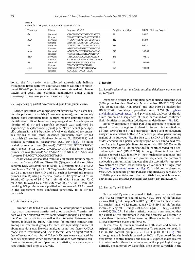
Table 1Primers for SYBR green quantitative real-time PCR assays.
Transcript Primer Sequence (50–30) Amplicon size (bp) % PCR efficiency (avg.)
dio1 Forward CAACAGACCGCTGGTGCTGAATTT 92 99.87Reverse TGAAGTCCCTGACGAGTTGCTTGA
dio2 Forward TTGGATTTCGAGTCATCGGATCGC 312 95.00Reverse ATTGTTGTCCATGCTGTCTGCCAC
dio3 Forward TGTGTGTCTCCGACTCCAACAAGA 280 99.33Reverse AAGTCCGCAATGTCTTGGTACTGG
traA Forward AAGCGCAAGTTCTTGCCAGATGAC 275 102.03Reverse TCGCCGCTTAGTGTCAATGTCTCA
traB Forward TATGGCAGCTCGCAGTGACATGA 249 103.10Reverse CTCCCAGTGGAAACAGAAACGCAAA
trb Forward AAAGCCACGGGCTACCACTA 121 101.67Reverse TGACGCATTTCCCCTCGTA
ef1a Forward CATCGACATTGCTCTGTGGAA 121 100.23Reverse GGCGCAGTCAGCCTGAGA
508 K.M. Johnson, S.C. Lema / General and Comparative Endocrinology 172 (2011) 505–517
gonad; the first section was collected approximately halfwaythrough the tissue with two additional sections collected at subse-quent 100–200 lm intervals. All sections were stained with hema-toxylin and eosin, and examined qualitatively under a lightmicroscope to confirm gonadal sexual identity.
2.7. Sequencing of partial cytochrome B gene from genomic DNA
Striped parrotfish are morphological similar to their sister tax-on, the princess parrotfish (Scarus taeniopterus), and commonlychange body coloration upon capture making definitive speciesidentification difficult based on morphology alone. As such, speciesidentity of all striped parrotfish collected was confirmed bysequencing the cytochrome B (cytB) gene. Nested sets of gene-spe-cific primers for a 381-bp region of cytB were designed to consen-sus regions of the genes described previously from stripedparrotfish (Scarus iserti, GenBank Accession No. EU601382) andprincess parrotfish (S. taeniopterus, EU601394) [66]. The outernested primer set was (forward) 50-CCTAGTTGACCTCCCTGC-30
and (reverse) 50-GTTGCACCTCAGAAGGA-30, and the inner nestedprimer set was (forward) 50-TCAAACATCTCTGTCTGATGAA-30 and(reverse) 50-TAGCCTACGAATGCTGTTAT-30.
Genomic DNA was isolated from skeletal muscle tissue samplesusing the DNeasy Cell and Tissue Kit (Qiagen), and the resultinggenomic DNA was amplified in 50 ll PCRs containing 2 ll of DNAtemplate (�62–168 ng), 25 ll GoTaq Colorless Master Mix (Prome-ga), 21 ll nuclease-free H2O, and 1 ll each of forward and reverseprimer (10 mM) using a thermal profile of 42 cycle of 94 �C for10 min, 42 cycles of 95 �C for 1 min, 48 �C for 1 min, and 72 �Cfor 2 min, followed by a final extension of 72 �C for 10 min. Theresulting PCR products were purified and sequenced. All fish usedin the experiment were confirmed genetically to be stripedparrotfish.
2.8. Statistical analyses
Hormone data failed to conform to the assumptions of normal-ity, so was square-root transformed prior to analysis. Transformeddata was then analyzed by two-factor ANOVA models using ‘treat-ment’ and ‘sex’ as factors, as well as the interaction between thesefactors, followed by Tukey HSD tests for multiple comparisonsamong the treatment groups for each sex separately. Transcriptabundance data was likewise analyzed using two-factor ANOVAmodels with ‘treatment’ and ‘sex’ as factors. When a significant ef-fect of ‘treatment’ was found, Tukey HSD tests were calculated foreach sex separately. When transcript abundance data failed to con-form to the assumptions of parametric statistics, data were squareroot transformed prior to analysis.
3. Results
3.1. Identification of partial cDNAs encoding deiodinase enzymes andTH receptors
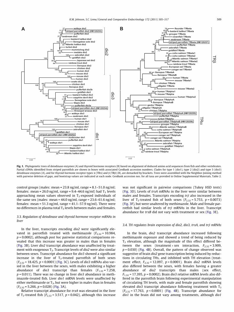
Degenerate primer PCR amplified partial cDNAs encoding dio1(169-bp nucleotides, GenBank Accession No. HM120152), dio2(363-bp nucleotides, HM120253) and dio3 (480-bp nucleotides,HM120254) from striped parrotfish liver. BLAST (http://blas-t.ncbi.nlm.nih.gov/Blast.cgi) and phylogenetic analyses of the de-duced amino acid sequences of these partial cDNAs confirmedtheir identities as encoding iodothyronine deiodinases (Fig. 1A).
Similarly, degenerate primer PCR using degenerate primers de-signed to consensus regions of teleost tra transcripts identified twodistinct cDNAs from striped parrotfish. BLAST and phylogeneticanalyses revealed that both cDNAs encoded putative partial codingregions of tra subtypes (Fig. 1B). One partial cDNA of 540-bp nucle-otides encoded for a partial coding region of 179 amino acid resi-dues for a traA gene (GenBank Accession No. HM120255), whilea second cDNA of 630-bp nucleotides in length encoded for a sec-ond receptor traB (HM120256). Although these traA and traBcDNAs showed 83.0% identify in their nucleotide sequences and91.6% identity in their deduced protein sequences, the pattern ofnucleotide differentiation suggests that the two mRNAs representtwo distinct tra genes, rather than splice variants of a single gene(On-line Supplemental materials, Fig. 1). In addition to these twotra cDNAs, degenerate primer PCR also amplified a trb partial cDNAof 1080-bp nucleotides from the parrotfish liver, which encoded359 amino acid residues (GenBank Accession No. HM120257).
3.2. Plasma T4 and T3 levels
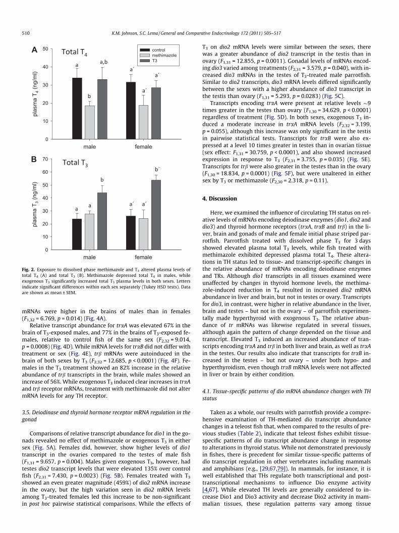
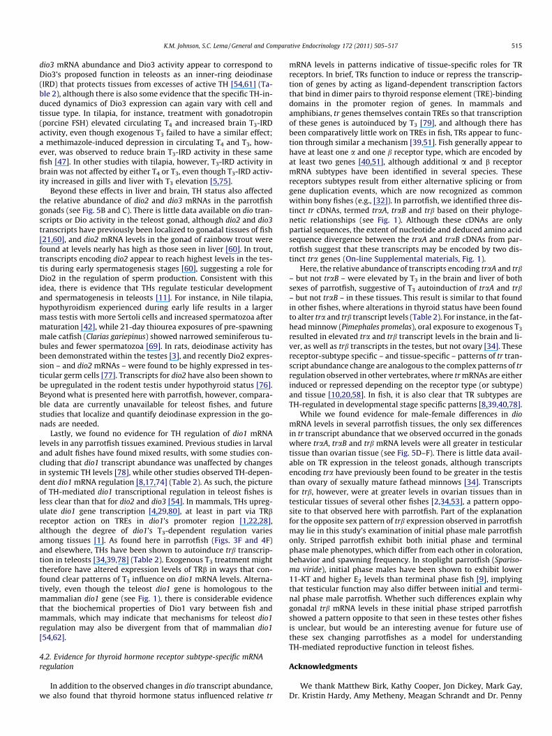
Plasma total T4 levels decreased in fish treated with methima-zole (males: mean = 18.4 ng/ml, range = 10.0–30.6 ng/ml; females:mean = 18.8 ng/ml, range = 9.5–28.7 ng/ml) from levels in controlfish (males: mean = 33.9 ng/ml, range = 23.3–39.8 ng/ml; females:mean = 31.5 ng/ml, range = 21.6–52.0 ng/ml) (F2,22 = 4.3037,p = 0.026) (Fig. 2A). Pairwise statistical comparisons revealed thatthe extent of this methimazole-induced decrease was greater inmales than in females. There were no differences in plasma totalT4 levels between males and females.
Plasma total T3 levels increased in both male and female IPstriped parrotfish exposed to exogenous T3 compared to levels infish in the control group (F2,27 = 13.403, p < 0.0001) (Fig. 2B).Although mean T3 levels increased by 106% in T3-exposed femalesover levels in control females, and 85% in T3-exposed males relativeto control males, these increases were in the physiological rangenormally encountered by parrotfish, since some parrotfish in the
walleye dio2 striped parrotfish dio2 (HM120253)
pufferfish dio2 seabream dio2
bastard halibut dio2 Senegalese sole dio2
Atlantic halibut dio2 turbot dio2
mummichog dio2 medaka dio2 zebrafish dio2
goldfish dio2 Japanese eel dio2
rainbow trout dio2 Xenopus dio2 chicken dio2 human dio2
mouse dio2 mouse dio3 human dio3
snake dio3 chicken dio3
Xenopus dio3 pufferfish dio3b goldfish dio3
bastard halibut dio3 sapphire devil dio3
Senegelese sole dio3 tilapia dio3
pufferfish dio3a striped parrotfish dio3 (HM120254)
threespot wrasse dio3 walleye dio3
seabream dio3 human dio1
mouse dio1 chicken dio1
Xenopus dio1 zebrafish dio1
goldfish dio1 striped parrotfish dio1 (HM120252)
mudsucker dio1 pufferfish dio1
walleye dio1 sapphire devil dio1
tilapia dio1 bastard halibut dio1
Atlantic halibut dio1
100
100
99
100
100
99
4041
45
36
60
78
67
98
84
100
31
37
616
15
23
17
99
98
96
65
87
100
99
76100
100
4430
76
27
3552
4916
76
21
1713
0.05
dio2
dio1
dio3
flounder TRbeta bastard halibut TRbeta
grouper TRbeta clownfish TRbeta
seabream TRbeta striped parrotfish TRbeta (HM120257)
pufferfish TRbeta zebrafish TRbeta goldfishTRbeta
coho salmon TRbeta1 coho salmon TRbeta2 Atlantic salmon TRbeta1 Atlantic salmon TRbeta2 rainbow trout TRbeta
European eel TRbeta conger eel TRbeta1
Xenopus TRbetaA Xenopus TRbetaB
human TRbeta mouse TRbeta1
chicken TRbeta alligator TRbeta
mouse TRbeta2 human TRalpha1 mouse TRalpha
human TRalpha2 Xenopus TRalphaA
Xenopus TRalphaB gecko TRalpha
alligator TRalphaA chicken TRalpha
striped parrotfish TRalphaA (HM120255) pufferfish TRalpha
turbot TRalpha striped parrotfish TRalphaB (HM120256)
grouper TRalpha Atlantic salmon TRalphaB
coho salmon TRalphaB European eel TRalpha conger eel TRalphaA
conger eel TRalphaB Atlantic salmon TRalpha
zebrafish TRalpha goldfish TRalpha1
coho salmon TRalphaA rainbow trout TRalpha
99
99
99
99
87
3541
99
99
7965
5139
77
71
63
77
24
99
28
96
93
64
32
99
6186
69
75
98
87
62
6980
5563
57
46
39
65
38
23
13
0.02
TRβ
TRα
A B
Fig. 1. Phylogenetic trees of deiodinase enzymes (A) and thyroid hormone receptors (B) based on alignment of deduced amino acid sequences from fish and other vertebrates.Partial cDNAs identified from striped parrotfish are shown in boxes with associated GenBank accession numbers. Clades for type 1 (dio1), type 2 (dio2) and type 3 (dio3)deiodinase enzymes (A), and for thyroid hormone receptor types a (TRa) and b (TRb) (B), are demarked by brackets. Trees were assembled with the Neighbor-Joining methodwith pairwise deletion of gaps, and bootstrap values are indicated at each node. GenBank accession nos. for all taxa are provided in Online Supplemental Materials, Table 2.
K.M. Johnson, S.C. Lema / General and Comparative Endocrinology 172 (2011) 505–517 509
control groups (males: mean = 23.8 ng/ml, range = 8.1–51.0 ng/ml;females: mean = 26.0 ng/ml, range = 9.4–44.6 ng/ml) had T3 levelsapproaching mean values observed in T3-exposed individuals ofthe same sex (males: mean = 44.0 ng/ml, range = 23.6–61.6 ng/ml;females: mean = 51.3 ng/ml, range = 41.1–57.9 ng/ml). There wereno differences in plasma total T3 levels between males and females.
3.3. Regulation of deiodinase and thyroid hormone receptor mRNAs inliver
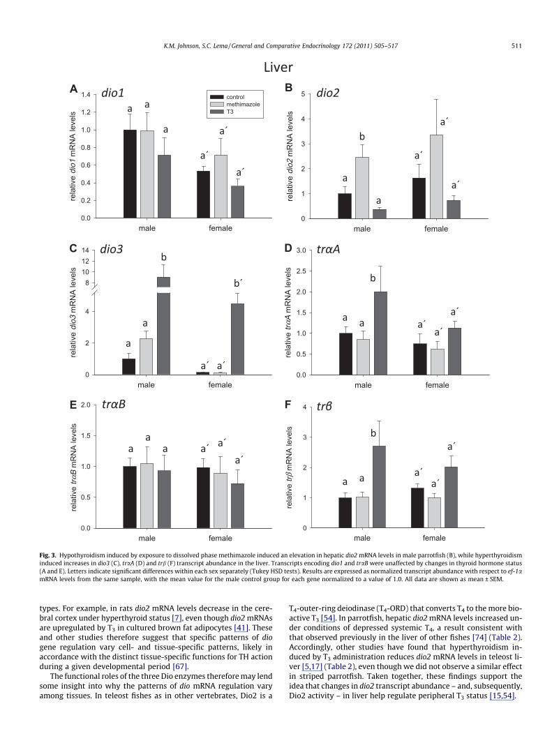
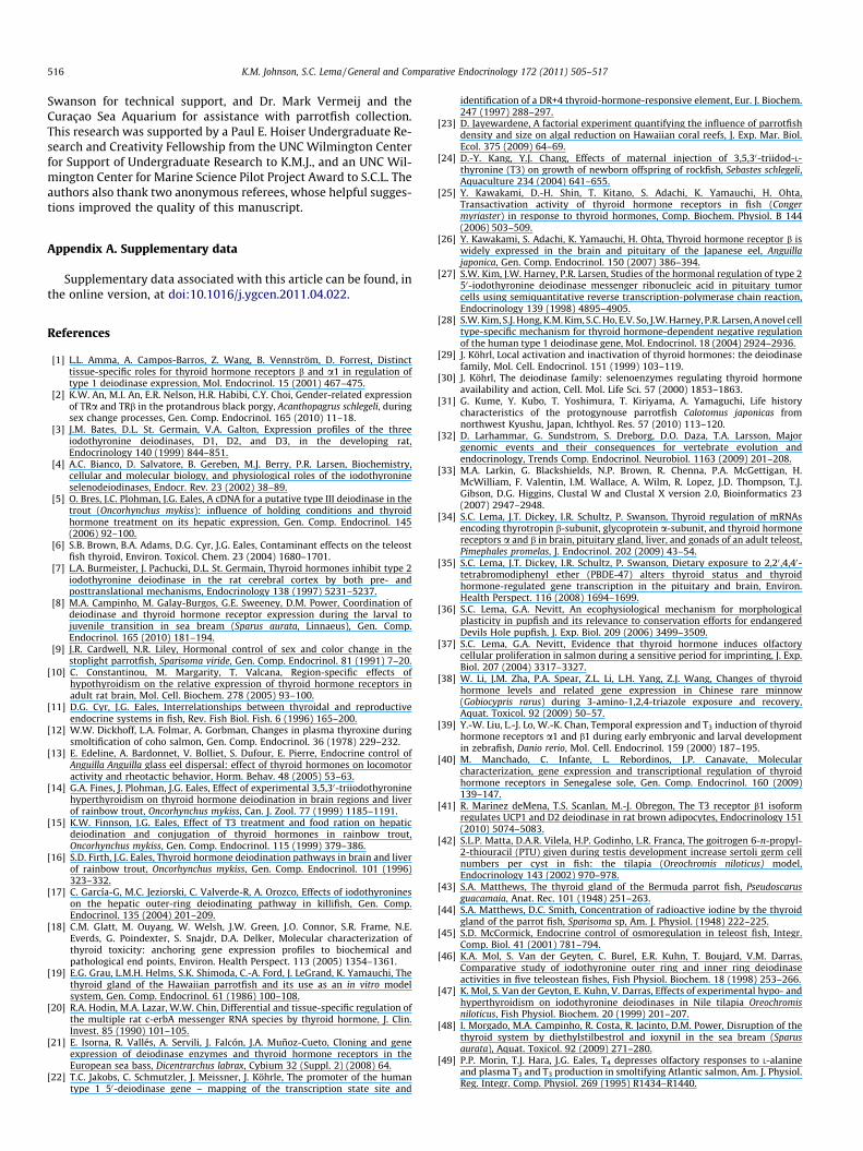
In the liver, transcripts encoding dio2 were significantly ele-vated in parrotfish treated with methimazole (F2,32 = 10.984,p = 0.0002), although post hoc pairwise statistical comparisons re-vealed that this increase was greater in males than in females(Fig. 3B). Liver dio2 transcript abundance was unaffected by treat-ment with exogenous T3. Transcript levels of dio2 were also similarbetween sexes. Transcript abundance for dio3 showed a significantincrease in the liver of T3-treated parrotfish of both sexes(F2,32 = 18.425, p < 0.0001) (Fig. 3C). Levels of dio3 mRNAs also var-ied in the liver between the sexes, with males exhibiting a higherabundance of dio3 transcript than females (F1,32 = 7.258,p = 0.011). There was no change in liver dio3 abundance in meth-imazole-treated fish. Liver dio1 mRNA levels were unaffected byeither methimazole or T3, but were higher in males than in females(F2,32 = 5.266, p = 0.028) (Fig. 3A).
Relative transcript abundance for traA was elevated in the liverof T3-treated fish (F2,32 = 3.517, p = 0.042), although this increase
was not significant in pairwise comparisons (Tukey HSD tests)(Fig. 3D). Levels of traA mRNAs in the liver were similar betweenmales and females. Transcripts encoding trb also increased in theliver of T3-treated fish of both sexes (F2,32 = 5.753, p = 0.0073)(Fig. 3F), but were unaltered by methimazole. Male and female par-rotfish had similar levels of trb mRNAs in the liver. Transcriptabundance for traB did not vary with treatment or sex (Fig. 3E).
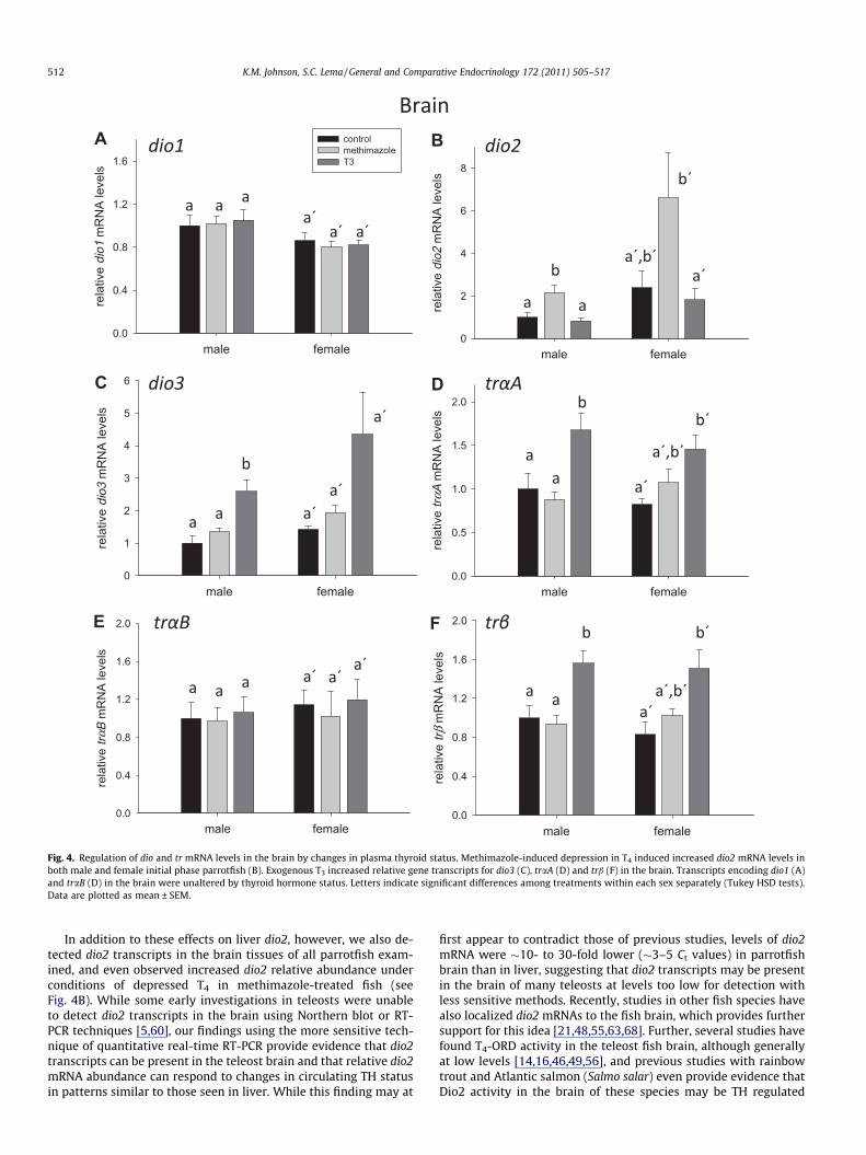
3.4. TH regulates brain expression of dio2, dio3, traA, and trb mRNAs
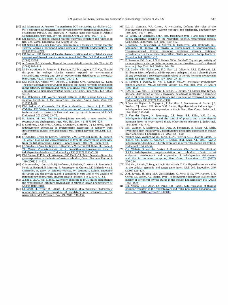
In the brain, dio2 transcript abundance increased followingmethimazole exposure and showed a trend of being reduced byT3 elevation, although the magnitude of this effect differed be-tween the sexes (treatment � sex interaction, F2,32 = 3.909,p = 0.030) (Fig. 4B). Overall, the pattern of change observed wassuggestive of brain dio2 gene transcription being induced by reduc-tions in circulating THs, and inhibited with TH elevation (treat-ment effect, F2,32 = 12.097, p = 0.0001). Brain dio2 mRNA levelsalso differed between the sexes, with females having a greaterabundance of dio2 transcripts than males (sex effect,F1,32 = 17.395, p = 0.0002). Brain dio3 relative mRNA levels also dif-fered in the parrotfish brain following experimental manipulationof circulating TH levels, with male and female parrotfish showingelevated dio3 transcript abundance following treatment with T3
(F2,32 = 17.763, p < 0.0001) (Fig. 4B). Transcript abundance fordio1 in the brain did not vary among treatments, although dio1
male female
plas
ma
T 4 (ng
/ml)
0
10
20
30
40
50 controlmethimazoleT3
male female
plas
ma
T 3 (n
g/m
l)
0
10
20
30
40
50
60
70
A
B
Fig. 2. Exposure to dissolved phase methimazole and T3 altered plasma levels oftotal T4 (A) and total T3 (B). Methimazole depressed total T4 in males, whileexogenous T3 significantly increased total T3 plasma levels in both sexes. Lettersindicate significant differences within each sex separately (Tukey HSD tests). Dataare shown as mean ± SEM.
510 K.M. Johnson, S.C. Lema / General and Comparative Endocrinology 172 (2011) 505–517
mRNAs were higher in the brains of males than in females(F1,32 = 6.769, p = 0.014) (Fig. 4A).
Relative transcript abundance for traA was elevated 67% in thebrain of T3-exposed males, and 77% in the brains of T3-exposed fe-males, relative to control fish of the same sex (F2,32 = 9.014,p = 0.0008) (Fig. 4D). While mRNA levels for traB did not differ withtreatment or sex (Fig. 4E), trb mRNAs were autoinduced in thebrain of both sexes by T3 (F2,32 = 12.685, p < 0.0001) (Fig. 4F). Fe-males in the T3 treatment showed an 82% increase in the relativeabundance of trb transcripts in the brain, while males showed anincrease of 56%. While exogenous T3 induced clear increases in traAand trb receptor mRNAs, treatment with methimazole did not altermRNA levels for any TH receptor.
3.5. Deiodinase and thyroid hormone receptor mRNA regulation in thegonad
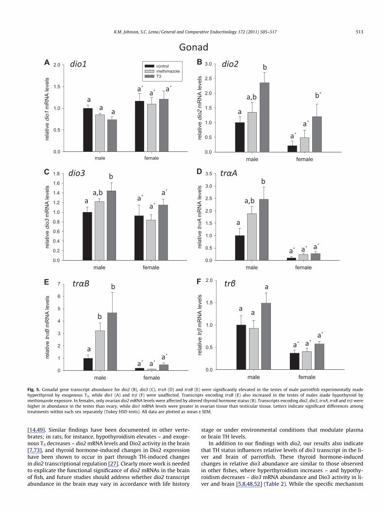
Comparisons of relative transcript abundance for dio1 in the go-nads revealed no effect of methimazole or exogenous T3 in eithersex (Fig. 5A). Females did, however, show higher levels of dio1transcript in the ovaries compared to the testes of male fish(F1,31 = 9.657, p = 0.004). Males given exogenous T3, however, hadtestes dio2 transcript levels that were elevated 135% over controlfish (F2,31 = 7.430, p = 0.0023) (Fig. 5B). Females treated with T3
showed an even greater magnitude (459%) of dio2 mRNA increasein the ovary, but the high variation seen in dio2 mRNA levelsamong T3-treated females led this increase to be non-significantin post hoc pairwise statistical comparisons. While the effects of
T3 on dio2 mRNA levels were similar between the sexes, therewas a greater abundance of dio2 transcript in the testis than inovary (F1,31 = 12.855, p = 0.0011). Gonadal levels of mRNAs encod-ing dio3 varied among treatments (F2,31 = 3.579, p = 0.040), with in-creased dio3 mRNAs in the testes of T3-treated male parrotfish.Similar to dio2 transcripts, dio3 mRNA levels differed significantlybetween the sexes with a higher abundance of dio3 transcript inthe testis than ovary (F1,31 = 5.293, p = 0.0283) (Fig. 5C).
Transcripts encoding traA were present at relative levels �9times greater in the testes than ovary (F1,30 = 34.629, p < 0.0001)regardless of treatment (Fig. 5D). In both sexes, exogenous T3 in-duced a moderate increase in traA mRNA levels (F2,32 = 3.199,p = 0.055), although this increase was only significant in the testisin pairwise statistical tests. Transcripts for traB were also ex-pressed at a level 10 times greater in testes than in ovarian tissue(sex effect: F1,31 = 30.759, p < 0.0001), and also showed increasedexpression in response to T3 (F2,31 = 3.755, p = 0.035) (Fig. 5E).Transcripts for trb were also greater in the testes than in the ovary(F1,30 = 18.834, p = 0.0001) (Fig. 5F), but were unaltered in eithersex by T3 or methimazole (F2,30 = 2.318, p = 0.11).
4. Discussion
Here, we examined the influence of circulating TH status on rel-ative levels of mRNAs encoding deiodinase enzymes (dio1, dio2 anddio3) and thyroid hormone receptors (traA, traB and trb) in the li-ver, brain and gonads of male and female initial phase striped par-rotfish. Parrotfish treated with dissolved phase T3 for 3 daysshowed elevated plasma total T3 levels, while fish treated withmethimazole exhibited depressed plasma total T4. These altera-tions in TH status led to tissue- and transcript-specific changes inthe relative abundance of mRNAs encoding deiodinase enzymesand TRs. Although dio1 transcripts in all tissues examined wereunaffected by changes in thyroid hormone levels, the methima-zole-induced reduction in T4 resulted in increased dio2 mRNAabundance in liver and brain, but not in testes or ovary. Transcriptsfor dio3, in contrast, were higher in relative abundance in the liver,brain and testes – but not in the ovary – of parrotfish experimen-tally made hyperthyroid with exogenous T3. The relative abun-dance of tr mRNAs was likewise regulated in several tissues,although again the pattern of change depended on the tissue andtranscript. Elevated T3 induced an increased abundance of tran-scripts encoding traA and trb in both liver and brain, as well as traAin the testes. Our results also indicate that transcripts for traB in-creased in the testes – but not ovary – under both hypo- andhyperthyroidism, even though traB mRNA levels were not affectedin liver or brain by either condition.
4.1. Tissue-specific patterns of dio mRNA abundance changes with THstatus
Taken as a whole, our results with parrotfish provide a compre-hensive examination of TH-mediated dio transcript abundancechanges in a teleost fish that, when compared to the results of pre-vious studies (Table 2), indicate that teleost fishes exhibit tissue-specific patterns of dio transcript abundance change in responseto alterations in thyroid status. While not demonstrated previouslyin fishes, there is precedent for similar tissue-specific patterns ofdio transcript regulation in other vertebrates including mammalsand amphibians (e.g., [29,67,79]). In mammals, for instance, it iswell established that THs regulate both transcriptional and post-transcriptional mechanisms to influence Dio enzyme activity[4,67]. While elevated TH levels are generally considered to in-crease Dio1 and Dio3 activity and decrease Dio2 activity in mam-malian tissues, these regulation patterns vary among tissue
male female
rela
tive
trαB
mR
NA
leve
ls
0.0
0.5
1.0
1.5
2.0
male female
rela
tive
trβ m
RN
A le
vels
0
1
2
3
4
male female
rela
tive
dio3
mR
NA
leve
ls
0
2
4
8101214
male female
rela
tive
trαA
mR
NA
leve
ls
0.0
0.5
1.0
1.5
2.0
2.5
3.0
male female
rela
tive
dio2
mR
NA
leve
ls
0
1
2
3
4
5
male female
rela
tive
dio1
mR
NA
leve
ls
0.0
0.2
0.4
0.6
0.8
1.0
1.2
1.4 controlmethimazoleT3
A B
C D
E F
Fig. 3. Hypothyroidism induced by exposure to dissolved phase methimazole induced an elevation in hepatic dio2 mRNA levels in male parrotfish (B), while hyperthyroidisminduced increases in dio3 (C), traA (D) and trb (F) transcript abundance in the liver. Transcripts encoding dio1 and traB were unaffected by changes in thyroid hormone status(A and E). Letters indicate significant differences within each sex separately (Tukey HSD tests). Results are expressed as normalized transcript abundance with respect to ef-1amRNA levels from the same sample, with the mean value for the male control group for each gene normalized to a value of 1.0. All data are shown as mean ± SEM.
K.M. Johnson, S.C. Lema / General and Comparative Endocrinology 172 (2011) 505–517 511
types. For example, in rats dio2 mRNA levels decrease in the cere-bral cortex under hyperthyroid status [7], even though dio2 mRNAsare upregulated by T3 in cultured brown fat adipocytes [41]. Theseand other studies therefore suggest that specific patterns of diogene regulation vary cell- and tissue-specific patterns, likely inaccordance with the distinct tissue-specific functions for TH actionduring a given developmental period [67].
The functional roles of the three Dio enzymes therefore may lendsome insight into why the patterns of dio mRNA regulation varyamong tissues. In teleost fishes as in other vertebrates, Dio2 is a
T4-outer-ring deiodinase (T4-ORD) that converts T4 to the more bio-active T3 [54]. In parrotfish, hepatic dio2 mRNA levels increased un-der conditions of depressed systemic T4, a result consistent withthat observed previously in the liver of other fishes [74] (Table 2).Accordingly, other studies have found that hyperthyroidism in-duced by T3 administration reduces dio2 mRNA levels in teleost li-ver [5,17] (Table 2), even though we did not observe a similar effectin striped parrotfish. Taken together, these findings support theidea that changes in dio2 transcript abundance – and, subsequently,Dio2 activity – in liver help regulate peripheral T3 status [15,54].
male female
rela
tive
dio1
mR
NA
leve
ls
0.0
0.4
0.8
1.2
1.6
controlmethimazoleT3
male female
rela
tive
dio2
mR
NA
leve
ls
0
2
4
6
8
male female
rela
tive
dio3
mR
NA
leve
ls
0
1
2
3
4
5
6
male female
rela
tive
trαA
mR
NA
leve
ls
0.0
0.5
1.0
1.5
2.0
male female
rela
tive
trβ m
RN
A le
vels
0.0
0.4
0.8
1.2
1.6
2.0
male female
rela
tive
trαB
mR
NA
leve
ls
0.0
0.4
0.8
1.2
1.6
2.0
A B
C D
E F
Fig. 4. Regulation of dio and tr mRNA levels in the brain by changes in plasma thyroid status. Methimazole-induced depression in T4 induced increased dio2 mRNA levels inboth male and female initial phase parrotfish (B). Exogenous T3 increased relative gene transcripts for dio3 (C), traA (D) and trb (F) in the brain. Transcripts encoding dio1 (A)and traB (D) in the brain were unaltered by thyroid hormone status. Letters indicate significant differences among treatments within each sex separately (Tukey HSD tests).Data are plotted as mean ± SEM.
512 K.M. Johnson, S.C. Lema / General and Comparative Endocrinology 172 (2011) 505–517
In addition to these effects on liver dio2, however, we also de-tected dio2 transcripts in the brain tissues of all parrotfish exam-ined, and even observed increased dio2 relative abundance underconditions of depressed T4 in methimazole-treated fish (seeFig. 4B). While some early investigations in teleosts were unableto detect dio2 transcripts in the brain using Northern blot or RT-PCR techniques [5,60], our findings using the more sensitive tech-nique of quantitative real-time RT-PCR provide evidence that dio2transcripts can be present in the teleost brain and that relative dio2mRNA abundance can respond to changes in circulating TH statusin patterns similar to those seen in liver. While this finding may at
first appear to contradict those of previous studies, levels of dio2mRNA were �10- to 30-fold lower (�3–5 Ct values) in parrotfishbrain than in liver, suggesting that dio2 transcripts may be presentin the brain of many teleosts at levels too low for detection withless sensitive methods. Recently, studies in other fish species havealso localized dio2 mRNAs to the fish brain, which provides furthersupport for this idea [21,48,55,63,68]. Further, several studies havefound T4-ORD activity in the teleost fish brain, although generallyat low levels [14,16,46,49,56], and previous studies with rainbowtrout and Atlantic salmon (Salmo salar) even provide evidence thatDio2 activity in the brain of these species may be TH regulated
male female
rela
tive
dio1
mR
NA
leve
ls
0.0
0.5
1.0
1.5
2.0 controlmethimazoleT3
male female
rela
tive
trβ m
RN
A le
vels
0.0
0.5
1.0
1.5
2.0
male female
rela
tive
trαB
mR
NA
leve
ls
0
1
2
3
4
5
6
7
male female
rela
tive
trαA
mR
NA
leve
ls
0.0
0.5
1.0
1.5
2.0
2.5
3.0
3.5
male female
rela
tive
dio3
mR
NA
leve
ls
0.0
0.2
0.4
0.6
0.8
1.0
1.2
1.4
1.6
1.8
male female
rela
tive
dio2
mR
NA
leve
ls
0.0
0.5
1.0
1.5
2.0
2.5
3.0A B
C D
E F
Fig. 5. Gonadal gene transcript abundance for dio2 (B), dio3 (C), traA (D) and traB (E) were significantly elevated in the testes of male parrotfish experimentally madehyperthyroid by exogenous T3, while dio1 (A) and trb (F) were unaffected. Transcripts encoding traB (E) also increased in the testes of males made hypothyroid bymethimazole exposure. In females, only ovarian dio2 mRNA levels were affected by altered thyroid hormone status (B). Transcripts encoding dio2, dio3, traA, traB and trb werehigher in abundance in the testes than ovary, while dio1 mRNA levels were greater in ovarian tissue than testicular tissue. Letters indicate significant differences amongtreatments within each sex separately (Tukey HSD tests). All data are plotted as mean ± SEM.
K.M. Johnson, S.C. Lema / General and Comparative Endocrinology 172 (2011) 505–517 513
[14,49]. Similar findings have been documented in other verte-brates; in rats, for instance, hypothyroidism elevates – and exoge-nous T3 decreases – dio2 mRNA levels and Dio2 activity in the brain[7,73], and thyroid hormone-induced changes in Dio2 expressionhave been shown to occur in part through TH-induced changesin dio2 transcriptional regulation [27]. Clearly more work is neededto explicate the functional significance of dio2 mRNAs in the brainof fish, and future studies should address whether dio2 transcriptabundance in the brain may vary in accordance with life history
stage or under environmental conditions that modulate plasmaor brain TH levels.
In addition to our findings with dio2, our results also indicatethat TH status influences relative levels of dio3 transcript in the li-ver and brain of parrotfish. These thyroid hormone-inducedchanges in relative dio3 abundance are similar to those observedin other fishes, where hyperthyroidism increases – and hypothy-roidism decreases – dio3 mRNA abundance and Dio3 activity in li-ver and brain [5,8,48,52] (Table 2). While the specific mechanism
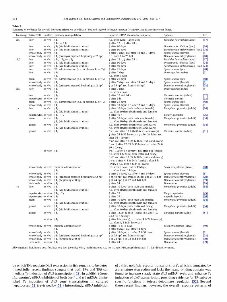
Table 2Summary of evidence for thyroid hormone effects on deiodinase (dio) and thyroid hormone receptor (tr) mRNA abundance in teleost fishes.
Transcript Tissue/cell Context Hormone manipulation Relative mRNA abundance response Species Ref.
dio1 liver in vivo " T4 n.c. after 12 h; ; after 24 h Fundulus heteroclitus (adult) [17]" T3, or " T2 ; after 12 h; ; after 24 h
liver in vivo ; T3 (via MMI administration) " after 90 days Oreochromis niloticus (juv.) [74]liver in vivo ; T3 (via MMI administration) " after 90 days Sarotherodon melanotheron (juv.) [74]whole body in vivo " T3 ; after 7 days; n.c. after 18 and 31 days Sparus aurata (larval) [8]whole body in vivo " T3 (embryos exposed beginning at 2 hpf) n.c. from 8 to 75 hpf Danio rerio (embryo/larval) [78]
dio2 liver in vivo " T4, " T3, or " T2 ; after 12 h; ; after 24 h Fundulus heteroclitus (adult) [17]liver in vivo ; T3 (via MMI administration) " after 90 days Oreochromis niloticus (juv.) [74]liver in vivo ; T3 (via MMI administration) " after 90 days Sarotherodon melanotheron (juv.) [74]liver in vivo PTU administration (n.c. in plasma T4 or T3) " after 21 days Sparus aurata (juv.) [48]liver in vivo " T3 ; after 7 days Oncorhynchus mykiss [5]
" T4 n.c. after 7 daysbrain in vivo PTU administration (n.c. in plasma T4 or T3) ; after 21 days Sparus aurata (juv.) [48]whole body in vivo " T3 ; after 7 days; n.c. after 18 and 31 days Sparus aurata (larval) [8]whole body in vivo " T3 (embryos exposed beginning at 2 hpf) ; at 75 hpf; n.c. from 8-48 hpf Danio rerio (embryo/larval) [78]
dio3 liver in vivo " T3 " after 7 days Oncorhynchus mykiss [5]" T4 n.c. after 7 days
liver in vivo " T3 " after 12 and 24 h Carassius auratus (adult) [52]hepatocytes in vitro " T3 " after 5 h Carassius auratus [52]brain in vivo PTU administration (n.c. in plasma T4 or T3) ; after 21 days Sparus aurata (juv.) [48]whole body in vivo " T3 ; after 18 days; n.c. after 7 and 31 days Sparus aurata (larval) [8]
tra liver in vivo " T3 " after 10 days (both male and female) Pimephales promelas (adult) [34]; T4 (via MMI administration) n.c. after 10 days (both male and female)
hepatocytes in vitro " T3, " T4 " after 19 h Conger myriaster [25]brain in vivo " T3 " after 10 days (both male and female) Pimephales promelas (adult) [34]
; T4 (via MMI administration) n.c. after 10 days (both male and female)gonad in vivo " T3 n.c. after 10 days (both testis and ovary) Pimephales promelas (adult) [34]
; T4 (via MMI administration) n.c. after 10 days (both testis and ovary)gonad in vivo " T3 tra1: n.c. after 12 h (both testis and ovary);
; after 24 & 36 h (testis); ; after 24 h but n.c.after 36 h (ovary)
Carassius auratus (adult) [81]
tra2: n.c. after 12, 24 & 36 h (testis and ovary)tra-t: " after 12, 24 & 36 h (testis) " after 24 &36 h (ovary)
in vitro " T3 tra1: ; after 8 h (ovary); n.c. after 8 h (testis);n.c. after 4 & 24 h (both testis and ovary)tra2: n.c. after 12, 24 & 36 h (testis and ovary)tra-t: " after 4, 8 & 24 h (testis) " after 8 h(ovary); n.c. after 4 & 24 h (ovary)
whole body in vivo thiourea administration n.c. after 8 days, " after 13 days Solea senegalensis (larval) [40]" T4 n.c. after 8 and 13 days
whole body in vivo " T3 ; after 31 days; n.c. after 7 and 18 days Sparus aurata (larval) [8]whole body in vivo " T3 (embryos exposed beginning at 2 hpf) " at 48 hpf; n.c. from 8-36 hpf and at 75 hpf Danio rerio (embryo/larval) [78]whole body in vivo " T3 (beginning at 0 hpf) ; at 24 hpf, " at 72 and 144 hpf Danio rerio (embryo/larval) [39]HeLa cells in vitro " T3 " after 24 h Danio rerio [39]
trb liver in vivo " T3 " after 10 days (both male and female) Pimephales promelas (adult) [34]; T4 (via MMI administration) n.c. after 10 days (both male and female)
hepatocytes in vitro " T3, " T4 " after 19 h Conger myriaster [25]hepatocytes in vitro " T3 " after 19 h Anguilla japonica [26]brain in vivo " T3 " after 10 days (both male and female) Pimephales promelas (adult) [34]
; T4 (via MMI administration) n.c. after 10 days (both male and female)gonad in vivo " T3 " after 10 days (both testis and ovary) Pimephales promelas (adult) [34]
; T4 (via MMI administration) n.c. after 10 days (both male and female)gonad in vivo " T3 ; after 12, 24 & 36 h (testis); n.c. after 12,
24 & 36 h (ovary)Carassius auratus (adult) [81]
in vitro " T3 ; after 8 h (ovary); n.c. after 4 & 24 h (ovary);n.c. after 4, 8 & 24 h (testis)
whole body in vivo thiourea administration ; after 11 & 15 days Solea senegalensis (larval) [40]" T4 " after 8 days; n.c. after 13 days
whole body in vivo " T3 ; after 18 days; n.c. after 7 & 31 days Sparus aurata (larval) [8]whole body in vivo " T3 (embryos exposed beginning at 2 hpf) " at 75 hpf; n.c. from 8-48 hpf Danio rerio (embryo/larval) [78]whole body in vivo " T3 (beginning at 0 hpf) ; at 24 hpf, " at 72 and 144 hpf Danio rerio (embryo/larval) [39]HeLa cells in vitro " T3 " after 24 h Danio rerio [39]
Abbreviations: hpf, hours post-fertilization; juv, juvenile; MMI, methimazole; n.c., no change; PTU, propilthiouracil; T2, 3,5-diiodothyronine.
514 K.M. Johnson, S.C. Lema / General and Comparative Endocrinology 172 (2011) 505–517
by which THs regulate Dio3 expression in fish remains to be deter-mined fully, recent findings suggest that both TRa and TRb canmediate T3-induction of dio3 transcription [52]. In goldfish (Caras-sius auratus), siRNA inhibition of both tra-1 and trb mRNAs dimin-ished T3 induction of dio3 gene transcription in culturedhepatocytes [52] (reviewed by [51]). Interestingly, siRNA inhibition
of a third goldfish receptor transcript (tra-t), which is truncated bya premature stop codon and lacks the ligand-binding domain, wasfound to increase steady-state dio3 mRNA levels and enhance T3
induction of dio3 transcription, providing evidence for TR-subtypespecific functions in teleost deiodinase regulation [52]. Beyondthese recent findings, however, the overall response patterns of
K.M. Johnson, S.C. Lema / General and Comparative Endocrinology 172 (2011) 505–517 515
dio3 mRNA abundance and Dio3 activity appear to correspond toDio3’s proposed function in teleosts as an inner-ring deiodinase(IRD) that protects tissues from excesses of active TH [54,61] (Ta-ble 2), although there is also some evidence that the specific TH-in-duced dynamics of Dio3 expression can again vary with cell andtissue type. In tilapia, for instance, treatment with gonadotropin(porcine FSH) elevated circulating T4 and increased brain T3-IRDactivity, even though exogenous T3 failed to have a similar effect;a methimazole-induced depression in circulating T4 and T3, how-ever, was observed to reduce brain T3-IRD activity in these samefish [47]. In other studies with tilapia, however, T3-IRD activity inbrain was not affected by either T4 or T3, even though T3-IRD activ-ity increased in gills and liver with T3 elevation [5,75].
Beyond these effects in liver and brain, TH status also affectedthe relative abundance of dio2 and dio3 mRNAs in the parrotfishgonads (see Fig. 5B and C). There is little data available on dio tran-scripts or Dio activity in the teleost gonad, although dio2 and dio3transcripts have previously been localized to gonadal tissues of fish[21,60], and dio2 mRNA levels in the gonad of rainbow trout werefound at levels nearly has high as those seen in liver [60]. In trout,transcripts encoding dio2 appear to reach highest levels in the tes-tis during early spermatogenesis stages [60], suggesting a role forDio2 in the regulation of sperm production. Consistent with thisidea, there is evidence that THs regulate testicular developmentand spermatogenesis in teleosts [11]. For instance, in Nile tilapia,hypothyroidism experienced during early life results in a largermass testis with more Sertoli cells and increased spermatozoa aftermaturation [42], while 21-day thiourea exposures of pre-spawningmale catfish (Clarias gariepinus) showed narrowed seminiferous tu-bules and fewer spermatozoa [69]. In rats, deiodinase activity hasbeen demonstrated within the testes [3], and recently Dio2 expres-sion – and dio2 mRNAs – were found to be highly expressed in tes-ticular germ cells [77]. Transcripts for dio2 have also been shown tobe upregulated in the rodent testis under hypothyroid status [76].Beyond what is presented here with parrotfish, however, compara-ble data are currently unavailable for teleost fishes, and futurestudies that localize and quantify deiodinase expression in the go-nads are needed.
Lastly, we found no evidence for TH regulation of dio1 mRNAlevels in any parrotfish tissues examined. Previous studies in larvaland adult fishes have found mixed results, with some studies con-cluding that dio1 transcript abundance was unaffected by changesin systemic TH levels [78], while other studies observed TH-depen-dent dio1 mRNA regulation [8,17,74] (Table 2). As such, the pictureof TH-mediated dio1 transcriptional regulation in teleost fishes isless clear than that for dio2 and dio3 [54]. In mammals, THs upreg-ulate dio1 gene transcription [4,29,80], at least in part via TRbreceptor action on TREs in dio1’s promoter region [1,22,28],although the degree of dio1’s T3-dependent regulation variesamong tissues [1]. As found here in parrotfish (Figs. 3F and 4F)and elsewhere, THs have been shown to autoinduce trb transcrip-tion in teleosts [34,39,78] (Table 2). Exogenous T3 treatment mighttherefore have altered expression levels of TRb in ways that con-found clear patterns of T3 influence on dio1 mRNA levels. Alterna-tively, even though the teleost dio1 gene is homologous to themammalian dio1 gene (see Fig. 1), there is considerable evidencethat the biochemical properties of Dio1 vary between fish andmammals, which may indicate that mechanisms for teleost dio1regulation may also be divergent from that of mammalian dio1[54,62].
4.2. Evidence for thyroid hormone receptor subtype-specific mRNAregulation
In addition to the observed changes in dio transcript abundance,we also found that thyroid hormone status influenced relative tr
mRNA levels in patterns indicative of tissue-specific roles for TRreceptors. In brief, TRs function to induce or repress the transcrip-tion of genes by acting as ligand-dependent transcription factorsthat bind in dimer pairs to thyroid response element (TRE)-bindingdomains in the promoter region of genes. In mammals andamphibians, tr genes themselves contain TREs so that transcriptionof these genes is autoinduced by T3 [79], and although there hasbeen comparatively little work on TREs in fish, TRs appear to func-tion through similar a mechanism [39,51]. Fish generally appear tohave at least one a and one b receptor type, which are encoded byat least two genes [40,51], although additional a and b receptormRNA subtypes have been identified in several species. Thesereceptors subtypes result from either alternative splicing or fromgene duplication events, which are now recognized as commonwithin bony fishes (e.g., [32]). In parrotfish, we identified three dis-tinct tr cDNAs, termed traA, traB and trb based on their phyloge-netic relationships (see Fig. 1). Although these cDNAs are onlypartial sequences, the extent of nucleotide and deduced amino acidsequence divergence between the traA and traB cDNAs from par-rotfish suggest that these transcripts may be encoded by two dis-tinct tra genes (On-line Supplemental materials, Fig. 1).
Here, the relative abundance of transcripts encoding traA and trb– but not traB – were elevated by T3 in the brain and liver of bothsexes of parrotfish, suggestive of T3 autoinduction of traA and trb– but not traB – in these tissues. This result is similar to that foundin other fishes, where alterations in thyroid status have been foundto alter tra and trb transcript levels (Table 2). For instance, in the fat-head minnow (Pimephales promelas), oral exposure to exogenous T3
resulted in elevated tra and trb transcript levels in the brain and li-ver, as well as trb transcripts in the testes, but not ovary [34]. Thesereceptor-subtype specific – and tissue-specific – patterns of tr tran-script abundance change are analogous to the complex patterns of trregulation observed in other vertebrates, where tr mRNAs are eitherinduced or repressed depending on the receptor type (or subtype)and tissue [10,20,58]. In fish, it is also clear that TR subtypes areTH-regulated in developmental stage specific patterns [8,39,40,78].
While we found evidence for male-female differences in diomRNA levels in several parrotfish tissues, the only sex differencesin tr transcript abundance that we observed occurred in the gonadswhere traA, traB and trb mRNA levels were all greater in testiculartissue than ovarian tissue (see Fig. 5D–F). There is little data avail-able on TR expression in the teleost gonads, although transcriptsencoding tra have previously been found to be greater in the testisthan ovary of sexually mature fathead minnows [34]. Transcriptsfor trb, however, were at greater levels in ovarian tissues than intesticular tissues of several other fishes [2,34,53], a pattern oppo-site to that observed here with parrotfish. Part of the explanationfor the opposite sex pattern of trb expression observed in parrotfishmay lie in this study’s examination of initial phase male parrotfishonly. Striped parrotfish exhibit both initial phase and terminalphase male phenotypes, which differ from each other in coloration,behavior and spawning frequency. In stoplight parrotfish (Spariso-ma viride), initial phase males have been shown to exhibit lower11-KT and higher E2 levels than terminal phase fish [9], implyingthat testicular function may also differ between initial and termi-nal phase male parrotfish. Whether such differences explain whygonadal trb mRNA levels in these initial phase striped parrotfishshowed a pattern opposite to that seen in these testes other fishesis unclear, but would be an interesting avenue for future use ofthese sex changing parrotfishes as a model for understandingTH-mediated reproductive function in teleost fishes.
Acknowledgments
We thank Matthew Birk, Kathy Cooper, Jon Dickey, Mark Gay,Dr. Kristin Hardy, Amy Metheny, Meagan Schrandt and Dr. Penny
516 K.M. Johnson, S.C. Lema / General and Comparative Endocrinology 172 (2011) 505–517
Swanson for technical support, and Dr. Mark Vermeij and theCuraçao Sea Aquarium for assistance with parrotfish collection.This research was supported by a Paul E. Hoiser Undergraduate Re-search and Creativity Fellowship from the UNC Wilmington Centerfor Support of Undergraduate Research to K.M.J., and an UNC Wil-mington Center for Marine Science Pilot Project Award to S.C.L. Theauthors also thank two anonymous referees, whose helpful sugges-tions improved the quality of this manuscript.
Appendix A. Supplementary data
Supplementary data associated with this article can be found, inthe online version, at doi:10.1016/j.ygcen.2011.04.022.
References
[1] L.L. Amma, A. Campos-Barros, Z. Wang, B. Vennström, D. Forrest, Distincttissue-specific roles for thyroid hormone receptors b and a1 in regulation oftype 1 deiodinase expression, Mol. Endocrinol. 15 (2001) 467–475.
[2] K.W. An, M.I. An, E.R. Nelson, H.R. Habibi, C.Y. Choi, Gender-related expressionof TRa and TRb in the protandrous black porgy, Acanthopagrus schlegeli, duringsex change processes, Gen. Comp. Endocrinol. 165 (2010) 11–18.
[3] J.M. Bates, D.L. St. Germain, V.A. Galton, Expression profiles of the threeiodothyronine deiodinases, D1, D2, and D3, in the developing rat,Endocrinology 140 (1999) 844–851.
[4] A.C. Bianco, D. Salvatore, B. Gereben, M.J. Berry, P.R. Larsen, Biochemistry,cellular and molecular biology, and physiological roles of the iodothyronineselenodeiodinases, Endocr. Rev. 23 (2002) 38–89.
[5] O. Bres, J.C. Plohman, J.G. Eales, A cDNA for a putative type III deiodinase in thetrout (Oncorhynchus mykiss): influence of holding conditions and thyroidhormone treatment on its hepatic expression, Gen. Comp. Endocrinol. 145(2006) 92–100.
[6] S.B. Brown, B.A. Adams, D.G. Cyr, J.G. Eales, Contaminant effects on the teleostfish thyroid, Environ. Toxicol. Chem. 23 (2004) 1680–1701.
[7] L.A. Burmeister, J. Pachucki, D.L. St. Germain, Thyroid hormones inhibit type 2iodothyronine deiodinase in the rat cerebral cortex by both pre- andposttranslational mechanisms, Endocrinology 138 (1997) 5231–5237.
[8] M.A. Campinho, M. Galay-Burgos, G.E. Sweeney, D.M. Power, Coordination ofdeiodinase and thyroid hormone receptor expression during the larval tojuvenile transition in sea bream (Sparus aurata, Linnaeus), Gen. Comp.Endocrinol. 165 (2010) 181–194.
[9] J.R. Cardwell, N.R. Liley, Hormonal control of sex and color change in thestoplight parrotfish, Sparisoma viride, Gen. Comp. Endocrinol. 81 (1991) 7–20.
[10] C. Constantinou, M. Margarity, T. Valcana, Region-specific effects ofhypothyroidism on the relative expression of thyroid hormone receptors inadult rat brain, Mol. Cell. Biochem. 278 (2005) 93–100.
[11] D.G. Cyr, J.G. Eales, Interrelationships between thyroidal and reproductiveendocrine systems in fish, Rev. Fish Biol. Fish. 6 (1996) 165–200.
[12] W.W. Dickhoff, L.A. Folmar, A. Gorbman, Changes in plasma thyroxine duringsmoltification of coho salmon, Gen. Comp. Endocrinol. 36 (1978) 229–232.
[13] E. Edeline, A. Bardonnet, V. Bolliet, S. Dufour, E. Pierre, Endocrine control ofAnguilla Anguilla glass eel dispersal: effect of thyroid hormones on locomotoractivity and rheotactic behavior, Horm. Behav. 48 (2005) 53–63.
[14] G.A. Fines, J. Plohman, J.G. Eales, Effect of experimental 3,5,30-triiodothyroninehyperthyroidism on thyroid hormone deiodination in brain regions and liverof rainbow trout, Oncorhynchus mykiss, Can. J. Zool. 77 (1999) 1185–1191.
[15] K.W. Finnson, J.G. Eales, Effect of T3 treatment and food ration on hepaticdeiodination and conjugation of thyroid hormones in rainbow trout,Oncorhynchus mykiss, Gen. Comp. Endocrinol. 115 (1999) 379–386.
[16] S.D. Firth, J.G. Eales, Thyroid hormone deiodination pathways in brain and liverof rainbow trout, Oncorhynchus mykiss, Gen. Comp. Endocrinol. 101 (1996)323–332.
[17] C. García-G, M.C. Jeziorski, C. Valverde-R, A. Orozco, Effects of iodothyronineson the hepatic outer-ring deiodinating pathway in killifish, Gen. Comp.Endocrinol. 135 (2004) 201–209.
[18] C.M. Glatt, M. Ouyang, W. Welsh, J.W. Green, J.O. Connor, S.R. Frame, N.E.Everds, G. Poindexter, S. Snajdr, D.A. Delker, Molecular characterization ofthyroid toxicity: anchoring gene expression profiles to biochemical andpathological end points, Environ. Health Perspect. 113 (2005) 1354–1361.
[19] E.G. Grau, L.M.H. Helms, S.K. Shimoda, C.-A. Ford, J. LeGrand, K. Yamauchi, Thethyroid gland of the Hawaiian parrotfish and its use as an in vitro modelsystem, Gen. Comp. Endocrinol. 61 (1986) 100–108.
[20] R.A. Hodin, M.A. Lazar, W.W. Chin, Differential and tissue-specific regulation ofthe multiple rat c-erbA messenger RNA species by thyroid hormone, J. Clin.Invest. 85 (1990) 101–105.
[21] E. Isorna, R. Vallés, A. Servili, J. Falcón, J.A. Muñoz-Cueto, Cloning and geneexpression of deiodinase enzymes and thyroid hormone receptors in theEuropean sea bass, Dicentrarchus labrax, Cybium 32 (Suppl. 2) (2008) 64.
[22] T.C. Jakobs, C. Schmutzler, J. Meissner, J. Köhrle, The promoter of the humantype 1 50-deiodinase gene – mapping of the transcription state site and
identification of a DR+4 thyroid-hormone-responsive element, Eur. J. Biochem.247 (1997) 288–297.
[23] D. Jayewardene, A factorial experiment quantifying the influence of parrotfishdensity and size on algal reduction on Hawaiian coral reefs, J. Exp. Mar. Biol.Ecol. 375 (2009) 64–69.
[24] D.-Y. Kang, Y.J. Chang, Effects of maternal injection of 3,5,30-triidod-L-thyronine (T3) on growth of newborn offspring of rockfish, Sebastes schlegeli,Aquaculture 234 (2004) 641–655.
[25] Y. Kawakami, D.-H. Shin, T. Kitano, S. Adachi, K. Yamauchi, H. Ohta,Transactivation activity of thyroid hormone receptors in fish (Congermyriaster) in response to thyroid hormones, Comp. Biochem. Physiol. B 144(2006) 503–509.
[26] Y. Kawakami, S. Adachi, K. Yamauchi, H. Ohta, Thyroid hormone receptor b iswidely expressed in the brain and pituitary of the Japanese eel, Anguillajaponica, Gen. Comp. Endocrinol. 150 (2007) 386–394.
[27] S.W. Kim, J.W. Harney, P.R. Larsen, Studies of the hormonal regulation of type 250-iodothyronine deiodinase messenger ribonucleic acid in pituitary tumorcells using semiquantitative reverse transcription-polymerase chain reaction,Endocrinology 139 (1998) 4895–4905.
[28] S.W. Kim, S.J. Hong, K.M. Kim, S.C. Ho, E.V. So, J.W. Harney, P.R. Larsen, A novel celltype-specific mechanism for thyroid hormone-dependent negative regulationof the human type 1 deiodinase gene, Mol. Endocrinol. 18 (2004) 2924–2936.
[29] J. Köhrl, Local activation and inactivation of thyroid hormones: the deiodinasefamily, Mol. Cell. Endocrinol. 151 (1999) 103–119.
[30] J. Köhrl, The deiodinase family: selenoenzymes regulating thyroid hormoneavailability and action, Cell. Mol. Life Sci. 57 (2000) 1853–1863.
[31] G. Kume, Y. Kubo, T. Yoshimura, T. Kiriyama, A. Yamaguchi, Life historycharacteristics of the protogynouse parrotfish Calotomus japonicas fromnorthwest Kyushu, Japan, Ichthyol. Res. 57 (2010) 113–120.
[32] D. Larhammar, G. Sundstrom, S. Dreborg, D.O. Daza, T.A. Larsson, Majorgenomic events and their consequences for vertebrate evolution andendocrinology, Trends Comp. Endocrinol. Neurobiol. 1163 (2009) 201–208.
[33] M.A. Larkin, G. Blackshields, N.P. Brown, R. Chenna, P.A. McGettigan, H.McWilliam, F. Valentin, I.M. Wallace, A. Wilm, R. Lopez, J.D. Thompson, T.J.Gibson, D.G. Higgins, Clustal W and Clustal X version 2.0, Bioinformatics 23(2007) 2947–2948.
[34] S.C. Lema, J.T. Dickey, I.R. Schultz, P. Swanson, Thyroid regulation of mRNAsencoding thyrotropin b-subunit, glycoprotein a-subunit, and thyroid hormonereceptors a and b in brain, pituitary gland, liver, and gonads of an adult teleost,Pimephales promelas, J. Endocrinol. 202 (2009) 43–54.
[35] S.C. Lema, J.T. Dickey, I.R. Schultz, P. Swanson, Dietary exposure to 2,20 ,4,40-tetrabromodiphenyl ether (PBDE-47) alters thyroid status and thyroidhormone-regulated gene transcription in the pituitary and brain, Environ.Health Perspect. 116 (2008) 1694–1699.
[36] S.C. Lema, G.A. Nevitt, An ecophysiological mechanism for morphologicalplasticity in pupfish and its relevance to conservation efforts for endangeredDevils Hole pupfish, J. Exp. Biol. 209 (2006) 3499–3509.
[37] S.C. Lema, G.A. Nevitt, Evidence that thyroid hormone induces olfactorycellular proliferation in salmon during a sensitive period for imprinting, J. Exp.Biol. 207 (2004) 3317–3327.
[38] W. Li, J.M. Zha, P.A. Spear, Z.L. Li, L.H. Yang, Z.J. Wang, Changes of thyroidhormone levels and related gene expression in Chinese rare minnow(Gobiocypris rarus) during 3-amino-1,2,4-triazole exposure and recovery,Aquat. Toxicol. 92 (2009) 50–57.
[39] Y.-W. Liu, L.-J. Lo, W.-K. Chan, Temporal expression and T3 induction of thyroidhormone receptors a1 and b1 during early embryonic and larval developmentin zebrafish, Danio rerio, Mol. Cell. Endocrinol. 159 (2000) 187–195.
[40] M. Manchado, C. Infante, L. Rebordinos, J.P. Canavate, Molecularcharacterization, gene expression and transcriptional regulation of thyroidhormone receptors in Senegalese sole, Gen. Comp. Endocrinol. 160 (2009)139–147.
[41] R. Marinez deMena, T.S. Scanlan, M.-J. Obregon, The T3 receptor b1 isoformregulates UCP1 and D2 deiodinase in rat brown adipocytes, Endocrinology 151(2010) 5074–5083.
[42] S.L.P. Matta, D.A.R. Vilela, H.P. Godinho, L.R. Franca, The goitrogen 6-n-propyl-2-thiouracil (PTU) given during testis development increase sertoli germ cellnumbers per cyst in fish: the tilapia (Oreochromis niloticus) model,Endocrinology 143 (2002) 970–978.
[43] S.A. Matthews, The thyroid gland of the Bermuda parrot fish, Pseudoscarusguacamaia, Anat. Rec. 101 (1948) 251–263.
[44] S.A. Matthews, D.C. Smith, Concentration of radioactive iodine by the thyroidgland of the parrot fish, Sparisoma sp, Am. J. Physiol. (1948) 222–225.
[45] S.D. McCormick, Endocrine control of osmoregulation in teleost fish, Integr.Comp. Biol. 41 (2001) 781–794.
[46] K.A. Mol, S. Van der Geyten, C. Burel, E.R. Kuhn, T. Boujard, V.M. Darras,Comparative study of iodothyronine outer ring and inner ring deiodinaseactivities in five teleostean fishes, Fish Physiol. Biochem. 18 (1998) 253–266.
[47] K. Mol, S. Van der Geyton, E. Kuhn, V. Darras, Effects of experimental hypo- andhyperthyroidism on iodothyronine deiodinases in Nile tilapia Oreochromisniloticus, Fish Physiol. Biochem. 20 (1999) 201–207.
[48] I. Morgado, M.A. Campinho, R. Costa, R. Jacinto, D.M. Power, Disruption of thethyroid system by diethylstilbestrol and ioxynil in the sea bream (Sparusaurata), Aquat. Toxicol. 92 (2009) 271–280.
[49] P.P. Morin, T.J. Hara, J.G. Eales, T4 depresses olfactory responses to L-alanineand plasma T3 and T3 production in smoltifying Atlantic salmon, Am. J. Physiol.Reg. Integr. Comp. Physiol. 269 (1995) R1434–R1440.
K.M. Johnson, S.C. Lema / General and Comparative Endocrinology 172 (2011) 505–517 517
[50] A.S. Mortensen, A. Arukwe, The persistent DDT metabolite, 1,1-dichloro-2,2-bis(q-chlrorphenyl)ethylene, alters thyroid hormone-dependent genes, hepaticcytochrome P4503A, and pregnane X receptor gene expression in Atlanticsalmon (Salmo salar) parr, Environ. Toxicol. Chem. 25 (2006) 1607–1615.
[51] E.R. Nelson, H.R. Habibi, Thyroid receptor subtypes: structure and function infish, Gen. Comp. Endocrinol. 161 (2009) 90–96.
[52] E.R. Nelson, H.R. Habibi, Functional significance of a truncated thyroid receptorsubtype lacking a hormone-binding domain in goldfish, Endocrinology 149(2008) 4702–4709.
[53] E.R. Nelson, H.R. Habibi, Molecular characterization and sex-related seasonalexpression of thyroid receptor subtypes in goldfish, Mol. Cell. Endocrinol. 253(2006) 83095.
[54] A. Orozco, R.C. Valverde, Thyroid hormone deiodination in fish, Thyroid 15(2005) 799–813.
[55] M. Picard-Aitken, H. Fournier, R. Pariseau, D.J. Marcogliese, D.G. Cyr, Thyroiddisruption in walleye (Sander vitreus) exposed to environmentalcontaminants: cloning and use of iodothyronine deiodinases as molecularbiomarkers, Aquat. Toxicol. 83 (2007) 200–211.
[56] E.M. Plate, B.A. Adams, W.T. Allison, G. Martens, C.W. Hawryshyn, J.G. Eales,The effects of thyroxine or a GnRH analogue on thyroid hormone deiodinationin the olfactory epithelium and retina of rainbow trout, Oncorhynchus mykiss,and sockeye salmon, Oncorhynchus nerka, Gen. Comp. Endocrinol. 127 (2002)59–65.
[57] D.R. Robertson, R.R. Warner, Sexual patterns of the labroid fishes of thewestern Caribbean, II. The parrotfishes (Scaridae), Smith. Contr. Zool. 255(1978) 1–26.
[58] P.M. Sadow, O. Chassande, E.K. Koo, K. Gauthier, J. Samarut, J. Xu, B.W.O’Malley, R.E. Weiss, Regulation of expression of thyroid hormone receptorisoforms and coactivators in liver and heart by thyroid hormone, Mol. Cell.Endocrinol. 203 (2003) 65–75.
[59] N. Saitou, M. Nei, The Neighbor-Joining method: a new method forreconstructing phylogenetic trees, Mol. Biol. Evol. 4 (1987) 406–425.
[60] E. Sambroni, S. Gutieres, C. Cauty, Y. Guiguen, B. Breton, J.-J. La Reyre, Type IIiodothyronine deiodinase is preferentially expressed in rainbow trout(Oncorhynchus mykiss) liver and gonads, Mol. Reprod. Develop. 60 (2001) 338–350.
[61] J.P. Sanders, S. Van der Geyten, E. Kaptein, V.M. Darras, E.R. Kühn, J.L. Leonard,T.J. Visser, Cloning and characterization of type III iodothyronine deiodinasefrom the fish Oreochromis niloticus, Endocrinology 140 (1999) 3666–3673.
[62] J.P. Sanders, S. Van der Geyten, E. Kaptein, V.M. Darras, E.R. Kuhn, J.L. Leonard,T.J. Visser, Characterization of a propylthiouracil-insensitive type 1iodothyronine deiodinase, Endocrinology 138 (1997) 5153–5160.
[63] E.M. Santos, P. Kille, V.L. Workman, G.C. Paull, C.R. Tyler, Sexually dimorphicgene expression in the brains of mature zebrafish, Comp. Biochem. Physiol. A149 (2008) 314–324.
[64] C. Schmutzler, I. Gotthardt, P.J. Hofmann, B. Radovic, G. Kovacs, L. Stemmier, I.Nobis, A. Bacinski, B. Mentrup, P. Ambrugger, A. Gruters, L.K. Malendowicz, J.Christoffel, H. Jarry, D. Seidlova-Wuttke, W. Wuttke, J. Kohrle, Endocrinedisruptors and the thyroid gland: a combined in vitro and in vivo analysis ofpotential new biomarkers, Environ. Health Perspect. 115 (2007) 77–83.
[65] X. Shi, C. Liu, G. Wu, B. Zhou, Waterborn exposure to PFOS causes disruption ofthe hypothalamus–pituitary–thyroid axis in zebrafish larvae, Chemosphere 77(2009) 1010–1018.
[66] L.L. Smith, J.L. Fessler, M.E. Alfaro, J.T. Streelman, M.W. Westneat, Phylogenteicrelationships and the evolution of regulatory gene sequences in theparrotfishes, Mol. Phylogen. Evol. 49 (2008) 136–152.
[67] D.L. St. Germain, V.A. Galton, A. Hernandez, Defining the roles of theiodothyronine deiodinases: current concepts and challenges, Endocrinology150 (2009) 1097–1107.
[68] M. Sutija, T.J. Longhurst, J.M.P. Joss, Deiodinase type II and tissue specificmRNA alternative splicing in the Australian lungfish, Neoceratodus forsteri,Gen. Comp. Endocrinol. 132 (2003) 409–417.
[69] I. Swapna, A. Rajasekhar, A. Supriya, K. Raghuveer, M.K. Rasheeda, K.C.Majumdar, H. Kagawa, H. Tanaka, A. Dutta-Gupta, B. Senthilkumaran,Thiourea-induced thyroid hormone depletion impairs testicularrecrudescence in the air-breathing catfish, Clarias gariepinus, Comp. Biochem.Physiol. A 144 (2006) 1–10.
[70] P. Swanson, E.G. Grau, L.M.H. Helms, W.W. Dickhoff, Thyrotropic activity ofsalmon pituitary glycoprotein hormones in the Hawaiian parrotfish thyroidin vitro, J. Exp. Zool. 245 (1988) 194–199.
[71] D.T. Szabo, V.M. Richardson, D.G. Ross, J.J. Diliberto, P.R.S. Kodavanti, L.S.Birnbaum, Effects of perinatal PBD exposure on hepatic phase I, phase II, phaseIII, and deiodinase 1 gene expression involved in thyroid hormone metabolismin male rat pups, Toxicol. Sci. 107 (2009) 27–39.
[72] K. Tamura, J. Dudley, M. Nei, S. Kumar, MEGA4: molecular evolutionarygenetics analysis (MEGA) software version 4.0, Mol. Biol. Evol. 24 (2007)1596–1599.
[73] H.M. Tu, S.W. Kim, D. Salvatore, T. Bartha, G. Legradi, P.R. Larsen, R.M. Lechan,Regional distribution of type 2 thyroxine deiodinase messenger ribonucleicacid in rat hypothalamus and pituitary and its regulation by thyroid hormone,Endocrinology 138 (1997) 3359–3368.
[74] S. Van der Geyten, A. Toguyeni, J.F. Baroiller, B. Fauconneau, A. Fostier, J.P.Sanders, T.J. Visser, E.R. Kühn, V.M. Darras, Hypothyroidism induces type 1iodothyronine deiodinase expression in tilapia liver, Gen. Comp. Endocrinol.124 (2001) 333–342.
[75] S. Van der Geyten, N. Byamungu, G.E. Reyns, E.R. Kühn, V.M. Darras,Iodothyronine deiodinases and the control of plasma and tissue thyroidhormone levels in hyperthyroid tilapia (Oreochromis niloticus), J. Endocrinol.184 (2005) 467–479.
[76] M.S. Wagner, R. Morimoto, J.M. Dora, A. Benneman, R. Pavan, A.L. Maia,Hypothyroidism induces type 2 iodothyronine deiodinase expression in mouseheart and testis, J. Endocrinol. 31 (2003) 541–550.
[77] Wajner, S.M., Wagner, M. dS., Melo, R.C.N., Parreira, G.G., Chiarini-Garcia, H.,Bianco, A.C., Fekete, C., Sanchez, E., Lechan, R.M., Maia, A.L., 2007. Type 2iodothyronine deiodinase is highly expressed in germ cells of adult rat testis. J.Endocrinol. 194, 47–54.
[78] C.N. Walpita, S. Van der Geyten, E. Rurangwa, V.M. Darras, The effect of3,5,30-triiodothyronine supplementation on zebrafish (Danio rerio)embyronic development and expression of iodothyronine deiodinasesand thyroid hormone receptors, Gen. Comp. Endocrinol. 152 (2007)206–214.
[79] P.M. Yen, S. Ando, X. Feng, Y. Liu, P. Maruvanda, X. Xia, Thyroid hormone actionat the cellular, genomic and target gene levels, Mol. Cell. Endocrinol. 246(2006) 121–127.
[80] A.M. Zavacki, H. Ying, M.A. Christoffolete, G. Aerts, E. So, J.W. Harney, S.-Y.Cheng, P.R. Larsen, A.C. Bianco, Type 1 iodothyronine deiodinase is a sensitivemarker of peripheral thyroid status in the mouse, Endocrinology 146 (2005)1568–1575.
[81] E.R. Nelson, E.R.O. Allan, F.Y. Pang, H.R. Habibi, Auto-regulation of thyroidhormone receptors in the goldfish ovary and testis, Gen. Comp. Endocrinol., inpress, doi:10.1016/j.ygcen.2010.12.017.


