Embed Size (px)
Citation preview

JOURNAL OF VIROLOGY, Nov. 2010, p. 11359–11373 Vol. 84, No. 210022-538X/10/$12.00 doi:10.1128/JVI.00804-10Copyright © 2010, American Society for Microbiology. All Rights Reserved.
Toll-Like Receptor 4-Mediated Activation of p38 Mitogen-ActivatedProtein Kinase Is a Determinant of Respiratory Virus Entry and Tropism�
David Marchant, Gurpreet K. Singhera, Soraya Utokaparch, Tillie L. Hackett,John H. Boyd, Zongshu Luo, Xiaoning Si, Delbert R. Dorscheid,
Bruce M. McManus,* and Richard G. Hegele*UBC James Hogg Research Centre, Providence Heart � Lung Institute, Department of Pathology and Laboratory Medicine,
University of British Columbia, Room 166, Burrard Building, St. Paul’s Hospital,1081 Burrard Street, Vancouver, British Columbia, Canada V6Z 1Y6
Received 15 April 2010/Accepted 28 July 2010
Respiratory viruses exert a heavy toll of morbidity and mortality worldwide. Despite this burden there arefew specific treatments available for respiratory virus infections. Since many viruses utilize host cell enzymaticmachinery such as protein kinases for replication, we determined whether pharmacological inhibition ofkinases could, in principle, be used as a broad antiviral strategy for common human respiratory virusinfections. A panel of green fluorescent protein (GFP)-expressing recombinant respiratory viruses, includingan isolate of H1N1 influenza virus (H1N1/Weiss/43), was used to represent a broad range of virus familiesresponsible for common respiratory infections (Adenoviridae, Paramyxoviridae, Picornaviridae, and Orthomyxo-viridae). Kinase inhibitors were screened in a high-throughput assay that detected virus infection in humanairway epithelial cells (1HAEo-) using a fluorescent plate reader. Inhibition of p38 mitogen-activated proteinkinase (MAPK) signaling was able to significantly inhibit replication by all viruses tested. Therefore, thepathways involved in virus-mediated p38 and extracellular signal-regulated kinase (ERK) MAPK activationwere investigated using bronchial epithelial cells and primary fibroblasts derived from MyD88 knockout mouselungs. Influenza virus, which activated p38 MAPK to approximately 10-fold-greater levels than did respiratorysyncytial virus (RSV) in 1HAEo- cells, was internalized about 8-fold faster and more completely than RSV. Weshow for the first time that p38 MAPK is a determinant of virus infection that is dependent upon MyD88expression and Toll-like receptor 4 (TLR4) ligation. Imaging of virus-TLR4 interactions showed significantclustering of TLR4 at the site of virus-cell interaction, triggering phosphorylation of downstream targets of p38MAPK, suggesting the need for a signaling receptor to activate virus internalization.
Respiratory virus infections cause considerable morbidityand mortality worldwide; it was recently reported that hospi-talizations due to respiratory syncytial virus (RSV) exceed 2million per year in the Unites States alone (16). An H1N1swine influenza pandemic took place during the 2009-2010winter season (14), and there is the lingering threat of an H5N1avian influenza pandemic, with mortality due to direct bird-to-human H5N1 infection in hospitalized patients between 30 and100% (3). The severe acute respiratory syndrome (SARS)-associated coronavirus, isolated in 2003, resulted in devastatingrespiratory tract infections with few treatment options (40).For most common respiratory viruses, treatment is symptom-atic, and for pathogens such as influenza viruses for whichspecific treatments are available, oseltamivir (Tamiflu)- and
amantidine-resistant strains are emerging and being transmit-ted globally (33).
All functions within a cell are triggered and regulated by cellsignaling cues. Since viruses are obligate intracellular parasites,they rely upon cell signaling to regulate all processes within thecell that drive virus replication. In this study we investigatedthe effects of kinase inhibitors as a therapeutic strategy and toinvestigate the roles played by some kinases during virus rep-lication. The extracellular signal-regulated kinase (ERK) andp38 mitogen-activated protein kinases (MAPKs) have beenshown by us and others to play important roles during virusreplication in vitro (19, 20, 26, 30, 42), and we have recentlyreported that inhibition of p38 MAPK activation is an effectiveand novel antiviral strategy in vivo (29). The significance of p38MAPK activity in vivo is such that inadvertent and coincidentactivation of this kinase by some pharmaceutical agents en-hances virus replication (29). Antiviral strategies may existwhereby inhibition of host cell kinases may stem the spreadand replication of numerous different viral species. Such broadantiviral strategies would permit administration of kinase in-hibitors to patients suspected of having respiratory viral infec-tion, and to health care workers or inhabitants within the localeof a viral outbreak, prior to the availability of results fromlaboratory diagnostic testing.
The activation of p38 MAPK by pattern recognition recep-tors (PRRs) has been studied in the context of the antiviralimmune response (reviewed in reference 22). We report here
* Corresponding author. Mailing address for Bruce M. McManus:UBC James Hogg Research Centre, Providence Heart � Lung Insti-tute, Department of Pathology and Laboratory Medicine, University ofBritish Columbia, Room 166, Burrard Building, St. Paul’s Hospital,1081 Burrard Street, Vancouver, British Columbia, Canada V6Z 1Y6.Phone: (604) 806-8586. Fax: (604) 806-9274. E-mail: [email protected]. Mailing address for Richard G. Hegele: KeenanResearch Centre, Li Ka Shing Knowledge Institute, St. Michael’sHospital, Department of Laboratory Medicine and Pathobiology,University of Toronto, 1 King’s College Circle, Toronto, Ontario,Canada M5S 1A8. Phone: (416) 978-2557. Fax: (416) 978-7361.E-mail: [email protected].
� Published ahead of print on 11 August 2010.
11359

that viruses usurp these responses for the benefit of virusreplication through activation of p38 MAPK, mediated by aPRR (Toll-like receptor 4 [TLR4]) and MyD88, providing thebasis for a broad-spectrum antiviral.
MATERIALS AND METHODS
Viruses, cells, and inhibitors. Coxsackievirus B3 (CVB3)-green fluorescentprotein (GFP), a molecular clone of CVB3 Woodruff (pH 3; GenBank accessionno. U57056) containing an enhanced GFP (eGFP) expression cassette (CVB3-GFP) described previously (12, 44), was used in this study (provided by RalphFeuer and J. Lindsay Whitton [The Scripps Institute, La Jolla, CA]). Influenzavirus A/Weiss/43 (H1N1) (VR96) was obtained from the American Type CultureCollection (ATCC; Manassas, VA), and stocks were produced in HeLa cells inserum-free Dulbecco modified Eagle medium (DMEM) containing 10 �g/mltrypsin. For experiments, 1 vial of influenza virus was treated at 37°C in thepresence of serum-free 10-�g/ml trypsin for 30 min prior to dilution and additionto cells. RSV-A2-GFP was provided by Mark Peeples (Department of Pediatrics,The Ohio State University, Children’s Research Institute, Columbus, OH) (46,49), and human parainfluenza virus type 3 (hPIV3)-GFP was provided by PeterCollins (National Institutes of Health, Bethesda, MD) (45, 48). Human adeno-virus (AdV) 5 was obtained from ATCC (catalog no. VR-1516). AdV-GFP(adenovirus 5-derived delta E1 and E3 vector with GFP expression driven froma cytomegalovirus [CMV] promoter) was purchased from Vector Biolabs (Phil-adelphia, PA). All viruses except adenovirus E-deleted vector were produced inHeLa cells. Adenovirus vector stocks were produced in HEK 293 cells. In allexperiments, infections were conducted in parallel with uninfected control-con-ditioned medium from HeLa cells to control for possible activation of cells bycell-conditioned medium.
Immortalized epithelial cell lines used were 1HAEo- (bronchial epithelial),A549 (alveolar epithelial), and HeLa (cervical carcinoma). These were culturedin DMEM-10% fetal bovine serum (FBS) and obtained from ATCC, except for1HAEo- cells, which have been described previously (6).
Isolation of primary fibroblasts. All animals were treated humanely and inaccordance with the regulations of the UBC Animal Care Committee and stan-dards of the Canadian Council on Animal Care. Pulmonary fibroblasts wereisolated from MyD88 knockout (KO) and wild-type (WT) C57 backgroundcontrol mouse lungs. Briefly, lungs were removed from mice that were firstanesthetized by isofluorane and then euthanized by cervical dislocation. Lungswere minced briefly and placed into the appropriately sized tissue culture tray.Lung pieces were dried for approximately 5 min and overlaid with DMEM, 10%FBS, and penicillin-streptomycin. Lungs were removed on the following day, andcells were replated 1 week later.
Kinase inhibitors and chemicals. Inhibitors purchased from Tocris Pharma-ceuticals (Ellisville, MO) were used at the final concentrations indicated (targetkinase is in parentheses): API-2 (Akt/protein kinase B [PKB]), 1 �M; BAY11-7085 (NF-�B), 10 �M; LY294002 (phosphatidylinositol 3-kinase [PI3K]), 25 �M;MG132 (proteasome), 10 �M; PP2 (Src family kinases), 5 �M; SB203580 (p38MAPK), 5 �M; SB216763 (glycogen synthase kinase 3� [GSK3�]), 10 �M;SP600125 (Jun N-terminal protein kinase [JNK]), 20 �M; U0126 (MEK1/2), 20�M. Anisomycin was purchased from Sigma-Aldrich (St. Louis, MO) and used ata concentration of 10 �M.
Correlation of PFU with GFP IU/ml. Infected cultures of each of the recom-binant viruses used were subject to plaque assay in parallel to flow cytometry ofGFP expression of infected cultures, at least once. Infectious units per ml (in-fluenza virus staining) or GFP-positive (GFP�) cells per ml were calculated bymultiplying the proportion of Alexa 594 (influenza virus)- or GFP-positive cellsby the total number of cells in the well, resulting in the total number of infectedcells. This value was divided by the dilution factor and the volume of inoculumin ml, which is the number of GFP� cells per ml (recombinant GFP viruses) orinfectious units per ml (IU/ml; influenza virus).
Antibodies. Antiadenovirus monoclonal antibody (MAb) 8052 from ChemiconInternational (clone 20/11; Millipore, Billerica, MA) detects the hexon protein ofmost adenoviruses. Anti-VP1 of CVB3 was purchased from Novocastra (antien-terovirus VP1 clone D5/8; Newcastle, United Kingdom); goat anti-influenzaH1N1 virus catalog no. 18-783-77828-1 was purchased from GenWay Biotech(San Diego, CA); anti-RSV3 mouse monoclonal glycoprotein F, protein P, andprotein N antibodies were purchased from Novocastra. Goat anti-RSV (allantigens) polyclonal antibody was purchased from Biodesign International(Saco, ME; catalog no. B65860G). Parainfluenza virus type 3 was detected byWestern blotting using goat anti-parainfluenza virus 2 and 3 polyclonal sera fromBiodesign International (catalog no. B65130). Anti-Toll-like receptor 4 goat
polyclonal IgG and monoclonal humanized IgA antibodies were purchased fromAbCam (ab53629) and Invivogen (catalog no. maba2-htlr4), respectively. TheRSV nucleoprotein (N) was detected using a mouse monoclonal antibody(ab10016) that detects only mature virion N protein.
Determination of infectivity via flow cytometry. Influenza virus infection wasmeasured by antibody staining followed by flow cytometry. Briefly, cells weredetached and fixed in 5 mM EDTA and 3% formalin, respectively. Cells under-went centrifugation (500 � g, 5 min) and were reconstituted in phosphate-buffered saline (PBS), 0.3% Triton X-100, and 1% bovine serum albumin (BSA)for 10 min at room temperature (RT). Goat anti-influenza was added at a 1/100dilution for 30 min at RT in 0.3% Triton X-100, 1% BSA, and PBS. Cells werewashed by 3 consecutive centrifugation (500 � g, 3 min) and PBS washing stepswith 5 min between each wash. Secondary Alexa 594 donkey anti-goat antibodywas added at a 1/200 dilution in 0.3% Triton X-100 in PBS for 30 min at RT.Cells were washed 3 times and enumerated with a Beckman Coulter EpicsXL-MCS flow cytometer.
Determination of viral genome copy number in cell-free virus via quantitativereverse transcription-PCR (RT-PCR). Virus RNA or adenovirus DNA was ex-tracted from cell-free virus aliquots using the viral RNA minikit or DNA bloodand tissue minikit, respectively (Qiagen, Mississauga, ON, Canada). Sequencesfor the primers and probes for each respiratory virus were chosen using PrimerExpress software (Applied Biosystems, Foster City, CA) following recommendedcriteria. Synthetic standards were produced for each virus to allow absolutequantification of cDNA from the virus aliquots. The standards were comprised ofpGEM4Z (Promega, Madison, WI) incorporating the target amplicon of eachvirus. A restriction site integrated within the amplicon allowed for the detectionof any cross-contamination between the virus aliquot specimen and the syntheticone. Briefly, RNA with priming sites and amplification attributes identical tothose of the real viral target was created from the synthetic standard. Twentymicrograms of plasmid DNA was linearized using 40 U of XbaI for 2 h at 37°Cand then heated at 65°C for 15 min to inactivate the XbaI. Two micrograms ofdigested plasmid was in vitro transcribed using a MEGAshortscript kit (Ambion,Austin, TX). Transcription buffer, deoxynucleoside triphosphates (dNTPs), plas-mid, and enzyme mix were combined to a final volume of 20 �l and incubated for2 h. RNase-free DNase I was added to the reaction mixture and allowed toincubate at 37°C for an additional 15 min to remove template DNA. To recoverthe transcript, RNA was purified by ammonium acetate precipitation. The RNApellet was then resuspended in 100 �l of RNase- and DNase-free water. ThecDNA produced from the in vitro-transcribed transcripts was serially dilutedfrom 108 to 101 copy numbers, aliquoted, and stored along with the specimencDNA at �80°C until use. Quantitative PCR assays were performed on an ABI7900HT (Applied Biosystems, Foster City, CA) machine in triplicate.
Kinase inhibitor screen of GFP-virus-infected cells. HeLa, 1HAEo-, or A549cells were seeded at confluence in 96-well optical-bottom plates. Kinase inhibi-tors were added 30 min prior to infection, and AdV-GFP, RSV-GFP, andhPIV3-GFP infection mixtures at multiplicities of infection (MOI) of 1 and 10were fixed in methanol-acetone (3:1, vol/vol) 24 h postinfection. CVB3 infectionmixtures were harvested 8 to 10 h postinfection. GFP fluorescence was detectedand quantified by a Tecan GENios fluorescence plate reader (excitation [Ex],485/emission [Em], 535; gain, 60; MTX Lab Systems). Infections were thentreated with Hoechst 33342 (1/10,000 for 10 min) to stain nuclei, and fluores-cence was detected (Ex, 360/Em, 465; gain, 60) as a cell input control.
The activity of kinase inhibitors upon influenza virus infection was detected byantibody staining of influenza virus protein at 16 h postinfection and enumera-tion by flow cytometry. Kinase inhibitors were added 30 min prior to infection,and influenza virus-infected cells were detached with 5 mM EDTA and fixed with3.7% formalin-PBS for 10 min. Cells were stained using polyclonal anti-influenzavirus sera as outlined above.
Determination of cell viability by 3-(4,5-dimethylthiazol-2-yl)-5-(3-carboxyme-thoxyphenyl)-2-(4-sulfophenyl)-2H-tetrazolim assay. Cell viability was deter-mined with the Promega MTS assay kit (Madison, WI), per the manufacturer’sinstructions.
Determination of MAPK phosphorylation by phospho-ELISA and Westernblot assay. Cells were seeded into 12- or 24-well trays and infected with virus orcell-conditioned medium in 10% FBS-DMEM. Cell lysates were harvested usingprotein lysis buffer (10 mM HEPES [pH 7.4], 50 mM Na4P2O7, 50 mM NaF, 50mM NaCl, 5 mM EDTA, 5 mM EGTA, 1 mM Na3VO4, 0.5% Triton X-100, 10�g/ml leupeptin, and 1 mM phenylmethylsulfonyl fluoride). Ten micrograms ofsample was loaded per well of each enzyme-linked immunosorbent assay(ELISA) mixture. ERK, JNK, ATF2, and p38 MAPK phosphorylation was de-termined using ELISA kits purchased from Biosource (Invitrogen, Carlsbad,CA). Samples were assayed per the manufacturer’s instructions. To better visu-alize increases in MAPK phosphorylation in Fig. 1a to d, the basal levels of
11360 MARCHANT ET AL. J. VIROL.

MAPK activation (conditioned medium) were subtracted from the values ob-tained from the virus-infected cells at each time point.
Western blot assay. Alternatively, lysates were subjected to Western blotting,whereby cells were harvested in protein lysis buffer, as described above. Post-nuclear supernatant was harvested by centrifugation at 14,000 � g for 10 min at4°C, boiled and resolved on a 12% polyacrylamide gel at 120 V for 1 h, andtransferred to a nitrocellulose membrane. Lysates were probed for RSV, CVB3,p42/p44 total ERK MAPK, MyD88, and total and phospho-p38 MAPK.
Neutralization of Toll-like receptor 4 signaling with a neutralizing antibody.1HAEo- and HeLa cells were treated with a dilution series of rabbit IgG control(ab46540) and rabbit polyclonal anti-human TLR4 antibody from AbCam(ab13556; Cambridge, MA), in serum-free DMEM. Cells were infected with adilution series of RSV-A2 and influenza virus H1N1/Weiss/43 with 1 h of pre-incubation with anti-TLR4 or control antibodies. Infected cells were enumeratedby flow cytometry the following day, and lysates were harvested for Westernblotting in parallel to monitor successful inhibition of phospho-p38 MAPK orphospho-ATF-2 (see “Western blot assay” above). Influenza virus-infected cellswere enumerated by flow cytometry after antibody staining (described under“Determination of infectivity via flow cytometry”).
Detection of virus particles and host cell proteins using immunofluorescenceconfocal microscopy. Immunofluorescence of virus during cell entry has beendescribed previously (30). Virus (MOI, 3 to 5) was added to cells on ice for 1 h,cells were washed once with PBS, and infection proceeded in a 37°C incubatoruntil the indicated time points. Cells were fixed by addition of room temperature3:1 (vol/vol) methanol-acetone for 2 min and blocked with 1% BSA-PBS orblocked and permeabilized with 0.3% Triton X-100, 1% BSA, and PBS (PERMbuffer) for 10 min at room temperature. Primary antibodies were added at a1/100 dilution at 4°C overnight in PERM buffer. Rabbit anti-phospho-ATF2(catalog no. 9221; Cell Signaling technologies, Danvers, MA) and goat anti-TLR4 (ab53629; AbCam, Cambridge, MA) were stained with goat anti-rabbit ordonkey anti-goat secondary antibodies conjugated to Alexa 488 or 594, at adilution of 1/400 in PERM buffer. The goat anti-rabbit secondary antibody wasadded after the donkey anti-goat antibody, with an additional wash step, toprevent secondary antibody cross-reactivity, in the experiments that requiredmouse (RSV3 MAbs), goat (TLR4), and rabbit (phospho-ATF2) antibody de-tection. Virus was stained with monoclonal anti-RSV (RSV3; a mix of 3 mono-clonal RSV antibodies: glycoprotein F, protein P, and protein N [Novocastra])and Alexa 405 or 488 secondary antibodies purchased from Invitrogen. Each stepwas followed with 3 washes in PBS of 5 min each. Nuclei were stained with DAPI(4�,6-diamidino-2-phenylindole) contained within the mounting medium,Vectashield (Vector Laboratories, Burlingame, CA). The non-PERM stainingcontrol assays were conducted as described above but in the absence of 0.3%Triton X-100.
Image acquisition, 3-dimensional modeling, and rendering. Immunofluores-cence confocal microscopy was conducted using an AOBS Leica confocal TCSSP2 microscope (63�/numerical aperture [NA], 1.2) (Heidelberg, Germany).Lambda scans were performed throughout the experiments to confirm the spec-ificity of Alexa Fluor dyes. Sequential scans prevented the detection of artifactualcolocalization. The AOBS SP2 system allows for user-defined photo multipliertube (PMT) filters of custom bandwidth, for specific and custom detection ofeach fluorophore by a separate PMT detector. Our purpose for this system wasthe exclusion of bleed-through of signal into the emission profile of fluorophoreswith longer wavelengths from fluorophores with emission at shorter wavelengths.Image stacks (100 to 150 � 512 � 512 pixels; 6-frame average) were collatedusing Improvision Volocity v5 software (Coventry, United Kingdom), with minorbrightness and contrast adjustment (not greater than 20% of original values).Minor background noise filtering was conducted for all fluorescence confocalimages using the fine noise filter in Volocity. Virus particles were countedautomatically in a defined region of interest (ROI) at t � 0 min for RSV andinfluenza virus using the measurement function, detected by intensity greaterthan 30% and larger than 0.05 �m. The ROI was drawn around each DAPI-stained nucleus with an extra 1-�m buffer to isolate each individual cell fordetermination of the number of virus particles per cell.
Pearson’s correlation. Pearson’s correlation of image stacks was conducted tocorrelate overlap of image voxels (pixels in 3 dimensions) from 2 differentchannels using Volocity software (as described previously [29]). A value of �1indicates complete voxel-to-voxel overlap of the voxels from 2 channels, through-out the image Z-stack. A value of 0 indicates random correlation of voxels from2 different channels, and �1 indicates complete disparity of voxels from the 2channels that have been compared.
Statistical methods. Statistical significance between two experimental groupswas determined using Student’s t test. Comparisons involving more than twoexperimental groups were conducted using analysis of variance (ANOVA) with
Tukey’s family comparison to determine statistical significance between the com-parisons. A P value of 0.05 was considered to be significant.
RESULTS
Suppression of p38 MAPK activation with SB203580 inhib-ited infection by every virus in a screen of kinase inhibitorsusing 1HAEo- bronchial epithelial cells. Human airway epi-thelial cells (1HAEo-) were treated with pharmacological in-hibitors to major host cell kinases for 30 min prior to infection,as we have previously described: JNK (SP600125) (42), ERK(U0126) (26, 30), and p38 (SB203580) (42) MAPKs; Src kinase(PP2) (47); PI3K (LY294002) (37); NF-�B (BAY11-7085) (8);Akt (API-2) (9, 10); and GSK3� (SB216763) (47). Plates werefixed, and GFP fluorescence produced by GFP-virus replica-tion was detected by a fluorescence plate reader. Readoutswere normalized to the no-treatment (dimethyl sulfoxide[DMSO]) control and by using Hoechst 33342 nuclear stainfluorescence. Figure 1 shows the magnitude of GFP fluores-cence produced 8 to 10 h after infection with CVB3 (30) (Fig.1a) and 24 h after infection with hPIV3-GFP (Fig. 1b), RSV-GFP (Fig. 1c), and AdV-GFP (Fig. 1d). Figure 1e shows theinfectivity of H1N1 influenza virus in the presence of kinaseinhibitors. These viruses represent four respiratory virus fam-ilies (AdV; Adenoviridae, RSV/hPIV3; Paramyxoviridae,CVB3; Picornaviridae [respiratory transmission reviewed inreference 39], influenza A H1N1 virus; Orthomyxoviridae) andtwo subfamilies (RSV, subfamily Pneumovirinae, genus Pneu-movirus; hPIV3, subfamily Paramyxovirinae, genus Respirovi-rus) within the Paramyxoviridae family.
The pharmacologic agents that inhibited replication of allviruses tested targeted p38 (SB203580) and ERK (U0126)MAPKs. There were 2.8-fold (P � 0.04), 3.6-fold (P � 0.005),3.2-fold (P � 0.002), 1.7-fold (P � 0.04) and 9.4-fold (P � 0.01)decreases in fluorescence intensity of CVB3-GFP, hPIV3-GFP, RSV-GFP, AdV-GFP, and influenza H1N1 virus infec-tions associated with SB203580 treatment compared to un-treated controls, respectively (Fig. 1a to e). A549 (alveolar[type 1 pneumocyte] epithelial) cells and HeLa (cervical car-cinoma) cells were also tested by this method with similarresults: suppression of p38 using SB203580 consistently inhib-ited infection by all viruses tested (data not shown).
Cell viability curve of SB203580 compared to cisplatin after24 h of treatment. Prior to further investigating a potentialantiviral role for p38 inhibition, we tested the toxicity ofSB203580 on the viability of 1HAEo- cells. We determined cellviability at 24 h in the presence of cisplatin or SB203580 asmeasured by MTS assay (Fig. 1f). As expected, cisplatin wascytotoxic in a dose-dependent manner, in which MTS absor-bance at 500 nm decreased in a concentration-dependent man-ner. In contrast, SB203580 had little or no effect on MTS-related cell viability when administered in concentrations ashigh as 333 �M, suggesting that SB203580 did not inhibit virusreplication via host cell death.
Virus-activated phosphorylation of p38 and ERK MAPKs isbiphasic. We determined the activation profile of p38 andERK MAPKs during virus infection to identify the points inthe virus life cycle that were most susceptible to the p38 inhib-itor SB203580. We added CVB3, hPIV3, RSV, and a labora-tory-adapted strain of H1N1 influenza virus, at an RNA ge-
VOL. 84, 2010 TOLL-LIKE RECEPTOR 4-MEDIATED ACTIVATION OF p38 MAPK 11361

11362 MARCHANT ET AL. J. VIROL.

nome copy number of 10 per cell, to 1HAEo- cells. Adenoviruswas added at a DNA copy number of about 10 per cell. Ge-nome copy numbers of virus stocks were determined by quan-titative PCR against an in-house standard curve for each virus[see Materials and Methods, “Determination of viral genomecopy number in cell-free virus via quantitative reverse tran-scription-PCR (RT-PCR)”] as a means of standardizing thevirus particle input. A time course assay was conducted, andthe phosphorylation states of ERK and p38 were determinedby phospho-ELISA (Fig. 2a), as well as JNK (data not shownand reference 42). All viruses activated p38 MAPK 10 minpostinfection, except for CVB3, which, in addition to cell-conditioned medium, was used as an internal control for p38activation given that this virus activated p38 weakly or not at allduring entry, 10 min postinfection (42). Human PIV3 (P �0.001), RSV (P � 7.9 � 10�5), AdV (P � 0.01), and influenzavirus (P � 0.03) activated p38 MAPK 10 min postinfection. Atthis time point influenza virus activated p38 to the highest level(3.1 � 102 U/ml) and AdV activated p38 to the lowest level(1.6 � 101 U/ml). In addition, influenza virus activated p38MAPK to a 9.7-fold-higher level than did RSV (3.2 � 101
U/ml) at 10 min (Fig. 2a). As reported previously, CVB3 ac-tivated ERK 1 h postinfection and again at 8 h postinfection(27). Phosphorylation of ERK and p38 corresponded with theonset of virus protein production 8 h postinfection as deter-mined by the detection of capsid protein VP1 (data notshown). Human PIV3 (P � 0.03), RSV (P � 7.0 � 10�4), andAdV (P � 0.05) activated p38 at 8 h postinfection. The activationof p38 and ERK MAPKs at 8 h postinfection was associated withthe onset of virus protein production at approximately this timepoint, except for RSV, for which protein synthesis was not appar-ent until 16 h postinfection (data not shown).
We wanted to determine the point in the virus life cycle (Fig.2b) at which p38 MAPK phosphorylation was a requirement ofvirus replication. Figure 2c shows how p38 inhibitor SB203580inhibited RSV protein production at 8 h postinfection whenadded at 0.16 h (10 min) postinoculation, compared to theno-treatment control and the addition of SB203580 at 4 hpostinoculation (i.e., after virus had successfully entered targetcells). In contrast, CVB3 showed decreased VP1 productionwhen SB203580 was added at both early (0.16 h) and later (4h) time points postinfection. This difference is reflected in thedifferent p38 MAPK activation profiles of RSV and CVB3(Fig. 2a).
Early activation of p38 MAPK during virus entry is depen-dent upon MyD88. Influenza virus, hPIV3, RSV, and AdV
produced identical profiles of p38 MAPK activation in1HAEo- cells at 10 min postinfection (Fig. 2a). Therefore, wepostulated that PRRs such as the Toll-like receptors (TLRs)may recognize high-mannose residues expressed on the surfaceof viruses (17, 21) or the virus surface glycoproteins that me-diate virus entry into the host cell (15, 23, 38). As virusesinteract with the surface of the cell, before or during entryreceptor ligation, we reasoned that viruses might interact withPRRs expressed at the cell surface and initiate signaling and aresponse that affects virus uptake into the cell.
We isolated pulmonary fibroblasts from myeloid differenti-ation factor 88 (MyD88) KO mice. Myeloid differentiationfactor 88 is a signaling adaptor that is required for the signalpropagation of many of the Toll-like receptors (TLRs) (35).Therefore, determining the ERK and p38 MAPK activationprofiles in MyD88 KO cells may implicate or rule out TLRinvolvement in MAPK activation 10 min post-virus infection.So we determined the expression of ERK MAPK, p38 MAPK,and MyD88 in MyD88 KO cells (Fig. 3a) compared to theirexpression in WT cells. As expected, there were equivalentamounts of ERK and p38 in WT and MyD88 KO cells, andMyD88 was absent from the KO cells.
We wanted to determine the ERK and p38 MAPK activa-tion profiles in the WT and MyD88 KO cells during an 8-hinfection time course. We postulated that activation of p38 andERK may occur through a TLR pathway given the very earlyactivation of p38 and ERK, 10 min postinfection (Fig. 2a). Aspostulated, we observed a lack of p38 activation in murineMyD88 KO pulmonary fibroblasts upon virus entry, comparedto the p38 MAPK activation that occurs in WT cells within 1 hof virus addition (Fig. 3b); however, there was apparent en-hancement of ERK activation 10 min postinfection. The dif-ferent activation profiles of p38 and ERK in MyD88 KO cellsduring virus entry are an indication of the different mecha-nisms/pathways of activation of these two MAPKs: in pulmo-nary fibroblasts p38 is apparently MyD88 dependent and ERKis MyD88 independent. Furthermore, we noted that p38 acti-vation occurred in primary WT fibroblasts 10 to 15 min laterthan it did in the 1HAEo- cell line, explained by the inherentdifferences between primary cells and cell lines and fibroblastsand epithelial cells.
Figure 3c shows that there was significantly less virus infec-tion of MyD88 KO cells than of WT cells. Adenovirus vector,influenza virus, and RSV infections were 14-, 2.3-, and 6.0-foldlower in MyD88 KO cells than in cells from the WT counter-parts, respectively.
FIG. 1. The p38 and ERK MAPKs are principal kinases during host cell infection by a broad array of respiratory viruses. (a to d) Human airwayepithelial (1HAEo-) cells were treated with kinase inhibitors for 1 h and infected with CVB3-eGFP (a), hPIV3-eGFP (b), RSV-eGFP (c), andAdV-eGFP (d), at MOI of 1 and 10, and GFP production as an indicator of virus infectivity was detected on a fluorescence plate reader on thefollowing day. (e) Human airway epithelial (1HAEo-) cells were treated with kinase inhibitors for 1 h and infected with H1N1 influenza virus.Infectivity was determined by fluorescent antibody staining and flow cytometry on the following day: cells (1HAEo-) were treated with kinaseinhibitors for 1 h and infected with influenza virus H1N1/Weiss/43 at an MOI of 0.01 to 1. Cells were harvested, fixed, and permeabilized 24 h laterand stained for H1N1 influenza virus antigen with a polyclonal antibody followed by an Alexa 594 anti-goat secondary antibody, and infected cellswere enumerated by flow cytometry. IU/ml, infectious units per ml. (f) Cisplatin but not SB203580 is cytotoxic to cells in a dose-dependent manner.1HAEo- cells were treated with cisplatin and SB203580 and control DMSO in a dose titration (3 nM to 333 �M). Cell viability was measured byMTS assay at 24 h post-drug addition. The substrate MTT [3-(4,5-dimethylthiazol-2-yl)-2,5-diphenyltetrazolium bromide, a yellow tetrazole] isreduced to MTS [3-(4,5-dimethylthiazol-2-yl)-5-(3-carboxymethoxyphenyl)-2-(4-sulfophenyl)-2H-tetrazolium] by mitochondrial reductase in livingcells. Loss of cell viability due to the treatment of cells with cisplatin is indicated by reduced absorbance at 33 and 333 �M, but there was no lossof cell viability with increasing doses of SB203580.
VOL. 84, 2010 TOLL-LIKE RECEPTOR 4-MEDIATED ACTIVATION OF p38 MAPK 11363

Anisomycin is an antibiotic that activates p38 and JNKMAPKs (5, 18, 34). We wanted to determine whether p38MAPK was a determinant of infection and tropism in targethost cells. JNK was excluded since its inhibitor SP600125 didnot affect viral replication (Fig. 1a to e), and the activation of
JNK was particularly weak compared to the activation profilesof p38 and ERK MAPKs (data not shown). Therefore, wedetermined whether supplementary ectopic p38 activationcould rescue virus infection in MyD88 KO cells, given that p38was the MAPK not activated in these cells. We observed the
FIG. 2. The p38 and ERK MAPKs are activated at virus-host cell entry and late time points postinfection. (a) Cells (1HAEo-) were infectedwith viruses using 10 genome equivalents per cell as determined by qPCR of virus stocks (Materials and Methods), and cell lysates were harvestedusing cell lysis buffer at indicated time points. Phospho-ERK (bar graph) and total ERK (line chart) (left panels) and p38 MAPK activation (rightpanels) were detected using phospho-ELISA and total kinase ELISA kits. (b) Timeline of virus inoculation and SB203580 (SB) treatment for thepulse-chase experiment shown in panel c. (c) Cells were inoculated with virus and treated with SB as in panel b, and cell lysates were collectedusing cell lysis buffer. The Western blot shows that SB treatment (p38 MAPK inhibition) inhibited RSV protein production when added at 10 minpost-virus inoculation but that SB inhibited CVB3 when it was added at both early (0.16 h [10 min]) and later (4 h) time points. Results arerepresentative of at least 3 independent experiments.
11364 MARCHANT ET AL. J. VIROL.
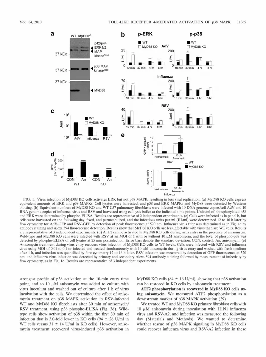
strongest profile of p38 activation at the 10-min entry timepoint, and so 10 �M anisomycin was added to culture withvirus inoculum and washed out of culture after 1 h of virusincubation with the cells. We determined the effect of aniso-mycin treatment on p38 MAPK activation in RSV-infectedWT and MyD88 KO fibroblasts after 30 min of anisomycin/RSV treatment, using p38 phospho-ELISA (Fig. 3d). Wild-type cells show activation of p38 within the first 30 min ofinfection that is 3.0-fold lower in KO cells (94 26 U/ml inWT cells versus 31 14 U/ml in KO cells). However, aniso-mycin treatment recovered virus-induced p38 activation in
MyD88 KO cells (84 16 U/ml), showing that p38 activationcan be restored in KO cells by anisomycin treatment.
ATF2 phosphorylation is recovered in MyD88 KO cells us-ing anisomycin. We measured ATF2 phosphorylation as adownstream marker of p38 MAPK activation (29).
We treated WT and MyD88 KO primary fibroblast cells with10 �M anisomycin during inoculation with H1N1 influenzavirus and RSV-A2, and infection was measured the followingday (Materials and Methods). We wanted to determinewhether rescue of p38 MAPK signaling in MyD88 KO cellscould recover influenza virus and RSV-A2 infection in these
FIG. 3. Virus infection of MyD88 KO cells activates ERK but not p38 MAPK, resulting in less viral replication. (a) MyD88 KO cells expressequivalent amounts of ERK and p38 MAPKs. Cell lysates were harvested, and p38 and ERK MAPKs and MyD88 were detected by Westernblotting. (b) Equivalent numbers of MyD88 KO and WT C57 pulmonary fibroblasts were infected with 10 DNA genome copies/cell AdV and 10RNA genome copies of influenza virus and RSV and harvested using cell lysis buffer at the indicated time points. Units/ml of phosphorylated p38and ERK were determined by phospho-ELISA. Results are representative of 2 independent experiments. (c) Cells were infected as in panel b, butcells were harvested on the following day, fixed, and permeabilized, and the infectious units per ml (IU/ml) were determined 12 to 16 h later byflow cytometry for AdV-GFP and RSV-GFP by detection of peak fluorescence at 520 nm. Influenza virus titer was determined as in Fig. 1e byantibody staining and Alexa 594 fluorescence detection. Results show that MyD88 KO cells are less infectable with virus than are WT cells. Resultsare representative of 3 independent experiments. (d) ATF2 can be activated in MyD88 KO cells during virus entry in the presence of anisomycin.Wild-type and MyD88 KO cells were infected with RSV at an MOI of 1 with or without 10 �M anisomycin, and the level of phospho-p38 wasdetected by phospho-ELISA of cell lysates at 25 min postinfection. Error bars denote the standard deviation. CON, control; An, anisomycin. (e)Anisomycin treatment during virus entry recovers virus infection of MyD88 KO cells to WT levels. Cells were infected with RSV and influenzavirus using MOI of 0.01 to 0.1 or infected and treated simultaneously with 10 �M anisomycin during virus entry and washed with fresh mediumafter 1 h, and infection was quantified by flow cytometry 12 to 16 h later. RSV infection was measured by detection of GFP fluorescence at 520nm, and influenza virus infection was detected by primary and secondary Alexa 594 antibody staining followed by measurement of infectivity byflow cytometry, as in Fig. 1e. Results are representative of 3 independent experiments.
VOL. 84, 2010 TOLL-LIKE RECEPTOR 4-MEDIATED ACTIVATION OF p38 MAPK 11365

cells to WT levels. Influenza virus and RSV were chosen sincethey are enveloped viruses and activated p38 MAPK to thehighest levels among the viruses studied (Fig. 2a). Influenzavirus and RSV infections were recovered in the anisomycin-treated MyD88 KO cells to WT levels (Fig. 3e). Taken to-gether, the results in Fig. 3 emphasize the importance of p38MAPK activation via MyD88, in preference to ERK MAPKactivation, to trigger virus entry and replication.
Imaging the signaling and entry of RSV and influenza virusin 1HAEo- airway bronchial epithelial cells. We imaged RSVand influenza virus during the entry phase of the virus life cycleto determine the effect of SB203580 on these 2 viruses duringentry. We were also curious as to any potential differencesbetween entry phases since p38 MAPK was activated to 10-fold-greater levels during influenza virus entry (310 84 U/ml)than were seen at 10 min post-RSV infection (31 34 U/ml[Fig. 2a]).
Activation of p38 MAPK signaling occurs at the site ofvirus-host cell contact. We wanted to determine the origin inthe cell where virus activated p38 MAPK during entry, byvisualizing the phosphorylation of p38 immediate downstreamtargets. Cells were treated with SB203580 or control (DMSO),and a time course of virus entry and phospho-ATF2 signalingwas followed. Figure 4a shows a 40-min time course of RSVentry. We observed that phospho-ATF2, a downstream targetof activated p38 MAPK, was phosphorylated above virus par-ticles (10 min postinfection, YZ and XZ sections) or adjacentto the site of virus-cell interaction (10 min postinfection, XYsection, and 25 min postinfection). At 40 min postwarming,under control conditions in Fig. 4b, we noted that RSV parti-cles were located apically and in close perinuclear apposition.However, treatment with p38 MAPK inhibitor SB203580 re-sulted in virus particles located at the basal periphery of thetarget cells. Successful RSV replication is therefore associatedwith localization to apical perinuclear regions in the cell within40 min of entry trafficking.
Activation of p38 MAPK was associated with virus traffick-ing to perinuclear regions and emergence of the RSV nucleo-protein during entry into the host cell. The close nuclear as-sociation of RSV virions after entry permitted quantitativeanalysis of internalized particle proximity to the nucleus (Fig.4c) and correlation with successful virus entry. We used Pear-son’s correlation to determine the proximity of RSV to thenucleus during permeabilization control assays. The Pearsoncorrelation ranges from �1 to �1, whereby a correlation of �1indicates complete overlap of voxels (a pixel in 3 dimensions)from 2 different channels and a correlation of �1 indicatescomplete voxel disparity between the 2 channels being com-pared. Although RSV replicates solely in the cytosol, the closeassociation of green RSV particles with DAPI signal (whichstains both DNA and RNA) 40 min postentry afforded bleed-through colocalization during close proximity of RSV to thenucleus (Fig. 4c), reflected by a Pearson correlation that wasgreater than 0. The disparity of virus staining with DAPI attime zero, or when virus entry was inhibited by SB203580 (Fig.4d), resulted in a Pearson correlation that was negative (greenvirus voxels and blue DAPI voxels do not overlap). The non-permeabilization control assay permitted staining of only ex-tracellular virions; virus that trafficked to intracellular perinu-clear regions was not stained, as reflected in the negative
Pearson correlation of the “No PERM” control at 40 minpostwarming in Fig. 4c.
We hypothesized that if p38 MAPK is activated upon con-tact with a virion, entry may be facilitated by the subsequentcellular activation and endocytosis that result from p38 MAPKactivation. p38 MAPK can phosphorylate the Rab5 effectorsEEA1 and Rabenosyn-5 on Thr-1392 and Ser-215, respectively(28), enhancing endocytic internalization. Therefore, this in-ternalization enhancement activity of p38 MAPK may be uti-lized by viruses to increase virus entry efficiency.
Treatment of cells with p38 inhibitor SB203580 resulted ininhibition of ATF2 phosphorylation (Fig. 4b), as expected.Virus remained at the leading edge of the SB203580-treatedcells at 40 min postinfection, in contrast to cells treated withDMSO vehicle; virus trafficked to more apical perinuclear re-gions in DMSO-treated cells, with virus moving entirely awayfrom the basal surface. As in Fig. 4c, we quantified the local-ization of RSV particles to the perinuclear RNA staining byDAPI. Figure 4d shows a negative Pearson correlation at 40min with SB203580 treatment (Pearson’s correlation � �0.11[P 0.001]), whereas the Pearson correlation in the DMSOcontrol is positive at this time point (Pearson’s correlation �0.03 [P � 0.003]).
The RSV nucleoprotein can be imaged 40 min postentry butnot during p38 MAPK inhibition. To support the observationsthat RSV entry is dependent upon p38 MAP kinase activation,we stained cells under control and SB203580 treatment condi-tions using an anti-RSV-N antibody (Fig. 4e). The RSV-Nprotein (nucleocapsid) binds genomic and antigenomic viralRNAs to form an RNase-resistant nucleocapsid, packagedwithin virus virions. The RSV-N protein can be imaged 40 minpostentry but not at 0 min postentry, suggesting that N proteinimaging may be a measure of successful virion disassembly.Finally, SB203580 treatment resulted in a loss of RSV-N de-tection 40 min postentry, similar to the images at 0 min posten-try (Fig. 4e), suggesting that SB203580 treatment inhibitedrelease of RSV virion contents.
p38 MAPK inhibition by SB203580 slows influenza H1N1virus traffic to the nucleus during virus entry. We also imagedthe entry phase of influenza virus replication using H1N1/Weiss/43, in the presence and absence of p38 inhibitorSB203580. Influenza virus was detected using polyclonal goatanti-H1N1 influenza virus sera (see “Antibodies” in Materialsand Methods). Influenza virus is one of the few RNA virusesthat replicates in the nucleus, and it is dependent upon acidi-fied late endosomes for fusion and delivery of virion contentswithin the target host cell, so that it utilizes different entrypathways than does RSV, which fuses at the cell surface andreplicates in the cytosol. We costained influenza virus withearly endosome antigen 1 (EEA1) since p38 MAPK has beenshown to phosphorylate this protein and enhance endocytosis.We surmised that in the absence of p38 MAPK activation, inthe presence of SB203580, influenza virus particles may beinhibited within EEA1-positive endosomes.
Figure 5a shows influenza virus particles sitting upon thesurface of 1HAEo- bronchial epithelial cells prior to beingwarmed to 37°C. After 5 min of warming to 37°C, influenzavirus ribonucleoproteins (rNPs) can be imaged within the nu-cleus of the control cells (Fig. 5b). There is apparently less
11366 MARCHANT ET AL. J. VIROL.

influenza virus protein in the nucleus of the SB203580-treatedcells.
At 20 min after warming of SB203580-treated cells, we ob-served influenza virus particles within EEA1-positive endo-somes, whereas all influenza virus protein appeared to haveattained the nucleus of the control cells (Fig. 5c). Our resultsreflect those reported previously that p38 MAPK can enhanceendocytosis internalization via EEA1 and Rabenosyn-5 (28),where we observed slowed trafficking of influenza virus to thenucleus upon inhibition of p38 MAPK activation. To quanti-tate the entry of influenza virus into the nucleus, we performedPearson’s colocalization analysis of nuclei stained with DAPI(blue voxels) and the green Alexa 488-stained influenza virusvirions (green voxels), at the 0-, 5-, and 20-min time points ofinfluenza virus entry into 1HAEo- cells. The values shown inFig. 5d for the 20-min time point are greater than those of thePearson correlation of RSV entry (Fig. 5e) since influenzavirus, unlike RSV, enters and replicates within the nucleus. Wenoted that there was significantly less influenza virus detectedwithin the nucleus at 5 and 20 min postinfection when p38activation was inhibited with SB203580 (Fig. 5d), compared tothe DMSO-treated control cells. Comparison of influenza virusand RSV entry was aided in part by the automatic counting of
FIG. 4. Viruses cause activation of ATF2 via p38 MAPK from thesite of virus-host cell contact, during entry. (a) RSV-A2 was added to1HAEo- cells on ice for 1 h at an MOI of 1, transferred to a 37°Cincubator, fixed, and stained for RSV and phospho-ATF2 at the indi-cated time points after being warmed to 37°C. RSV is stained green,activated phospho-ATF2 (signaling product of activated p38 MAPK) isred, and nuclei are blue. The location of the presented XY slice isindicated with a gray line and a black arrow in the XZ pane. The10-min image shows a burst of red (phospho-ATF2 [arrow]) above aninternalized virus particle. The peak of phospho-ATF2 was most oftenseen at 25 min postinfection with a loss of signal at 40 min postinfec-tion with residual signal remaining. Perinuclear localization of RSVwas always observed by 40 min postinfection (arrowheads). (b) Acti-vation of p38 MAPK was associated with apical perinuclear localiza-tion of virus after 40 min of entry with little or no basal localization ofvirion (arrowheads). Residual phospho-ATF2 activation is shown inred, RSV is shown in green, and nuclei were imaged blue with DAPI.Inhibitor (SB203580) suppression of p38 MAPK was associated withbasal localization of virus at the leading edge of the cell/tissue culturemargin (arrows), also illustrated by transmitted light detection (TLD)overlay. Note the less apparent red phospho-ATF2 staining (red; in-dicator of p38 MAPK activation) in the SB203580-treated cells in theright panels. The location of the presented XY slice is indicated witha gray line and a black arrow in the XZ aspect. (c) Pearson’s correla-tion of image stacks (not shown) permeabilized (PERM) or not with0.3% Triton X-100 during staining. ‡, P 0.001. Permeabilization ofcells during staining permits imaging of internalized virus particles/constituents. The Pearson correlation is positive 40 min after entry dueto DAPI-RSV protein colocalization, which was lost if cells were notTriton X-100 permeabilized during staining. (d) Pearson’s correlationof image stacks in panel a and accompanying SB203580 (SB)-treatedcontrols (not shown) of green (Alexa 488) RSV particles to blueDAPI-stained nuclei. The graph shows a negative Pearson correlationat 40 min postwarming due to SB203580 inhibition of p38 MAPK,indicating that there was no RSV trafficking to perinuclear regions. †,P 0.005; ‡, P 0.001. CON, control. (e) RSV nucleoprotein(N) could be imaged during control conditions 40 min postentry, but itwas lost with SB203580 treatment. Cells were infected, fixed, andstained as in panel a and stained for RSV-N using a monoclonalantibody (Materials and Methods). Bars, 6.2 �m. Results are repre-sentative of 4 independent experiments.
VOL. 84, 2010 TOLL-LIKE RECEPTOR 4-MEDIATED ACTIVATION OF p38 MAPK 11367

virus particles by Volocity software (Materials and Methods),which confirmed equivalent virus particle inputs (Fig. 5f).
Virus infection and subsequent TLR signaling can be neu-tralized with anti-TLR antibodies. Given the previous reportsthat the innate antiviral response may be activated by TLR4binding to virus glycoproteins (15, 17, 21, 23, 38), we per-formed neutralization experiments with anti-TLR4 antibodies,followed by influenza virus and RSV inoculation. Since MyD88
is the adaptor molecule for several different TLRs (35), wewanted to test the possibility that p38-associated signalingcould indeed be inhibited by blocking virus interaction withone of the TLRs. These experiments would assist us in deter-mining whether a pathway(s) of MyD88-dependent patternrecognition may be responsible for rapid p38 MAPK activationduring virus entry. We chose antibody neutralization since thistechnique is commonly used to block virus interactions with
FIG. 5. Inhibition of p38 MAPK results in retention of influenza virus particles within early endosome antigen-positive endosomes. Imagesshow confocal microscopy of influenza virus entry into upper airway 1HAEo- epithelial cells stained using an anti-H1N1 influenza virus goatpolyclonal antibody. (a) Equivalent amounts of influenza virus (MOI, 3) were added to 1HAEo- cells treated with control (CON) DMSO andSB203580 (SB), on ice (0 min). The arrow indicates the XY image of a bisected nucleus, showing an absence of influenza virus localization to thenucleus at 0 min. Bars, 21 �m. (b) At 5 min postwarming most virus is visualized in the nucleus, whereas there is apparently less nuclear stainingin the nucleus during treatment with SB203580. Bars, 10 �m. (c) At 20 min postwarming virus retention in EEA1-stained regions was most evident(arrows). (d) Pearson’s correlation shows a significant loss of influenza virus localization to the nucleus during entry in the SB203580-treated cellscompared to DMSO treatment control conditions (*, P 0.05; †, P 0.005). (e) Pearson’s correlation of RSV during entry demonstrates thegreater rate at which influenza virus attains the nucleus in panel d than at which RSV attains perinuclear regions by 40 min postwarming (†, P 0.005). (f) Comparison of the number of influenza virus and RSV particles on cells, on ice, at 0 min postwarming, indicating that equivalent virusparticle numbers were delivered to cells in entry comparison experiments. Results are representative of 4 independent experiments.
11368 MARCHANT ET AL. J. VIROL.

receptors and to elucidate the physiology of virus-receptorinteractions. Therefore, we postulated that if a TLR-virus in-teraction is genuine, then it (and all downstream effects)should, in principle, be neutralized with an anti-TLR4 receptorantibody. Figure 6a shows inhibition of p38 MAPK, but notERK, signaling by treatment with an anti-TLR4 antibody at aconcentration of 0.1 �g/ml. Figures 6b and c show that RSV
and influenza virus infections were reduced similarly, 68% and68.5%, respectively, by treatment with an anti-TLR4 antibody,compared to control antibody. Anti-TLR4 antibody added athigher concentrations caused enhanced p38 MAPK activationand corresponding increases in infection (data not shown). IfTLR4 acted as an entry and fusion receptor for these viruses,we would have expected these higher concentrations of anti-body to block infection in a dose-dependent manner (11, 13),rather than enhance infection as was observed.
p38 MAPK signaling is associated with clustering of TLR4at the site of virus-cell attachment. Fluorescence confocal im-aging of RSV, TLR4, and phospho-ATF2 demonstrated clus-tering of TLR4 at the site of RSV binding to the host cell, 10min postinfection, whereas there was no TLR4 clustering orphospho-ATF2 signal observed in cells that were not bound byvirus (Fig. 7a).
Overlapping confocal fluorescent signals that are attributedto colocalization/interaction of 2 or more different moleculescan be the result of imaging artifacts due to fluorophore bleed-ing into the detection channel of the other fluorophore. Wetherefore wanted to determine the effect of blocking TLR4-virus interaction during entry in order to rule out potentialimaging artifacts in the observation of the TLR4-virus cluster-ing phenomenon. Custom bandwidth PMT filters of the LeicaSP2 microscope were also utilized to eliminate colocalizationartifacts (see Materials and Methods for technical descrip-tions). Goat polyclonal TLR4 antibody was added before virusaddition and during virus entry. Low-magnification images inFig. 7b show qualitative suppression of ATF2 phosphorylationdue to 1 �g/ml of anti-TLR4 polyclonal antibody in a 25-minsnapshot image.
Images in Fig. 7c, “0 min,” show clusters of virus with TLR4at 4°C (the ability of the receptor to remain mobile within theplane of the plasma membrane at 0°C has been describedpreviously for influenza virus [1, 32]), under control conditions(Fig. 7c, arrows); there was also patching of TLR4 (green)beneath virus particles (cyan) under anti-TLR4 antibody treat-ment conditions (Fig. 7c, arrowheads). These results suggestthat there is significant mobility of receptor at 4°C, supportedby the work of others (1, 32). There were equivalent numbersof virus particles attached to the surface of infected cells undercontrol and antibody treatment conditions, determined by au-tomatic spot counting by Volocity software, suggesting thatantibody treatment did not block virus interaction with thetarget cells (data not shown). The patching of TLR4 with virusoccurred under antibody treatment conditions (Fig. 7d, inset),also suggesting that virus-TLR4 interaction was not affected byantibody treatment.
Treatment with antibody directed against TLR4 resulted inless coalescence of virus-TLR4 complex early after infection,as shown at the 0- (Fig. 7c), 10- (Fig. 7d), and 25-min (Fig. 7e)time points. Volocity software was used to identify virus-recep-tor complexes using a threshold of detection for virus cyanstaining, and the complexes were then measured (Materialsand Methods). Figure 7f shows the results of automatic Vo-locity measurements of virus-TLR4 complexes. Clusters ofTLR4-virus complexes were significantly smaller at 0 min un-der antibody treatment conditions (6.7 � 10�1 2.4 � 10�1
�m) than under control conditions (1.2 � 100 4.8 � 10�1
�m; P � 0.004). The size of virus-receptor complexes did not
FIG. 6. Activation and phosphorylation of p38 MAPK/virus infec-tion can be inhibited with anti-TLR4 antibodies. (a) An anti-TLR4antibody titration was added to 1HAEo- cells for 30 min, RSV wasadded at an MOI of 1, cells were harvested with lysis buffer after 20min of virus treatment, and phospho-p38 MAPK and phospho-ERKwere detected, with total p38 as a loading control. pAb, polyclonalantibody. (b) Cells were treated as in panel a, but infection proceededovernight before detection of RSV-GFP by flow cytometry and influ-enza virus infection via antibody staining and flow cytometry (Materi-als and Methods). The infection resulting from RSV-GFP was de-tected directly by flow cytometry of infected cells whereas influenzavirus infection was detected by antibody staining of influenza virus-infected cells with a polyclonal anti-influenza virus antibody followedby staining with an Alexa 594 secondary antibody. (c) The percentneutralization assay was conducted on the 0.1-�g/ml anti-TLR4 dosefor both RSV and influenza virus calculated from the data in panel bby dividing infection obtained in the presence of TLR4 antibody bycontrol infection and multiplying the value by 100%. Results are rep-resentative of 2 independent experiments. Con, control.
VOL. 84, 2010 TOLL-LIKE RECEPTOR 4-MEDIATED ACTIVATION OF p38 MAPK 11369

11370 MARCHANT ET AL. J. VIROL.

change significantly between control 0- and 10-min postwarm-ing time points; however, there was a significant decrease invirus-receptor cluster size in a comparison of the antibodytreatment 0-min (6.7 � 10�1 2.4 � 10�1 �m) and 10-min(4.4 � 10�1 8.0 � 10�2 �m) time points (P � 0.006).
The XZ sections of the 10-min time point in the inset of Fig.7d show an absence of RSV-TLR4 overlap in the TLR4 anti-body-treated cell images, suggesting that TLR4 clustering atthe site of virus attachment is not the product of image arti-facts, though patching of TLR4 beneath virus particles contin-ued to be evident in the presence of TLR4 antibody. Thus,these data support TLR4 binding and clustering around virusto activate p38 MAPK during virus entry.
DISCUSSION
We demonstrate that PRR recognition of virus, prior to hostcell entry, is required to activate p38 MAPK via MyD88 toactivate cellular internalization machinery. Our work also em-phasizes that the virus entry receptor(s) is not the only proteinwith which virus interacts at the host cell surface prior to entry.The sugar residues that make up the virion surface provide anopportunity for the virus to induce signaling via nonreceptorhost cell proteins, such as TLRs. Therefore, viruses haveevolved mechanisms which they use to harness the activation ofthe innate immune system via pattern recognition receptors.We show that blocking TLR4 with an antibody prior to virusinoculation inhibited virus-induced p38 MAPK activation butalso infection by both RSV and influenza virus. Influenza virus,which activated p38 MAPK to significantly greater levels thanRSV, was internalized faster and more completely than RSV,of which some particles were not internalized. We believe thatenveloped viruses like RSV and influenza virus trigger TLR4activation upon initial interaction with the surface of the targethost cell to drive virus internalization and entry. TLR4 hasalready been shown to recognize the surface glycoproteinsfrom a diverse range of viruses (15, 23, 38). Furthermore, thisglycosylation may be a necessity of virus entry, required byenveloped viruses to activate p38 MAPK.
The activation of p38 MAPK by virus interaction with PRRsis normally investigated in the context of the antiviral immuneresponse. For instance, MyD88 deficiency in the host organism
results in greater viral loads/viremia (4, 7, 41, 50), due to a lackof antiviral CD8 T-cell expansion (36). Here we show thatviruses have evolved the ability to activate their entry andreplication in parallel with these responses, in nonimmunecells. This strategy has been successful such that p38 MAPKactivation has evolved as a critical tropism determinant of virusreplication, rescued in MyD88 KO cells by ectopic activation ofp38. An effective approach of treating viral replication may be,paradoxically, to inhibit the host antiviral response mediatedvia p38 MAPK activation, as we have demonstrated in vivo(29). MyD88 KO cells endocytose less actively than do theirWT counterparts, and this lack of activity may be a reason whythese cells are less infectable (25), since influenza virus, forexample, is well known to be dependent upon endocytosis forsuccessful host cell entry.
Our results are consistent with the kinetics of influenza virusentry reported previously (2). In comparison, we noted thatRSV entry requires significantly more time to attain regionswithin the cell that are conducive to virus replication thaninfluenza virus. Influenza virus also activated p38 MAPK tolevels that were about 10-fold greater than those of RSV, soinfluenza virus may have evolved to activate this signalingmolecule to higher levels to enhance endocytic uptake andentry. The report that p38 MAPK enhances endocytosis viaRabenosyn-5 and EEA1 is consistent with what we observedfor influenza virus entry, where we observed significant local-ization of influenza virus with the early endosome networkduring treatment with a p38 inhibitor. However, p38 inhibitiondid not completely halt influenza virus entry, as it did with RSVentry, suggesting that other minor signaling pathways may beactivated by influenza virus as well that could drive entry at alower rate. But we do not think that alternate signaling path-ways are associated with the alternate nonclathrin route ofentry described for influenza virus (24, 31, 43), since this al-ternate pathway internalizes, on average, 40% of virus (2), andwe observed a 10-fold loss of influenza virus correlation withnuclear staining when p38 MAPK was inhibited.
p38 was not always activated at early time points postinfec-tion, as was the case for CVB3. It has been shown previouslythat p38 MAPK activation during later replication time pointsof CVB3 activates caspase-3 initiation of cell death, a require-
FIG. 7. TLR4 clustering around virus particles is associated with p38 MAPK activation during virus entry. (a and b) Phospho-ATF2, TLR4, andRSV were imaged during virus entry into 1HAEo- cells using a Leica SP2 confocal microscope. Virus was added to cells on ice for 1 h at an MOIof 1, washed and transferred to a 37°C incubator for the indicated time points, and fixed and stained for RSV, TLR4, and phospho-ATF2 as anindicator of p38 MAPK activation. (a) Phospho-ATF2 staining was associated with TLR4 clustering at the point of virus attachment to the hostcell 10 min postinfection (PI) (arrow). Shown are 2 cells: one is bound by virus (arrow) and the other is not (note lack of TLR4 clustering and redphospho-ATF2 staining). Clustering of TLR4 and phospho-ATF2 staining were absent from cells not bound by virus (adjacent cell). (b) Cells wereimaged as in panel a, but an anti-TLR4 polyclonal antibody (1 �g/ml) was added to the cells 1 h before and during incubation with virus inoculumor with control isotype antibody as a control. Low-magnification images show that anti-TLR4 antibody treatment results in less phospho-ATF-2activation 25 min after warming to 37°C. (c to e) Higher-magnification image scans of the cells in panel b show reduced clustering of virus withTLR4 at 0 (c), 10 (d), and 25 (e) min after being warmed to 37°C due to anti-TLR4 antibody treatment. (d, inset) The XZ sections were enlargedfrom the 10-min postentry time point to show reduced TLR4 clustering under anti-TLR4 antibody treatment conditions. Enlarged XZ sections alsodemonstrate specificity of virus and TLR4 staining/imaging; TLR4 clustering is not an imaging artifact due to fluorescent dye bleed-through, sincedistinct virus particles can be seen atop TLR4 clusters under the anti-TLR4 antibody treatment conditions (arrowheads). Phospho-ATF2 stainingdid not follow the pixel density of the TLR4 channel, suggesting that this signal was not a result of green Alexa 488 (TLR4) channel into the redAlexa 594 channel (phospho-ATF2 [arrow]). Large TLR4-to-RSV clustering events at 25 min postwarming were associated with strong phospho-ATF2 staining and were absent in culture wells treated with anti-TLR4 antibody (1 �g/ml). (f) Volocity software identified TLR4 clustering thatwas positive for RSV staining, which was measured automatically in �m throughout the image Z-stacks (Materials and Methods; †, P 0.005; ‡,P 0.001). Results are representative of 3 independent experiments. CON, control.
VOL. 84, 2010 TOLL-LIKE RECEPTOR 4-MEDIATED ACTIVATION OF p38 MAPK 11371

ment of efficient progeny virus release (42). Si et al. alsoshowed that CVB3 activated p38 very weakly or not at allduring entry, consistent with our current results. In the absenceof p38 MAPK activation during virus entry, we have shown thatpicornaviruses may utilize ERK-induced endocytosis, which isactivated by virus entry receptor binding (30); transmembraneprotein entry receptor interaction with subsequent MAPK sig-naling may be required by these viruses to activate cellularinternalization mechanisms. Adenovirus was also relatively un-able to activate the MAPKs during entry. Taken together, ourresults suggest that the absence of an envelope or the lack ofglycosylation on these viruses may preclude them from PRRactivation during entry and that p38 can be activated by othermechanisms at later replication time points. Moreover, amongthe enveloped viruses we detected significantly more MAPKactivation by influenza virus than by any other virus studied,perhaps contributing to the virulence of this pathogen.
We noted that ERK MAPK was activated in MyD88 KOcells whereas p38 MAPK activation was absent. These twoMAPKs are often referred to interchangeably; however, wehave shown here that these 2 kinases had very different acti-vation requirements with regard to MyD88 in fibroblasts andepithelial cells. We have shown that ERK activation is requiredfor virus entry and infection, but alone, it was clearly notsufficient for the productive infection of pulmonary fibroblasts,since p38 MAPK must also be activated to effect entry andinfection. However, epithelial cells and fibroblasts are not theonly cells infected within the airways; alveolar macrophagesand other immune cells are often the first cell types to beinfected by respiratory viruses. Whether viruses require thesame p38 and ERK MAPK signaling for infection of immunecells remains to be seen and is a potential research focus,particularly since these cells tend to express a different reper-toire of pattern recognition receptors.
Latz et al. showed that TLR4-mediated internalization ofLPS is independent of NF-�B activation (25), which is consis-tent with our findings. We propose that the receptor used by avirus to activate cellular MAPKs to permit expedient uptake betermed the signaling receptor, in conjunction with the proteinor carbohydrate entry-fusion receptor required for host celltargeting and virus-cell fusion. The PRRs are other receptorsthat the virus may bind to enhance entry, in conjunction withthe entry-fusion receptor; the entry and signaling receptorsmay comprise an entry complex that is unique to each virus.
We have shown that the p38 MAPK inhibitor SB203580 iswell tolerated and effective at inhibiting viral replication invivo, as we have shown previously (29). We now believe thatp38 inhibitors are potential broad-spectrum antiviral drugsthat could be administered to a patient with suspected viralinfection prior to laboratory virus identification. This strategycould also be useful as a temporizing measure during viraloutbreaks in order to inhibit viral replication prior to identifi-cation of the specific viral pathogen.
ACKNOWLEDGMENTS
This work was supported by grants from the Canadian Institutes ofHealth Research (CIHR). D.M. and T.L.H. are supported by Impact-STIHR fellowships. D.M. is supported by the Heart and Stroke Foun-dation of Canada and the Myocarditis Foundation.
REFERENCES
1. Anderson, C. M., G. N. Georgiou, I. E. Morrison, G. V. Stevenson, and R. J.Cherry. 1992. Tracking of cell surface receptors by fluorescence digital im-aging microscopy using a charge-coupled device camera. Low-density li-poprotein and influenza virus receptor mobility at 4 degrees C. J. Cell Sci.101:415–425.
2. Bachi, T., W. Gerhard, and J. W. Yewdell. 1985. Monoclonal antibodiesdetect different forms of influenza virus hemagglutinin during viral penetra-tion and biosynthesis. J. Virol. 55:307–313.
3. Beigel, J. H., J. Farrar, A. M. Han, F. G. Hayden, R. Hyer, M. D. de Jong, S.Lochindarat, T. K. Nguyen, T. H. Nguyen, T. H. Tran, A. Nicoll, S. Touch,and K. Y. Yuen. 2005. Avian influenza A (H5N1) infection in humans.N. Engl. J. Med. 353:1374–1385.
4. Browne, E. P., and D. R. Littman. 2009. Myd88 is required for an antibodyresponse to retroviral infection. PLoS Pathog. 5:e1000298.
5. Cano, E., C. A. Hazzalin, and L. C. Mahadevan. 1994. Anisomycin-activatedprotein kinases p45 and p55 but not mitogen-activated protein kinasesERK-1 and -2 are implicated in the induction of c-fos and c-jun. Mol. Cell.Biol. 14:7352–7362.
6. Dorscheid, D. R., A. E. Conforti, K. J. Hamann, K. F. Rabe, and S. R. White.1999. Characterization of cell surface lectin-binding patterns of human air-way epithelium. Histochem. J. 31:145–151.
7. Dyer, K. D., C. M. Percopo, E. R. Fischer, S. J. Gabryszewski, and H. F.Rosenberg. 2009. Pneumoviruses infect eosinophils and elicit MyD88-depen-dent release of chemoattractant cytokines and interleukin-6. Blood 114:2649–2656.
8. Esfandiarei, M., S. Boroomand, A. Suarez, X. Si, M. Rahmani, and B.McManus. 2007. Coxsackievirus B3 activates nuclear factor kappa B tran-scription factor via a phosphatidylinositol-3 kinase/protein kinase B-depen-dent pathway to improve host cell viability. Cell. Microbiol. 9:2358–2371.
9. Esfandiarei, M., H. Luo, B. Yanagawa, A. Suarez, D. Dabiri, J. Zhang, andB. M. McManus. 2004. Protein kinase B/Akt regulates coxsackievirus B3replication through a mechanism which is not caspase dependent. J. Virol.78:4289–4298.
10. Esfandiarei, M., A. Suarez, A. Amaral, X. Si, M. Rahmani, S. Dedhar, andB. M. McManus. 2006. Novel role for integrin-linked kinase in modulationof coxsackievirus B3 replication and virus-induced cardiomyocyte injury.Circ. Res. 99:354–361.
11. Evans, D. 2008. Viral receptors, p. 319–324. In B. W. J. Mahy and M. H. V.van Regenmortel (ed.), Encyclopedia of virology, 3rd ed. Academic Press,New York, NY.
12. Feuer, R., I. Mena, R. Pagarigan, M. K. Slifka, and J. L. Whitton. 2002. Cellcycle status affects coxsackievirus replication, persistence, and reactivation invitro. J. Virol. 76:4430–4440.
13. Flint, S. J., et al. 2000. Principles of virology: molecular biology, pathogen-esis, and control. ASM Press, Washington, DC.
14. Gambotto, A., S. M. Barratt-Boyes, M. D. de Jong, G. Neumann, and Y.Kawaoka. 2008. Human infection with highly pathogenic H5N1 influenzavirus. Lancet 371:1464–1475.
15. Hahm, B., J. H. Cho, and M. B. Oldstone. 2007. Measles virus-dendritic cellinteraction via SLAM inhibits innate immunity: selective signaling throughTLR4 but not other TLRs mediates suppression of IL-12 synthesis. Virology358:251–257.
16. Hall, C. B., G. A. Weinberg, M. K. Iwane, A. K. Blumkin, K. M. Edwards,M. A. Staat, P. Auinger, M. R. Griffin, K. A. Poehling, D. Erdman, C. G.Grijalva, Y. Zhu, and P. Szilagyi. 2009. The burden of respiratory syncytialvirus infection in young children. N. Engl. J. Med. 360:588–598.
17. Hartshorn, K. L., K. Sastry, M. R. White, E. M. Anders, M. Super, R. A.Ezekowitz, and A. I. Tauber. 1993. Human mannose-binding protein func-tions as an opsonin for influenza A viruses. J. Clin. Invest. 91:1414–1420.
18. Hazzalin, C. A., E. Cano, A. Cuenda, M. J. Barratt, P. Cohen, and L. C.Mahadevan. 1996. p38/RK is essential for stress-induced nuclear responses:JNK/SAPKs and c-Jun/ATF-2 phosphorylation are insufficient. Curr. Biol.6:1028–1031.
19. Holloway, G., and B. S. Coulson. 2006. Rotavirus activates JNK and p38signaling pathways in intestinal cells, leading to AP-1-driven transcriptionalresponses and enhanced virus replication. J. Virol. 80:10624–10633.
20. Jafri, M., B. Donnelly, M. McNeal, R. Ward, and G. Tiao. 2007. MAPKsignaling contributes to rotaviral-induced cholangiocyte injury and viral rep-lication. Surgery 142:192–201.
21. Ji, X., G. G. Olinger, S. Aris, Y. Chen, H. Gewurz, and G. T. Spear. 2005.Mannose-binding lectin binds to Ebola and Marburg envelope glycoproteins,resulting in blocking of virus interaction with DC-SIGN and complement-mediated virus neutralization. J. Gen. Virol. 86:2535–2542.
22. Kopp, E., and R. Medzhitov. 2003. Recognition of microbial infection byToll-like receptors. Curr. Opin. Immunol. 15:396–401.
23. Kurt-Jones, E. A., L. Popova, L. Kwinn, L. M. Haynes, L. P. Jones, R. A.Tripp, E. E. Walsh, M. W. Freeman, D. T. Golenbock, L. J. Anderson, andR. W. Finberg. 2000. Pattern recognition receptors TLR4 and CD14 mediateresponse to respiratory syncytial virus. Nat. Immunol. 1:398–401.
11372 MARCHANT ET AL. J. VIROL.

24. Lakadamyali, M., M. J. Rust, and X. Zhuang. 2004. Endocytosis of influenzaviruses. Microbes Infect. 6:929–936.
25. Latz, E., A. Visintin, E. Lien, K. A. Fitzgerald, B. G. Monks, E. A. Kurt-Jones, D. T. Golenbock, and T. Espevik. 2002. Lipopolysaccharide rapidlytraffics to and from the Golgi apparatus with the toll-like receptor 4-MD-2-CD14 complex in a process that is distinct from the initiation of signaltransduction. J. Biol. Chem. 277:47834–47843.
26. Luo, H., B. Yanagawa, J. Zhang, Z. Luo, M. Zhang, M. Esfandiarei, C.Carthy, J. E. Wilson, D. Yang, and B. M. McManus. 2002. Coxsackievirus B3replication is reduced by inhibition of the extracellular signal-regulated ki-nase (ERK) signaling pathway. J. Virol. 76:3365–3373.
27. Luo, H., J. Zhang, C. Cheung, A. Suarez, B. M. McManus, and D. Yang.2003. Proteasome inhibition reduces coxsackievirus B3 replication in murinecardiomyocytes. Am. J. Pathol. 163:381–385.
28. Mace, G., M. Miaczynska, M. Zerial, and A. R. Nebreda. 2005. Phosphory-lation of EEA1 by p38 MAP kinase regulates mu opioid receptor endocy-tosis. EMBO J. 24:3235–3246.
29. Marchant, D., Y. Dou, H. Luo, F. S. Garmaroudi, J. E. McDonough, X. Si,E. Walker, Z. Luo, A. Arner, R. G. Hegele, I. Laher, and B. M. McManus.2009. Bosentan enhances viral load via endothelin-1 receptor type-A-medi-ated p38 mitogen-activated protein kinase activation while improving cardiacfunction during coxsackievirus-induced myocarditis. Circ. Res. 104:813–821.
30. Marchant, D., A. Sall, X. Si, T. Abraham, W. Wu, Z. Luo, T. Petersen, R. G.Hegele, and B. M. McManus. 2009. ERK MAP kinase-activated Arf6 traf-ficking directs coxsackievirus type B3 into an unproductive compartmentduring virus host-cell entry. J. Gen. Virol. 90:854–862.
31. Matlin, K. S., H. Reggio, A. Helenius, and K. Simons. 1981. Infectious entrypathway of influenza virus in a canine kidney cell line. J. Cell Biol. 91:601–613.
32. Morrison, I. E., C. M. Anderson, G. N. Georgiou, G. V. Stevenson, and R. J.Cherry. 1994. Analysis of receptor clustering on cell surfaces by imagingfluorescent particles. Biophys. J. 67:1280–1290.
33. Moscona, A. 2009. Global transmission of oseltamivir-resistant influenza.N. Engl. J. Med. 360:953–956.
34. Ogawa, T., T. Hayashi, S. Kyoizumi, Y. Kusunoki, K. Nakachi, D. G.MacPhee, J. E. Trosko, K. Kataoka, and N. Yorioka. 2004. Anisomycindownregulates gap-junctional intercellular communication via the p38 MAP-kinase pathway. J. Cell Sci. 117:2087–2096.
35. O’Neill, L. A. 2002. Signal transduction pathways activated by the IL-1receptor/toll-like receptor superfamily. Curr. Top. Microbiol. Immunol. 270:47–61.
36. Rahman, A. H., W. Cui, D. F. Larosa, D. K. Taylor, J. Zhang, D. R. Gold-stein, E. J. Wherry, S. M. Kaech, and L. A. Turka. 2008. MyD88 plays acritical T cell-intrinsic role in supporting CD8 T cell expansion during acutelymphocytic choriomeningitis virus infection. J. Immunol. 181:3804–3810.
37. Rahmani, M., J. T. Read, J. M. Carthy, P. C. McDonald, B. W. Wong, M.Esfandiarei, X. Si, Z. Luo, H. Luo, P. S. Rennie, and B. M. McManus. 2005.Regulation of the versican promoter by the beta-catenin-T-cell factor com-plex in vascular smooth muscle cells. J. Biol. Chem. 280:13019–13028.
38. Rolland, A., E. Jouvin-Marche, C. Viret, M. Faure, H. Perron, and P. N.
Marche. 2006. The envelope protein of a human endogenous retrovirus-Wfamily activates innate immunity through CD14/TLR4 and promotes Th1-like responses. J. Immunol. 176:7636–7644.
39. Romero, J. R. 2008. Pediatric group B coxsackievirus infections. Curr. Top.Microbiol. Immunol. 323:223–239.
40. Rota, P. A., M. S. Oberste, S. S. Monroe, W. A. Nix, R. Campagnoli, J. P.Icenogle, S. Penaranda, B. Bankamp, K. Maher, M. H. Chen, S. Tong, A.Tamin, L. Lowe, M. Frace, J. L. DeRisi, Q. Chen, D. Wang, D. D. Erdman,T. C. Peret, C. Burns, T. G. Ksiazek, P. E. Rollin, A. Sanchez, S. Liffick, B.Holloway, J. Limor, K. McCaustland, M. Olsen-Rasmussen, R. Fouchier, S.Gunther, A. D. Osterhaus, C. Drosten, M. A. Pallansch, L. J. Anderson, andW. J. Bellini. 2003. Characterization of a novel coronavirus associated withsevere acute respiratory syndrome. Science 300:1394–1399.
41. Sheahan, T., T. E. Morrison, W. Funkhouser, S. Uematsu, S. Akira, R. S.Baric, and M. T. Heise. 2008. MyD88 is required for protection from lethalinfection with a mouse-adapted SARS-CoV. PLoS Pathog. 4:e1000240.
42. Si, X., H. Luo, A. Morgan, J. Zhang, J. Wong, J. Yuan, M. Esfandiarei, G.Gao, C. Cheung, and B. M. McManus. 2005. Stress-activated protein kinasesare involved in coxsackievirus B3 viral progeny release. J. Virol. 79:13875–13881.
43. Sieczkarski, S. B., and G. R. Whittaker. 2002. Influenza virus can enter andinfect cells in the absence of clathrin-mediated endocytosis. J. Virol. 76:10455–10464.
44. Slifka, M. K., R. Pagarigan, I. Mena, R. Feuer, and J. L. Whitton. 2001.Using recombinant coxsackievirus B3 to evaluate the induction and protec-tive efficacy of CD8� T cells during picornavirus infection. J. Virol. 75:2377–2387.
45. Tao, T., A. P. Durbin, S. S. Whitehead, F. Davoodi, P. L. Collins, and B. R.Murphy. 1998. Recovery of a fully viable chimeric human parainfluenza virus(PIV) type 3 in which the hemagglutinin-neuraminidase and fusion glyco-proteins have been replaced by those of PIV type 1. J. Virol. 72:2955–2961.
46. Techaarpornkul, S., P. L. Collins, and M. E. Peeples. 2002. Respiratorysyncytial virus with the fusion protein as its only viral glycoprotein is lessdependent on cellular glycosaminoglycans for attachment than completevirus. Virology 294:296–304.
47. Yuan, J., J. Zhang, B. W. Wong, X. Si, J. Wong, D. Yang, and H. Luo. 2005.Inhibition of glycogen synthase kinase 3beta suppresses coxsackievirus-in-duced cytopathic effect and apoptosis via stabilization of beta-catenin. CellDeath Differ. 12:1097–1106.
48. Zhang, L., A. Bukreyev, C. I. Thompson, B. Watson, M. E. Peeples, P. L.Collins, and R. J. Pickles. 2005. Infection of ciliated cells by human parain-fluenza virus type 3 in an in vitro model of human airway epithelium. J. Virol.79:1113–1124.
49. Zhang, L., M. E. Peeples, R. C. Boucher, P. L. Collins, and R. J. Pickles.2002. Respiratory syncytial virus infection of human airway epithelial cells ispolarized, specific to ciliated cells, and without obvious cytopathology. J. Vi-rol. 76:5654–5666.
50. Zhu, J., X. Huang, and Y. Yang. 2009. The TLR9-MyD88 pathway is criticalfor adaptive immune responses to adeno-associated virus gene therapy vec-tors in mice. J. Clin. Invest. 119:2388–2398.
VOL. 84, 2010 TOLL-LIKE RECEPTOR 4-MEDIATED ACTIVATION OF p38 MAPK 11373


