Embed Size (px)
Citation preview

77ROMANIAN JOURNAL OF RHEUMATOLOGY – VOLUME XXVI, NO. 2, 2017
CASE REPORTS
Rheumatoid arthritis: too high a price for corticosteroids
ABSTRACTRheumatoid arthritis (RA) is a chronic rheumatic systemic disease characterized by proliferative synovitis, with cartilage and bone destruction, affecting joint functions with serious effects on the patient‘s quality of life. Hetero-genicity of the disease, complications and comorbidities are important in choosing the treatment.Corticosteroids as treatment of RA represent an important issue both through their positive effects on inflamma-tion and its consequences and through their negative effects especially when used excessively. Complications due to excessive corticosteroid therapy such are infections, osteoporotic bone fractures and cataract represent a very high burden as our title suggests. Hence, the need for this presentation of a clinical case in order to illustrate the side effects of such a therapy as well as the choice of the best solution.The evolution of our patient‘s disease demonstrates that our approach is good and our results encourage us to continue in this way.
Keywords: corticosteroids, rheumatoid arthritis, side effects
Corresponding author: Denisa Predeteanu, MD, PhD E-mail: [email protected]
INTRODUCTIONRheumatoid arthritis (RA) is the most common
systemic inflammatory rheumatic disease character-ized by deforming, destructive and debilitating ar-thropathy. RA incidence and prevalence are higher in women than in men; the disease affects about 1% of the general population with an incidence of 0.5/1000 in women and of 0.2/1000 in men and a prevalence of about 1.7% in women and of 0.7% in men. Compared to the general population RA pa-tients have a higher risk of death, particularly from infection and gastrointestinal disease (1). Three im-portant concepts have become standard of care in treating RA: development of new drugs such are bio-logic agents, therapeutic strategies represented by combination of different drugs and treat to target (T2T) that means to treat the early disease in order to obtain remission and established disease in order to obtain low-disease activity (2,3). Many convention-al synthetic (cs) and biologic (b) disease-modifying antirheumatic drugs (DMARDs) (4) are used in the treatment of RA, and the best strategy using csD-
MARDs and bDMARDs in combination with short-term corticosteroids is applied to treat patients ac-cording to T2T. Corticosteroid therapy has an anti-inflammatory effect, thus decreasing the signs and symptoms of the disease, but its excessive use may cause considerable side effects (5). We have ex-emplified all this by presenting a case of a patient with severe RA, excessively treated with corticoste-roids, which resulted in several life-threatening complications. The correct therapeutic approach, as indicated by the latest recommendations regarding the maximal dosage of DMARDs, and the progres-sive decrease of corticosteroids had a favorable ef-fect. Collaboration between specialists has proved fruitful for the correct management of the disease, contributing to a better quality of life of the patient.
CASE REPORTWe present the case of a female patient, aged 59,
smoker for over 20 years, professional driver, diag-nosed with seronegative RA approximately 17 years ago and recently admitted to our department for
Ref: Ro J Rheumatol. 2017;26(2) DOI: 10.37897/RJR.2017.2.6
Anca-Maria Dima1, Madalina Duna1, Lavinia Stanescu1, Traian-Costin Mitulescu2, Denisa Predeteanu1
1Department of Internal Medicine and Rheumatology, “Sf. Maria” Clinical Hospital, Bucharest, Romania 2Department of Ophtalmology, Bucharest Emergency University Hospital, “Carol Davila” University of Medicine and Pharmacy, Bucharest, Romania
Article History: Received: 12 May 2017 Accepted: 30 May 2017

78 ROMANIAN JOURNAL OF RHEUMATOLOGY – VOLUME XXVI, NO. 2, 2017
evaluation of the disease and optimization of the treatment. The patient has complained of inflamma-tion of bilateral wrists and metacarpo-phalangeal (MCP) and proximal interphalangeal (PIP) joints of the hands accompanied by morning stiffness for more than 1 hour.
The history of the patient is not relevant, except an early menopause at the age of 42, without any hormonal treatment.
The rheumatic disease of the patient began in 2000, with pain and swelling in the right knee; X-ray of the right knee aroused doctor’s suspicion of a torn meniscus and consequently arthroscopy was per-formed. Three months later, pain and swelling reap-peared in both knees and a total knee synovectomy allowed the analysis of the synovial membrane. As the inflammation of the synovial membrane was considered nonspecific, the patient was referred to the rheumatologist who indicated dexamethasone i.v. followed by nonsteroidal anti-inflammatorydrugs (NSAIDs) which caused improvement inabout 3 weeks.
Subsequent pain, swelling and deformities of the hands appeared in 2004, when the diagnosis of RA was made and methotrexate (MTX) therapy up to 15 mg/week was initiated. As remission was not achieved with that medication, biologic treatment with infliximab (IFX) 5 mg/kg every 8 weeks was added in 2005, initially to MTX and subsequently to leflunomide (LFN), and therapeutic response was good.
In autumn 2008 the disease was in remission, so the attending physician decided spacing biological therapy but that caused a decrease in the overall ther-apeutic response. At the end of 2008 the patient suf-fered a right femur fracture after a fall from her own height and a osteosynthesis with gamma nail was performed to immobilize the fractured hip. At that moment, the rheumatologist considered the disease unresponsive to treatment and decided to switch from IFX to Etanercept (ETN) 50 mg/week, which was stopped after 7 months, due to repeated allergic reactions at the injection site. In 2009 there was a switch from ETN to Rituximab (RTX) 1,000 mg x2/cycle, because disease activity was maintained at the same high level. After the first perfusion of the sec-ond cycle the patient suffered a second femur frac-ture at the level of the left hip, requiring the same surgical treatment as the first fracture. Difficulty in walking prevented her from going to hospital to con-tinue treatment with RTX.
From 2009 to 2013 the patient continued the treatment with LFN, and, as she felt better under treatment with corticosteroids, she administered on her own decision dexamethasone i.m. initially ev-ery 3-4 days and then daily. Contrary to the doc-tors’ warnings about self-medication, especially with corticosteroids, the patient took this decision which brought about severe complications.
Therefore in 2013 the patient presented to our clinic for the first time carried on a stretcher, com-plaining of severe low back pain and presenting a painful ulceration of the right leg that had evolved for 5 months, nonresponsive to local treatment with a disinfectant. The clinical examination of peripheral arteries was normal and additional investigations such as Doppler arterial ultrasound or arteriography were not possible at that moment. The lab tests per-formed at that time showed an important inflamma-tory syndrome with leukocytosis (>20,000/mmc), high level of ESR (109 mm/h) and high level of C reactive protein (320 mg/dl) together with altered re-nal function with high level of urea (134 mg/dl), uric acid (20,50 mg/dl) and creatinine (4.80 mg/dl) prob-ably through extrarenal mechanisms. The patient did not have a histopathological examination and exclu-sion of pyoderma gangraenosum, which is a nonfec-tious neutrophilic dermatosis, was not possible as that moment. The lumbar resonance magnetic imag-ing (MRI) raised the suspicion of bacterial osteomy-elitis of the lumbar spine of unknown etiology. In order to rule out sepsis, procalcitonin was indicated and the result was negative, all the more the patient had not fever. After 2 weeks of antibiotic treatment according to the result of antibiogram of the leg ul-ceration, the patient was referred to the Department of Neurosurgery of the “Bagdasar–Arseni” Hospital for a vertebral biopsy. There, the ulceration of the right leg was demonstrated to be caused by Pseudo-monas aeruginosa and after a long treatment with antibiotics, chosen accordingly to bacteriological data, a skin graft was performed and the result was favorable. As antibiotic treatment had a good effect also on her lumbar pain, vertebral biopsy was no longer considered and surgery was performed con-sisting of metal plate fastened with screws at the level of L2-L5, with subsequent improvement of posture and gait. As the lumbar MRI showed osteo-porotic lumbar spine, multiple osteoporotic vertebral fractures and lumbar scoliosis, osteodensitometry was ordered and revealed severe osteoporosis (lum-bar spine L1-L4: T score -4.6, BMD:0.599 g/cm2),

79ROMANIAN JOURNAL OF RHEUMATOLOGY – VOLUME XXVI, NO. 2, 2017
probably with an important contribution of exces-sive corticosteroids.
Back to our clinic in 2014 for the reevaluation of treatment, the disease being intensely active (DAS 28 = 6.9) with significant impairment of life quality (HAQ = 2), maximal doses of MTX (20 mg/week) with small doses of corticosteroids with prednisone (Pred) (10 mg/day) were indicated. Due to severe rheumatoid involvement of the left knee through secondary osteoarthritis and the deformity of the knee affecting posture and walking, left knee re-placement with metallic prosthesis was performed with good results. The subsequent evaluation showed low disease activity (DAS 28 = 2.8) with moderate influence on life quality (HAQ = 1) and therefore the decision of maintaining the dose of MTX (20 mg/week) and decreasing the dose of Pred (5 mg/day) was indicated. At the same time, bisphosphonates (risedronate) in association with calcium and vita-min D were also recommended for the treatment of severe osteoporosis.
At the same time, the patient presented eye prob-lems and an ophthalmologic exam performed at the Bucharest Emergency University Hospital made the diagnosis of bilateral corticosteroid cataract; lens surgery was successfully performed.
Between 2014 and 2017 the patient was lost to follow up; she came back to our clinic in May 2017 saying that she discontinued the treatment with MTX
but she had maintained the treatment with Pred (10 mg/day) for RA and the treatment with bisphos-phonate for osteoporosis.
The clinical examination showed a patient with a good general state of health with relatively good posture and lumbar post-surgery scar (Fig. 1) with a noticeable inferior limbs inequality with moderate swellings of the wrists (left > right) and MCP joints, together with important interosseous muscle atrophy (Fig. 2) and chronic ulcerative lesions in both legs (right > left) (Fig. 3).
FIGURE 2. Clinical aspect of the hands
Laboratory tests performed between 2013 and 2017 revealed normal blood counts and confirmed seronegativity of the disease, even if at the begin-ning of the disease the level of rheumatoid factor (RF) was slightly increased (23.7 UI/L versus 9.4 UI/L); the tests for anti-citrullinated protein antibod-ies (ACPA) were invariably negative. The lab tests showed a favorable evolution of the nonspecific in-flammatory syndrome with the normalization of re-nal function (Table 1).
TABLE 1. Laboratory results of the patient (2013-2017)February 2013 April 2014 May 2017
Hemoglobin (g/dl) 11,1 12,1 11,6Thrombocytes (mm3) 420,000 220,000 170,000WBC (mm3) 20,300 8,810 7,800ESR (mm/h) 48 23 52CRP (mg/l) 184,3 41,5 114Fibrinogen (mg/dl) 496,0 357,0 457,0RF (UI/l) 23,7 9,4 <20ACPA (U/ml) <0,5 11,79 1,86Glucose (mg/dl) 123 64 87Creatinine (mg/dl) 1,19 0,97 1,2Urea (mg/dl) 134 0,97 47Uric acid (mg/dl) 20,5 – 8,48GOT (U/l) 30 11 10GPT (U/l) 47 5 6FIGURE 1. Lumbar post-surgery scar

80 ROMANIAN JOURNAL OF RHEUMATOLOGY – VOLUME XXVI, NO. 2, 2017
X-ray of the hands showed periarticular osteope-nia, narrowing of the joint spaces and the presence of multiple erosions (Fig. 4) and X-ray of the pelvis presented the bilateral metallic plate with gamma nail inserted for bilateral hip fractures (Fig. 5). X-ray of the knees showed metallic prosthesis of the left knee (Fig. 6) inserted for secondary osteoarthritis of the left knee with involvement of the posture and gait.
FIGURE 4. X-ray of the hands: periarticular osteoporosis, joint narrowing, erosions
Lumbar spine magnetic resonance imaging (MRI) performed in 2013 before spine surgery showed scoliosis, vertebral fractures and L4-L5, L5-S1 spondylodiscitis (Fig. 7); in 2015, MRI per-
FIGURE 3. Chronic ulcerative lesions in both legs (right > left)
FIGURE 5. X-ray of the pelvis: bilateral metallic plate with gamma nail
FIGURE 6. X-ray of the knees: metallic prosthesis of the left knee
81ROMANIAN JOURNAL OF RHEUMATOLOGY – VOLUME XXVI, NO. 2, 2017
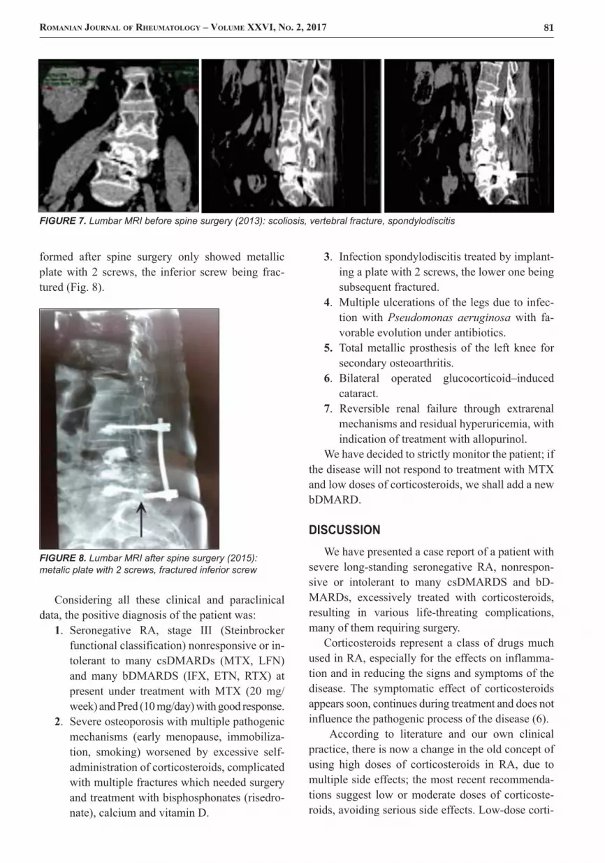
formed after spine surgery only showed metallic plate with 2 screws, the inferior screw being frac-tured (Fig. 8).
FIGURE 8. Lumbar MRI after spine surgery (2015): metalic plate with 2 screws, fractured inferior screw
Considering all these clinical and paraclinical data, the positive diagnosis of the patient was:
1. Seronegative RA, stage III (Steinbrockerfunctional classification) nonresponsive or in-tolerant to many csDMARDs (MTX, LFN)and many bDMARDS (IFX, ETN, RTX) atpresent under treatment with MTX (20 mg/week) and Pred (10 mg/day) with good response.
2. Severe osteoporosis with multiple pathogenicmechanisms (early menopause, immobiliza-tion, smoking) worsened by excessive self-administration of corticosteroids, complicatedwith multiple fractures which needed surgeryand treatment with bisphosphonates (risedro-nate), calcium and vitamin D.
3. Infection spondylodiscitis treated by implant-ing a plate with 2 screws, the lower one beingsubsequent fractured.
4. Multiple ulcerations of the legs due to infec-tion with Pseudomonas aeruginosa with fa-vorable evolution under antibiotics.
5. Total metallic prosthesis of the left knee forsecondary osteoarthritis.
6. Bilateral operated glucocorticoid–inducedcataract.
7. Reversible renal failure through extrarenalmechanisms and residual hyperuricemia, withindication of treatment with allopurinol.
We have decided to strictly monitor the patient; if the disease will not respond to treatment with MTX and low doses of corticosteroids, we shall add a new bDMARD.
DISCUSSIONWe have presented a case report of a patient with
severe long-standing seronegative RA, nonrespon-sive or intolerant to many csDMARDS and bD-MARDs, excessively treated with corticosteroids, resulting in various life-threating complications, many of them requiring surgery.
Corticosteroids represent a class of drugs much used in RA, especially for the effects on inflamma-tion and in reducing the signs and symptoms of the disease. The symptomatic effect of corticosteroids appears soon, continues during treatment and does not influence the pathogenic process of the disease (6).
According to literature and our own clinical practice, there is now a change in the old concept of using high doses of corticosteroids in RA, due to multiple side effects; the most recent recommenda-tions suggest low or moderate doses of corticoste-roids, avoiding serious side effects. Low-dose corti-
FIGURE 7. Lumbar MRI before spine surgery (2013): scoliosis, vertebral fracture, spondylodiscitis
82 ROMANIAN JOURNAL OF RHEUMATOLOGY – VOLUME XXVI, NO. 2, 2017
costeroid therapy (Pred 5-10 mg/day) for a limited period, in combination with standard therapy is con-sidered basic strategy in early RA (in the first 2 years of treatment) (7). The combination therapy of a low dose of corticosteroids with cs or bDMARDs repre-sents the gold standard in most therapeutic protocols involving treatment, according to the concept of T2T, both in early and long-standing RA. Medium and high doses of corticosteroids are used as „bridge therapy“ when starting DMARDs, until these be-come effective. Intra-articular steroid injections also can be effective in reducing local inflammation (8).
Side effects of corticosteroids are more likely to occur when these are used as regular treatment over a longer period of time. This is why physicians try to limit the long-term reliance on corticosteroids for RA treatment. Following a strategy in Ovid Medline search (January 2000 – December 2012) using words like Population (PR), Intervention (GC), comparator (comparison), Outcome (AR) were identified in 1031 papers, 26 observational studies and 6 system-atic reviews of adverse effects of GC. The major side effects consist of bone loss, increased risk of cardio-vascular events and infection, fatigue, increased blood sugar, cataracts, mood disruption including depression, anxiety and insomnia. However the risk appears to be dose-dependent (9).
Osteoporosis is the most serious adverse effects experienced by patients receiving long term cortico-steroid therapy. Bone loss occurs soon after the ini-tiation of corticosteroid treatment (common during the first 6-12 months) and results from a complex mechanism involving osteoblastic suppression and increased bone resorption. Patients on corticosteroid therapy are at increased risk of sustaining fractures and fracture risk is dose-dependent (10). Risk factors for fractures in patients treated with GC are divided into individual factors (high daily doses/cumulative dose increased, age extremes < 15 year > 60 years, postmenopausal status, low pre-existing bone min-eral density, pre-existing fractures from minimal trauma, type of inflammatory disease, prolonged im-mobilization) and general factors (female, white race, low bone mineral index, diet with low intake of calcium and vitamin D, smoking, family history of osteoporosis) (11).
A study of a cohort of patients in RA Danish Reg-istry (DREAM) followed predictors of risk of infec-tion treated with anti-TNF. Thus, after 5 years of follow-up, 128 of 2044 (6.3%) patients developed a serious infection first with a total of 141 serious in-
fections. The incidence rate in the first year after start of TNF inhibiting therapy was 4.57 first serious infections per 100 patient-years and 2.91 per 100 patient-years over 5 years. Age, corticosteroids use, visual analogue scale (VAS) pain, HAQ, tender joint count 28 joints (TJC28) and the presence of comor-bidities were significant predictors for developing a serious infection during TNF inhibiting therapy in the multivariate model (12).
Recently, European League Against Rheumatism (EULAR) recommendations for the managing of RA with synthetic and biological disease-modifying an-tirheumatic drugs – 2016 update, established that short term glucocorticoids should be consider when initiating or changing csDMARDs in different dose regimens and routes of administrations, but should be tapered as rapidly as clinically feasible (13).
The presentation of this case is very important for clinical practice, because it shows both the multiple corticosteroid-induced complications, due to high doses of corticosteroids used as self-medication (skin ulcerations with superimposed infections, se-vere multiple osteoporosis fractures surgically solved with metal implants and bilateral cataract), and the need for adapting the therapeutic strategy, strictly monitoring disease activity and treatment safety.
CONCLUSIONS1. Corticosteroids represent an important and ef-
fective treatment for RA when administeredin correct recommended doses.
2. Rheumatologists should weigh the pros andcons of corticosteroid treatment in the thera-peutic strategy of RA.
3. Excessive doses of corticosteroids for a longperiod bring about complications which maybe even life threatening.
4. Patients should be warned that self-adminis-tration of corticosteroids, as well as anychanges without the doctor‘ s advice, may bedangerous with dramatic results.
5. The correct doses of corticosteroids within thetherapeutic strategy of RA and close monitor-ing of patient proved to give good results.
6. Management of complex cases such as theone presented requires an interdisciplinnaryapproach to solve specific problems by vari-ous specialists.

83ROMANIAN JOURNAL OF RHEUMATOLOGY – VOLUME XXVI, NO. 2, 2017
7. The general practitioner has the obligation tofollow up the patient with RA treated withDMARDs and low doses of corticosteroids in
order to diminish the side effects of an exces-sive and uncontrolled treatment.
1. Kuo C-F., Luo S-F., See L-C. et al. Rheumatoid arthritisprevalence, incidence and mortality rates: a nationwide populationstudy in Taiwan. Rheum Int 2013; 33(2):355-60 .
2. Bijlsma J.W. Optimal treatment of rheumatoid arthritis: EULARrecommendations for clinical practice. Pol Archy Med Wewn 2010Sep; 120(9):347-53.
3. Smolen J.S., Aletaha D., Bijlsma J.W. et al for the T2TExpert Committee. Treating rheumatoid arthritis to target:recommendations of an international task force. Ann Rheum Dis2010;69:631-37
4. Smolen J.S., van der Heijde D., Machold K.P. et al. Proposal fora new nomenclature of disease-modifying antirheumatic drugs. AnnRheum Dis 2014Jan 26;73(1):3-5 .
5. Townsend H.B., Saag K.G. Glucocorticoid use in rheumatoidarthritis: benefists, mechanisms and risks. Clin Exp Rheumatol2004;22(Suppl 35):S77-S82.
6. Bălănescu Andra. Poliartrita reumatoidă în „Esenţialul înreumatologie“ Ruxandra Ionescu (coord), Ediţia I, Ed. Amaltea,Bucureşti, 2006, 214-251.
7. Strand V., Simon L.S. Low dose glucocorticoids in earlyrheumatoid arthritis. Clin Exp Rheumatol 200321 (5 Suppl31):S186-90.
8. Hoes J.N., Jacobs J.W., Buttgereit F., Bijlsma J.W. Current viewof glucocorticoid co-therapy with DMARDs in rheumatoid arthritisNat Rev Rheumatol 2010 6 ( 12): 693-702.
9. Ethgen O., de Lemos Esteves F., Bruvere O., Reginster J.Y.What do we know about the safety of corticosteroids in rheumatoidarthritis?. Curr Med Res Opin 2013, 29 (9): 1147-60.
10. Ionescu Ruxandra. Glucocorticoizii în „Esenţialul în reumatologie“,Ruxandra Ionescu (coord). Ediţia a II-a revizuită. Ed. Amaltea,Bucureşti, 2007. IV: 151-161.
11. Canalis E., Mazziotti G., Giustina A. et al. Glucocorticoid-inducedosteoporosis: pathophysiology and therapy. Osteoporosis Int 2007;18: 1319-22.
12. Van Dartel S.A., Fransen J., Kievit W. et al. Predictors forthe 5-year risk of serious infections in patients with rheumatoidarthritis treated with anti-TNF therapy: a cohort study in the DuchRheumatoid Arthritis Monitoring (DREAM) registry, Rheumatology(Oxford) 2013, 52 (6): 1052-7.
13. Smolen J.S., Landewe R., Bijlsma J. et al. EULARrecommendations for the management of rheumatoid arthritis withsynthetic and biological disease-modifying antirheumatic drugs:2016 update, Ann Rheum Dis, March 2017, http://ard.bmj.com/content/early/2017/03/17/annrheumdis-2016-210715.
REFERENCES
Conflict of interest: none declared Financial support: none declared