Embed Size (px)
Citation preview
Toward a New Generation of Therapeutics 1
Applied Biochemistry and Biotechnology Vol. 128, 2006
Copyright © 2006 by Humana Press Inc.All rights of any nature whatsoever reserved.0273-2289/06/128/0001/$30.00
1
*Author to whom all correspondence and reprint requests should be addressed.
Toward a New Generation of TherapeuticsArtificial Cell Targeted Delivery of Live Cells for Therapy
SATYA PRAKASH* AND CHRISTOPHER MARTONI
Biomedical Technology and Cell Therapy Research Laboratory,Departments of Biomedical Engineering and Artificial Cells and Organs
Research Center, Faculty of Medicine, McGill University,3775 University Street, Montreal, Québec, H3A 2B4, Canada,
E-mail: [email protected]
Received July 27, 2005; Accepted August 9, 2005
Abstract
Scientific evidence in the prevention and treatment of various disorders isaccumulating regarding probiotics. The health benefits supported byadequate clinical data include increased resistance to infectious disease,decreased duration of diarrhea, management of inflammatory bowel dis-ease, reduction of serum cholesterol, prevention of allergy, modulation ofcytokine gene expression, and suppression of carcinogen production. Recentventures in metabolic engineering and heterologous protein expression haveenhanced the enzymatic and immunomodulatory effects of probiotics and,with time, may allow more active intervention among critical care patients.In addition, a number of approaches are currently being explored, includingthe physical and chemical protection of cells, to increase probiotic viabilityand its health benefits. Traditional immobilization of probiotics in gelmatrices, most notably calcium alginate and κ-carrageenan, has frequentlybeen employed, with noted improvements in viability during freezing andstorage. Conflicting reports exist, however, on the protection offered byimmobilization from harsh physiologic environments. An alternativeapproach, microencapsulation in “artificial cells,” builds on immobilizationtechnologies by combining enhanced mechanical stability of the capsulemembrane with improved mass transport, increased cell loading, and greatercontrol of parameters. This review summarizes the current clinical status ofprobiotics, examines the promises and challenges of current immobiliza-tion technologies, and presents the concept of artificial cells for effectivedelivery of therapeutic bacterial cells.
Index Entries: Artificial cells; biotherapy; probiotic; gastrointestinal tract;immobilization; microencapsulation; biopharmaceutics.
2 Prakash and Martoni
Applied Biochemistry and Biotechnology Vol. 128, 2006
Introduction
Probiotics refer to ingestion of live microorganisms for health ben-efits (1). Nearly a century ago, Russian physiologist Eli Metchnikoff pro-posed ingesting fermented milk on a daily basis to maintain a properequilibrium of intestinal microflora (2). More recently, probiotic bacteriahave been the focus of much scientific and commercial interest owing to amyriad of preventative and curative effects. Most probiotics belong to alarge group of bacteria designated as lactic acid bacteria, especially but notexclusively lactobacilli and bifidobacteria, which are important compo-nents of the human gastrointestinal (GI) microflora. In addition, newprobiotics include a variety of different microbes including engineeredmicroorganisms with acquired or enhanced health-promoting properties.The probiotic concept yet remains ineffective partly as a result of viabilitylosses in traditional probiotic products.
Beyond selection of strain, a number of approaches are currently beingexplored to increase viability, including stress adaptation to simulatedenvironments, incorporation of micronutrients, and two-step fermentationprocesses (3). In addition, the physical and chemical protection of cells hasbeen proposed. Immobilization of probiotics in gel matrices, most notablycalcium alginate and κ-carrageenan, is a mild process that has been used toprotect cells from storage and GI transit (4,5). However, many current gel-based entrapment carriers are acid sensitive (6) and provide a suboptimalenvironment for cell proliferation and metabolic activity (7).
An alternative approach for probiotic protection utilizes semiperme-able microcapsules with strong and thin multilayer membrane componentswith specific mass transport properties. The concept of microencapsula-tion in “artificial cells” was first discovered by Chang in the early 1960s forthe encasement of biologic materials such as transplanted cells, enzymes,and adsorbents (8). Since then, numerous other applications have emerged,including the encapsulation of probiotics for oral administration. Thepermselective microcapsule environment has been shown to support cel-lular metabolism and proliferation and to permit higher cell loading. Fur-thermore, the final microcapsule properties, namely the permeability andcomposition of the membrane, can be varied to suit the needs of a particularapplication.
The review that follows focuses on the current clinical status of probiotictherapy, the development of immobilization technologies for the entrap-ment and protection of viable cells, and the introduction of microencapsula-tion in artificial cells as a probiotic delivery vehicle toward designing a newgeneration of therapeutics.
Probiotic Therapy
The human GI tract represents a complex ecosystem in which a finebalance exists between the intestinal microflora and the host. The vast com-munities of microorganisms, representing up to 500 species, are in constant
Toward a New Generation of Therapeutics 3
Applied Biochemistry and Biotechnology Vol. 128, 2006
interaction with their environment, including other bacteria, the gut epi-thelium, and the mucosal immune system (9). Functionally, the indigenousflora has significant metabolic activity and is crucial for maturation of theimmune system and the development of normal intestinal morphology(10). For several decades, probiotic therapy has been stressed as a means tooptimize indigenous flora, enhance the metabolic activity of the GI tractand reinforce the natural defense mechanisms of the host. More recently,probiotic bacteria have been the focus of much scientific and commercialinterest owing to a myriad of preventative and curative effects.
Present Status in Clinical Practice
The consumption of specific probiotic strains is associated with a rangeof clinically relevant health benefits. Placebo-controlled clinical trials haveshown Lactobacillus reuteri, Lactobacillus rhamnosus GG, Lactobacillus casei,and Saccharomyces boulardii to be effective in reducing the duration of acutediarrhea (11,12). L. rhamnosus GG administered to infants reduced the riskof nosocomial diarrhea and rotavirus gastroenteritis (13). Studies by Asoet al. (14) revealed that L. casei Shirota increases the percentage of T-helpercells and natural killer cells in adult patients with colorectal cancer and hasa protective effect on the recurrence of superficial bladder cancer. In addi-tion, select strains of lactobacilli have been shown to significantly suppressintestinal tumors by chemical mutagens (15,16). Lactic acid bacteria havebeen administered to prevent sepsis in patients with severe acute pancre-atitis (9). A randomized study by Rayes et al. (17) involving liver transplantpatients revealed that postoperative infections were significantly reducedby feeding live Lactobacillus plantarum cells in comparison to standardantibiotic treatment (9). As a means of preventing allergy, a randomizedcontrolled study by Lodinova-Zadnikova and Sonnenborn (18) investi-gated the effect of at-birth colonization with nonpathogenic Escherichia coliNissle 1917. Subjects inoculated with the E. coli strain showed significantlyreduced colonization of bacterial pathogens as well as significantly lowerincidence of allergies after 10 and 20 yr in comparison with control subjects(18). Several species of lactobacilli have been implicated in lowering choles-terol. For example, administration of L. plantarum 299v lowered total andlow-density lipoprotein cholesterol levels in a placebo-controlled study (19).
Probiotics have been used as treatment options for managing inflam-matory bowel diseases (IBDs) such as Crohn disease, ulcerative colitis, andpouchitis. Recently, genetically modified food-grade microorganisms havebeen shown to appropriately target therapeutic proteins to the gut mucosa.In an animal model, Lactococcus lactis, genetically engineered to secrete theanti-inflammatory cytokine interleukin-10, was shown to cure or preventexperimental enterocolitis, a model designed to mimic human IBD (20,21).Future research should continue to elucidate live normal or geneticallyengineered probiotics with specific health benefits and, in doing so, isolatemechanistic pathways by which they function (see Table 1). However, the
4 Prakash and Martoni
Applied Biochemistry and Biotechnology Vol. 128, 2006
aforementioned clinical data highlight the important potential of probiotictherapy.
Limiting Factors
Although probiotic therapy shows significant therapeutic potential,there are limitations precluding its use in clinical or supplemental therapy.Probiotic-containing products, such as yogurts and other dairy-based bev-erages, tablets, or health foods, present unique challenges to manufactur-ers. To provide functional properties, the minimum level of viable bacteriais approx 106 CFU/g of product, and the suggested therapeutic dose is108–109 viable cells/d (3). However, the stability of probiotic cultures isoften significantly diminished prior to administration. Many probiotic
Table 1Mechanisms of Action of Select Probiotic Therapiesa
• Enhancement of immune system: Probiotic studies have reported increasedproduction of a vast array of cytokines such as IL-1, IL-2, IL-6, IL-10, IL-12,IL-18, TNF-α, and IFN-γ (22,23). These cytokines are known to exert a range ofimmunomodulatory functions. For example, IL-12 and IL-18 induce IFN-γ pro-duction, which, in turn, enhances phagocyte-mediated clearance of microbesand augments the cytotoxic capacity of T-cells and NK cells. By comparison,TNF-α increases macrophage activity, IL-1 stimulates proliferation of T- andB-cells, and IL-10 possesses potent anti-inflammatory properties (9,24).
• Management of IBD: In most chronic cases, abnormal activation of the mucosalimmune system against the enteric flora is the key event triggering inflamma-tory mechanisms. Probiotic therapy increases the gut IgA immune response,promoting the gut’s immunogenic barrier (25).
• Cancer therapy: Studies in human subjects have revealed that probiotic therapymay reduce the risk of colon cancer by inhibiting transformation of pro-carcinogen to active carcinogens, binding/inactivating mutagenic compounds,suppressing the growth of procarcinogenic bacteria, reducing the absorption ofmutagens from the intestine, and enhancing host immune response (9,26).
• Allergy prevention: Administration of probiotic bacteria has contributed toantiallergy effects by strengthening mucosal defenses (through enhanced gut-specific IgA responses); promoting gut mucosal barrier function; down-regulating inflammatory responses; and increasing the production of TGF-β,IL-10, and cytokines known to produce IgE antibodies (27,28).
• Lowering of cholesterol: Potential mechanisms by which probiotics lower cho-lesterol levels include assimilation of cholesterol by bacterial cells, binding ofcholesterol to bacterial cell walls, and deconjugation of bile salts by the bacterialenzyme bile salt hydrolase. In addition, short-chain fatty acids, the end productof carbohydrate fermentation in the gut, could prompt inhibition of hepaticcholesterol synthesis and/or redistribution of cholesterol from plasma to theliver (29,30).aInitial and ongoing efforts in probiotic research have been focused on studying their
effectiveness in placebo-controlled clinical trials. However, of increasing interest is theidentification of mechanisms of action. IL, interleukin; TNF-α, tumor necrosing factor-α;IFN-γ, interferon-γ; NK, natural killer; TGF-β, transforming growth factor-β.
Toward a New Generation of Therapeutics 5
Applied Biochemistry and Biotechnology Vol. 128, 2006
cultures (e.g., bifidobacteria) are fastidious, noncompetitive, and verysensitive to environmental parameters such as oxygen and low pH. In addi-tion, probiotics in storage cultures encounter inhibitory metabolic byprod-ucts such as lactic and acetic acid, hydrogen peroxide, and bacteriocins(3,31). Tablets incorporating freeze-dried microorganisms prolong shelflife especially with added protectants; however, viability losses are never-theless observed (32).
Orally administered probiotics must also circumvent passage fromthe mouth to the intestine. Physiologic conditions such as acidic pH,mechanical stresses, digestive enzymes (e.g., pepsin and pancreatin), bileacids, and oxygen provide an effective barrier against entry (33,34). Selec-tion of strain for therapy is therefore dependent on the tolerance of theprobiotic to the aforementioned stresses as well as the ability to persist andpotentially colonize the human GI tract. Recently, with the advent of geneticengineering, strategies have been developed to accelerate improvement ofstrains. However, novel microorganisms may present a risk of systemicinfections, deleterious metabolic activities, adjuvant side effects, immuno-modulation, and gene transfer (35). Furthermore, repeated large dosescould result in their replacing the normal intestinal flora (36–39). Therefore,although there have been many publications describing the use of probioticsto prevent and treat GI disorders, safety and efficacy issues have resultedin only a few studies contributing significantly to the knowledge of healtheffects in humans.
Technologies for Improving Probiotic Survival
Beyond selection of strain, a number of approaches are currently beingexplored to increase the viability of probiotics in commercial or experimen-tal products. Stress adaptation to simulated environments; incorporationof micronutrients such as peptides and amino acids; and two-step fermen-tation, whereby probiotic bacteria are permitted to proliferate prior to theaddition of yogurt starter cultures, have proven useful in improvingprobiotic survival (3). In addition, the use of immobilization and micro-encapsulation technologies to retain high cell concentrations during stor-age and gastric transit, whereby the probiotic is separated from itsenvironment by a protective coating, has been proposed.
Immobilization Technologies
Immobilization of whole cells has been defined as the physical con-finement or localization of intact cells to a defined region of space withoutappreciable loss of catalytic activity (40). Currently, a myriad of methodsare available for immobilization of probiotics, with selection of a particularapplication dependent on the nature of the microorganism being immobi-lized as well as the availability and cost of the carrier material. Adsorptionto a preformed carrier and ionic bonding methods are mild and inexpen-
6 Prakash and Martoni
Applied Biochemistry and Biotechnology Vol. 128, 2006
sive, utilizing weak interaction forces to immobilize cells. However, thebeads are highly sensitive to pH and prone to cell leakage (41). By compari-son, covalent binding and crosslinking offer significant improvements instrength over methods employing weak bonds; however, potential toxicityof reagents employed during processing has limited this method’s applica-bility (42). As a result, the physical retention of probiotics has far outnum-bered attempts at binding cells to carriers in food- and medical-basedapplications. Of these, the most widely used immobilization technique isthe entrapment of cells within a gel matrix.
Application of Probiotic Immobilization in Commercialand Experimental Products
Entrapment techniques based on the formation of thermal andionotropic gels have proven useful for probiotic applications. An array ofpolymers, such as agarose, alginate, chitosan, cellulose, gellan, and κ-car-rageenan, have been chosen as the support materials for the gel matrix.These are generally nontoxic, readily available, and acceptable for use asadditives in the food and dairy industries.
Probiotic entrapment by alginate and κ-carrageenan is widelyencountered in the literature. Alginate forms a gel matrix in the presenceof polyvalent cations such as Ca2+ or Ba2+ and κ-carrageenan solidifies onexposure to potassium ions. The mild yet sturdy properties of the gel-likematrix allow the probiotic to remain viable with retention of enzymaticactivity for extended periods of time. A study by Adhikari et al. (43)revealed that bifidobacteria entrapped in κ-carrageenan appeared toincrease their viability in yogurt over a 30-d storage period. Sheu andMarshall (44) observed that lactobacilli immobilized in calcium alginatesurvived 40% better than free cells during the freezing of ice milk. Fur-thermore, Kebary et al. (45) reported that entrapping Bifidobacteriumbifidum and Bifidobacterium infantis in alginate or κ-carrageenan beadsimproved their viability in frozen ice milk over 10 wk. Sultana et al. (46)modified the method of calcium alginate immobilization and found thatincorporation of starch as a prebiotic improved probiotic survival over an8-wk storage period in yogurt. In addition, gel entrapment of lactobacilliand bifidobacteria has been reported to protect probiotic cells from lyo-philization and rehydration (47).
Conflicting reports exist, however, on the protection offered by immo-bilization from gastric conditions. Shah and Ravula (48) reported thatprobiotic bacteria immobilized in calcium alginate were able to survive atpH 2.5. Similarly, when Bifidobacterium longum entrapped in calcium algi-nate beads were exposed to simulated gastric juice and bile salt solution,the death rate of the cells in the beads decreased proportionally with anincrease in both the alginate gel concentration and the bead size (49). Otherstudies, however, indicate that calcium alginate beads are acid sensitive
Toward a New Generation of Therapeutics 7
Applied Biochemistry and Biotechnology Vol. 128, 2006
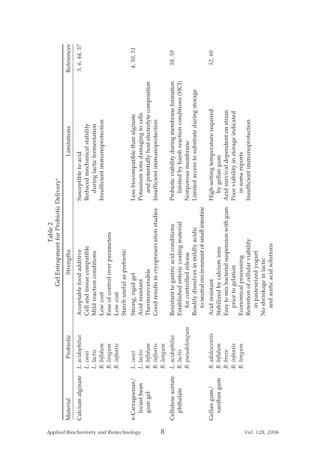
and are not able to prevent cell death at low pH (6). By contrast, κ-carrag-eenan/locust bean gum gel beads have been shown to be more resistant toacidic conditions; however, potassium ions required during processing aredamaging to certain probiotic strains (50,51). Sun and Griffiths (52) reportedthat gellan gum/xanthan gum form spherical beads in the presence ofcalcium ions at room temperature and effectively protect bifidobacteria inyogurt storage and gastric juice. However, survival at acidic pH wasstrongly dependent on the strain employed. Among other formulations,gelatin and polymer-coated gelatin capsules have been studied for oraldelivery of probiotic cells (53,54). The latter, with 20% (w/v) of the poly-mer, has shown promising results in vitro (54). Attempts have been madeat adhering probiotic bacteria to prebiotics (55). The presence of high-amy-lase maize-resistant starch was shown to increase survival of bifidobacteriaat low pH, in bile, and during intestinal transit in mice (56). However,Crittenden et al. (55) reported that adhesion of bifidobacteria to starch issensitive to acid and protease activity and likely would not survive throughthe stomach. A host of other formulations has been proposed for beadentrapment (Table 2); however, general limitations persist.
General Limitations of Immobilization Technologies
Reports on the added protective benefit of gel entrapment in the oraldelivery of probiotics are, as we have described, frequently inconclusive.Although probiotic survival in the gel matrix has been shown to beenhanced in response to environmental stresses (freezing, storage, andsimulated gastric transit), reports are often conflicting and dependent onthe strain employed. It has therefore become difficult to isolate a particularentrapment procedure as a candidate for rigorous optimization studies andeventual scale-up. Support materials for gel entrapment are also insuffi-ciently immunoprotective for novel microorganisms as well as traditionalprobiotics that have shown in certain cases to instigate an inflammatoryresponse (35). In addition, diffusional properties and inadequate mechani-cal strength limit the proliferation of the entrapped probiotic. Encased cellshave been reported to leak; escape from the gel matrix; and, as a result,grow in the surrounding solution (7). Growth in gel beads is restricted,especially in larger beads, in which proliferation occurs only in the periph-ery, owing to substrate limitation. In many cases, the maximum cell loadingof entrapped gel beads is limited to 25% by volume, owing to weakmechanical strength (7). Furthermore, diffusional limitations of both sub-strates and metabolic byproducts can lead to the development of steepgradients regarding pH and inhibitory products that can hinder the viabil-ity and metabolic activity of the entrapped probiotic. Therefore, despitereported advantages, the associated drawbacks of immobilizing probioticsin gel matrices have limited the influence of this technology in commercialand experimental products.
8 Prakash and Martoni
Applied Biochemistry and Biotechnology Vol. 128, 2006
Tab
le 2
Gel
Ent
rap
men
t for
Pro
biot
ic D
eliv
erya
Mat
eria
lP
robi
otic
Stre
ngth
sL
imit
atio
nsR
efer
ence
s
Cal
ciu
m a
lgin
ate
L. a
cido
philu
sA
ccep
tabl
e fo
od a
dd
itiv
eSu
scep
tibl
e to
aci
d5,
6, 4
4, 5
7L.
cas
eiC
ell a
nd ti
ssu
e co
mp
atib
leR
edu
ced
mec
hani
cal s
tabi
lity
L. la
ctis
Mil
d r
eact
ion
cond
itio
nsd
uri
ng la
ctic
fer
men
tati
onB
. bifi
dum
Low
cos
tIn
suff
icie
nt im
mu
nop
rote
ctio
nB
. lon
gum
Eas
e of
con
trol
ove
r p
aram
eter
sB
. inf
anti
sL
ow c
ost
Star
ch u
sefu
l as
pre
biot
ic
κ-C
arra
geen
an/
L. c
asei
Stro
ng, r
igid
gel
Les
s bi
ocom
pat
ible
than
alg
inat
e4,
50,
51
locu
st b
ean
L. la
ctis
Aci
d r
esis
tant
Pot
assi
um
ions
dam
agin
g to
cel
ls g
um
gel
B. b
ifidu
mT
herm
orev
ersi
ble
and
pote
ntia
lly h
ost e
lect
roly
te c
ompo
sitio
nB
. inf
anti
sG
ood
res
ult
s in
cry
opre
serv
atio
n st
ud
ies
Insu
ffic
ient
imm
uno
pro
tect
ion
B. l
ongu
m
Cel
lulo
se a
ceta
teL.
aci
doph
ilus
Res
ista
nt to
gas
tric
aci
d c
ond
itio
nsPr
obio
tic v
iabi
lity
duri
ng m
embr
ane
form
atio
n58
, 59
pht
hala
teB
. lac
tis
Est
abli
shed
ent
eric
coa
ting
mat
eria
lli
mit
ed b
y ha
rsh
reac
tion
con
dit
ions
(H
Cl)
B. p
seud
olon
gum
for
cont
roll
ed r
elea
seN
onp
orou
s m
embr
ane
Rea
dil
y d
isso
lves
in m
ild
ly a
cid
icL
imit
ed a
cces
s to
su
bstr
ate
du
ring
sto
rage
to n
eutr
al e
nvir
onm
ent o
f sm
all i
ntes
tine
Gel
lan
gum
/B
. ado
lesc
enti
sA
cid
res
ista
ntH
igh-
sett
ing
tem
per
atu
re r
equ
ired
52, 6
0 x
anth
an g
um
B. b
ifidu
mSt
abil
ized
by
calc
ium
ions
by g
ella
n gu
mB
. bre
veEa
sy to
mix
bac
teri
al s
uspe
nsio
n w
ith g
umA
cid
su
rviv
al d
epen
den
t on
stra
inB
. inf
anti
sp
rior
to g
elat
ion
Poo
r vi
abil
ity
in s
tora
ge in
dic
ated
B. l
ongu
mE
cono
mic
al p
roce
ssin
gin
som
e re
por
tsR
eten
tion
of
cell
ula
r vi
abil
ity
Insu
ffic
ient
imm
uno
pro
tect
ion
in p
aste
uri
zed
yog
urt
No
shri
nkag
e in
lact
ican
d a
ceti
c ac
id s
olu
tion
s
8
Toward a New Generation of Therapeutics 9
Applied Biochemistry and Biotechnology Vol. 128, 2006
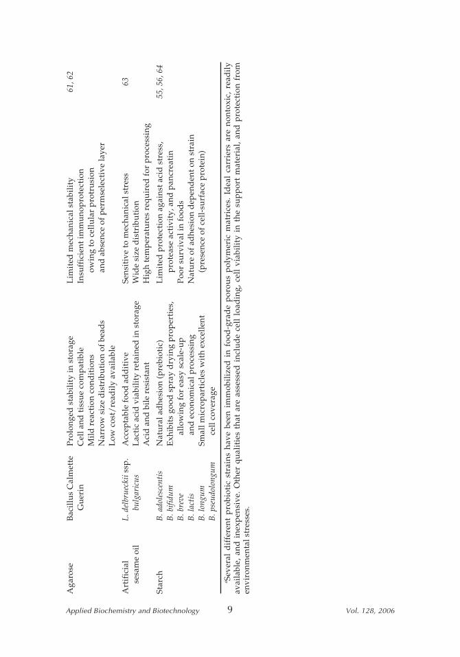
Aga
rose
Bac
illu
s C
alm
ette
Pro
long
ed s
tabi
lity
in s
tora
geL
imit
ed m
echa
nica
l sta
bili
ty61
, 62
Gu
erin
Cel
l and
tiss
ue
com
pat
ible
Insu
ffic
ient
imm
uno
pro
tect
ion
Mil
d r
eact
ion
cond
itio
nsow
ing
to c
ellu
lar
pro
tru
sion
Nar
row
siz
e d
istr
ibu
tion
of
bead
san
d a
bsen
ce o
f p
erm
sele
ctiv
e la
yer
Low
cos
t/re
adil
y av
aila
ble
Art
ific
ial
L. d
elbr
ueck
ii ss
p.
Acc
epta
ble
food
ad
dit
ive
Sens
itiv
e to
mec
hani
cal s
tres
s63
ses
ame
oil
bul
gari
cus
Lac
tic
acid
via
bili
ty r
etai
ned
in s
tora
geW
ide
size
dis
trib
uti
onA
cid
and
bil
e re
sist
ant
Hig
h te
mp
erat
ure
s re
quir
ed f
or p
roce
ssin
g
Star
chB
. ado
lesc
enti
sN
atu
ral a
dhe
sion
(p
rebi
otic
)L
imit
ed p
rote
ctio
n ag
ains
t aci
d s
tres
s,55
, 56,
64
B. b
ifidu
mE
xhib
its
good
sp
ray
dry
ing
pro
per
ties
,p
rote
ase
acti
vity
, and
pan
crea
tin
B. b
reve
allo
win
g fo
r ea
sy s
cale
-up
Poo
r su
rviv
al in
foo
ds
B. l
acti
san
d e
cono
mic
al p
roce
ssin
gN
atu
re o
f ad
hesi
on d
epen
den
t on
stra
inB
. lon
gum
Smal
l mic
rop
arti
cles
wit
h ex
cell
ent
(pre
senc
e of
cel
l-su
rfac
e p
rote
in)
B. p
seud
olon
gum
cell
cov
erag
e
a Sev
eral
dif
fere
nt p
robi
otic
str
ains
hav
e be
en i
mm
obil
ized
in
food
-gra
de
por
ous
pol
ymer
ic m
atri
ces.
Id
eal
carr
iers
are
non
toxi
c, r
ead
ily
avai
labl
e, a
nd i
nexp
ensi
ve.
Oth
er q
ual
itie
s th
at a
re a
sses
sed
inc
lud
e ce
ll l
oad
ing,
cel
l vi
abil
ity
in t
he s
up
por
t m
ater
ial,
and
pro
tect
ion
from
envi
ronm
enta
l str
esse
s.
9
10 Prakash and Martoni
Applied Biochemistry and Biotechnology Vol. 128, 2006
Microencapsulation in Semipermeable Artificial Cells
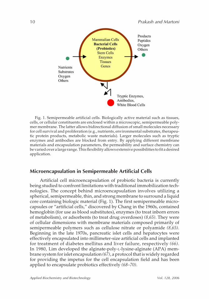
Artificial cell microencapsulation of probiotic bacteria is currentlybeing studied to confront limitations with traditional immobilization tech-nologies. The concept behind microencapsulation involves utilizing aspherical, semipermeable, thin, and strong membrane to surround a liquidcore containing biologic material (Fig. 1). The first semipermeable micro-capsules or “artificial cells,” discovered by Chang in the 1960s, containedhemoglobin (for use as blood substitutes), enzymes (to treat inborn errorsof metabolism), or adsorbents (to treat drug overdoses) (8,65). They wereof cellular dimensions with membrane materials composed primarily ofsemipermeable polymers such as cellulose nitrate or polyamide (8,65).Beginning in the late 1970s, pancreatic islet cells and hepatocytes wereeffectively encapsulated into millimeter-size artificial cells and implantedfor treatment of diabetes mellitus and liver failure, respectively (66).In 1980, Lim developed the alginate-poly-L-lysine-alginate (APA) mem-brane system for islet encapsulation (67), a protocol that is widely regardedfor providing the impetus for the cell encapsulation field and has beenapplied to encapsulate probiotics effectively (68–70).
Fig. 1. Semipermeable artificial cells. Biologically active material such as tissues,cells, or cellular constituents are enclosed within a microscopic, semipermeable poly-mer membrane. The latter allows bidirectional diffusion of small molecules necessaryfor cell survival and proliferation (e.g., nutrients, environmental substrates, therapeu-tic protein products, metabolic waste materials). Larger molecules such as trypticenzymes and antibodies are blocked from entry. By applying different membranematerials and encapsulation parameters, the permeability and surface chemistry canbe varied over a large range. This flexibility allows extensive possibilities to fit a desiredapplication.
Toward a New Generation of Therapeutics 11
Applied Biochemistry and Biotechnology Vol. 128, 2006
Artificial Cell Membrane Design
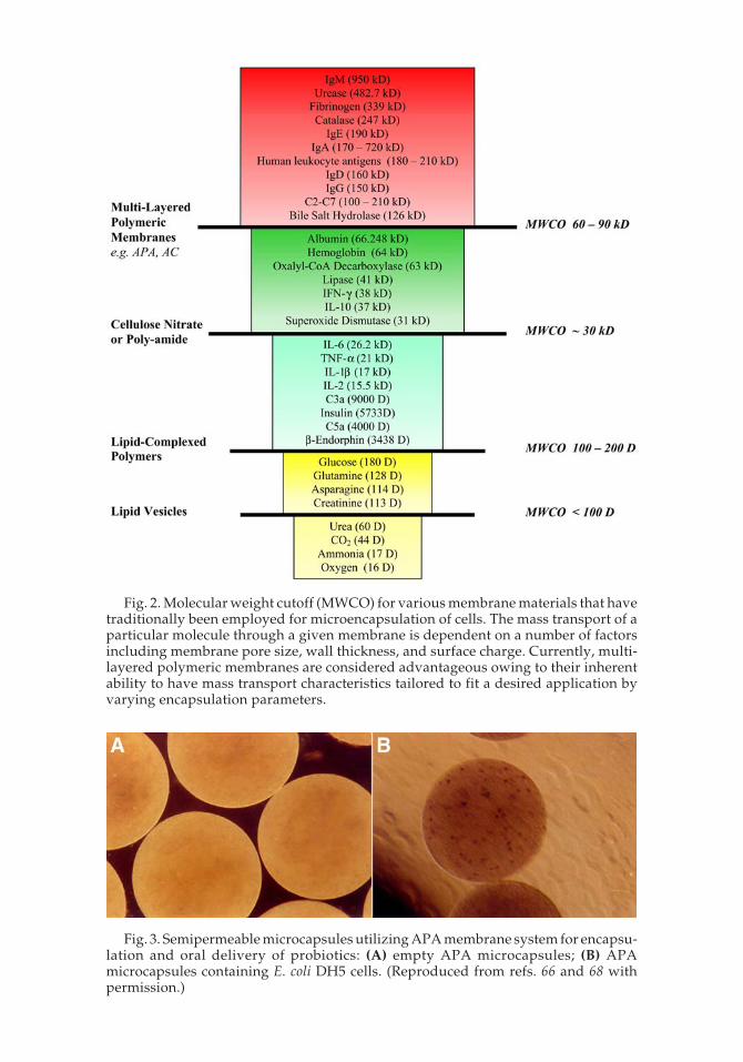
A number of microencapsulation techniques have been developed byindependent laboratories in recent years. The most widely used amongthem is polyelectrolyte complexation, a technique utilizing the interactionof oppositely charged polymers to form a physical membrane around theprobiotic (71). Choice material can be natural or synthetic, with naturalpolymers exhibiting greater cell compatibility and milder conditions(Table 3). Other techniques employ interfacial polymerization, coacerva-tion, or conformal coating for production of single- or multilayered mem-branes that can range in surface chemistry from hydrophilic to lipophilic.The use of different membranes allows variations in permeability, masstransfer, mechanical stability, biocompatibility, and buffering capabilitythat can be exploited to fit a desired application. The mass transport prop-erties of a membrane are critical because the influx rate of molecules, essen-tial for cell survival and proliferation, and the outflow rate of metabolicwaste ultimately determine the viability of encapsulated cells. Frequently,membrane permeability is defined by the molecular weight cutoff, themaximum molecular weight of a molecule that may freely pass through thepores of the capsule membrane (Fig. 2). The molecular weight cutoff oforally delivered microcapsules must allow passage of substrates from theGI tract as well as unwanted metabolites from the plasma and then eitherfacilitate the subsequent removal of the altered molecule or provide for itsstorage. Figure 2 outlines a variety of microcapsule membrane materialsthat have historically been used with their mass transport properties. Theversatility of various microcapsule membranes has been well documented,with many novel formulations coming to fruition today, leading to a rangeof preparation methods for probiotic therapy.
APA Membrane SystemThe well-established interaction between alginate and poly-L-lysine
for the production of APA microcapsules utilizes polyelectrolyte complex-ation. Calcium alginate beads are first prepared according to traditional gelentrapment procedures. Binding of poly-L-lysine to alginate occurs electro-statically by long-chain alkyl amino groups that extend from the polya-mide backbone of poly-L-lysine and interact with carboxyl groups of thecalcium alginate bead. Reexposure of the crosslinked bead to dilute algi-nate neutralizes positively charged poly-L-lysine residues still present atthe capsule surface. The calcium-alginate core is then liquefied by exposingthe freshly made microcapsules to a chelating agent such as sodium citrate.
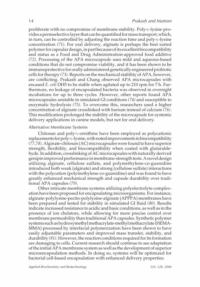
APA microcapsules have been used to encase probiotic bacteria, withseveral advantages noted over traditional immobilization technologies(Fig. 3). The aqueous core provides minimal mass transfer resistance, which,coupled with the large surface area-to-volume ratio of the semipermeablemembrane, allows permeant substrates and products to diffuse rapidly (7).In addition, microorganisms have a larger accessible volume to grow and
12 Prakash and Martoni
Applied Biochemistry and Biotechnology Vol. 128, 200612
Tab
le 3
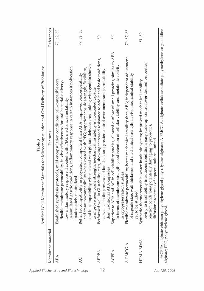
Art
ific
ial C
ell M
embr
ane
Mat
eria
ls f
or M
icro
enca
psu
lati
on a
nd O
ral D
eliv
ery
of P
robi
otic
sa
Mem
bran
e m
ater
ial
Feat
ure
sR
efer
ence
s
AP
AE
stab
lishe
d s
ynth
esis
pro
toco
ls, m
ild e
ncap
sula
tion
con
dit
ions
, cel
l-co
mp
atib
le c
ore,
73, 8
2, 8
3fl
exib
le m
embr
ane
per
mea
bilit
y, in
viv
o ef
fect
iven
ess
for
oral
bac
teri
al d
eliv
ery,
low
infl
amm
ator
y re
spon
se if
coa
ted
wit
h P
EG
, mec
hani
cal i
nsta
bilit
yin
sim
ula
ted
gas
tric
con
dit
ions
, inf
lam
mat
ory
resp
onse
in c
erta
in in
stan
ces
if p
olyc
atio
nin
adeq
uat
ely
neu
tral
ized
AC
Bet
ter
bioc
omp
atib
ility
in p
olyc
atio
n co
mp
onen
t tha
n A
PA
; im
pro
ved
bio
com
pat
ibili
ty77
, 84,
85
and
imm
uno
com
pat
ibili
ty w
hen
coat
ed w
ith
PE
G; s
up
erio
r ca
psu
le s
tren
gth,
fle
xibi
lity,
and
bio
com
pat
ibili
ty w
hen
coat
ed w
ith
glu
tara
ldeh
yde;
cro
sslin
king
wit
h ge
nip
in s
how
nto
imp
rove
mem
bran
e st
reng
th; m
echa
nica
l ins
tabi
lity
in n
onco
ated
cap
sule
AP
PP
AP
erfo
rmed
wel
l in
GI
stab
ility
test
s, s
how
ing
incr
ease
d r
esis
tanc
e to
aci
dic
and
bas
ic c
ond
itio
ns,
80as
wel
l as
in th
e p
rese
nce
of io
n ch
elat
ors;
gre
ater
con
trol
ove
r m
embr
ane
per
mea
bilit
yth
an tr
adit
iona
l AP
A c
apsu
les
AC
PP
ASu
per
ior
to A
PA
and
AC
in c
ell v
iabi
lity
stu
die
s, a
llow
ed o
utf
low
of
smal
l pro
tein
s, s
imila
r to
AP
A86
in te
rms
of m
embr
ane
stre
ngth
, goo
d r
eten
tion
of
cellu
lar
viab
ility
and
met
abol
ic a
ctiv
ity
in c
ryop
rese
rvat
ion
stu
die
sA
-PM
CG
-AFl
exib
le m
embr
ane
per
mea
bilit
y; b
ette
r m
echa
nica
l sta
bilit
y th
an A
PA
; ind
epen
den
t ad
just
men
t79
, 87,
88
of c
apsu
le s
ize,
wal
l thi
ckne
ss, a
nd m
echa
nica
l str
engt
h; in
viv
o m
echa
nica
l sta
bilit
yye
t to
be s
tud
ied
HE
MA
-MM
ASy
nthe
tic,
ther
mor
ever
sibl
e, w
ater
-ins
olu
ble
syst
em; i
mp
rove
d m
echa
nica
l sta
bilit
y81
, 89
owin
g to
inso
lubi
lity
in a
queo
us
cond
itio
ns; e
asy
scal
e-u
p; c
ontr
ol o
ver
des
ired
pro
per
ties
;re
acti
on c
ond
itio
ns p
oten
tial
ly d
amag
ing
to p
robi
otic
s;d
iffu
sion
pro
per
ties
of
aqu
eou
s so
lute
s lim
ited
a AC
PP
A, a
lgin
ate-
chit
osan
-pol
yeth
ylen
e gl
ycol
-pol
y-L-l
ysin
e-al
gina
te; A
-PM
CG
-A, a
lgin
ate-
cell
ulo
se s
ulf
ate-
pol
ymet
hyle
ne-c
o-gu
anid
ine-
algi
nate
; PE
G, p
olye
thyl
ene
glyc
ol.
Toward a New Generation of Therapeutics 13
Applied Biochemistry and Biotechnology Vol. 128, 2006
Fig. 2. Molecular weight cutoff (MWCO) for various membrane materials that havetraditionally been employed for microencapsulation of cells. The mass transport of aparticular molecule through a given membrane is dependent on a number of factorsincluding membrane pore size, wall thickness, and surface charge. Currently, multi-layered polymeric membranes are considered advantageous owing to their inherentability to have mass transport characteristics tailored to fit a desired application byvarying encapsulation parameters.
Fig. 3. Semipermeable microcapsules utilizing APA membrane system for encapsu-lation and oral delivery of probiotics: (A) empty APA microcapsules; (B) APAmicrocapsules containing E. coli DH5 cells. (Reproduced from refs. 66 and 68 withpermission.)
14 Prakash and Martoni
Applied Biochemistry and Biotechnology Vol. 128, 2006
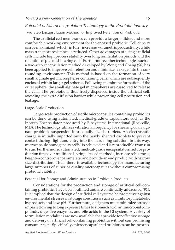
proliferate with no compromise of membrane stability. Poly-L-lysine pro-vides a permselective layer that can be quantified for mass transport, which,in turn, can be controlled by adjusting the reaction time and poly-L-lysineconcentration (71). For oral delivery, alginate is perhaps the best suitedpolymer for capsular design, in part because of its excellent biocompatibilityand status as a Food and Drug Administration-approved food additive(72). Processing of the APA microcapsule uses mild and aqueous-basedconditions that do not compromise viability, and it has been shown to beimmunoprotective for orally administered genetically engineered probioticcells for therapy (73). Reports on the mechanical stability of APA, however,are conflicting. Prakash and Chang observed APA microcapsules withencased E. coli DH5 to be stable when agitated up to 210 rpm for 7 h. Fur-thermore, no leakage of encapsulated bacteria was observed in overnightincubations for up to three cycles. However, other reports found APAmicrocapsules unstable in simulated GI conditions (74) and susceptible toenzymatic hydrolysis (75). To overcome this, researchers used a higherconcentration of alginate crosslinked with barium instead of calcium (76).This modification prolonged the stability of the microcapsule for systemicdelivery applications in canine models, but not for oral delivery.
Alternative Membrane SystemsChitosan and poly-L-ornithine have been employed as polycationic
replacements for poly-L-lysine, with noted improvements in biocompatibility(77,78). Alginate-chitosan (AC) microcapsules were found to have superiorstrength, flexibility, and biocompatibility when coated with glutaralde-hyde. In addition, crosslinking of AC microcapsules with naturally derivedgenipin improved performance in membrane-strength tests. A novel designutilizing alginate, cellulose sulfate, and polymethylene-co-guanidineintroduced both weak (alginate) and strong (cellulose sulfate) interactionswith the polycation (polymethylene-co-guanidine) and was found to havegreatly enhanced mechanical strength and capsule durability over tradi-tional APA capsules (79).
Other intricate membrane systems utilizing polyelectrolyte complex-ation have been proposed for encapsulating microorganisms. For instance,alginate-polylysine-pectin-polylysine-alginate (APPPA) membranes havebeen prepared and tested for stability in simulated GI fluid (80). Resultsindicate increased resistance to acidic and basic conditions, as well as in thepresence of ion chelators, while allowing for more precise control overmembrane permeability than traditional APA capsules. Synthetic polymersystems such as hydroxymethyl methacrylate-methyl methacrylate (HEMA-MMA) processed by interfacial polymerization have been shown to haveeasily adjustable parameters and improved mass transfer, stability, anddurability (81). However, the reaction conditions required for its formationare damaging to cells. Current research should continue to see adaptationof the initial APA membrane system as well as the development of superiormicroencapsulation methods. In doing so, systems will be optimized forbacterial cell-based encapsulation with enhanced delivery properties.
Toward a New Generation of Therapeutics 15
Applied Biochemistry and Biotechnology Vol. 128, 2006
Potential of Microencapsulation Technology in the Probiotic Industry
Two-Step Encapsulation Method for Improved Retention of Probiotic
The artificial cell membranes can provide a larger, milder, and morecomfortable working environment for the encased probiotic. Cell densitycan be maximized, which, in turn, increases volumetric productivity, whilemass transport resistance is reduced. Other advantages of using artificialcells include high process stability over long fermentation periods and theretention of plasmid-bearing cells. Furthermore, other technologies such asa two-step encapsulation method developed by Wong and Chang (90) hasbeen applied to improve cell retention and minimize leakage into the sur-rounding environment. This method is based on the formation of verysmall alginate gel microspheres containing cells, which are subsequentlyenclosed within larger gel spheres. Following membrane formation of theouter sphere, the small alginate gel microspheres are dissolved to releasethe cells. The probiotic is thus freely dispersed inside the artificial cell,avoiding the extra diffusion barrier while preventing cell protrusion andleakage.
Large-Scale Production
Large-scale production of sterile microcapsules containing probioticscan be done using automated, medical-grade encapsulators such as theInotech Encapsulator produced by Biosystems International (Rockville,MD). The technology utilizes vibrational frequency for shearing of an algi-nate-probiotic suspension into equally sized droplets. An electrostaticcharge is initially imparted onto the newly sheared droplets to preventcontact during flight and entry into the hardening solution. In this way,microcapsule homogeneity >95% is achieved and is reproducible from runto run. Furthermore, automated, medical-grade encapsulators reduce pro-duction time over traditional syringe-based methods, increase robustness,heighten control over parameters, and provide an end product with narrowsize distribution. Thus, there is available technology for manufacturinglarge numbers of superior quality microcapsules without compromisingprobiotic viability.
Potential for Storage and Administration in Probiotic Products
Considerations for the production and storage of artificial cell-con-taining probiotics have been outlined and are continually addressed (91).It is implied that the design of artificial cell systems be protective againstenvironmental stresses in storage conditions such as inhibitory metabolicbyproducts and low pH. Furthermore, designers must minimize stressesimparted owing to long exposure times to stomach acid, antimicrobial com-pounds, digestive enzymes, and bile acids in the GI system. A variety offormulation modalities are now available that provide for effective storageand delivery of artificial cell-containing probiotics without compromisingconsumer taste. Specifically, microencapsulated probiotics can be incorpo-
16 Prakash and Martoni
Applied Biochemistry and Biotechnology Vol. 128, 2006
rated into foodstuffs such as fermented milk products (yogurt, ice cream,cheeses) as active ingredients or, alternatively, be dehydrated and incorpo-rated into capsules or pill form with a shelf life up to 2 yr (92). Furthermore,artificial cells can be stored for extended periods at low temperature inminimal solution and subsequently delivered in drink format. Future workwill continue to investigate industrial platforms for storage and adminis-tration, which should increase delivery rates as well as palatability.
Clinical Applications of Artificial Cells Containing Live Bacterial CellsEarly developments of microencapsulated bacterial therapy utilizing
genetically engineered microorganisms with novel or enhanced probioticproperties are emerging. For instance, researchers have developed induc-ible expression promoters for high-level production of heterologous pro-teins. In this way, it is possible to control gene expression in engineeredlactic acid bacteria by an inductor; a repressor; or environmental factorssuch as temperature, pH, or ion concentrations (93). Additionally, one mayexpand the range of possible active components beyond protein therapeu-tics by integrating foreign enzymes.
To accomplish this end, Prakash and Chang (68) developed nonpatho-genic E. coli strain DH5 expressing Klebsiella aerogenes urease for use as anoral artificial kidney substitute. For patients with chronic renal failure,dialysis is conventionally used for the removal of waste metabolites fromplasma; however, it is time-consuming and uncomfortable as well as inac-cessible to low-income households. This study shows that the oral admin-istration of genetically engineered E. coli encased in APA artificial cellsdecomposes urea that diffuses into the capsule from the GI tract intoammonia, which the bacteria can utilize for its biosynthesis (68). Whenorally administered daily to rats with surgically induced renal failure, APAartificial cells containing E. coli reduced plasma urea from 52 to 9 mg andsubsequently maintained the normal level throughout a 21-d experimentalperiod (68). Extrapolation of these results to a 70-kg uremic patient revealsthat approx 4 g of microcapsules containing E. coli DH5 would be requiredfor effective daily therapy, significantly less than alternative urea absorp-tion protocols such as oxystarch or microencapsulated zirconium phos-phate–urease (68).
Recent interest in characterizing oxalate-degrading enzymes inBifidobacterium has given rise to microencapsulating such bacteria for oralprobiotic therapy. Oxalate, a component of fruits and vegetables consumedin normal human diets, represents a highly oxidized organic compoundwith strong chelating capacity and limited potential for catabolic removal(94). As such, accumulation in humans can prove toxic and result in anumber of pathologic conditions including hyperoxaluria, calcium oxalatenephrolithiasis, and cardiomyopathy (95,96). The degradation of intestinaloxalate is dependent on the persistence of oxalate-degrading microorgan-isms in the gut, the most important being Oxalobacter formigenes, whoseintestinal absence has been associated with the aforementioned pathologic
Toward a New Generation of Therapeutics 17
Applied Biochemistry and Biotechnology Vol. 128, 2006
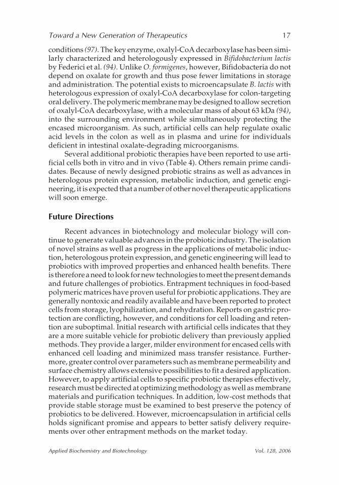
conditions (97). The key enzyme, oxalyl-CoA decarboxylase has been simi-larly characterized and heterologously expressed in Bifidobacterium lactisby Federici et al. (94). Unlike O. formigenes, however, Bifidobacteria do notdepend on oxalate for growth and thus pose fewer limitations in storageand administration. The potential exists to microencapsulate B. lactis withheterologous expression of oxalyl-CoA decarboxylase for colon-targetingoral delivery. The polymeric membrane may be designed to allow secretionof oxalyl-CoA decarboxylase, with a molecular mass of about 63 kDa (94),into the surrounding environment while simultaneously protecting theencased microorganism. As such, artificial cells can help regulate oxalicacid levels in the colon as well as in plasma and urine for individualsdeficient in intestinal oxalate-degrading microorganisms.
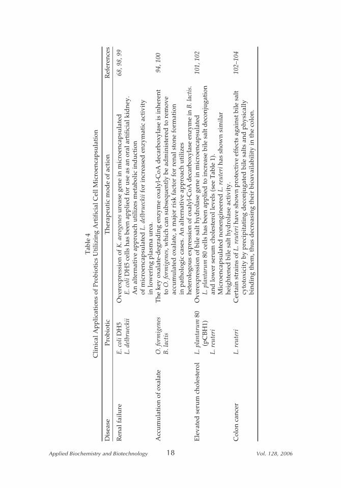
Several additional probiotic therapies have been reported to use arti-ficial cells both in vitro and in vivo (Table 4). Others remain prime candi-dates. Because of newly designed probiotic strains as well as advances inheterologous protein expression, metabolic induction, and genetic engi-neering, it is expected that a number of other novel therapeutic applicationswill soon emerge.
Future Directions
Recent advances in biotechnology and molecular biology will con-tinue to generate valuable advances in the probiotic industry. The isolationof novel strains as well as progress in the applications of metabolic induc-tion, heterologous protein expression, and genetic engineering will lead toprobiotics with improved properties and enhanced health benefits. Thereis therefore a need to look for new technologies to meet the present demandsand future challenges of probiotics. Entrapment techniques in food-basedpolymeric matrices have proven useful for probiotic applications. They aregenerally nontoxic and readily available and have been reported to protectcells from storage, lyophilization, and rehydration. Reports on gastric pro-tection are conflicting, however, and conditions for cell loading and reten-tion are suboptimal. Initial research with artificial cells indicates that theyare a more suitable vehicle for probiotic delivery than previously appliedmethods. They provide a larger, milder environment for encased cells withenhanced cell loading and minimized mass transfer resistance. Further-more, greater control over parameters such as membrane permeability andsurface chemistry allows extensive possibilities to fit a desired application.However, to apply artificial cells to specific probiotic therapies effectively,research must be directed at optimizing methodology as well as membranematerials and purification techniques. In addition, low-cost methods thatprovide stable storage must be examined to best preserve the potency ofprobiotics to be delivered. However, microencapsulation in artificial cellsholds significant promise and appears to better satisfy delivery require-ments over other entrapment methods on the market today.
18 Prakash and Martoni
Applied Biochemistry and Biotechnology Vol. 128, 200618
Tab
le 4
Clin
ical
Ap
plic
atio
ns o
f P
robi
otic
s U
tiliz
ing
Art
ific
ial C
ell M
icro
enca
psu
lati
on
Dis
ease
Pro
biot
icT
hera
peu
tic
mod
e of
act
ion
Ref
eren
ces
Ren
al fa
ilure
E. c
oli D
H5
Ove
rexp
ress
ion
of K
. aer
ogen
es u
reas
e ge
ne in
mic
roen
cap
sula
ted
68, 9
8, 9
9L.
del
brue
ckii
E. c
oli D
H5
cells
has
bee
n ap
plie
d f
or u
se a
s an
ora
l art
ific
ial k
idne
y.A
n al
tern
ativ
e ap
pro
ach
uti
lizes
met
abol
ic in
du
ctio
nof
mic
roen
cap
sula
ted
L. d
elbr
ueck
ii fo
r in
crea
sed
enz
ymat
ic a
ctiv
ity
in lo
wer
ing
pla
sma
ure
a.A
ccu
mu
lati
on o
f ox
alat
eO
. for
mig
enes
The
key
oxa
late
-deg
rad
ing
enzy
me
oxal
yl-C
oA d
ecar
boxy
lase
is in
here
nt94
, 100
B. l
acti
sto
O. f
orm
igen
es, w
hich
can
su
bseq
uen
tly
be a
dm
inis
tere
d to
rem
ove
accu
mu
late
d o
xala
te, a
maj
or r
isk
fact
or f
or r
enal
sto
ne f
orm
atio
nin
pat
holo
gic
case
s. A
n al
tern
ativ
e ap
pro
ach
uti
lizes
hete
rolo
gous
exp
ress
ion
of o
xaly
l-C
oA d
ecar
boxy
lase
enz
yme
in B
. lac
tis.
Ele
vate
d s
eru
m c
hole
ster
olL.
pla
ntar
um 8
0O
vere
xpre
ssio
n of
bile
sal
t hyd
rola
se g
ene
in m
icro
enca
psu
late
d10
1, 1
02 (
pC
BH
1)L.
pla
ntar
um 8
0 ce
lls h
as b
een
app
lied
to in
crea
se b
ile s
alt d
econ
juga
tion
L. r
eute
rian
d lo
wer
ser
um
cho
lest
erol
leve
ls (
see
Tab
le 1
).M
icro
enca
psu
late
d n
onen
gine
ered
L. r
eute
ri h
as s
how
n si
mila
rhe
ight
ened
bile
sal
t hyd
rola
se a
ctiv
ity.
Col
on c
ance
rL.
reu
teri
Cer
tain
str
ains
of L
. reu
teri
hav
e sh
own
pro
tect
ive
effe
cts
agai
nst b
ile s
alt
102–
104
cyto
toxi
city
by
pre
cip
itat
ing
dec
onju
gate
d b
ile s
alts
and
phy
sica
llybi
ndin
g th
em, t
hus
dec
reas
ing
thei
r bi
oava
ilabi
lity
in th
e co
lon.
Toward a New Generation of Therapeutics 19
Applied Biochemistry and Biotechnology Vol. 128, 2006
Acknowledgments
We gratefully acknowledge financial support from the Dairy Farmersof Canada, Natural Sciences and Engineering Research Council of Canada(NSERC), and Canadian Institutes of Health Research (CIHR).
References
1. Roberfroid, M. B. (2000), Am. J. Clin. Nutr. 71, 1682S–1687S.2. Metchnikoff, E. (1908), The Prolongation of Life, G. P. Putnam’s Sons, New York.3. Shah, N. P. (2000), J. Dairy Sci. 83, 894–907.4. Audet, P., Paquin, C., and Lacroix, C. (1988), Appl. Microbiol. Biotechnol. 29, 11–18.5. Chandramouli, V., Kailasapathy, K., Peiris, P., and Jones, M. (2004), J. Microbiol.
Methods 56, 27–35.6. Hansen, L. T., Allan-Wojtas, P. M., Jin, Y. L., and Paulson, A. T. (2002), Food Microbiol.
19, 35–45.7. Park, J. K. and Chang, H. N. (2000), Biotechnol. Adv. 18, 303–319.8. Chang, T. M. S. (1964), Science 146, 524–525.9. Gill, H. S. and Guarner, F. (2004), Postgrad. Med. J. 80, 516–526.
10. Gionchetti, P., Rizzello, F., Venturi, A., and Campieri, M. (2000), J. Gastroenterol.Hepatol. 15, 489–493.
11. Huang, J. S., Bousvaros, A., Lee, J. W., Diaz, A., and Davidson, E. J. (2002), Dig. Dis.Sci. 47, 2625–2634.
12. Szajewska, H. and Mrukowicz, J. Z. (2001), J. Pediatr. Gastroenterol. Nutr. 33(Suppl. 2),S17–S25.
13. Szajewska, H., Kotowska, M., Mrukowicz, J. Z., Armanska, M., and Mikolajczyk, W.(2001), J. Pediatr. 138, 361–365.
14. Aso, Y., Akaza, H., Kotake, T., Tsukamoto, T., Imai, K., and Naito, S. (1995), Eur. Urol.27, 104–109.
15. McIntosh, G. H., Royle, P. J., and Playne, M. J. (1999), Nutr. Cancer 35, 153–159.16. Singh, J., Rivenson, A., Tomita, M., Shimamura, S., Ishibashi, N., and Reddy, B. S.
(1997), Carcinogenesis 18, 833–841.17. Rayes, N., Seehofer, D., Hansen, S., et al. (2002), Transplantation 74, 123–127.18. Lodinova-Zadnikova, R. and Sonnenborn, U. (1997), Biol. Neonate 71, 224–232.19. Bukowska, H., PieczulMroz, J., Chelstowski, K., and Naruszewicz, M. (1997), Athero-
sclerosis 134, 325.20. Steidler, L., Hans, W., Schotte, L., et al. (2000), Science 289, 1352–1355.21. Steidler, L. (2002), Antonie Van Leeuwenhoek 82, 323–331.22. Gill, H. S. (1998), Int. Dairy J. 8, 535–544.23. Meydani, S. N. and Ha, W. K. (2000), Am. J. Clin. Nutr. 71, 861–872.24. Gill, H. S. (2003), Best Pract. Res. Clin. Gastroenterol. 17, 755–773.25. Guarner, F. and Malagelada, J. R. (2003), Best Pract. Res. Clin. Gastroenterol. 17,
793–804.26. Van’t Veer, P., Dekker, J., Lamers, J., et al. (1989), Cancer Res. 49, 4020–4023.27. Kirjavainen, P. V., Salminen, S. J., and Isolauri, E. (2003), J. Pediatr. Gastroenterol. Nutr.
36, 223–227.28. Matricardi, P. M., Bjorksten, B., Bonini, S., et al. (2003), Allergy 58, 461–471.29. Gilliland, S. E., Nelson, C. R., and Maxwell, C. (1985), Appl. Environ. Microbiol. 49,
377–381.30. Schaafsma, G., Meuling, W. J., van Dokkum, W., and Bouley, C. (1998), Eur. J. Clin.
Nutr. 52, 436–440.31. Godward, G. and Kailasapathy, K. (2003), Milchwissenschaft-Milk Sci. Int. 58, 396–399.32. Wang, Y. C., Yu, R. C., and Chou, C. C. (2004), Int. J. Food Microbiol. 93, 209–217.33. Holzapfel, W. H., Haberer, P., Snel, J., Schillinger, U., and Huis in’t Veld, J. H. J.
(1998), Int. J. Food Microbiol. 41, 85–101.
20 Prakash and Martoni
Applied Biochemistry and Biotechnology Vol. 128, 2006
34. Huang, Y. and Adams, M. C. (2004), Int. J. Food Microbiol. 91, 253–260.35. Salminen, S., von Wright, A., Morelli, L., et al. (1998), Int. J. Food Microbiol. 44, 93–106.36. Lu, L. and Walker, W. A. (2001), Am. J. Clin. Nutr. 73, 1124S–1130S.37. Saavedra, J. M. and Tschernia, A. (2002), Br. J. Nutr. 87, S241–S246.38. Vitini, E., Alvarez, S., Medina, M., Medici, M., de Budeguer, M. V., and Perdigon, G.
(2000), Biocell 24, 223–232.39. Chin, J., Turner, B., Barchia, I., and Mullbacher, A. (2000), Immunol. Cell Biol. 78, 55–66.40. Karel, S. F., Libicki, S. B., and Robertson, C. R. (1985), Chem. Eng. Sci. 40, 1321–1354.41. Witter, L. (1996), in Physical Chemistry of Food Processes, van Nostrand Reinhold,
New York, pp. 475–486.42. Babu, P. S., Panda, T., and Babu, M. K. M. (1991), Enzyme Microb. Technol. 13, 676–682.43. Adhikari, K., Mustapha, A., Grun, I. U., and Fernando, L. (2000), J. Dairy Sci. 83, 1946–
1951.44. Sheu, T. Y. and Marshall, R. T. (1993), J. Food Sci. 58, 557–561.45. Kebary, K. M. K., Hussein, S. A., and Badawi, R. M. (1998), Egypt. J. Dairy Sci. 26,
319–337.46. Sultana, K., Godward, G., Reynolds, N., Arumugaswamy, R., Peiris, P., and
Kailasapathy, K. (2000), Int. J. Food Microbiol. 62, 47–55.47. Kim, Y. D., Morr, C. V., and Schenz, T. W. (1996), J. Agric. Food Chem. 44, 1308–1313.48. Shah, N. P. and Ravula, R. R. (2000), Aust. J. Dairy Technol. 55, 139–144.49. Lee, K. Y. and Heo, T. R. (2000), Appl. Environ. Microbiol. 66, 869–873.50. Audet, P., Paquin, C., and Lacroix, C. (1990), Appl. Microbiol. Biotechnol. 32, 662–668.51. Audet, P., Paquin, C., and Lacroix, C. (1991), Biotechnol. Techniques 5, 307–312.52. Sun, W. and Griffiths, M. W. (2000), Int. J. Food Microbiol. 61, 17–25.53. Narayani, R. and Rao, K. P. (1993), Int. J. Pharmaceutics 95, 85–91.54. Narayani, R. and Rao, K. P. (1995), J. Biomater. Sci.-Polym. Ed. 7, 39–48.55. Crittenden, R., Laitila, A., Forssell, P., et al. (2001), Appl. Environ. Microbiol. 67, 3469–
3475.56. Wang, X., Brown, I. L., Evans, A. J., and Conway, P. L. (1999), J. Appl. Microbiol. 87,
631–639.57. Prevost, H. and Divies, C. (1992), Biotechnol. Lett. 14, 583–588.58. Favaro-Trindade, C. S. and Grosso, C. R. (2002), J. Microencapsul. 19, 485–494.59. Rao, A. V., Shiwrarain, N., and Maharaj, I. (1989), Can. Inst. Food Sci. Technol. J. 22,
345–349.60. Sanderson, G. R. (1990), in Food Gels, Elsevier, New York, pp. 201–233.61. Esquisabel, A., Hernandez, R. M., Igartua, M., Gascon, A. R., Calvo, B., and Pedraz,
J. L. (2002), J. Microencapsul. 19, 237–244.62. Losgen, H., Brunner, G., Holloway, C. J., et al. (1978), Biomater. Med. Devices Artif.
Organs 6, 151–173.63. Hou, R. C., Lin, M. Y., Wang, M. M., and Tzen, J. T. (2003), J. Dairy Sci. 86, 424–428.64. O’Riordan, K., Andrews, D., Buckle, K., and Conway, P. (2001), J. Appl. Microbiol. 91,
1059–1066.65. Chang, T. M. S., MacIntosh, F. C., and Mason, S. G. (1966), Can. J. Physiol. Pharmacol.
44, 115–128.66. Chang, T. M. S. and Prakash, S. (1998), Mol. Med. Today 4, 221–227.67. Lim, F. and Sun, A. M. (1980), Science 210, 908–910.68. Prakash, S. and Chang, T. M. S. (1996), Nat. Med. 2, 883–887.69. Prakash, S. and Chang, T. M. S. (1999), Artif. Cells Blood Substitutes Immobilization
Biotechnol. 27, 475–481.70. Prakash, S. and Chang, T. M. S. (2000), Int. J. Artif. Organs 23, 429–435.71. Uludag, H., de Vos, P., and Tresco, P. A. (2000), Adv. Drug Deliv. Rev. 42, 29–64.72. Strand, B. L., Ryan, L., Veld, P. I., et al. (2001), Cell Transplant. 10, 263–275.73. Gugerli, R., Cantana, E., Heinzen, C., von Stockar, U., and Marison, I. W. (2002),
J. Microencapsul. 19, 571–590.74. Ma, X. J., Vacek, I., and Sun, A. (1994), Artif. Cells Blood Substitutes Immobilization
Biotechnol. 22, 43–69.
Toward a New Generation of Therapeutics 21
Applied Biochemistry and Biotechnology Vol. 128, 2006
75. Quong, D., Yeo, J. N., and Neufeld, R. J. (1999), J. Microencapsul. 16, 73–82.76. Petruzzo, P., Cappai, A., Ruiu, G., Dessy, E., Rescigno, A., and Brotzu, G. (1997),
Transplant. Proc. 29, 2129–2130.77. Bartkowiak, A. and Hunkeler, D. (1999), Bioartif. Organs Technol., Med., Mater. 875,
36–45.78. Calafiore, R., Basta, G., Luca, G., et al. (1999), Ann. NY Acad. Sci. 875, 219–232.79. Wang, T., Lacik, I., Brissova, M., et al. (1997), Nat. Biotechnol. 15, 358–362.80. Ouyang, W., Chen, H., Jones, M. L., et al. (2004), J. Pharm. Pharmaceut. Sci. 7, 315–324.81. Tse, M., Uludag, H., Sefton, M. V., and Chang, P. L. (1996), Biotechnol. Bioeng. 51,
271–280.82. Awrey, D. E., Tse, M., Hortelano, G., and Chang, P. L. (1996), Biotechnol. Bioeng. 52,
472–484.83. Strand, B. L., Gaserod, O., Kulseng, B., Espevik, T., and Skjak-Baek, G. (2002), J. Micro-
encapsul. 19, 615–630.84. Bartkowiak, A. and Hunkeler, D. (2000), Chem. Mater. 12, 206–212.85. Bartkowiak, A. (2001), Ann. NY Acad. Sci. 944, 120–134.86. Haque, T., Chen, H., Ouyang, W., et al. (2005), Mol. Pharm. 2, 29–36.87. Lacik, I., Brissova, M., Anilkumar, A. V., Powers, A. C., and Wang, T. (1998), J. Biomed.
Mater. Res. 39, 52–60.88. Rehor, A., Canaple, L., Zhang, Z., and Hunkeler, D. (2001), J. Biomater. Sci. Polym. Ed.
12, 157–170.89. Crooks, C. A., Douglas, J. A., Broughton, R. L., and Sefton, M. V. (1990), J. Biomed.
Mater. Res. 24, 1241–1262.90. Wong, H. and Chang, T. M. S. (1991), Biomater. Artif. Cells Immobilization Biotechnol.
19, 687–697.91. Siuta-Cruce, P. and Goulet, J. (2001), Food Technol. 55, 36–42.92. Institut Rosell-Lallemand (2002), Newsletter Number 2, Institut Rosell-Lallemand,
Montreal, QC, Canada.93. Steidler, L. (2003), Best Pract. Res. Clin. Gastroenterol. 17, 861–876.94. Federici, F., Vitali, B., Gotti, R., et al. (2004), Appl. Environ. Microbiol. 70, 5066–5073.95. Rodby, R. A., Tyszka, T. S., and Williams, J. W. (1991), Am. J. Med. 90, 498–504.96. Williams, H. E. and Smith, L. H., Jr. (1968), Am. J. Med. 45, 715–735.97. Goldkin, L., Cave, D., Jaffin, B., Robinson, W., and Bliss, C. (1985), Am. J. Gastroenterol
80, 860–865.98. Chow, K. M., Liu, Z. C., Prakash, S., and Chang, T. M. S. (2003), Artif. Cells Blood
Substitutes Immobilization Biotechnol. 31, 425–434.99. Prakash, S. and Chang, T. M. S. (1995), Biotechnol. Bioeng. 46, 621–626.
100. Duncan, S. H., Richardson, A. J., Kaul, P., Holmes, R. P., Allison, M. J., and Stewart,C. S. (2002), Appl. Environ. Microbiol. 68, 3841–3847.
101. De Smet, I., Vanhoorde, L., Desaeyer, N., Vandewoestyne, M., and Verstraete, W.(1994), Microbial. Ecol. Health Dis. 7, 315–329.
102. Jones, M., Chen, H., Ouyang, W., Metz, T., and Prakash, S. (2004), J. Biomed. Biotechnol.1, 61–69.
103. De Boever, P., Wouters, R., Verschaeve, L., Berckmans, P., Schoeters, G., andVerstraete, W. (2000), Appl. Microbiol. Biotechnol. 53, 709–714.
104. De Smet, I., Vanhoorde, L., Woestyne, M. V., Christiaens, H., and Verstraete, W.(1995), J. Appl. Bacteriol. 79, 292–301.


